Introduction
Legionella are aerobic, environmental,
gram-negative bacteria accounting for 61 different strains and
approximately 70 serogroups; some of these strains are able to
induce severe pathological manifestations in humans. The name
'Legionella' derives from a group of veterans of the
American Legion that in 1976 contracts the infection during a stay
in a Hotel in Philadelphia where an outbreak of Legionella
was present in the air conditioning system. This outbreak caused 34
deaths among 221 infected, however, only one year later the
causative agent of infections was recognized and named as
Legionella pneumophila(L. pneumophila) (1).
L. pneumophila accounts for 35 different
serogroups of which L. pneumophila serogroup 1 is the most
frequently identified in Legionella infections (2). There are two clinical manifestations
of legionellosis: the first form is a self-limiting disease, called
Pontiac fever, that does not imply lung involvement, while the
second form is called Legionnaires' disease that causes a pneumonia
characterized by fever (in some cases even higher than 40°C),
chills, coughs, chest pain and in some cases by extrapulmonary
symptoms such as diarrhea, nausea, vomiting, and neurological
manifestations (1).
Legionellosis is a global health problem; according
to the data provided by the European Epidemiological Report of the
European Center for Disease Prevention and Control (ECDC) in 2017,
9,238 cases were reported, approximately 30% more infections than
recorded in 2016 (3). An
increasing trend was also found in Italy, where in 2017, 2,014
cases were reported, corresponding to a rate of 33.2 cases per
million inhabitants, compared to the previous year where the
incidence was 28.2 cases per million inhabitants. The mortality
rate calculated on all cases with known outcome is 10.1% (4).
As mentioned, Legionella is widely spread in
nature, in both natural and artificial water habitats. In
particular, water systems, cooling towers and hydric pipeline are
the ideal environment for its proliferation representing potential
reservoir of infection. In addition, Legionella prefers warm
habitats with ideal temperature ranging from 25°C to 42°C (under
20°C, Legionella survives in a latent state). On this basis,
it is possible to identify areas that due to the recirculation of
hot water, such as water heaters, whirlpools, boilers, etc. can
represent critical points of contamination (1,5,6).
In these favorable environments the conditions of development are
optimal for the constant proliferation of Legionella and are
potentially favorable also for biofilm formation (7,8).
L. pneumophila is not transmissible from
person to person but occurs after inhalation of contaminated
aerosol droplets (9).
Therefore, the monitoring and management of all
infrastructures and water systems are extremely important. Indeed,
some operating conditions or structural characteristics of
pipelines and water heaters can favor the growth and spread of
Legionella. Guidelines have been issued in order to propose
standard methods for the monitoring of legionellosis. Such
guidelines contain the methods and indications necessary to
guarantee levels of acceptability of Legionella in the
putative sites at risk of contamination. In particular, various
methods of prevention and control of contamination are indicated,
such as water chlorination and heat treatment. A key point of
existing guidelines is the time point of checks that have to be
performed periodically in order to assess the effectiveness of the
treatments or to establish the need to carry out further
interventions (10,11).
As reported in the ISO 11731:2017 protocol, the main
method for detecting Legionella in environmental samples is
represented by the culture-based method that allows to detect the
presence and quantity of bacteria present in the analyzed samples
(11). However, this method
presents some problems, represented by rather long waiting times
due to the low growth rate of L. pneumophila. Furthermore,
in some cases, the culture method is impractical as the growth of
Legionella can be inhibited by the presence of other
bacteria (12), making it
necessary to pre-treat the sample in order to avoid growth
inhibition or bacterial contamination (13,14).
More recently, molecular identification methods have
been proposed to overcome the main problems of culture-based
methods. Among these molecular techniques, reverse
transcription-quantitative polymerase chain reaction (RT-qPCR)
allows the identification of Legionella in environmental
samples faster than culture methods (15-17). In addition, this method has a
greater sensitivity than the cultural examination, however, RT-qPCR
is not able to distinguish between viable and non-viable organisms
(18).
It is evident that the possibility of having a rapid
evaluation of the number of live or dead microorganisms present in
water samples is of fundamental importance for the maintenance of
public health. The use of highly sensitive methods especially in
high-risk environment, such as hospitals, hospice or nursing homes
for the elderly, are fundamental in order to avoid outbreak of
legionellosis. Indeed, in hospitals the limits established by law
indicate that the microbial load of Legionella is below 102
CFU/l, especially in those Departments with immunosuppressed
patients (e.g., Intesive Care Units, Medical Oncology Units, etc.)
where the total absence of L. pneumophila is mandatory
(10). Therefore, in these
high-risk environments it is of fundamental importance to implement
effective strategies to monitor and control the onset of possible
contamination. In this context, culture-based methods fail to
promptly identify possible outbreaks of infection due to the low
growth rate of L. pneumophila. Therefore, there is an urgent
need to standardize novel methods for the early and rapid
identification of contaminated sites.
On these bases, the aim of the present study is to
propose droplet digital PCR (ddPCR) as a novel high-sensitive
method for the rapid detection of L. pneumophila. For this
purpose, ddPCR and RT-qPCR were used for the detection of L.
pneumophila in an in vitro model of water tank
contaminated with a known concentration of L. pneumophila.
The sensitivity and accuracy of ddPCR and RT-qPCR were also tested
by simulating water heat shock treatments in order to assess and
validate the clinical application of ddPCR for the early diagnosis
of Legionellosis.
Materials and methods
Bacterial strain and culture
conditions
L. pneumophila serotype 1 was obtained from
an environmental contaminated water site. Serotype 1 was identified
by using lactic test (Oxoid, Cambridge, UK). L. pneumophila
was grown on liquid broth and then on GVPC medium (Oxoid),
corresponding to buffered charcoal yeast extract medium plus
antimicrobial agents, at 37°C in a 5% CO2-enriched
atmosphere for 5 days. After growth, single colonies were
resuspended in sterile water until a turbidity of 0.5 McFarland
(1.5×10⁸ CFU/ml) was obtained.
L. pneumophila dilution and DNA
extraction
A dilution of 107 CFU/ml was obtained
starting from a concentration of 1.5×10⁸ CFU/ml. Then,
10-fold serial dilutions in sterile water were performed until a
concentration of 10 CFU/ml was obtained. The 10-fold serial
dilutions were used to assess the sensitivity of both methods.
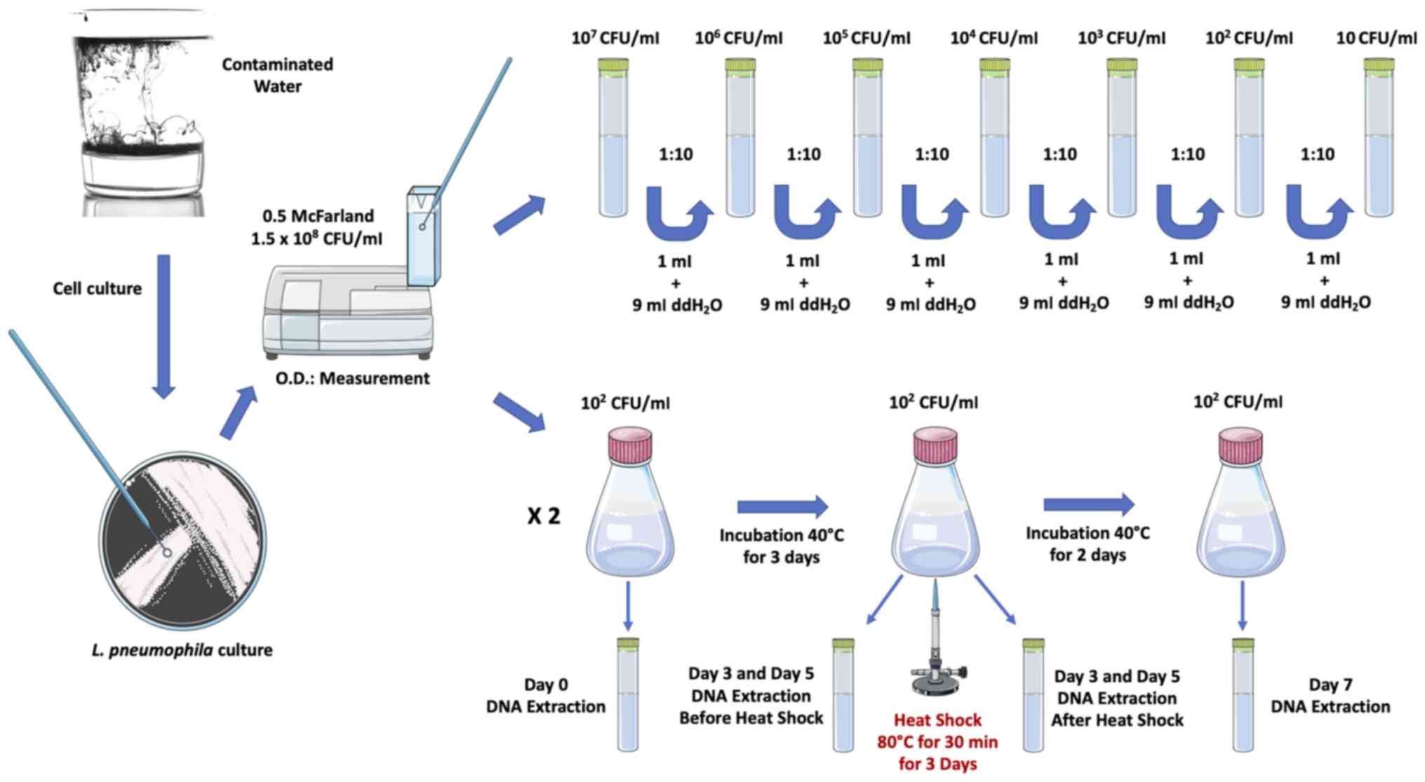
In parallel, L. pneumophila was seeded into
two flasks with sterile water with a final concentration
102 CFU/ml. One of the two flasks was subjected to
thermal shock at 80°C for 30 min for 3 consecutive days in order to
kill L. pneumophila. Different samples were obtained at
different time points from the two flasks in order to assess the
efficacy of thermal shock and the sensitivity of both ddPCR and
RT-qPCR (Fig. 1).
Then, 1 ml of each dilution or sample obtained from
the two flasks was extracted using the PureLink Genomic DNA Mini
kit extraction kit following the manufacturer's instructions (cat.
no. K1820-01; Invitrogen; Thermo Fisher Scientific, Inc.). The
extracted DNA was quantified by using spectrophotometric assay
(Nanodrop 1000; Thermo Fisher Scientific, Inc.).
L. pneumophila RT-qPCR and ddPCR
amplification
Extracted DNA (4.7 µl) was amplified by using
both SYBR-Green RT-qPCR and EvaGreen ddPCR.
For RT-qPCR, the Luminaris Color HiGreen qPCR Master
Mix, high ROX (Thermo Fisher Scientific, Inc.) was used according
to the manufacturer's protocol. L. pneumophila were
amplified with a 7300 Real-Time PCR System (Applied Biosystems;
Thermo Fisher Scientific, Inc.) using the following primer pairs
and thermal conditions: forward: AGGGTTGATAGGTTAAGAGC; reverse:
CCAACAGCTAGTTGACATCG; RT-qPCR thermal profile: UDG pre-treatment at
50°C for 2 min, followed by an initial denaturation step at 95°C
for 10 min and a 3-step PCR program at 95°C for 15 sec, 60°C for 30
sec and 72°C for 30 sec, for 40 cycles. The selected primers are
specific for all L. pneumophila serogroups.
For EvaGreen ddPCR, the reaction mix was prepared by
using 11 µl of 2X QX200™ ddPCR™ EvaGreen Supermix (cat. no.
1864034; Bio-Rad Laboratories, Inc.), 0.15 µl of 20
µM forward and reverse primers (same primers used for
RT-qPCR), 6 µl of RNase and DNase free-water and 4.7
µl of cDNA in order to obtain a final volume of 22
µl.
Twenty microliters of the reaction mix were used to
generate droplets with the QX200 droplet generator (Bio-Rad
Laboratories, Inc.). After generation, the droplets were
transferred into a 96-well plate, sealed and amplified in a C1000
Thermal Cycler (Bio-Rad Laboratories, Inc.) under the following
thermal conditions: polymerase activation at 95°C for 10 min, 40
cycles of amplification at 94°C for 30 sec (denaturation) and 60°C
for 1 min (annealing/elongation), droplets stabilization at 98°C
for 10 min followed by an infinite hold at 4°C. A ramp rate of
2°C/sec was used among the steps of the amplification.
Statistical analysis
The ddPCR data were statistically analyzed by using
the QuantaSoft software provided by Bio-Rad Laboratories, Inc.
Linear regression analysis was performed by using GraphPad Prism
V.6 (GraphPad Software, Inc.). P<0.05 was considered to indicate
a statistically significant difference. All experiments were
performed in triplicate.
Results
Comparison of specificity of ddPCR and
RT-qPCR
The serial dilutions obtained were used to assess
the sensitivity of both ddPCR and RT-qPCR systems by using EvaGreen
and SYBR-Green technologies, respectively. The obtained results
showed that both methods are sensitive enough to detect the
presence of L. pneumophila at concentrations established by
law (10 CFU/ml, i.e., 102 CFU/l if the standard protocol
for L. pneumophila detection starting from a 1 liter
filtered water sample is used) (10). However, the signal related to the
sample diluted at 10 CFU/ml was obtained at a very late Ct value
(31.10) when RT-qPCR is used, while ddPCR effectively detects as
positive the signals obtained for the same concentration (Table I; Fig. S1).
 | Table IRT-qPCR Ct values and ddPCR L.
pneumophila absolute quantification. |
Table I
RT-qPCR Ct values and ddPCR L.
pneumophila absolute quantification.
| No. | RT-qPCR
| ddPCR
|
|---|
| Sample | Ct | Sample |
Copies/µl |
|---|
| 1 | 107
CFU/ml | 11.26 | 107
CFU/ml | 2,298 |
| 2 | 106
CFU/ml | 15.02 | 106
CFU/ml | 1,762 |
| 3 | 105
CFU/ml | 17.75 | 105
CFU/ml | 913 |
| 4 | 104
CFU/ml | 22.62 | 104
CFU/ml | 91.60 |
| 5 | 103
CFU/ml | 25.64 | 103
CFU/ml | 9.30 |
| 6 | 102
CFU/ml | 28.06 | 102
CFU/ml | 1.40 |
| 7 | 10 CFU/ml | 31.10 | 10 CFU/ml | 0.29 |
| 8 | NTC | - | NTC | - |
Noteworthy, the absolute quantification performed by
ddPCR showed that the copies/µl obtained for each sample
better reflects the 10-fold serial dilutions performed, except for
the concentration of 107 CFU/ml that was underestimated
due to the high number of positive droplets that saturated the
ddPCR system (Fig. S1A and B).
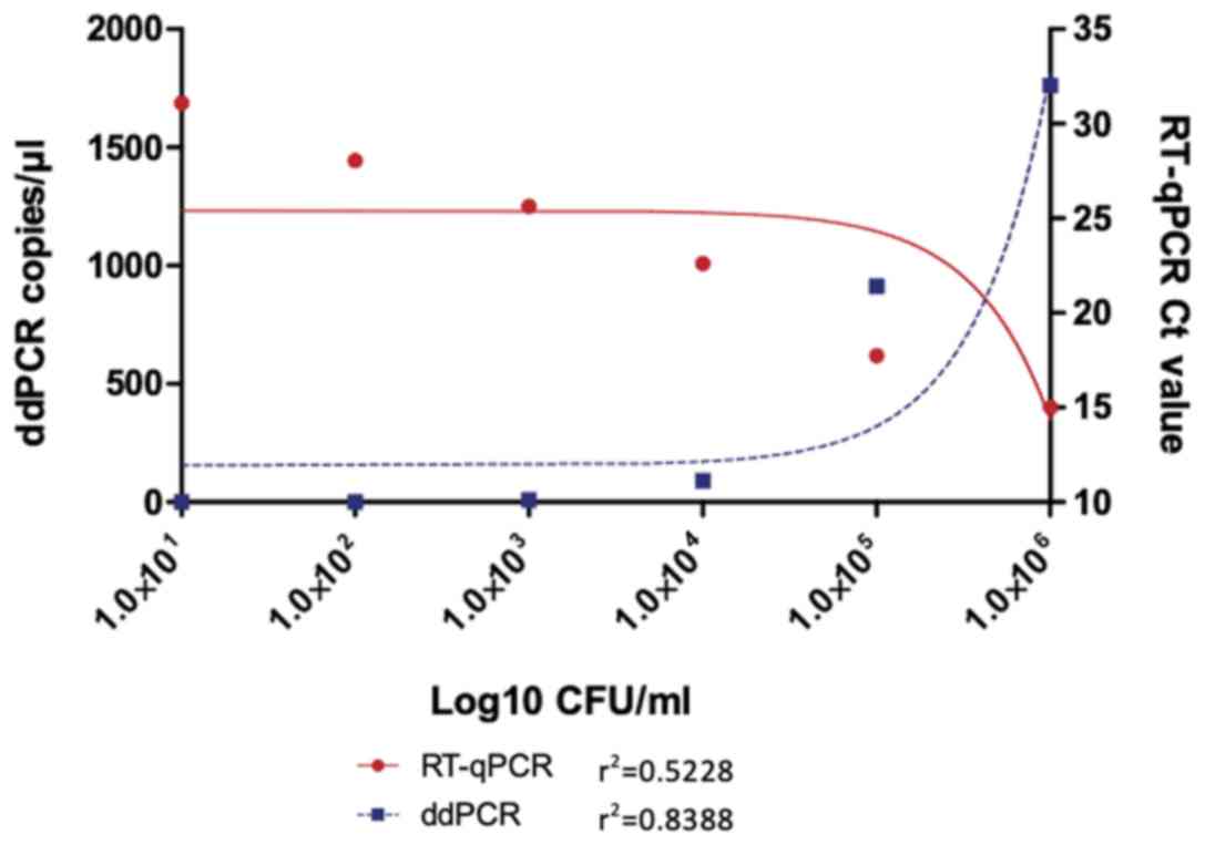
In addition, linear regression analysis showed that ddPCR has a
greater accuracy and robustness compared to RT-qPCR. By excluding
the 107 CFU/ml concentration that saturated both ddPCR
and RT-qPCR systems, linear regression analysis revealed that ddPCR
has a better r2 coefficient compared to RT-qPCR
(r2=0.8388 vs. r2=0.5228) (Fig. 2).
ddPCR shows higher accuracy than RT-qPCR
in monitoring the efficacy of thermal shock
Although ddPCR and RT-qPCR showed similar
sensitivity in the detection of L. pneumophila, the
simulation of heat shock treatment in an in vitro
contaminated water tank highlighted the important differences
existing between methods. Indeed, RT-qPCR detected false-positive
signals in the sample treated at 80°C for three days probably due
to cell debris and residual degraded DNA that produced a
nonspecific amplification signal. In addition, no significant
variation was observed between the untreated samples after one day
and three days of growth (Ct values of 22.00 and 23.06,
respectively) (Table II;
Fig. S2).
 | Table IIRT-qPCR Ct values before and after
heat shock treatment. |
Table II
RT-qPCR Ct values before and after
heat shock treatment.
| No. | Sample | RT-qPCR Ct |
|---|
| 1 | 102
CFU/ml day 1 no shock | 29.72 |
| 2 | 102
CFU/ml day 3 no shock | 28.51 |
| 3 | 102
CFU/ml day 3 post shock | - |
| 4 | 102
CFU/ml day 5 no shock | 25.35 |
| 5 | 102
CFU/ml day 5 post shock | 35.18a |
| 6 | 102
CFU/ml day 7 post shock | - |
| 7 | NTC | - |
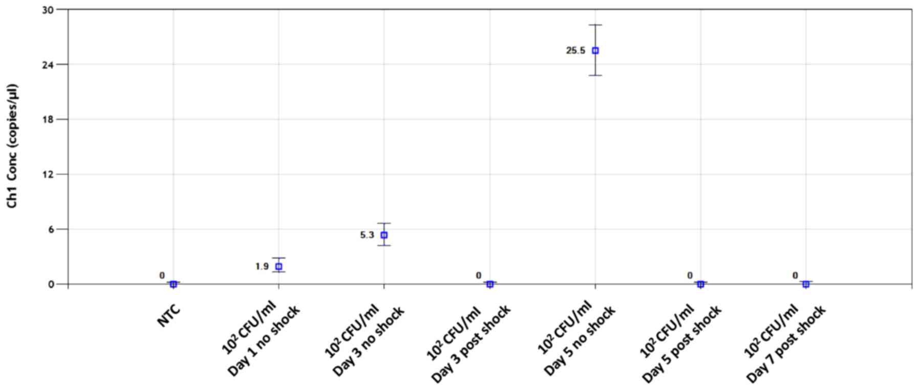
On the contrary, ddPCR effectively identify as
positive all the untreated samples and as negative the samples
shocked at 80°C both after one day and three days of treatment.
Moreover, ddPCR finely detected slight variation in the number of
L. pneumophila after one day, three days and five days of
growth without thermal treatments. In particular, after three days
of incubation the number of copies increased from 1.9 to 5.3
copies/µl, while after 5 days of incubation the
concentration increased at 25.5 copies/µl thus passing from
an initial concentration of 102 CFU/ml to a
concentration of 2.7×10³ CFU/ml (Fig. 3). Of note, no nonspecific signals
were observed for the sample treated at 80°C for three days,
suggesting that ddPCR is less prone to interference from degraded
DNA or cellular debris.
Discussion
Different studies have demonstrated the higher
sensitivity of ddPCR compared to RT-qPCR (19,20). At present, ddPCR is one of the
most sensitive methods used for the detection of low amounts of
targets, including circulating DNA, microRNAs, circulating
mutations, rare copy number variants, low viral nucleic acid
targets representing a promising technology for use in clinical
practice and in public and environmental health (21-24). Several studies have tried to
propose and validate RT-qPCR-based molecular methods for the
detection of L. pneumophila in contaminated water samples or
for the diagnosis of Legionellosis in patients with suspected
pneumonia, however, the sensitivity of the technique and the
presence of inhibitor or contaminants may produce false-positive
and false-negative results (18,25,26). In order to overcome the
limitations of RT-qPCR and to propose novel effective methods for
L. pneumophila identification, here we compared the
sensitivity and accuracy of ddPCR compared to RT-qPCR in detecting
low levels of L. pneumophila and in monitoring the efficacy
of water treatments.
The results here obtained demonstrated that both
ddPCR and RT-qPCR have a good sensitivity, however, high-sensitive
RT-qPCR detected low concentration of L. pneumophila at a
very late Ct value (Ct 31.10 for 10 CFU/ml concentration). On the
contrary, ddPCR accurately identified low concentrations of L.
pneumophila allowing absolute quantification of the bacterial
load.
Moreover, interesting data were obtained by using
both RT-qPCR and ddPCR for the evaluation of the efficacy of heat
shock treatment. Indeed, RT-qPCR detected a false-positive sample
after three days of heat treatments probably due to the presence of
L. pneumophila cell debris and fragmented DNA that produced
a nonspecific signal while ddPCR recognized as negative all the
heat shock treated samples. Furthermore, the results obtained
showed that ddPCR may be used also to detect weak increment of
bacterial load in very limited time frames. Actually, ddPCR
precisely detected the increase of L. pneumophila
concentration after 3 and 5 days of growth without heat shock
treatments. In particular, the initial concentration of 1.9
copies/µl reached a concentration of 5.3 copies/µl
(more than 2.5-fold higher) after three days of growth, an increase
that had not been identified by RT-qPCR. After 5 days of incubation
L. pneumophila reached a concentration of 25.5
copies/µl. These results suggest that ddPCR may be used for
the frequent monitoring of water samples before and after clean-up
treatments in order to detect early L. pneumophila growth
without waiting the long time necessary for the culture-based
methods.
On the basis of our results, the present study
represents the starting point for future analyses performed in both
water samples and human samples obtained from patients with
suspected Legionellosis in order to validate the clinical
application of ddPCR for the early and effective detection of L.
pneumophila.
Of note, the present study represents an in
vitro simulation of L. pneumophila growth and treatment,
therefore, it is subjected to some limitations. In particular, here
we took into account only L. pneumophila. It is known that
in environmental or human samples there are several bacteria which,
together with cellular debris and degraded DNA, can interfere with
the correct detection of Legionella. However, although this
represents a limit for culture methods and for molecular methods
based on RT-qPCR, here we demonstrated that ddPCR is not affected
by the presence of fragmented DNA or cell debris thanks to the
nanopartitions of gene targets and the dilutions of contaminants
into thousands of droplets. In this context, other studies support
our findings and the use of ddPCR for the detection of bacterial
DNAemia during infection or for the monitoring of bacterial load in
contaminated samples with PCR inhibitors (27,28).
In conclusion, overall, the results of the present
study strongly support the adoption of ddPCR for the effective
detection of L. pneumophila in water samples and for the
constant monitoring of bacterial load in sites considered at risk,
such as the hospital environment. Although this study represents
only an in vitro simulation of the Legionella growth,
the results obtained encourage the use of ddPCR also in the
clinical setting for the evaluation of patients with suspected
Legionellosis.
In particular, ddPCR would allow the early detection
of any L. pneumophila increment in the contaminated site or
in patients thus establishing the efficacy of water and antibiotic
treatments, respectively. In this context, the use of ddPCR could
have important impacts from both health and socio-economic points
of view allowing the reduction of the long times necessary to
diagnose L. pneumophila with the standard culture methods.
Accordingly, through ddPCR analysis, the structure, whether public
or private hospital, will more effectively monitor possible
contamination and the efficacy of treatments thus restarting more
rapidly their activities with a significant economic benefit.
Supplementary Data
Funding
No funding was received.
Availability of data and materials
The data generated and analyzed during the current
study are available from the corresponding author on reasonable
request.
Authors' contributions
ML, DAS, MS and LF conceived the study. LF, GG, CMG,
GL and CL were involved in the methodologies used and in the
validation of methods. LF, GG, CMG, GL and CL performed the formal
and statistical analyses. LF and GG wrote the manuscript. ML, DAS
and MS revised the manuscript draft. All authors have read and
agreed to the published version of the manuscript.
Ethics approval and consent to
participate
Not applicable.
Patient consent for publication
Not applicable.
Competing interests
DAS is the Editor-in-Chief for the journal, but had
no personal involvement in the reviewing process, or any influence
in terms of adjudicating on the final decision, for this article.
The other authors declare that they have no competing
interests.
Acknowledgments
This study was supported by the Italian League
Against Cancer (LILT).
References
|
1
|
Cunha BA, Burillo A and Bouza E:
Legionnaires' disease. Lancet. 387:376–385. 2016. View Article : Google Scholar
|
|
2
|
Stout JE and Yu VL: Legionellosis. N Engl
J Med. 337:682–687. 1997. View Article : Google Scholar : PubMed/NCBI
|
|
3
|
European Centre for Disease Prevention and
Control: Legionnaires' Disease in Europe, Annual Epidemiological
Report for 2017. ECDC; Stockholm: 2019
|
|
4
|
Istituto Superiore di Sanità (ISS):
Rapporto Annuale sulla Legionellosi in Italia Nel 2017. 31. ISS;
Rome: 2018
|
|
5
|
Bartram J, Chartier Y, Lee JV, Pond K and
Surman-Lee S: Legionella and the prevention of legionellosis. World
Health Organization Press; Geneva: 2007
|
|
6
|
Fliermans CB, Soracco RJ and Pope DH:
Measure of Legionella pneumophila activity in situ. Curr Microbiol.
6:89–94. 1981. View Article : Google Scholar
|
|
7
|
Abdel-Nour M, Duncan C, Low DE and Guyard
C: Biofilms: The stronghold of Legionella pneumophila. Int J Mol
Sci. 14:21660–21675. 2013. View Article : Google Scholar : PubMed/NCBI
|
|
8
|
Liu Z, Lin YE, Stout JE, Hwang CC, Vidic
RD and Yu VL: Effect of flow regimes on the presence of Legionella
within the biofilm of a model plumbing system. J Appl Microbiol.
101:437–442. 2006. View Article : Google Scholar : PubMed/NCBI
|
|
9
|
van Heijnsbergen E, Schalk JA, Euser SM,
Brandsema PS, den Boer JW and de Roda Husman AM: Confirmed and
potential sources of Legionella reviewed. Environ Sci Technol.
49:4797–4815. 2015. View Article : Google Scholar : PubMed/NCBI
|
|
10
|
Linee guida per la prevenzione ed il
controllo della legionellosi: Conferenza Stato-Regioni nella seduta
del 7 maggio 2015. http://www.salute.gov.it/portale/documentazione/p6_2_2_1.jsp?lingua=italiano&id=2362uri.
Accessed May 13, 2015.
|
|
11
|
ISO 11731:2017: Water Quality-Enumeration
of Legionella. International Organization for Standardization;
Geneva: 2017, https://www.iso.org/standard/61782.htmluri.
|
|
12
|
Kimura S, Tateda K, Ishii Y, Horikawa M,
Miyairi S, Gotoh N, Ishiguro M and Yamaguchi K: Pseudomonas
aeruginosa Las quorum sensing autoinducer suppresses growth and
biofilm production in Legionella species. Microbiol Read.
155:1934–1939. 2009. View Article : Google Scholar
|
|
13
|
Leoni E and Legnani PP: Comparison of
selective procedures for isolation and enumeration of Legionella
species from hot water systems. J Appl Microbiol. 90:27–33. 2001.
View Article : Google Scholar : PubMed/NCBI
|
|
14
|
Ta AC, Stout JE, Yu VL and Wagener MM:
Comparison of culture methods for monitoring Legionella species in
hospital potable water systems and recommendations for
standardization of such methods. J Clin Microbiol. 33:2118–2123.
1995. View Article : Google Scholar : PubMed/NCBI
|
|
15
|
Touron-Bodilis A, Pougnard C,
Frenkiel-Lebossé H and Hallier-Soulier S: Usefulness of real-time
PCR as a complementary tool to the monitoring of Legionella spp.
and Legionella pneumophila by culture in industrial cooling
systems. J Appl Microbiol. 111:499–510. 2011. View Article : Google Scholar : PubMed/NCBI
|
|
16
|
Bonetta S, Bonetta S, Ferretti E, Balocco
F and Carraro E: Evaluation of Legionella pneumophila contamination
in Italian hotel water systems by quantitative real-time PCR and
culture methods. J Appl Microbiol. 108:1576–1583. 2010. View Article : Google Scholar
|
|
17
|
Edagawa A, Kimura A, Doi H, Tanaka H,
Tomioka K, Sakabe K, Nakajima C and Suzuki Y: Detection of
culturable and nonculturable Legionella species from hot water
systems of public buildings in Japan. J Appl Microbiol.
105:2104–2114. 2008. View Article : Google Scholar
|
|
18
|
Lee S and Lee J: Outbreak investigations
and identification of Legionella in contaminated water. Methods Mol
Biol. 954:87–118. 2013. View Article : Google Scholar
|
|
19
|
Arvia R, Sollai M, Pierucci F, Urso C,
Massi D and Zakrzewska K: Droplet digital PCR (ddPCR) vs
quantitative real-time PCR (qPCR) approach for detection and
quantification of Merkel cell polyomavirus (MCPyV) DNA in formalin
fixed paraffin embedded (FFPE) cutaneous biopsies. J Virol Methods.
246:15–20. 2017. View Article : Google Scholar : PubMed/NCBI
|
|
20
|
Hayden RT, Gu Z, Ingersoll J, Abdul-Ali D,
Shi L, Pounds S and Caliendo AM: Comparison of droplet digital PCR
to real-time PCR for quantitative detection of cytomegalovirus. J
Clin Microbiol. 51:540–546. 2013. View Article : Google Scholar :
|
|
21
|
Filetti V, Falzone L, Rapisarda V,
Caltabiano R, Eleonora Graziano AC, Ledda C and Loreto C:
Modulation of microRNA expression levels after naturally occurring
asbestiform fibers exposure as a diagnostic biomarker of
mesothelial neoplastic transformation. Ecotoxicol Environ Saf.
198:1106402020. View Article : Google Scholar : PubMed/NCBI
|
|
22
|
Battaglia R, Palini S, Vento ME, La
Ferlita A, Lo Faro MJ, Caroppo E, Borzì P, Falzone L, Barbagallo D,
Ragusa M, et al: Identification of extracellular vesicles and
characterization of miRNA expression profiles in human blastocoel
fluid. Sci Rep. 9:842019. View Article : Google Scholar : PubMed/NCBI
|
|
23
|
Salemi R, Falzone L, Madonna G, Polesel J,
Cinà D, Mallardo D, Ascierto PA, Libra M and Candido S: MMP-9 as a
candidate marker of response to BRAF inhibitors in melanoma
patients with BRAFV600E mutation detected in circulating-free DNA.
Front Pharmacol. 9:8562018. View Article : Google Scholar :
|
|
24
|
Falzone L, Musso N, Gattuso G, Bongiorno
D, Palermo CI, Scalia G, Libra M and Stefani S: Droplet digital PCR
as the best sensitive assay for the SARS-CoV-2 detection. Int J Mol
Med. 46:957–964. 2020. View Article : Google Scholar : PubMed/NCBI
|
|
25
|
Krøjgaard LH, Krogfelt KA, Albrechtsen HJ
and Uldum SA: Detection of Legionella by quantitative-polymerase
chain reaction (qPCR) for monitoring and risk assessment. BMC
Microbiol. 11:2542011. View Article : Google Scholar : PubMed/NCBI
|
|
26
|
Shen SM, Chou MY, Hsu BM, Ji WT, Hsu TK,
Tsai HF, Huang YL, Chiu YC, Kao ES, Kao PM, et al: Assessment of
Legionella pneumophila in recreational spring water with
quantitative PCR (Taqman) assay. Pathog Glob Health. 109:236–241.
2015. View Article : Google Scholar : PubMed/NCBI
|
|
27
|
Ziegler I, Lindström S, Källgren M,
Strålin K and Mölling P: 16S rDNA droplet digital PCR for
monitoring bacterial DNAemia in bloodstream infections. PLoS One.
14:e02246562019. View Article : Google Scholar : PubMed/NCBI
|
|
28
|
Singh G, Sithebe A, Enitan AM, Kumari S,
Bux F and Stenström TA: Comparison of droplet digital PCR and
quantitative PCR for the detection of Salmonella and its
application for river sediments. J Water Health. 15:505–508. 2017.
View Article : Google Scholar : PubMed/NCBI
|


