Introduction
Cardiovascular disease is the leading cause of
mortality worldwide and includes coronary, congenital and rheumatic
heart, as well as peripheral arterial disease and chronic heart
failure (CHF) (1). Pathological
myocardial remodeling is a necessary stage of CHF that refers to
the constant adjustment of the structure and function of the heart
under the influence of endogenous and/or exogenous factors
(2,3). The pathogenesis of pathological
myocardial remodeling remains unclear. Therefore, it is important
to understand the pathogenesis of myocardial remodeling as this may
help to develop more efficient treatment strategies to improve
myocardial remodeling. In recent years, epigenetic modification has
also been shown to be involved in the occurrence and development of
a variety of cardiovascular diseases (4). Epigenetic modification is comprises
three processes, namely DNA methylation, histone modification
(including methylation, acetylation, phosphorylation,
ubiquitination, and SUMOylation), and ncRNAs-based mechanisms
(including microRNAs-miRNAs, long non-coding RNAs-lncRNAs, and
circular RNAs-circRNAs) (5). It is
increasingly recognized that epigenetic gene regulation serves an
active role in pathological myocardial remodeling (6,7). Our
previous studies confirmed that an imbalance of histone H3K9ac
acetylation is involved in the early stages of pathological
myocardial remodeling induced by pressure overload and is mediated
by histone acetylase (HAT) (8-10).
In addition, the association between histone acetylation and both
cardiac hypertrophy and heart failure have been extensively studied
worldwide (11,12). Histone acetylation modification is
regulated by HATs and histone deacetylases (HDACs); different
subtypes of HATs and HDACs may serve different roles in
pathological myocardial remodeling (13,14).
The natural compound epigallocatechin gallate (EGCG)
is an active polyphenolic catechin and accounts for ~59% of total
catechins from the leaves of green tea (15). EGCG has been extensively studied as
a bioactive dietary component against various types of carcinomas
through multiple mechanisms such as antioxidation, induction of
apoptosis, inhibition of angiogenesis and metastasis (16). The role of EGCG in cardiovascular
disease has been studied, however, the specific mechanism of EGCG
remains unknown (17,18). An increasing number of preclinical
and clinical studies indicate that EGCG, a green tea extract,
regulate histone acetylation modified by HDACs (19,20).
The HDAC family comprises 18 members divided into four classes
based on their structure and function; HDAC5 is a class II HDAC and
is involved in cardiac hypertrophy (21,22).
Furthermore, the abnormal expression of HDAC5 has been investigated
during pathological myocardial remodeling in a mouse model of
transverse aortic constriction (TAC) (23). However, whether EGCG can improve
myocardial remodeling in the heart of TAC mice via HDAC5-mediated
histone acetylation remains unknown. The present study investigated
the effect of HDAC5-mediated histone acetylation on pathological
myocardial remodeling in a mouse model of TAC to identify novel
strategies to prevent and treat CHF.
Materials and methods
Animal treatment
A total of 120 pathogen-free Kunming male mice
(weight, 25-30 g; age, 8-10 weeks) were purchased from the Animal
Center of Zunyi Medical University (Guizhou, China). All animal
experiments were approved by the Animal Protection and Use
Committee of Zunyi Medical University and complied with Directive
2010/63/EU of the European Parliament. Animal studies were
performed in the Physiology Research Laboratories of Zunyi Medical
University, Guizhou, China. The mice were maintained in
individually ventilated cages (25°C and 55-65% humidity) with a
12/12-h light/dark cycle and free access to standard laboratory
mouse chow and water. Mice were exposed to pressure-overload by
thoracic aortic banding (TAB); this procedure decreased the
thoracic aortic diameter by 70%, as previously described (24,25).
Prior to TAB surgery, 0.8% sodium pentobarbital (50 mg/kg) was
intraperitoneally injected into each mouse for general anesthesia.
Anesthesia was confirmed by the disappearance of the righting and
pedal withdrawal reflex. Mice received pre- and post-operative
analgesia (6 h post-surgery) via subcutaneous injection of 0.02 ml
(0.3 mg/ml) buprenorphine hydrochloride solution (Merck KGaA), as
previously described (26)
Additional analgesia was administered as required. Mice were
randomly divided into five groups (n=24/group): Normal, Sham,
Vehicle (Veh), TAC and TAC + EGCG. For each animal, the left side
of the chest was opened along the median of the sternum to expose
the aortic arch. The transverse aortic arch was then ligated
between the brachial and left common carotid arteries. Prior to
ligation, a curved needle (diameter, ~0.4 mm) was used to narrow
the vessel. Finally, the chest was sutured. The postoperative
survival rate of TAC mice was 60-70%. After 4, 8 and 12 weeks of
TAC, the hearts of experimental mice were collected to evaluate the
effect of HDAC5-mediated modification on histone H3K27ac
acetylation during myocardial remodeling induced by pressure
overload. In addition, the effect of EGCG on myocardial remodeling
induced by pressure overload was investigated, and different
concentrations (0, 25, 50, 75 and 100 mg/kg) EGCG were used to
screen the optimal dose of EGCG. Following the completion of TAB,
each mouse received an intraperitoneal injection of EGCG (Selleck
Chemicals; PubChem CID 65064) at a dose of 50 mg/kg/day for 12
weeks. Mice in the Sham group received surgery but were not
ligated. In the Veh group, normal saline was injected
intraperitoneally at the same dose and timepoints beginning 1 week
after sham operation for 12 weeks. After 12 weeks, mice were
sacrificed using CO2 overdose; death was verified by
applying pressure on the mouse nail bed (toe-pinch reflex) and the
heart from each animal was collected for analysis.
Stereoscopy and cardiac and lung mass
index
The heart and lungs of mice were collected, and
cardiac (heart weight/body weight) and lung mass index (lung
weight/body weight) were tested. Subsequently, the morphology of
hearts and lungs of mice were observed by stereoscope.
Hematoxylin and eosin (H&E) and
Masson’s staining
Following harvesting, the heart tissue was fixed in
4% paraformaldehyde for 48 h, embedded in paraffin wax and cut into
serial sections (4 µm thick). Following dewaxing and
dehydration in a series of alcohol concentrations, the sections
were washed, soaked with tap water for 5 min and then stained with
0.5% hematoxylin dye for 5 min (to highlight the nucleus), faded in
1% hydrochloric acid with ethanol for 5 min and then placed in 0.5%
eosin solution for 2 min (Beijing Solarbio Scince & Technology
Co., Ltd.). Additional sections were washed with distilled water
and then stained with 0.7% Masson Lichun red acid compound red
solution for 5 min (Beijing Solarbio Science & Technology Co.,
Ltd.). The sections were then stained in 2% glacial acetic acid
solution for 3 min. Next, sections were differentiated with 1%
phosphomolybdate aqueous solution for 2 min, stained with 0.5%
aniline blue for 5 min and then soaked with 0.2% glacial acetic
acid aqueous solution for 1 min. H&E- and Masson’s-stained
sections were successively treated with 95 and 100% ethanol and
xylene I and II, then sealed with neutral resin. Sections were
observed under a fluorescence microscope (H&E, magnification,
×4; Masson’s, magnification, ×40). All processes were completed at
room temperature.
Echocardiography measurement
Transthoracic echocardiograms were performed on
experimental mice using a Vevo 770 High-Resolution echocardiograph
(Visual Sonics), as described previously (27,28).
Blood flow velocity was measured using p-mode images. The efficacy
of TAC was detected by chest Doppler echocardiography 4 weeks after
the operation.
Chromatin immunoprecipitation (ChIP)
ChIP trials were performed using a ChIP Assay kit
(Merck KGaA). First, a glass homogenizer was used to prepare
homogenate from heart tissue. This homogenate was then mixed with
formaldehyde (1%) to cross-link DNA/protein complexes at room
temperature for 15 min. After cross-linking, 1% SDS Lysis Buffer
(cat. no. 20-163, Merck KGaA) was added and then the DNA was
fragmented by sonication. Following centrifugation at 100 × g for 2
min for 30 min at 4°C with rotation, pre-cleared solution was used
as a DNA input control. A total of 1,800 µl ChIP Dilution
Buffer (cat. no. 20-153; Merck KGaA) was added to 200 µl
sonicated cell supernatant for a final volume of 2 ml. Next, the
protein-DNA complex was precipitated using monoclonal antibodies
(anti-MEF2A; 4 µl, 1:500, cat. no. sc-17785; Santa Cruz
Biotechnology, Inc.). The antibody-chromatin complexes were
precipitated by overnight incubation with Protein A Agarose/Salmon
Sperm DNA (60 µl) at 4°C. The agarose pellet was obtained by
centrifugation (1,500 × g) at 4°C for 1 min. The immunoprecipitated
complexes of Ab-protein-DNA were collected and washed with
low-salt, high-salt and LiCl buffer and Tris-EDTA, and then
buffered with an elution buffer. The cross-linking of protein-DNA
complexes was reversed by incubation with 5 M NaCl at 65°C for 4 h,
and the DNA was digested with proteinase K for 1 h at 45°C. A DNA
Fragment Purification kit (Merck KGaA) was used to extract the DNA
in accordance with the manufacturer’s instructions. All experiments
included a positive (precipitated by anti-RNA polymerase II
antibody) and a negative control (precipitated by normal mouse IgG)
groups. Quantitative (q)PCR was performed after ChIP detection
using ChIP Assay kit (Merck KGaA). SYBR-Green Master Mix II (Takara
Biotechnology Co., Ltd.) was used to perform qPCR. The
thermocycling conditions were as follows: Initial denaturation 95°C
for 5 min, followed by 40 cycles of denaturation at 95°C for 15 sec
and annealing/elongation at 58°C for 30 sec. β-actin was
used as an internal reference and the 2−ΔΔCq method was
used to determine relative gene expression (29). Lertpa-v1.0 software (Applied
Biosystems, Thermo Fisher Scientific, Inc.) was used to analyze the
results. The following primers were used for RT-qPCR: ANP
forward, 5′-TCC TTG GTG TCT CTC GCT CT-3′ and reverse, 5′-CGC TGG
CTT GCT TGT TGT A-3′; BNP forward, 5′-TGC TGT CCC TCT ATG
CTT CC-3′ and reverse, 5′-CGC TGG CTT GCT TGT TGT A-3′;
β-actin forward, 5′-CCT TTA TCG GTA TGG AGT CTG CG-3′ and
reverse, 5′-CCT GAC ATG ACG TTG TTG GCA-3′.
Co-IP
Co-IP trials were performed using a Co-IP Assay kit
(Thermo Fisher Scientific, Inc.). A total of 300 µl IP
binding Buffer was added to 40 mg heart tissue with 1 mM PMSF
protease inhibitor on ice for 15 min. Lysates were centrifuged at
14,000 × g for 10 min at 4°C. Then, ~25% of the supernatant was
subjected to input assays and the remaining supernatant was used
for the Co-IP assay. IP was performed by combining primary
anti-HDAC5 (1 µl, 1:250; cat. no. 16166-1-AP; ProteinTech
Group, Inc.), anti-H3K27ac (2.5 µl, 1:100; cat. no. ab4729;
Abcam) and anti-MEF2A rabbit polyclonal anti-bodies (1 µl,
1:250; cat. no. sc-17785; Santa Cruz Biotechnology, Inc.), with
Dynabeads protein G (1:250; cat. no. 2729S; Invitrogen; Thermo
Fisher Scientific, Inc.) and western blotting. All procedures were
performed in accordance with the manufacturer’s instructions.
First, the primary antibody was combined with protein G magnetic
beads. Then, a magnet was used to immunoprecipitate the target
antigen (HDAC5) into immunoprecipitation buffer containing 1%
Triton X-100, 0.5% NP-40, 20 mmol/l HEPES, 50 mmol/l NaCl and
protease inhibitor, at pH 7.4. Next, the samples were washed three
times with IP Washing Buffer for 2 min. The immobilized protein
complex was eluted at 95°C in 1X SDS-PAGE Loading Buffer (25
µl) for 10 min. The supernatant was collected after magnetic
separation. Western blotting was then performed with anti-HDAC5,
anti-MEF2A and anti-H3K27ac. IgG was used as a negative control.
The HDAC5 IP experiments were performed in triplicate.
Western blotting
Myocardial tissues were dissected. Nucleoproteins
were extracted using a Nuclear Extraction kit (Merck KGaA). Protein
concentration were tested by BCA method. An equal amount of
nucleoprotein extracts (40 µg/lane) were separated by 8/12%
SDS-PAGE and then transferred onto polyvinylidene difluoride (PVDF)
membranes (Merck KGaA). Next, the PVDF membranes were blocked with
5% non-fat milk in tris-buffered saline containing 0.1% Tween-20
for 2 h at room temperature and incubated at 4°C overnight with
monoclonal antibodies [anti-HDAC5, 1:1,000, cat. no. 16166-1-AP;
ProteinTech Group Inc.; anti-H3K27ac, 1:200, cat. no. ab4729,
Abcam; anti-myocyte enhancer factor (MEF) 2A, 1:1,000, cat. no.
sc-17785; Santa Cruz Biotechnology, Inc.; anti-brain natriuretic
peptide (BNP), 1:1,000, cat. no. ab239510; Abcam; anti-atrial
(A)NP, 1:1,000, cat. no. ab236101; Abcam; anti-H3, 1:2,000, cat.
no. ab1791; Abcam and anti-β-actin, 1:1,000; cat. no. TA-09;
OriGene Technologies, Inc.]. The next day, membranes were washed in
Tris-buffered saline containing 0.1% Tween-20. The membranes were
then incubated for 1 h at room temperature with horseradish
peroxidase-conjugated secondary antibody (1:2,000, cat. no.
Sc-47778; Santa Cruz Biotechnology). Protein bands were visualized
by Enhanced Chemiluminescence System (GE Healthcare). Positive
bands were quantified using Quantity One (version 4.4) software
package (Bio-Rad Laboratories, Inc.).
RNA isolation and reverse transcription
(RT)-qPCR
Total RNA was extracted from myocardial tissue using
an RNA Extraction kit (BioTeke Corporation) in accordance with the
manufacturer’s instructions. Single-stranded complementary DNA
(cDNA) was synthesized from total RNA using primers and PrimeScript
RT reagent (Takara Biotechnology Co., Ltd.). cDNA was subjected to
RT-qPCR using gene-specific primers and SYBR-Green Master Mix II
(Takara Biotechnology Co., Ltd.). The thermocycling conditions were
conducted as follows: Initial denaturation 95°C for 5 min, followed
by 40 cycles of denaturation at 95°C for 15 sec and
annealing/elongation at 57°C for 30 sec. β-actin was used as
an internal reference and the 2−ΔΔCq method was used to
determine relative gene expression (29). Lertpa-v1.0 software was used to
analyze the results. The following primers were used for RT-qPCR:
MEF2A forward, 5′-CAC TTC CTT GGA CTA CTT GTT TCG T-3′ and
reverse, 5′-GTT CCT GCT TTT CTA CTG CTC TGT T-3′ and β-actin
forward, 5′-AGA AAA TCT GGC ACC ACA CC-3′ and reverse, 5′-CAG AGG
CGT ACA GGG ATA GC-3′.
Wheat germ agglutinin (WGA) staining
Paraffin-embedded sections were treated with EDTA
antigen repair buffer (pH 8.0; Beijing Solarbio Science &
Technology Co., Ltd.) for antigen repair at 37°C for 20 min. After
cooling, slides were washed in PBS buffer. Next, WGA dye (1:1,000;
GeneTex) was added dropwise, then washed with PBS buffer. After the
slides had partially dried, DAPI (5 ng/ml) solution (Beijing
Solarbio Science & Technology Co., Ltd.) was added and
incubated at room temperature for 3 min in the dark. The slides
were then PBS washed and incubated with Autofluorescence Quenching
Agent kit (Thermo Fisher Scientific, Inc.). The sections were then
shaken dry and sealed with anti-fluorescence quenching sealing
tablets. Finally, sections were observed under a fluorescence
microscope (magnification, ×20; Olympus Corporation) and images
were captured.
Statistical analysis
All data are expressed as the mean ± SD of 3
independent experiments. All statistical analysis was performed
using SPSS software version 18.0 (SPSS, Inc.). Differences between
multiple groups were analyzed by one-way ANOVA followed by Tukey’s
post hoc test. P<0.05 was considered to indicate a statistically
significant difference.
Results
Myocardial remodeling in the mouse model
of TAC
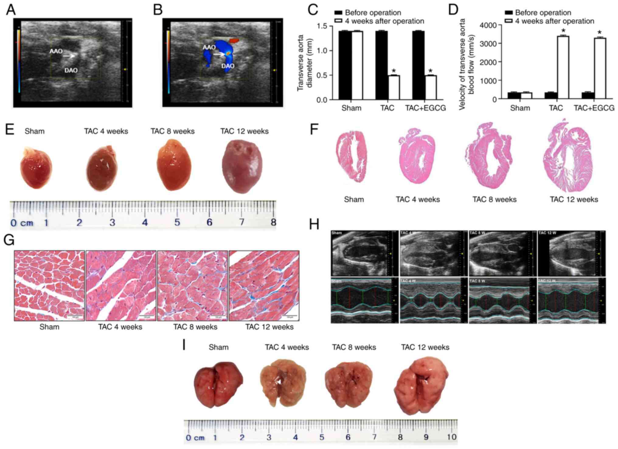
Echocardiography indicated that the TAC model was
successfully generated via TAB (Fig.
1A). Color Doppler imaging showed that blood flow was blocked
in the aortic arch (Fig. 1B). The
diameter of the transverse aorta decreased following TAC; the
diameter of this vessel was similar between the TAC and EGCG + TAC
groups (~0.4 mm; Fig. 1C), thus
resulting in a similar increase of blood flow in the transverse
aorta in both groups (Fig. 1D). In
the present study, stereoscopy and H&E staining showed that the
hearts of TAC mice at 4, 8 and 12 weeks after TAC were enlarged
compared with the Sham group (Fig. 1E
and F). Masson’s staining further revealed interstitial
fibrosis and collagen accumulation in the myocardial tissue of TAC
mice. Myocardial fibrosis and collagen deposition became more
notable from 4 to 12 weeks after TAC (Fig. 1G). Echocardiography further showed
that the left ventricular (LV) wall in TAC mice thickened between 4
and 8 weeks after TAC compared with the Sham group. At 8-12 weeks
after TAC, the LV volume of TAC mice began to gradually increase;
this was accompanied by a decrease in myocardial tissue (Fig. 1H). To determine morphological
changes of lung in TAC mice, stereoscopy was performed. The results
of stereoscopy showed that the lungs at 4, 8 and 12 weeks after TAC
were enlarged compared with the Sham group (Fig. 1I). There was a decrease in the LV
anterior wall thickness (AWT) in mice at 12 weeks after TAC. The LV
ejection fraction (EF) also increased significantly in TAC mice
over time. However, 8 weeks after TAC, LVEF showed a significant
decrease, indicating that heart failure may have occurred (Table I).
 | Table ICardiac function measurement via
echocardiography (n=6). |
Table I
Cardiac function measurement via
echocardiography (n=6).
| Parameter | Sham | Transverse aortic
constriction
| F-value | P-value |
|---|
| 4 weeks | 8 weeks | 12 weeks |
|---|
| Body weight, g | 38.66±0.22 | 39.15±0.18 | 38.40±0.60 | 38.66±0.60 | 1.500 | 0.2900 |
| Heart rate,
bpm | 466.00±4.00 | 466.00±4.00 | 474.00±3.00 | 466.00±6.00 | 2.680 | 0.1200 |
| LV end
diastole | | | | | | |
| AWT, mm | 1.04±0.55 | 1.59±0.11 | 1.32±0.06 | 1.28±0.98a | 47.550 | <0.0001 |
| IVS, mm | 0.89±0.01 | 0.97±0.01 | 0.95±0.25 | 1.02±0.21a | 32.923 | <0.0001 |
| Dimension, mm | 2.74±0.13 | 1.88±0.13 | 1.58±0.29 | 3.37±0.16a | 57.660 | <0.0001 |
| Volume,
µl | 75.33±0.74 | 75.81±0.75 | 80.00±1.00 | 77.80±0.12a | 27.270 | <0.0001 |
| LV end systole | | | | | | |
| AWT, mm | 1.40±0.12 | 1.76±0.05 | 1.67±0.03 | 1.86±0.06a | 13.670 | <0.0001 |
| IVS, mm | 1.05±0.10 | 1.11±0.12 | 1.08±0.10 | 1.12±0.11 | 2.100 | 0.1800 |
| Dimension, mm | 1.14±0.12 | 0.83±0.35 | 0.76±0.02 | 1.42±0.03a | 45.310 | <0.0001 |
| Volume,
µl | 22.42±1.27 | 20.08±0.89 | 17.46±0.95 | 25.65±0.77a | 37.500 | <0.0001 |
| EF, % | 82.00±3.00 | 93.33±4.16 | 69.33±1.53 | 58.33±6.03a | 42.630 | <0.0001 |
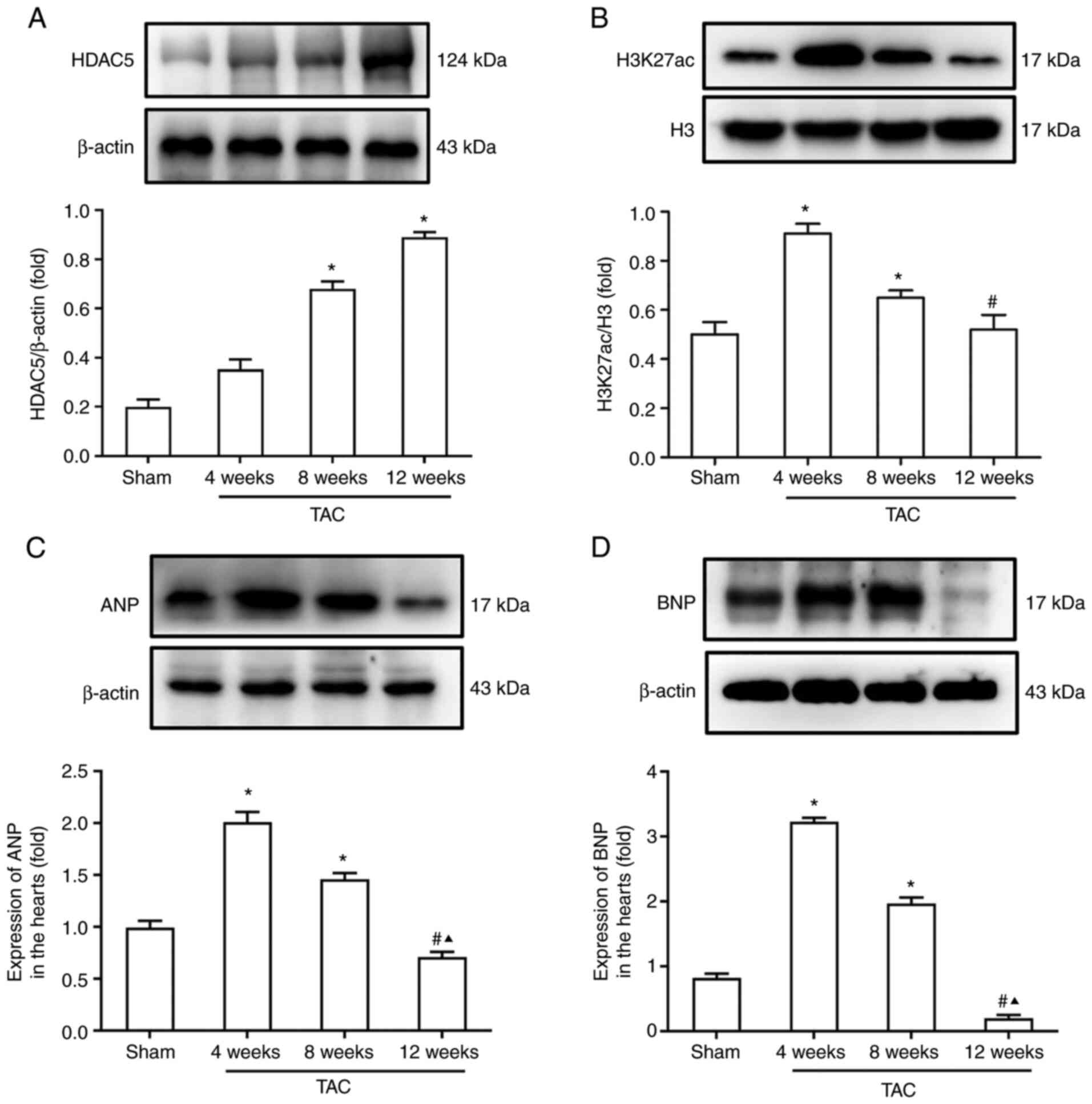
HDAC5 may be involved in histone
acetylation in the heart of TAC mice
Pathological myocardial remodeling is a key
pathophysiological process of heart failure that is regulated by
HDACs. Hence, protein expression levels of HDAC5 and H3K27ac were
assessed by western blotting. Results showed that expression levels
of HDAC5 in the heart of TAC mice increased gradually from 4 to 12
weeks after TAC (Fig. 2A). Western
blotting showed that the acetylation levels of histone H3K27ac in
TAC mice at 4 and 8 weeks after TAC were significantly higher than
at 12 weeks (Fig. 2B). In
addition, the expression levels of ANP and BNP at 4 and 8 weeks
were significantly higher than those of the Sham group. However,
the expression levels of ANP and BNP at 12 weeks after TAC were
significantly lower than that at 4 and 8 weeks (Fig. 2C and D).
EGCG reverses hypoacetylation of H3K27ac
by inhibiting HDAC5 and normalizing transcriptional activity of
MEF2A
First, the optimal exposure dose for EGCG was
determined. According to previous data (30,31),
mice were injected intraperitoneally with various concentrations of
EGCG (0, 25, 50, 75 and 100 mg/kg). The optimal concentration (50
mg/kg) was selected based on the lowest levels of HDAC5 in the
heart of TAC mice (Fig. 3A). Next,
the effect of EGCG on myocardial remodeling in TAC mice was
investigated. EGCG (50 mg/kg/day) was intraperitoneally injected
into mice after TAC for 12 weeks, and the hearts were collected for
analysis. Western blotting showed that EGCG reversed the
hypoacetylation of H3K27ac in the heart of TAC mice (Fig. 3B). EGCG attenuated the
overexpression of HDAC5 in TAC mice (Fig. 3C). Analysis showed that the mRNA
and protein expression levels of MEF2A were lower in the TAC
group than those in the Sham group, while EGCG improved the mRNA
and protein expression levels of MEF2A in TAC mice treated
with EGCG (Fig. 3D and E). In
addition, Co-IP was performed to verify the formation of a complex
between H3K27ac and MEF2A; this demonstrated that HDAC5 may
interact with H3K27ac and regulate gene expression levels of
MEF2A (Fig. 3F).
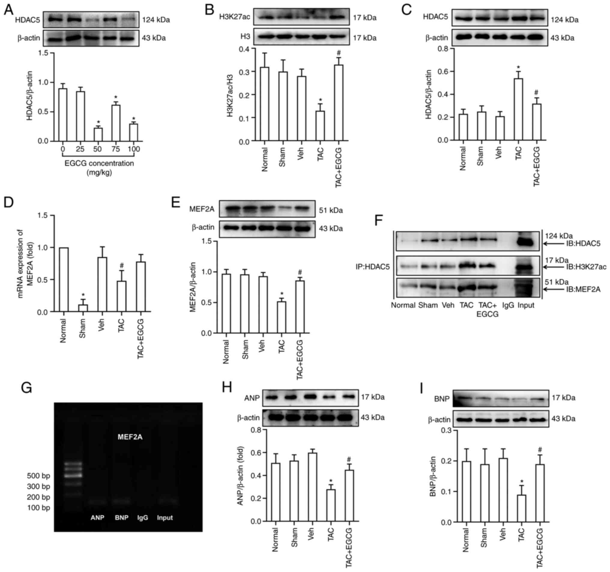 | Figure 3EGCG reverses abnormal expression of
ANP and BNP by inhibiting overexpression of HDAC5 and reversing
hypoacetylation of H3K27ac in the heart of mice 12 W after TAC. (A)
Expression of HDAC5 following treatment with different
concentrations of EGCG. (B) Acetylation levels of H3K27ac. (C)
Expression of HDAC5. (D) Reverse transcription-quantitative PCR
analysis of mRNA levels of MEF2A. (E) Expression of MEF2A. (F)
Co-IP using anti-HDAC5-protein G magnetic beads and immunoblotting
with anti-H3K27ac, anti-MEF2A or anti-HDAC5 antibody. (G) Chromatin
IP-PCR assay was used to identify the regulatory association
between MEF2A, ANP and BNP. Input, positive control; IgG, negative
control. Western blotting showing expression of (H) ANP and (I)
BNP. *P<0.05 vs. Sham; #P<0.05 vs. TAC.
n=6/group. EGCG, epigallocatechin gallate; ANP, atrial natriuretic
peptide; BNP, brain natriuretic peptide; TAC, transverse aortic
constriction; HDAC5, histone deacetylase 5; H3K27ac, histone
acetylated lysine 27 on histone H3; MEF2A, myocyte enhancer factor
2A; IP, immunoprecipitation; IB, immunoblot. |
Expression of ANP and BNP is regulated by
heart nuclear transcription factor MEF2A
Next, ChIP-PCR was performed to investigate the
regulatory association between MEF2A and downstream genes
associated with heart development (ANP and BNP). Data
showed that MEF2A bound to the promoter of ANP and
BNP in the mouse heart (Fig.
3G). Western blotting was performed to investigate the
expression levels of ANP and BNP during myocardial remodeling. Data
showed that the expression levels of ANP and BNP in the heart of
TAC mice were significantly lower compared with the Sham group.
Data also showed that EGCG treatment rescued the decreased levels
of ANP and BNP in the heart of TAC mice (Fig. 3H and I).
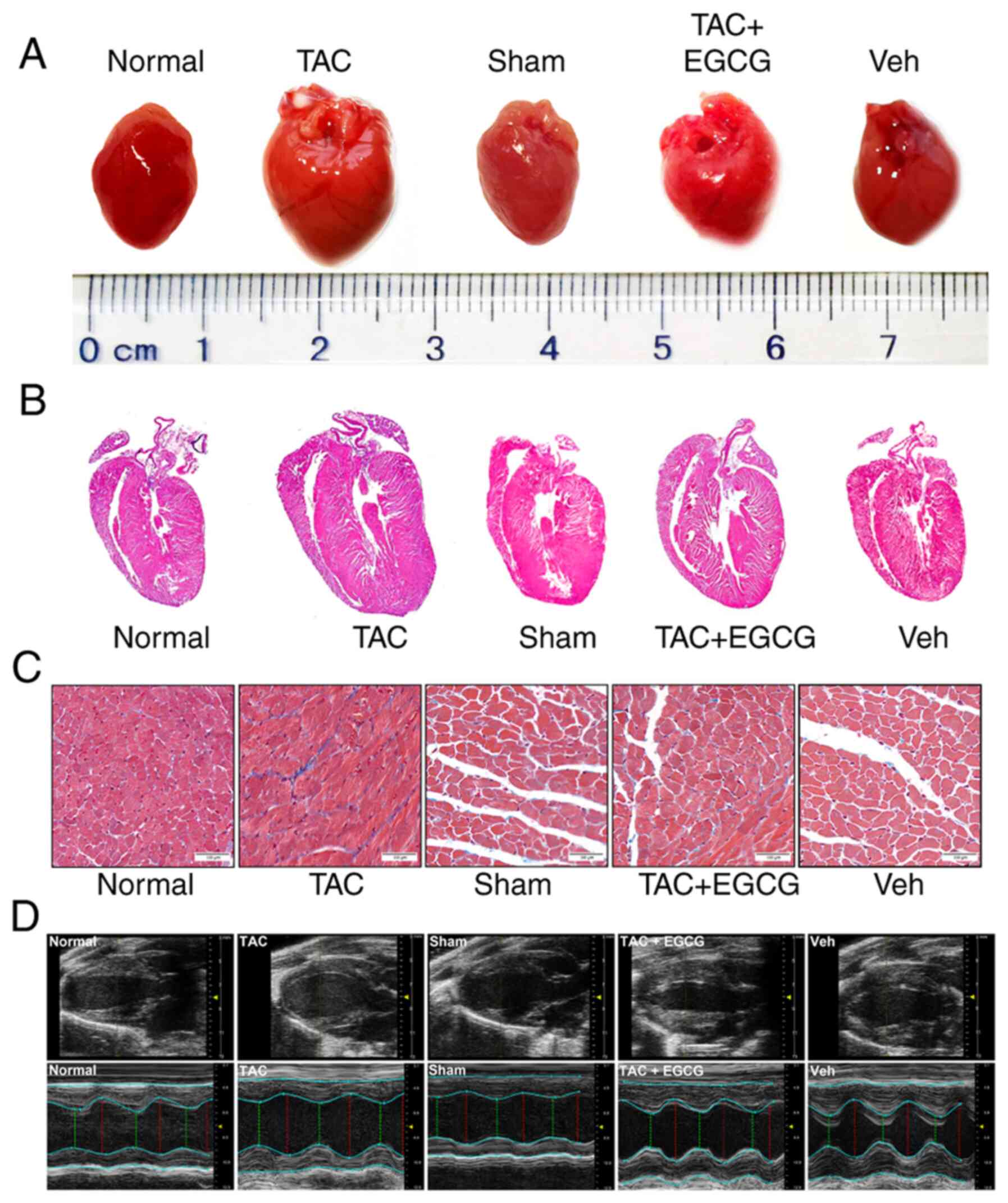
EGCG attenuates myocardial remodeling in
TAC mice
Stereoscopy and H&E staining were used to detect
the preventive and restorative effects of EGCG in the heart of TAC
mice. Stereoscopy showed that the heart of mice in TAC group was
larger compared with the Sham group and EGCG reversed cardiac
dilatation (Fig. 4A). H&E
staining showed that LV volume was higher in the TAC group than in
the Sham group. However, EGCG treatment attenuated the thickening
of LV in TAC mice (Fig. 4B).
Masson’s staining revealed a greater extent of interstitial
fibrosis and collagen deposition in the TAC group than in the Sham
group. Following treatment with EGCG, the extent of interstitial
fibrosis and collagen deposition was notably decreased (Fig. 4C). Furthermore, echocardiography
showed that the LV end diastolic dimension (EDD) and end systolic
dimension (ESD) in the TAC group were significantly higher than
those in the Sham group. However, EGCG treatment attenuated the
increased LVEDD and LVESD in the mouse heart. A significant
decrease in LVEF was observed in TAC mice; however, EGCG treatment
improved LVEF in the heart of TAC mice (Fig. 4D; Table II). In addition, EGCG
significantly decreased the cardiac mass index of TAC mice,
although the lung mass index remained unchanged (Table III). These data suggested that
EGCG attenuated pathological myocardial remodeling in TAC mice.
| Table IICardiac function measurement via
echocardiography (n=6). |
Table II
Cardiac function measurement via
echocardiography (n=6).
| Parameter | Normal | TAC | Sham | TAC + EGCG | Vehicle | F-value | P-value |
|---|
| Body weight, g | 38.28±0.31 | 39.11±0.15 | 38.42±0.38 | 38.43±0.48 | 38.43±0.48 | 2.24 | 0.1400 |
| Heart rate,
bpm) | 465.00±4.00 | 468.00±3.00 | 464.00±54.00 | 462.00±3.00 | 461.00±1.00 | 2.47 | 0.1100 |
| LV end
diastole | | | | | | | |
| AWT, mm | 1.04±0.04 | 1.23±0.68a | 1.04±0.13 | 1.07±0.05b | 1.03±0.01 | 21.25 | <0.0001 |
| IVS, mm | 0.83±0.15 | 1.02±0.16a | 0.84±0.26 | 0.92±0.15b | 0.85±0.10 | 44.18 | <0.0001 |
| Dimension, mm | 2.92±0.72 | 3.29±0.29a | 2.85±0.50 | 3.01±0.13b | 2.80±0.62 | 15.56 | <0.0001 |
| Volume,
µl | 75.33±0.74 | 80.00±1.00a | 75.81±0.75 | 77.80±0.12b | 75.12±0.49 | 27.27 | <0.0001 |
| LV end systole | | | | | | | |
| AWT, mm | 1.43±0.04 | 1.88±0.03a | 1.45±0.06 | 1.63±0.04b | 1.47±0.04 | 65.78 | <0.0001 |
| IVS, mm | 1.08±0.15 | 1.06±0.10 | 1.07±0.10 | 1.09±0.15 | 1.07±0.10 | 1.89 | 0.1900 |
| Dimension, mm | 1.12±0.03 | 1.43±0.03a | 1.17±0.02 | 1.30±0.02b | 1.14±0.02 | 48.72 | <0.0001 |
| Volume,
µl | 21.63±1.34 | 29.88±0.96a | 22.81±0.71 | 24.78±0.77b | 23.32±1.02 | 27.56 | <0.0001 |
| EF, % | 82.33±2.52 | 61.00±5.29a | 84.67±4.51 | 76.33±2.52b | 84.33±4.16 | 18.90 | <0.0001 |
| Table IIICardiac and lung MI in mice
(n=6). |
Table III
Cardiac and lung MI in mice
(n=6).
| Parameter | Normal | TAC | Sham | TAC + EGCG | Vehicle | F-value | P-value |
|---|
| BW, g | 36.37±1.50 | 42.22±1.11a | 37.35±0.78 | 41.23±1.24 | 38.80±1.57 | 21.17 | <0.0001 |
| HW, mg | 173.48±9.12 |
282.87±20.52a | 171.06±12.78 |
223.46±15.73b | 183.912±14.33 | 23.12 | <0.0001 |
| LW, mg | 333.87±23.56 |
398.13±21.89a | 341.38±23.34 |
360.55±18.95b | 348.03±24.74 | 6.86 | 0.0400 |
| HW/BW, mg/g | 4.77±0.88 | 6.70±0.15a | 4.58±0.36 | 5.42±0.16b | 4.74±0.24 | 78.48 | <0.0001 |
| LW/BW, mg/g | 9.18±0.39 | 9.43±0.23 | 9.14±0.31 | 9.23±0.37 | 8.97±0.54 | 1.83 | 0.1500 |
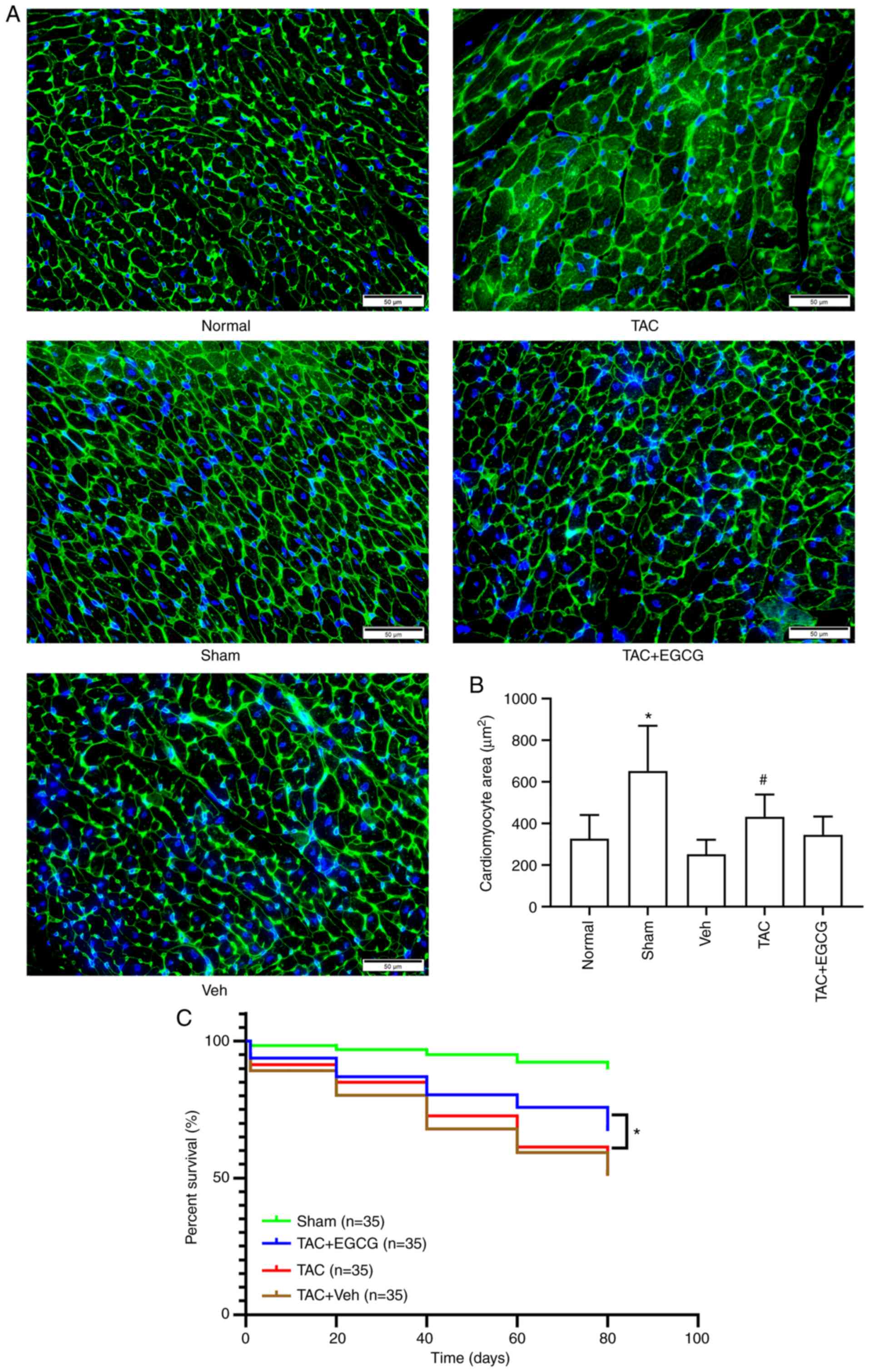
EGCG attenuates myocardial cell
hypertrophy and improves survival rate in TAC mice
Next, wheat germ agglutinin experiments were
performed to evaluate the surface area of cardiomyocytes in TAC
mice. Data showed that the surface area of cardiomyocytes in the
TAC group increased significantly compared with the Sham group.
However, EGCG treatment attenuated the increased surface area of
cardiomyocytes in the heart of TAC mice (Fig. 5A and B). For the HDAC inhibitor
EGCG to be used clinically, it is important to evaluate survival
rate following administration. To investigate this issue, mice were
subjected to TAC or sham operation and treated with EGCG (50
mg/kg/day) for 8 weeks, a period that corresponds to 6-8 years in
humans (8). The data showed that
EGCG treatment improved the survival rate of TAC mice [Sham + Veh,
90; TAC, 51; TAC + Veh, 51% and TAC + EGCG, 67% (n=35/group);
Fig. 5C].
Discussion
Studies have confirmed that one of the basic
mechanisms underlying the occurrence and development of chronic
heart failure is ventricular remodeling (32-34).
Over past years, epigenetic regulation has become a novel target
for intervention in modern medical research, especially with
regards to the reversible regulatory effect of histone acetylation
modification, which makes it possible to switch gene expression on
or off (35). Histone acetylation
includes numerous subtypes and acetylation sites, such as H3K4ac,
H3K9ac, H3K14ac, H3K18ac, H3K27ac, H3K36ac and H3K56ac (36). Previous studies confirmed that
multiple histone acetylation sites (H3K9ac, H3K14ac and H3K27ac)
are involved in pathological cardiac hypertrophy (8-11,37,38).
However, the mechanism by which histone acetylation is modified by
HDAC5 in pathological myocardial remodeling remains unclear.
Specifically, expression levels of HDAC5 remain at low levels
during the CHF compensatory period (39-41).
However, during the decompensated stage of this pathological
process, HDAC5 is expressed at high levels. In addition, genes
associated with cardiac development (ANP and BNP) are
highly expressed during the early stages of pathological myocardial
remodeling (cardiac hypertrophy stage) and expressed at low levels
during the subsequent CHF decompensated stage (42,43).
It was hypothesized that the decreased expression of ANP and BNP,
markers of cardiac hypertrophy, during the decompensated stage of
myocardial remodeling may be due to the inability to synthesize ANP
and BNP under cardiac decompensation (due to a decreased number of
myocardial cells and severe myocardial fibrosis in the heart of
model mice). Therefore, it was hypothesized that HDAC5-mediated
histone hypoacetylation is a key regulatory factor for pathological
myocardial remodeling in response to pressure overload.
HDACs are considered effective interventional
targets for the treatment of numerous types of human disease
(44,45). Studies have confirmed that HDACs
are involved in the occurrence and development of cardiac
hypertrophy in mice (46), and
that HDAC-specific inhibitors may improve heart failure and
maintain normal systolic function of the heart (47). It has also been reported that deer
antler in traditional Chinese medicine improves cardiac hypertrophy
and CHF by regulating histone acetylation modification (48). Further studies have revealed that
an imblance in HDAC-mediated histone acetylation serves an
important role in pathological myocardial remodeling (49,50).
Furthermore, HDAC inhibitors exert cardiac and vascular protective
effects in rats with cardiac hypertrophy caused by pressure
overload (51,52). In addition, Liu et al
(30), and Bagchi and Weeks
(53) proposed that HDAC
inhibitors possess clinical value for the treatment of myocardial
fibrosis, cardiac hypertrophy and heart failure by inhibiting the
levels of histone acetylation modification. Studies have also
confirmed that EGCG specifically inhibits activity of HDAC1/3/8
subtypes in tumors and other types of disease (54), and that EGCG downregulates activity
of HDAC1 in mouse myocardial tissue, thus regulating expression of
the cardiac structural gene cardiac troponin I (31). Other researchers reported that
HDAC5 may be involved in regulating the occurrence and development
of cardiac hypertrophy (22,55).
Therefore, it was hypothesized that HDAC5 may be a key regulator of
EGCG in the prevention of pathological myocardial remodeling in
mice. ChIP-PCR data indicated that MEF2A bound to the
promoter of ANP and BNP; this implied that there may
be a regulatory association between these factors. These results
suggested that an imbalance in HDAC5-mediated histone acetylation
is involved in abnormal expression of genes associated with
myocardial remodeling. However, the potential role of upstream
signaling pathways, and whether histone modification other than
deacetylation and acetylation are involved in myocardial
remodeling, remain unclear. Further studies are needed in the field
of myocardial remodeling to address this.
EGCG is the primary polyphenolic compound in green
tea and exhibits cardiovascular health-promoting activity by
regulating various pathways (56,57).
Because EGCG is extracted from green tea, it has almost no side
effects (56,58,59),
and this compound exhibits cardioprotection, neuroprotection, renal
protection, osteoprotection and anticancer properties, as well as
the ability to manage obesity, metabolic syndrome and type 2
diabetes (60), thus making it an
ideal safe and effective drug for the treatment of myocardial
remodeling (61). In certain
epidemiological studies, drinking green tea has been shown to
decrease the risk of cardiovascular disease (62,63).
In recent years, studies have found that EGCG serves an important
role in epigenetic regulation (20,64,65).
For example, EGCG inhibits DNA methyltransferase from turning on
genes that have been silenced by methylation and inhibits the
ability of HDACs to regulate histone acetylation modification in
vivo (31,66). EGCG improves congestive heart
failure caused by knockout of the manganese superoxide gene in a
mouse model of dilated cardiomyopathy and significantly improves
survival rate (67). Furthermore,
EGCG may inhibit cardiac hypertrophy, fibrosis and apoptosis caused
by aging (68). These data suggest
that EGCG serves a key role in cardiac protection. However, it
remains unclear whether EGCG improves cardiac function in patients
with CHF. In the present study, low expression levels of
MEF2A mRNA were detected in TAC mice. MEF2A is a
transcriptional regulator associated with cardiac hypertrophy and
leads to low expression levels of genes associated with cardiac
hypertrophy, thus inducing cardiac remodeling and heart failure.
Inhibiting HDAC activity by drug action could either increase or
decrease the expression of associated genes (69). Expression levels of MEF2A protein
in the heart of TAC mice were significantly decreased. However,
administration of EGCG reversed the low acetylation levels of
H3K27ac by inhibiting the action of HDAC5, thus increasing
MEF2A transcription activity. EGCG also normalized the
abnormal expression of two genes associated with cardiac
development, ANP and BNP, in the cardiac muscle
tissue of TAC mice. Echocardiographic data demonstrated that EGCG
improved cardiac function in TAC mice. However, more preclinical
studies are needed to confirm that this novel HDAC inhibitor can be
used to prevent or reverse cardiac remodeling and CHF before novel
drugs can be developed.
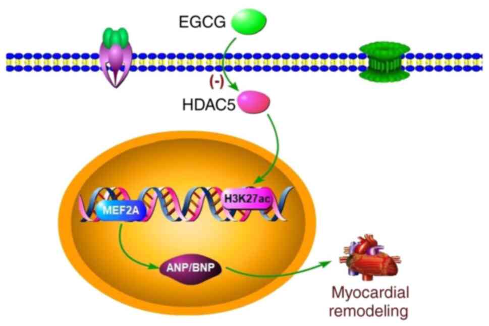
In conclusion, myocardial remodeling is a complex
process and is associated with gene transcription and modification.
In the present study, EGCG downregulated histone H3K27ac
acetylation mediated by HDAC5 to attenuate myocardial remodeling
induced by pressure overload in mice (Fig. 6). Meanwhile, the present results
provided evidence that HDAC inhibition is a potential treatment
strategy for treatment of cardiac dysfunction caused by pressure
overload.
Availability of data and materials
The datasets used and/or analyzed during the current
study are available from the corresponding author on reasonable
request.
Authors’ contributions
XH and CP conceived and designed the experiments.
XH, XL and LH performed the experiments. CP, QM and SW analyzed the
data and confirm the authenticity of all the raw data. XH and HZ
wrote the paper. CP revised the paper. All authors read and
approved the final version of the manuscript.
Ethics approval and consent to
participate
The present study was approved by the Institutional
Animal Care and Use Committee of Zunyi Medical University (approval
no. SYXK 2017-0012).
Patient consent for publication
Not applicable.
Competing interests
The authors declare they have no competing
interests.
Acknowledgments
Not applicable.
Funding
The present study was supported by the National Natural Science
Foundation of China (grant nos. 82060046 and 81560040).
Abbreviations:
|
ANP
|
atrial natriuretic peptide
|
|
BNP
|
brain natriuretic peptide
|
|
CHF
|
chronic heart failure
|
|
ChIP
|
chromatin immunoprecipitation
|
|
Co-IP
|
co-immunoprecipitation
|
|
EGCG
|
epigallocatechin gallate
|
|
HAT
|
histone acetylase
|
|
H3K27ac
|
histone acetylated lysine 27 on
histone H3
|
|
HDAC5
|
histone deacetylase 5
|
|
LVAWT
|
left ventricular anterior wall
thickness
|
|
LVEDD
|
left ventricular end diastolic
dimension
|
|
LVEF
|
left ventricular ejection fraction
|
|
LVESD
|
left ventricular end systolic
dimension
|
|
TAB
|
thoracic aortic banding
|
|
TAC
|
transverse aortic constriction
|
References
|
1
|
Khan N and Mukhtar H: Tea polyphenols in
promotion of human health. Nutrients. 11:392018. View Article : Google Scholar
|
|
2
|
Zhao L, Cheng G, Choksi K, Samanta A,
Girgis M, Soder R, Vincent RJ, Wulser M, De Ruyter M, McEnulty P,
et al: Transplantation of human umbilical cord blood-derived
cellular fraction improves left ventricular function and remodeling
after myocardial ischemia/reperfusion. Circ Res. 125:759–772. 2019.
View Article : Google Scholar : PubMed/NCBI
|
|
3
|
Grobe JL, Der Sarkissian S, Stewart JM,
Meszaros JG, Raizada MK and Katovich MJ: ACE2 overexpression
inhibits hypoxia-induced collagen production by cardiac
fibroblasts. Clin Sci (Lond). 113:357–364. 2007. View Article : Google Scholar
|
|
4
|
Li Y, Du W, Zhao R, Hu J, Li H, Han R, Yue
Q, Wu R, Li W and Zhao J: New insights into epigenetic
modifications in heart failure. Front Biosci (Landmark Ed).
22:230–247. 2017. View
Article : Google Scholar
|
|
5
|
Kim JK, Samaranayake M and Pradhan S:
Epigenetic mechanisms in mammals. Cell Mol Life Sci. 66:596–612.
2009. View Article : Google Scholar :
|
|
6
|
Ghosh AK, Rai R, Flevaris P and Vaughan
DE: Epigenetics in reactive and reparative cardiac fibrogenesis:
The promise of epigenetic therapy. J Cell Physiol. 232:1941–1956.
2017. View Article : Google Scholar
|
|
7
|
Segers VFM, Gevaert AB, Boen JRA, Van
Craenenbroeck EM and De Keulenaer GW: Epigenetic regulation of
intercellular communication in the heart. Am J Physiol Heart Circ
Physiol. 316:H1417–H1425. 2019. View Article : Google Scholar : PubMed/NCBI
|
|
8
|
Li S, Peng B, Luo X, Sun H and Peng C:
Anacardic acid attenuates pressure-overload cardiac hypertrophy
through inhibiting histone acetylases. J Cell Mol Med.
23:2744–2752. 2019. View Article : Google Scholar : PubMed/NCBI
|
|
9
|
Peng C, Zhang W, Zhao W, Zhu J, Huang X
and Tian J: Alcohol-induced histone H3K9 hyperacetylation and
cardiac hypertrophy are reversed by a histone acetylases inhibitor
anacardic acid in developing murine hearts. Biochimie. 113:1–9.
2015. View Article : Google Scholar : PubMed/NCBI
|
|
10
|
Peng C, Luo X, Li S and Sun H:
Phenylephrine-induced cardiac hypertrophy is attenuated by a
histone acetylase inhibitor anacardic acid in mice. Mol Biosyst.
13:714–724. 2017. View Article : Google Scholar : PubMed/NCBI
|
|
11
|
Ooi JY, Tuano NK, Rafehi H, Gao XM,
Ziemann M, Du XJ and El-Osta A: HDAC inhibition attenuates cardiac
hypertrophy by acetylation and deacetylation of target genes.
Epigenetics. 10:418–430. 2015. View Article : Google Scholar : PubMed/NCBI
|
|
12
|
Gorski PA, Jang SP, Jeong D, Lee A, Lee P,
Oh JG, Chepurko V, Yang DK, Kwak TH, Eom SH, et al: Role of SIRT1
in modulating acetylation of the sarco-endoplasmic reticulum
Ca2+-ATPase in heart failure. Circ Res. 124:pp. e63–e80. 2019,
View Article : Google Scholar :
|
|
13
|
Wang Y, Miao X, Liu Y, Li F, Liu Q, Sun J
and Cai L: Dysregulation of histone acetyltransferases and
deacetylases in cardiovascular diseases. Oxid Med Cell Longev.
2014.641979:2014.
|
|
14
|
Yang M, Zhang Y and Ren J: Acetylation in
cardiovascular diseases: Molecular mechanisms and clinical
implications. Biochim Biophys Acta Mol Basis Dis. 1866(165836):
2020
|
|
15
|
Steinmann J, Buer J, Pietschmann T and
Steinmann E: Anti-infective properties of
epigallocatechin-3-gallate (EGCG), a component of green tea. Br J
Pharmacol. 168:1059–1073. 2013. View Article : Google Scholar :
|
|
16
|
Yang CS, Landau JM, Huang MT and Newmark
HL: Inhibition of carcinogenesis by dietary polyphenolic compounds.
Annu Rev Nutr. 21:381–406. 2001. View Article : Google Scholar : PubMed/NCBI
|
|
17
|
Khurana S, Venkataraman K, Hollingsworth
A, Piche M and Tai TC: Polyphenols: Benefits to the cardiovascular
system in health and in aging. Nutrients. 5:3779–3827. 2013.
View Article : Google Scholar : PubMed/NCBI
|
|
18
|
Li K, Teng C and Min Q: Advanced
nanovehicles-enabled delivery systems of epigallocatechin gallate
for cancer therapy. Front Chem. 8(573297): 2020
|
|
19
|
Hu Y, McIntosh GH, Le Leu RK, Somashekar
R, Meng XQ, Gopalsamy G, Bambaca L, McKinnon RA and Young GP:
Supplementation with Brazil nuts and green tea extract regulates
targeted biomarkers related to colorectal cancer risk in humans. Br
J Nutr. 116:1901–1911. 2016. View Article : Google Scholar : PubMed/NCBI
|
|
20
|
Evans LW, Athukorala M, Martinez-Guryn K
and Ferguson BS: The role of histone acetylation and the microbiome
in phytochemical efficacy for cardiovascular diseases. Int J Mol
Sci. 21(4006): 2020
|
|
21
|
Gregoretti IV, Lee YM and Goodson HV:
Molecular evolution of the histone deacetylase family: Functional
implications of phylogenetic analysis. J Mol Biol. 338:17–31. 2004.
View Article : Google Scholar : PubMed/NCBI
|
|
22
|
Zhang L, Deng M, Lu A, Chen Y, Chen Y, Wu
C, Tan Z, Boini KM, Yang T, Zhu Q and Wang L: Sodium butyrate
attenuates angiotensin II-induced cardiac hypertrophy by inhibiting
COX2/PGE2 pathway via a HDAC5/HDAC6-dependent mechanism. J Cell Mol
Med. 23:8139–8150. 2019. View Article : Google Scholar : PubMed/NCBI
|
|
23
|
Chandrasekaran S, Peterson RE, Mani SK,
Addy B, Buchholz AL, Xu L, Thiyagarajan T, Kasiganesan H, Kern CB
and Menick DR: Histone deacetylases facilitate sodium/calcium
exchanger up-regulation in adult cardiomyocytes. FASEB J.
23:3851–3864. 2009. View Article : Google Scholar : PubMed/NCBI
|
|
24
|
Melleby AO, Romaine A, Aronsen JM, Veras
I, Zhang L, Sjaastad I, Lunde IG and Christensen G: A novel method
for high precision aortic constriction that allows for generation
of specific cardiac phenotypes in mice. Cardiovasc Res.
114:1680–1690. 2018. View Article : Google Scholar : PubMed/NCBI
|
|
25
|
Riehle C and Bauersachs J: Small animal
models of heart failure. Cardiovasc Res. 115:1838–1849. 2019.
View Article : Google Scholar : PubMed/NCBI
|
|
26
|
Mecklenburg J, Patil MJ, Koek W and
Akopian AN: Effects of local and spinal administrations of
mu-opioids on postoperative pain in aged versus adult mice. Pain
Rep. 2:pp. e5842017, View Article : Google Scholar : PubMed/NCBI
|
|
27
|
Zaw AM, Williams CM, Law HK and Chow BK:
Minimally invasive transverse aortic constriction in mice. J Vis
Exp:. 55293:2017.
|
|
28
|
Zhao Y, Wang C, Hong X, Miao J, Liao Y,
Hou FF, Zhou L and Liu Y: Wnt/β-catenin signaling mediates both
heart and kidney injury in type 2 cardiorenal syndrome. Kidney Int.
95:815–829. 2019. View Article : Google Scholar : PubMed/NCBI
|
|
29
|
Livak KJ and Schmittgen TD: Analysis of
relative gene expression data using real-time quantitative PCR and
the 2(-Delta Delta C(T)) method. Methods. 25:402–408. 2001.
View Article : Google Scholar
|
|
30
|
Liu L, Zhao W, Liu J, Gan Y, Liu L and
Tian J: Epigallocatechin-3 gallate prevents pressure
overload-induced heart failure by up-regulating SERCA2a via histone
acetylation modification in mice. PLoS One. 13:pp. e02051232018,
View Article : Google Scholar : PubMed/NCBI
|
|
31
|
Pan B, Quan J, Liu L, Xu Z, Zhu J, Huang X
and Tian J: Epigallocatechin gallate reverses cTnI-low
expression-induced age-related heart diastolic dysfunction through
histone acetylation modification. J Cell Mol Med. 21:2481–2490.
2017. View Article : Google Scholar : PubMed/NCBI
|
|
32
|
Uriel N, Sayer G, Annamalai S, Kapur NK
and Burkhoff D: Mechanical unloading in heart failure. J Am Coll
Cardiol. 72:569–580. 2018. View Article : Google Scholar : PubMed/NCBI
|
|
33
|
Bouwens E, Brankovic M, Mouthaan H, Baart
S, Rizopoulos D, van Boven N, Caliskan K, Manintveld O, Germans T,
van Ramshorst J, et al: Temporal patterns of 14 blood biomarker
candidates of cardiac remodeling in relation to prognosis of
patients with chronic heart failure-the Bio-SH i FT study. J Am
Heart Assoc. 8:pp. e0095552019, View Article : Google Scholar
|
|
34
|
Zhou X, Ferrara F, Contaldi C and Bossone
E: Right ventricular size and function in chronic heart failure:
Not to be forgotten. Heart Fail Clin. 15:205–217. 2019. View Article : Google Scholar : PubMed/NCBI
|
|
35
|
Sabia C, Picascia A, Grimaldi V, Amarelli
C, Maiello C and Napoli C: The epigenetic promise to improve
prognosis of heart failure and heart transplantation. Transplant
Rev (Orlando). 31:249–256. 2017. View Article : Google Scholar
|
|
36
|
Barnes CE, English DM and Cowley SM:
Acetylation & Co: An expanding repertoire of histone acylations
regulates chromatin and transcription. Essays Biochem. 63:97–107.
2019. View Article : Google Scholar : PubMed/NCBI
|
|
37
|
Wu X, Pan B, Liu L, Zhao W, Zhu J, Huang X
and Tian J: In utero exposure to PM2.5 during gestation caused
adult cardiac hypertrophy through histone acetylation modification.
J Cell Biochem. 120:4375–4384. 2019. View Article : Google Scholar
|
|
38
|
Chen K, Jian D, Zhao L, Zang X, Song W, Ma
J, Jia Z, Wang X and Gao C: Protective effect of histone
methyltransferase NSD3 on ISO-induced cardiac hypertrophy. FEBS
Lett. 593:2556–2565. 2019. View Article : Google Scholar : PubMed/NCBI
|
|
39
|
Xu WP, Yao TQ, Jiang YB, Zhang MZ, Wang
YP, Yu Y, Li JX and Li YG: Effect of the angiotensin II receptor
blocker valsartan on cardiac hypertrophy and myocardial histone
deacetylase expression in rats with aortic constriction. Exp Ther
Med. 9:2225–2228. 2015. View Article : Google Scholar : PubMed/NCBI
|
|
40
|
Parra M: Class IIa HDACs-new insights into
their functions in physiology and pathology. FEBS J. 282:1736–1744.
2015. View Article : Google Scholar
|
|
41
|
Vega RB, Harrison BC, Meadows E, Roberts
CR, Papst PJ, Olson EN and McKinsey TA: Protein kinases C and D
mediate agonist-dependent cardiac hypertrophy through nuclear
export of histone deacetylase 5. Mol Cell Biol. 24:8374–8385. 2004.
View Article : Google Scholar : PubMed/NCBI
|
|
42
|
Luo Y, Xu Y, Liang C, Xing W and Zhang T:
The mechanism of myocardial hypertrophy regulated by the
interaction between mhrt and myocardin. Cell Signal. 43:11–20.
2018. View Article : Google Scholar
|
|
43
|
Gaggin HK and Januzzi JL Jr: Biomarkers
and diagnostics in heart failure. Biochim Biophys Acta.
1832.2442–2450. 2013.
|
|
44
|
Scholz B, Schulte JS, Hamer S, Himmler K,
Pluteanu F, Seidl MD, Stein J, Wardelmann E, Hammer E, Völker U and
Müller FU: HDAC (histone deacetylase) inhibitor valproic acid
attenuates atrial remodeling and delays the onset of atrial
fibrillation in mice. Circ Arrhythm Electrophysiol. 12:pp.
e0070712019, View Article : Google Scholar : PubMed/NCBI
|
|
45
|
Zou G, Zhong W, Wu F, Wang X and Liu L:
Catalpol attenuates cardiomyocyte apoptosis in diabetic
cardiomyopathy via Neat1/miR-140-5p/HDAC4 axis. Biochimie.
165:90–99. 2019. View Article : Google Scholar : PubMed/NCBI
|
|
46
|
Yoon S, Kim M, Min HK, Lee YU, Kwon DH,
Lee M, Lee S, Kook T, Joung H, Nam KI, et al: Inhibition of heat
shock protein 70 blocks the development of cardiac hypertrophy by
modulating the phosphorylation of histone deacetylase 2. Cardiovasc
Res. 115:1850–1860. 2019. View Article : Google Scholar : PubMed/NCBI
|
|
47
|
Lee E, Lee HA, Kim M, Do GY, Cho HM, Kim
GJ, Jung H, Song JH, Cho JM and Kim I: Upregulation of C/EBPβ and
TSC2 by an HDAC inhibitor CG200745 protects heart from DOCA-induced
hypertrophy. Clin Exp Pharmacol Physiol. 46:226–236. 2019.
View Article : Google Scholar
|
|
48
|
Zhao W, Hu W, Wang X, Xia N, Hu Q and Zhou
H: A traditional Chinese medicine, Lujiao prescription, as a
potential therapy for hypertrophic cardiomyocytes by acting on
histone acetylation. J Chin Med Assoc. 78:486–493. 2015. View Article : Google Scholar : PubMed/NCBI
|
|
49
|
Habibian J and Ferguson BS: The crosstalk
between acetylation and phosphorylation: Emerging new roles for
hDAC inhibitors in the heart. Int J Mol Sci. 20:1022018. View Article : Google Scholar
|
|
50
|
Wallner M, Eaton DM, Berretta RM,
Liesinger L, Schittmayer M, Gindlhuber J, Wu J, Jeong MY, Lin YH,
Borghetti G, et al: HDAC inhibition improves cardiopulmonary
function in a feline model of diastolic dysfunction. Sci Transl
Med. 12:pp. eaay72052020, View Article : Google Scholar : PubMed/NCBI
|
|
51
|
Jung H, Lee E, Kim I, Song JH and Kim GJ:
Histone deacetylase inhibition has cardiac and vascular protective
effects in rats with pressure overload cardiac hypertrophy. Physiol
Res. 68:727–737. 2019. View Article : Google Scholar : PubMed/NCBI
|
|
52
|
Yan H, Yi S, Zhuang H, Wu L, Wang DW and
Jiang J: Sphingosine-1-phosphate ameliorates the cardiac
hypertrophic response through inhibiting the activity of histone
deacetylase-2. Int J Mol Med. 41:1704–1714. 2018.
|
|
53
|
Bagchi RA and Weeks KL: Histone
deacetylases in cardiovascular and metabolic diseases. J Mol Cell
Cardiol. 130:151–159. 2019. View Article : Google Scholar : PubMed/NCBI
|
|
54
|
Kim DJ, Dunleavey JM, Xiao L, Ollila DW,
Troester MA, Otey CA, Li W, Barker TH and Dudley AC: Suppression of
TGFβ-mediated conversion of endothelial cells and fibroblasts into
cancer associated (myo)fibroblasts via HDAC inhibition. Br J
Cancer. 118:1359–1368. 2018. View Article : Google Scholar : PubMed/NCBI
|
|
55
|
Hu T, Schreiter FC, Bagchi RA, Tatman PD,
Hannink M and McKinsey TA: HDAC5 catalytic activity suppresses
cardiomyocyte oxidative stress and NRF2 target gene expression. J
Biol Chem. 294:8640–8652. 2019. View Article : Google Scholar : PubMed/NCBI
|
|
56
|
Eng QY, Thanikachalam PV and Ramamurthy S:
Molecular understanding of epigallocatechin gallate (EGCG) in
cardiovascular and metabolic diseases. J Ethnopharmacol.
210:296–310. 2018. View Article : Google Scholar
|
|
57
|
Papadaki M, Vikhorev PG, Marston SB and
Messer AE: Uncoupling of myofilament Ca2+ sensitivity from troponin
I phosphorylation by mutations can be reversed by
epigallocatechin-3-gallate. Cardiovasc Res. 108:99–110. 2015.
View Article : Google Scholar : PubMed/NCBI
|
|
58
|
Oliveira MR, Nabavi SF, Daglia M,
Rastrelli L and Nabavi SM: Epigallocatechin gallate and
mitochondria-a story of life and death. Pharmacol Res. 104:70–85.
2016. View Article : Google Scholar : PubMed/NCBI
|
|
59
|
Isbrucker RA, Edwards JA, Wolz E,
Davidovich A and Bausch J: Safety studies on epigallocatechin
gallate (EGCG) preparations. Part 3: Teratogenicity and
reproductive toxicity studies in rats. Food Chem Toxicol.
44:651–661. 2006. View Article : Google Scholar : PubMed/NCBI
|
|
60
|
Afzal M, Safer AM and Menon M: Green tea
polyphenols and their potential role in health and disease.
Inflammopharmacology. 23:151–161. 2015. View Article : Google Scholar : PubMed/NCBI
|
|
61
|
Qin S, Chen MH, Fang W, Tan XF, Xie L,
Yang YG, Qin T and Li N: Cerebral protection of epigallocatechin
gallate (EGCG) via preservation of mitochondrial function and ERK
inhibition in a rat resuscitation model. Drug Des Devel Ther.
13:2759–2768. 2019. View Article : Google Scholar : PubMed/NCBI
|
|
62
|
Hertog MG, Feskens EJ, Hollman PC, Katan
MB and Kromhout D: Dietary antioxidant flavonoids and risk of
coronary heart disease: The zutphen elderly study. Lancet.
342:1007–1011. 1993. View Article : Google Scholar : PubMed/NCBI
|
|
63
|
Nakachi K, Matsuyama S, Miyake S, Suganuma
M and Imai K: Preventive effects of drinking green tea on cancer
and cardiovascular disease: Epidemiological evidence for multiple
targeting prevention. Biofactors. 13:49–54. 2000. View Article : Google Scholar
|
|
64
|
Potenza MA, Iacobazzi D, Sgarra L and
Montagnani M: The intrinsic virtues of EGCG, an extremely good cell
guardian, on prevention and treatment of diabesity complications.
Molecules. 25(3061): 2020
|
|
65
|
Yang L and Zhang W, Chopra S, Kaur D, Wang
H, Li M, Chen P and Zhang W: The epigenetic modification of
epigallocatechin gallate (EGCG) on cancer. Curr Drug Targets.
21:1099–1104. 2020PubMed/NCBI
|
|
66
|
Sheng J, Shi W, Guo H, Long W, Wang Y, Qi
J, Liu J and Xu Y: The inhibitory effect of
(-)-epigallocatechin-3-gallate on breast cancer progression via
reducing SCUBE2 methylation and DNMT activity. Molecules.
24:28992019. View Article : Google Scholar
|
|
67
|
Oyama JI, Shiraki A, Nishikido T, Maeda T,
Komoda H, Shimizu T, Makino N and Node K: EGCG, a green tea
catechin, attenuates the progression of heart failure induced by
the heart/muscle-specific deletion of MnSOD in mice. J Cardiol.
69:417–427. 2017. View Article : Google Scholar
|
|
68
|
Muhammed I, Sankar S and Govindaraj S:
Ameliorative effect of epigallocatechin gallate on cardiac
hypertrophy and fibrosis in aged rats. J Cardiovasc Pharmacol.
71:65–75. 2018. View Article : Google Scholar : PubMed/NCBI
|
|
69
|
Li C, Sun XN, Chen BY, Zeng MR, Du LJ, Liu
T, Gu HH, Liu Y, Li YL, Zhou LJ, et al: Nuclear receptor
corepressor 1 represses cardiac hypertrophy. EMBO Mol Med. 11:pp.
e91272019, View Article : Google Scholar : PubMed/NCBI
|