Human interaction with ionizing radiation has
evolved from passive environmental exposure to a complex interface
defined by advanced medical diagnostics, nuclear industry expansion
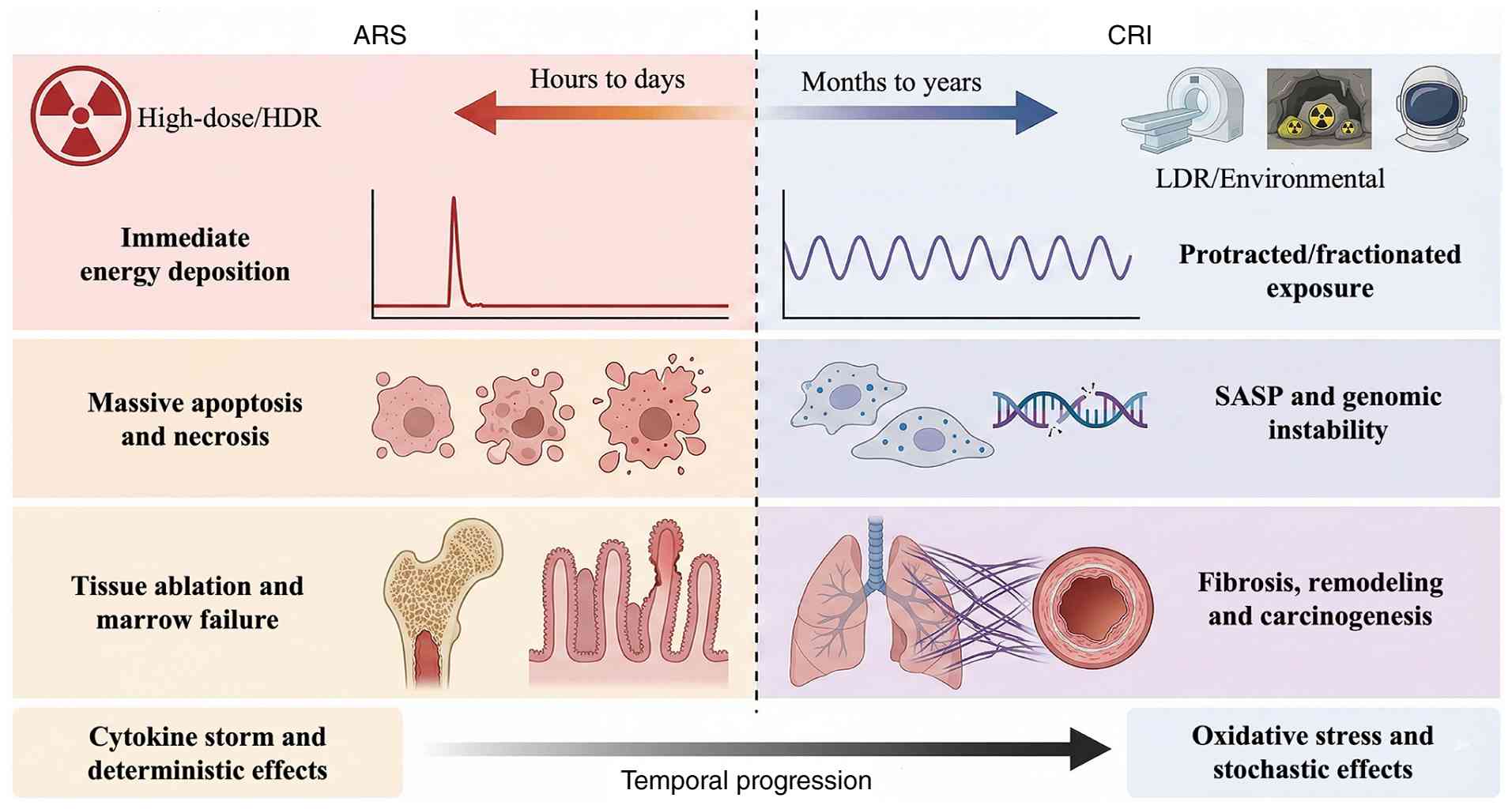
and long-duration space exploration (1). While acute radiation syndrome (ARS)
resulting from high-dose exposure is characterized, the biological
consequences of chronic, low-dose-rate (LDR) exposure represent a
knowledge gap (2). It is
necessary to distinguish CRI from the delayed effects of acute
radiation exposure (DEARE). While DEARE refers to late-onset
pathology emerging months to years following an acute
high-dose/high-dose-rate exposure, CRI results from protracted LDR
irradiation over months to years, characterized by continuous
sublethal damage accumulation, cell senescence and inflammaging
(3).
Unlike the rapid cell ablation seen in acute
scenarios, CRI is characterized by persistent, progressive
pathological changes (4). LDR
exposure triggers a unique interplay of mitochondrial dysfunction
and cytosolic DNA sensing pathways (cyclic GMP-AMP-STING
(cGAS/STING), leading to a Senescence-Associated Secretory
Phenotype (SASP, a complex pro-inflammatory secretome produced by
senescent cells) (5). This
inflammaging microenvironment drives progressive fibrosis, immune
dysregulation and genomic instability distinct from acute injury
patterns (6). Consequently, the
linear no-threshold model-which assumes that biological risk is
directly proportional to radiation dose without any safe lower
limit-often fails to predict the non-linear, stochastic risks of
chronic exposure, necessitating re-evaluation through dedicated
experimental systems (7).
Animal models of CRI are key tools for deconvoluting
these complex biological responses (8). They provide the only rigorous means
to isolate the effects of dose rate from total absorbed dose,
enabling the study of temporal damage accumulation vs. repair
kinetics (9). Moreover, these
models serve as the platforms for the rigorous validation of
next-generation medical countermeasures (MCMs) targeting late-onset
effects of both CRI and acute high-dose irradiation (DEARE), such
as radiation-induced lung fibrosis and secondary malignancy
(10). However, the fidelity of
current CRI models faces scrutiny (11). Many historical models using
fractionated acute irradiation inadequately mimic the continuous,
low-intensity stress of environmental LDR (12-14). Furthermore, translating findings
from inbred rodent strains to genetically diverse human populations
is challenging due to species-specific differences in telomere
biology and immune surveillance (13). Future models must integrate
humanized systems and reflect the heterogeneity of real-world
exposure (14).
Complementing experimental data with epidemiological
evidence from human cohorts provides a foundational understanding
of the long-term risks associated with chronic radiation. Long-term
follow-up studies of Chernobyl liquidators and Fukushima cleanup
workers have revealed elevated risks of non-cancerous pathologies,
such as cataracts and circulatory disease, alongside increased
incidences of leukemia and thyroid cancer (15-17). Similarly, occupational cohorts of
nuclear power plant workers and healthcare professionals,
particularly interventional radiologists and cardiologists,
demonstrate an association between cumulative low-dose exposure and
hematopoietic dysregulation or genomic instability (16,17). However, human studies are
typically confounded by lifestyle variables, inaccurate
retrospective dosimetry and the substantial latency period between
exposure and symptomatic manifestation. These limitations highlight
the translational gap that necessitates the development of
high-fidelity animal models (17). By accurately simulating human
exposure paradigms under controlled conditions, these models enable
the identification of insidious molecular mechanisms such as niche
remodeling and SASP-driven inflammaging that remain elusive in
retrospective human analyses. Thus, integrating human
epidemiological findings with mechanistic animal studies is key for
establishing evidence-based occupational safety thresholds and
precision radioprotection strategies (18).
The present review provides a comprehensive critique
of the methodological and conceptual evolution of CRI animal
models. The present study collected literature across major
databases, including PubMed (pubmed.ncbi.nlm.nih.gov/), Scopus (scopus.com/), and Web of Science (webofscience. com/)
from inception up to December 2025, specifically focusing on LDR
irradiation, mechanistic modeling (SASP, oxidative stress) and
translational countermeasures. The present study aimed to summarize
advances in continuous external exposure and internal radionuclide
contamination models, highlighting their relevance to modern
radiobiology. The present study aimed to address the identification
of robust cross-species biomarkers, including circulating microRNAs
(miRs) and inflammatory signatures, as well as the integration of
single-cell multi-omics into model validation, proposing a
forward-looking framework to enhance the translational power of
chronic radiation research (Fig.
1).
Populations at risk extend beyond traditional
nuclear industry workers to include interventional radiologists,
astronauts exposed to galactic cosmic rays and residents in high
background radiation areas (23). Unlike the rapid onset of acute
symptoms, CRI is characterized by a latent window, in which
sub-clinical metabolic reprogramming and oxidative fluctuations
occur prior to symptomatic manifestation (24). This progression complicates early
risk stratification, rendering traditional biodosimetry markers
ineffective and posing a challenge for precision radiation
protection (Table I) (25).
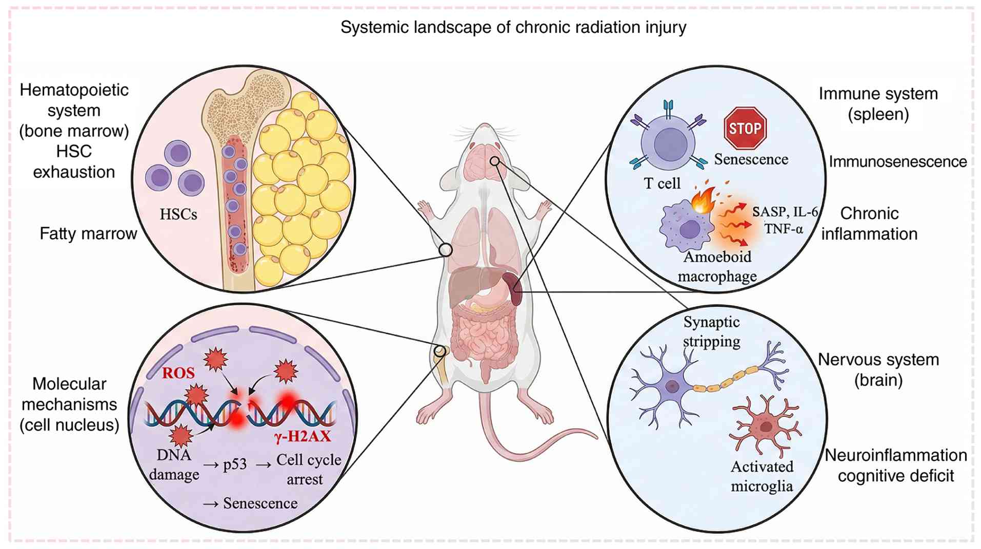
CRI is a systemic disease, propagated by complex
inter-organ signaling rather than isolated tissue injury. The
hematopoietic system serves as the most sensitive biological
indicator and primary responsive target of LDR toxicity (26). Beyond direct DNA damage to
hematopoietic stem cells (HSCs), chronic exposure remodels the bone
marrow microenvironment (27).
Radiation-induced stromal senescence and adipogenesis (fatty
marrow) create a hostile niche that fails to support HSC
self-renewal, leading to HSC exhaustion, persistent cytopenia and a
pre-leukemic state of clonal instability (28). The immune system undergoes a
paradoxical shift termed inflammaging. While radiation depletes
naïve lymphocyte pools (immunosenescence), damage-associated
molecular patterns released from stressed cells trigger the
cGAS/STING pathway, driving a sustained release of pro-inflammatory
cytokines (IL-6, TNF-α) (29).
This chronic low-grade inflammation not only impairs pathogen
clearance but also creates a tumor-permissive microenvironment
(30). The central nervous
system (CNS), previously considered radioresistant, exhibits
vulnerability through neuroinflammation and the inhibition of
hippocampal neurogenesis, manifesting as cognitive rigidity
(diminished mental flexibility and impaired adaptation to changing
environmental demands) and autonomic dysregulation (31). Furthermore, reproductive toxicity
involves germline DNA fragmentation and endocrine axis disruption,
creating a cycle of systemic physiological decline amplified by
oxidative stress (32).
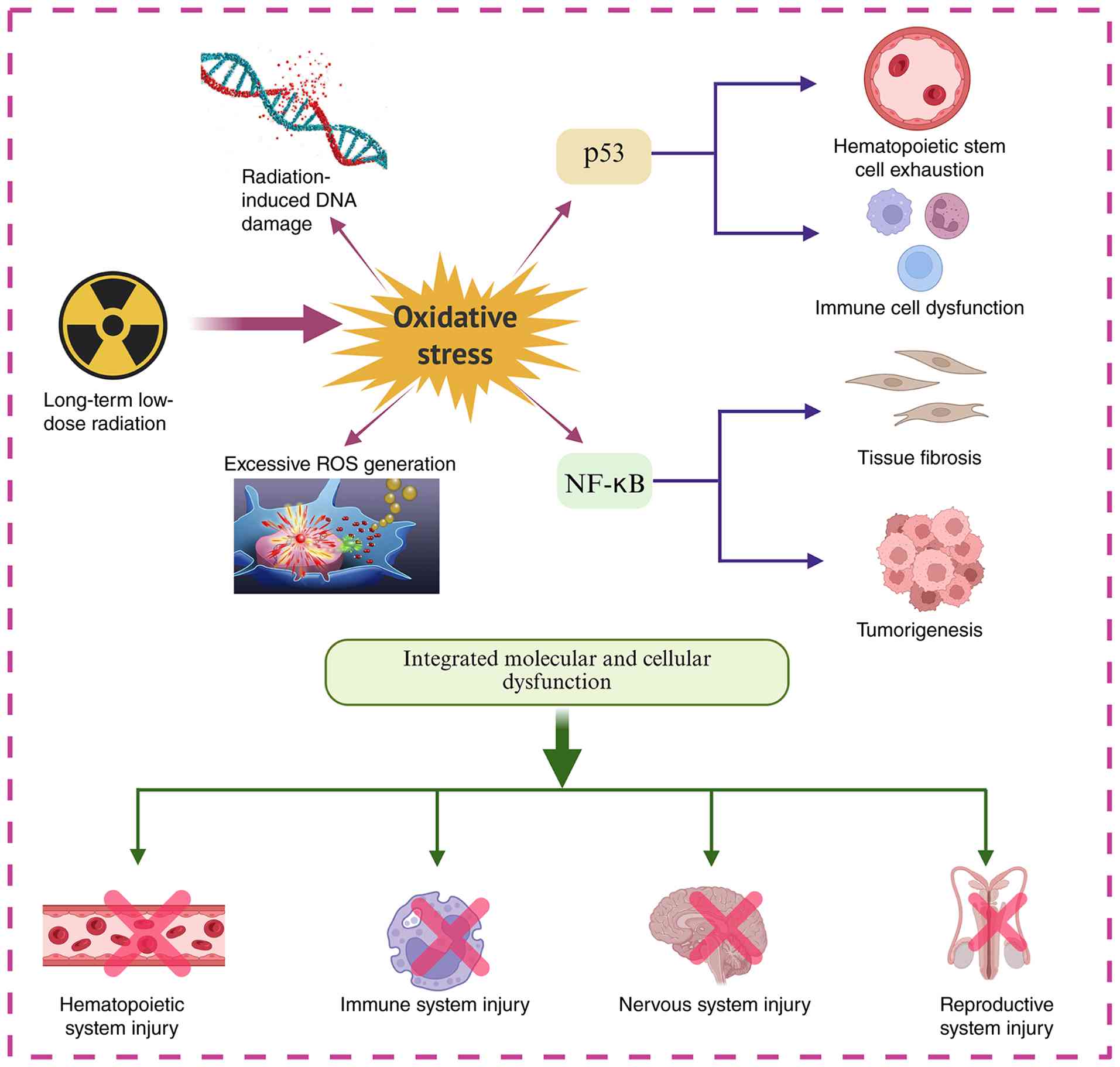
The molecular hallmark of CRI is genomic
instability, a state where the genome acquires an increased
tendency to alteration. Unlike the transient reactive oxygen
species (ROS) burst in acute exposure, CRI establishes a cycle of
chronic oxidative stress driven by mitochondrial dysfunction
(33). Leakage of electrons from
damaged electron transport chains generates persistent ROS, causing
continuous oxidative base damage-specifically the formation of
8-oxo-7,8-dihydro-2'-deoxyguanosine (34). Damage is not confined to directly
irradiated cells (35). Through
the radiation-induced bystander effect, irradiated cells transmit
distress signals (via exosomes and gap junctions) to non-irradiated
neighbors, propagating genomic instability (36). Studies highlight the role of
epigenetic drift (aberrant DNA methylation and histone
modifications) in locking cells into a dysfunctional state
(37,38). These heritable epigenetic marks
explain why radiation effects persist after exposure ceases,
potentially affecting unexposed offspring via transgenerational
inheritance (Fig. 2) (38).
A key long-term consequence of CRI is the elevation
of stochastic carcinogenic risk (39). According to the LNT model,
chronic accumulation of DNA mis-repairs increases the probability
of malignant transformation (40).
Under continuous immune pressure and oxidative
stress, somatic cells with advantageous mutations (in TP53 or DNA
methyltransferase 3A) may undergo clonal expansion, a phenomenon
known as clonal hematopoiesis of indeterminate potential (41). This clonal evolution serves as a
precursor to radiation-induced leukemias and solid tumors (42).
Compared with spontaneous cancer,
radiation-associated malignancies exhibit distinct mutational
signatures and extended latency periods (43). The spectrum includes leukemia,
thyroid papillary carcinoma and lung fibrosis-associated cancers
(44). These mechanistic
insights underscore the need for refined radiobiological models
that recapitulate these stochastic events to guide international
occupational safety standards (Fig.
3; Table II).
The translational validity of animal models hinges
on the fidelity with which they replicate the physics and dosimetry
of human exposure (45). While
acute high-dose models are well-established, simulating the
protracted nature of CRI requires precise control over dose
accumulation, temporal distribution and linear energy transfer
(LET) (46-48). Existing methodological approaches
use X-ray and γ-ray sources (60Co, 137Cs) due
to their stable energy output and tissue penetration profiles
(47,48). However, the field is evolving
from static exposures to sophisticated dynamic systems that
decouple DR from total absorbed dose, enabling the differentiation
between repairable sublethal damage and cumulative irreversible
injury (48).
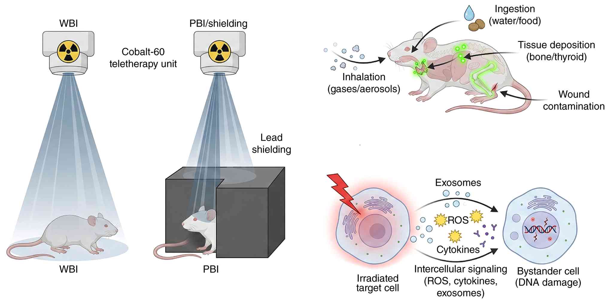
External beam irradiation is the cornerstone of
radiobiological modeling, categorized by the extent of anatomical
coverage. Whole-body irradiation (WBI) is the standard for
simulating systemic catastrophe, such as large-scale nuclear
accidents or prolonged occupational exposure in unshielded
environments (49). By
delivering a homogeneous dose across all physiological
compartments, WBI effectively reproduces the syndromic nature of
RI, capturing the interplay between bone marrow failure
(hematopoietic syndrome) and systemic immune collapse (50). A challenge in WBI is achieving
dosimetric homogeneity (51).
Variations in animal geometry can lead to hot spots (overdose) or
cold spots (underdose), potentially confounding survival data
(52). Furthermore, WBI
typically induces lethal acute GI toxicity before chronic fibrotic
phenotypes manifest (53). To
address this, fractionated low-dose regimens are employed to allow
animals to survive the acute phase and develop late-onset pathology
(54). To mimic scenarios such
as localized radiotherapy toxicity or partial shielding, partial
body irradiation selectively targets specific organs (thorax,
brain) while sparing the bone marrow (55). Research has moved beyond lead
shielding to use small animal radiation research platforms
(56). These systems employ
micro-CT guidance to deliver conformal radiation beams with
sub-millimeter precision (57).
This allows the study of organ-specific radiosensitivity
(radiation-induced lung fibrosis) without the confounding variable
of systemic hematopoietic collapse (Fig. 4) (58).
Emerging evidence suggests CRI is not solely the
result of direct energy deposition but is also mediated by NTEs
(59). Models of indirect
irradiation focus on how radiation signals are propagated through
the microenvironment (60-63). Bystander and abscopal models
investigate how irradiated cells communicate with non-irradiated
neighbors via gap junctions, exosomes and soluble factors (ROS,
cytokines) (61). By irradiating
a specific volume (such as the liver) and assessing damage in
distant organs (such as the lung), researchers can elucidate the
systemic propagation of inflammatory signals (62). In ecological toxicology contexts,
indirect exposure also refers to the interaction with irradiated
media (water or soil) (63).
These models are essential for distinguishing between direct
radiotoxicity and the secondary toxicity of radiation-induced
chemical species (radiolysis products) (64).
While external beams simulate prompt exposure,
internal contamination is the most biologically faithful model for
occupational hazards (such as uranium mining) or post-accident
fallout (65). The pathology is
dictated by the chemical nature of the radionuclide (66). For example, osteotropic isotopes
(90Sr, 226Ra) accumulate in the bone matrix,
causing chronic marrow suppression and osteosarcoma, whereas
radioiodine (131I) targets the thyroid (67). These models require precise
calculation of body burden (the total amount of a radionuclide
present in the organism at a given time) and effective half-life
(68). Internal models are key
for studying high-LET radiation (α-particles from plutonium or
radon) (69). Unlike low-LET
γ-rays, α-particles cause dense ionization tracks and complex DNA
double-strand breaks that are refractory to repair (70). This biological insult highlights
the limitation of using external X-rays to mimic internal α-emitter
contamination (71).
To bridge the gap between laboratory conditions and
real-world catastrophes, advanced models integrate multiple
stressors (72,73). Real-world exposure typically
co-occurs with trauma or burns (73-75). Specialized facilities-such as
gamma gardens (open-field irradiation facilities equipped with a
central radioactive source) or prolonged housing near radioactive
sources (60Co or 137Cs)-allow continuous
exposure over months (0.05-0.50 Gy/week) (75). These models are suited to test
the dose-rate effect (the phenomenon where the biological
effectiveness of a radiation dose decreases as the rate of delivery
is reduced, primarily due to ongoing cellular repair), providing
key data for setting occupational safety thresholds and validating
environmental risk models (Table
III) (76).
Animal models of CRI are defined by a distinct
phenotypic landscape that diverges from acute syndromes (77). These models capture the
progressive, multidimensional failure of organ systems driven by
the continuous accumulation of sublethal damage and the propagation
of SASP (77,78).
Chronic radiation accelerates immunosenescence,
characterized by thymic involution and a functional paralysis of
the adaptive immune repertoire (86). Peripheral T cell pools become
oligoclonal, dominated by exhausted memory phenotypes
(PD-1+) with decreased proliferative capacity (87). Simultaneously, the innate immune
system enters a state of hyper-activation known as inflammaging
(88). Irradiated macrophages
activate the NLRP3 inflammasome and the cGAS/STING pathway,
secreting a constant stream of pro-inflammatory cytokines (IL-1β,
IL-6) (89). This immune
dysregulation creates a permissive microenvironment that impairs
pathogen clearance and reduces immunosurveillance against
neoplastic transformation (90).
Physiologically, these changes translate into
sickness behaviors, such as lethargy, anxiety-like thigmotaxis and
cognitive rigidity (95).
Furthermore, radiation disrupts the hypothalamic-pituitary-adrenal
axis, leading to circadian dysregulation (96). This neuro-immune-endocrine
crosstalk highlights the systemic nature of CRI, where CNS injury
exacerbates peripheral immune suppression (Fig. 5) (97).
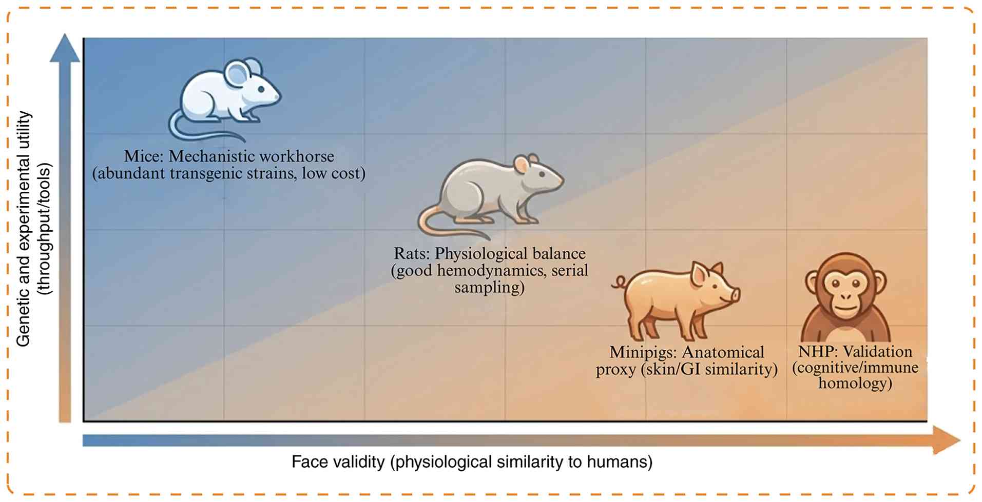
The utility of any animal model is determined by the
balance between its face (phenotypic similarity to humans) and
construct validity (mechanistic similarity). Compared with
fractionated acute doses, LDR sources more accurately replicate the
continuous radiation exposure patterns associated with occupational
hazards and environmental contamination (98). LDR models capture the abscopal
interactions between organs (such as the kidney-heart axis),
allowing for the evaluation of holistic therapeutics rather than
single-organ protectants (99).
The latency of CRI requires extended housing (6-24 months),
increasing costs (100). The
confounding factor of aging becomes critical; distinguishing
radiation effects from natural geriatric decline requires robust
age-matched controls (101).
Rodents possess different metabolic rates and DNA repair kinetics
than humans (102). For
example, the lethal dose required to kill 50% of the population
within 30 days) varies, complicating the direct extrapolation of
dose-response associations (103).
Selecting the appropriate model involves ensuring a
match between the specific biological mechanisms under
investigation and the inherent physiological attributes of the
chosen animal (104).
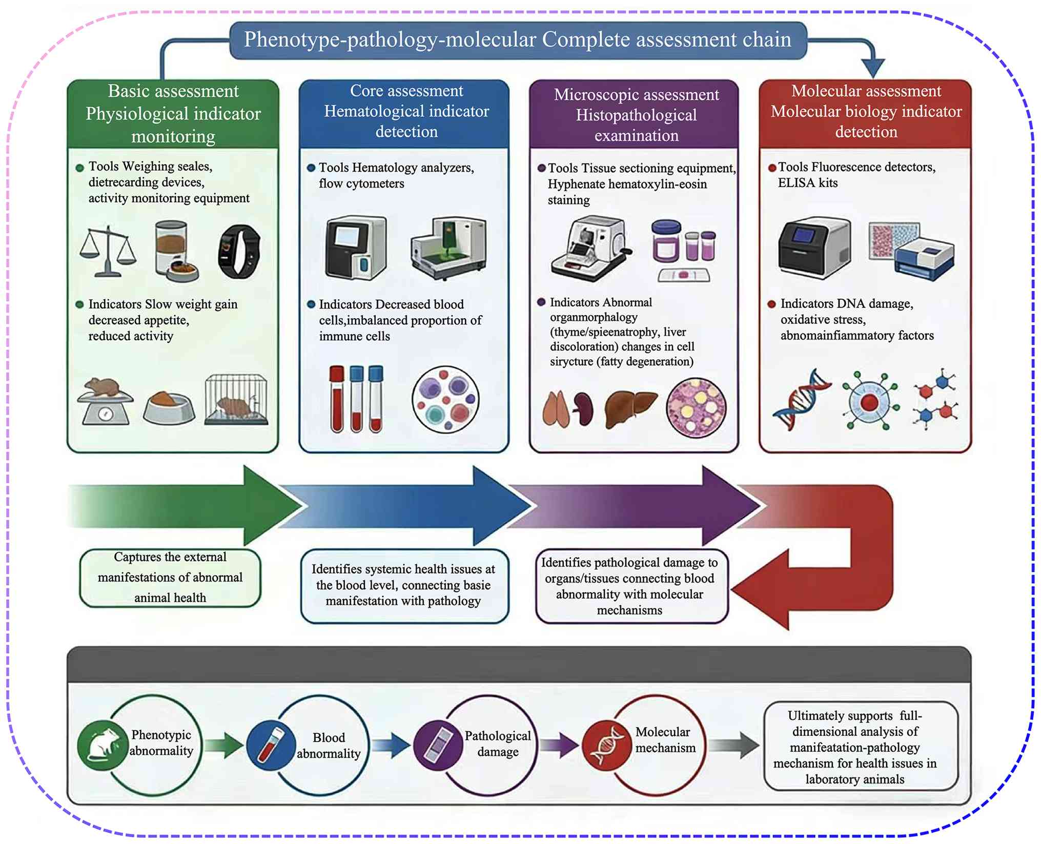
The utility of a preclinical model relies on its
construct validity, the fidelity with which it recapitulates the
human pathological landscape (120). Establishing a standardized
validation framework is key, as CRI presents a subtle, non-linear
progression distinct from the clear endpoints of acute syndrome
(121). A robust validation
strategy must triangulate data across physiological performance,
hematological integrity, histopathological architecture and
molecular signatures, providing systems-level corroboration of
radiation toxicity (122).
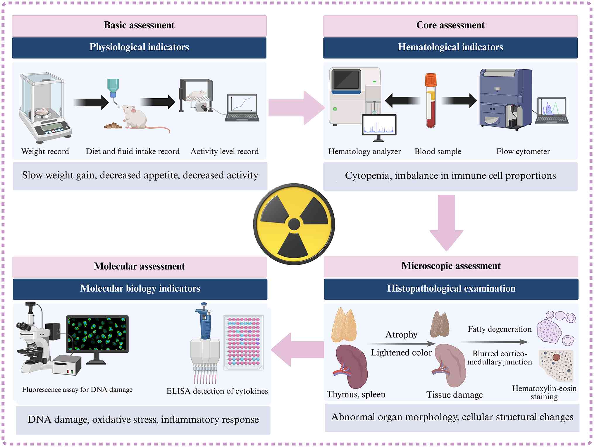
Physiological metrics serve as the frontline
indicators of systemic stress and sickness behavior, typically
preceding clinically detectable organ failure (123). Unlike the rapid weight loss
seen in acute syndrome, chronic radiation induces a wasting
phenotype akin to cancer cachexia (124). Longitudinal monitoring
typically reveals a blunted growth trajectory or gradual BMI
decline, driven by hypothalamic inflammation and sustained
catabolic signaling (125).
Clinical frailty index, an aggregate score of deficits (alopecia,
gait anomalies, grip strength), is a superior metric for
quantifying radiation-induced accelerated aging compared with body
weight alone (126). Changes in
feeding behavior reflect the disruption of the gut-brain axis
(127). Radiation-induced
mucositis and hypothalamic dysregulation manifest as anorexia and
polydipsia (128). A sustained
20-30% decline in caloric intake suggests a transition from
transient stress to chronic metabolic dysregulation (129). Quantitative behavioral assays
are key for validating CNS injury (130). Automated tracking using open
field test and Morris water maze detects decreased locomotor
velocity (lethargy), anxiety-like thigmotaxis and cognitive
deficits in spatial memory (131). These behavioral phenotypes are
associated with hippocampal neuroinflammation and serve as
non-invasive biomarkers for successful model induction (132).
Peripheral blood analysis provides a dynamic liquid
biopsy of the bone marrow microenvironment (133). In CRI models, the validation
endpoint is lineage skewing (a persistent imbalance in the
production of different hematopoietic cell types that reflects
underlying marrow dysfunction and stem cell injury. While acute
exposure causes rapid pancytopenia, chronic exposure typically
leads to a myeloid-biased output (increased granulocyte/lymphocyte
ratio) at the expense of lymphopoiesis, a hallmark of hematopoietic
aging (134). Progressive,
refractory normocytic anemia (hemoglobin <100 g/l) and elevated
red cell distribution width serve as diagnostic criteria for
cumulative marrow failure (135). Flow cytometric profiling must
assess functional status (136). Validated models demonstrate an
inverted CD4/CD8 ratio and the expansion of regulatory T cells
(137,138). Upregulation of exhaustion
markers on T cells-specifically programmed cell death protein 1
(PD-1) and T-cell immunoglobulin and mucin domain-containing
protein 3, along with decreased natural killer cell cytotoxicity,
provide definitive immunophenotypic evidence of chronic radiation
stress (138).
Histopathology remains the gold standard for
defining irreversible tissue remodeling (139). However, modern validation
requires specific staining and digital quantification beyond
standard hematoxylin and eosin staining (140). A pathognomonic feature of CRI
is the replacement of hematopoietic cellularity with adipose tissue
(141). Validated models
demonstrate a quantifiable shift in the marrow
adipocyte-to-hematopoietic cell ratio (142-144). A hallmark of late-stage CRI is
the excessive deposition of extracellular matrix (143). Validation should utilize
Masson's trichrome or Sirius Red staining to visualize collagen
(144). Immunohistochemistry
for α-smooth muscle actin (identifying myofibroblasts) is key for
distinguishing active fibrogenesis from static scarring (145). To enhance reproducibility,
studies employ AI-driven image analysis algorithms to score
fibrosis area fraction and cellularity, eliminating inter-observer
variability inherent in manual scoring (145,146).
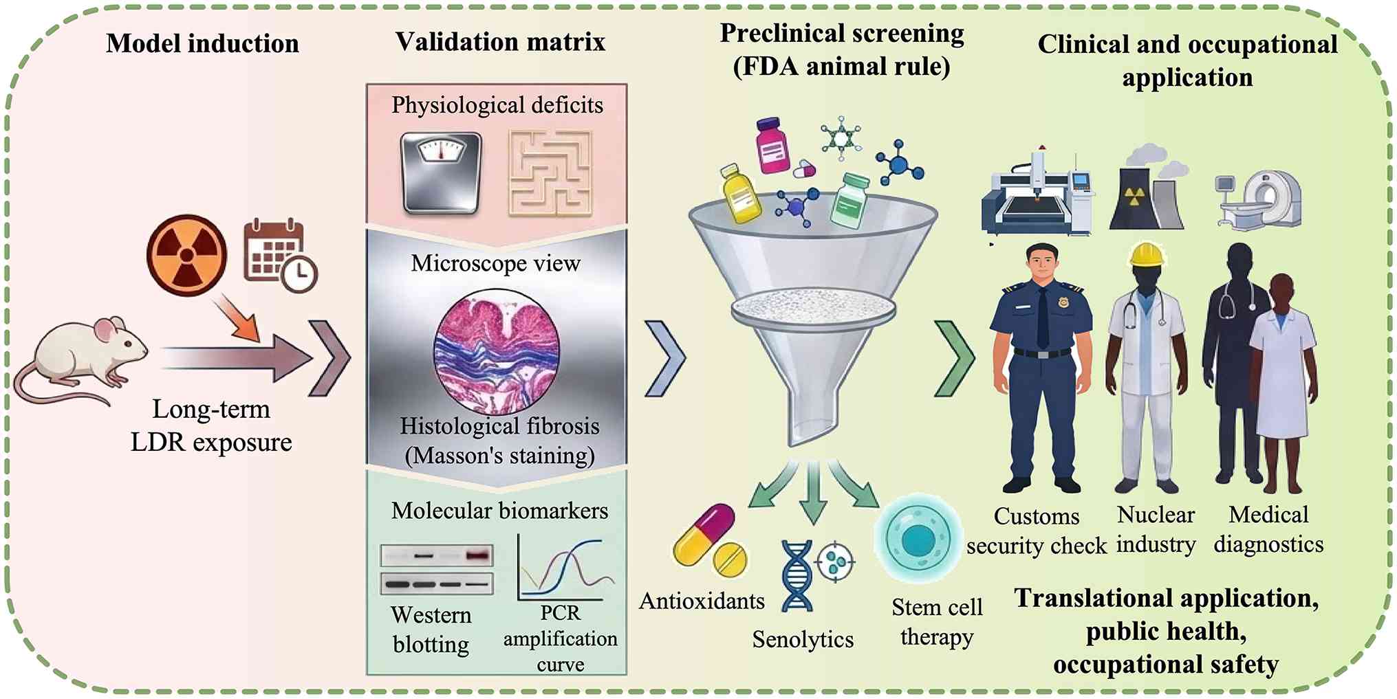
A model is considered validated only when it
satisfies this multidimensional matrix: Exhibiting the phenotypic
frailty, hematological skewing, histological fibrosis and the
molecular SASP signature characteristic of human CRI pathology
(Fig. 7; Table V) (155).
Animal models of chronic radiation injury allow the
mapping of complex signal transduction networks that drive chronic
pathology (158,159). Chronic LDR exposure triggers a
senescence-like arrest distinct from acute apoptosis (159). Models have been pivotal in
identifying the p53-p21 axis as the gatekeeper of this arrest and
the subsequent activation of NF-κB, which drives the SASP (160,161). This demonstrates how irradiated
cells survive but remain metabolically active, propagating
inflammation to bystander tissue (161). epigenetic scars (persistent,
long-term alterations in the epigenome (such as stable changes in
DNA methylation patterns) that continue to drive abnormal gene
expression long after the initial radiation exposure has ceased
(162,163). High-throughput bisulfite
sequencing in chronically exposed rodents has identified persistent
hypermethylation of tumor suppressor promoters
(p16INK4a) and global hypomethylation of
retrotransposons (163).
Integrative multi-omics (transcriptomics + metabolomics) has mapped
the dysregulation of the tricarboxylic acid cycle and fatty acid
oxidation, providing a systems-level view of radiation-induced
metabolic rewiring (164).
The primary industrial application of animal models
of chronic radiation injury is the rigorous screening of MCMs,
distinguishing between prophylactic agents (pre-exposure) and
mitigators (post-exposure) (165). In the screening phase, models
quantify the dose reduction factor of candidate agents (166). Effective agents must
demonstrate significant preservation of the HSC pool and a
reduction in marrow adiposity (167). Research has moved beyond free
radical scavengers to targeted therapy (168). Models are validating senolytics
(navitoclax, quercetin) that selectively eliminate senescent cells
to rejuvenate tissue (168,169). Furthermore, pharmacodynamic
studies optimize dosing to mitigate DEARE, ensuring that agents do
not interfere with DNA repair in a manner that promotes secondary
carcinogenesis (Table VII)
(57,170).
Beyond pharmaceuticals, models are key for testing
holistic strategies suitable for long-term occupational exposure
(171). Comparative studies
demonstrate that dietary polyphenols and probiotics remodel the gut
microbiome, which is often dysbiotic after radiation (171,172). This gut-bone marrow axis
modulation enhances Nrf2-ARE signaling, the master regulator of
antioxidant defense, thereby decreasing systemic oxidative stress
(malondialdehyde levels) (173). Controlled exercise studies
reveal that mechanical loading upregulates AMPK signaling,
promoting mitochondrial biogenesis (174,175). This counteracts
radiation-induced sarcopenia and fatigue, offering a
non-pharmacological strategy for astronauts or nuclear workers
(175).
Animal models provide the empirical data necessary
to construct AOPs, which link a molecular initiating event (such as
DNA oxidation) to an adverse outcome (such as fibrosis). By
exposing large cohorts to low-dose rates, researchers test the
validity of the LNT model vs. hormesis (adaptive response)
(176,177). This data is critical for
setting occupational limits (such as for interventional
cardiologists) (178).
Real-world risk involves combined stressors (179). Advanced models simulate the
exposome by co-exposing animals to radiation in the presence of
heavy metals (such as uranium mining) or microgravity (spaceflight)
(178,179). These studies reveal synergistic
toxicities that single-stressor models miss, refining ecological
risk assessments (179,180).
Future mechanistic inquiry will transcend bulk
tissue analysis to identify the spatiotemporal dynamics of injury
(189). By integrating
single-cell RNA sequencing with spatial transcriptomics,
researchers may move beyond merely identifying which cells are
damaged to understanding where they are located relative to the
niche (190). This will map the
neighborhood effects (how a senescent stromal cell spatially
corrupts adjacent stem cells via SASP) (191). Advanced genetic fate-mapping
will unveil the clonal trajectories of surviving cells,
distinguishing between regenerative and pre-leukemic clones
(192). This high-resolution
mapping may identify specific molecular switch points (such as
epigenetic loci) that determine whether a tissue undergoes repair
or irreversible fibrosis (193).
The therapeutic landscape is shifting from symptom
management to disease modification (194). A notable frontier is the
targeting of senescent cells (195). Future models will rigorously
validate senolytics (drugs that induce apoptosis in senescent
cells) and senomorphics (agents that suppress SASP without killing
the cell) (196). The goal is
to restore the irradiated microenvironment to a pre-exposure state
(197). While SC therapy holds
promise, the future may lie in extracellular vesicles (EVs) and
exosomes (198). Derived from
mesenchymal SCs, these cell-free cargoes carry regenerative miRs
and proteins with lower immunogenicity and tumorigenic risk than
live cells (199). Models
optimize engineered EVs loaded with specific radioprotective cargos
(mitochondria or mRNA) for targeted delivery (199,200). Clustered regularly interspaced
short palindromic repeats-associated protein 9 and base editing
in vivo will evolve toward correcting radiation-induced
mutations in somatic tissue or epigenetically silencing
pro-fibrotic genes (TGF-β) via modifiable promotors (201).
While the present review provides a methodological
framework for CRI modeling, limitations must be acknowledged.
First, due to the logistical, ethical and financial constraints of
conducting life-span studies in large animal models (such as
non-human primates), the majority of the mechanistic data,
particularly regarding SASP and inflammaging, is derived from
murine models. Extrapolating these rodent-derived mechanisms to
humans requires extreme caution, given the species-specific
differences in telomere dynamics, metabolic rate and immune
surveillance. Second, experimental models traditionally isolate
radiation as a single stressor to maintain controlled variables.
However, real-world human exposure (spaceflight or nuclear
accidents) typically involves complex exposome interactions, such
as concomitant exposure to heavy metals, psychological stress or
microgravity, which may alter the CRI trajectory. Finally, spatial
multi-omics and AI-driven digital twins are currently in their
early developmental or conceptual phases and require empirical
validation before widespread translational application.
In conclusion, CR animal models are transitioning
from descriptive pathology to predictive precision medicine
(211). This may facilitate
development of robust, personalized defenses against adverse health
effects of ionizing radiation (212).
Not applicable.
ZX, XiaodongL, ChunhuiY, LW, JM, QLiu, CL, QLi, YH,
JW, ChengzhuoY, FS, ChaoY, MW, BY, YL, QH, LZ, XuemeiL and
XiaobingL wrote the manuscript and constructed figures. XuemeiL and
XiaobingL edited the manuscript and supervised the study. Data
authentication is not applicable. All authors have read and
approved the final manuscript.
Not applicable.
Not applicable.
The authors declare that they have no competing
interests.
The authors would like to thank Professor Sheng Li
(Chongqing Medical and Pharmaceutical College, Chongqing, China)
for providing expert guidance on the conceptual framework and
feedback during the preparation of the manuscript.
The present study was supported by 2023 Chongqing Medical
Scientific Research Project (Joint Project of Chongqing Health
Commission and Science and Technology Bureau; grant no.
2023GGXM006), Joint Project of Chongqing Health Commission and
Science and Technology Bureau (Joint Key Laboratory Open Project)
(grant no. 2026KFXM051), Natural Science Foundation of Chongqing
(grant no. CSTB2025NSCO-GPX1116), 2026 Chongqing Municipal Health
Commission Traditional Chinese Medicine Research Project (grant no.
2026WSJK158), Technological Innovation Project of Shapingba
District, Chongqing (grant no. 2025016), 2024 Scientific research
Project of Chongqing Medical and Pharmaceutical College (grant no.
ygzrc2024101), Chongqing Municipal Education Commission Youth
Project (grant nos. KJQN202302811, KJQN202502819 and
KJQN202402821), 2024 Chongqing Medical and Pharmaceutical College
Innovation Research Group Project (grant no. ygz2024401), Science
and Health Joint Medical Research Project of Shapingba District,
Chongqing (grant no. 2024SQKWLHMS051), 2025 Teaching Research &
Reform Project of Chongqing Medical and Pharmaceutical College
(grant no. YGZJG2025322), The Key Laboratory Project of Chongqing
Medical and Pharmaceutical College (grant no. YGZPT2025101), 2025
Scientific Research Project of Chongqing Medical and Pharmaceutical
College (grant no. YGZZK2025116), 2025 Technological Innovation
Project of Shapingba District, Chongqing (grant no. 2025031) and
Joint project of Chongqing Health Commission and Science and
Technology Bureau (grant no. 2024MSXM115).
|
1
|
Abbas SF, Abdulkadim H, Al-Hashemi HA and
Hadi NR: Assessment of cardioprotective effect of necrostatin-1
stable in a mice model of acute doxorubicin-induced cardiotoxicity.
Wiad Lek. 76:1600–1607. 2023. View Article : Google Scholar : PubMed/NCBI
|
|
2
|
Ahmad A, Mallela SK, Ansari S, Alnukhali
M, Ali M, Merscher S, Pollack A, Zeidan YH, Fornoni A and Marples
B: Radiation-induced nephrotoxicity: Role of sphingomyelin
phosphodiesterase acid-like 3b. Int J Radiat Oncol Biol Phys.
121:1271–1281. 2025. View Article : Google Scholar
|
|
3
|
Ahmad A, Shi J, Ansari S, Afaghani J,
Molina J, Pollack A, Merscher S, Zeidan YH, Fornoni A and Marples
B: Noninvasive assessment of radiation-induced renal injury in
mice. Int J Radiat Biol. 97:664–674. 2021. View Article : Google Scholar : PubMed/NCBI
|
|
4
|
Ahmad A, Shi J, Ansari S, Merscher S,
Pollack A, Zeidan Y, Fornoni A and Marples B: Radiation
nephropathy: Mechanisms of injury and recovery in a murine model.
Radiother Oncol. 187:1098132023. View Article : Google Scholar : PubMed/NCBI
|
|
5
|
Akter R, Rahman MA, Rafi MKJ, Siddique TA,
Bithy FY, Akter S, Nisa FY, Khan MAN and Sultana F: The protective
effect of lasia spinosa (Linn.) dissipates chemical-induced
cardiotoxicity in an animal model. Cardiovasc Toxicol. 23:32–45.
2023. View Article : Google Scholar : PubMed/NCBI
|
|
6
|
Al-Rubaiey S, Senger C, Bukatz J,
Krantchev K, Janas A, Eitner C, Nieminen-Kelhä M, Brandenburg S,
Zips D, Vajkoczy P and Acker G: Determinants of cerebral
radionecrosis in animal models: A systematic review. Radiother
Oncol. 199:1104442024. View Article : Google Scholar : PubMed/NCBI
|
|
7
|
Ancel L, Grison S, Gabillot O, Gueguen J,
Svilar L, Guen BL, Gruel G, Benderitter M, Martin JC, Souidi M, et
al: Metabolomics identifies plasma biomarkers of localized
radiation injury. Sci Rep. 15:21662025. View Article : Google Scholar : PubMed/NCBI
|
|
8
|
Ancel L, Gueguen J, Thoër G, Marçais J,
Chemloul A, Le Guen B, Benderitter M, Tamarat R, Souidi M,
Benadjaoud MA and Flamant S: Presymptomatic microRNA-based
biomarker signatures for the prognosis of localized radiation
injury in mice. PLoS One. 20:e03297372025. View Article : Google Scholar : PubMed/NCBI
|
|
9
|
Aqeel M, Medhora M, Gore E, Borkenhagen J,
Klawikowski S, Eastwood D, Banerjee A and Jacobs ER: Evaluation of
radiation-induced pleural effusions after radiotherapy to support
development of animal models of radiation pneumonitis. Health Phys.
121:434–443. 2021. View Article : Google Scholar : PubMed/NCBI
|
|
10
|
Athanasiadi I, Perez WD, Plantenga JM,
Jones-Hall Y and Perez-Torres CJ: Feasibility of a mini-pig model
of radiation-induced brain injury to one cerebral hemisphere.
Radiat Oncol. 16:302021. View Article : Google Scholar : PubMed/NCBI
|
|
11
|
Azmoonfar R, Mirzaei F, Najafi M, Varkeshi
M, Ghazikhanlousani K, Momeni S and Saber K: Radiation-induced
testicular damage in mice: Protective effects of apigenin revealed
by histopathological evaluation. Curr Radiopharm. 17:238–246. 2024.
View Article : Google Scholar
|
|
12
|
Beach T, Authier S, Javitz HS, Wong K,
Bakke J, Gahagen J, Bunin DI and Chang PY: Total body irradiation
models in NHPs-consideration of animal sex and provision of
supportive care to advance model development. Int J Radiat Biol.
97:126–130. 2021. View Article : Google Scholar
|
|
13
|
Beach T, Bakke J, McDonald JT, Riccio E,
Javitz HS, Nishita D, Kapur S, Bunin DI and Chang PY: Delayed
effects of radiation exposure in a C57L/J mouse model of partial
body irradiation with ~2.5% bone marrow shielding. Front Public
Health. 12:13495522024. View Article : Google Scholar : PubMed/NCBI
|
|
14
|
Behnke V, Wolf A, Hector M and Langmann T:
C3aR1-deletion delays retinal degeneration in a white-light damage
mouse model. Invest Ophthalmol Vis Sci. 66:152025. View Article : Google Scholar : PubMed/NCBI
|
|
15
|
Kamiya K, Ozasa K, Akiba S, Niwa O, Kodama
K, Takamura N, Zaharieva EK, Kimura Y and Wakeford R: Long-term
effects of radiation exposure on health. Lancet. 386:469–478. 2015.
View Article : Google Scholar : PubMed/NCBI
|
|
16
|
Lin RT, Boonhat H, Lin YY, Klebe S and
Takahashi K: Health effects of occupational and environmental
exposures to nuclear power plants: A meta-analysis and
meta-regression. Curr Environ Health Rep. 11:329–339. 2024.
View Article : Google Scholar : PubMed/NCBI
|
|
17
|
Britten RA, Fesshaye AS, Tidmore A, Tamgue
EN and Alvarado-Arriaga PA: Different spectrum of space radiation
induced cognitive impairments in radiation-naïve and adapted rats.
Life Sci Space Res (Amst). 43:68–74. 2024.PubMed/NCBI
|
|
18
|
Bolduc DL, Cary LH, Kiang JG, Kurada L,
Kumar VP, Edma SA, Olson MG, Vergara VB, Bistline DD, Reese M, et
al: Natural-history characterization of a murine partial-body
irradiation model system: Establishment of a multiple-parameter
based GI-ARS severity-scoring system. Radiat Res. 201:406–417.
2024. View Article : Google Scholar : PubMed/NCBI
|
|
19
|
Camargo CP, Zavala JPM, Serafim GP,
Carvalho HA, Santos BL, Nunes VA, Uno M, Alves MJF, Furuya TK and
Gemperli R: Effects of adipocyte-derived stem cells treatment for
moderate radiodermatitis: Animal model. Int J Radiat Biol.
101:827–834. 2025. View Article : Google Scholar : PubMed/NCBI
|
|
20
|
Cao H, Yue L, Shao J, Kong F, Liu S, Huai
H, He Z, Mao Z, Yang Y, Tan Y and Wang H: Small extracellular
vesicles derived from umbilical cord mesenchymal stem cells
alleviate radiation-induced cardiac organoid injury. Stem Cell Res
Ther. 15:4932024. View Article : Google Scholar : PubMed/NCBI
|
|
21
|
Chan LSA, Gu LC and Wells RA: The effects
of secondary iron overload and iron chelation on a
radiation-induced acute myeloid leukemia mouse model. BMC Cancer.
21:5092021. View Article : Google Scholar : PubMed/NCBI
|
|
22
|
Chen X, Chen L, Tan J, Zhang L, Xia J,
Cheng B and Zhang W: Rspo1-LGR4 axis in BMSCs protects bone against
radiation-induced injury through the mTOR-dependent autophagy
pathway. J Cell Physiol. 236:4273–4289. 2021. View Article : Google Scholar
|
|
23
|
Chen Y, Cheng Z, Ma L and Shi CM: Analysis
of the number, type, and functional heterogeneity of senescent
cells in the radiation-induced skin wounds in mice. Zhonghua Shao
Shang Yu Chuang Mian Xiu Fu Za Zhi. 41:577–586. 2025.In Chinese.
PubMed/NCBI
|
|
24
|
Cyran M, Stawarz K, Chambily L, Kusza K
and Siemionow M: Assessment of hematopoietic response to total body
irradiation in a rat experimental model. Ann Plast Surg.
93:100–106. 2024. View Article : Google Scholar : PubMed/NCBI
|
|
25
|
Dai J, Gao Y, Wang J, Zhang S and Liu P:
Effects of ionizing radiation on intestinal bile acid metabolism:
Mechanism of the radioprotective effect of glycoursodeoxycholic
acid. Sichuan Da Xue Xue Bao Yi Xue Ban. 55:1195–1201. 2024.In
Chinese. PubMed/NCBI
|
|
26
|
Dar HH, Epperly MW, Tyurin VA, Amoscato
AA, Anthonymuthu TS, Souryavong AB, Kapralov AA, Shurin GV,
Samovich SN, St Croix CM, et al: P. aeruginosa augments irradiation
injury via 15-lipoxygenase-catalyzed generation of 15-HpETE-PE and
induction of theft-ferroptosis. JCI Insight. 7:e1560132022.
View Article : Google Scholar : PubMed/NCBI
|
|
27
|
Dasgupta Q, Jiang A, Wen AM, Mannix RJ,
Man Y, Hall S, Javorsky E and Ingber DE: A human lung
alveolus-on-a-chip model of acute radiation-induced lung injury.
Nat Commun. 14:65062023. View Article : Google Scholar : PubMed/NCBI
|
|
28
|
Dawoud M, Attallah KM, Ibrahim IT, Karam
HM and Ibrahim AA: MitoQ and its hyaluronic acid-based
nanopreparation mitigating gamma radiation-induced intestinal
injury in mice: Alleviation of oxidative stress and apoptosis.
Naunyn Schmiedebergs Arch Pharmacol. 397:5193–5205. 2024.
View Article : Google Scholar
|
|
29
|
Dhingra N and Gupta V, Tyagi A, Agrawala
PK and Gupta V: Trichostatin A ameliorated combined radiation and
skin wound injury-induced mortality and hematopoietic suppression
in a rat model. Int J Radiat Biol. 101:952–972. 2025. View Article : Google Scholar : PubMed/NCBI
|
|
30
|
Diaz MF, Horton PD, Dumbali SP, Kumar A,
Livingston M, Skibber MA, Mohammadalipour A, Gill BS, Zhang S, Cox
CS Jr and Wenzel PL: Bone marrow stromal cell therapy improves
survival after radiation injury but does not restore endogenous
hematopoiesis. Sci Rep. 10:222112020. View Article : Google Scholar : PubMed/NCBI
|
|
31
|
Dos Santos M, Demarquay C, Ermeneux L,
Aberkane F, Bléry P, Weiss P, Milliat F and Mathieu N: Refining the
mandibular osteoradionecrosis rat model by in vivo longitudinal μCT
analysis. Sci Rep. 11:222412021. View Article : Google Scholar
|
|
32
|
Down JD, Cornwall-Brady MR, Huang W,
Hurwitz M, Floyd SR and Yilmaz OH: Selecting the most relevant
mouse strains for evaluating radiation-induced multiple tissue
injury after leg-shielded partial-body gamma irradiation. Radiat
Res. 202:510–522. 2024. View Article : Google Scholar : PubMed/NCBI
|
|
33
|
Dreyfuss AD, Goia D, Shoniyozov K, Shewale
SV, Velalopoulou A, Mazzoni S, Avgousti H, Metzler SD, Bravo PE,
Feigenberg SJ, et al: A novel mouse model of radiation-induced
cardiac injury reveals biological and radiological biomarkers of
cardiac dysfunction with potential clinical relevance. Clin Cancer
Res. 27:2266–2276. 2021. View Article : Google Scholar : PubMed/NCBI
|
|
34
|
Duval KEA, Aulwes E, Zhang R, Rahman M,
Ashraf MR, Sloop A, Sunnerberg J, Williams BB, Cao X, Bruza P, et
al: Comparison of tumor control and skin damage in a mouse model
after ultra-high dose rate irradiation and conventional
irradiation. Radiat Res. 200:223–231. 2023. View Article : Google Scholar : PubMed/NCBI
|
|
35
|
Ewing LE, Biju PG, Pathak R, Melnyk S,
Hauer-Jensen M and Koturbash I: Methods for induction and
assessment of intestinal permeability in rodent models of radiation
injury. Methods Cell Biol. 168:235–247. 2022. View Article : Google Scholar : PubMed/NCBI
|
|
36
|
Fang D, Zhao H, Pei L, Jiang K, Gan Y,
Zhai X, Zhang L, Cheng Y, Liu C, Du J and Gao F: Diprovocim
protects against the radiation-induced damage via the TLR2
signaling pathway. Mol Med. 31:1392025. View Article : Google Scholar : PubMed/NCBI
|
|
37
|
Farber E, Kwiecien JM, Bojic D, Ngu M,
Akohene-Mensah P, Vanhie JJ, Lloyd J, Larkin J and DE Lisio M:
Exercise improves cancer-free survival and health span in a model
of radiation-induced cancer. Med Sci Sports Exerc. 53:2254–2263.
2021. View Article : Google Scholar
|
|
38
|
Farese AM, Drouet M, Herodin F, Bertho JM,
Thrall KD, Authier S, Doyle-Eisele M and MacVittie TJ: Acute
radiation effects, the H-ARS in the non-human primate: A review and
new data for the cynomolgus macaque with reference to the rhesus
macaque. Health Phys. 121:304–330. 2021. View Article : Google Scholar : PubMed/NCBI
|
|
39
|
Farh ME, Kim HJ, Kim SY, Lee JH, Lee H,
Cui R, Han S, Kim DW, Park S, Lee YJ, et al: Transcriptional
changes in radiation-induced lung injury: A comparative analysis of
two radiation doses for preclinical research. Int J Mol Sci.
25:37662024. View Article : Google Scholar : PubMed/NCBI
|
|
40
|
Feng J, Liu X, Xu L, Wang K, He S, Wang X,
Huang Y, Wang S, Lei X, Huang L, et al: Optimization and
characterization of acute radiation-induced esophagitis in mice.
Radiat Oncol. 20:1472025. View Article : Google Scholar : PubMed/NCBI
|
|
41
|
Feng Y, Kong J, Sun W, Li Y, Ren F, Sun X,
Li M, Liu Y, Sun S and Qin H: NLRP3 regulates macrophage function
by M-CSF/M-CSFR signaling in acute radiation-induced lung injury.
Mol Immunol. 183:213–224. 2025. View Article : Google Scholar : PubMed/NCBI
|
|
42
|
Fish BL, Hart B, Gasperetti T, Narayanan
J, Gao F, Veley D, Pierce L, Himburg HA, MacVittie T and Medhora M:
IPW-5371 mitigates the delayed effects of acute radiation exposure
in WAG/RijCmcr rats when started 15 days after PBI with bone marrow
sparing. Int J Radiat Biol. 99:1119–1129. 2023. View Article : Google Scholar : PubMed/NCBI
|
|
43
|
Fish BL, MacVittie TJ, Gao F, Narayanan J,
Gasperetti T, Scholler D, Sheinin Y, Himburg HA, Hart B and Medhora
M: Rat Models of partial-body irradiation with bone marrow-sparing
(Leg-out PBI) Designed for FDA approval of countermeasures for
mitigation of acute and delayed injuries by radiation. Health Phys.
121:419–433. 2021. View Article : Google Scholar : PubMed/NCBI
|
|
44
|
Fu J, Liu X, Zhou Y, Zhao S, Zeng L, Pan
Y, Zhang J, Prise KM, Shao C and Xu Y: Development of delayed
pulmonary toxicities and transcriptional changes in pre-existing
interstitial lung disease mice after partial thoracic irradiation.
Radiat Oncol. 20:202025. View Article : Google Scholar : PubMed/NCBI
|
|
45
|
Fuentes A, Hyde D, Johnson I and Haston
CK: Acute radiation-induced hematopoietic depletion does not alter
the onset or severity of pneumonitis in mice. Radiat Res.
196:297–305. 2021. View Article : Google Scholar : PubMed/NCBI
|
|
46
|
Gans I, El Abiad JM, James AW, Levin AS
and Morris CD: Administration of TGF-ß inhibitor mitigates
radiation-induced fibrosis in a mouse model. Clin Orthop Relat Res.
479:468–474. 2021. View Article : Google Scholar
|
|
47
|
Gao J, Li Y, Chen J, Feng W, Bu J, Lu Z
and Wang J: Emodin ameliorates acute radiation proctitis in mice by
regulating AKT/MAPK/NF-κB/VEGF pathways. Int Immunopharmacol.
132:1119452024. View Article : Google Scholar
|
|
48
|
Gassert FT, Burkhardt R, Gora T, Pfeiffer
D, Fingerle AA, Sauter AP, Schilling D, Rummeny EJ, Schmid TE,
Combs SE, et al: X-ray dark-field CT for early detection of
radiation-induced lung injury in a murine model. Radiology.
303:696–698. 2022. View Article : Google Scholar : PubMed/NCBI
|
|
49
|
Gibbs A, Gupta P, Mali B, Poirier Y,
Gopalakrishnan M, Newman D, Zodda A, Down JD, Serebrenik AA, Kaytor
MD and Jacksone IL: A C57L/J mouse model of the delayed effects of
acute radiation exposure in the context of evolving multi-organ
dysfunction and failure after total-body irradiation with 2.5% bone
marrow sparing. Radiat Res. 199:319–335. 2023. View Article : Google Scholar : PubMed/NCBI
|
|
50
|
Glowacki J, Epperly MW, Bellare A, Wipf P
and Greenberger JS: Combined injury: Irradiation with skin or bone
wounds in rodent models. J Radiol Prot. 41:2021. View Article : Google Scholar : PubMed/NCBI
|
|
51
|
Gu J, Zhao L, Chen YZ, Guo YX, Sun Y, Guo
Q, Duan GX, Li C, Tang ZB, Zhang ZX, et al: Preventive effect of
sanguinarine on intestinal injury in mice exposed to whole
abdominal irradiation. Biomed Pharmacother. 146:1124962022.
View Article : Google Scholar
|
|
52
|
Guan B, Li D and Meng A: Development of
radiation countermeasure agents for acute radiation syndromes.
Animal Model Exp Med. 6:329–336. 2023. View Article : Google Scholar : PubMed/NCBI
|
|
53
|
Guan H, Zhang W, Xie D, Nie Y, Chen S, Sun
X, Zhao H, Liu X, Wang H, Huang X, et al: Cytosolic release of
mitochondrial DNA and associated cGAS signaling mediates
radiation-induced hematopoietic injury of mice. Int J Mol Sci.
24:40202023. View Article : Google Scholar : PubMed/NCBI
|
|
54
|
Guan Z, Zhang J, Jiang N, Tian M, Wang H
and Liang B: Efficacy of mesenchymal stem cell therapy in rodent
models of radiation-induced xerostomia and oral mucositis: A
systematic review. Stem Cell Res Ther. 14:822023. View Article : Google Scholar : PubMed/NCBI
|
|
55
|
Hansel C, Barr S, Schemann AV, Lauber K,
Hess J, Unger K, Zitzelsberger H, Jendrossek V and Klein D:
Metformin protects against radiation-induced acute effects by
limiting senescence of bronchial-epithelial cells. Int J Mol Sci.
22:70642021. View Article : Google Scholar : PubMed/NCBI
|
|
56
|
Hanson I, Juvkam IS, Zlygosteva O, Søland
TM, Galtung HK, Malinen E and Edin NFJ: TGF-β3 increases the
severity of radiation-induced oral mucositis and salivary gland
fibrosis in a mouse model. Int J Radiat Biol. 100:767–776. 2024.
View Article : Google Scholar
|
|
57
|
Holmes-Hampton GP, Kumar VP, Valenzia K
and Ghosh SP: FSL-1: A synthetic peptide increases survival in a
murine model of hematopoietic acute radiation syndrome. Radiat Res.
201:449–459. 2024. View Article : Google Scholar : PubMed/NCBI
|
|
58
|
Hormozi Moghaddam Z, Mokhtari-Dizaji M,
Nilforoshzadeh MA, Bakhshandeh M and Ghaffari Khaligh S:
Low-intensity ultrasound combined with allogenic adipose-derived
mesenchymal stem cells (AdMSCs) in radiation-induced skin injury
treatment. Sci Rep. 10:200062020. View Article : Google Scholar : PubMed/NCBI
|
|
59
|
Horseman T, Rittase WB, Slaven JE,
Bradfield DT, Frank AM, Anderson JA, Hays EC, Ott AC, Thomas AE,
Huppmann AR, et al: Ferroptosis, inflammation, and microbiome
alterations in the intestine in the Göttingen minipig model of
hematopoietic-acute radiation syndrome. Int J Mol Sci. 25:45352024.
View Article : Google Scholar
|
|
60
|
Horseman TS, Parajuli B, Frank AM, Weaver
A, Schauer DA, Moran S, Anderson JA, Holmes-Hampton GP and
Burmeister DM: Microbiome and inflammasome alterations found during
radiation dose finding in a sinclair minipig model of
gastrointestinal acute radiation syndrome. Shock. 62:556–564. 2024.
View Article : Google Scholar : PubMed/NCBI
|
|
61
|
Hritzo B, Aghdam SY, Legesse B, Kaur A,
Cao M, Boerma M, Chakraborty N, Dimitrov G, Gautam A, Hammamieh R,
et al: Late health effects of partial body irradiation injury in a
minipig model are associated with changes in systemic and cardiac
IGF-1 signaling. Int J Mol Sci. 22:32862021. View Article : Google Scholar : PubMed/NCBI
|
|
62
|
Hritzo B, Legesse B, Ward JM, Kaur A,
Aghdam SY, Kenchegowda D, Holmes-Hampton GP and Moroni M:
Investigating the multifaceted nature of radiation-induced
coagulopathies in a Göttingen minipig model of hematopoietic acute
radiation syndrome. Radiat Res. 196:156–174. 2021. View Article : Google Scholar : PubMed/NCBI
|
|
63
|
Huang B, An L, Su W, Yan T, Zhang H and Yu
DJ: Exploring the alterations and function of skin microbiome
mediated by ionizing radiation injury. Front Cell Infect Microbiol.
12:10295922022. View Article : Google Scholar : PubMed/NCBI
|
|
64
|
Huang H, Shi Y, Liu X, Hao L, Zhu J and Lu
J: Protective effect of vinegar powder on 60Coγ
radiation-injured mice and its potential mechanism. J Nutr.
155:3663–3675. 2025. View Article : Google Scholar : PubMed/NCBI
|
|
65
|
Huang J, Zhang Y, Jiang F, Zhang Y, Li S,
He S, Sun J, Chen D, Pang Q and Wu Y: Bach1 deficiency ameliorates
radiation pneumonitis via activating TFAM signaling pathway.
Antioxid Redox Signal. 43:75–91. 2025. View Article : Google Scholar : PubMed/NCBI
|
|
66
|
Hunter KS, Carnell LS, DiCarlo AL, Hoffman
CM, Loelius SG and Homer M: Interagency approaches to animal models
for acute radiation exposure. Int J Radiat Biol. 97(Suppl 1):
S2–S5. 2021. View Article : Google Scholar : PubMed/NCBI
|
|
67
|
Iddins CJ, DiCarlo AL, Ervin MD,
Herrera-Reyes E and Goans RE: Cutaneous and local radiation
injuries. J Radiol Prot. 42:2022. View Article : Google Scholar :
|
|
68
|
Jackson IL and Doyle-Eisele M: Animal
model considerations for medical countermeasure development for
radiation and sulfur mustard exposures: Animal models for radiation
and HD exposures. Disaster Med Public Health Prep.
18:e812023.PubMed/NCBI
|
|
69
|
Jackson IL, Gurung G, Poirier Y,
Gopalakrishnan M, Cohen EP, Donohue TS, Newman D and Vujaskovic Z:
A New Zealand white rabbit model of thrombocytopenia and
coagulopathy following total body irradiation across the dose range
to induce the hematopoietic-subsyndrome of acute radiation
syndrome. Int J Radiat Biol. 97(Suppl 1): S19–S31. 2021. View Article : Google Scholar
|
|
70
|
Jacobson JR: Sphingolipids as a novel
therapeutic target in radiation-induced lung injury. Cell Biochem
Biophys. 79:509–516. 2021. View Article : Google Scholar : PubMed/NCBI
|
|
71
|
Jiajia D, Yiping W, Enyan J, Shouwu Z,
Shuai Y, Xiaojian Z, Juan J, Zhen F, Jia Z, Jikai W, et al:
Maresin-1 alleviates lipid peroxidation-induced ferroptosis after
radiation-induced brain injury in mice through the RORα/NRF2
pathway. Exp Neurol. 389:1152582025. View Article : Google Scholar
|
|
72
|
Jiang H, Wang R, Liu A, Liu J, Wang X, Lin
W, Ren C, Liu K, Zhao X and Li Y: RAS-RH up-regulates the level of
miR-126 and inhibits the opening of mPTP in a rat model of coronary
microvascular disease. Microvasc Res. 162:1048562025. View Article : Google Scholar : PubMed/NCBI
|
|
73
|
Jiao W, Cheng Y, Liu C, Feng J, Lin J and
Shen Y: SGLT1 inhibition alleviates radiation-induced intestinal
damage through promoting mitochondrial homeostasis. Free Radic Biol
Med. 224:831–845. 2024. View Article : Google Scholar : PubMed/NCBI
|
|
74
|
Kayalı A, Arda DB, Bora ES, Uyanikgil Y,
Atasoy Ö and Erbaş O: Oxytocin: A shield against radiation-induced
lung injury in rats. Tomography. 10:1342–1353. 2024. View Article : Google Scholar
|
|
75
|
Kenchegowda D, Bolduc DL, Kurada L and
Blakely WF: Severity scoring systems for radiation-induced GI
injury-prioritization for use of GI-ARS medical countermeasures.
Int J Radiat Biol. 99:1037–1045. 2023. View Article : Google Scholar
|
|
76
|
Kenchegowda D, Seed TM and Singh VK: What
are the practical, ethical, and pathobiological considerations in
the use of minipigs as an animal model in drug discovery for acute
radiation syndrome and delayed effects of acute radiation exposure?
Expert Opin Drug Discov. 16:119–124. 2021. View Article : Google Scholar
|
|
77
|
Khodamoradi E, Rahmani N, Rashidi K,
Najafi M, Shahsavari S and Mohammadi M: Exploring the potential of
metformin in mitigating radiation-induced gastrointestinal and
hematopoietic system injury in rats after whole-body X-ray
radiation: An experimental study. Curr Radiopharm. 17:200–208.
2024. View Article : Google Scholar : PubMed/NCBI
|
|
78
|
Kim HC, Oh H, You JS and Chung YE:
Glycyrrhizin ameliorating sterile inflammation induced by low-dose
radiation exposure. Sci Rep. 11:183562021. View Article : Google Scholar : PubMed/NCBI
|
|
79
|
Kim SY, Park S, Cui R, Lee H, Choi H, Farh
ME, Jo HI, Lee JH, Song HJ, Lee YJ, et al: NXC736 Attenuates
radiation-induced lung fibrosis via regulating NLRP3/IL-1β
signaling pathway. Int J Mol Sci. 24:162652023. View Article : Google Scholar
|
|
80
|
Klaus R, Niyazi M and Lange-Sperandio B:
Radiation-induced kidney toxicity: Molecular and cellular
pathogenesis. Radiat Oncol. 16:432021. View Article : Google Scholar : PubMed/NCBI
|
|
81
|
Kleiman NJ, Edmondson EF, Weil MM,
Fallgren CM, King A, Schmidt C and Hall EJ: Radiation cataract in
heterogeneous stock mice after γ-ray or HZE ion exposure. Life Sci
Space Res (Amst). 40:97–105. 2024.PubMed/NCBI
|
|
82
|
Kočović DM, Bajuk-Bogdanović D, Pećinar I,
Nedeljković BB, Daković M and Andjus PR: Assessment of cellular and
molecular changes in the rat brain after gamma radiation and
radioprotection by anisomycin. J Radiat Res. 62:793–803. 2021.
View Article : Google Scholar
|
|
83
|
Krasnoselsky MV, Pushkar OS, Simonova LI
and Myroshnychenko MS: The effect of photodynamic therapy and
platelet-enriched plasma on the healing of skin radiation ulcers
infected by staphylococcus aureus. Probl Radiac Med Radiobiol.
25:338–352. 2020.In English, Ukrainian. View Article : Google Scholar : PubMed/NCBI
|
|
84
|
Kumar VP, Holmes-Hampton GP, Biswas S,
Stone S, Sharma NK, Hritzo B, Guilfoyle M, Eichenbaum G, Guha C and
Ghosh SP: Mitigation of total body irradiation-induced mortality
and hematopoietic injury of mice by a thrombopoietin mimetic
(JNJ-26366821). Sci Rep. 12:34852022. View Article : Google Scholar : PubMed/NCBI
|
|
85
|
Kumar VP, Jaiswal S, Wuddie K, Ward JM,
Lawrence M and Ghosh SP: Development of a radiation-induced
pulmonary fibrosis partial body irradiation model in C57BL/6 mice.
Radiat Res. 201:460–470. 2024. View Article : Google Scholar : PubMed/NCBI
|
|
86
|
Kumar VP, Wuddie K, Tsioplaya A, Weaver A,
Holmes-Hampton GP and Ghosh SP: Development of a multi-organ
radiation injury model with precise dosimetry with focus on GI-ARS.
Radiat Res. 201:19–34. 2024.
|
|
87
|
Kura B, Kalocayova B, Szeiffova Bacova B,
Fulop M, Sagatova A, Sykora M, Andelova K, Abuawad Z and Slezak J:
The effect of selected drugs on the mitigation of myocardial injury
caused by gamma radiation. Can J Physiol Pharmacol. 99:80–88. 2021.
View Article : Google Scholar : PubMed/NCBI
|
|
88
|
Kwak SY, Jang WI, Park S, Cho SS, Lee SB,
Kim MJ, Park S, Shim S and Jang H: Metallothionein 2 activation by
pravastatin reinforces epithelial integrity and ameliorates
radiation-induced enteropathy. EBioMedicine. 73:1036412021.
View Article : Google Scholar : PubMed/NCBI
|
|
89
|
L'Abbate S, Chianca M, Fabiani I, Del
Franco A, Giannoni A, Vergaro G, Grigoratos C, Kusmic C, Passino C,
D'Alessandra Y, et al: In vivo murine models of cardiotoxicity due
to anticancer drugs: challenges and opportunities for clinical
translation. J Cardiovasc Transl Res. 15:1143–1162. 2022.
View Article : Google Scholar : PubMed/NCBI
|
|
90
|
Lai YF, Wang HY and Peng RY: Establishment
of injury models in studies of biological effects induced by
microwave radiation. Mil Med Res. 8:122021.PubMed/NCBI
|
|
91
|
Laiakis EC, McCart EA, Deziel A, Rittase
WB, Bouten RM, Jha J, Wilkins WL, Day RM and Fornace AJ Jr: Effect
of 3,3'-diindolylmethane on pulmonary injury following thoracic
irradiation in CBA mice. Health Phys. 119:746–757. 2020. View Article : Google Scholar : PubMed/NCBI
|
|
92
|
Lawrence J, Seelig D, Demos-Davies K,
Ferreira C, Ren Y, Wang L, Alam SK, Yang R, Guedes A, Craig A and
Hoeppner LH: Radiation dermatitis in the hairless mouse model
mimics human radiation dermatitis. Sci Rep. 14:248192024.
View Article : Google Scholar : PubMed/NCBI
|
|
93
|
Lebon C, Malaise D, Rimbert N, Billet M,
Ramasamy G, Villaret J, Pouzoulet F, Matet A and Behar-Cohen F:
Role of inflammation in a rat model of radiation retinopathy. J
Neuroinflammation. 21:1622024. View Article : Google Scholar : PubMed/NCBI
|
|
94
|
Lee CL, Wright AO, Lee JW, Brownstein J,
Hasapis S, Satow S, Da Silva Campos L, Williams N, Ma Y, Luo L, et
al: Sensitization of endothelial cells to ionizing radiation
exacerbates delayed radiation myelopathy in mice. Radiat Res.
197:193–198. 2021. View Article : Google Scholar
|
|
95
|
Lee J, Jeon BS, Kang S, Son Y, Lim YB, Bae
MJ, Jo WS, Lee CG, Shin IS, Moon C, et al: Protective effects of
tauroursodeoxycholate against radiation-induced intestinal injury
in a mouse model. Biochem Biophys Res Commun. 724:1502262024.
View Article : Google Scholar : PubMed/NCBI
|
|
96
|
Levy K, Natarajan S, Wang J, Chow S,
Eggold JT, Loo PE, Manjappa R, Melemenidis S, Lartey FM, Schüler E,
et al: Abdominal FLASH irradiation reduces radiation-induced
gastrointestinal toxicity for the treatment of ovarian cancer in
mice. Sci Rep. 10:216002020. View Article : Google Scholar : PubMed/NCBI
|
|
97
|
Li L, Nie X, Zhang P, Huang Y, Ma L, Li F,
Yi M, Qin W and Yuan X: Dexrazoxane ameliorates radiation-induced
heart disease in a rat model. Aging (Albany NY). 13:3699–3711.
2021. View Article : Google Scholar : PubMed/NCBI
|
|
98
|
Li L, Zhang S, Ge C, Ji L, Lv Y, Zhao C,
Xu L, Zhang J, Song C, Chen J, et al: HSCs transdifferentiate
primarily to pneumonocytes in radiation-induced lung damage repair.
Aging (Albany NY). 13:8335–8354. 2021. View Article : Google Scholar : PubMed/NCBI
|
|
99
|
Li N, Zhao W, Li J, Zhang D, Li K, Yang M,
Lu X, Du L, Xu C and Liu Q: Autophagy and exosome dynamics in
radiation-induced pulmonary fibrosis: The critical role of TRIB3.
Respir Res. 26:1942025. View Article : Google Scholar : PubMed/NCBI
|
|
100
|
Li W, Jiang L, Lu X, Liu X and Ling M:
Curcumin protects radiation-induced liver damage in rats through
the NF-κB signaling pathway. BMC Complement Med Ther. 21:102021.
View Article : Google Scholar
|
|
101
|
Li W, Lin Y, Luo Y, Wang Y, Lu Y, Li Y and
Guo H: Vitamin D receptor protects against radiation-induced
intestinal injury in mice via inhibition of intestinal crypt
stem/progenitor cell apoptosis. Nutrients. 13:29102021. View Article : Google Scholar : PubMed/NCBI
|
|
102
|
Li W, Lu L, Liu B and Qin S: Effects of
phycocyanin on pulmonary and gut microbiota in a radiation-induced
pulmonary fibrosis model. Biomed Pharmacother. 132:1108262020.
View Article : Google Scholar : PubMed/NCBI
|
|
103
|
Li X, Liang J, Qu L, Liu S, Qin A, Liu H,
Wang T, Li W and Zou W: Exploring the role of ferroptosis in the
doxorubicin-induced chronic cardiotoxicity using a murine model.
Chem Biol Interact. 363:1100082022. View Article : Google Scholar : PubMed/NCBI
|
|
104
|
Lin Y, Xia P, Cao F, Zhang C, Yang Y,
Jiang H, Lin H, Liu H, Liu R, Liu X and Cai J: Protective effects
of activated vitamin D receptor on radiation-induced intestinal
injury. J Cell Mol Med. 27:246–258. 2023. View Article : Google Scholar :
|
|
105
|
Lipiec SM, Torres LN, Ryan KL, Walters TJ,
Klemcke HG and Xiang L: A combat-relevant model of traumatic
hemorrhage in rats for the study of combined radiation injury: A
pilot study. Radiat Res. 204:253–258. 2025. View Article : Google Scholar : PubMed/NCBI
|
|
106
|
Liu D, Dong S, Liu C, Du J, Wang S, Yu H,
Li W, Chen Z, Peng R, Jiang Q, et al: CRX-527 induced
differentiation of HSCs protecting the intestinal epithelium from
radiation damage. Front Immunol. 13:9272132022. View Article : Google Scholar : PubMed/NCBI
|
|
107
|
Liu E, Tamplin MR, Rosius J, Tedeschi TR,
Gramlich OW, Kardon RH and Grumbach IM: Mouse model of radiation
retinopathy reveals vascular and neuronal injury. Exp Eye Res.
238:1097292024. View Article : Google Scholar :
|
|
108
|
Liu G, Chen Y, Dai S, Wu G, Wang F, Chen
W, Wu L, Luo P and Shi C: Targeting the NLRP3 in macrophages
contributes to senescence cell clearance in radiation-induced skin
injury. J Transl Med. 23:1962025. View Article : Google Scholar : PubMed/NCBI
|
|
109
|
Liu P, Fu M, Liu D, Chao T and Zhang J:
Mechanisms of radiation-induced brain injury in mice based on
bioinformatics analysis. Radiat Res. 203:321–332. 2025. View Article : Google Scholar : PubMed/NCBI
|
|
110
|
Liu P, Yu D, Sheng W, Geng F, Zhang J and
Zhang S: PPARα activation by fenofibrate ameliorates
radiation-induced skin injury. J Eur Acad Dermatol Venereol.
36:e207–e210. 2022. View Article : Google Scholar
|
|
111
|
Liu Z, Liu J, Hu D, Du J, Liu D, Wang X,
Zhang J and Hou Y: Activation of neural modeling-related genes in
the heart of mice after gamma irradiation. Comput Math Methods Med.
2021:85224172021. View Article : Google Scholar
|
|
112
|
Maan K, Tyagi R, Dutta A, Bakhshi R and
Rana P: Comparative metabolic profiles of total and partial body
radiation exposure in mice using an untargeted metabolomics
approach. Metabolomics. 16:1242020. View Article : Google Scholar : PubMed/NCBI
|
|
113
|
Maciag M, Wnorowski A, Bednarz K and
Plazinska A: Evaluation of β-adrenergic ligands for development of
pharmacological heart failure and transparency models in zebrafish.
Toxicol Appl Pharmacol. 434:1158122022. View Article : Google Scholar
|
|
114
|
MacVittie TJ: Where are the medical
countermeasures against the ARS and DEARE? A current topic relative
to an animal model research platform, radiation exposure context,
the acute and delayed effects of acute exposure, and the FDA animal
rule. Int J Radiat Biol. 99:994–1008. 2023. View Article : Google Scholar : PubMed/NCBI
|
|
115
|
Martello S, Ueda Y, Bylicky MA, Pinney J,
Dalo J, Scott KMK, Aryankalayil MJ and Coleman CN: Developing an
RNA signature for radiation injury using a human liver-on-a-chip
model. Radiat Res. 202:489–502. 2024. View Article : Google Scholar : PubMed/NCBI
|
|
116
|
Matsuu-Matsuyama M, Shichijo K, Matsuda K,
Fujimoto N, Kondo H, Miura S, Kurashige T, Nagayama Y and Nakashima
M: Age-dependent effects on radiation-induced carcinogenesis in the
rat thyroid. Sci Rep. 11:190962021. View Article : Google Scholar : PubMed/NCBI
|
|
117
|
Medhora M, Gao F, Gasperetti T, Narayanan
J, Himburg H, Jacobs ER, Clough AV, Fish BL and Szabo A: Biomarkers
to predict lethal radiation injury to the rat lung. Int J Mol Sci.
24:56272023. View Article : Google Scholar : PubMed/NCBI
|
|
118
|
Meng C, Fan L, Wang X, Wang Y, Li Y, Pang
S, Lv S and Zhang J: Preparation and evaluation of animal models of
cardiotoxicity in antineoplastic therapy. Oxid Med Cell Longev.
2022:38205912022. View Article : Google Scholar : PubMed/NCBI
|
|
119
|
Mohamed HA and Said RS: Coenzyme Q10
attenuates inflammation and fibrosis implicated in radiation
enteropathy through suppression of NF-kB/TGF-β/MMP-9 pathways. Int
Immunopharmacol. 92:1073472021. View Article : Google Scholar
|
|
120
|
Mohammadi M, Kiani A, Aghaz F, Arkan E,
Rashidi K and Najafi M: Mitigation of radiation-induced acute
hematopoietic system and intestine injury by resveratrol-loaded
polymeric nanoparticles after whole body irradiation in mice. Curr
Radiopharm. 18:182–200. 2025. View Article : Google Scholar : PubMed/NCBI
|
|
121
|
Mohammed QKQ and Kancha RK: Evaluation of
cardiotoxicity of cancer chemotherapeutics using daphnia magna as a
preclinical model. Curr Protoc. 4:e700352024. View Article : Google Scholar : PubMed/NCBI
|
|
122
|
Moraes LR, Lima ML, Ribeiro APA, Rocha NN,
Gonçalves TCW, Barbosa RJV, Roque KS, Costa Ferreira Santos G, Cruz
APD, Veras RG, et al: Integrative physiological study of
radiation-induced lung injury: Effects on cardiac function and
kidney integrity. J Appl Physiol (1985). 139:1050–1063. 2025.
View Article : Google Scholar : PubMed/NCBI
|
|
123
|
Mujahid A, Alotaibi B, DeMellier C,
Gallegos C, Sherwani M, Alexandrian A, Sorace A, Brady A and George
R: Characterization of cutaneous radiation syndrome in a mouse
model using [18 F]F-fluorodeoxyglucose positron emission
tomography. Health Phys. 129:204–213. 2025. View Article : Google Scholar : PubMed/NCBI
|
|
124
|
Mukherjee T, Elliott S, Manikandan N,
Higgins TJ, Zhong Y, Montalvo SK, Saha D, Wansapura J,
Avazmohammadi R and Alluri PG: Principal strain analysis for early
detection of radiation-induced cardiotoxicity in a mouse model. Int
J Radiat Oncol Biol Phys. 123:306–315. 2025. View Article : Google Scholar : PubMed/NCBI
|
|
125
|
Muller L, Huang W, Jones JW, Farese AM,
MacVittie TJ and Kane MA: Complementary lipidomic, proteomic, and
mass spectrometry imaging approach to the characterization of the
acute effects of radiation in the non-human primate mesenteric
lymph node after partial-body irradiation with minimal bone marrow
sparing. Health Phys. 121:372–383. 2021. View Article : Google Scholar : PubMed/NCBI
|
|
126
|
Niu S and Zhang Y, Cong C, Wu Z, Wang Z,
Sun M, Yao C and Zhang Y: Comparative study of radiation-induced
lung injury model in two strains of mice. Health Phys. 122:579–585.
2022. View Article : Google Scholar : PubMed/NCBI
|
|
127
|
Otluoglu GD, Yılmaz B, Ekinci G, Bayri Y,
Bozkurt SU and Dağçınar A: Pentoxifylline and vitamin E can
restrict radiation necrosis via vascular pathways, experimental
study in an animal model. World Neurosurg. 179:e530–e538. 2023.
View Article : Google Scholar : PubMed/NCBI
|
|
128
|
Ou M, Fan W, Sun F, Li M, Lin M, Yu Y,
Liang S, Liao H, Jie W, Cai Y, et al: Nasal delivery of
antagomir-741 protects against the radiation-induced brain injury
in mice. Radiat Res. 195:355–365. 2021. View Article : Google Scholar : PubMed/NCBI
|
|
129
|
Pannkuk EL, Laiakis EC, Garty GY, Shuryak
I, Kumar K, Suman S, Ghandhi SA, Tan Y, Ponnaiya B, Wu X, et al:
Sex differences in urinary metabolite profiles between survivors
and non-survivors of radiation-induced lung injury in the C57L/J
murine model. Radiat Res. 204:1–14. 2025. View Article : Google Scholar : PubMed/NCBI
|
|
130
|
Paredes A, Lindeblad M, Patil R, Neal MD,
Hong Y, Smith B, Nanda JP, Mousafeiris V, Moulder J, Bosland MC, et
al: The New Zealand white rabbit animal model of acute radiation
syndrome: hematopoietic and coagulation-based parameters by
radiation dose following supportive care. Int J Radiat Biol.
97(Suppl 1): S45–S62. 2021. View Article : Google Scholar
|
|
131
|
Park JH, Byun HJ, Kim HJ, Oh SJ, Choi C,
Noh JM, Oh D, Lee JH and Lee DY: Effect of photobiomodulation
therapy on radiodermatitis in a mouse model: An experimental animal
study. Lasers Med Sci. 36:843–853. 2021. View Article : Google Scholar
|
|
132
|
Park SW, Shin J, Jeong BK, Byun S, Lee KS
and Choi J: The effects of extracorporeal shock wave therapy on
cutaneous radiation injury in a mouse model. Plast Reconstr Surg.
155:813–825. 2025. View Article : Google Scholar
|
|
133
|
Patterson AM, Sellamuthu R, Plett PA,
Sampson CH, Chua HL, Fisher A, Vemula S, Feng H, Katz BP, Tudor G,
et al: Establishing pediatric mouse models of the hematopoietic
acute radiation syndrome and the delayed effects of acute radiation
exposure. Radiat Res. 195:307–323. 2021. View Article : Google Scholar : PubMed/NCBI
|
|
134
|
Pawliczek D, Fuchs H, Gailus-Durner V,
Hrabě de Angelis M, Graw J and Dalke C: Ionising radiation causes
vision impairment in neonatal B6C3F1 mice. Exp Eye Res.
204:1084322021. View Article : Google Scholar : PubMed/NCBI
|
|
135
|
Pezeshki PS, Mahboubi A, Mohammadi
Ganjaroudi N, Ghalehtaki R, Majidi Zolbin M, Salarvand S, Jolany
Vangah S, Mehrjoo A and Kajbafzadeh AM: Human umbilical cord
mesenchymal stem cells-derived extracellular vesicles as a
therapeutic approach to ameliorate bladder injury in animal models
of radiation cystitis. Stem Cell Res Ther. 16:3872025. View Article : Google Scholar : PubMed/NCBI
|
|
136
|
Plett PA, Pelus LM and Orschell CM:
Establishing a murine model of the hematopoietic acute radiation
syndrome. Methods Mol Biol. 2567:251–262. 2023. View Article : Google Scholar
|
|
137
|
Qiu Y, Xie X and Lin L: circFOXO3 protects
cardiomyocytes against radiation-induced cardiotoxicity. Mol Med
Rep. 23:1772021. View Article : Google Scholar
|
|
138
|
Qu M, Xiong L, Lyu Y, Zhang X, Shen J,
Guan J, Chai P, Lin Z, Nie B, Li C, et al: Establishment of
intestinal organoid cultures modeling injury-associated epithelial
regeneration. Cell Res. 31:259–271. 2021. View Article : Google Scholar : PubMed/NCBI
|
|
139
|
Rao X, Zhou D, Deng H, Chen Y, Wang J,
Zhou X, Jie X, Xu Y, Wu Z, Wang G, et al: Activation of NLRP3
inflammasome in lung epithelial cells triggers radiation-induced
lung injury. Respir Res. 24:252023. View Article : Google Scholar : PubMed/NCBI
|
|
140
|
Rittase WB, McCart EA, Muir JM, Bouten RM,
Slaven JE, Mungunsukh O, Bylicky MA, Wilkins WL, Lee SH,
Gudmundsson KO, et al: Effects of captopril against radiation
injuries in the Göttingen minipig model of hematopoietic-acute
radiation syndrome. PLoS One. 16:e02562082021. View Article : Google Scholar
|
|
141
|
Rittase WB, Slaven JE, Suzuki YJ, Muir JM,
Lee SH, Rusnak M, Brehm GV, Bradfield DT, Symes AJ and Day RM: Iron
deposition and ferroptosis in the spleen in a murine model of acute
radiation syndrome. Int J Mol Sci. 23:110292022. View Article : Google Scholar : PubMed/NCBI
|
|
142
|
Ruaro A, Taboni S, Chan HHL, Mondello T,
Lindsay P, Komal T, Alessandrini L, Sbaraglia M, Bellan E, Maroldi
R, et al: Development of a preclinical double model of mandibular
irradiated bone and osteoradionecrosis in New Zealand rabbits. Head
Neck. 47:625–634. 2025. View Article : Google Scholar :
|
|
143
|
Safarbalou A, Ebrahimi F, Talebpour Amiri
F and Hosseinimehr SJ: The potential radioprotective effect of
piperine against radiation-induced lung injury in mice:
Histopathological and biochemical evaluations. Curr Pharm Des.
30:2179–2186. 2024. View Article : Google Scholar : PubMed/NCBI
|
|
144
|
Saul-McBeth J, Dillon J, Lee A, Launder D,
Kratch JM, Abutaha E, Williamson AA, Schroering AG, Michalski G,
Biswas P, et al: Tissue damage in radiation-induced oral mucositis
is mitigated by IL-17 receptor signaling. Front Immunol.
12:6876272021. View Article : Google Scholar : PubMed/NCBI
|
|
145
|
Schültke E, Bayat S, Bartzsch S,
Bräuer-Krisch E, Djonov V, Fiedler S, Fernandez-Palomo C, Jaekel F,
Pellicioli P, Trappetti V and Hildebrandt G: A mouse model for
microbeam radiation therapy of the lung. Int J Radiat Oncol Biol
Phys. 110:521–525. 2021. View Article : Google Scholar : PubMed/NCBI
|
|
146
|
Seo KJ, Alam MR, Abdul-Ghafar J, Kim SW,
Kim HK, Choi HH, Sin SH, Lee HK and Chae HS: Tranilast treatment
prevents chronic radiation-induced colitis in rats by inhibiting
mast cell infiltration. Pharmacology. 110:77–86. 2025.
|
|
147
|
Sharma AK, Kalonia A, Kumar R, Kirti, Shaw
P, Yashvarddhan MH, Vibhuti A and Shukla SK: Alleviation of
radiation combined skin injury in rat model by topical application
of ascorbate formulation. Int J Radiat Biol. 100:689–708. 2024.
View Article : Google Scholar : PubMed/NCBI
|
|
148
|
Shi XY, Zhu YQ, Liang CJ, Chen T, Shi Z
and Wang W: Single-cell transcriptomic analysis of
radiation-induced lung injury in rat. Biomol Biomed. 24:1331–1349.
2024. View Article : Google Scholar : PubMed/NCBI
|
|
149
|
Siemionow M, Cyran M, Stawarz K, Chambily
L and Kusza K: Transplantation of donor-recipient chimeric cells
restores peripheral blood cell populations and increases survival
after total body irradiation-induced injury in a rat experimental
model. Arch Immunol Ther Exp (Warsz). 72:2024. View Article : Google Scholar : PubMed/NCBI
|
|
150
|
Singh VK, Fatanmi OO, Wise SY, Carpenter
AD, Janocha B and Seed TM: Novel biomarkers for acute radiation
injury and countermeasures using large and small animal models and
multi-omics approach. Radiat Prot Dosimetry. 199:1526–1532. 2023.
View Article : Google Scholar : PubMed/NCBI
|
|
151
|
Singh VK and Seed TM: New opportunities
and current challenges using animal models for the discovery of
novel countermeasures for acute radiation syndrome. Expert Opin
Drug Discov. 20:1045–1060. 2025. View Article : Google Scholar : PubMed/NCBI
|
|
152
|
Singh VK, Srivastava M and Seed TM:
Protein biomarkers for radiation injury and testing of medical
countermeasure efficacy: Promises, pitfalls, and future directions.
Expert Rev Proteomics. 20:221–246. 2023. View Article : Google Scholar : PubMed/NCBI
|
|
153
|
Slautin V, Ivanov V, Bugakov A,
Chernysheva A, Gavrilov I, Maklakova I, Bazarnyi V, Grebnev D and
Kovtun O: Preconditioning with rapamycin improves therapeutic
potential of placenta-derived mesenchymal stem cells in mouse model
of hematopoietic acute radiation syndrome. Int J Mol Sci.
26:48042025. View Article : Google Scholar : PubMed/NCBI
|
|
154
|
Stabinska J, Piccolo J, Chhabra A, Liatsou
I, Gabrielson K, Li Z, Mohanta Z, Sedaghat F, Hobbs RF, Sgouros G
and McMahon MT: MRI detects tubulointerstitial changes in mouse
models of radiation-induced nephropathy. Magn Reson Med.
94:251–261. 2025. View Article : Google Scholar : PubMed/NCBI
|
|
155
|
Stouten S, Verduyn Lunel S, Finnon R,
Badie C and Dekkers F: Modeling low-dose radiation-induced acute
myeloid leukemia in male CBA/H mice. Radiat Environ Biophys.
60:49–60. 2021. View Article : Google Scholar :
|
|
156
|
Wang Q, Wang Y, Du L, Xu C, Liu Q and Fan
S: The effects of melatonin administration on intestinal injury
caused by abdominal irradiation from mice. Int J Mol Sci.
22:97152021. View Article : Google Scholar : PubMed/NCBI
|
|
157
|
Wang Q, Wang Y, Du L, Xu C, Liu Y, Liu Q
and Fan S: Quantitative proteomic analysis of the effects of
melatonin treatment for mice suffered from small intestinal damage
induced by γ-ray radiation. Int J Radiat Biol. 97:1206–1216. 2021.
View Article : Google Scholar
|
|
158
|
Wang X, Yuan R, Miao L, Li X, Guo Y and
Tian H: Protective mechanism of a novel aminothiol compound on
radiation-induced intestinal injury. Int J Radiat Biol. 99:259–269.
2023. View Article : Google Scholar
|
|
159
|
Wang Y, Cheng Y, Zhang P, Huang D, Zhai X,
Feng Z, Fang D, Liu C, Du J and Cai J: FG-4592 protected
haematopoietic system from ionising radiation in mice. Immunology.
172:614–626. 2024. View Article : Google Scholar : PubMed/NCBI
|
|
160
|
Wang Y, Wang X, Liu Z, Li Y, Lu H, Mo D
and Wang D: Establishment of an osteoradionecrosis model and its
mechanism via single ionizing radiation exposure. Int Dent J.
75:1672–1682. 2025. View Article : Google Scholar : PubMed/NCBI
|
|
161
|
Wang YM, Xin T, Deng H, Chen J, Tang SL,
Liu LS and Chen XL: Keratin/chitosan film promotes wound healing in
rats with combined radiation-wound injury. J Mater Sci Mater Med.
36:152025. View Article : Google Scholar : PubMed/NCBI
|
|
162
|
Wu S, Tian C, Tu Z, Guo J, Xu F, Qin W,
Chang H, Wang Z, Hu T, Sun X, et al: Protective effect of total
flavonoids of Engelhardia roxburghiana Wall. leaves against
radiation-induced intestinal injury in mice and its mechanism. J
Ethnopharmacol. 311:1164282023. View Article : Google Scholar : PubMed/NCBI
|
|
163
|
Wu T and Orschell CM: The delayed effects
of acute radiation exposure (DEARE): Characteristics, mechanisms,
animal models, and promising medical countermeasures. Int J Radiat
Biol. 99:1066–1079. 2023. View Article : Google Scholar : PubMed/NCBI
|
|
164
|
Xia C, Shao L, Ma L, Gao Y, Xin Y, Chen K
and Dong L: Captopril alleviates radiation-induced pulmonary
fibrosis by suppressing PAI-1 expression and cytoskeleton-dependent
epithelial-to-mesenchymal transition. Eur J Pharmacol.
1005:1780452025. View Article : Google Scholar : PubMed/NCBI
|
|
165
|
Xiao M, Li X, Wang L, Lin B, Zhai M, Hull
L, Zizzo A, Cui W and Kiang JG: Skin wound following irradiation
aggravates radiation-induced brain injury in a mouse model. Int J
Mol Sci. 24:107012023. View Article : Google Scholar : PubMed/NCBI
|
|
166
|
Xie LW, Lu HY, Tang LF, Tang FL, Zhu RQ,
Wang DF, Cai S, Tian Y and Li M: Probiotic consortia protect the
intestine against radiation injury by improving intestinal
epithelial homeostasis. Int J Radiat Oncol Biol Phys. 120:189–204.
2024. View Article : Google Scholar : PubMed/NCBI
|
|
167
|
Xu J, Yang X, Pu K, Zhang Q, Wei Q, Ma X
and He Z: Cynaroside alleviates radiation-induced intestinal injury
by inhibiting dynamin 2. Int Immunopharmacol. 162:1151672025.
View Article : Google Scholar : PubMed/NCBI
|
|
168
|
Yang B, Wei R and Dai J: Deep learning
applications in motion management for radiotherapy. Intell Oncol.
1:244–255. 2025. View Article : Google Scholar
|
|
169
|
Xu T, Chakraborty S, Wei D, Tran M, Rhea
R, Wei B, Nguyen P, Gagea M, Xie X, Wu L, et al: Evaluation of the
protective effect of Compound Kushen Injection against
radiation-induced lung injury in mice. Mol Med Rep. 31:882025.
View Article : Google Scholar
|
|
170
|
Xu W, Gao L, Zou W, Tang X, Nian W, Zheng
W, Huang R and Wang P: Compound kushen injection improves radiation
enteritis via cannabinoid receptor 1 in rats. BMC Complement Med
Ther. 25:702025. View Article : Google Scholar : PubMed/NCBI
|
|
171
|
Xu Y, Tu W, Sun D, Chen X, Ge Y, Yao S, Li
B, Zhang Z and Liu Y: Nrf2 alleviates radiation-induced rectal
injury by inhibiting of necroptosis. Biochem Biophys Res Commun.
554:49–55. 2021. View Article : Google Scholar : PubMed/NCBI
|
|
172
|
Yan T, Yang P, Bai H, Song B, Liu Y, Wang
J, Zhang Y, Tu W, Yu D and Zhang S: Single-cell RNA-Seq analysis of
molecular changes during radiation-induced skin injury: the
involvement of Nur77. Theranostics. 14:5809–5825. 2024. View Article : Google Scholar : PubMed/NCBI
|
|
173
|
Zeng Q, Cheng J, Wu H, Liang W and Cui Y:
The dynamic cellular and molecular features during the development
of radiation proctitis revealed by transcriptomic profiling in
mice. BMC Genomics. 23:4312022. View Article : Google Scholar : PubMed/NCBI
|
|
174
|
Zhai S, Xu H, Xue J, Gan L, Gao F and Hu
L: Resveratrol reduces radiation-induced liver damage and fibrosis,
and may be related to inhibiting cellular aging and reducing
inflammation. Innate Immun. 31:175342592513526232025. View Article : Google Scholar : PubMed/NCBI
|
|
175
|
Zhang J, Li K, Zhang Q, Zhu Z, Huang G and
Tian H: Polycysteine as a new type of radio-protector ameliorated
tissue injury through inhibiting ferroptosis in mice. Cell Death
Dis. 12:1952021. View Article : Google Scholar : PubMed/NCBI
|
|
176
|
Zhang K, Ren L and Zhai Y: Effect and
mechanism of Nintedanib on acute and chronic radiation-induced lung
injury in mice. PLoS One. 20:e03243392025. View Article : Google Scholar : PubMed/NCBI
|
|
177
|
Zhang LL, Xu JY, Xing Y, Wu P, Jin YW, Wei
W, Zhao L, Yang J, Chen GC and Qin LQ: Lactobacillus rhamnosus GG
alleviates radiation-induced intestinal injury by modulating
intestinal immunity and remodeling gut microbiota. Microbiol Res.
286:1278212024. View Article : Google Scholar : PubMed/NCBI
|
|
178
|
Zhang M, Lan H, Jiang M, Yang M, Chen H,
Peng S, Wang X, Zhang Y, Huang X, Li L, et al: NLRP3 inflammasome
mediates pyroptosis of alveolar macrophages to induce radiation
lung injury. J Hazard Mater. 484:1367402025. View Article : Google Scholar
|
|
179
|
Zhang R, Xie K, Lian Y, Hong S and Zhu Y:
Dexmedetomidine ameliorates X-ray-induced myocardial injury via
alleviating cardiomyocyte apoptosis and autophagy. BMC Cardiovasc
Disord. 24:3232024. View Article : Google Scholar : PubMed/NCBI
|
|
180
|
Zhang T, He R, Ding X, Zhao M, Wang C, Zhu
S, Liao Y, Wang D, Wang H, Guo J, et al: Fullerenols mitigate
radiation-induced myocardial injury. Adv Healthc Mater.
12:e23008192023. View Article : Google Scholar : PubMed/NCBI
|
|
181
|
Ni J, Guo T, Zhou Y, Jiang S, Zhang L and
Zhu Z: The role of cGAS-STING pathway in the development of
radiation-induced lung injury. J Cancer Res Clin Oncol. 151:482025.
View Article : Google Scholar
|
|
182
|
Zhao Z, He D, Wang J, Xiao Y, Gong L, Tang
C, Peng H, Qiu X, Liu R, Zhang T and Li J: Swertiamarin relieves
radiation-induced intestinal injury by limiting DNA damage. Mol
Cell Biochem. 480:2277–2290. 2025. View Article : Google Scholar
|
|
183
|
Zhi Y, Chen H, Huang Y, Li L, Chen Z,
Huang H, Ke F, Guo Y and Chen C: Novel NLRP3 inhibitors mitigate
acute radiation-induced lung injury by suppressing pyroptosis in
alveolar epithelial cells. Toxicol Appl Pharmacol. 502:1174582025.
View Article : Google Scholar : PubMed/NCBI
|
|
184
|
Zhu R, Li M, Wang D, Liu C, Xie L, Yang Y,
Gu X, Zhao K, Tian Y and Cai S: USP15 regulates radiation-induced
DNA damage and intestinal injury through K48-linked
deubiquitination and stabilisation of ATM. Mol Med. 30:2052024.
View Article : Google Scholar : PubMed/NCBI
|
|
185
|
Zhu S, Liang J, Zhu F, Zhang X, Xu M, Zhao
K, Zeng L and Xu K: The effects of myeloablative or
non-myeloablative total body irradiations on intestinal tract in
mice. Biosci Rep. 41:BSR202029932021. View Article : Google Scholar : PubMed/NCBI
|
|
186
|
Strandberg J, Louie A, Lee S, Hahn M,
Srinivasan P, George A, De La Cruz A, Zhang L, Hernandez Borrero L,
Huntington KE, et al: TRAIL agonists rescue mice from
radiation-induced lung, skin, or esophageal injury. J Clin Invest.
135:e1736492025. View Article : Google Scholar : PubMed/NCBI
|
|
187
|
Su L, Dong Y, Guan B, Wang Y, Lu Y, Wang
X, Li W, Huo Q, Meng A and Li D: GSDME knockout alleviates
hematopoietic stem cell irradiation injury and aggravates
myeloid-biased differentiation. Front Cell Dev Biol.
13:15443202025. View Article : Google Scholar : PubMed/NCBI
|
|
188
|
Su L, Dong Y, Wang Y, Wang Y, Guan B, Lu
Y, Wu J, Wang X, Li D, Meng A and Fan F: Potential role of
senescent macrophages in radiation-induced pulmonary fibrosis. Cell
Death Dis. 12:5272021. View Article : Google Scholar : PubMed/NCBI
|
|
189
|
Sun M, Zhao B, Wang C, Ma S, Shen Z, Xi J,
Zhou J, Jia Y and Yue W: RNA-Seq reveals Th17 cell differentiation
pathway as a mechanism of radiation-induced brain injury. J Vis
Exp. 2025. View
Article : Google Scholar
|
|
190
|
Suzuki K, Imaoka T, Tomita M, Sasatani M,
Doi K, Tanaka S, Kai M, Yamada Y and Kakinuma S: Molecular and
cellular basis of the dose-rate-dependent adverse effects of
radiation exposure in animal models. Part I: Mammary gland and
digestive tract. J Radiat Res. 64:210–227. 2023. View Article : Google Scholar : PubMed/NCBI
|
|
191
|
Suzuki K, Tsuruoka C, Morioka T, Seo H,
Ogawa M, Kambe R, Imaoka T, Kakinuma S and Takahashi A: Combined
effects of radiation and simulated microgravity on intestinal
tumorigenesis in C3B6F1 ApcMin/+ mice. Life Sci Space
Res (Amst). 41:202–209. 2024.PubMed/NCBI
|
|
192
|
Takami M, Hara T, Okimoto T, Suga M,
Fukuzawa K, Kiuchi K, Suehiro H, Akita T, Takemoto M, Nakamura T,
et al: Electrophysiological and pathological impact of medium-dose
external carbon ion and proton beam radiation on the left ventricle
in an animal model. J Am Heart Assoc. 10:e0196872021. View Article : Google Scholar : PubMed/NCBI
|
|
193
|
Tang LF, Tang FL, Zhou H, Li ZK, Pi CQ, He
Y and Li M: Bacillus coagulans BC99 protects ionizing
radiation-induced intestinal injury and modulates gut microbiota
and metabolites in mice. Mol Nutr Food Res. 69:e700572025.
View Article : Google Scholar : PubMed/NCBI
|
|
194
|
Tekin YB, Tumkaya L, Mercantepe T, Topal
ZS, Samanci TC, Yilmaz HK, Rakici S and Topcu A: Evaluation of the
protective effect of coenzyme Q10 against x-ray irradiation-induced
ovarian injury. J Obstet Gynaecol Res. 50:1242–1249. 2024.
View Article : Google Scholar : PubMed/NCBI
|
|
195
|
Thandar M, Zhang L, Yang X, Chi P and Li
Y: Proteomics analysis revealed the therapeutic role of
adipose-derived mesenchymal stem cells on radiation-induced
colorectal fibrosis in rats. Biomed Pharmacother. 182:1177632025.
View Article : Google Scholar
|
|
196
|
Thrall KD, Mahendra S, Lovaglio J and
Jackson MK: The impact of supportive care on survival in large
animal models of total body irradiation. Int J Radiat Biol.
97(Suppl 1): S88–S99. 2021. View Article : Google Scholar
|
|
197
|
Tranquilan-Aranilla C, Barba BJ, Relleve
L, Estacio MA and Abad L: Hemostatic efficacy evaluation of
radiation-crosslinked carboxymethyl cellulose granules and
kappa-carrageenan/polyethylene oxide/polyethylene glycol dressing
in rat bleeding models. J Biomater Appl. 35:1143–1152. 2021.
View Article : Google Scholar
|
|
198
|
Tsogbadrakh B, Lee M, Jung JA, Choi YK,
Lee YJ and Seo JH: A novel mouse model of image-guided
radiation-induced acute kidney injury using SARRP. Biochem Biophys
Res Commun. 745:1512642025. View Article : Google Scholar : PubMed/NCBI
|
|
199
|
Tu S, Huang Y, Tian H, Xu L, Wang X, Huang
L, Lei X, Xu Z and Liu D: Berberine enhances the function of
intestinal stem cells in healthy and radiation-injured mice. Int
Immunopharmacol. 136:1122782024. View Article : Google Scholar : PubMed/NCBI
|
|
200
|
Tu W, Tang S, Yan T, Feng Y, Mo W, Song B,
Wang J, Cheng S, Geng F, Shi Y, et al: Integrative multi-omic
analysis of radiation-induced skin injury reveals the alteration of
fatty acid metabolism in early response of ionizing radiation. J
Dermatol Sci. 108:178–186. 2022. View Article : Google Scholar
|
|
201
|
Tzelnick S, Mizrachi A, Barkan N,
Shivatzki S, Yosefof E, Hikri E, Attias J and Hilly O: The
protective effect of aspirin-induced temporary threshold shift in
an animal model of cisplatin-related ototoxicity. J Cancer Res Clin
Oncol. 149:2009–2016. 2023. View Article : Google Scholar
|
|
202
|
Udho EB, Huebner SM, Albrecht DM,
Matkowskyj KA, Clipson L, Hedican CA, Koth R, Snow SM, Eberhardt
EL, Miller D, et al: Tumor aggressiveness is independent of
radiation quality in murine hepatocellular carcinoma and mammary
tumor models. Int J Radiat Biol. 97:1140–1151. 2021. View Article : Google Scholar : PubMed/NCBI
|
|
203
|
Ueda Y, Omiya S, Pinney J, Bylicky MA and
Aryankalayil MJ: Liver quad culture chip as a model for radiation
injury research. Sci Rep. 15:124142025. View Article : Google Scholar : PubMed/NCBI
|
|
204
|
Vitali R, Palone F, De Stefano I, Fiorente
C, Novelli F, Pasquali E, Fratini E, Tanori M, Leonardi S, Tanno B,
et al: Characterization of early and late damage in a mouse model
of pelvic radiation disease. Int J Mol Sci. 24:88002023. View Article : Google Scholar : PubMed/NCBI
|
|
205
|
Walls GM, Ghita M, Herron B, Edgar KS,
Kuburas R, Watson CJ, Grieve DJ, Cole AJ, Jain S and Butterworth
KT: A multimodality assessment of the protective capacity of statin
therapy in a mouse model of radiation cardiotoxicity. Radiother
Oncol. 190:1100042024. View Article : Google Scholar
|
|
206
|
Walls GM, O'Kane R, Ghita M, Kuburas R,
McGarry CK, Cole AJ, Jain S and Butterworth KT: Murine models of
radiation cardiotoxicity: A systematic review and recommendations
for future studies. Radiother Oncol. 173:19–31. 2022. View Article : Google Scholar : PubMed/NCBI
|
|
207
|
Wang C, Wu Y, Liu C, Li Y, Mi S, Yang X,
Liu T, Tian Y, Zhang Y, Hu P, et al: Nervonic acid alleviates
radiation-induced early phase lung inflammation by targeting
macrophages activation in mice. Front Immunol. 15:14050202024.
View Article : Google Scholar : PubMed/NCBI
|
|
208
|
Wang J, Wang T, Zhu L, Wang J, Gao Q, Guo
L, Lv G, Zhang W, Zhang Z, Yang C, et al: The emerging role of
IL-22 as a potential radiosensitivity biomarker for
radiation-induced intestinal injury. Int Immunopharmacol.
155:1145732025. View Article : Google Scholar : PubMed/NCBI
|
|
209
|
Wang L, Lin B, Zhai M, Hull L, Cui W and
Xiao M: Endothelial dysfunction and impaired wound healing
following radiation combined skin wound injury. Int J Mol Sci.
25:124982024. View Article : Google Scholar : PubMed/NCBI
|
|
210
|
Wang L, Wang A, Fu Q, Shi Z, Chen X, Wang
Y, Xu W, Wang T, Zhang S and Hu S: Ferroptosis plays an important
role in promoting ionizing radiation-induced intestinal injuries.
Biochem Biophys Res Commun. 595:7–13. 2022. View Article : Google Scholar : PubMed/NCBI
|
|
211
|
Wang L, Zhai M, Lin B, Cui W, Hull L, Li
X, Anderson MN, Smith JT, Umali MV, Jiang S, et al: PEG-G-CSF and
L-citrulline combination therapy for mitigating skin wound combined
radiation injury in a mouse model. Radiat Res. 196:113–127. 2021.
View Article : Google Scholar : PubMed/NCBI
|
|
212
|
Wang Q, Duan J, Hong J, Ding K, Tai F, Zhu
J, Fu H, Zheng X and Ge C: Toll-like receptor agonist CBLB502
protects against radiation-induced intestinal injury in mice. In
Vivo. 38:1636–1648. 2024. View Article : Google Scholar : PubMed/NCBI
|