In previous years, the relationship between kidney
failure and heart disease has gained more attention and is now a
major focus of research in the scientific community (1-4).
Extensive research supports the idea of a bidirectional
relationship, demonstrating that renal failure might affect the
progression and development of cardiac diseases, and vice versa
(3,5). This pathophysiological concept is
known as cardiorenal syndrome (CRS) and consists of five distinct
subtypes based on the organ of origin (heart or kidney) and the
rate of progression (acute or chronic). Type 1 CRS is characterized
by an acute decline in cardiac function, causing acute renal
damage; type 2 leads to chronic cardiac abnormalities, resulting in
chronic renal damage; type 3 leads to acute renal damage causing
acute cardiac dysfunction; type 4 leads to chronic renal damage,
resulting in chronic cardiac dysfunction; and finally type 5
involves systemic diseases causing simultaneous cardiac and renal
damage (Table I) (1,3,5).
The present narrative review aims to provide an
overview of the principal experimental models used to investigate
CRS. Relevant publications were identified through structured
searches of the PubMed/MEDLINE (https://pubmed.ncbi.nlm.nih.gov), Web of Science
(https://www.webofscience.com) and Scopus
(https://www.scopus.com) databases. The search
strategy targeted experimental studies describing renal injury
models associated with cardiac dysfunction, as well as the
pathophysiological mechanisms underlying kidney/heart interactions.
Keywords and their combinations included 'cardiorenal syndrome',
'renal injury', 'acute kidney injury', 'chronic kidney disease',
'renal ischemia-reperfusion', '5/6 nephrectomy', 'unilateral
ureteral obstruction', 'cisplatin nephrotoxicity', 'adenine-induced
CKD', 'experimental models', 'cardiac remodelling' and
'kidney-heart axis'.
The search primarily covered studies published
between 1990 and 2025, although earlier landmark publications
describing the development of classical experimental models were
also considered when relevant. Studies were prioritized if they, i)
described well-characterized experimental models of renal injury,
ii) reported cardiac structural or functional consequences of
kidney dysfunction or iii) provided mechanistic insights into
renal/cardiac interactions, including inflammatory, metabolic and
neurohormonal pathways. Both surgical and chemical models of kidney
injury were considered. Additional references were identified
through manual screening of reference lists from key reviews and
primary articles.
Given the narrative nature of this review, the aim
was not to perform an exhaustive systematic synthesis but rather to
highlight representative, mechanistically informative studies that
have contributed to the current understanding of experimental CRS
types 3 and 4 models and their translational relevance to human
disease.
CRS3 is triggered by acute renal damage, resulting
in an acute cardiac dysfunction. A central pathophysiological
driver is neurohormonal activation. Indeed, AKI from haemodynamic
or organic causes leads to impaired renal perfusion, interstitial
oedema, tubular obstruction, hypoxia or endothelial dysfunction,
and induces an activation of the renin-angiotensin-aldosterone
system (RAAS) and the sympathetic nervous system (SNS), leading to
pre-renal vasoconstriction, sodium and fluid retention. An increase
in peripheral and systemic vascular resistance elevates cardiac
afterload and workload, thereby predisposing the heart to failure
(24,25). At the same time, oxidative stress
and inflammation carry out a pivotal role, as AKI stimulates the
release of reactive oxygen species (ROS) and pro-inflammatory
cytokines, thereby directly promoting damage to endothelial and
myofibrillar cells and further increasing vascular permeability
(26).
Electrolyte and acid-base imbalances are also
involved in CRS3 pathophysiology. When AKI is associated with
tubular renal dysfunction, potassium excretion and acid-base
buffering homeostasis are dysregulated, causing dyskalemia and
metabolic acidosis, both influencing cardiac contractility and
electrical conduction, increasing the risk of arrhythmias and
sudden cardiac arrest (26,27). As the kidneys carry out a central
role in regulating fluid homeostasis, AKI can result in venous
congestion, increased cardiac preload and subsequent cardiac
dysfunction. These alterations contribute to the development of
pulmonary oedema, all of which can substantially compromise cardiac
output (28). Emerging studies
have also highlighted the contribution of mitochondrial dysfunction
in CRS3, demonstrating that ischemic kidney injury promotes toxin
accumulation, which impairs mitochondrial activity in
cardiomyocytes, reducing energy production and increasing
susceptibility to apoptosis (29-31). Accordingly, during AKI, excretion
of uremic toxins such as indoxyl sulphate and p-cresyl sulphate is
associated with direct cardiotoxic effects, worsening myocardial
structure and function (32).
CRS4 is a chronic process where CKD gradually
worsens CV disease and increases mortality (33). Similar to CRS3, neurohormonal
activation is essential since constant RAAS and SNS stimulation
causes systemic hypertension, left ventricular hypertrophy (LVH)
and myocardial fibrosis. Chronic renal injury leads to impaired
tubulo-glomerular function, resulting in elevated plasma urea
levels and accumulation of uremic toxins, particularly in advanced
stages of CKD. This contributes to the development of uremic
cardiomyopathy, a condition characterized by left ventricular
impairment, diastolic abnormalities and pericardial effusion.
Prolonged exposure to uremic toxins promotes myocardial fibrosis
and microvascular damage, which are hallmarks of CRS4 (34-36).
Further aggravating CV health, CKD disrupts calcium
and phosphate metabolism through altered parathyroid hormone, and
vitamin D regulation. This imbalance promotes vascular
calcification, arterial stiffness and endothelial dysfunction,
enhancing the risk of developing ischemic heart disease (37). Another main process in CRS4 is
chronic inflammation, which contributes substantially to CV
complications. Indeed, this chronic inflammation promotes
endothelial dysfunction, accelerates the progression of
atherosclerosis and triggers adverse myocardial remodelling
(38,39). These processes collectively
impair vascular integrity and cardiac function, increasing the risk
of ischemic events, arrhythmias and HF (40,41).
Since CRS represents a primarily pathophysiological
syndrome, its clinical diagnosis is complicated by the convergence
and interaction of multiple overlapping signs and symptoms. Thus,
comprehensive diagnostics are necessary to determine both the
nature and severity of cardiorenal involvement and to guide patient
management. Clinical examination should integrate signs of cardiac
and renal dysfunction, including manifestations of volume overload
(for example, peripheral edema, pulmonary crackles, jugular venous
distention) alongside careful monitoring of urine output.
Laboratory investigations typically include the measurement of
renal function, such as serum creatinine and eGFR, as well as
cardiac biomarkers, including troponins and natriuretic peptides [B
natriuretic peptide (BNP) or NT-proBNP], which reflect myocardial
injury and wall stress. Additional parameters, including
urinalysis, serum electrolyte and a complete blood count, provide
insight into systemic homeostasis.
Importantly, aligning preclinical readouts with
clinical staging frameworks requires not only the selection of
analogous parameters but also their consistent and standardized
reporting. To reduce study heterogeneity and enhance comparability
with human CRS definitions, the present review recommended that
preclinical investigations include a defined core outcome set
covering both organ systems. For renal injury, these outcomes
should include: i) Functional markers [serum creatinine, blood urea
nitrogen (BUN), urine output], ii) structural damage assessments
[such as kidney injury molecule-1 (KIM-1) for tubular injury] and
iii) validated injury biomarkers [such as neutrophil
gelatinase-associated lipocalin (NGAL) or tissue inhibitor of
metalloproteinase-2 (TIMP-2) and insulin-like growth factor-binding
protein-7 (IGFBP7)] where available. For cardiac dysfunction, the
core dataset should include: i) Functional parameters (ejection
fraction, fractional shortening), ii) structural remodelling
indices (hypertrophy and/or fibrosis) and iii) circulating cardiac
biomarkers (for example, BNP or NT-proBNP) when technically
feasible. Explicit reporting of these parameters would facilitate
alignment with clinical CRS staging, enable cross-study comparisons
and strengthen translational interpretability of experimental
findings.
Finally, given the bidirectional and progressive
nature of cardiac and renal dysfunction in CRS types 3 and 4, early
identification and intervention depend on integrated, longitudinal
assessment across these parameters. A standardized and clinically
anchored evaluation framework in preclinical models is essential to
improving mechanistic insight and advancing therapeutic
development.
Both surgical and chemical models are widely used to
study kidney diseases associated with cardiac injury. Surgical
approaches, such as nephrectomy, ureteral obstruction or ischaemia,
allow precise control over the location and extent of renal damage,
facilitating the investigation of pathophysiological mechanisms and
therapeutic interventions (42,43). By contrast, chemical models using
nephrotoxic agents such as cisplatin provide a less invasive,
cost-effective and scalable alternative to induce renal injury.
While each method has its advantages and limitations, their
combined use offers complementary insights into the cross-talk
between kidneys and the heart and supports the development of
targeted therapeutic strategies.
For >5 decades, surgical models of kidney damage
have been created to mimic the pathophysiology of several renal
disease causes (44-46). Among these models, 5/6
nephrectomy (Nx5/6) was considered to closely mimic the
pathophysiology of CKD (47), as
the reduction in renal mass triggers a progressive decline in
kidney function. Another widely used experimental model is
unilateral ureteral obstruction (UUO), which replicates key
characteristics of obstructive nephropathy, a condition affecting
10% of patients with CKD (48).
Finally, the renal ischaemia-reperfusion (rIR) model, which covers
situations highlighting a short-term kidney damage followed by a
later restoration of renal function, is frequently used to
investigate AKI (49,50).
Multiple studies using this model have reported
varying degrees of cardiac damage, occurring as early as 5 days up
to 32 weeks after surgery. The CV phenotype following
Nx5/6 includes hypertension, LVH and cardiac fibrosis.
Additional characteristics, such as reduction of ejection fraction
and fractional shortening, have been reported. However, according
to the literature, variations in these parameters may be influenced
by multiple factors, including the duration of the model, species,
strain and housing conditions [Table II (53-111)]. Aside from the
well-characterized renal damage, including immune cell infiltration
(53-55,63), elevated levels of
pro-inflammatory cytokines and chemokines (IL-6, TNF-α, IL-1β and
monocyte chemoattractant protein-1) (56,60,64), and increased interstitial and
glomerular fibrosis (53,61,63,65),
this model also exhibits a notable increase in apoptosis markers
within renal and cardiac tissue (57,60,66).
Systemic complications following haemodynamic and
inflammatory alterations may promote an elevated oxidative stress
in both renal and cardiac tissues over time (54,69,71,76-78,80), following heightened protein
(79) and lipid oxidation
(76,80). This seems to be partly due to a
decrease in erythropoietin production after Nx5/6
(60,89), decreasing both haemoglobin and
haematocrit levels (60,66,76,83), thus maintaining an oxidative
condition. In addition, haemodynamic alteration is maintained due
to SNS activity modulation (84-86) and activation of the RAAS
(62,68,80,84,87,88,90,114). Experimental therapies targeting
antioxidant delivery and pharmacological RAAS blockade have been
reported, showing varying levels of renal and cardiac protection
(115,116). Moreover, Nx5/6
exhibited metabolic disturbances, including raised ionic and lipid
metabolite blood levels (68,69,71,91-94), despite increased diuresis in
affected animals (61,93). These findings highlighted the
possibility of dietary approaches to help slow the disease
progression. To resume, the Nx5/6 model is a
well-established method for inducing CKD and studying its systemic
effects, particularly CRS. By substantially reducing nephron mass,
it mimics key features of human CKD, including inflammation,
fibrosis, oxidative stress and CV complications such as hypertrophy
and fibrosis.
The UUO model involves surgical obstruction of one
ureter, either partially or completely. This is a well-established
model that causes gradual renal damage marked by tubulointerstitial
inflammation and later fibrosis. Ureteral occlusion increases
intratubular pressure, initiating nephron damage. This mechanical
stress, along with exposure to harmful substances, induces tubular
cell damage, triggering aberrant cellular activation, dysregulated
cell cycle progression or cell death (117). Notably, this model allows
researchers to investigate tissue damage without renal dysfunction
(defined by a decrease in eGFR or an increased serum creatinine);
thus, cardiac complications such as LVH and cardiac fibrosis still
manifest [Table III (98,112,118-136)].
The UUO model is characterized by the local
production of cytokines, chemokines and adhesion molecules, thus
promoting the recruitment of immune cells. Several studies have
shown that neutrophil and macrophage infiltration occurs ≤3 days
following ureteral obstruction (137-139), whereas CD4+ and
CD8+ lymphocyte infiltration was more pronounced after 7
days (140-142). All these elements aggravate
tubule cell damage by intensifying inflammation.
UUO causes extensive functional and structural
alterations in the kidney, including tubular atrophy, interstitial
fibrosis and glomerular sclerosis beyond inflammation. Activation
of the TGF-β/Smad signalling pathway, upregulation of pro-fibrotic
mediators such as connective tissue growth factor and plasminogen
activator inhibitor-1 (143,144), drives these pathological
changes. This promotes in particular myofibroblast activation,
expressing α-SMA and further, the progression of interstitial
fibrosis characterized by an increase in activated fibroblasts and
the accumulation of extracellular matrix components (118,122,123).
Beyond its renal effects, UUO also exerts a
considerable influence on the CV system, contributing to cardiac
remodelling and dysfunction through systemic inflammation,
oxidative stress and altered haemodynamics. Indeed, even before the
onset of overt kidney failure, early signs of cardiac hypertrophy
and fibrosis suggest that renal injury can directly contribute to
cardiac remodelling. Increased oxidative stress, upregulation of
the TGF-β/Smad signalling pathway and elevated inflammatory
cytokine expression are associated with the fibrotic modifications
in the heart (119,123,124). In addition to myocardial
fibrosis, UUO may promote endothelial dysfunction, increased
arterial stiffness, poor vasodilation and elevated cardiac damage
markers such as BNP and ANP (119,145,146), therefore aggravating CRS. The
extent of these CV alterations varies depending on the rodent
strain and experimental conditions.
Further aggravating the renal injury, the RAAS is
upregulated as it drives pre-renal vasoconstriction, sodium
retention and inflammation (122,147). Experimental studies using the
UUO model have shown that inhibition of the RAAS by
angiotensin-converting enzyme (ACE) inhibitors or aldosterone
antagonists may reduce cardiac fibrosis, hypertrophy and vascular
dysfunction (119,122,124,125). These results highlight the
therapeutic potential of RAAS blockade in treating CKD-associated
CV disease.
In conclusion, along with notable CV effects
including cardiac hypertrophy and fibrosis, the UUO model
appropriately shows the progression of renal injury marked by
tubulointerstitial inflammation and later fibrosis. It is a useful
instrument for investigating cardiorenal interactions and possible
therapies since it highlights the essential roles of inflammation,
TGF-β/Smad signalling and RAAS activation in driving both renal and
cardiac pathology.
rIR injury represents a key model for studying AKI
and transition to CKD. This model could also contribute to the
knowledge of CRS, a condition in which AKI promotes cardiac
dysfunction and structural damage. Reperfusion, after renal
ischemia induced by a transient renal pedicle clamping, which
causes a brief period of reduced blood flow, results in extensive
cellular injury due to the abrupt restoration of oxygen and
nutrients. Moreover, this process leads to oxidative stress,
inflammation and tissue damage. The rIR model can be applied in
different ways, depending on the type of ischemia (unilateral or
bilateral), the duration of ischemia or the need to increase the
renal severity of the model through unilateral nephrectomy,
promoting transient renal dysfunction, regardless of the
reperfusion time (Table III).
The pathophysiology of rIR may lead to mitochondrial dysfunction,
enhanced ROS generation and activation of pro-inflammatory pathways
(148).
Systemic release of plasma pro-inflammatory
cytokines (TNF-α, IFN-γ, IL-1β, IL-6 and IL-10) is one of the major
effects of rIR (126-128). These cytokines stimulate NF-κB
and JAK/STAT pathways, contributing to cardiac inflammation and
fibrosis (128,149,150). Endothelial dysfunction, which
is characterized by impaired vasodilation and increased vascular
permeability (151), also
results from the inflammatory response, thus contributing to renal
and cardiac dysfunction. Oxidative stress is also observed after
rIR. ROS accumulation results in lipid peroxidation, protein
oxidation and mitochondrial damage, and can impair cardiac
contractility and induce apoptosis in cardiomyocytes (127,129,152). Additionally, oxidative stress
could induce the activation of RAAS (153), promoting hypertension, cardiac
hypertrophy and myocardial fibrosis.
rIR can also alter cardiac function, inducing
cardiomyocyte calcium handling by cardiomyocytes. Perturbation of
intracellular calcium homeostasis results in the inability of the
cell to regulate excitation-contraction coupling, predisposing to
arrhythmias and decreasing cardiac output (128,154). Furthermore, the renal
dysfunction leads to the accumulation of uremic toxins such as
p-cresyl sulphate and indoxyl sulphate which also contribute to
cardiomyocyte injury, increased oxidative stress and myocardial
fibrosis (155,156).
Neurohormonal activation secondary to rIR is
characterized by elevated SNS activity and impairment in the RAAS
(157,158). The resulting alterations cause
a dysregulation of the vasoreactivity, and the heart afterload
promotes left ventricle hypertrophy (159). Fluid retention from chronic
volume overload adds to the worsening of cardiac remodelling and
the risk for HF.
Pharmacologic interventions that reduce
inflammation, oxidative stress and neurohumoral activation have
been shown to attenuate heart injury in experimental models of rIR.
Indeed, antioxidant, anti-inflammatory and RAAS-inhibiting drugs
(including ACE inhibitors or angiotensin receptor blockers) can
decrease myocardial fibrosis and enhance cardiac function after rIR
(127,130,160-162). For instance, pharmacological
inhibition of the lectin Galectin-3 prevented cardiac injury
following AKI, by reducing renal damage and inflammation, thereby
limiting cytokine release, cardiac macrophage infiltration and
fibrosis, ultimately restoring cardiac function (126).
Hence, rIR injury may emerge as a major element of
CRS, connecting AKI with secondary cardiac dysfunction through
systemic inflammation, oxidative stress, calcium mishandling,
neurohumoral activation and uremic toxicity. Elucidating these
mechanisms is key to the development of therapeutic approaches
targeted at blocking the advancement of CRS3 and improving patient
outcomes.
Among the various chemical nephrotoxic agents used
in experimental nephrology, cisplatin and adenine stand out as the
most widely used compounds upon which to model AKI and CKD,
respectively. These nephrotoxins induce reproducible renal injury
through well-defined mechanisms; cisplatin causes acute tubular
necrosis and inflammation, while adenine leads to
tubulointerstitial fibrosis and progressive CKD. Both models have
been useful in elucidating the systemic consequences of kidney
dysfunction on the heart, enabling the investigation of
haemodynamic alterations, oxidative stress, fibrosis and
inflammatory pathways that contribute to cardiac remodelling in
cardiorenal syndromes [Table IV
(98,163-187)].
Cisplatin is a widely used chemotherapeutic agent
whose major dose-limiting side effect is acute nephrotoxicity,
primarily targeting the proximal tubules. In rodent models,
systemic administration induces a marked decline of renal function,
proximal tubular necrosis (188), inflammation (189) and oxidative stress (190), closely reproducing the clinical
profile of nephrotoxic AKI observed in patients with cancer
(163,191-193). The severity of injury depends
on the dose, strain and treatment duration, and in prolonged
regimens, may progress to persistent injury with interstitial
fibrosis.
Although this model is primarily used to study
drug-induced nephrotoxicity, multiple studies have shown that
cisplatin induced AKI. In rodent models, cisplatin-induced AKI is
typically induced using doses ranging from 3 to 10 mg/kg, mostly
administered intraperitoneally, either as a single injection or as
repeated weekly injections over 1-3 weeks, depending on the desired
severity of renal injury. These regimens produce dose-dependent
tubular injury, inflammation and oxidative stress (Table IV). In C57BL/6 mice, weekly
regimens of 6 mg/kg for 3 weeks may reduce ejection fraction and
stroke volume, induce LVH, impair diastolic relaxation and promote
myocardial fibrosis (163).
These changes are accompanied by cardiomyocyte apoptosis,
activation of inflammatory pathways and dysregulation of PI3K/Akt
signalling (164,165). Other studies have identified
endoplasmic reticulum stress and mitochondrial ultrastructural
damage as central drivers of contractile impairment (166) as well as gut microbiota
dysbiosis which exacerbates systemic inflammation (163). Antioxidant and
anti-inflammatory interventions have demonstrated both renal and
cardiac protection. Taurine (165), salvianolic acid B (167), maltol (164), probiotics
(Lactobacillus) (163)
and polyphenol-rich plant extracts (168) may reduce oxidative stress,
attenuate myocardial fibrosis and preserve left ventricular
function.
Taken together, the cisplatin-induced AKI model
provides a reproducible and clinically relevant platform for
studying kidney and heart interactions in CRS3, particularly during
the acute phase. However, its application to the study of chronic
cardiac remodelling and dysfunction is more limited, as the
majority of protocols focus on short-term outcomes and CV injury is
typically secondary to renal and systemic toxicity. Extended or
repeated dosing regimens may help to model the AKI to CKD
transition and to characterize mechanisms of sustained cardiac
injury.
Adenine-induced CKD is a well-established,
non-surgical rodent model extensively used to study CRS4, in which
chronic renal injury contributes to progressive CV disease. This
model is typically generated through dietary administration of
adenine at concentrations ranging from 0.15 to 0.75%, incorporated
into rodent chow. Protocol duration generally varies from 2 to 20
weeks, depending on the severity of renal injury required.
Alternative protocols include gavage or intraperitoneal
administration, although dietary exposure remains the most widely
used approach (Table IV). The
adenine is metabolized in the liver to 2,8-dihydroxyadenine, a
poorly soluble metabolite that precipitates in renal tubules. This
crystal deposition induces tubular obstruction, inflammation and
progressive tubulointerstitial fibrosis, ultimately leading to a
sustained decline in renal function (194). This model faithfully reproduces
several hallmarks of human CKD, including elevated serum creatinine
and BUN, azotaemia, proteinuria, altered urine output, mineral
metabolism disorders, systemic inflammation and interstitial
fibrosis (171,187).
Importantly, multiple studies have demonstrated
that adenine-induced CKD is associated with consistent CV
alterations characteristic of CRS4 (172,173). For instance, prolonged
administration of adenine (0.15% for 20 weeks) has been shown to
impair systolic function (reduced ejection fraction) and induce
myocardial fibrosis with extracellular matrix accumulation
(174). Early metabolic
remodelling has also been described, with fibroblast growth factor
23 (FGF23)-FGFR4 signalling driving mitochondrial dysfunction and
concentric LVH in C57BL/6 mice (175). Other studies report diastolic
dysfunction with preserved systolic performance, mimicking the HF
with the preserved ejection fraction (HFpEF) phenotype frequently
observed in patients with CKD (176). Additional haemodynamic changes
include hypertension, altered circadian blood pressure rhythms and
non-dipping profiles contributing to CV burden (177-179,187).
Additionally, sex-specific differences have been
documented. For instance, in Wistar rats, both sexes developed
myocardial fibrosis under adenine feeding, but males exhibited more
severe renal impairment, concentric hypertrophy and alterations in
ERK1/2 and oestrogen receptor signalling pathways (178). Furthermore, adenine-induced
uraemia increases myocardial susceptibility to secondary insults;
ischaemia-reperfusion injury severity is exacerbated in uremic
rats, particularly under air pollution exposure, underscoring
heightened mitochondrial vulnerability and oxidative stress
(186).
Collectively, these findings establish the adenine
model as a robust and mechanistically informative platform for
studying CRS4. It recapitulates key features of renal injury,
systemic inflammation, myocardial fibrosis, cardiac hypertrophy and
haemodynamic alterations relevant to human disease. Nevertheless,
certain limitations must be considered, including dose-dependent
weight loss, variability in disease severity based on dietary
concentration and duration and limited progression to
glomerulosclerosis (194).
Despite this, adenine-induced CKD remains a cornerstone
experimental model for dissecting the mechanisms linking chronic
renal dysfunction to adverse CV remodelling.
Overall, both surgical and chemical models provide
complementary and well-established platforms for studying renal
disease-induced cardiac dysfunction in CRS (Table V). Surgical approaches such as
Nx5/6, UUO and rIR enable controlled investigation of
CKD and AKI-driven mechanisms, including inflammation, fibrosis,
oxidative stress and neurohumoral activation, all of which
contribute to cardiac remodeling and dysfunction. In parallel,
chemical models such as cisplatin and adenine offer reproducible
and scalable alternatives that recapitulate acute and chronic renal
injury and their systemic cardiovascular consequences. While each
model captures specific aspects of CRS pathophysiology, none fully
reflects the complexity of human disease. Therefore, their combined
and context-dependent use remains essential for elucidating
kidney-heart crosstalk and advancing translational therapeutic
strategies.
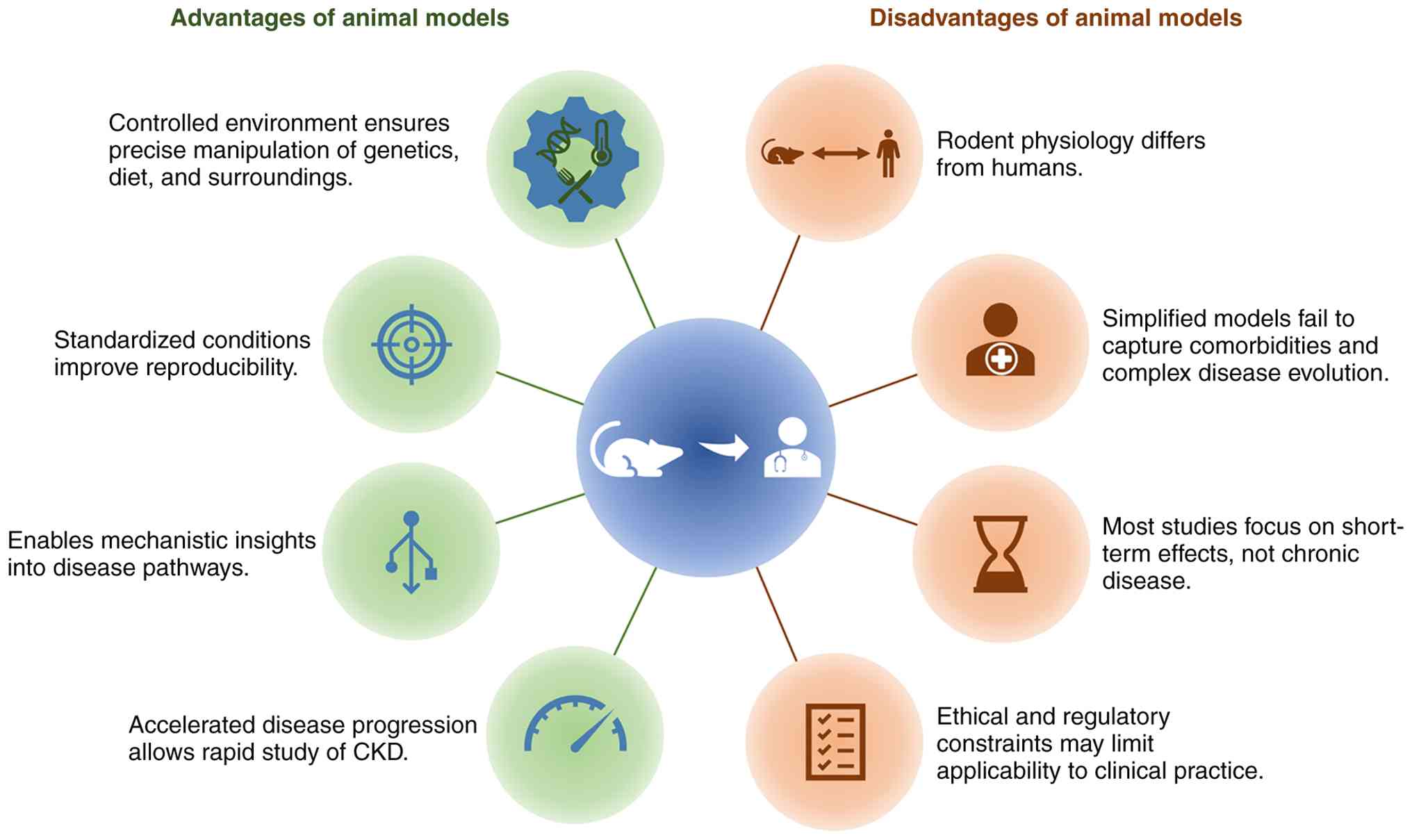
Animal models are indispensable for investigating
the complex pathophysiology of CRS, offering controlled and
reproducible environments that are difficult to achieve in human
studies enabling detailed exploration of disease mechanisms and
therapeutic interventions (195,196). A major advantage of these
models lies in the ability to tightly regulate genetic, dietary and
environmental variables, thereby enhancing experimental
reproducibility and facilitating the validation of mechanistic
hypotheses and the preclinical testing of novel therapies (195-198). In addition, animal models allow
investigation of organ crosstalk and the progression of dysfunction
in one organ following injury to the other, processes that are
difficult to isolate in clinical settings (197-199). The relatively rapid progression
of the disease in these models, occurring over weeks or months
rather than the years seen in humans, facilitates efficient study
of disease onset, trajectory and treatment response (196,200,201). Consequently, a range of CRS
phenotypes, including those driven by CKD, HF or metabolic
syndrome, can be simulated to support the development of targeted
interventions (196,201,202).
However, translating findings from experimental
models to clinical CRS remains challenging. In clinical practice,
the interpretation of renal and cardiac biomarkers in CRS is often
complicated by several confounding factors. Baseline CKD may alter
serum creatinine and natriuretic peptides levels (203,204), while fluid overload,
haemodynamic instability or diuretic therapy can affect urine
output and circulating biomarkers (1,205,206). Similarly, medications such as
vasopressors, renin-angiotensin system inhibitors or nephrotoxic
drugs may modify both renal and cardiac function (207-209). These factors should be
considered when interpreting preclinical findings, particularly in
CRS types 3 and 4, where establishing the temporal relationship
between renal and cardiac dysfunction may be difficult.
Despite their strengths, current animal models have
several limitations. Species-specific physiological differences,
particularly in cardiovascular and renal systems, may limit the
extrapolation to humans (195,199,210). Moreover, the majority of models
focus on isolated organ injury or acute pathological processes and
therefore fail to capture the chronic, multifactorial and
progressive nature of human CRS (197,198,211). In particular, the frequent
absence of common comorbidities such as diabetes, hypertension,
atherosclerosis and ageing represents a major gap as these factors
substantially shape disease trajectory and patient outcomes
(196,200). To improve clinical fidelity,
experimental models can be refined by combining classical renal
injury models with established cardiometabolic conditions. For
example, renal ischaemia-reperfusion or adenine-induced CKD may be
studied using a diabetic background (for example,
streptozotocin-induced diabetes or db/db mice), hypertensive models
(for example, angiotensin II infusion or spontaneously hypertensive
rats) or atherosclerosis-prone strains such as
ApoE−/− mice. The incorporation of aged
animals or dietary interventions, including high-fat or
high-phosphate diets, can further approximate the metabolic and
vascular environment typical observed in human CRS. Importantly,
incorporating comorbidities enhances clinical fidelity but
complicates interpretation, as they alter renal baselines,
biomarkers and cardiac remodelling. Combined models improve
translational relevance but increase complexity, so model choice
should align with mechanistic vs. translational study goals.
Ethical and regulatory constraints, particularly large animal
studies, must also be considered (198,210) (Fig. 1) and the clinical relevance of
selected preclinical models are discussed [Table VI (51,212-220)].
The cardiotoxic effect of uraemia has been
evaluated through this model, in the cardiac pathology of rats with
Nx5/6 (without acute myocardial infarction), where
capillary/myocardial cell mismatch and interstitial fibrosis were
found. Similarly, autopsy studies have shown that the number of
capillaries per myocardial cell decreases, and fibrosis increases
in uremic patients (221,222). Accumulation of uremic toxins
not only affects myocardial remodelling but is also associated with
an increase in the incidence of ischemic heart disease. Indeed,
Nx5/6 induced uremic rats have lower myocardial cell
volume density, a substantially larger ratio of infarct area and
reduced intrinsic tolerance of myocardium to ischemic injury
(223,224). These findings suggest that
uremic cardiomyopathy may increase susceptibility to ischemic
injury, supporting clinical observations that patients with
advanced CKD have a higher risk of cardiovascular events. They also
advocate for the immediate use of currently available
anti-remodelling strategies, such as β-blockers, ACE inhibitors or
angiotensin II type 1 (AT1) receptor blockers, in
patients with CKD after an acute myocardial infarction.
From a translational standpoint, UUO has been
highly involved in transcriptomic profiling studies, revealing both
coding and non-coding RNA signatures that may serve as biomarkers
or therapeutic targets for renal fibrosis and potentially CRS
(226). Furthermore, due to its
consistency and rapid progression, the model is well-suited for
early-phase drug screening and mechanistic validation of
nephroprotective compounds.
The UUO model is a key experimental tool for
studying obstructive nephropathy, hydronephrosis and renal
interstitial fibrosis. It induces rapid urine obstruction by
ligating one ureter, resulting in hydronephrosis and renal oedema.
Clinically, hydronephrosis is characterized by loss of renal
medullary tissue, most often as a consequence of obstructive
nephropathy. In humans, the leading causes include congenital
anomalies, urolithiasis, malignancy or fibrotic inflammatory
processes. Both paediatric and adult case reports have shown an
association between hydronephrosis and elevated blood pressure,
where the surgical relief of the obstruction often alleviates the
hypertension (227,228). In a study using Sprague Dawley
rats with spontaneous hydronephrosis, impaired cardiac autonomic
regulation, including elevated resting heart rate, reduced heart
rate variability and blunted baroreflex sensitivity, has been
reported. These changes occurred independently of peripheral RAAS
activation and were attributed to enhanced angiotensin II activity
in the nucleus tractus solitarius, a key point in clinical CRS
(229).
However, the UUO model does not fully capture the
chronic, recurrent and multifactorial characteristics of human CKD
and obstructive uropathy. Most importantly, UUO induces profound
structural changes with relatively modest or absent long-term
functional decline in the contralateral kidney, which limits its
utility in modelling the progressive renal impairment
characteristic of CRS (230).
Despite these constraints, emerging evidence suggests that
prolonged UUO can lead to systemic consequences, including cardiac
fibrosis, inflammation and lymphangiogenesis, indicating its
partial utility in modelling the renal-to-cardiac axis central to
CRS 3/4 (214,231). Species-specific signalling must
be considered when using animal models. Although
endothelial-to-mesenchymal transition may play a role in fibrosis
in UUO mice, its contribution seems limited in human kidneys
(232,233).
rIR injury models are widely employed in
preclinical research to simulate AKI, delayed graft function and
early post-transplant complications. These models are also highly
relevant for studying CRS, particularly type 3, in which acute
renal insults lead to secondary cardiac dysfunction, and type 4,
where persistent renal injury contributes to progressive CV
remodelling. rIR models replicate key pathophysiological events
observed in human AKI and kidney transplantation. From a
translational perspective, these models recapitulate key features
of human ischemic injury and post-transplant pathology, including
the transition from acute to chronic injury characterized by
interstitial fibrosis, tubular atrophy and persistent inflammation.
In clinical settings involving rIR, such as kidney transplantation,
partial nephrectomy, renal artery angioplasty, cardiopulmonary
bypass, aortic bypass surgery or other medical conditions, these
procedures remain among the most frequent causes of acute renal
failure.
Molecular and transcriptomic signatures from these
models show substantial overlap with human kidney transplant
biopsies, supporting their utility in preclinical evaluation of
targeted therapies and identification of prognostic biomarkers
(234). Some biomarkers
validated by animal experiments have also been applied to predict
human AKI, including neutrophil NGAL, liver-type fatty acid-binding
protein (L-FABP), KIM-1, tissue inhibitor of metalloproteinase-2
(TIMP-2) and insulin-like growth factor-binding protein-7 (IGFBP7).
Serum IL-6 and IL-8 have been confirmed as early indicators of AKI
in patients undergoing cardiac bypass surgery (235-237). Importantly, these models have
also revealed the systemic impact of renal ischemia on distant
organs, including the heart. Experimental data show that cytokine
release following rIR can contribute to myocardial inflammation and
dysfunction, offering mechanistic insight into the renal-to-cardiac
axis that defines CRS (238-240). As such, these models are
clinically relevant for investigating the early inflammation and
haemodynamic drivers of cardiac injury following AKI.
The rIR rodent model shares similarities with
certain aspects of AKI in humans, such as kidney tissue damage,
tubular epithelial cell proliferation, inflammatory response and
fibrosis. In both rats and mice, ischemic injury leads to the
proliferation of proximal renal tubular cells (241). Similar evidence of post-injury
recovery response has also been observed in human biopsy samples
after ischaemic or renal injury, as well as in cases of delayed
transplant function (242).
However, the regenerative capacity of human kidneys
is limited, resulting in a slower and incomplete recovery process
after AKI compared with that of mice (242,243). Therefore, the progression and
severity of diseases in rodent rIR models may not be the same as in
human AKI. Renal tubular injury is evident in human kidneys, but
necrosis after ischemia appears patchy, while in rodent models, it
is more pronounced as cell death. Furthermore, these models lack
key human-specific modifiers such as alloimmune responses, chronic
immunosuppression and patient-level comorbidities (for example,
diabetes or CV disease), which influence both renal and cardiac
outcomes in clinical settings (216).
Cisplatin is a widely used chemotherapeutic agent
with well-documented nephrotoxicity and emerging evidence of CV
complications in humans. In preclinical research, cisplatin-induced
AKI serves as a clinically relevant model for investigating the
kidney-heart axis in patients with cancer, particularly within the
framework of CRS3, where AKI precipitates or exacerbates cardiac
dysfunction. Cisplatin-induced AKI may contribute to systemic
endothelial dysfunction, electrolyte disturbances and a
pro-thrombotic state, factors known to impact CV homeostasis. When
compounded by high phosphate diets, aging or Klotho deficiency,
these models exhibit uremic vasculopathy and cardiac remodelling,
reinforcing their utility for studying CRS (244,245). Importantly, combination models
that integrate cisplatin exposure with dietary or genetic risk
factors (for example, high phosphate, aged mice, Klotho deficiency)
allow the simulation of AKI-to-CKD progression and concurrent CV
injury, thereby extending the model's relevance to CRS 3/4
(244,245). The mechanistic pathways
activated in cisplatin-induced injury may not fully represent those
in metabolic or haemodynamic CKD. Additionally, preclinical models
often lack confounding factors common in patients with cancer, such
as polypharmacy, pre-existing CV disease and heterogeneous tumour
biology. The clinical relevance of these models is underscored by
known risk factors for cisplatin nephrotoxicity, including
pre-existing CKD, CV disease and NSAID use (246). Standard preventive strategies,
such as intravenous hydration, magnesium supplementation and
mannitol-induced diuresis, are mirrored in preclinical designs.
Novel therapeutic approaches, including antioxidant compounds,
mitochondrial protectants and natural products, are currently under
investigation in these models (247-249).
Adenine-induced CKD models in rodents induce
tubulointerstitial injury, renal insufficiency and a spectrum of
metabolic disturbances that closely mimic the human CKD phenotype.
Animals consistently develop elevated plasma urea and creatinine,
anaemia, hyperphosphatemia, hypocalcemia and altered levels of
FGF23, all of which are hallmarks of advanced CKD and drivers of CV
disease in patients (176,196,250). Cardiac alterations observed in
adenine-induced CKD include LVH, increased end-diastolic pressure
and diastolic dysfunction with preserved systolic function. This
mirrors the clinical presentation of HFpEF, a common cardiac
manifestation in patients with CKD (176,251). Furthermore, this model induces
substantial cardiac oxidative stress, inflammation and DNA damage,
with upregulation of Nrf2 and pro-inflammatory cytokines (250,252), but also endothelial
dysfunction, characterized by impaired nitric oxide (NO)-dependent
vasodilation in the aorta, and a prothrombotic state with elevated
platelet counts, both of which reinforce the model's relevance for
studying vascular contributions to CRS and CV risk in CKD and mimic
the closely reproduce the complex CV alterations observed in human
(250,253). Although the abrupt onset and
severity of renal dysfunction in experimental models may differ
from the more gradual and heterogeneous progression seen in
clinical settings, this discrepancy may limit the generalizability
of therapeutic outcomes.
CRS3 and CRS4 highlight the complex bidirectional
interactions between renal and cardiac dysfunction through shared
mechanisms including inflammation, oxidative stress and
neurohormonal activation. Experimental models such as
Nx5/6, UUO, rIR, cisplatin and adenine-induced injury
reproduce key aspects of these syndromes and have substantially
improved the mechanistic understanding of kidney-heart
interactions. However, important limitations remain, including
species-specific differences, the frequent absence of common
comorbidities, and the limited ability to reproduce the progressive
and multifactorial nature of human CRS. Addressing these
limitations, future research should prioritize the development of
models incorporating cardiometabolic comorbidities, improved
standardization of experimental readouts aligned with clinical CRS
definitions and integrative multiorgan models that better capture
the chronic and bidirectional progression of cardiorenal
dysfunction. Such approaches may help bridge the gap between
experimental discovery and clinical translation.
Not applicable.
SMF, SH, JRO, JJB, and LB prepared the manuscript.
CAA, LB, and CEC critically reviewed and edited the manuscript. All
the authors contributed to the article, and all authors read and
approved the final manuscript. Data authentication not
applicable.
Not applicable.
Not applicable.
The authors declare that they have no competing
interests.
Not applicable.
This work was supported by the French National Research Agency
(grant no. ANR-22-CE14-0024) and the National Agency of Research
and Development (ANID) Fondecyt Regular (grant no. 1231909) and the
ECOS ANID (grant no. 20210024/C21S03).
|
1
|
Ronco C, McCullough P, Anker SD, Anand I,
Aspromonte N, Bagshaw SM, Bellomo R, Berl T, Bobek I, Cruz DN, et
al: Cardio-renal syndromes: Report from the consensus conference of
the acute dialysis quality initiative. Eur Heart J. 31:703–711.
2010. View Article : Google Scholar :
|
|
2
|
Schytz PA, Blanche P, Nissen AB,
Torp-Pedersen C, Gislason GH, Nelveg-Kristensen KE, Hommel K and
Carlson N: Acute kidney injury and risk of cardiovascular outcomes:
A nationwide cohort study. Nefrologia (Engl Ed). 42:338–346. 2022.
View Article : Google Scholar : PubMed/NCBI
|
|
3
|
Prastaro M, Nardi E, Paolillo S, Santoro
C, Parlati ALM, Gargiulo P, Basile C, Buonocore D, Esposito G and
Filardi PP: Cardiorenal syndrome: Pathophysiology as a key to the
therapeutic approach in an under-diagnosed disease. J Clin
Ultrasound. 50:1110–1124. 2022. View Article : Google Scholar : PubMed/NCBI
|
|
4
|
Young JB and Eknoyan G: Cardiorenal
syndrome: An evolutionary appraisal. Circ Heart Fail.
17:e0115102024. View Article : Google Scholar : PubMed/NCBI
|
|
5
|
Rangaswami J, Bhalla V, Blair JEA, Chang
TI, Costa S, Lentine KL, Lerma EV, Mezue K, Molitch M, Mullens W,
et al: Cardiorenal syndrome: Classification, pathophysiology,
diagnosis, and treatment strategies: A scientific statement from
the american heart association. Circulation. 139:e840–e878. 2019.
View Article : Google Scholar : PubMed/NCBI
|
|
6
|
Kellum JA, Romagnani P, Ashuntantang G,
Ronco C, Zarbock A and Anders HJ: Acute kidney injury. Nat Rev Dis
Primers. 7:522021. View Article : Google Scholar : PubMed/NCBI
|
|
7
|
Hoste EAJ, Kellum JA, Selby NM, Zarbock A,
Palevsky PM, Bagshaw SM, Goldstein SL, Cerdá J and Chawla LS:
Global epidemiology and outcomes of acute kidney injury. Nat Rev
Nephrol. 14:607–625. 2018. View Article : Google Scholar : PubMed/NCBI
|
|
8
|
Kovesdy CP: Epidemiology of chronic kidney
disease: An update 2022. Kidney Int Suppl (2011). 12:7–11. 2022.
View Article : Google Scholar : PubMed/NCBI
|
|
9
|
Xie Y, Bowe B, Mokdad AH, Xian H, Yan Y,
Li T, Maddukuri G, Tsai CY, Floyd T and Al-Aly Z: Analysis of the
global burden of disease study highlights the global, regional, and
national trends of chronic kidney disease epidemiology from 1990 to
2016. Kidney Int. 94:567–581. 2018. View Article : Google Scholar : PubMed/NCBI
|
|
10
|
Silver SA, Harel Z, McArthur E, Nash DM,
Acedillo R, Kitchlu A, Garg AX, Chertow GM, Bell CM and Wald R:
30-Day readmissions after an acute kidney injury hospitalization.
Am J Med. 130:163–172.e4. 2017. View Article : Google Scholar
|
|
11
|
Go AS, Hsu CY, Yang J, Tan TC, Zheng S,
Ordonez JD and Liu KD: Acute kidney injury and risk of heart
failure and atherosclerotic events. Clin J Am Soc Nephrol.
13:833–841. 2018. View Article : Google Scholar : PubMed/NCBI
|
|
12
|
Odutayo A, Wong CX, Farkouh M, Altman DG,
Hopewell S, Emdin CA and Hunn BH: AKI and long-term risk for
cardiovascular events and mortality. J Am Soc Nephrol. 28:377–387.
2017. View Article : Google Scholar
|
|
13
|
Rodríguez E, Arias-Cabrales C, Bermejo S,
Sierra A, Burballa C, Soler MJ, Barrios C and Pascual J: Impact of
recurrent acute kidney injury on patient outcomes. Kidney Blood
Press Res. 43:34–44. 2018. View Article : Google Scholar : PubMed/NCBI
|
|
14
|
Arias-Cabrales C, Rodríguez E, Bermejo S,
Sierra A, Burballa C, Barrios C, Soler MJ and Pascual J: Short- and
long-term outcomes after non-severe acute kidney injury. Clin Exp
Nephrol. 22:61–67. 2018. View Article : Google Scholar
|
|
15
|
Gallagher M, Cass A, Bellomo R, Finfer S,
Gattas D, Lee J, Lo S, McGuinness S, Myburgh J, Parke R, et al:
Long-term survival and dialysis dependency following acute kidney
injury in intensive care: Extended follow-up of a randomized
controlled trial. PLoS Med. 11:e10016012014. View Article : Google Scholar : PubMed/NCBI
|
|
16
|
de Jager DJ, Grootendorst DC, Jager KJ,
van Dijk PC, Tomas LM, Ansell D, Collart F, Finne P, Heaf JG, De
Meester J, et al: Cardiovascular and noncardiovascular mortality
among patients starting dialysis. JAMA. 302:1782–1789. 2009.
View Article : Google Scholar : PubMed/NCBI
|
|
17
|
Foley RN, Parfrey PS and Sarnak MJ:
Clinical epidemiology of cardiovascular disease in chronic renal
disease. Am J Kidney Dis. 32(5 Suppl 3): S112–S119. 1998.
View Article : Google Scholar : PubMed/NCBI
|
|
18
|
Baigent C, Burbury K and Wheeler D:
Premature cardiovascular disease in chronic renal failure. Lancet.
356:147–152. 2000. View Article : Google Scholar : PubMed/NCBI
|
|
19
|
Cheung AK, Sarnak MJ, Yan G, Berkoben M,
Heyka R, Kaufman A, Lewis J, Rocco M, Toto R, Windus D, et al:
Cardiac diseases in maintenance hemodialysis patients: Results of
the HEMO Study. Kidney Int. 65:2380–2389. 2004. View Article : Google Scholar : PubMed/NCBI
|
|
20
|
Go AS, Chertow GM, Fan D, McCulloch CE and
Hsu C: Chronic kidney disease and the risks of death,
cardiovascular events, and hospitalization. N Engl J Med.
351:1296–1305. 2004. View Article : Google Scholar : PubMed/NCBI
|
|
21
|
Kottgen A, Russell SD, Loehr LR,
Crainiceanu CM, Rosamond WD, Chang PP, Chambless LE and Coresh J:
Reduced kidney function as a risk factor for incident heart
failure: The atherosclerosis risk in communities (ARIC) study. J Am
Soc Nephrol. 18:13072007. View Article : Google Scholar : PubMed/NCBI
|
|
22
|
House AA, Wanner C, Sarnak MJ, Piña IL,
McIntyre CW, Komenda P, Kasiske BL, Deswal A, deFilippi CR, Cleland
JGF, et al: Heart failure in chronic kidney disease: Conclusions
from a kidney disease: Improving global outcomes (KDIGO)
controversies conference. Kidney Int. 95:1304–1317. 2019.
View Article : Google Scholar : PubMed/NCBI
|
|
23
|
Bikbov B, Purcell CA, Levey AS, Smith M,
Abdoli A, Abebe M, Adebayo OM, Afarideh M, Agarwal SK,
Agudelo-Botero M, et al: Global, regional, and national burden of
chronic kidney disease, 1990-2017: A systematic analysis for the
global burden of disease study 2017. Lancet. 395:709–733. 2020.
View Article : Google Scholar
|
|
24
|
Wang Y, Liu S, Liu Q and Lv Y: The
interaction of central nervous system and acute kidney injury:
Pathophysiology and clinical perspectives. Front Physiol.
13:8266862022. View Article : Google Scholar : PubMed/NCBI
|
|
25
|
Sapna F, Raveena F, Chandio M, Bai K,
Sayyar M, Varrassi G, Khatri M, Kumar S and Mohamad T: Advancements
in heart failure management: A comprehensive narrative review of
emerging therapies. Cureus. 15:e464862023.PubMed/NCBI
|
|
26
|
Hunter RW and Bailey MA: Hyperkalemia:
Pathophysiology, risk factors and consequences. Nephrol Dial
Transplant. 34(Suppl 3): iii2–iii11. 2019. View Article : Google Scholar : PubMed/NCBI
|
|
27
|
Villa G, Husain-Syed F, Saitta T,
Degl'Innocenti D, Barbani F, Resta M, Castellani G and Romagnoli S:
Hemodynamic instability during acute kidney injury and acute renal
replacement therapy: Pathophysiology and clinical implications.
Blood Purif. 50:729–739. 2021. View Article : Google Scholar : PubMed/NCBI
|
|
28
|
Abassi Z, Khoury EE, Karram T and Aronson
D: Edema formation in congestive heart failure and the underlying
mechanisms. Front Cardiovasc Med. 9:9332152022. View Article : Google Scholar : PubMed/NCBI
|
|
29
|
Yao C, Li Z, Sun K, Zhang Y, Shou S and
Jin H: Mitochondrial dysfunction in acute kidney injury. Ren Fail.
46:23932622024. View Article : Google Scholar : PubMed/NCBI
|
|
30
|
Iglesias M, Wang H, Krause-Hauch M, Ren D,
Zoungrana LI, Li Z, Zhang J, Wei J, Yadav N, Patel K, et al:
Sestrin2 mediates metformin rescued the age-related cardiac
dysfunctions of cardiorenal syndrome type 3. Cells. 12:8452023.
View Article : Google Scholar : PubMed/NCBI
|
|
31
|
Neres-Santos RS, Junho CVC, Panico K,
Caio-Silva W, Pieretti JC, Tamashiro JA, Seabra AB, Ribeiro CAJ and
Carneiro-Ramos MS: Mitochondrial dysfunction in cardiorenal
syndrome 3: Renocardiac effect of vitamin C. Cells. 10:30292021.
View Article : Google Scholar : PubMed/NCBI
|
|
32
|
Curaj A, Vanholder R, Loscalzo J, Quach K,
Wu Z, Jankowski V and Jankowski J: Cardiovascular consequences of
uremic metabolites: An overview of the involved signaling pathways.
Circ Res. 134:592–613. 2024. View Article : Google Scholar : PubMed/NCBI
|
|
33
|
Minciunescu A, Genovese L and deFilippi C:
Cardiovascular alterations and structural changes in the setting of
chronic kidney disease: A review of cardiorenal syndrome type 4. SN
Compr Clin Med. 5:152023. View Article : Google Scholar
|
|
34
|
Law JP, Pickup L, Pavlovic D, Townend JN
and Ferro CJ: Hypertension and cardiomyopathy associated with
chronic kidney disease: Epidemiology, pathogenesis and treatment
considerations. J Hum Hypertens. 37:1–19. 2023. View Article : Google Scholar :
|
|
35
|
Junho CVC, Frisch J, Soppert J,
Wollenhaupt J and Noels H: Cardiomyopathy in chronic kidney
disease: Clinical features, biomarkers and the contribution of
murine models in understanding pathophysiology. Clin Kidney J.
16:1786–1803. 2023. View Article : Google Scholar : PubMed/NCBI
|
|
36
|
H S, BS A, Moger V and Swamy M:
Cardiorenal syndrome type 4: A study of cardiovascular diseases in
chronic kidney disease. Indian Heart J. 69:11–16. 2017. View Article : Google Scholar : PubMed/NCBI
|
|
37
|
Reimer KC, Nadal J, Meiselbach H, Schmid
M, Schultheiss UT, Kotsis F, Stockmann H, Friedrich N, Nauck M,
Krane V, et al: Association of mineral and bone biomarkers with
adverse cardiovascular outcomes and mortality in the German chronic
kidney disease (GCKD) cohort. Bone Res. 11:522023. View Article : Google Scholar : PubMed/NCBI
|
|
38
|
Kadatane SP, Satariano M, Massey M, Mongan
K and Raina R: The role of inflammation in CKD. Cells. 12:15812023.
View Article : Google Scholar : PubMed/NCBI
|
|
39
|
Zoccali C, Vanholder R, Massy ZA, Ortiz A,
Sarafidis P, Dekker FW, Fliser D, Fouque D, Heine GH, Jager KJ, et
al: The systemic nature of CKD. Nat Rev Nephrol. 13:344–358. 2017.
View Article : Google Scholar : PubMed/NCBI
|
|
40
|
Hung S, Lai Y, Kuo K and Tarng D: Volume
overload and adverse outcomes in chronic kidney disease: Clinical
observational and animal studies. J Am Heart Assoc. 4:e0019182015.
View Article : Google Scholar : PubMed/NCBI
|
|
41
|
Tan JK, Kadir HA, Lim GH, Thumboo J, Bee
YM and Lim CC: Trends in fluid overload-related hospitalisations
among patients with diabetes mellitus The impact of chronic kidney
disease. Ann Acad Med Singap. 53:435–445. 2024. View Article : Google Scholar : PubMed/NCBI
|
|
42
|
Bao YW, Yuan Y, Chen JH and Lin WQ: Kidney
disease models: Tools to identify mechanisms and potential
therapeutic targets. Zool Res. 39:72–86. 2018.PubMed/NCBI
|
|
43
|
Rabe M and Schaefer F: Non-transgenic
mouse models of kidney disease. Nephron. 133:53–61. 2016.
View Article : Google Scholar : PubMed/NCBI
|
|
44
|
Róth E, Halmágyi G and Török B: Structural
changes in temporarily ischaemized and reperfused dog kidneys. Acta
Chir Acad Sci Hung. 18:393–415. 1977.PubMed/NCBI
|
|
45
|
Coburn JW, Gonick HC, Rubini ME and
Kleeman CR: Studies of experimental renal failure in dogs. I.
Effect of 5-6 nephrectomy on concentrating and diluting capacity of
residual nephrons. J Clin Invest. 44:603–614. 1965. View Article : Google Scholar : PubMed/NCBI
|
|
46
|
Jaenike JR: The renal response to ureteral
obstruction: A model for the study of factors which influence
glomerular filtration pressure. J Lab Clin Med. 76:373–382.
1970.PubMed/NCBI
|
|
47
|
Tan RZ, Zhong X, Li JC, Zhang YW, Yan Y,
Liao Y, Wen D, Diao H, Wang L and Shen HC: An optimized 5/6
nephrectomy mouse model based on unilateral kidney ligation and its
application in renal fibrosis research. Ren Fail. 41:555–566. 2019.
View Article : Google Scholar : PubMed/NCBI
|
|
48
|
Yaxley J and Yaxley W: Obstructive
uropathy-acute and chronic medical management. World J Nephrol.
12:1–9. 2023. View Article : Google Scholar : PubMed/NCBI
|
|
49
|
Saat TC, van den Akker EK, IJzermans JNM,
Dor FJMF and de Bruin RWF: Improving the outcome of kidney
transplantation by ameliorating renal ischemia reperfusion injury:
Lost in translation? J Transl Med. 14:202016. View Article : Google Scholar : PubMed/NCBI
|
|
50
|
Lerman LO, Textor SC and Grande JP:
Mechanisms of tissue injury in renal artery stenosis: Ischemia and
beyond. Prog Cardiovasc Dis. 52:196–203. 2009. View Article : Google Scholar : PubMed/NCBI
|
|
51
|
Adam RJ, Williams AC and Kriegel AJ:
Comparison of the surgical resection and infarct 5/6 nephrectomy
rat models of chronic kidney disease. Am J Physiol Renal Physiol.
322:F639–F654. 2022. View Article : Google Scholar : PubMed/NCBI
|
|
52
|
Fong D, Denton KM, Moritz KM, Evans R and
Singh RR: Compensatory responses to nephron deficiency: Adaptive or
maladaptive? Nephrology (Carlton). 19:119–128. 2014. View Article : Google Scholar : PubMed/NCBI
|
|
53
|
Amador-Martínez I, García-Ballhaus J,
Buelna-Chontal M, Cortés-González C, Massó F, Jaisser F and
Barrera-Chimal J: Early inflammatory changes and CC chemokine
ligand-8 upregulation in the heart contribute to uremic
cardiomyopathy. FASEB J. 35:e217612021. View Article : Google Scholar : PubMed/NCBI
|
|
54
|
Liu S, Kompa A, Kumfu S, Nishijima F,
Kelly D, Krum H and Wang B: Subtotal nephrectomy accelerates
pathological cardiac remodeling post-myocardial infarction:
Implications for cardiorenal syndrome. Int J Cardiol.
168:1866–1880. 2013. View Article : Google Scholar : PubMed/NCBI
|
|
55
|
Chang D, Xu TT, Zhang SJ, Cai Y, Min SD,
Zhao Z, Lu CQ, Wang YC and Ju S: Telmisartan ameliorates cardiac
fibrosis and diastolic function in cardiorenal heart failure with
preserved ejection fraction. Exp Biol Med (Maywood). 246:2511–2521.
2021. View Article : Google Scholar : PubMed/NCBI
|
|
56
|
Kobayashi R, Wakui H, Azushima K, Uneda K,
Haku S, Ohki K, Haruhara K, Kinguchi S, Matsuda M, Ohsawa M, et al:
An angiotensin II type 1 receptor binding molecule has a critical
role in hypertension in a chronic kidney disease model. Kidney Int.
91:1115–1125. 2017. View Article : Google Scholar : PubMed/NCBI
|
|
57
|
Huang D, Yan M, Chen K, Sun R, Dong ZF, Wu
PL, Li S, Zhu GS, Ma SX, Pan YS, et al: Cardiac-specific
overexpression of silent information regulator 1 protects against
heart and kidney deterioration in cardiorenal syndrome via
inhibition of endoplasmic reticulum stress. Cell Physiol Biochem.
46:9–22. 2018. View Article : Google Scholar : PubMed/NCBI
|
|
58
|
Liu T, Lu X, Gao W, Zhai Y, Li H, Li S,
Yang L, Ma F, Zhan Y and Mao H: Cardioprotection effect of
Yiqi-Huoxue-Jiangzhuo formula in a chronic kidney disease mouse
model associated with gut microbiota modulation and NLRP3
inflammasome inhibition. Biomed Pharmacother. 152:1131592022.
View Article : Google Scholar : PubMed/NCBI
|
|
59
|
Di Marco GS, Reuter S, Kentrup D, Grabner
A, Amaral AP, Fobker M, Stypmann J, Pavenstädt H, Wolf M, Faul C
and Brand M: Treatment of established left ventricular hypertrophy
with fibroblast growth factor receptor blockade in an animal model
of CKD. Nephrol Dial Transplant. 29:2028–2035. 2014. View Article : Google Scholar : PubMed/NCBI
|
|
60
|
Toblli JE, Cao G, Rivas C and Kulaksiz H:
Heart and iron deficiency anaemia in rats with renal insufficiency:
The role of hepcidin. Nephrology (Carlton). 13:636–645. 2008.
View Article : Google Scholar : PubMed/NCBI
|
|
61
|
Ulu N, Mulder GM, Vavrinec P, Landheer SW,
Duman-Dalkilic B, Gurdal H, Goris M, Duin M, van Dokkum RP, Buikema
H, et al: Epidermal growth factor receptor inhibitor PKI-166
governs cardiovascular protection without beneficial effects on the
kidney in hypertensive 5/6 nephrectomized rats. J Pharmacol Exp
Ther. 345:393–403. 2013. View Article : Google Scholar : PubMed/NCBI
|
|
62
|
Liu B, Shalamu A, Pei Z, Liu L, Wei Z, Qu
Y, Song S, Luo W, Dong Z, Weng X and Ge J: A novel mouse model of
heart failure with preserved ejection fraction after chronic kidney
disease induced by retinol through JAK/STAT pathway. Int J Biol
Sci. 19:3661–3677. 2023. View Article : Google Scholar : PubMed/NCBI
|
|
63
|
Fernandes-Charpiot IMM, Caldas HC, Mendes
GEF, Gomes de Sá Neto L, Oliveira HL, Baptista MASF and Abbud-Filho
M: Validation of an experimental model to study less severe chronic
renal failure. J Invest Surg. 29:309–315. 2016. View Article : Google Scholar : PubMed/NCBI
|
|
64
|
Beraldo JI, Benetti A, Borges-Júnior FA,
Arruda-Junior DF, Martins FL, Jensen L, Dariolli R, Shimizu MH,
Seguro AC, Luchi WM and Girardi ACC: Cardioprotection conferred by
sitagliptin is associated with reduced cardiac angiotensin
II/angiotensin-(1-7) balance in experimental chronic kidney
disease. Int J Mol Sci. 20:19402019. View Article : Google Scholar : PubMed/NCBI
|
|
65
|
Paterson MR, Geurts AM and Kriegel AJ:
miR-146b-5p has a sex-specific role in renal and cardiac pathology
in a rat model of chronic kidney disease. Kidney Int. 96:1332–1345.
2019. View Article : Google Scholar : PubMed/NCBI
|
|
66
|
Uchida L, Tanaka T, Saito H, Sugahara M,
Wakashima T, Fukui K and Nangaku M: Effects of a prolyl hydroxylase
inhibitor on kidney and cardiovascular complications in a rat model
of chronic kidney disease. Am J Physiol Renal Physiol.
318:F388–F401. 2020. View Article : Google Scholar
|
|
67
|
Kalk P, Godes M, Relle K, Rothkegel C,
Hucke A, Stasch JP and Hocher B: NO-independent activation of
soluble guanylate cyclase prevents disease progression in rats with
5/6 nephrectomy. Br J Pharmacol. 148:853–859. 2006. View Article : Google Scholar : PubMed/NCBI
|
|
68
|
Freundlich M, Li YC, Quiroz Y, Bravo Y,
Seeherunvong W, Faul C, Weisinger JR and Rodriguez-Iturbe B:
Paricalcitol downregulates myocardial renin-angiotensin and
fibroblast growth factor expression and attenuates cardiac
hypertrophy in uremic rats. Am J Hypertens. 27:720–726. 2014.
View Article : Google Scholar
|
|
69
|
Aoki K, Teshima Y, Kondo H, Saito S, Fukui
A, Fukunaga N, Nawata T, Shimada T, Takahashi N and Shibata H: Role
of indoxyl sulfate as a predisposing factor for atrial fibrillation
in renal dysfunction. J Am Heart Assoc. 4:e0020232015. View Article : Google Scholar : PubMed/NCBI
|
|
70
|
Koch V, Weber C, Riffel JH, Buchner K,
Buss SJ, Hein S, Mereles D, Hagenmueller M, Erbel C, März W, et al:
Impact of homoarginine on myocardial function and remodeling in a
rat model of chronic renal failure. J Cardiovasc Pharmacol Ther.
27:107424842110546202022. View Article : Google Scholar : PubMed/NCBI
|
|
71
|
Fukunaga N, Takahashi N, Hagiwara S, Kume
O, Fukui A, Teshima Y, Shinohara T, Nawata T, Hara M, Noguchi T and
Saikawa T: Establishment of a model of atrial fibrillation
associated with chronic kidney disease in rats and the role of
oxidative stress. Heart Rhythm. 9:2023–2031. 2012. View Article : Google Scholar : PubMed/NCBI
|
|
72
|
Lin CY, Hsu YJ, Hsu SC, Chen Y, Lee HS,
Lin SH, Huang SM, Tsai CS and Shih CC: CB1 cannabinoid receptor
antagonist attenuates left ventricular hypertrophy and Akt-mediated
cardiac fibrosis in experimental uremia. J Mol Cell Cardiol.
85:249–261. 2015. View Article : Google Scholar : PubMed/NCBI
|
|
73
|
Li Y, Wu J, He Q, Shou Z, Zhang P, Pen W,
Zhu Y and Chen J: Angiotensin (1-7) prevent heart dysfunction and
left ventricular remodeling caused by renal dysfunction in 5/6
nephrectomy mice. Hypertens Res. 32:369–374. 2009. View Article : Google Scholar : PubMed/NCBI
|
|
74
|
Ding W, Wang B, Zhang M and Gu Y:
Involvement of endoplasmic reticulum stress in uremic
cardiomyopathy: Protective effects of tauroursodeoxycholic acid.
Cell Physiol Biochem. 38:141–152. 2016. View Article : Google Scholar : PubMed/NCBI
|
|
75
|
Song Y, Yu Q, Zhang J, Huang W, Liu Y, Pei
H, Liu J, Sun L, Yang L, Li C, et al: Increased myocardial
ischemia-reperfusion injury in renal failure involves cardiac
adiponectin signal deficiency. Am J Physiol Endocrinol Metab.
306:E1055–E1064. 2014. View Article : Google Scholar : PubMed/NCBI
|
|
76
|
Ogino A, Takemura G, Kawasaki M, Tsujimoto
A, Kanamori H, Li L, Goto K, Maruyama R, Kawamura I, Takeyama T, et
al: Erythropoietin receptor signaling mitigates renal
dysfunction-associated heart failure by mechanisms unrelated to
relief of anemia. J Am Coll Cardiol. 56:1949–1958. 2010. View Article : Google Scholar : PubMed/NCBI
|
|
77
|
Aires RS, Vieira LD, Freitas ACN, de Lima
ME, Lima NKS, Farias JS and Paixão AD: NO mediates the effect of
the synthetic natriuretic peptide NPCdc on kidney and aorta in
nephrectomised rats. Eur J Pharmacol. 866:1727802020. View Article : Google Scholar
|
|
78
|
Liu Y, Liu Y, Liu X, Chen J, Zhang K,
Huang F, Wang JF, Tang W and Huang H: Apocynin attenuates cardiac
injury in type 4 cardiorenal syndrome via suppressing cardiac
fibroblast growth factor-2 with oxidative stress inhibition. J Am
Heart Assoc. 4:e0015982015. View Article : Google Scholar : PubMed/NCBI
|
|
79
|
Feng W, Zhang K, Liu Y, Chen J, Cai Q, He
W, Zhang Y, Wang MH, Wang J and Huang H: Advanced oxidation protein
products aggravate cardiac remodeling via cardiomyocyte apoptosis
in chronic kidney disease. Am J Physiol Heart Circ Physiol.
314:H475–H483. 2018. View Article : Google Scholar
|
|
80
|
García-Trejo EMA, Arellano-Buendía AS,
Argüello-García R, Loredo-Mendoza ML, García-Arroyo FE,
Arellano-Mendoza MG, Castillo-Hernández MC, Guevara-Balcázar G,
Tapia E, Sánchez-Lozada LG and Osorio-Alonso H: Effects of allicin
on hypertension and cardiac function in chronic kidney disease.
Oxid Med Cell Longev. 2016:38504022016. View Article : Google Scholar : PubMed/NCBI
|
|
81
|
Okada Y, Nakata M, Izumoto H, Takasu M,
Tazawa N, Takaoka M, Gariepy CE, Yanagisawa M and Matsumura Y: Role
of endothelin ETB receptor in partial ablation-induced chronic
renal failure in rats. Eur J Pharmacol. 494:63–71. 2004. View Article : Google Scholar : PubMed/NCBI
|
|
82
|
Baraka A and El Ghotny S: Cardioprotective
effect of renalase in 5/6 nephrectomized rats. J Cardiovasc
Pharmacol Ther. 17:412–416. 2012. View Article : Google Scholar : PubMed/NCBI
|
|
83
|
McMahon AC, Greenwald SE, Dodd SM, Hurst
MJ and Raine AEG: Prolonged calcium transients and myocardial
remodelling in early experimental uraemia. Nephrol Dial Transplant.
17:759–764. 2002. View Article : Google Scholar : PubMed/NCBI
|
|
84
|
Rodríguez-Ayala E, Ávila-Díaz M,
Foyo-Niembro E, Amato D, Ramirez-San-Juan E and Paniagua R: Effect
of parathyroidectomy on cardiac fibrosis and apoptosis: Possible
role of aldosterone. Nephron Physiol. 103:p112–p118. 2006.
View Article : Google Scholar : PubMed/NCBI
|
|
85
|
Mizobuchi M, Ogata H, Yamazaki-Nakazawa A,
Hosaka N, Kondo F, Koiwa F, Kinugasa E and Shibata T: Cardiac
effect of vitamin D receptor modulators in uremic rats. J Steroid
Biochem Mol Biol. 163:20–27. 2016. View Article : Google Scholar : PubMed/NCBI
|
|
86
|
Wu-Wong JR, Chen Y and Wessale JL: Vitamin
D receptor agonist VS-105 improves cardiac function in the presence
of enalapril in 5/6 nephrectomized rats. Am J Physiol Renal
Physiol. 308:F309–F319. 2015. View Article : Google Scholar
|
|
87
|
Ismail B, deKemp RA, Croteau E, Hadizad T,
Burns KD, Beanlands RS and DaSilva JN: Treatment with enalapril and
not diltiazem ameliorated progression of chronic kidney disease in
rats, and normalized renal AT1 receptor expression as measured with
PET imaging. PLoS One. 12:e01774512017. View Article : Google Scholar : PubMed/NCBI
|
|
88
|
Schön A, Leifheit-Nestler M, Deppe J,
Fischer DC, Bayazit AK, Obrycki L, Canpolat N, Bulut IK, Azukaitis
K, Yilmaz A, et al: Active vitamin D is cardioprotective in
experimental uraemia but not in children with CKD stages 3-5.
Nephrol Dial Transplant. 36:442–451. 2021. View Article : Google Scholar
|
|
89
|
Leelahavanichkul A, Yan Q, Hu X, Eisner C,
Huang Y, Chen R, Mizel D, Zhou H, Wright EC, Kopp JB, et al:
Angiotensin II overcomes strain-dependent resistance of rapid CKD
progression in a new remnant kidney mouse model. Kidney Int.
78:1136–1153. 2010. View Article : Google Scholar : PubMed/NCBI
|
|
90
|
Hamzaoui M, Djerada Z, Brunel V, Mulder P,
Richard V, Bellien J and Guerrot D: 5/6 Nephrectomy induces
different renal, cardiac and vascular consequences in 129/Sv and
C57BL/6JRj mice. Sci Rep. 10:15242020. View Article : Google Scholar : PubMed/NCBI
|
|
91
|
Moench I, Aravindhan K, Kuziw J,
Schnackenberg CG, Willette RN, Toomey JR and Gatto GJ Jr: High
FGF23 levels failed to predict cardiac hypertrophy in animal models
of hyperphosphatemia and chronic renal failure. J Endocr Soc.
5:bvab0662021. View Article : Google Scholar : PubMed/NCBI
|
|
92
|
Rahman M, Kim SJ, Kim JS, Kim SZ, Lee YU
and Kang HS: Myocardial calcification and hypertension following
chronic renal failure and ameliorative effects of furosemide and
captopril. Cardiology. 116:194–205. 2010. View Article : Google Scholar : PubMed/NCBI
|
|
93
|
Jolma P, Kööbi P, Kalliovalkama J, Kähönen
M, Fan M, Saha H, Helin H, Lehtimäki T and Pörsti I: Increased
calcium intake reduces plasma cholesterol and improves
vasorelaxation in experimental renal failure. Am J Physiol Heart
Circ Physiol. 285:H1882–H1889. 2003. View Article : Google Scholar : PubMed/NCBI
|
|
94
|
Kööbi P, Kalliovalkama J, Jolma P, Rysä J,
Ruskoaho H, Vuolteenaho O, Kähönen M, Tikkanen I, Fan M, Ylitalo P
and Pörsti I: AT1 receptor blockade improves vasorelaxation in
experimental renal failure. Hypertension. 41:1364–1371. 2003.
View Article : Google Scholar : PubMed/NCBI
|
|
95
|
Gava AL, Freitas FP, Balarini CM, Vasquez
EC and Meyrelles SS: Effects of 5/6 nephrectomy on renal function
and blood pressure in mice. Int J Physiol Pathophysiol Pharmacol.
4:167–173. 2012.PubMed/NCBI
|
|
96
|
Guo J, Zhu J, Ma L, Shi H, Hu J, Zhang S,
Hou L, Xu F, An Y, Yu H and Ge J: Chronic kidney disease
exacerbates myocardial ischemia reperfusion injury: Role of
endoplasmic reticulum stress-mediated apoptosis. Shock. 49:712–720.
2018. View Article : Google Scholar
|
|
97
|
Verkaik M, Oranje M, Abdurrachim D, Goebel
M, Gam Z, Prompers JJ, Helmes M, Ter Wee PM, van der Velden J,
Kuster DW, et al: High fibroblast growth factor 23 concentrations
in experimental renal failure impair calcium handling in
cardiomyocytes. Physiol Rep. 6:e135912018. View Article : Google Scholar : PubMed/NCBI
|
|
98
|
Chen C, Xie C, Wu H, Wu L, Zhu J, Mao H
and Xing C: Uraemic cardiomyopathy in different mouse models. Front
Med (Lausanne). 8:6905172021. View Article : Google Scholar : PubMed/NCBI
|
|
99
|
Feng J, Li H and Wang S: Hydrogen sulfide
alleviates uremic cardiomyopathy by regulating
PI3K/PKB/mTOR-mediated overactive autophagy in 5/6 nephrectomy
mice. Front Pharmacol. 13:10275972022. View Article : Google Scholar :
|
|
100
|
Chen J, Kieswich JE, Chiazza F, Moyes AJ,
Gobbetti T, Purvis GS, Salvatori DC, Patel NS, Perretti M, Hobbs
AJ, et al: IκB kinase inhibitor attenuates sepsis-induced cardiac
dysfunction in CKD. J Am Soc Nephrol. 28:94–105. 2017. View Article : Google Scholar
|
|
101
|
Mohamed RMSM, Elshazly SM, Nafea OE and
Abd El Motteleb DM: Comparative cardioprotective effects of
carvedilol versus atenolol in a rat model of cardiorenal syndrome
type 4. Naunyn Schmiedebergs Arch Pharmacol. 394:2117–2128. 2021.
View Article : Google Scholar : PubMed/NCBI
|
|
102
|
Mak RH, Chang SL, Draksharapu A and Pak
YK: Gene expression in uremic left ventricular hypertrophy: Effects
of hypertension and anemia. Exp Mol Med. 36:251–258. 2004.
View Article : Google Scholar : PubMed/NCBI
|
|
103
|
Kalliovalkama J, Jolma P, Tolvanen JP,
Kähönen M, Hutri-Kähönen N, Saha H, Tuorila S, Moilanen E and
Pörsti I: Potassium channel-mediated vasorelaxation is impaired in
experimental renal failure. Am J Physiol. 277:H1622–H1629.
1999.PubMed/NCBI
|
|
104
|
Ghosh SS, Salloum FN, Abbate A, Krieg R,
Sica DA, Gehr TW and Kukreja RC: Curcumin prevents cardiac
remodeling secondary to chronic renal failure through deactivation
of hypertrophic signaling in rats. Am J Physiol Heart Circ Physiol.
299:H975–H984. 2010. View Article : Google Scholar : PubMed/NCBI
|
|
105
|
Kurdián M, Herrero-Fresneda I, Lloberas N,
Gimenez-Bonafe P, Coria V, Grande MT, Boggia J, Malacrida L, Torras
J, Arévalo MA, et al: Delayed mTOR inhibition with low dose of
everolimus reduces TGFβ expression, attenuates proteinuria and
renal damage in the renal mass reduction model. PLoS One.
7:e325162012. View Article : Google Scholar
|
|
106
|
Hernández-Reséndiz S, Correa F,
García-Niño WR, Buelna-Chontal M, Roldán FJ, Ramírez-Camacho I,
Delgado-Toral C, Carbó R, Pedraza-Chaverrí J, Tapia E and Zazueta
C: Cardioprotection by curcumin post-treatment in rats with
established chronic kidney disease. Cardiovasc Drugs Ther.
29:111–120. 2015. View Article : Google Scholar : PubMed/NCBI
|
|
107
|
Sárközy M, Márványkövi FM, Szűcs G, Kovács
ZZA, Szabó MR, Gáspár R, Siska A, Kővári B, Cserni G, Földesi I and
Csont T: Ischemic preconditioning protects the heart against
ischemia-reperfusion injury in chronic kidney disease in both males
and females. Biol Sex Differ. 12:492021. View Article : Google Scholar : PubMed/NCBI
|
|
108
|
Kuncová J, Svíglerová J, Kummer W, Rajdl
D, Chottová-Dvoráková M, Tonar Z, Nalos L and Stengl M:
Parasympathetic regulation of heart rate in rats after 5/6
nephrectomy is impaired despite functionally intact cardiac vagal
innervation. Nephrol Dial Transplant. 24:2362–2370. 2009.
View Article : Google Scholar : PubMed/NCBI
|
|
109
|
Vettoretti S, Vavrinec P, Ochodnicky P,
Deelman LE, De Zeeuw D, Henning RH and Buikema H: Renal endothelial
function is associated with the anti-proteinuric effect of ACE
inhibition in 5/6 nephrectomized rats. Am J Physiol Renal Physiol.
310:F1047–F1053. 2016. View Article : Google Scholar : PubMed/NCBI
|
|
110
|
Sharkovska Y, Kalk P, Lawrenz B, Godes M,
Hoffmann LS, Wellkisch K, Geschka S, Relle K, Hocher B and Stasch
JP: Nitric oxide-independent stimulation of soluble guanylate
cyclase reduces organ damage in experimental low-renin and
high-renin models. J Hypertens. 28:1666–1675. 2010. View Article : Google Scholar : PubMed/NCBI
|
|
111
|
Gut N, Piecha G, Aldebssi F, Schaefer S,
Bekeredjian R, Schirmacher P, Ritz E and Gross-Weissmann ML:
Erythropoietin combined with ACE inhibitor prevents heart
remodeling in 5/6 nephrectomized rats independently of blood
pressure and kidney function. Am J Nephrol. 38:124–135. 2013.
View Article : Google Scholar : PubMed/NCBI
|
|
112
|
Amador-Martínez I, Pérez-Villalva R, Uribe
N, Cortés-González C, Bobadilla NA and Barrera-Chimal J: Reduced
endothelial nitric oxide synthase activation contributes to
cardiovascular injury during chronic kidney disease progression. Am
J Physiol Renal Physiol. 317:F275–F285. 2019. View Article : Google Scholar : PubMed/NCBI
|
|
113
|
Hamzaoui M, Roche C, Coquerel D, Duflot T,
Brunel V, Mulder P, Richard V, Bellien J and Guerrot D: Soluble
epoxide hydrolase inhibition prevents experimental type 4
cardiorenal syndrome. Front Mol Biosci. 7:6040422021. View Article : Google Scholar : PubMed/NCBI
|
|
114
|
Bigelman E, Cohen L, Aharon-Hananel G,
Levy R, Rozenbaum Z, Saada A, Keren G and Entin-Meer M:
Pathological presentation of cardiac mitochondria in a rat model
for chronic kidney disease. PLoS One. 13:e01981962018. View Article : Google Scholar : PubMed/NCBI
|
|
115
|
Iwanami J, Mogi M, Iwai M and Horiuchi M:
Inhibition of the renin-angiotensin system and target organ
protection. Hypertens Res. 32:229–237. 2009. View Article : Google Scholar : PubMed/NCBI
|
|
116
|
Rosenbaugh EG, Savalia KK, Manickam DS and
Zimmerman MC: Antioxidant-based therapies for angiotensin
II-associated cardiovascular diseases. Am J Physiol Regul Integr
Comp Physiol. 304:R917–R928. 2013. View Article : Google Scholar : PubMed/NCBI
|
|
117
|
Ucero AC, Benito-Martin A, Izquierdo MC,
Sanchez-Niño MD, Sanz AB, Ramos AM, Berzal S, Ruiz-Ortega M, Egido
J and Ortiz A: Unilateral ureteral obstruction: Beyond obstruction.
Int Urol Nephrol. 46:765–776. 2014. View Article : Google Scholar
|
|
118
|
Mishima K, Nakasatomi M, Takahashi S,
Ikeuchi H, Sakairi T, Kaneko Y, Hiromura K, Nojima Y and Maeshima
A: Attenuation of renal fibrosis after unilateral ureteral
obstruction in mice lacking the N-type calcium channel. PLoS One.
14:e02234962019. View Article : Google Scholar : PubMed/NCBI
|
|
119
|
Ham O, Jin W, Lei L, Huang HH, Tsuji K,
Huang M, Roh J, Rosenzweig A and Lu HAJ: Pathological cardiac
remodeling occurs early in CKD mice from unilateral urinary
obstruction, and is attenuated by Enalapril. Sci Rep. 8:160872018.
View Article : Google Scholar : PubMed/NCBI
|
|
120
|
Wakui H, Yamaji T, Azushima K, Uneda K,
Haruhara K, Nakamura A, Ohki K, Kinguchi S, Kobayashi R, Urate S,
et al: Effects of Rikkunshito treatment on renal
fibrosis/inflammation and body weight reduction in a unilateral
ureteral obstruction model in mice. Sci Rep. 10:17822020.
View Article : Google Scholar : PubMed/NCBI
|
|
121
|
Florens N, Kasam RK, Rudman-Melnick V, Lin
SC, Prasad V and Molkentin JD: Interleukin-33 mediates
cardiomyopathy after acute kidney injury by signaling to
cardiomyocytes. Circulation. 147:746–758. 2023. View Article : Google Scholar : PubMed/NCBI
|
|
122
|
Wu WP, Chang CH, Chiu YT, Ku CL, Wen MC,
Shu KH and Wu MJ: A reduction of unilateral ureteral
obstruction-induced renal fibrosis by a therapy combining valsartan
with aliskiren. Am J Physiol Renal Physiol. 299:F929–F941. 2010.
View Article : Google Scholar : PubMed/NCBI
|
|
123
|
Prieto-Carrasco R, Silva-Palacios A,
Rojas-Morales P, Aparicio-Trejo OE, Medina-Reyes EI, Hernández-Cruz
EY, Sánchez-Garibay C, Salinas-Lara C, Pavón N, Roldán FJ, et al:
Unilateral ureteral obstruction for 28 days in rats is not
associated with changes in cardiac function or alterations in
mitochondrial function. Biology (Basel). 10:6712021.PubMed/NCBI
|
|
124
|
Han Y, Xian Y, Gao X, Qiang P, Hao J, Yang
F, Shimosawa T, Chang Y and Xu Q: Eplerenone inhibits the
macrophage-to-myofibroblast transition in rats with UUO-induced
type 4 cardiorenal syndrome through the MR/CTGF pathway. Int
Immunopharmacol. 113:1093962022. View Article : Google Scholar : PubMed/NCBI
|
|
125
|
Prado NJ, Casarotto M, Calvo JP, Mazzei L,
Ponce Zumino AZ, García IM, Cuello-Carrión FD, Fornés MW, Ferder L,
Diez ER and Manucha W: Antiarrhythmic effect linked to melatonin
cardiorenal protection involves AT1 reduction and Hsp70-VDR
increase. J Pineal Res. 65:e125132018. View Article : Google Scholar
|
|
126
|
Prud'homme M, Coutrot M, Michel T, Boutin
L, Genest M, Poirier F, Launay JM, Kane B, Kinugasa S, Prakoura N,
et al: Acute kidney injury induces remote cardiac damage and
dysfunction through the galectin-3 pathway. JACC Basic Transl Sci.
4:717–732. 2019. View Article : Google Scholar : PubMed/NCBI
|
|
127
|
Tang CY, Lai CC, Huang PH, Yang AH, Chiang
SC, Huang PC, Tseng KW and Huang CH: Magnolol reduces myocardial
injury induced by renal ischemia and reperfusion. J Chin Med Assoc.
85:584–596. 2022. View Article : Google Scholar : PubMed/NCBI
|
|
128
|
Trentin-Sonoda M, da Silva RC, Kmit FV,
Abrahão MV, Monnerat Cahli G, Brasil GV, Muzi-Filho H, Silva PA,
Tovar-Moll FF, Vieyra A, et al: Knockout of Toll-like receptors 2
and 4 prevents renal ischemia-reperfusion-induced cardiac
hypertrophy in mice. PLoS One. 10:e01393502015. View Article : Google Scholar : PubMed/NCBI
|
|
129
|
Sumida M, Doi K, Ogasawara E, Yamashita T,
Hamasaki Y, Kariya T, Takimoto E, Yahagi N, Nangaku M and Noiri E:
Regulation of mitochondrial dynamics by dynamin-related protein-1
in acute cardiorenal syndrome. J Am Soc Nephrol. 26:2378–2387.
2015. View Article : Google Scholar : PubMed/NCBI
|
|
130
|
Chen TH, Yang YC, Wang JC and Wang JJ:
Curcumin treatment protects against renal ischemia and reperfusion
injury-induced cardiac dysfunction and myocardial injury.
Transplant Proc. 45:3546–3549. 2013. View Article : Google Scholar : PubMed/NCBI
|
|
131
|
Chevalier RL and Thornhill BA: Ureteral
obstruction in the neonatal rat: Renal nerves modulate hemodynamic
effects. Pediatr Nephrol. 9:447–450. 1995. View Article : Google Scholar : PubMed/NCBI
|
|
132
|
Cirino-Silva R, Kmit FV, Trentin-Sonoda M,
Nakama KK, Panico K, Alvim JM, Dreyer TR, Martinho-Silva H and
Carneiro-Ramos MS: Renal ischemia/reperfusion-induced cardiac
hypertrophy in mice: Cardiac morphological and morphometric
characterization. JRSM Cardiovasc Dis.
6:20480040166894402017.PubMed/NCBI
|
|
133
|
Lieberthal W, Fuhro R, Andry C and Valeri
CR: Effects of hemoglobin-based oxygen-carrying solutions in
anesthetized rats with acute ischemic renal failure. J Lab Clin
Med. 135:73–81. 2000. View Article : Google Scholar : PubMed/NCBI
|
|
134
|
Müller V, Losonczy G, Heemann U, Vannay A,
Fekete A, Reusz G, Tulassay T and Szabó AJ: Sexual dimorphism in
renal ischemia-reperfusion injury in rats: Possible role of
endothelin. Kidney Int. 62:1364–1371. 2002. View Article : Google Scholar : PubMed/NCBI
|
|
135
|
Regner KR, Zuk A, Van Why SK, Shames BD,
Ryan RP, Falck JR, Manthati VL, McMullen ME, Ledbetter SR and Roman
RJ: Protective effect of 20-HETE analogues in experimental renal
ischemia reperfusion injury. Kidney Int. 75:511–517. 2009.
View Article : Google Scholar :
|
|
136
|
Ibrahim IY, Elbassuoni EA, Ragy MM and
Habeeb WN: Gender difference in the development of cardiac lesions
following acute ischemic-reperfusion renal injury in albino rats.
Gen Physiol Biophys. 32:421–428. 2013. View Article : Google Scholar : PubMed/NCBI
|
|
137
|
Chen WY, Yang JL, Wu YH, Li LC, Li RF,
Chang YT, Dai LH, Wang WC and Chang YJ: IL-33/ST2 axis mediates
hyperplasia of intrarenal urothelium in obstructive renal injury.
Exp Mol Med. 50:1–11. 2018.
|
|
138
|
He Y, Deng B, Liu S, Luo S, Ning Y, Pan X,
Wan R, Chen Y, Zhang Z, Jiang J, et al: Myeloid Piezo1 deletion
protects renal fibrosis by restraining macrophage infiltration and
activation. Hypertension. 79:918–931. 2022. View Article : Google Scholar : PubMed/NCBI
|
|
139
|
Wang Y, Li Y, Chen Z, Yuan Y, Su Q, Ye K,
Chen C, Li G, Song Y, Chen H and Xu Y: GSDMD-dependent neutrophil
extracellular traps promote macrophage-to-myofibroblast transition
and renal fibrosis in obstructive nephropathy. Cell Death Dis.
13:6932022. View Article : Google Scholar : PubMed/NCBI
|
|
140
|
Tapmeier TT, Brown KL, Tang Z, Sacks SH,
Sheerin NS and Wong W: Reimplantation of the ureter after
unilateral ureteral obstruction provides a model that allows
functional evaluation. Kidney Int. 73:885–889. 2008. View Article : Google Scholar : PubMed/NCBI
|
|
141
|
Marquez-Exposito L, Rodrigues-Diez RR,
Rayego-Mateos S, Fierro-Fernandez M, Rodrigues-Diez R, Orejudo M,
Santos-Sanchez L, Blanco EM, Laborda J, Mezzano S, et al: Deletion
of delta-like 1 homologue accelerates renal inflammation by
modulating the Th17 immune response. FASEB J. 35:e212132021.
View Article : Google Scholar
|
|
142
|
Li X, Zhao J, Naini SM, Sabiu G, Tullius
SG, Shin SR, Bromberg JS, Fiorina P, Tsokos GC, Abdi R and Kasinath
V: Kidney-draining lymph node fibrosis following unilateral
ureteral obstruction. Front Immunol. 12:7684122021. View Article : Google Scholar
|
|
143
|
Nazari Soltan Ahmad S, Kalantary-Charvadeh
A, Hamzavi M, Ezzatifar F, Aboutalebi Vand Beilankouhi E,
Toofani-Milani A, Geravand F, Golshadi Z and Mesgari-Abbasi M:
TGF-β1 receptor blockade attenuates unilateral ureteral
obstruction-induced renal fibrosis in C57BL/6 mice through
attenuating Smad and MAPK pathways. J Mol Histol. 53:691–698. 2022.
View Article : Google Scholar : PubMed/NCBI
|
|
144
|
Schwalm S, Beyer S, Frey H, Haceni R,
Grammatikos G, Thomas D, Geisslinger G, Schaefer L, Huwiler A and
Pfeilschifter J: Sphingosine kinase-2 deficiency ameliorates kidney
fibrosis by up-regulating Smad7 in a mouse model of unilateral
ureteral obstruction. Am J Pathol. 187:2413–2429. 2017. View Article : Google Scholar : PubMed/NCBI
|
|
145
|
Sun YBY, Qu X, Li X, Nikolic-Paterson DJ
and Li J: Endothelial dysfunction exacerbates renal interstitial
fibrosis through enhancing fibroblast Smad3 linker phosphorylation
in the mouse obstructed kidney. PLoS One. 8:e840632013. View Article : Google Scholar
|
|
146
|
Chang FC, Chiang WC, Tsai MH, Chou YH, Pan
SY, Chang YT, Yeh PY, Chen YT, Chiang CK, Chen YM, et al:
Angiopoietin-2-induced arterial stiffness in CKD. J Am Soc Nephrol.
25:1198–1209. 2014. View Article : Google Scholar : PubMed/NCBI
|
|
147
|
Figueroa SM, Lozano M, Lobos C, Hennrikus
MT, Gonzalez AA and Amador CA: Upregulation of cortical renin and
downregulation of medullary (Pro)Renin receptor in unilateral
ureteral obstruction. Front Pharmacol. 10:13142019. View Article : Google Scholar : PubMed/NCBI
|
|
148
|
Li C, Yu Y, Zhu S, Hu Y, Ling X, Xu L,
Zhang H and Guo K: The emerging role of regulated cell death in
ischemia and reperfusion-induced acute kidney injury: Current
evidence and future perspectives. Cell Death Discov. 10:2162024.
View Article : Google Scholar : PubMed/NCBI
|
|
149
|
Ghiasi M: Investigating the NF-κB
signaling pathway in heart failure: Exploring potential therapeutic
approaches. Heliyon. 10:e408122024. View Article : Google Scholar
|
|
150
|
Jiang H, Yang J, Li T, Wang X, Fan Z, Ye Q
and Du Y: JAK/STAT3 signaling in cardiac fibrosis: A promising
therapeutic target. Front Pharmacol. 15:13361022024. View Article : Google Scholar : PubMed/NCBI
|
|
151
|
Baaten CCFMJ, Vondenhoff S and Noels H:
Endothelial cell dysfunction and increased cardiovascular risk in
patients with chronic kidney disease. Circ Res. 132:970–992. 2023.
View Article : Google Scholar : PubMed/NCBI
|
|
152
|
Caio-Silva W, da Silva Dias D, Junho CVC,
Panico K, Neres-Santos RS, Pelegrino MT, Pieretti JC, Seabra AB, De
Angelis K and Carneiro-Ramos MS: Characterization of the oxidative
stress in renal ischemia/reperfusion-induced cardiorenal syndrome
type 3. Biomed Res Int. 2020:16053582020. View Article : Google Scholar : PubMed/NCBI
|
|
153
|
Luo H, Wang X, Chen C, Wang J, Zou X, Li
C, Xu Z, Yang X, Shi W and Zeng C: Oxidative stress causes
imbalance of renal renin angiotensin system (RAS) components and
hypertension in obese Zucker rats. J Am Heart Assoc. 4:e0015592015.
View Article : Google Scholar : PubMed/NCBI
|
|
154
|
Junho CVC, González-Lafuente L,
Navarro-García JA, Rodríguez-Sánchez E, Carneiro-Ramos MS and
Ruiz-Hurtado G: Unilateral acute renal ischemia-reperfusion injury
induces cardiac dysfunction through intracellular calcium
mishandling. Int J Mol Sci. 23:22662022. View Article : Google Scholar : PubMed/NCBI
|
|
155
|
Falconi CA, Fogaça-Ruiz F, da Silva JV,
Neres-Santos RS, Sanz CL, Nakao LS, Stinghen AEM, Junho CVC and
Carneiro-Ramos MS: Renocardiac effects of p-Cresyl sulfate
administration in acute kidney injury induced by unilateral
ischemia and reperfusion injury in vivo. Toxins (Basel).
15:6492023. View Article : Google Scholar : PubMed/NCBI
|
|
156
|
Shen WC, Chou YH, Shi LS, Chen ZW, Tu HJ,
Lin XY and Wang GJ: AST-120 improves cardiac dysfunction in acute
kidney injury mice via suppression of apoptosis and proinflammatory
NF-κB/ICAM-1 signaling. J Inflamm Res. 14:505–518. 2021. View Article : Google Scholar
|
|
157
|
Lima NKS, Farias WRA, Cirilo MAS, Oliveira
AG, Farias JS, Aires RS, Muzi-Filho H, Paixão ADO and Vieira LD:
Renal ischemia-reperfusion leads to hypertension and changes in
proximal tubule Na+ transport and
renin-angiotensin-aldosterone system: Role of NADPH oxidase. Life
Sci. 266:1188792021. View Article : Google Scholar
|
|
158
|
Mutoh J, Ohsawa M and Hisa H: Involvement
of renal sympathetic nerve activation on the progression of
ischemic acute kidney injury in the mouse. J Pharmacol Sci.
125:415–421. 2014. View Article : Google Scholar : PubMed/NCBI
|
|
159
|
Basile DP and Yoder MC: Renal endothelial
dysfunction in acute kidney ischemia reperfusion injury. Cardiovasc
Hematol Disord Drug Targets. 14:3–14. 2014. View Article : Google Scholar : PubMed/NCBI
|
|
160
|
Saberi S, Najafipour H, Rajizadeh MA,
Etminan A, Jafari E and Iranpour M: NaHS protects brain, heart, and
lungs as remote organs from renal ischemia/reperfusion-induced
oxidative stress in male and female rats. BMC Nephrol. 25:3732024.
View Article : Google Scholar : PubMed/NCBI
|
|
161
|
Agrawal V, Gupta JK, Qureshi SS and
Vishwakarma VK: Role of cardiac renin angiotensin system in
ischemia reperfusion injury and preconditioning of heart. Indian
Heart J. 68:856–861. 2016. View Article : Google Scholar : PubMed/NCBI
|
|
162
|
Zaman A and Banday AA: Angiotensin (1-7)
protects against renal ischemia-reperfusion injury via regulating
expression of NRF2 and microRNAs in Fisher 344 rats. Am J Physiol
Renal Physiol. 323:F33–F47. 2022. View Article : Google Scholar : PubMed/NCBI
|
|
163
|
Zhao L, Xing C, Sun W, Hou G, Yang G and
Yuan L: Lactobacillus supplementation prevents cisplatin-induced
cardiotoxicity possibly by inflammation inhibition. Cancer
Chemother Pharmacol. 82:999–1008. 2018. View Article : Google Scholar : PubMed/NCBI
|
|
164
|
Xing JJ, Mi XJ, Hou JG, Cai EB, Zheng SW,
Wang SH, Wang Z, Chen C and Li W: Maltol mitigates cisplatin-evoked
cardiotoxicity via inhibiting the PI3K/Akt signaling pathway in
rodents in vivo and in vitro. Phytother Res. 36:1724–1735. 2022.
View Article : Google Scholar : PubMed/NCBI
|
|
165
|
Chowdhury S, Sinha K, Banerjee S and Sil
PC: Taurine protects cisplatin induced cardiotoxicity by modulating
inflammatory and endoplasmic reticulum stress responses.
Biofactors. 42:647–664. 2016. View Article : Google Scholar : PubMed/NCBI
|
|
166
|
Ma H, Jones KR, Guo R, Xu P, Shen Y and
Ren J: Cisplatin compromises myocardial contractile function and
mitochondrial ultrastructure: Role of endoplasmic reticulum stress.
Clin Exp Pharmacol Physiol. 37:460–465. 2010. View Article : Google Scholar
|
|
167
|
Lin Z, Bao Y, Hong B, Wang Y, Zhang X and
Wu Y: Salvianolic acid B attenuated cisplatin-induced cardiac
injury and oxidative stress via modulating Nrf2 signal pathway. J
Toxicol Sci. 46:199–207. 2021. View Article : Google Scholar : PubMed/NCBI
|
|
168
|
Ibrahim MA, Bakhaat GA, Tammam HG, Mohamed
RM and El-Naggar SA: Cardioprotective effect of green tea extract
and vitamin E on Cisplatin-induced cardiotoxicity in mice:
Toxicological, histological and immunohistochemical studies. Biomed
Pharmacother. 113:1087312019. View Article : Google Scholar : PubMed/NCBI
|
|
169
|
Xing JJ, Hou JG, Liu Y, Zhang RB, Jiang S,
Ren S, Wang YP, Shen Q, Li W, Li XD and Wang Z: Supplementation of
saponins from leaves of Panax quinquefolius mitigates
cisplatin-evoked cardiotoxicity via inhibiting oxidative
stress-associated inflammation and apoptosis in mice. Antioxidants
(Basel). 8:3472019. View Article : Google Scholar : PubMed/NCBI
|
|
170
|
Xu J, Zhang B, Chu Z, Jiang F and Han J:
Wogonin alleviates cisplatin-induced cardiotoxicity in mice via
inhibiting gasdermin D-mediated pyroptosis. J Cardiovasc Pharmacol.
78:597–603. 2021. View Article : Google Scholar : PubMed/NCBI
|
|
171
|
Sakai M, Tokunaga S, Kawai M, Murai M,
Kobayashi M, Kitayama T, Saeki S and Kawata T: Evocalcet prevents
ectopic calcification and parathyroid hyperplasia in rats with
secondary hyperparathyroidism. PLoS One. 15:e02324282020.
View Article : Google Scholar : PubMed/NCBI
|
|
172
|
Shobeiri N, Pang J, Adams MA and Holden
RM: Cardiovascular disease in an adenine-induced model of chronic
kidney disease: The temporal link between vascular calcification
and haemodynamic consequences. J Hypertens. 31:160–168. 2013.
View Article : Google Scholar
|
|
173
|
Diwan V, Mistry A, Gobe G and Brown L:
Adenine-induced chronic kidney and cardiovascular damage in rats. J
Pharmacol Toxicol Methods. 68:197–207. 2013. View Article : Google Scholar : PubMed/NCBI
|
|
174
|
Kieswich J, Chen J, Alliouachene S, Caton
PW, Mccafferty K, Thiemermann C and Yaqoob MM: A novel model of
reno-cardiac syndrome in the C57BL/6 mouse strain. BMC Nephrol.
19:3462018. View Article : Google Scholar
|
|
175
|
Fuchs MAA, Burke EJ, Latic N, Murray SL,
Li H, Sparks MA, Abraham D, Zhang H, Rosenberg P, Saleem U, et al:
Fibroblast growth factor 23 and fibroblast growth factor receptor 4
promote cardiac metabolic remodeling in chronic kidney disease.
Kidney Int. 107:852–868. 2025. View Article : Google Scholar : PubMed/NCBI
|
|
176
|
Kashioulis P, Lundgren J, Shubbar E, Nguy
L, Saeed A, Guron CW and Guron G: Adenine-induced chronic renal
failure in rats: A model of chronic renocardiac syndrome with left
ventricular diastolic dysfunction but preserved ejection fraction.
Kidney Blood Press Res. 43:1053–1064. 2018. View Article : Google Scholar : PubMed/NCBI
|
|
177
|
Motohashi H, Tahara Y, Whittaker DS, Wang
HB, Yamaji T, Wakui H, Haraguchi A, Yamazaki M, Miyakawa H, Hama K,
et al: The circadian clock is disrupted in mice with
adenine-induced tubulointerstitial nephropathy. Kidney Int.
97:728–740. 2020. View Article : Google Scholar : PubMed/NCBI
|
|
178
|
Diwan V, Small D, Kauter K, Gobe GC and
Brown L: Gender differences in adenine-induced chronic kidney
disease and cardiovascular complications in rats. Am J Physiol
Renal Physiol. 307:F1169–F1178. 2014. View Article : Google Scholar : PubMed/NCBI
|
|
179
|
Diwan V, Gobe G and Brown L: Glibenclamide
improves kidney and heart structure and function in the
adenine-diet model of chronic kidney disease. Pharmacol Res.
79:104–110. 2014. View Article : Google Scholar
|
|
180
|
Wollenhaupt J, Frisch J, Harlacher E, Wong
DWL, Jin H, Schulte C, Vondenhoff S, Moellmann J, Klinkhammer BM,
Zhang L, et al: Pro-oxidative priming but maintained cardiac
function in a broad spectrum of murine models of chronic kidney
disease. Redox Biol. 56:1024592022. View Article : Google Scholar : PubMed/NCBI
|
|
181
|
Nanto-Hara F, Kanemitsu Y, Fukuda S,
Kikuchi K, Asaji K, Saigusa D, Iwasaki T, Ho HJ, Mishima E, Suzuki
T, et al: The guanylate cyclase C agonist linaclotide ameliorates
the gut-cardio-renal axis in an adenine-induced mouse model of
chronic kidney disease. Nephrol Dial Transplant. 35:250–264. 2020.
View Article : Google Scholar
|
|
182
|
Moellmann J, Glandien K, Klinkhammer BM,
Wollenhaupt J, Noels H, Jankowski J, Lebherz C, Boor P, Lehrke M
and Marx N: Development of a mouse model of uremic cardiomyopathy:
investigating the impact of chronic kidney disease on cardiac
function and signaling pathway. FASEB J. 39:e706392025. View Article : Google Scholar : PubMed/NCBI
|
|
183
|
Moellmann J, Krueger K, Wong DWL,
Klinkhammer BM, Buhl EM, Dehairs J, Swinnen JV, Noels H, Jankowski
J, Lebherz C, et al: 2,8-Dihydroxyadenine-induced nephropathy
causes hexosylceramide accumulation with increased mTOR signaling,
reduced levels of protective SirT3 expression and impaired renal
mitochondrial function. Biochim Biophys Acta Mol Basis Dis.
1870:1668252024. View Article : Google Scholar
|
|
184
|
King BMN, Mintz S, Lin X, Morley GE,
Schlamp F, Khodadadi-Jamayran A and Fishman GI: Chronic kidney
disease induces proarrhythmic remodeling. Circ Arrhythm
Electrophysiol. 16:e0114662023. View Article : Google Scholar : PubMed/NCBI
|
|
185
|
Nayak SPRR, Boopathi S, Chandrasekar M,
Panda SP, Manikandan K, Chitra V, Almutairi BO, Arokiyaraj S, Guru
A and Arockiaraj J: Indole-3-acetic acid exposure leads to
cardiovascular inflammation and fibrosis in chronic kidney disease
rat model. Food Chem Toxicol. 192:1149172024. View Article : Google Scholar : PubMed/NCBI
|
|
186
|
Sivakumar B and Kurian GA: The worsening
of myocardial ischemia-reperfusion injury in uremic cardiomyopathy
is further aggravated by PM2.5 exposure: mitochondria
serve as the central focus of pathology. Cardiovasc Toxicol.
24:1236–1252. 2024. View Article : Google Scholar : PubMed/NCBI
|
|
187
|
Sabra MS, Hemida FK and Allam EAH: Adenine
model of chronic renal failure in rats to determine whether MCC950,
an NLRP3 inflammasome inhibitor, is a renopreventive. BMC Nephrol.
24:3772023. View Article : Google Scholar : PubMed/NCBI
|
|
188
|
Ramesh G and Reeves WB: TNFR2-mediated
apoptosis and necrosis in cisplatin-induced acute renal failure. Am
J Physiol Renal Physiol. 285:F610–F618. 2003. View Article : Google Scholar : PubMed/NCBI
|
|
189
|
Liu P, Li X, Lv W and Xu Z: Inhibition of
CXCL1-CXCR2 axis ameliorates cisplatin-induced acute kidney injury
by mediating inflammatory response. Biomed Pharmacother.
122:1096932020. View Article : Google Scholar
|
|
190
|
Soni H, Kaminski D, Gangaraju R and
Adebiyi A: Cisplatin-induced oxidative stress stimulates renal Fas
ligand shedding. Ren Fail. 40:314–322. 2018. View Article : Google Scholar : PubMed/NCBI
|
|
191
|
McSweeney KR, Gadanec LK, Qaradakhi T, Ali
BA, Zulli A and Apostolopoulos V: Mechanisms of cisplatin-induced
acute kidney injury: Pathological mechanisms, pharmacological
interventions, and genetic mitigations. Cancers (Basel).
13:15722021. View Article : Google Scholar : PubMed/NCBI
|
|
192
|
Rosner MH and Perazella MA: Acute kidney
injury in the patient with cancer. Kidney Res Clin Pract.
38:295–308. 2019. View Article : Google Scholar : PubMed/NCBI
|
|
193
|
Arita M, Watanabe S, Aoki N, Kuwahara S,
Suzuki R, Goto S, Abe Y, Takahashi M, Sato M, Hokari S, et al:
Combination therapy of cisplatin with cilastatin enables an
increased dose of cisplatin, enhancing its antitumor effect by
suppression of nephrotoxicity. Sci Rep. 11:7502021. View Article : Google Scholar : PubMed/NCBI
|
|
194
|
Yang Q, Su S, Luo N and Cao G:
Adenine-induced animal model of chronic kidney disease: Current
applications and future perspectives. Ren Fail. 46:23361282024.
View Article : Google Scholar : PubMed/NCBI
|
|
195
|
Szymanski MK, de Boer RA, Navis GJ, van
Gilst WH and Hillege HL: Animal models of cardiorenal syndrome: A
review. Heart Fail Rev. 17:411–420. 2011. View Article : Google Scholar : PubMed/NCBI
|
|
196
|
Verhulst A, Neven E and D'Haese PC:
Characterization of an animal model to study risk factors and new
therapies for the cardiorenal syndrome, a major health issue in our
aging population. Cardiorenal Med. 7:234–244. 2017. View Article : Google Scholar : PubMed/NCBI
|
|
197
|
Bongartz L, Braam B, Gaillard C, Cramer
MJ, Goldschmeding R, Verhaar MC, Doevendans PA and Joles JA: Target
organ cross talk in cardiorenal syndrome: Animal models. Am J
Physiol Renal Physiol. 303:F1253–F1263. 2012. View Article : Google Scholar : PubMed/NCBI
|
|
198
|
Funahashi Y, Chowdhury S, Eiwaz MB and
Hutchens M: Acute cardiorenal syndrome: Models and heart-kidney
connectors. Nephron. 144:629–633. 2020. View Article : Google Scholar : PubMed/NCBI
|
|
199
|
Zhang J, Bottiglieri T and McCullough PA:
The central role of endothelial dysfunction in cardiorenal
syndrome. Cardiorenal Med. 7:104–117. 2017. View Article : Google Scholar : PubMed/NCBI
|
|
200
|
Hayden MR and Sowers JR:
Childhood-adolescent obesity in the cardiorenal syndrome: Lessons
from animal models. Cardiorenal Med. 1:75–86. 2011. View Article : Google Scholar
|
|
201
|
Sanz-Gómez M, Manzano-Lista FJ,
Vega-Martín E, González-Moreno D, Alcalá M, Gil-Ortega M, Somoza B,
Pizzamiglio C, Ruilope LM, Aránguez I, et al: Finerenone protects
against progression of kidney and cardiovascular damage in a model
of type 1 diabetes through modulation of proinflammatory and
osteogenic factors. Biomed Pharmacother. 168:1156612023. View Article : Google Scholar : PubMed/NCBI
|
|
202
|
Mavropoulos SA, Aikawa T, Mazurek R,
Sakata T, Yamada K, Watanabe K, Sunagawa G, Veera S, Singleton DT,
Leonard K, et al: A minimally invasive swine model of chronic
kidney disease-associated heart failure. Am J Physiol Heart Circ
Physiol. 328:H260–H270. 2025. View Article : Google Scholar :
|
|
203
|
Levey AS and Inker LA: Assessment of
glomerular filtration rate in health and disease: A state of the
art review. Clin Pharmacol Ther. 102:405–419. 2017. View Article : Google Scholar : PubMed/NCBI
|
|
204
|
Vickery S, Price CP, John RI, Abbas NA,
Webb MC, Kempson ME and Lamb EJ: B-type natriuretic peptide (BNP)
and amino-terminal proBNP in patients with CKD: Relationship to
renal function and left ventricular hypertrophy. Am J Kidney Dis.
46:610–620. 2005. View Article : Google Scholar : PubMed/NCBI
|
|
205
|
Malbrain MLNG, Tantakoun K, Zara AT, Ferko
NC, Kelly T and Dabrowski W: Urine output is an early and strong
predictor of acute kidney injury and associated mortality: A
systematic literature review of 50 clinical studies. Ann Intensive
Care. 14:1102024. View Article : Google Scholar : PubMed/NCBI
|
|
206
|
Macedo E, Bouchard J, Soroko SH, Chertow
GM, Himmelfarb J, Ikizler TA, Paganini EP and Mehta RL; Program to
Improve Care in Acute Renal Disease Study: Fluid accumulation,
recognition and staging of acute kidney injury in critically-ill
patients. Crit Care. 14:R822010. View
Article : Google Scholar : PubMed/NCBI
|
|
207
|
VanValkinburgh D and Hashmi MF: Inotropes
and vasopressors. StatPearls [Internet] Treasure Island (FL):
StatPearls Publishing; 2026
|
|
208
|
Schoolwerth AC, Sica DA, Ballermann BJ and
Wilcox CS; Council on the Kidney in Cardiovascular Disease and the
Council for High Blood Pressure Research of the American Heart
Association: Renal considerations in angiotensin converting enzyme
inhibitor therapy: A statement for healthcare professionals from
the Council on the kidney in cardiovascular disease and the council
for high blood pressure research of the American heart association.
Circulation. 104:1985–1991. 2001. View Article : Google Scholar : PubMed/NCBI
|
|
209
|
Perazella MA: Renal vulnerability to drug
toxicity. Clin J Am Soc Nephrol. 4:1275–1283. 2009. View Article : Google Scholar : PubMed/NCBI
|
|
210
|
Hasenfuss G: Animal models of human
cardiovascular disease, heart failure and hypertrophy. Cardiovasc
Res. 39:60–76. 1998. View Article : Google Scholar : PubMed/NCBI
|
|
211
|
Cops J, Haesen S, De Moor B, Mullens W and
Hansen D: Current animal models for the study of congestion in
heart failure: An overview. Heart Fail Rev. 24:387–397. 2019.
View Article : Google Scholar : PubMed/NCBI
|
|
212
|
Fraser SDS, Roderick PJ, May CR, McIntyre
N, McIntyre C, Fluck RJ, Shardlow A and Taal MW: The burden of
comorbidity in people with chronic kidney disease stage 3: A cohort
study. BMC Nephrol. 16:1932015. View Article : Google Scholar : PubMed/NCBI
|
|
213
|
Fan X, Ye W, Ma J, Wang L, Heng W, Zhou Y,
Wei S, Xuehe Z, Sun Y, Cui R, et al: Metabolic differences between
unilateral and bilateral renal stones and their association with
markers of kidney injury. J Urol. 207:144–151. 2022. View Article : Google Scholar
|
|
214
|
Wang Y, Deng X, Yang Z and Wu H: Global
research trends in unilateral ureteral obstruction-induced renal
fibrosis: A bibliometric and visualized study. Medicine
(Baltimore). 102:e347132023. View Article : Google Scholar : PubMed/NCBI
|
|
215
|
Harwood R, Bridge J, Ressel L, Scarfe L,
Sharkey J, Czanner G, Kalra PA, Odudu A, Kenny S, Wilm B and Murray
P: Murine models of renal ischemia reperfusion injury: An
opportunity for refinement using noninvasive monitoring methods.
Physiol Rep. 10:e152112022. View Article : Google Scholar : PubMed/NCBI
|
|
216
|
Nieuwenhuijs-Moeke GJ, Pischke SE, Berger
SP, Sanders JSF, Pol RA, Struys MMRF, Ploeg RJ and Leuvenink HGD:
Ischemia and reperfusion injury in kidney transplantation: Relevant
mechanisms in injury and repair. J Clin Med. 9:2532020. View Article : Google Scholar : PubMed/NCBI
|
|
217
|
Sharp CN and Siskind LJ: Developing better
mouse models to study cisplatin-induced kidney injury. Am J Physiol
Renal Physiol. 313:F835–F841. 2017. View Article : Google Scholar : PubMed/NCBI
|
|
218
|
Su HW and Qiu CW: A comparative review of
murine models of repeated low-dose cisplatin-induced chronic kidney
disease. Lab Anim (NY). 54:42–49. 2025. View Article : Google Scholar : PubMed/NCBI
|
|
219
|
Rahman A, Yamazaki D, Sufiun A, Kitada K,
Hitomi H, Nakano D and Nishiyama A: A novel approach to
adenine-induced chronic kidney disease associated anemia in
rodents. PLoS One. 13:e01925312018. View Article : Google Scholar : PubMed/NCBI
|
|
220
|
Lair B, Lac M, Frassin L, Brunet M, Buléon
M, Feuillet G, Maslo C, Marquès M, Monbrun L, Bourlier V, et al:
Common mouse models of chronic kidney disease are not associated
with cachexia. Commun Biol. 7:3462024. View Article : Google Scholar : PubMed/NCBI
|
|
221
|
Dikow R, Schmidt U, Kihm L, Schaier M,
Schwenger V, Gross ML, Katus HA, Zeier M and Hardt SE: Uremia
aggravates left ventricular remodeling after myocardial infarction.
Am J Nephrol. 32:13–22. 2010. View Article : Google Scholar : PubMed/NCBI
|
|
222
|
Amann K, Breitbach M, Ritz E and Mall G:
Myocyte/capillary mismatch in the heart of uremic patients. J Am
Soc Nephrol. 9:1018–1022. 1998. View Article : Google Scholar : PubMed/NCBI
|
|
223
|
Dikow R, Kihm LP, Zeier M, Kapitza J,
Törnig J, Amann K, Tiefenbacher C and Ritz E: Increased infarct
size in uremic rats: Reduced ischemia tolerance? J Am Soc Nephrol.
15:1530–1536. 2004. View Article : Google Scholar : PubMed/NCBI
|
|
224
|
Dikow R, Zeier M and Ritz E:
Pathophysiology of cardiovascular disease and renal failure.
Cardiol Clin. 23:311–317. 2005. View Article : Google Scholar : PubMed/NCBI
|
|
225
|
Liu P, Li Y, Shi B, Zhang Q and Guo H: The
outcome of sutureless in partial nephrectomy: A systematic review
and meta-analysis. Biomed Res Int. 2022:52601312022. View Article : Google Scholar : PubMed/NCBI
|
|
226
|
Marstrand-Jørgensen AB, Sembach FE, Bak
ST, Ougaard M, Christensen-Dalsgaard M, Rønn Madsen M, Jensen DM,
Secher T, Heimbürger SMN, Fink LN, et al: Shared and distinct renal
transcriptome signatures in 3 standard mouse models of chronic
kidney disease. Nephron. 148:487–502. 2024. View Article : Google Scholar : PubMed/NCBI
|
|
227
|
Carlström M: Hydronephrosis and risk of
later development of hypertension. Acta Paediatr. 108:50–57. 2019.
View Article : Google Scholar
|
|
228
|
Chalisey A and Karim M: Hypertension and
hydronephrosis: Rapid resolution of high blood pressure following
relief of bilateral ureteric obstruction. J Gen Intern Med.
28:478–481. 2013. View Article : Google Scholar
|
|
229
|
Arnold AC, Shaltout HA, Gilliam-Davis S,
Kock ND and Diz DI: Autonomic control of the heart is altered in
Sprague-Dawley rats with spontaneous hydronephrosis. Am J Physiol
Heart Circ Physiol. 300:H2206–H2213. 2011. View Article : Google Scholar : PubMed/NCBI
|
|
230
|
Eddy AA, López-Guisa JM, Okamura DM and
Yamaguchi I: Investigating mechanisms of chronic kidney disease in
mouse models. Pediatr Nephrol. 27:1233–1247. 2012. View Article : Google Scholar
|
|
231
|
Homma K, Enoki Y, Uchida S, Taguchi K and
Matsumoto K: A combination of 5/6-nephrectomy and unilateral
ureteral obstruction model accelerates progression of remote organ
fibrosis in chronic kidney disease. FASEB Bioadv. 5:377–394. 2023.
View Article : Google Scholar : PubMed/NCBI
|
|
232
|
Chevalier RL, Forbes MS and Thornhill BA:
Ureteral obstruction as a model of renal interstitial fibrosis and
obstructive nephropathy. Kidney Int. 75:1145–1152. 2009. View Article : Google Scholar : PubMed/NCBI
|
|
233
|
Kuppe C, Ibrahim MM, Kranz J, Zhang X,
Ziegler S, Perales-Patón J, Jansen J, Reimer KC, Smith JR, Dobie R,
et al: Decoding myofibroblast origins in human kidney fibrosis.
Nature. 589:281–286. 2021. View Article : Google Scholar :
|
|
234
|
Liu J, Kumar S, Dolzhenko E, Alvarado GF,
Guo J, Lu C, Chen Y, Li M, Dessing MC, Parvez RK, et al: Molecular
characterization of the transition from acute to chronic kidney
injury following ischemia/reperfusion. JCI Insight. 2:e947162017.
View Article : Google Scholar : PubMed/NCBI
|
|
235
|
Liu KD, Altmann C, Smits G, Krawczeski CD,
Edelstein CL, Devarajan P and Faubel S: Serum interleukin-6 and
interleukin-8 are early biomarkers of acute kidney injury and
predict prolonged mechanical ventilation in children undergoing
cardiac surgery: A case-control study. Crit Care. 13:R1042009.
View Article : Google Scholar : PubMed/NCBI
|
|
236
|
Greenberg JH, Zappitelli M, Jia Y,
Thiessen-Philbrook HR, de Fontnouvelle CA, Wilson FP, Coca S,
Devarajan P and Parikh CR: Biomarkers of AKI progression after
pediatric cardiac surgery. J Am Soc Nephrol. 29:1549–1556. 2018.
View Article : Google Scholar : PubMed/NCBI
|
|
237
|
Parikh CR, Puthumana J, Shlipak MG, Koyner
JL, Thiessen-Philbrook H, McArthur E, Kerr K, Kavsak P, Whitlock
RP, Garg AX and Coca SG: Relationship of kidney injury biomarkers
with long-term cardiovascular outcomes after cardiac surgery. J Am
Soc Nephrol. 28:3699–3707. 2017. View Article : Google Scholar : PubMed/NCBI
|
|
238
|
Prem PN and Kurian GA: Cardiac damage
following renal ischemia reperfusion injury increased with
excessive consumption of high fat diet but enhanced the cardiac
resistance to reperfusion stress in rat. Heliyon. 9:e222732023.
View Article : Google Scholar : PubMed/NCBI
|
|
239
|
Tinti F, Lai S, Noce A, Rotondi S, Marrone
G, Mazzaferro S, Di Daniele N and Mitterhofer AP: Chronic kidney
disease as a systemic inflammatory syndrome: Update on mechanisms
involved and potential treatment. Life (Basel).
11:4192021.PubMed/NCBI
|
|
240
|
Panico K, Abrahão MV, Trentin-Sonoda M,
Muzi-Filho H, Vieyra A and Carneiro-Ramos MS: Cardiac inflammation
after ischemia-reperfusion of the kidney: Role of the sympathetic
nervous system and the renin-angiotensin system. Cell Physiol
Biochem. 53:587–605. 2019. View Article : Google Scholar : PubMed/NCBI
|
|
241
|
Kim SR, Puranik AS, Jiang K, Chen X, Zhu
XY, Taylor I, Khodadadi-Jamayran A, Lerman A, Hickson LJ, Childs
BG, et al: Progressive cellular senescence mediates renal
dysfunction in ischemic nephropathy. J Am Soc Nephrol.
32:1987–2004. 2021. View Article : Google Scholar : PubMed/NCBI
|
|
242
|
Fu Y, Xiang Y, Wei Q, Ilatovskaya D and
Dong Z: Rodent models of AKI and AKI-CKD transition: An update in
2024. Am J Physiol Renal Physiol. 326:F563–F583. 2024. View Article : Google Scholar : PubMed/NCBI
|
|
243
|
Anders HJ: Immune system modulation of
kidney regeneration-mechanisms and implications. Nat Rev Nephrol.
10:347–358. 2014. View Article : Google Scholar : PubMed/NCBI
|
|
244
|
Shi S, Zhang B, Li Y, Xu X, Lv J, Jia Q,
Chai R, Xue W, Li Y, Wang Y, et al: Mitochondrial dysfunction: An
emerging link in the pathophysiology of cardiorenal syndrome. Front
Cardiovasc Med. 9:8372702022. View Article : Google Scholar : PubMed/NCBI
|
|
245
|
Dugbartey GJ, Peppone LJ and de Graaf IAM:
An integrative view of cisplatin-induced renal and cardiac
toxicities: Molecular mechanisms, current treatment challenges and
potential protective measures. Toxicology. 371:58–66. 2016.
View Article : Google Scholar : PubMed/NCBI
|
|
246
|
Sato K, Watanabe S, Ohtsubo A, Shoji S,
Ishikawa D, Tanaka T, Nozaki K, Kondo R, Okajima M, Miura S, et al:
Nephrotoxicity of cisplatin combination chemotherapy in thoracic
malignancy patients with CKD risk factors. BMC Cancer. 16:2222016.
View Article : Google Scholar : PubMed/NCBI
|
|
247
|
Fang CY, Lou DY, Zhou LQ, Wang JC, Yang B,
He QJ, Wang JJ and Weng QJ: Natural products: Potential treatments
for cisplatin-induced nephrotoxicity. Acta Pharmacol Sin.
42:1951–1969. 2021. View Article : Google Scholar : PubMed/NCBI
|
|
248
|
Li S, He X, Ruan L, Ye T, Wen Y, Song Z,
Hu S, Chen Y, Peng B and Li S: Protective Effect of mannitol on
cisplatin-induced nephrotoxicity: A systematic review and
meta-analysis. Front Oncol. 11:8046852021. View Article : Google Scholar :
|
|
249
|
Mapuskar KA, Pulliam CF, Zepeda-Orozco D,
Griffin BR, Furqan M, Spitz DR and Allen BG: Redox regulation of
Nrf2 in cisplatin-induced kidney injury. Antioxidants (Basel).
12:17282023. View Article : Google Scholar : PubMed/NCBI
|
|
250
|
Nemmar A, Al-Salam S, Beegam S, Zaaba NE,
Yasin J, Hamadi N and Ali BH: Cardiac inflammation, oxidative
stress, Nrf2 expression, and coagulation events in mice with
experimental chronic kidney disease. Oxid Med Cell Longev.
2021:88456072021. View Article : Google Scholar :
|
|
251
|
Beikoghli Kalkhoran S, Basalay M, He Z,
Golforoush P, Roper T, Caplin B, Salama AD, Davidson SM and Yellon
DM: Investigating the cause of cardiovascular dysfunction in
chronic kidney disease: Capillary rarefaction and inflammation may
contribute to detrimental cardiovascular outcomes. Basic Res
Cardiol. 119:937–955. 2024. View Article : Google Scholar : PubMed/NCBI
|
|
252
|
Harlacher E, Wollenhaupt J, Baaten CCFMJ
and Noels H: Impact of uremic toxins on endothelial dysfunction in
chronic kidney disease: A systematic review. Int J Mol Sci.
23:5312022. View Article : Google Scholar : PubMed/NCBI
|
|
253
|
Mori-Kawabe M, Yasuda Y, Ito M and Matsuo
S: Reduction of NO-mediated relaxing effects in the thoracic aorta
in an experimental chronic kidney disease mouse model. J
Atheroscler Thromb. 22:845–853. 2015. View Article : Google Scholar : PubMed/NCBI
|