Introduction
Human papillomavirus (HPV) is a sexually transmitted
virus with high contagion. It is a non-enveloped DNA virus and
there are >400 genotypes (1).
HPV infection induces immune escape by suppressing host immune
surveillance system, resulting in decreased effectiveness of
antigen presentation and immune response (2). Existing vaccines prevent partial
HPV infection, but patients with condyloma acuminate (CA) typically
experience refractory recurrence (3). HPV is classified into low-(mainly
including HPV 6 and 11) and high-risk (mainly including HPV 16 and
18) groups. High-risk HPVs are associated with oncogenesis and CA
which is a sexually transmitted disease affecting the genitals,
anus and surrounding area (4,5).
HPV 16 and 18 cause >70% of cervical cancers (6). More than half of healthy
individuals show the presence of high-risk HPV types in CA lesions,
including HPV 16, 18, 55 and 59 (7). Consequently, management must
address not only the removal of warts but also the reduction of
potential oncogenesis risk from high-risk HPV.
Hyperthermia treatment is applied in multiple immune
disorders as an immunoregulatory method (8,9).
Hyperthermia is typically induced in vitro via 44°C water
bath incubation (10-17). Hyperthermia treatment is
efficient in treating viral warts clinically (12). Infrared hyperthermia treatment is
used in clinical therapy with high efficiency and low recurrence
and can enhance immune recognition and immune activation via
inducing local hyperthermia (18). Hyperthermia treatment can
facilitate tissue repair, immunoefficacy and immune surveillance
(18,19). Hyperthermia treatment decreases
the expression of CCL-20 with concomitant decrease in IL-1α, and
decreases the number of Langerhans cells in HPV-infected skin
(20). Meanwhile, hyperthermia
decreases HaCaT cell proliferation and promotes cytokine
expression, which is responsible for anti-viral activity, through a
NF-κB-dependent pathway (15).
Local hyperthermia combined with imiquimod clears recalcitrant and
extensive warts in patients with systemic lupus erythematosus
(21). However, how hyperthermia
treatment regulates immune surveillance function remains
elusive.
Although hyperthermia elevates major
histocompatibility complex class I (MHC-I) expression in tumor
cells, its role during HPV infection remains unclear (22). MHC-I is an essential regulator
during immune evasion and serves a key role in antigen presentation
(23). MHC-I presents endogenous
antigen peptides to the cell surface; binding to T cell receptors
activates CD8+ T cells and triggers the immune response
(24). MHC-I-mediated antigen
recognition induces the proliferation and differentiation of
cytotoxic T lymphocytes (CTLs), which directly participates in the
death of virus-infected and tumor cells (23,25). HPV infection inhibits expression
of MHC-I on host cells, and leads to escape from immune
surveillance and persistent infection (26,27). Increased expression of MHC-I is
considered as an effective way to strengthen immune response of
HPV-induced diseases (27-29). The present study aimed to reveal
whether hyperthermia treatment can treat HPV infection through
regulating MHC-I-mediated immunological recognition.
High mobility group box (HMGB)1 is a DNA-binding
protein but not a histone (30).
HMGB1 is categorized as a damage-associated molecular pattern
(DAMP) member and is associated with inflammatory disorders among
numerous types of tumor, such as non-small cell lung, colon and
breast cancer, liver hepatocellular carcinoma (31-35). HMGB1 is an intracellular signal
involved in immune regulation and cellular stress through
regulating gene transcription and chromatin structure (36,37). HMGB1 undergoes extracellular
secretion following stimulation and damage sensing, then activates
innate and acquired immune responses (38). It is reported that JNK pathway
signaling modulates external secretion of HMGB1 (39). In idiopathic inflammatory
myopathy, as an early pro-inflammatory molecule, HMGB1 induces high
expression of MHC-I on muscle fibers via autocrine and paracrine
pathways, which results in a pro-inflammatory microenvironment
(40). The neutralization of
HMGB1 downregulates the expression of MHC-I in muscle tissue of
mice with idiopathic inflammatory myopathy, and decreases
inflammatory cytokines infiltration (41). Therefore, hyperthermia may serve
as an advantageous stimulating signal to drive HMGB1 secretion and
activate immune response locally.
Although studies have proven that hyperthermia
regulates the immune response (42-45), it remains unclear how
hyperthermia treats HPV infection. To mimic CA, the present study
used HPV 16 pseudovirus (HPV.PSV)-infected HaCaT cells and
HPV-positive CaSki cells to analyze the regulatory role of
hyperthermia and its role in MHC-I-related immune regulation.
Materials and methods
HPV.PSV preparation
HPV.PSV was synthesized by Sangon Biotech Co., Ltd.
Briefly, the chemical synthetic HPV16 gene sequence (NCBI accession
no. LC511112.1) was cloned and constructed into adeno-associated
virus vector. The constructed vector was transfected into 293 cells
for packaging and preparation of PSV. The samples were purified by
chromatographic columns to remove cell debris and impurities, and
concentrated by ultracentrifugation to obtain high concentration of
HPV.PSV. The detailed procedures for pseudovirus synthesis,
chromatography and ultracentrifugation are not publicly
available.
Cell culture and HSP.PSV infection
HaCaT (cat. no. SCSP-5091) and CaSki (cat. no.
TCHu137) cells were purchased from Cell Bank, Chinese Academy of
Sciences. HaCaT cells were authenticated by STR profile analysis by
Genetic Testing Biotechnology Corporation (Suzhou, China). HaCaT
cells were cultured with DMEM, 10% FBS (both Procell Life Science
& Technology Co., Ltd.) and 1% penicillin-streptomycin at 37°C
in a 5% CO2 incubator. CaSki cells were cultured with
RPMI 1640 (Procell Life Science & Technology Co., Ltd.), 10%
FBS, and 1% penicillin-streptomycin at 37°C in a 5% CO2
incubator. HaCaT cells were seeded in a 6-well plate at a density
of 70% and treated with HPV.PSV at 100 MOI for 24 h at 37°C. To
assess infection efficiency, reverse transcription-quantitative
(RT-q)PCR was used to evaluate the HPV16 mRNA expression between
HaCaT cells transfected with HPV.PSV and control virus.
Cell treatment with rHMGB1
HaCaT and CaSki cells were seeded into 6-cm cell
culture dishes until reached a density of 70% at 37°C, then the
culture medium was replaced with fresh medium. The lyophilized
powder of rHMGB1 (MCE, cat. no. HY-P70274,
protein-human-hek-293-his.html) was dissolved in sterile PBS
buffer. Cells were treated with rHMGB1 at a final concentration of
200 ng/ml at 37°C for 12 h, while control cells received an equal
volume of sterile PBS buffer.
Hyperthermia treatment
HPV.PSV-infected HaCaT and CaSki cells were
incubated at 37 or 44°C in a water bath for 30 min. A 30-min
treatment in a 44°C water bath is a standardized in vitro
treatment protocol in thermal therapy research (46). All samples were put back in a
37°C, 5% CO2 incubator after treatment. The cells were
recovered for 12 h at 37°C.
Clinical samples collecting
In total, nine skin warty samples from patients with
CA were collected between February 2023 and February 2024 from the
Department of Dermatology, The First Affiliated Hospital of China
Medical University, Shenyang, China. Written informed consent was
obtained from all patients prior to enrollment in this study. All
procedures were approved by Hospital Medical Science Research
Ethics Committee of the First Affiliated Hospital of China Medical
University [approval no. (2023)214]. The inclusion criteria were as
follows: i) Patients with CA aged 18-60 years; ii) no other
concurrent sexually transmitted diseases (such as gonorrhea,
syphilis); and iii) no other concurrent systemic disease (such as
malignant tumors, autoimmune diseases). The exclusion criteria were
as follows: i) Topical medication within the previous 2 weeks; ii)
treatment with glucocorticoids or immunosuppressants within the
previous 1 month; iii) other sexually transmitted diseases (such as
gonorrhea, syphilis); and iv) systemic diseases (such as malignant
tumors, autoimmune disease). The patients included five females and
four males, with an age range of 19-35 years. The lesion sites were
distributed as follows: Vulva (5 cases), penile coronal sulcus (3
cases) and groin (1 case).
Samples were preserved in pre-cooled RPMI-1640 and
treated in a water bath as aforementioned.
Flow cytometry
Cultured HaCaT and CaSki cells were digested with
trypsin and washed by pre-cooled PBS. In total, 1×10⁷
cells were harvested for flow cytometry. Cells were resuspended in
binding buffer and incubated with anti-MHC-I-FITC (1:1,000,
Elabscience; Elabscience Bionovation, Inc.; cat. no. E-AB-F1130L)
for 30 min in the dark at 4°C. After washing with binding buffer,
cells underwent detection by flow cytometer (BD FACSymphony A1, BD
Biosciences). The flow cytometry data was analyzed by FlowJo
(v10.8, BD Biosciences). Mean fluorescence intensity was used to
compare MHC-I expression.
4D-FastDIA quantitative proteomics
In total, 2 μl supernatant from CaSki cells
underwent 4D-FastDIA quantitative proteomics analysis, performed by
PTM Biolabs, Inc. For mass spectrometry (MS) and proteomics, the
tryptic peptides were dissolved in solvent A, directly loaded onto
a reversed-phase analytical column (25 cm length, 100 μm
inner diameter). The mobile phase consisted of solvent A (0.1%
formic acid, 2% acetonitrile in water) and solvent B (0.1% formic
acid in acetonitrile). Peptides were separated with following
gradient: 0-13 min, 6-24% B; 14-15 min, 24-35% B; 16-17 min, 35-80%
B; 18-20 min, 80% B, all at a constant flow rate of 500 nl/min on a
NanoElute UHPLC system (Bruker Daltonics). The peptides were
subjected to capillary source followed by the timsTOF Pro 2 MS
(Bruker, cat. no. tims TOF PRO 2). The electrospray voltage was
1.75 kV. Precursors and fragments were analyzed at the time of
flight detector. The timsTOF Pro was operated in data independent
parallel accumulation serial fragmentation (DIA-PASEF) mode. The
full scan was set at 300-1,500 m/z with 20PASEF-mode MS/MS scans
included in each acquisition cycle. The MS/MS scan range was
400-850 m/z and isolation window was 7 m/z. The DIA data were
processed using DIA-NN search engine (v.1.8) (github.com/vdemichev/DiaNN) (47). MS/MS were searched against
Homo_sapiens_9606_SP_20231220.fasta (20,429 entries; uniprot.org/) concatenated with reverse decoy
database. Trypsin/P was specified as cleavage enzyme allowing up to
1 missing cleavages. Excision on N-term Met and carbamidomethyl on
Cys were specified as fixed modification. False discovery rate was
adjusted to <1%.
The analysis was based on the raw files obtained
from MS. First, a specific protein database was constructed
according to the sample origins. Quality control of the proteins
and peptide segments was based on the database retrieval. Protein
quantitative analysis was performed, covering quantitative
distribution and repeatability assessment. The identified proteins
underwent functional annotation using Gene Ontology (geneontology.org/), Kyoto Encyclopedia of Genes and
Genomes (kegg.jp/), Search Tool for the Retrieval of Interacting
Genes/Proteins (string-db.org/) and Clusters of
Orthologous Groups/Eukaryotic Orthologous Groups, ncbi.
nlm.nih.gov/research/cog/). Protein domains were analyzed by
UniProt (uniprot.org/). Differential expression
proteins were screened based on log2|fold-change|>1.2 and
P-value <0.05. A heat map was constructed by GraphPad Prism 8.0
(Dotmatics) to show the top ranking differently expressed
proteins.
RT-qPCR
Cells were washed with PBS and added with TransZol
Up. RT was performed using RT kit (StarScript III; cat. no. A240)
and 2X RealStar universal SYBR qPCR Mix (both Beijing Kangrun
Chengye Biotechnology Co., Ltd.; cat. no. A308), according to the
manufacturer's instructions. The primer sequences were shown in
Table I. GAPDH was used
as the endogenous reference genes. Applied Biosystems QuantStudio 1
(Thermo Fisher Scientific, Inc.) was used to complete the RT-qPCR
detection. The thermocycling conditions were as follows: Initial
denaturation at 95°C for 2 min, followed by 40 cycles of
denaturation at 95°C for 15 sec, annealing at 60°C for 30 sec and
extension at 72°C for 30 sec. The 2−ΔΔCq method was used
to calculate the differential expression (48). The primers were synthesized by
Sangon Biotech Co., Ltd. (Table
I).
 | Table IPrimer and siRNA sequences. |
Table I
Primer and siRNA sequences.
| Name | Sequence,
5'→3' |
|---|
| HPV16-Forward |
GGAACACTGGGGCAAAGGAT |
| HPV16-Reverse |
AGTCCATAGCACCAAAGCCA |
| HSPA6-Forward |
GCGCAACGTGCTCATTTTTG |
| HSPA6-Reverse |
ACACCAGCGTCAATGGAGAG |
| HMGB1-Forward |
CCTAAGAAGCCGAGAGGCAA |
| HMGB1-Reverse |
AAGTTGACAGAAGCATCCGGG |
| GAPDH-Forward |
AAGAGCACAAGAGGAAGAGAGAGAC |
| GAPDH-Reverse |
GTCTACATGGCAACTGTGAGGAG |
|
si-HMGB1-1-Forward |
CCCAGAUGCUUCAGUCAACUUTT |
|
si-HMGB1-1-Reverse |
AAGUUGACUGAAGCAUCUGGGTT |
|
si-HMGB1-2-Forward |
CAAGGCCCGUUAUGAAAGATT |
|
si-HMGB1-2-Reverse |
UCUUUCAUAACGGGCCUUGTT |
|
si-HMGB1-3-Forward |
CCUGUCCAUUGGUGAUGUUTT |
|
si-HMGB1-3-Reverse |
AACAUCACCAAUGGACAGGTT |
| si-NC-Forward |
UUCUCCGAACGUGUCACGUdTdT |
| si-NC-Reverse |
ACGUGACACGUUCGGAGAAdTdT |
Western blotting
Total cell protein was extracted with total protein
extraction kit (cat.no. cat. no. PC101, Epizyme). Cell nuclear and
cytoplasmic protein was extracted with protein extraction kit (cat.
no. P0028, Beyotime Biotechnology), according to the manufacturer's
instructions. BCA method was used for protein quantification.
Protein was loaded (20 μg/lane) in 10% SDS-PAGE, and
transferred to the 0.45 μm polyvinylidene fluoride membrane.
After blocking non-specific binding sites with 5% non-fat milk for
1 h at room temperature, samples were incubated with primary
antibodies at 4°C overnight. Primary antibodies were as follows:
Anti-MHC-I (cat. no. 66013-1-Ig), anti-HMGB1 (cat. no. 10829-1-AP),
anti-tapasin (cat. no. 30500-1-AP), anti-chromosome region
maintenance 1 (CRM1; cat. no. 66763-1-Ig), anti-JNK (all
Proteintech Group, Inc.; cat. no. 24164-1-AP), anti-phosphorylated
(p-)JNK (Cell Signaling Technology, Inc.; cat. no. 4668T),
anti-heat shock protein (HSP)A6 (all 1:1,000, cat. no. 13616-1-AP),
anti-GAPDH (both Proteintech Group, Inc.; cat. no. 10494-1-AP),
anti-β-tubulin (marker of cytoplasmic protein; both 1:10,000,
Selleck Chemicals; cat. no. F0167) (49) and anti-Laminb1 (marker of nuclear
proteins; 1:1,000, PTM Bio, cat. no. PTM-5495) (50). Membranes were incubated with
HRP-conjugated Goat anti-Mouse (cat. no. SA00001-1) and
HRP-conjugated Goat anti-Rabbit (both 1:10,000, Proteintech Group,
Inc.; cat. no. SA00001-2) for 1 h at room temperature. Membranes
were visualized with enhanced chemiluminescence horseradish
peroxidase substrate kit (cat no. 180-501), and detected by using a
Tanon 5200 system (both Tanon Science and Technology Co., Ltd.)
Densitometry of the protein bands was quantified using ImageJ
software (version 1.53e, National Institutes of Health).
Co-immunoprecipitation (co-IP)
Cells were lysed in NP-40 buffer (Beyotime
Biotechnology; cat. no. P0013F). Lysates (500 μl) were
incubated overnight at 4°C with 2 μg anti-HSPA6 antibody
(cat. no. LS-C816530, LifeSpan Biosciences, Inc.) or control IgG
(cat. no. HY-P73904, MedChemExpress), followed by incubation with
20 μl magnetic protein A/G beads (Beyotime Biotechnology;
cat. no. P2108) for 2 h at room temperature. Beads were washed
three times with 1X TBS and binding proteins were eluted by boiling
in 1xSDS loading buffer, then placed on the magnetic rack and let
it separate for 1 min and detected by western blotting, as
aforementioned.
Immunohistochemistry
Harvested hyperthermia-treated skin tissues were
fixed in 4% paraformaldehyde at 4°C for 24 h. The tissues underwent
dehydration, embedded in paraffin wax and sectioned at 4-μm
thick. Sections were deparaffinized in xylene and rehydrated
through a descending ethanol series for 5 min each, and finally
rinsed with distilled water for 5 min. A pressure cooker was used
for antigen retrieval for 2 min at 121°C before reheating to room
temperature for 30 min. The sections were washed with PBS 3 times,
5 min each. The staining procedure was performed using
Immunohistochemistry UltraSensitive kit (Fuzhou Maixin
Biotechnology Development Co., Ltd., cat. no. KIT-9710) according
to the manufacturer's instructions. Briefly, the sections were
incubated with endogenous peroxidase inhibitor and non-specific
staining blocking buffer for 10 min at room temperature. The
sections were incubated with primary anti-MHC-I (1:200, Proteintech
Group, Inc.; cat. no. 66013-1-Ig) at 4°C overnight. The sections
were washed three times in PBS (5 min each), then incubated with
biotin-labeled goat anti-rabbit IgG polymers and
streptavidin-peroxidase for 10 min at room temperature. The
sections were incubated with DAB and counterstained with
hematoxylin for 5 min at room temperature. Finally, the sections
were dehydrated through graded ethanol and sealed with neutral
resin. The stained sections were imaged with a light microscope
(high-resolution panoramic imaging system, Leica GmbH). Analysis
was performed using ImageJ software (version 1.53e, National
Institutes of Health).
ELISA
Cells were preprocessed with JNK pathway inhibitor
SP600125 (cat. no. S1460, Selleck Chemicals): HaCaT cells were
pretreated with 20 μM for 30 min and CaSki cells were
treated with 10 μM for 24 h at 37°C. HaCaT cells were
treated with 0.5 μM anisomycin and CaSki cells with 5
μM for 24 h at 37°C. The cells were treated in 37 or 44°C
water bath for 30 min. Supernatant was collected for ELISA. The
supernatant was centrifuged for 15 min at 4°C and 1,000 × g to
remove cell debris. HMGB1 secretion was detected using a commercial
kit (cat. no. CSB-E08223h, Cusabio Technology, LLC) according to
the manufacturer's instructions.
Short hairpin (sh)RNA transfection
HSPA6 shRNA (5'-CAGCAGTTGTGGCACTCAAGC-3') and
negative control shRNA (5'-TTCTCCGAACGTGTCACGT-3') were synthesized
by Shanghai GeneChem Co., Ltd. The lentiviral vectors for
shRNA/negative control were constructed based on the GV493 vector.
The lentiviral transfer vector GV493, packaging plasmid pHelper
1.0, and the envelope plasmid pHelper 2.0 were obtained from
GeneChem Co., Ltd., with an empty vector used as a negative
control. A 2nd-generation self-inactivating lentiviral packaging
system was used, and viral particles were produced in 293T cells
(Genechem Co., Ltd.). For transfection of cells in a 10-cm dish, 20
μg of GV493 transfer vector, 15 μg of pHelper 1.0,
and 10 μg of pHelper 2.0 were mixed at a ratio of 4:3:2 and
incubated with the cells at 37°C for 6 h, after which the medium
was replaced and viral supernatants were harvested 48-72 h later.
For transduction, target cells were incubated with the lentivirus
at 37°C for 24 h, after which the virus-containing medium was
replaced with fresh complete medium. 4X10⁵ HaCaT and
CaSki cells were cultured in 6-well plates at a density of 70% for
transfection. shRNA was transfected by HitransG P viral infection
reagent (Shanghai GeneChem Co., Ltd.; cat. no. REVG005) according
to the manufacturer's instruction. MOI of HaCaT was set as 20, and
10 for CaSki cells. When cell confluence reached >50%, puromycin
(2 μg/ml) was added for selection, and the cells were
cultured for 2-4 weeks at 37°C to completely eliminate
untransfected cells. The transfection efficiency was detected by
RT-qPCR after 72 h incubation, as aforementioned. Stable cells were
maintained with puromycin at a concentration of 1 μg/ml.
Small interfering (si)RNA
transfection
In total, 4×10⁵ HaCaT and CaSki cells
were planted in 6-well plates for 24 h at 37°C and reached a
density of 70% before transfection. Culture medium was replaced
with antibiotic-free culture medium. siRNA (Table I) was synthesized by Hippo
Biotechnology Co., Ltd. and transfected using Lipo8000 at a final
concentration of 100 pmol at 37°C. Fresh complete medium was added
6 h later. The cells were cultured continuously until harvested to
validate the interference efficiency at 48 h by RT-qPCR and 72 h by
western blot detection, as aforementioned. Based on the
interference efficiency, si-HMGB1-1 was used for subsequent
experiments.
Cell Counting Kit-8 (CCK8) assay
HaCaT and CaSki cells were seeded in 96-well plates
at a density of 8×10³ cells/well) in 100 μl
medium and incubated overnight at 37°C in a 5% CO2
incubator. A total of 10 μl CCK8 solution [Seven Innovation
(Beijing) Biotechnology Co., Ltd.; cat. no. SC119] was added to
each well and the plates were incubated for 1 h at 37°C. The
absorbance was then measured at 450 nm using a microplate reader
and cell viability was calculated.
Statistical analysis
GraphPad Prism 8.0 (Dotmatics) was used for
statistical analysis. ImageJ1 (National Institutes of Health) was
used for semi-quantitative analysis. The unpaired Student's t-test
was used for analysis between two groups. One-way ANOVA followed by
Tukey's HSD post hoc test was used to compare >2 groups. All
data are presented as the mean ± SD of ≥3 independent experimental
repeats. P<0.05 was considered to indicate a statistically
significant difference.
Results
HPV infection decreases the expression of
MHC-I
The present study used HPV.PSV infected HaCaT cells
to determine the role of hyperthermia treatment in patients with
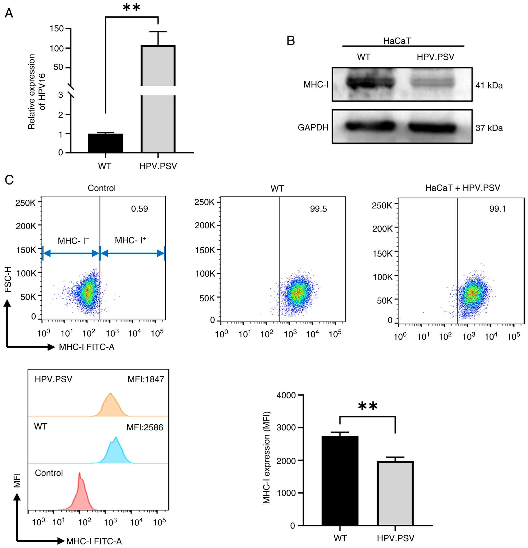
CA. As shown by RT-qPCR, HPV.PSV successfully infected HaCaT cells
(Fig. 1A). The presented study
aimed to detect the influence of hyperthermia treatment on the
expression of MHC-I in infected cells. Western blotting showed that
the expression of MHC-I was remarkably downregulated compared with
wild-type cells (Fig. 1B). In
addition, flow cytometry demonstrated HPV.PSV-infected HaCaT cells
exhibited lower expression of MHC-I (Fig. 1C). The data confirmed that the
present study successfully established HPV.PSV-infected cells.
Hyperthermia enhances expression of MHC-I
on HPV-infected cells and CA tissue
To analyze the regulatory role of hyperthermia on
MHC-I expression, HPV.PSV-infected HaCaT cells were incubated in a
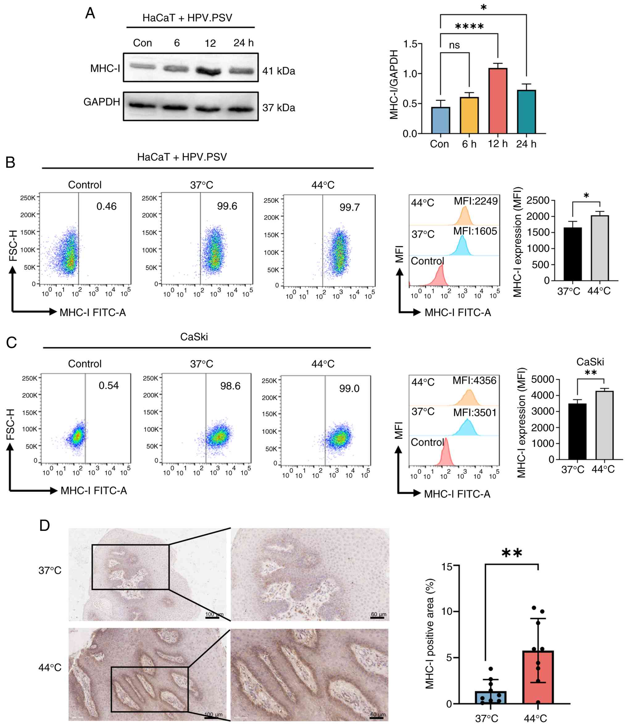
44°C water bath for 30 min. Expression of MHC-I reached a peak at
12 h post-incubation (Fig. 2A).
Flow cytometry revealed that MHC-I was significantly elevated in
HPV.PSV-infected HaCaT cells at 12 h post-hyperthermia treatment
(Fig. 2B). Flow cytometry
demonstrated that hyperthermia increased the expression of MHC-I in
CaSki cells (Fig. 2C).
Hyperthermia-treated samples from patients with CA expressed higher
levels of MHC-I than 37°C-treated samples (Fig. 2D). Hyperthermia treatment
increased the expression of MHC-I in HPV-infected epithelial cells
and CA tissue.
Hyperthermia induces the expression of
HMGB1
To determine the detailed regulatory association
between hyperthermia and MHC-I, 4D-FastDIA quantitative proteomics
was performed to analyze the differently expressed proteins. The
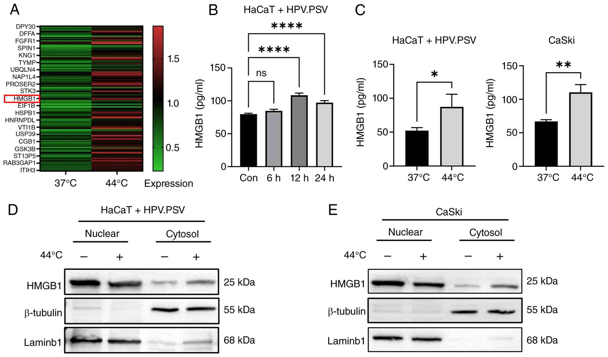
heat map demonstrated the top 21 upregulated proteins in
supernatant of 44°C-treated CaSki cells (Fig. 3A). Among them, HMGB1 activates
innate immunity and strengthens adaptive response via TLR4/RAGE as
a DAMP and participates in multiple immune-related disorders
(51). Additionally, the present
results showed time-course induction of HMGB1 following
hyperthermia (Fig. 3B).
Secretion of HMGB1 reached a peak at 12 h post-treatment, which was
consistent with the dynamic expression of MHC-I. The present study
detected the secretion of HMGB1 in hyperthermia treated
HPV.PSV-infected HaCaT and CaSki cells (Fig. 3C). In CaSki cells, HMGB1
secretion increased by almost 2.5-fold in the hyperthermia-treated
group compared with the control group (Fig. 3C). HMGB1 is secreted by a
cytoplasmic transport pathway (52). The present study analyzed the
expression of HMGB1 in nuclear and cytosolic protein. Both in
HPV.PSV-infected HaCaT and CaSki cells, hyperthermia promoted HMGB1
translocation from nucleus to the cytosol (Fig. 3D and E). Hyperthermia promoted
the secretion of HMGB1.
HMGB1 regulates the expression of
MHC-I
Following transfection of si-HMGB1into HaCaT and
CaSki cells, the interference efficiency in both cells achieved a
75% gene silencing rate in si-1 (si-HMGB1-1) and si-3 (si-HMGB1-3)
transfected groups (Fig. 4A),
which was also confirmed by western blotting (Fig. 4B). To confirm the role of HMGB1
knockdown on MHC-I expression, flow cytometry was performed. The
MHC-I expression of si-HMGB1-interfered HaCaT cells was reduced
after hyperthermia treatment compared with the hyperthermia-treated
negative control group (Fig.
4C). HaCaT cell viability assay was performed to confirm the
effect of HMGB1 on MHC-I expression was not caused by cytotoxicity
(Fig. 4D). Hyperthermia-treated
CaSki cells demonstrated downregulated expression of MHC-I compared
with the NC group (Fig. 4E).
CaSki cell viability assay was performed to rule out whether the
effect of HMGB1 on MHC-I expression was caused by cytotoxicity
(Fig. 4F). Furthermore,
recombinant HMGB1 protein (rHMGB1) was added to verify the role of
HMGB1 in MHC-I expression. Flow cytometry demonstrated that rHMGB1
recovered MHC-I expression in HPV. PSV-infected HaCaT and CaSki
cells (Fig. S1A and B). These
results complemented the proteomics analysis, confirming the
regulatory effect of differentially expressed HMGB1 on immunity.
HMGB1 is key for the expression of MHC-I following hyperthermia and
HPV infection. To rule out other regulatory molecule, the present
study detected the expression of tapasin. During the antigen
presentation of MHC-I, peptide loading complex (PLC) serves a key
role. Tapasin, as an endoplasmic reticulum chaperone, is a central
element of PLC that serves as a molecular chaperone to promote
proper folding of MHC-I molecules and loading of antigenic peptides
(53). In the present study,
hyperthermia did not influence the expression of tapasin (Fig. S2A and B). Therefore,
hyperthermia may influence the expression of MHC-I through
non-classical antigen-presenting mechanisms.
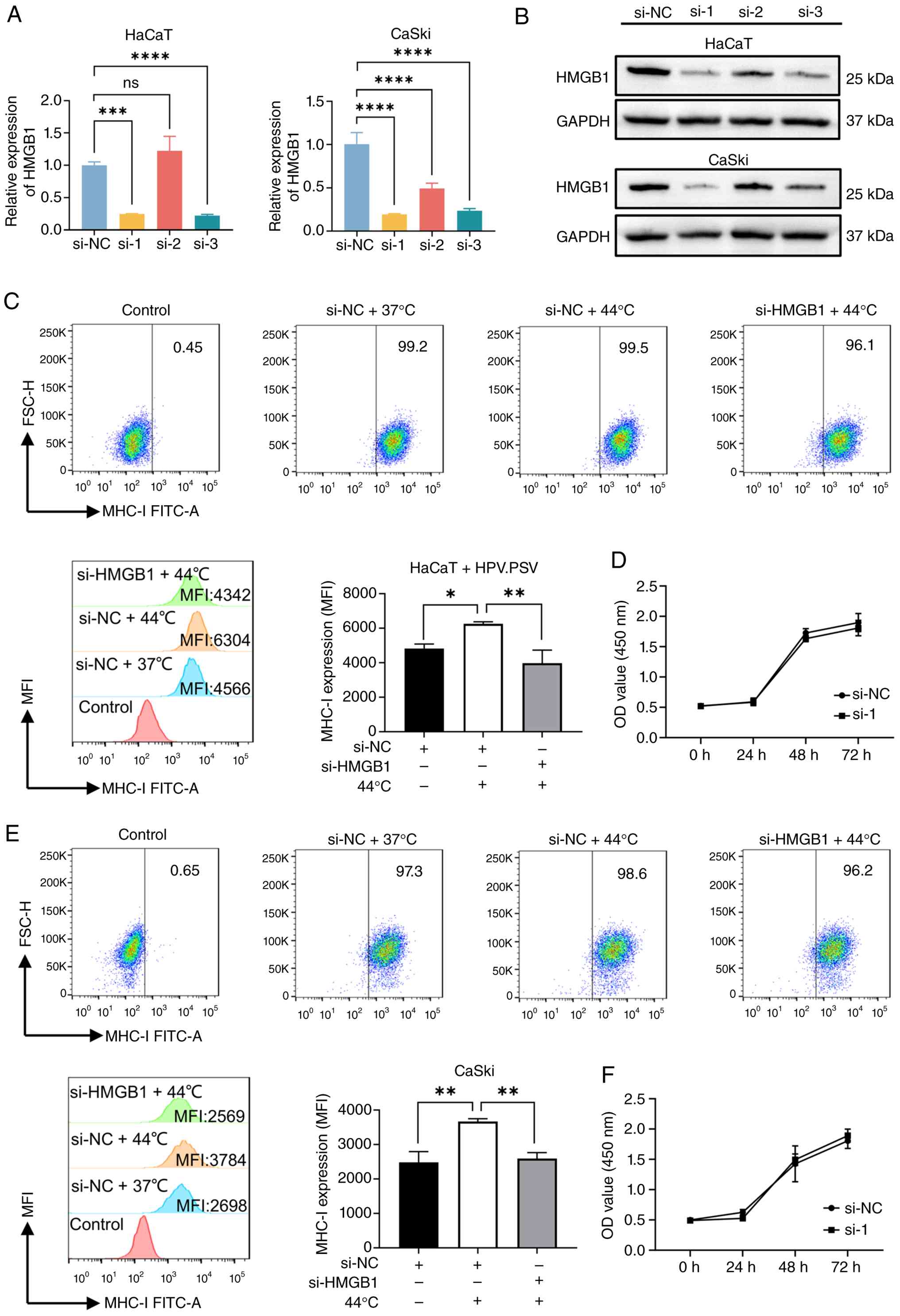 | Figure 4HMGB1 regulates the expression of
MHC-I. (A) Reverse transcription-quantitative PCR was performed to
detect the si-HMGB1 interference efficiency in HaCaT and CaSki
cells. (B) Western blotting was performed to detect the protein
levels of HMGB1 after si-HMGB1 interference in HaCaT and CaSki
cells. GAPDH was used as loading control. (C) Flow cytometry was
performed to detect MHC-I expression. (D) CCK8 assay was performed
to detect the HaCaT cell viability following transfection with si-1
or si-NC. (E) Flow cytometry to detect MHC-I expression in
si-HMGB1-transfected CaSki cells (F) CCK8 to detect the CaSki cell
viability after transfection with si-1 or si-NC.
*P<0.05, **P<0.01,
***P<0.001, ****P<0.0001. HMGB, high
mobility group box; MHC-I, major histocompatibility complex class
I; si, small interfering; CCK8, Cell Counting Kit-8; NC, negative
control; FSC-H, forward scatter height; ns, not significant; MFI,
mean fluorescence intensity; OD, optical density. |
Hyperthermia-regulated HSPA6 expression
influences the secretion of HMGB1 through JNK phosphorylation
Based on our previous findings, HSPA6 expression is
significantly induced by hyperthermia treatment (data not been
published). Here, HSPA6 was dynamically expressed in HPV.
PSV-infected HaCaT and CaSki cells (Fig. 5A). At 4-12 h post-incubation in a
44°C water bath, HSPA6 presented high expression in both cell
lines. Stable cell lines with lentivirus-mediated HSPA6
interference were constructed using sh-HSPA6. Knockdown efficiency
was up to 75% in HaCaT and 40% in CaSki cells (Fig. 5B). HSPA6 knockdown notably
decreased the expression of HSPA6 and p-JNK in both
HPV.PSV-infected HaCaT and CaSki cells following hyperthermia
treatment (Fig. 5C). As a member
of the HSP70 family, HSPA6 is predicted to participate in JNK
phosphorylation (54). HSP70
serves a key role in the protection of dopaminergic neurons by
regulating the activation of the JNK pathway (55). The consistent expression of HSPA6
and p-JNK indicated HSPA6 was key for JNK activation. In addition,
JNK phosphorylation was upregulated at 12 h post-hyperthermia
treatment in HPV.PSV-infected HaCaT cells, as well as 6, 12 and 24
h post-hyperthermia incubation in CaSki cells (Fig. 5D). To study whether HSPA6
directly regulated JNK phosphorylation, Co-IP assay validated the
interaction between endogenous HSPA6 and p-JNK in HaCaT and CaSki
cells. (Fig. 5E). Finally, to
verify the key regulatory molecules, the JNK pathway inhibitor
SP600125 and activator anisomycin were applied simultaneously
following hyperthermia and secretion of HMGB1 was detected in HPV.
PSV-infected HaCaT and CaSki cells. Western blotting analysis
revealed that SP600125 significantly suppressed JNK pathway
activation at 20 μM in HaCaT cells and at 10 μM in
CaSki cells (Fig. S3A and B).
Conversely, anisomycin markedly enhanced JNK pathway activation at
0.5 μM in HaCaT cells and at 5 μM in CaSki cells
(Fig. S3C and D). JNK
activation promoted HMGB1 expression, whereas JNK inhibition
suppressed HMGB1 expression in hyperthermia-treated
HPV.PSV-infected HaCaT and CaSki cell lines (Fig. 5F). To rule out other regulatory
mechanisms regulating HMGB1 secretion, western blotting showed
hyperthermia did not influence CRM1 expression in HPV-infected
cells (Fig. S4A and B). These
results suggested that hyperthermia facilitated HSPA6-modulated JNK
phosphorylation, which led to HMGB1 secretion and finally enhanced
the expression of MHC-I in HPV-infected epithelial cells, as well
as strengthening host immune regulation and recognition.
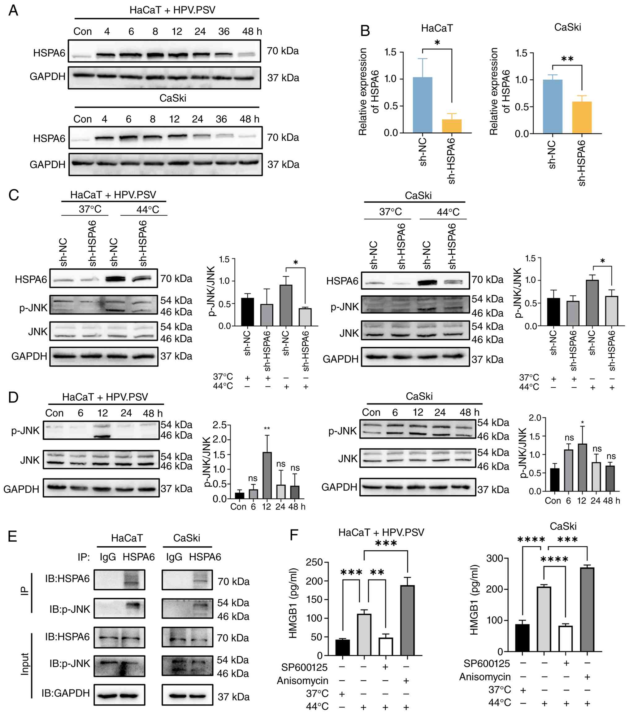 | Figure 5Hyperthermia-regulated HSPA6
expression influences the secretion of HMGB1 through JNK
phosphorylation. (A) Western blotting was performed to detect the
protein level of HSPA6 in HPV.PSV-infected HaCaT and CaSki cells
following hyperthermia treatment. (B) Reverse
transcription-quantitative PCR was used to detect the sh-HSPA6
knockdown efficiency in HaCaT and CaSki cells. (C) Western blotting
was performed to detect the expression of HSPA6 and JNK/p-JNK. (D)
Western blot to detect the expression of JNK/p-JNK. (E) Co-IP was
performed to detect the physical binding between HSPA6 and p-JNK in
HaCaT and CaSki cells. (F) ELISA was used to detect the secretion
of HMGB1 in HPV.PSV-infected HaCaT and CaSki cells stimulated with
JNK pathway inhibitor and activator. *P<0.05,
**P<0.01, ***P<0.001,
****P<0.0001. HMGB, high mobility group box; HSP,
heat shock protein; HPV.PSV, HPV 16 pseudovirus; sh, short hairpin;
p-, phosphorylated; IP, immunoprecipitation; Con, control; NC,
negative control; ns, not significant; IB, immunoblot. |
Discussion
CA is a common sexually transmitted disease caused
by HPV infection, which is associated with the escape of host
immune surveillance following virus infection. The present study
aimed to investigate the regulatory mechanism of hyperthermia
treatment on MHC-I expression of HPV-infected epithelial cells. As
a key molecule during adaptive immune response, MHC-I presents
endogenous antigenic peptides to CD8+ T cells, then
activates the CTL response. However, as a highly adapted host
virus, HPV has evolved multiple mechanisms to decrease MHC-I
expression to evade immune surveillance. HPV16 downregulates MHC-I
transcription and surface presentation through histone deacetylase
recruitment via its zinc finger domain, and suppresses MHC-I
expression by downregulating transporter associated with antigen
processing 1 (26,27). Hyperthermia treatment increased
the expression of MHC-I in HPV-infected epithelial cells and
tissues from patients with CA, which was in accordance with the
clinical therapeutic effect of hyperthermia (12). Local hyperthermia alone is
effective for genital warts (56). Given its immune-activating
effects (T cell infiltration, dendritic cell activation), combining
hyperthermia with existing modalities (CO2 laser,
photodynamic therapy) (57) may
enhance clearance and decrease recurrence.
Furthermore, 4D-FastDIA quantitative proteomics was
performed to analyze the differentially expressed proteins between
37 and 44°C hyperthermia-treated cells. HMGB1 is a highly conserved
nucleoprotein that translocates to the cytoplasm and is secreted to
the extracellular layer under stimulation, including cytokine
stimulation and cell death, to function as a DAMP (35,58). HMGB1 participates in the
maintenance of chromatin structure under a physiological state and
undergoes subcellular redistribution under stress conditions
(59). HMGB1, a ligand for
pattern recognition receptors including TLR4 and RAGE, activates
NF-κB and MAPK, which are important for MHC-I expression (60). The present study confirmed that
HMGB1 knockdown reduced the expression of MHC-I.
It is unclear whether hyperthermia modulated HMGB1
expression directly. Based on our previous study (data not
published), hyperthermia upregulates expression of HSPA6, which is
a member of HSP70 family. HSP70 cross-talks with the MAPK pathway.
HSP70 serves a neuro-protective role by regulating the activity of
the JNK pathway (55).
Hyperthermia induces HSP accumulation and triggers isoform-specific
JNK phosphorylation in the rat cerebellum (61). Here, hyperthermia increased HSPA6
expression in a time-dependent manner and loss-of-function
experiments demonstrated that HSPA6 regulated JNK phosphorylation
directly. HSPA6, as a member of the MAPK signaling pathway family,
especially the JNK pathway, is considered to be a key molecular
switch regulating HMGB1 secretion under oxidative stress (39). Expression of HMGB1 was regulated
by JNK activation. The present study demonstrated that JNK
phosphorylation was key for the secretion of HMGB1.
A number of molecular pathways regulate HMGB1
subcellular localization. Acetylation modification mediated by
histone acetyltransferase and deacetylase regulates the dynamic
distribution of HMGB1 in the karyoplasm (62). HSP90AA1 participates in the
nucleoplasmic translocation of HMGB1 and promotes the secretion of
HMGB1 through the autophagy polyvesicular pathway (63). To determine other regulatory
mechanisms regulating HMGB1 secretion, the present study detected
the expression of CRM1, which mediates classical nucleocytoplasmic
translocation of HMGB1 (64).
Results showed hyperthermia did not influence CRM1 expression in
HPV-infected cells which indicated that hyperthermia may regulate
the subcellular localization of HMGB1 through other non-classical
pathways. The nuclear translocation mechanism of HMGB1 requires
further study.
However, there are certain limitations to the
present study. There is still no appropriate animal model to mimic
HPV infection, which limits the ability to study the immune
regulatory mechanism and effect of hyperthermia on CA. The small
sample size of the present study (n=9) limits the generalizability
of findings. A larger sample size is therefore needed to further
evaluate the association between HPV genotype distribution and
hyperthermia sensitivity. Future studies with full HPV genotyping
and longitudinal follow-up are needed to address genotype-specific
responses to hyperthermia treatment. Although the present study
focused on the functional role of HMGB1 and MHC-I as
secreted/membrane proteins, mRNA expression of HMGB1 and MHC-I
should be examined to understand how HMGB1 and MHC-I are regulated
by hyperthermia. The involvement of NF-κB also should be explored
as HMGB1 is a known DAMP.
In conclusion, hyperthermia treatment facilitated
HSPA6-modulated JNK phosphorylation, which led to HMGB1 secretion,
enhanced the expression of MHC-I in HPV-infected epithelial cells
and strengthened host immune regulation and recognition. The
present study established a regulatory axis and potential
therapeutic targets for HPV-associated treatment.
Supplementary Data
Availability of data and materials
The data generated in the present study may be found
in the iProX under accession number IPX0012564000 or at the
following URL: iprox.cn/page/home.html.
Authors' contributions
HW performed the experiments. RQQ and TZ designed
the experiments and confirm the authenticity of all the raw data.
YG and TS analyzed and interpreted data. HL conceived and designed
the study. YB analyzed data. All authors read and approved the
final manuscript.
Ethics approval and consent to
participate
The samples used in the present study were harvested
from participants who provided written informed consent, and the
procedures were approved by Hospital Medical Science Research
Ethics Committee of the First Affiliated Hospital of China Medical
University, Shenyang, Liaoning 110122, P.R. China [approval no.
(2023)214].
Patient consent for publication
Not applicable.
Competing interests
The authors declare that they have no competing
interests.
Acknowledgements
Not applicable.
Funding
The present study was supported by National Key R&D Program
of China (grant no. 2023YFC2508200) and National Natural Science
Foundation of China (grant nos. 82173401 and 82373483).
References
|
1
|
Molina MA, Steenbergen RDM, Pumpe A,
Kenyon AN and Melchers WJG: HPV integration and cervical cancer: A
failed evolutionary viral trait. Trends Mol Med. 30:890–902. 2024.
View Article : Google Scholar : PubMed/NCBI
|
|
2
|
Seliger B, Massa C, Yang B, Bethmann D,
Kappler M, Eckert AW and Wickenhauser C: Immune escape mechanisms
and their clinical relevance in head and neck squamous cell
carcinoma. Int J Mol Sci. 21:70322020. View Article : Google Scholar : PubMed/NCBI
|
|
3
|
Murray ML, Meadows J, Doré CJ, Copas AJ,
Haddow LJ, Lacey C, Jit M, Soldan K, Bennett K, Tetlow M, et al:
Human papillomavirus infection: Protocol for a randomised
controlled trial of imiquimod cream (5%) versus podophyllotoxin
cream (0.15%), in combination with quadrivalent human
papillomavirus or control vaccination in the treatment and
prevention of recurrence of anogenital warts (HIPvac trial). Bmc
Med Res Methodol. 18:1252018. View Article : Google Scholar
|
|
4
|
Stuqui B, Provazzi PJS, Lima MLD, Cabral
ÁS, Leonel ECR, Candido NM, Taboga SR, da Silva MG, Lima FO, Melli
PPDS, et al: Condyloma acuminata: An evaluation of the immune
response at cellular and molecular levels. PLoS One.
18:e02842962023. View Article : Google Scholar : PubMed/NCBI
|
|
5
|
Kofoed K, Sand C, Forslund O and Madsen K:
Prevalence of human papillomavirus in anal and oral sites among
patients with genital warts. Acta Derm Venereol. 94:207–211. 2014.
View Article : Google Scholar
|
|
6
|
Ye J, Zheng L, He Y and Qi X: Human
papillomavirus associated cervical lesion: Pathogenesis and
therapeutic interventions. MedComm (2020). 4:e3682023. View Article : Google Scholar :
|
|
7
|
Brown DR, Schroeder JM, Bryan JT, Stoler
MH and Fife KH: Detection of multiple human papillomavirus types in
Condylomata acuminata lesions from otherwise healthy and
immunosuppressed patients. J Clin Microbiol. 37:3316–3322. 1999.
View Article : Google Scholar : PubMed/NCBI
|
|
8
|
Wang J, Chang YZ, Luo H, Jiang W, Xu L,
Chen T and Zhu X: Designing immunogenic nanotherapeutics for
photothermal-triggered immunotherapy involving reprogramming
immunosuppression and activating systemic antitumor responses.
Biomaterials. 255:1201532020. View Article : Google Scholar : PubMed/NCBI
|
|
9
|
Mahmood J, Alexander AA, Samanta S,
Kamlapurkar S, Singh P, Saeed A, Carrier F, Cao X, Shukla HD and
Vujaskovic Z: A combination of radiotherapy, hyperthermia, and
immunotherapy inhibits pancreatic tumor growth and prolongs the
survival of mice. Cancers (Basel). 12:10152020. View Article : Google Scholar : PubMed/NCBI
|
|
10
|
Huo W, Gao YL, Wang HY, Bi GJ, Qiao S, Cai
YF, Qi RQ, Yang Y, Lan J, Yao ZR, et al: Local hyperthermia versus
cryotherapy for treatment of plantar warts: A prospective
multi-centre non-randomized concurrent controlled clinical trial.
Acta Derm Venereol. 102:adv006552022. View Article : Google Scholar : PubMed/NCBI
|
|
11
|
Sun YZ, Li JF, Wei ZD, Jiang HH, Hong YX,
Zheng S, Qi RQ and Gao XH: Proteomic and bioinformatic analysis of
condyloma acuminata: Mild hyperthermia treatment reveals
compromised HPV infectivity of keratinocytes via regulation of
metabolism, differentiation and anti-viral responses. Int J
Hyperther. 36:383–393. 2019. View Article : Google Scholar
|
|
12
|
Yang Y, Zhang L, Zhang Y, Huo W, Qi R, Guo
H, Li X, Wu X, Bai F, Liu K, et al: Local hyperthermia at 44°C is
effective in clearing cervical high-risk human papillomaviruses: A
proof-of-concept, randomized controlled clinical trial. Clin Infect
Dis. 73:1642–1649. 2021. View Article : Google Scholar : PubMed/NCBI
|
|
13
|
Wei ZD, Sun YZ, Tu CX, Qi RQ, Huo W, Chen
HD and Gao XH: DNAJA4 deficiency augments hyperthermia-induced
Clusterin and ERK activation: Two critical protective factors of
human keratinocytes from hyperthermia-induced injury. J Eur Acad
Dermatol Venereol. 34:2308–2317. 2020. View Article : Google Scholar : PubMed/NCBI
|
|
14
|
Niu X, Zhao Y, Zhang T, Sun Y, Wei Z, Fu
K, Li J, Tang M, Wan W, Gao X, et al: Comprehensive succinylome
analyses reveal that hyperthermia upregulates lysine succinylation
of annexin A2 by downregulating sirtuin7 in human keratinocytes. J
Transl Int Med. 12:424–436. 2023. View Article : Google Scholar : PubMed/NCBI
|
|
15
|
Sun YZ, Ren Y, Zhang YJ, Han Y, Yang Y,
Gao YL, Zhu LL, Qi RQ, Chen HD and Gao XH: DNAJA4 deficiency
enhances NF-kappa B-related growth arrest induced by hyperthermia
in human keratinocytes. J Dermatol Sci. 91:256–267. 2018.
View Article : Google Scholar : PubMed/NCBI
|
|
16
|
Han X, Wang HF, Ma L, Chen RX, Wang Q,
Zhang CH, Zhang HH, Han Y, Yang Y, Guo H, et al: Human
papillomavirus genotypes affect the efficacy of hyperthermia
therapy for plantar warts: A quality improvement study. J Am Acad
Dermatol. 93:536–538. 2025. View Article : Google Scholar : PubMed/NCBI
|
|
17
|
Jiang F, Shao J, Chen L, Yang N, Liu J and
Li Z: Successful treatment of periungual warts with local
hyperthermia: Report of two cases. J Dermatolog Treat.
33:2380–2382. 2022. View Article : Google Scholar
|
|
18
|
Sengedorj A, Hader M, Heger L, Frey B,
Dudziak D, Fietkau R, Ott OJ, Scheidegger S, Barba SM, Gaipl US and
Rückert M: The effect of hyperthermia and radiotherapy sequence on
cancer cell death and the immune phenotype of breast cancer cells.
Cancers (Basel). 14:20502022. View Article : Google Scholar : PubMed/NCBI
|
|
19
|
Chen O, Michlíková S, Eckhardt L, Wondrak
M, De Mendoza AM, Krause M, McLeod DD and Kunz-Schughart LA:
Efficient heat shock response affects hyperthermia-induced
radiosensitization in a tumor spheroid control probability assay.
Cancers (Basel). 13:31682021. View Article : Google Scholar : PubMed/NCBI
|
|
20
|
Wang X, Gao XH, Hong Y, Li X and Chen HD:
Local hyperthermia decreases the expression of CCL-20 in condyloma
acuminatum. Virol J. 7:3012010. View Article : Google Scholar : PubMed/NCBI
|
|
21
|
Wang GH, Wang YB, Cheng YA, Chen HD, Gao
XH and Qi RQ: Local hyperthermia combined with imiquimod cleared
recalcitrant and extensive warts in a patient with systemic lupus
erythematosus. Clin Cosmet Investig Dermatol. 18:241–244. 2025.
View Article : Google Scholar : PubMed/NCBI
|
|
22
|
Tu YC, Yeh WC, Fang YW, Lo KH, Liang LN,
Liu XC, Tsai CC, Cheng CC, Lin MC, Yu HH and Su BC:
Hyperthermia-induced cytotoxicity and modulation of PD-L1 and MHC-I
expression in human non-small cell lung cancer cell lines. Exp
Physiol. Jul 8–2025.Epub ahead of print. View Article : Google Scholar : PubMed/NCBI
|
|
23
|
Wang X, Waschke BC, Woolaver RA, Chen SMY,
Chen Z and Wang JH: MHC class I-independent activation of virtual
memory CD8 T cells induced by chemotherapeutic agent-treated cancer
cells. Cell Mol Immunol. 18:723–734. 2021. View Article : Google Scholar
|
|
24
|
Tobian AAR, Canaday DH, Boom WH and
Harding CV: Bacterial heat shock proteins promote CD91-dependent
class I MHC cross-presentation of chaperoned peptide to CD8+ T
cells by cytosolic mechanisms in dendritic cells versus vacuolar
mechanisms in macrophages. J Immunol. 172:5277–5286. 2004.
View Article : Google Scholar : PubMed/NCBI
|
|
25
|
Wooldridge L, Lissina A, Vernazza J,
Gostick E, Laugel B, Hutchinson SL, Mirza F, Dunbar PR, Boulter JM,
Glick M, et al: Enhanced immunogenicity of CTL antigens through
mutation of the CD8 binding MHC class I invariant region. Eur J
Immunol. 37:1323–1333. 2007. View Article : Google Scholar : PubMed/NCBI
|
|
26
|
Heller C, Weisser T, Mueller-Schickert A,
Rufer E, Hoh A, Leonhardt RM and Knittler MR: Identification of key
amino acid residues that determine the ability of high risk
HPV16-E7 to dysregulate major histocompatibility complex class I
expression. J Biol Chem. 286:10983–10997. 2011. View Article : Google Scholar : PubMed/NCBI
|
|
27
|
Deng XM, Li W, Zhang X, Wang CX, Dong ZG,
Zhang X, Zheng GX, Zhang XH, Zheng N, Wang LL, et al: RNA
interference of human papillomavirus type 16 E7 increases HLA class
I antigen expression in HaCaT-E7 cells. Int J Gynecol Cancer.
21:28–34. 2011. View Article : Google Scholar : PubMed/NCBI
|
|
28
|
Taylor BC and Balko JM: Mechanisms of
MHC-I downregulation and role in immunotherapy response. Front
Immunol. 13:8448662022. View Article : Google Scholar : PubMed/NCBI
|
|
29
|
Westrich JA, Vermeer DW, Silva A, Bonney
S, Berger JN, Cicchini L, Greer RO, Song JI, Raben D, Slansky JE,
et al: CXCL14 suppresses human papillomavirus-associated head and
neck cancer through antigen-specific CD8+ T-cell
responses by upregulating MHC-I expression. Oncogene. 38:7166–7180.
2019. View Article : Google Scholar : PubMed/NCBI
|
|
30
|
Pang X, Zhang Y, Wei H, Zhang J, Luo Q,
Huang C and Zhang S: Expression and effects of high-mobility group
box 1 in cervical cancer. Int J Mol Sci. 15:8699–8712. 2014.
View Article : Google Scholar : PubMed/NCBI
|
|
31
|
Guo L, Wang D, Jiang X and He G: HMGB1:
From molecular functions to clinical applications in cancer and
inflammatory diseases. Med Res Rev. 46:408–444. 2026. View Article : Google Scholar
|
|
32
|
Wang S and Zhang Y: HMGB1 in inflammation
and cancer. J Hematol Oncol. 13:1162020. View Article : Google Scholar : PubMed/NCBI
|
|
33
|
Dong H, Zhang L and Liu S: Targeting
HMGB1: An available therapeutic strategy for breast cancer therapy.
Int J Biol Sci. 18:3421–3434. 2022. View Article : Google Scholar : PubMed/NCBI
|
|
34
|
Yang HM, Zhao XN, Li XL, Wang X, Pu Y, Wei
DK and Li Z: A pan-cancer analysis of the oncogenic function of
HMGB1 in human tumors. Biochem Biophys Rep.
40:1018512024.PubMed/NCBI
|
|
35
|
Sims GP, Rowe DC, Rietdijk ST, Herbst R
and Coyle AJ: HMGB1 and RAGE in inflammation and cancer. Annu Rev
Immunol. 28:367–388. 2010. View Article : Google Scholar : PubMed/NCBI
|
|
36
|
Luo Y, Li SJ, Yang J, Qiu YZ and Chen FP:
HMGB1 induces an inflammatory response in endothelial cells via the
RAGE-dependent endoplasmic reticulum stress pathway. Biochem
Biophys Res Commun. 438:732–738. 2013. View Article : Google Scholar : PubMed/NCBI
|
|
37
|
Tang YT, Zhao X, Antoine D, Xiao X, Wang
H, Andersson U, Billiar TR, Tracey KJ and Lu B: Regulation of
posttranslational modifications of HMGB1 during immune responses.
Antioxid Redox Sign. 24:620–634. 2016. View Article : Google Scholar
|
|
38
|
Bianchi ME, Crippa MP, Manfredi AA,
Mezzapelle R, Rovere Querini P and Venereau E: High-mobility group
box 1 protein orchestrates responses to tissue damage via
inflammation, innate and adaptive immunity, and tissue repair.
Immunol Rev. 280:74–82. 2017. View Article : Google Scholar : PubMed/NCBI
|
|
39
|
Tang D, Shi Y, Kang R, Li T, Xiao W, Wang
H and Xiao X: Hydrogen peroxide stimulates macrophages and
monocytes to actively release HMGB1. J Leukocyte Biol. 81:741–747.
2007. View Article : Google Scholar :
|
|
40
|
Grundtman C, Bruton J, Yamada T, Ostberg
T, Pisetsky DS, Harris HE, Andersson U, Lundberg IE and Westerblad
H: Effects of HMGB1 on in vitro responses of isolated muscle fibers
and functional aspects in skeletal muscles of idiopathic
inflammatory myopathies. FASEB J. 24:570–578. 2010. View Article : Google Scholar
|
|
41
|
Wan Z, Zhang X, Peng A, He M, Lei Z and
Wang Y: TLR4-HMGB1 signaling pathway affects the inflammatory
reaction of autoimmune myositis by regulating MHC-I. Int
Immunopharmacol. 41:74–81. 2016. View Article : Google Scholar : PubMed/NCBI
|
|
42
|
Li Z, Deng J, Sun J and Ma Y: Hyperthermia
targeting the tumor microenvironment facilitates immune checkpoint
inhibitors. Front Immunol. 11:5952072020. View Article : Google Scholar : PubMed/NCBI
|
|
43
|
Zhao W, An H, Zhou J, Xu H, Yu Y and Cao
X: Hyperthermia differentially regulates TLR4 and TLR2-mediated
innate immune response. Immunol Lett. 108:137–142. 2007. View Article : Google Scholar : PubMed/NCBI
|
|
44
|
Wang L, Fu H, Zhao J, Liu Z, Chen S, Zhang
CQ, Hu P, Wang J, Shi J and Jia W: Cascade magnetic hyperthermia
therapy for biofilm eradication and bone regeneration via dual
osteoimmuno-regulation. ACS Nano. 19:21679–21695. 2025. View Article : Google Scholar : PubMed/NCBI
|
|
45
|
Anbar M: Hyperthermia of the cancerous
breast: Analysis of mechanism. Cancer Lett. 84:23–29. 1994.
View Article : Google Scholar : PubMed/NCBI
|
|
46
|
Teicher BA, Holden SA, Liu CJ, Ara G and
Herman TS: Minocycline as a modulator of chemotherapy and
hyperthermia in vitro and in vivo. Cancer Lett. 82:17–25. 1994.
View Article : Google Scholar : PubMed/NCBI
|
|
47
|
Demichev V, Messner CB, Vernardis SI,
Lilley KS and Ralser M: DIA-NN: Neural networks and interference
correction enable deep proteome coverage in high throughput. Nat
Methods. 17:41–44. 2020. View Article : Google Scholar :
|
|
48
|
Livak KJ and Schmittgen TD: Analysis of
relative gene expression data using real-time quantitative PCR and
the 2(-Delta Delta C(T)) method. Methods. 25:402–408. 2001.
View Article : Google Scholar
|
|
49
|
Janke C and Bulinski JC:
Post-translational regulation of the microtubule cytoskeleton:
mechanisms and functions. Nat Rev Mol Cell Bio. 12:773–786. 2011.
View Article : Google Scholar
|
|
50
|
Dechat T, Pfleghaar K, Sengupta K, Shimi
T, Shumaker DK, Solimando L and Goldman RD: Nuclear lamins: Major
factors in the structural organization and function of the nucleus
and chromatin. Gene Dev. 22:832–853. 2008. View Article : Google Scholar : PubMed/NCBI
|
|
51
|
Zhong H, Li X, Zhou S, Jiang P, Liu X,
Ouyang M, Nie Y, Chen X, Zhang L, Liu Y, et al: Interplay between
RAGE and TLR4 regulates HMGB1-induced inflammation by promoting
cell surface expression of RAGE and TLR4. J Immunol. 205:767–775.
2020. View Article : Google Scholar : PubMed/NCBI
|
|
52
|
Gillespie KP, Pirnie R, Mesaros C and
Blair IA: Cisplatin dependent secretion of immunomodulatory high
mobility group box 1 (HMGB1) protein from lung cancer cells.
Biomolecules. 13:13352023. View Article : Google Scholar : PubMed/NCBI
|
|
53
|
Boulanger DSM, Douglas LR, Duriez PJ, Kang
Y, Dalchau N, James E and Elliott T: Tapasin-mediated editing of
the MHC I immunopeptidome is epitope specific and dependent on
peptide off-rate, abundance, and level of tapasin expression. Front
Immunol. 13:9566032022. View Article : Google Scholar : PubMed/NCBI
|
|
54
|
Wang SH, Jiang JX, Zheng KT, Zhang SJ,
Chen TD and Wang Z: Exosome-derived Menin from cancer-associated
fibroblasts promotes gastric cancer progression by activating the
HSPA6/JNK/JunD pathway and inducing EMT. J Transl Med. 24:2742025.
View Article : Google Scholar : PubMed/NCBI
|
|
55
|
Shukla AK, Pragya P, Chaouhan HS, Tiwari
AK, Patel DK, Abdin MZ and Chowdhuri DK: Heat shock protein-70
(Hsp-70) suppresses paraquat-induced neurodegeneration by
inhibiting JNK and caspase-3 activation in Drosophila model of
Parkinson's disease. PLoS One. 9:e988862014. View Article : Google Scholar : PubMed/NCBI
|
|
56
|
Piguet V: Heat-induced editing of HPV
genes to clear mucocutaneous warts? J Invest Dermatol. 137:796–797.
2017. View Article : Google Scholar : PubMed/NCBI
|
|
57
|
Scheinfeld N: Update on the treatment of
genital warts. Dermatol Online J. 19:185592013. View Article : Google Scholar : PubMed/NCBI
|
|
58
|
Papatheodorou A, Stein A, Bank M, Sison
CP, Gibbs K, Davies P and Bloom O: High-mobility group box 1
(HMGB1) is elevated systemically in persons with acute or chronic
traumatic spinal cord injury. J Neurotraum. 34:746–754. 2017.
View Article : Google Scholar
|
|
59
|
Zhao G, Fu C, Wang L, Zhu L, Yan Y, Xiang
Y, Zheng F, Gong F, Chen S and Chen G: Down-regulation of nuclear
HMGB1 reduces ischemia-induced HMGB1 translocation and release and
protects against liver ischemia-reperfusion injury. Sci Rep.
7:462722017. View Article : Google Scholar : PubMed/NCBI
|
|
60
|
Zhang Z, Ji R, Liu Z, Jiang Z, Chu M, Wang
Y and Zhao J: hUMSC-Exosomes suppress TREM1-p38 MAPK signaling via
HMGB1-dependent mechanisms to reprogram microglial function and
promote neuroprotection in ischemic stroke. J Nanobiotechnology.
23:5722025. View Article : Google Scholar : PubMed/NCBI
|
|
61
|
Schiaffonati L, Maroni P, Bendinelli P,
Tiberio L and Piccoletti R: Hyperthermia induces gene expression of
heat shock protein 70 and phosphorylation of mitogen activated
protein kinases in the rat cerebellum. Neurosci Lett. 312:75–78.
2001. View Article : Google Scholar : PubMed/NCBI
|
|
62
|
Chen RC, Kang R and Tang DL: The mechanism
of HMGB1 secretion and release. Exp Mol Med. 54:91–102. 2022.
View Article : Google Scholar : PubMed/NCBI
|
|
63
|
Kim YH, Kwak MS, Lee B, Shin JM, Aum S,
Park IH, Lee MG and Shin JS: Secretory autophagy machinery and
vesicular trafficking are involved in HMGB1 secretion. Autophagy.
17:2345–2362. 2021. View Article : Google Scholar :
|
|
64
|
Fukuda M, Asano S, Nakamura T, Adachi M,
Yoshida M, Yanagida M and Nishida E: CRM1 is responsible for
intracellular transport mediated by the nuclear export signal.
Nature. 390:308–311. 1997. View
Article : Google Scholar : PubMed/NCBI
|