Introduction
Sports-related bone injuries represent a major
public health concern across athletic populations and include acute
traumatic fractures, stress fractures, and overuse injuries that
frequently compromise athletic performance and quality of life.
Epidemiological evidence indicates that lower extremity stress
fractures are highly prevalent in adolescent and young adult
athletes, with distal femoral stress fractures demonstrating
particularly elevated surgical intervention rates (1). Similarly, pelvic avulsion fractures
in pediatric athletes require specialized management approaches
across trauma centers (2). The
clinical management of these injuries is associated with
substantial challenges, as delayed union and nonunion remain
persistent complications that prolong rehabilitation and limit
return to sport. A recent meta-analysis revealed that patients with
lisfranc injuries treated with different surgical approaches
achieved comparable outcomes in terms of return to sport,
highlighting the need for optimized treatment strategies (3). Furthermore, the burden of these
injuries extends beyond acute management, as long-term functional
outcomes and complication rates following surgical intervention
demonstrate considerable variability (4). These persistent clinical challenges
underscore the urgent need for novel therapeutic approaches that
can accelerate bone healing and improve functional recovery in
athletic populations.
Physical activity exerts profound beneficial effects
on the skeletal system through mechanical loading that stimulates
bone formation and remodeling. The mechanostat concept, originally
proposed by Frost, describes how bone tissue adapts to mechanical
demands by modulating osteoblast and osteoclast activity (5). Exercise-induced mechanical loading
promotes osteogenesis through activation of canonical Wnt/β-catenin
signaling and other anabolic pathways, thereby maintaining skeletal
integrity (6). Despite the
well-established epidemiological evidence linking exercise to
improved bone health, the fundamental molecular mechanisms by which
mechanical forces are transduced into cellular responses remain
incompletely understood. Recent investigations have demonstrated
that mechanical loading not only directly affects bone cells but
also orchestrates complex intercellular communication networks
involving multiple tissue types (7). The muscle-bone axis represents a
particularly intriguing paradigm, as skeletal muscle contraction
during exercise generates signals that influence bone metabolism
(8). However, the identity of
the key molecular mediators that convey exercise-induced signals
from muscle to bone and the precise signaling cascades activated in
recipient bone cells have remained elusive, representing a critical
knowledge gap in skeletal physiology.
Exosomes, nano-sized extracellular vesicles ranging
from 30 to 150 nm in diameter, have emerged as fundamental
mediators of intercellular communication in the musculoskeletal
system. These vesicles transport diverse bioactive cargoes,
including proteins, lipids, and noncoding RNAs (ncRNAs), that
reflect the physiological state of parent cells and modulate the
function of recipient cells (9).
Within the bone microenvironment, exosomes facilitate critical
crosstalk among osteoblasts, osteoclasts, osteocytes, and
mesenchymal stem cells, thereby coordinating the tightly regulated
processes of bone resorption and formation (10). ncRNAs encapsulated within
exosomes, particularly microRNAs (miRNAs or miRs), long noncoding
RNAs (lncRNAs), and circular RNAs (circRNAs), have garnered
substantial research interest due to their capacity to
simultaneously target multiple signaling pathways (8). Recent studies have demonstrated
that exosomal miRNAs such as miR-21-5p, miR-23b-3p, and miR-29a-3p
regulate osteoblast differentiation and bone formation through
modulation of key transcription factors and signaling cascades
(11-13). Specifically, miR-21-5p was shown
to promote osteogenesis by targeting sprouty homolog 1 to enhance
bone morphogenetic protein (BMP)/Smad signaling (12), miR-23b-3p suppressed osteoblast
differentiation by inhibiting runt-related transcription factor 2
(Runx2) expression (13), and
miR-29a-3p facilitated bone formation by targeting Dickkopf-related
protein (DKK)1 and sclerostin to activate the Wnt/β-catenin pathway
(11). The exponential growth of
studies in this field reflects the recognition that exosomal ncRNAs
represent a previously unappreciated layer of regulatory complexity
in bone biology, with implications for understanding both
physiological adaptation and pathological conditions.
Although several recent reviews have addressed
exosomal ncRNAs in bone metabolism or exercise-induced
extracellular vesicles separately, a systematic synthesis
integrating these two rapidly evolving fields remains conspicuously
absent. Previous reviews have primarily focused on mesenchymal stem
cell-derived exosomes for bone regeneration (14,15), while others have examined the
muscle-bone axis without specifically addressing the exosomal ncRNA
intermediary (8). Furthermore,
existing literature has largely concentrated on pathological states
such as osteoporosis and osteoarthritis rather than sports-related
bone injuries in athletic populations (16,17). A recent review by Zhang et
al (8) comprehensively
summarized exosomal miRNAs in muscle-bone crosstalk with
implications for sarcopenia and osteoporosis; however, the study
primarily focused on age-related musculoskeletal disorders rather
than exercise-induced bone adaptation in the context of
sports-related injuries. Critically, the mechanistic link between
exercise as a physiological stimulus and exosomal ncRNA-mediated
bone adaptation has not been systematically reviewed. Notably,
Zhang et al (8) did not
comprehensively address how different exercise modalities modulate
exosomal cargo beyond miRNAs, nor did they focus on the sequential
phases of fracture healing or translational applications for
sports-related bone injuries. The present review fills this
critical gap by providing the first systematic synthesis of
exercise-induced exosomal ncRNAs as key mediators of bone
remodeling, with specific emphasis on translational applications
for sports-related bone injuries.
Fundamental biological basis: Exercise,
exosomal ncRNAs, and bone remodeling
Bone remodeling is a dynamic process of resorption
and formation that maintains skeletal integrity and adapts to
mechanical demands. Exercise, a potent physiological stimulus, not
only influences bone cell activity but also modulates the release
of exosomes-extracellular vesicles that carry bioactive ncRNAs
between cells. These exercise-induced exosomal ncRNAs, including
miRNAs, lncRNAs, and circRNAs, have emerged as critical mediators
that translate mechanical forces into molecular signals regulating
osteoblast and osteoclast function. A conceptual overview of these
core components is presented (Fig.
1).
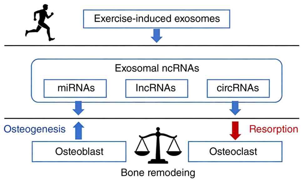 | Figure 1Schematic overview of the core triad:
Exercise, exosomal ncRNAs, and bone remodeling. Core framework
linking exercise, exosomal ncRNAs, and bone remodeling. Exercise
stimulates the release of exosomes containing miRNAs, lncRNAs, and
circRNAs, which regulate osteoblast-mediated osteogenesis and
osteoclast-mediated resorption to maintain skeletal homeostasis.
ncRNAs, noncoding RNAs; miRNAs, microRNAs; lncRNAs, long noncoding
RNAs; circRNAs, circular RNAs. |
Bone remodeling and fracture healing
dynamics
Bone remodeling is a continuous process executed by
the coordinated actions of osteoclasts, which resorb bone, and
osteoblasts, which synthesize new bone matrix. This
osteoblast-osteoclast coupling is essential for skeletal
homeostasis and is profoundly influenced by mechanical loading
(18). The process of fracture
healing recapitulates embryonic skeletal development, involving an
inflammatory phase, soft and hard callus formation, and remodeling
(19). During this cascade,
various signaling pathways, including Wnt/β-catenin and BMP/Smad,
are sequentially activated to ensure proper repair (20). Recent evidence has highlighted
the critical role of skeletal stem cells and their niche
interactions in coordinating regenerative responses (21). Pathological conditions, such as
stress fractures common in athletes, arise when this delicate
balance is disrupted, leading to an imbalance between bone
resorption and formation (22).
Moreover, immune cells and vascular endothelial cells contribute
significantly to the regenerative microenvironment, influencing the
recruitment and differentiation of skeletal progenitor cells
(23).
Exosomes: Biogenesis and intercellular
communication in the musculoskeletal system
Exosomes are nano-sized extracellular vesicles
(30-150 nm in diameter) that mediate paracrine and endocrine
signaling by transferring bioactive molecules, including proteins,
lipids, and ncRNAs, between cells (24). Their biogenesis involves the
endosomal pathway, beginning with the formation of intraluminal
vesicles within multivesicular bodies, which subsequently fuse with
the plasma membrane to release exosomes into the extracellular
space (25). In the
musculoskeletal system, exosomes facilitate critical crosstalk
between various cell types, such as between skeletal muscle and
bone, a concept central to the muscle-bone axis (26). For instance, exosomes derived
from myoblasts can influence the activity of osteoclasts,
highlighting their role in systemic skeletal regulation (27). The specific cargo of these
vesicles, particularly ncRNAs, dictates their functional impact on
recipient cells. Notably, the molecular composition of exosomes
reflects the physiological or pathological state of the parent
cell, making them attractive targets for biomarker discovery
(28).
Classification and functional roles of
exosomal ncRNAs in bone metabolism
ncRNAs encapsulated within exosomes are pivotal
regulators of gene expression in bone cells. Among these, miRNAs
are the most extensively studied, functioning by binding to
complementary sequences in target mRNAs to inhibit translation or
promote degradation (24). In
the context of bone, exosomal miRNAs have been shown to both
promote and inhibit osteogenesis. For example, exosomal miR-26a
derived from stem cells enhanced bone regeneration by targeting
specific inhibitory factors (29), whereas other miRNAs suppressed
osteoblast differentiation by modulating key transcription factors
(30). LncRNAs and circRNAs add
another layer of complexity, acting as competing endogenous RNAs
that sponge miRNAs to modulate their activity. For instance,
lncRNAs can sequester osteogenic suppressor miRNAs, thereby
derepressing osteogenic gene expression (31). This complex regulatory network is
now recognized as a fundamental mechanism governing the fate of
bone marrow mesenchymal stem cells (BMSCs) and other skeletal
progenitors (32). Furthermore,
exosomal ncRNAs can simultaneously target multiple signaling nodes,
providing a coordinated regulatory mechanism that single soluble
factors cannot achieve (8).
Exercise-induced modulation of exosome
secretion and ncRNA cargo
Physical activity acts as a potent systemic cue that
profoundly influences the exosomal landscape. Exercise,
particularly resistance and high-intensity interval training,
alters the number of circulating extracellular vesicles and
modifies their molecular cargo (33). Garner et al (34) demonstrated that acute exercise
rapidly increases the expression of genes involved in the
multivesicular body and exosome pathways in skeletal muscle.
Mechanical strain on skeletal muscle is a potent driver of exosome
secretion, with these muscle-derived vesicles carrying specific
myomiRs and other factors that can target distant organs, including
bone (35). Recent research has
shown that mechanically stimulated osteocytes also release exosomes
enriched with osteogenic miRNAs that promote osteoblast
differentiation (36).
Furthermore, the metabolic and mechanical stress associated with
exercise can be sensed by various tissues, such as adipose tissue
and the vascular endothelium, leading to the secretion of
tissue-specific exosomes (37).
This exercise-induced 'exosomal signature' is dynamic, with
distinct differences observed between acute bouts of exercise and
long-term training, as well as being influenced by factors such as
age and sex (38). Notably, the
dose-effect relationship between exercise parameters, including
intensity, duration, and frequency, and the resulting exosomal
ncRNA profile remains incompletely characterized, representing a
critical knowledge gap (39).
Exercise modulation of exosomal ncRNAs:
Expression characteristics, regulatory patterns, and tissue
sources
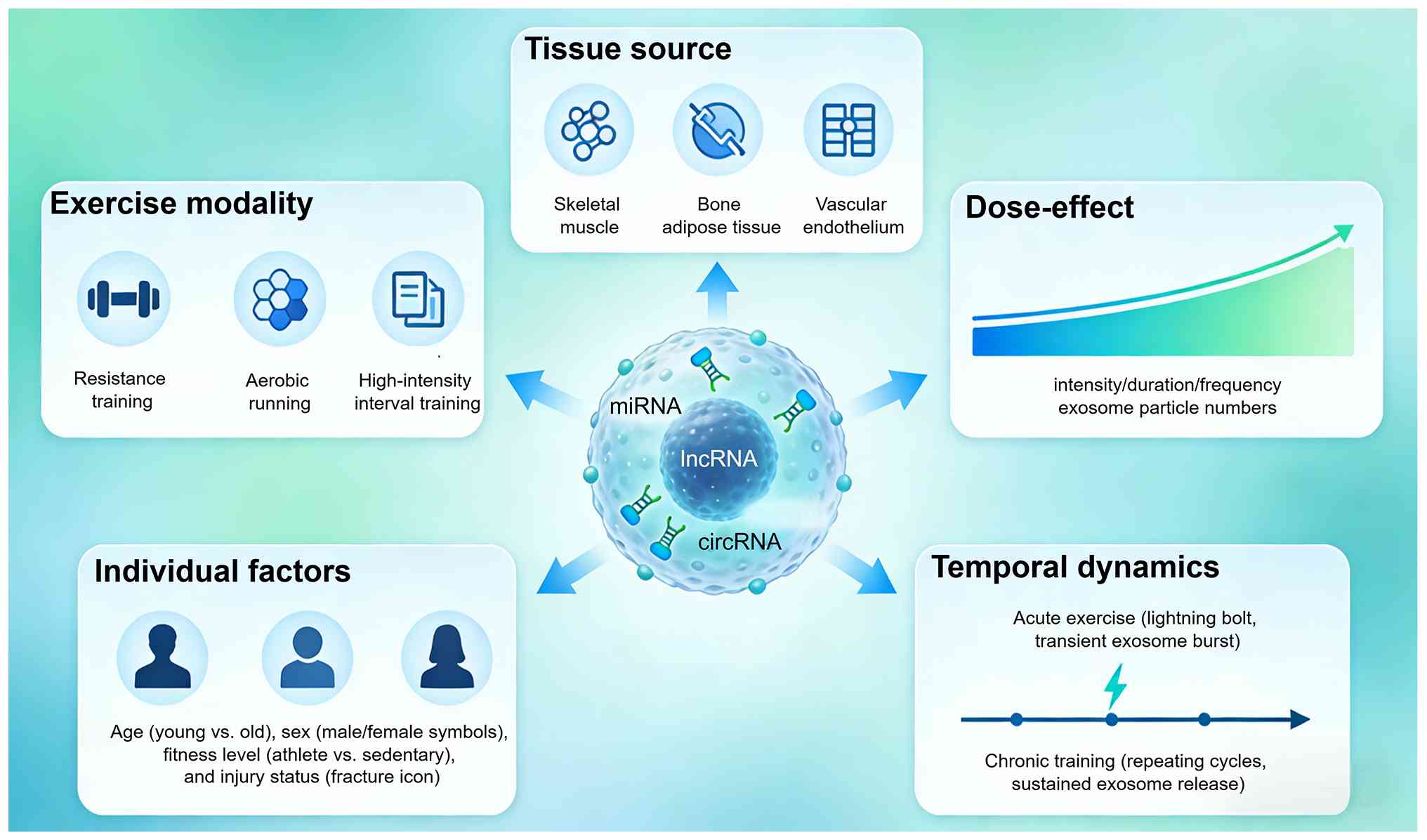
Physical activity dynamically reshapes the landscape
of circulating exosomal ncRNAs, with exercise modalities, tissue
sources, and individual factors collectively determining the
specific molecular signatures that mediate systemic adaptation. The
regulatory patterns are further influenced by the dominant tissue
origins, particularly skeletal muscle-derived exosomes as the
primary exercise-responsive carriers, alongside contributions from
bone, adipose tissue, and the vascular endothelium. Individual
factors such as age, sex, and fitness level introduce substantial
heterogeneity in exosomal responses, while the temporal dynamics
distinguish acute exercise effects from chronic training
adaptations (Fig. 2). A
comprehensive overview of these regulatory patterns, summarizing
key studies on exercise modalities, tissue sources, dose-effect
relationships, individual factors, and temporal dynamics, is
presented in Table I.
 | Table ISummary of exercise-induced exosomal
ncRNA expression characteristics, regulatory patterns, and tissue
sources. |
Table I
Summary of exercise-induced exosomal
ncRNA expression characteristics, regulatory patterns, and tissue
sources.
| Authors, year | Exercise
modality/parameter | Tissue source | ncRNA cargo | Regulatory
pattern | Key finding | (Refs.) |
|---|
| Just et al,
2020 | Blood
flow-restricted resistance exercise | Circulating
EVs | miRNAs | Differential
modulation | Blood
flow-restricted resistance exercise alters surface profile and
miRNA cargo of circulating EVs distinct from traditional resistance
training | (33) |
| Garner et
al, 2020 | Acute exercise | Skeletal
muscle | Exosome pathway
genes | Acute response | Acute exercise
rapidly upregulates genes involved in multivesicular body and
exosome pathways in skeletal muscle | (34) |
| Burke et al,
2024 | Resistance
exercise | Muscle-derived
EVs | miR-1 | Tissue source | Extracellular
vesicle transfer of miR-1 to adipose tissue modifies lipolytic
pathways following resistance exercise | (37) |
| Xhuti et al,
2023 | Resistance
training | Circulating
exosome-like vesicles, skeletal muscle | miRNAs | Individual
factors | Circulating
exosome-like vesicle and skeletal muscle miRNAs are altered with
age and resistance training | (38) |
| Lovett et
al, 2024 | Exercise-induced
muscle damage | Plasma-derived
sEVs | miRNAs | Dose-effect | Time-dependent
alterations in miRNA cargo following exercise-induced skeletal
muscle damage | (39) |
| de Mendonça et
al, 2020 | Aerobic exercise
training | sEVs | miRNAs | Differential
modulation | Aerobic training
regulates serum EV miRNAs linked to obesity-related metabolic
pathways | (40) |
| Castaño et
al, 2020 | HIIT | Muscle-derived
exosomes | miRNAs | Differential
modulation | HIIT induces
muscle-derived exosomal miRNAs that improve insulin sensitivity via
hepatic FoxO1 downregulation | (41) |
| Maggio et
al, 2023 | Moderate continuous
training vs. HIIT | Circulating
EVs | miRNA, protein
cargo | Differential
modulation | Different exercise
regimens produce distinct modulatory effects on circulating EV
cargo and inflammatory properties | (42) |
| Vechetti et
al, 2021 | Mechanical
overload | Muscle-derived
EVs | miRNAs | Tissue source | Mechanical
overload-induced muscle-derived EVs carry specific miRNAs that
promote adipose tissue lipolysis | (43) |
| Guescini et
al, 2015 | Exercise | Skeletal
muscle | miRNAs | Tissue source | Skeletal muscle
releases α-sarcoglycan-positive EVs carrying miRNAs into the
bloodstream | (44) |
| Zhao et al,
2022 | Exercise | Brown adipose
tissue | miRNAs | Tissue source | Small EVs from
brown adipose tissue mediate exercise-induced cardioprotection | (45) |
| Lou et al,
2022 | Exercise | Liver-derived
EVs | miR-122-5p | Tissue source | Liver-derived EVs
carrying miR-122-5p promote exercise-induced angiogenesis | (46) |
| Sapp et al,
2019 | Moderate vs.
high-intensity exercise | Circulating
vesicles | Endothelial
markers | Dose-effect | Moderate and
high-intensity exercise differentially affect vesicle release in an
intensity-dependent manner | (47) |
| Warnier et
al, 2023 | 6-Week sprint
interval training | Circulating
EVs | miRNA, protein
cargo | Dose-effect | Sustained training
regimens produce cumulative effects on exosomal profiles | (48) |
| Doncheva et
al, 2022 | Exercise | EVs | miRNAs | Dose-effect | EV and miRNA
alterations in response to exercise are modulated by insulin
sensitivity and overweight status | (49) |
| Estébanez et
al, 2021 | Resistance
training | Exosomes | CD63 protein | Individual
factors | Resistance training
diminishes exosome CD63 protein expression in the elderly without
modifying plasma miR-146a-5p levels | (50) |
| Kargl et al,
2024 | Concurrent exercise
training (12 weeks) | Circulating
EVs | miRNA, protein
cargo | Individual
factors | Circulating EV
characteristics differ between men and women following concurrent
exercise training | (51) |
| Kargl et al,
2026 | Resistance
exercise | Circulating
EVs | miRNA,
metabolite | Individual
factors | Menstrual cycle
phase and hormonal contraceptive use variably influence resistance
exercise-induced EV signaling | (52) |
| Fernandez-Sanjurjo
et al, 2024 | Exercise
training | Plasma EVs | miRNAs | Individual
factors | Distinct plasma EV
miRNA cargo differentiates sedentary young males from athletes | (53) |
| D'Souza et
al, 2018 | Intense
exercise | Circulatory
exosomes | miRNAs | Acute vs.
chronic | Circulatory
exosomal miRNA following intense exercise is unrelated to muscle
and plasma miRNA abundances | (54) |
| Silver et
al, 2020 | Acute
moderate-intensity exercise | EVs | miRNAs | Acute vs.
chronic | Extracellular
vesicular miRNA expression is not a proxy for skeletal muscle miRNA
expression following acute exercise | (55) |
| Hou et al,
2019 | Long-term
exercise | Exosomes | miR-342-5p | Acute vs.
chronic | Long-term
exercise-derived exosomal miR-342-5p functions as a novel exerkine
for cardioprotection | (56) |
| Di et al,
2020 | Long-term
exercise | EVs | miR-191a-5p | Acute vs.
chronic | Long-term
exercise-secreted EVs promote browning of white adipocytes by
suppressing miR-191a-5p | (57) |
Differential modulation by exercise
modalities
Different exercise modalities elicit distinct
exosomal ncRNA expression profiles, reflecting the unique
physiological demands of each training type. As summarized in
Table I, resistance exercise has
been shown to alter the surface profile and miRNA cargo of
circulating extracellular vesicles, with Just et al
(33) demonstrating that blood
flow-restricted resistance exercise modifies the miRNA landscape in
a manner distinct from traditional resistance training. Similarly,
Garner et al (34)
reported that a single bout of acute exercise comprising 45 min of
cycling followed by resistance exercise rapidly upregulates genes
involved in multivesicular body and exosome pathways in skeletal
muscle, establishing a mechanistic link between mechanical stress
and exosome biogenesis. By contrast, aerobic exercise training
regulates serum extracellular vesicle miRNAs linked to metabolic
health, as shown by de Mendonça et al (40), who observed that aerobic training
modulates miRNA profiles associated with obesity-related pathways.
High-intensity interval training (HIIT) was shown to induce
muscle-derived exosomal miRNAs that improve insulin sensitivity
through hepatic forkhead box protein O1 (FoxO1) downregulation, as
demonstrated by Castaño et al (41). Maggio et al (42) further characterized that
different exercise regimens, including moderate continuous training
and HIIT, produce distinct modulatory effects on circulating
extracellular vesicle cargo and their inflammatory properties.
Collectively, these findings indicate that exercise modality serves
as a critical determinant of exosomal ncRNA composition, with
implications for targeted therapeutic applications.
Dominant tissue sources
Skeletal muscle represents the predominant tissue
source of exercise-induced exosomal ncRNAs, functioning as a
central mediator of the muscle-bone axis. Vechetti et al
(43) demonstrated that
mechanical overload-induced muscle-derived extracellular vesicles
carry specific miRNAs that promote adipose tissue lipolysis,
highlighting the endocrine capacity of myogenic exosomes. The
release of muscle-derived exosomes into the circulation following
exercise has been well characterized, with Guescini et al
(44) providing foundational
evidence that skeletal muscle releases α-sarcoglycan-positive
extracellular vesicles carrying miRNAs into the bloodstream. Beyond
skeletal muscle, adipose tissue contributes significantly to the
exercise-responsive exosomal pool. Burke et al (37) showed that extracellular vesicle
transfer of miR-1 to adipose tissue modifies lipolytic pathways
following resistance exercise, establishing a muscle-adipose
crosstalk mechanism. Additionally, Zhao et al (45) identified that small extracellular
vesicles from brown adipose tissue mediate exercise-induced
cardioprotection, expanding the understanding of adipose-derived
exosomal signaling. Vascular endothelial cells and bone tissue also
contribute to the exosomal response, with Lou et al
(46) demonstrating that
liver-derived extracellular vesicles carrying miR-122-5p promote
exercise-induced angiogenesis. The multiplicity of tissue sources
underscores the complexity of exercise-induced exosomal signaling
networks.
Dose-effect relationships
The relationship between exercise parameters,
including intensity, duration, frequency, and training cycle, and
exosomal ncRNA expression follows dose-dependent patterns, although
systematic characterization remains incomplete. Sapp et al
(47) reported that moderate and
high-intensity exercise differentially affect circulating markers
of endothelial integrity, with intensity-dependent effects on
vesicle release. The duration of exercise bouts also influences
exosomal cargo, as demonstrated by Lovett et al (39), who analyzed plasma-derived small
extracellular vesicle characteristics following exercise-induced
skeletal muscle damage and observed time-dependent alterations in
miRNA cargo. Regarding training frequency and cycle length, Warnier
et al (48) examined the
effects of a 6-week sprint interval training protocol at different
altitudes on circulating extracellular vesicles, revealing that
sustained training regimens produce cumulative effects on exosomal
profiles. Doncheva et al (49) further demonstrated that
extracellular vesicles and miRNAs are altered in response to
exercise in conjunction with insulin sensitivity and overweight
status, suggesting that the dose-response relationship is modulated
by underlying metabolic conditions. The emerging evidence supports
a non-linear, context-dependent relationship between exercise dose
and exosomal ncRNA expression.
Modulatory effects of individual
factors
As detailed in Table
I, individual characteristics substantially modify the exosomal
ncRNA response to exercise. Age-related differences have been
systematically characterized, with Xhuti et al (38) demonstrating that circulating
exosome-like vesicle and skeletal muscle miRNAs are altered with
age and resistance training, indicating that older adults exhibit
distinct exosomal responses compared with younger individuals.
Specifically, aging is associated with changes in exosome
concentration and altered miRNA cargo, including increased levels
of senescence-associated miRNAs such as miR-34a and miR-126-3p,
which may contribute to age-related declines in muscle-bone
crosstalk. Similarly, Estébanez et al (50) reported that resistance training
diminishes the expression of exosome CD63 protein in the elderly
without modifying plasma miR-146a-5p levels, highlighting
age-dependent differences in exosome biogenesis and cargo. Sex
differences represent another critical modifying factor, as Kargl
et al (51) recently
demonstrated that circulating extracellular vesicle characteristics
differ between men and women following 12 weeks of concurrent
exercise training. This includes baseline differences in EV
concentration, size distribution, and protein cargo between sexes,
as well as sex-specific responses to exercise training. Moreover,
Kargl et al (52) further
elucidated the varying influence of menstrual cycle phase and
hormonal contraceptive use on resistance exercise-induced
circulating extracellular vesicle signaling, emphasizing the
complexity of sex hormone interactions with exosomal responses.
Physical fitness level also modulates exosomal profiles, with
Fernandez-Sanjurjo et al (53) showing that next-generation
sequencing reveals distinct plasma extracellular vesicle miRNA
cargo differentiating sedentary young males from athletes. Athletes
exhibited significantly different levels of miR-16-5p, miR-19a-3p,
miR-451a, and miR-25-3p compared with sedentary controls,
suggesting that long-term training status induces lasting changes
in exosomal miRNA signatures. Injury status further influences
exosomal responses, as demonstrated by Lovett et al
(39), who analyzed exosomal
characteristics following exercise-induced skeletal muscle
damage.
Dynamic expression patterns: Acute vs.
chronic training
The temporal dynamics of exosomal ncRNA expression
distinguish acute exercise responses from chronic training
adaptations, with distinct molecular signatures characterizing each
physiological state. Acute exercise induces rapid but transient
changes in exosomal cargo, as demonstrated by Garner et al
(34), who observed that acute
exercise rapidly increases the expression of genes involved in
multivesicular body and exosome pathways in skeletal muscle.
Similarly, D'Souza et al (54) reported that circulatory exosomal
miRNA following intense exercise is unrelated to muscle and plasma
miRNA abundances, suggesting that acute exercise mobilizes
pre-existing exosomal pools rather than reflecting ongoing
transcriptional activity. Silver et al (55) corroborated these findings,
demonstrating that extracellular vesicular miRNA expression is not
a proxy for skeletal muscle miRNA expression following acute
moderate-intensity exercise, indicating compartmentalized
regulation. By contrast, chronic training has been shown to induce
sustained alterations in exosomal ncRNA profiles. Hou et al
(56) demonstrated that
long-term exercise-derived exosomal miR-342-5p functions as a novel
exerkine for cardioprotection, establishing that sustained training
produces durable exosomal signatures. Di et al (57) further reported that long-term
exercise-secreted extracellular vesicles promote browning of white
adipocytes by suppressing miR-191a-5p, confirming that chronic
exercise induces persistent changes in exosomal cargo. Beyond these
specific miRNAs, profiling studies have identified numerous other
exosomal miRNAs altered by chronic exercise training, including
miR-16-5p, miR-19a-3p, miR-451a, and miR-25-3p, which differentiate
athletes from sedentary individuals (53). The temporal resolution of
exosomal responses, ranging from minutes following acute exercise
to weeks of training adaptation, reveals a dynamic continuum of
molecular regulation.
Molecular signaling cascades of
exercise-induced exosomal ncRNAs in bone remodeling
Upon release, exercise-induced exosomal ncRNAs exert
their effects on recipient bone cells by activating or suppressing
specific intracellular signaling cascades. These molecular
mechanisms translate mechanical stimuli into coordinated anabolic
and anti-catabolic effects. The signaling networks primarily
involve canonical pathways that govern osteoblast differentiation,
osteoclast formation, and multicellular interactions within the
bone microenvironment. A systematic overview of these molecular
cascades is presented (Fig.
3).
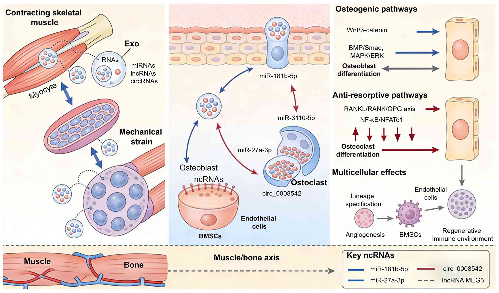 | Figure 3Molecular signaling cascades of
exercise-induced exosomal ncRNAs in bone remodeling. Mechanical
strain during exercise stimulates release of exosomes from skeletal
muscle and osteocytes. These exosomes carry ncRNAs (miRNAs,
lncRNAs, circRNAs) that activate osteogenic pathways
(Wnt/β-catenin, BMP/Smad, MAPK/ERK) in osteoblasts, suppress
osteoclastogenesis via RANKL/RANK/OPG and NF-κB/NFATc1 signaling,
and coordinate multi-cellular effects (BMSC lineage commitment,
angiogenesis, immune modulation). ncRNAs, noncoding RNAs; miRNAs,
microRNAs; lncRNAs, long noncoding RNAs; circRNAs, circular RNAs;
BMP, bone morphogenetic protein; MAPK, mitogen-activated protein
kinase; ERK, extracellular signal-regulated kinase; RANK, receptor
activator of nuclear factor-κB; RANKL, receptor activator of
nuclear factor-κB ligand; OPG, osteoprotegerin; NF-κB, nuclear
factor-κB; NFATc1, nuclear factor of activated T-cells 1; BMSC,
bone marrow mesenchymal stem cell. |
Osteogenic signaling pathways:
Wnt/β-catenin, BMP/Smad, and mitogen-activated protein kinase
(MAPK)/extracellular signal-regulated kinase (ERK)
Exercise-induced exosomal ncRNAs promote osteoblast
differentiation and bone formation primarily through activation of
the Wnt/β-catenin, BMP/Smad, and MAPK/ERK signaling cascades
(58). Mechanical
strain-stimulated osteocyte-derived exosomes have been shown to
enhance osteogenic differentiation via miRNA-mediated pathway
activation. Lv et al (59) demonstrated that mechanically
induced osteocyte-derived exosomes promote periodontal ligament
stem cell proliferation and osteogenic differentiation through the
miR-181b-5p/phosphatase and tensin homolog (PTEN)/protein kinase B
(AKT) signaling axis. This finding establishes a direct
mechanotransduction link where exosomal miRNAs modulate PTEN
expression to relieve AKT pathway inhibition. Similarly, Zhu et
al (36) reported that
mechanically strained osteocyte-derived exosomes promote
osteoblastic differentiation, further supporting the concept that
mechanical loading generates osteogenic exosomal signals.
The Wnt/β-catenin pathway represents a central
convergence point for exosomal ncRNA-mediated osteogenesis. Ibrahim
et al (60) demonstrated
that augmenting canonical Wnt signaling in therapeutically inert
cells converts them into potent exosome factories, highlighting the
capacity of the pathway to enhance exosomal therapeutic efficacy.
In the context of muscle-bone crosstalk, skeletal muscle-derived
exosomes have been identified as key mediators of osteogenesis.
Chen et al (27) showed
that histone deacetylase inhibition enhances extracellular vesicles
from muscle to promote osteogenesis via miR-873-3p, indicating that
muscle-derived exosomes can directly activate osteogenic programs
in bone cells. Xu et al (61) provided complementary evidence
that exosomes from C2C12 myoblasts enhance osteogenic
differentiation of MC3T3-E1 pre-osteoblasts by delivering
miR-27a-3p. Across these studies, a common regulatory pattern
emerges: Exosomal ncRNAs from both osteocytes and myocytes converge
on the Wnt/β-catenin pathway as a central effector. Specifically,
these ncRNAs function by relieving endogenous inhibitors of the
pathway (such as PTEN and DKK2) or by directly targeting
transcriptional repressors of osteogenic genes. This pattern
suggests that diverse exercise stimuli may converge on a shared
exosomal ncRNA-mediated mechanism to activate Wnt/β-catenin
signaling, representing a potential therapeutic node for bone
regeneration.
Collectively, these studies demonstrate that
exosomal miRNAs from both osteocytes and myocytes converge on
osteogenic signaling pathways, with the Wnt/β-catenin cascade
serving as a primary effector mechanism. It should be noted,
however, that the majority of these studies are correlative in
nature, reporting associations between exosomal ncRNA levels and
osteogenic outcomes. Direct causal evidence through
loss-of-function (such as exosomal ncRNA knockdown) or
gain-of-function (such as ncRNA overexpression or mimic delivery)
approaches remains limited, leaving the necessity and sufficiency
of specific exosomal ncRNAs for exercise-induced bone formation
incompletely validated.
Anti-resorptive mechanisms: Receptor
activator of nuclear factor κB (RANK) ligand
(RANKL)/RANK/osteoprotegerin (OPG) axis and nuclear factor-κB
(NF-κB) signaling
The inhibitory effects of exercise-induced exosomal
ncRNAs on osteoclastogenesis involve targeting the RANKL/RANK/OPG
axis and suppressing NF-κB and nuclear factor of activated T-cells
1 (NFATc1) signaling. Nie et al (62) demonstrated that skeletal
muscle-derived exosomes regulate endothelial cell functions via
reactive oxygen species-activated NF-κB signaling, establishing a
mechanistic link between exercise-induced oxidative stress and
downstream transcriptional regulation. This pathway also modulates
osteoclast precursor sensitivity to RANKL stimulation. Wang et
al (63) reported that
circ_0008542 in osteoblast exosomes promotes osteoclast-induced
bone resorption through m6A methylation, revealing that exosomal
circRNAs can influence the osteoblast-osteoclast coupling balance.
The opposing effects of different exosomal ncRNA species on
osteoclastogenesis underscore the complexity of exercise-mediated
skeletal regulation.
Muscle-derived exosomes also contribute to
anti-resorptive effects through delivery of miRNAs that target
osteoclast differentiation factors. Fulzele et al (64) demonstrated that muscle-derived
miR-34a increases with age in circulating extracellular vesicles
and induces senescence of bone marrow stem cells, suggesting that
age-related changes in exosomal cargo may compromise bone
homeostasis. Conversely, Qu et al (65) showed that mechanically stimulated
periodontal ligament cell-derived exosomes promote osteoblast
differentiation via the miR-181d-5p/tumor necrosis factor (TNF)
signaling pathway, indicating that mechanical loading generates
exosomal signals that suppress inflammatory osteoclastogenesis.
Synthesizing these findings, a recurrent mechanistic
theme is that exosomal ncRNAs modulate osteoclastogenesis by
targeting three distinct nodes: i) The RANKL/RANK/OPG ratio, ii)
NF-κB transcriptional activity, and iii) NFATc1-mediated downstream
gene expression. Notably, different exosomal ncRNA species can
exert opposing effects on the same pathway depending on cellular
context, suggesting that the net anti-resorptive outcome of
exercise is determined by the balance of pro-osteogenic and
anti-osteoclastogenic exosomal signals. This balance, rather than
the absolute level of any single ncRNA, likely dictates the overall
effect on bone mass.
Multicellular regulatory effects in the
bone microenvironment
Exercise-induced exosomal ncRNAs exert pleiotropic
effects on the bone microenvironment by coordinating mesenchymal
stem cell lineage commitment, angiogenesis, and immune homeostasis.
Ma et al (26)
demonstrated that skeletal muscle-derived extracellular vesicles
transport glycolytic enzymes to mediate muscle-to-bone crosstalk,
revealing a metabolic dimension to exosomal communication beyond
ncRNA cargo. This finding expands the understanding of how exercise
generates integrated signals that support bone formation. For
angiogenesis coupling, Hayashi et al (66) reported that exosomal miR-206
secreted from growing muscle promotes angiogenic response in
endothelial cells, establishing a direct mechanism by which
muscle-derived exosomes support the vascular niche required for
bone remodeling. Similarly, Lou et al (46) demonstrated that exercise promotes
angiogenesis by enhancing endothelial cell fatty acid utilization
via liver-derived extracellular vesicle miR-122-5p, highlighting
the multi-organ contribution to exercise-induced vascular
adaptation.
Mesenchymal stem cell lineage commitment is
critically regulated by exosomal ncRNAs from various sources. Xing
et al (67) showed that
skeletal muscle-derived exosomes prevent osteoporosis by promoting
osteogenesis, providing direct evidence for the therapeutic
potential of muscle-derived exosomes in bone disorders. By
contrast, Peruzzi et al (68) demonstrated that circulating
extracellular vesicles from adolescents with obesity impair
mesenchymal stromal cell differentiation, favoring adipogenic
rather than osteogenic differentiation, indicating that
pathological states alter exosomal cargo to disrupt bone
homeostasis. Immune modulation represents another key mechanism, as
Qiu et al (69) reported
that exosomes derived from BMSCs of exercise-trained mice improve
wound healing by inhibiting macrophage M1 polarization, suggesting
that exercise-conditioned exosomes promote a regenerative immune
environment.
Mechanobiological coupling
mechanisms
The transduction of mechanical forces into
biochemical signals via exosomal ncRNAs constitutes a fundamental
mechanobiological coupling mechanism in bone adaptation. Osteocytes
serve as primary mechanosensors in bone, and their exosomal
response to mechanical loading mediates downstream effects on
osteoblasts and osteoclasts. Shen et al (70) demonstrated that mechanically
activated mesenchymal-derived bone cells drive vessel formation via
an extracellular vesicle-mediated mechanism, establishing that
mechanical stimulation generates pro-angiogenic exosomal signals
that coordinate bone formation with vascular support. Shang et
al (71) further elucidated
that extracellular vesicles allow epigenetic mechanotransduction
between chondrocytes and osteoblasts, revealing that mechanical
signals can be propagated across cell types through exosomal
delivery of regulatory ncRNAs.
The molecular machinery underlying exosomal
mechanotransduction involves specific miRNA species that respond to
mechanical strain. Zheng et al (72) reported the biological
characteristics of miRNAs secreted by exosomes of periodontal
ligament stem cells due to mechanical force, demonstrating that
mechanical loading induces distinct exosomal miRNA profiles. Qu
et al (65) confirmed
that cyclic stretch-induced exosomes from periodontal ligament
cells promote osteoblast differentiation via the miR-181d-5p/TNF
signaling pathway, providing mechanistic insight into how cyclical
mechanical forces are converted into anabolic signals.
Collectively, these studies establish that exosomal ncRNAs function
as mechanotransducers, converting physical stimuli into molecular
signals that orchestrate adaptive bone remodeling.
Synergistic crosstalk in the muscle-bone
axis
The muscle-bone axis represents a paradigm of
inter-organ communication mediated by exercise-induced exosomal
ncRNAs, where synergistic and antagonistic signaling interactions
coordinate skeletal adaptation. Multiple recent reviews have
synthesized this emerging field. Zhang et al (8) comprehensively summarized exosomal
miRNAs in muscle-bone crosstalk, detailing mechanistic links and
exercise modulation relevant to sarcopenia and osteoporosis. Li
et al (73) provided a
systematic overview of molecular communication from bone to
skeletal muscle, establishing that exosomal signaling is
bidirectional between these tissues. Kirk et al (74) further contextualized bone and
muscle crosstalk in ageing and disease, highlighting the clinical
relevance of exosome-mediated inter-organ communication.
Specific molecular mechanisms underlying muscle-bone
crosstalk have been elucidated. Qin et al (75) demonstrated that myostatin
inhibits osteoblastic differentiation by suppressing
osteocyte-derived exosomal miR-218, revealing a novel mechanism by
which muscle-derived factors regulate bone formation through
exosomal pathways. Li et al (76) showed that myoblast-derived
exosomal paired related homeobox 2 attenuates osteoporosis via
transcriptional regulation of lncRNA-MIR22HG to activate the Hippo
pathway, establishing a non-canonical signaling axis in muscle-bone
communication. Conversely, Wang et al (77) reported that slowpoke homolog 1
(Slo1)-deficient myoblast exosome-derived miR-222-3p inhibits
osteogenic differentiation via targeting signal transducer and
activator of transcription 3 (STAT3), demonstrating that
pathological alterations in muscle exosomal cargo can negatively
impact bone. The emerging evidence supports that exercise optimizes
muscle-bone crosstalk by enriching exosomal ncRNA cargo with
pro-osteogenic and anti-osteoclastogenic signals, while
antagonistic signals from pathological muscle states can disrupt
skeletal homeostasis.
Roles of exercise-induced exosomal ncRNAs in
sports-related bone injuries
Exercise-induced exosomal ncRNAs participate in the
pathophysiological processes of sports-related bone injuries by
modulating inflammation, angiogenesis, and tissue regeneration
across different injury types. These molecular mediators exert
context-dependent effects that vary according to injury mechanism,
healing stage, and the specific tissue microenvironment.
Understanding their regulatory roles provides mechanistic insights
into how exercise conditioning influences bone injury outcomes and
identifies potential therapeutic targets. A systematic overview of
these injury-specific roles is presented (Table II).
| Table IIRoles of exercise-induced exosomal
ncRNAs in sports-related bone injuries: A summary of key
studies. |
Table II
Roles of exercise-induced exosomal
ncRNAs in sports-related bone injuries: A summary of key
studies.
| Authors, year | Injury type | Cell/Tissue
source | Model | Exosomal ncRNA | Target/Pathway | Key findings | (Refs.) |
|---|
| Shang et al,
2021 | Osteochondral
injury |
Chondrocytes/osteoblasts | In
vitro | Epigenetic
regulators |
Mechanotransduction | Enabled epigenetic
mechanotransduction between chondrocytes and osteoblasts | (71) |
| Wang et al,
2025 | Disuse
osteopenia | Slo1-deficient
myoblasts | In
vitro | miR-222-3p | STAT3 | Inhibited
osteogenic differentiation via targeting STAT3, demonstrating
quality-dependent effects | (77) |
| Lu et al,
2022 | Traumatic
fracture |
Osteoblast/osteoclast/immune cells | Animal (rat) | Exosome/drug
delivery hydrogel | Multicellular
coordination | Accelerated
fracture repair through coordinated multicellular responses | (78) |
| Liu et al,
2020 | Traumatic
fracture | Hypoxic MSCs | Animal (rat) | miR-126 | Angiogenesis and
osteogenesis | Promoted bone
fracture healing by enhancing angiogenesis and osteogenesis
simultaneously | (79) |
| Jiang et al,
2020 | Traumatic
fracture | BMSCs | Animal (mouse) | miR-25 | SMURF1/Runx2 | Regulated Runx2
ubiquitination and degradation to promote fracture healing in
mice | (80) |
| Yang et al,
2025 | Osteochondral
injury (TMJ) | MSCs | Animal (rat) | let-7a-5p | Integrin β3 | Suppressed
osteoclast activity and ameliorated TMJ osteoarthritis | (81) |
| Xu et al,
2020 | Stress
fracture | Aged rat MSCs | Animal (rat) | miR-128-3p | Smad5 | Regulated
osteogenesis and bone fracture healing in aged rats | (82) |
| Tian et al,
2025 | Nonunion
fracture | Mechanical
stretch-induced ADSCs | Animal (rat) | miR-877 | Not identified | Enhanced fracture
healing in nonunion rats with type 2 diabetes mellitus | (85) |
| Wu et al,
2021 | Osteoarthritis | Osteoarthritic
subchondral bone | Animal (mouse) | miR-210-5p | Cartilage
degeneration | Released exosomes
promoting cartilage degeneration, establishing pathological
feedback loop | (86) |
| Lin et al,
2025 | Osteoarthritis | Osteocytes | In
vitro/Animal | miR-214-3p | Bone-to-cartilage
crosstalk | Mediated
bone-to-cartilage crosstalk and promoted osteoarthritis
progression | (87) |
| Wang et al,
2022 | Osteoporosis | MSCs | Animal (mouse) | miR-27a |
DKK2/Wnt/β-catenin | Inhibited
osteoporosis via miR-27a-induced inhibition of DKK2-mediated
Wnt/β-catenin pathway | (88) |
| Van Pelt et
al, 2020 | Disuse
osteopenia | Serum EVs | Human | miR-203a-3p | Muscle protein
turnover | Associated with
skeletal muscle mass and protein turnover during disuse atrophy and
regrowth | (90) |
| Yang et al,
2020 | Disuse
osteoporosis | hUC-MSCs | Animal (rat) | miR-1263 | Mob1/Hippo | Prevented apoptosis
in disuse osteoporosis via miR-1263/Mob1/Hippo signaling
pathway | (91) |
| Shao et al,
2026 | Disuse
osteoporosis | Atrophic skeletal
muscle | Animal (mouse) | miR-125a-5p | Bone formation | Transferred
miR-125a-5p to inhibit bone formation during aging-related
osteoporosis | (92) |
Traumatic bone fractures in sports
The regulatory roles of exercise-induced exosomal
ncRNAs in traumatic fracture healing span the sequential phases of
inflammation, soft callus formation, hard callus mineralization,
and remodeling. During the inflammatory phase, exosomal miRNAs
modulate macrophage polarization and cytokine secretion, creating a
pro-regenerative environment. Recent evidence from Lu et al
(78) demonstrated that an
osteoblast/osteoclast and immune cocktail therapy using
exosome/drug delivery multi-functional hydrogel accelerates
fracture repair, highlighting the potential of exosome-based
strategies to coordinate multicellular responses during bone
healing. The transition to soft callus formation involves exosomal
transfer of osteogenic signals between mesenchymal stem cells and
osteoprogenitors. Liu et al (79) showed that hypoxic mesenchymal
stem cell-derived exosomes promote bone fracture healing by
transferring miR-126, which enhances angiogenesis and osteogenesis
simultaneously.
During hard callus mineralization, exosomal ncRNAs
regulate osteoblast differentiation and matrix deposition. Jiang
et al (80) reported that
BMSC-derived exosomal miR-25 regulates the ubiquitination and
degradation of Runx2 by SMAD specific E3 ubiquitin protein ligase 1
(SMURF1) to promote fracture healing in mice. More recent work by
Yang et al (81)
demonstrated that mesenchymal stem cell-derived extracellular
vesicles ameliorate temporomandibular joint osteoarthritis by
suppressing osteoclast activity via the let-7a-5p/integrin β3 axis,
establishing that exosomal mechanisms are relevant across different
joint contexts. In the remodeling phase, exosomal signals
coordinate osteoclast-mediated resorption with new bone formation.
The stage-dependent effects of exosomal ncRNAs suggest that
therapeutic strategies targeting specific healing phases may
optimize fracture repair outcomes.
Stress fractures and overuse bone
injuries
Stress fractures and overuse bone injuries in
athletes represent a distinct pathological entity characterized by
repetitive mechanical loading-induced bone fatigue damage, where
exercise-induced exosomal ncRNAs may serve as early warning signals
and repair mediators. The identification of circulating exosomal
miRNA signatures associated with bone fatigue damage offers
potential for early detection before radiographic changes appear.
Xu et al (82)
demonstrated that exosomal miR-128-3p from mesenchymal stem cells
of aged rats regulates osteogenesis and bone fracture healing by
targeting Smad5. The mechanosensitive nature of exosomal release
positions these vesicles as ideal candidates for monitoring
cumulative bone fatigue. Shang (83) comprehensively reviewed
mechanosensitive miRNAs in cartilage and subchondral bone
remodeling, identifying them as emerging targets for osteoarthritis
therapy, with direct relevance to overuse injuries.
Furthermore, Yang et al (84) confirmed that cyclic
stretch-induced exosomes from periodontal ligament cells promote
osteoblast differentiation via the miR-181d-5p/TNF signaling
pathway, indicating that mechanically stimulated exosomes actively
participate in the repair of fatigue-damaged bone. Recent work by
Tian et al (85)
demonstrated that miR-877, an exosomal miRNA from mechanical
stretch-induced adipose derived stromal cells, enhances fracture
healing in nonunion rats with type 2 diabetes mellitus, providing
evidence that mechanically conditioned exosomes can overcome
healing impairments. The dual role of exercise-induced exosomes,
both as sensors of mechanical overload and as mediators of adaptive
repair, positions them as critical regulators of the bone fatigue
response.
Sports-related osteochondral injury and
osteoarthritis
Osteochondral injuries and post-traumatic
osteoarthritis represent common sequelae of sports-related joint
trauma, with exercise-induced exosomal ncRNAs modulating
cartilage-subchondral bone crosstalk and disease progression. The
bidirectional communication between cartilage and bone via exosomal
ncRNAs influences both acute injury responses and chronic
degenerative changes. Shang et al (71) elucidated that extracellular
vesicles allow epigenetic mechanotransduction between chondrocytes
and osteoblasts, revealing that mechanical signals can be
propagated across joint tissues through exosomal delivery of
regulatory ncRNAs. This mechanism is particularly relevant to
sports injuries where aberrant joint loading disrupts normal
cartilage-bone homeostasis. Wu et al (86) reported that osteoarthritic
subchondral bone releases exosomes that promote cartilage
degeneration, establishing a pathological feedback loop that
accelerates osteoarthritis progression. Recent advances by Lin
et al (87) demonstrated
that osteocyte-derived extracellular vesicles mediate the
bone-to-cartilage crosstalk and promote osteoarthritis progression,
providing mechanistic insight into how bone pathology drives
cartilage degeneration.
Conversely, protective exosomal signals from healthy
tissues can mitigate injury-induced degeneration. Wang et al
(88) demonstrated that
mesenchymal stem cell-derived extracellular vesicles inhibit
osteoporosis via miR-27a-induced inhibition of the DKK2-mediated
Wnt/β-catenin pathway. More recent work by Gu et al
(16) showed that bone
remodeling stimulated by Wnt-mediated mitophagy-regulated
extracellular vesicles in subchondral bone contributes to
osteoarthritis development, highlighting the interconnected nature
of bone and cartilage pathology. The identification of
osteoarthritis-specific exosomal miRNA signatures provides
potential biomarkers for early detection of post-traumatic
osteoarthritis following sports injuries. Qiu et al
(89) comprehensively reviewed
the therapeutic potential of exosomal ncRNAs in bone metabolic
diseases, focusing on osteoarthritis and rheumatoid arthritis.
Disuse osteopenia after sports
injury
Immobilization and disuse following sports injury
create a catabolic environment characterized by rapid bone loss,
where exercise-induced exosomal ncRNAs may exert protective effects
against disuse osteopenia. The absence of mechanical loading during
immobilization reduces endogenous exosomal signals that normally
maintain bone mass, creating an opportunity for exogenous or
exercise-mimetic exosome therapies. Van Pelt et al (90) demonstrated that serum
extracellular vesicle miR-203a-3p content is associated with
skeletal muscle mass and protein turnover during disuse atrophy and
regrowth, establishing a link between exosomal cargo and
disuse-induced tissue loss. This finding suggests that monitoring
exosomal miRNAs could guide rehabilitation strategies following
immobilization. Yang et al (91) reported that human umbilical cord
mesenchymal stem cell-derived exosomes act via the miR-1263/Mps one
binder 1 (Mob1)/Hippo signaling pathway to prevent apoptosis in
disuse osteoporosis. Recent work by Shao et al (92) demonstrated that atrophic skeletal
muscle-derived extracellular vesicles transfer miR-125a-5p to
inhibit bone formation in osteoporosis during aging, revealing that
pathological changes in muscle during disuse can actively promote
bone loss. This finding underscores the importance of maintaining
muscle activity during immobilization. Bao and He (93) showed that skeletal muscle-derived
exosomes prevent osteoporosis by promoting osteogenesis, indicating
that delivering muscle-derived exosomes could counteract disuse
osteopenia. More recently, Wang et al (77) reported that Slo1-deficient
myoblast exosomes-derived miR-222-3p inhibits osteogenic
differentiation via targeting STAT3, demonstrating that the quality
of muscle-derived exosomes critically determines their effects on
bone. The concept of using exercise-conditioned exosomes as a
replacement therapy during immobilization represents a promising
translational strategy for athletes recovering from injury.
Translational applications of
exercise-induced exosomal ncRNAs in sports-related bone
injuries
The translational potential of exercise-induced
exosomal noncoding RNAs spans diagnostic, therapeutic, and
rehabilitative applications in sports-related bone injuries. These
endogenous nanovesicles offer advantages as non-invasive biomarkers
for injury assessment and as naturally derived therapeutic agents
that recapitulate the beneficial effects of exercise. Their
clinical translation requires standardization of isolation
protocols, validation of response characteristics for personalized
prescription, and integration with existing treatment modalities. A
systematic overview of these translational strategies is presented
(Table III).
| Table IIITranslational applications of
exercise-induced exosomal ncRNAs in sports-related bone
injuries. |
Table III
Translational applications of
exercise-induced exosomal ncRNAs in sports-related bone
injuries.
| Authors, year | Study type | ncRNA cargo | Application
domain | Key finding | Translational
implication | (Refs.) |
|---|
| Shi et al,
2022 | Human cohort | miRNAs | Non-invasive
biomarkers | Identified miRNAs
in serum exosomes as circulating biomarkers for postmenopausal
osteoporosis | Demonstrates
feasibility of exosomal miRNA-based diagnostics for bone
disorders | (94) |
| Shao et al,
2020 | Human cohort | miRNAs | Non-invasive
biomarkers | High-throughput
sequencing identified serum exosomal miRNA expression profiles in
osteoporotic females | Provides reference
for biomarker discovery in bone metabolic conditions | (95) |
| Hou et al,
2024 | Human cohort | miR-21 | Non-invasive
biomarkers | Elevated exosomal
miR-21 levels associated with nonunion in clavicular fractures
post-ORIF | Supports exosomal
miRNAs to predict delayed healing or nonunion following
sports-related fractures | (96) |
| Zhao and Xu,
2018 | Human cohort | lncRNA PCGEM1 | Non-invasive
biomarkers | Synovial
fluid-derived exosomal lncRNA PCGEM1 differentiates osteoarthritis
stages | Enables staging of
sports-related joint injuries | (97) |
| Sun et al,
2023 | Human cohort | miRNAs | Non-invasive
biomarkers | Circulating
exosomal miRNAs identified for postmenopausal osteoporosis
diagnosis | Validates
multi-panel exosomal miRNA approaches for diagnostic accuracy | (98) |
| Deininger et
al, 2025 | Preclinical | sEVs | Therapeutic
strategy | Anatomical implant
region critically determines osteogenic potency of sEVs | Highlights
importance of delivery site optimization for exosome-based
therapy | (99) |
| Deluca et
al, 2024 | Preclinical | sEVs | Combination
therapy | Umbilical cord sEVs
synergize with rhBMP-2 to enhance femoral defect regeneration | Supports combining
exosomes with growth factors for enhanced efficacy | (100) |
| Wu et al,
2024 | Preclinical | Exosomes | Therapeutic
strategy | Regulatory T
cell-derived exosomes mediate macrophage polarization for
osteogenic differentiation | Reveals immune
cell-derived exosomes complement exercise-derived vesicles | (101) |
| Li et al,
2022 | Preclinical | Exosomes | Engineered
therapy | Engineered stem
cells produce exosomes with enhanced bone regeneration effects | Offers alternative
gene therapy strategy avoiding direct stem cell
transplantation | (102) |
| Huang et al,
2022 | Preclinical | lncRNA MEG3 | Engineered
therapy | Engineered exosomes
serve as targeted lncRNA MEG3 delivery vehicles for
osteosarcoma | Demonstrates
versatility of exosome engineering for musculoskeletal
applications | (103) |
| Xu et al,
2023 | Preclinical | Anti-miRNA
oligonucleotides | Engineered
therapy | Osteoclast-targeted
delivery achieved via red blood cell EV engineering | Shows cell-specific
targeting through engineering approaches | (104) |
| Wang et al,
2022 | Preclinical | miR-150-3p | Engineered
therapy | EV-mediated
miR-150-3p delivery in joint homeostasis as potential
osteoarthritis treatment | Illustrates
therapeutic potential of miRNA-based exosome therapies | (105) |
| Yang et al,
2023 | Preclinical | BMSC-derived
exosomes | Combination
therapy | Combination therapy
with BMSC-exosomes and porous tantalum enhances femoral defect
repair | Shows exosomes
enhance osteointegration of metallic implants | (107) |
| Zheng et al,
2024 | Preclinical | sEVs | Combination
therapy | Incorporation of
sEVs in PEG/HA-Bio-Oss hydrogel composite scaffold for bone
regeneration | Illustrates synergy
between exosomes and biomaterial scaffolds | (108) |
| Yu et al,
2024 | Preclinical | Shed-derived
exosomes | Combination
therapy | Synergistic effects
with Cu2+ and injectable hyaluronic acid hydrogel for
periodontal bone regeneration | Showcases
multi-component combination approaches | (109) |
| Zhang et al,
2024 | Preclinical | Exosome-loaded
hydrogel | Combination
therapy | Exosome-loaded
hyaluronic acid hydrogel with oxygen-producing 3D printed PLA
scaffolds | Combines advanced
manufacturing with exosome delivery for bone repair | (110) |
| Frank et al,
2025 | Human cohort | miRNAs | Non-invasive
biomarkers | Circulating miRNAs
associated with successful bone regeneration | Provides framework
for using exosomal ncRNAs as prognostic markers | (111) |
| Gao et al,
2024 | Human cohort | miRNA
signature | Non-invasive
biomarkers | Plasma-derived
exosomal miRNA signature established for early detection of
postmenopausal osteoporosis | Demonstrates
feasibility of exosomal miRNA-based diagnostics | (113) |
Non-invasive biomarkers
Circulating exosomal ncRNAs have emerged as
promising non-invasive biomarkers for early diagnosis, risk
stratification, and prognosis evaluation of sports-related bone
injuries. Their stability in body fluids and tissue-specific origin
make them attractive candidates for liquid biopsy approaches. Shi
et al (94) identified
miRNAs in serum exosomes as circulating biomarkers for
postmenopausal osteoporosis, demonstrating the feasibility of
exosomal miRNA-based diagnostics for bone disorders. Similarly,
Shao et al (95)
established serum exosomal miRNA expression profiling in menopausal
females with osteoporosis using high-throughput sequencing,
providing a reference for biomarker discovery. The prognostic value
of exosomal ncRNAs has been demonstrated in fracture healing
complications. Hou et al (96) reported a prospective analysis
showing that elevated exosomal miR-21 levels are associated with
nonunion in clavicular fractures post-open reduction and internal
fixation (ORIF), providing direct clinical evidence linking
exosomal cargo to fracture healing outcomes. For osteoarthritis,
Zhao and Xu (97) identified
synovial fluid-derived exosomal lncRNA PCGEM1 as a biomarker for
different stages of osteoarthritis, highlighting the potential for
staging sports-related joint injuries. More recently, Sun et
al (98) identified and
evaluated circulating exosomal miRNAs for the diagnosis of
postmenopausal osteoporosis, further validating the biomarker
potential of exosomal ncRNAs. The specificity of exosomal
signatures for distinct injury types and healing stages suggests
that multi-panel approaches may achieve higher diagnostic accuracy
than single biomarkers.
Therapeutic strategies based on native
exercise-derived exosomes
Native exosomes derived from exercise-conditioned
tissues offer naturally optimized therapeutic vehicles for bone
injury repair. These vesicles inherently carry the molecular cargo
that mediates the beneficial effects of exercise, potentially
circumventing safety concerns associated with synthetic delivery
systems. Deininger et al (99) demonstrated that anatomical
implant region critically determines the osteogenic potency of
small extracellular vesicles, highlighting the importance of
delivery site optimization. The extraction and standardization of
exercise-modulated exosomes face challenges related to donor
variability, exercise protocol standardization, and scalable
production. Deluca et al (100) showed a synergistic effect of
umbilical cord extracellular vesicles and recombinant human bone
morphogenetic protein-2 (rhBMP-2) to enhance regeneration of
metaphyseal femoral defects in osteoporotic rats, suggesting that
exosomes can be combined with established growth factors for
enhanced efficacy. Wu et al (101) demonstrated that regulatory T
cell-derived exosomes mediated macrophage polarization for
osteogenic differentiation in fracture repair, revealing that
immune cell-derived exosomes may complement exercise-derived
vesicles in promoting bone healing. The clinical application
potential of exercise-derived exosomes requires rigorous quality
control and standardization comparable to pharmaceutical
products.
Engineered exosomes and ncRNA-targeted
therapy
Engineered exosomes designed to deliver specific
ncRNA cargo represent a next-generation therapeutic strategy for
refractory bone injuries. These approaches leverage the natural
targeting capabilities of exosomes while incorporating synthetic
modifications to enhance therapeutic efficacy. Strategies include
direct engineering of parent cells to overexpress therapeutic
ncRNAs, which are then naturally packaged into secreted exosomes,
as well as post-isolation modification of exosomes with targeting
ligands or therapeutic cargo. Li et al (102) demonstrated the use of
engineered stem cells to produce exosomes with enhanced bone
regeneration effects, presenting an alternative strategy for gene
therapy that avoids direct stem cell transplantation. The delivery
of ncRNA therapeutics through engineered exosomes offers advantages
including protection from degradation, targeted delivery, and
sustained release. For example, exosomes can be surface-modified
with targeting peptides to achieve bone-specific delivery, or
loaded with therapeutic ncRNAs via electroporation or chemical
transfection to enhance their osteogenic or anti-resorptive
potency. Huang et al (103) developed engineered exosomes as
targeted lncRNA MEG3 delivery vehicles for osteosarcoma therapy,
demonstrating the versatility of exosome engineering for
musculoskeletal applications. Xu et al (104) developed osteoclast-targeted
delivery of anti-miRNA oligonucleotides by red blood cell
extracellular vesicles, showing that cell-specific targeting can be
achieved through engineering approaches. For osteoarthritis, Wang
et al (105) described
extracellular vesicle-mediated miR-150-3p delivery in joint
homeostasis as a potential treatment, illustrating the therapeutic
potential of miRNA-based exosome therapies. More recently, Yan
et al (106) engineered
delta-like ligand 4 (Dll4)-overexpressing osteocyte-derived
exosomes to enhance bone regeneration by regulating osteogenesis
and angiogenesis, providing a blueprint for tissue-specific exosome
engineering.
Beyond these established approaches, recent advances
have focused on optimizing the stoichiometry and spatio-temporal
control of ncRNA loading to enhance therapeutic efficacy.
Comparative studies have shown that electroporation achieves the
highest loading efficiency for small ncRNAs (miRNAs and siRNAs)
into exosomes, whereas chemical transfection methods (such as
Lipofectamine) yield lower encapsulation rates and may introduce
residual carrier toxicity that confounds functional outcomes
(15). For bone-targeted
delivery, surface modification of exosomes with bone-homing
peptides, such as the (DSS)6 bone-homing peptide (a
six-repeat aspartate-serine-serine sequence) that binds
hydroxyapatite, has been successfully employed to concentrate
therapeutic ncRNAs at fracture sites while minimizing systemic
off-target effects (17,21). Furthermore, emerging evidence
supports the use of sequential loading strategies, where
pro-angiogenic ncRNAs (such as miR-126) and osteogenic ncRNAs (such
as miR-26a) are packaged into distinct exosome batches and
administered according to the temporal phases of bone healing,
first promoting vascularization during soft callus formation,
followed by enhancing mineralization during hard callus remodeling
(23,79). This phased approach mimics the
natural time-course of fracture repair and has shown superior
outcomes compared with single-cargo or bolus administration in
preclinical models (23).
However, the clinical translation of such multi-cargo, temporally
programmed exosome therapeutics requires rigorous validation of
batch-to-batch consistency, long-term storage stability, and
scalable manufacturing under Good Manufacturing Practice
conditions, barriers that remain incompletely addressed (15,21).
Combination intervention strategies
The integration of exercise-induced exosomal ncRNAs
with existing treatment modalities offers synergistic opportunities
for enhanced therapeutic outcomes. Combination strategies encompass
stem cell therapy, biomaterials, physical therapy, and
pharmacotherapy. Yang et al (107) demonstrated combination therapy
with BMSC-derived exosomes and porous tantalum for the repair of
femur supracondylar defects, showing that exosomes can enhance the
osteointegration of metallic implants. Similarly, Zheng et
al (108) incorporated
small extracellular vesicles in polyethylene glycol
(PEG)/hyaluronic acid (HA)-Bio-Oss hydrogel composite scaffold for
bone regeneration, illustrating the synergy between exosomes and
biomaterial scaffolds. Yu et al (109) demonstrated synergistic effects
of Shed-derived exosomes, Cu2+, and an injectable
hyaluronic acid hydrogel on antibacterial, anti-inflammatory, and
osteogenic activity for periodontal bone regeneration, showcasing
multi-component combination approaches. Zhang et al
(110) developed exosome-loaded
hyaluronic acid hydrogel composite with oxygen-producing 3D printed
polylactic acid scaffolds, combining advanced manufacturing with
exosome delivery for bone tissue repair. For fracture repair, Wu
et al (101) showed that
regulatory T cell-derived exosomes mediated macrophage polarization
for osteogenic differentiation, suggesting that immune modulation
combined with osteogenic stimulation may optimize healing. The
comparative analysis of these combination strategies reveals that
scaffold-based delivery provides sustained local release, while
systemic administration may benefit multifocal injuries. Future
studies should systematically evaluate the relative efficacy of
different combination approaches to establish evidence-based
guidelines for clinical translation.
Clinical evidence: Human cohort
studies
Clinical evidence linking exercise-induced exosomal
ncRNAs to the occurrence, severity, and prognosis of sports-related
bone injuries is emerging through human cohort studies. Circulating
exosomal ncRNAs have been investigated as non-invasive biomarkers
for fracture healing outcomes and complication risk. Shi et
al (94) identified miRNAs
in serum exosomes as circulating biomarkers for postmenopausal
osteoporosis, establishing a foundation for similar approaches in
sports-related bone injuries. The association between exosomal
miRNA profiles and fracture healing complications has been
demonstrated in clinical populations. Hou et al (96) reported a prospective analysis
showing that elevated exosomal miR-21 levels are associated with
nonunion in clavicular fractures post-ORIF, providing direct
clinical evidence linking exosomal cargo to fracture healing
outcomes. This finding supports the potential of exosomal miRNAs to
predict delayed healing or nonunion following sports-related
fractures. Recent advances by Frank et al (111) demonstrated that circulating
miRNAs are associated with successful bone regeneration, providing
a framework for using exosomal ncRNAs as prognostic markers. The
prognostic value of exosomal signatures extends to bone metabolic
status following injury. Mihanfar et al (112) demonstrated that extracellular
vesicles and miRNAs are altered in response to exercise, insulin
sensitivity and overweight status, indicating that metabolic
factors influence exosomal profiles and may confound biomarker
interpretation. Gao et al (113) recently established a
plasma-derived exosomal miRNA signature by small RNA sequencing for
early detection of postmenopausal osteoporosis, demonstrating the
feasibility of exosomal miRNA-based diagnostics. Zhang et al
(8) comprehensively reviewed
exosomal miRNAs in muscle-bone crosstalk, detailing mechanistic
links and exercise modulation with direct implications for
sarcopenia, osteoporosis, and osteosarcopenia. Longitudinal studies
tracking exosomal ncRNA changes during rehabilitation are needed to
establish causality and clinical utility. The integration of
exosomal biomarkers into clinical practice for sports-related bone
injuries requires standardization of isolation protocols and
validation across diverse athlete populations.
Current limitations, controversies, and
unresolved scientific issues
Despite the rapid advancement in understanding
exercise-induced exosomal ncRNAs as key mediators of bone
remodeling, the field faces substantial limitations, ongoing
controversies, and unresolved scientific issues that critically
hinder clinical translation. These challenges span methodological
inconsistencies in exosome isolation, insufficient causal evidence
linking exosomal ncRNAs to specific skeletal outcomes, significant
gaps in clinical study design, and formidable barriers to
therapeutic implementation. A systematic overview of these
limitations and controversies is presented.
Clinical study limitations
Current clinical evidence on exercise-induced
exosomal ncRNAs in bone metabolism is predominantly derived from
studies with inherent methodological constraints that limit
generalizability. The majority of investigations feature small
sample sizes and lack multi-center validation, compromising
statistical power and external validity. For instance, studies
examining circulating exosomal miRNA signatures in fracture healing
typically enroll fewer than 100 participants, precluding robust
subgroup analyses (114,115).
Population heterogeneity represents another critical concern, as
individual characteristics such as age, sex, fitness level, and
hormonal status profoundly influence exosomal responses, yet
stratification is frequently inadequate (116,117). Furthermore, exercise
intervention protocols vary considerably across studies, with
inconsistent parameters regarding intensity, duration, and training
modality, impeding direct comparisons and meta-analytic synthesis
(118,119). The absence of standardized
pre-analytical procedures, including sample collection timing
relative to exercise bouts and anticoagulant selection, introduces
additional variability (120).
Collectively, these limitations underscore the urgent need for
prospective, multi-center large-cohort studies employing harmonized
protocols to establish clinically actionable exosomal
biomarkers.
Mechanistic controversies and gaps
Fundamental mechanistic questions persist regarding
whether exercise-induced exosomal ncRNAs exert direct causal
effects on bone remodeling or merely represent correlative
biomarkers of systemic adaptation. A substantial proportion of
published studies relies on descriptive profiling without
functional validation, leaving causal inference inadequately
supported. Specifically, numerous studies cited in this review
(such as those reporting acute exercise-induced changes in
circulating exosomal miRNA levels) demonstrate correlation rather
than causation. While these findings are valuable for hypothesis
generation, they do not constitute molecular mechanism
demonstration. Although in vitro and animal models have
demonstrated that exosomal miRNAs such as miR-181b-5p and
miR-27a-3p directly modulate osteogenic pathways (121,122), the translational relevance of
these findings to human physiology remains uncertain due to species
differences in ncRNA sequences, target repertoires, and exosome
biogenesis machinery (123).
A more fundamental issue is that even in animal
studies, few have employed rigorous causal validation frameworks.
The minimum requirements for establishing causality in this field
should include: i) Selective knockout or knockdown of specific
exosomal ncRNAs in relevant parent cells (such as skeletal muscle
or osteocytes) followed by exercise intervention; ii) rescue
experiments using ncRNA mimics or engineered exosomes; and iii)
tissue-specific ablation of exosome biogenesis machinery (such as
Rab27a or nSMase2 knockout) to determine whether exosomal ncRNAs
are necessary for exercise-mediated bone protection. To date, no
study has comprehensively met these criteria, representing a
critical gap in the literature.
Cell and tissue specificity of exosomal ncRNA
actions constitutes another unresolved controversy. Current
evidence suggests that exosomes from distinct sources, including
skeletal muscle, osteocytes, and adipose tissue, may exert
overlapping yet distinct effects on bone cells (124,125); however, the molecular
determinants governing recipient cell specificity remain poorly
characterized. Additionally, the potential off-target effects of
exosomal ncRNAs on non-skeletal tissues raise safety concerns that
have not been systematically evaluated. Moreover, most current
studies have examined the effect of a given exosomal ncRNA on only
one cell type in isolation, without systematically comparing its
functional consequences across osteoblasts, osteoclasts, and BMSCs;
direct extrapolation of these reductionistic in vitro
findings to in vivo bone adaptation lacks scientific
validity. The field therefore requires a paradigm shift from
descriptive correlative studies to hypothesis-driven mechanistic
investigations using loss-of-function and gain-of-function
approaches with cell-type-specific targeting.
Methodological challenges
The absence of standardized protocols for exosome
isolation and characterization represents a fundamental barrier to
data reproducibility and cross-study comparability.
Ultracentrifugation, the most widely employed isolation method,
exhibits variable efficiency and co-pellets non-vesicular
contaminants, including protein aggregates and lipoproteins
(126,127). Size-exclusion chromatography
offers improved purity but suffers from lower yield and longer
processing times, while polymer-based precipitation methods
frequently co-precipitate non-exosomal materials that confound
downstream analyses (120,128). The distinction between exosomes
and other extracellular vesicle subtypes, as well as lipoprotein
particles of similar size, remains technically challenging, with
conventional characterization methods lacking sufficient resolution
(129). For ncRNA detection,
the choice of isolation method profoundly influences miRNA
profiles, yet consensus guidelines for method selection are
lacking. Functional validation methodologies also exhibit
heterogeneity, with limited standardization in exosome labeling,
tracking, and uptake assays (128). The development of reference
materials and validation protocols, as recently advocated (130), represents a critical priority
for advancing the field toward clinical implementation.
Barriers to clinical translation
Translating exercise-induced exosomal ncRNAs into
clinical applications for sports-related bone injuries confronts
formidable obstacles spanning manufacturing, safety, and regulatory
domains. Scalable production of therapeutic exosomes with
consistent quality attributes remains technically demanding, as
current culture conditions for exercise-mimetic exosome generation
lack standardization (131).
Good Manufacturing Practice compliance necessitates rigorous
quality control measures, including characterization of size
distribution, cargo composition, and batch-to-batch consistency,
which are not routinely implemented in research settings (132). The optimal route of
administration for exosome-based therapies in bone injuries remains
undefined, with limited comparative data on local vs. systemic
delivery, dosing frequency, and tissue biodistribution (133). Long-term safety evaluations,
including immunogenicity, potential for off-target effects, and
carcinogenicity, have not been systematically conducted (134). Furthermore, regulatory
frameworks for exosome-based therapeutics are still evolving,
creating uncertainty regarding approval pathways and quality
requirements (132,135). These translational barriers
necessitate coordinated efforts between academic researchers,
regulatory agencies, and industry partners to establish clear
guidelines and accelerate clinical development.
Conclusions and future perspectives
The preceding sections have established
exercise-induced exosomal ncRNAs as central mediators of bone
remodeling, yet the field stands at a critical juncture where
fundamental discoveries must be translated into clinical
applications. Building upon the limitations and unresolved
questions outlined above, several strategic directions warrant
prioritization to advance both mechanistic understanding and
therapeutic implementation. Rather than reiterating existing
consensus, this section identifies specific unresolved scientific
issues that require investigation. Notably, four priority issues
emerge from the current literature: i) Elucidating the molecular
mechanisms that govern selective ncRNA packaging into exosomes in
response to distinct exercise modalities; ii) determining whether
exercise-induced exosomal ncRNAs directly cause bone adaptation, or
merely serve as correlative biomarkers; iii) overcoming
inter-individual variability in exosomal responses for personalized
exercise prescription; and iv) addressing the safety barriers that
must be addressed before exercise-mimetic exosome therapies can
enter clinical trials for sports-related bone injuries.
For basic research, the integration of multi-omics
approaches with systems biology represents a transformative
opportunity to decipher the complex regulatory networks governing
exercise-induced exosomal ncRNA function. Recent advances enable
simultaneous profiling of transcriptomic, proteomic, and lipidomic
signatures from limited sample volumes, which could elucidate how
mechanical stimuli are translated into specific exosomal cargo
selection mechanisms (136,137). Single-cell and spatial
transcriptomic technologies offer unprecedented resolution to map
exosomal ncRNA dynamics within the bone microenvironment,
identifying rare cell populations that serve as primary exosomal
targets and revealing how exosomal signaling gradients establish
regional specialization during fracture healing (138). A fundamental unresolved issue
concerns causality. To address whether exosomal ncRNAs are
necessary and sufficient for exercise-mediated bone protection,
future investigations should employ conditional knockout models
targeting exosome biogenesis machinery in tissue-specific manners,
combined with in vivo tracking technologies and CRISPR-Cas9
functional interrogation (139-141).
The translation of exosomal ncRNA biology into
clinical applications for sports-related bone injuries requires
substantial investment in large-scale, multi-center cohort studies.
Current clinical evidence is constrained by small sample sizes,
heterogeneous populations, and inconsistent methodological
standards (142). A key
clinical question is whether exosomal ncRNA signatures can predict
fracture healing complications with sufficient sensitivity and
specificity to guide decision-making. Future studies should adopt
harmonized protocols for sample collection, processing, and data
reporting to enable cross-study comparability and meta-analyses.
The development of standardized reference materials for exosome
isolation and characterization, as recently advocated by
international consortia, represents a critical prerequisite for
clinical implementation (137).
Such standardization efforts must extend to pre-analytical
variables including anticoagulant selection, storage conditions,
and timing of sample collection relative to exercise bouts, factors
that profoundly influence exosomal cargo composition yet remain
inconsistently reported (143).
Personalized exercise prescription guided by
exosomal ncRNA response profiles represents an emerging frontier in
sports medicine. The substantial inter-individual variability in
exosomal responses to standardized exercise protocols suggests that
molecular phenotyping could optimize exercise interventions for
bone health (8). Future studies
should characterize how age, sex, genetic background, and baseline
fitness level modulate exosomal ncRNA responses, thereby
establishing normative reference ranges against which individual
responses can be benchmarked. Machine learning approaches
integrating multi-dimensional molecular data with clinical
parameters hold promise for developing predictive algorithms that
recommend optimal exercise modalities, intensities, and durations
for individuals at risk of bone injury (136,141). This precision exercise paradigm
represents a shift from population-based recommendations to
individualized molecularly guided interventions.
Therapeutic strategies based on engineered exosomes
offer unprecedented opportunities for treating refractory bone
injuries. Recent proof-of-concept studies have demonstrated that
exosomes can be engineered to display targeting ligands,
encapsulate therapeutic ncRNA cargo, and achieve controlled release
profiles (144,145). However, safety issues remain
unanswered, including whether exercise-mimetic exosomes are
immunogenic; and what the potential off-target effects on
non-skeletal tissues are. Future translational efforts should
prioritize the development of exercise-mimetic exosomes that
recapitulate the multi-targeted effects of physical activity
without requiring patient participation, as this represents a
particularly valuable approach for injured athletes undergoing
immobilization. Scalable manufacturing processes compliant with
Good Manufacturing Practice standards are essential for clinical
translation, requiring rigorous quality control measures
encompassing size distribution, cargo composition, and
batch-to-batch consistency (139,146). Furthermore, combination
strategies integrating exosome therapeutics with biomaterial
scaffolds or pharmacological agents may achieve synergistic effects
that surpass either approach alone. In summary, addressing the four
priority issues outlined above, namely cargo selection mechanisms,
causality, biomarker utility, and safety, will determine whether
exercise-induced exosomal ncRNAs remain a descriptive phenomenon or
become a translatable paradigm for sports medicine.
In conclusion, the study of exercise-induced
exosomal ncRNAs in bone remodeling represents an emerging paradigm
that links physical activity to skeletal adaptation. Current
evidence supports a correlative framework in which exercise
dynamically modulates exosomal ncRNA cargo and these changes are
associated with osteogenic outcomes. However, whether this paradigm
is causally valid remains to be established. Direct evidence
demonstrating that specific exosomal ncRNAs are necessary and
sufficient for exercise-induced bone protection is still lacking.
The present review offered several distinctive advantages: It
systematically integrated exercise physiology, exosome biology, and
bone remodeling into a cohesive framework; detailed specific
signaling cascades (Wnt/β-catenin, BMP/Smad, and RANKL/RANK/OPG)
modulated by exercise-induced exosomal ncRNAs; and addressed the
translational potential for sports medicine applications including
biomarker development, personalized exercise prescription, and
exosome-based therapeutics. The convergence of multi-omics
technologies, single-cell resolution analyses, rigorous causal
validation frameworks, and standardized clinical protocols provide
a roadmap for addressing current limitations. For sports medicine
practitioners, these advances promise the development of
noninvasive diagnostic biomarkers, personalized exercise
prescriptions, and novel exosome-based therapeutics that harness
the regenerative potential of physical activity. Realizing this
potential will require sustained interdisciplinary collaboration
among molecular biologists, bioengineers, exercise physiologists,
and clinicians, united by the shared goal of translating
mechanistic insights into improved outcomes for athletes with bone
injuries.
Availability of data and materials
Not applicable.
Authors' contributions
BT and XK conceived and designed the review
framework. XC and JZ performed the literature search, screening,
and synthesis of relevant studies. All authors participated in
drafting and critically revising the manuscript, and read and
approved the final version, and accept accountability for all
aspects of the work. Data.
Ethics approval and consent to
participate
Not applicable.
Patient consent for publication
Not applicable.
Competing interests
The authors declare that they have no competing
interests.
Abbreviations:
|
BMSC
|
bone marrow mesenchymal stem cell
|
|
BMP
|
bone morphogenetic protein
|
|
circRNA
|
circular RNA
|
|
ERK
|
extracellular signal-regulated
kinase
|
|
HIIT
|
high-intensity interval training
|
|
lncRNA
|
long noncoding RNA
|
|
MAPK
|
mitogen-activated protein kinase
|
|
miRNA
|
microRNA
|
|
ncRNA
|
noncoding RNA
|
|
NF-κB
|
nuclear factor κB
|
|
NFATc1
|
nuclear factor of activated T-cells
1
|
|
OPG
|
osteoprotegerin
|
|
ORIF
|
open reduction and internal
fixation
|
|
PEG
|
polyethylene glycol
|
|
RANK
|
receptor activator of nuclear
factor-κB
|
|
RANKL
|
receptor activator of nuclear
factor-κB ligand
|
|
rhBMP-2
|
recombinant human bone morphogenetic
protein-2
|
|
Runx2
|
runt-related transcription factor
2
|
|
STAT3
|
signal transducer and activator of
transcription 3
|
Acknowledgements
Not applicable.
Funding
No funding was received.
References
|
1
|
DeFoor MT, Mullin EP, Cuenca RA and
Pierrie SN: Distal femoral stress fractures has high operation
rates in adolescent and young adult athletes. J Pediatr Orthop.
44:e439–e445. 2024. View Article : Google Scholar : PubMed/NCBI
|
|
2
|
Salášek M, Stančák A, Čepelík M, Pešl T,
Havlas V, Pavelka T, Havránek P and Džupa V: Pelvic avulsion
fractures in children: A retrospective study from four trauma
centers. Acta Chir Orthop Traumatol Cech. 92:7–14. 2025.In Czech.
View Article : Google Scholar
|
|
3
|
Feldman MW, Fucaloro S, Krivicich L, Kent
S and Salzler MJ: Studies directly comparing Lisfranc injuries
treated with primary arthrodesis or open reduction and internal
fixation show no significant difference in return to sport and
complications: A systematic review and meta-analysis. J Foot Ankle
Surg. 64:318–327. 2025. View Article : Google Scholar : PubMed/NCBI
|
|
4
|
Williams BA, Titus M, Chaclas N, Cardin S,
Wells L, Maguire KJ and Lawrence JT: Surgically treated ischial
tuberosity avulsion fractures in adolescents: Risks and outcomes of
3 fixation constructs. J Pediatr Orthop. 45:e30–e36. 2025.
View Article : Google Scholar
|
|
5
|
Frost HM: The mechanostat: A proposed
pathogenic mechanism of osteoporoses and the bone mass effects of
mechanical and nonmechanical agents. Bone Miner. 2:73–85.
1987.PubMed/NCBI
|
|
6
|
Yang J, Pei Q, Wu X, Dai X, Li X, Pan J
and Wang B: Stress reduction through cortical bone thickening
improves bone mechanical behavior in adult female
Beclin-1+/− mice. Front Bioeng Biotechnol.
12:13576862024. View Article : Google Scholar
|
|
7
|
Penna F, Garcia-Castillo L and Costelli P:
Extracellular vesicles and exosomes in the control of the
musculoskeletal health. Curr Osteoporos Rep. 22:257–265. 2024.
View Article : Google Scholar : PubMed/NCBI
|
|
8
|
Zhang B, Chen Y, Chen Q and Zhang H:
Exosomal miRNAs in muscle-bone crosstalk: Mechanistic links,
exercise modulation and implications for sarcopenia, osteoporosis
and osteosarcopenia. Metabolism. 170:1563332025. View Article : Google Scholar : PubMed/NCBI
|
|
9
|
Binet F, Entz L, Amiot M, Tessier S,
Véziers J, De Beaurepaire L, André-Grégoire G, Dingli F, Loew D,
Weiss P, et al: Exploring the molecular cargos associated with
extracellular vesicles extracted from bone tissue to identify novel
players in osteoblast function. Am J Physiol Cell Physiol.
329:C1521–C1538. 2025. View Article : Google Scholar : PubMed/NCBI
|
|
10
|
Alhazmi AS: Bone remodeling optimization
in orthodontics: Harnessing periodontal ligament cell-derived
exosomes and anti-inflammatory compounds. J Contemp Dent Pract.
26:77–85. 2025. View Article : Google Scholar : PubMed/NCBI
|
|
11
|
Zhang F, Cao K, Du G, Zhang Q and Yin Z:
miR-29a promotes osteoblast proliferation by downregulating DKK-1
expression and activating Wnt/β-catenin signaling pathway. Adv Clin
Exp Med. 28:1293–1300. 2019. View Article : Google Scholar : PubMed/NCBI
|
|
12
|
Yang N, Wang G, Hu C, Shi Y, Liao L, Shi
S, Cai Y, Cheng S, Wang X, Liu Y, et al: Tumor necrosis factor α
suppresses the mesenchymal stem cell osteogenesis promoter miR-21
in estrogen deficiency-induced osteoporosis. J Bone Miner Res.
28:559–573. 2013. View Article : Google Scholar
|
|
13
|
Yu H, Li Y, Tang J, Lu X, Hu W and Cheng
L: Long non-coding RNA RP11-84C13.1 promotes osteogenic
differentiation of bone mesenchymal stem cells and alleviates
osteoporosis progression via the miR-23b-3p/RUNX2 axis. Exp Ther
Med. 22:13402021. View Article : Google Scholar : PubMed/NCBI
|
|
14
|
Jiang Y, Shi J, Di W, Teo KYW and Toh WS:
Mesenchymal stem cell-based therapies for temporomandibular joint
repair: A systematic review of preclinical studies. Cells.
13:9902024. View Article : Google Scholar : PubMed/NCBI
|
|
15
|
Li X, Wang C, Du K, Wang J, Lin J and Yang
C: Mesenchymal stem cell-derived exosomes: A promising therapeutic
strategy for spinal cord injury. Int J Nanomedicine.
20:15901–15920. 2025. View Article : Google Scholar
|
|
16
|
Gu Y, Zhou Q, Sheng S, Yang H, Huang D,
Zhang Q, Zhang H, Cao Z, Zhang Y, Li Z, et al: Bone remodeling
stimulated by Wnt-mediated mitophagy regulated extracellular
vesicles in subchondral bone contributes to osteoarthritis
development. Theranostics. 15:10007–10027. 2025. View Article : Google Scholar : PubMed/NCBI
|
|
17
|
Minami S, Fujii Y, Yoshioka Y, Hatori A,
Kaneko K, Ochiya T and Chikazu D: Extracellular vesicles from mouse
bone marrow macrophages-derived osteoclasts treated with zoledronic
acid contain miR-146a-5p and miR-322-3p, which inhibit osteoclast
function. Bone. 190:1173232025. View Article : Google Scholar
|
|
18
|
Petrousek SR, Kronemberger GS, O'Brien G,
Hughes C, O'Rourke SA, Lally C, Dunne A, Kelly DJ and Hoey DA:
Mechano-immunomodulation of macrophages influences the regenerative
environment of fracture healing through the regulation of
angiogenesis and osteogenesis. Acta Biomater. 200:187–201. 2025.
View Article : Google Scholar : PubMed/NCBI
|
|
19
|
Zhong F, Hao W, Chao H, Li Z, Chen S, Li
S, Zhang S, Zhang K, Li Z and Liu H: Tenascin-C promotes
endochondral ossification and fracture healing through Hedgehog and
Hippo signaling. Biochem Biophys Res Commun. 703:1496342024.
View Article : Google Scholar : PubMed/NCBI
|
|
20
|
Haffner-Luntzer M, Ragipoglu D, Ahmad M,
Schoppa A, Steppe L, Fischer V, Luther J, Yorgan T, Bockamp E,
Amling M, et al: Wnt1 boosts fracture healing by enhancing bone
formation in the fracture callus. J Bone Miner Res. 38:749–764.
2023. View Article : Google Scholar : PubMed/NCBI
|
|
21
|
Hu Y, Chen Y, Peng X, Li H, Zuo G, Xu H,
Wu F, Wang Y, Shao Z and Wei Y: Overactivation of EGFR signaling in
skeletal stem/progenitor cells promotes bone formation and repair.
Theranostics. 15:8117–8136. 2025. View Article : Google Scholar : PubMed/NCBI
|
|
22
|
Qiao Z, Wang W, Luo P, Hofman M, Horst K,
Müller-Newen G, Greven J and Hildebrand F: The impact of
plasma-derived microvesicles from a femoral fracture animal model
on osteoblast function. Shock. 53:78–87. 2020. View Article : Google Scholar
|
|
23
|
Wu T, Wang L, Jian C, Zhang Z, Zeng R, Mi
B, Liu G, Zhang Y and Shi C: A distinct 'repair' role of regulatory
T cells in fracture healing. Front Med. 18:516–537. 2024.
View Article : Google Scholar : PubMed/NCBI
|
|
24
|
Garcia-Martin R, Wang G, Brandão BB,
Zanotto TM, Shah S, Kumar Patel S, Schilling B and Kahn CR:
MicroRNA sequence codes for small extracellular vesicle release and
cellular retention. Nature. 601:446–451. 2022. View Article : Google Scholar :
|
|
25
|
Hu L, Zheng X, Zhou M, Wang J, Tong L,
Dong M, Xu T and Li Z: Optimized AF4 combined with density cushion
ultracentrifugation enables profiling of high-purity human blood
extracellular vesicles. J Extracell Vesicles. 13:e124702024.
View Article : Google Scholar : PubMed/NCBI
|
|
26
|
Ma S, Xing X, Huang H, Gao X, Xu X, Yang
J, Liao C, Zhang X, Liu J, Tian W and Liao L: Skeletal
muscle-derived extracellular vesicles transport glycolytic enzymes
to mediate muscle-to-bone crosstalk. Cell Metab. 35:2028–2043.e7.
2023. View Article : Google Scholar : PubMed/NCBI
|
|
27
|
Chen M, Li Y, Zhang M, Ge S, Feng T, Chen
R, Shen J, Li R, Wang Z, Xie Y, et al: Histone deacetylase
inhibition enhances extracellular vesicles from muscle to promote
osteogenesis via miR-873-3p. Signal Transduct Target Ther.
9:2562024. View Article : Google Scholar : PubMed/NCBI
|
|
28
|
Liu Y, Lu S, Yang J, Yang Y, Jiao L, Hu J,
Li Y, Yang F, Pang Y, Zhao Y, et al: Analysis of the aging-related
biomarker in a nonhuman primate model using multilayer omics. BMC
Genomics. 25:6392024. View Article : Google Scholar : PubMed/NCBI
|
|
29
|
Zuo R, Kong L, Wang M, Wang W, Xu J, Chai
Y, Guan J and Kang Q: Exosomes derived from human CD34+
stem cells transfected with miR-26a prevent glucocorticoid-induced
osteonecrosis of the femoral head by promoting angiogenesis and
osteogenesis. Stem Cell Res Ther. 10:3212019. View Article : Google Scholar
|
|
30
|
Kim DK, Bandara G, Cho YE, Komarow HD,
Donahue DR, Karim B, Baek MC, Kim HM, Metcalfe DD and Olivera A:
Mastocytosis-derived extracellular vesicles deliver miR-23a and
miR-30a into pre-osteoblasts and prevent osteoblastogenesis and
bone formation. Nat Commun. 12:25272021. View Article : Google Scholar : PubMed/NCBI
|
|
31
|
Sun Q, Zhang C, Chen B, Liao J, Bao A,
Zhao C, Chen H, He D, Huang W and Dai G: Knockdown of LncRNA H19
inhibits vascularization and endochondral ossification via the
MiRNA-21a-5p-Smad7/p-Smad2/3 pathway in fracture repair. Sci Rep.
15:260542025. View Article : Google Scholar : PubMed/NCBI
|
|
32
|
Gatti M, Beretti F, Malenchini M, Bertucci
E, Ceneri E, Follo MY and Maraldi T: Mesenchymal stromal
cell-derived extracellular vesicles as a therapeutic treatment for
osteosarcopenia: Crosstalk among neurons, muscle, and bone. Int J
Mol Sci. 26:78752025. View Article : Google Scholar : PubMed/NCBI
|
|
33
|
Just J, Yan Y, Farup J, Sieljacks P, Sloth
M, Venø M, Gu T, de Paoli FV, Nyengaard JR, Bæk R, et al: Blood
flow-restricted resistance exercise alters the surface profile,
miRNA cargo and functional impact of circulating extracellular
vesicles. Sci Rep. 10:58352020. View Article : Google Scholar : PubMed/NCBI
|
|
34
|
Garner RT, Solfest JS, Nie Y, Kuang S,
Stout J and Gavin TP: Multivesicular body and exosome pathway
responses to acute exercise. Exp Physiol. 105:511–521. 2020.
View Article : Google Scholar : PubMed/NCBI
|
|
35
|
Mullen M, Williams K, LaRocca T, Duke V,
Hambright WS, Ravuri SK, Bahney CS, Ehrhart N and Huard J:
Mechanical strain drives exosome production, function, and miRNA
cargo in C2C12 muscle progenitor cells. J Orthop Res. 41:1186–1197.
2023. View Article : Google Scholar
|
|
36
|
Zhu Y, Li Y, Cao Z, Xue J, Wang X, Hu T,
Han B and Guo Y: Mechanically strained osteocyte-derived exosomes
contained miR-3110-5p and miR-3058-3p and promoted osteoblastic
differentiation. Biomed Eng Online. 23:442024. View Article : Google Scholar : PubMed/NCBI
|
|
37
|
Burke BI, Ismaeel A, Long DE, Depa LA,
Coburn PT, Goh J, Saliu TP, Walton BJ, Vechetti IJ, Peck BD, et al:
Extracellular vesicle transfer of miR-1 to adipose tissue modifies
lipolytic pathways following resistance exercise. JCI Insight.
9:e1825892024. View Article : Google Scholar : PubMed/NCBI
|
|
38
|
Xhuti D, Nilsson MI, Manta K, Tarnopolsky
MA and Nederveen JP: Circulating exosome-like vesicle and skeletal
muscle microRNAs are altered with age and resistance training. J
Physiol. 601:5051–5073. 2023. View Article : Google Scholar : PubMed/NCBI
|
|
39
|
Lovett J, McColl RS, Durcan P, Vechetti I
and Myburgh KH: Analysis of plasma-derived small extracellular
vesicle characteristics and microRNA cargo following
exercise-induced skeletal muscle damage in men. Physiol Rep.
12:e700562024. View Article : Google Scholar : PubMed/NCBI
|
|
40
|
de Mendonça M, Rocha KC, de Sousa É,
Pereira BMV, Oyama LM and Rodrigues AC: Aerobic exercise training
regulates serum extracellular vesicle miRNAs linked to obesity to
promote their beneficial effects in mice. Am J Physiol Endocrinol
Metab. 319:E579–E591. 2020. View Article : Google Scholar : PubMed/NCBI
|
|
41
|
Castaño C, Mirasierra M, Vallejo M,
Novials A and Párrizas M: Delivery of muscle-derived exosomal
miRNAs induced by HIIT improves insulin sensitivity through
down-regulation of hepatic FoxO1 in mice. Proc Natl Acad Sci USA.
117:30335–30343. 2020. View Article : Google Scholar : PubMed/NCBI
|
|
42
|
Maggio S, Canonico B, Ceccaroli P,
Polidori E, Cioccoloni A, Giacomelli L, Ferri Marini C, Annibalini
G, Gervasi M, Benelli P, et al: Modulation of the circulating
extracellular vesicles in response to different exercise regimens
and study of their inflammatory effects. Int J Mol Sci.
24:30392023. View Article : Google Scholar : PubMed/NCBI
|
|
43
|
Vechetti IJ Jr, Peck BD, Wen Y, Walton RG,
Valentino TR, Alimov AP, Dungan CM, Van Pelt DW, von Walden F,
Alkner B, et al: Mechanical overload-induced muscle-derived
extracellular vesicles promote adipose tissue lipolysis. FASEB J.
35:e216442021. View Article : Google Scholar : PubMed/NCBI
|
|
44
|
Guescini M, Canonico B, Lucertini F,
Maggio S, Annibalini G, Barbieri E, Luchetti F, Papa S and Stocchi
V: Muscle releases alpha-sarcoglycan positive extracellular
vesicles carrying miRNAs in the bloodstream. PLoS One.
10:e01250942015. View Article : Google Scholar : PubMed/NCBI
|
|
45
|
Zhao H, Chen X, Hu G, Li C, Guo L, Zhang
L, Sun F, Xia Y, Yan W, Cui Z, et al: Small extracellular vesicles
from brown adipose tissue mediate exercise cardioprotection. Circ
Res. 130:1490–1506. 2022. View Article : Google Scholar : PubMed/NCBI
|
|
46
|
Lou J, Wu J, Feng M, Dang X, Wu G, Yang H,
Wang Y, Li J, Zhao Y, Shi C, et al: Exercise promotes angiogenesis
by enhancing endothelial cell fatty acid utilization via
liver-derived extracellular vesicle miR-122-5p. J Sport Health Sci.
11:495–508. 2022. View Article : Google Scholar :
|
|
47
|
Sapp RM, Evans WS, Eagan LE, Chesney CA,
Zietowski EM, Prior SJ, Ranadive SM and Hagberg JM: The effects of
moderate and high-intensity exercise on circulating markers of
endothelial integrity and activation in young, healthy men. J Appl
Physiol (1985). 127:1245–1256. 2019. View Article : Google Scholar : PubMed/NCBI
|
|
48
|
Warnier G, DE Groote E, Delcorte O,
Nicolas Martinez D, Nederveen JP, Nilsson MI, Francaux M, Pierreux
CE and Deldicque L: Effects of a 6-wk sprint interval training
protocol at different altitudes on circulating extracellular
vesicles. Med Sci Sports Exerc. 55:46–54. 2023. View Article : Google Scholar
|
|
49
|
Doncheva AI, Romero S, Ramirez-Garrastacho
M, Lee S, Kolnes KJ, Tangen DS, Olsen T, Drevon CA, Llorente A,
Dalen KT and Hjorth M: Extracellular vesicles and microRNAs are
altered in response to exercise, insulin sensitivity and
overweight. Acta Physiol (Oxf). 236:e138622022. View Article : Google Scholar : PubMed/NCBI
|
|
50
|
Estébanez B, Visavadiya NP, de Paz JA,
Whitehurst M, Cuevas MJ, González-Gallego J and Huang CJ:
Resistance training diminishes the expression of exosome CD63
protein without modification of plasma miR-146a-5p and cfDNA in the
elderly. Nutrients. 13:6652021. View Article : Google Scholar : PubMed/NCBI
|
|
51
|
Kargl CK, Sterczala AJ, Santucci D,
Conkright WR, Krajewski KT, Martin BJ, Greeves JP, O'Leary TJ,
Wardle SL, Sahu A, et al: Circulating extracellular vesicle
characteristics differ between men and women following 12 weeks of
concurrent exercise training. Physiol Rep. 12:e160162024.
View Article : Google Scholar
|
|
52
|
Kargl CK, Nindl BC, Goulart JB, Feigel ED,
Sahu A, Fazeli PK, Lee AL, Rajasundaram D, Ambrosio F, Hubal MJ, et
al: Varying influence of menstrual cycle phase and hormonal
contraceptive use on resistance exercise-induced circulating
extracellular vesicle and metabolite signalling. J Extracell
Vesicles. 15:e702442026. View Article : Google Scholar : PubMed/NCBI
|
|
53
|
Fernandez-Sanjurjo M, Pinto-Hernandez P,
Dávalos A, Díaz-Martínez ÁE, Martín-Hernández R, Castilla-Silgado
J, Toyos-Rodríguez C, Whitham M, Amado-Rodríguez L, Muñiz-Albaiceta
G, et al: Next-generation sequencing reveals that miR-16-5p,
miR-19a-3p, miR-451a, and miR-25-3p cargo in plasma extracellular
vesicles differentiates sedentary young males from athletes. Eur J
Sport Sci. 24:766–776. 2024. View Article : Google Scholar : PubMed/NCBI
|
|
54
|
D'Souza RF, Woodhead JST, Zeng N,
Blenkiron C, Merry TL, Cameron-Smith D and Mitchell CJ: Circulatory
exosomal miRNA following intense exercise is unrelated to muscle
and plasma miRNA abundances. Am J Physiol Endocrinol Metab.
315:E723–E733. 2018. View Article : Google Scholar : PubMed/NCBI
|
|
55
|
Silver JL, Alexander SE, Dillon HT, Lamon
S and Wadley GD: Extracellular vesicular miRNA expression is not a
proxy for skeletal muscle miRNA expression in males and females
following acute, moderate intensity exercise. Physiol Rep.
8:e145202020. View Article : Google Scholar : PubMed/NCBI
|
|
56
|
Hou Z, Qin X, Hu Y, Zhang X, Li G, Wu J,
Li J, Sha J, Chen J, Xia J, et al: Longterm exercise-derived
exosomal miR-342-5p: A novel exerkine for cardioprotection. Circ
Res. 124:1386–1400. 2019. View Article : Google Scholar : PubMed/NCBI
|
|
57
|
Di W, Amdanee N, Zhang W and Zhou Y:
Long-term exercise-secreted extracellular vesicles promote browning
of white adipocytes by suppressing miR-191a-5p. Life Sci.
263:1184642020. View Article : Google Scholar : PubMed/NCBI
|
|
58
|
Chidambaram D, Subashini V, Nanthanalaxmi
M and Selvamurugan N: Role of mesenchymal stem cell-derived
exosomal non-coding RNAs in bone and bone-related disorders. World
J Stem Cells. 17:1112412025. View Article : Google Scholar : PubMed/NCBI
|
|
59
|
Lv PY, Gao PF, Tian GJ, Yang YY, Mo FF,
Wang ZH, Sun L, Kuang MJ and Wang YL: Osteocyte-derived exosomes
induced by mechanical strain promote human periodontal ligament
stem cell proliferation and osteogenic differentiation via the
miR-181b-5p/PTEN/AKT signaling pathway. Stem Cell Res Ther.
11:2952020. View Article : Google Scholar : PubMed/NCBI
|
|
60
|
Ibrahim AGE, Li C, Rogers R, Fournier M,
Li L, Vaturi SD, Antes T, Sanchez L, Akhmerov A, Moseley JJ, et al:
Augmenting canonical Wnt signalling in therapeutically inert cells
converts them into therapeutically potent exosome factories. Nat
Biomed Eng. 3:695–705. 2019. View Article : Google Scholar : PubMed/NCBI
|
|
61
|
Xu Q, Cui Y, Luan J, Zhou X, Li H and Han
J: Exosomes from C2C12 myoblasts enhance osteogenic differentiation
of MC3T3-E1 pre-osteoblasts by delivering miR-27a-3p. Biochem
Biophys Res Commun. 498:32–37. 2018. View Article : Google Scholar : PubMed/NCBI
|
|
62
|
Nie Y, Sato Y, Garner RT, Kargl C, Wang C,
Kuang S, Gilpin CJ and Gavin TP: Skeletal muscle-derived exosomes
regulate endothelial cell functions via reactive oxygen
species-activated nuclear factor-κB signalling. Exp Physiol.
104:1262–1273. 2019. View Article : Google Scholar : PubMed/NCBI
|
|
63
|
Wang W, Qiao SC, Wu XB, Sun B, Yang JG, Li
X, Zhang X, Qian SJ, Gu YX and Lai HC: Circ_0008542 in osteoblast
exosomes promotes osteoclast-induced bone resorption through m6A
methylation. Cell Death Dis. 12:6282021. View Article : Google Scholar : PubMed/NCBI
|
|
64
|
Fulzele S, Mendhe B, Khayrullin A, Johnson
M, Kaiser H, Liu Y, Isales CM and Hamrick MW: Muscle-derived
miR-34a increases with age in circulating extracellular vesicles
and induces senescence of bone marrow stem cells. Aging (Albany
NY). 11:1791–1803. 2019. View Article : Google Scholar : PubMed/NCBI
|
|
65
|
Qu F, Zhang YF, Wang YY, Cao XM, Shen YY,
Wu ZA, Wu YQ and Xu C: Cyclic stretch-induced exosomes from
periodontal ligament cells promote osteoblasts osteogenic
differentiation via the miR-181d-5p/TNF signaling pathway. Arch
Oral Biol. 157:1058432024. View Article : Google Scholar
|
|
66
|
Hayashi H, Izumiya Y, Ishida T, Arima Y,
Hayashi O, Yoshiyama M, Tsujita K and Fukuda D: Exosomal miR206
secreted from growing muscle promotes angiogenic response in
endothelial cells. Circ J. 88:425–433. 2024. View Article : Google Scholar
|
|
67
|
Xing Z, Guo L, Li S, Huang W, Su J, Chen
X, Li Y and Zhang J: Skeletal muscle-derived exosomes prevent
osteoporosis by promoting osteogenesis. Life Sci. 357:1230792024.
View Article : Google Scholar : PubMed/NCBI
|
|
68
|
Peruzzi B, Urciuoli E, Mariani M, Chioma
L, Tomao L, Montano I, Algeri M, Luciano R, Fintini D and Manco M:
Circulating extracellular vesicles impair mesenchymal stromal cell
differentiation favoring adipogenic rather than osteogenic
differentiation in adolescents with obesity. Int J Mol Sci.
24:4472022. View Article : Google Scholar
|
|
69
|
Qiu J, Zhao Y, Chen Y, Wang Y, Du J, Xu J,
Guo L and Liu Y: Exosomes derived from bone marrow-derived
mesenchymal stem cells of exercise-trained mice improve wound
healing by inhibiting macrophage M1 polarization. Stem Cells.
43:sxae0812025. View Article : Google Scholar : PubMed/NCBI
|
|
70
|
Shen N, Maggio M, Woods I, C Lowry M,
Almasri R, Gorgun C, Eichholz KF, Stavenschi E, Hokamp K, Roche FM,
et al: Mechanically activated mesenchymal-derived bone cells drive
vessel formation via an extracellular vesicle mediated mechanism. J
Tissue Eng. 14:204173142311869182023. View Article : Google Scholar : PubMed/NCBI
|
|
71
|
Shang X, Böker KO, Taheri S, Lehmann W and
Schilling AF: Extracellular vesicles allow epigenetic
mechanotransduction between chondrocytes and osteoblasts. Int J Mol
Sci. 22:132822021. View Article : Google Scholar : PubMed/NCBI
|
|
72
|
Zheng X, Zhao N, Peng L, Li Z, Liu C, You
Q and Fang B: Biological characteristics of microRNAs secreted by
exosomes of periodontal ligament stem cells due to mechanical
force. Eur J Orthod. 45:408–417. 2023. View Article : Google Scholar : PubMed/NCBI
|
|
73
|
Li G, Qi M, Wang Y, Liang S and Xu H:
Molecular communication from bone to skeletal muscle: An overview.
Front Cell Dev Biol. 13:17150092025. View Article : Google Scholar : PubMed/NCBI
|
|
74
|
Kirk B, Lombardi G and Duque G: Bone and
muscle crosstalk in ageing and disease. Nat Rev Endocrinol.
21:375–390. 2025. View Article : Google Scholar : PubMed/NCBI
|
|
75
|
Qin Y, Peng Y, Zhao W, Pan J,
Ksiezak-Reding H, Cardozo C, Wu Y, Divieti Pajevic P, Bonewald LF,
Bauman WA and Qin W: Myostatin inhibits osteoblastic
differentiation by suppressing osteocyte-derived exosomal
microRNA-218: A novel mechanism in muscle-bone communication. J
Biol Chem. 292:11021–11033. 2017. View Article : Google Scholar : PubMed/NCBI
|
|
76
|
Li Y, Wang X, Pan C, Yuan H, Li X, Chen Z
and He H: Myoblast-derived exosomal Prrx2 attenuates osteoporosis
via transcriptional regulation of lncRNA-MIR22HG to activate Hippo
pathway. Mol Med. 29:542023. View Article : Google Scholar : PubMed/NCBI
|
|
77
|
Wang Y, Ma X, Chen Y, Xia C, Lei X, Liu J,
Jiang Y, Xu R and Gao Y: Slo1 deficient myoblast exosomes-derived
miR-222-3p inhibits osteogenic differentiation via targeting of
STAT3. J Cachexia Sarcopenia Muscle. 16:e701152025. View Article : Google Scholar : PubMed/NCBI
|
|
78
|
Lu H, Yang Y, Ou S, Qi Y, Li G, He H, Lu
F, Li W and Sun H: miRNA-382-5p carried by extracellular vesicles
in osteoarthritis reduces cell viability and proliferation, and
promotes cell apoptosis by targeting PTEN. DNA Cell Biol.
41:1012–1025. 2022. View Article : Google Scholar : PubMed/NCBI
|
|
79
|
Liu W, Li L, Rong Y, Qian D, Chen J, Zhou
Z, Luo Y, Jiang D, Cheng L, Zhao S, et al: Hypoxic mesenchymal stem
cell-derived exosomes promote bone fracture healing by the transfer
of miR-126. Acta Biomater. 103:196–212. 2020. View Article : Google Scholar
|
|
80
|
Jiang Y, Zhang J, Li Z and Jia G: Bone
marrow mesenchymal stem cell-derived exosomal miR-25 regulates the
ubiquitination and degradation of Runx2 by SMURF1 to promote
fracture healing in mice. Front Med (Lausanne). 7:5775782020.
View Article : Google Scholar
|
|
81
|
Yang Z, Zhang S, Faqeer A, Anjum S, Deng
Y, Cao J, Yu T and Zhang Y: Mesenchymal Stem cell-derived
extracellular vesicles ameliorate temporomandibular joint
osteoarthritis by suppressing osteoclast activity via
let-7a-5p/integrin β3 axis. ACS Nano. 19:34928–34941. 2025.
View Article : Google Scholar : PubMed/NCBI
|
|
82
|
Xu T, Luo Y, Wang J, Zhang N, Gu C, Li L,
Qian D, Cai W, Fan J and Yin G: Exosomal miRNA-128-3p from
mesenchymal stem cells of aged rats regulates osteogenesis and bone
fracture healing by targeting Smad5. J Nanobiotechnology.
18:472020. View Article : Google Scholar : PubMed/NCBI
|
|
83
|
Shang X: Mechanosensitive miRNAs in
cartilage and subchondral bone remodeling: emerging targets for
osteoarthritis therapy. J Inflamm Res. 18:9609–9625. 2025.
View Article : Google Scholar : PubMed/NCBI
|
|
84
|
Yang S, Guo S, Tong S and Sun X: Exosomal
miR-130a-3p regulates osteogenic differentiation of human
adipose-derived stem cells through mediating SIRT7/Wnt/β-catenin
axis. Cell Prolif. 53:e128902020. View Article : Google Scholar
|
|
85
|
Tian L, Zhang D, Wang Z, Su J, Liu C, Jian
C and Yu A: MiR-877, an exosomal miRNA from mechanical stretch
induced adipose derived stromal cells, enhances fracture healing in
nonunion rats with type 2 diabetes mellitus. Stem Cell Res Ther.
16:5752025. View Article : Google Scholar : PubMed/NCBI
|
|
86
|
Wu X, Crawford R, Xiao Y, Mao X and
Prasadam I: Osteoarthritic subchondral bone release exosomes that
promote cartilage degeneration. Cells. 10:2512021. View Article : Google Scholar : PubMed/NCBI
|
|
87
|
Lin S, Xia J, Ding J and Wan J: Exosomal
miR-214-3p reprograms BMSC fate: A novel intercellular mechanism
linking vascular insufficiency to impaired bone regeneration in
nontraumatic ONFH. Mol Immunol. 186:147–160. 2025. View Article : Google Scholar : PubMed/NCBI
|
|
88
|
Wang Y, Zhou X and Wang D: Mesenchymal
stem cell-derived extracellular vesicles inhibit osteoporosis via
MicroRNA-27a-induced inhibition of DKK2-mediated Wnt/β-catenin
pathway. Inflammation. 45:780–799. 2022. View Article : Google Scholar
|
|
89
|
Qiu D, Yan B, Xue H, Xu Z, Tan G and Liu
Y: Perspectives of exosomal ncRNAs in the treatment of bone
metabolic diseases: Focusing on osteoporosis, osteoarthritis, and
rheumatoid arthritis. Exp Cell Res. 446:1144572025. View Article : Google Scholar : PubMed/NCBI
|
|
90
|
Van Pelt DW, Vechetti IJ Jr, Lawrence MM,
Van Pelt KL, Patel P, Miller BF, Butterfield TA and
Dupont-Versteegden EE: Serum extracellular vesicle miR-203a-3p
content is associated with skeletal muscle mass and protein
turnover during disuse atrophy and regrowth. Am J Physiol Cell
Physiol. 319:C419–C431. 2020. View Article : Google Scholar : PubMed/NCBI
|
|
91
|
Yang BC, Kuang MJ, Kang JY, Zhao J, Ma JX
and Ma XL: Human umbilical cord mesenchymal stem cell-derived
exosomes act via the miR-1263/Mob1/Hippo signaling pathway to
prevent apoptosis in disuse osteoporosis. Biochem Biophys Res
Commun. 524:883–889. 2020. View Article : Google Scholar : PubMed/NCBI
|
|
92
|
Shao X, Zhang P, Fan Z, Lin J, Chen X, Liu
N, Gong W, He Y, Zhou Y, Shi T, et al: Atrophic skeletal
muscle-derived extracellular vesicles transfer miR-125a-5p to
inhibit bone formation in osteoporosis during aging. Adv Sci
(Weinh). 13:e153622026. View Article : Google Scholar : PubMed/NCBI
|
|
93
|
Bao C and He C: The role and therapeutic
potential of MSC-derived exosomes in osteoarthritis. Arch Biochem
Biophys. 710:1090022021. View Article : Google Scholar : PubMed/NCBI
|
|
94
|
Shi H, Jiang X, Xu C and Cheng Q:
MicroRNAs in serum exosomes as circulating biomarkers for
postmenopausal osteoporosis. Front Endocrinol (Lausanne).
13:8190562022. View Article : Google Scholar : PubMed/NCBI
|
|
95
|
Shao JL, Li H, Zhang XR, Zhang X, Li ZZ,
Jiao GL and Sun GD: Identification of serum exosomal MicroRNA
expression profiling in menopausal females with osteoporosis by
high-throughput sequencing. Curr Med Sci. 40:1161–1169. 2020.
View Article : Google Scholar
|
|
96
|
Hou X, Peng G, Zhao W, Cheng X, Wang Q,
Gan T, Yang Q and Zhang J: Association of elevated exosomal miR-21
levels with nonunion in clavicular fractures post-ORIF: A
prospective analysis. Injury. 55:1119632024. View Article : Google Scholar : PubMed/NCBI
|
|
97
|
Zhao Y and Xu J: Synovial fluid-derived
exosomal lncRNA PCGEM1 as biomarker for the different stages of
osteoarthritis. Int Orthop. 42:2865–2872. 2018. View Article : Google Scholar : PubMed/NCBI
|
|
98
|
Sun Z, Shi J, Yang C, Chen X, Chu J, Chen
J, Wang Y, Zhu C, Xu J, Tang G and Shao S: Identification and
evaluation of circulating exosomal miRNAs for the diagnosis of
postmenopausal osteoporosis. J Orthop Surg Res. 18:5332023.
View Article : Google Scholar : PubMed/NCBI
|
|
99
|
Deininger C, Wagner A, Wichlas F, Rohde E,
Freude T, Deluca A, Fierlbeck J, Holzleitner M, Senck S, Tempfer H,
et al: Anatomical implant region-a critical determinant for the
osteogenic potency of small extracellular vesicles and rhBMP-2. Eur
J Trauma Emerg Surg. 51:3592025. View Article : Google Scholar
|
|
100
|
Deluca A, Wagner A, Heimel P, Deininger C,
Wichlas F, Redl H, Rohde E, Tempfer H, Gimona M and Traweger A:
Synergistic effect of umbilical cord extracellular vesicles and
rhBMP-2 to enhance the regeneration of a metaphyseal femoral defect
in osteoporotic rats. Stem Cell Res Ther. 15:1442024. View Article : Google Scholar : PubMed/NCBI
|
|
101
|
Wu T, Wang L, Jian C, Gao C, Liu Y, Fu Z
and Shi C: Regulatory T cell-derived exosome mediated macrophages
polarization for osteogenic differentiation in fracture repair. J
Control Release. 369:266–282. 2024. View Article : Google Scholar : PubMed/NCBI
|
|
102
|
Li F, Wu J, Li D, Hao L, Li Y, Yi D, Yeung
KWK, Chen D, Lu WW, Pan H, et al: Engineering stem cells to produce
exosomes with enhanced bone regeneration effects: An alternative
strategy for gene therapy. J Nanobiotechnology. 20:1352022.
View Article : Google Scholar : PubMed/NCBI
|
|
103
|
Huang X, Wu W, Jing D, Yang L, Guo H, Wang
L, Zhang W, Pu F and Shao Z: Engineered exosome as targeted lncRNA
MEG3 delivery vehicles for osteosarcoma therapy. J Control Release.
343:107–117. 2022. View Article : Google Scholar : PubMed/NCBI
|
|
104
|
Xu L, Xu X, Liang Y, Wen C, Ouyang K,
Huang J, Xiao Y, Deng X, Xia J and Duan L: Osteoclast-targeted
delivery of anti-miRNA oligonucleotides by red blood cell
extracellular vesicles. J Control Release. 358:259–272. 2023.
View Article : Google Scholar : PubMed/NCBI
|
|
105
|
Wang H, Shu J, Zhang C, Wang Y, Shi R,
Yang F and Tang X: Extracellular vesicle-mediated miR-150-3p
delivery in joint homeostasis: A potential treatment for
osteoarthritis? Cells. 11:27662022. View Article : Google Scholar : PubMed/NCBI
|
|
106
|
Yan Y, Wang P, Tang X, Wang Y, Xiao M, Liu
Z, Tu X and Li X: Engineered Dll4-overexpressing osteocyte-derived
exosomes enhanced bone regeneration by regulating osteogenesis and
angiogenesis. Theranostics. 16:2780–2797. 2026. View Article : Google Scholar : PubMed/NCBI
|
|
107
|
Yang F, Wu M, Chen H, Ma S, Liu J, Li C,
Li Y, Yang J, Liu B and Zhao D: Combination therapy with
BMSCs-exosomes and porous tantalum for the repair of femur
supracondylar defects. Mol Med Rep. 28:1302023. View Article : Google Scholar
|
|
108
|
Zheng W, Zhu Z, Hong J, Wang H, Cui L,
Zhai Y, Li J, Wang C, Wang Z, Xu L, et al: Incorporation of small
extracellular vesicles in PEG/HA-Bio-Oss hydrogel composite
scaffold for bone regeneration. Biomed Mater. 19:0650142024.
View Article : Google Scholar
|
|
109
|
Yu Y, Li X, Ying Q, Zhang Z, Liu W and Su
J: Synergistic effects of shed-derived exosomes, Cu2+,
and an injectable hyaluronic acid hydrogel on antibacterial,
anti-inflammatory, and osteogenic activity for periodontal bone
regeneration. ACS Appl Mater Interfaces. 16:33053–33069. 2024.
View Article : Google Scholar : PubMed/NCBI
|
|
110
|
Zhang Y, Fang M, Zhu J, Li T, Li N, Su B,
Sun GD, Li L and Zhou C: Exosome-loaded hyaluronic acid hydrogel
composite with oxygen-producing 3D printed polylactic acid
scaffolds for bone tissue repair and regeneration. Int J Biol
Macromol. 274:1329702024. View Article : Google Scholar : PubMed/NCBI
|
|
111
|
Frank JK, Kampleitner C, Heimel P,
Leinfellner G, Hanetseder D, Sperger S, Frischer A, Schädl B, Tangl
S, Lindner C, et al: Circulating miRNAs are associated with
successful bone regeneration. Front Bioeng Biotechnol.
13:15274932025. View Article : Google Scholar : PubMed/NCBI
|
|
112
|
Mihanfar A, Shakouri SK, Khadem-Ansari MH,
Fattahi A, Latifi Z, Nejabati HR and Nouri M: Exosomal miRNAs in
osteoarthritis. Mol Biol Rep. 47:4737–4748. 2020. View Article : Google Scholar : PubMed/NCBI
|
|
113
|
Gao P, Li S, Dong Z, Luo Y, Zhang X, Han
L, Peng S, Shen J, Xu F and Deng Z: A plasma-derived exosomal
microRNA signature by small RNA sequencing for early detection of
postmenopausal osteoporosis. Clin Transl Med. 14:e16372024.
View Article : Google Scholar : PubMed/NCBI
|
|
114
|
Lozano N, Prescilla-Ledezma A, Calabuig E,
Trelis M, Arce JMS, López Hontangas JL, de Pablos LM, Gomez-Samblas
M and Osuna A: Circulating extracellular vesicles in sera of
chronic patients as a method for determining active parasitism in
Chagas disease. PLoS Negl Trop Dis. 18:e00123562024. View Article : Google Scholar : PubMed/NCBI
|
|
115
|
Marassi V, La Rocca G, Placci A, Muntiu A,
Vincenzoni F, Vitali A, Desiderio C, Maraldi T, Beretti F, Russo E,
et al: Native characterization and QC profiling of human amniotic
mesenchymal stromal cell vesicular fractions for secretome-based
therapy. Talanta. 276:1262162024. View Article : Google Scholar : PubMed/NCBI
|
|
116
|
Yuan R, Zhou Y, Arias GF and Dittmer DP:
Extracellular vesicle isolation by a tangential-flow
filtration-based large-scale purification method. Methods Mol Biol.
2668:45–55. 2023. View Article : Google Scholar : PubMed/NCBI
|
|
117
|
Zhang Q, Jeppesen DK, Higginbotham JN,
Franklin JL and Coffey RJ: Comprehensive isolation of extracellular
vesicles and nanoparticles. Nat Protoc. 18:1462–1487. 2023.
View Article : Google Scholar : PubMed/NCBI
|
|
118
|
Takizawa K, Nishimura T and Harita Y:
Enzyme-linked immunosorbent assay to detect surface marker proteins
of extracellular vesicles purified from human urine. STAR Protoc.
4:1024152023. View Article : Google Scholar : PubMed/NCBI
|
|
119
|
Wang Y, Zhang Y, Li Z, Wei S, Chi X, Yan
X, Lv H and Zhao L and Zhao L: Combination of size-exclusion
chromatography and ion exchange adsorption for improving the
proteomic analysis of plasma-derived extracellular vesicles.
Proteomics. 23:e22003642023. View Article : Google Scholar : PubMed/NCBI
|
|
120
|
Taha HB: Plasma versus serum for
extracellular vesicle (EV) isolation: A duel for reproducibility
and accuracy for CNS-originating EVs biomarker analysis. J Neurosci
Res. 101:1677–1686. 2023. View Article : Google Scholar : PubMed/NCBI
|
|
121
|
Batool SM, Escobedo AK, Hsia T, Ekanayake
E, Khanna SK, Gamblin AS, Zheng H, Skog J, Miller JJ,
Stemmer-Rachamimov AO, et al: Clinical utility of a blood based
assay for the detection of IDH1.R132H-mutant gliomas. Nat Commun.
15:70742024. View Article : Google Scholar : PubMed/NCBI
|
|
122
|
Bockorny B, Muthuswamy L, Huang L,
Hadisurya M, Maria Lim C, Tsai LL, Gill RR, Wei JL, Bullock AJ,
Grossman JE, et al: A large-scale proteomics resource of
circulating extracellular vesicles for biomarker discovery in
pancreatic cancer. Elife. 12:RP873692024. View Article : Google Scholar : PubMed/NCBI
|
|
123
|
García-Fernández J and Fuente Freire M:
Exosome-like systems: Nanotechnology to overcome challenges for
targeted cancer therapies. Cancer Lett. 561:2161512023. View Article : Google Scholar : PubMed/NCBI
|
|
124
|
Moccia V, Sammarco A, Ferro S, Cavicchioli
L and Zappulli V: Characterization and function of extracellular
vesicles in a canine mammary tumour cell line: Ultracentrifugation
versus size exclusion chromatography. Vet Comp Oncol. 21:36–44.
2023. View Article : Google Scholar
|
|
125
|
Wu H, Chen G, Zhang G, Lv Q, Gu D and Dai
M: Mechanism of vascular endothelial cell-derived exosomes modified
with vascular endothelial growth factor in steroid-induced femoral
head necrosis. Biomed Mater. 18:0250172023. View Article : Google Scholar
|
|
126
|
André-Grégoire G, Roux Q and Gavard J:
Isolating plasma extracellular vesicles from mouse blood using
size-exclusion chromatography, density gradient, and
ultracentrifugation. STAR Protoc. 4:1027402023. View Article : Google Scholar : PubMed/NCBI
|
|
127
|
Chin AR: Challenges for studying and
isolating extracellular vesicles from cell-conditioned media.
Methods Mol Biol. 2666:299–315. 2023. View Article : Google Scholar : PubMed/NCBI
|
|
128
|
Soares M, Pinto MM, Nobre RJ, de Almeida
LP, da Graça Rasteiro M, Almeida-Santos T, Ramalho-Santos J and
Sousa AP: Isolation of extracellular vesicles from human follicular
fluid: Size-exclusion chromatography versus ultracentrifugation.
Biomolecules. 13:2782023. View Article : Google Scholar : PubMed/NCBI
|
|
129
|
Welsh JA, Arkesteijn GJA, Bremer M,
Cimorelli M, Dignat-George F, Giebel B, Görgens A, Hendrix A,
Kuiper M, Lacroix R, et al: A compendium of single extracellular
vesicle flow cytometry. J Extracell Vesicles. 12:e122992023.
View Article : Google Scholar : PubMed/NCBI
|
|
130
|
Poudel S, Nelson DL, Yen JH, Wang Y, Zhang
H, He Z, Green AB, Veerland WN, Cleveland IV, TE IV, Lehman SE, et
al: Intermethod characterization of commercially available
extracellular vesicles as reference materials. Biomolecules.
16:662025. View Article : Google Scholar
|
|
131
|
Zhou Y, Ren C, Zhang Q, Xie P, Chen M,
Zhang H, Wang Y, Niu Q, Bianbaduojie, Liu Y and Bian B: Isolation
of small extracellular vesicles derived from mesenchymal stem cells
from large-volume samples with GMP compliance for clinical trials.
J Vis Exp. 2025.
|
|
132
|
Primorac D, Brlek P, Bulić L, Hrvatin N,
Škaro V, Projić P, Glavan M, Oleru I, Rocheteau P, Tremolada C, et
al: Therapeutic potential of extracellular vesicles: From
biogenesis, isolation and molecular characterization to addressing
translational gaps and regulatory barriers. Int J Mol Sci.
27:16762026. View Article : Google Scholar : PubMed/NCBI
|
|
133
|
Chuah BH, Law JX, Leong XF, Pang KL, Farm
YR, Razali M and Ng SL: Safety assessment of extracellular
vesicle-based therapy in regenerative dentistry. Int J Mol Sci.
27:7982026. View Article : Google Scholar : PubMed/NCBI
|
|
134
|
Lee KWA, Chan LKW, Hung LC, Phoebe LKW,
Park Y and Yi KH: Clinical applications of exosomes: A critical
review. Int J Mol Sci. 25:77942024. View Article : Google Scholar : PubMed/NCBI
|
|
135
|
Zhang J, Pan Y, She P and Rao L: From
bench to bedside: The promise and roadblocks of extracellular
vesicle therapeutics. Theranostics. 16:5044–5064. 2026. View Article : Google Scholar : PubMed/NCBI
|
|
136
|
Ragni E: Extracellular vesicles: Recent
advances and perspectives. Front Biosci (Landmark Ed).
30:364052025. View Article : Google Scholar : PubMed/NCBI
|
|
137
|
Zhang J, Tian X, Li Y, Fang C, Yang F,
Dong L, Shen Y, Pu S, Li J, Chang D, et al: Stem Cell-derived
exosomes: A comprehensive review of biomedical applications,
challenges, and future directions. Int J Nanomedicine.
20:10857–10905. 2025. View Article : Google Scholar : PubMed/NCBI
|
|
138
|
Dalle Carbonare L, Minoia A, Braggio M,
Piritore FC, Vareschi A, Cominacini M, Gandini A, Antoniazzi F, Cui
D, Romanelli MG and Valenti MT: Extracellular vesicles in
osteogenesis: Comparative analysis of stem cell sources,
conditioning strategies, and in vitro models toward advanced bone
regeneration. Cells. 15:272025. View Article : Google Scholar
|
|
139
|
Yoo D, Jung SY, Go D, Park JY, You DG,
Jung WK, Li Y, Ding J, Park JH and Um W: Functionalized
extracellular vesicles of mesenchymal stem cells for regenerative
medicine. J Nanobiotechnology. 23:2192025. View Article : Google Scholar : PubMed/NCBI
|
|
140
|
Kong H, Luo J, Zou Z, Li Y, Tang X, Han J,
Meng X, Wang X, Fu Y, He M and Zhang XA: Exercise-derived exosomes:
Molecular mediators of systemic health and disease therapy. J
Nanobiotechnology. 24:2322026. View Article : Google Scholar : PubMed/NCBI
|
|
141
|
Zhong Y, Liu X, Ma L, Fu P, Chen X and
Zhang C: MicroRNAs as key paracrine factors in stem cell therapy:
Molecular mechanisms, therapeutic applications, and future
perspectives. Eur J Pharmacol. 1008:1783332025. View Article : Google Scholar : PubMed/NCBI
|
|
142
|
Sung SE, Seo MS, Park WT, Lim YJ, Park S
and Lee GW: Extracellular vesicles: Their challenges and benefits
as potential biomarkers for musculoskeletal disorders. J Int Med
Res. 53:30006052513174762025. View Article : Google Scholar : PubMed/NCBI
|
|
143
|
Wang Y, Wang H, Tan J, Cao Z, Wang Q, Wang
H, Yue S, Li W and Wang D: Therapeutic effect of mesenchymal stem
cells and their derived exosomes in diseases. Mol Biomed. 6:342025.
View Article : Google Scholar : PubMed/NCBI
|
|
144
|
Emami A and Oskouie IM: Engineered
exosomes for targeted bone regeneration: Design, delivery, and
functionalization. Cell Tissue Bank. 27:92026. View Article : Google Scholar : PubMed/NCBI
|
|
145
|
Staubli F, Zhou Y, Vader P, Hofmann S,
Bergsma JE, Gawlitta D and Man K: Harnessing extracellular vesicles
for endochondral bone regeneration: Mechanisms and applications.
Acta Biomater. 212:168–186. 2026. View Article : Google Scholar : PubMed/NCBI
|
|
146
|
Aziziyan F, Asl SS, Mahdipour M, Fard RN
and Sheykhhasan M: Mesenchymal stem cell-derived extracellular
vesicles in musculoskeletal regeneration: Mechanisms, applications,
and future prospects. Stem Cell Res Ther. 17:662026. View Article : Google Scholar : PubMed/NCBI
|