Neurofibromatosis type 1 (NF1) is an autosomal
dominant genetic disorder with an estimated global incidence of ~1
in 2,500-3,000 live births (1,2).
NF1 is caused by variants in the NF1 gene, which is located on
chromosome 17q11.2 and encodes neurofibromin, a multifunctional
tumor suppressor protein that negatively regulates RAS signaling
(3,4). Loss of neurofibromin leads to
hyperactivation of downstream signaling cascades, resulting in
widespread effects on cell proliferation, differentiation and
tissue homeostasis (5).
Clinically, NF1 is characterized by café-au-lait macules, cutaneous
and plexiform neurofibromas (PNs), optic pathway gliomas and a
broad spectrum of skeletal abnormalities, which substantially
contribute to morbidity and reduced quality of life (6).
Skeletal manifestations are among the earliest and
most debilitating complications of NF1 and are increasingly
recognized as essential features of the disease (7). NF1-associated skeletal diseases
comprise a heterogeneous group of abnormalities, including
congenital pseudarthrosis of the tibia (CPT), scoliosis and low
bone mineral density (BMD) (8-10). These conditions frequently
present in early childhood, often progress despite intervention,
and can result in fractures, deformities and long-term functional
impairment (9). Notably,
multiple lines of evidence have indicated that NF1-associated
skeletal diseases reflect intrinsic defects in skeletal development
and remodeling, rather than mechanical or secondary effects,
emphasizing the central role of neurofibromin in skeletal biology
(11,12).
Over the past few decades, substantial advances have
been achieved in elucidating the molecular and cellular mechanisms
underlying NF1-associated skeletal diseases. Neurofibromin is now
recognized as a key regulator of osteoblast differentiation,
osteoclast activity and the fate of mesenchymal stem cells (MSCs),
thereby coordinating bone formation and resorption (11-13). Dysregulation of these processes
results in abnormal bone remodeling and impaired skeletal
integrity. Despite these mechanistic insights, the clinical
management of NF1-associated skeletal diseases remains challenging.
Current treatment strategies are largely surgical and supportive,
particularly for severe manifestations. For example, surgical
reconstruction of CPT is technically demanding, often requiring
multiple procedures over a number of years with risks of nonunion
and refracture (14).
Pharmacological interventions have also been explored to address
fracture risk and low BMD, but their efficacy in promoting bone
healing and modifying disease progression has been inconsistent
(15).
Collectively, NF1-related skeletal diseases
represent a complex intersection of developmental biology, signal
transduction dysregulation and clinical orthopedic pathology. An
improved understanding of how neurofibromin regulates skeletal
homeostasis is essential to bridge the gap between molecular
mechanisms and effective therapies. The present review synthesizes
the current knowledge on the function of neurofibromin in bone
homeostasis, summarizes advances in mechanistic and translational
research, discusses emerging assessment and therapeutic strategies,
and proposes future directions aimed at improving skeletal outcomes
in patients with NF1.
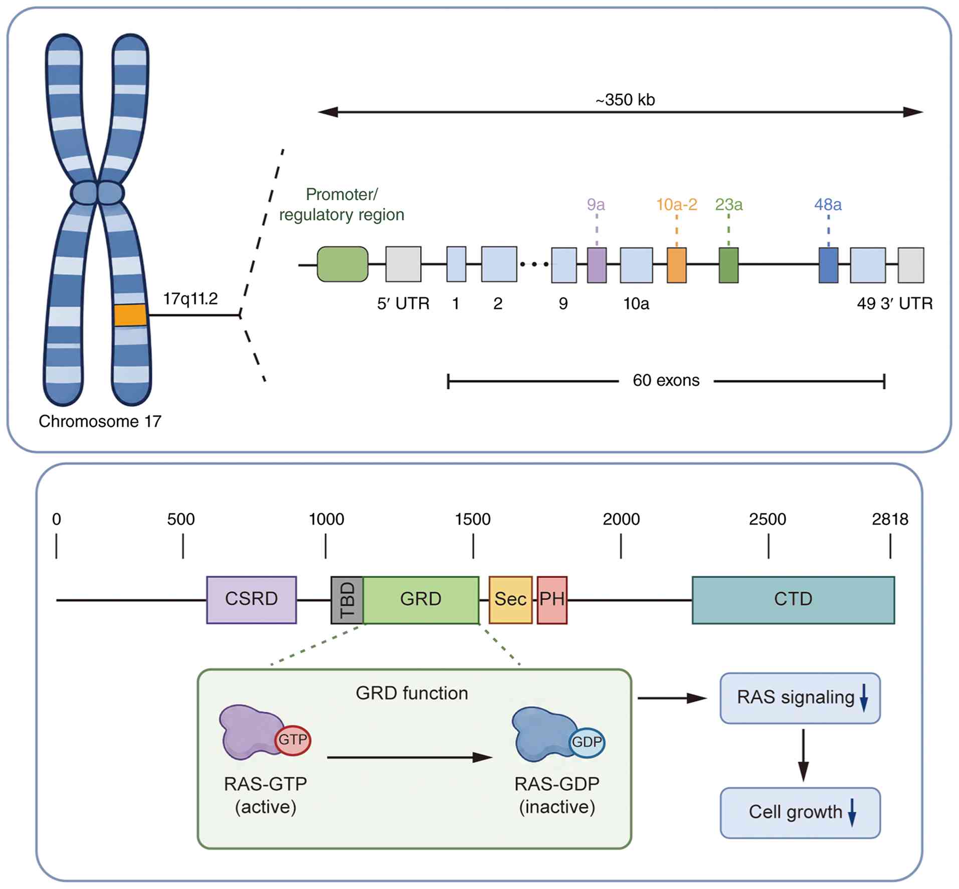
The human NF1 gene is located on chromosome 17q11.2,
representing a large genomic locus spanning ~350 kb (16,17) (Fig. 1). The NF1 locus contains an
upstream promoter/regulatory region, followed by the 5'
untranslated region and a complex gene body composed of 60 exons
separated by introns (18). The
alternative splicing exons contribute to transcript diversity,
including exon 9a, exon 10a-2, exon 23a and exon 48a. For example,
alternative splicing of exon 23a results in two types of
transcripts, with type I encoding neurofibromin, a tumor suppressor
protein comprising 2,818 amino acids (19,20). Neurofibromin contains several
functional domains, including a cysteine/serine-rich domain, a
central GAP-related domain (GRD), a Sec14 domain, a pleckstrin
homologous domain and a C-terminal domain (18,21). Among these domains, the GRD
primarily accelerates the conversion of active RAS-guanosine
triphosphate (GTP) to its inactive RAS-guanosine diphosphate (GDP)
form, thereby negatively regulating RAS signaling and cell growth
(22-24).
RAS acts as a central negative upstream regulator of
the RAF/mitogen-activated protein kinase kinase (MEK)/ERK signaling
cascade, which governs essential cellular processes including
development, differentiation, proliferation and apoptosis (25,26). In addition, neurofibromin
modulates other RAS-dependent pathways; loss of neurofibromin leads
to hyperactivation of the PI3K/AKT/mTOR axis, thereby influencing
cell survival, metabolism and lineage commitment (27). Beyond RAS-dependent mechanisms,
accumulating evidence has indicated that neurofibromin exerts
important RAS-independent functions (3). In particular, neurofibromin
deficiency reduces cyclic adenosine monophosphate (cAMP) levels and
alters protein kinase A (PKA) activity, thereby impairing
osteogenic differentiation and extracellular matrix mineralization
(28,29), revealing an additional layer of
metabolic and transcriptional regulation relevant to skeletal
homeostasis.
Neurofibromin is widely expressed across multiple
tissues during development, providing a molecular basis for the
multi-systemic manifestations of NF1 (30). At the molecular level, pathogenic
NF1 variants result not only in sustained MAPK activation but also
in widespread disruption of interconnected intracellular signaling
networks. These signaling abnormalities impair skeletal
development, bone remodeling and tissue repair, highlighting
neurofibromin as a central integrator of the signaling pathways
essential for bone homeostasis (11). Accordingly, loss-of-function
mutations in NF1 lead to neurofibromin deficiency and dysregulated
downstream signaling, thereby contributing to the development of
NF1-associated skeletal diseases (31).
The pathogenesis of skeletal diseases, including
those associated with NF1, is fundamentally rooted in the
disruption of bone homeostasis. Loss of neurofibromin disrupts this
balance, thereby predisposing patients with NF1 to localized
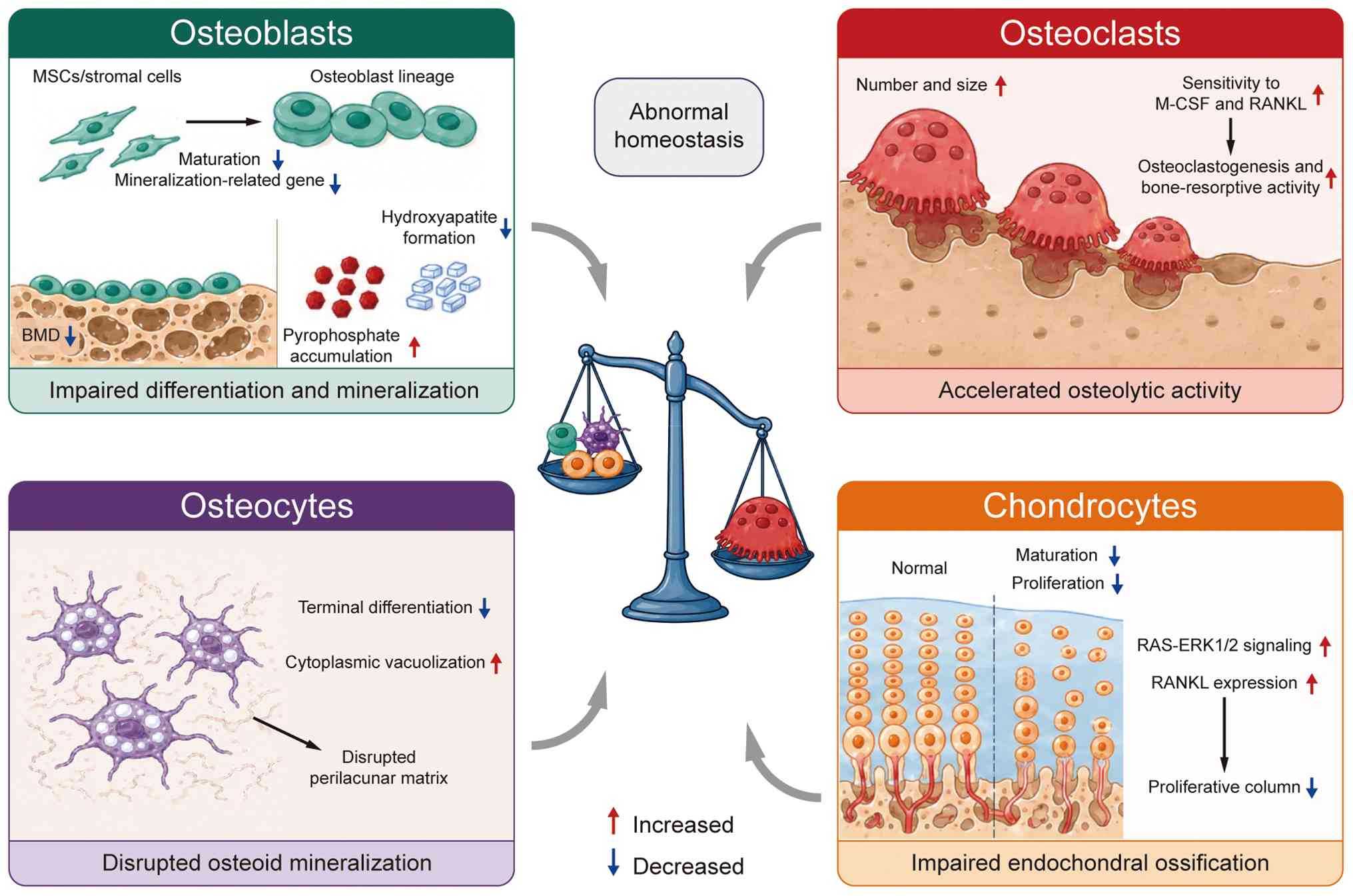
skeletal lesions (32-34). The present review examines the
cellular effects of neurofibromin deficiency on bone homeostasis
(Fig. 2).
While complete loss of NF1 allows for the initial
osteogenic commitment of stromal cells, it profoundly hinders their
functional maturation (35).
Even NF1 haploinsufficiency in MSCs is sufficient to impair
osteoblast differentiation and subsequent mineralization (36). Mechanistically, this
differentiation defect is largely RAS-dependent, as re-expression
of the NF1 GRD can restore osteogenic differentiation (12). Consistent with this mechanism,
complete loss of NF1 suppresses the expression of key
mineralization-related genes (35); however, pyrophosphate
accumulation and the resulting inhibition of hydroxyapatite
formation likely represent downstream mineralization abnormalities
rather than direct RAS-dependent events (37). This chemical imbalance directly
impairs hydroxyapatite formation, creating a 'mineralization trap'
that underlies the clinically observed low BMD.
In contrast to the braking effect on osteoblasts,
both neurofibromin deficiency and complete loss promote osteoclast
formation and activity, thereby accelerating skeletal degradation
(38,39). In NF1-haploinsufficient mice,
osteoclasts were found to be more numerous, larger, and contained
more nuclei (40). Moreover, NF1
haploinsufficiency enhances the responsiveness of osteoclasts to
macrophage colony-stimulating factor and receptor activator of
NF-κB ligand (RANKL) through hyperactivation of RAS-dependent
signaling, particularly the RAS/PI3K pathway, thereby promoting
osteoclastogenesis and bone-resorptive activity (41). This excessive osteoclastogenesis
highlights a specific requirement for neurofibromin in restraining
osteolytic activity within the myeloid lineage (13).
Beyond the classic osteoblast-osteoclast axis,
neurofibromin is essential for maintaining the cellular
architecture and growth plate dynamics of the skeleton. A complete
loss of neurofibromin disrupts the terminal differentiation of
osteoblasts into osteocytes through RAS-dependent mechanisms.
Osteocytes with a complete loss of neurofibromin exhibit
cytoplasmic vacuolization and a failure to properly organize the
perilacunar matrix, which disrupts calcium-phosphorus homeostasis
and contributes to defective osteoid mineralization (42,43).
Neurofibromin deficiency also disrupts chondrocyte
function and endochondral ossification through RAS-dependent
mechanisms. A complete loss of neurofibromin impairs chondrocyte
proliferation and maturation, thereby disrupting endochondral
ossification (44). Moreover,
neurofibromin restrains RAS-ERK1/2 signaling and inhibits RANKL
expression, undermining the proper organization of proliferative
chondrocyte columns (45,46).
Without this regulation, the developmental program of endochondral
ossification is fundamentally dismantled. Collectively, these
findings establish neurofibromin as a critical regulator of
skeletal homeostasis and provide a mechanistic foundation for the
development of NF1-associated skeletal diseases.
Currently, research on RAS-independent mechanisms
remains largely focused on the nervous system, such as neuronal
excitability (47). Although the
cAMP/PKA pathway has been reported to function in NF1-knockdown
osteoblasts, it remains unclear whether this effect is
RAS-independent (29).
Therefore, further studies are needed to elucidate the roles of
RAS-independent mechanisms in bone homeostasis.
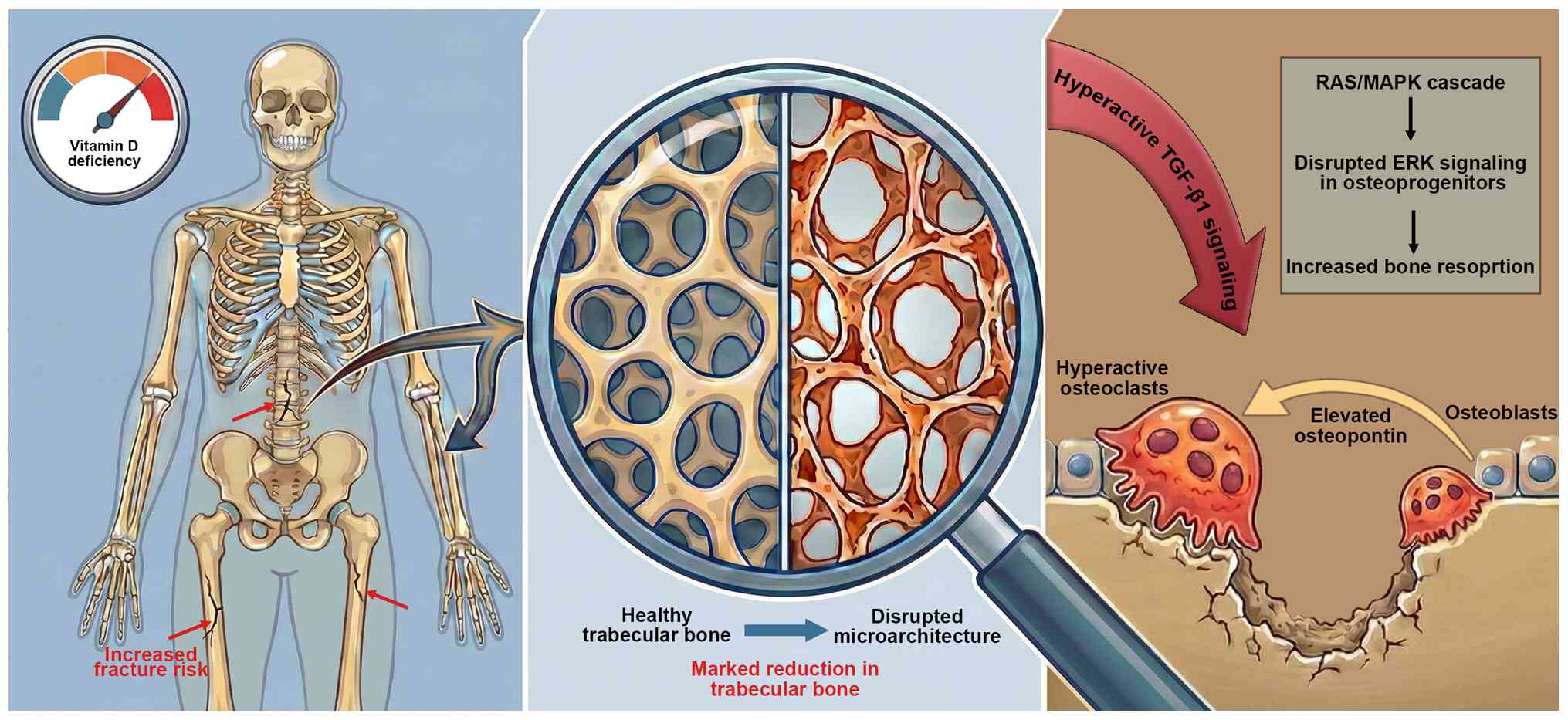
Notably, approximately half of individuals with NF1
develop osteopenia or osteoporosis, which substantially increases
their risk of fractures and contributes to notable skeletal
morbidity (48-50) (Fig. 3). Patients with NF1 typically
exhibit lower BMD compared with the general population,
particularly in the lumbar spine and femur (51,52). Additionally, vitamin D deficiency
has been observed in these patients, and it is considered to
contribute to the clinical severity of neurofibromas (53,54). Low vitamin D levels are
associated with a more aggressive phenotype and larger
neurofibromas, and likely underlie the impaired bone status in NF1
(15). This deficiency may also
serve a role in the reduced BMD observed in NF1, further
exacerbating skeletal fragility (55,56).
NF1-associated osteopenia often progresses to
osteoporosis, as BMD continues to decline with age, even in young
patients (57), and patients
with NF1 have an increased age-dependent fracture risk (58). Histological analysis of bone
samples from patients with NF1 has revealed marked disruption of
bone microarchitecture, driven primarily by reduced trabecular bone
(59). Furthermore, children
with NF1 exhibit increased urinary excretion of pyridinium
crosslinks, reflecting increased bone resorption and suggesting
excessive osteoclast activity in NF1 progression (32). Moreover, increased osteoclast
activity has been considered to partly account for osteopenia and
osteoporosis (39). Hyperactive
TGF-β1 signaling has a pivotal role in the pathogenesis of
osteoporosis in NF1 (60).
Additionally, the elevated osteoblast-secreted osteopontin in NF1
activates intrinsically hyperresponsive osteoclasts, promoting
osteopenia and osteoporosis (61). ERK signaling downstream of the
RAS/MAPK cascade in osteoprogenitors is required for normal
skeletal development and bone maintenance, and loss of MEK in these
cells induces severe osteopenia (62).
CPT, which is characterized by anterolateral bowing
deformities and refracture, is a rare and challenging skeletal
disease in children (63).
Currently, the Crawford classification is the predominant system
for CPT, dividing patients with CPT into four progressive types
(64).
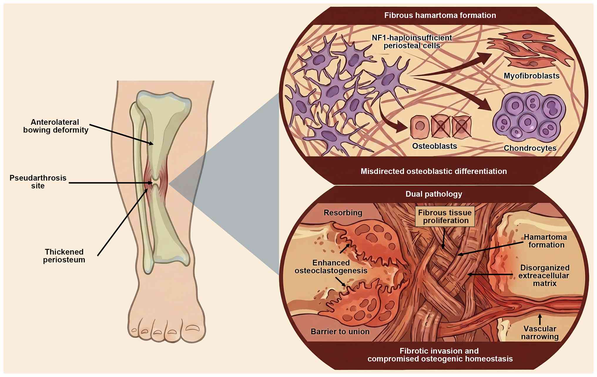
The hallmark of NF1-associated CPT is the presence
of a markedly thickened, pathogenic periosteum surrounding the
pseudarthrosis site (65)
(Fig. 4). Exome sequencing has
shown that somatic NF1 mutations are concentrated in soft tissue at
the pseudarthrosis site but are absent from cortical bone (66). CPT appears to originate from
fibrous hamartoma formed by aberrantly expanding
NF1-haploinsufficient periosteal cells, which fail to complete
terminal osteoblastic differentiation and exhibit an enhanced
ability toward osteoclastogenesis (67,68). Critical histological analysis has
revealed that these abnormal cells are misdirected toward
alternative lineages, including myofibroblasts or chondrocytes
(69). This cellular
reprogramming results in a highly proliferative fibrous tissue that
actively invades the fracture site (70). Consequently, NF1 deficiency
drives a dual pathology: It promotes fibrotic invasion while
simultaneously compromising the osteogenic homeostasis required for
repair, creating a mechanical and biological barrier to union.
Spinal deformity is the most common orthopedic
manifestation in NF1, and is broadly classified into dystrophic and
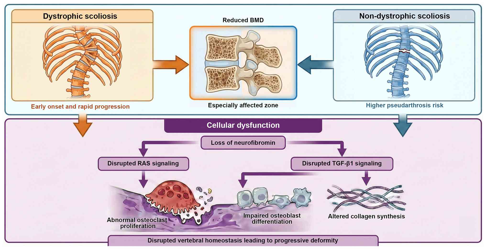
non-dystrophic types (76,77) (Fig. 5). Dystrophic scoliosis represents
a hallmark skeletal feature, characterized by early onset, rapid
progression and difficult clinical management (78). The etiology of dystrophic
scoliosis is multifactorial, with somatic NF1 mutations
contributing to its development (79), and dystrophic curves
predominantly involve the upper thoracic spine (80). By contrast, non-dystrophic
scoliosis behaves similarly to adolescent idiopathic scoliosis, but
tends to present earlier and exhibits a higher risk of
pseudarthrosis (81).
Patients with NF1 exhibit substantially reduced BMD,
with the lumbar spine being more severely affected than the whole
skeleton (50); this reduction
appears to be even more pronounced in patients with NF1 and with
scoliosis requiring surgical intervention (51). At the molecular level,
neurofibromin deficiency disrupts RAS and TGF-β1 signaling
cascades, promoting abnormal osteoclast proliferation, impairing
osteoblast differentiation and altering collagen biosynthesis,
collectively contributing to structural abnormalities in the spine
(8). Consistent with these
clinical observations, mouse models with a complete deletion of
periosteal or osteoblastic NF1 exhibit defective vertebral
remodeling, frequent segmental fusion and obliteration of the
intervertebral disc, which closely parallels human cases (82). Taken together, these findings
demonstrate that NF1-associated scoliosis originates from intrinsic
cellular dysfunction that disrupts vertebral homeostasis and
induces a predisposition to progressive spinal deformity.
Hypophosphatemic osteomalacia has been reported in
patients with NF1, even in the presence of additional congenital
anomalies such as intracranial glioma and unilateral renal
agenesis, highlighting the broad systemic involvement of NF1
(83). Among the craniofacial
skeletal manifestations, sphenoid wing agenesis represents a
distinctive and progressive NF1-associated skeletal defect. This
defect is typically associated with intraorbital PNs or progressive
temporal lobe herniation, often necessitating surgical intervention
(84). More broadly,
NF1-associated skull defects frequently coexist with adjacent PNs
or dural ectasia, underscoring the close relationship between
osseous abnormalities and soft-tissue pathologies in this condition
(85). Additionally,
epidemiological data have suggested that individuals with NF1 may
have a markedly higher risk of developing bone sarcomas; however,
further large-scale studies are required to more precisely
characterize this association and its underlying mechanisms
(86).
Follow-up and assessment during childhood are
important for implementing preventive strategies to maximize peak
bone mass for children with NF1 (10). The current review presents
established and emerging assessment modalities.
Patients with NF1 frequently exhibit vitamin D
insufficiency, emphasizing the need for routine evaluation of bone
status and timely vitamin D replacement (53). Furthermore, low BMD is markedly
more common in NF1 and occurs more frequently than in the general
population, largely due to the secondary hyperparathyroidism
associated with vitamin D deficiency (56). Notably, supplementation of
vitamin D has been shown to markedly improve BMD, underscoring the
clinical importance of monitoring and correcting vitamin D
deficiency in NF1 (87).
The evaluation of bone pathophysiology is
complemented by a range of instrumental modalities that offer more
direct information than plasma assessments. Beyond conventional
dual-energy X-ray absorptiometry, which aids in identifying
patients with NF1 at risk of clinical bone complications and
monitoring therapeutic responses, qualitative parameters such as
trabecular bone scores further enable longitudinal monitoring and
finer characterization of bone impairment (10,49). Quantitative computed tomography
has a unique value in separately evaluating trabecular and cortical
bone compartments (88).
Furthermore, radiation-free ultrasound techniques, specifically
bone waveform and average ultrasound modalities, have demonstrated
high sensitivity in detecting adverse bone matrix alterations in
preclinical models, indicating considerable promise as non-invasive
biomarkers for the early detection of bone fragility in pediatric
NF1 (89). At the molecular
level, exome sequencing has emerged as an essential tool for
delineating complex skeletal phenotypes resulting from dual
molecular diagnoses, thereby revealing underlying genetic
complexity (90). Together,
these approaches are advancing the assessment of skeletal diseases
into an integrated and multidimensional framework.
The management of NF1-associated skeletal diseases
remains challenging due to the complex interplay between intrinsic
cellular defects, aberrant signaling pathways and mechanical
instability. Current treatment strategies can be broadly divided
into conservative approaches and surgical interventions. While
conservative therapies aim to modulate the dysregulated bone
microenvironment and enhance skeletal regeneration, surgical
treatment remains indispensable for severe deformities and
nonunion.
Emerging evidence has indicated that NF1-associated
skeletal abnormalities are driven by profound disturbances in the
local bone microenvironment, including impaired osteoblast
differentiation, dysregulated RAS/ERK and β-catenin signaling,
excessive pyrophosphate accumulation and increased cortical
porosity (82,91,92). These mechanistic insights have
revealed multiple potential molecular targets for pharmacological
intervention (Table I).
In preclinical models, MEK inhibition has been shown
to attenuate fibrotic differentiation and enhance bone regeneration
(93), particularly when
combined with Src homology 2 domain-containing protein tyrosine
phosphatase-2 inhibitors or recombinant human bone morphogenetic
protein-2 (rhBMP-2) (94,95).
Additional signaling pathways have also demonstrated therapeutic
relevance. Modulation of β-catenin signaling was shown to restore
osteogenic differentiation, while inhibition of c-Jun N-terminal
kinase was demonstrated to enhance osteogenesis in NF1-deficient
progenitor cells, particularly when combined with BMP-2 (96,97). In addition, inhibition of
Dickkopf-related protein 1, an inhibitor of WNT signaling, was
shown to promote endochondral ossification (98). Furthermore, icariin was found to
activate the cAMP/PKA/CREB pathway, promoting osteoblast
differentiation in NF1-deficient models and supporting its
potential role as an anabolic adjunct targeting bone metabolism
(29).
Although MEK inhibitors are currently being
evaluated in clinical trials, their application has primarily
focused on NF1-associated PNs (99). Their efficacy in NF1-associated
skeletal disease remains to be further investigated through
systematic clinical trials. BMPs have been explored in the
treatment of CPT, with some early clinical evidence suggesting
potential therapeutic value (100-102); however, their efficacy appears
to be limited when used alone and their efficacy for other
NF1-associated skeletal diseases remains unknown. By contrast,
other candidate therapeutic agents remain largely at the
preclinical stage and their clinical utility has yet to be
established through systematic studies.
Cell-based and regenerative strategies represent a
promising option for addressing the intrinsic osteogenic defects
characteristic of NF1-associated skeletal diseases. Stem cell-based
interventions have demonstrated potential in enhancing
mineralization capacity in refractory CPT, and dental pulp stem
cells derived from patients with NF1 retain regenerative
capability, supporting their prospective utility in skeletal repair
(103,104). Additional studies have further
highlighted the potential of MSC-based therapy. Notably, MSCs in
the iliac crest have been reported to exhibit stronger osteogenic
capacity than those at the lesion site in patients with CPT
(105). Moreover, MSC
transplantation has been shown to normalize bone remodeling,
suppress excessive bone resorption, and improve bone mineral
content and structural integrity in experimental models (106).
Adjunctive biological strategies have shown strong
promise. Combined anabolic and anti-resorptive therapy,
specifically rhBMP-2 with zoledronic acid, has been shown to
notably increase callus bone volume, reduce fibrous tissue
infiltration and restore mechanical properties to near-normal
levels in NF1-associated pseudarthrosis models (107-109). Clinical experience similarly
suggests that integrating anti-resorptive agents with anabolic
stimulation may improve bone healing and postoperative
consolidation in pediatric patients with NF1 (110). Additional pharmacological
adjuncts have also shown benefit. Local delivery of low-dose
lovastatin was demonstrated to enhance callus maturation and
mechanical strength during fracture repair in NF1-deficient models,
highlighting its potential as an intraoperative adjunct (111). Enzyme replacement therapy with
asfotase-α was also found to improve bone growth, mineralization
and strength in NF1 mouse models, suggesting potential
translational relevance for perioperative optimization (112).
Metabolic modulation represents an additional
therapeutic aspect in NF1-associated skeletal diseases. Altered
lipid metabolism contributes to skeletal fragility, and
supplementation with L-carnitine and medium-chain fatty acids
reduces cortical porosity, while protein restriction improves bone
formation (113). Classical
metabolic treatments have also exhibited therapeutic value. Vitamin
D3 supplementation has been reported to improve BMD in adults with
NF1 (114,115). Bisphosphonates were also shown
to exert measurable anti-resorptive effects, although NF1-derived
osteoclasts demonstrated relative resistance in vitro,
underscoring the need for combination strategies (116).
Non-pharmacological interventions may also
contribute to skeletal health. Promotion of muscle development and
physical activity has been associated with partial mitigation of
progressive lower-limb bone mass deficits observed during
childhood, emphasizing the functional interdependence of muscle and
bone in NF1 pathophysiology (117).
Collectively, these findings support a promising and
expanding therapeutic framework that includes targeting
pharmacological therapies, cell-based and regenerative therapies,
adjunctive strategies, and metabolic and physical interventions,
offering multiple approaches for treating NF1-associated skeletal
diseases.
Despite ongoing advances in conservative therapy,
surgical intervention remains the cornerstone of treatment for
severe NF1-associated skeletal diseases. Conservative approaches
alone are often insufficient to achieve durable correction,
particularly in cases involving nonunion, progressive deformity or
mechanical instability.
Surgical management of CPT aims to address both the
biological and mechanical barriers to bone union. A large
single-center database built at Hunan Children's Hospital
(Changsha, China) has substantially improved the clinical
understanding and management of CPT (118). Techniques such as the
'Four-in-One' or 'Three-in-One' integrate resection of the
pathological periosteum, intramedullary fixation, autologous iliac
crest bone grafting and Ilizarov external fixation to
simultaneously enhance osteogenesis and provide mechanical
stability (119-121). Vascularized fibular grafting is
an effective option for managing severe bone defects and in cases
with previous treatment failure (122,123); this option offers high union
rates when combined with complete resection of the diseased bone
and stable compression plating (124). Notably, intramedullary
vascularized fibular grafting combined with Ilizarov distraction
has achieved primary union in all reported cases within certain
cohorts (125). Autologous bone
grafting remains critical for large tibial defects, with iliac
crest grafts widely used due to their robust osteogenic and
cellular properties (126,127). Limb-lengthening procedures may
also be chosen in select patients with complex growth disturbances,
as reported in NF1 cases with concurrent hemihypertrophy and
longitudinal growth retardation (128).
Surgical correction is essential for severe
NF1-associated spinal deformities, particularly dystrophic
scoliosis, which is characterized by an early onset, rapid
progression and technical complexity (129). Selection of the stable vertebra
is critical in non-dystrophic scoliosis to reduce postoperative
complications (130). Multiple
corrective strategies have demonstrated efficacy across different
clinical scenarios. Traditional growing rods represent a safe and
effective option for early-onset dystrophic scoliosis with
intraspinal rib head dislocation, allowing partial rib head
reduction during distraction (131). Compared with posterior fusion,
growing-rod constructs may better preserve trunk growth in
early-onset cases (132). Other
techniques, including halo-gravity traction combined with dual
growing rods, have shown promising deformity correction, although
long-term outcomes require further evaluation (133).
Advances in fixation techniques have improved
outcomes in patients with pedicle dysplasia. Segmentation
correction achieves superior deformity correction compared with
traditional methods and may serve as an alternative in patients
lacking apical pedicles (134).
Sectional correction using a concave domino connector effectively
restores coronal balance while reducing implant failure risk
(135). For severe and rigid
deformities, a one-stage posterior pedicle screw approach is both
safe and effective, with posterior vertebral column resection
recommended for curves demonstrating <35% flexibility, and
fusion to stable vertebrae advised to ensure durable alignment
(136). In addition, a recent
comparative analysis demonstrated that both three-column osteotomy
and halo-gravity traction combined with posterior column osteotomy
provide notable postoperative improvement in coronal and sagittal
parameters, supporting tailored selection of surgical pathways
based on the severity of the deformity and patient characteristics
(137).
For other orthopedic manifestations, such as
NF1-associated osteoarthritis, standardized surgical protocols are
currently lacking. Management decisions should therefore be
individualized based on preoperative functional status, disease
severity and patient-specific considerations (138).
Collectively, the management of NF1-associated
skeletal diseases requires comprehensive surgical approaches.
Existing surgical options offer a wider range of therapeutic
possibilities, ensuring improved outcomes and enhancing the quality
of life for patients.
NF1-associated skeletal diseases represent one of
the most complex manifestations of NF1, arising from the
convergence of genetic alterations, dysregulated intracellular
signaling, cellular dysfunction and biomechanical instability.
Although notable progress has been made in elucidating the role of
neurofibromin in skeletal biology, multiple knowledge gaps remain
between mechanistic insights and durable clinical solutions.
Addressing these gaps will require coordinated advances across
genetics, molecular therapeutics, surgical innovation and
longitudinal patient management.
Recent advances in genetic research have expanded
both the genotypic and phenotypic spectrum of NF1-associated
skeletal diseases. Novel NF1 mutations and modifier genes have been
implicated in CPT, providing new insights into disease
heterogeneity and potential therapeutic targets (139). In addition, emerging evidence
has suggested that non-coding regulatory elements, including long
non-coding RNAs, participate in the pathogenesis of CPT by
modulating osteogenic differentiation and extracellular matrix
organization (140). In
NF1-associated dystrophic scoliosis, transcriptomic and
network-based analyses have identified dysregulated immune and
osteoimmunological pathways, highlighting an underappreciated role
of immune-bone interactions in disease progression (141). Taken together, these findings
underscore the need for integrative genomic and multi-omics
approaches to further define disease mechanisms, identify high-risk
patients and uncover actionable molecular targets.
Targeting dysregulated signaling pathways remains a
promising option for improving outcomes in NF1-associated skeletal
diseases. While MEK inhibitors have demonstrated clinical efficacy
in NF1-associated tumors, their effects on bone formation,
remodeling and fracture healing require systematic investigation
(93). Future studies should
continually evaluate how targeting drugs influence osteoblast and
osteoclast function, bone regeneration and the unique
pathophysiology of NF1-associated skeletal lesions.
Surgical intervention will continue to serve a
central role in the management of severe skeletal manifestations;
however, current surgical strategies are associated with
substantial complication rates and inconsistent long-term outcomes.
For example, vascularized fibular grafting, despite high union
rates, remains technically demanding and carries a marked risk of
morbidity (124). Future
directions should therefore prioritize the development and
refinement of surgical strategies that can improve long-term
outcomes and minimize morbidity.
As an autosomal dominant disorder, NF1 typically
manifests in early childhood, making timely surveillance critical
for preventing progressive skeletal morbidity. Longitudinal
follow-up beginning in early childhood is therefore essential to
identify early reductions in BMD, vitamin D deficiency, progressive
bowing deformities and evolving spinal pathology. However, an
improved clinical understanding of the diseases requires
multicenter research and collaboration. Future surveillance
frameworks should move beyond phenotype-based assessment toward
multidimensional risk stratification models, which integrate
genetic data, molecular biomarkers, imaging parameters and
validated clinical predictors.
NF1-associated skeletal diseases represent a
multifaceted disorder arising from neurofibromin deficiency and the
consequent dysregulation of key signaling pathways that govern
skeletal development, remodeling and repair. Accumulating evidence
has demonstrated that NF1 mutations disrupt the coordinated
function of osteoblasts, osteoclasts, osteocytes and chondrocytes,
resulting in uncoupled bone remodeling, defective mineralization,
abnormal endochondral ossification and a permissive
microenvironment for fibrotic tissue invasion. Together, these
intrinsic and microenvironmental abnormalities underlie the diverse
skeletal manifestations observed in NF1, including osteopenia and
osteoporosis, CPT, scoliosis and other less common but clinically
important bone defects.
Despite substantial progress being made in the
mechanistic understanding of NF1, clinical management remains
challenging. Current therapies rely heavily on complex surgical
interventions and supportive metabolic strategies, which are often
associated with high complication rates and incomplete efficacy.
Emerging translational studies targeting dysregulated RAS/MAPK,
PI3K/AKT and cAMP/PKA signaling pathways, together with
regenerative and cell-based approaches, offer promising
opportunities to develop mechanism-based and biologically informed
treatments. However, robust clinical evidence supporting these
strategies is still limited.
In the future, integrating mechanistic insights with
advances in genetics, biomaterials, pharmacology and surgical
innovation will be essential to improve outcomes for patients with
NF1-associated skeletal diseases. Early risk stratification,
longitudinal surveillance and personalized treatment strategies
based on molecular and clinical features are likely to serve
central roles in improving outcomes. Ultimately, coordinated
multicenter efforts and standardized outcome frameworks will be
essential to translate biological discoveries into effective and
durable therapies that meaningfully improve skeletal health and
quality of life in individuals with NF1.
Not available.
SZ and GY designed the scope and structure of the
review, performed structured literature searches, as well as wrote
and revised major sections of the manuscript. SZ critically
synthesized and interpreted the findings. Both authors have read
and approved the final manuscript. Data authentication is not
applicable.
Not applicable.
Not applicable.
The authors declare that they have no competing
interests.
Not available.
This study was supported by the Health Research Project of Hunan
Provincial Health Commission (grant no. W20243208), the National
Key Clinical Specialty Scientific Research Project (grant no.
Z2023057), the Special Fund of the Hunan Provincial Key Laboratory
of Pediatric Orthopedics (grant no. 2023TP1019) and the Hunan
Provincial Medical Discipline Development Project, Category C:
Pediatric Orthopedics.
|
1
|
Lee TJ, Chopra M, Kim RH, Parkin PC and
Barnett-Tapia C: Incidence and prevalence of neurofibromatosis type
1 and 2: A systematic review and meta-analysis. Orphanet J Rare
Dis. 18:2922023. View Article : Google Scholar : PubMed/NCBI
|
|
2
|
Williams VC, Lucas J, Babcock MA, Gutmann
DH, Korf B and Maria BL: Neurofibromatosis type 1 revisited.
Pediatrics. 123:124–133. 2009. View Article : Google Scholar : PubMed/NCBI
|
|
3
|
Anastasaki C, Orozco P and Gutmann DH: RAS
and beyond: The many faces of the neurofibromatosis type 1 protein.
Dis Model Mech. 15:dmm0493622022. View Article : Google Scholar : PubMed/NCBI
|
|
4
|
Mo J, Moye SL, McKay RM and Le LQ:
Neurofibromin and suppression of tumorigenesis: Beyond the GAP.
Oncogene. 41:1235–1251. 2022. View Article : Google Scholar : PubMed/NCBI
|
|
5
|
Báez-Flores J, Rodríguez-Martín M and
Lacal J: The therapeutic potential of neurofibromin signaling
pathways and binding partners. Commun Biol. 6:4362023. View Article : Google Scholar : PubMed/NCBI
|
|
6
|
Friedman JM: Neurofibromatosis 1: Clinical
manifestations and diagnostic criteria. J Child Neurol. 17:548–554.
571–542. 646–551. 2002. View Article : Google Scholar : PubMed/NCBI
|
|
7
|
Stewart DR, Korf BR, Nathanson KL,
Stevenson DA and Yohay K: Care of adults with neurofibromatosis
type 1: A clinical practice resource of the American College of
Medical Genetics and Genomics (ACMG). Genet Med. 20:671–682. 2018.
View Article : Google Scholar : PubMed/NCBI
|
|
8
|
Kaspiris A, Savvidou OD, Vasiliadis ES,
Hadjimichael AC, Melissaridou D, Iliopoulou-Kosmadaki S, Iliopoulos
ID, Papadimitriou E and Chronopoulos E: Current aspects on the
pathophysiology of bone metabolic defects during progression of
scoliosis in neurofibromatosis Type 1. J Clin Med. 11:4442022.
View Article : Google Scholar : PubMed/NCBI
|
|
9
|
Elefteriou F, Kolanczyk M, Schindeler A,
Viskochil DH, Hock JM, Schorry EK, Crawford AH, Friedman JM, Little
D, Peltonen J, et al: Skeletal abnormalities in neurofibromatosis
type 1: Approaches to therapeutic options. Am J Med Genet A.
149a:2327–2338. 2009. View Article : Google Scholar : PubMed/NCBI
|
|
10
|
Rodari G, Scuvera G, Ulivieri FM, Profka
E, Menni F, Saletti V, Esposito S, Bergamaschi S, Ferrante E,
Eller-Vainicher C, et al: Progressive bone impairment with age and
pubertal development in neurofibromatosis type I. Arch Osteoporos.
13:932018. View Article : Google Scholar : PubMed/NCBI
|
|
11
|
Schindeler A and Little DG: Recent
insights into bone development, homeostasis, and repair in type 1
neurofibromatosis (NF1). Bone. 42:616–622. 2008. View Article : Google Scholar : PubMed/NCBI
|
|
12
|
Wu X, Estwick SA, Chen S, Yu M, Ming W,
Nebesio TD, Li Y, Yuan J, Kapur R, Ingram D, et al: Neurofibromin
plays a critical role in modulating osteoblast differentiation of
mesenchymal stem/progenitor cells. Hum Mol Genet. 15:2837–2845.
2006. View Article : Google Scholar : PubMed/NCBI
|
|
13
|
Rhodes SD, Yang H, Dong R, Menon K, He Y,
Li Z, Chen S, Staser KW, Jiang L, Wu X, et al: Nf1
haploinsufficiency alters myeloid lineage commitment and function,
leading to deranged skeletal homeostasis. J Bone Miner Res.
30:1840–1851. 2015. View Article : Google Scholar : PubMed/NCBI
|
|
14
|
Shah H, Rousset M and Canavese F:
Congenital pseudarthrosis of the tibia: Management and
complications. Indian J Orthop. 46:616–626. 2012. View Article : Google Scholar
|
|
15
|
Ferrara UP, Tortora C, Rosano C, Assunto
A, Rossi A, Pagano S, Falco M, Simeoli C, Ferrigno R, D'Amico A, et
al: Bone metabolism in patients with type 1 neurofibromatosis: Key
role of sun exposure and physical activity. Sci Rep. 12:43682022.
View Article : Google Scholar
|
|
16
|
Wallace MR, Marchuk DA, Andersen LB,
Letcher R, Odeh HM, Saulino AM, Fountain JW, Brereton A, Nicholson
J, Mitchell AL, et al: Type 1 neurofibromatosis gene:
Identification of a large transcript disrupted in three NF1
patients. Science. 249:181–186. 1990. View Article : Google Scholar : PubMed/NCBI
|
|
17
|
Dorschner MO, Sybert VP, Weaver M,
Pletcher BA and Stephens K: NF1 microdeletion breakpoints are
clustered at flanking repetitive sequences. Hum Mol Genet. 9:35–46.
2000. View Article : Google Scholar
|
|
18
|
Bergoug M, Doudeau M, Godin F, Mosrin C,
Vallée B and Bénédetti H: Neurofibromin structure, functions and
regulation. Cells. 9:23652020. View Article : Google Scholar : PubMed/NCBI
|
|
19
|
Marchuk DA, Saulino AM, Tavakkol R,
Swaroop M, Wallace MR, Andersen LB, Mitchell AL, Gutmann DH,
Boguski M and Collins FS: cDNA cloning of the type 1
neurofibromatosis gene: Complete sequence of the NF1 gene product.
Genomics. 11:931–940. 1991. View Article : Google Scholar : PubMed/NCBI
|
|
20
|
Cawthon RM, Weiss R, Xu GF, Viskochil D,
Culver M, Stevens J, Robertson M, Dunn D, Gesteland R, O'Connell P,
et al: A major segment of the neurofibromatosis type 1 gene: CDNA
sequence, genomic structure, and point mutations. Cell. 62:193–201.
1990. View Article : Google Scholar : PubMed/NCBI
|
|
21
|
Miller AH and Halloran MC: Mechanistic
insights from animal models of neurofibromatosis type 1 cognitive
impairment. Dis Model Mech. 15:dmm0494222022. View Article : Google Scholar : PubMed/NCBI
|
|
22
|
Xu GF, Lin B, Tanaka K, Dunn D, Wood D,
Gesteland R, White R, Weiss R and Tamanoi F: The catalytic domain
of the neurofibromatosis type 1 gene product stimulates ras GTPase
and complements ira mutants of S. cerevisiae. Cell. 63:835–841.
1990. View Article : Google Scholar : PubMed/NCBI
|
|
23
|
Ballester R, Marchuk D, Boguski M, Saulino
A, Letcher R, Wigler M and Collins F: The NF1 locus encodes a
protein functionally related to mammalian GAP and yeast IRA
proteins. Cell. 63:851–859. 1990. View Article : Google Scholar : PubMed/NCBI
|
|
24
|
Xu GF, O'Connell P, Viskochil D, Cawthon
R, Robertson M, Culver M, Dunn D, Stevens J, Gesteland R, White R,
et al: The neurofibromatosis type 1 gene encodes a protein related
to GAP. Cell. 62:599–608. 1990. View Article : Google Scholar : PubMed/NCBI
|
|
25
|
Molina JR and Adjei AA: The Ras/Raf/MAPK
pathway. J Thorac Oncol. 1:7–9. 2006. View Article : Google Scholar
|
|
26
|
Downward J: Control of ras activation.
Cancer Surv. 27:87–100. 1996.PubMed/NCBI
|
|
27
|
Giraud JS, Bièche I, Pasmant É and
Tlemsani C: NF1 alterations in cancers: Therapeutic implications in
precision medicine. Expert Opin Investig Drugs. 32:941–957. 2023.
View Article : Google Scholar : PubMed/NCBI
|
|
28
|
Tong J, Hannan F, Zhu Y, Bernards A and
Zhong Y: Neurofibromin regulates G protein-stimulated adenylyl
cyclase activity. Nat Neurosci. 5:95–96. 2002. View Article : Google Scholar : PubMed/NCBI
|
|
29
|
Chen M, Lu L, Cheng D, Zhang J, Liu X,
Zhang J and Zhang T: Icariin promotes osteogenic differentiation in
a cell model with NF1 gene knockout by activating the cAMP/PKA/CREB
Pathway. Molecules. 28:51282023. View Article : Google Scholar : PubMed/NCBI
|
|
30
|
Daston MM and Ratner N: Neurofibromin, a
predominantly neuronal GTPase activating protein in the adult, is
ubiquitously expressed during development. Dev Dyn. 195:216–226.
1992. View Article : Google Scholar : PubMed/NCBI
|
|
31
|
Karnoub AE and Weinberg RA: Ras oncogenes:
Split personalities. Nat Rev Mol Cell Biol. 9:517–531. 2008.
View Article : Google Scholar : PubMed/NCBI
|
|
32
|
Stevenson DA, Schwarz EL, Viskochil DH,
Moyer-Mileur LJ, Murray M, Firth SD, D'Astous JL, Carey JC and
Pasquali M: Evidence of increased bone resorption in
neurofibromatosis type 1 using urinary pyridinium crosslink
analysis. Pediatr Res. 63:697–701. 2008. View Article : Google Scholar : PubMed/NCBI
|
|
33
|
Yu X, Chen S, Potter OL, Murthy SM, Li J,
Pulcini JM, Ohashi N, Winata T, Everett ET, Ingram D, et al:
Neurofibromin and its inactivation of Ras are prerequisites for
osteoblast functioning. Bone. 36:793–802. 2005. View Article : Google Scholar : PubMed/NCBI
|
|
34
|
Kuorilehto T, Nissinen M, Koivunen J,
Benson MD and Peltonen J: NF1 tumor suppressor protein and mRNA in
skeletal tissues of developing and adult normal mouse and
NF1-deficient embryos. J Bone Miner Res. 19:983–989. 2004.
View Article : Google Scholar : PubMed/NCBI
|
|
35
|
Paria N, Khalid A, Shen B, Lemoine B, Chan
J, Kidane YH, Oxendine I, Cornelia R, Wise CA and Rios JJ:
Molecular dissection of somatic skeletal disease in
neurofibromatosis type 1. J Bone Miner Res. 38:288–299. 2023.
View Article : Google Scholar :
|
|
36
|
Darle A, Mahiet T, Aubin D, Doyen M, El
Kassar L, Parfait B, Lemaitre G, Baldeschi C, Allouche J and Holic
N: Generation of heterozygous and homozygous NF1 lines from
human-induced pluripotent stem cells using CRISPR/Cas9 to
investigate bone defects associated with neurofibromatosis type 1.
Front Cell Dev Biol. 12:13595612024. View Article : Google Scholar : PubMed/NCBI
|
|
37
|
de la Croix Ndong J, Makowski AJ,
Uppuganti S, Vignaux G, Ono K, Perrien DS, Joubert S, Baglio SR,
Granchi D, Stevenson DA, et al: Corrigendum: Asfotase-α improves
bone growth, mineralization and strength in mouse models of
neurofibromatosis type-1. Nat Med. 21:4142015. View Article : Google Scholar
|
|
38
|
Alanne MH, Siljamäki E, Peltonen S,
Väänänen K, Windle JJ, Parada LF, Määttä JA and Peltonen J:
Phenotypic characterization of transgenic mice harboring Nf1+/− or
Nf1−/− osteoclasts in otherwise Nf1+/+ background. J Cell Biochem.
113:2136–2146. 2012. View Article : Google Scholar : PubMed/NCBI
|
|
39
|
Heervä E, Alanne MH, Peltonen S,
Kuorilehto T, Hentunen T, Väänänen K and Peltonen J: Osteoclasts in
neurofibromatosis type 1 display enhanced resorption capacity,
aberrant morphology, and resistance to serum deprivation. Bone.
47:583–590. 2010. View Article : Google Scholar : PubMed/NCBI
|
|
40
|
Liu N, Xu N, Wei LH and Chai GL: Mammalian
target of rapamycin inhibitor abrogates abnormal osteoclastogenesis
in neurofibromatosis type 1. Chin Med J (Engl). 126:101–107. 2013.
View Article : Google Scholar : PubMed/NCBI
|
|
41
|
Yang FC, Chen S, Robling AG, Yu X, Nebesio
TD, Yan J, Morgan T, Li X, Yuan J, Hock J, et al: Hyperactivation
of p21ras and PI3K cooperate to alter murine and human
neurofibromatosis type 1-haploinsufficient osteoclast functions. J
Clin Invest. 116:2880–2891. 2006. View Article : Google Scholar : PubMed/NCBI
|
|
42
|
Kühnisch J, Seto J, Lange C, Stumpp S,
Kobus K, Grohmann J, Elefteriou F, Fratzl P, Mundlos S and
Kolanczyk M: Neurofibromin inactivation impairs osteocyte
development in Nf1Prx1 and Nf1Col1 mouse models. Bone. 66:155–162.
2014. View Article : Google Scholar : PubMed/NCBI
|
|
43
|
Kamiya N, Yamaguchi R, Aruwajoye O, Kim
AJ, Kuroyanagi G, Phipps M, Adapala NS, Feng JQ and Kim HK:
Targeted disruption of NF1 in osteocytes increases FGF23 and
osteoid with Osteomalacia-like bone phenotype. J Bone Miner Res.
32:1716–1726. 2017. View Article : Google Scholar : PubMed/NCBI
|
|
44
|
Kolanczyk M, Kossler N, Kühnisch J,
Lavitas L, Stricker S, Wilkening U, Manjubala I, Fratzl P, Spörle
R, Herrmann BG, et al: Multiple roles for neurofibromin in skeletal
development and growth. Hum Mol Genet. 16:874–886. 2007. View Article : Google Scholar : PubMed/NCBI
|
|
45
|
Ono K, Karolak MR, Ndong Jde L, Wang W,
Yang X and Elefteriou F: The ras-GTPase activity of neurofibromin
restrains ERK-dependent FGFR signaling during endochondral bone
formation. Hum Mol Genet. 22:3048–3062. 2013. View Article : Google Scholar : PubMed/NCBI
|
|
46
|
Karolak MR, Yang X and Elefteriou F: FGFR1
signaling in hypertrophic chondrocytes is attenuated by the Ras-GAP
neurofibromin during endochondral bone formation. Hum Mol Genet.
24:2552–2564. 2015. View Article : Google Scholar : PubMed/NCBI
|
|
47
|
Anastasaki C, Mo J, Chen JK, Chatterjee J,
Pan Y, Scheaffer SM, Cobb O, Monje M, Le LQ and Gutmann DH:
Neuronal hyperexcitability drives central and peripheral nervous
system tumor progression in models of neurofibromatosis-1. Nat
Commun. 13:27852022. View Article : Google Scholar : PubMed/NCBI
|
|
48
|
Dulai S, Briody J, Schindeler A, North KN,
Cowell CT and Little DG: Decreased bone mineral density in
neurofibromatosis type 1: Results from a pediatric cohort. J
Pediatr Orthop. 27:472–475. 2007. View Article : Google Scholar : PubMed/NCBI
|
|
49
|
Stevenson DA, Moyer-Mileur LJ, Murray M,
Slater H, Sheng X, Carey JC, Dube B and Viskochil DH: Bone mineral
density in children and adolescents with neurofibromatosis type 1.
J Pediatr. 150:83–88. 2007. View Article : Google Scholar
|
|
50
|
Lammert M, Kappler M, Mautner VF, Lammert
K, Störkel S, Friedman JM and Atkins D: Decreased bone mineral
density in patients with neurofibromatosis 1. Osteoporos Int.
16:1161–1166. 2005. View Article : Google Scholar : PubMed/NCBI
|
|
51
|
Lodish MB, Dagalakis U, Sinaii N,
Bornstein E, Kim A, Lokie KB, Baldwin AM, Reynolds JC, Dombi E,
Stratakis CA and Widemann BC: Bone mineral density in children and
young adults with neurofibromatosis type 1. Endocr Relat Cancer.
19:817–825. 2012. View Article : Google Scholar : PubMed/NCBI
|
|
52
|
Kaspiris A, Vasiliadis E, Iliopoulos ID,
Panagopoulos F, Melissaridou D, Lianou I, Ntourantonis D, Savvidou
OD, Papadimitriou E and Pneumaticos SG: Bone mineral density,
vitamin D and osseous metabolism indices in neurofibromatosis type
1: A systematic review and meta-analysis. Bone. 180:1169922024.
View Article : Google Scholar
|
|
53
|
Modica R, Altieri B, D'Aniello F,
Benevento E, Cannavale G, Minotta R, Liccardi A, Colao A and
Faggiano A: Vitamin D and bone metabolism in adult patients with
neurofibromatosis type 1. Metabolites. 13:2552023. View Article : Google Scholar : PubMed/NCBI
|
|
54
|
Petramala L, Giustini S, Zinnamosca L,
Marinelli C, Colangelo L, Cilenti G, Formicuccia MC, D'Erasmo E,
Calvieri S and Letizia C: Bone mineral metabolism in patients with
neurofibromatosis type 1 (von Recklingausen disease). Arch Dermatol
Res. 304:325–331. 2012. View Article : Google Scholar
|
|
55
|
Filopanti M, Verga U, Ulivieri FM, Giavoli
C, Rodari G, Arosio M, Natacci F and Spada A: Trabecular bone score
(TBS) and bone metabolism in patients affected with type 1
neurofibromatosis (NF1). Calcif Tissue Int. 104:207–213. 2019.
View Article : Google Scholar
|
|
56
|
Jalabert M, Ferkal S, Souberbielle JC,
Sbidian E, Mageau A, Eymard F, Le Corvoisier P, Allanore L,
Chevalier X, Wolkenstein P and Guignard S: Bone status according to
neurofibromatosis type 1 phenotype: A descriptive study of 60 women
in France. Calcif Tissue Int. 108:738–745. 2021. View Article : Google Scholar : PubMed/NCBI
|
|
57
|
Heervä E, Leinonen P, Kuorilehto T,
Peltonen S, Pöyhönen M, Väänänen K and Peltonen J:
Neurofibromatosis 1-related osteopenia often progresses to
osteoporosis in 12 years. Calcif Tissue Int. 92:23–27. 2013.
View Article : Google Scholar
|
|
58
|
Heervä E, Koffert A, Jokinen E, Kuorilehto
T, Peltonen S, Aro HT and Peltonen J: A controlled register-based
study of 460 neurofibromatosis 1 patients: Increased fracture risk
in children and adults over 41 years of age. J Bone Miner Res.
27:2333–2337. 2012. View Article : Google Scholar : PubMed/NCBI
|
|
59
|
Brunetti-Pierri N, Doty SB, Hicks J, Phan
K, Mendoza-Londono R, Blazo M, Tran A, Carter S, Lewis RA, Plon SE,
et al: Generalized metabolic bone disease in Neurofibromatosis type
I. Mol Genet Metab. 94:105–111. 2008. View Article : Google Scholar : PubMed/NCBI
|
|
60
|
Rhodes SD, Wu X, He Y, Chen S, Yang H,
Staser KW, Wang J, Zhang P, Jiang C, Yokota H, et al: Hyperactive
transforming growth factor-β1 signaling potentiates skeletal
defects in a neurofibromatosis type 1 mouse model. J Bone Miner
Res. 28:2476–2489. 2013. View Article : Google Scholar : PubMed/NCBI
|
|
61
|
Li H, Liu Y, Zhang Q, Jing Y, Chen S, Song
Z, Yan J, Li Y, Wu X, Zhang X, et al: Ras dependent paracrine
secretion of osteopontin by Nf1+/- osteoblasts promote osteoclast
activation in a neurofi- bromatosis type I murine model. Pediatr
Res. 65:613–618. 2009. View Article : Google Scholar : PubMed/NCBI
|
|
62
|
Kim JM, Yang YS, Park KH, Oh H, Greenblatt
MB and Shim JH: The ERK MAPK pathway is essential for skeletal
development and homeostasis. Int J Mol Sci. 20:18032019. View Article : Google Scholar : PubMed/NCBI
|
|
63
|
Banchhor H and Chimurkar V: Congenital
pseudoarthrosis of the tibia: A narrative review. Cureus.
14:e325012022.
|
|
64
|
Crawford AH Jr and Bagamery N: Osseous
manifestations of neurofibromatosis in childhood. J Pediatr Orthop.
6:72–88. 1986. View Article : Google Scholar : PubMed/NCBI
|
|
65
|
Hermanns-Sachweh B, Senderek J, Alfer J,
Klosterhalfen B, Büttner R, Füzesi L and Weber M: Vascular changes
in the periosteum of congenital pseudarthrosis of the tibia. Pathol
Res Pract. 201:305–312. 2005. View Article : Google Scholar : PubMed/NCBI
|
|
66
|
Sant DW, Margraf RL, Stevenson DA,
Grossmann AH, Viskochil DH, Hanson H, Everitt MD, Rios JJ,
Elefteriou F, Hennessey T and Mao R: Evaluation of somatic
mutations in tibial pseudarthrosis samples in neurofibromatosis
type 1. J Med Genet. 52:256–261. 2015. View Article : Google Scholar : PubMed/NCBI
|
|
67
|
Cho TJ, Seo JB, Lee HR, Yoo WJ, Chung CY
and Choi IH: Biologic characteristics of fibrous hamartoma from
congenital pseudarthrosis of the tibia associated with
neurofibromatosis type 1. J Bone Joint Surg Am. 90:2735–2744. 2008.
View Article : Google Scholar : PubMed/NCBI
|
|
68
|
Lee DY, Cho TJ, Lee HR, Lee K, Moon HJ,
Park MS, Yoo WJ, Chung CY and Choi IH: Disturbed osteoblastic
differentiation of fibrous hamartoma cell from congenital
pseudarthrosis of the tibia associated with neurofibromatosis type
I. Clin Orthop Surg. 3:230–237. 2011. View Article : Google Scholar : PubMed/NCBI
|
|
69
|
Sakamoto A, Yoshida T, Yamamoto H, Oda Y,
Tsuneyoshi M and Iwamoto Y: Congenital pseudarthrosis of the tibia:
Analysis of the histology and the NF1 gene. J Orthop Sci.
12:361–365. 2007. View Article : Google Scholar : PubMed/NCBI
|
|
70
|
El-Hoss J, Sullivan K, Cheng T, Yu NY,
Bobyn JD, Peacock L, Mikulec K, Baldock P, Alexander IE, Schindeler
A and Little DG: A murine model of neurofibromatosis type 1 tibial
pseudarthrosis featuring proliferative fibrous tissue and
osteoclast-like cells. J Bone Miner Res. 27:68–78. 2012. View Article : Google Scholar
|
|
71
|
Van Royen K, Brems H, Legius E, Lammens J
and Laumen A: Prevalence of neurofibromatosis type 1 in congenital
pseudar- throsis of the tibia. Eur J Pediatr. 175:1193–1198. 2016.
View Article : Google Scholar : PubMed/NCBI
|
|
72
|
Lee SM, Choi IH, Lee DY, Lee HR, Park MS,
Yoo WJ, Chung CY and Cho TJ: Is double inactivation of the Nf1 gene
responsible for the development of congenital pseudarthrosis of the
tibia associ- ated with NF1? J Orthop Res. 30:1535–1540. 2012.
View Article : Google Scholar : PubMed/NCBI
|
|
73
|
Zheng Y, Zhu G, Liu Y, Zhao W, Yang Y, Luo
Z, Fu Y, Mei H and Hu Z: Case series of congenital pseudarthrosis
of the tibia unful- filling neurofibromatosis type 1 diagnosis: 21%
with somatic NF1 haploinsufficiency in the periosteum. Hum Genet.
141:1371–1383. 2022. View Article : Google Scholar : PubMed/NCBI
|
|
74
|
Liu Y, Qin ZQ, Zheng Y, Wu J, Yang G, Tan
Q, Zhu G, Liu K and Mei H: New insights into pathogenesis of
congenital pseudar- throsis of tibia in children using periosteum
proteomics analysis. Rapid Commun Mass Spectrom. 36:e93742022.
View Article : Google Scholar
|
|
75
|
Xu J, Zhang Y, Zhu K, Li J, Guan Y, He X,
Jin X, Bai G and Hu L: Clinical characteristics and in silico
analysis of congenital pseudar- throsis of the tibia combined with
neurofibromatosis type 1 caused by a novel NF1 mutation. Front
Genet. 13:9913142022. View Article : Google Scholar
|
|
76
|
Welborn M, Tambe A, Adeyemi A, Dupuis M,
Simoneau D and Brandi ML: Burden of disease and unmet needs
associated with scoliosis in neurofibromatosis type 1: A systematic
literature review. JBMR Plus. 9:ziaf0722025. View Article : Google Scholar : PubMed/NCBI
|
|
77
|
Tsirikos AI, Saifuddin A and Noordeen MH:
Spinal deformity in neurofibromatosis type-1: Diagnosis and
treatment. Eur Spine J. 14:427–439. 2005. View Article : Google Scholar : PubMed/NCBI
|
|
78
|
Wang D, Zhang BH, Wen X, Chen KH, Xiao HT,
Xu XW and Li QF: Clinical features and surgical treatments of
scoliosis in neurofibromatosis type 1: A systemic review and
meta-analysis. Eur Spine J. 33:2646–2665. 2024. View Article : Google Scholar : PubMed/NCBI
|
|
79
|
Margraf RL, VanSant-Webb C, Mao R,
Viskochil DH, Carey J, Hanson H, D'Astous J, Grossmann A and
Stevenson DA: NF1 somatic mutation in dystrophic scoliosis. J Mol
Neurosci. 68:11–18. 2019. View Article : Google Scholar : PubMed/NCBI
|
|
80
|
Du Y, Bai T, Song J, Zhang L, Huang H, Han
P, Gai F, Guo J, Li J, Lv C, et al: The genotypic and family
characteristics and clinical intervention of neurofibromatosis type
1 gene are associ- ated with dystrophic scoliosis by whole-exome
sequencing. Front Neurol. 16:16416652025. View Article : Google Scholar
|
|
81
|
Feldman DS, Jordan C and Fonseca L:
Orthopaedic manifesta- tions of neurofibromatosis type 1. J Am Acad
Orthop Surg. 18:346–357. 2010. View Article : Google Scholar : PubMed/NCBI
|
|
82
|
Wang W, Nyman JS, Ono K, Stevenson DA,
Yang X and Elefteriou F: Mice lacking Nf1 in osteochondroprogenitor
cells display skeletal dysplasia similar to patients with
neurofibroma- tosis type I. Hum Mol Genet. 20:3910–3924. 2011.
View Article : Google Scholar : PubMed/NCBI
|
|
83
|
Kaspiris A, Vasiliadis E, Melissaridou D,
Iliopoulos ID, Papagelopoulos PJ and Savvidou OD: Hypophosphatemic
osteomalacia in neurofibromatosis 1 associated with intracranial
gliomas and congenital renal agenesis: A rare case report and
review of the literature. J Orthop Case Rep. 12:23–29. 2022.
View Article : Google Scholar : PubMed/NCBI
|
|
84
|
Chauvel-Picard J, Lion-Francois L, Beuriat
PA, Paulus C, Szathmari A, Mottolese C, Gleizal A and Di Rocco F:
Craniofacial bone alterations in patients with neurofibromatosis
type 1. Childs Nerv Syst. 36:2391–2399. 2020. View Article : Google Scholar : PubMed/NCBI
|
|
85
|
Arrington DK, Danehy AR, Peleggi A,
Proctor MR, Irons MB and Ullrich NJ: Calvarial defects and skeletal
dysplasia in patients with neurofibromatosis type 1. J Neurosurg
Pediatr. 11:410–416. 2013. View Article : Google Scholar : PubMed/NCBI
|
|
86
|
Chowdhry M, Hughes C, Grimer RJ, Sumathi
V, Wilson S and Jeys L: Bone sarcomas arising in patients with
neurofibroma- tosis type 1. J Bone Joint Surg Br. 91:1223–1226.
2009. View Article : Google Scholar : PubMed/NCBI
|
|
87
|
Seitz S, Schnabel C, Busse B, Schmidt HU,
Beil FT, Friedrich RE, Schinke T, Mautner VF and Amling M: High
bone turnover and accumulation of osteoid in patients with
neurofibromatosis 1. Osteoporos Int. 21:119–127. 2010. View Article : Google Scholar
|
|
88
|
Tezol O, Balcı Y, Alakaya M, Gundogan B
and Cıtak EC: Bone densitometry measurements in children with
neurofibromatosis Type 1 using quantitative computed tomography.
Singapore Med J. 63:520–526. 2022. View Article : Google Scholar :
|
|
89
|
Ahmed R, Uppuganti S, Derasari S, Meyer J,
Pennings JS, Elefteriou F and Nyman JS: Identifying bone matrix
impair- ments in a mouse model of neurofibromatosis type 1 (NF1) by
clinically translatable techniques. J Bone Miner Res. 37:1603–1621.
2022. View Article : Google Scholar : PubMed/NCBI
|
|
90
|
Liu L, Sun L, Chen Y, Wang M, Yu C, Huang
Y, Zhao S, Du H, Chen S, Fan X, et al: Delineation of dual
molecular diagnosis in patients with skeletal deformity. Orphanet J
Rare Dis. 17:1392022. View Article : Google Scholar : PubMed/NCBI
|
|
91
|
Ma Y, Gross AM, Dombi E, Pemov A, Choi K,
Chaney K, Rhodes SD, Angus SP, Sciaky N, Clapp DW, et al: A
molecular basis for neurofibroma-associated skeletal manifestations
in NF1. Genet Med. 22:1786–1793. 2020. View Article : Google Scholar : PubMed/NCBI
|
|
92
|
Wu X, Chen S, He Y, Rhodes SD, Mohammad
KS, Li X, Yang X, Jiang L, Nalepa G, Snider P, et al: The
haploinsufficient hematopoietic microenvironment is critical to the
pathological fracture repair in murine models of neurofibromatosis
type 1. PLoS One. 6:e249172011. View Article : Google Scholar : PubMed/NCBI
|
|
93
|
Paria N, Oxendine I, Podeszwa D, Wassell
M, Cornelia R, Wise CA and Rios JJ: Molecular evidence supporting
MEK inhibitor therapy in NF1 pseudarthrosis. J Bone Joint Surg Am.
107:1098–1106. 2025. View Article : Google Scholar : PubMed/NCBI
|
|
94
|
El-Hoss J, Cheng T, Carpenter EC, Sullivan
K, Deo N, Mikulec K, Little DG and Schindeler A: A combination of
rhBMP-2 (Recombinant Human Bone Morphogenetic Protein-2) and MEK
(MAP Kinase/ERK Kinase) Inhibitor PD0325901 increases bone
formation in a murine model of neurofibroma- tosis type I
pseudarthrosis. J Bone Joint Surg Am. 96:e1172014. View Article : Google Scholar
|
|
95
|
Perrin S, Protic S, Bretegnier V,
Laurendeau I, de Lageneste OD, Panara N, Ruckebusch O, Luka M,
Masson C, Maillard T, et al: MEK-SHP2 inhibition prevents Tibial
pseudarthrosis caused by NF1 loss in Schwann cells and skeletal
stem/progenitor cells. Sci Transl Med. 16:eadj15972024. View Article : Google Scholar : PubMed/NCBI
|
|
96
|
Sullivan K, El-Hoss J, Little DG and
Schindeler A: JNK inhibitors increase osteogenesis in Nf1-deficient
cells. Bone. 49:1311–1316. 2011. View Article : Google Scholar : PubMed/NCBI
|
|
97
|
Ghadakzadeh S, Kannu P, Whetstone H,
Howard A and Alman BA: β-catenin modulation in neurofibromatosis
type 1 bone repair: Therapeutic implications. FASEB J.
30:3227–3237. 2016. View Article : Google Scholar : PubMed/NCBI
|
|
98
|
Jin H, Wang B, Li J, Xie W, Mao Q, Li S,
Dong F, Sun Y, Ke HZ, Babij P, et al: Anti-DKK1 antibody promotes
bone fracture healing through activation of β-catenin signaling.
Bone. 71:63–75. 2015. View Article : Google Scholar :
|
|
99
|
de Blank PMK, Gross AM, Akshintala S,
Blakeley JO, Bollag G, Cannon A, Dombi E, Fangusaro J, Gelb BD,
Hargrave D, et al: MEK inhibitors for neurofibromatosis type 1
manifestations: Clinical evidence and consensus. Neuro Oncol.
24:1845–1856. 2022. View Article : Google Scholar : PubMed/NCBI
|
|
100
|
Lee FY, Sinicropi SM, Lee FS, Vitale MG,
Roye DP Jr and Choi IH: Treatment of congenital pseudarthrosis of
the tibia with recombinant human bone morphogenetic protein-7
(rhBMP-7). A report of five cases. J Bone Joint Surg Am.
88:627–633. 2006. View Article : Google Scholar : PubMed/NCBI
|
|
101
|
Richards BS, Oetgen ME and Johnston CE:
The use of rhBMP-2 for the treatment of congenital pseudarthrosis
of the tibia: A case series. J Bone Joint Surg Am. 92:177–185.
2010. View Article : Google Scholar : PubMed/NCBI
|
|
102
|
Das SP, Ganesh S, Pradhan S, Singh D and
Mohanty RN: Effectiveness of recombinant human bone morphogenetic
protein-7 in the management of congenital pseudoarthrosis of the
tibia: A randomised controlled trial. Int Orthop. 38:1987–1992.
2014. View Article : Google Scholar : PubMed/NCBI
|
|
103
|
Yan M, Wang W, Speth U, Kluwe L, Fuest S,
Gosau M, Smeets R, Feng HC and Friedrich RE: Characterization of
dental pulp stem cell populations in the teeth of patients with
neurofibromatosis type 1-therapeutic potential for bone tissue
engineering. In Vivo. 37:548–558. 2023. View Article : Google Scholar : PubMed/NCBI
|
|
104
|
Granchi D, Devescovi V, Baglio SR, Magnani
M, Donzelli O and Baldini N: A regenerative approach for bone
repair in congenital pseudarthrosis of the tibia associated or not
asso- ciated with type 1 neurofibromatosis: Correlation between
laboratory findings and clinical outcome. Cytotherapy. 14:306–314.
2012. View Article : Google Scholar
|
|
105
|
Granchi D, Devescovi V, Baglìo SR,
Leonardi E, Donzelli O, Magnani M, Stilli S, Giunti A and Baldini
N: Biological basis for the use of autologous bone marrow stromal
cells in the treatment of congenital pseudarthrosis of the tibia.
Bone. 46:780–788. 2010. View Article : Google Scholar
|
|
106
|
Tikkanen J, Leskelä HV, Lehtonen ST,
Vähäsarja V, Melkko J, Ahvenjärvi L, Pääkkö E, Väänänen K and
Lehenkari P: Attempt to treat congenital pseudarthrosis of the
tibia with mesenchymal stromal cell transplantation. Cytotherapy.
12:593–604. 2010. View Article : Google Scholar : PubMed/NCBI
|
|
107
|
Bobyn J, Rasch A, Kathy M, Little DG and
Schindeler A: Maximizing bone formation in posterior spine fusion
using rhBMP-2 and zoledronic acid in wild type and NF1 deficient
mice. J Orthop Res. 32:1090–1094. 2014. View Article : Google Scholar : PubMed/NCBI
|
|
108
|
Schindeler A, Ramachandran M, Godfrey C,
Morse A, McDonald M, Mikulec K and Little DG: Modeling bone
morphogenetic protein and bisphosphonate combination therapy in
wild-type and Nf1 haploinsufficient mice. J Orthop Res. 26:65–74.
2008. View Article : Google Scholar
|
|
109
|
Deo N, Cheng TL, Mikulec K, Peacock L,
Little DG and Schindeler A: Improved union and bone strength in a
mouse model of NF1 pseudarthrosis treated with recombinant human
bone morphogenetic protein-2 and zoledronic acid. J Orthop Res.
36:930–936. 2018. View Article : Google Scholar
|
|
110
|
Schindeler A, Birke O, Yu NY, Morse A,
Ruys A, Baldock PA and Little DG: Distal Tibial fracture repair in
a neurofibroma- tosis type 1-deficient mouse treated with
recombinant bone morphogenetic protein and a bisphosphonate. J Bone
Joint Surg Br. 93:1134–1139. 2011. View Article : Google Scholar : PubMed/NCBI
|
|
111
|
Wang W, Nyman JS, Moss HE, Gutierrez G,
Mundy GR, Yang X and Elefteriou F: Local low-dose lovastatin
delivery improves the bone-healing defect caused by Nf1 loss of
function in osteo- blasts. J Bone Miner Res. 25:1658–1667. 2010.
View Article : Google Scholar : PubMed/NCBI
|
|
112
|
de la Croix Ndong J, Makowski AJ,
Uppuganti S, Vignaux G, Ono K, Perrien DS, Joubert S, Baglio SR,
Granchi D, Stevenson DA, et al: Asfotase-α improves bone growth,
miner- alization and strength in mouse models of neurofibromatosis
type-1. Nat Med. 20:904–910. 2014. View Article : Google Scholar : PubMed/NCBI
|
|
113
|
O'Donohue AK, Li XC, Lee LR, Vasiljevski
ER, Little DG, Munns CF and Schindeler A: Dietary intervention
rescues a bone porosity phenotype in a murine model of
Neurofibromatosis Type 1 (NF1). PLoS One. 19:e03047782024.
View Article : Google Scholar : PubMed/NCBI
|
|
114
|
Heervä E, Huilaja L, Leinonen P, Peltonen
S and Peltonen J: Follow-up of six patients with neurofibromatosis
1-related osteo- porosis treated with alendronate for 23 months.
Calcif Tissue Int. 94:608–612. 2014. View Article : Google Scholar
|
|
115
|
Schnabel C, Jett K, Friedman JM, Frieling
I, Kruse HP and Mautner V: Effect of vitamin D3 treatment on bone
density in neurofibromatosis 1 patients: A retrospective clinical
study. Joint Bone Spine. 80:315–319. 2013. View Article : Google Scholar
|
|
116
|
Heervä E, Peltonen S, Svedström E, Aro HT,
Väänänen K and Peltonen J: Osteoclasts derived from patients with
neuro- fibromatosis 1 (NF1) display insensitivity to
bisphosphonates in vitro. Bone. 50:798–803. 2012. View Article : Google Scholar
|
|
117
|
Ireland A, Riddell A, Prentice A, Eelloo
J, Mughal MZ and Ward KA: Development of tibia & fibula bone
deficits in chil- dren with neurofibromatosis type I-A longitudinal
case-control comparison. Bone. 154:1161832022. View Article : Google Scholar
|
|
118
|
Zhou Y, Tan Q, Liu K, Liu Y, Zhu G, Mei H
and Yang G: Epidemiological and clinical characteristics of
congenital pseudarthrosis of the tibia in China. Front Pediatr.
10:9439172022. View Article : Google Scholar : PubMed/NCBI
|
|
119
|
Yalikun A, Yushan M, Hamiti Y, Lu C and
Yusufu A: Combination of the Ilizarov method and intramedullary
fixa- tion for the treatment of congenital pseudarthrosis of the
tibia in children: A retrospective observational study. Front Surg.
9:9012622022. View Article : Google Scholar
|
|
120
|
Shannon CE, Huser AJ and Paley D:
Cross-union surgery for congenital pseudarthrosis of the tibia.
Children (Basel). 8:5472021.PubMed/NCBI
|
|
121
|
Li Z, Yu H, Huang Y, Liu Y, Zhu G, Tan Q,
Mei H and Yang G: Analysis of risk factors affecting union and
refracture after combined surgery for congenital pseudarthrosis of
the tibia: A retrospective study of 255 cases. Orphanet J Rare Dis.
17:2452022. View Article : Google Scholar : PubMed/NCBI
|
|
122
|
Al Ramlawi A, Chenard SW, Sidani M,
Herzenberg JE, Schoenecker JG and McClure PK: Congenital
Pseudarthrosis of the tibia: A comprehensive literature review.
JBJS Rev. 13:2025. View Article : Google Scholar : PubMed/NCBI
|
|
123
|
Wakefield SM, Giannoudis VP and Giannoudis
PV: Clavicular bone defects managed with free vascularised fibular
grafting: Evidence to date. Eur J Orthop Surg Traumatol.
33:3307–3318. 2023. View Article : Google Scholar : PubMed/NCBI
|
|
124
|
Katchburian M, Bodansky D and Pickford MA:
Free fibula flap to achieve bone union after congenital
pseudoarthrosis of the forearm in neurofibromatosis: Technical
report based on 3 cases, and literature review. J Plast Reconstr
Aesthet Surg. 95:170–180. 2024. View Article : Google Scholar : PubMed/NCBI
|
|
125
|
El-Gammal TA, Ali AE, Kotb MM, Saleh WR,
Ragheb YF, Refai OA, Morsy MM and El-Gammal YT: Congenital
pseudarthrosis of the tibia: Long-term outcome of treatment with
intramedullary vascularized fibular graft combined with Ilizarov
distraction. J Pediatr Orthop. 43:e487–e492. 2023. View Article : Google Scholar : PubMed/NCBI
|
|
126
|
Morris MT, Tarpada SP and Cho W:
Correction to: Bone graft materials for posterolateral fusion made
simple: A systematic review. Eur Spine J. 30:2410–2411. 2021.
View Article : Google Scholar : PubMed/NCBI
|
|
127
|
Lee DH, Bae BS, Kim SA, Cho ML and Kim SJ:
Autologous collagen-induced chondrogenesis with high tibial
osteotomy for large collapsed steroid-induced osteonecrosis in a
patient with systemic lupus erythematosus: A case report and
literature review. BMC Musculoskelet Disord. 26:7262025. View Article : Google Scholar : PubMed/NCBI
|
|
128
|
Abraham E, Mungalpara NK, Choubey A and
Alvarez J: Confounding association of lower limb hypertrophy with
retarded long bone growth in mosaic somatic neurofibroma- tosis 1-A
genetic review and femoral lengthening treatment. J Am Acad Orthop
Surg Glob Res Rev. 9:e25.000192025.
|
|
129
|
Dastagirzada Y, Neifert S, Kurland DB, Kim
NC, Panicucci-Roma T, Frempong-Boadu A and Lau D: Vertebral column
subluxation in neurofibromatosis type 1-Associated dystrophic
scoliosis: A report of two cases and narrative review. Oper
Neurosurg. 28:762–771. 2025. View Article : Google Scholar : PubMed/NCBI
|
|
130
|
Shao X, Zhang T, Yang J, Deng Y, Huang Z,
Yang J and Sui W: How to select the lowest instrumented vertebra in
NF-1 non-dystrophic scoliosis. Eur Spine J. 32:1153–1160. 2023.
View Article : Google Scholar : PubMed/NCBI
|
|
131
|
Gao R, Bai Y, Zhang X, Cao J, Guo D, Yao Z
and Liu H: Outcomes and safety of traditional growing rod technique
in the treatment of Early-onset dystrophic scoliosis secondary to
Type 1 neurofibromatosis with intraspinal rib head dislocation in
Children. J Pediatr Orthop. 43:e223–e229. 2023. View Article : Google Scholar
|
|
132
|
Cai S, Cui L, Qiu G, Shen J and Zhang J:
Comparison between surgical fusion and the growing-rod technique
for early-onset neurofibromatosis type-1 dystrophic scoliosis. BMC
Musculoskelet Disord. 21:4552020. View Article : Google Scholar : PubMed/NCBI
|
|
133
|
Xu E, Gao R, Jiang H, Lin T, Shao W and
Zhou X: Combined halo gravity traction and dual growing rod
technique for the treatment of early onset dystrophic scoliosis in
neurofibroma- tosis type 1. World Neurosurg. 126:e173–e180. 2019.
View Article : Google Scholar
|
|
134
|
Wu J, Tao Z, Jiang H, Lin T, Ma J, Zhou X
and Wang C: A novel hybrid technique in the treatment of dystrophic
scoliosis secondary to neurofibromatosis type 1 lacking pedicles in
the apical area. World Neurosurg. 169:e171–e180. 2023. View Article : Google Scholar
|
|
135
|
Zhao J, Meng Y, Ma J, Zhou X and Jiang H:
Sectional correction technique in dystrophic scoliosis secondary to
neurofibroma- tosis type 1: A comparison with traditional 2-rod
correction technique. World Neurosurg. 167:e507–e514. 2022.
View Article : Google Scholar
|
|
136
|
Wang Z, Fu C, Leng J, Qu Z, Xu F and Liu
Y: Treatment of dystrophic scoliosis in neurofibromatosis Type 1
with one-stage posterior pedicle screw technique. Spine J.
15:587–595. 2015. View Article : Google Scholar
|
|
137
|
Lo YS, Dai YT, Qiu Y, Lin EE, Hsieh HL,
Wei XP, Tsai CH, Fong YC, Chen HT, Tzeng ST, et al: Three-column
osteotomy versus Halo-gravity traction combined with posterior
column osteotomy in the treatment of dystrophic neurofibromatosis
type 1 kyphoscoliosis: A retrospective comparative cohort study. J
Orthop Surg Res. 20:4702025. View Article : Google Scholar : PubMed/NCBI
|
|
138
|
Balaji A, Toga A, Kano J, Fujimaru A,
Matsumoto T and Katoh S: Unicompartmental knee arthroplasty for
severe osteo- arthritis and pseudarthrosis in a patient with
neurofibromatosis. Orthop Res Rev. 13:63–71. 2021.
|
|
139
|
Leskelä HV, Kuorilehto T, Risteli J,
Koivunen J, Nissinen M, Peltonen S, Kinnunen P, Messiaen L,
Lehenkari P and Peltonen J: Congenital pseudarthrosis of
neurofibromatosis type 1: Impaired osteoblast differentiation and
function and altered NF1 gene expression. Bone. 44:243–250. 2009.
View Article : Google Scholar
|
|
140
|
Li Z, Mei H, Liu K and Yang G:
Differential expression and effect analysis of lncRNA-mRNA in
congenital pseudarthrosis of the tibia. Front Genet.
14:10942982023. View Article : Google Scholar : PubMed/NCBI
|
|
141
|
Li S, Wu Z, Ma Y, Zhu Y, Feng Z, Zhu Z,
Qiu Y and Mao S: Differential gene expression profiles and pathways
highlight the role of osteoimmunology in neurofibromatosis type
1-Related dystrophic scoliosis with osteopenia. Spine (Phila Pa
1976). 48:1588–1598. 2023. View Article : Google Scholar : PubMed/NCBI
|
|
142
|
Ram T, Singh AK, Kumar A, Singh H, Pathak
P, Grishina M, Khalilullah H, Jaremko M, Emwas AH, Verma A, et al:
MEK inhibitors in cancer treatment: Structural insights,
regulation, recent advances and future perspectives. RSC Med Chem.
14:1837–1857. 2023. View Article : Google Scholar : PubMed/NCBI
|
|
143
|
Richards BS and Anderson TD: rhBMP-2 and
intramedullary fixation in congenital pseudarthrosis of the tibia.
J Pediatr Orthop. 38:230–238. 2018. View Article : Google Scholar
|