Neuroendocrine neoplasms (NENs) are a heterogeneous
group of tumors with neuroendocrine differentiation that can arise
in virtually any organ, including the gastrointestinal tract,
lungs, head and neck, thymus, thyroid, breasts, skin and
genitourinary system. NENs often secrete peptide hormones or
biogenic amines, such as serotonin and gastrin, leading to
endocrine imbalances (1,2). In the uterine cervix, NENs account
for 0.9-1.5% of all cervical malignancies (1). Small-cell neuroendocrine carcinoma
(SCNEC) is the most common subtype, followed by large-cell NEC
(LCNEC), and their etiology is typically linked to human
papillomavirus (HPV) infection, particularly HPV16 and HPV18
(3,4). The 2014 World Health Organization
(WHO) classification divided NECs of the cervix (NECCs) into
low-grade (carcinoid and atypical carcinoid) and high-grade (small
cell carcinoma and LCNEC) groups (5,6).
With the growing recognition of notable differences in etiology,
pathogenesis, clinical behavior, pathological features, molecular
alterations, treatment responses and prognoses, the 2020 WHO
classification further distinguished well-differentiated
neuroendocrine tumors (NETs) from poorly differentiated NECs
(7).
Clinically, patients with NECC often present with
abnormal vaginal bleeding or post-coital bleeding, sometimes
accompanied by abdominal pain or dysuria, whereas overt carcinoid
syndrome is rare (8-12). Gynecological examination may
reveal visible lesions, and liquid-based cytology of cervical
samples can aid screening (8,13). Compared with cervical squamous
cell carcinoma (SCC) or adenocarcinoma (ADC), NECC is markedly more
prone to lymphovascular space invasion, lymph node involvement, and
both local and distant recurrence (14). However, in clinical practice, the
diagnosis of NECC presents notable challenges, as these lesions
often share morphological features with poorly differentiated ADC
and SCC, rendering routine histopathological evaluation
insufficient for accurate classification. Therefore,
immunohistochemistry has become essential for differential
diagnosis. Neuroendocrine differentiation is typically confirmed by
detecting the expression of markers such as chromogranin A (CgA),
synaptophysin (SYN), CD56 and neuron-specific enolase (NSE); among
these, SYN and CD56 offer the highest sensitivity, whereas CgA is
the most specific. More recently, insulinoma-associated protein 1
(INSM1) has emerged as a highly sensitive and specific adjunctive
diagnostic marker. Given that nearly all cervical high-grade NECCs
are associated with high-risk HPV infection, immunohistochemical
staining for p16 also serves as a useful surrogate marker
supporting diagnosis (15).
Due to its rarity and the absence of prospective
clinical trials, no disease-specific treatment guidelines currently
exist; despite advances in genomic profiling, NECC remains an
orphan disease in terms of therapeutic development because of its
rarity. Clinical decisions are mainly derived from two sources:
Surgical options follows the principles of cervical cancer
treatment (for example, radical hysterectomy), whereas chemotherapy
and chemoradiation regimens are borrowed from small-cell lung
cancer (SCLC) (16). This hybrid
approach often leads to inconsistent outcomes and high recurrence
rates. Moreover, the lack of standardized diagnostic criteria
contributes to misclassification. The uncertainty at both
diagnostic and therapeutic levels represents an urgent clinical
problem to be addressed; moreover, since >60% of patients with
stage I-II NECC already exhibit early lymphovascular invasion or
lymph node metastasis, this poses additional challenges for
prognosis (17).
Current treatment strategies include radical
hysterectomy followed by adjuvant chemotherapy or concurrent
chemoradiation for early-stage disease; definitive concurrent
chemoradiation with or without neoadjuvant chemotherapy for locally
advanced disease; and palliative chemotherapy for metastatic
disease (1). Platinum combined
with etoposide is the most commonly used regimen for advanced
stages (18). For recurrent or
progressive disease, chemotherapy agents such as topotecan and
paclitaxel, as well as the anti-angiogenic targeted agent
bevacizumab, are often employed (19,20). Notably, the rapid development of
targeted therapies and immunotherapies has provided novel options
for treatment. For example, immune checkpoint inhibitors (ICIs),
such as the anti-programmed cell death protein 1 (PD-1) monoclonal
antibody pembrolizumab, have shown activity against small-cell NECC
(SCNECC) and may also benefit HPV-associated metastatic or
recurrent non-SCNECC (21,22). Combining ICIs with radiotherapy
and chemotherapy holds promise for improving clinical outcomes.
Nevertheless, current evidence remains limited to case reports, and
the lack of robust clinical trials and evidence-based guidelines
poses notable challenges for gynecological oncologists.
Furthermore, recent studies have identified potentially actionable
molecular alterations in NECC, including delta-like 3 (DLL3)
expression and activation of the PI3K/AKT/mTOR (PAM) pathway
(23,24). Therefore, translating these
molecular insights into targeted interventions has academic
importance and clinical value. The present systematic review
summarizes the pathogenesis, pathological features, treatment
modalities and overall prognosis of NECC, explores future
therapeutic prospects, and offers practical recommendations to aid
clinicians in diagnosing and managing this aggressive disease. A
deeper understanding of NECC biology is expected to reveal novel
therapeutic targets for personalized treatment, ultimately leading
to innovative and effective therapies.
In the presence of these typical morphological
features, immunohistochemical diagnosis is not necessary (1); however, poorly differentiated NECC
is easily confused with poorly differentiated ADC, adenosquamous
cell carcinoma (ADSCC) or undifferentiated carcinoma, resulting in
misdiagnosis or delayed diagnosis. Immunohistochemical markers
serve an important role in differential diagnosis and targeted
therapy. The four most commonly used neuroendocrine markers include
Syn, CD56, NSE and CgA, with Syn and CD56 being the most sensitive
(33). However, CD56 is also
positive in other types of cervical cancer and therefore has the
lowest specificity (37). In
addition, single CgA-positive neuroendocrine cells have been
detected in a few cases of NET-related intraepithelial or
infiltrating ADC (38).
Therefore, accurate diagnostic classification requires the combined
use of multiple molecular markers. The expression of other
neuroendocrine markers, such as serotonin, somatostatin, gastrin
and glucagon, have been detected in SCNEC (39). Thyroid transcription factor 1
(TTF-1), a typical marker of SCLC, is rare in cervical SCNEC.
McCluggage et al (38)
tested 13 cases of SCNEC and 8 cases of LCNEC, of which 15 cases
were TTF-1 positive (11 SCNEC cases and 4 LCNEC cases).
Furthermore, p16 is a cyclin-dependent kinase inhibitor associated
with high-risk HPV, and NECC typically exhibits strong expression
of p16 (4). In recent years, new
immunohistochemical markers have been applied for the diagnosis of
NECC. Kuji et al (40)
studied the expression of INSM1 in high-grade NECC, and revealed
that the sensitivity and specificity of INSM1 expression were 94
and 98%, respectively. The notable expression of transcription
factors, such as causal-type homeobox transcription factor 2 and
somatostatin receptor (SSTR) subtypes SSTR2-SSTR5, also provides
potential novel biomarkers for the differential diagnosis and
targeted treatment of this rare disease (41). For the present review, a
retrospective analysis of >60 cases of data reported in the
literature in recent years was conducted; the results revealed that
both pure and mixed samples exhibit expression of at least two
neuroendocrine markers (Table
III) (30,42-46). The positivity rates of the
aforementioned indicators were 74.6% (CgA), 87.5% (SYN), 77.6%
(CD56) and 54.2% (NSE), respectively. Almost all tumors exhibited a
high Ki67 proliferation index of 45-98% (median, 87.5%). In some
cases, mixed cervical neoplasms with a NECC component
(intraepithelial or invasive) were found, strongly suggesting a
cervical origin. Overall, further research is needed to validate
the prognostic and predictive value of immunohistochemical
expression in patients with NECC. Table IV lists the major mutated
pathways and potential targeted drugs for NECC (47-49).
NENs are rare diseases that develop in
neuroendocrine cells, most commonly involving the gastrointestinal,
pancreatic and pulmonary systems (50). Cervical NENs are most likely to
undergo neuroendocrine metaplasia and proliferation from normal
cervical endometrial neuroendocrine cells or cervical epithelial
neuroendocrine multipotent reserve cells (51). However, the specific pathogenesis
remains unclear. High-risk HPV is involved in the occurrence of
tumors, as well as a series of molecular biological events, such as
gene mutations and epigenetic regulation (30).
With the continuous advancement of sequencing
technology and publication of a large number of publicly available
pan-cancer studies, mutations driving the occurrence and
development of NECC are constantly being discovered and proven.
Comparative genomic data have shown that SCNECC is genetically
closer to common subtypes of cervical cancer than other small-cell
neuroendocrine cancers of the lungs and bladder (52). A high-level NECC (n=97) genome
map containing 79 SCNECCs revealed that the most frequently altered
genes were PIK3CA (19.6%), Myc (15.5%), TP53 (15.5%) and PTEN
(11.3%) (53). Additionally,
somatic mutations such as ERBB2, c-Myc, Notch1, BCL6, NCOA3, RB1,
BRCA1/2 and ARID1B are involved (53), and research into the underlying
molecular mechanisms and clinical trials are currently being
carried out. The application of the PAM pathway inhibitors
everolimus/uprosertib (54,55), the RTK/RAS pathway inhibitors
sunitinib/trametinib and the VEGF pathway inhibitor bevacizumab
suggest great promise for gene-targeted therapy in NETs (56-60).
Research has suggested that integration of HPV into
the host genome is the most important event in the evolution of
cervical cancer. In a large-scale international study on invasive
cervical cancer, it was revealed that the distribution of HPV types
in NET is similar to that in ADC and ADSCC, and the detection rate
of HPV18 is higher than that of HPV16, indicating that HPV18 has a
higher affinity for glands and neuroendocrine cells (61). SCNECC is the most common subtype
of NEC, whereas the incidence of large-cell NECC (LCNECC) is
relatively low. This may be because of the recognition of focal
areas based on squamous or glandular differentiation. The
neuroendocrine characteristics of LCNECC are easily overlooked,
making differential diagnosis between poorly differentiated SCC and
ADC more difficult (62). In a
previous study, Grayson et al (63) reported that the positivity rates
of HPV16 and HPV18 were 77.8 and 22.2% in LCNECC, respectively.
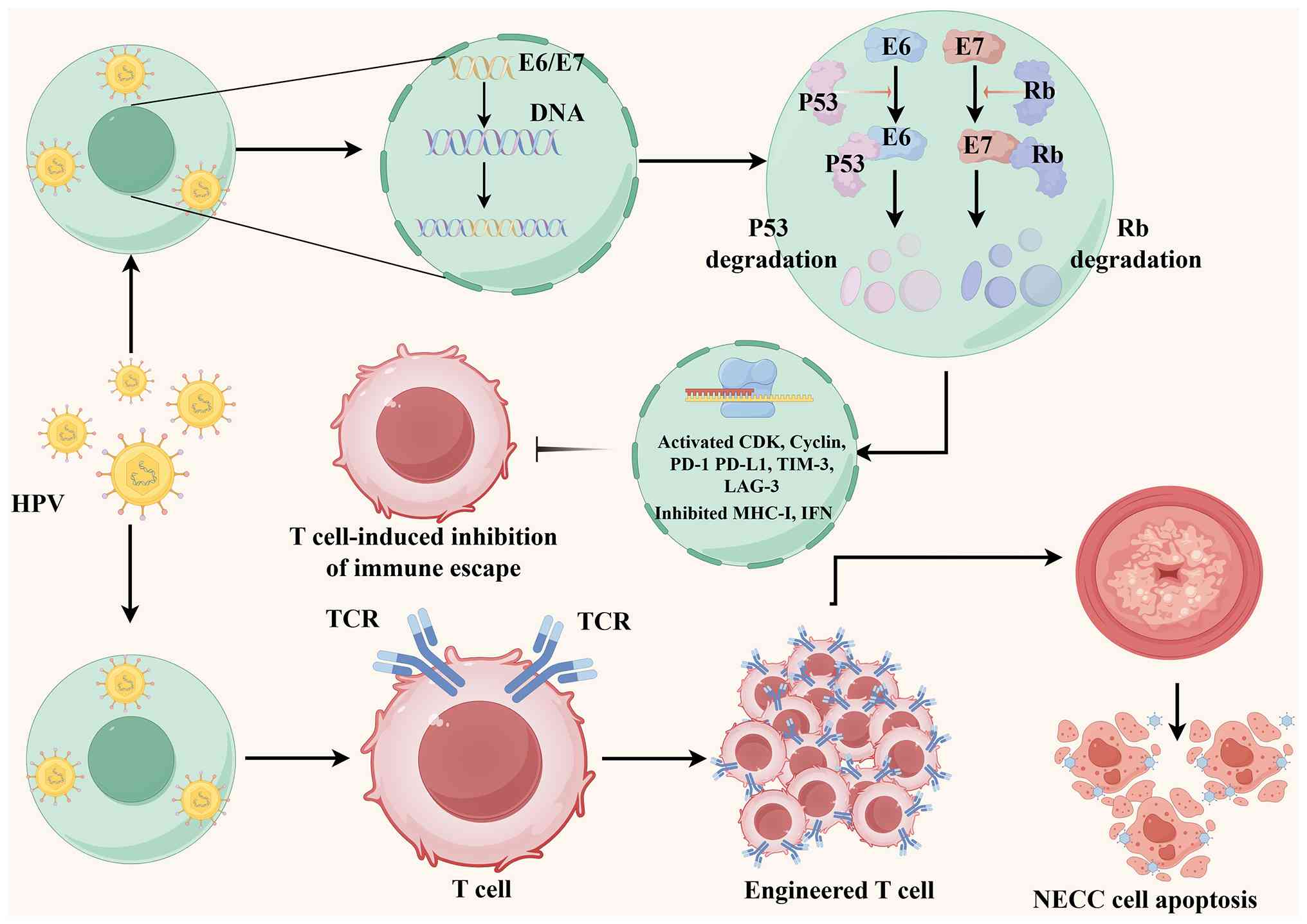
After integrating the viral genome into infected host cells, the
encoding proteins E6 and E7 isolate the cell ubiquitin proteasome
system, promoting rapid degradation of the tumor suppressor
proteins p53 and Rb1, respectively (64). In addition, the E6 protein leads
to cell immortalization by activating the telomerase catalytic
subunit (65). In addition to
binding to Rb, E7 promotes cell proliferation and reduces apoptosis
by upregulating the expression of phosphorylated-cdc2 and
activating cyclin-dependent kinase 2 (66). Furthermore, BRCA1 interacts with
the HPV oncoprotein and antagonizes the inhibition of c-Myc
transactivation by BRCA1 through the zinc finger domains of E6 and
E7, which may be involved in NENs (67). Targeted drugs for HPV oncogene
E6/E7 are constantly being developed (68).
In addition to its direct oncogenic effects,
persistent high-risk HPV infection may profoundly shape the immune
microenvironment of NECC. The viral oncoproteins E6 and E7 are
continuously expressed in HPV-associated cervical cancer and can
theoretically serve as tumor-specific antigens; however, HPV-driven
tumors often develop multiple mechanisms to evade immune
surveillance. HPV E6 and E7 have been reported to interfere with
antigen processing and presentation by downregulating components of
the major histocompatibility complex class I pathway, impairing
transporter associated with antigen processing activity, and
suppressing interferon-related antiviral signaling. These changes
may reduce the recognition of tumor cells by cytotoxic
CD8+ T lymphocytes and contribute to immune escape
during cervical carcinogenesis (69-72).
HPV-associated tumorigenesis may also alter T-cell
function and promote an immunosuppressive microenvironment.
Persistent viral antigen stimulation can induce T-cell exhaustion,
which is characterized by increased expression of inhibitory
receptors, such as PD-1, T cell immunoglobulin and mucin-domain
containing-3 (TIM-3) and lymphocyte activation gene-3 (LAG-3), and
reduced effector cytokine production (73). Furthermore, tumor cells and
tumor-associated immune cells may upregulate programmed death
ligand 1 (PD-L1), thereby suppressing antitumor T-cell activity
through the PD-1/PD-L1 axis (74). In NECC, PD-L1 expression has been
reported to possess considerable heterogeneity across studies, and
its expression has been observed in tumor cells and/or
tumor-infiltrating immune cells (75-77). This heterogeneity may partly
explain the inconsistent responses to ICIs in NECC and highlights
the need for a more comprehensive immune profiling strategy rather
than reliance on PD-L1 alone (78).
The immune microenvironment of NECC may also be
influenced by neuroendocrine lineage programs and HPV-related
molecular alterations. High-grade NECs frequently exhibit
abnormalities in TP53, RB1, PAM and Notch signaling, which may
interact with inflammatory and immune regulatory pathways (79-81). For example, activation of the PAM
pathway has been linked to PD-L1 regulation and immune resistance
in several types of cancer, suggesting that HPV-related oncogenic
signaling may indirectly contribute to immune suppression (82). Future studies should therefore
characterize NECC using multiplex immunohistochemistry, spatial
transcriptomics, single-cell sequencing, and integrated analyses of
HPV integration, antigen presentation machinery, T-cell
infiltration, immune checkpoint expression and myeloid cell
composition. Such approaches may help identify patients who are
more likely to benefit from immune checkpoint blockade, or
combinations of immunotherapy with chemotherapy, radiotherapy,
poly(ADP-ribose) polymerase inhibitors (PARPi), anti-angiogenic
agents or targeted therapy. Fig.
1 shows the mechanism of HPV infection, genetic alterations,
tumor immune microenvironment and therapeutic resistance in
NECC.
The angiogenic microenvironment in solid tumors
generates hypoxic regions, which can regulate the differentiation
of certain tumors and pluripotent stem cells (83,84). Kubota et al (85) established a cancer
tissue-originated spheroid cell line to study the effect of hypoxia
on the neuroendocrine differentiation of SCNECC. The results showed
that under hypoxic conditions, the dedifferentiation of SCNECC was
regulated by hypoxia inducible factor-1α (HIF-1α) and Notch
signaling (85), and the Notch
pathway has been shown to be activated under hypoxic conditions in
the aforementioned cancer tissue-originated spheroid cell line
(85,86). By contrast, knockdown of HIF-1α
and the Notch inhibitor DAPT have been shown to attenuate the
hypoxia-induced expression inhibition of the neuroendocrine markers
CgA, SYN and CD99 (membrane protein marker of SCNECC), thus
indicating that hypoxia might be one of the factors regulating the
differentiation status of NECC. In summary, these results suggest
that hypoxia may be an important factor in regulating
neuroendocrine differentiation in SCNECC; however, gene mutations
and activation of some signaling pathways may also serve important
roles in this process.
The Notch pathway is a highly conserved cellular
signaling pathway associated with malignant transformation, cell
proliferation, cell cycle arrest and apoptosis,
epithelial-mesenchymal transition and inhibition of neuroendocrine
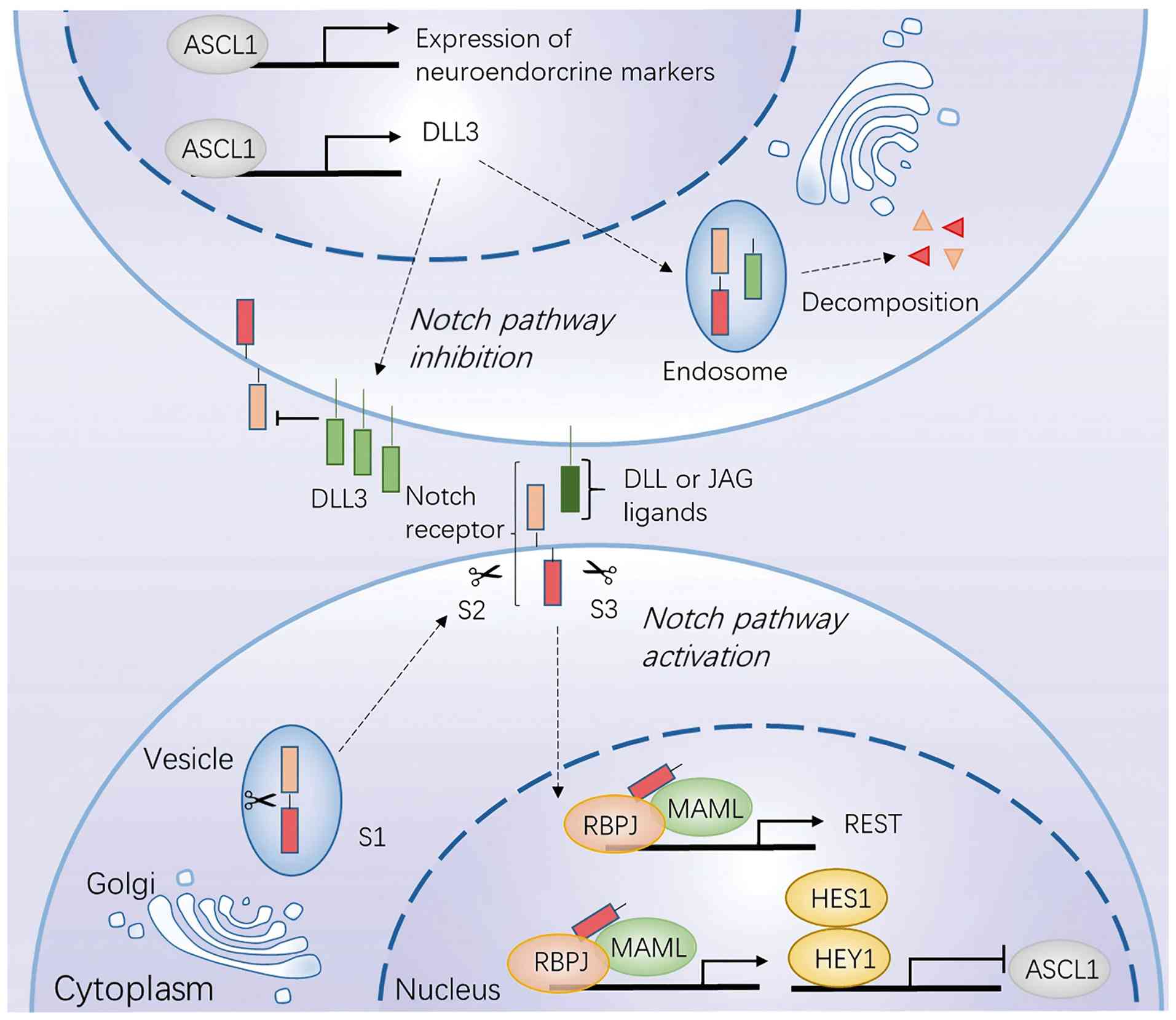
differentiation (87). DLL3 is
an atypical inhibitory ligand of Notch receptors involved in
NEC/NET development, which is located in the cytoplasm and can also
be expressed on the tumor cell membrane (88). A large NECC cohort study on
potential targeted biomarkers showed that high expression of DLL3
was observed in 81% of tumors (89). DLL3 regulates Notch signaling by
blocking the localization of Notch receptors on cell surfaces and
redirecting them to endosomes for degradation, thereby promoting
tumor development (90). The
combination of DLL3 inhibition and immunotherapy is considered a
therapeutic option for LCNECC (91). DLL3-negative NECCs tend to carry
PIK3CA and PTEN mutations, which are involved in the regulation of
the PAM signaling pathway (89).
A previous study revealed that a Notch1 mutation was present in
SCNEC without PIK3CA mutation, and that mutant Notch1 can activate
c-Myc and PAM signals through transcriptional inhibition of PTEN,
and promote growth factor receptor signal transduction to PI3K/AKT
(56). Fig. 2 shows the molecular mechanisms of
Notch signaling pathway in NETs.
Abnormal activation of the PAM pathway is common in
numerous types of cancer, leading to malignant growth and treatment
resistance. Thus, it is a promising therapeutic target for cancer
treatment in clinical studies (92,93). The crosstalk between this pathway
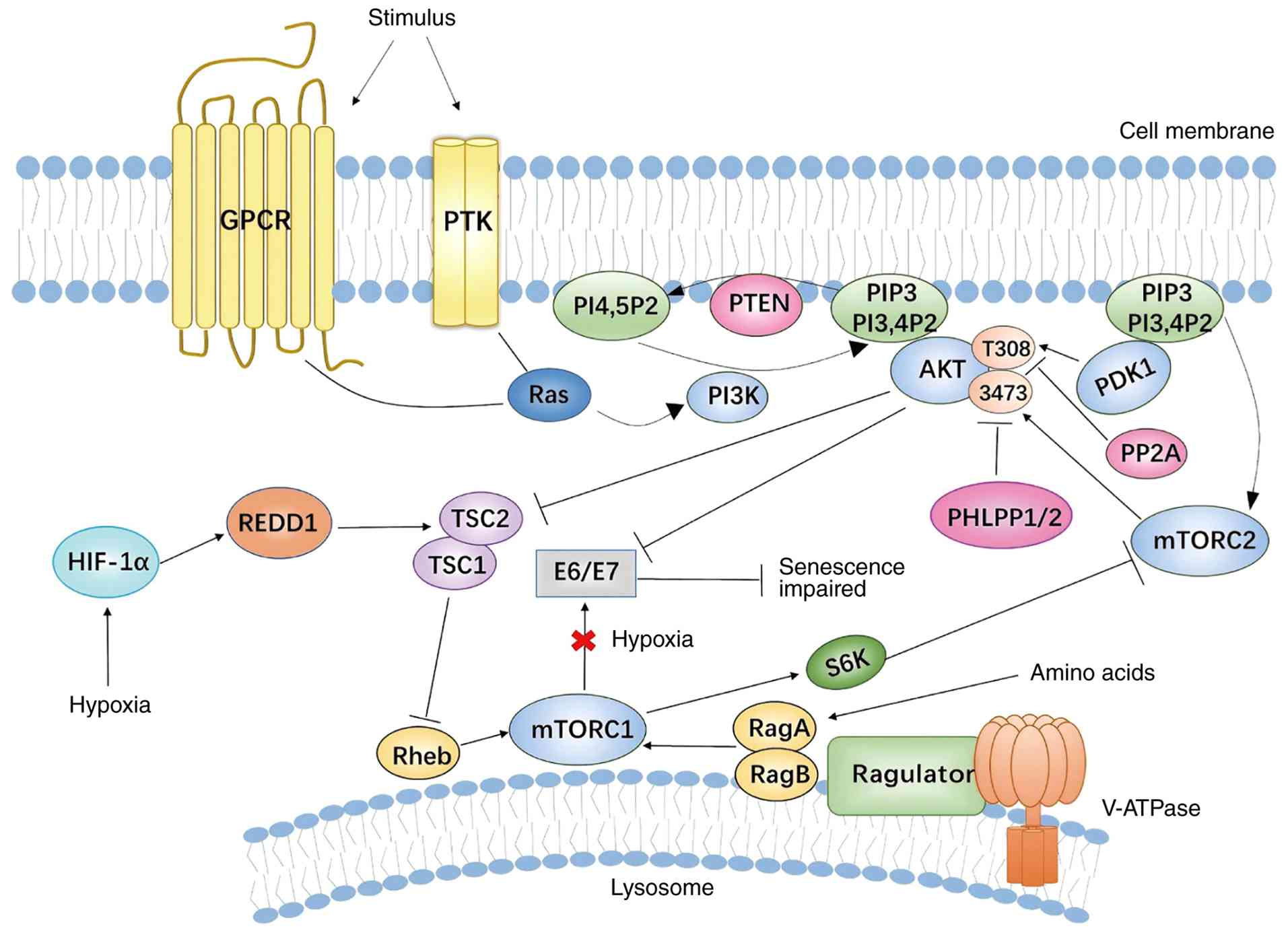
and HPV oncogenes depends on the cellular metabolism and
oxygenation state (94). Under
normoxic conditions, active mTOR complex 1 (mTORC1) signaling
inhibits E6/E7 expression to achieve cellular senescence. However,
when mTORC1 signaling is damaged by hypoxia, under the regulation
of the upstream regulator mTORC2 and active PI3K, AKT mediates
E6/E7 downregulation, causing HPV-positive cancer cells to escape
senescence (95). Extensive
research over the past few years has provided a better
understanding of the mechanism of this complex network (94,95). Fig. 3 shows the crosstalk between
carcinogenic HPV and the PI3K/mTOR/AKT signaling cascade. Studies
have revealed that PAM pathway inhibitors regulate tumor cells by
regulating immune cells and affecting the tumor microenvironment
(96,97). Targeting the PAM pathway can
reduce PD-L1 expression in tumor cells to enhance the antitumor
effect of immune checkpoint blockage (98). A previous study reported that the
KRAS mutation rate was 12% in NECC in human patients worldwide
(3). A case report showed that
one patient with a KRAS mutation (c.35G>Ap.G12D) received
treatment with the MAPK kinase (MEK) inhibitor trametinib and
achieved complete imaging remission after 3 cycles (99), thus indicating that patients with
KRAS mutations may benefit from MEK inhibitors. G12D mutations can
simultaneously activate the PI3K/AKT and MAPK pathways; however,
G12V KRAS mutations activate MEK signaling and result in KRAS
losing its ability to bind to PAM. Tumor cells with G12V KRAS
mutations may be more sensitive to MEK inhibitors (Fig. 3) (59).
NECs arising from different organs often share
aggressive biological behavior, high Ki-67 index, early metastatic
potential and dependence on immunohistochemical confirmation. Our
previous reports on gallbladder NEC and endometrial NEC further
support the diagnostic and therapeutic challenges of rare
extrapulmonary NECs, providing a useful reference for understanding
NECC (100,101).
Because NECC is rare, treatment strategies have
historically been extrapolated from SCLC, especially the use of
platinum-etoposide-based chemotherapy and, more recently, ICI
combinations. This extrapolation is biologically reasonable to some
extent. SCNECC and SCLC share several aggressive neuroendocrine
features, including poorly differentiated morphology, high
proliferative activity, a high Ki-67 index, expression of
neuroendocrine markers such as SYN, CgA, CD56 and INSM1, early
lymphovascular invasion and a propensity for distant metastasis
(33,102-106). These shared features partly
explain why platinum-etoposide regimens, which are active in SCLC,
have been adopted as the chemotherapy backbone for high-grade NECC
(107).
However, these similarities should not obscure
important disease-specific differences. Unlike SCLC, which is
strongly associated with tobacco exposure and is characterized by
near-universal inactivation of TP53 and RB1 (108), NECC is predominantly driven by
high-risk HPV infection, particularly HPV16 and HPV18 (81). HPV E6 and E7 oncoproteins mediate
functional inactivation of p53 and Rb and create a distinct viral
oncogenic background that is absent in SCLC (109). In addition, genomic studies
have suggested that NECC is molecularly closer to HPV-associated
cervical carcinoma than to pulmonary SCLC, with recurrent
alterations involving PIK3CA, Myc, PTEN, KRAS, ERBB2, and
components of the PAM and Notch pathways (79,81). By contrast, SCLC typically shows
a smoking-related mutational signature, very high genomic
instability and frequent concurrent TP53/RB1 loss; therefore, SCLC
should be considered a useful but imperfect therapeutic model for
NECC (108).
These biological distinctions have direct clinical
implications. Although platinum-etoposide remains a rational
first-line regimen for high-grade NECC, its benefit and optimal
combination partners should be validated in NECC-specific cohorts
(110). Similarly, the success
of adding PD-1/PD-L1 inhibitors to platinum-etoposide in
extensive-stage SCLC cannot be directly translated to NECC, because
NECC shows heterogeneous PD-L1 expression, generally preserved
mismatch repair (MMR) status, variable immune-cell infiltration and
an HPV-driven tumor microenvironment (111-113). Future studies should therefore
stratify patients according to NECC-specific biomarkers, including
HPV status, PD-L1/tumor-infiltrating lymphocytes (TILs),
microsatellite instability (MSI)/MMR status, PARP1 expression, DLL3
expression and actionable genomic alterations. Dedicated
multicenter trials or rare-tumor basket studies are needed to
determine which SCLC-derived regimens are truly applicable to NECC
and which strategies should be developed specifically for this
disease.
Given the invasiveness of NECC, current treatment
plans focus on multimodal therapy, combining the experience of
cervical ADC and SCC with SCLC, and performing platinum
chemotherapy, radical surgery and/or radiotherapy according to
staging (114). Owing to the
higher incidence of early lymph node metastasis and distant
metastasis in NECC, the prognosis of NECC is worse than that of
other types of cervical cancer, such as SCC (16). Late stage, lymph node metastasis,
deep interstitial infiltration, lack of chemotherapy and large
tumor volume are all associated with a poor prognosis (29). A study of 172 patients with NET
revealed that patients receiving radiotherapy had a better 5-year
overall survival (OS) rate and 5-year progression-free survival
(PFS) rate than those of patients not receiving radiotherapy, but
the difference was not statistically significant (29). In addition, Ruiz et al
(115) demonstrated that the
5-year OS of patients with stage I NECC treated primarily with
radiotherapy and chemotherapy was similar to that of patients
treated with surgical intervention. Hou et al (116) also reported that there was no
significant difference in 5-year OS between patients with
high-grade NECC undergoing radical surgery and those undergoing
initial radiotherapy without surgery. Surgery combined with
adjuvant radiotherapy and chemotherapy may be the best treatment
for patients with early NET. Research has shown that the
combination of external radiation therapy and close-range radiation
therapy improves the median survival of locally advanced NECC
(117). Research on the
treatment of distant metastatic NECC is limited, and radical pelvic
radiotherapy combined with chemotherapy may improve the survival
rate of patients with stage IVB NECC (118). NECC does not have a
standardized chemotherapy regimen; therefore, it is usually treated
in a similar manner to pulmonary NEC, which includes systemic
platinum chemotherapy or platinum combined with etoposide
chemotherapy. Effective treatment options for recurrent high-grade
NECC main unknown. Chemotherapy with topotecan, paclitaxel and
bevacizumab has been shown to improve NECC prognosis (119). Furthermore, combination therapy
with nivolumab and iprivumab may be a potential novel treatment
option for recurrent SCNECC (120).
Numerous genomic analyses have revealed a high
degree of similarity between NECC and non-neuroendocrine cervical
cancer (56,121,122). Identifying common genetic and
immune microenvironmental features in non-endocrine cervical cancer
may help in selecting targeted treatment options for rare and
difficult-to-treat NECC. Immunotherapy for cervical cancer is
considered to have future potential, especially after the approval
of pembrolizumab (anti-PD1) combined with chemotherapy as a
first-line treatment for metastatic and recurrent PD-L1-positive
cervical cancer (52). In
addition, immunotherapy and targeted therapy for NEC also
progressed, including PD-1/PD-L1 inhibitors and PARPi. Previous
case reports on metastatic NECC have described notable therapeutic
effects using the anti-PD1 drug nivolumab as monotherapy or in
combination with radiotherapy (123). As an anti-PD-1 antibody,
pembrolizumab has been approved by the United States Food and Drug
Administration for the treatment of recurrent cervical cancer with
PD-L1 expression [combined positive score (CPS) ≥1] (124). In a comprehensive analysis of
two prospective studies, pembrolizumab has been shown to be safe
for use in metastatic high-grade NENs, but its efficacy as a single
drug is limited (125).
Furthermore, a double-blind, placebo-controlled phase III trial
(n=403) targeting previously untreated patients with extensive SCLC
confirmed that adding the ICI atezolizumab (anti-PD-L1) to
carboplatin and etoposide chemotherapy markedly improved OS and PFS
compared with chemotherapy alone (126).
However, treatment methods for advanced NECC remain
unclear. In a study of 20 patients with NECC, 70% (n=14) had high
expression of PD-L1 (112), and
similar results were found in other cohorts of NECC (127). Another case report described a
patient with late-stage NECC with liver metastasis who achieved
sustained complete remission using neoadjuvant platinum therapy and
a multimodal approach of atezolizumab, surgical resection and
atezolizumab maintenance (128). Furthermore, patients with stage
IVB SCNECC and bone and liver metastases have achieved notable
outcomes with a combination of pembrolizumab and chemoradiation
(129). Another PD-1 inhibitor,
nivolumab, combined with radiotherapy has shown therapeutic
efficacy in patients with LCNECC (130). In addition, one patient with
metastatic SCNECC has achieved complete remission following
treatment with nivolumab and radiotherapy (131). Unfortunately, a phase II trial
using pembrolizumab monotherapy for recurrent SCNECC was
discontinued owing to unsatisfactory efficacy (132,133). Based on these experimental
results, it may be hypothesized that targeting PD-1/PD-L1 alone,
especially with ICI monotherapy, may not ensure sufficient control
of NECC after first-line treatment failure. Nonetheless, the
potential utility of ICIs in combination with radiotherapy and
chemotherapy deserves further study; for example, the data from the
KEYNOTE-604 study emphasized that adding pembrolizumab to etoposide
+ cisplatin as first-line treatment improved PFS (HR=0.75;
P=0.0023) and OS (HR=0.8; P=0.0164) in patients with extensive SCLC
(134). The PD-L1 blockers
atezolizumab and durvalumab have also been shown to be effective in
extensive-stage SCLC as first-line treatments (HR value for OS rate
for atezolizumab, 0.70; P=0.007, and HR value for OS rate for
durvalumab, 0.73; P=0.0047) (126,135). These findings suggest that
adding ICIs to the current chemotherapy regimens may enhance
therapeutic efficacy in SCNECC.
The response to ICIs in NECC remains heterogeneous,
and PD-L1 expression alone is unlikely to be sufficient for patient
selection. Reported PD-L1 positivity rates in NECC vary widely,
ranging from ~10 to 70% across different studies (77,89,112,127,136,137). This discrepancy may be
explained by several factors, including small cohort sizes,
differences in antibody clones and scoring systems, variable
cut-off values for PD-L1 positivity, evaluation of tumor cells vs.
tumor-infiltrating immune cells, and differences in sample source,
such as primary tumors, metastatic lesions, biopsies or resection
specimens. In addition, intratumoral heterogeneity and prior
chemotherapy or radiotherapy may alter PD-L1 expression and
immune-cell infiltration. Therefore, PD-L1 should be interpreted
together with the broader immune contexture rather than as an
isolated biomarker. In particular, tumors with low T-cell
infiltration, low antigen presentation activity or scarce
inflammatory cytokine signaling may represent an 'immune-cold'
phenotype, which could partly explain the limited activity of ICI
monotherapy observed in recurrent SCNECC (132).
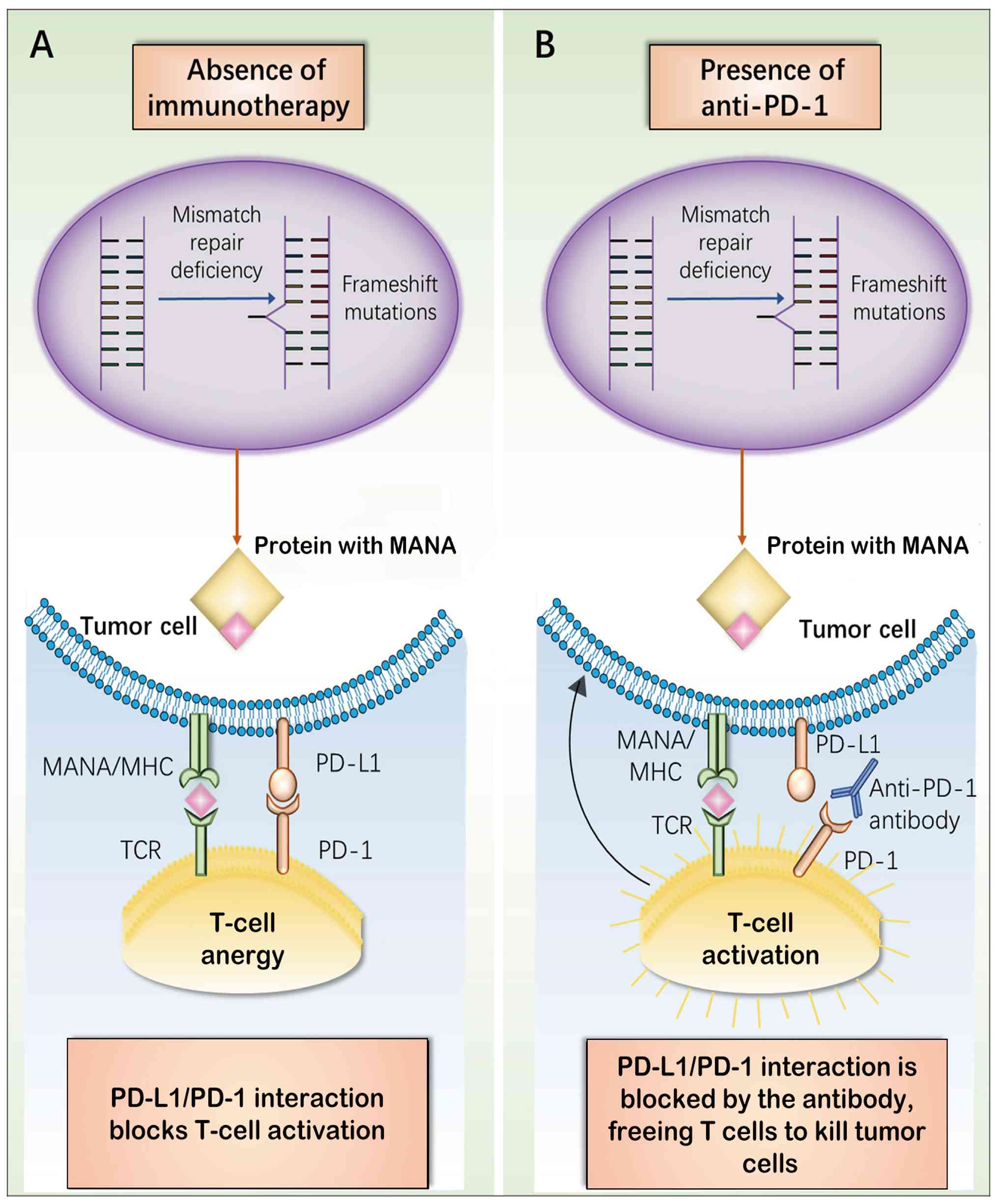
The DNA MMR system can correct errors that occur
during DNA replication, and its defects can lead to the
accumulation of coding and non-coding microsatellite mutations.
This phenotype is called MSI, and immunohistochemical detection of
MMR proteins (MLH1, MSH2, MSH6 and PMS2) can accurately predict MSI
tumors. In cancer cells, a MMR deficient (dMMR) state has been
shown to result in upregulation of PD-1/PD-L1 (138,139). Notably, PD-1 inhibitors have
been approved in clinical trials for the treatment of
advanced/recurrent MSI-high (MSI-H) solid tumors owing to the
efficacy of ICIs in the treatment of advanced solid tumors
(140,141). Therefore, MMR status is another
important factor in the selection of anti-PD-L1 drugs (142). Pagès et al (143) reported that, in colorectal
cancer, the infiltration rate of CD8+ cytotoxic T
lymphocytes (CTLs) and T helper 1 (Th1) cells could inhibit tumor
growth and metastasis. These results provide a new insight about
immune therapy for NECC. Tumors with high Th1/CTL infiltration
exhibit a dMMR state, leading to MSI (142,144). An increased mutational load in
MSI tumors can lead to the production of new antigens, thereby
increasing the immunogenicity of tumor cells. Fig. 4 shows the relationship between
MSI status and immune response. In a large cohort study, NTRK
protein expression was observed in 21% of cases, but none exhibited
an NTRK gene fusion (89). In
addition, Chen et al (136) evaluated the expression of
PD-L1, proteins associated with MMR and NTRK fusion using
immunohistochemical techniques, and revealed that all SCNECCs in
the cohort were stable in MMR and exhibited NTRK fusion deficiency.
In another study, 5% (1/22) of patients with SCNECC and 50% of
patients with mixed NEC showed positive PD-L1 expression in tumor
samples, and a stable MMR status was detected in 28 cases of SCNECC
(137). However, the clinical
value of the immune microenvironment in NECC has not yet been fully
studied. Notably, 68.5% of patients with SCNECC exhibit PD-L1 CPS
positivity, and PD-L1 CPS is markedly positively associated with
tumor-associated immune cell levels, possibly involving the
regulation of the tumor inflammatory microenvironment, which
provides potential biomarkers for SCNECC immunotherapy (77). Notably, in a patient cohort,
Carroll et al (137)
reported that all patients with high-grade NECC presented with
microsatellite stability status, the expression of PD-L1 was
negative in the vast majority of specimens and PARP1 expression was
detected in most of the tested tumors. PARP1 has been shown to
serve an important role in DNA repair and methylation, as well as
in transcription processes. A previous study showed that PARP1 is
highly expressed at the mRNA and protein levels in SCLC, and SCLC
is sensitive to PARPi, thereby enhancing the efficacy of
chemotherapy (145). Therefore,
the inclusion of PARPi may be considered in clinical trials of
high-grade NECC (137). A
recent case study analyzed the expression of PD-L1 and PARP1 in
NECC and revealed that the positive expression rates of both
markers were high, and that their expression statuses were
consistent with each other in the MSI subgroup (P=0.004),
indicating that MSI patients with PD-L1 and PARP1 co-expression may
benefit from a combination of immune checkpoint therapy and PARPi
targeted therapy (112). Sen
et al (146) reported
that PARP inhibition can enhance antitumor immunity induced by
PD-L1 inhibitors in SCLC, and the corresponding mouse models also
confirmed this result. In these models, the combination of PD-L1
and PARPi showed significantly higher efficacy than a single drug.
This indicates that PARP inhibition may induce the expression of
PD-L1, and PD-1/PD-L1 inhibitors in combination with PARPi may be a
promising therapeutic option. The University of Kentucky has
developed three small-molecule mixed inhibitors of PARP and PD-L1
that have more pronounced apoptotic and cytotoxic effects than
single drugs (147). Further
studies have found that PARPi cause upregulation of PD-L1 through
GSK3β inactivation, but the specific mechanism remains to be
explored (148).
Mutations in the TP53 gene can lead to loss or
upregulation of protein expression, which is closely related to
tumor prognosis. The high mutation rate of TP53 in NECC indicates
that the TP53-related pathway has important research value for
targeted therapy. Abnormal expression of p53 is closely related to
the expression of PD-L1, and this should be taken into account in
clinical studies concerning anti-PD-1/PD-L1 immune checkpoint
blockade therapy. Research has indicated that p53 reactivation can
promote innate and adaptive immunity through various molecular
pathways and increase the immunogenicity of tumor cells, providing
a theoretical basis for targeted p53 drug combination immunotherapy
(149). Furthermore,
anti-angiogenic drugs have been widely used in the clinical
treatment of cancer by single drug or combined drug regimens to
inhibit tumor neovascularization, reshape the tumor
microenvironment, modulate tumor-induced immunosuppression, and
thereby enhance the efficacy of radiotherapy, chemotherapy,
immunotherapy and targeted therapy (150-152). The efficacy of anti-angiogenic
drugs in cervical NENs requires further investigation.
From a translational perspective, these biomarkers
should be integrated into a clinically meaningful decision
framework. PD-L1 positivity, especially when accompanied by
abundant TILs, may support the use of ICIs, preferably in
combination with chemotherapy, radiotherapy or anti-angiogenic
therapy, rather than as monotherapy. MSI-H/dMMR status, although
uncommon in SCNECC according to available studies, should still be
assessed because it represents a tumor-agnostic indication for PD-1
blockade (136-138,153). PARP1 expression or other DNA
damage repair-related alterations may identify patients who could
benefit from PARPi, particularly in combination with platinum
chemotherapy or immune checkpoint blockade (112,137,146). By contrast, NTRK fusion appears
to be rare or absent in reported SCNECC cohorts (136); nevertheless, NTRK testing
remains clinically relevant because patients with confirmed NTRK
fusions may benefit from TRK inhibitors. Thus, a practical
biomarker-driven strategy for NECC should include combined
assessment of PD-L1/TILs, MSI/MMR status, DNA damage repair markers
such as PARP1, and actionable gene fusions such as NTRK, thereby
linking molecular features to rational treatment selection.
The current understanding of the pathogenesis of
NECC is limited and low-quality evidence hinders the development of
clinical management. Radical surgery and combined or non-combined
radiotherapy with cisplatin and etoposide are the main treatment
methods for early stage disease, whereas chemotherapy with
cisplatin and etoposide or topotecan, paclitaxel and bevacizumab is
suitable for women with locally advanced or recurrent NECC. In the
future, multi-omics integration may serve a central role in
refining the biological classification and individualized treatment
of NECC. With the increasing number of genomes in The Cancer Genome
Atlas database, genetic testing provides broad prospects for
personalized treatment of cervical NENs with targeted drugs.
Further research is needed to investigate the molecular, biological
and therapeutic associations between cervical NETs and
extracervical NETs arising from other organs (for example, lungs,
gastrointestinal tract and pancreas). In-depth exploration of the
underlying molecular abnormalities and signal transduction,
including genetic and epigenetic factors, is key to improving
therapeutic efficacy.
Current histopathological classification mainly
relies on morphology, neuroendocrine marker expression, mitotic
activity and Ki-67 index; however, these parameters are
insufficient to fully capture the molecular heterogeneity of NECC.
Genomic studies have demonstrated that high-grade NECC harbors
recurrent alterations in several cancer-related genes and pathways,
including PIK3CA, TP53, Myc, PTEN, RB1, KRAS, ARID1A, and
components of the PAM and Notch signaling pathways (57,60,79,102,109,154,155). Eskander et al (53) reported a unique genomic landscape
of high-grade NECC and highlighted frequent alterations in PIK3CA,
Myc, TP53 and PTEN, suggesting that genomic profiling may help
identify therapeutically actionable subgroups. Similarly, Wang
et al (156) performed
whole-exome sequencing of cervical and endometrial SCNECs and
identified shared mutational features, supporting the value of
comparative genomic analysis in defining NEC across gynecological
sites.
Beyond DNA-level alterations, transcriptomic,
epigenomic, proteomic and immune microenvironmental profiling may
further improve the precision of NECC stratification. For example,
expression of neuroendocrine lineage regulators such as ASCL1,
NEUROD1, DLL3 and Notch pathway-related molecules may help
distinguish biologically distinct NECC subsets (75,85,88,157). In addition, immune biomarkers,
including PD-L1 expression, TILs, MSI/MMR status, tumor mutational
burden and DNA damage repair-related markers such as PARP1, may
contribute to predicting sensitivity to ICIs or DNA damage
response-targeted therapy. Previous studies have reported
heterogeneous PD-L1 expression, largely preserved MMR status and
frequent PARP1 expression in NECC, indicating that single
biomarkers may be insufficient for treatment selection (113,158). Therefore, a multi-dimensional
model integrating histology, HPV status, somatic mutations,
transcriptional programs, epigenetic changes, immune contexture and
druggable surface antigens may provide a more reliable framework
for precision diagnosis and risk stratification.
For clinical translation, future studies should
incorporate next-generation sequencing, RNA sequencing, methylation
profiling, multiplex immunohistochemistry, spatial transcriptomics
and single-cell sequencing into prospective NECC cohorts. Such
approaches may help clarify tumor origin, distinguish
well-differentiated NETs from poorly differentiated NECs, identify
mechanisms of treatment resistance and guide rational combination
strategies. Given the rarity of NECC, international multi-center
collaboration and shared molecular databases are essential for
validating molecular subtypes and establishing biomarker-driven
clinical trials.
Notably, antibody-drug conjugates (ADCs) represent
another promising direction for the treatment of recurrent or
metastatic NECC. ADCs combine the target specificity of monoclonal
antibodies with the cytotoxic potency of chemotherapy payloads,
allowing selective delivery of highly active agents to tumor cells
expressing specific surface antigens (159). This strategy may be
particularly attractive for NECC because conventional chemotherapy
has limited durability, while several potentially targetable
antigens, including DLL3, HER2, TROP-2, tissue factor and SSTRs
have been explored in neuroendocrine or cervical malignancies
(41,89,104,160-162).
DLL3 is one of the most biologically relevant
targets in high-grade NEC. Other ADC targets may also be relevant
to NECC. The future development of ADCs in NECC should be
biomarker-driven rather than empiric. Routine assessment of DLL3,
HER2, TROP-2 and tissue factor by immunohistochemistry or molecular
assays may help identify patients suitable for ADC-based therapy.
Moreover, ADCs may be combined with ICIs, PARPi, anti-angiogenic
agents or radiotherapy to enhance antitumor activity, especially in
tumors with DNA damage repair deficiency, high antigen expression
or immune-active microenvironments. However, because direct
clinical evidence in NECC remains limited, prospective basket
trials, rare tumor registries and translational studies using
patient-derived organoids or xenograft models are urgently needed.
Overall, ADCs provide a rational and potentially effective
precision treatment strategy for NECC, but their success will
depend on accurate target selection, toxicity management and
collaborative clinical trial design.
Various clinical trials are currently underway to
expand the existing options for monotherapy and combination
therapies. In a phase II clinical trial (NCT05910177) on
neoadjuvant therapy for cervical NEN, the application of
camrelizumab in combination with etoposide and cisplatin was
investigated (163). In
addition, the safety and tolerability of AK104 as a new
chemotherapy drug for advanced/recurrent high-grade NECC are
currently being studied (phase II-NCT05063916) (164). Camrelizumab (an anti-PD-1
antibody) has antitumor activity in NEN, and its efficacy combined
with cisplatin/paclitaxel/bevacizumab in the treatment of recurrent
or advanced NECC is currently being evaluated (phase
II-NCT04635956) (165). In an
ongoing phase II clinical trial, the role of XmAb20717 in advanced
rare cancers, such as NEC, was evaluated to explore its prognostic
biomarkers (NCT05337735) (166). Upregulation of SSTR2A has been
detected in NECC (167), which
is an important target for nuclear medicine molecular imaging
diagnosis and peptide receptor-mediated radionuclide therapy
(168). The efficacy of
177Lu-DOTA-TATE in treating patients with SSTR-positive
NET (phase II-NCT01876771) (169), and the combination of
177Lu and nivolumab for the treatment of grade 3
well-differentiated NETs or poorly differentiated NEC is currently
being studied (phase II-NCT04525638) (170). MSI-H/dMMR tumors express a
large number of new antigens due to high mutation, forming a
microenvironment of immune-cell infiltration and upregulated
expression of immune checkpoint proteins in tumor cells (153). The application of pembrolizumab
and nivolumab in MSI-H/dMMR tumors has shown the potential of ICIs
and marks the progress of precision medicine (171). Surufatinib is a potent small
molecule tyrosine kinase inhibitor that selectively targets VEGF
receptors 1, 2 and 3, fibroblast growth factor receptor 1 and
colony stimulating factor 1 receptor, and NENs are highly
vascularized tumors (172).
Surufatinib may therefore have potential in treating NENs. In a
dose escalation/expansion study, the 4-month and 11-month PFS rates
in an extrapancreatic NET cohort treated with surufatinib were
93.8% (95% CI: 63.2, 99.1) and 51.1% (95% CI: 12.8, 80.3),
respectively, supporting the antitumor efficacy of surufatinib for
extrapancreatic NETs (173).
Serplulimab is a novel anti-PD-1 antibody; serplulimab and
surufatinib combined with standard chemotherapy
(platinum/etoposide) are being studied to determine whether they
can improve the efficacy in patients with NEN (phase II:
NCT05747729) (174).
Furthermore, new drugs are being tested in advanced
or metastatic extra-pulmonary NECs. NP-101 has antioxidant and
anti-angiogenic effects; combined with immunotherapy drugs, such as
nivolumab and ipilimumab, it may enhance efficacy in advanced
neuroendocrine cancer (NCT05262556) (175). Histone deacetylase (HDAC)
serves an important role in tumor development by modifying the
structure of chromosomes and regulating gene expression, and the
anticancer effect of HDAC inhibitors is notable (176). Among them, chidamide is being
studied as a single treatment drug or in combination with
cyclooxygenase (COX)-1/COX-2 inhibitors and ICIs for a variety of
solid malignant tumors, which can slow tumor progression by
altering the tumor immune microenvironment (177,178). In addition, the efficacy and
safety of chidamide combined with etoposide and
cisplatin/carboplatin in the treatment of advanced extrapulmonary
NEC are being studied (NCT05076786) (179). The results of these clinical
trials are awaited, and by evaluating various combinations of
treatments for better tumor control, it is hoped that in the near
future, effective management models can be developed to improve
patient with NECs prognosis and quality of life (Table V).
The metastatic potential, morphological
characteristics and manifestations of endocrine paraneoplastic
syndrome, such as carcinoid syndrome and syndrome of inappropriate
antidiuretic hormone secretion, in cervical NENs provide evidence
for early diagnosis (180). Our
latest understanding of the histological, pathological and genetic
factors of these tumors may help to provide better diagnostic
markers and treatment options, thereby improving prognosis. Various
clinical trials are being extensively conducted with the aim of
expanding the selection of single and combination therapies for
NENs. Although ICIs may be beneficial, there is a lack of effective
strategies to predict response, manage immune-related adverse
events or select suitable patients for these therapies.
Improvements in molecular analysis and further mechanistic research
are required to determine which patients will benefit from
immunotherapy, targeted monotherapy or combination therapy, and to
customize personalized treatment plans according to different
situations, which will markedly improve the treatment effectiveness
of patients. Further research is needed to evaluate the potential
of MSI status in guiding immunotherapy across tumor types. This
represents a notable advancement in precision medicine. Targeted
therapies, such as somatostatin analogs, PAM inhibitors and
anti-angiogenic drugs, have been used in extra-cervical NEN therapy
(181,182). However, to the best of our
knowledge, there are currently no data available on their
application in cervical NENs, which urgently requires strong
cooperation between gynecological oncologists and relevant
researchers to ensure progress in the treatment of these invasive
diseases.
Not applicable.
YT, XH and RG conceptualized the study. YT, YZ and
XX wrote the first manuscript. ZL, LW, SL, SY and XR participated
in writing the manuscript, and generated the figures and tables.
MX, XC and QZ edited, reviewed and supervised the manuscript. YT,
QZ and RG provided the funding. Data authentication is not
applicable. All authors read and approved the final manuscript.
Not applicable.
Not applicable.
The authors declare that they have no competing
interests.
Not applicable.
The present study was supported by the Henan Medical Science and
Technology Research Program (grant no. LHGJ20220359), the Key
Scientific Research Project of Higher Education Institutions in
Henan Province (grant no. 24A320080), the Henan Medical Science and
Technology Research Project (grant no. 242102311038), the National
Natural Science Foun dation of China (grant no. 82273229) and the
Henan Provincial Key Medical Discipline-Gynecologic and Obstetrics
Surgery.
|
1
|
Gadducci A, Carinelli S and Aletti G:
Neuroendrocrine tumors of the uterine cervix: A therapeutic
challenge for gynecologic oncologists. Gynecol Oncol. 144:637–646.
2017.
|
|
2
|
Dasari A, Shen C, Halperin D, Zhao B, Zhou
S, Xu Y, Shih T and Yao JC: Trends in the incidence, prevalence,
and survival outcomes in patients with neuroendocrine tumors in the
United States. JAMA Oncol. 3:1335–1342. 2017.
|
|
3
|
Tempfer CB, Tischoff I, Dogan A, Hilal Z,
Schultheis B, Kern P and Rezniczek GA: Neuroendocrine carcinoma of
the cervix: A systematic review of the literature. BMC Cancer.
18:5302018.
|
|
4
|
Castle PE, Pierz A and Stoler MH: A
systematic review and meta-analysis on the attribution of human
papillomavirus (HPV) in neuroendocrine cancers of the cervix.
Gynecol Oncol. 148:422–429. 2018.
|
|
5
|
Lu J, Li Y and Wang J: Small cell
(neuroendocrine) carcinoma of the cervix: An analysis for 19 cases
and literature review. Front Cell Infect Microbiol.
12:9165062022.
|
|
6
|
Kurman RJ, Carcangiu ML, Herrington CS and
Young RH: WHO classification of tumours of female reproductive
organs. WHO Classification of Tumours. 6. 4th edition. IARC
Publications; 2014
|
|
7
|
Cree IA, White VA, Indave BI and Lokuhetty
D: Revising the WHO classification: Female genital tract tumours.
Histopathology. 76:151–156. 2020.
|
|
8
|
Albores-Saavedra J, Gersell D, Gilks CB,
Henson DE, Lindberg G, Santiago H, Scully RE, Silva E, Sobin LH,
Tavassoli FJ, et al: Terminology of endocrine tumors of the uterine
cervix: Results of a workshop sponsored by the college of american
pathologists and the national cancer institute. Arch Pathol Lab
Med. 121:34–39. 1997.
|
|
9
|
Chan JK, Tsui WM, Tung SY and Ching RC:
Endocrine cell hyperplasia of the uterine cervix. A precursor of
neuroendocrine carcinoma of the cervix? Am J Clin Pathol.
92:825–830. 1989.
|
|
10
|
Lojek MA, Fer MF, Kasselberg AG, Glick AD,
Burnett LS, Julian CG, Greco FA and Oldham RK: Cushing's syndrome
with small cell carcinoma of the uterine cervix. Am J Med.
69:140–144. 1980.
|
|
11
|
Ishibashi-Ueda H, Imakita M, Yutani C,
Ohmichi M, Chiba Y, Kubo T and Waki M: Small cell carcinoma of the
uterine cervix with syndrome of inappropriate antidiuretic hormone
secretion. Mod Pathol. 9:397–400. 1996.
|
|
12
|
Koch CA, Azumi N, Furlong MA, Jha RC,
Kehoe TE, Trowbridge CH, O'Dorisio TM, Chrousos GP and Clement SC:
Carcinoid syndrome caused by an atypical carcinoid of the uterine
cervix. J Clin Endocrinol Metab. 84:4209–4213. 1999.
|
|
13
|
Boruta DM II, Schorge JO, Duska LA, Crum
CP, Castrillon DH and Sheets EE: Multimodality therapy in
early-stage neuroendocrine carcinoma of the uterine cervix. Gynecol
Oncol. 81:82–87. 2001.
|
|
14
|
Chen J, Macdonald OK and Gaffney DK:
Incidence, mortality, and prognostic factors of small cell
carcinoma of the cervix. Obstet Gynecol. 111:1394–1402. 2008.
|
|
15
|
Georgescu TA, Bohiltea RE, Munteanu O,
Furtunescu F, Lisievici AC, Grigoriu C, Gherghiceanu F, Vlădăreanu
EM, Berceanu C, Ducu I and Iordache AM: Emerging therapeutic
concepts and latest diagnostic advancements regarding
neuroendocrine tumors of the gynecologic tract. Medicina (Kaunas).
57:13382021.
|
|
16
|
Zhang Y, Li L, Wang Z, Huang Y, Luo S,
Peng Y, Li N, He Y, Li C, Zhang K, et al: Preferred method of
therapy for patients with early-stage high-grade neuroendocrine
carcinoma of the cervix. Am J Cancer Res. 11:4595–4606. 2021.
|
|
17
|
Margolis B, Tergas AI, Chen L, Hou JY,
Burke WM, Hu JC, Ananth CV, Neugut AI, Hershman DL and Wright JD:
Natural history and outcome of neuroendocrine carcinoma of the
cervix. Gynecol Oncol. 141:247–254. 2016.
|
|
18
|
Akdag G, Alan Ö, Dogan A, Yildirim S,
Kinikoglu O, Batu A, Kudu E, Geçmen GG, Isik D, Sever ON, et al:
Prognostic scores in pulmonary large cell neuroendocrine carcinoma:
A retrospective cohort study. Heliyon. 10:e250292024.
|
|
19
|
Apostolidis L, Bergmann F, Jäger D and
Winkler EC: Efficacy of topotecan in pretreated metastatic poorly
differentiated extrapulmonary neuroendocrine carcinoma. Cancer Med.
5:2261–2267. 2016.
|
|
20
|
Abdel-Rahman O and Fouad M:
Bevacizumab-based combination therapy for advanced
gastroenteropancreatic neuroendocrine neoplasms (gep-nens): A
systematic review of the literature. J Cancer Res Clin Oncol.
141:295–305. 2015.
|
|
21
|
Takayanagi D, Hirose S, Kuno I, Asami Y,
Murakami N, Matsuda M, Shimada Y, Sunami K, Komatsu M, Hamamoto R,
et al: Comparative analysis of genetic alterations, HPV-status, and
PD-L1 expression in neuroendocrine carcinomas of the cervix.
Cancers (Basel). 13:12152021.
|
|
22
|
Liu R, He X and Li Z: Positive clinical
outcomes following therapy with programmed cell death protein
1/programmed cell death ligand 1 inhibitors in neuroendocrine
carcinoma of the cervix. Front Pharmacol. 13:10295982022.
|
|
23
|
Onofrei Popa A, Gurau G, Patrichi G, Gurau
AM, Mehedinti RC and Satala CB: Decoding cervical neuroendocrine
carcinoma: A practical review of diagnostic pitfalls, differential
diagnosis and molecular insights. Diagnostics (Basel).
16:17812026.
|
|
24
|
Tsang M, Zhou R, Kang Y, Memarzadeh S,
Ostrzega N and Moatamed N: Assessment of delta-like ligand 3 (DLL3)
immunostaining in neuroendocrine tumors of the gynecological tract
and comparing expression with other high-grade gynecological
malignancies. Ann Diagn Pathol. 84:1526642026.
|
|
25
|
Albores-Saavedra J, Rodríguez-Martínez HA
and Larraza-Hernández O: Carcinoid tumors of the cervix. Pathol
Annu. 14(Pt 1): 273–291. 1979.
|
|
26
|
Rindi G, Mete O, Uccella S, Basturk O, La
Rosa S, Brosens LAA, Ezzat S, de Herder WW, Klimstra DS, Papotti M
and Asa SL: Overview of the 2022 WHO classification of
neuroendocrine neoplasms. Endocr Pathol. 33:115–154. 2022.
|
|
27
|
Mete O and Wenig BM: Update from the 5th
edition of the World health organization classification of head and
neck tumors: Overview of the 2022 WHO classification of head and
neck neuroendocrine neoplasms. Head Neck Pathol. 16:123–142.
2022.
|
|
28
|
Mccluggage WG, Singh N and Gilks CB: Key
changes to the World health organization (WHO) classification of
female genital tumours introduced in the 5th edition (2020).
Histopathology. 80:762–778. 2022.
|
|
29
|
Chen J, Sun Y, Chen L, Zang L, Lin C, Lu
Y, Lin L, Lin A, Dan H, Chen Y and He H: Prognostic factors and
treatment of neuroendocrine tumors of the uterine cervix based on
the FIGO 2018 staging system: A single-institution study of 172
patients. PeerJ. 9:e115632021.
|
|
30
|
Ling C and Shen Y: Atypical carcinoid of
the uterine cervix accompanying adenocarcinoma in situ. J Clin
Pathol. 71:10302018.
|
|
31
|
Virarkar M, Vulasala SS, Morani AC, Waters
R, Gopireddy DR, Kumar S, Bhosale P and Lall C: Neuroendocrine
neoplasms of the gynecologic tract. Cancers (Basel).
14:18352022.
|
|
32
|
Papatsimpas G, Samaras I, Theodosiou P,
Papacharalampous K, Maragkouli E, Papadopoulos NV, Tsapakidis K,
Litos I, Sogka E, Kostopoulou E and Koukoulis GK: A case of
cervical carcinoid and review of the literature. Case Rep Oncol.
10:737–742. 2017.
|
|
33
|
Huang R, Yu L, Zheng C, Liang Q, Suye S,
Yang X, Yin H, Ren Z, Shi L, Zhang Z, et al: Diagnostic value of
four neuroendocrine markers in small cell neuroendocrine carcinomas
of the cervix: A meta-analysis. Sci Rep. 10:149752020.
|
|
34
|
Gujarathi R, Abou Azar S, Tobias J, Polite
BN, Setia N, Feinberg N, Appelbaum DE, Keutgen XM and Liao CY:
Men1/daxx/atrx mutations enhance progression-free survival in
gastroenteropancreatic neuroendocrine tumors treated with peptide
receptor radionuclide therapy. Endocr Relat Cancer.
31:e2400652024.
|
|
35
|
Rindi G, Klimstra DS, Abedi-Ardekani B,
Asa SL, Bosman FT, Brambilla E, Busam KJ, de Krijger RR, Dietel M,
El-Naggar AK, et al: A common classification framework for
neuroendocrine neoplasms: An International agency for research on
cancer (IARC) and World health organization (WHO) expert consensus
proposal. Mod Pathol. 31:1770–1786. 2018.
|
|
36
|
Lu Z and Chen J: Introduction of WHO
classification of tumours of female reproductive organs, fourth
edition. Zhonghua Bing Li Xue Za Zhi. 43:649–650. 2014.In
Chinese.
|
|
37
|
Albores-Saavedra J, Latif S, Carrick KS,
Alvarado-Cabrero I and Fowler MR: Cd56 reactivity in small cell
carcinoma of the uterine cervix. Int J Gynecol Pathol. 24:113–117.
2005.
|
|
38
|
McCluggage WG, Kennedy K and Busam KJ: An
immunohistochemical study of cervical neuroendocrine carcinomas:
Neoplasms that are commonly TTF1 positive and which may express
CK20 and P63. Am J Surg Pathol. 34:525–532. 2010.
|
|
39
|
Ishida GM, Kato N, Hayasaka T, Saito M,
Kobayashi H, Katayama Y, Sasou S, Yaegashi N, Kurachi H and
Motoyama T: Small cell neuroendocrine carcinomas of the uterine
cervix: A histological, immunohistochemical, and molecular genetic
study. Int J Gynecol Pathol. 23:366–372. 2004.
|
|
40
|
Kuji S, Endo A, Kubota M, Uekawa A,
Kawakami F, Mikami Y, Koike J and Suzuki N: Immunosensitivity and
specificity of insulinoma-associated protein 1 (INSM1) for
neuroendocrine neoplasms of the uterine cervix. J Gynecol Oncol.
34:e12023.
|
|
41
|
Inzani F, Santoro A, Angelico G, Feraco A,
Spadola S, Arciuolo D, Valente M, Carlino A, Piermattei A,
Scaglione G, et al: Neuroendocrine carcinoma of the uterine cervix:
A clinicopathologic and immunohistochemical study with focus on
novel markers (Sst2-Sst5). Cancers (Basel). 12:12112020.
|
|
42
|
Habeeb A and Habeeb H: Large cell
neuroendocrine carcinoma of the uterine cervix. BMJ Case Rep.
12:bcr-2018-2258802019.
|
|
43
|
Pang L, Yang H, Ning Y and Zheng C:
Retrospective analysis of clinicopathological features and
prognosis of gynecological small-cell carcinoma. Cancer Manag Res.
13:4529–4540. 2021.
|
|
44
|
Sah S, Borkar PV, Wight C, Kelly P, Park
KJ and Mccluggage WG: Low-grade neuroendocrine tumor of the cervix:
Report of 3 cases of a rare neoplasm with review of the literature.
Int J Gynecol Pathol. 41:437–446. 2022.
|
|
45
|
Ortiz WJ, Gutierrez MA, Mabrie H and
Cervantes M: Neuroendocrine carcinoma of the uterine cervix with
metastases to the thyroid gland: A case report and clinical
pathological review. Cureus. 14:e295642022.
|
|
46
|
Lamiman K, Wilhelm AB, Eyzaguirre E and
Richardson G: Diagnostic challenges and long-term outcomes of
neuroendocrine carcinoma of the cervix: A case series. Int J
Gynecol Pathol. 43:149–157. 2024.
|
|
47
|
Subbiah V, Baik C and Kirkwood JM:
Clinical development of BRAF plus MEK inhibitor combinations.
Trends Cancer. 6:797–810. 2020.
|
|
48
|
Hong DS, Fakih MG, Strickler JH, Desai J,
Durm GA, Shapiro GI, Falchook GS, Price TJ, Sacher A, Denlinger CS,
et al: KRAS(G12C) inhibition with sotorasib in advanced solid
tumors. N Engl J Med. 383:1207–1217. 2020.
|
|
49
|
Martins D, Spada F, Lambrescu I, Rubino M,
Cella C, Gibelli B, Grana C, Ribero D, Bertani E, Ravizza D, et al:
Predictive markers of response to everolimus and sunitinib in
neuroendocrine tumors. Target Oncol. 12:611–622. 2017.
|
|
50
|
Guadagno E, De Rosa G and Del Basso De
Caro M: Neuroendocrine tumours in rare sites: Differences in
nomenclature and diagnostics-a rare and ubiquitous histotype. J
Clin Pathol. 69:563–574. 2016.
|
|
51
|
Rashed MM and Bekele A: Neuroendocrine
differentiation in a case of cervical cancer. Pan Afr Med J.
6:42010.
|
|
52
|
Hillman RT, Cardnell R, Fujimoto J, Lee W,
Zhang J, Byers LA, Ramalingam P, Leitao M, Swisher E, Futreal PA
and Frumovitz M: Comparative genomics of high grade neuroendocrine
carcinoma of the cervix. PLoS One. 15:e2345052020.
|
|
53
|
Eskander RN, Elvin J, Gay L, Ross JS,
Miller VA and Kurzrock R: Unique genomic landscape of high-grade
neuroendocrine cervical carcinoma: Implications for rethinking
current treatment paradigms. JCO Precis Oncol.
4:PO.19.002482020.
|
|
54
|
Ge W, Zhou D, Zhu L, Song W and Wang W:
Efficacy and safety of everolimus plus somatostatin analogues in
patients with neuroendocrine tumors. J Cancer. 9:4783–4790.
2018.
|
|
55
|
Tolcher AW, Kurzrock R, Valero V, Gonzalez
R, Heist RS, Tan AR, Means-Powell J, Werner TL, Becerra C, Wang C,
et al: Phase I dose-escalation trial of the oral AKT inhibitor
uprosertib in combination with the oral MEK1/MEK2 inhibitor
trametinib in patients with solid tumors. Cancer Chemother
Pharmacol. 85:673–683. 2020.
|
|
56
|
Xing D, Zheng G, Schoolmeester JK, Li Z,
Pallavajjala A, Haley L, Conner MG, Vang R, Hung CF, Wu TC and
Ronnett BM: Next-generation sequencing reveals recurrent somatic
mutations in small cell neuroendocrine carcinoma of the uterine
cervix. Am J Surg Pathol. 42:750–760. 2018.
|
|
57
|
Cho SY, Choi M, Ban HJ, Lee CH, Park S,
Kim H, Kim YS, Lee YS and Lee JY: Cervical small cell
neuroendocrine tumor mutation profiles via whole exome sequencing.
Oncotarget. 8:8095–8104. 2017.
|
|
58
|
Zhu R, Wu H, Chen B, Pang J and Huo Z:
Clinicopathological characteristics and molecular abnormalities of
primary grade 2 neuroendocrine tumors of the cervix. Diagn Pathol.
14:642019.
|
|
59
|
Frumovitz M, Burzawa JK, Byers LA, Lyons
YA, Ramalingam P, Coleman RL and Brown J: Sequencing of mutational
hotspots in cancer-related genes in small cell neuroendocrine
cervical cancer. Gynecol Oncol. 141:588–591. 2016.
|
|
60
|
Pei X, Xiang L, Chen W, Jiang W, Yin L,
Shen X, Zhou X and Yang H: The next generation sequencing of
cancer-related genes in small cell neuroendocrine carcinoma of the
cervix. Gynecol Oncol. 161:779–786. 2021.
|
|
61
|
Alejo M, Alemany L, Clavero O, Quiros B,
Vighi S, Seoud M, Cheng-Yang C, Garland SM, Juanpere N, Lloreta J,
et al: Contribution of human papillomavirus in neuroendocrine
tumors from a series of 10,575 invasive cervical cancer cases.
Papillomavirus Res. 5:134–142. 2018.
|
|
62
|
Gilks CB, Young RH, Gersell DJ and Clement
PB: Large cell neuroendocrine [corrected] carcinoma of the uterine
cervix: A clinicopathologic study of 12 cases. Am J Surg Pathol.
21:905–914. 1997.
|
|
63
|
Grayson W, Rhemtula HA, Taylor LF, Allard
U and Tiltman AJ: Detection of human papillomavirus in large cell
neuroendocrine carcinoma of the uterine cervix: A study of 12
cases. J Clin Pathol. 55:108–114. 2002.
|
|
64
|
Han X, Gao Z, Cheng Y, Wu S, Chen J and
Zhang W: A therapeutic DNA vaccine targeting HPV16 E7 in
combination with anti-PD-1/PD-L1 enhanced tumor regression and
cytotoxic immune responses. Int J Mol Sci. 24:154692023.
|
|
65
|
Zur Hausen H: Papillomaviruses and cancer:
From basic studies to clinical application. Nat Rev Cancer.
2:342–350. 2002.
|
|
66
|
Zivanovic O, Leitao MM Jr, Park KJ, Zhao
H, Diaz JP, Konner J, Alektiar K, Chi DS, Abu-Rustum NR and
Aghajanian C: Small cell neuroendocrine carcinoma of the cervix:
Analysis of outcome, recurrence pattern and the impact of
platinum-based combination chemotherapy. Gynecol Oncol.
112:590–593. 2009.
|
|
67
|
Dust K, Carpenter M, Chen JC, Grant C,
McCorrister S, Westmacott GR and Severini A: Human papillomavirus
16 E6 and E7 oncoproteins alter the abundance of proteins
associated with DNA damage response, immune signaling and epidermal
differentiation. Viruses. 14:17642022.
|
|
68
|
Hoppe-Seyler K, Bossler F, Braun JA,
Herrmann AL and Hoppe-Seyler F: The HPV E6/E7 oncogenes: Key
factors for viral carcinogenesis and therapeutic targets. Trends
Microbiol. 26:158–168. 2018.
|
|
69
|
Stanley MA: Epithelial cell responses to
infection with human papillomavirus. Clin Microbiol Rev.
25:215–222. 2012.
|
|
70
|
Jain M, Yadav D, Jarouliya U, Chavda V,
Yadav AK, Chaurasia B and Song M: Epidemiology, molecular
pathogenesis, immuno-pathogenesis, immune escape mechanisms and
vaccine evaluation for HPV-associated carcinogenesis. Pathogens.
12:13802023.
|
|
71
|
Chen Z and Zhao B: The role of
tumor-associated macrophages in HPV induced cervical cancer. Front
Immunol. 16:15868062025.
|
|
72
|
Bonfill-Teixidor E, Neva-Alejo A, Arias A,
Cuartas I, Iurlaro R, Planas-Rigol E, Solé L, Pecharromán I,
Cabrera S, García Á, et al: Cervical cancer evades the host immune
system through the inhibition of type I interferon and CXCL9 by
LIF. Clin Cancer Res. 30:4505–4516. 2024.
|
|
73
|
Peng X, Zhu Y, Han Y and Cai C: Enhancing
anti-tumor immunity by targeting BATF and the STAT1/PD-L1 pathway
in cervical carcinoma. Cell Mol Life Sci. 82:3532025.
|
|
74
|
Ruan Y, Xia R, Luo S, Li R, Dong X, Wang J
and Zheng H: Upregulation of PD-L1 and MMP9 in HR-HPV-associated
cervical squamous cell carcinoma: correlation with histological
progression and immune microenvironment remodeling. Am J Transl
Res. 17:8364–8374. 2025.
|
|
75
|
Xiang X, Tao X, Hua K, Jiang H and Ding J:
Single-cell RNA sequencing reveals tumor heterogeneity in small
cell neuroendocrine cervical carcinoma. Commun Biol. 8:1842025.
|
|
76
|
Qiu H, Wang M, Wang D, Wang Y, Su N, Yan
S, Han L and Guo R: Efficacy of PD-1/PD-L1 blockade immunotherapy
in recurrent/metastatic high-grade neuroendocrine carcinoma of the
cervix: A retrospective study. Heliyon. 10:e375032024.
|
|
77
|
Sun X, Liu L, Wan T, Huang Q, Chen J, Luo
R and Liu J: The prognostic impact of the immune microenvironment
in small-cell neuroendocrine carcinoma of the uterine cervix: PD-L1
and immune cell subtypes. Cancer Cell Int. 22:3482022.
|
|
78
|
Yamada R, Kawakami F, Maeda N, Nagao S,
Yamamoto M, Motohara T, Kondoh E, Mikami Y and Komohara Y: Immune
cell densities, HLA status, and PD-L1 expression in stage I
neuroendocrine neoplasm of the uterine cervix. Pathol Res Pract.
273:1561462025.
|
|
79
|
Bellone S, Jeong K, Halle MK, Krakstad C,
McNamara B, Greenman M, Mutlu L, Demirkiran C, Hartwich TMP,
Yang-Hartwich Y, et al: Integrated mutational landscape analysis of
poorly differentiated high-grade neuroendocrine carcinoma of the
uterine cervix. Proc Natl Acad Sci USA. 121:e23218981212024.
|
|
80
|
Flores Legarreta A, Salvo G, Gonzales NR,
Chisholm G, Hillman RT and Frumovitz M: RB1 alteration and poor
prognosis in women with high-grade neuroendocrine carcinoma of the
uterine cervix: A NeCTuR study. J Gynecol Oncol. 34:e502023.
|
|
81
|
Ordulu Z, Mino-Kenudson M, Young RH, Van
de Vijver K, Zannoni GF, Félix A, Burandt E, Wong A, Nardi V and
Oliva E: Morphologic and molecular heterogeneity of cervical
neuroendocrine neoplasia: A report of 14 cases. Am J Surg Pathol.
46:1670–1681. 2022.
|
|
82
|
Yu M, Jiang M, Zhang X, Chen H and Ye X:
Adenocarcinoma admixed with neuroendocrine carcinoma of the cervix:
A clinicopathological diagnostic study and molecular features.
Diagn Pathol. 21:462026.
|
|
83
|
Pouysségur J, Dayan F and Mazure NM:
Hypoxia signalling in cancer and approaches to enforce tumour
regression. Nature. 441:437–443. 2006.
|
|
84
|
Abdollahi H, Harris LJ, Zhang P, Mcilhenny
S, Srinivas V, Tulenko T and DiMuzio PJ: The role of hypoxia in
stem cell differentiation and therapeutics. J Surg Res.
165:112–117. 2011.
|
|
85
|
Kubota S, Tanaka M, Endo H, Ito Y, Onuma
K, Ueda Y, Kamiura S, Yoshino K, Kimura T, Kondo J, et al:
Dedifferentiation of neuroendocrine carcinoma of the uterine cervix
in hypoxia. Biochem Biophys Res Commun. 524:398–404. 2020.
|
|
86
|
Sahlgren C, Gustafsson MV, Jin S,
Poellinger L and Lendahl U: Notch signaling mediates
hypoxia-induced tumor cell migration and invasion. Proc Natl Acad
Sci USA. 105:6392–6397. 2008.
|
|
87
|
Kunnimalaiyaan M and Chen H: Tumor
suppressor role of Notch-1 signaling in neuroendocrine tumors.
Oncologist. 12:535–542. 2007.
|
|
88
|
Leonetti A, Facchinetti F, Minari R,
Cortellini A, Rolfo CD, Giovannetti E and Tiseo M: Notch pathway in
small-cell lung cancer: From preclinical evidence to therapeutic
challenges. Cell Oncol (Dordr). 42:261–273. 2019.
|
|
89
|
Cimic A, Vranic S, Arguello D, Contreras
E, Gatalica Z and Swensen J: Molecular profiling reveals limited
targetable biomarkers in neuroendocrine carcinoma of the cervix.
Appl Immunohistochem Mol Morphol. 29:299–304. 2021.
|
|
90
|
Yang W, Wang W, Li Z, Wu J, Huang X, Li J,
Zhang X and Ye X: Delta-like ligand 3 in small cell lung cancer:
Potential mechanism and treatment progress. Crit Rev Oncol Hematol.
191:1041362023.
|
|
91
|
Derks JL, Leblay N, Lantuejoul S,
Dingemans AC, Speel EM and Fernandez-Cuesta L: New insights into
the molecular characteristics of pulmonary carcinoids and large
cell neuroendocrine carcinomas, and the impact on their clinical
management. J Thorac Oncol. 13:752–766. 2018.
|
|
92
|
Lim HJ, Crowe P and Yang JL: Current
clinical regulation of PI3K/PTEN/Akt/mTOR signalling in treatment
of human cancer. J Cancer Res Clin Oncol. 141:671–689. 2015.
|
|
93
|
Shah A and Barrientos JC: Oral PI3K-δ,γ
inhibitor for the management of people with chronic lymphocytic
leukemia and small lymphocytic lymphoma: A narrative review on
duvelisib. Onco Targets Ther. 14:2109–2119. 2021.
|
|
94
|
Bossler F, Kuhn BJ, Günther T, Kraemer SJ,
Khalkar P, Adrian S, Lohrey C, Holzer A, Shimobayashi M, Dürst M,
et al: Repression of human papillomavirus oncogene expression under
hypoxia is mediated by PI3K/mTORC2/AKT signaling. mBio.
10:e2323–18. 2019.
|
|
95
|
Bossler F, Hoppe-Seyler K and Hoppe-Seyler
F: PI3K/AKT/mTOR signaling regulates the virus/host cell crosstalk
in HPV-positive cervical cancer cells. Int J Mol Sci.
20:21882019.
|
|
96
|
Sun P and Meng LH: Emerging roles of class
I PI3K inhibitors in modulating tumor microenvironment and
immunity. Acta Pharmacol Sin. 41:1395–1402. 2020.
|
|
97
|
Guo L, Zhou X, Zhang Z and Zhang X: TLR7
modulates glioblastoma progression through PI3K/AKT/mTOR pathway
and immune microenvironment remodeling. Ir J Med Sci. 195:597–607.
2026.
|
|
98
|
Glaviano A, Foo ASC, Lam HY, Yap KCH,
Jacot W, Jones RH, Eng H, Nair MG, Makvandi P, Geoerger B, et al:
PI3K/AKT/mTOR signaling transduction pathway and targeted therapies
in cancer. Mol Cancer. 22:1382023.
|
|
99
|
Lyons YA, Frumovitz M and Soliman PT:
Response to MEK inhibitor in small cell neuroendocrine carcinoma of
the cervix with a KRAS mutation. Gynecol Oncol Rep. 10:28–29.
2014.
|
|
100
|
Liao Y, Cao W, Li Z, Xu X, Zhang Y, Liu Z,
Miao J, Zhou Y, Zhen Z, Liu D, et al: Gallbladder neuroendocrine
carcinoma: A report of two cases and literature review. Oncol Lett.
25:2292023.
|
|
101
|
Liu X, Tian Y, Yan S, Fu H, Si L, Lai T,
Mao M, Wang Q, Bai J, Li H and Guo R: Neuroendocrine carcinoma of
the endometrium: A retrospective analysis of data from a single
center. BMC Cancer. 24:6362024.
|
|
102
|
Liu H, Zhang Y, Chang J, Liu Z and Tang N:
Differential expression of neuroendocrine markers, TTF-1, p53, and
Ki-67 in cervical and pulmonary small cell carcinoma. Medicine
(Baltimore). 97:e116042018.
|
|
103
|
Megyesfalvi Z, Gay CM, Popper H, Pirker R,
Ostoros G, Heeke S, Lang C, Hoetzenecker K, Schwendenwein A,
Boettiger K, et al: Clinical insights into small cell lung cancer:
Tumor heterogeneity, diagnosis, therapy, and future directions. CA
Cancer J Clin. 73:620–652. 2023.
|
|
104
|
Wang T, Zhang L, Mei S, Wang B, Liu J,
Yang W, Liao J and Wang C: Single-cell RNA sequencing highlights
the unique tumor microenvironment of small cell neuroendocrine
cervical carcinoma. J Transl Med. 23:192025.
|
|
105
|
Sakakibara R, Kobayashi M, Takahashi N,
Inamura K, Ninomiya H, Wakejima R, Kitazono S, Yanagitani N,
Horiike A, Ichinose J, et al: Insulinoma-associated protein 1
(INSM1) is a better marker for the diagnosis and prognosis
estimation of small cell lung carcinoma than neuroendocrine
phenotype markers such as chromogranin a, synaptophysin, and CD56.
Am J Surg Pathol. 44:757–764. 2020.
|
|
106
|
Kuji S, Watanabe R, Sato Y, Iwata T,
Hirashima Y, Takekuma M, Ito I, Abe M, Nagashio R, Omae K, et al: A
new marker, insulinoma-associated protein 1 (INSM1), for high-grade
neuroendocrine carcinoma of the uterine cervix: Analysis of 37
cases. Gynecol Oncol. 144:384–390. 2017.
|
|
107
|
Chao A, Wu RC, Lin CY, Chang TC and Lai
CH: Small cell neuroendocrine carcinoma of the cervix: From
molecular basis to therapeutic advances. Biomed J.
46:1006332023.
|
|
108
|
Rudin CM, Brambilla E, Faivre-Finn C and
Sage J: Small-cell lung cancer. Nat Rev Dis Primers. 7:32021.
|
|
109
|
Schultheis AM, de Bruijn I, Selenica P,
Macedo GS, Da Silva EM, Piscuoglio S, Jungbluth AA, Park KJ,
Klimstra DS, Wardelmann E, et al: Genomic characterization of small
cell carcinomas of the uterine cervix. Mol Oncol. 16:833–845.
2022.
|
|
110
|
Burzawa J, Gonzales N and Frumovitz M:
Challenges in the diagnosis and management of cervical
neuroendocrine carcinoma. Expert Rev Anticancer Ther. 15:805–810.
2015.
|
|
111
|
Nabet BY, Hamidi H, Lee MC, Banchereau R,
Morris S, Adler L, Gayevskiy V, Elhossiny AM, Srivastava MK, Patil
NS, et al: Immune heterogeneity in small-cell lung cancer and
vulnerability to immune checkpoint blockade. Cancer Cell.
42:429–443. 2024.
|
|
112
|
Ji X, Sui L, Song K, Lv T, Zhao H and Yao
Q: PD-l1, PARP1, and MMRs as potential therapeutic biomarkers for
neuroendocrine cervical cancer. Cancer Med. 10:4743–4751. 2021.
|
|
113
|
Zhang Y, Li C and Xiang Y: Tumor immune
microenvironment in cervical cancer: Histological subtype-specific
immune landscapes and therapeutic implications. Biochim Biophys
Acta Rev Cancer. 1881:1895812026.
|
|
114
|
Satoh T, Takei Y, Treilleux I,
Devouassoux-Shisheboran M, Ledermann J, Viswanathan AN, Mahner S,
Provencher DM, Mileshkin L, Avall-Lundqvist E, et al: Gynecologic
cancer intergroup (GCIG) consensus review for small cell carcinoma
of the cervix. Int J Gynecol Cancer. 24(Suppl 3): S102–S108.
2014.
|
|
115
|
Ruiz MP, Dziadek OL and Algren SD:
Nonsurgical management of neuroendocrine cancer of the cervix:
Brief report. Int J Gynecol Cancer. 26:1290–1292. 2016.
|
|
116
|
Hou W, Schultheiss TE, Wong JY,
Wakabayashi MT and Chen YJ: Surgery versus radiation treatment for
high-grade neuroendocrine cancer of uterine cervix: A surveillance
epidemiology and end results database analysis. Int J Gynecol
Cancer. 28:188–193. 2018.
|
|
117
|
Robin TP, Amini A, Schefter TE, Behbakht K
and Fisher CM: Brachytherapy should not be omitted when treating
locally advanced neuroendocrine cervical cancer with definitive
chemoradiation therapy. Brachytherapy. 15:845–850. 2016.
|
|
118
|
Salvo G, Jhingran A, Ramalingam P,
Legarreta AF, Bhosale P, Gonzales NR, Chisholm GB and Frumovitz M:
Definitive pelvic radiation therapy improves survival in stage IVB
neuroendocrine cervical carcinoma: A NeCTuR study. Gynecol Oncol.
165:530–537. 2022.
|
|
119
|
Frumovitz M, Munsell MF, Burzawa JK, Byers
LA, Ramalingam P, Brown J and Coleman RL: Combination therapy with
topotecan, paclitaxel, and bevacizumab improves progression-free
survival in recurrent small cell neuroendocrine carcinoma of the
cervix. Gynecol Oncol. 144:46–50. 2017.
|
|
120
|
Paterniti TA, Dorr K, Ullah A, White J,
Williams H and Ghamande S: Complete response to combination
nivolumab and ipilimumab in recurrent neuroendocrine carcinoma of
the cervix. Obstet Gynecol. 138:813–816. 2021.
|
|
121
|
Rose PG and Sierk A: Treatment of
neuroendocrine carcinoma of the cervix with a PARP inhibitor based
on next generation sequencing. Gynecol Oncol Rep.
30:1004992019.
|
|
122
|
Mahdi H, Joehlin-Price A, Elishaev E,
Dowlati A and Abbas A: Genomic analyses of high-grade
neuroendocrine gynecological malignancies reveal a unique
mutational landscape and therapeutic vulnerabilities. Mol Oncol.
15:3545–3558. 2021.
|
|
123
|
Walsh RJ and Tan DSP: The role of
immunotherapy in the treatment of advanced cervical cancer: Current
status and future perspectives. J Clin Med. 10:45232021.
|
|
124
|
Strosberg J, Mizuno N, Doi T, Grande E,
Delord JP, Shapira-Frommer R, Bergsland E, Shah M, Fakih M,
Takahashi S, et al: Efficacy and safety of pembrolizumab in
previously treated advanced neuroendocrine tumors: Results from the
phase II KEYNOTE-158 study. Clin Cancer Res. 26:2124–2130.
2020.
|
|
125
|
Vijayvergia N, Dasari A, Deng M, Litwin S,
Al-Toubah T, Alpaugh RK, Dotan E, Hall MJ, Ross NM, Runyen MM, et
al: Pembrolizumab monotherapy in patients with previously treated
metastatic high-grade neuroendocrine neoplasms: Joint analysis of
two prospective, non-randomised trials. Br J Cancer. 122:1309–1314.
2020.
|
|
126
|
Horn L, Mansfield AS, Szczęsna A, Havel L,
Krzakowski M, Hochmair MJ, Huemer F, Losonczy G, Johnson ML, Nishio
M, et al: First-line atezolizumab plus chemotherapy in
extensive-stage small-cell lung cancer. N Engl J Med.
379:2220–2229. 2018.
|
|
127
|
Morgan S, Slodkowska E, Parra-Herran C and
Mirkovic J: PD-L1, RB1 and mismatch repair protein
immunohistochemical expression in neuroendocrine carcinoma, small
cell type, of the uterine cervix. Histopathology. 74:997–1004.
2019.
|
|
128
|
Suresh A and Ferriss JS: Incorporation of
immunotherapy into the treatment of metastatic neuroendocrine
carcinoma of the cervix: A case report. Gynecol Oncol Rep.
49:1012632023.
|
|
129
|
Tung HJ, Wang CC, Liu FY and Lai CH:
Complete remission of advanced and recurrent cervical cancer with
pembrolizumab treatment- 3 case reports. Taiwan J Obstet Gynecol.
60:938–941. 2021.
|
|
130
|
Sharabi A, Kim SS, Kato S, Sanders PD,
Patel SP, Sanghvi P, Weihe E and Kurzrock R: Exceptional response
to nivolumab and stereotactic body radiation therapy (SBRT) in
neuroendocrine cervical carcinoma with high tumor mutational
Burden: Management considerations from the center for personalized
cancer therapy at UC san diego moores cancer center. Oncologist.
22:631–637. 2017.
|
|
131
|
Paraghamian SE, Longoria TC and Eskander
RN: Metastatic small cell neuroendocrine carcinoma of the cervix
treated with the PD-1 inhibitor, nivolumab: A case report. Gynecol
Oncol Res Pract. 4:32017.
|
|
132
|
Frumovitz M, Westin SN, Salvo G, Zarifa A,
Xu M, Yap TA, Rodon AJ, Karp DD, Abonofal A, Jazaeri AA and Naing
A: Phase II study of pembrolizumab efficacy and safety in women
with recurrent small cell neuroendocrine carcinoma of the lower
genital tract. Gynecol Oncol. 158:570–575. 2020.
|
|
133
|
Chung HC, Ros W, Delord JP, Perets R,
Italiano A, Shapira-Frommer R, Manzuk L, Piha-Paul SA, Xu L,
Zeigenfuss S, et al: Efficacy and safety of pembrolizumab in
previously treated advanced cervical cancer: Results from the phase
II KEYNOTE-158 study. J Clin Oncol. 37:1470–1478. 2019.
|
|
134
|
Adusumilli PS, Zauderer MG, Rivière I,
Solomon SB, Rusch VW, O'Cearbhaill RE, Zhu A, Cheema W, Chintala
NK, Halton E, et al: A phase I trial of regional
mesothelin-targeted CAR T-cell therapy in patients with malignant
pleural disease, in combination with the anti-PD-1 agent
pembrolizumab. Cancer Discov. 11:2748–2763. 2021.
|
|
135
|
Paz-Ares L, Dvorkin M, Chen Y, Reinmuth N,
Hotta K, Trukhin D, Statsenko G, Hochmair MJ, özgüroğlu M, Ji JH,
et al: Durvalumab plus platinum-etoposide versus platinum-etoposide
in first-line treatment of extensive-stage small-cell lung cancer
(CASPIAN): A randomised, controlled, open-label, phase 3 trial.
Lancet. 394:1929–1939. 2019.
|
|
136
|
Chen L, Yang F, Feng T, Wu S, Li K, Pang
J, Shi X and Liang Z: PD-L1, mismatch repair protein, and NTRK
immunohistochemical expression in cervical small cell
neuroendocrine carcinoma. Front Oncol. 11:7524532021.
|
|
137
|
Carroll MR, Ramalingam P, Salvo G,
Fujimoto J, Solis Soto LM, Phoolcharoen N, Hillman RT, Cardnell R,
Byers L and Frumovitz M: Evaluation of PARP and PDL-1 as potential
therapeutic targets for women with high-grade neuroendocrine
carcinomas of the cervix. Int J Gynecol Cancer. 30:1303–1307.
2020.
|
|
138
|
Dudley JC, Lin M, Le DT and Eshleman JR:
Microsatellite instability as a biomarker for PD-1 blockade. Clin
Cancer Res. 22:813–820. 2016.
|
|
139
|
Sloan EA, Ring KL, Willis BC, Modesitt SC
and Mills AM: PD-L1 expression in mismatch repair-deficient
endometrial carcinomas, including lynch syndrome-associated and
MLH1 promoter hypermethylated tumors. Am J Surg Pathol. 41:326–333.
2017.
|
|
140
|
Wang S, Gu R, Lu J, Wang S, Tao S, Zhang
X, Chen L, An Y, Gu X, Li Z and Qu M: Efficacy and risk analysis of
monotherapy and dual immunotherapy in dMMR/MSI-H metastatic
colorectal cancer: A meta-analysis based on randomized controlled
trials. BMC Cancer. 26:6632026.
|
|
141
|
Mirza W, Khan ME, Iqbal H, Khan A,
Moeen-Ud-Din MB, Khan HM and Dadan S: Neoadjuvant immune checkpoint
inhibition in MSI-H/dMMR colorectal cancer: A systematic review of
prospective trials evaluating efficacy, pathologic response, and
surgical outcomes. J Gastrointest Cancer. 56:2362025.
|
|
142
|
Llosa NJ, Cruise M, Tam A, Wicks EC,
Hechenbleikner EM, Taube JM, Blosser RL, Fan H, Wang H, Luber BS,
et al: The vigorous immune microenvironment of microsatellite
instable colon cancer is balanced by multiple counter-inhibitory
checkpoints. Cancer Discov. 5:43–51. 2015.
|
|
143
|
Pagès F, Berger A, Camus M, Sanchez-Cabo
F, Costes A, Molidor R, Mlecnik B, Kirilovsky A, Nilsson M, Damotte
D, et al: Effector memory T cells, early metastasis, and survival
in colorectal cancer. N Engl J Med. 353:2654–2666. 2005.
|
|
144
|
Schwitalle Y, Kloor M, Eiermann S,
Linnebacher M, Kienle P, Knaebel HP, Tariverdian M, Benner A and
von Knebel Doeberitz M: Immune response against frameshift-induced
neopeptides in HNPCC patients and healthy HNPCC mutation carriers.
Gastroenterology. 134:988–997. 2008.
|
|
145
|
Byers LA, Wang J, Nilsson MB, Fujimoto J,
Saintigny P, Yordy J, Giri U, Peyton M, Fan YH, Diao L, et al:
Proteomic profiling identifies dysregulated pathways in small cell
lung cancer and novel therapeutic targets including PARP1. Cancer
Discov. 2:798–811. 2012.
|
|
146
|
Sen T, Rodriguez BL, Chen L, Corte CMD,
Morikawa N, Fujimoto J, Cristea S, Nguyen T, Diao L, Li L, et al:
Targeting DNA damage response promotes antitumor immunity through
STING-mediated T-cell activation in small cell lung cancer. Cancer
Discov. 9:646–661. 2019.
|
|
147
|
Ofori S and Awuah SG: Small-molecule
poly(ADP-ribose) polymerase and PD-L1 inhibitor conjugates as
dual-action anticancer agents. ACS Omega. 4:12584–12597. 2019.
|
|
148
|
Jiao S, Xia W, Yamaguchi H, Wei Y, Chen
MK, Hsu JM, Hsu JL, Yu WH, Du Y, Lee HH, et al: PARP inhibitor
upregulates PD-L1 expression and enhances cancer-associated
immunosuppression. Clin Cancer Res. 23:3711–3720. 2017.
|
|
149
|
Sanz G, Singh M, Peuget S and Selivanova
G: Inhibition of p53 inhibitors: Progress, challenges and
perspectives. J Mol Cell Biol. 11:586–599. 2019.
|
|
150
|
Garcia J, Hurwitz HI, Sandler AB, Miles D,
Coleman RL, Deurloo R and Chinot OL: Bevacizumab (avastin®) in
cancer treatment: A review of 15 years of clinical experience and
future outlook. Cancer Treat Rev. 86:1020172020.
|
|
151
|
Chambers A, Kundranda M, Rao S, Mahmoud F
and Niu J: Anti-angiogenesis revisited: Combination with
immunotherapy in solid tumors. Curr Oncol Rep. 23:1002021.
|
|
152
|
Zhao S, Ren S, Jiang T, Zhu B, Li X, Zhao
C, Jia Y, Shi J, Zhang L, Liu X, et al: Low-dose apatinib optimizes
tumor microenvironment and potentiates antitumor effect of
PD-1/PD-L1 blockade in lung cancer. Cancer Immunol Res. 7:630–643.
2019.
|
|
153
|
Qin S, Li J, Zhong H, Jin C, Chen L, Yuan
X, Fan Q, Chen K, Cao P, Xiao J, et al: Serplulimab, a novel
anti-PD-1 antibody, in patients with microsatellite
instability-high solid tumours: An open-label, single-arm,
multicentre, phase II trial. Br J Cancer. 127:2241–2248. 2022.
|
|
154
|
Dowlati A: Targeted therapy with CDK4/6
inhibitors in chemo-refractory/relapsed, rb wild-type extensive
small cell lung cancer (sclc), large cell neuroendocrine lung
cancer, extrapulmonary small cell cancers and other high grade
neuroendocrine cancers of the lung, an open label phase 2 trial.
Case Comprehensive Cancer Center; 2020, https://clinicaltrials.gov/study/NCT04010357#study-overview.
|
|
155
|
Monte Rosa Therapeutics Inc: A phase 1/2
study of oral MRT-2359 in patients with myc-driven and other
selected solid tumors including lung cancer and diffuse b-cell
lymphoma. 2022, https://clinicaltrials.gov/study/NCT05546268?cond=NCT05546268&viewType==Card&rank=1#collaborators-and-investigators.
|
|
156
|
Wang W, Zhang F, Li Y, Chen B, Gu Y, Shan
Y, Li Y, Chen W, Jin Y and Pan L: Whole exome sequencing identifies
common mutational landscape of cervix and endometrium small cell
neuroendocrine carcinoma. Front Oncol. 13:11820292023.
|
|
157
|
Kim G, Kim M, Nam EJ, Lee JY and Park E:
Application of small cell lung cancer molecular subtyping markers
to small cell neuroendocrine carcinoma of the cervix: NEUROD1 as a
poor prognostic factor. Am J Surg Pathol. 48:364–372. 2024.
|
|
158
|
Yoh K, Smithgall MC, Tessler G, Kulkarni
A, Lenis AT, Ghiassi Z, Wright JD and Dioun S: Prolonged
disease-free survival with immunotherapy combined with chemotherapy
in a patient with advanced stage large cell neuroendocrine
carcinoma of the uterus. Gynecol Oncol Rep. 65:1020902026.
|
|
159
|
Ren P, Guan M, Tang J, Yin S, Qi L, Yue J,
Li Z, Fan X, Lei G, Zuo T, et al: A novel dual-payload ADC platform
integrating exatecan and triptolide to enhance antitumor efficacy
and overcome resistance. Mol Cancer Ther. 25:529–540. 2026.
|
|
160
|
Chao WR, Lee MY, Sheu GT, Lee YJ, Shen HP
and Han CP: HER2 mutations in advanced cervical neuroendocrine
carcinoma: Implications for trastuzumab deruxtecan therapy. Naunyn
Schmiedebergs Arch Pharmacol. 397:7615–7622. 2024.
|
|
161
|
Yao J, Bergsland E, Aggarwal R, Aparicio
A, Beltran H, Crabtree JS, Hann CL, Ibrahim T, Byers LA, Sasano H,
et al: DLL3 as an emerging target for the treatment of
neuroendocrine neoplasms. Oncologist. 27:940–951. 2022.
|
|
162
|
Vergote I, González-Martín A, Fujiwara K,
Kalbacher E, Bagaméri A, Ghamande S, Lee JY, Banerjee S, Maluf FC,
Lorusso D, et al: Tisotumab vedotin as second- or third-line
therapy for recurrent cervical cancer. N Engl J Med. 391:44–55.
2024.
|
|
163
|
Fujian Cancer Hospital: Neoadjuvant
treatment of neuroendocrine cervix carcinomar with camrelizumab
combined with etoposide and cisplatin. 2023, https://clinicaltrials.gov/study/NCT05910177#collaborators-and-investigators.
|
|
164
|
Frumovitz M: Phase II study of ak104
(cadonilimab) for recurrent small cell neuroendocrine carcinomas of
the cervix. 2022, https://clinicaltrials.gov/study/NCT05063916?tab=study#collaborators-and-investigators.
|
|
165
|
Li L: Anti-pd-1 antibody camrelizumab
combined with cisplatin/paclitaxel/bevacizumab for recurrent or
advanced cervical neuroendocrine carcinomas: Α single arm phase II
trial. 2020, https://clinicaltrials.gov/study/NCT04635956#collaborators-and-investigators.
|
|
166
|
Dasaru AN: A phase II clinical trial to
evaluate safety and efficacy of xmab20717 in advanced rare cancers.
2022, https://clinicaltrials.gov/study/NCT05337735#collaborators-and-investigators.
|
|
167
|
Kajiwara H, Hirabayashi K, Miyazawa M,
Nakamura N, Hirasawa T, Muramatsu T, Mikami M, Yasuda M and Osamura
RY: Immunohistochemical expression of somatostatin type 2A receptor
in neuroendocrine carcinoma of uterine cervix. Arch Gynecol Obstet.
279:521–525. 2009.
|
|
168
|
Theodoropoulou M and Stalla GK:
Somatostatin receptors: From signaling to clinical practice.
34:228–252. 2013.
|
|
169
|
Morrish DW: An open-label phase II study
of lutetium-177 [dota0tyr3] octreotate (lu-dota-tate) treatment in
patients with somatostatin receptor positive tumours. 2014,
https://clinicaltrials.gov/study/NCT01876771#collaborators-and-investigators.
|
|
170
|
Cubillo A: A phase II single arm trial
evaluating the preliminary efficacy of the combination of
177lu-dotatate and nivolumab in grade 3 well-differentiated
neuroendocrine tumours (net) or poorly differentiated
neuroendocrine carcinomas (nec). 2020, https://clinicaltrials.gov/study/NCT04525638#collaborators-and-investigators.
|
|
171
|
Morse MA, Hochster H and Benson A:
Perspectives on treatment of metastatic colorectal cancer with
immune checkpoint inhibitor therapy. Oncologist. 25:33–45.
2020.
|
|
172
|
Xu J, Li J, Bai C, Xu N, Zhou Z, Li Z,
Zhou C, Jia R, Lu M, Cheng Y, et al: Surufatinib in advanced
well-differentiated neuroendocrine tumors: A multicenter,
single-arm, open-label, phase IB/II trial. Clin Cancer Res.
25:3486–3494. 2019.
|
|
173
|
Dasari A, Hamilton EP, Falchook GS, Wang
JS, Li D, Sung MW, Chien C, Nanda S, Tucci C, Hahka-Kemppinen M and
Paulson AS: A dose escalation/expansion study evaluating dose,
safety, and efficacy of the novel tyrosine kinase inhibitor
surufatinib, which inhibits VEGFR 1, 2, & 3, FGFR 1, and CSF1R,
in US patients with neuroendocrine tumors. Invest New Drugs.
41:421–430. 2023.
|
|
174
|
Zhang T: A single-arm open-label,
single-center clinical trial of surufatinib and serplulimab
combined with standard chemotherapy (platinum/etoposide) in
neuroendocrine carcinoma. 2023, https://clinicaltrials.gov/study/NCT05747729#contacts-and-locations.
|
|
175
|
Mohamed A: Pilot study of np-101 (tq
formula) in combination with nivolumab and ipilimumab in advanced
and metastatic extra-pulmonary neuroendocrine carcinomas (epnecas).
2022, https://clinicaltrials.gov/study/NCT05262556#collaborators-and-investigators.
|
|
176
|
Verza FA, Das U, Fachin AL, Dimmock JR and
Marins M: Roles of histone deacetylases and inhibitors in
anticancer therapy. Cancers (Basel). 12:16642020.
|
|
177
|
Gao S, Li X, Zang J, Xu W and Zhang Y:
Preclinical and clinical studies of chidamide (CS055/HBI-8000), an
orally available subtype-selective HDAC inhibitor for cancer
therapy. Anticancer Agents Med Chem. 17:802–812. 2017.
|
|
178
|
Chen JS, Chou CH, Wu YH, Yang MH, Chu SH,
Chao YS and Chen CN: CC-01 (chidamide plus celecoxib) modifies the
tumor immune microenvironment and reduces tumor progression
combined with immune checkpoint inhibitor. Sci Rep.
12:11002022.
|
|
179
|
Bai C: Chidamide plus etoposide and
cisplatin/carboplatin as first-line treatment for advanced
extrapulmonary neuroendocrine carcinoma: A prospective multicenter,
single-arm phase 2 clinical study. 2021, https://clinicaltrials.gov/study/NCT05076786#study-overview.
|
|
180
|
Rico JG II, Bou Zgheib N, Pramrod S and
Olajide O: Recurrent neuroendocrine tumor of the cervix presenting
with ectopic cushing's syndrome. Cureus. 14:e315412022.
|
|
181
|
Stueven AK, Kayser A, Wetz C, Amthauer H,
Wree A, Tacke F, Wiedenmann B, Roderburg C and Jann H: Somatostatin
analogues in the treatment of neuroendocrine tumors: Past, present
and future. Int J Mol Sci. 20:30492019.
|
|
182
|
Garcia-Alvarez A, Hernando Cubero J and
Capdevila J: Drug development in neuroendocrine tumors: What is on
the horizon? Curr Treat Options Oncol. 22:432021.
|