Introduction
Idiopathic pulmonary fibrosis is a progressive and
fatal interstitial lung disease characterized by aberrant
fibroblast activation and excessive extracellular matrix
deposition, leading to irreversible scarring and respiratory
failure (1,2). Although the understanding of its
pathogenesis has advanced, the molecular mechanisms underlying
fibroblast metabolic reprogramming, a hallmark of fibrosis
progression, remain incompletely defined (3,4).
Current therapeutic agents, including pirfenidone and nintedanib,
primarily attenuate disease progression rather than reverse
established fibrosis. Therefore, identifying novel pathogenic
drivers and therapeutic targets remains essential (5,6).
A growing body of evidence suggests that metabolic
reprogramming is a hallmark of fibrotic remodeling (4,7,8).
Similar to rapidly proliferating cells, activated fibroblasts
upregulate aerobic glycolysis to support biosynthesis, redox
balance, contractility and extracellular matrix production
(9,10). In pulmonary fibrosis, this shift
is not merely a secondary consequence of injury, but a critical
driver of myofibroblast persistence and tissue remodeling. These
observations have sparked interest in metabolic enzymes as
potential upstream regulators of fibrogenesis.
Among these enzymes, pyruvate kinase M2 (PKM2) has
emerged as a particularly critical metabolic and signaling node
(11-13). PKM2, an alternatively spliced
isoform of the PKM gene, exists in a dynamic equilibrium between a
highly active tetrameric form and a less active dimeric form
(14). Tetrameric PKM2
efficiently catalyzes the final step of glycolysis in the cytosol,
whereas dimeric PKM2 favors the diversion of glycolytic
intermediates into anabolic pathways and can translocate to the
nucleus, where it functions as a protein kinase or transcriptional
co-activator (9,11,15). Recent studies support a direct
role for PKM2 in organ fibrosis. For example, in pulmonary
fibrosis, PKM2 has been shown to promote disease progression by
stabilizing TGF-β receptor I and amplifying profibrotic signaling
(16). In addition, in the
kidneys, PKM2-dependent glycolytic remodeling has been shown to
drive pericyte activation and fibrogenic transition (17), whereas the pharmacological
activation of PKM2 with TEPP-46 suppresses aberrant glycolysis and
renal fibrosis (18).
Furthermore, in the liver, enforced PKM2 tetramerization restrains
hepatic stellate cell activation and protects against fibrosis
(19). Finally, studies using
liver and vascular injury models further support PKM2 as a
determinant of profibrotic metabolic remodeling (20,21). Taken together, these findings
indicate that the dimer-tetramer equilibrium of PKM2 is
functionally critical in fibrosis.
Diverse post-translational modifications (PTMs)
regulate PKM2 activity, localization and oligomeric state. Among
these, lysine succinylation has attracted increasing interest owing
to its ability to notably alter residue charge and reshape protein
conformation (22). Previoius
studies have indicated that succinylation-dependent PKM2
dimerization contributes to fibrosis-related phenotypes in the
heart and other pathological contexts (23,24). SIRT5 is the most
well-characterized desuccinylase and regulates multiple metabolic
enzymes, including PKM2 (25-27). However, the upstream mechanisms
that control PKM2 succinylation-dependent conformational switching
in pulmonary fibrosis remain largely unknown. In particular,
whether a disease-associated non-coding RNA directly modulates PKM2
succinylation and thereby influences its dimer-tetramer equilibrium
remains elusive.
Circular RNAs (circRNAs) are covalently closed RNA
molecules generated by back-splicing of precursor transcripts
(28-30). As they lack free 5' and 3' ends,
circRNAs often exhibit enhanced resistance to exonucleases,
although their stability in biological and clinical specimens is
context-dependent (31,32). circRNAs have emerged as key
regulators of fibrotic disorders, such as pulmonary fibrosis, in
which they modulate fibroblast activation, extracellular matrix
production, inflammatory signaling and metabolic adaptation
(1,3,33,34). The majority of mechanistic
studies have focused on their roles as microRNA sponges or
modulators of RNA-binding proteins (35). However, more recently, circRNAs
have also been recognized as direct protein-binding molecules
capable of altering protein localization, enzymatic activity and
assembly into functional complexes (35,36). This emerging concept raises a
critical, yet unexplored possibility, whereby specific circRNAs may
directly regulate PKM2 conformational dynamics and metabolic
signaling in pulmonary fibrosis.
In the present study, circCACNA1D was identified as
an upregulated circRNA in fibrotic lungs and activated fibroblasts.
In addition, it was demonstrated that circCACNA1D directly binds to
PKM2, promotes desuccinylation at K135, K166 and K270, and shifts
PKM2 from the tetrameric toward the dimeric state. This
conformational switch enhances glycolytic reprogramming and nuclear
PKM2 signaling, leading to the induction of the downstream effector
kinesin family member 4A (KIF4A) and the promotion of fibroblast
activation and pulmonary fibrosis. These findings define a
circCACNA1D-PKM2 axis that links circRNA-mediated protein
regulation to metabolic plasticity in pulmonary fibrosis.
Materials and methods
Cells, cell culture and treatments
The human fetal lung fibroblast cell line, MRC-5,
was obtained from The American Type Culture Collection. Cells were
cultured in minimum essential medium (Gibco; Thermo Fisher
Scientific, Inc.) supplemented with 10% fetal bovine serum (Gibco;
Thermo Fisher Scientific, Inc.), 1% penicillin-streptomycin
solution (cat. no. CM0004-100ML; Shandong Sparkjade Scientific
Instruments Co., Ltd.), 1% sodium pyruvate (cat. no. 11360070;
Gibco; Thermo Fisher Scientific, Inc.), 1% GlutaMAX (cat. no.
35050061; Gibco; Thermo Fisher Scientific, Inc.) and 1%
non-essential amino acids (cat. no. 11140050; Gibco; Thermo Fisher
Scientific, Inc.). The cells were maintained at 37°C in a
humidified atmosphere containing 5% CO2.
MRC-5 cells were cultured as aforementioned. To
establish an in vitro model of fibrosis, the cells were
exposed to 5 ng/ml recombinant human TGF-β1 (cat. no. PHG9202;
Gibco; Thermo Fisher Scientific, Inc.) for 0, 48 or 72 h. For gain-
and loss-of-function experiments, the cells were transfected with a
circCACNA1D overexpression (OE) plasmid, small interfering RNAs
(siRNAs) targeting circCACNA1D (sense, CAAAUCAUAACUUGAGGCATT;
antisense, UGCCUCAAGUUAUGAUUUGTT) and KIF4A (sense,
CGUCAAGCGCAGAUGUCUATT; antisense, UAGACAUCUGCGCUUGACGTT), a
PKM2-NLS (nuclear localization signal) plasmid, a KIF4A OE plasmid
or corresponding negative controls (si-NC sense,
UUCUCCGAACGUGUCACGUTT; antisense, ACGUGACACGUUCGGAGAATT) (Keyybio)
using Lipo3.0 (cat. no. BMU111-CN; Abbkine Scientific Co., Ltd.)
according to the manufacturer's instructions. For siRNA
transfection, the final concentration of each siRNA was 50 nM. The
siRNA-Lipo3.0 complexes were incubated at room temperature for 15
min before being added to the cells. Following 6 h of transfection,
the medium was replaced with fresh complete medium, and cells were
harvested 48 h post-transfection for subsequent analyses. The
transfection efficiency was verified at 48 h post-transfection,
whereby circCACNA1D OE and siRNA knockdown were confirmed using
reverse transcription-quantitative PCR (RT-qPCR), with only
experiments achieving >5-fold OE or >70% reduction relative
to the controls included in downstream analyses (Fig. S1A and B). In addition, the
expression levels of PKM2-NLS and KIF4A were confirmed using
western blot analysis (Fig.
S1C-E). In co-transfection rescue experiments, MRC-5 cells were
first transfected with si-circCACNA1D/si-KIF4A (50 nM) or a
scrambled siRNA control. A total of 6 h following the initial
transfection, the cells were transfected with the PKM2-NLS plasmid
(2 μg/well; 6-well plate format) or an empty vector control,
followed by TGF-β1 (5 ng/ml) stimulation for 48 h. In all
co-transfection experiments, total nucleic acid was kept consistent
across groups using empty vectors or scrambled siRNAs (negative
controls) as appropriate. For pharmacological intervention,
TGF-β1-stimulated cells were treated with TEPP-46 (MedChemExpress;
cat. no. HY-18657) at the indicated concentrations for 48 h.
Animal model of pulmonary fibrosis
All animal procedures were approved by The Animal
Ethics Committee of Shandong Medical and Pharmaceutical University,
Yantai, China (approval no. 2021-355). Male C57BL/6 mice (age, 6
weeks; weighing 20-24 g) were purchased from The Nanjing University
Model Animal Research Center. A total of 24 mice were used in the
present study. All mice were housed under specific pathogen-free
barrier conditions on a 12-h light/dark cycle (lights on from 07:00
to 19:00) at 22±2°C with 50±10% relative humidity. Light intensity
within the cage position was maintained at 15-20 lux to stabilize
circadian rhythms. As mice are nocturnal rodents, all sample
collection and experimental procedures were fixed to the same time
window during the light phase to avoid potential interference from
circadian variation. Mice were randomly assigned to the following
four groups (n=6/group): i) Sham control (saline); ii) bleomycin
(BLM); iii) BLM + vehicle (DMSO); and iv) BLM + TEPP-46.
Pulmonary fibrosis was induced by a single
intratracheal instillation of BLM (5 mg/kg body weight in 50
μl sterile saline/mouse; Nippon Kayaku Co., Ltd.) using a
Penn-Century MicroSprayer (Penn-Century Inc.). The mice in the sham
group received an equal volume of sterile saline. From day 3
following instillation, mice in the TEPP-46 group received daily
oral gavage of TEPP-46 (cat. no. HY-18657; MedChemExpress) (30
mg/kg in the vehicle), whereas the vehicle group received an equal
volume of 2.5% DMSO in saline. On day 28, lung tissues were
harvested for molecular and histopathological analyses.
Animal euthanasia
In accordance with the American Veterinary Medical
Association Guidelines for the Euthanasia of Animals (2020
Edition), all animals were humanely euthanized at the end of the
experimental protocol. At the experimental endpoint (day 28 after
BLM instillation), all mice were deeply anesthetized by
intraperitoneal injection with pentobarbital sodium (150 mg/kg body
weight). Following the loss of pedal reflex, euthanasia was
performed by cervical dislocation. After which, lung tissues were
immediately harvested for subsequent analyses.
RNA immunoprecipitation (RIP)-PCR
analysis
RIP assays were performed using an RNA
immunoprecipitation kit (cat. no. P0101, Guangzhou Geneseed
Biotech. Co., Ltd.) according to the manufacturer's instructions.
MRC-5 cells (~1×10⁷ cells/sample) were lysed in lysis
buffer supplemented with 1% (v/v) protease inhibitor and 1% (v/v)
RNase inhibitor (included with the immunoprecipitation kit).
Lysates were incubated overnight at 4°C with magnetic beads
pre-conjugated to 5 μg anti-PKM2 antibody (cat. no. 4053S;
Cell Signaling Technology, Inc.) or normal rabbit IgG (cat. no.
2729S; Cell Signaling Technology, Inc.) as a negative control.
Following extensive washing (with washing buffer provided with the
kit), RNA was extracted using the RNA extraction reagent provided
with the RNA immunoprecipitation kit (Guangzhou Geneseed Biotech.
Co., Ltd.) according to the manufacturer's instructions, and
reverse-transcribed using the Evo M-MLV reverse transcription kit
(AG11706, Hunan Accurate Bio-Medical Technology Co., Ltd.). The
enrichment of circCACNA1D was quantified using RT-qPCR with
SYBR-Green (SYBR-Green Premix Pro Taq HS; Hunan Accurate
Bio-Medical Technology Co., Ltd.). The qPCR program consisted of an
initial denaturation at 94°C for 600 sec, followed by 45 cycles of
94°C for 5 sec, 60°C for 32 sec and 72°C for 30 sec. The following
primer sequences were used: circCACNA1D forward,
5'-GCCAATTGTGTGGCCTTAGCT-3' and reverse,
5'-CCTCTTGCATAGTTTGCCTCAAG-3'. Normal IgG was used as a negative
control, and relative enrichment was calculated using the
2−ΔΔCq method (37).
RNA pull-down and mass spectrometry
Biotin-labeled RNA probes complementary to the sense
sequence of circCACNA1D and an antisense control were synthesized
by Guangzhou RiboBio Co., Ltd. Probes were conjugated to
streptavidin magnetic beads (cat. no. Bes5102; BersinBio), and
whole-cell lysates from MRC-5 cells were incubated with the
probe-bound beads overnight at 4°C. Following stringent washing
with the washing buffer provided with the kit, the magnetic beads
were collected using a magnetic stand at room temperature. Bound
proteins were then eluted by boiling at 98°C for 10 min and
separated by 10% SDS-PAGE. Gels were silver-stained, and the
specific band at ~60 kDa was excised for in-gel tryptic digestion.
Peptides were analyzed by liquid chromatography-tandem mass
spectrometry (LC-MS/MS) using an Orbitrap Exploris 480 mass
spectrometer (Thermo Fisher Scientific, Inc.). For LC-MS/MS
identification of succinylation sites, raw data were analyzed using
MaxQuant (v1.6.15.0). Searches were performed against the Mus
musculus reference proteome (Mus_musculus_10090_SP_20220107.
fasta; 17,097 sequences). The false discovery rate was set to
<1% at the peptide and protein levels, and each identified
protein was required to contain at least one unique peptide. The
mass spectrometry proteomics data have been submitted to the
ProteomeXchange Consortium through the PRIDE partner repository
with the dataset identifier PXD075455, and are accessible at
https://www.ebi.ac.uk/pride/archive/projects/PXD075455.
RT-qPCR
Total RNA was extracted from cells or tissues using
TRIzol reagent (AG21102; Hunan Accurate Bio-Medical Technology Co.,
Ltd.), and cDNA was synthesized using the Evo M-MLV RT Premix Kit
(AG11706, Hunan Accurate Bio-Medical Technology Co., Ltd). RT-qPCR
was performed on a Rotor-Gene 3000 real-time PCR system using
SYBR-Green Premix Pro Taq HS (Hunan Accurate Bio-Medical Technology
Co., Ltd). β-actin was used as an internal control, and relative
expression levels were calculated using the 2−ΔΔCq
method (37). The primer
sequences are provided in Table
SI. Melting curve analysis was performed after each qPCR run to
confirm the specificity of amplification, and all primer pairs
produced a single melting peak. Primer amplification efficiencies,
validated by standard curve analysis using serial dilutions of
cDNA, ranged from 90 to 110%. The stability of β-actin as a
reference gene was confirmed under all experimental conditions,
with no significant variation in Ct values observed between
treatment groups.
Immunofluorescence and RNA fluorescence
in situ hybridization (FISH)
MRC-5 cells grown on coverslips were fixed with 4%
paraformaldehyde and permeabilized with 0.5% Triton X-100. A
Cy3-labeled probe (30 nucleotides in length) targeting the
circCACNA1D back-splice junction (Guangzhou RiboBio Co., Ltd.) was
used for FISH. Hybridization was performed overnight at 37°C in a
humidified chamber according to the manufacturer's instructions.
Following FISH, the cells were blocked with 10% goat serum (cat.
no. C0265; Beyotime Biotechnology) at room temperature for 1 h and
incubated overnight at 4°C with anti-PKM2 antibody (1:50; cat. no.
4053S; Cell Signaling Technology, Inc.). After washing with PBS,
the cells were incubated with Alexa Fluor 488-conjugated secondary
antibody (1:200; cat. no. S0018; Affinity Biosciences) for 30 min
at room temperature in the dark. Nuclei were counterstained with
DAPI (Beijing Solarbio Science & Technology Co., Ltd.) at room
temperature for 8 min, and images were captured using a
laser-scanning confocal microscope (Stellaris 5; Leica Microsystems
GmbH).
Nuclear-cytoplasmic fractionation and
western blot analysis
Nuclear and cytoplasmic fractions were isolated
using NE-PERTM Nuclear and Cytoplasmic Extraction
Reagents (Thermo Fisher Scientific, Inc.) according to the
manufacturer's protocol. For total protein extraction, cells or
tissues were lysed in RIPA buffer supplemented with protease and
phosphatase inhibitors. Protein concentrations were measured using
a BCA assay. Equal amounts of protein (20 μg/lane) were
separated by 10% SDS-PAGE and transferred to PVDF membranes by wet
transfer at 200 mA for 120 min on ice. The membranes were blocked
with 5% non-fat milk and incubated overnight at 4°C with primary
antibodies. Following incubation with HRP-conjugated secondary
antibodies (1:2,000; SAB48169; Bioswamp, Wuhan Bienle Biotechnology
Co., Ltd.) at room temperature for 1 h, protein bands were detected
using an enhanced chemiluminescence kit (ED0015-B; Shandong
Sparkjade Scientific Instruments Co., Ltd.). Densitometric
quantification was performed using ImageJ software (National
Institutes of Health). The relative expression level of each target
protein was calculated as the ratio of target band intensity to
that of β-tubulin on the same membrane and was further normalized
to the control group (set as 1). Results are expressed as the fold
change relative to the control. Primary antibodies included: PKM2
(1:1,000; cat. no. 4053S; Cell Signaling Technology, Inc.), α-SMA
(1:1,000; cat. no. AF1032; Affinity Biosciences), Vimentin
(1:1,000; cat. no. MAB61559; Bioswamp; Wuhan Bienle Biotechnology
Co., Ltd.), collagen I (1:1,000; cat. no. AF7001; Affinity
Biosciences), β-actin (1:5,000; cat. no. AF7018; Affinity
Biosciences), β-tubulin (1:5,000; cat. no. AF7011; Affinity
Biosciences) and Lamin B1 (1:5,000; cat. no. RMAB60267; Bioswamp;
Wuhan Bienle Biotechnology Co., Ltd.).
Analysis of the PKM2 oligomeric state
(dimer/tetramer)
The oligomeric state of PKM2 was assessed by
chemical cross-linking followed by 6% non-reducing SDS-PAGE. Cell
lysates were incubated with freshly prepared disuccinimidyl
suberate (Thermo Fisher Scientific, Inc.) dissolved in anhydrous
DMSO at a final concentration of 2 mM for 30 min at room
temperature. The reaction was quenched with 1 M Tris-HCl (pH 7.5)
to a final concentration of 20 mM. Samples were separated on a 6%
non-reducing polyacrylamide gel at 80 V for 2 h, and then
transferred to PVDF membranes by wet transfer at 200 mA for 2.5 h
on ice. Western blot analysis was performed under non-reducing
conditions, without β-mercaptoethanol in the loading buffer, using
an anti-PKM2 antibody (1:1,000; cat. no. 4053S; Cell Signaling
Technology, Inc.). The remaining steps were the same as the western
blot protocol described above.
Co-immunoprecipitation (Co-IP) and
succinylation modification analysis
For Co-IP assays, cell lysates were prepared using
IP lysis buffer [provided with the Immunoprecipitation (IP/CoIP)
kit; abs955-50T; Absin Bioscience Inc.]. To assess PKM2
succinylation, 500 μg of total protein lysates were
incubated overnight at 4°C with anti-PKM2 antibody (1:1,000; cat.
no. 4053S; Cell Signaling Technology, Inc.). Immune complexes were
captured using Protein A/G agarose beads (abs955-50T; Absin
Bioscience Inc.). Following extensive washing, with 1× washing
buffer (provided in the same kit), bound proteins were eluted by
boiling at 98°C for 10 min and subjected to western blot analysis,
which was performed as described above, using a
pan-anti-succinyllysine antibody (1:500, PTM BIO, #PTM-401).
Centrifugation steps were performed at 13,523 g for 1 min at room
temperature.
Wound healing and cell proliferation
assays
For wound healing assays, the MRC-5 cells were
seeded in 96-well plates and grown to 90% confluency. A uniform
scratch was generated using the IncuCyte S3 scratcher. Cells were
washed and cultured in serum-free medium with or without the
indicated treatments. Wound closure was monitored every 3 h using
the IncuCyte S3 Live-Cell Analysis System (Sartorius, Germany) with
a 10X objective. Wound healing was quantified by wound density,
which was automatically calculated using IncuCyte S3 software. Cell
proliferation was assessed by measuring confluence over time at 4-h
intervals in 96-well plates using the same system.
Measurement of pyruvate kinase activity
and lactate production
Pyruvate kinase activity was measured using a
Pyruvate Kinase Activity Assay kit (cat. no. BC2205; Beijing
Solarbio Science & Technology Co., Ltd.) according to the
manufacturer's instructions. Lactate concentrations in cell culture
supernatants were determined using a Lactate Assay kit (cat. no.
BC2235; Beijing Solarbio Science & Technology Co., Ltd.).
Histopathological staining (H&E and
Masson's trichrome staining)
Lung tissues were fixed in 4% paraformaldehyde at
room temperature for 1 h, dehydrated through a graded ethanol
series and embedded in paraffin. Serial sections (4-μm
thick) were prepared using a Leica RM2255 rotary microtome. For
morphological evaluation, sections were stained with H&E using
a commercial kit (cat. no. G1120; Beijing Solarbio Science &
Technology Co., Ltd.) at room temperature, with hematoxylin
staining for 5-10 min and eosin staining for 5 min. Collagen
deposition was assessed by Masson's trichrome staining according to
the manufacturer's protocol (Beijing Solarbio Science &
Technology Co., Ltd.) at room temperature, with Ponceau S staining
for 5-10 min and aniline blue staining for 1-2 min. Following
staining, the sections were dehydrated in absolute ethanol, cleared
in xylene and mounted with neutral resin. Images were captured
using an Olympus light microscope (Olympus Corporation).
Lung function measurements
Pulmonary function was evaluated using the
AniRes2005 Animal Lung Function Analysis System (Bestlab Technology
Co., Ltd.). Mice were anesthetized by intraperitoneal injection of
2.5% Avertin (250 mg/kg). Following deep anesthesia, a midline
cervical incision was made to expose the trachea, which was
intubated and secured with a suture. Mice were placed in a
whole-body plethysmography chamber, which was preset with an
initial negative pressure of 30 cmH2O, a respiratory
rate of 65 breaths/min and an inspiration-to-expiration ratio of
20:10. The automated mode was initiated to record forced vital
capacity (FVC) and lung compliance during passive inhalation to the
target pressure followed by passive exhalation.
RNA sequencing and bioinformatics
analysis
Total RNA was extracted from the MRC-5 cells under
four conditions: i) Control; ii) TGF-β1; iii) TGF-β1 + vehicle
(DMSO); and iv) TGF-β1 + TEPP-46. RNA quality was evaluated prior
to library preparation. Libraries were constructed and sequenced on
an Illumina NovaSeq 6000 platform by Shenzhen E-Gene Biotechnology
Co., Ltd. Raw reads were processed, aligned to the reference genome
and quantified, with differential gene expression analysis
performed using DESeq2. Gene Ontology (GO) and Kyoto Encyclopedia
of Genes and Genomes (KEGG) pathway enrichment analyses were
conducted using clusterProfiler. Venn diagrams were generated to
identify overlapping differentially expressed genes. The raw
sequencing data generated in this study are openly available in the
NCBI SRA database accessible with the following link: https://www.ncbi.nlm.nih.gov/sra/PRJNA1475466.
Chromatin immunoprecipitation
quantitative PCR (ChIP-qPCR)
ChIP assays were conducted using the Simple ChIP
Enzymatic Chromatin IP kit (cat. no. 9002; Cell Signaling
Technology, Inc.). MRC-5 cells were cross-linked with 1%
formaldehyde (cat. no. F809702; Shanghai Macklin Biochemical Co.,
Ltd.) at room temperature for 10 min. Crosslinking was terminated
by the addition of 2.5 M glycine (cat. no. MB4166; MeilunBio) for 5
min. Chromatin was fragmented to 200-500 bp by sonication and
immunoprecipitated overnight at 4°C with anti-PKM2 antibody (5
μl per IP reaction; cat. no. 15822-1-AP; Proteintech Group,
Inc.) or normal rabbit IgG (cat. no. 2729P; Cell Signaling
Technology, Inc.). The enrichment of specific genomic regions,
including the KIF4A promoter, was quantified by qPCR. Primers
targeting predicted PKM2-binding sites are listed in Table SI. Due to the head-to-head
genomic arrangement of KIF4A and PDZD11, the ChIP-qPCR primers
targeting the KIF4A promoter region also amplify the overlapping
PDZD11 promoter. Nevertheless, subsequent expression analyses
confirmed that PDZD11 is not regulated under the experimental
conditions used in this study.
Statistical analysis
For in vitro experiments, assays were
performed in triplicate as independent biological replicates. For
in vivo experiments, each group contained 6 mice, and
representative data are shown. Statistical analyses were performed
using GraphPad Prism 8.0.2 (263) software (Dotmatics). Student's
t-test (unpaired t-test) was used for comparisons between two
groups, and one-way ANOVA followed by Tukey's test was applied for
comparisons among multiple groups. Data are presented as the mean ±
SD. P<0.05 was considered to indicate a statistically
significant difference.
Results
Identification of PKM2 as a direct
binding partner of circCACNA1D in pulmonary fibrosis
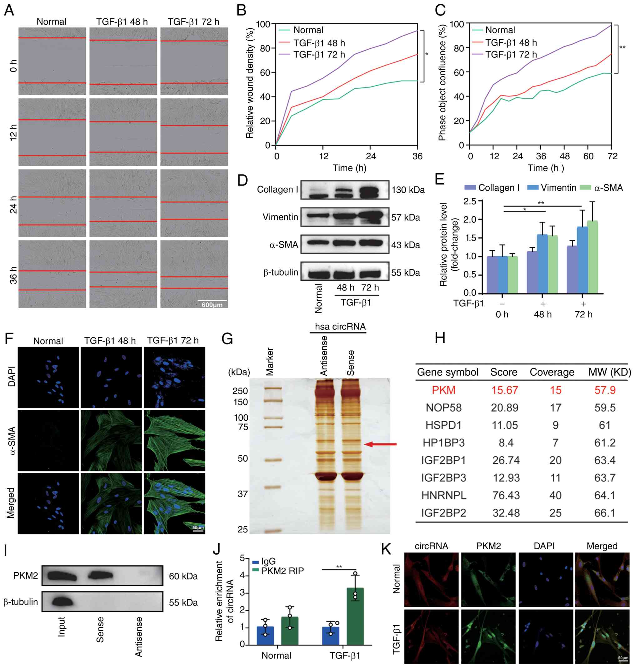
To establish an in vitro model of pulmonary
fibrosis, MRC-5 cells were exposed to TGF-β1 for 48 and 72 h, as
previously described (38,39). Scratch-wound healing assays
revealed a time-dependent increase in cell migration (Fig. 1A and B), and cell proliferation
also increased over time (Fig.
1C). Western blot analysis demonstrated the significant
upregulation of fibrotic markers, including α-SMA, vimentin and
collagen I, following TGF-β1 stimulation. Densitometric
quantification from three independent experiments confirmed these
changes (Fig. 1D and E).
Immunofluorescence staining further revealed progressively enhanced
α-SMA signals, indicating the acquisition of a myofibroblast
phenotype (Fig. 1F).
It was previously reported that circ0066187, now
termed circCACNA1D, is upregulated in pulmonary fibrosis and exerts
pro-fibrotic effects partly by sponging miR-29b-2-5p (33). Therefore, the present study
examined whether circCACNA1D also functions through direct
RNA-protein interactions. As circCACNA1D is predominantly
cytoplasmic and has documented fibrogenic activity, it was
hypothesized that it may function as a protein scaffold or decoy to
regulate key fibrotic mediators. An unbiased RNA pull-down assay
followed by LC-MS/MS was performed using a biotin-labeled sense
probe targeting circCACNA1D. A specific protein band at ~60 kDa was
enriched by the sense probe, but not by the antisense control
(Fig. 1G). Mass spectrometry
identified PKM as a candidate binding protein (Fig. 1H). Given the molecular weight and
functional relevance, the present study focused on PKM2.
The interaction was validated using complementary
biochemical approaches. RNA pull-down followed by western blot
analysis confirmed the specific enrichment of PKM2 by the
circCACNA1D sense probe (Fig.
1I). RIP assays further revealed that TGF-β1 stimulation
significantly increased circCACNA1D enrichment by the anti-PKM2
antibody compared with the IgG control (Fig. 1J). Combined RNA-FISH and
immunofluorescence analysis demonstrated increased circCACNA1D
expression and the enhanced nuclear accumulation of PKM2 following
exposure of the cells to TGF-β1, with marked co-localization
signals (Fig. 1K). These
findings indicate that circCACNA1D directly interacts with PKM2
during pulmonary fibrosis.
circCACNA1D promotes PKM2 nuclear
translocation and dimerization
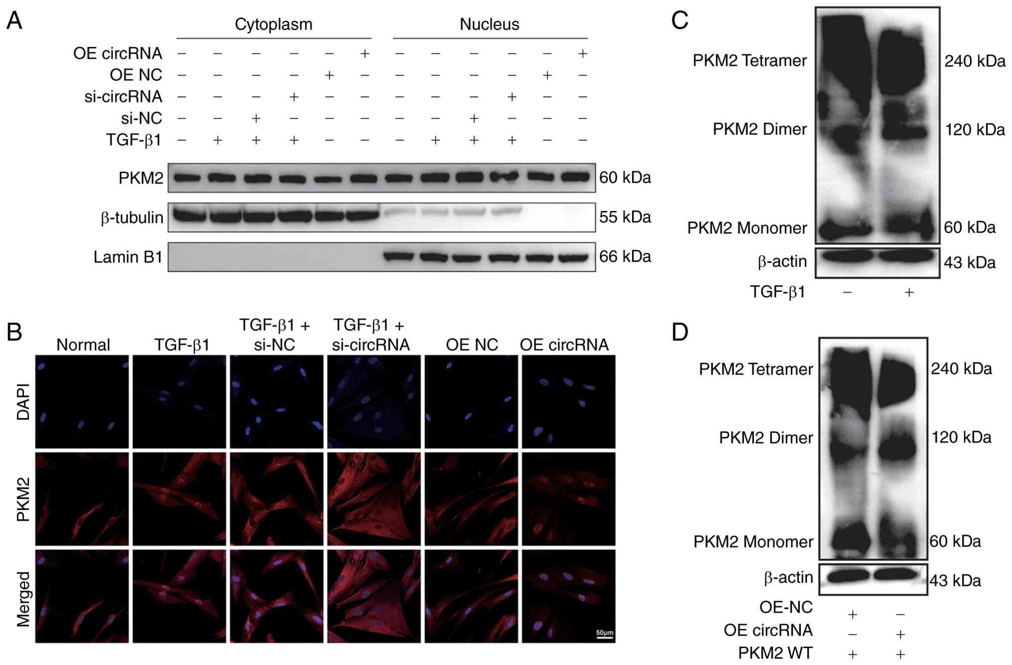
Given the TGF-β1-induced nuclear accumulation of
PKM2, whether circCACNA1D regulates this process was then assessed.
Nuclear-cytoplasmic fractionation followed by western blot analysis
revealed that TGF-β1 stimulation or circCACNA1D OE (OE circRNA)
significantly increased nuclear PKM2 levels. By contrast,
circCACNA1D knockdown (si-circRNA) markedly reduced TGF-β1-induced
PKM2 nuclear translocation (Fig.
2A). Immunofluorescence analysis confirmed these findings.
Specifically, nuclear PKM2 signals were enhanced following TGF-β1
treatment or circCACNA1D OE, whereas circCACNA1D silencing resulted
in predominant cytoplasmic localization (Fig. 2B).
PKM2 performs distinct functions depending on its
oligomeric state, acting as a high-activity tetramer in the
cytoplasm and as a low-activity dimer with transcriptional
co-activator activity in the nucleus. It was first determined
whether TGF-β1 alters endogenous PKM2 conformation. Chemical
cross-linking followed by non-reducing SDS-PAGE revealed that
TGF-β1 induced a shift from tetrameric PKM2 (~240 kDa) to the
dimeric form (~120 kDa; Fig.
2C). Subsequently, the present study examined whether
circCACNA1D influences this conformational balance. CircCACNA1D OE
increased the proportion of dimeric PKM2 and reduced tetrameric
PKM2, as determined by cross-linking and non-reducing SDS-PAGE
(Fig. 2D). These results
indicate that circCACNA1D promotes both nuclear translocation and
dimerization of PKM2, similar to the effect of TGF-β1.
circCACNA1D promotes PKM2 desuccinylation
at specific lysine residues to facilitate dimer formation
To define the molecular mechanism underlying
circCACNA1D-mediated PKM2 dimerization, PTMs were examined, with a
focus on lysine succinylation. Immunoprecipitation of PKM2 followed
by western blot analysis with a pan-anti-succinyllysine antibody
revealed that TGF-β1 significantly reduced PKM2 succinylation in
MRC-5 cells (Fig. 3A).
circCACNA1D OE further decreased PKM2 succinylation levels
(Fig. 3B), suggesting that
circCACNA1D promotes PKM2 desuccinylation under fibrotic
conditions.
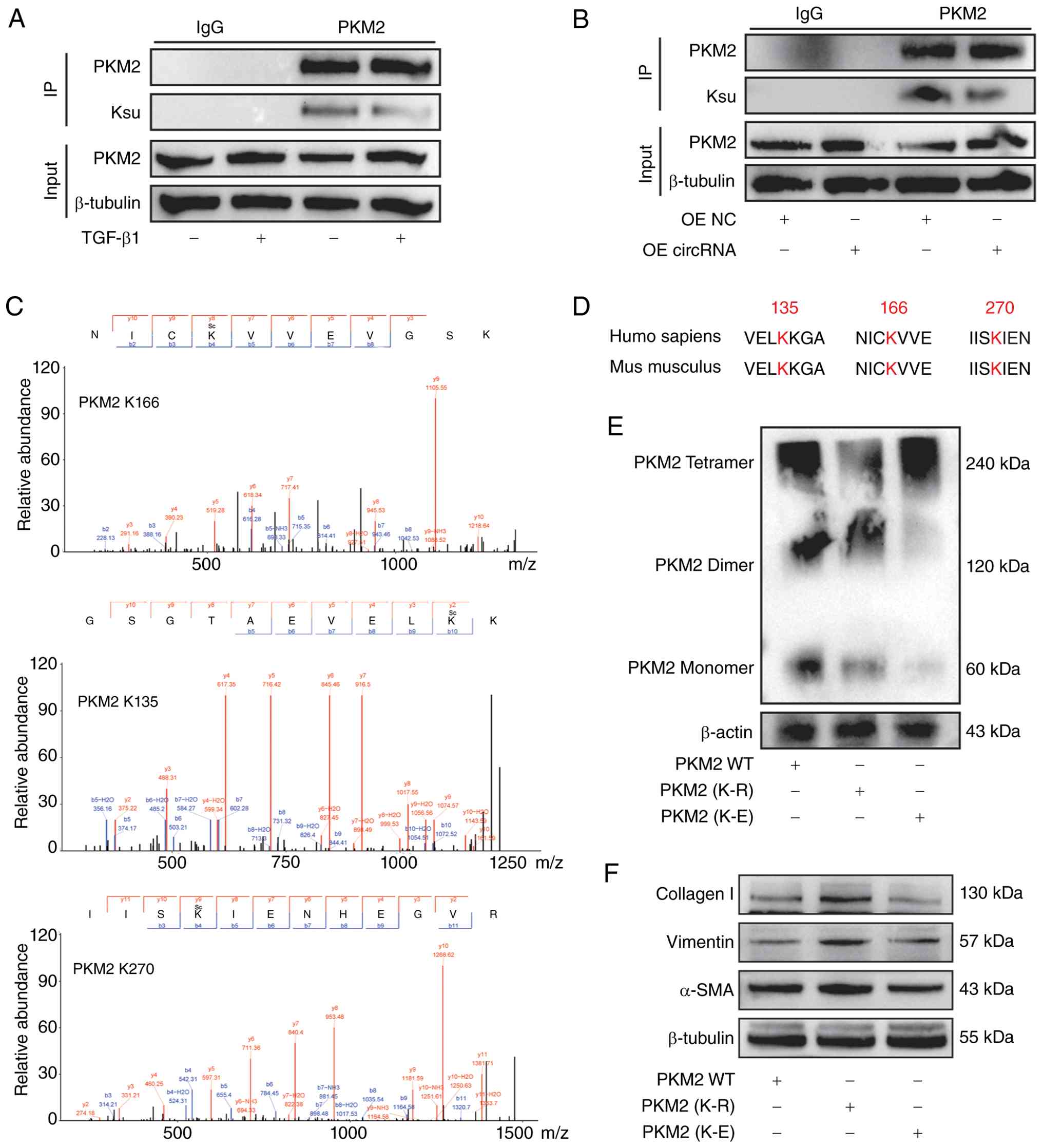 | Figure 3circCACNA1D promotes PKM2
desuccinylation at specific lysine residues to facilitate dimer
formation. (A) Immunoprecipitation and western blot analysis of
PKM2 succinylation (Ksu, succinyllysine) levels in MRC-5 cells
treated with or without TGF-β1 (5 ng/ml; 48 h). Whole-cell lysate
and IgG controls are shown. (B) Immunoprecipitation and western
blot analysis of PKM2 succinylation (Ksu, succinyllysine) levels in
MRC-5 cells following circCACNA1D overexpression. (C)
Representative MS/MS spectra confirming succinyllysine modification
at K135, K166 and K270 on PKM2 in lung tissues. (D) Sequence
alignment of PKM2 protein sequences from humans and mice,
highlighting conserved lysine residues (K135, K166 and K270)
identified in panel C. (E) Chemical cross-linking followed by
non-reducing SDS-PAGE and western blot analysis to assess the
oligomeric state of WT PKM2 and its succinylation-mimetic mutants
(3KR and 3KE). Positions of tetrameric (~240 kDa) and dimeric (~120
kDa) PKM2 are indicated. (F) Western blot analysis of α-SMA,
vimentin and collagen I in MRC-5 cells transfected with PKM2 WT,
PKM2 (K-R) or PKM2 (K-E). β-tubulin served as a loading control.
WT, wild type; PKM2, pyruvate kinase M2; circRNA, circular RNA. |
To identify specific residues, PKM2 was
immunoprecipitated from lung tissues of BLM-treated and control
mice and analyzed by LC-MS/MS. A total of three lysine residues,
K135, K166 and K270, exhibited significantly reduced succinylation
in fibrotic tissues. Representative MS/MS spectra confirmed
succinyllysine modification at these sites (Fig. 3C). Sequence alignment
demonstrated that these residues are conserved between human and
mouse PKM2 (Fig. 3D), supporting
their functional relevance.
To evaluate the role of these sites in PKM2
oligomerization, PKM2 mutants in which K135, K166 and K270 were
substituted with arginine (PKM2-3KR, mimicking constitutive
desuccinylation) or glutamate (PKM2-3KE, mimicking constitutive
succinylation) were generated. Cross-linking and non-reducing
SDS-PAGE analysis showed that PKM2-3KR predominantly formed dimers
with reduced tetramer levels, whereas PKM2-3KE favored the
tetrameric state (Fig. 3E).
These findings demonstrate that desuccinylation at K135, K166 and
K270 promotes PKM2 dimerization. To determine whether the
succinylation status of K135, K166 and K270 directly influences
fibrotic phenotypes, WT PKM2, the 3KR mutant or the 3KE mutant in
MRC-5 cells were overexpressed and fibrotic marker expression was
assessed by western blotting. The 3KR mutant, which mimics
constitutive desuccinylation and predominantly forms dimers,
significantly increased α-SMA, vimentin and collagen I protein
levels compared with WT PKM2. By contrast, the 3KE mutant, which
mimics constitutive succinylation and favors the tetrameric state,
did not elevate fibrotic marker expression (Fig. 3F). These findings establish a
functional link between site-specific PKM2 desuccinylation and
fibroblast activation.
Collectively, these results define a
post-translational regulatory mechanism in which circCACNA1D
promotes desuccinylation of PKM2 at conserved lysine residues,
thereby shifting PKM2 toward its pro-fibrotic, transcriptionally
active dimeric conformation.
Pharmacological stabilization of PKM2
tetramers alleviates fibrotic phenotypes in vitro
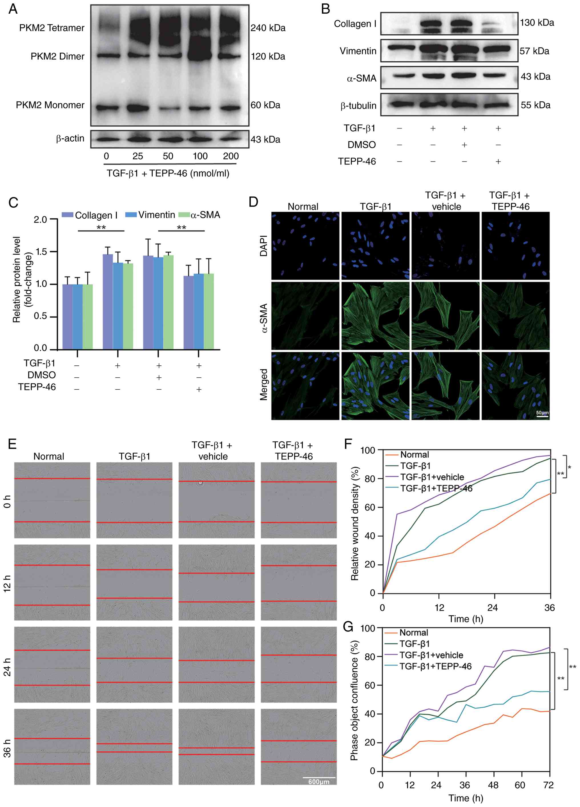
The present study then investigated whether the
pharmacological stabilization of PKM2 tetramers counteracts
fibrotic activation. Treatment with the selective PKM2 tetramer
stabilizer, TEPP-46, increased tetramer formation in a
concentration-dependent manner in theTGF-β1-stimulated MRC-5 cells,
as shown by cross-linking and non-reducing SDS-PAGE (Fig. 4A). TEPP-46 significantly reduced
the TGF-β1-induced upregulation of α-SMA, vimentin and collagen I
at the protein level (Fig. 4B and
C). Immunofluorescence staining further demonstrated that
TEPP-46 attenuated TGF-β1-induced α-SMA-positive stress fiber
formation (Fig. 4D).
Functionally, TEPP-46 significantly suppressed TGF-β1-induced cell
migration in scratch-wound healing assays (Fig. 4E and F). Real-time proliferation
analysis using the IncuCyte S3 system revealed that TEPP-46
inhibited TGF-β1-driven fibroblast growth (Fig. 4G). Taken together, these data
support the concept that the tetramer-dimer balance of PKM2
functions as a molecular switch in fibrotic activation and that
pharmacological stabilization of PKM2 tetramers mitigates fibrotic
responses in vitro.
TEPP-46 attenuates pulmonary fibrosis in
a murine model of pulmonary fibrosis induced by BLM
To assess the therapeutic effect of stabilizing PKM2
tetramers in vivo, a murine model of pulmonary fibrosis
induced by BLM was used. Beginning on day 3 following the
administration of BLM, mice received TEPP-46 to promote PKM2
tetramer formation. TEPP-46 significantly improved BLM-induced lung
dysfunction, as evidenced by increased FVC (Fig. 5A), decreased lung resistance (RL;
Fig. 5B) and increased dynamic
compliance (Fig. 5C).
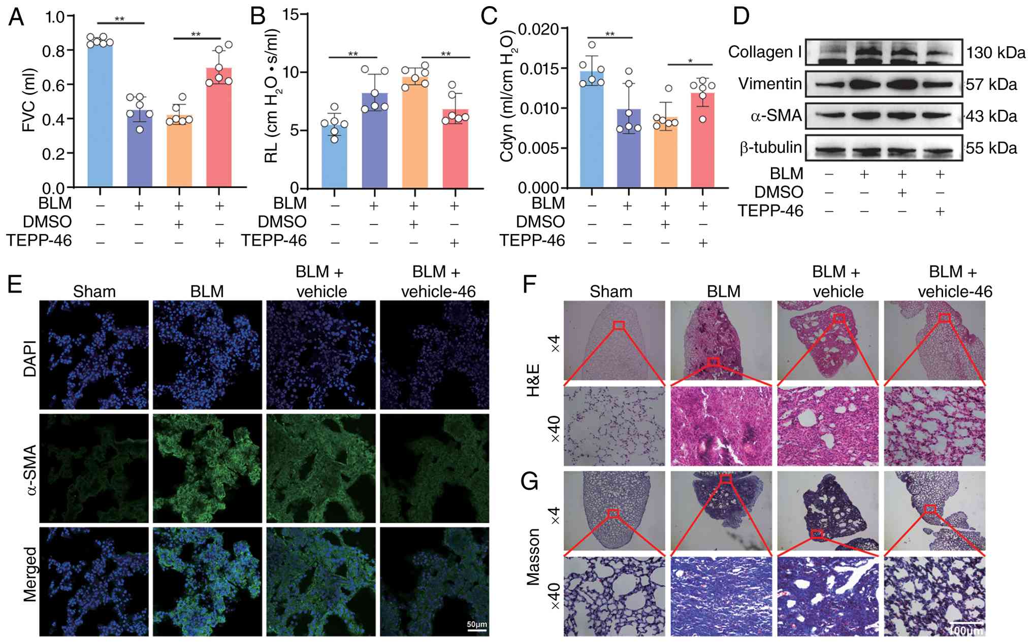 | Figure 5TEPP-46 attenuates BLM-induced
pulmonary fibrosis in mice. (A) FVC measured on day 28 after BLM
instillation in mice treated with saline (control), BLM alone, BLM
+ vehicle or BLM + TEPP-46. (B) RL assessed under the same
treatment conditions as in (A). (C) Cdyn evaluated under the same
treatment conditions as in (A). (D) Western blot analysis of α-SMA,
vimentin and collagen I protein levels in lung tissue homogenates.
β-tubulin served as a loading control. (E) Immunofluorescence
staining of α-SMA in lung tissue sections (scale bar, 50
μm). (F) Histopathological assessment of lung sections by
H&E staining. (G) Masson's trichrome staining of lung sections
(scale bar, 100 μm). *P<0.05,
**P<0.01. FVC, forced vital capacity; RL, lung
resistance; Cdyn, dynamic compliance; BLM, bleomycin. |
Consistent with these functional improvements,
TEPP-46 markedly reduced the expression of fibrotic markers in lung
tissues. Western blot analysis of lung homogenates confirmed the
reduced expression of α-SMA, vimentin and collagen I (Fig. 5D). Immunofluorescence analysis
revealed the decreased expression of α-SMA in theTEPP-46-treated
mice compared with vehicle-treated BLM controls (Fig. 5E). Histopathological evaluation
further supported these findings. H&E staining revealed
preserved alveolar structure and reduced inflammatory infiltration
(Fig. 5F), while Masson's
trichrome staining demonstrated reduced collagen deposition in
TEPP-46-treated lungs (Fig. 5G).
These in vivo results indicate that TEPP-46 alleviates
pulmonary fibrosis and support PKM2 tetramer stabilization as a
therapeutic strategy.
Profibrotic function of circCACNA1D is
dependent on PKM2 nuclear dimerization
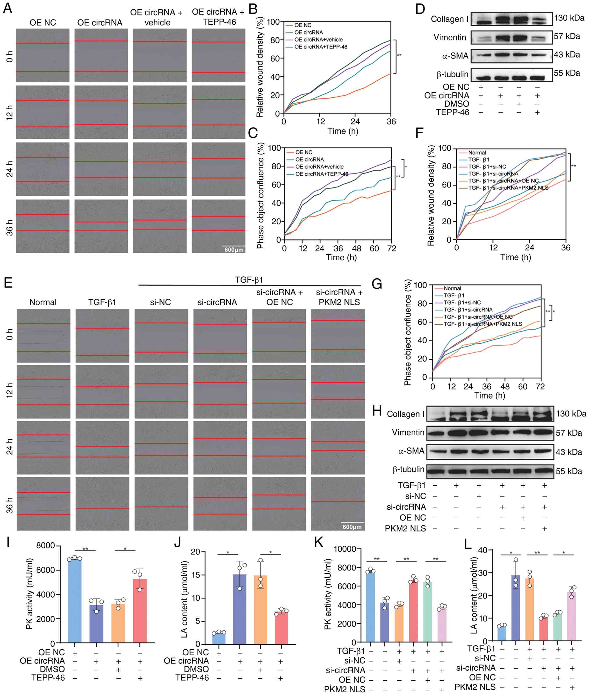
To define the functional association between
circCACNA1D and PKM2 dimerization, gain- and loss-of-function
rescue experiments were conducted. circCACNA1D OE enhanced
fibroblast migration (Fig. 6A and
B), promoted cell proliferation (Fig. 6C) and increased the expression of
fibrotic markers (α-SMA, vimentin and collagen I; Fig. 6D). These pro-fibrotic effects
were abolished by co-treatment with TEPP-46 (Fig. 6A-D). By contrast, circCACNA1D
knockdown in TGF-β1-stimulated cells reduced migration (Fig. 6E and F), proliferation (Fig. 6G) and fibrotic marker expression
(Fig. 6H). Reintroduction of
nuclear-localized PKM2 (PKM2-NLS), which enforces a
transcriptionally active nuclear dimeric state, restored the
fibrotic phenotypes suppressed by circCACNA1D knockdown (Fig. 6E-H). The transfection efficiency
of si-circCACNA1D and PKM2-NLS was verified using RT-qPCR and
western blot analysis, respectively, prior to functional assays (as
described in the Materials and methods section). These findings
demonstrate that circCACNA1D promotes pulmonary fibrosis by
facilitating PKM2 nuclear dimerization and this effect can be
disrupted by stabilizing PKM2 tetramers.
The metabolic consequences of the circCACNA1D-PKM2
axis were then examined. As PKM2 dimerization alters glycolytic
flux, pyruvate kinase activity, which primarily reflects tetrameric
PKM2 function, was assessed. circCACNA1D OE significantly reduced
pyruvate kinase activity, and this reduction was reversed by
TEPP-46 (Fig. 6I). circCACNA1D
OE also increased lactate secretion, a hallmark of enhanced aerobic
glycolysis, whereas TEPP-46 suppressed this increase (Fig. 6J). These data indicate a
metabolic shift from oxidative phosphorylation toward glycolysis
following circCACNA1D-induced PKM2 dimerization.
To confirm that these metabolic effects depend on
nuclear PKM2 dimers, rescue experiments under loss-of-function
conditions were performed. circCACNA1D knockdown in
TGF-β1-stimulated cells increased pyruvate kinase activity and
decreased lactate production. Both effects were reversed by
PKM2-NLS expression (Fig. 6K and
L). Taken together, these findings demonstrate that the
circCACNA1D/PKM2 axis drives glycolytic reprogramming. By promoting
PKM2 dimerization and nuclear localization, circCACNA1D suppresses
pyruvate kinase enzymatic activity and increases lactate
production, thereby supporting the proliferative and biosynthetic
demands of activated fibroblasts during fibrotic remodeling.
Nuclear PKM2 dimer drives a pro-fibrotic
transcriptional program by directly activating target genes
In addition to its metabolic function, nuclear PKM2
functions as a transcriptional co-activator. Therefore, the present
study then investigated whether circCACNA1D-induced nuclear PKM2
dimers regulate a pro-fibrotic transcriptional program. RNA
sequencing of MRC-5 cells under four conditions (control, TGF-β1,
TGF-β1 + vehicle and TGF-β1 + TEPP-46) identified 529 genes
upregulated by TGF-β1 and 389 genes downregulated by TEPP-46, with
42 overlapping candidates (Fig. 7A
and B). GO and KEGG analyses revealed enrichment in
fibrosis-related processes, including cell-cycle progression, cell
division and the p53 signaling pathway (Fig. 7C and D). A total of seven
candidate genes associated with proliferation and fibrosis were
selected for validation. The overexpression of PKM2-NLS induced the
marked upregulation of KIF4A mRNA (>6-fold), identifying KIF4A
as a primary transcriptional target of nuclear PKM2 (Fig. 7E). TGF-β1 increased KIF4A
expression at both the mRNA and protein levels (Fig. 7F and G). PKM2-NLS OE also
elevated KIF4A protein expression (Fig. 7H). Notably, TEPP-46 treatment
suppressed TGF-β1-induced KIF4A expression at both the mRNA and
protein levels (Fig. 7I and J).
Functional assays revealed that KIF4A knockdown reduced the
expression of fibrotic markers, including α-SMA, vimentin and
collagen I (Fig. 7K). To further
confirm KIF4A as a functional downstream effector of nuclear PKM2,
KIF4A gain-of-function experiments were performed. The
overexpression of KIF4A in MRC-5 cells increased the protein levels
of fibrotic markers, including α-SMA, vimentin and collagen I
(Fig. 7L). These data, together
with the KIF4A knockdown results, establish that KIF4A is both
necessary and sufficient to drive pro-fibrotic gene expression
downstream of the circCACNA1D/PKM2 axis. ChIP-qPCR analysis
demonstrated the direct enrichment of PKM2 at multiple regions of
the KIF4A promoter under TGF-β1 stimulation (Fig. 7M), indicating increased DNA
binding activity associated with the dimeric form. Of note, the
KIF4A and PDZD11 genes are arranged in a head-to-head bidirectional
configuration on the X chromosome, sharing an overlapping promoter
region; therefore, the ChIP primers inevitably amplify both
promoters. However, PDZD11 expression was not significantly altered
by TGF-β1, TEPP-46, or PKM2-NLS overexpression (Fig. S2), confirming that the observed
ChIP signals predominantly reflect PKM2 occupancy at the KIF4A
locus. These findings identify KIF4A as a critical downstream
effector of nuclear PKM2 dimers and establish a mechanistic link
between PKM2-driven metabolic reprogramming and transcriptional
activation in pulmonary fibrosis.
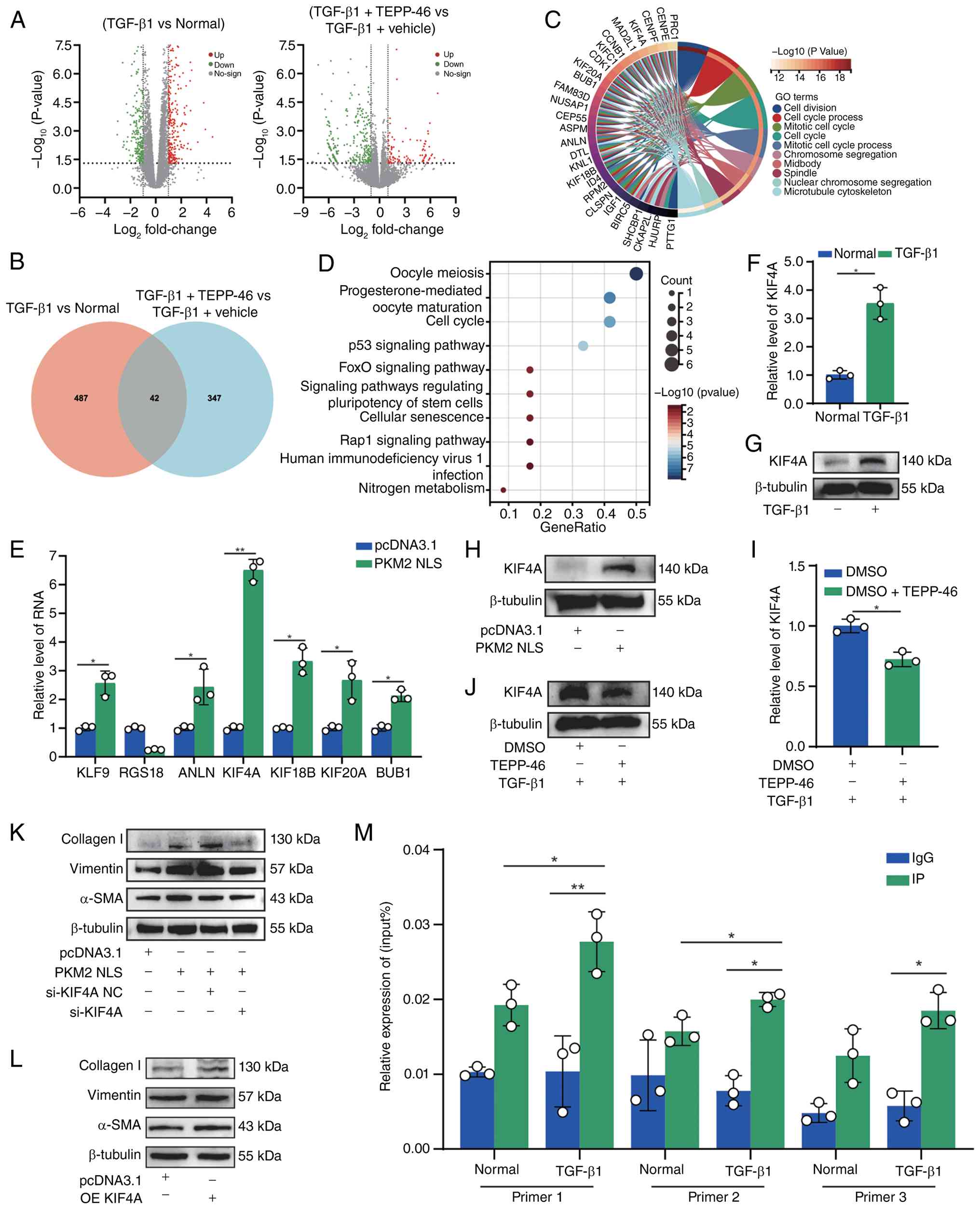 | Figure 7Nuclear PKM2 dimer drives a
pro-fibrotic transcriptional program via direct activation of
KIF4A. (A) Volcano plot of differentially expressed genes between
control vs. TGF-β1 (left) and TGF-β1 + vehicle vs. TGF-β1 + TEPP-46
(right). (B) Venn diagram demonstrating overlap between
TGF-β1-upregulated (529) and TEPP-46-downregulated (389) genes,
yielding 42 candidates. (C) GO enrichment analysis of the 42
overlapping genes. (D) KEGG pathway enrichment analysis of the same
gene set. (E) qPCR validation of candidate gene expression in cells
overexpressing PKM2-NLS. (F) qPCR analysis of KIF4A mRNA levels
under the indicated treatments. (G) Western blot analysis of KIF4A
protein levels under the same conditions as those in panel F. (H)
Western blot analysis of KIF4A protein levels under PKM2-NLS
overexpression. (I) qPCR analysis of KIF4A mRNA levels with TEPP-46
treatment. (J) Western blot analysis of KIF4A protein levels under
the same conditions as those in panel I. (K) Western blot analysis
of fibrotic markers (α-SMA, vimentin and collagen I) in MRC-5 cells
overexpressing PKM2-NLS with or without concurrent KIF4A knockdown
(siKIF4A). (L) Western blot analysis of fibrotic markers (α-SMA,
vimentin and collagen I) in MRC-5 cells overexpressing KIF4A or an
empty vector control. β-tubulin served as a loading control. (M)
ChIP-qPCR showing PKM2 enrichment at the KIF4A promoter region in
TGF-β1-stimulated MRC-5 cells. *P<0.05,
**P<0.01. PKM2, pyruvate kinase; KIF4A, kinesin
family member 4A; GO, Gene Ontology; KEGG, Kyoto Encyclopedia of
Genes and Genomes; PKM2-NLS, nuclear-localized PKM2; ChIP-qPCR,
chromatin immunoprecipitation quantitative PCR. |
Discussion
The present study identified circCACNA1D as a novel
regulator of PKM2 conformational switching in pulmonary fibrosis.
The data support a model in which circCACNA1D directly binds PKM2,
attenuates its succinylation at K135, K166 and K270, and shifts the
enzyme toward the dimeric, transcriptionally competent state.
Functionally, this conformational shift suppresses pyruvate kinase
activity, increases lactate production, enhances fibroblast
migration and proliferation, and promotes expression of fibrotic
markers. At the transcriptional level, KIF4A was identified as a
critical downstream effector of the circCACNA1D/PKM2 pathway. These
findings are conceptually important as they extend circRNA biology
beyond canonical miRNA-sponging mechanisms and demonstrate that a
circRNA can directly reshape the biochemical state of a metabolic
enzyme to coordinate metabolic and transcriptional remodeling in
fibrosis (11,30).
The results of the present study also contribute to
the increasing amount of literature implicating PKM2 in fibrotic
diseases. Recent studies have demonstrated that PKM2 promotes
pulmonary fibrosis by stabilizing TGF-β receptor I (16), drives fibrogenic metabolic
rewiring in kidney injury (17,18), restrains or promotes fibrogenesis
depending on its oligomeric state in hepatic stellate cells
(19) and contributes to
fibrosis-associated lactate accumulation in multiple organs
(21,40,41). Together, these studies establish
PKM2 as a major determinant of fibrotic remodeling. However, the
majority of previous studies have focused on downstream
consequences of PKM2 activation (42,43), while the upstream mechanisms
governing its dimer-tetramer switching in pulmonary fibrosis
remained unclear. The data from the present study indicate that
circCACNA1D functions as an upstream regulator of PKM2 to couple
post-transcriptional regulation to metabolic reprogramming and
nuclear gene activation. This provides an explanation for the
mechanisms by which fibroblasts sustain a stable profibrotic
phenotype despite fluctuating extracellular signals.
A key question raised by the findings of the present
study is why circCACNA1D is upregulated in fibrotic lungs. Although
the present study did not directly elucidate the upstream
regulatory mechanism, several non-mutually exclusive hypotheses
merit consideration. First, chronic TGF-β/SMAD signaling may
enhance the transcription of the CACNA1D host locus or alter local
chromatin architecture, thereby favoring circular RNA production
(44). Second,
fibrosis-associated RNA-binding proteins that regulate
back-splicing may increase circCACNA1D biogenesis under stress
conditions (45). Third,
epitranscriptomic regulation may contribute, as m6A
modification has been implicated in circRNA processing, export and
stability (46). These
hypotheses are not mutually exclusive, and determining which
mechanism predominates will be key for understanding how the
circCACNA1D-PKM2 axis is engaged during disease initiation and
progression.
A central mechanistic finding of the present study
is that circCACNA1D promotes PKM2 desuccinylation. The mutational
data support the functional importance of K135, K166 and K270 in
controlling PKM2 oligomerization, as the desuccinylation-mimetic
3KR mutant favored dimer formation and enhanced fibrotic marker
expression, whereas the succinylation-mimetic 3KE mutant favored
tetramer formation and exhibited diminished profibrotic activity.
These observations position succinylation among other PTMs as a key
determinant of PKM2 structural and functional plasticity (23,47-49). Notably, the present study
demonstrated the consequence of circCACNA1D binding, but did not
fully define the molecular mechanism by which desuccinylation
occurs. One plausible model is a scaffold mechanism, in which
circCACNA1D facilitates the association of PKM2 with a
desuccinylase, such as SIRT5, thereby promoting targeted removal of
succinyl groups (35,50). An alternative, non-mutually
exclusive model is steric regulation, whereby circCACNA1D binding
alters the local conformation of PKM2 or limits the accessibility
of these lysine residues to succinylation machinery or succinyl-CoA
(51). At present, the data from
the present study support a circCACNA1D-dependent desuccinylation
process; however, additional biochemical studies will be required
to distinguish whether the recruitment of a desuccinylase, the
steric exclusion of succinylation or a combination of both
underlies this phenomenon.
The results of the present study also underscore the
importance of contextualizing PKM2 succinylation within a broader
PTM network, rather than as an isolated event. PKM2 is extensively
regulated via phosphorylation, acetylation, oxidation, lysine
acylation and protein-partner interactions, all of which can
influence its enzymatic activity, subcellular localization or
non-metabolic signaling functions (9,11). Recent studies have shown that
S-nitrosylation can promote PKM2 dimerization in cardiac fibrosis
(47), succinate-driven
succinylation can accelerate age-associated fibrotic remodeling
(23) and diverse binding
partners can redirect PKM2 toward noncanonical transcriptional
outputs (48,49). Consequently, it is plausible that
distinct PTMs and interacting proteins cooperate or compete to
determine whether PKM2 adopts a catalytically active tetrameric
state or a signaling-competent dimeric state in fibroblasts.
Systematic mapping of PTM crosstalk on PKM2 in fibrotic cells is
essential to elucidate the mechanisms by which metabolic
reprogramming becomes stabilized during chronic tissue injury.
A notable contribution of the present study is the
expansion of the emerging field of circRNA-mediated metabolic
regulation. Several circRNAs have already been implicated in
pulmonary fibrosis, but most act indirectly through ceRNA circuits
or upstream signaling pathways. For example, circHIPK3 promotes
pulmonary fibrosis by facilitating glycolysis through a
miR-30a-3p/FOXK2-dependent mechanism (3), while previous research has
demonstrated that circ0066187/circCACNA1D can also promote
pulmonary fibrogenesis through STAT3-associated metabolic signaling
(33). By contrast, the present
study established a direct circRNA-protein regulatory mechanism, in
which circCACNA1D binds PKM2 and modulates its post-translational
and oligomeric state. This mechanism is distinct from recently
described circRNA-PKM2 signaling in other disease contexts. For
instance, circABCC4 in pancreatic cancer-associated fibroblasts
drives glycolytic reprogramming and chemoresistance by promoting
PKM2 nuclear translocation through KPNA2 recruitment (52), whereas other non-coding RNAs or
protein partners influence PKM2 activity through different
structural or catalytic mechanisms (49,53). Thus, circCACNA1D regulates PKM2
through a mechanism governed by succinylation-dependent
dimer-tetramer switching, rather than solely on transcript
abundance or trafficking adaptors. This mechanistic distinction
underscores the novelty of the present study.
At the transcriptional level, the present study
identified KIF4A as a pivotal downstream effector of the
circCACNA1D/PKM2 axis. Nuclear PKM2 is increasingly recognized as a
signaling hub that couples metabolic state to gene regulation
(11,54), and the RNA-seq, gain-of-function,
loss-of-function and ChIP-qPCR data of the present study
collectively substantiate KIF4A as a functionally relevant target
in fibrotic fibroblasts. However, KIF4A is unlikely to be the sole
effector of nuclear PKM2 in this setting. Rather, it likely
constitutes part of a broader transcriptional program through which
dimeric PKM2 drives proliferation, matrix production and fibroblast
persistence. Genome-wide chromatin occupancy and target-gene
analyses are required to define this network more
comprehensively.
From a translational perspective, the findings of
the present study underscore the therapeutic potential of PKM2
conformational control. TEPP-46, a selective PKM2 activator that
stabilizes the tetrameric state, abrogated the profibrotic effects
of circCACNA1D in vitro and attenuated BLM-induced pulmonary
fibrosis in vivo (18).
These observations suggest that the dimeric and nuclear forms of
PKM2 are not simply a byproduct of metabolic stress, but an active
driver of fibrotic pathology. More broadly, they suggest that
promoting tetrameric PKM2 may represent a viable antifibrotic
strategy, particularly in disease contexts characterized by
persistent glycolytic rewiring and pathological nuclear PKM2
signaling.
The present study has several limitations. First,
although the present study established PKM2 desuccinylation as a
key regulatory event downstream of circCACNA1D, the specific
desuccinylase responsible has not yet been identified nor have any
additional PKM2 PTMs which are involved in this regulatory
mechanism. Second, the upstream mechanisms leading to circCACNA1D
upregulation in fibrosis remain to be elucidated. Third, although
KIF4A was validated herein as a critical downstream effector,
nuclear PKM2 likely regulates additional genes relevant to
fibroblast activation. Finally, investigating the applicability of
this pathway to other fibrotic organs and validation in larger
human cohorts will be important for defining its broader
translational importance.
In conclusion, a pathogenic circCACNA1D-PKM2
signaling axis in pulmonary fibrosis was identified in the present
study. By promoting PKM2 desuccinylation and shifting the
dimer-tetramer equilibrium toward the dimeric state, circCACNA1D
couples glycolytic reprogramming with nuclear transcriptional
activation, thereby driving fibroblast activation. These findings
deepen the understanding of the mechanisms by which non-coding RNAs
directly regulate metabolic enzymes in fibrotic disease and
highlight PKM2 conformational control as a potentially druggable
vulnerability in pulmonary fibrosis.
Supplementary Data
Availability of data and materials
The data generated in the present study may be
requested from the corresponding author. The mass spectrometry
proteomics data have been submitted to the ProteomeXchange
Consortium through the PRIDE partner repository with the dataset
identifier PXD075455, and are accessible at https://www.ebi.ac.uk/pride/archive/projects/PXD075455.
The raw sequencing data generated in this study are openly
available in the NCBI SRA database accessible with the following
link: https://www.ncbi.nlm.nih.gov/sra/PRJNA1475466.
Authors' contributions
SZ, XS and JZ designed the study. YW, XL and JZ
performed the experiments, analyzed the data, and wrote the
manuscript. MW and NZ contributed to the conception and design of
the study. MJ assisted with the animal experiments and data
collection. BL participated in the study design and interpretation
of data. CL participated in the data analysis and critical review
of the manuscript. SZ and XS prepared the figures and revised the
manuscript. SZ and XS confirm the authenticity of all the raw data.
All authors have read and approved the final manuscript.
Ethics approval and consent to
participate
All animal procedures were approved by the Animal
Ethics Committee of Shandong Medical and Pharmaceutical University
(approval no. 2021-355).
Patient consent for publication
Not applicable.
Competing interests
The authors declare that they have no competing
interests.
Acknowledgements
Not applicable.
Funding
The present study was supported by the Special Funding for the
'Case-by-Case Introduction of Top Talent (Teams)' Program in
Yantai, China. The present study was also funded by the National
Natural Science Foundation of China (grant nos. 82500104, 82400117,
82370079, 82370094, 82170085, 81970064 and 81870001).
References
|
1
|
Li J, Li P, Zhang G, Qin P, Zhang D and
Zhao W: CircRNA TADA2A relieves idiopathic pulmonary fibrosis by
inhibiting proliferation and activation of fibroblasts. Cell Death
Dis. 11:5532020.
|
|
2
|
Hong SH, Yun B, Cho S, Hong OY, Lee SH,
Kim WH and Hong JY: Loss of ATF3 exacerbates pulmonary fibrosis via
enhanced neutrophil recruitment and profibrotic macrophage
polarization. Clin Sci (Lond). 140:179–199. 2026.
|
|
3
|
Xu Q, Cheng D, Li G, Liu Y, Li P, Sun W,
Ma D and Ni C: CircHIPK3 regulates pulmonary fibrosis by
facilitating glycolysis in miR-30a-3p/FOXK2-dependent manner. Int J
Biol Sci. 17:2294–2307. 2021.
|
|
4
|
Li J, Zhai X, Sun X, Cao S, Yuan Q and
Wang J: Metabolic reprogramming of pulmonary fibrosis. Front
Pharmacol. 13:10318902022.
|
|
5
|
Richeldi L, Baldi F, Pasciuto G, Macagno F
and Panico L: Current and future idiopathic pulmonary fibrosis
therapy. Am J Med Sci. 357:370–373. 2019.
|
|
6
|
Koudstaal T and Wijsenbeek MS: Idiopathic
pulmonary fibrosis. Presse Med. 52:1041662023.
|
|
7
|
Bueno M, Calyeca J, Rojas M and Mora AL:
Mitochondria dysfunction and metabolic reprogramming as drivers of
idiopathic pulmonary fibrosis. Redox Biol. 33:1015092020.
|
|
8
|
Li Z, Sun C and Qin Z: Metabolic
reprogramming of cancer-associated fibroblasts and its effect on
cancer cell reprogramming. Theranostics. 11:8322–8336. 2021.
|
|
9
|
Lv S, Cao M, Luo J, Fu K and Yuan W:
Search progress of pyruvate kinase M2 (PKM2) in organ fibrosis. Mol
Biol Rep. 51:3892024.
|
|
10
|
Vander Heiden MG, Cantley LC and Thompson
CB: Understanding the Warburg effect: The metabolic requirements of
cell proliferation. Science. 324:1029–1033. 2009.
|
|
11
|
Chen TJ, Wu CH, Hung MC, Wang WC and Kung
HJ: Nuclear PKM2: A signal receiver, a gene programmer, and a
metabolic modulator. J Biomed Sci. 32:752025.
|
|
12
|
Israelsen WJ and Vander Heiden MG:
Pyruvate kinase: Function, regulation and role in cancer. Semin
Cell Dev Biol. 43:43–51. 2015.
|
|
13
|
Dayton TL, Jacks T and Vander Heiden MG:
PKM2, cancer metabolism, and the road ahead. EMBO Rep.
17:1721–1730. 2016.
|
|
14
|
Christofk HR, Vander Heiden MG, Harris MH,
Ramanathan A, Gerszten RE, Wei R, Fleming MD, Schreiber SL and
Cantley LC: The M2 splice isoform of pyruvate kinase is important
for cancer metabolism and tumour growth. Nature. 452:230–233.
2008.
|
|
15
|
Zhang Z, Zheng L, Chen Y, Chen Y, Hou J,
Xiao C, Zhu X, Zhao SM and Xiong JW: AARS2 ameliorates myocardial
ischemia via fine-tuning PKM2-mediated metabolism. Elife.
13:RP996702025.
|
|
16
|
Gao S, Li X, Jiang Q, Liang Q, Zhang F, Li
S, Zhang R, Luan J, Zhu J, Gu X, et al: PKM2 promotes pulmonary
fibrosis by stabilizing TGF-β1 receptor I and enhancing TGF-β1
signaling. Sci Adv. 8:eabo09872022.
|
|
17
|
Chen Y, Bai X, Chen J, Huang M, Hong Q,
Ouyang Q, Sun X, Zhang Y, Liu J, Wang X, et al: Pyruvate kinase M2
regulates kidney fibrosis through pericyte glycolysis during the
progression from acute kidney injury to chronic kidney disease.
Cell Prolif. 57:e135482024.
|
|
18
|
Liu H, Takagaki Y, Kumagai A, Kanasaki K
and Koya D: The PKM2 activator TEPP-46 suppresses kidney fibrosis
via inhibition of the EMT program and aberrant glycolysis
associated with suppression of HIF-1α accumulation. J Diabetes
Investig. 12:697–709. 2021.
|
|
19
|
Zheng D, Jiang Y, Qu C, Yuan H, Hu K, He
L, Chen P, Li J, Tu M, Lin L, et al: Pyruvate kinase M2
tetramerization protects against hepatic stellate cell activation
and liver fibrosis. Am J Pathol. 190:2267–2281. 2020.
|
|
20
|
Rao J, Wang H, Ni M, Wang Z, Wang Z, Wei
S, Liu M, Wang P, Qiu J, Zhang L, et al: FSTL1 promotes liver
fibrosis by reprogramming macrophage function through modulating
the intracellular function of PKM2. Gut. 71:2539–2550. 2022.
|
|
21
|
Xu Y, Ma X, Ni W, Zheng L, Lin Z, Lai Y,
Yang N, Dai Z, Yao T, Chen Z, et al: PKM2-driven lactate
overproduction triggers endothelial-to-mesenchymal transition in
ischemic flap via mediating TWIST1 lactylation. Adv Sci (Weinh).
11:e24061842024.
|
|
22
|
Zhang Z, Tan M, Xie Z, Dai L, Chen Y and
Zhao Y: Identification of lysine succinylation as a new
post-translational modification. Nat Chem Biol. 7:58–63. 2011.
|
|
23
|
Wang Z, Zhang Z, Ping Z, Yang S, Li Y,
Jiang T, Zheng X, Zhang Q, Liu Z, Zhang X, et al: Succinate-driven
PKM2 succinylation and dimerization accelerates age-associated
cardiac fibrosis. Commun Biol. 9:6332025.
|
|
24
|
Wang F, Wang K, Xu W, Zhao S, Ye D, Wang
Y, Xu Y, Zhou L, Chu Y, Zhang C, et al: SIRT5 desuccinylates and
activates pyruvate kinase M2 to block macrophage IL-1β production
and to prevent DSS-induced colitis in mice. Cell Rep. 19:2331–2344.
2017.
|
|
25
|
Du J, Zhou Y, Su X, Yu JJ, Khan S, Jiang
H, Kim J, Woo J, Kim JH, Choi BH, et al: Sirt5 is a NAD-dependent
protein lysine demalonylase and desuccinylase. Science.
334:806–809. 2011.
|
|
26
|
Zhang L, Lv T, Hou P, Jin Y and Jia F:
Sirt5-mediated polarization and metabolic reprogramming of
macrophage sustain brain function following ischemic stroke. Brain
Res. 1857:1496132025.
|
|
27
|
Zhao Q, Jing Y, Jiang X, Zhang X, Liu F,
Huang H, Zhang Z, Wang H, Sun S, Ma S, et al: SIRT5 safeguards
against primate skeletal muscle ageing via desuccinylation of TBK1.
Nat Metab. 7:556–573. 2025.
|
|
28
|
Song Z, Zhou M, Lin J, You J and Huang C:
Translatable circular RNAs: Mechanism, function and clinical
application. Int J Biol Macromol. 335:1491852026.
|
|
29
|
Huang A, Zheng H, Wu Z, Chen M and Huang
Y: Circular RNA-protein interactions: Functions, mechanisms, and
identification. Theranostics. 10:3503–3517. 2020.
|
|
30
|
Dong J, Zeng Z, Huang Y, Chen C, Cheng Z
and Zhu Q: Challenges and opportunities for circRNA identification
and delivery. Crit Rev Biochem Mol Biol. 58:19–35. 2023.
|
|
31
|
Chen LL: The expanding regulatory
mechanisms and cellular functions of circular RNAs. Nat Rev Mol
Cell Biol. 21:475–490. 2020.
|
|
32
|
Rochow H, Franz A, Jung M, Weickmann S,
Ralla B, Kilic E, Stephan C, Fendler A and Jung K: Instability of
circular RNAs in clinical tissue samples impairs their reliable
expression analysis using RT-qPCR: From the myth of their advantage
as biomarkers to reality. Theranostics. 10:9268–9279. 2020.
|
|
33
|
Liu B, Liu W, Li H, Zhai N, Lv C, Song X
and Yang S: circ0066187 promotes pulmonary fibrogenesis through
targeting STAT3-mediated metabolism signal pathway. Cell Mol Life
Sci. 82:792025.
|
|
34
|
You Q, Yu J, Pan R, Feng J, Guo H and Liu
B: Decoding the regulatory roles of circular RNAs in cardiac
fibrosis. Noncoding RNA Res. 11:115–130. 2024.
|
|
35
|
Zhou WY, Cai ZR, Liu J, Wang DS, Ju HQ and
Xu RH: Circular RNA: Metabolism, functions and interactions with
proteins. Mol Cancer. 19:1722020.
|
|
36
|
Shen Y, Zhang N, Chai J, Wang T, Ma C, Han
L and Yang M: CircPDIA4 induces gastric cancer progression by
promoting ERK1/2 activation and enhancing biogenesis of oncogenic
circRNAs. Cancer Res. 83:538–552. 2023.
|
|
37
|
Livak KJ and Schmittgen TD: Analysis of
relative gene expression data using real-time quantitative PCR and
the 2(-Delta Delta C(T)) method. Methods. 25:402–408. 2001.
|
|
38
|
Wang Y, Zhao Y, Cao G, Jiang M, Yuan X, Li
H, Song X, Zhang J, Lv C and Zhang S: STAT3 facilitates super
enhancer formation to promote fibroblast-to-myofibroblast
differentiation by the analysis of ATAC-Seq, RNA-Seq and ChIP-Seq.
J Cell Mol Med. 29:e706392025.
|
|
39
|
Zhang J, Wang H, Chen H, Li H, Xu P, Liu
B, Zhang Q, Lv C and Song X: ATF3-activated accelerating effect of
LINC00941/lncIAPF on fibroblast-to-myofibroblast differentiation by
blocking autophagy depending on ELAVL1/HuR in pulmonary fibrosis.
Autophagy. 18:2636–2655. 2022.
|
|
40
|
Xiang T, Wang X, Huang S, Zhou K, Fei S,
Zhou B, Yue K, Li Q, Xue S, Dai Y, et al: Inhibition of PKM2 by
shikonin impedes TGF-β1 expression by repressing histone
lactylation to alleviate renal fibrosis. Phytomedicine.
136:1563242025.
|
|
41
|
Liu JY, Liao W, Chen ZY, Liu T, Wang R,
Gong JP, Xu XS and Wu H: PKM2 drives lactate-mediated fibrosis in
liver transplantation. Transplantation. 109:e631–e646. 2025.
|
|
42
|
Wang Y, Hu R, Zhang X, Cai G, Ding Q and
Che P: PKM2 mediates the glycolytic response to matrix stiffness in
lung fibroblasts. Front Cell Dev Biol. 14:17710782026.
|
|
43
|
Feng J, Huang X, Peng Y, Yang W, Yang X,
Tang R, Xu Q, Gao Y, He Z, Xing S and Mei S: Pyruvate kinase M2
modulates mitochondrial dynamics and EMT in alveolar epithelial
cells during sepsis-associated pulmonary fibrosis. J Transl Med.
23:2052025.
|
|
44
|
Gélabert C, Papoutsoglou P, Golán I,
Ahlström E, Ameur A, Heldin CH, Caja L and Moustakas A: The long
non-coding RNA LINC00707 interacts with Smad proteins to regulate
TGFβ signaling and cancer cell invasion. Cell Commun Signal.
21:2712023.
|
|
45
|
Chiusa M, Lee YA, Zhang MZ, Harris RC,
Sherrill T, Lindner V, Brooks CR, Yu G, Fogo AB, Flynn CR, et al:
Cytoplasmic retention of the DNA/RNA-binding protein FUS
ameliorates organ fibrosis in mice. J Clin Invest.
134:e1751582024.
|
|
46
|
Di Timoteo G, Dattilo D, Centrón-Broco A,
Colantoni A, Guarnacci M, Rossi F, Incarnato D, Oliviero S, Fatica
A, Morlando M and Bozzoni I: Modulation of circRNA metabolism by
m6A modification. Cell Rep. 31:1076412020.
|
|
47
|
Luo S, Ye D, Zhang Y, Wang Y, Zhou M, Lv
M, Wang X, Zhong K, Zhang Y, Hu L, et al: S-Nitrosylation of
pyruvate kinase isoform 2 drives cardiac fibrosis by promoting
mitochondrial fission. Circulation. 153:338–357. 2026.
|
|
48
|
Wu Y, Tang L, Huang H, Yu Q, Hu B, Wang G,
Ge F, Yin T, Li S and Yu X: Phosphoglycerate dehydrogenase
activates PKM2 to phosphorylate histone H3T11 and attenuate
cellular senescence. Nat Commun. 14:13232023.
|
|
49
|
Yu JT, Xie SS, Shen XY, Li Z, Hu XW, Zhang
Y, Dong ZH, Wang JN, Li XY, Dong YH, et al: Renal tubular
epithelial IGFBP7 interacts with PKM2 to drive renal lipid
accumulation and fibrosis. Mol Ther. 33:3757–3777. 2025.
|
|
50
|
Wang J, Gao J, Ding L, Yang X, Zheng D,
Zeng Y, Zhu J, Lei W, Chen C, Liu Z and Huang JA: Circular
RNA-cacna1d plays a critical role in sepsis-induced lung injury by
sponging microRNA-185-5p. Am J Respir Cell Mol Biol. 72:181–194.
2025.
|
|
51
|
Sandoval JE, Carullo NVN, Salisbury AJ,
Day JJ and Reich NO: Mechanism of non-coding RNA regulation of
DNMT3A. Epigenetics Chromatin. 18:152025.
|
|
52
|
He R, Hu C, Yuan Y, Li T, Tian Q, Huang T,
Lin Q, Zheng S, Chen C, Fu Z and Chen R: Glycolysis reprogramming
in CAFs promotes oxaliplatin resistance in pancreatic cancer
through circABCC4 mediated PKM2 nuclear translocation. Cell Death
Dis. 16:1262025.
|
|
53
|
Fei M, Li X, Liang S, Zhou S, Wu H, Sun L,
Liu Y, Hu Q, Liu L and Wang J: LncRNA PWRN1 inhibits the
progression of hepatocellular carcinoma by activating PKM2
activity. Cancer Lett. 584:2166202024.
|
|
54
|
Yang W, Xia Y, Ji H, Zheng Y, Liang J,
Huang W, Gao X, Aldape K and Lu Z: Nuclear PKM2 regulates β-catenin
transactivation upon EGFR activation. Nature. 480:118–122.
2011.
|