Introduction
Flaviviruses are reported to infect up to 400
million individuals annually and can cause a wide spectrum of
diseases, representing an ongoing threat to global public health
(1). Tick-borne encephalitis
virus (TBEV), a member of the flaviviruses, is an emerging health
concern with an increasing geographical distribution worldwide
(2). TBEV is prevalent in
forested areas of the Eurasian continent and northeastern Asia.
Tick bites are the primary route of TBEV transmission along with
infected milk consumption, aerosols and breast milk from infected
mothers to infants (3,4). As a neurotropic arbovirus, TBEV
infection attacks the central nervous system (CNS) in humans and
may lead to severe neurological disorders such as TBE, long-lasting
neurological sequelae and death. In total, 5 TBEV subtypes
including the European, Siberian, Far Eastern, Baikalian and
Himalayan subtypes have been characterized, which are closely
associated with the severity and mortality of TBEV infection
(5). The Far Eastern subtype
induces the most severe form of TBE with a high mortality rate of
up to 30%. In China, TBE is endemic in the northeast, the northwest
and the southeast and nearly all the TBEV isolates belong to the
Far Eastern subtype (6). There
is no specific treatment for the diseases associated with TBEV. Due
to an increase in TBEV prevalence and transmission, efforts to
combat the growing threat must be made.
Cepharanthine, a biscoclaurine alkaloid mainly
extracted from plant Stephania cepharantha Hayata, was named
in 1934 and rapidly approved for clinical applications in Japan due
to the traditional use of the medicinal plant (7). Cepharanthine possesses a variety of
pharmacological properties including anti-oxidative,
anti-inflammatory, anti-proliferative, anti-metastatic,
anti-atherosclerosis, anticancer, anti-parasitic, antiviral and
immunomodulatory properties (7,8).
These properties of cepharanthine are responsible for the treatment
of cancer, shock and inflammatory diseases (9). Moreover, cepharanthine is the only
bisbenzylisoquinoline alkaloid in clinical use to treat human
diseases and has unique pharmacological properties, excellent
safety and limited side effects (10). After its initial use for the
treatment of tuberculosis, cepharanthine has been approved to treat
a diverse range of acute and chronic diseases such as venomous
snakebites, radiation-induced leukopenia, alopecia, malaria,
xerostomia, sarcoidosis and refractory anemia (11). Cepharanthine has also been
applied for the treatment of T cell acute lymphoblastic leukemia,
autoimmune diseases and immune thrombocytopenic purpura (12,13). Therefore, cepharanthine is a
fascinating drug with various clinical benefits and complex action
modes.
In addition to the aforementioned clinical
applications, the antiviral properties of cepharanthine have been
implicated in the management of certain viral diseases.
Cepharanthine was shown to be a potent inhibitor of human
immunodeficiency virus type 1 replication in a chronically infected
monocytic cell line, U1 (14).
Additionally, cepharanthine has an inhibitory efficacy towards
herpes simplex virus type 1 (15). Cepharanthine can also inhibit the
replication and hepatitis B e antigen production of wild-type or
lamivudine-resistant hepatitis B virus isolates (16). Cepharanthine induces the
apoptosis of the SIT leukemia cell line infected with human
T-lymphotropic virus type 1 (17). The antiviral efficacy of
cepharanthine has been proven in PK-15 cells infected with porcine
circovirus type 2 (18).
Cepharanthine has been identified as one of the most potent
inhibitors of porcine reproductive and respiratory syndrome virus
(19). Inhibition of porcine epidemic diarrhea virus
propagation by cepharanthine is observable in vitro and
in vivo (20).
Cepharanthine blocks entry of swine acute diarrhea syndrome
coronavirus (21). Cepharanthine
also inhibits dengue virus infection at the initial viral
replication state and decreases secretion of interleukin (IL-) 6
(22). Furthermore,
cepharanthine markedly suppresses the replication of human
coronavirus OC43 and expression of viral proteins (23). Additionally, cepharanthine was
considered a promising repurposing drug for coronavirus disease
2019 therapy (24).
Cepharanthine has been identified as one of the most potent
inhibitors of severe acute respiratory syndrome coronavirus type 2,
with predominant inhibition of entry and replication, suggesting
therapeutic potential as an antiviral agent (25).
TBEV is a major pathogen of arboviral encephalitis
with increasing expansion and potential outbreaks (2,3). The
development of effective anti-TBEV components is needed to control
this emerging disease. Cepharanthine displays distinct antiviral
activities that are crucial to modulating viral propagation and the
inflammatory response. Viruses are obligate intracellular parasites
and viral proteins are synthesized by the endoplasmic reticulum
(ER) of host cells. ER stress is closely regulated to maintain
cellular homeostasis. Viruses may comply with ER regulatory
processes or subvert them to support viral propagation. TBEV
infection induces neuron damage as well as immunopathology due to
extreme inflammation (4,5). As ER stress and inflammatory
response are involved in TBEV pathogenesis, the antiviral efficacy
of cepharanthine against TBEV should be investigated. Therefore,
the present study aimed to elucidate whether cepharanthine
possesses anti-TBEV efficacy, which step(s) of the TBEV life cycle
might be affected and how the stress and inflammatory response are
modulated.
Materials and methods
Cells and TBEV
Human lung adenocarcinoma A549 cells (cat. no.
CCL-185), human neuroblastoma SH-SY5Y cells (cat. no. CRL-2266),
African green monkey kidney Vero cells (cat. no. CCL-81) and
porcine kidney PK-15 cells (cat. no. CCL-33) were kindly provided
by Professor Rong Ye (School of Basic Medical Sciences, Fudan
University, Shanghai, China) and were originally sourced from ATCC.
All cell lines were maintained in Dulbecco's modified Eagle's
medium supplemented with 10% fetal bovine serum (both Gibco; Thermo
Fisher Scientific, Inc.), 1% L-glutamine and 1% non-essential amino
acid solution, 100 μg/ml streptomycin and 100 U/ml
penicillin at 37°C in a humidified atmosphere of 5% CO2.
Unless otherwise stated, the products for cell culture were from
Invitrogen (Thermo Fisher Scientific, Inc.).
TBEV (provided by Professor Rui-Wen Ren; Center for
Disease Control and Prevention of Southern Theater Command,
Guangzhou, China) propagation in Vero cells was previously
described (26). Vero cells
seeded in a T-75 culture flask were cultured and reached 90%
confluency. The medium was then replaced with a mixture of 1 ml
TBEV stock and 5 ml culture medium and allowed to proceed for 2-h
adsorption at 37°C. After the adsorption, fresh culture medium was
added to the cells, followed by an additional 72-h incubation at
37°C. The culture supernatants were collected at 72 h post
incubation and centrifuged at 1,000 × g for 10 min to remove cell
debris at room temperature. TBEV stock obtained was frozen in
aliquots at -80°C until use. Titers of TBEV were determined by
plaque assay with PK-15 cells before use. Infection experiments
with TBEV were conducted in the Biological Safety Level 3
Laboratory in accordance with the guidelines by the Committee on
Safety of Biomedicine at Naval Medical University (Shanghai,
China).
Plaque assay
Plaque assay was performed to determine TBEV titers
in the culture supernatants collected from TBEV-infected cells with
and without cepharanthine treatment. PK-15 cells were cultured
overnight in 12-well culture plates to form a confluent monolayer.
Serial ten-fold dilutions of the culture supernatants were prepared
and added to the confluent monolayer of PK-15 cells (three wells
per dilution). After 3 h of adsorption at 37°C, the viral inoculum
was removed. The cells were washed once with phosphate-buffered
saline (PBS) and incubated with 2% carboxymethylcellulose sodium
(cat. no. SLBN8656V; Sigma-Aldrich; Merck KGaA) overlay medium.
Following an incubation period of 6 days, the cells were washed
once with PBS and fixed for 15 min with 4% paraformaldehyde at room
temperature. The cells were then stained with 1% crystal violet for
15 min at room temperature. Plaques formed were counted and the
plaque-forming unit (PFU) was calculated. The virus titers are
expressed as PFU/ml.
Cytotoxicity assay
Cepharanthine (cat. no. S4238; Selleck Chemicals)
was dissolved in dimethyl sulfoxide (DMSO; cat. no. D2650;
Sigma-Aldrich; Merck KGaA) to yield a stock solution of 10 mM.
Cytotoxicity of cepharanthine in A549, SH-SY5Y and Vero cells was
assessed. Cells seeded into 96-well culture plates were cultured
overnight to form a confluent monolayer. After removal of the
culture medium, the cells were treated for 48 h at 37°C with fresh
culture medium containing cepharanthine at increasing
concentrations ranging from 10 to 60 μM (experiment group).
The cells maintained in fresh culture medium were used as the
control group. DMSO was added to the cell monolayer at a final
concentration of 0.1% as the dissolvent group. The culture medium
was directly added to cell-free wells as a blank group. All the
groups were examined in three separate wells. Cell viability was
determined using CellTiter 96® AQueous One Solution Cell
Proliferation Assay Kit containing MTS (cat. no. G3582; Promega
Corporation) according to the manufacturer's protocol. The optical
density (OD) was measured at 490 nm on a Synergy 2 Multi-Mode
Microplate Reader (BioTek; Agilent Technologies, Inc.). Cell
viability was determined using the following formula:
(ODexperiment group/dissolvent group-ODblank
group/ODcontrol group-ODblank group)
×100%.
TBEV infection
Cells were seeded into 12-well culture plates and
cultured overnight to form a confluent monolayer. The cells were
then infected with TBEV at a multiplicity of infection (MOI) of 0.1
and allowed to proceed for 2 h adsorption at 37°C. Following the
adsorption, unbound virus was removed by washing with PBS. The
cells were subsequently maintained in fresh culture medium for 48
h. The time point for cepharanthine addition experiments was
measured from the end of the 2 h adsorption. Cells were infected
with TBEV at an MOI of 0.1 throughout the present study.
Cepharanthine addition experiments
In total, three cepharanthine addition experiments
were adopted to treat A549, SH-SY5Y and Vero cells. Confluent
monolayer cells in 12-well culture plates were treated with
cepharanthine at increasing concentrations (5, 10 and 20 μM;
three wells per concentration). For co-treatment of cepharanthine,
the cells were maintained in fresh culture medium containing TBEV
and cepharanthine for 48 h. For pre-treatment of cepharanthine, the
cells were pre-treated with cepharanthine in fresh culture medium
for 12 h, washed once with PBS and incubated for 2 h with TBEV.
Following adsorption for 2 h, the cells were washed once with PBS
and cultured with fresh culture medium for another 48 h. For
post-treatment with cepharanthine, the cells were incubated for 2 h
with TBEV. After removal of the viral inoculum, the cells were
washed once with PBS and cultured in fresh culture medium
containing cepharanthine for 48 h. Cells without TBEV infection
served as a mock control (Mock; three wells). Cells infected with
TBEV served as an untreated control (0 μM; three wells).
TBEV-infected cells were treated with DMSO at a final concentration
of 0.1% as a dissolvent control (DMSO; three wells).
Immunofluorescence staining
A549 cells cultured in 96-well culture plates were
treated with cepharanthine at increasing concentrations (10 and 20
μM; three wells per concentration) and TBEV. Cells just
infected with TBEV served as an untreated control (0 μM
cepharanthine; three wells). TBEV-infected A549 cells treated with
0.1% DMSO served as the dissolvent control (DMSO; three wells).
Following a 48 h incubation, the cells were subjected to
immunofluorescence staining for TBEV-positive cells as described
previously (26). Briefly, the
cells were fixed in 4% paraformaldehyde for 15 min at room
temperature, washed with PBS and permeabilized with methanol for 20
min at -20°C. After washing with PBS, blocking was achieved with 3%
bovine serum albumin (BSA; lot. no. 4255715; Affymetrix Inc.) for 2
h at room temperature. The cells were incubated with
formaldehyde-inactivated TBEV immunized mouse ascites recognizing
the viral surface antigen (1:500; provided by Professor Rui-Wen
Ren) overnight at 4°C, washed with PBS and incubated with Alexa
Fluor 488-conjugated goat anti-mouse antibody (1:1,000; cat. no.
ab150113; Abcam) for 1 h at room temperature. The cells were washed
with PBS and subsequently treated with mounting medium with
4',6'-diamidino-2-phenylindole (cat. no. ab104139; Abcam) for 5 min
at room temperature to visualize cell nuclei. The cells were
counted and analyzed with Gen5 3.10 software (BioTek; Agilent
Technologies, Inc.) by a Cytation 5 imaging reader (BioTek; Agilent
Technologies, Inc.). Finally, the cells were imaged under a
fluorescence microscope (Olympus Corporation).
ELISA
Culture supernatants from TBEV-infected A549 and
SH-SY5Y cells with and without cepharanthine treatment were
collected and tested for cytokine production. ELISA was conducted
with Quantikine ELISA kits (R&D Systems China Co., Ltd.) for
human tumor necrosis factor-α (TNF-α; cat. no. VAL105), human IL-1β
(cat. no. VAL101), human C reactive protein (CRP; cat. no. VAL120)
or human IL-11 (cat. no. D1100) following the manufacturer's
protocol. The OD was measured at 450 nm on the Synergy 2 Multi-Mode
Microplate Reader. Cytokine concentrations were calculated based on
standard curves generated for each assay with Sigma Plot version
10.0 software (Systat; Grafiti LLC) and are shown as pg/ml.
Reverse transcription-quantitative PCR
(RT-qPCR)
For extraction of total RNA, cells were washed once
with ice-cold PBS and RNA was extracted using TRIzol®
Reagent (cat. no. 15596026; Thermo Fisher Scientific, Inc.). The
concentration and purity of the extracted RNA were examined using
the Synergy 2 Multi-Mode Microplate Reader. Subsequently, cDNA was
synthesized from the RNA with moloney murine leukemia virus reverse
transcriptase (cat. no. M1705; Promega Corporation) and dNTP mix
(cat. no. C1141S; Promega Corporation) following the manufacturer's
protocol. qPCR was carried out to detect gene expression of TBEV
and C/EBP homologous protein (CHOP). The reaction was performed in
a Rotor-Gene 3000 Thermal Cycler (Corbett Life Science) in volumes
of 20 μl containing 10 μl SYBR Green 2X PCR Master
Mix (cat. no. LS2062; Promega Corporation), 2 μl cDNA
template, 0.4 μl of each primer (10 μM) and 7.2
μl nuclease-free water. The thermocycler conditions for qPCR
were as follows: An initial denaturation at 2 min at 95°C and 40
cycles of 10 sec at 95°C, 10 sec at 55°C and 25 sec at 72°C.
Expression of genes was normalized to glyceraldehyde-3-phosphate
dehydrogenase (GAPDH). The levels of TBEV RNA and CHOP mRNA were
quantified compared with the relative control by the threshold
cycle (2−ΔΔCq) method (27). The primer sequences for CHOP were
as follows: 5'-AGCTGGAACCTGAGGAGAGA-3' (forward) and
5'-TGGATCAGTCTGGAAAAGCA-3' (reverse). The primer sequences for TBEV
and GAPDH were described previously (26).
Western blotting
For total protein extracts, cells were washed once
with ice-cold PBS and lysed in blue loading buffer containing
dithiothreitol (cat. no. 7722; Cell Signaling Technology, Inc.)
according to the manufacturer's protocol. Protein samples were
prepared and total protein concentration was determined by BCA
protein assay. Equal amounts of protein samples (30 μg/lane)
were separated by SDS-PAGE on 12% gels and transferred onto a
polyvinylidene difluoride membrane (cat. no. 1620177; Bio-Rad
Laboratories, Inc.). The membranes were blocked for 2 h at room
temperature in Tris-buffered saline containing 0.1% Tween 20 (TBST)
with 5% non-fat milk powder. Mouse monoclonal antibody for CHOP
(cat. no. 2895) and rabbit antibodies specific for eukaryotic
initiation factor 2α (eIF2α; cat. no. 5324), phospho-eIF2α (cat.
no. 3398), β-actin (cat. no. 4967) or GAPDH (cat. no. 5174) were
used for immunoblotting overnight at 4°C. All primary antibodies
(1:1,000; Cell Signaling Technology, Inc.) were diluted with 5% BSA
in TBST. After three washes with TBST, the membranes were incubated
for 2 h with horseradish peroxidase-conjugated goat anti-mouse
(1:2,000; cat. no. 1706516; Bio-Rad Laboratories, Inc.) or goat
anti-rabbit secondary antibodies (1:2,000; cat. no. 7074; Cell
Signaling Technology, Inc.) at room temperature. The membranes were
washed three times with TBST and the bound antibodies were detected
with Clarity Western ECL Substrate (cat. no. 1705060; Bio-Rad
Laboratories, Inc.) on a GeneGnome HR image capture. The bands were
quantified by densitometric analysis with GeneTools version 4.01.02
software (SynGene; Synoptics Ltd.).
Statistical analysis
All data are presented as the mean ± standard
deviation of three independent experiments. Group comparisons were
conducted using one-way ANOVA followed by Tukey's post hoc test
with GraphPad Prism software (version 8.0; Dotmatics). P<0.05
was considered to indicate a statistically significant
difference.
Results
Influence of cepharanthine on the
viability of A549 and SH-SY5Y cells
Human lung adenocarcinoma A549 cells are highly
susceptible to TBEV and are therefore used to characterize strains
of TBEV (28). Some compounds
have been evaluated for their antiviral activities against TBEV in
A549 cells (29). Human
neuroblastoma SH-SY5Y cells have been used to explore the
replication and pathogenesis of the Zika virus, which is a major
pathogen of human neurological disorders (30). Food Drug Administration-approved
bioactive small molecules have been screened for their abilities to
inhibit the death of SH-SY5Y cells infected with La Crosse virus,
which is a mosquito-borne Orthobunyavirus that leads to viral
encephalitis (31). In the
present study, the effects of cepharanthine on TBEV propagation and
pathogenicity were assessed using both A549 and SH-SY5Y cells.
Time-of-drug addition experiments were adopted to
explore which step(s) of the viral life cycle may be disturbed by
cepharanthine (32). Anti-TBEV
activity of polyphenol complex isolated from Zosteraceae
seagrass (main components containing rosmarinic acid, luteolin
disulfate and bioflavonoids) has been characterized using several
versions of the addition experiments (33). In the present study, three
schemes including co-treatment, pre-treatment and post-treatment
with cepharanthine were adopted for evaluating anti-TBEV efficacy.
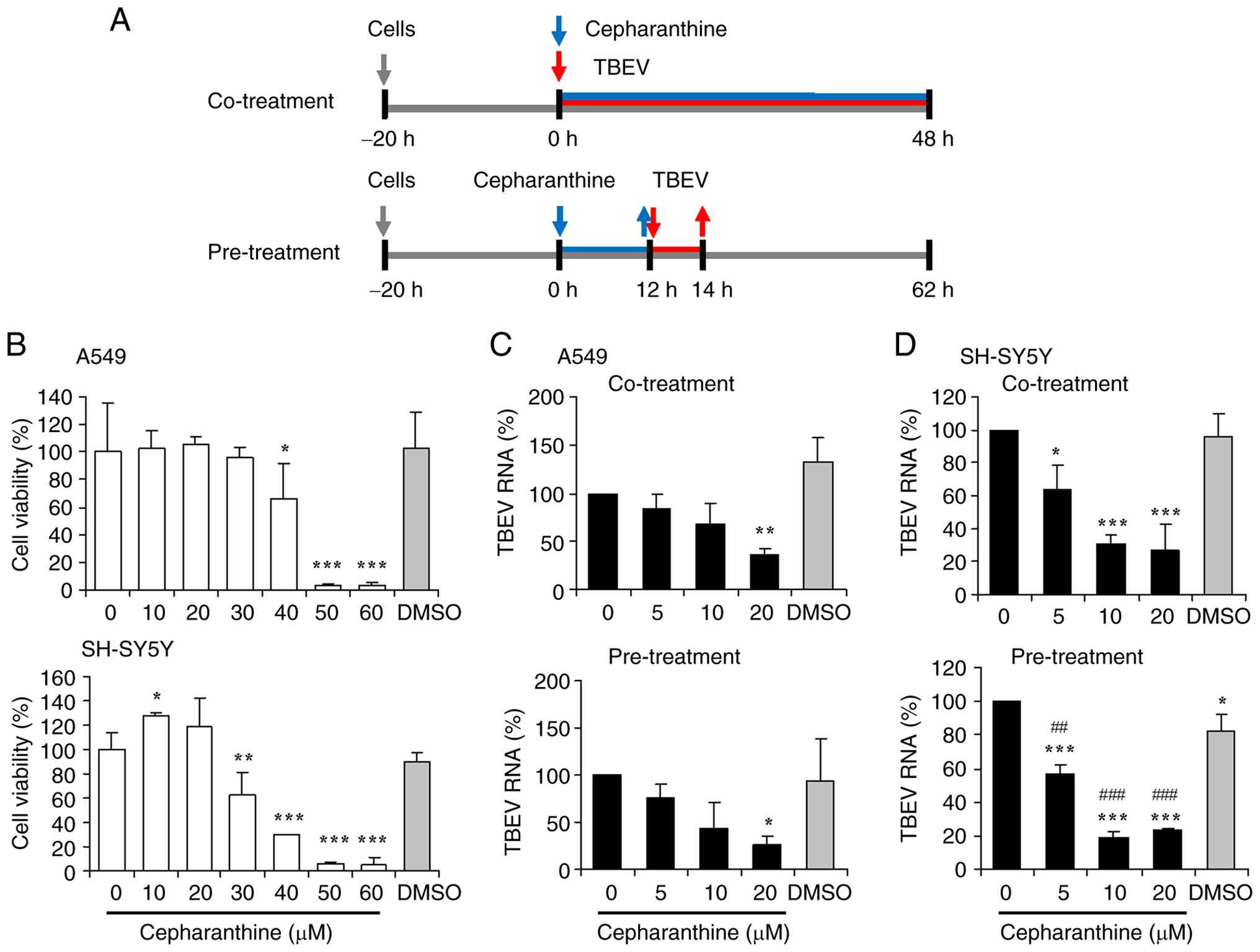
A schematic presentation of the co-treatment and pre-treatment
cepharanthine experiments is shown in Fig. 1A. First, the co-treatment and
pre-treatment experiments were carried out to investigate the
anti-TBEV efficacy of cepharanthine. TBEV RNA replication, viral
protein expression and virus propagation were synchronously
detected in cells following cepharanthine treatment.
It was necessary to analyze the cytotoxicity of
cepharanthine prior to determining its antiviral efficacy. For
this, A549 and SH-SY5Y cells were treated with increasing
concentrations of cepharanthine. MTS assay showed that
cepharanthine was not cytotoxic to A549 cells at concentrations of
up to 30 μM (Fig. 1B). A
significant reduction in A549 cell viability was observed upon
treatment with cepharanthine at concentrations of 40, 50 and 60
μM (P<0.05 or P<0.001). As for SH-SY5Y cells,
cepharanthine did not exert a cytotoxic effect at concentrations of
up to 20 μM. Cepharanthine at concentrations ranging from 30
to 60 μM significantly decreased SH-SY5Y cell viability
(P<0.01 or P<0.001). There was no significant difference in
the cell viability between the DMSO dissolvent and the untreated
control groups. The maximal concentration of 20 μM of
cepharanthine was thereby selected for evaluation of its antiviral
efficacy.
Inhibition of TBEV RNA replication and
protein expression by cepharanthine
Whether co-treatment or pre-treatment with
cepharanthine displays antiviral efficacy against TBEV was next
assessed. The cellular RNA obtained from the infected cells treated
with cepharanthine at the increasing concentrations was analyzed
for TBEV RNA expression. RT-qPCR analysis showed that TBEV RNA
replication was inhibited by cepharanthine in a
concentration-dependent manner (Fig.
1C). The TBEV RNA levels were significantly reduced in the
infected A549 cells with co-treatment or pre-treatment with 20
μM cepharanthine (P<0.05 or P<0.01). There was no
notable reduction in TBEV RNA levels in the infected A549 cells
treated with the DMSO dissolvent. Co-treatment or pre-treatment
with cepharanthine also inhibited TBEV RNA replication in SH-SY5Y
cells (Fig. 1D). For
co-treatment, the TBEV RNA levels were significantly reduced in
SH-SY5Y cells treated with cepharanthine at 5, 10 or 20 μM
(P<0.05 or P<0.001). For pre-treatment, a significant
reduction in TBEV RNA levels was observable in SH-SY5Y cells
treated with the cepharanthine at 5, 10 and 20 μM
(P<0.001 or P<0.0001, respectively). While the TBEV RNA
levels were significantly decreased in SH-SY5Y cells treated with
the DMSO dissolvent (P<0.05; Fig.
1D, pre-treatment) compared with the untreated group, the
cepharanthine at 5, 10 and 20 μM significantly inhibited the
viral RNA replication compared with the DMSO dissolvent (P<0.01
or P<0.001). In summary, co-treatment or pre-treatment with
cepharanthine displayed a potent inhibitory effect on TBEV RNA
replication.
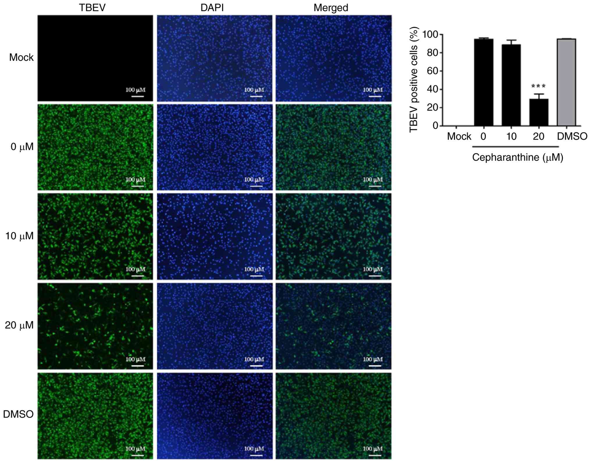
Next, immunofluorescence staining was performed to
detect TBEV protein expression in infected A549 cells with
cepharanthine co-treatment. As shown in Fig. 2, expression of TBEV protein was
detectable in TBEV-infected A549 cells and that treatment with
cepharanthine at 20 μM effectively inhibited such
expression. TBEV protein expression was undetectable in the mock
group. The DMSO group showed a minor influence on viral protein
expression. In parallel with the marked inhibition of TBEV RNA
replication by cepharanthine at 20 μM (Fig. 1C, co-treatment), the percentage
of TBEV-positive cells was significantly decreased upon
cepharanthine treatment (P<0.001). These results demonstrated
that TBEV RNA replication and protein expression were inhibited by
cepharanthine.
Effects of different cepharanthine
treatment conditions on TBEV propagation
The antiviral efficacy of cepharanthine was further
confirmed by plaque assay to detect TBEV titers in the culture
supernatants obtained from cells treated with cepharanthine via
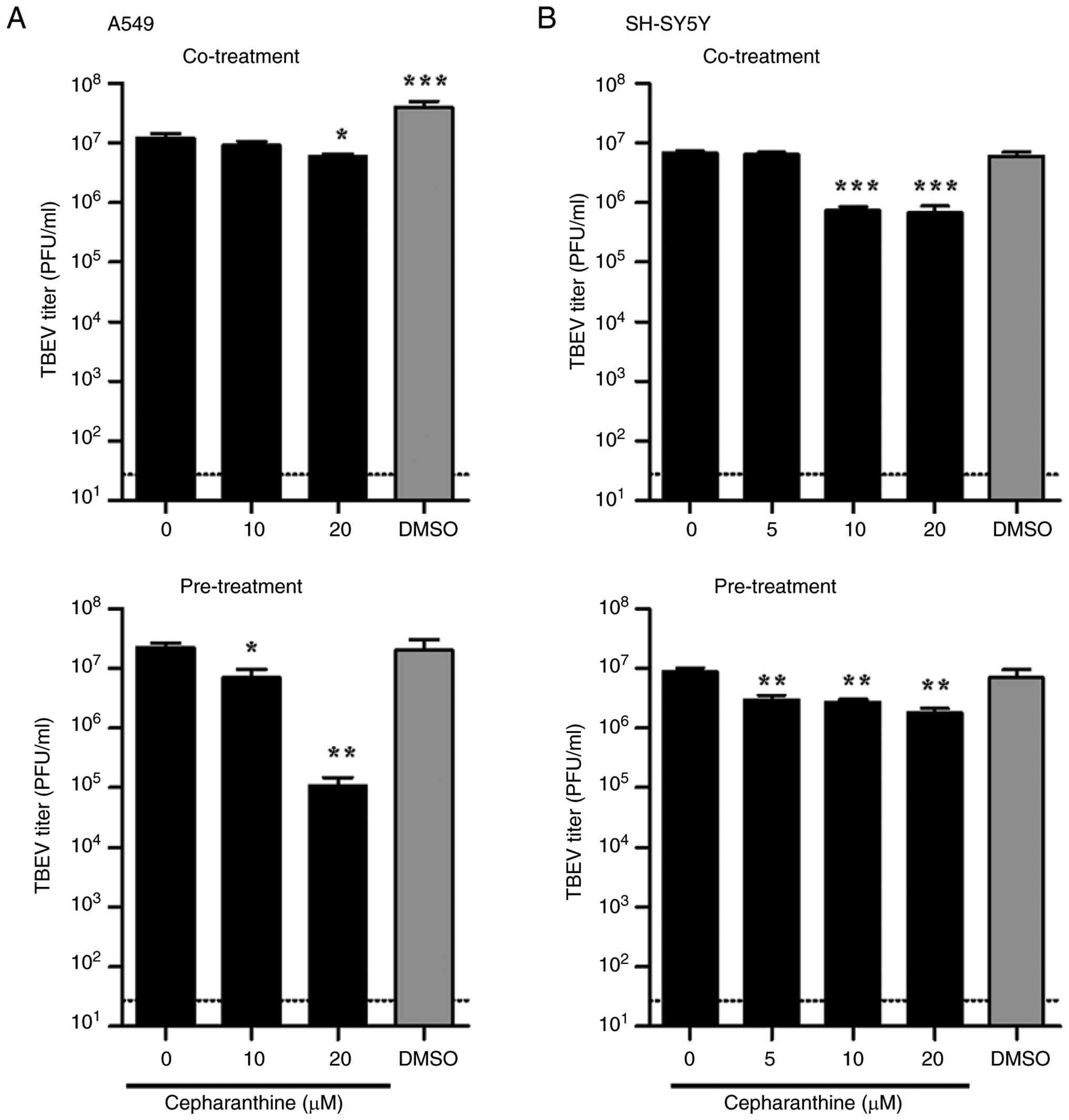
different schemes. The virus titers were significantly decreased in
infected A549 cells co-treated with cepharanthine at 20 μM
(P<0.05) or pre-treated with cepharanthine at 10 and 20
μM (P<0.05 or P<0.01; Fig. 3A). In SH-SY5Y cells, the TBEV
titers were also significantly decreased upon co-treatment with
cepharanthine at 10 and 20 μM (P<0.001) or pre-treatment
with cepharanthine at concentrations ranging from 5 to 20 μM
(P<0.01) (Fig. 3B). The DMSO
group showed no or a minor inhibitory effect on the virus titers,
suggesting that the inhibition of TBEV propagation was attributed
to the cepharanthine treatment. These results were consistent with
the significant inhibition of TBEV RNA replication in A549 and
SH-SY5Y cells co-treated or pre-treated with cepharanthine
(Fig. 1C and D).
In addition to the co-treatment and pre-treatment
schemes, post-treatment of cepharanthine was also adopted for
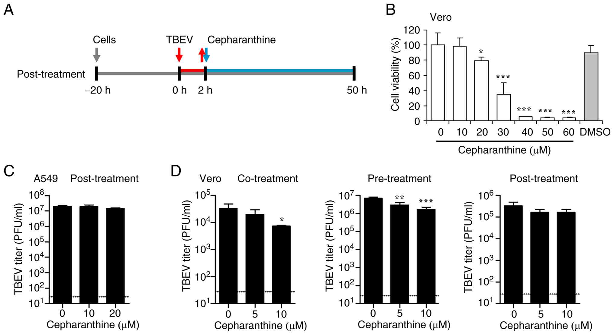
evaluating antiviral efficacy. A schematic presentation of
cepharanthine post-treatment is shown in Fig. 4A. TBEV titers were not influenced
in the A549 cells with cepharanthine post-treatment (Fig. 4C). Furthermore, whether
cepharanthine exhibited antiviral efficacy against TBEV was
analyzed using Vero cells. Cepharanthine at concentrations ranging
from 20 to 60 μM exerted a marked cytotoxic effect on Vero
cells, implying variable cytotoxicity of cepharanthine among
different cell types (Fig. 4B).
Vero cells were thus treated with cepharanthine at 5 and 10
μM for subsequent assays. Plaque assays showed that TBEV
titers were markedly decreased upon co-treatment with cepharanthine
at 10 μM (P<0.05) and that a significant reduction in the
virus titers was observed in Vero cells with cepharanthine
pre-treatment at 5 and 10 μM (P<0.01 or P<0.001)
(Fig. 4D). Conversely,
post-treatment with cepharanthine did not lead to a notable
reduction in TBEV titers.
In A549, SH-SY5Y and Vero cells, TBEV propagation
was inhibited by cepharanthine co-treatment and pre-treatment. This
characterization allowed for further examination of the mechanisms
underlying the effects of cepharanthine on TBEV propagation and
pathogenicity based on the co-treatment and pre-treatment
schemes.
Management of TBEV-induced inflammatory
cytokine release by cepharanthine
Inflammatory cytokines are closely linked to severe
inflammatory symptoms during TBEV infection. In mouse brain and
human neural cells, TBEV is responsible for the elevated production
of a variety of cytokines and chemokines that cause pathological
neuroinflammation (34). In the
present study, the influence of cepharanthine on the TBEV-induced
inflammatory response was evaluated. ELISA was carried out to
detect the production of inflammatory cytokines in the culture
supernatants obtained from infected cells treated with the
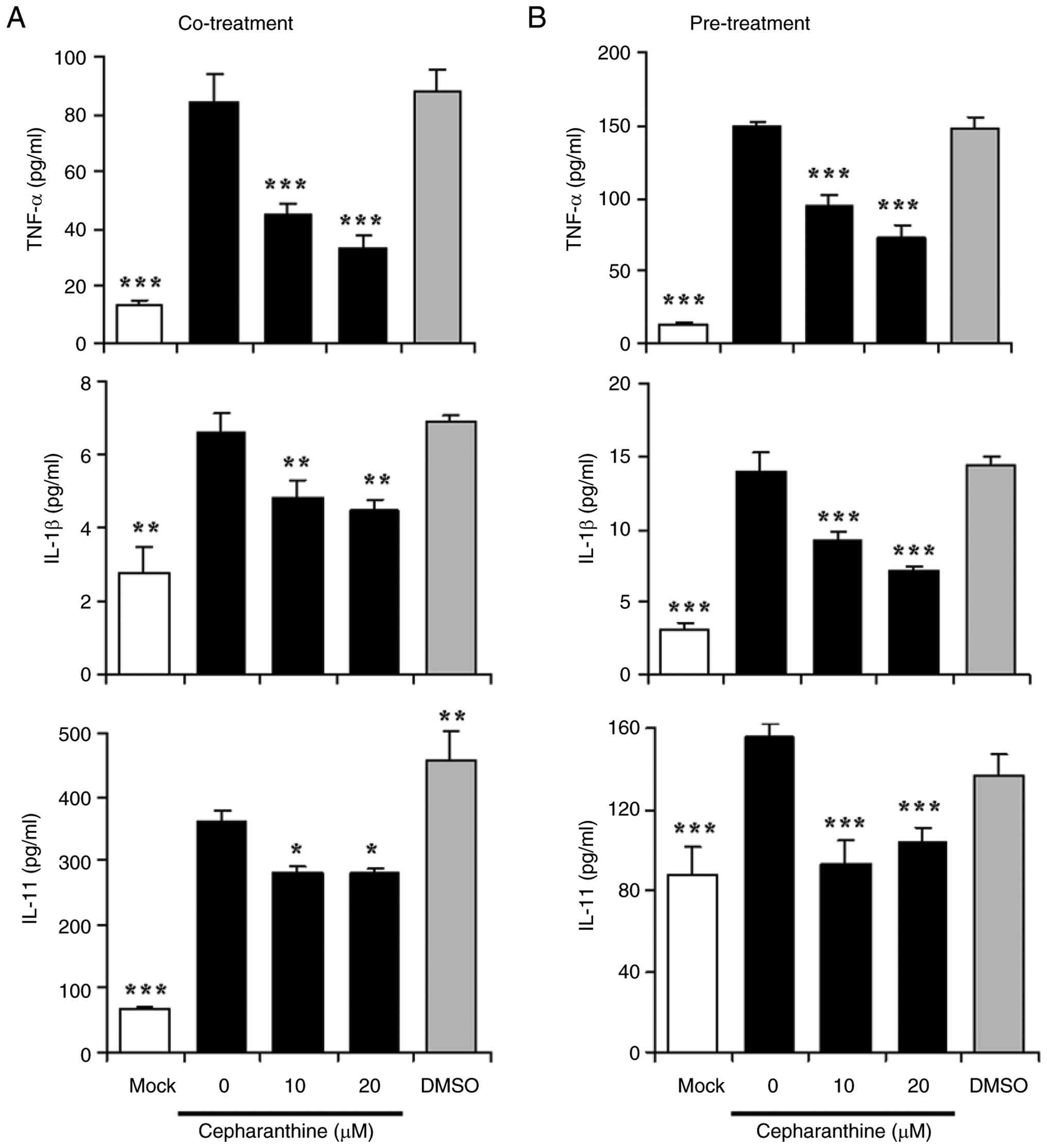
cepharanthine. The levels of TNF-α, IL-1β and IL-11 were notably
increased in TBEV-infected A549 cells compared with the mock group
(P<0.01 or P<0.001; Fig. 5A
and B). The co-treatment with cepharanthine at 10 and 20
μM led to a significant reduction in the production of
TNF-α, IL-1β and IL-11 induced by TBEV (P<0.05, P<0.01 or
P<0.001; Fig. 5A). The
amounts of TNF-α, IL-1β and IL-11 induced by TBEV were
significantly decreased in A549 cells pre-treated with
cepharanthine at 10 and 20 μM (P<0.001; Fig. 5B). DMSO dissolvent showed no
notable inhibitory effects on the production of these cytokines.
The production of TNF-α, IL-1β and IL-11 induced by TBEV was
reduced by co-treatment or pre-treatment with cepharanthine.
CRP is an acute inflammatory protein that plays
important roles in inflammation processes and the host response to
pathogen infection (35).
However, little is known about the contribution of TBEV infection
to CRP release. Therefore, whether CRP release is associated with
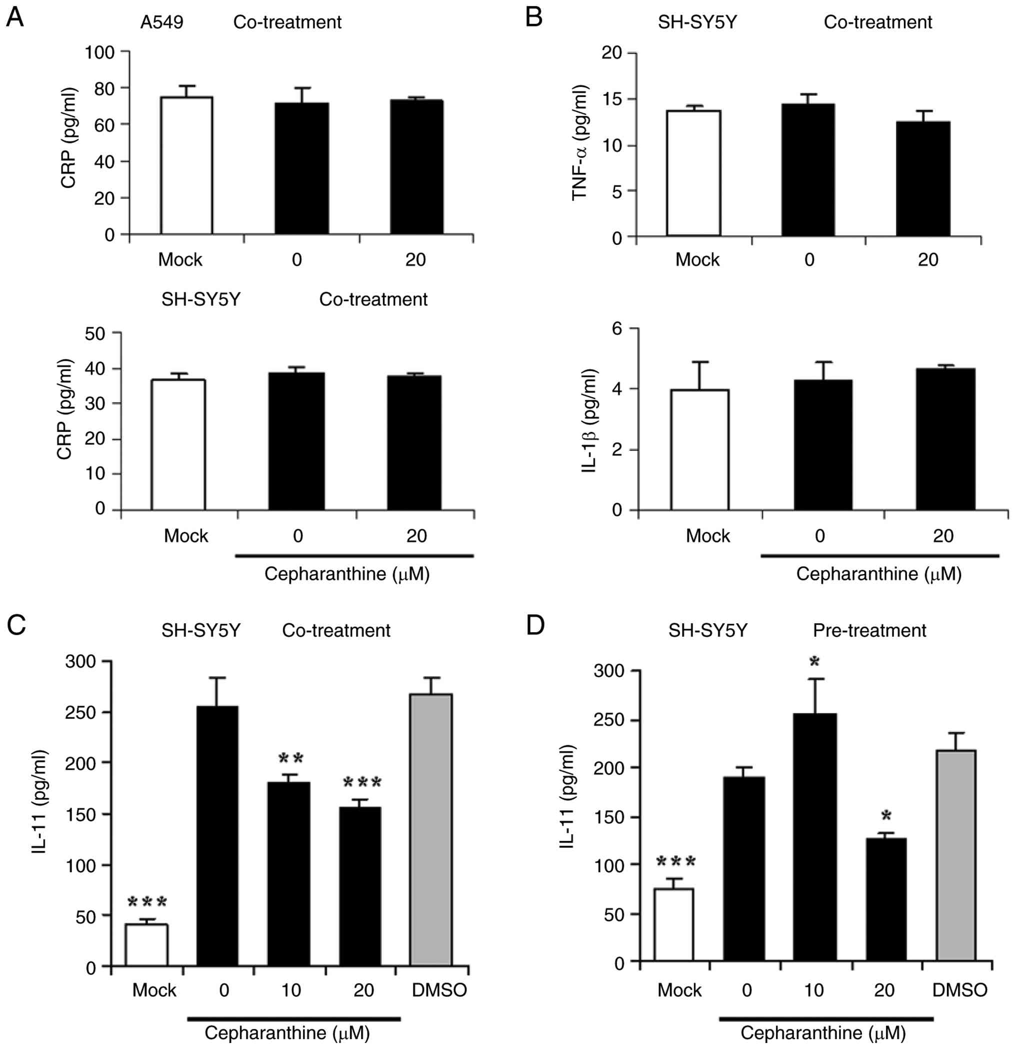
TBEV infection was assessed. As shown in Fig. 6A, TBEV infection showed little
influence on the levels of CRP in A549 and SH-SY5Y cells and
co-treatment with cepharanthine had a minor effect on the CRP
levels, indicating that CRP was not likely involved in the
inflammatory response to TBEV infection. Similarly, the release of
TNF-α and IL-1β was not notably affected in the TBEV-infected
SH-SY5Y cells (Fig. 6B),
suggesting differential regulation of TNF-α and IL-1β by TBEV in a
cell type-dependent manner. There were no notable changes in the
production of TNF-α and IL-1β in infected SH-SY5Y cells co-treated
with cepharanthine. Based on these data, the influence of
cepharanthine pre-treatment on the release of CRP, TNF-α and IL-1β
was not further analyzed in TBEV-infected SH-SY5Y cells. However,
TBEV infection markedly enhanced IL-11 release from SH-SY5Y cells
and a significant reduction in the IL-11 production induced by TBEV
was observed upon co-treatment with cepharanthine at 10 and 20
μM (P<0.01 or P<0.001; Fig. 6C). Pre-treatment with
cepharanthine at 20 μM significantly reduced IL-11
production in infected SH-SY5Y cells (P<0.05; Fig. 6D). Notably, 10 μM
cepharanthine resulted in a significant increase in IL-11
production (P<0.05), implying that IL-11 management may be
related to the cell type and cepharanthine concentration.
Inhibitory effects of the DMSO dissolvent on IL-11 release were
ruled out. These results suggested that co-treatment and
pre-treatment with cepharanthine exhibited differential inhibitory
effects on the TNF-α, IL-1β and IL-11 production induced by
TBEV.
Downregulation of TBEV-induced CHOP
expression by cepharanthine
CHOP, a major component of ER stress-induced
apoptosis, is implicated in the progression of human diseases as
well as in microbial infection processes (36). Host cells maintain ER homeostasis
by triggering the defense response to viral infection, which in
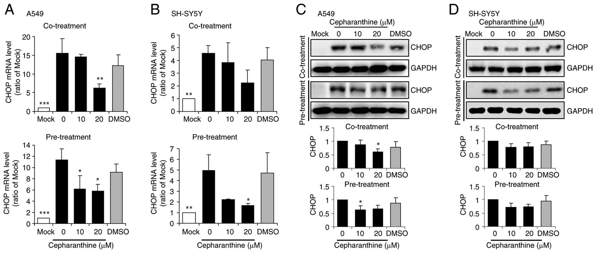
turn impacts viral survival and replication. Fig. 7A showed that CHOP mRNA expression
was significantly upregulated in TBEV-infected A549 cells
(P<0.001). Similarly, significant upregulation of CHOP mRNA
expression was observed in infected SH-SY5Y cells (P<0.01;
Fig. 7B). In infected A549
cells, the CHOP mRNA levels were significantly decreased upon
co-treatment (20 μM; P<0.01) or pre-treatment (10 and 20
μM; P<0.05) with cepharanthine (Fig. 7A). In infected SH-SY5Y cells,
co-treatment as well as pre-treatment with cepharanthine also led
to a significant decrease in the CHOP mRNA levels (P<0.05;
Fig. 7B). The DMSO dissolvent
showed a minor inhibitory effect on the CHOP mRNA expression.
Moreover, the expression of CHOP was verified at the
protein level. Western blot analysis showed that TBEV infection
markedly induced CHOP expression in A549 cells and that
co-treatment (20 μM) or pre-treatment (10 μM) with
cepharanthine significantly reduced such an induction (P<0.05;
Fig. 7C). The CHOP protein
levels were increased in infected SH-SY5Y cells and these levels
were decreased upon treatment with cepharanthine (Fig. 7D), but these results were not
statistically significant. Pre-treatment with cepharanthine
displayed an inhibitory effect on CHOP expression in A549 and
SH-SY5Y cells. Thus, co-treatment and pre-treatment with
cepharanthine effectively downregulated TBEV-induced CHOP
expression.
Enhancement of eIF2α phosphorylation by
cepharanthine during TBEV infection
eIF2α plays pivotal roles in stress response and
eIF2α/ATF4/CHOP signaling is involved in viral infection (37,38). Since CHOP expression was
upregulated in TBEV infected cells, a potential association between
TBEV infection and eIF2α phosphorylation was investigated, together
with an evaluation of the effects of cepharanthine on eIF2α
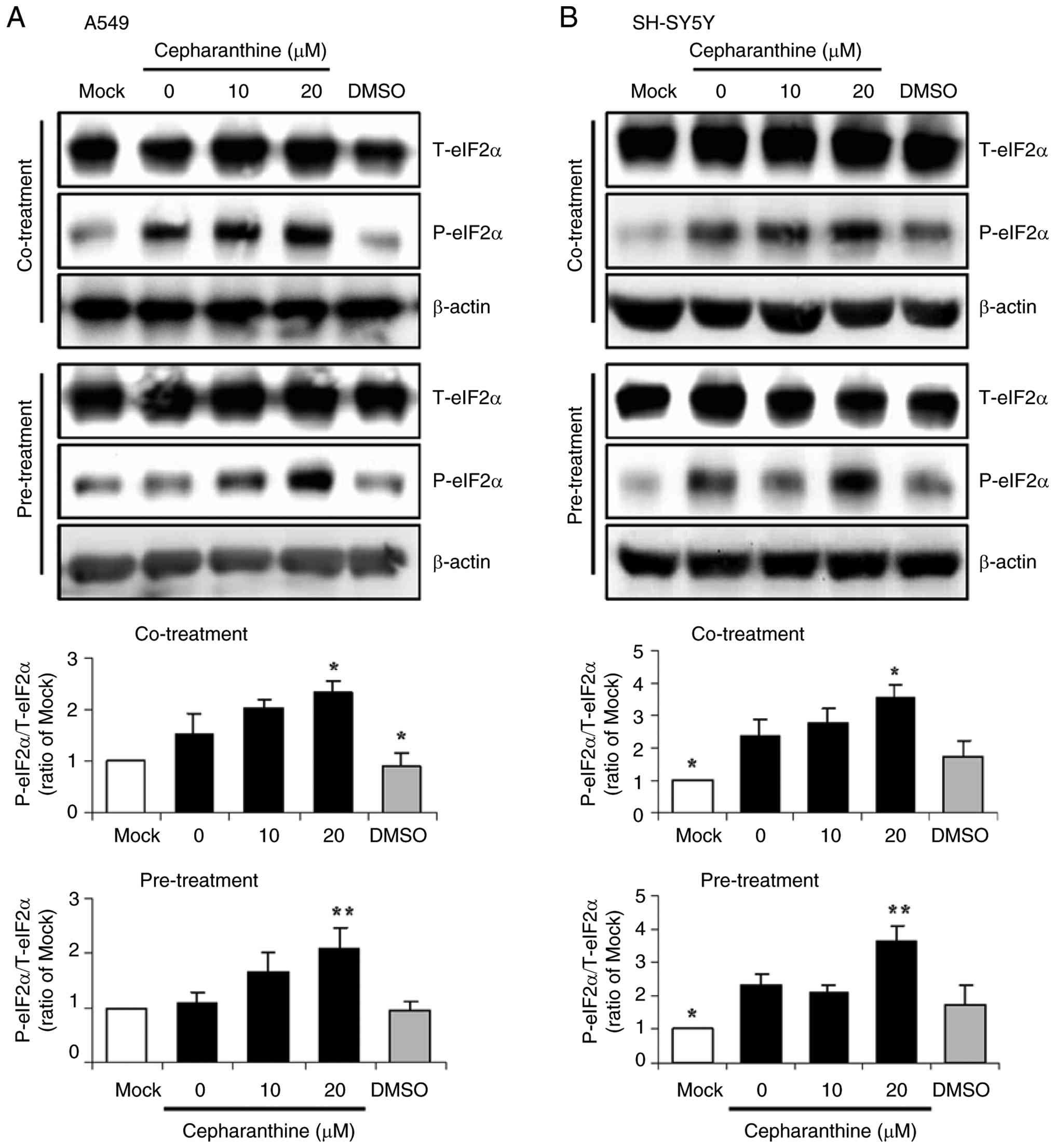
phosphorylation. Western blotting was performed to measure the
total and phosphorylated levels of eIF2α in the cell lysates. In
parallel with the upregulation of CHOP expression (Fig. 7), the levels of phosphorylated
eIF2α were increased in TBEV-infected A549 and SH-SY5Y cells.
Compared with levels of phosphorylated eIF2α in the mock group,
TBEV infection resulted in a notable (co-treatment) or minor
(pre-treatment) elevation in eIF2α phosphorylation in the A549
cells and a marked elevation in the SH-SY5Y cells (Fig. 8A and B), suggesting the
phosphorylation and activation of eIF2α induced by TBEV. The levels
of phosphorylated eIF2α were significantly increased in the A549
and SH-SY5Y cells co-treated or pre-treated with cepharanthine at
20 μM (P<0.05 or P<0.01; Fig. 8A and B). DMSO dissolvent did not
exert enhancement of eIF2α phosphorylation in the infected cells.
Treatment with cepharanthine via the two schemes enhanced eIF2α
phosphorylation during TBEV infection.
Taken together, co-treatment or pre-treatment with
cepharanthine displayed an anti-TBEV effect in A549 and SH-SY5Y
cells. The management of eIF2α phosphorylation by cepharanthine may
contribute to the antiviral effect against TBEV.
Discussion
TBEV infection leads to severe neurological
manifestations and does not have a specific antiviral therapy.
Using a cell-based infection model, we previously performed
high-throughput screening of the Food Drug Administration-approved
drug library consisting of 2,580 compounds for potential anti-TBEV
agents and identified ribavirin and cepharanthine as potent
inhibitors of TBEV replication by immunofluorescence assay (Tang
and Zhao, unpublished data). The anti-TBEV efficacy and mechanisms
of ribavirin and cepharanthine required further investigation using
various experimental systems. We previously reported that
co-treatment or post-treatment with ribavirin exhibited a marked
anti-TBEV effect in A549 and SH-SY5Y cells by upregulating the
expression of myxovirus resistance A and promoting the
phosphorylation of signal transducer and activator of transcription
3, implying that ribavirin primarily inhibits TBEV replication
through the upregulation of antiviral response (26). The present in vitro study
demonstrated that cepharanthine exerted the anti-TBEV and
anti-inflammatory properties by modulating ER stress and
inflammatory response, providing new insights into TBEV-host
interaction and guiding future in vivo studies.
Recent studies have provided evidence for
controlling virus infection using cepharanthine. For instance,
cepharanthine displays anti-porcine deltacoronavirus activity
through prevention of viral entry by competing with porcine
aminopeptidase N protein for viral binding as well as inhibition of
viral replication by downregulating the immune response and
inducing autophagy (39).
Cepharanthine may also impair Japanese encephalitis virus infection
(40). Cepharanthine has proven
to be the top inhibitor for Nipah virus glycoprotein, which plays a
critical role in viral transmission (41). In particular, cepharanthine has
been proposed as a prophylactic and therapeutic agent in preventing
and treating human immunodeficiency virus-associated CNS disorders,
upon its penetration of the CNS and inhibiting release of
inflammatory cytokines that are responsible for CNS damage
(14). Similarly, TBEV infection
disseminates to the CNS and leads to viral encephalitis. These
properties led to the evaluation of the potential anti-TBEV
efficacy of cepharanthine in the present study.
The cytotoxicity of cepharanthine is variable among
cell lines, which may be associated with its impairment of cell
growth by targeting relevant signaling pathways (42). Therefore, in the present study,
the concentrations of cepharanthine ranging from low to the maximal
without cytotoxicity were chosen for the evaluation of antiviral
efficacy. Co-treatment and pre-treatment, but not the
post-treatment, with cepharanthine displayed an anti-TBEV effect.
Therefore, the antiviral effect and underlying mechanism of
cepharanthine were investigated using the co-treatment and
pre-treatment schemes. Compared with co-treatment, pre-treatment
with cepharanthine exhibited a more notable inhibition of TBEV RNA
replication and virus propagation, implying that the early steps of
the viral life cycle, such as attachment, entry and replication,
may be impacted by cepharanthine. The results of the present study
showed that cepharanthine exhibited notable anti-TBEV efficacy,
supporting its broad-spectrum antiviral activities and highlighting
its potential as an antiviral agent against TBEV. This in
vitro finding deserves further research based on various
experimental models.
TBE is a neuropathological disorder characterized by
pathological neuroinflammation. The host inflammatory response
plays a pivotal role in the development and progression of TBE.
TBEV non-structural protein 1 elevates the production of early
pro-inflammatory cytokines such as TNF-α, IL-6 and IL-10 in mouse
splenocytes or mice (43). The
levels of early inflammatory cytokines are differentially increased
in different samples from patients with TBE (44). Consistent with the elicitation of
the inflammatory response by TBEV, the present study showed that
the release of TNF-α, IL-1β and IL-11 was markedly upregulated in
A549 cells while only IL-11 release was upregulated in SH-SY5Y
cells following TBEV infection. However, there were no notable
changes in CRP release in the infected cells. These results
indicated that the release of inflammatory cytokines upon TBEV
infection was dependent on the cytokine and cell type. Since
cepharanthine possesses anti-inflammatory properties, the present
study evaluated the effect of cepharanthine on the inflammatory
response to TBEV infection. Notably, the release of TNF-α, IL-1β
and IL-11 induced by TBEV was significantly downregulated in A549
cells treated with cepharanthine. The marked downregulation of
IL-11 release was detectable in infected SH-SY5Y cells treated with
cepharanthine. Such downregulation of TBEV-induced inflammatory
cytokines by cepharanthine not only represent its anti-inflammatory
property but also run in parallel with its inhibition of TBEV
propagation. Cepharanthine is a therapeutic agent for certain
inflammatory diseases. Cepharanthine inhibits the release of TNF-α,
IL-6 and IL-1β in RAW264.7 cells and downregulates the levels of
cytokines in a mouse acute lung injury model (45). Cepharanthine suppresses
replication of human coronavirus OC43 and reduces the virus-induced
inflammatory response in MRC-5 cells (23). In an acute lung injury rat model,
cepharanthine reduces pulmonary edema and hemorrhage, accompanied
by a reduction in TNF-α and IL-6 levels (46). Additionally, cepharanthine
ameliorates synovial inflammation and reduces TNF-α levels in the
serum and joints of arthritic mice (47). In keeping with these reports, the
findings of the present study indicated that co-treatment or
pre-treatment with cepharanthine differentially downregulated the
release of inflammatory cytokines induced by TBEV, providing in
vitro experimental evidence for the antiviral efficacy of
cepharanthine. The reduction in inflammatory cytokine release by
cepharanthine requires further validation in animal models.
Perturbation of ER homeostasis causes ER stress,
which is involved in a variety of diseases such as diabetes,
ischemia and neurodegenerative disorders (48). CHOP, an important downstream
transcription factor of multiple stress signaling pathways, plays
multifunctional roles in ER stress. Dissection of CHOP function and
regulation during microbial infection has implications for the
development of antimicrobial therapy (36). West Nile virus infection
activates the unfolded protein response and leads to CHOP induction
(49). CHOP expression is
upregulated at the late phase of human astrovirus infection
(50). TBEV infection induces
the gene expression of CHOP in a mouse macrophage cell line
(51). The reports regarding ER stress in TBEV infection are
limited. In the present study, a notable induction of CHOP
expression in infected A549 and SH-SY5Y cells was observed. The
data also established that co-treatment and pre-treatment with
cepharanthine inhibited the TBEV-induced CHOP expression at the
mRNA and protein levels. The management of TBEV-induced CHOP
expression by cepharanthine may contribute to its antiviral effect
against TBEV. Consistent with these findings, suppression of
hepatitis B virus replication by nucleos(t)ide analogs has been
linked to its suppression of CHOP-mediated apoptosis; AMG487
inhibits Japanese encephalitis virus infection in conjunction with
downregulation of the unfolded protein response via CHOP (52,53). The improved understanding of the
interplay between TBEV infection and CHOP expression may assist
with the development of antiviral agents.
Cellular signaling events triggered by viruses are
the subject of intense research due to their importance in the
pathogenesis of viruses. Phosphorylation of eIF2α results in
translation suppression or decline in protein synthesis, and viral
infection triggers cellular stress response, leading to translation
shut-off via eIF2α phosphorylation and viral protein synthesis
arrest (54). A number of
viruses have evolved gene products targeting the protein kinase R
(PKR)/eIF2α pathway, suggesting involvement of this pathway in
antiviral defense (55,56). The results of the present study
showed that TBEV infection increased eIF2α phosphorylation, which
was in line with the upregulation of CHOP expression. Although the
role of eIF2α activation in maintaining ER stress remains to be
explored, the present findings suggest that eIF2α activation may
play a critical role in TBEV pathogenesis. Supporting this,
Newcastle disease virus-induced translation shut-off is attributed
to sustaining the phosphorylation of eIF2α (57). Peste des petits ruminants virus
nucleocapsid protein induces the formation of stress granules
through activation of the PKR/eIF2α pathway (58). The molecular mechanisms
underlying the pharmacological properties of cepharanthine include
the modulation of signaling pathways such as nuclear factor-κB,
apoptosis, mitogen-activated protein kinase (MAPK) and
phosphatidylinositol 3-kinase (PI3K)/protein kinase B
(Akt)/mammalian target of rapamycin (8). Cepharanthine has been identified as
an inhibitor of porcine reproductive and respiratory syndrome virus
by suppressing the integrins/integrin-linked kinase/receptor for
activated C kinase 1/protein kinase C α/nuclear factor-κB signaling
axis (19). The upregulation of
the STING/TBK1/P62 pathway and the inhibition of the PI3K/Akt and
p38 MAPK pathways by cepharanthine are responsible for its
inhibitory effects on herpes simplex virus type 1 (59,60). The treatment of coronavirus
disease 2019 with cepharanthine is associated with its modulation
of PI3K/Akt, relaxin, vascular endothelial growth factor and
hypoxia-induced factor 1 pathways (61). However, the molecular mechanisms
of cepharanthine against TBEV remain unclear. Considering the
involvement of eIF2α activation in ER stress and the inhibition of
TBEV propagation by cepharanthine, the present study examined the
influence of cepharanthine on eIF2α signaling in infected cells.
Consistent with its inhibition of TBEV propagation, co-treatment
and pre-treatment with cepharanthine was sufficient to enhance the
levels of phosphorylated eIF2α, implying that cepharanthine may
exert an anti-TBEV effect via the activation of eIF2α. Although a
direct association between eIF2α activation and decline in TBEV
protein synthesis remains to be elucidated, these results have
implications for antiviral strategies adopted by cepharanthine
through modulating the ER stress to limit TBEV propagation.
However, the present study has some limitations that
need to be addressed in future research. First, evaluation of the
antiviral efficacy of cepharanthine primarily relied on data
generated from human cancer cell lines without validation with
normal human cell lines and in vivo experimental models.
Although the two cell lines are widely used in TBEV research, the
use of normal human cell lines may mimic natural viral infection,
enable efficacy assessment in a cellular environment close to
physiological status and provide a reliable platform for
development of antiviral agents. Future research based on normal
human cell lines remains to be pursued for cross-validation of the
antiviral efficacy of cepharanthine against TBEV. Second, the lack
of representative images for the stained plaque assays is a
concern. Although all the plaque assays were strictly conducted and
the data were statistically analyzed, the visual documentation in
supporting quantitative data is important. Thus, future research
will provide PFU calculation combined with stained cell images for
presentation of TBEV titers to demonstrate reliability and
integrity of the results. Third, the effect of cepharanthine via
the post-treatment scheme was only presented as the changes in the
virus titers, which would be strengthened with analysis of
inflammatory cytokines and the eIF2α/CHOP pathway. Finally,
assessment of other ER stress-related genes and cell apoptosis may
provide important functional insights into the antiviral mechanisms
of cepharanthine. To more comprehensively explore the anti-TBEV
effect and underlying mechanisms of cepharanthine, further studies
are needed to clarify these issues.
Due to the various clinical applications and the
distinct antiviral activities of cepharanthine, the present study
focused on the antiviral effect and underlying mechanisms of
cepharanthine against TBEV in vitro. The findings
demonstrated the antiviral and anti-inflammatory effects of
cepharanthine, together with the management of ER stress response
during TBEV infection and should be further explored as an
antiviral agent against TBEV using animal experiments.
Availability of data and materials
The data generated in the present study may be
requested from the corresponding author.
Authors' contributions
WDT carried out the experiments and contributed to
the analysis and interpretation of the data. LJZ was responsible
for the study design, validation, supervision, funding acquisition,
project administration, conceptualization and writing, reviewing
and editing the manuscript. WDT and LJZ confirm the authenticity of
all the raw data. All authors have read and approved the final
version of the manuscript.
Ethics approval and consent to
participate
Not applicable.
Patient consent for publication
Not applicable.
Competing interests
The authors declare that they have no competing
interests.
Acknowledgements
The authors appreciate Professor Rong Ye (Fudan
University, Shanghai, China) for providing the cell lines and
Professor Rui-Wen Ren (Center for Disease Control and Prevention of
Southern Theater Command, Guangzhou, China) for providing TBEV and
TBEV immunized mouse ascites. The authors thank Professor Ping Zhao
(Naval Medical University, Shanghai, China) for discussions
throughout the work.
Funding
The present study was supported by the National Key Research and
Development Program of China (grant no. 2024YFC2310500).
References
|
1
|
Pierson TC and Diamond MS: The continued
threat of emerging flaviviruses. Nat Microbiol. 5:796–812. 2020.
View Article : Google Scholar : PubMed/NCBI
|
|
2
|
Kwasnik M, Rola J and Rozek W: Tick-borne
encephalitis-review of the current status. J Clin Med. 12:66032023.
View Article : Google Scholar : PubMed/NCBI
|
|
3
|
Chiffi G, Grandgirard D, Leib SL, Chrdle A
and Růžek D: Tick-borne encephalitis: A comprehensive review of the
epidemiology, virology, and clinical picture. Rev Med Virol.
33:e24702023. View
Article : Google Scholar : PubMed/NCBI
|
|
4
|
Dabas R, Sharma N, Taksande AB, Prasad R,
Munjewar PK and Wanjari MB: Breast milk: A potential route of
tick-borne encephalitis virus transmission from mother to infant.
Cureus. 15:e415902023.PubMed/NCBI
|
|
5
|
Ruzek D, AvšičŽupanc T, Borde J, Chrdle A,
Eyer L, Karganova G, Kholodilov I, Knap N, Kozlovskaya L, Matveev
A, et al: Tick-borne encephalitis in Europe and Russia: Review of
pathogenesis, clinical features, therapy, and vaccines. Antiviral
Res. 164:23–51. 2019. View Article : Google Scholar : PubMed/NCBI
|
|
6
|
Yoshii K, Song JY, Park SB, Yang J and
Schmitt HJ: Tick-borne encephalitis in Japan, Republic of Korea and
China. Emerg Microbes Infect. 6:e822017. View Article : Google Scholar : PubMed/NCBI
|
|
7
|
Bailly C: Cepharanthine: An update of its
mode of action, pharmacological properties and medical
applications. Phytomedicine. 62:1529562019. View Article : Google Scholar : PubMed/NCBI
|
|
8
|
Liang D, Li Q, Du L and Dou G:
Pharmacological effects and clinical prospects of cepharanthine.
Molecules. 27:89332022. View Article : Google Scholar : PubMed/NCBI
|
|
9
|
Furusawa S and Wu J: The effects of
biscoclaurine alkaloid cepharanthine on mammalian cells:
Implications for cancer, shock, and inflammatory diseases. Life
Sci. 80:1073–1079. 2007. View Article : Google Scholar : PubMed/NCBI
|
|
10
|
Liu K, Hong B, Wang S, Lou F, You Y, Hu R,
Shafqat A, Fan H and Tong Y: Pharmacological activity of
cepharanthine. Molecules. 28:50192023. View Article : Google Scholar : PubMed/NCBI
|
|
11
|
Rogosnitzky M and Danks R: Therapeutic
potential of the biscoclaurine alkaloid, cepharanthine, for a range
of clinical conditions. Pharmacol Rep. 63:337–347. 2011. View Article : Google Scholar : PubMed/NCBI
|
|
12
|
Tabata R, Tabata C, Tazoh A and Nagai T:
Low dose cepharanthine ameliorates immune thrombocytopenic purpura
associated with multiple myeloma. Int Immunopharmacol. 13:242–244.
2012. View Article : Google Scholar : PubMed/NCBI
|
|
13
|
Xu W, Chen S, Wang X, Tanaka S, Onda K,
Sugiyama K, Yamada H and Hirano T: Molecular mechanisms and
therapeutic implications of tetrandrine and cepharanthine in T cell
acute lymphoblastic leukemia and autoimmune diseases. Pharmacol
Ther. 217:1076592021. View Article : Google Scholar
|
|
14
|
Okamoto M, Ono M and Baba M: Potent
inhibition of HIV type 1 replication by an antiinflammatory
alkaloid, cepharanthine, in chronically infected monocytic cells.
AIDS Res Hum Retroviruses. 14:1239–1245. 1998. View Article : Google Scholar : PubMed/NCBI
|
|
15
|
Liu X, Wang Y, Zhang M, Li G and Cen Y:
Study on the inhibitory effect of cepharanthine on herpes simplex
type-1 virus (HSV-1) in vitro. Zhong Yao Cai. 27:107–110. 2004.In
Chinese. PubMed/NCBI
|
|
16
|
Zhou YB, Wang YF, Zhang Y, Zheng LY, Yang
XA, Wang N, Jiang JH, Ma F, Yin DT, Sun CY and Wang QD: In vitro
activity of cepharanthine hydrochloride against clinical wild-type
and lamivudine-resistant hepatitis B virus isolates. Eur J
Pharmacol. 683:10–15. 2012. View Article : Google Scholar : PubMed/NCBI
|
|
17
|
Toyama M, Hamasaki T, Uto T, Aoyama H,
Okamoto M, Hashmoto Y and Baba M: Synergistic inhibition of
HTLV-1-infected cell proliferation by combination of cepharanthine
and a tetramethylnaphthalene derivative. Anticancer Res.
32:2639–2645. 2012.PubMed/NCBI
|
|
18
|
Xu Y, Zheng J, Sun P, Guo J, Zheng X, Sun
Y, Fan K, Yin W, Li H and Sun N: Cepharanthine and Curcumin
inhibited mitochondrial apoptosis induced by PCV2. BMC Vet Res.
16:3452020. View Article : Google Scholar : PubMed/NCBI
|
|
19
|
Yang C, Zuo Q, Liu X, Zhao Q, Pu H, Gao L,
Zhao L, Guo Z, Lin Y, Liu J, et al: Small molecule screening
identified cepharanthine as an inhibitor of porcine reproductive
and respiratory syndrome virus infection in vitro by suppressing
integrins/ILK/RACK1/PKCα/NF-κB signalling axis. Vet Microbiol.
255:1090162021. View Article : Google Scholar
|
|
20
|
Dong S, Yu R, Wang X, Chen B, Si F, Zhou
J, Xie C, Li Z and Zhang D: Bis-benzylisoquinoline alkaloids
inhibit porcine epidemic diarrhea virus in vitro and in vivo.
Viruses. 14:12312022. View Article : Google Scholar : PubMed/NCBI
|
|
21
|
Chen Y, You Y, Wang S, Jiang L, Tian L,
Zhu S, An X, Song L, Tong Y and Fan H: Antiviral drugs screening
for swine acute diarrhea syndrome coronavirus. Int J Mol Sci.
23:112502022. View Article : Google Scholar : PubMed/NCBI
|
|
22
|
Phumesin P, Panaampon J, Kariya R,
Limjindaporn T, Yenchitsomanus PT and Okada S: Cepharanthine
inhibits dengue virus production and cytokine secretion. Virus Res.
325:1990302023. View Article : Google Scholar : PubMed/NCBI
|
|
23
|
Kim DE, Min JS, Jang MS, Lee JY, Shin YS,
Song JH, Kim HR, Kim S, Jin YH and Kwon S: Natural
bis-benzylisoquinoline alkaloids-tetrandrine, fangchinoline, and
cepharanthine, inhibit human coronavirus OC43 infection of MRC-5
human lung cells. Biomolecules. 9:6962019. View Article : Google Scholar : PubMed/NCBI
|
|
24
|
Fan H, He ST, Han P, Hong B, Liu K, Li M,
Wang S and Tong Y: Cepharanthine: A promising old drug against
SARS-CoV-2. Adv Biol (Weinh). 6:e22001482022. View Article : Google Scholar : PubMed/NCBI
|
|
25
|
Rogosnitzky M, Okediji P and Koman I:
Cepharanthine: A review of the antiviral potential of a
Japanese-approved alopecia drug in COVID-19. Pharmacol Rep.
72:1509–1516. 2020. View Article : Google Scholar : PubMed/NCBI
|
|
26
|
Tang WD, Tang HL, Peng HR, Ren RW, Zhao P
and Zhao LJ: Inhibition of tick-borne encephalitis virus in cell
cultures by ribavirin. Front Microbiol. 14:11827982023. View Article : Google Scholar : PubMed/NCBI
|
|
27
|
Livak KJ and Schmittgen TD: Analysis of
relative gene expression data using real-time quantitative PCR and
the 2(-Delta Delta C(T)) method. Methods. 25:402–408. 2001.
View Article : Google Scholar
|
|
28
|
Hoornweg TE, Godeke GJ, Hoogerwerf MN, van
Kasteren PB, de Vries A, Sprong H, Verjans GMGM, van Riel D,
Reimerink JHJ, Rockx B and Reusken CBEM: Rescue and in vitro
characterization of a divergent TBEV-Eu strain from the
Netherlands. Sci Rep. 13:28722023. View Article : Google Scholar : PubMed/NCBI
|
|
29
|
Krol E, Wandzik I, Brzuska G, Eyer L,
Růžek D and Szewczyk B: Antiviral activity of uridine derivatives
of 2-deoxy sugars against tick-borne encephalitis virus. Molecules.
24:11292019. View Article : Google Scholar : PubMed/NCBI
|
|
30
|
Dias SSG, Cunha-Fernandes T, Souza-Moreira
L, Soares VC, Lima GB, Azevedo-Quintanilha IG, Santos J,
Pereira-Dutra F, Freitas C, Reis PA, et al: Metabolic reprogramming
and lipid droplets are involved in Zika virus replication in neural
cells. J Neuroinflammation. 20:612023. View Article : Google Scholar : PubMed/NCBI
|
|
31
|
Ojha D, Winkler CW, Leung JM, Woods TA,
Chen CZ, Nair V, Taylor K, Yeh CD, Tawa GJ, Larson CL, et al:
Rottlerin inhibits La Crosse virus-induced encephalitis in mice and
blocks release of replicating virus from the Golgi body in neurons.
Nat Microbiol. 6:1398–1409. 2021. View Article : Google Scholar : PubMed/NCBI
|
|
32
|
Aoki-Utsubo C, Chen M and Hotta H:
Time-of-addition and temperature-shift assays to determine
particular step(s) in the viral life cycle that is blocked by
antiviral substance(s). Bio Protoc. 8:e28302018. View Article : Google Scholar : PubMed/NCBI
|
|
33
|
Krylova NV, Leonova GN, Maystrovskaya OS,
Popov AM and Artyukov AA: Mechanisms of antiviral activity of the
polyphenol complex from seagrass of the Zosteraceae family against
tick-borne encephalitis virus. Bull Exp Biol Med. 165:61–63. 2018.
View Article : Google Scholar : PubMed/NCBI
|
|
34
|
PokornaFormanova P, Palus M, Salat J,
Hönig V, Stefanik M, Svoboda P and Ruzek D: Changes in cytokine and
chemokine profiles in mouse serum and brain, and in human neural
cells, upon tick-borne encephalitis virus infection. J
Neuroinflammation. 16:2052019. View Article : Google Scholar
|
|
35
|
Sproston NR and Ashworth JJ: Role of
C-reactive protein at sites of inflammation and infection. Front
Immunol. 9:7542018. View Article : Google Scholar : PubMed/NCBI
|
|
36
|
Hu H, Tian M, Ding C and Yu S: The C/EBP
homologous protein (CHOP) transcription factor functions in
endoplasmic reticulum stress-induced apoptosis and microbial
infection. Front Immunol. 9:30832019. View Article : Google Scholar : PubMed/NCBI
|
|
37
|
Boye E and Grallert B: eIF2α
phosphorylation and the regulation of translation. Curr Genet.
66:293–297. 2020. View Article : Google Scholar
|
|
38
|
Liu Y, Wang M, Cheng A, Yang Q, Wu Y, Jia
R, Liu M, Zhu D, Chen S, Zhang S, et al: The role of host eIF2α in
viral infection. Virol J. 17:1122020. View Article : Google Scholar
|
|
39
|
Sun Y, Liu Z, Shen S, Zhang M, Liu L,
Ghonaim AH, Li Y, Zhang S and Li W: Inhibition of porcine
deltacoronavirus entry and replication by cepharanthine. Virus Res.
340:1993032024. View Article : Google Scholar :
|
|
40
|
Yin C, Yang P, Xiao Q, Sun P, Zhang X,
Zhao J, Hu X and Shan C: Novel antiviral discoveries for Japanese
encephalitis virus infections through reporter virus-based
high-throughput screening. J Med Virol. 96:e293822024. View Article : Google Scholar : PubMed/NCBI
|
|
41
|
Ebrahimi M and Alijanianzadeh M:
Evaluation of the interaction between potent small molecules
against the Nipah virus Glycoprotein in Malaysia and Bangladesh
strains, accompanied by the human Ephrin-B2 and Ephrin-B3
receptors; a simulation approach. Mol Divers. 28:851–874. 2024.
View Article : Google Scholar
|
|
42
|
Takahashi-Makise N, Suzu S, Hiyoshi M,
Ohsugi T, Katano H, Umezawa K and Okada S: Biscoclaurine alkaloid
cepharanthine inhibits the growth of primary effusion lymphoma in
vitro and in vivo and induces apoptosis via suppression of the
NF-kappaB pathway. Int J Cancer. 125:1464–1472. 2009. View Article : Google Scholar : PubMed/NCBI
|
|
43
|
Starodubova E, Tuchynskaya K, Kuzmenko Y,
Latanova A, Tutyaeva V, Karpov V and Karganova G: Activation of
early proinflammatory responses by TBEV NS1 varies between the
strains of various subtypes. Int J Mol Sci. 24:10112023. View Article : Google Scholar : PubMed/NCBI
|
|
44
|
Zidovec-Lepej S, Vilibic-Cavlek T, Ilic M,
Gorenec L, Grgic I, Bogdanic M, Radmanic L, Ferenc T, Sabadi D,
Savic V, et al: Quantification of antiviral cytokines in serum,
cerebrospinal fluid and urine of patients with tick-borne
encephalitis in Croatia. Vaccines (Basel). 10:18252022. View Article : Google Scholar : PubMed/NCBI
|
|
45
|
Huang H, Hu G, Wang C, Xu H, Chen X and
Qian A: Cepharanthine, an alkaloid from Stephania cepharantha
Hayata, inhibits the inflammatory response in the RAW264.7 cell and
mouse models. Inflammation. 37:235–246. 2014. View Article : Google Scholar
|
|
46
|
Liang D, Wang W, Chen G, Li J, Dou G, Gan
H, Han P, Du L and Gu R: Cepharanthine dry powder inhaler for the
treatment of acute lung injury. Molecules. 28:44412023. View Article : Google Scholar : PubMed/NCBI
|
|
47
|
Lu C, Cheng RJ, Zhang Q, Hu Y, Pu Y, Wen
J, Zhong Y, Tang Z, Wu L, Wei S, et al: Herbal compound
cepharanthine attenuates inflammatory arthritis by blocking
macrophage M1 polarization. Int Immunopharmacol. 125:1111752023.
View Article : Google Scholar : PubMed/NCBI
|
|
48
|
Oyadomari S and Mori M: Roles of
CHOP/GADD153 in endoplasmic reticulum stress. Cell Death Differ.
11:381–389. 2004. View Article : Google Scholar
|
|
49
|
Medigeshi GR, Lancaster AM, Hirsch AJ,
Briese T, Lipkin WI, Defilippis V, Früh K, Mason PW,
Nikolich-Zugich J and Nelson JA: West Nile virus infection
activates the unfolded protein response, leading to CHOP induction
and apoptosis. J Virol. 81:10849–10860. 2007. View Article : Google Scholar : PubMed/NCBI
|
|
50
|
Isobe T, Tange S, Tasaki H, Kanamori K,
Kato A and Nakanishi A: Upregulation of CHOP participates in
caspase activation and virus release in human astrovirus-infected
cells. J Gen Virol. 100:778–792. 2019. View Article : Google Scholar : PubMed/NCBI
|
|
51
|
Beránková Z, Khanna R, Spěváková M,
Langhansová H, Kopecký J and Lieskovská J: Cellular stress is
triggered by tick-borne encephalitis virus and limits the virus
replication in PMJ2-R mouse macrophage cell line. Ticks Tick Borne
Dis. 15:1022692024. View Article : Google Scholar
|
|
52
|
Liang YJ, Chiou YW, Chiu AP, Shiao MS,
Teng W, Lin CW, Cheng ML, Huang YH, Liang KH, Su CW, et al:
Antiviral therapy reduces hepatocellular carcinoma through
suppressing hepatitis B virus replication may improve ER stress,
mitochondrial and metabolic dysfunctions and decrease p62 in
hybridized mice with single HBV transgene and miR-122. J Med Virol.
95:e293252023. View Article : Google Scholar : PubMed/NCBI
|
|
53
|
Singh A, Ghosh R and Guchhait P: CXCR3
antagonist rescues ER stress and reduces inflammation and JEV
infection in mice brain. Cytokine. 172:1563802023. View Article : Google Scholar : PubMed/NCBI
|
|
54
|
Donnelly N, Gorman AM, Gupta S and Samali
A: The eIF2α kinases: Their structures and functions. Cell Mol Life
Sci. 70:3493–3511. 2013. View Article : Google Scholar : PubMed/NCBI
|
|
55
|
Sharon E and Frenkel N: Human herpesvirus
6A exhibits restrictive propagation with limited activation of the
protein kinase R-eIF2α stress pathway. J Virol. 91:e02120–16. 2017.
View Article : Google Scholar
|
|
56
|
Li H, Liu S, Feng Q, Deng R, Wang J, Wang
X, Tian R, Xu Y, Chen S, Liu Q, et al: Regulation of PKR-dependent
RNA translation inhibition by TRIM21 upon virus infection or other
stress. PLoS Pathog. 19:e10114432023. View Article : Google Scholar : PubMed/NCBI
|
|
57
|
Liao Y, Gu F, Mao X, Niu Q, Wang H, Sun Y,
Song C, Qiu X, Tan L and Ding C: Regulation of de novo translation
of host cells by manipulation of PERK/PKR and GADD34-PP1 activity
during Newcastle disease virus infection. J Gen Virol. 97:867–879.
2016. View Article : Google Scholar : PubMed/NCBI
|
|
58
|
Chen S, Yang F, Cao W, Zhang X, Liu H,
Zhang K, Liu X, Wang J, Zheng H and Zhu Z: Peste des petits
ruminants virus nucleocapsid protein interacts with protein kinase
R-activating protein and induces stress granules to promote viral
replication. J Virol. 97:e01712222023. View Article : Google Scholar : PubMed/NCBI
|
|
59
|
Liu Y, Chen L, Liu W, Li D, Zeng J, Tang
Q, Zhang Y, Luan F and Zeng N: Cepharanthine suppresses herpes
simplex virus type 1 replication through the downregulation of the
PI3K/Akt and p38 MAPK signaling pathways. Front Microbiol.
12:7957562021. View Article : Google Scholar : PubMed/NCBI
|
|
60
|
Liu Y, Tang Q, Rao Z, Fang Y, Jiang X, Liu
W, Luan F and Zeng N: Inhibition of herpes simplex virus 1 by
cepharanthine via promoting cellular autophagy through
up-regulation of STING/TBK1/P62 pathway. Antivir Res.
193:1051432021. View Article : Google Scholar : PubMed/NCBI
|
|
61
|
Jiang P, Ye J, Jia M, Li X, Wei S and Li
N: The common regulatory pathway of COVID-19 and multiple
inflammatory diseases and the molecular mechanism of cepharanthine
in the treatment of COVID-19. Front Pharmacol. 13:9602672022.
View Article : Google Scholar : PubMed/NCBI
|