Introduction
Osteoporosis (OP) is a common skeletal disorder
involving reduction in bone strength, posing high risk of fracture.
Worldwide, it is estimated to affect more than 200 million
individuals, making it a major public health issue (1). The burden of OP is especially in
older individuals, and women are particularly affected by the
disease, with a higher rate of affected patients and a higher
number of disabilities-adjusted life years (DALYs) than men.
Analyses from the Global Burden of Study found that there were 17.2
million DALYs associated with low bone mineral density (BMD), the
main determinant of OP, in 2020 (2). This increasing prevalence equates
to a staggering number of fractures, with more than 8.9 million
osteoporotic fractures occurring every year, with epidemiological
estimates indicating that an osteoporotic fracture occurs
approximately every three seconds globally (1). These fractures not only affect
quality of life, they also have a major economic impact; for
example, a recent multinational real world study reported
significant healthcare costs occurring with fracture management
(3). Furthermore, OP is often
accompanied by other chronic diseases, such as chronic kidney
disease, rheumatoid arthritis and Parkinson's disease, complicating
its management even further (4-6).
Despite the improvements in the technology used for assessments and
in the pharmacological treatments available, there are still major
gaps in clinical care. Notably, the recommendations for OP
management are somewhat different in different parts of the world,
especially in the Asia-Pacific where systematic reviews have shown
a wide variation in treatment and fracture risk management
(7). Even if effective therapies
are available, problems still exist, including poor patient
adherence and rare but serious adverse effects, as well as
pronounced inter-individual variability in treatment response.
These limitations have fueled increasing interest in a
'treat-to-target' approach, which seeks not only to prevent
fracture but to restore pre-fracture function and minimize future
fracture risk. Achieving such approach requires more personalized
and metabolically informed interventions. In recent years, the gut
microbiota has become a key regulator of the bone and is considered
to constitute a 'metabolic organ' and communicate bi-directional
with the skeleton. The gut-bone axis acts through various
mechanisms such as immune modulation (including modulation of
T-cell differentiation and systemic inflammation), endocrine
signaling (nutrient absorption and hormone production), neural
pathways, and metabolic crosstalk, in which metabolites produced by
microbes directly influence bone cells (8). Long-term dietary behaviors and
nutritional status influence the gut microbiome and in turn, bone
health and disease indirectly. Animal models, including germ-free
mice and models exposed to antibiotic treatment, present strong
evidence that changes in, or loss of, the gut microbiota greatly
affect BMD and micro-architecture, demonstrating causality
(9). Within this regulatory
network, metabolites produced by the microbes play key roles.
Short-chain fatty acids (SCFAs), specifically acetate, propionate
and butyrate, are formed through by bacterial fermentation of
dietary fibers and have been shown to regulate bone metabolism.
Preclinical studies suggested that SCFAs are able to inhibit
osteoclastogenesis, enhance osteoblast activity, and modulate
immune responses. Emerging clinical data also suggests beneficial
effects on bone turnover markers in post-menopausal women (10). In addition, recent studies have
pointed out the importance of polyamines (for example, spermidine
and spermine) produced by gut microbes, which can directly improve
osteoblast differentiation and suppress osteoclast activities. To
elucidate the exact role of all these metabolites, the use of
advanced metabolomics is indispensable. Comprehensive profiling of
gut-derived compounds, instead of the identification of microbial
taxa alone, uncovers a deeper mechanistic insight into how specific
metabolites impact bone biology. This metabolite-centric approach
holds promise for discovering novel therapeutic opportunities as
well as targeted interventions within the gut-bone axis for
improving skeletal health and complementing the existing treatments
for OP. While existing reviews of the gut-bone axis have addressed
microbiota composition, estrogen metabolism and broad immune
mechanisms, the present review uniquely focuses on the
metabolite-centric perspective, providing an integrated mechanistic
and translational synthesis. Specifically, (i) the
receptor-mediated (GPR43 and GPR109A) and epigenetic (HDAC
inhibition) mechanisms by which SCFAs regulate osteoblast and
osteoclast function were delineated; (ii) polyamine biosynthesis,
catabolism and direct bone-cell effects including the novel eIF5A
hypusination pathway were characterized; (iii) microbial
metabolomics platforms and their annotation challenges in the bone
context were evaluated; and (iv) a hierarchical translational
framework was proposed spanning dietary prebiotics, probiotics,
postbiotics and next-generation pharmacological strategies. This
metabolite-centric approach offers actionable insights for
developing precision interventions targeting the gut-bone axis.
Gut-bone axis from compositional dysbiosis
to functional signaling
Dysbiosis in OP: Compositional and
functional shifts
The pathogenesis of OP is increasingly recognized to
involve alterations in gut microbiota composition and function,
this phenomenon called as 'dysbiosis'. Two-sample Mendelian
randomization (MR) studies have provided genetic evidence that
specific gut taxa are causally linked to BMD, rather than merely
being correlated. For example, one MR study identified multiple
microbial taxa whose genetically predicted abundance is associated
with lumbar spine, forearm and femoral neck BMD (11-13). Moreover, reverse causality was
excluded in these analyses, strengthening the case for a direct gut
bone effect (12).
In osteoporotic individuals, dysbiosis often
manifests as reduced diversity (alpha- and beta-diversity) and loss
of beneficial SCFA-producing bacteria such as those belonging to
Clostridium clusters IV and XIVa, as well as
Faecalibacterium prausnitzii (F. prausnitzii)
(14,15). Observational and mechanistic
studies suggest that such losses impair butyrate production,
shifting the microbial milieu toward pro-inflammatory species (for
example, Proteobacteria), which may increase circulating
lipopolysaccharide (LPS) and systemic endotoxemia thereby creating
a pro-resorptive environment (10,15). The contrasting physiological
landscapes of the healthy (eubiotic) vs. osteoporotic (dysbiotic)
gut-bone axis, highlighting these immunological and metabolic
shifts, are illustrated in Fig.
1.
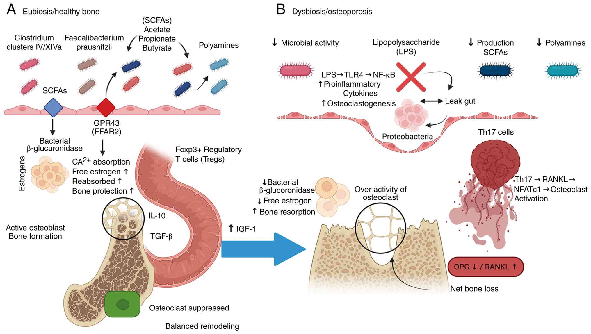 | Figure 1Gut-bone axis crosstalk in eubiosis
and dysbiosis. (A) In eubiosis, fermentation of dietary fiber by
Clostridium clusters IV/XIVa and Faecalibacterium
prausnitzii produces SCFAs (acetate, propionate and butyrate)
and polyamines. SCFAs activate GPR43 (FFAR2) on intestinal
epithelial cells, enhancing Ca2+ absorption and barrier
integrity. Bacterial β-glucuronidase deconjugates estrogens,
promoting enterohepatic recirculation and bone protection.
SCFA-driven Foxp3+ Treg expansion sustains IL-10/TGF-β
secretion, suppressing osteoclastogenesis and supporting balanced
bone remodeling. (B) Dysbiosis depletes SCFA-producing taxa and
expands pro-inflammatory Proteobacteria, compromising epithelial
barrier integrity. LPS translocation activates TLR4 → NF-κB
signaling, elevating pro-inflammatory cytokines and driving
osteoclastogenesis. Reduced β-glucuronidase activity impairs
estrogen recirculation. Th17 expansion upregulates RANKL →
NFATc1-mediated osteoclast activation. The resulting OPG↓/RANKL↑
imbalance shifts bone remodeling toward net bone loss. SCFAs,
short-chain fatty acids; LPS, lipopolysaccharide; Tregs, regulatory
T cells; OPG, osteoprotegerin. |
Microbial metabolite pathways: From
substrates to systemic mediators
One of the central routes by which dysbiosis
contributes to bone loss is via microbial metabolites. SCFAs
particularly acetate, propionate and butyrate are generated by
bacterial fermentation of dietary fibers [such as inulin, pectin
and resistant starch (RS)] via pathways including the acetyl-CoA
route. The efficiency of SCFA production depends on the abundance
and metabolic capacity of SCFA-producing taxa; when these taxa
decline, SCFA output and their systemic bioavailability fall.
Beyond carbohydrate fermentation, gut bacteria also
metabolize amino acids into bioactive compounds. For instance,
arginine and glutamate metabolism can lead to the synthesis of
polyamines (for example, spermidine and putrescine), which have
been shown in animal models to enhance osteoblast differentiation
and suppress osteoclast activity, thereby supporting bone formation
(16).
Other important pathways include the microbial
conversion of primary bile acids into secondary bile acids, which
signal via receptors such as the farnesoid X receptor (FXR) and
TGR5. Activation of these receptors can modulate immune responses
and bone remodeling (17,18).
Similarly, tryptophan metabolism by the microbiota gives rise to
indole derivatives that act as ligands for the aryl hydrocarbon
receptor (AhR). Through AhR signaling, these molecules regulate
differentiation of Th17 cells and influence osteoclastogenesis and
bone turnover (19).
Metabolite bioavailability depends on efficient
absorption in the colon. SCFAs are taken up by colonocytes via
monocarboxylate transporters (for example, MCT1 and MCT4) and
paracellular routes, and their absorption is sensitive to changes
in colonic pH. Dysbiosis-induced alterations in fermentation and pH
may impair SCFA uptake, exacerbating the deficit.
RANKL/OPG axis and Th17 dysregulation:
Osteoimmunology link
A key immunological mechanism by which dysbiosis
affects bone is through an imbalance in the RANKL/OPG axis,
mediated by T-cell subsets. In post-menopausal OP, estrogen
deficiency is associated with gut dysbiosis, which in turn promotes
expansion of Th17 cells. These Th17 cells produce
pro-osteoclastogenic cytokines including RANKL and TNF-α driving
bone resorption. Conversely, SCFA-producing bacteria (especially
butyrate producers) foster the differentiation and expansion of
Foxp3+ regulatory T cells (Tregs). Butyrate acts via
inhibition of histone deacetylases (HDACs), promoting Foxp3
acetylation and stable Treg lineage commitment. These Tregs secrete
anti-inflammatory cytokines such as IL-10 and TGF-β, which suppress
Th17 differentiation and blunt RANKL expression, thus countering
bone loss (20,21).
IGF-1 and calcium bioavailability:
Nutrient-level consequences of dysbiosis
Beyond immune regulation, dysbiosis impairs bone
health through effects on nutrient absorption and anabolic
bone-hormone signaling. Insulin-like growth factor-1 (IGF-1) is a
central anabolic hormone that promotes osteoblast differentiation
and bone formation, while suppressing osteoclastogenesis.
SCFA-mediated signaling, particularly via GPR43 and GPR109A on
hepatocytes and intestinal epithelial cells, has been shown to
upregulate serum IGF-1; therefore, a dysbiosis-associated reduction
in SCFA production can lead to diminished circulating IGF-1 and
impaired bone anabolism. Recent reviews highlight the critical role
of microbiota-derived SCFAs in modulating IGF-1 levels and
consequently supporting bone formation (20,22).
Furthermore, fermentation of dietary fiber by
SCFA-producing bacteria generates organic acids that acidify the
colonic lumen, decreasing pH and thereby increasing the solubility
and bioavailability of dietary calcium. This acidification, driven
by acetate, propionate and butyrate, enhances passive paracellular
calcium absorption. Dysbiosis-associated depletion of these
SCFA-producing bacteria causes colonic pH to rise, promoting
calcium precipitation and reducing its bioavailability, which
impairs the mineral substrate necessary for bone formation
(21).
Estrobolome: Estrogen metabolism and
dysbiosis
The estrobolome, the aggregate genetic and metabolic
capacity of the gut microbiota to metabolize estrogens, represents
a key mechanism linking dysbiosis to post-menopausal bone loss.
Estrogens, after liver conjugation (to glucuronides/sulfates), are
excreted into the bile and delivered to the gut. There, bacterial
β-glucuronidase enzymes deconjugate these metabolites, regenerating
free, active estrogens that can be reabsorbed across the intestinal
epithelium via enterohepatic circulation (20,23).
Dysbiosis, characterized by a reduction in the
abundance or activity of β-glucuronidase-expressing bacteria,
impairs estrogen deconjugation and consequently decreases systemic
estrogen levels. This phenomenon exacerbates the estrogen
deficiency inherent to the post-menopausal state, thereby
accelerating bone loss. Notably, estrogen deficiency itself can
further aggravate dysbiosis by compromising intestinal barrier
integrity and altering immune homeostasis, and establishing a
bidirectional feedback loop that perpetuates both dysbiosis and
bone loss. Recent studies in rodent models have confirmed that
estrogen deficiency induces gut barrier disruption and dysbiosis,
which in turn promote low-grade inflammation and bone loss
(24).
Barrier integrity, serotonin and leptin:
Systemic consequences of dysbiosis
Dysbiosis also impacts bone health by compromising
intestinal barrier function and dysregulating key
microbiota-derived signaling molecules. The enrichment of
pro-inflammatory taxa, particularly Proteobacteria, and promotes
intestinal inflammation which increased permeability (often termed
'leaky gut'), facilitating the translocation of LPS and other
pathogen-associated molecular patterns into systemic circulation.
This low-grade endotoxemia drives chronic systemic inflammation,
which favors osteoclastogenesis and inhibits osteoblast activity.
Recent investigations in post-menopausal bone-loss models have
linked elevated circulating LPS to increased bone resorption in
estrogen-deficient states (25).
The gut microbiota further regulates peripheral serotonin (5-HT)
production, with spore-forming bacteria (for example, Clostridium
genera) influencing enterochromaffin cells to synthesize serotonin.
Dysbiosis-associated loss of these bacteria may reduce gut-derived
serotonin, with downstream effects on gut barrier function and
systemic immune signaling, which ultimately impacting bone
metabolism. Reviews of aging-related bone disease underscore the
role of the microbiota in modulating 5-HT levels and, consequently,
bone health via immune and endocrine pathways (26). Finally, commensal microbes,
particularly Lactobacillus and Bifidobacterium
species, modulate systemic leptin levels. Leptin which is an
adipokine with bone-protective properties, supports osteoblast
differentiation both centrally (via hypothalamic pathways) and
peripherally inhibiting osteoclastogenesis. Dysbiosis-associated
depletion of leptin-regulating bacteria may reduce circulating
leptin, thereby impairing its anabolic effects on bone. This
mechanism is increasingly recognized in gut-bone axis research,
especially in the context of aging (20,26).
SCFAs and bone metabolism - molecular
mechanisms and clinical evidence
SCFA biosynthesis and microbiota
production
SCFAs are important metabolites produced mainly by
the microbial breakdown and fermentation of complex carbohydrates,
such as dietary fiber, in the gut microbiota (27). This biochemical phenomenon
represents a cornerstone of the interaction between microbes and
their hosts and has extensive influences on host physiology. The
fermentability of dietary fibers is extremely heterogeneous, for
example cellulose shows a low fermentability when used in
vitro and in vivo, while pectin fermentability is high,
thus resulting in high production of SCFA (28). Dietary fiber intake can have a
significant impact on endogenous sterol synthesis and enhancing
colonic SCFA production, which is of special interest in the
pathophysiology of inflammatory bowel diseases (IBD) (28,29). The biosynthesis pathways of SCFAs
involve various metabolic pathways within the gut microbiota.
Although the supplied contexts do not specifically delineate
mechanisms of bacterial synthesis under distinct CoA compounds in
the formation of SCFAs, they highlight a critical role of such
metabolic precursors as acetyl-CoA and malonyl-CoA. These cofactors
are key intermediates for both polyketide and fatty acid
biosynthesis and are partitioned in several subcellular locations
in eukaryotes (30,31). A recent study has also
investigated non-carboxylative routes for the efficient synthesis
of malonyl-CoA, highlighting its central metabolic role (32). Several bacterial taxa are
recognized for their substantial contributions to SCFA production.
Whereas, the butyrate, a particularly significant SCFA, is
synthesized by species such as Roseburia hominis and F.
prausnitzii, both being members of the Firmicutes phylum. A
reduction in these butyrate-producing species is associated with
dysbiosis in patients with ulcerative colitis (UC) (33). Additionally, the anaerobic
butyrate-producing bacterium Coprococcus eutactus has been
correlated with development in rural Ugandan children, suggesting a
role for butyrate in gut-brain axis communication (34). Depletion of SCFA-producing taxa,
including Clostridia and Lachnospiraceae, has also been linked to
autism spectrum disorder-like phenotypes in rat models, and
underscoring their importance in neurodevelopmental health
(35). Cross-feeding
interactions among gut microbiota members are vital for optimizing
SCFA production. For instance, xylitol, a polyol, can promote the
proliferation of beneficial bacteria and enhance propionate
synthesis in the colon through microbial cross-feeding (36). Similarly, 2'-fucosyllactose
(2'-FL), a human milk oligosaccharide, in combination with
cross-feeding bifidobacteria, synergistically increases the
production of SCFAs, particularly acetate and propionate, while
reshaping the gut microbiota in infants with atopic dermatitis
(37). Microbiota-derived
indoles have been shown to mediate inter-microbial communication
and influence the gut microbiome and host physiology (27,38,39). Dietary interventions can modulate
SCFA profiles; for example, increasing dietary concentrate levels
in Holstein heifers has been shown to result in a linear decrease
in the proportion of acetate in feces (40). Dysbiosis, or an imbalance in the
microbial community of the gut is often accompanied by a
significant decrease in the concentrations of beneficial
metabolites such as SCFAs. This reduction is connected to increased
chronic inflammatory processes, that are known to be the main cause
of numerous chronic diseases, such as OP (41). The gut microbiota therefore plays
an integral role in bone health, with reduction of SCFAs in
dysbiosis being closely linked to OP progression (41,42). Notably, melatonin, a tryptophan
metabolite produced by the gut microbiota, has been shown to
improve OP through the modulation of SCFAs and
trimethylamine-N-oxide metabolism, further reflecting the close
link between gut microbiota, SCFAs and bone health (41,42).
Free fatty acid receptor (FFAR) 2 (GPR43)
and FFAR3 (GPR41) signaling
Differential coupling to G-protein and immune
regulation SCFAs have varied physiological effects, such as immune
modulation via activation of certain G-protein-coupled receptors
(GPCRs), notably FFAR2 (also referred to as GPR43) and FFAR3 (also
referred to as GPR41) (43).
These receptors mediate a variety of cellular processes via
distinct signaling pathways. The FFAR3 (GPR41) is characterized by
its coupling to Gi/o proteins. Upon activation by SCFAs such as
propionate, FFAR3 signaling leads to a reduction in cyclic
3',5'-adenosine monophosphate (cAMP) levels and activation of
mitogen-activated protein kinases (MAPKs), including p38 MAPK
(43,44). This Gi/o protein coupling is
critical for mediating several actions of propionate, including
effects on inflammation and fibrosis (44). The FFAR3 is highly expressed in
peripheral sympathetic ganglia, where it enhances sympathetic
nervous system outflow (43).
The regulator of G protein signaling (RGS)-4, a member of the RGS
superfamily, is essential for inactivating Gi/o-protein signaling.
RGS4 dampens propionate FFAR3-dependent signaling in
cardiomyocytes, and its depletion significantly amplifies the
effects of propionic acid on cAMP reduction, Gi/o activation and
p38 MAPK activation (43,44).
Furthermore, RGS4 depletion augments pro-inflammatory interleukin
(IL)-1β and IL-6 production, as well as pro-fibrotic transforming
growth factor (TGF)-β synthesis, indicating that RGS4-mediated
attenuation of FFAR3 signaling can mitigate inflammatory and
fibrotic responses (44). Unlike
FFAR3's exclusive Gαi/o coupling, FFAR2 (GPR43) exhibits dual
G-protein coupling: It signals through both Gαi/o reducing
intracellular cAMP and Gαq proteins, which activate phospholipase C
to generate inositol triphosphate and diacylglycerol, triggering
downstream ERK1/2 activation (43). This dual coupling mechanism
confers a broader signaling repertoire on FFAR2 relative to FFAR3.
In terms of tissue distribution, FFAR2 is highly expressed in
hematopoietic cells including monocytes, neutrophils and dendritic
cells as well as in intestinal L cells and Foxp3+ Tregs.
This expression pattern is of direct relevance to bone homeostasis;
osteoclast precursors belong to the monocyte/macrophage lineage,
and FFAR2 activation by acetate (its preferred short-chain ligand)
has been shown to suppress RANKL-induced osteoclastogenesis and
reduce bone resorption markers. Regarding ligand selectivity,
acetate preferentially activates FFAR2, propionate activates both
FFAR2 and FFAR3, and butyrate preferentially signals via GPR109A
over FFAR3 (43). Although the
precise extent of FFAR2 expression in mature osteoblasts and
cortical bone tissue has not yet been fully characterized, current
evidence positions FFAR2 as the more bone-relevant receptor of this
pair given its direct expression on osteoclast precursor
lineages.
The anti-inflammatory effects of SCFAs are also
partially mediated through the endocannabinoid system, suggesting
additional, receptor-independent pathways of immune modulation
operating via the gut microbiome (45). Overall, the principal signaling
cascade for FFAR3 involves coupling to Gαi/o proteins, resulting in
decreased intracellular cAMP levels and activation of MAPKs such as
p38 MAPK, negatively regulated by RGS4, which attenuates FFAR3
signaling and thereby modulates inflammatory and fibrotic responses
(43,44).
GPR109A [hydroxycarboxylic acid receptor
2 (HCAR2)]: Butyrate specificity and epigenetic HDAC
inhibition
HCAR2, which is also known as 'GPR109A', functions
as a metabolite-sensing GPCR involved in mediating the
physiological effects of butyrate (46-48). Its expression has been reported
in a variety of cell types and tissues. Specifically, GPR109A
expression is detected in medullary thymic epithelial cells (mTECs)
in both murine and human systems, based on single-cell RNA
sequencing and flow cytometric analyses (49). Elevated GPR109A expression has
also been found on renal podocytes (46). In addition, GPR109A is detected
on myeloid cells in the context of hepatocellular carcinoma,
suggesting the presence of GPR109A on certain immune cell
populations (50). Although not
universally established for all intestinal epithelial cells,
butyric acid is a natural ligand for GPR109A and also plays a part
in the energetic requirements of epithelial cells and is therefore
suggestive of a functional interaction in the gastrointestinal
tract (47). Butyrate's effect
on the glucose metabolism of cancerous colonocytes (intestinal
epithelial cells) is additionally associated with the GPR109A-AKT
signaling pathway (48). GPR109A
expression and signaling are also involved in maintaining an
immunoinhibitory microenvironment in the retina, modulating the
interaction between resident retinal cells and immune cells from
other sites (51). Butyrate
exerts its actions through GPR109A, and these interactions are
critical for different physiological processes. For example, SCFAs,
including butyrate, can induce peripheral Treg differentiation
through the activation of GPCRs such as GPR109A (49). Notably, recent investigations
have helped to elucidate a novel role of GPR109A in thymic Treg
development, in which Gpr109a-deficient mice have an increased
number of Tregs in multiple organs under basal conditions. This
observation suggests that GPR109A may have a negative regulatory
influence on thymic Treg development, which is mediated by GPR109A
expression on mTECs rather than by GPR109A expression on T cells
(49).
HDAC inhibition increases Histone H3/H4
acetylation
Beyond receptor-mediated signaling at GPR109A,
butyrate is known as a potent HDAC inhibitor (46-48). By blocking the functions of
HDACs, butyrate induces histone acetylation, which in turn causes
changes in chromatin structure and gene expression. This
intranuclear epigenetic mechanism operates independently of surface
receptor engagement and represents a second, distinct pathway
through which butyrate exerts its bone-relevant effects. SCFAs,
especially butyrate, are strong inhibitors of HDACs. HDAC
inhibition triggers a strong increase in histone H3 and H4 tail
acetylation, which results in a more permissive chromatin structure
and increased transcriptional accessibility of specific genomic
loci. Indicatively, sodium butyrate, a well-characterized SCFA, has
been demonstrated to increase histone acetylation in the
hippocampus, a modification that is associated with improved
learning and memory in rodent models (52). In the osteogenic and immune
regulation contexts, butyrate-induced HDAC inhibition or
pharmacological inhibition by romidepsin increases histone
acetylation of promoters of genes required to differentiate
osteoblasts and regulate T-cells, leading to their transcriptional
activation (52,53).
Foxp3 expression in Tregs; Treg frequency
increased in bone marrow
Butyrate and other SCFAs stimulate differentiation
and expansion of Foxp3+ Tregs in both receptor-mediated
and epigenetic ways. Specifically, HDAC inhibition by butyrate
results in the enhanced acetylation of Foxp3 promoter and enhancer
regions, leading to enhanced Foxp3 transcription and Treg lineage
stability (54). In the bone
marrow, this mechanism leads to an upregulation of Tregs, which are
key to maintaining immune homeostasis and the hematopoietic and
bone-forming niches (55).
Furthermore, studies have shown that bone marrow grafts containing
higher numbers of Tregs are correlated with a lower incidence of
graft vs. host disease and improved bone marrow immune regulation
(55,56). The functional consequences of
this Treg expansion, specifically its coupling to osteoblast
activation and suppression of osteoclastogenesis, are integrated
below.
Osteoblast gene upregulation [Runx2,
osterix (Sp7), alkaline phosphatase (ALP) and osteocalcin
(Bglap)]
HDAC inhibition and SCFA signaling are responsible
for upregulating the expression of key osteogenic transcription
factors and markers, such as Runx2, Osterix, ALP and osteocalcin.
These genes are necessary for the development, maturation and
mineralization of cells in bone formation (osteoblasts). For
example, some materials such as Chitosan nanofiber scaffolds and
natural substances such as Petasites japonicus extract have
been demonstrated to enhance bone repair and osteoblast development
through enhancing the expression of key markers including Runx2,
Osterix, ALP and osteocalcin (57-59). Additionally, epigenetic
mechanisms, such as enhanced acetylation of histone proteins at the
promoters of these genes further stimulate their activation and
transcription (60). How this
osteoblast gene activation is coordinated with Treg-mediated
suppression of osteoclastogenesis within a unified bone-remodeling
framework is discussed below.
Coupling of bone formation and
resorption: Immuno-epigenetic integration
Bone remodeling is carefully regulated by the
balance between the actions of osteoblasts and osteoclasts.
Previous research has suggested that SCFAs from the gut are
important regulators of this balance, regulating it via immune and
epigenetic mechanisms (55-58). As aforementioned, SCFAs in
particular butyrate promote the expansion and stabilization of
Foxp3+ Tregs in the bone marrow through both
GPCR-mediated and HDAC inhibitory mechanisms (54-56). These Tregs then secrete
anti-inflammatory mediators such as IL-10 and TGF-β, which slow the
production of osteoclasts and attenuate signals that accelerate
bone destruction, ultimately resulting in an immune environment
that is protective of bone and reduces resorption (55-58).
Osteo-blastogenic gene activation:
Epigenetic and transcriptional control
As aforementioned, SCFA-mediated HDAC inhibition
upregulates key osteogenic transcription factors including Runx2
and Osterix, and matrix proteins such as ALP and osteocalcin
(57-60). Mechanistically, butyrate's potent
inhibition of class I and IIa HDACs results in histone H3K9 and
H4K12 hyperacetylation (52,60), which increases the accessibility
of gene promoters to osteogenic transcription factors, supporting
osteoblast development and matrix formation. These integrated
immune and epigenetic processes ensure that bone formation and
resorption are tightly coupled. Tregs inhibit the activity of
osteoclasts, and SCFAs stimulate osteoblast activity, which in
concert ensures strong bone integrity and normal turnover.
Disruption of this balance through dysbiosis or reduced SCFA
availability can result in unregulated bone remodeling, and any
such disruption can lead to OP and fragile bones.
Direct suppression of osteoclastogenesis
by metabolic reprogramming TRAF6/NFATc1 pathway
In addition to indirect immune modulating roles,
SCFAs directly impair development and function of osteoclasts.
Specifically, SCFAs, especially butyrate, directly suppress
osteoclastogenesis induced by receptor activator of nuclear factor
kB ligand (RANKL). This is achieved by dampening the signaling
pathway of TRAF6/NF-kB/MAPK, which is required for the activation
of NFATc1, a key transcription factor in the formation of
osteoclasts. It has been found that when the expression of NFATc1
is decreased, the expression of certain osteoclast markers
including tartrate resistant acid phosphatase (TRAP), dendritic
cell specific transmembrane (DC-Stamp) and c-Fos are also decreased
(52,60).
Metabolic reprogramming of osteoclast
precursors beyond the transcriptional repression
SCFAs promote a metabolic transition in osteoclast
precursors from aerobic glycolysis to oxidative phosphorylation.
This change in metabolism reduces the available energy for
osteoclast development and bone-resorbing function, decreasing
further the availability of building new osteoclasts to inhibit
bone density reduction.
Synthesis and therapeutic
implications
Collectively, these results show that SCFAs maintain
bone density through multiple interconnected mechanisms: (i) SCFAs
increase Treg cells to suppress osteoclasts; (ii) SCFAs
epigenetically promote osteoblast development; and (iii) SCFAs
directly inhibit signals and metabolic processes that lead to
osteoclast formation. These insights underscore the therapeutic
potential of SCFA-based interventions or microbiota-based
interventions for OP and other bone loss-related conditions
(55-60). The dual immuno-metabolic and
epigenetic mechanisms by which SCFAs modulate these bone cell
populations specifically through GPR43/GPR109A signaling and HDAC
inhibition are detailed in Fig.
2.
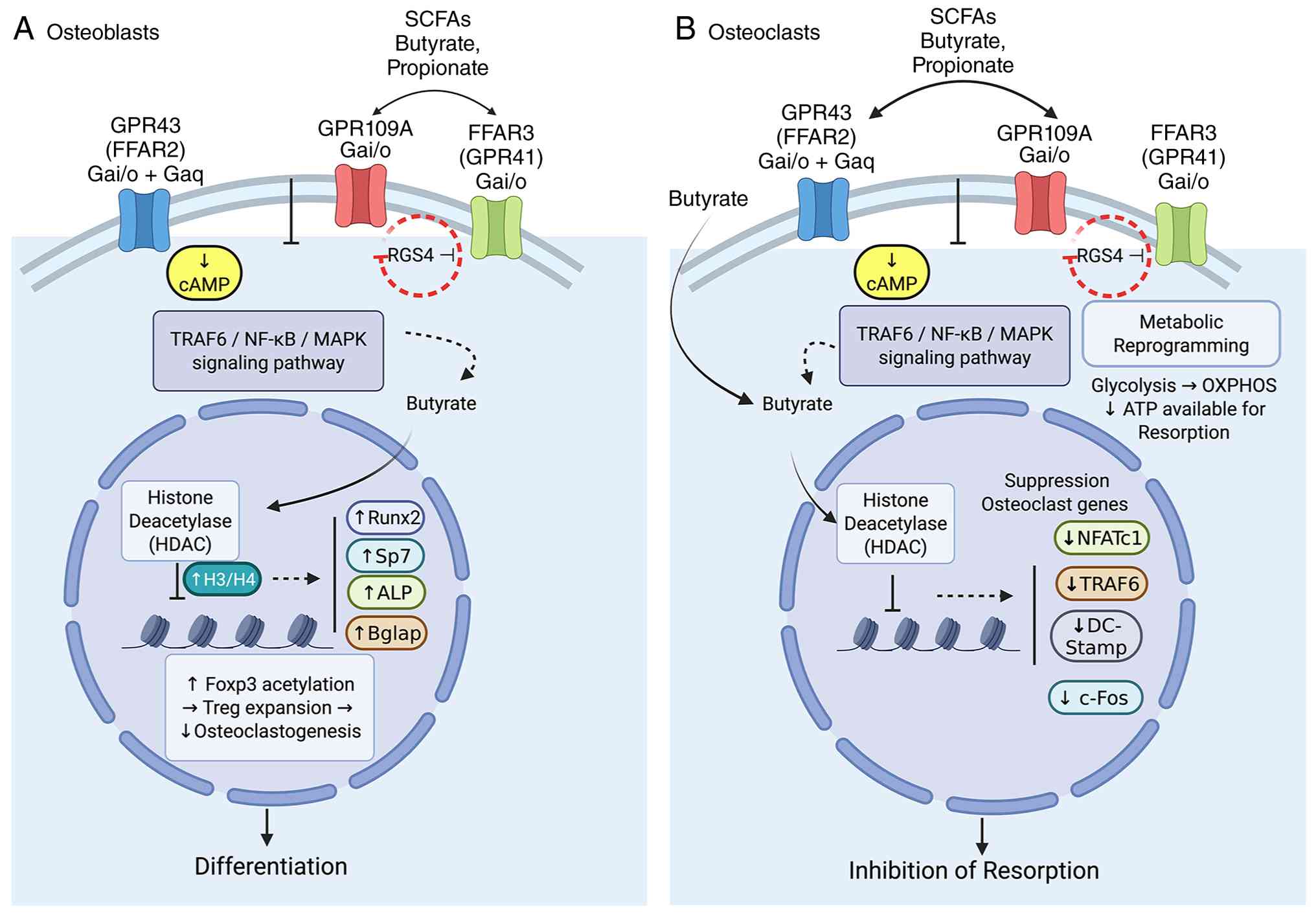 | Figure 2Dual mechanisms of SCFA-mediated bone
cell regulation. (A) In osteoblasts, SCFAs activate GPR43/FFAR2
(Gαi/o + Gαq), GPR109A (Gαi/o), and FFAR3/GPR41 (Gαi/o; negatively
regulated by RGS4), suppressing cAMP and attenuating
TRAF6/NF-κB/MAPK signaling. Independently, butyrate inhibits HDACs,
increasing H3/H4 acetylation at promoters of Runx2, Osterix (Sp7),
ALP and osteocalcin (Bglap), promoting differentiation. HDAC
inhibition also stabilizes Foxp3+ Tregs, indirectly
suppressing osteoclastogenesis. (B) In osteoclast precursors, the
same receptor cascade attenuates TRAF6/NF-κB/MAPK signaling.
Butyrate-mediated HDAC inhibition downregulates NFATc1, TRAF6,
DC-Stamp and c-Fos. SCFAs additionally reprogram metabolism from
glycolysis to OXPHOS, reducing ATP availability for resorption.
HDAC, histone deacetylase; ALP, alkaline phosphatase; OXPHOS,
oxidative phosphorylation. |
Polyamines in bone homeostasis: From
biosynthesis to osteogenesis
Polyamines are vital organic cations involved in
numerous cellular functions, such as cell growth, differentiation
and metabolism. Their diverse functions also contribute to
maintaining physiological balance, and recent studies have
highlighted their significance in bone homeostasis. Notably,
endogenous polyamines such as putrescine, spermidine and spermine
have been demonstrated to inhibit osteoclast migration and fusion,
acting through signaling pathways such as
Ca2+-PYK2-Src-NFATc1 to suppress osteoclast
differentiation (61).
Polyamines also promote osteoblast differentiation;
exogenous administration of putrescine, spermidine and spermine has
been shown to upregulate ALP activity and the expression of
osteogenic genes (for example, Runx2 and osteocalcin) in
mesenchymal stem cells (62).
Furthermore, polyamines enhance bone formation in vivo, as
evidenced by increased mineralization and bone volume, alongside
suppression of osteoclast activity in animal models (63).
The physiological significance of polyamine
homeostasis is further highlighted by the observation that
mutations in polyamine-synthesizing enzymes such as spermine
synthase in Snyder-Robinson syndrome result in severe bone defects,
reduced osteoblast numbers and diminished bone volume (64). Similarly, genetic inactivation of
spermine synthase in murine models leads to osteopenia,
attributable to impaired osteoblast function (65). Polyamines such as putrescine,
spermidine and spermine are derived from multiple sources:
Endogenous host synthesis, dietary intake and intestinal microbial
production. Both diet and the composition of the gut microbiota
exert a significant influence on luminal polyamine
concentrations.
Microbiological de novo synthesis from
arginine
The gut microbiota plays an important role in the
de novo synthesis of polyamines. Diverse bacterial taxa have
the enzymatic ability to synthesize polyamines from precursors such
as arginine. While the canonical ornithine decarboxylase 1
(ODC1)-dependent pathway (arginine - ornithine - putrescine) is a
major route for polyamine biosynthesis, in the event of ODC1
deficiencies alternative pathways [for example the arginine
decarboxylase (ADC) agmatinase (AGMAT) dependent route] may play a
supplementary role (66). The
biochemical mechanisms by which arginine biosynthesis and
catabolism are completed are well characterized in bacteria such as
Escherichia coli (E. coli), in the following manner:
This provides an example of the proficiency of microorganisms in
the metabolism of polyamine precursors (67). Numerous gut bacteria function as
serious polyamine producers. For example, Staphylococcus
epidermidis FB146 isolated from fermented foods (for example,
miso) is an avid producer of putrefying substances and thus
highlights the potential for these organisms to be dietarily
important sources of polyamines for human consumption (68). Additional gut microbes such as
Akkermansia muciniphila, Bacteroides spp. and
Clostridium spp. (for example Clostridium lavalense)
represent recognized components of the gut microbiota that
contribute to the homeostasis of the immune system and overall gut
health (69-71). These groups of bacteria take part
in various metabolic processes in the gut, such as the biosynthesis
of beneficial substances such as polyamines.
Bioavailability studies depletion of
antibiotic and restoration from fecal microbiota transplantation
(FMT)
Research studying antibiotic-induced gut microbiota
depletion and its subsequent restoration through FMT is offering
more information into the relationship between the gut microbiota
and the host's polyamine status and well-being. Extensive
antibiotics administration causes severe changes in immune cells
and the intestinal epithelium, including the decrease of
antimicrobial peptides and intestinal atrophy (72,73). Such perturbations are often
accompanied by large changes in the abundance of bacterial
inhabitants of the gut. FMT has been shown to be able to
effectively reinstall the intestinal microbiota and thus also its
variety of host functions. For example, FMT can help to replenish
immune cell populations in numerous different areas of the small
and large intestines after the antibiotic-induced depletion
(72,74). In the context of
Clostridioides difficile infection, FMT has been shown to be
efficacious in normalizing a healthy gut microbiota while
antibiotic therapy does not yield the same result (75). The reconstitution of microbial
communities by FMT suggests a feasible approach with which to
reinstate microbial polyamine production and its availability to
the host, thereby underscoring the importance of a crucial
relationship between a healthy gut microbiota and the polyamine
status of the host.
Distinction from dietary source and host
synthesis
Polyamines are not only produced by microbes in the
gut but also come from dietary inputs and endogenous biosynthesis
of host cells. Both dietary polyamines as well as arginine are
recognized as nutritional determinants that influence physiological
functions such as fertility (76). Fermented foods are especially
rich in polyamines, presumably from the fermentative bacteria
involved in its manufacture (68). Lactic acid bacteria (LABs) which
is often found in foods such as curd, pickles, milk and wheat dough
is considered to be beneficial microorganisms that help to
supplement polyamine content of those foods (77). Host cells express the enzymatic
capacity required to synthesize polyamines, which are mainly formed
in an ODC1-dependent pathway, which changes arginine into ornithine
and then into putrescine (66).
The total pool of polyamines available to the organism is
controlled by a mixture of endogenous production, consumption of
the diet and microbial synthesis. This collective reservoir
modulates a variety of physiological processes including processes
that are important for skeletal health.
SSAT1 as the key catabolic enzyme
Intracellular polyamine concentrations are governed
by a dynamic equilibrium between biosynthesis, transport and
catabolism. An enzyme that plays a central role in polyamine
catabolism is spermidine/spermine N^1-acetyltransferase (SSAT1, or
SAT1). SSAT1 acts as an acetylase of molecular weight polyamines
(spermidine and spermidine) to earmark them for either efflux or
further degradation by polyamine oxidase. This acetylation process
is central in polyamine export and cellular polyamine homeostasis.
SSAT1 is distinct from other N-acetyltransferases such as SSAT2,
which acetylates thialysine and is not part of the polyamine
metabolism. Previous biochemical investigations confirmed that
SSAT1 performs these reactions and controls the level of polyamines
in the cell (78,79). On acetylation, polyamines are
released from the cell, limiting the intracellular concentrations.
Although the aforementioned studies do not specifically refer to
the use of acetylated polyamines in feces or urine as biomarkers,
the fact that the metabolites are being exported suggests that
forms of these metabolites that are excreted could potentially
reflect general polyamine metabolism and SSAT1 activity within an
organism. Indeed, the increased activity of SSAT1 under
physiological conditions results in an increase in the release of
acetylated polyamine derivatives, making them potential biomarkers
(80,81). Further studies should be
conducted to determine their specific utility as biomarkers for
bone-related conditions.
Dysregulation of SSAT1 activity has been implicated
in various pathological conditions, particularly in cancer. For
instance, elevated SAT1 expression has been linked to cell
radio-resistance in glioblastoma multiforme, suggesting that SAT1
could be a therapeutic target to sensitize cancer cells to
radiation therapies (82,83).
Furthermore, overexpression of SSAT1 modulates tumorigenesis, with
SSAT1 knockdown shown to reduce tumor growth in vivo
(23,83).
While the provided studies do not directly address
the dysregulation of SSAT1 in osteoclastogenesis, the established
role of SSAT1 in regulating polyamine levels and its potential as a
therapeutic target in other diseases suggests that its modulation
could be relevant in bone metabolism. Given the critical roles of
polyamines in cellular processes, investigating the specific impact
of SSAT1 dysregulation on osteoclast differentiation and function,
and its potential as a therapeutic target for bone diseases,
warrants further exploration. Emerging data in immune and metabolic
contexts (for example, in T-cell subsets) support a broader role
for SAT1 beyond cancer (84).
Eukaryotic initiation factor 5A (eIF5A)
hypusination: A novel mechanism for osteogenic translation
The eIF5A undergoes a unique post-translational
modification called hypusination, which is absolutely dependent on
the polyamine spermidine. In a two-step enzymatic reaction,
deoxy-hypusine synthase (DHPS) transfers the aminobutyl moiety from
spermidine to a conserved lysine residue of eIF5A, generating
deoxy-hypusine; deoxy-hypusine hydroxylase (DOHH) then completes
the modification to form hypusine (85).
The hypusinated form of eIF5A (hyp-eIF5A) is the
only known protein to contain hypusine, and this modification is
essential for eIF5A's function as a translation elongation factor.
Specifically, hyp-eIF5A facilitates ribosomal transit through mRNA
sequences encoding polyproline motifs by resolving ribosomal
stalling at the peptidyl-transferase center, enabling efficient
synthesis of proteins containing consecutive proline residues.
Beyond polyproline resolution, eIF5A has also been implicated in
translation termination efficiency and cellular differentiation
(86,87). DHPS and DOHH are evolutionarily
conserved from yeast to humans, underscoring the fundamental
importance of this modification (88).
In the context of bone biology, eIF5A hypusination
represents a mechanistically plausible link between gut microbial
spermidine availability and osteogenic protein synthesis, given
that collagen and other structural bone matrix proteins, as well as
transcription factors such as Runx2, contain polyproline stretches
whose efficient translation may depend on hyp-eIF5A activity
(86-89). However, direct experimental
evidence for this pathway in osteoblasts or osteoclasts remains to
be established; this represents a high-priority area for future
mechanistic investigation, as discussed below.
Direct effects of polyamines on bone
cells
Polyamines exert direct effects on both major
bone-remodeling cell lineages, osteoblasts and osteoclasts
collectively recalibrating the balance of bone remodeling in favor
of bone formation and stability. Effects on osteoblasts polyamines
(putrescine, spermidine and spermine) have been demonstrated to
promote differentiation of bone marrow derived stromal cells
(BMSCs) into osteoblasts. In human BMSCs, exogenous polyamines
increase ALP activity and enhance mRNA expression of key osteogenic
transcription factors, including Runx2 and osteocalcin (Bglap)
(62). At the same time, they
suppress genes associated with adipogenesis, such as PPARγ, thereby
redirecting cellular fate toward the osteoblastic lineage rather
than adipocytes (62). Although
direct evidence for activation of the PI3K-Akt pathway by
polyamines in osteoblasts is not well documented in the literature,
this signaling axis is well known to support osteoblast survival,
proliferation and differentiation (90). Effects on osteoclasts in
experiments with osteoclast precursors, natural polyamines
(spermidine and spermine) strongly inhibit RANKL-stimulated
osteoclastogenesis, reducing expression of NFATc1 and other
osteoclast marker genes, and decreasing TRAP activity (89). In RAW 264.7 macrophage-derived
osteoclasts, exogenous putrescine and spermine similarly reduce
RANKL-induced expression of NFATc1 and RANK, and suppress TRAP
activity, indicating impaired maturation toward resorbing
osteoclasts (63).
Mechanistically, these anti-osteoclast effects involve suppression
of the Ca2+-PYK2-Src-NFATc1 signaling axis, which is
normally activated by RANKL during osteoclast differentiation,
thereby limiting osteoclast migration, fusion and differentiation
(61). Moreover, in vitro
and in vivo work demonstrates that exogenous polyamines not
only enhance bone formation via osteoblast activation but also
suppress osteoclastogenesis, as evidenced in ovariectomized rat
models (63). Collectively, by
enhancing osteoblast differentiation and inhibiting
osteoclastogenesis, polyamines contribute to a net bone-anabolic
effect, favoring bone formation over resorption. The specific
signaling pathways driving these polyamine-mediated effects,
including the upregulation of osteogenic markers (Runx2 and ALP)
and the suppression of the Ca2+-PYK2-Src-NFATc1 axis in
osteoclasts, are depicted in Fig.
3.
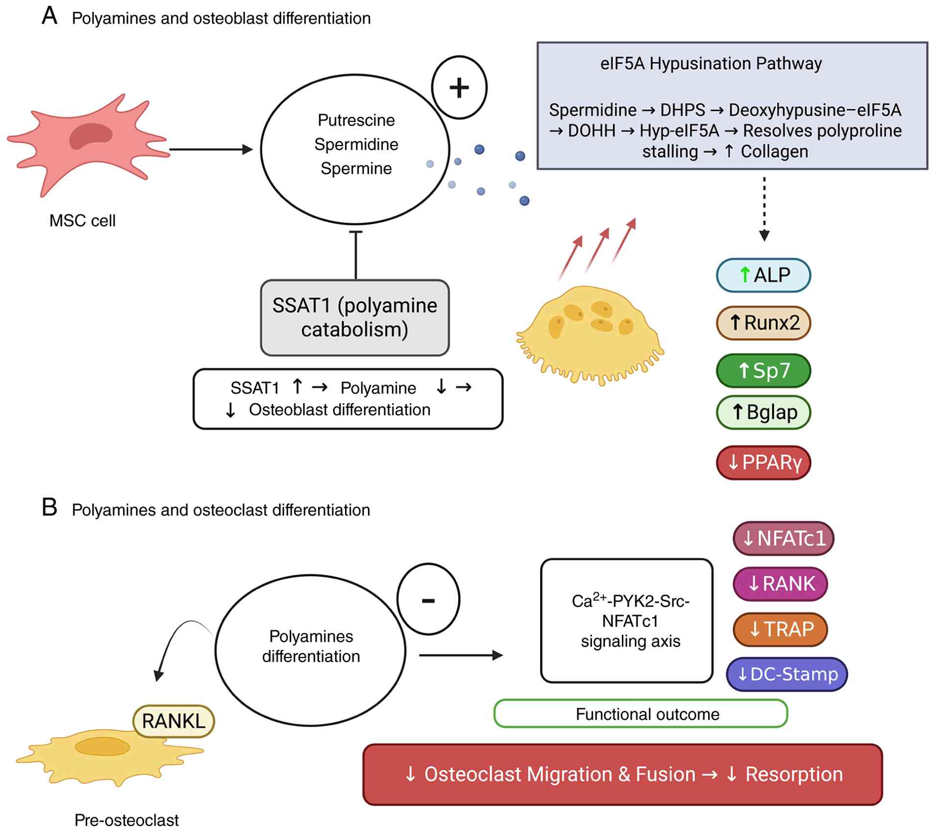 | Figure 3Polyamine-mediated regulation of
osteoblast and osteoclast differentiation. (A) Gut-derived
polyamines (putrescine, spermidine and spermine) promote MSC
osteoblast differentiation, upregulating ALP, Runx2, Osterix (Sp7)
and osteocalcin (Bglap), while suppressing the adipogenic regulator
PPARγ. Spermidine drives the eIF5A hypusination pathway (DHPS →
deoxyhypusine-eIF5A; DOHH → Hyp-eIF5A), resolving polyproline
ribosomal stalling and enhancing collagen and osteogenic protein
synthesis. SSAT1-mediated catabolism governs intracellular
polyamine availability; SSAT1 overactivation depletes the polyamine
pool and impairs differentiation. (B) In pre-osteoclasts,
polyamines suppress RANKL-induced differentiation by inhibiting the
Ca2+-PYK2-Src-NFATc1 signaling axis, reducing RANK,
NFATc1, TRAP and DC-Stamp expression and impairing osteoclast
maturation, migration and fusion. ALP, alkaline phosphatase; DHPS,
deoxy-hypusine synthase; DOHH, deoxy-hypusine hydroxylase; SSAT1,
spermidine/spermine N1-acetyltransferase 1; TRAP,
tartrate-resistant acid phosphatase; MSC, mesenchymal stem
cell. |
Microbial metabolomics for OP: Discovery and
integration
Recent advances in microbiome research have
elucidated the critical role of the gut microbiota in modulating
bone metabolism, thereby establishing the gut-bone axis as a
central paradigm in OP pathophysiology. The study of microbial
metabolomics, that is, the global analysis of small molecules that
are either produced or modified by gut microbes, has become an
important methodological tool to unravel the mechanistic basis of
this link. By determining the metabolic products of the gut
microbes, researchers can understand how gut microbial activities
affect bone remodeling and fracture susceptibility and, more
importantly, determine novel biomarkers or targets for therapy.
Notably, there is accumulating evidence to denote that metabolites
accessed from the gut including SCFAs, bile acids and several
bioactive compounds play a critical direct and indirect role in
modulating osteoblast and osteoclast function (91,92). A comprehensive comparison of the
biosynthetic origins, signaling targets, and cellular effects of
these key metabolite classes is provided in Table I.
 | Table IOverview of key microbial
metabolites, their biosynthetic origins, and their molecular
targets in bone remodeling. |
Table I
Overview of key microbial
metabolites, their biosynthetic origins, and their molecular
targets in bone remodeling.
| Metabolite
class | Key examples | Primary
orecursor | Key bacterial
producers | Receptor/signaling
target | Effect on
osteoblast | Effect on
osteoclast |
|---|
| SCFAs | Acetate,
Propionate, Butyrate C | Dietary Fiber
(Complex arbohydrates) | Faecalibacterium
prausnitzii, Roseburia hominis | GPR43 (FFAR2),
GPR109A (HCAR2), HDAC Inhibition | Upregulates
Runx2, Osterix, Alp, Bglap via histone
acetylation | Inhibits NFATc1 via
TRAF6/NF-κB pathway; induces metabolic shift from glycolysis to
OXPHOS |
| Polyamines | Spermidine,
Spermine, Putrescine | Dietary and
Endogenous Arginine | Staphylococcus
epidermidis, Bacteroides spp. | eIF5A hypusination,
Ca2+-PYK2-Src signaling | Enhances
translation of polyproline-motif proteins (for example, Collagen,
Runx2) | Inhibits migration
and fusion by suppressing Ca2+-PYK2-Src-NFATc1
signaling |
| Secondary bile
acids | Deoxycholic acid,
lithocholic acid | Primary bile acids
(host-derived) | Clostridium
spp. (via 7α-dehydroxylation) | FXR, TGR5 | Indirectly
modulates bone via immune cell regulation | Modulates
resorption activity |
Microbiological metabolomics analytical
platforms
The microbial metabolome is extremely chemically
heterogeneous, requiring careful consideration before choosing the
most suitable analytical tools to provide exhaustive and accurate
identification. The following advanced technologies are currently
being used in the study of microbial metabolomics, and each has its
own strengths and limitations.
Liquid chromatography tandem mass
spectrometry (LC-MS/MS)
LC-MS/MS is one of the most basic technologies in
microbial metabolomics that is appreciated for its remarkable
precision, selectivity and versatility. Reversed phase LC-MS/MS is
well suited to the separation and quantification of hydrophobic and
moderately polar entities such as lipids, bile acids and
xenobiotics. By contrast, hydrophilic interaction chromatography
coupled with MS/MS is effective for the detection of highly polar
molecules; for example, amino acids, nucleotides and SCFA
derivatives. High-resolution mass spectrometry in combination with
MS/MS provides the option for even stronger detection of multiple
metabolic markers per sample to distinguish between structurally
similar compounds often observed in biological matrices. These
methodological advances have played a key role in the recent study
into the metabolic complexities of gut microbiota and its
implications on bone health (18).
Gas chromatography-MS (GC-MS)
GC-MS spectrometry is highly respected for the fact
that the analysis of volatile and semi-volatile substances produces
excellent repeatability and precision. Chemical derivatization
allows for the separation and detection of metabolites SCFAs, for
example acetate, propionate, butyrate, organic acids and some amino
acids. Comprehensive electron ionization spectral libraries that
provide reliable identification and quantification of metabolites,
are available. This approach is often used in gut-bone axis
research, especially for SCFAs and other related compound-targeted
analysis (93).
Nuclear magnetic resonance spectroscopy
(NMR)
Although NMR spectroscopy is less sensitive than
MS-based approaches, it allows to get unmatched structural detail
and allows precision of quantification with no need of external
references. Its low sample preparation, as well as non-destructive
nature render NMR a fantastic compliment to MS platforms,
particularly when investigating the structures of new microbial
metabolites. Integrated NMR-MS studies have been shown to provide
more robust identification and quantification of metabolites, which
will further enable understanding of gut-bone interactions
(92). The coming together of a
variety of metabolomic platforms with more advanced bioinformatics
and systems biology is expected to further facilitate the
identification of gut microbiota-derived biomarkers and
therapeutics for OP. Comprehensive metabolite profiling in
combination with metagenomic, transcriptomic and clinical data
allows the construction of predictive models to understand the
complex relationship between host and microbiome in skeletal
health. In order to validate potential biomarkers and investigate
causal associations, future studies should focus on standardization
of analytical methods, extension of exhaustive metabolite
databases, and conduct longitudinal and interventional studies.
Integrative challenges and multi-omics
strategies
A major difficulty in microbial metabolomics is
accurately identifying the features that are found. The vast
quantity of signals, numerous of which represent unknown compounds,
necessitates thorough verification using fragmentation spectra,
matching retention times, and, if feasible, stable-isotope
standards. Integrating metabolomic data with microbiome sequencing
approaches such as 16S rRNA amplicon profiling and metagenomics has
proven instrumental in establishing functional links between
specific metabolites and bone outcomes, as demonstrated by an
increasing body of MR and observational evidence. Recent MR studies
have offered genetic proof of causal relationships between gut
microbiota, plasma metabolites and the risk of OP (10). In the aforementioned review,
specific bacterial taxa (such as Desulfobacterota) exhibited
associations with OP susceptibility, mediated in part by plasma
metabolites such as 3α-androstanediol monosulfate, accounting for
up to ~9% of the effect (10).
Another MR investigation focused on post-menopausal OP and
osteoclast biology, identifying the Burkholderiales order as
inversely linked with OP risk and positively correlated with
osteoclast number; plus, two osteoclast-related genes, FMNL2 and
SRBD1, were implicated as potential mediators (91). Observational studies also provide
strong support: In a cohort of 108 post-menopausal women, gut
microbiota composition and fecal metabolome were significantly
altered in those with OP, with metabolites such as L-pipecolic acid
correlating closely with BMD (94). More recently, a study in
Frontiers in Microbiology used large-scale GWAS and MR to explore
196 microbial taxa and 1,400 plasma metabolites, identifying ~96
metabolites potentially causally associated with OP, and
delineating six gut-microbe-metabolite-bone causal pathways
(95). Collectively, these
studies illustrate how the combined deployment of metabolomics,
metagenomics and genetic causal inference provides a methodological
foundation for both biomarker discovery and mechanistic validation
in gut-bone axis research.
Toward biomarkers and therapeutic
targets
Combining metabolomic and genomic data improves our
understanding of biological mechanisms and speeds up the
identification of biomarkers. For instance, certain plasma
metabolites found through MR could act as indicators of OP risk or
how it progresses. Meanwhile, microbial groups involved in these
pathways might be targeted for gut microbiota-focused treatments,
such as interventions (for example, probiotics, prebiotics, or even
specially engineered bacteria). Additionally, recent research has
started investigating new connections within the gut-bone
relationship. A 2025 study highlighted how Terrisporobacter
othiniensis protects against OP by influencing
ferroptosis-related proteins (specifically the MDM4/p53 pathway),
strongly suggesting a link between the gut, microbes, ferroptosis
and bone (96). These insights
into mechanisms could lead to the development of entirely new
treatments for OP.
Feature annotation and metabolite
identification challenges
Despite significant progress in analytical methods,
precisely identifying and labeling metabolites continues to be one
of the toughest hurdles in microbial metabolomics. The immense
chemical variety of microbial metabolites, numerous of which are
new and uncharacterized, makes it very difficult to confidently
assign compounds (97,98). The usual method for identifying
metabolites starts by comparing experimental MS/MS spectra with
existing public databases. Widely utilized resources include the
Human Metabolome Database (HMDB), METLIN, MassBank and molecular
networking platforms such as GNPS (Global Natural Products Social
Molecular Networking) (97,99). These platforms facilitate the
comparison of experimental spectra to known reference spectra,
support structural annotation, and enable the exploration of
related but unknown compounds through network-based similarity
analyses (97). To promote
consistency and transparency in metabolite reporting, the
Metabolomics Standards Initiative has established a tiered
framework for identification confidence. Level 1 (confirmed
identification) requires concordance with an authentic standard in
terms of retention time, mass accuracy and fragmentation pattern.
Level 2 (putatively annotated compounds) is based on MS/MS spectral
matching to library entries or contextual evidence. Level 3
(putatively characterized compound class) indicates assignment to a
known chemical class (for example, bile acids and fatty acids)
without full structural elucidation. Level 4 ('unknown compounds')
encompasses features for which no plausible structural assignment
can be made often referred to as the 'dark matter' of the
metabolome (99,100). A significant proportion of
features identified from gut microbiome metabolomics studies are
still at Level 4. This phenomenon can be explained by two main
reasons, which are the occurrence of novel molecules of microbial
origin and the lack of spectra attached to extant reference
databases (98,101). Although these unidentified
metabolites could have biologically important roles, for example in
bone metabolism, the accurate identification of such metabolites
requires sophisticated and heterogeneous analytical methods. Such
techniques include high resolution NMR spectroscopy, ex novo
synthetic generation of candidate molecules, and computer-based
techniques such as in silico fragmentation simulations and
statistical modeling (102).
Recent methodological reviews emphasize that this so-called
metabolic 'dark matter' continues to be a formidable obstacle
within the field (98). In the
specific context of the gut-bone axis, this dark matter problem
carries particular consequence. Given that established microbial
metabolites such as SCFAs and polyamines exert potent,
receptor-mediated and epigenetically driven effects on osteoblast
and osteoclast function, it is plausible that the pool of Level 4
features detected in gut metabolomics studies harbors additional,
yet uncharacterized bone-regulatory molecules. This inference is
supported by evidence already discussed in the present review:
Observational cohort studies correlating fecal metabolomes with BMD
have identified novel metabolite associations such as that of
L-pipecolic acid for which mechanistic bone pathways remain
undefined (94). Similarly, MR
analyses have implicated gut microbial taxa in OP risk through
metabolic mediators that are not yet fully characterized (10,95), suggesting that causally relevant
bone metabolites may reside within the current dark matter pool.
Targeted strategies to address this gap include: (i) Stable-isotope
labelling approaches (for example, 13C-labelled dietary
fiber) to trace microbial fermentation products into systemic
circulation and bone tissue; (ii) co-culture systems pairing gut
microbial communities with osteoblast or osteoclast cell lines to
enable functional screening of conditioned media fractions; (iii)
molecular networking via GNPS to identify structural analogues of
known bone-active metabolites within unannotated spectral clusters
(97,99); and (iv) longitudinal metabolomic
profiling in fracture cohorts with concurrent bone
histomorphometry, enabling correlation of unidentified features
with bone turnover endpoints. Advancing these approaches will be
essential to bridge the gap between metabolomics discovery and
mechanistic bone biology.
Multi-successor series integration:
Integration of metabolites and microbial taxa and function
Integrating microbial metabolomics and microbiome
sequencing techniques (that is, 16S rRNA gene amplicon sequencing
and shotgun metagenomics) significantly increases the usefulness of
these studies with regard to OP research. The use of the
multivariate, multi-omics framework provides a more granular and
mechanistic understanding of how the compositional architecture and
functional repertoire of microbial consortia modulate metabolite
synthesis, thereby affecting skeletal homeostasis. Taxonomic
correlation using 16S rRNA sequence data or metagenomic datasets,
research publications have been able to link relative abundance of
certain clades of organisms (for example, SCFA producing organisms)
such as F. prausnitzii, or Roseburia with quantified
levels of SCFAs (acetate, butyrate and propionate) in either fecal
or plasma samples. SCFAs have been shown to modulate bone
remodeling through changes in immune signaling pathways as well as
direct effects on osteogenic and osteoclastic cell populations
(20).
Functional linkage
Shotgun metagenomics provides fine gene level
information, which reveals the metabolic ability of the microbiome
- for example genes for butyrate synthesis. When combined with
metabolomics, this synergy allows us to reconstruct metabolic
pathways using of resources such as KEGG (https://www.kegg.jp/), or CAZy (https://www.cazy.org/) in order to link genetic
potential with actual metabolite production.
Co-occurrence networks
Advanced network-centric methods to construct
co-occurrence models incorporating microbial taxa, metabolites and
clinical phenotypes (such as BMD) Recent multi-omics studies of
fracture patients have indeed shown strong links between specific
types of bacteria, metabolites associated with inflammation (for
example, N-acetylneuraminate) and low bone mass, using network
analysis to show putative pathogenic trajectories (103).
Mediation and causality
MR studies further strengthen mechanistic insights
by exploring possible mechanisms in which microbial taxa may
mediate their effects on bone health by way of specific metabolite
intermediates. For example, a contemporary MR analysis revealed
numerous gut bacterial lineages, including those in the phylum
Desulfobacterota, that have an impact on OP susceptibility
partially mediated by plasma concentrations of 3a-radioligand
androgens, namely 3a-dihydro-5a-androstanediol monosulphate, and
explained ~9% of the effect (10). The ongoing challenge of
identifying the majority of features of the metabolome, or the
so-called 'dark matter' as these are colloquially called, continues
to hinder the discovery of new biomarkers and targets for drug
therapy. Consequently, a comprehensive characterization of these
cryptic metabolites by integrated multi-omics, sophisticated
analytical chemistry and computational strategies is an imperative.
As is evident from recent multi-omics studies focused on OP, vector
use of such synergistic methodologies can yield microbial
metabolite signatures with favorable correlation with BMD and risk
of fracture, opening up biomarker mining and microbiome-centric
therapeutic interventions.
Translational strategies and therapeutic
interventions
The aforementioned mechanistic insights toward a
hierarchy of microbiota-targeted interventions span from broad
dietary modifications to precision pharmacological strategies. For
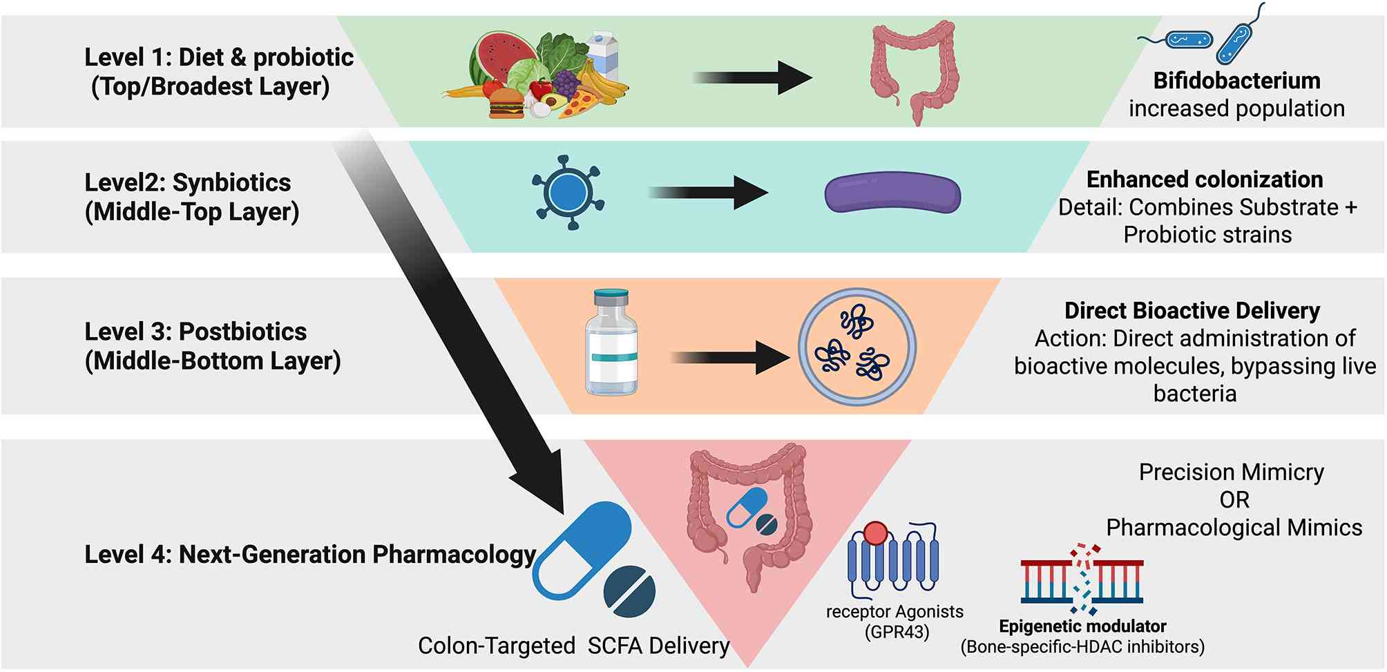
clarity, these are grouped into four tiers of increasing
mechanistic specificity: Dietary fibers and prebiotics, probiotics
and synbiotics, postbiotics, and next-generation pharmacological
approaches.
Dietary fibers and prebiotics
Inulin-type fructans (ITF) and microbiota-targeted
modulation ITF including inulin and oligofructose fulfil the
operational definition of prebiotics by being selectively utilized
by the gut microbiota to confer a health benefit (104). Human intervention studies
demonstrate that daily intakes in the 10-20 g range reproducibly
enrich saccharolytic taxa such as Bifidobacterium and F.
prausnitzii, with downstream effects on systemic metabolism
that are directly relevant to OP risk. In obese women, 16 g/day of
an inulin/oligofructose 50:50 mixture for three months induced
selective microbiota shifts characterized by increased
Bifidobacterium and other beneficial taxa, alongside modest
improvements in metabolic endotoxemia and metabolomic signatures
related to lipid and glucose handling (105). A large systematic review of
human ITF trials confirms that such doses consistently increase
Bifidobacterium, Lactobacillus and F.
prausnitzii across diverse populations, while also influencing
intestinal functions (barrier and immune tone) and extra-intestinal
outcomes including glucose homeostasis, lipid profile, mineral
absorption and bone health endpoints (104). Ecosystem-wide analyses in
healthy adults have further shown that inulin supplementation
reshapes the colonic microbiota beyond the classical 'bifidogenic'
effect, with coordinated changes in several genera and parallel
shifts in fecal metabolites, including SCFAs (106). Such SCFAs, particularly
butyrate and propionate, engage the receptor-mediated and
epigenetic mechanisms aforementioned, representing key mechanistic
links between ITF, gut microbiota and bone remodeling. Evidence
specific to post-menopausal women remains limited but is
biologically compelling. ITF improve cardiometabolic risk factors
that are over-represented in post-menopausal OP, including insulin
resistance, dyslipidemia and low-grade inflammation, as shown in
randomized trials in overweight/obese women with polycystic ovary
syndrome (107) and in type 2
diabetes mellitus (T2DM) populations (108,109). Given the recognized association
between T2DM, BMD and fracture risk (110), sustained ITF use in
post-menopausal women may offer a dual benefit: Correction of
dysmetabolism and indirect mitigation of diabetic bone fragility.
From a translational standpoint, a pragmatic target dose for
long-term use in post-menopausal women is 10-15 g/day, titrated
upward based on gastrointestinal tolerance. At this range, human
studies consistently demonstrate robust microbiota responses
(104-106) with acceptable tolerability,
while meta-analytic data support improvements in glycemic control
and lipid profile that are relevant to bone health and fracture
risk reduction (108,109).
RS and other fermentable fibers
RS represents another class of
microbiota-accessible carbohydrate with demonstrable effects on
bone in preclinical models. In meat ducks, graded inclusion of RS
derived from raw potato starch (RPS) produced linear and quadratic
increases in tibial strength and ash content, accompanied by
elevated cecal propionate and butyrate levels (111). Micro-CT and microbiota
profiling indicated that RPS-induced gains in bone mass correlated
with higher Firmicutes abundance and enhanced SCFA
production, as well as improved phosphorus absorption and reduced
digestive tract pH (111).
In a model of E. coli-induced bone loss in
ducks, a diet containing 12% RPS improved bone quality, suppressed
bone resorption and attenuated inflammatory responses in both ileum
and bone marrow (112). These
benefits coincided with increased Firmicutes abundance,
higher SCFA (especially propionate) production, and downregulation
of Malt1/NF-κB inflammasome activation, together with expansion of
Treg cells (112).
Collectively, these data provide mechanistic proof-of-concept that
RS-driven microbiota remodeling and SCFA generation can ameliorate
inflammation-related bone loss, highly relevant to post-menopausal
and inflammatory OP.
Although direct human evidence on RS and skeletal
endpoints is sparse, these preclinical findings support cautious
translation of RS-enriched foods (for example, high-amylose grains,
cooled potatoes and legumes) or purified RS as adjuncts to standard
OP management. Dosing regimens should likely mirror those used in
metabolic trials (10-30 g/day fermentable fiber), with careful
attention to gastrointestinal tolerance, fiber type and
interactions with other components of the habitual diet (104,113).
High-fiber dietary patterns and the
Mediterranean diet
Beyond isolated fibers, dietary patterns
characterized by high intake of plant foods and diverse fibers
appear favorable for bone health. A comprehensive review of dietary
patterns and bone outcomes underscores that diets rich in fruits,
vegetables, whole grains and legumes patterns overlapping
substantially with the Mediterranean diet are associated with
higher BMD and lower fracture risk, whereas Western-type patterns
high in sugar, fat and refined grains are detrimental (113).
Specifically in menopausal women, intervention
studies with Mediterranean-style diets demonstrate improvements in
body weight, blood pressure, lipid profiles and omega-6: omega-3
ratios (114), and broader
reviews implicate this pattern in the targeted prevention of
multiple chronic diseases, including OP (115). A recent review focusing on
nutrition and post-menopausal OP concludes that, in addition to
calcium and vitamin D, dietary patterns featuring abundant plant
foods, moderate high-quality protein, and low ultra-processed food
intake are likely to support bone health and should be prioritized
in non-pharmacological management (116).
The inflammatory tone of the diet is an additional,
mechanistically pertinent dimension. A systematic review and
meta-analysis using the Dietary Inflammatory Index showed that more
pro-inflammatory diets are significantly associated with lower
lumbar spine and total hip BMD, and with higher risks of OP and
fractures (117). These
findings offer a coherent framework whereby high-fiber, plant-rich,
low-inflammatory dietary patterns (for example, Mediterranean diet)
exert skeletal benefits in part via microbiota-mediated and
immunomodulatory mechanisms.
Nevertheless, the relationship between very high
fiber intake and bone is not unequivocally beneficial in all
contexts. In older individuals with T2DM, a 12-week high-fiber diet
intervention (38 g/day) improved metabolic outcomes but reduced
biochemical markers of bone formation, without detectable
alterations in bone microarchitecture or Wnt signaling, suggesting
potential suppression of bone turnover in an already high-risk
group (118). Given the
elevated baseline fracture risk and complex BMD trajectory in T2DM
(110), these findings call for
careful titration of fiber in diabetic OP, with monitoring of both
metabolic and skeletal endpoints.
Prebiotics: Dosage and clinical
considerations
Fructo-oligosaccharides (FOS),
galacto-oligosaccharides (GOS) and inulin are well established
prebiotic fibers. These carbohydrates resist digestion in the small
intestine and are accordingly fermented by colonic microbiota,
favoring (i) the increase of populations of useful bacteria such as
Bifidobacterium; (ii) the generation of SCFAs; (iii) the
decrease of gastrointestinal pH; and (iv) the use of minerals such
as calcium and magnesium, which might be responsible for the
increase of the whole mineral intake (104,113,116). A thorough review of human
studies uncovered the fact that ITF such as short-chain FOS as well
as longer-chain inulin, in general, reliably show prebiotic
activity. These compounds appear to have a positive effect on
glycemic control, lipid metabolism, mineral absorption,
inflammatory status and body composition (104). Further meta-analyses have
suggested the modest ability of ITF supplementation to enhance
lipid profile and glucose metabolism overall, with more profound
effects seen in individuals with T2DM, in which improvements in
HDL-cholesterol and glycemic control are significant (108,109). Clinical trials with women with
metabolic disturbance support these results. For example,
overweight and obese women, diagnosed with polycystic ovary
syndrome, who consumed ITF (10 g/d) for 12 weeks showed
ameliorations in insulin resistance, lipid parameters,
anthropometric measures and androgen levels as well as clinical
symptomatology (107). These
variables are implicated in the risk of OP, through effects on
adiposity, inflammation and sex hormones. In obese women, a 3-month
regimen of ITF supplementation caused changes in the composition of
gut microbiota and systemic metabolic profiles with specific
bacterial taxa that were linked to markers of metabolic endotoxemia
(105). This body of evidence
highlights a crucial conclusion; prebiotics should not be
considered only as 'gut health' supplements but as metabolic
modulators that can exert their biological effects on multiple
fracture risk determinants including insulin resistance,
dyslipidemia and inflammation, in post-menopausal women and
patients with T2DM (108-110,116). Nevertheless, large
interindividual differences in microbiota sensitivity require a
more customized strategy for dosing and patient consideration. A
crossover study showed that the increase in Bifidobacterium
that accompanied an ITF prebiotic was significantly higher in
people who consistently ate more dietary fiber than those who
consumed less, suggesting that an individual's baseline diet and
resulting gut microbiota configuration influences prebiotic
activity (119). Parallel
investigations in obese women reveal intricate associations between
species of bacteria, metabolic biomarkers and indices of metabolic
endotoxemia after ITF treatment (105).
These findings support the emerging notion of
'responders' vs. 'non-responders' to prebiotic interventions. For
OP-focused translation, this suggests that: (i) Prebiotic dosing
(typically 5-20 g/day for FOS/inulin) may need to be individualized
based on habitual fiber intake, baseline microbiota composition and
co-morbidities (104,119); (ii) Stratification by baseline
dysbiosis (for example, low Bifidobacterium/SCFA producers,
high pro-inflammatory taxa) and metabolic status (obesity, T2DM and
PCOS) should be incorporated into trial design (105,107-109); (iii) Bone-relevant endpoints
(BMD, bone turnover markers, microarchitecture and fracture risk)
must be systematically integrated into future prebiotic RCTs,
particularly in post-menopausal and diabetic cohorts in whom
metabolic and skeletal risks co-cluster (110,116,118).
Probiotics and synbiotics
In contrast to the relatively robust human evidence
for prebiotics, data on probiotic effects on human BMD and fracture
risk remain limited and were not directly captured in the research
papers reviewed here. Nonetheless, the mechanistic logic for
targeting specific probiotic strains in OP is supported indirectly
by the prebiotic and dietary fiber literature.
Preclinical work with RS shows that modifying the
gut microbial community toward SCFA-producing Firmicutes improves
bone mass and protects against pathogen-induced bone loss, in part
by dampening pro-inflammatory signaling (Malt1/NF-κB inflammasome)
and promoting regulatory T-cell expansion (111,112). These results align with the
general idea that certain probiotic strains, especially
Lactobacillus species and Bifidobacterium species,
could improve bone health by modulating the immune system,
strengthening gut barriers, and increasing the production of
beneficial postbiotic compounds (including SCFAs, indoles and
polyamines). Considering this, the following strategies can be
envisioned at the strain level (though comprehensive bone-related
data for these specific strains are largely beyond the scope of
this current research): (i) Lactobacillus reuteri (DSM
17938), widely studied in gastrointestinal and immune indications:
This strain exerts anti-inflammatory effects and can enhance
mucosal barrier function and regulatory T-cell responses,
mechanisms that mirror those implicated in RS-mediated protection
against bone loss (111,112);
(ii) Bifidobacterium longum (for example, BB536):
Bifidobacterium species are prominent responders to ITF
(104-106) and are known producers of
acetate and certain polyamines, molecules implicated in mucosal
integrity, osteoblast activity and regulation of oxidative stress;
(iii) Lacticaseibacillus paracasei: Members of this taxon
display immunomodulatory properties and may bias host responses
toward anti-inflammatory phenotypes, conceptually aligning with the
suppression of NF-κB-driven bone resorption observed in RPS-fed,
E. coli-challenged ducks (112); (iv) Akkermansia
muciniphila although not directly modulated by ITF in all
studies, barrier-strengthening and metabolic benefits attributed to
this mucin-degrading bacterium are congruent with the broader
evidence that improved gut integrity and reduced metabolic
endotoxemia accompany ITF and high-fiber interventions (104-106,113); (v) translationally, synbiotic
formulations that combine structurally compatible prebiotics (for
example, ITF or FOS) with carefully selected probiotic strains
represent a rational next step. Such combinations may enhance
colonization or persistence of beneficial strains, amplify SCFA and
other postbiotic production, and exert more consistent
immuno-skeletal effects than either component alone. The robust
prebiotic literature reviewed (104,105, 108,109,111,112,119) provides an essential scaffold
for designing and rationally titrating these synbiotic
interventions in osteopenic and osteoporotic populations.
Postbiotics: Metabolites and fermented
foods
Postbiotics defined as functional bioactive
compounds produced by microorganisms and released into the host
environment, include SCFAs, polyamines, exopolysaccharides, cell
wall fragments and extracellular vesicles. From the perspective of
OP, three classes of postbiotics stand out:
SCFAs (acetate, propionate and
butyrate)
The RPS studies in ducks elegantly demonstrate that
increases in cecal propionate and butyrate are tightly associated
with improved tibial strength, ash content and bone
microarchitecture, as well as with reduced intestinal and bone
marrow inflammation (111,112). These data strongly support
SCFAs as effector molecules linking fermentable fiber, the
microbiota and bone mass.
Polyamines and related metabolites
Although not directly quantified in the cited
animal studies, polyamines are recognized microbial products that
can influence osteoblast differentiation, collagen maturation and
redox balance. The enrichment of Bifidobacterium and other
saccharolytic taxa by ITF (104-106) suggests a plausible, if as yet
under-explored, route by which prebiotic-induced changes in
microbiota-derived polyamines could contribute to bone
anabolism.
Extracellular vesicles and exosomal
cargo
Milk-derived exosomes provide a striking example.
Oral administration of bovine colostrum-derived exosomes in an
OP-induced mouse model improved bone health in vivo and
promoted osteoblast proliferation and differentiation in
vitro, suggesting that dietary exosomal cargo can modulate bone
remodeling (120). These
vesicles, while not microbiota-derived, exemplify the concept that
orally delivered nanoscale particles, and their molecular cargos
(microRNAs, proteins and lipids) can act as 'postbiotic-like'
agents in bone tissue.
Fermented foods such as kefir, yogurt, miso and
sauerkraut deliver complex mixtures of live microbes and their
metabolites, potentially acting as natural synbiotic matrices.
While the specific studies summarized in the present review do not
provide direct RCT evidence for fermented foods and BMD, broader
nutritional reviews emphasize that dietary patterns rich in
minimally processed plant foods and fermented dairy are associated
with improved bone outcomes in post-menopausal women (113,116). Kefir and cultured dairy may be
particularly promising due to their combined provision of calcium,
vitamin K2, bioactive peptides and live LABs, though rigorously
controlled OP-focused trials remain scarce. From a translational
perspective, cell-free supernatants from probiotic cultures and
standardized postbiotic preparations (purified SCFAs,
exopolysaccharides and exosomes) offer a route to harness microbial
bioactivity while avoiding viability, safety and colonization
issues associated with live organisms. The SCFA-mediated skeletal
benefits observed in RPS-fed ducks (111,112) and the bone-protective effects
of bovine colostrum exosomes in mice (65), together suggest that targeted
delivery of defined postbiotic cocktails could complement existing
OP pharmacotherapies, particularly in patients unable or unwilling
to consume high-fiber or fermented foods.
Next-generation pharmacological
strategies
The convergent evidence that SCFAs and other
microbial metabolites modulate bone mass via immunological and
osteoclast/osteoblast pathways (111-113,116) provides a rationale for more
pharmacologically precise approaches that move beyond dietary
modulation alone. Emerging strategies can be conceptually grouped
as follows:
Direct administration of SCFAs (for example, sodium
butyrate) or prodrugs such as tributyrin has the potential to
recapitulate numerous of the bone-protective effects observed with
fermentable fibers, but with more predictable pharmacokinetics. The
duck studies highlight butyrate and propionate as plausible
mediators of improved bone mass and reduced inflammatory bone loss
(111,112). However, unencapsulated SCFA
salts are rapidly absorbed in the upper gut, limiting distal
colonic exposure and potentially increasing systemic side
effects.
Advances in pH-dependent coatings and encapsulation
technologies allow for colon-targeted SCFA delivery. By formulating
tributyrin or butyrate-releasing polymers that disintegrate at
colonic pH, it may be possible to locally increase SCFA
concentrations in proximity to the gut-associated lymphoid tissue
and portal circulation, thereby more precisely engaging the
osteo-immune axes implicated in bone remodeling. Such approaches
are particularly attractive for patients in whom high-dose
fermentable fiber is poorly tolerated or contraindicated [for
example, severe irritable bowel syndromes (IBS) and advanced
diabetic gastroparesis].
SCFAs signal through GPCRs such as FFAR2/GPR43
(acetate/propionate) and GPR109A (butyrate/niacin), which are
expressed on immune cells, intestinal epithelial cells and
potentially bone cells. The RS-induced suppression of Malt1/NF-κB
inflammasome activation and promotion of Treg expansion in ducks
(112) align well with known
SCFA-GPCR signaling networks that favor anti-inflammatory,
tolerogenic immune states.
These insights support the preclinical development
of selective FFAR2/GPR43 and GPR109A agonists as candidate
therapeutics for inflammatory and post-menopausal OP. Such agents
might: (i) Reproduce the Treg-expanding, NF-κB-suppressing effects
of SCFAs observed in RPS-fed animals (111,112); (ii) circumvent gastrointestinal
side effects of high-dose fibers, and be readily combined with
anti-resorptives or anabolics in high-risk patients (for example,
those with T2DM-related bone fragility (110); (iii) rigorous preclinical work
is needed to define optimal receptor selectivity, dosing, bone
tissue pharmacodynamics and long-term safety, with particular
attention to potential off-target effects in carcinogenesis or
immune suppression.
HDAC inhibition and bone-specific
epigenetic modulation
Butyrate is also a potent inhibitor of HDACs,
linking microbial fermentation to host epigenetic regulation. The
SCFA-mediated improvements in bone mass and suppression of
inflammatory pathways in RPS-fed ducks (111,112) are consistent with a model in
which HDAC inhibition skews gene expression toward
anti-inflammatory, pro-osteogenic programs in immune and stromal
cells.
This raises two translational possibilities:
Repurposing existing HDAC inhibitors (HDACi).
Several HDACi are already in clinical use for cancer and other
indications. However, their systemic toxicity and pleiotropic
actions necessitate caution. Any attempt to leverage HDACi for OP
would require bone-targeted delivery or very low dosing to avoid
hematologic, neurologic or oncogenic adverse effects.
Designing bone-specific HDAC modulators. By
integrating knowledge from dietary and prebiotic studies, showing
that localized gut-derived SCFA exposure can beneficially influence
bone via immune and endocrine axes (111-113,116). It may be possible to develop
novel HDAC modulators with preferential accumulation in bone marrow
or osteoblast/osteoclast lineages. Such agents could emulate the
epigenetic profile induced by physiologic SCFA exposure but with
greater pharmacologic precision. In both scenarios, mechanistic
insights derived from prebiotic, RS and diet-pattern studies
provide crucial guardrails: Therapies should aim to mimic the
spatially restricted, temporally pulsatile SCFA exposure generated
by a healthy microbiota, rather than induce chronic, high-level
systemic HDAC inhibition.
Safety considerations and
limitations
While the aforementioned interventions demonstrate
compelling mechanistic and preclinical rationale, responsible
translational implementation requires explicit consideration of
their safety profiles and clinical limitations.
Dietary fibers and prebiotic supplements are
generally well tolerated; however, dose-dependent gastrointestinal
side effects including bloating, flatulence, abdominal cramping and
altered bowel habits are consistently reported, particularly at
intakes above 15 g/day. Patients with IBS, IBD, or advanced
diabetic gastroparesis may require careful dose titration or may
not be suitable candidates for high-dose fiber interventions
(104,113). As aforementioned, very high
fiber intakes in older individuals with T2DM may paradoxically
suppress bone formation markers (118), underscoring the need for
bone-specific endpoint monitoring alongside metabolic outcomes in
this population.
Probiotic interventions carry a low but clinically
relevant risk of bacteremia or fungemia in severely
immunocompromised individuals, including those receiving
chemotherapy, post-transplant immunosuppression, or with advanced
HIV infection. Strain-specific safety documentation is therefore
essential prior to widespread clinical adoption. Postbiotic
preparations including purified SCFAs, extracellular vesicles and
exosomes face additional challenges of production standardization,
dose quantification and shelf stability that must be addressed
before they can be responsibly administered in clinical settings
(120).
For pharmacological approaches (GPCR agonists and
HDAC inhibitors), the risk of off-target effects is the primary
concern. HDAC inhibitors in particular carry well-characterized
risks in oncological contexts including hematologic toxicity and
immunosuppression that would require bone-targeted delivery
strategies or ultra-low dosing to be acceptable in an OP indication
(111-113,116). Selective GPCR agonists
targeting FFAR2/GPR43 and GPR109A require rigorous preclinical
characterization of receptor selectivity, bone tissue
pharmacodynamics, and long-term oncogenic and immune safety before
clinical development is warranted.
Finally, across all intervention tiers, large
interindividual variability in gut microbiota composition means
that therapeutic responses will be heterogeneous. Biomarker-guided
patient stratification incorporating baseline dysbiosis indices,
SCFA producer abundance profiles and metabolic comorbidity status
is essential to ensure that future clinical trials generate
interpretable and generalizable findings (105,107-109,119).
Collectively, the evidence from prebiotic fibers,
RS, dietary patterns and emerging postbiotic concepts suggests a
rich translational landscape in which microbiota-targeted
strategies can complement established pharmacological therapies for
OP. Key preclinical and clinical findings supporting the efficacy
of these specific interventions are summarized in Table II. ITF and RS stand out as
immediately actionable interventions with plausible skeletal
benefits mediated by SCFAs, improved metabolic control and reduced
inflammation (104,105,107-109,111-113,116). Future work should prioritize
stratified, mechanistically informed clinical trials in
post-menopausal and T2DM populations, integrating microbiome,
metabolomic, immunologic and bone structural endpoints to
rationally advance from dietary modulation toward next-generation
microbial-derived therapeutics. A hierarchical framework for these
translational interventions, classifying strategies from broad
ecological modulation (diet/prebiotics) to precision pharmacology
(postbiotics/mimetics), is presented in Fig. 4.
| Table IIKey preclinical and clinical studies
demonstrating the efficacy of microbiota-targeted
interventions. |
Table II
Key preclinical and clinical studies
demonstrating the efficacy of microbiota-targeted
interventions.
| Authors, year | Intervention
type | Study model | Dosage/regimen | Key observed
effects on bone/microbiota | (Refs.) |
|---|
| Zhang et al,
2022 | Resistant starch
(RPS) | Animal (meat
ducks) | Graded inclusion of
RPS | Increased tibial
strength and ash content; elevated cecal propionate and butyrate;
increased Firmicutes abundance. | (111) |
| Zhang et al,
2022 | Resistant starch
(RPS) | Animal (E.
coli-challenged Ducks) | Diet containing 12%
RPS | Suppressed bone
resorption and attenuated inflammation; downregulated Malt1/NF-κB
activation; expanded Treg cells. | (112) |
| Dewulf et
al, 2012 | Inulin-type
fructans | Human (obese
women) | 16 g/day of
inulin/oligofructose for 3 months | Selective
microbiota shifts (increased Bifidobacterium); modest
improvements in metabolic endotoxemia. | (105) |
| Lee, 2024 | Spermine synthase
KO | Animal (mouse
model) | Genetic
inactivation | Caused osteopenia
attributable to impaired osteoblast function, highlighting the role
of endogenous polyamines. | (63) |
| Yun et al,
2020 | Postbiotics
(exosomes) | Animal (mouse
model) | Oral administration
of bovine colostrum-derived exosomes | Improved bone
health in vivo and promoted osteoblast proliferation and
differentiation in vitro. | (120) |
Discussion
OP remains a major public-health challenge marked
by high fracture incidence, chronic morbidity and substantial
healthcare costs; its management is further complicated by various
guideline recommendations, inconsistent treatment adherence, and
heterogeneous patient responses. The emerging view of the gut
microbiota as an influential metabolic organ offers a compelling,
mechanistic complement to classical endocrine and mechanical models
of bone biology. Accumulating preclinical and human evidence
suggests that microbial metabolites, particularly SCFAs and
polyamines, may act as effectors that translate dietary and
microbial ecology into bone-remodeling signals. SCFAs generated
from fiber fermentation modulate osteoclastogenesis, bolster
osteoblast function, and temper systemic inflammation (9).
Polyamines derived from amino-acid metabolism
support osteogenic differentiation, collagen maturation and
translational control (for example, eIF5A hypusination), while
antagonizing RANKL-driven osteoclast activation (121). These metabolite-centric
mechanisms dovetail with observations from germ-free, antibiotic
and fecal-microbiota transplantation models, and are supported by
MR analyses linking specific taxa and plasma metabolites to BMD and
OP risk (122).
Translational prospects are promising but nascent.
Dietary modulation (prebiotics, fermented foods), rational
probiotic strains, standardized postbiotics (purified SCFAs,
polyamines, extracellular vesicles), and cell-free supernatants
each represent feasible routes to harness microbiota-derived
bone-protective activities while minimizing safety and colonization
concerns inherent to live-microbe therapies (123). Early animal and limited human
studies (for example, fiber-driven SCFA increases associated with
improved bone strength; cohort correlations between fecal
metabolites and BMD) provide preliminary support but stop short of
definitive clinical evidence (124,125).
Several methodological and knowledge gaps temper
enthusiasm and must be prioritized. Microbial metabolomics is
indispensable for mapping effector molecules, yet the field faces
substantial annotation challenges: A large proportion of detected
features remain 'dark matter'. Robust identification requires
combinatory analytics (LC-MS/MS, GC-MS and NMR), high-quality
fragmentation spectra, retention-time standards, and, where
possible, stable-isotope labeling. Integration of metabolomics with
metagenomics, host genomics and functional readouts (immune,
endocrine and bone histomorphometry) is essential to resolve
microbe-metabolite-host causal chains and discover actionable
biomarkers an approach exemplified by recent MR studies that
identified microbial taxa and mediating plasma metabolites (for
example, 3α-androstane derivatives) accounting for measurable OP
risk (103).
Clinical translation further requires rigorous
causal study designs, adequately powered RCTs of defined dietary,
probiotic or postbiotic interventions with bone-specific endpoints
(BMD, bone-turnover markers, fracture incidence) and mechanistic
sub study arms (metabolomics, microbiome sequencing and immune
profiling). Special populations (post-menopausal women, chronic
kidney disease and inflammatory comorbidities) demand tailored
strategies given their distinct microbiota signatures and metabolic
milieus (123). Safety
surveillance particularly for long-term modulation of microbial
metabolism and systemic polyamine exposure will be critical.
Overall, the gut bone axis reframes OP as a
multi-compartmental disorder in which diet, microbial ecology and
small-molecule mediators converge on skeletal homeostasis.
Translating these insights into precision prevention and therapy
requires coordinated advances in metabolomic annotation,
integrative multi-omics, causal human trials, and the development
of standardized, scalable microbiota-derived interventions. If
these priorities are addressed, microbiota and metabolite-based
strategies could meaningfully complement existing pharmacologic
approaches and broaden the armamentarium against OP.
Availability of data and materials
Not applicable.
Authors' contributions
FC and JS drafted the manuscript, edited and
revised the manuscript and participated in the literature search
and analysis of the data to be included in the review. XW designed
the review and revised the manuscript. All authors read and
approved the final version of the manuscript. Data authentication
is not applicable.
Ethics approval and consent to
participate
Not applicable.
Patient consent for publication
Not applicable.
Competing interests
The authors declare that they have no competing
interests.
Acknowledgements
Not applicable.
Funding
The present study was supported by Suzhou Science and Technology
Project (grant no. SYW2024091) and the Suzhou Medical College - Qi
Lu Medical Research Program of Soochow University (grant no.
24QL200213).
References
|
1
|
Salari N: The global prevalence of
osteoporosis in the world: A comprehensive systematic review and
meta-analysis. J Orthop Surg Res. 16:6092021. View Article : Google Scholar : PubMed/NCBI
|
|
2
|
GBD 2021 Low Bone Mineral Density
Collaborators: The global, regional, and national burden
attributable to low bone mineral density, 1990-2020: An analysis of
a modifiable risk factor from the Global Burden of disease study
2021. Lancet Rheumatol. 7:e873–e894. 2025. View Article : Google Scholar : PubMed/NCBI
|
|
3
|
Moayyeri A, Warden J, Han S, Suh HS,
Pinedo-Villanueva R, Harvey NC, Curtis JR, Silverman S, Multani JK
and Yeh EJ: Estimating the economic burden of osteoporotic
fractures in a multinational study: A real-world data perspective.
Osteoporos Int. 34:2121–2132. 2023. View Article : Google Scholar : PubMed/NCBI
|
|
4
|
Vera-Ponce VJ, Ballena-Caicedo J,
Zuzunaga-Montoya FE, Loayza-Castro JA, Valladolid-Sandoval LAM,
Vásquez-Romero LEM, Chenet SM, Tapia-Limonchi R and De Carrillo
CIG: Prevalence of osteoporosis in chronic diseases: An umbrella
review of 283 observational studies from 13 systematic reviews. BMC
Rheumatol. 9:662025. View Article : Google Scholar : PubMed/NCBI
|
|
5
|
Abacıoğlu HB, Analay P, Kara M and Özçakar
L: Diagnosing osteoporosis despite Schmorl's nodes and ankylosing
spondylitis. J Clin Densitom. 28:1016082025. View Article : Google Scholar
|
|
6
|
Lu L, Chen X, Liu Y and Yu X: Gut
microbiota and bone metabolism. FASEB J. 35:e217402021. View Article : Google Scholar : PubMed/NCBI
|
|
7
|
Chandran M, Brind'Amour K, Fujiwara S, Ha
YC, Tang H, Hwang JS, Tinker J and Eisman JA: Prevalence of
osteoporosis and incidence of related fractures in developed
economies in the Asia Pacific region: A systematic review.
Osteoporos Int. 34:1037–1053. 2023. View Article : Google Scholar : PubMed/NCBI
|
|
8
|
Lyu Z, Hu Y, Guo Y and Liu D: Modulation
of bone remodeling by the gut microbiota: A new therapy for
osteoporosis. Bone Res. 11:312023. View Article : Google Scholar : PubMed/NCBI
|
|
9
|
Tu Y, Yang R, Xu X and Zhou X: The
microbiota-gut-bone axis and bone health. J Leukoc Biol.
110:525–537. 2021. View Article : Google Scholar : PubMed/NCBI
|
|
10
|
Zhu W, Ma S, Zheng X and Yin J: The role
of gut microbiota and plasma metabolites in osteoporosis: Insights
from Mendelian randomization analysis. Medicine (Baltimore).
104:e444092025. View Article : Google Scholar : PubMed/NCBI
|
|
11
|
Xue Y, Wang X, Liu H, Kang J, Liang X, Yao
A and Dou Z: Assessment of the relationship between gut microbiota
and bone mineral density: A two-sample Mendelian randomization
study. Front Microbiol. 15:12988382024. View Article : Google Scholar : PubMed/NCBI
|
|
12
|
Chen S, Zhou G, Han H, Jin J and Li Z:
Causal effects of specific gut microbiota on bone mineral density:
A two-sample Mendelian randomization study. Front Endocrinol
(Lausanne). 14:11788312023. View Article : Google Scholar : PubMed/NCBI
|
|
13
|
Ni JJ, Yang XL, Zhang H, Xu Q, Wei XT,
Feng GJ, Zhao M, Pei YF and Zhang L: Assessing causal relationship
from gut microbiota to heel bone mineral density. Bone.
143:1156522021. View Article : Google Scholar
|
|
14
|
Feng B, Lu J, Han Y, Han Y, Qiu X and Zeng
Z: The role of short-chain fatty acids in the regulation of
osteoporosis: New perspectives from gut microbiota to bone health:
A review. Medicine (Baltimore). 103:e394712024. View Article : Google Scholar : PubMed/NCBI
|
|
15
|
Akinsuyi OS and Roesch LFW: Meta-analysis
reveals compositional and functional microbial changes associated
with osteoporosis. Microbiol Spectr. 11:e00322232023. View Article : Google Scholar : PubMed/NCBI
|
|
16
|
Abeltino A, Hatem D, Serantoni C, Riente
A, De Giulio MM, De Spirito M, De Maio F and Maulucci G: Unraveling
the gut microbiota: Implications for precision nutrition and
personalized medicine. Nutrients. 16:38062024. View Article : Google Scholar : PubMed/NCBI
|
|
17
|
Li Y, Shi Z, Zhang X, Ren H, Ji H, Yang F,
Fu Z, Kong X, Cheng X, Li J, et al: Metagenomic analysis revealing
links between age, gut microbiota and bone loss in Chinese adults.
NPJ Metab Health Dis. 3:182025. View Article : Google Scholar : PubMed/NCBI
|
|
18
|
Cheng M, Tan B, Wu X, Liao F, Wang F and
Huang Z: Gut microbiota is involved in alcohol-induced osteoporosis
in young and old rats through immune regulation. Front Cell Infect
Microbiol. 11:6362312021. View Article : Google Scholar : PubMed/NCBI
|
|
19
|
Wang Y, Xu M, Dong Q, Lin X, Zhang Q, Shi
H, Lin X, Liu H, Yu M and Shen J: The gut-bone axis. Impact of diet
on gut microbiome and osteoporosis. Bone Res. 14:612026. View Article : Google Scholar
|
|
20
|
Xie X, Liu H, Wan K, Li J and Qi P: The
gut microbiota in osteoporosis: Dual roles and therapeutic
prospects. Front Immunol. 16:16174592025. View Article : Google Scholar : PubMed/NCBI
|
|
21
|
Indrio F and Salatto A: Gut
microbiota-bone axis. Ann Nutr Metab. 81(Suppl 1): S47–S56. 2025.
View Article : Google Scholar
|
|
22
|
Han D, Wang W, Gong J, Ma Y and Li Y:
Microbiota metabolites in bone: Shaping health and confronting
disease. Heliyon. 10:e284352024. View Article : Google Scholar : PubMed/NCBI
|
|
23
|
Ervin SM, Li H, Lim L, Roberts LR, Liang
X, Mani S and Redinbo MR: Gut microbial β-glucuronidases reactivate
estrogens as components of the estrobolome that reactivate
estrogens. J Biol Chem. 294:18586–18599. 2019. View Article : Google Scholar : PubMed/NCBI
|
|
24
|
Wu J, Zhang S, Wu J, Luan Y, Yao X, Xu B,
Wang Y, Sheng Y, Xue Y and Ren Y: Targeting the gut-bone axis
through exercise: A novel approach to osteoporosis prevention and
treatment (Review). Int J Mol Med. 57:912026. View Article : Google Scholar
|
|
25
|
Li Y, Liu K, Li Y, Tang Z, Li Y, Li A and
Zhang H: The gut microbiota-derived metabolites regulate bone
extracellular matrix homeostasis: Mechanisms and therapeutic
implications. Cell Signal. 138:1122452026. View Article : Google Scholar
|
|
26
|
Medina-Gomez C: Bone and the gut
microbiome: A new dimension. J Lab Precis Med. 3:962018. View Article : Google Scholar
|
|
27
|
Liebisch G, Ecker J, Roth S, Schweizer S,
Öttl V, Schött HF, Yoon H, Haller D, Holler E, Burkhardt R and
Matysik S: Quantification of fecal short chain fatty acids by
liquid chromatography tandem mass spectrometry-investigation of
pre-analytic stability. Biomolecules. 9:1212019. View Article : Google Scholar : PubMed/NCBI
|
|
28
|
Stark AH and Madar Z: In vitro production
of short-chain fatty acids by bacterial fermentation of dietary
fiber compared with effects of those fibers on hepatic sterol
synthesis in rats. J Nutr. 123:2166–2173. 1993. View Article : Google Scholar : PubMed/NCBI
|
|
29
|
Gálvez J, Rodríguez-Cabezas ME and
Zarzuelo A: Effects of dietary fiber on inflammatory bowel disease.
Mol Nutr Food Res. 49:601–608. 2005. View Article : Google Scholar : PubMed/NCBI
|
|
30
|
Lin P, Fu Z, Liu X, Liu C, Bai Z, Yang Y
and Li Y: Direct utilization of peroxisomal Acetyl-CoA for the
synthesis of polyketide compounds in saccharomyces cerevisiae. ACS
Synth Biol. 12:1599–1607. 2023. View Article : Google Scholar : PubMed/NCBI
|
|
31
|
Nicastro R, Brohée L, Alba J, Nüchel J,
Figlia G, Kipschull S, Gollwitzer P, Romero-Pozuelo J, Fernandes
SA, Lamprakis A, et al: Malonyl-CoA is a conserved endogenous
ATP-competitive mTORC1 inhibitor. Nat Cell Biol. 25:1303–1318.
2023. View Article : Google Scholar : PubMed/NCBI
|
|
32
|
Li J, Mu X, Dong W, Chen Y, Kang Q, Zhao
G, Hou J, Gonzalez R, Bai L, Feng Y, et al: A non-carboxylative
route for the efficient synthesis of central metabolite malonyl-CoA
and its derived products. Nat Catal. 7:361–374. 2024. View Article : Google Scholar
|
|
33
|
Machiels K, Joossens M, Sabino J, De
Preter V, Arijs I, Eeckhaut V, Ballet V, Claes K, Van Immerseel F,
Verbeke K, et al: A decrease of the butyrate-producing species
Roseburia hominis and Faecalibacterium prausnitzii defines
dysbiosis in patients with ulcerative colitis. Gut. 63:1275–1283.
2013. View Article : Google Scholar : PubMed/NCBI
|
|
34
|
Kort R, Schlösser J, Vazquez AR, Atukunda
P, Muhoozi GKM, Wacoo AP, Sybesma WFH, Westerberg AC, Iversen PO
and Schoen ED: Model selection reveals the butyrate-producing gut
bacterium Coprococcus eutactus as predictor for language
development in 3-year-old rural Ugandan children. Front Microbiol.
12:6814852021. View Article : Google Scholar : PubMed/NCBI
|
|
35
|
Wu C, Li X, Wang H and Liu Z: Altered gut
microbial diversity and depletion of SCFA-producing taxa associated
with ASD-like phenotypes in a prenatal VPA rat model. Int J Mol
Sci. 26:89312025. View Article : Google Scholar : PubMed/NCBI
|
|
36
|
Xiang S, Ye K, Li M, Ying J, Wang H, Han
J, Shi L, Xiao J, Shen Y, Feng X, et al: Xylitol enhances synthesis
of propionate in the colon via cross-feeding of gut microbiota.
Microbiome. 9:622021. View Article : Google Scholar : PubMed/NCBI
|
|
37
|
Qi C, Li Z, Tu H, Sun F, Guo W, Di C, He
R, Ze X, Zhang L, Gao R, et al: 2'-FL and cross-feeding
bifidobacteria reshaped the gut microbiota of infants with atopic
dermatitis ex vivo and prevented dermatitis in mice post-microbiota
transplantation through retinol metabolism activation. Gut
Microbes. 17:24741482025. View Article : Google Scholar : PubMed/NCBI
|
|
38
|
Wang G, Fan Y, Zhang G, Cai S, Ma Y, Yang
L, Wang Y, Yu H, Qiao S and Zeng X: Microbiota-derived indoles
alleviate intestinal inflammation and modulate microbiome by
microbial cross-feeding. Microbiome. 12:592024. View Article : Google Scholar : PubMed/NCBI
|
|
39
|
Tews HC, Elger T, Gunawan S, Fererberger
T, Sommersberger S, Loibl J, Huss M, Liebisch G, Müller M,
Kandulski A and Buechler C: Fecal short chain fatty acids and
urinary 3-indoxyl sulfate do not discriminate between patients with
Crohn's disease and ulcerative colitis and are not of diagnostic
utility for predicting disease severity. Lipids Health Dis.
22:1642023. View Article : Google Scholar
|
|
40
|
Zhang J, Shi H, Wang Y, Cao Z, Yang H and
Li S: Effect of limit-fed diets with different forage to
concentrate ratios on fecal bacterial and archaeal community
composition in holstein heifers. Front Microbiol. 9:9762018.
View Article : Google Scholar : PubMed/NCBI
|
|
41
|
Meyer C, Brockmueller A, Ruiz de Porras V
and Shakibaei M: Microbiota and resveratrol: How are they linked to
osteoporosis? Cells. 13:11452024. View Article : Google Scholar : PubMed/NCBI
|
|
42
|
Chen Y, Yang C, Deng Z, Xiang T, Ni Q, Xu
J, Sun D and Luo F: Gut microbially produced tryptophan metabolite
melatonin ameliorates osteoporosis via modulating SCFA and TMAO
metabolism. J Pineal Res. 76:e129542024. View Article : Google Scholar : PubMed/NCBI
|
|
43
|
Carbone A, Cora N, Desimine V, Ghandour J,
Goldberger J, Iacobellis G, Suster M, Valiente R, Wang G and
Lymperopoulos A: PMON181 RGS4 Is essential for the sympathetic
regulation of the short-chain fatty acid receptor FFAR3 in the
heart. J Endocr Soc. 6(Suppl 1): A5892022. View Article : Google Scholar :
|
|
44
|
Carbone AM, Borges JI, Suster MS, Sizova
A, Cora N, Desimine VL and Lymperopoulos A: Regulator of G-Protein
signaling-4 attenuates cardiac adverse remodeling and neuronal
norepinephrine release-promoting free fatty acid receptor FFAR3
signaling. Int J Mol Sci. 23:58032022. View Article : Google Scholar : PubMed/NCBI
|
|
45
|
Vijay A, Kouraki A, Gohir S, Turnbull J,
Kelly A, Chapman V, Barrett DA, Bulsiewicz WJ and Valdes AM: The
anti-inflammatory effect of bacterial short chain fatty acids is
partially mediated by endocannabinoids. Gut Microbes.
13:19975592021. View Article : Google Scholar : PubMed/NCBI
|
|
46
|
Felizardo RJF, de Almeida DC, Pereira RL,
Watanabe IKM, Doimo NTS, Ribeiro WR, Cenedeze MA, Hiyane MI, Amano
MT, Braga TT, et al: Gut microbial metabolite butyrate protects
against proteinuric kidney disease through epigenetic- and
GPR109a-mediated mechanisms. FASEB J. 33:11894–11908. 2019.
View Article : Google Scholar : PubMed/NCBI
|
|
47
|
Feng W, Wu Y, Chen G, Fu S, Li B, Huang B,
Wang D, Wang W and Liu J: Sodium butyrate attenuates diarrhea in
weaned piglets and promotes tight junction protein expression in
colon in a GPR109A-Dependent manner. Cell Physiol Biochem.
47:1617–1629. 2018. View Article : Google Scholar : PubMed/NCBI
|
|
48
|
Geng HW, Yin FY, Zhang ZF, Gong X and Yang
Y: Butyrate suppresses glucose metabolism of colorectal cancer
cells via GPR109a-AKT signaling pathway and enhances chemotherapy.
Front Mol Biosci. 8:6348742021. View Article : Google Scholar : PubMed/NCBI
|
|
49
|
Ni D, Tan J, Robert R, Taitz J, Ge A,
Potier-Villette C, Reyes JGA, Spiteri A, Wishart C, Mackay C, et
al: GPR109A expressed on medullary thymic epithelial cells affects
thymic Treg development. Eur J Immunol. 53:e23505212023. View Article : Google Scholar : PubMed/NCBI
|
|
50
|
Yang Y, Pei T, Liu C, Cao M, Hu X, Yuan J,
Chen F, Guo B, Hong Y, Liu J, et al: Glutamine metabolic
competition drives immunosuppressive reprogramming of intratumour
GPR109A+ myeloid cells to promote liver cancer progression. Gut.
74:255–269. 2024. View Article : Google Scholar
|
|
51
|
Powell FL, Tawfik A, Arjunan P, Chopra DG,
Al-Shabrawey M, Singh N, Jadeja R, Kaufman M, Jones M, Fromal O, et
al: Hydroxycarboxylic acid receptor 2 (HCAR2/GPR109A) expression
and signaling promotes the maintenance of an immunoinhibitory
retinal environment. bioRxiv. https://doi.org/10.1101/2021.03.15.435493.
|
|
52
|
Topuz RD, Gunduz O, Taştekin E and Karadağ
ÇH: Effects of hippocampal histone acetylation and HDAC inhibition
on spatial learning and memory in the Morris water maze in rats.
Fundam Clin Pharmacol. 34:222–228. 2020. View Article : Google Scholar
|
|
53
|
Qin L, Ma K, Wang ZJ, Hu Z, Matas E, Wei J
and Yan Z: Social Deficits in Shank3-deficient Mouse Models of
Autism Are Rescued by histone deacetylase (HDAC) Inhibition. Nat
Neurosci. 21:564–575. 2018. View Article : Google Scholar : PubMed/NCBI
|
|
54
|
Schmidt A, Eriksson M, Shang MM, Weyd H
and Tegnér J: Comparative analysis of protocols to induce human
CD4+Foxp3+ Regulatory T cells by combinations of IL-2, TGF-beta,
Retinoic Acid, Rapamycin and Butyrate. PLoS One. 11:e01484742016.
View Article : Google Scholar : PubMed/NCBI
|
|
55
|
Zhao XY, Wang YT, Mo XD, Zhao XS, Wang YZ,
Chang YJ and Huang XJ: Higher frequency of regulatory T cells in
granulocyte colony-stimulating factor (G-CSF)-primed bone marrow
grafts compared with G-CSF-primed peripheral blood grafts. J Transl
Med. 13:1452015. View Article : Google Scholar : PubMed/NCBI
|
|
56
|
Bassani B, Tripodo C, Portararo P, Gulino
A, Botti L, Chiodoni C, Jachetti E, Bolli N, Ciciarello M, Joehrens
K, et al: CD40 activity on mesenchymal cells negatively regulates
OX40L to maintain bone marrow immune homeostasis under stress
conditions. Front Immunol. 12:6620482021. View Article : Google Scholar : PubMed/NCBI
|
|
57
|
Ho MH, Yao CJ, Liao MH, Lin PI, Liu SH and
Chen RM: Chitosan nanofiber scaffold improves bone healing via
stimulating trabecular bone production due to upregulation of the
Runx2/osteocalcin/alkaline phosphatase signaling pathway. Int J
Nanomedicine. 10:5941–5954. 2015.PubMed/NCBI
|
|
58
|
Zhang J, Zhang W, Dai J, Wang X and Shen
SG: Overexpression of Dlx2 enhances osteogenic differentiation of
BMSCs and MC3T3-E1 cells via direct upregulation of Osteocalcin and
Alp. Int J Oral Sci. 11:122019. View Article : Google Scholar : PubMed/NCBI
|
|
59
|
Kim EJ, Jung JI, Jeon YE and Lee HS:
Aqueous extract of Petasites japonicus leaves promotes osteoblast
differentiation via up-regulation of Runx2 and Osterix in MC3T3-E1
cells. Nutr Res Pract. 15:579–590. 2021. View Article : Google Scholar : PubMed/NCBI
|
|
60
|
Qin G, Li Y, Wang H, Yang J, Chen Q, Tang
H, Wang Y, Zhang M, Jiang T, Lin S and Huo S: Lysine-Specific
Demethylase 4A regulates osteogenic differentiation via regulating
the binding ability of H3K9me3 with the promoters of Runx2, osterix
and osteocalcin. J Biomed Nanotechnol. 16:899–909. 2020. View Article : Google Scholar : PubMed/NCBI
|
|
61
|
Yeon JT, Ryu BJ, Choi SW, Heo JC, Kim KJ,
Son YJ and Kim SH: Natural polyamines inhibit the migration of
preosteoclasts by attenuating Ca2+-PYK2-Src-NFATc1
signaling pathways. Amino Acids. 46:2605–2614. 2014. View Article : Google Scholar : PubMed/NCBI
|
|
62
|
Lee MJ, Chen Y, Huang YP, Hsu YC, Chiang
LH, Chen TY and Wang GJ: Exogenous polyamines promote osteogenic
differentiation by reciprocally regulating osteogenic and
adipogenic gene expression. J Cell Biochem. 114:2718–2728. 2013.
View Article : Google Scholar : PubMed/NCBI
|
|
63
|
Lee CC, Chuang CC, Chen CH, Huang YP,
Chang CY, Tung PY and Lee MJ: In vitro and in vivo studies on
exogenous polyamines and α-difluoromethylornithine to enhance bone
formation and suppress osteoclast differentiation. Amino Acids.
56:432024. View Article : Google Scholar
|
|
64
|
Albert JS, Bhattacharyya N, Wolfe LA, Bone
WP, Maduro V, Accardi J, Adams DR, Schwartz CE, Norris J, Wood T,
et al: Impaired osteoblast and osteoclast function characterize the
osteoporosis of Snyder - Robinson syndrome. Orphanet J Rare Dis.
10:272015. View Article : Google Scholar : PubMed/NCBI
|
|
65
|
Yorgan TA, Zhu Y, Wiedemann P, Schöneck K,
Pohl S, Schweizer M, Amling M, Barvencik F, Oheim R and Schinke T:
Inactivation of spermine synthase in mice causes osteopenia due to
reduced osteoblast activity. J Bone Miner Res. 39:1606–1620. 2024.
View Article : Google Scholar : PubMed/NCBI
|
|
66
|
Wang X, Ying W, Dunlap KA, Lin G,
Satterfield MC, Burghardt RC, Wu G and Bazer FW: Arginine
decarboxylase and agmatinase: An alternative pathway for de novo
biosynthesis of polyamines for development of mammalian
conceptuses1. Biol Reprod. 90:842014. View Article : Google Scholar
|
|
67
|
Charlier D and Bervoets I: Regulation of
arginine biosynthesis, catabolism and transport in Escherichia
coli. Amino Acids. 51:1103–1127. 2019. View Article : Google Scholar : PubMed/NCBI
|
|
68
|
Shirasawa H, Nishiyama C, Hirano R,
Koyanagi T, Okuda S, Takagi H and Kurihara S: Isolation of the high
polyamine-producing bacterium Staphylococcus epidermidis FB146 from
fermented foods and identification of polyamine-related genes.
Biosci Microbiota Food Health. 42:24–33. 2023. View Article : Google Scholar : PubMed/NCBI
|
|
69
|
Van den Abbeele P, Grootaert C, Marzorati
M, Possemiers S, Verstraete W, Gérard P, Rabot S, Bruneau A, El
Aidy S, Derrien M, et al: Microbial community development in a
dynamic gut model is reproducible, colon region specific, and
selective for bacteroidetes and clostridium cluster IX. Appl
Environ Microbiol. 76:5237–5246. 2010. View Article : Google Scholar : PubMed/NCBI
|
|
70
|
Bhutta NK, Xu X, Jian C, Wang Y, Liu Y,
Sun J, Han B, Wu S and Javeed A: Gut microbiota mediated T cells
regulation and autoimmune diseases. Front Microbiol.
15:14771872024. View Article : Google Scholar
|
|
71
|
Watanabe Y, Fujisaka S, Watanabe S,
Nishimura A, Kado T, Aslam M, Bilal M, Morinaga Y and Tobe K:
1672-P: Isoxanthohumol improves obesity and glucose metabolism via
regulation of gut barrier and intestinal lipid absorption with a
bloom of akkermansia muciniphila. Diabetes. 72(Supplement_1):
1672–P. 2023. View Article : Google Scholar
|
|
72
|
Ekmekciu I, von Klitzing E, Fiebiger U,
Escher U, Neumann C, Bacher P, Scheffold A, Kühl AA, Bereswill S
and Heimesaat MM: Immune responses to broad-spectrum antibiotic
treatment and fecal microbiota transplantation in mice. Front
Immunol. 8:3972017. View Article : Google Scholar : PubMed/NCBI
|
|
73
|
Wang J, Wang P, Tian H, Tian F, Zhang Y,
Zhang L, Gao X and Wang X: Aryl hydrocarbon receptor/IL-22/Stat3
signaling pathway is involved in the modulation of intestinal
mucosa antimicrobial molecules by commensal microbiota in mice.
Innate Immun. 24:297–306. 2018. View Article : Google Scholar : PubMed/NCBI
|
|
74
|
Ekmekciu I, von Klitzing E, Neumann C,
Bacher P, Scheffold A, Bereswill S and Heimesaat MM: Fecal
microbiota transplantation, commensal escherichia coli and
lactobacillus johnsonii strains differentially restore intestinal
and systemic adaptive immune cell populations following
broad-spectrum antibiotic treatment. Front Microbiol. 8:24302017.
View Article : Google Scholar
|
|
75
|
Arcay R, Barceló-Nicolau M, Suárez L,
Martín L, Reigada R, Höring M, Liebisch G, Garrido C, Cabot G,
Vílchez H, et al: Gut microbiome and plasma lipidome analysis
reveals a specific impact of Clostridioides difficile infection on
intestinal bacterial communities and sterol metabolism. mBio.
15:e01347242024. View Article : Google Scholar : PubMed/NCBI
|
|
76
|
Lefèvre PL, Palin MF and Murphy BD:
Polyamines on the reproductive landscape. Endocr Rev. 32:694–712.
2011. View Article : Google Scholar : PubMed/NCBI
|
|
77
|
Khushboo, Karnwal A and Malik T:
Characterization and selection of probiotic lactic acid bacteria
from different dietary sources for development of functional foods.
Front Microbiol. 14:11707252023. View Article : Google Scholar : PubMed/NCBI
|
|
78
|
Soda K: Overview of polyamines as
nutrients for human healthy long life and effect of increased
polyamine intake on DNA methylation. Cells. 11:642022. View Article : Google Scholar
|
|
79
|
Mou Y, Zhang L, Liu Z and Song X: Abundant
expression of ferroptosis-related SAT1 is related to unfavorable
outcome and immune cell infiltration in low-grade glioma. BMC
Cancer. 22:2152022. View Article : Google Scholar : PubMed/NCBI
|
|
80
|
Pegg AE:
Spermidine/spermine-N1-acetyltransferase: A key metabolic
regulator. Am J Physiol Endocrinol Metab. 294:E995–E1010. 2008.
View Article : Google Scholar : PubMed/NCBI
|
|
81
|
Zheng W, Xu C, Yin Q, Huang Y, Zhou H,
Shen G, Xu K and Wang S: Prognostic poteintial of polyamine
metabolism-related genes in hepatocellular carcinoma. Sci Rep.
15:236482025. View Article : Google Scholar : PubMed/NCBI
|
|
82
|
Yathindranath V, Safa N, Sajesh BV,
Schwinghamer K, Vanan MI, Bux R, Sitar DS, Pitz M, Siahaan TJ and
Miller DW: Spermidine/Spermine N1-Acetyltransferase 1 (SAT1)-A
potential gene target for selective sensitization of glioblastoma
cells using an ionizable lipid nanoparticle to deliver siRNA.
Cancers (Basel). 14:51792022. View Article : Google Scholar : PubMed/NCBI
|
|
83
|
Meng W, Inampudi R, Zhang X, Xu J, Huang
Y, Xie M, Bian J and Yin R: An interpretable population graph
network to identify rapid progression of Alzheimer's disease using
UK Biobank. AMIA Annu Symp Proc. 2024:808–817. 2025.PubMed/NCBI
|
|
84
|
Thomas T and Thomas TJ: Polyamine
metabolism and cancer. J Cell Mol Med. 7:113–126. 2003. View Article : Google Scholar : PubMed/NCBI
|
|
85
|
Paz EA, Garcia-Huidobro J and Ignatenko
NA: Polyamines in cancer. Adv Clin Chem. 54:45–70. 2011. View Article : Google Scholar : PubMed/NCBI
|
|
86
|
Gutierrez E, Shin BS, Woolstenhulme CJ,
Kim JR, Saini P, Buskirk AR and Dever TE: eIF5A promotes
translation of polyproline motifs. Mol Cell. 51:35–45. 2013.
View Article : Google Scholar : PubMed/NCBI
|
|
87
|
Schmidt C, Becker T, Heuer A, Braunger K,
Shanmuganathan V, Pech M, Berninghausen O, Wilson DN and Beckmann
R: Structure of the hypusinylated eukaryotic translation factor
eIF-5A bound to the ribosome. Nucleic Acids Res. 44:1944–1951.
2016. View Article : Google Scholar :
|
|
88
|
Pelechano V and Alepuz P: eIF5A
facilitates translation termination globally and promotes the
elongation of many non polyproline-specific tripeptide sequences.
Nucleic Acids Res. 45:7326–7338. 2017. View Article : Google Scholar : PubMed/NCBI
|
|
89
|
Yamamoto T, Hinoi E, Fujita H, Iezaki T,
Takahata Y, Takamori M and Yoneda Y: The natural polyamines
spermidine and spermine prevent bone loss through preferential
disruption of osteoclastic activation in ovariectomized mice. Br J
Pharmacol. 166:1084–1096. 2012. View Article : Google Scholar : PubMed/NCBI
|
|
90
|
Ervin SM: Gut microbial β-glucuronidases
reactivate estrogens. Front Cell Dev Biol. 7:2852019.
|
|
91
|
Wang Y, Zhang X, Tang G, Deng P, Qin Y,
Han J, Wang S, Sun X, Li D and Chen Z: The causal relationship
between gut microbiota and bone mineral density: A Mendelian
randomization study. Front Microbiol. 14:12689352023. View Article : Google Scholar : PubMed/NCBI
|
|
92
|
Quach D and Britton RA: Gut microbiota and
bone health. Adv Exp Med Biol. 1033:47–58. 2017. View Article : Google Scholar : PubMed/NCBI
|
|
93
|
Guan PL, Yuan CD, Han MY, Fang YH, Yu CF,
Zhao PP, Qian Y, Xia JW, Wei P, Liu CR, et al: Genetically informed
causal links between gut microbiota and bone mass: Pleiotropy and
metabolic mediation. Nat Commun. 16:117112025. View Article : Google Scholar : PubMed/NCBI
|
|
94
|
Hao L, Yan Y, Huang G and Li H: From gut
to bone: Deciphering the impact of gut microbiota on osteoporosis
pathogenesis and management. Front Cell Infect Microbiol.
14:14167392024. View Article : Google Scholar : PubMed/NCBI
|
|
95
|
Behera J, Ison J, Tyagi SC and Tyagi N:
The role of gut microbiota in bone homeostasis. Bone.
135:1153172020. View Article : Google Scholar : PubMed/NCBI
|
|
96
|
Guo Z, Di J, Qin Y, Wang X, Tian Z, Long
Y, Wang Y and Xiang C: New evidence for the protective effect of
gut microbiota regulation of ferroptosis-related proteins against
osteoporosis. World J Microbiol Biotechnol. 41:3682025. View Article : Google Scholar : PubMed/NCBI
|
|
97
|
Han JR, Li S, Li WJ and Dong L: Mining
microbial and metabolic dark matter in extreme environments: A
roadmap for harnessing the power of multi-omics data. Adv
Biotechnol (Singap). 2:262024. View Article : Google Scholar
|
|
98
|
Tisch AM, Inman JM, Mathé EA and Bennouna
D: MetScribeR: A semiautomated tool for data processing of in-house
LC-MS metabolite reference libraries. J Proteome Res. 24:6320–6327.
2025. View Article : Google Scholar : PubMed/NCBI
|
|
99
|
Bauermeister A, Mannochio-Russo H,
Costa-Lotufo LV, Jarmusch AK and Dorrestein PC: Mass
spectrometry-based metabolomics in microbiome investigations. Nat
Rev Microbiol. 20:143–160. 2022. View Article : Google Scholar :
|
|
100
|
Park HJ, Shin HY, Kang HC, Choi BO, Suh
BC, Kim HJ, Choi YC, Lee PH and Kim SM: Clinical and genetic
aspects in twelve Korean patients with adrenomyeloneuropathy.
Yonsei Med J. 55:676–682. 2014. View Article : Google Scholar : PubMed/NCBI
|
|
101
|
Schock T: Establishing a framework for
best practices for quality assurance and quality control in
untargeted metabolomics. Metabolomics. 20:202024. View Article : Google Scholar : PubMed/NCBI
|
|
102
|
Ferreira CR, Lima Gomes PCF, Robison KM,
Cooper BR and Shannahan JH: Implementation of multiomic mass
spectrometry approaches for the evaluation of human health
following environmental exposure. Mol Omics. 20:296–321. 2024.
View Article : Google Scholar : PubMed/NCBI
|
|
103
|
Zhao X, Wu B, Han P, Wang Z, Cao R, Chen
S, Cheng C, Lian H, Zha Y and Li M: Gut microbiota-metabolome
remodeling associated with low bone mass: An integrated multi-omics
study in fracture patients. Front Mol Biosci. 12:16463612025.
View Article : Google Scholar : PubMed/NCBI
|
|
104
|
Hughes RL, Alvarado DA, Swanson KS and
Holscher HD: the prebiotic potential of inulin-type fructans: A
systematic review. Adv Nutr. 13:492–529. 2022. View Article : Google Scholar :
|
|
105
|
Dewulf EM, Cani PD, Claus SP, Fuentes S,
Puylaert PG, Neyrinck AM, Bindels LB, de Vos WM, Gibson GR, Thissen
JP and Delzenne NM: Insight into the prebiotic concept: lessons
from an exploratory, double blind intervention study with
inulin-type fructans in obese women. Gut. 62:1112–1121. 2013.
View Article : Google Scholar :
|
|
106
|
Vandeputte D, Falony G, Vieira-Silva S,
Wang J, Sailer M, Theis S, Verbeke K and Raes J: Prebiotic
inulin-type fructans induce specific changes in the human gut
microbiota. Gut. 66:1968–1974. 2017. View Article : Google Scholar : PubMed/NCBI
|
|
107
|
Ziaei R, Shahshahan Z, Ghasemi-Tehrani H,
Heidari Z, Nehls MS and Ghiasvand R: Inulin-type fructans with
different degrees of polymerization improve insulin resistance,
metabolic parameters, and hormonal status in overweight and obese
women with polycystic ovary syndrome: A randomized double-blind,
placebo-controlled clinical trial. Food Sci Nutr. 12:2016–2028.
2023. View Article : Google Scholar
|
|
108
|
Liu F, Prabhakar M, Ju J, Long H and Zhou
HW: Effect of inulin-type fructans on blood lipid profile and
glucose level: A systematic review and meta-analysis of randomized
controlled trials. Eur J Clin Nutr. 71:9–20. 2017. View Article : Google Scholar
|
|
109
|
Wang L, Yang H, Huang H, Zhang C, Zuo HX,
Xu P, Niu YM and Wu SS: Inulin-type fructans supplementation
improves glycemic control for the prediabetes and type 2 diabetes
populations: Results from a GRADE-assessed systematic review and
dose-response meta-analysis of 33 randomized controlled trials. J
Transl Med. 17:4102019. View Article : Google Scholar : PubMed/NCBI
|
|
110
|
Luo W, Li X, Zhou Y, Xu D and Qiao Y:
Correlation between bone mineral density and type 2 diabetes
mellitus in elderly men and postmenopausal women. Sci Rep.
14:150782024. View Article : Google Scholar : PubMed/NCBI
|
|
111
|
Zhang H, Qin S, Zhu Y, Zhang X, Du P,
Huang Y, Michiels J, Zeng Q and Chen W: Dietary resistant starch
from potato regulates bone mass by modulating gut microbiota and
concomitant short-chain fatty acids production in meat ducks. Front
Nutr. 9:8600862022. View Article : Google Scholar : PubMed/NCBI
|
|
112
|
Zhang H, Qin S, Zhang X, Du P, Zhu Y,
Huang Y, Michiels J, Zeng Q and Chen W: Dietary resistant starch
alleviates Escherichia coli-induced bone loss in meat ducks by
promoting short-chain fatty acid production and inhibiting
Malt1/NF-κB inflammasome activation. J Anim Sci Biotechnol.
13:922022. View Article : Google Scholar
|
|
113
|
Liu X, Wu Y, Bennett S, Zou J, Xu J and
Zhang L: The effects of different dietary patterns on bone health.
Nutrients. 16:22892024. View Article : Google Scholar : PubMed/NCBI
|
|
114
|
Gonçalves C, Moreira H and Santos R:
Systematic review of mediterranean diet interventions in menopausal
women. AIMS Public Health. 11:110–129. 2024. View Article : Google Scholar : PubMed/NCBI
|
|
115
|
Xiao Y, Xiao X, Zhang X, Yi D, Li T, Hao
Q, Zhang F, Li X and Wang N: Mediterranean diet in the targeted
prevention and personalized treatment of chronic diseases:
Evidence, potential mechanisms, and prospects. EPMA J. 15:207–220.
2024. View Article : Google Scholar : PubMed/NCBI
|
|
116
|
Alabadí B, Civera M, Moreno-Errasquin B
and Cruz-Jentoft AJ: Nutrition-Based support for osteoporosis in
postmenopausal women: A review of recent evidence. Int J Womens
Health. 16:693–705. 2024. View Article : Google Scholar : PubMed/NCBI
|
|
117
|
Fang Y, Zhu J, Fan J, Sun L, Cai S, Fan C,
Zhong Y and Li Y: Dietary inflammatory index in relation to bone
mineral density, osteoporosis risk and fracture risk: A systematic
review and meta-analysis. Osteoporos Int. 32:633–643. 2021.
View Article : Google Scholar
|
|
118
|
Faraj M, Leanza G, Krug J, Cannata F,
Viola V, Zampogna B, Russo F, Banfi G, Lombardi G, Vadalà G, et al:
High-fiber diet reduces bone formation but does not affect bone
microarchitecture in type 2 diabetes individuals. JBMR Plus.
8:ziae1112024. View Article : Google Scholar : PubMed/NCBI
|
|
119
|
Healey G, Murphy R, Butts C, Brough L,
Whelan K and Coad J: Habitual dietary fibre intake influences gut
microbiota response to an inulin-type fructan prebiotic: A
randomised, double-blind, placebo-controlled, cross-over, human
intervention study. Br J Nutr. 119:176–189. 2018. View Article : Google Scholar : PubMed/NCBI
|
|
120
|
Yun B, Maburutse BE, Kang M, Park MR, Park
DJ, Kim Y and Oh S: Short communication: Dietary bovine
milk-derived exosomes improve bone health in an
osteoporosis-induced mouse model. J Dairy Sci. 103:7752–7760. 2020.
View Article : Google Scholar : PubMed/NCBI
|
|
121
|
Hwang D, Chong E, Li Y, Li Y and Roh K:
Deciphering the gut microbiome's metabolic code: pathways to bone
health and novel therapeutic avenues. Front Endocrinol (Lausanne).
16:15536552025. View Article : Google Scholar : PubMed/NCBI
|
|
122
|
Mahalingam SS and Pandiyan P: Polyamines:
Key players in immunometabolism and immune regulation. J Cell
Immunol. 6:196–208. 2024. View Article : Google Scholar : PubMed/NCBI
|
|
123
|
Okamoto N, Miya F, Tsunoda T, Kanemura Y,
Saitoh S, Kato M, Yanagi K, Kaname T and Kosaki K: Four pedigrees
with aminoacyl-tRNA synthetase abnormalities. Neurol Sci.
43:2765–2774. 2022. View Article : Google Scholar
|
|
124
|
Hidayat K, Zhu Y, Rizzoli R, Wei H, Shi BM
and Qin LQ: Probiotic supplementation and bone health parameters in
adults: A systematic review and meta-analysis. Osteoporos Int.
37:41–56. 2026. View Article : Google Scholar
|
|
125
|
Islam P, Ice JA, Alake SE, Adedigba P,
Hatter B, Robinson K, Clarke SL, Ford Versypt AN, Ritchey J, Lucas
EA and Smith BJ: Fructooligosaccharides act on the gut-bone axis to
improve bone independent of Tregs and alter osteocytes in young
adult C57BL/6 female mice. JBMR Plus. 8:ziae0212024. View Article : Google Scholar : PubMed/NCBI
|