Sepsis is a multi-organ dysfunction syndrome that
occurs following infection in the human body. It is a disease with
extremely high morbidity and mortality rates worldwide (1-5).
Among the multiple organs involved in the complications of sepsis,
the lungs are the earliest target organ to be invaded and damaged,
and the most likely organ in which sepsis occurs (2-6).
Studies have shown that ~25-45% of patients with sepsis may develop
acute lung injury (ALI) (2-5,7).
Globally, sepsis affects tens of millions of patients annually,
with a substantial proportion developing pulmonary complications,
such as ALI. Once complicated by ALI, mortality is substantially
elevated, with attributable mortality from ALI in patients with
sepsis being around ~12-37% and overall mortality often in the
30-50% range, depending on disease severity and setting. In the
intensive care unit (ICU), these patients often present with
heterogeneous clinical courses, with responses to standard
supportive therapy varying markedly (2-5,8).
This pronounced biological and clinical variability, reflecting
distinct underlying endotypes, explains the repeated failure of
large-scale randomized controlled trials that evaluated uniform
'one-size-fits-all' anti-inflammatory interventions. Emerging
evidence suggests that sepsis-induced ALI (S-ALI) may be broadly
stratified into at least two clinically relevant inflammatory
subphenotypes, including a hyperinflammatory pyroptosis-high
endotype and a relatively immunosuppressed or hypo-inflammatory
endotype. The hyperinflammatory subtype is typically characterized
by excessive inflammasome activation, elevated circulating
gasdermin D (GSDMD)-N-terminal (NT) levels, the increased release
of interleukin (IL)-1β and IL-18, and amplified innate immune
signaling, whereas the immunosuppressed subtype is associated with
attenuated cytokine responses, immune exhaustion, and reduced
inflammatory reactivity (2-5,9).
These endotypes may be identified through biomarker-guided
stratification strategies integrating plasma GSDMD fragments
(GSDMD-NT >120 ng/ml), IL-18, IL-1β and inflammasome-associated
signaling signatures, thereby enabling the more precise selection
of patients who may benefit from pyroptosis-targeted interventions
(2-5). This consistent therapeutic failure
underscores the urgent need for a transition from non-specific
symptomatic support to precision-guided, mechanism-driven
strategies in future ICU management. The main manifestation of ALI
is the massive infiltration of inflammatory response factors and
mediators, which leads to the destruction of the alveolar
endothelial barrier. The release of these substances by
inflammatory cells further activates effector T-cells and alveolar
epithelial cells. The massive inflammatory exudate leads to
increased alveolar perfusion load, seriously affecting alveolar
ventilation and gas exchange functions, thus leading to decreased
respiratory function (2-5,10). Currently, the primary treatment
for sepsis-induced ALI relies on antibiotics and symptomatic
supportive care, which often fail to address the specific molecular
executioners driving individual disease trajectories in critically
ill patients (2-5,11). This highlights the necessity of
integrating deep pathophysiological insights into individualized
ICU strategies to overcome the limitations of traditional,
broad-spectrum therapies.
The pathogenesis of S-ALI is complex, involving
multiple interconnected mechanisms, including inflammation,
oxidative stress, coagulation dysfunction and the gut-lung axis
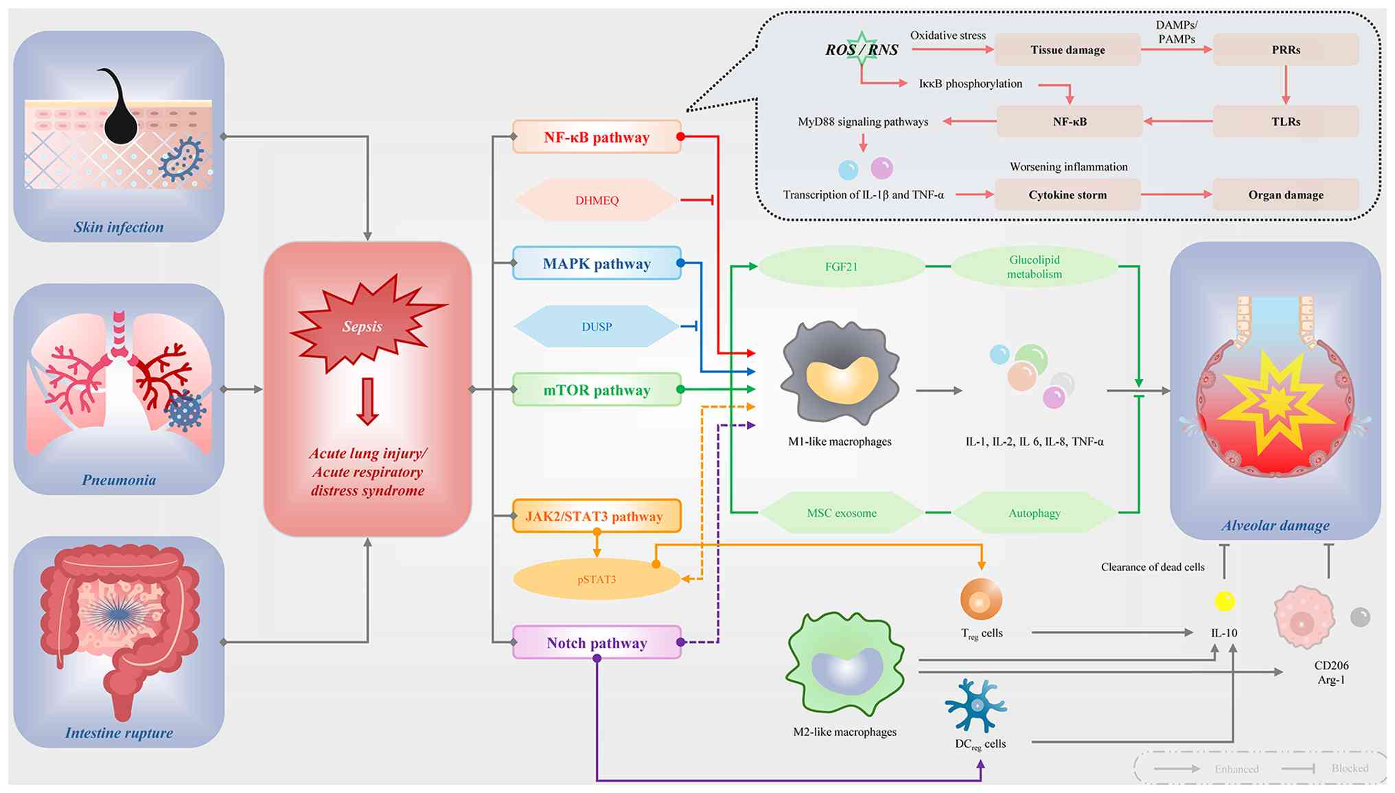
(2-5,12) (Fig. 1). In the early stages of sepsis,
pro-inflammatory cytokines and immune mediators are rapidly
released, triggering endothelial dysfunction and increased
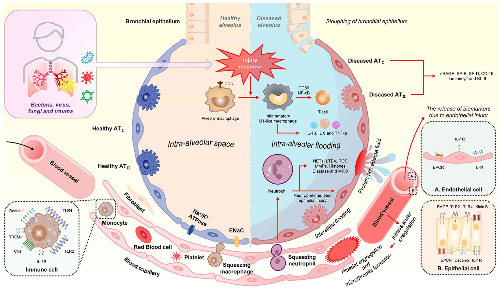
pulmonary capillary permeability (2-5,13). This leads to the leakage of
protein-rich fluid into the alveolar space, causing pulmonary edema
(2-5,14). The resulting disruption of the
alveolar-capillary barrier, which directly impairs gas exchange and
triggers the characteristic hypoxemia of ALI, represents the
primary clinical phenotype of S-ALI (Fig. 2) (2-5,15). Although scientists continue to
acquire a more in-depth understanding of the pathophysiology of
ALI, the pathophysiology of S-ALI remains complex, involving
multiple molecular pathways and cell types (2-5,16). Currently, clinical treatment
primarily involves early infection control, optimized mechanical
ventilation and supportive care for vital organs (2-5,17). However, existing treatment
options are ineffective in reducing mortality rates and may lead to
complications, such as ventilator-associated pneumonia and
barotrauma (2-5,18,19). Identifying the specific molecular
mechanisms that drive patient-specific disease trajectories is
therefore critical for the development of next-generation ICU
interventions and personalized therapeutic approaches.
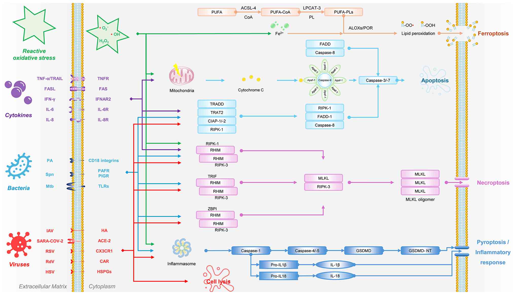
Pyroptosis is a highly regulated programmed cell
death process that has emerged as a critical, actionable driver of
inflammatory amplification in S-ALI (2-5,20,21). Pyroptosis, also known as
inflammatory cell death, is a mode of programmed cell death that is
dependent on cysteine aspartate-specific protease-1 (caspase-1)
(2-5,21,22). Unlike apoptosis and autophagy,
pyroptosis is characterized by triggering an intense inflammatory
response via gasdermin-mediated membrane pore formation (2,3,5,21,23). Although initially considered to
be a host-protective mechanism, recent studies have demonstrated
that excessive systemic cellular pyroptosis in the context of
sepsis accelerates the release of inflammatory markers in immune
cells (5,21,24,25), endothelial cells and epithelial
cells, leading to systemic organ tissue damage (5,26,27). This dysregulated pyroptotic
activity accounts for a substantial portion of the biological
heterogeneity in patients with sepsis, highlighting why
non-targeted therapies often fail; this emphasizes the need for
precision-guided interventions in ICU care.
Therefore, targeting pyroptosis has emerged as a
novel precision strategy for the treatment of sepsis and its
pulmonary complications (5,28). Inhibiting key pyroptotic
mediators may suppress uncontrolled lysis and prevent further organ
injury (5,29). Existing inhibitors of pyroptosis,
such as nucleotide-binding oligomerization domain-like receptor
protein 3 (NLRP3) or caspase-1 antagonists and natural
anti-inflammatory compounds, have demonstrated the potential for
attenuating the effects of pyroptosis in preclinical models
(5,30-32). These findings provide a
translational roadmap for integrating pyroptosis-targeted
interventions into individualized ICU care, allowing for the
selection of specific therapies based on the unique molecular
signatures of a patient. A summary of key preclinical and clinical
evidence of pyroptosis activation during S-ALI is presented in
Table I. Table I presents data from multiple
murine models [primarily lipopolysaccharide (LPS)- and cecal
ligation and puncture (CLP)-induced S-ALI] demonstrating the
elevated expression of pyroptosis-related markers, such as NLRP3,
cleaved caspase-1, GSDMD, IL-1β and IL-18 in lung tissues at
different time points (e.g., 8-24 h post-induction). Table I also includes human studies
demonstrating increased circulating levels of GSDMD p30, NLRP3 and
IL-1β/IL-18 in patients with sepsis in the ICU, which are
associated with disease severity and poor outcomes.
The NLRP3 inhibitor, MCC950, has been shown to
suppress the release of IL-1β and reduce inflammatory lung injury
in preclinical models (5).
Likewise, caspase-1 inhibitors, such as VX-765 have also
demonstrated potential in attenuating pyroptosis and protecting
against sepsis-induced lung damage (5,33). Blocking GSDMD activation
represents another promising strategy to halt pore formation and
cytokine efflux, providing a more surgically precise approach than
traditional broad-spectrum steroids (5,34).
The present review focuses on elucidating the
signaling pathways underlying pyroptosis in order to provide a
coherent understanding of its molecular mechanisms and regulatory
characteristics. The role of pyroptosis in the development and
progression of S-ALI is discussed with particular focus on its
contribution to inflammatory amplification and tissue damage in
critically ill patients. In addition, current advances in
strategies aimed at modulating pyroptotic processes are summarized
to clarify their potential therapeutic relevance and to identify
molecular targets that may support mechanism-guided intervention.
By connecting pyroptotic mechanisms with clinical heterogeneity and
potential targeted therapies, the present review aimed to provide
knowledge of precision-guided ICU strategies and to support the
future of individualized critical care.
The biological heterogeneity observed in patients
with S-ALI reflects distinct molecular cascades rather than a
uniform inflammatory endpoint (5,42). Multiple members of the caspase
family initiate pyroptosis by converging on the proteolytic
cleavage of gasdermin proteins, a programmed process characterized
by the formation of large transmembrane pores (5,43). This biochemical execution
triggers rapid cytoplasmic swelling, osmotic membrane rupture and
the massive efflux of inflammatory mediators and intracellular
damage-associated molecular patterns (DAMPs) into the alveolar
space (5,44). Crucially, the specific
caspase-gasdermin axis activated within the lung microenvironment
dictates the kinetics and magnitude of the pulmonary response,
providing a robust molecular framework for defining
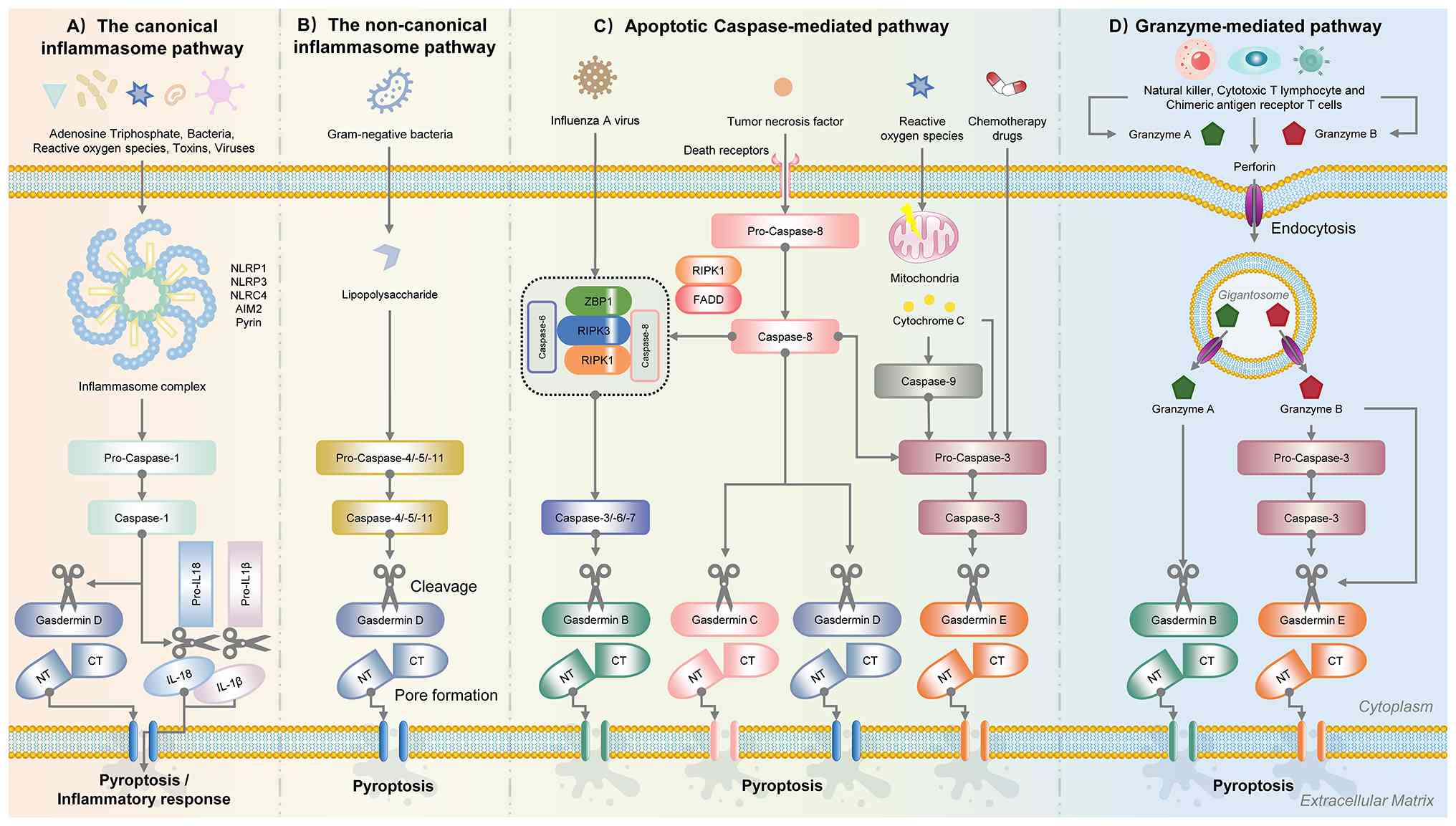
patient-specific inflammatory endotypes (5,42,45). Currently, four principal
signaling pathways have been identified as the primary executors of
this programmed cell death: the canonical, noncanonical, apoptotic
caspase-mediated, and granzyme-mediated pathways (Fig. 4).
The canonical pathway represents the most thoroughly
characterized mechanism driving pyroptosis in the septic lung. It
is initiated when intracellular pattern recognition receptors, such
as absent in melanoma 2, pyrin, or various nucleotide-binding
oligomerization domain-like receptors (NLRP1, NLRP3 and NLRC4),
detect conserved pathogen-associated molecular patterns or
endogenous DAMPs released during cellular stress (5,45,46) (Fig. 4A). Unlike stochastic inflammatory
triggers, the assembly of the NLRP3-apoptosis-associated speck-like
protein containing a caspase recruitment domain (ASC)-caspase-1
inflammasome functions as a high-fidelity molecular amplifier that
dictates the intensity of the early pulmonary cytokine storm
(47,48). Upon successful assembly of this
multiprotein complex, mature caspase-1 processes pro-IL-1β and
pro-IL-18 into their bioactive forms, while concurrently cleaving
GSDMD to liberate the N-terminal pore-forming domain (GSDMD-NT)
(49). In the complex landscape
of S-ALI, this pathway serves as the primary engine for early
hyper-inflammation and the metabolic priming of the alveolar
vascular endothelium (50,51). The significant individual
variability in the activation thresholds of these cytosolic sensors
explains the inconsistent clinical efficacy of broad-spectrum
anti-inflammatory agents, suggesting that patients with a
hyper-primed canonical axis require interventions specifically
targeting inflammasome assembly rather than downstream effector
cytokines (52).
For patients suffering from Gram-negative bacterial
sepsis, the noncanonical pathway provides a direct, autonomous
sensing mechanism that bypasses the classical receptor-mediated
signaling hierarchy (53,54).
In this axis, human caspase-4/5 (or murine caspase-11) functions as
an intracellular receptor that binds directly to cytosolic LPS, a
process significantly facilitated by interferon-inducible
guanylate-binding proteins that destabilize pathogen-containing
vacuoles (55,56) (Fig. 4B). This direct protein-LPS
interaction represents a rapid-response executioner of endothelial
lysis, occurring independently of the kinetic delays typically
associated with the assembly of large inflammasome platforms
(57). Furthermore,
caspase-11-mediated potassium efflux can secondarily trigger the
NLRP3 inflammasome, creating a potent, self-reinforcing
feed-forward loop that accelerates the collapse of the
alveolar-capillary barrier (58). From a precision medicine
perspective, recognizing the dominance of this LPS-caspase axis is
essential, as it identifies a subset of patients who remain
refractory to NLRP3-specific inhibition, but may demonstrate
clinical responsiveness to strategies targeting noncanonical
executioners.
The molecular plasticity between silent apoptosis
and explosive pyroptosis is governed by the strategic recruitment
of apoptotic caspases to the gasdermin execution machinery. As
illustrated in Fig. 4C, various
stimuli including influenza A virus, tumor necrosis factor-α
(TNF-α), death receptors, reactive oxygen species and
chemotherapeutic drugs can trigger this pathway (59-62). These stimuli activate the
extrinsic apoptotic pathway through death receptors, leading to the
recruitment of adaptor proteins, such as FADD and RIPK1, which
activate pro-caspase-8 into active caspase-8. Simultaneously, the
intrinsic (mitochondrial) pathway is engaged via cytochrome
c release from the mitochondria, resulting in the activation
of caspase-9. Both initiator caspases (caspase-8 and caspase-9)
then converge to activate the executioner caspases, primarily
caspase-3, as well as caspase-6 and caspase-7. These executioner
caspases cleave gasdermin family proteins, including gasdermin B
(GSDMB), gasdermin C, GSDMD and gasdermin E (GSDME), to generate
their pore-forming N-terminal fragments (23,60).
Caspase-3, traditionally viewed as a terminal
executioner of non-inflammatory apoptosis, can cleave GSDME to
generate GSDME-N fragments, which effectively switches the mode of
cell death to pro-inflammatory pyroptosis. The clinical trajectory
of S-ALI is profoundly influenced by the tissue-specific expression
profiles of GSDME, as high baseline expression in pulmonary
epithelial cells converts otherwise resolving apoptotic signals
into localized tissue lysis and inflammatory amplification
(63-65). Similarly, caspase-8 functions as
a molecular rheostat under conditions of TNF-α stimulation or
pathogenic stress, bridging these death pathways via the direct
cleavage of GSDMD in specific immune cell subsets (66). This molecular crosstalk provides
a profound stratification marker for clinicians in the ICU;
evaluating the GSDME signature in pulmonary cells predicts whether
a patient will exhibit a hyper-inflammatory phenotype or a more
favorable resolving death phenotype (66).
Beyond endogenous cell-autonomous signaling, the
granzyme-mediated pathway highlights the critical role of external
immune executioners in driving irreversible pulmonary tissue lysis.
Perforin-delivered granzymes (particularly GZMA and GZMB) from
over-activated cytotoxic T-lymphocytes or natural killer cells can
directly cleave gasdermin family members, such as GSDMB or GSDME,
in an action that is entirely independent of conventional caspase
activity (67-72) (Fig. 4D). In the hyper-active immune
phase often observed in early sepsis, this pathway represents a
significant mechanism of collateral damage where the adaptive
immune system of the exacerbates alveolar wall destruction and
tissue dysfunction. Identifying this lymphocyte-driven lysis shifts
the focus of personalized therapy toward immunomodulatory
stratification, particularly for those critically ill patients who
exhibit abnormally high cytotoxic activity in bronchoalveolar
lavage fluid, effectively moving the field beyond the traditional
constraints of innate immune-centric treatment models (73).
The pathological progression of S-ALI is dictated by
the coordinated execution of cell death across distinct pulmonary
compartments, creating a spatio-temporal map of organ failure
(74). Instead of a generic
'final common pathway', the cell-specific activation of pyroptotic
machinery dictates the biophysical transition from localized
infection to systemic respiratory collapse. Understanding these
cellular roles provides the necessary mechanistic depth to define
patient-specific clinical endotypes and to develop precision-guided
ICU interventions (75).
Alveolar macrophages function as the frontline
sensors of the pulmonary microenvironment. In the initial phase of
S-ALI, macrophage pyroptosis functions as a high-potency 'molecular
detonator' that amplifies pathogen-driven signals into an expansive
cytokine storm (76). The
activation of the NLRP3 inflammasome facilitates the rapid,
unconventional secretion of IL-1β and high mobility group box 1,
which orchestrate neutrophil recruitment and prime the lung
parenchyma for secondary damage (77). This macrophage-centric execution
accounts for the systemic hyper-inflammation observed in
'hyper-inflammatory' subphenotypes (78). The intensity of this detonator
phase suggests that early-window targeting of macrophage-specific
gasdermin activation could preemptively halt the inflammatory
cascade before irreversible structural destruction occurs.
The structural integrity of the alveolar-capillary
barrier is fundamentally dependent on the semi-permeable
microvascular endothelium. In the evolution of S-ALI, endothelial
pyroptosis serves as the primary driver for the catastrophic loss
of vascular barrier function, leading to the hallmark accumulation
of protein-rich edema (26). The
intracellular sensing of LPS triggers non-canonical signaling
through caspase-11, which executes endothelial lysis via
GSDMD-mediated pore formation (79). This cell-specific destruction
explains the clinical transition to profound hypoxemia, as
endothelial pore formation represents a decisive biophysical
transition point (80).
Furthermore, the release of pro-thrombotic factors following
membrane rupture exacerbates microcirculatory dysfunction, linking
pyroptosis to the coagulation abnormalities frequently observed in
patients with sepsis (81).
While endothelial damage drives leakage, the
pyroptosis of alveolar epithelial cells (AECs), particularly type
II cells, dictates the failure of respiratory mechanics. The
depletion of AECII through pyroptotic pathways directly impairs
pulmonary surfactant production, contributing to alveolar collapse
and refractory atelectasis (82). Unlike apoptosis, this regulated
lysis prevents effective lung repair and potentially initiates
pathways driving late-stage fibroproliferation (83). Recognizing the epithelial
compartment as a terminal executioner provides a translational
rationale for strategies that specifically preserve AEC integrity.
Such protection is essential for maintaining mechanical stability
in patients undergoing invasive mechanical ventilation, where
epithelial loss increases the risk of ventilator-induced lung
injury (84).
Critical to the exacerbation of S-ALI is the
molecular crosstalk between these cellular compartments, often
converging on the assembly of the PANoptosome (85,86). This integrated death mode
involves the coordinated activation of pyroptosis, apoptosis and
necroptosis, allowing the cell to bypass individual signaling
bottlenecks (87). The discovery
of PANoptosis explains the 'therapeutic escape' observed in
clinical trials, where inhibiting a single protease fails due to
the compensatory activation of parallel death modes (83). For instance, selective caspase-1
inhibitors (such as VX-765) in inflammatory lung injury models and
related acute respiratory distress syndrome (ARDS) trials have
exhibited limited sustained efficacy, as the blockade of pyroptosis
leads to the compensatory activation of apoptosis and necroptosis
pathways via caspase-8 and RIPK3/MLKL, resulting in persistent
tissue damage and cytokine release (88). Similarly, attempts at single
caspase-8 inhibition in sepsis-associated contexts have
demonstrated incomplete protection due to upregulation of
alternative PANoptosome components (89). Integrating PANoptosome dynamics
into the S-ALI framework underscores the necessity for 'combination
precision medicine' that addresses these convergent molecular hubs
to effectively mitigate multifaceted tissue destruction (90).
Pyroptosis, a pro-inflammatory programmed cell death
mediated by the gasdermin family, is defined as a distinct
inflammatory phenotype in S-ALI, with a pathological trajectory
that contributes to irreversible alveolar damage (91). From a precision medicine
perspective, pyroptosis serves as both a mechanistic driver and a
stratification-relevant therapeutic target. Lineage plasticity, a
fundamental property that enables cells to shift between different
functional states, has attracted increased interest in recent years
in the context of sepsis-associated acute lung injury.
Intratumoral-like heterogeneity partly reflects the lineage
plasticity of lung cells during S-ALI progression. Distinct
clusters of dying cells co-exist in both canonical and
non-canonical pyroptosis pathways compared to traditional
single-mode death models. These clusters differ significantly in
marker genes, molecular signaling pathways, differentiation states
and transcriptional profiles, indicating a marked increase in
transcriptional heterogeneity during the progression if S-ALI. The
intermediate and transitional state of pyroptotic injury is
evident, as multiple pathways exhibit consistent shifts along the
histological transition from early inflammation to terminal lysis,
consistent with previous bulk sequencing data (90). This dynamic transition provides a
biological foundation for stage-dependent and biomarker-guided
therapeutic intervention. An epithelial-endothelial cluster shared
among injury subtypes demonstrates potent differentiation potential
according to the transcriptional differentiation trajectory. Gene
expression, pathway enrichment and clinical biomarker data point to
the stem-like characteristic of this subpopulation, suggesting it
as the potentially pioneering force of lineage plasticity for the
progression of S-ALI. Previous rseearch tracing histological
transformation in septic lungs have identified an undifferentiated,
stem-like state that emerges during the transition, with cells
exhibiting basal-like features and the activation of NF-κB and
complement signaling pathways (92). Key regulatory nodes exhibit basal
origins with marked upregulation of NLRP3 and complement component
5a (C5a) receptor (C5aR) signaling pathways (92). The expression of the associated
gene module gradually increases along the histological transition
in experimental models. Significant discordance exists between
preclinical success and clinical reality. This discordance may be
partially attributed to the lack of patient stratification
(2). In this context, linking
biomarker-defined endotypes with pathway-specific interventions
forms the core of a precision roadmap. The hyperinflammatory or
pyroptosis-high endotype, characterized by elevated plasma GSDMD
fragments and IL-18, is associated with activation of canonical or
noncanonical inflammasome pathways and may be matched with
corresponding targeted therapies, with the potential to improve
clinically relevant outcomes such as ventilator-free days and organ
failure scores. Current therapeutic strategies mainly focus on a
single pathway, but underestimate the high plasticity of septic
cells. Therefore, a shift toward biomarker-guided, stage-specific
intervention is required to overcome treatment resistance and
improve therapeutic precision. Further studies are warranted to
elucidate the precise role of stem-like cells in S-ALI progression
and to assess their potential therapeutic implications.
The canonical NLRP3-caspase-1 axis serves as a
primary molecular detonator in S-ALI, with the intensity of its
activation being closely associated with alveolar-capillary barrier
disruption (93). Distinct
regulatory clusters are governed by endogenous rheostats, such as
heat shock factor 1 and heat shock protein 8, which modulate NLRP3
ubiquitination to prevent the irreversible formation of the ASC
speck complex (94,95).
These regulatory clusters differ significantly in
molecular signaling pathways and differentiation states, indicating
a dramatic increase in transcriptional heterogeneity during sepsis
progression. The intermediate and transitional state of NLRP3
priming is evident, as multiple genes and pathways show consistent
shifts from early priming to terminal execution. A priming cluster
shared across models demonstrates potent differentiation potential
according to the transcriptional differentiation trajectory.
Pathway enrichment points to the stem-like characteristic of this
subpopulation, suggesting it as the potentially pioneering force of
lineage plasticity for canonical pyroptosis. A vast repertoire of
pharmacological agents has demonstrated robust efficacy in
preclinical models (96-122); however, the discordance between
murine success and clinical reality underscores the challenge of
target engagement in the human septic niche. For example,
colchicine suppresses NLRP3 inflammasome assembly and oxidative
stress in LPS-induced ALI (123), while metformin inhibits GSDMD
activation and downstream inflammatory amplification in CLP models
(96), both demonstrating
consistent attenuation of pyroptosis-associated lung injury in
vivo. A critical barrier in translating pyroptosis-targeted
strategies from murine models to human sepsis lies in
species-specific divergence in inflammasome signaling, particularly
the absence of direct orthology between murine caspase-11 and human
caspase-4/5, which exhibit distinct activation thresholds and
ligand sensitivities to cytosolic LPS (53). This molecular non-equivalence
complicates the extrapolation of caspase-11-centered findings from
LPS- or CLP-induced murine models of SALI to human patients in the
ICU, where caspase-4/5-driven noncanonical inflammasome activation
may follow different kinetics and cellular distribution (55).
Several therapies with potential applicability in
the ICU setting have been described. These include anakinra (IL-1
receptor antagonist), which is already used in critically ill
patients and has favorable pharmacologic feasibility due to its
subcutaneous or intravenous administration, relatively predictable
pharmacokinetics even in organ dysfunction, and established safety
profile in sepsis trials (3).
Mesenchymal stromal cells (MSCs) have also been administered
intravenously in multiple ARDS ICU trials, although their
feasibility is limited by the need for cell preparation and
variable engraftment (124). By
contrast, direct NLRP3 inhibitors (e.g., DFV890) and GSDMD
inhibitors remain largely investigational, with limited data on ICU
pharmacokinetics, potential drug interactions and tissue
penetration in critically ill patients with altered hemodynamics
and pulmonary vascular injury (125).
In addition, pharmacokinetic and pharmacodynamic
constraints in critically ill patients, including altered drug
absorption, organ dysfunction and unpredictable tissue distribution
within injured pulmonary microvasculature, further limit reliable
target engagement despite robust preclinical efficacy signals
(126).
The expression of NLRP3-associated gene modules
gradually increases along the histological transition in
experimental models, while clinical translation remains limited by
the lack of patient stratification (127,128). Clinical applicability of this
axis depends on the early identification of patients with high
inflammasome activity through plasma GSDMD cleavage products and
IL-18 levels, preferably within 24-48 h of admission to the ICU.
Emerging clinical signals from NLRP3 inhibitors, such as DFV890 and
the soluble urokinase plasminogen activator receptor (suPAR)-guided
administration of anakinra suggest that identifying
hyper-pyroptotic endotypes is the only viable path to translating
these findings into survival benefits (125,129). Therapeutic efficacy is likely
to be maximized during the early hyperinflammatory phase, whereas
late-stage administration risks exacerbating immunosuppression. The
full pharmacological landscape of inhibitors targeting the
canonical NLRP3-caspase-1-GSDMD axis is synthesized in Table II. Table II provides a comprehensive
summary of various pharmacological agents targeting different nodes
(priming, assembly and execution) of the canonical inflammasome
pathway. It lists specific agents (e.g., MCC950, metformin,
colchicine and natural compounds), their experimental inducers
(mainly LPS or CLP), molecular mechanisms, and key outcomes in
murine models, such as reduced pulmonary damage, reduced
pyroptosis, reduced NLRP3 activation, reduced GSDMD cleavage, and
improved survival, along with associated risks and limitations.
Targeting the canonical NLRP3 inflammasome pathway
has shown promising results in preclinical studies (Table II).
Lineage plasticity is particularly evident in the
non-canonical caspase-4/5/11 pathway (86). Unlike the canonical axis, this
pathway directly senses cytosolic LPS within pulmonary endothelium,
triggering rapid GSDMD-mediated membrane permeabilization (130). Distinct clusters of endothelial
cells co-exist in both acute and subacute phases of lung injury
compared to myeloid-dominant canonical models. These clusters
differ significantly in marker genes, molecular signaling pathways
and differentiation states, indicating a marked increase in
transcriptional heterogeneity during endothelial transformation.
The intermediate and transitional state of non-canonical activation
is evident, as multiple pathways show consistent shifts along the
histological transition from localized inflammation to systemic
alveolar flooding (4). An
upstream humoral-cellular crosstalk cluster shared among different
sepsis models demonstrates potent differentiation potential
according to the transcriptional differentiation trajectory.
Pathway enrichment points to the stem-like characteristic of this
subpopulation, suggesting it as the potentially pioneering force of
lineage plasticity for non-canonical pyroptosis. The complement
component 3a/C5a-C5aR axis serves as a key modulator, while
vilobelimab has shown clinical success in severe respiratory
failure (4). Notably,
vilobelimab-mediated C5a blockade represents a clinically
translatable example of complement-targeted intervention that has
progressed beyond preclinical validation into randomized human ARDS
trials, highlighting a partial bridge between murine non-canonical
inflammasome models and ICU feasibility (131). The significant activation of
non-canonical clusters occurs across disease phases. Current
therapeutic strategies for this axis remain limited and require
careful temporal control to avoid compromising host defense
mechanisms in the early phase of sepsis. Targeting intratumoral
heterogeneity or stem-like endothelial nodes has been proposed as a
novel strategy to overcome treatment resistance. The full list of
inhibitors targeting the non-canonical inflammasome pathway is
synthesized in Table III.
Table III summarizes
therapeutic strategies targeting the non-canonical (caspase-4/5/11)
pathway in S-ALI, including upstream complement modulation,
caspase-11 inhibition and cellular therapies. Table III lists representative agents,
molecular mechanisms, key outcomes (such as reduced endothelial
pyroptosis, reduced vascular leakage, reduced lung injury and
improved survival), as well as risks and limitations for each
approach.
The concept of lineage plasticity in the septic lung
is further reflected in the integrated PANoptosome complex, which
enables a distinct phenotype of cell death with potential for
state-shifting (87). Distinct
clusters of dying cells where pyroptosis co-exists with apoptosis
and necroptosis are more evident compared to traditional
single-mode death models (90).
These clusters differ significantly in marker genes, molecular
signaling pathways and differentiation states, indicating a
dramatic increase in transcriptional heterogeneity during
death-mode switching. The intermediate and transitional state of
GSDME-mediated injury is evident, as multiple pathways show
consistent shifts from early apoptosis to terminal lysis (83). An epithelial cluster shared
across injury groups demonstrates potent differentiation potential
according to the transcriptional differentiation trajectory. Gene
expression and pathway enrichment point to the stem-like
characteristic of this subpopulation, suggesting it as the
potentially pioneering force of lineage plasticity for PANoptosis
in S-ALI. Caspase-3 and caspase-8 function as critical hubs for
death-mode switching (132).
For instance, ligustrazine suppresses both caspase-8 and
NLRP3-caspase-1 signaling simultaneously, illustrating a multi-node
regulatory strategy capable of modulating pyroptosis-apoptosis
crosstalk within PANoptotic networks in septic lung injury
(133). The significant
activation of compensatory pathways occurs when one executioner is
inhibited (134). This
coordinated activation highlights the limitation of single-pathway
inhibition and supports the need for multi-target or adaptive
therapeutic strategies. Current therapies focusing on a single
pathway underestimate the high plasticity of septic cells.
Targeting intratumoral heterogeneity or stem-like protective cells,
such as MSCs acting as biological rheostats, has been proposed as a
novel strategy to overcome treatment resistance (135).
The full list of agents targeting apoptotic
caspase-mediated pyroptosis is synthesized in Table IV. Table IV summarizes inhibitors of the
apoptotic caspase-mediated pathway in S-ALI, detailing targeting
nodes (primarily caspase-3, caspase-8 and caspase-9),
representative agents (e.g., irisin, resveratrol, ligustrazine,
MSCs), molecular mechanisms and key outcomes in murine models, such
as reduced caspase-3 activity, reduced lung injury, reduced
microvascular permeability and reduced epithelial pyroptosis, along
with associated risks and limitations.
Although human evidence for the modulation of
pyroptosis in S-ALI remains limited, it is increasingly promising.
Circulating GSDMD fragments, IL-1β, IL-18 and
inflammasome-associated signatures have been reported to be
elevated in patients with sepsis in the ICU and in
COVID-19-associated ARDS (56,136). Notably, these signals appear to
be more prominent in hyperinflammatory bacterial sepsis than in
certain viral ARDS cohorts, suggesting substantial inflammatory
heterogeneity across patient populations (137).
Current biomarker-guided studies and key
translational and clinical investigations targeting
complement-inflammasome-pyroptosis-associated pathways have begun
to provide early, yet heterogeneous evidence for clinical efficacy
in critically ill patients. In particular, vilobelimab (anti-C5a
monoclonal antibody) has been evaluated in randomized clinical
trials in severe COVID-19-associated ARDS, where treatment was
shown to be associated with a reduction in 28-day mortality and
trends toward improved ventilator-free days in selected
hyperinflammatory subgroups, although overall effects across
unselected populations remained variable (138,139).
In addition to vilobelimab, earlier clinical
attempts to broadly suppress inflammatory injury in sepsis and
ARDS, without targeting specific pyroptosis-related pathways or
applying biomarker-based stratification, have largely failed to
demonstrate consistent improvements in mortality or organ failure
scores, highlighting the limitations of non-selective
anti-inflammatory approaches in critical illness. These findings
collectively indicate that the therapeutic modulation of
pyroptosis-related signaling requires both pathway specificity and
patient stratification to achieve clinical efficacy.
Among candidate therapeutic nodes, upstream NLRP3
inflammasome blockade may provide the broader suppression of
caspase-1 activation, IL-1β/IL-18 maturation and downstream
pyroptotic amplification during the early hyperinflammatory stage
of S-ALI (140). By contrast,
direct GSDMD inhibition may more specifically prevent membrane pore
formation and terminal cell lysis, while preserving part of the
upstream immune sensing cascade (9). The modulation of the IL-1 pathway
represents a downstream anti-inflammatory strategy that may
attenuate cytokine-driven tissue injury, but does not completely
suppress inflammasome activation or pyroptotic execution (141). Compared with conventional
broad-spectrum anti-inflammatory therapies, pyroptosis-targeted
interventions provide the potential advantage of mechanism-guided
precision modulation by directly interrupting regulated
inflammatory cell death pathways closely linked to alveolar barrier
disruption, endothelial leakage, and maladaptive innate immune
amplification in S-ALI (140).
Among the currently available translational
strategies, substantial heterogeneity exists regarding both
mechanistic specificity and clinical applicability. Upstream
inflammasome-directed approaches, such as NLRP3 inhibition, may
provide the broader suppression of caspase-1 activation and
downstream cytokine maturation during the early hyperinflammatory
phase; however, they may also increase the risk of excessive immune
suppression when administered during later septic stages (142). By contrast, downstream IL-1
pathway blockade appears to represent a comparatively safer and
clinically feasible strategy, particularly in biomarker-enriched
hyperinflammatory subgroups, as illustrated by anakinra-related
research (129).
Complement-targeted interventions, such as vilobelimab may be
particularly relevant in endothelial-dominant inflammatory
phenotypes characterized by excessive C5a activation and
neutrophil-driven tissue injury (138). Notably, several translational
studies summarized in Table V
have demonstrated that non-stratified anti-inflammatory
interventions have frequently failed to achieve consistent survival
benefits, highlighting that therapeutic efficacy is highly
dependent on appropriate endotype selection, the timing of
intervention and the preservation of essential host defense
mechanisms. Collectively, current evidence supports a transition
from empiric broad immunosuppression toward biomarker-guided
precision modulation of pyroptosis-associated inflammatory pathways
in S-ALI.
Notably, a substantial proportion of currently
available human evidence is derived from COVID-19-associated ARDS
cohorts rather than classical bacterial sepsis-induced ALI
(137). Although these studies
provide valuable proof-of-concept support for inflammasome and
pyroptosis modulation, significant differences exist between viral
and bacterial inflammatory programs, including pathogen-recognition
pathways, neutrophil predominance, complement activation patterns
and immune exhaustion trajectories (137). Therefore, caution is required
when extrapolating COVID-19-based findings directly to
heterogeneous sepsis populations.
Nevertheless, a substantial discordance persists
between robust preclinical efficacy and relatively modest clinical
outcomes. This discrepancy may partly reflect the dual role of
pyroptosis in host defense and tissue injury, the existence of
narrow therapeutic timing windows and the marked immunological
plasticity observed during different stages of sepsis (143). A number of experimental
inhibitors fail to translate clinically as they insufficiently
account for patient stratification, inflammatory endotype, and
stage-specific immune status. Patients with excessive inflammasome
activation or elevated circulating IL-1β/IL-18 signatures may
represent the most plausible candidates for pyroptosis-targeted
interventions. Based on these signals, a biomarker-guided
stratification framework may be used to identify a
'pyroptosis-high' inflammatory subphenotype in patients with
S-ALI/ARDS (144), whereas
inappropriate late-stage suppression could potentially aggravate
sepsis-associated immunosuppression. A biomarker-guided
stratification framework for S-ALI is defined as follows: Candidate
pyroptosis-related biomarkers include circulating GSDMD cleavage
products (GSDMD p30), IL-1β and IL-18, primarily measured in plasma
or serum samples obtained within 24-48 h of admission to the ICU
(9). Bronchoalveolar lavage
fluid may be used in research settings; however, is not routinely
feasible in critically ill patients. Real-world application is
constrained by delayed laboratory turnaround time, variability in
sampling timing, and dynamic changes in inflammatory status over
the disease course. Biomarker interpretation should also consider
confounding factors, such as acute kidney injury, extracorporeal
membrane oxygenation and multi-organ dysfunction, which may
significantly alter circulating inflammatory profiles (145).
Based on this framework, patients can be stratified
into hyperinflammatory pyroptosis-high and hypo-inflammatory
endotypes to support mechanism-guided therapeutic selection
(9). In the early
hyperinflammatory phase, characterized by elevated levels of GSDMD
p30, IL-1β, and IL-18, therapeutic strategies are primarily
directed toward inhibition of the canonical inflammasome axis,
including NLRP3 and caspase-1 blockade, as well as downstream IL-1
signaling modulation. By contrast, in later stages or in patients
progressing toward immunosuppression, excessive inflammasome
inhibition should be avoided, and supportive or immune-restorative
approaches may be more appropriate (9). In endothelial-predominant injury
phenotypes, targeting the C5a-complement axis may provide
additional benefit, whereas in mixed or highly plastic PANoptotic
states, combination or multi-target strategies are likely required
to achieve effective disease control (134).
To provide a coherent overview of translational
readiness, representative human studies and ongoing or terminated
clinical trials relevant to pyroptosis-modulating strategies in
sepsis, ARDS and S-ALI are summarized in Table V.
Of note, these studies collectively demonstrate
that successful clinical translation depends not only on pathway
inhibition itself, but also on appropriate patient stratification,
inflammatory endotype characterization, therapeutic timing and the
preservation of antimicrobial host defense.
Targeting pyroptosis in S-ALI presents intrinsic
challenges that arise from the dual role of inflammatory cell death
in host defense and tissue injury. Controlled activation
contributes to pathogen clearance and immune surveillance, whereas
dysregulated or sustained activation amplifies inflammatory
cascades, disrupts alveolar-capillary barrier integrity and
accelerates organ dysfunction. This dual nature makes
indiscriminate or prolonged suppression of pyroptotic signaling
potentially detrimental to immune competence and infection control
(146). The molecular
heterogeneity of pyroptosis further complicates therapeutic
translation, as distinct inflammasome sensors, upstream caspases
and gasdermin family members are differentially engaged across
disease stages, cell populations and inflammatory
microenvironments, leading to context-dependent outcomes that are
not adequately captured by single-pathway inhibition strategies
(147). In the setting of
sepsis, dynamic shifts in immune status occur over time, with early
hyperinflammation frequently transitioning toward
immunosuppression, indicating that the timing and intensity of
pyroptosis modulation are likely to determine therapeutic efficacy
and safety rather than simple pathway blockade (148). Notably, a number of candidate
inhibitors targeting the NLRP3-caspase-1-GSDMD axis, including
MCC950 and VX-765, have demonstrated substantial efficacy in murine
LPS or CLP models, but remain limited in clinical translation due
to insufficient pharmacokinetic validation, heterogeneous patient
responses and concerns regarding excessive suppression of
antimicrobial immunity (149,150). For example, it was previously
demonstrated that MCC950 (50 mg/kg) significantly reduced
neutrophil infiltration, IL-1β/IL-18 levels and lung injury, scores
and improved the survival of mice with LPS-induced ALI and
CLP-induced sepsis (151).
Similarly, VX-765 attenuated caspase-1-mediated pyroptosis and lung
injury in murine models of LPS-induced ALI, although its protective
effects were incomplete due to pathway compensation (5). This heterogeneity is largely driven
by differences in underlying inflammatory endotypes (e.g.,
hyperinflammatory endotype with high IL-6/IL-8 showing better
response to NLRP3 inhibition vs. hypoinflammatory endotype), the
timing of intervention relative to disease stage (early
administration protective in LPS models, but detrimental in late
immunosuppressive phase of CLP sepsis), variability in baseline
comorbidities (e.g., obesity and diabetes causing exaggerated NLRP3
priming and worse outcomes), genetic polymorphisms in
inflammasome-related genes (e.g., NLRP3 gain-of-function variants
rs35829419 associated with higher IL-1β production and severe
ARDS), and pathogen-specific factors (stronger NLRP3 activation in
Gram-negative vs. Gram-positive infections), resulting in highly
variable target engagement and therapeutic responses across
patients in the ICU (152).
Since basal pyroptotic signaling contributes to
intracellular pathogen clearance and innate immune surveillance,
indiscriminate or prolonged inhibition may inadvertently increase
susceptibility to secondary infection and late-stage immune
paralysis in critically ill patients (144). These findings collectively
suggest that the successful clinical application of
pyroptosis-targeted therapies will likely require biomarker-guided
patient stratification and stage-specific intervention rather than
uniform pathway inhibition across all phases of sepsis-associated
acute lung injury (144).
From a translational perspective, the majority of
currently available evidence supporting pyroptosis inhibition in
S-ALI is derived from experimental models that only partially
reproduce the clinical complexity of sepsis, including
inter-individual variability, comorbid conditions and heterogeneous
infectious sources, which collectively influence inflammatory
trajectories and treatment responsiveness (153). Differences in species-specific
inflammasome regulation, gasdermin expression patterns and immune
cell composition further limit the direct extrapolation of
preclinical findings to critically ill patients (146). Clinical observations have shown
that patients with sepsis-associated ARDS exhibit elevated levels
of inflammasome-associated biomarkers, such as IL-1β and IL-18, and
preliminary interventional studies using inflammasome-targeted
agents are being explored in ARDS cohorts with elevated caspase-1
activity, providing early human evidence linking inflammasome
modulation to lung inflammation in acute respiratory failure
(154). Furthermore, serum
concentrations of NLRP3 are significantly higher in patients with
sepsis who develop ARDS, and are associated with disease severity
and 28-day mortality, suggesting that inflammasome activation
markers have clinical diagnostic and prognostic value in human
S-ALI (21). Emerging clinical
data also indicate that an elevated expression of NLRP3 in patients
with ARDS is associated with worse oxygenation and organ
dysfunction scores, highlighting the translational relevance of
inflammasome-related cell death pathways in human lung injury
(155).
In addition, pyroptosis does not occur in isolation
but intersects with apoptosis, necroptosis, autophagy and metabolic
reprogramming, forming an integrated cell death network in which
the selective modulation of a single node may trigger compensatory
mechanisms that sustain inflammation or tissue damage, underscoring
the need for a systems-level understanding of regulated cell death
in septic lung injury (156).
Observational clinical analyses using single-cell transcriptomics
and pyroptosis-related gene signatures have stratified patients
with sepsis-induced ARDS into prognostic groups based on immune
cell composition and pyroptosis signaling profiles, supporting the
potential for personalized approaches to targeting regulated cell
death in human disease (157).
Future research is thus warranted to focus on
refining the precision of pyroptosis-targeted interventions by
integrating disease stage, cellular specificity and host immune
status into therapeutic design. The identification of reliable
biomarkers reflecting pyroptotic activity in lung tissue and
circulation may enable the stratification of patients who are most
likely to benefit from targeted modulation, while avoiding
unnecessary immune suppression in others (158). Advances in multi-omics
profiling, spatial transcriptomics and single-cell analyses are
expected to provide deeper insight into cell type-specific
pyroptotic responses and their temporal evolution during sepsis
(158). Therapeutic strategies
that achieve the balanced regulation rather than the complete
inhibition of pyroptosis, particularly approaches that preserve
antimicrobial defense, while limiting inflammatory amplification
and barrier disruption, may provide a more viable path toward
clinical application (75).
Through the continued integration of mechanistic research with
translational and clinical investigation, targeting pyroptosis
holds promise for improving outcomes in S-ALI, while aligning with
the broader goal of individualized management in critical care
medicine (75).
In summary, pyroptosis represents a pivotal and
actionable programmed cell death mode that drives the biological
and clinical heterogeneity of S-ALI. The transition from a
single-pathway model to an integrated landscape involving
canonical, non-canonical and PANoptosome-mediated signaling has
redefined the understanding of pulmonary inflammatory
amplification. While preclinical studies have identified a vast
repertoire of pharmacological agents, the path to clinical
translation hinges on the shift from 'one-size-fits-all' strategies
to biomarker-guided, stage-specific interventions. By integrating
high-resolution molecular signatures with individual patient
endotypes, targeting the pyroptotic cascade provides a promising
frontier for achieving precision-guided management and improving
the survival of critically ill patients with sepsis-induced
respiratory failure.
Not applicable.
WW was involved in the conceptualization of the
study, analysis and tge interpretation of published literature,
visualization and validation, and the drafting of the manuscript.
NL and GD were involved in the literature search, screening,
extraction, the synthesis of published evidence, validation and
visualization. SY, RC and RZ contributed to the preparation of the
manuscript, reference checking and content organization, and
provided administrative support. JL was responsible for the study
design, project administration, supervision, funding acquisition,
quality control of the manuscript, critical revision of the
intellectual content, and manuscript review and editing. All
authors have read and approved the final manuscript. Data
authentication is not applicable.
Not applicable.
Not applicable.
The authors declare that they have no competing
interests.
Not applicable.
The present study was supported by the National Science and
Technology Major Project for the Prevention and Treatment of
Cancer, Cardiovascular, Respiratory, and Metabolic Diseases (grant
no. 2025ZD0549300); the Qihuang Scholars Cultivation Program; the
National Natural Science Foundation of China (grant no. 82474409);
Graduate Student Innovation Enhancement Project of Guangzhou
University of Chinese Medicine (grant no. A3-0317-25-429-010); the
Traditional Chinese Medicine (TCM) Science and Technology Research
and Cultivation Project of Guangdong Provincial Laboratory of
Chinese Medicine (grant no. HQL2024PZ004); and the National
Multidisciplinary Innovation Team Project in Traditional Chinese
Medicine (grant no. ZYYCXTD-D202406).
|
1
|
Cajander S, Kox M, Scicluna BP, Weigand
MA, Mora RA, Flohe SB, Martin-Loeches I, Lachmann G, Girardis M,
Garcia-Salido A, et al: Profiling the dysregulated immune response
in sepsis: Overcoming challenges to achieve the goal of precision
medicine. Lancet Respir Med. 12:305–322. 2024. View Article : Google Scholar
|
|
2
|
Moore AR, Pienkos SM, Sinha P, Guan J,
O'Kane CM, Levitt JE, Wilson JG, Shankar-Hari M, Matthay MA, Calfee
CS, et al: Elevated plasma interleukin-18 identifies high-risk
acute respiratory distress syndrome patients not distinguished by
prior latent class analyses using traditional inflammatory
cytokines: A retrospective analysis of two randomized clinical
trials. Crit Care Med. 51:e269–e274. 2023. View Article : Google Scholar : PubMed/NCBI
|
|
3
|
Shakoory B, Carcillo JA, Chatham WW, Amdur
RL, Zhao H, Dinarello CA, Cron RQ and Opal SM: Interleukin-1
receptor blockade is associated with reduced mortality in sepsis
patients with features of macrophage activation syndrome:
Reanalysis of a prior phase iii trial. Crit Care Med. 44:275–281.
2016. View Article : Google Scholar
|
|
4
|
Li Z, Wang Y, Feng Q, Zhang Y, Zhuang Y,
Xie Z and Bai X: Inhibition of the c3a receptor attenuates
sepsis-induced acute lung injury by suppressing pyroptosis of the
pulmonary vascular endothelial cells. Free Radic Biol Med.
184:208–217. 2022. View Article : Google Scholar : PubMed/NCBI
|
|
5
|
Li H, Guan Y, Liang B, Ding P, Hou X, Wei
W and Ma Y: Therapeutic potential of MCC950, a specific inhibitor
of NLRP3 inflammasome. Eur J Pharmacol. 928:1750912022. View Article : Google Scholar : PubMed/NCBI
|
|
6
|
Darkwah S, Kotey F, Ahenkorah J,
Adutwum-Ofosu KK and Donkor ES: Sepsis-related lung injury and the
complication of extrapulmonary pneumococcal pneumonia. Diseases.
12:722024. View Article : Google Scholar : PubMed/NCBI
|
|
7
|
GBD 2021 Global Sepsis Collaborators:
Global, regional, and national sepsis incidence and mortality,
1990-2021: A systematic analysis. Lancet Globa Health.
13:e2013–e2026. 2025. View Article : Google Scholar
|
|
8
|
Wang D, Jia H, Zheng X, Xi X, Zheng Y and
Li W: Attributable mortality of ARDS among critically ill patients
with sepsis: A multicenter, retrospective cohort study. BMC Pulm
Med. 24:1102024. View Article : Google Scholar : PubMed/NCBI
|
|
9
|
Wang Z, Li X, Yuan B and Zhang J:
Gasdermin D-driven pyroptosis in sepsis: Mechanisms, therapeutic
strategies, and clinical translation. Front Immunol.
17:18018962026. View Article : Google Scholar : PubMed/NCBI
|
|
10
|
Ding Z, Zhong R, Yang Y, Xia T, Wang W,
Wang Y, Xing N, Luo Y, Li S, Shang L and Shu Z: Systems
pharmacology reveals the mechanism of activity of Ge-Gen-Qin-Lian
decoction against LPS-induced acute lung injury: A novel strategy
for exploring active components and effective mechanism of TCM
formulae. Pharmacol Res. 156:1047592020. View Article : Google Scholar : PubMed/NCBI
|
|
11
|
Zheng Y, Wang J, Ling Z, Zhang J, Zeng Y,
Wang K, Zhang Y, Nong L, Sang L, Xu Y, et al: A diagnostic model
for sepsis-induced acute lung injury using a consensus machine
learning approach and its therapeutic implications. J Transl Med.
21:6202023. View Article : Google Scholar : PubMed/NCBI
|
|
12
|
Kumar V: Pulmonary innate immune response
determines the outcome of inflammation during pneumonia and
sepsis-associated acute lung injury. Front Immunol. 11:17222020.
View Article : Google Scholar : PubMed/NCBI
|
|
13
|
Dolmatova EV, Forrester SJ, Wang K, Ou Z,
Williams HC, Joseph G, Kumar S, Valdivia A, Kowalczyk AP, Qu H, et
al: Endothelial poldip2 regulates sepsis-induced lung injury via
rho pathway activation. Cardiovasc Res. 118:2506–2518. 2022.
View Article : Google Scholar :
|
|
14
|
Parker JC: Acute lung injury and pulmonary
vascular permeability: Use of transgenic models. Compr Physiol.
1:835–882. 2011. View Article : Google Scholar : PubMed/NCBI
|
|
15
|
Long ME, Mallampalli RK and Horowitz JC:
Pathogenesis of pneumonia and acute lung injury. Clin Sci (Lond).
136:747–769. 2022. View Article : Google Scholar : PubMed/NCBI
|
|
16
|
Li W, Li D, Chen Y, Abudou H, Wang H, Cai
J, Wang Y, Liu Z, Liu Y and Fan H: Classic signaling pathways in
alveolar injury and repair involved in sepsis-induced ALI/ARDS: New
research progress and prospect. Dis Markers.
2022:63623442022.PubMed/NCBI
|
|
17
|
Zhang F, Guo F, Zhang Y, Xu H, Liu Y, Lin
L, Li H, Yang H and Huang L: Huashibaidu formula attenuates
sepsis-induced acute lung injury via suppressing cytokine storm:
Implications for treatment of COVID-19. Phytomedicine.
109:1545492023. View Article : Google Scholar : PubMed/NCBI
|
|
18
|
von During S, Liu K, Munshi L, Kim SJ,
Urner M, Adhikari NK, Parhar K and Fan E: The association between
mechanical power within the first 24 hours and ICU mortality in
mechanically ventilated adult patients with acute hypoxemic
respiratory failure: A registry-based cohort study. Chest.
168:901–911. 2025. View Article : Google Scholar : PubMed/NCBI
|
|
19
|
Antimicrobial Resistance Collaborators:
Global burden of bacterial antimicrobial resistance in 2019: A
systematic analysis. Lancet. 399:629–655. 2022. View Article : Google Scholar : PubMed/NCBI
|
|
20
|
Coll RC, Schroder K and Pelegrin P: NLRP3
and pyroptosis blockers for treating inflammatory diseases. Trends
Pharmacol Sci. 43:653–668. 2022. View Article : Google Scholar : PubMed/NCBI
|
|
21
|
Yang Q, Zhang X, Luo L and Shen J:
Clinical application of serum NLRP3 on the diagnosis and prognosis
of sepsis patients complicated with acute respiratory distress
syndrome. Front Immunol. 14:12051322023. View Article : Google Scholar : PubMed/NCBI
|
|
22
|
Fink SL and Cookson BT:
Caspase-1-dependent pore formation during pyroptosis leads to
osmotic lysis of infected host macrophages. Cell Microbiol.
8:1812–1825. 2006. View Article : Google Scholar : PubMed/NCBI
|
|
23
|
Fink SL and Cookson BT: Pyroptosis and
host cell death responses during salmonella infection. Cell
Microbiol. 9:2562–2570. 2007. View Article : Google Scholar : PubMed/NCBI
|
|
24
|
Zhang YY and Ning BT: Signaling pathways
and intervention therapies in sepsis. Signal Transduct Target Ther.
6:4072021. View Article : Google Scholar : PubMed/NCBI
|
|
25
|
Dolmatova EV, Wang K, Mandavilli R and
Griendling KK: The effects of sepsis on endothelium and clinical
implications. Cardiovasc Res. 117:60–73. 2021. View Article : Google Scholar :
|
|
26
|
Joffre J, Hellman J, Ince C and
Ait-Oufella H: Endothelial responses in sepsis. Am J Respir Crit
Care Med. 202:361–370. 2020. View Article : Google Scholar : PubMed/NCBI
|
|
27
|
Zhang H, Wang Y, Qu M, Li W, Wu D, Cata JP
and Miao C: Neutrophil, neutrophil extracellular traps and
endothelial cell dysfunction in sepsis. Clin Transl Med.
13:e11702023. View Article : Google Scholar : PubMed/NCBI
|
|
28
|
Zheng X, Chen W, Gong F, Chen Y and Chen
E: The role and mechanism of pyroptosis and potential therapeutic
targets in sepsis: A review. Front Immunol. 12:7119392021.
View Article : Google Scholar : PubMed/NCBI
|
|
29
|
Mei Y, Chen X, Shi S, Lin W, Cheng Z, Fan
X, Wu W, Han J, Huang W, Ye B and Dai S: GI-Y2, a novel gasdermin D
inhibitor, attenuates sepsis-induced myocardial dysfunction by
inhibiting gasdermin D-mediated pyroptosis in macrophages. Br J
Pharmacol. 182:3503–3521. 2025. View Article : Google Scholar : PubMed/NCBI
|
|
30
|
Kasana S, Kumar S, Patel P, Kurmi BD, Jain
S, Sahu S and Vaidya A: Caspase inhibitors: A review on recently
patented compounds (2016-2023). Expert Opin Ther Pat. 34:1047–1072.
2024. View Article : Google Scholar : PubMed/NCBI
|
|
31
|
Joshi GP, Kehlet H and Lobo DN:
Nonsteroidal anti-inflammatory drugs in the perioperative period:
Current controversies and concerns. Br J Anaesth. 134:294–296.
2025. View Article : Google Scholar
|
|
32
|
Li Y, Hu C, Zhai P, Zhang J, Jiang J, Suo
J, Hu B, Wang J, Weng X, Zhou X, et al: Fibroblastic reticular
cell-derived exosomes are a promising therapeutic approach for
septic acute kidney injury. Kidney Int. 105:508–523. 2024.
View Article : Google Scholar : PubMed/NCBI
|
|
33
|
Tao H, Zhao H, Mo A, Shao L, Ge D, Liu J,
Hu W, Xu K, Ma Q, Wang W, et al: VX-765 attenuates silica-induced
lung inflammatory injury and fibrosis by modulating alveolar
macrophages pyroptosis in mice. Ecotoxicol Environ Saf.
249:1143592023. View Article : Google Scholar
|
|
34
|
Fontana P, Du G, Zhang Y, Zhang H, Vora
SM, Hu JJ, Shi M, Tufan AB, Healy LB, Xia S, et al: Small-molecule
GSDMD agonism in tumors stimulates antitumor immunity without
toxicity. Cell. 187:6165–6181. 2024. View Article : Google Scholar : PubMed/NCBI
|
|
35
|
Vasudevan SO, Behl B and Rathinam VA:
Pyroptosis-induced inflammation and tissue damage. Semin Immunol.
69:1017812023. View Article : Google Scholar : PubMed/NCBI
|
|
36
|
Cao Z, Li W, Shao Z, Liu X, Zeng Y, Lin P,
Lin C, Zhao Y, Li T, Zhao Z, et al: Apelin ameliorates
sepsis-induced myocardial dysfunction via inhibition of
NLRP3-mediated pyroptosis of cardiomyocytes. Heliyon.
10:e245682024. View Article : Google Scholar : PubMed/NCBI
|
|
37
|
Gu L, Sun M, Li R, Zhang X, Tao Y, Yuan Y,
Luo X and Xie Z: Didymin suppresses microglia pyroptosis and
neuroinflammation through the Asc/caspase-1/GSDMD pathway following
experimental intracerebral hemorrhage. Front Immunol.
13:8105822022. View Article : Google Scholar : PubMed/NCBI
|
|
38
|
Feng Y, Li M, Yangzhong X, Zhang X, Zu A,
Hou Y, Li L and Sun S: Pyroptosis in inflammation-related
respiratory disease. J Physiol Biochem. 78:721–737. 2022.
View Article : Google Scholar : PubMed/NCBI
|
|
39
|
Ma X, Hao J, Wu J, Li Y, Cai X and Zheng
Y: Prussian blue nanozyme as a pyroptosis inhibitor alleviates
neurodegeneration. Adv Mater. 34:e21067232022. View Article : Google Scholar : PubMed/NCBI
|
|
40
|
Zhao P, Yin S, Qiu Y, Sun C and Yu H:
Ferroptosis and pyroptosis are connected through autophagy: A new
perspective of overcoming drug resistance. Mol Cancer. 24:232025.
View Article : Google Scholar : PubMed/NCBI
|
|
41
|
Loveless R, Bloomquist R and Teng Y:
Pyroptosis at the forefront of anticancer immunity. J Exp Clin
Cancer Res. 40:2642021. View Article : Google Scholar : PubMed/NCBI
|
|
42
|
Kesavardhana S, Malireddi R and Kanneganti
TD: Caspases in cell death, inflammation, and pyroptosis. Annu Rev
Immunol. 38:567–595. 2020. View Article : Google Scholar : PubMed/NCBI
|
|
43
|
Rogers C, Fernandes-Alnemri T, Mayes L,
Alnemri D, Cingolani G and Alnemri ES: Cleavage of DFNA5 by
caspase-3 during apoptosis mediates progression to secondary
necrotic/pyroptotic cell death. Nat Commun. 8:141282017. View Article : Google Scholar : PubMed/NCBI
|
|
44
|
Zheng D, Liwinski T and Elinav E:
Inflammasome activation and regulation: Toward a better
understanding of complex mechanisms. Cell Discov. 6:362020.
View Article : Google Scholar : PubMed/NCBI
|
|
45
|
Xu H, Yang J, Gao W, Li L, Li P, Zhang L,
Gong YN, Peng X, Xi JJ, Chen S, et al: Innate immune sensing of
bacterial modifications of Rho GTPases by the Pyrin inflammasome.
Nature. 513:237–241. 2014. View Article : Google Scholar : PubMed/NCBI
|
|
46
|
Wu J, Fernandes-Alnemri T and Alnemri ES:
Involvement of the AIM2, NLRC4, and NLRP3 inflammasomes in
caspase-1 activation by Listeria monocytogenes. J Clin Immunol.
30:693–702. 2010. View Article : Google Scholar : PubMed/NCBI
|
|
47
|
Ting JP, Lovering RC, Alnemri ES, Bertin
J, Boss JM, Davis BK, Flavell RA, Girardin SE, Godzik A, Harton JA,
et al: The NLR gene family: A standard nomenclature. Immunity.
28:285–287. 2008. View Article : Google Scholar : PubMed/NCBI
|
|
48
|
Mitchell PS, Sandstrom A and Vance RE: The
NLRP1 inflammasome: New mechanistic insights and unresolved
mysteries. Curr Opin Immunol. 60:37–45. 2019. View Article : Google Scholar : PubMed/NCBI
|
|
49
|
Hornung V, Ablasser A, Charrel-Dennis M,
Bauernfeind F, Horvath G, Caffrey DR, Latz E and Fitzgerald KA:
AIM2 recognizes cytosolic dsDNA and forms a caspase-1-activating
inflammasome with ASC. Nature. 458:514–518. 2009. View Article : Google Scholar : PubMed/NCBI
|
|
50
|
Elliott EI and Sutterwala FS: Initiation
and perpetuation of NLRP3 inflammasome activation and assembly.
Immunol Rev. 265:35–52. 2015. View Article : Google Scholar : PubMed/NCBI
|
|
51
|
Kahlenberg JM and Dubyak GR: Mechanisms of
caspase-1 activation by P2X7 receptor-mediated K+ release. Am J
Physiol Cell Physiol. 286:C1100–C1108. 2004. View Article : Google Scholar : PubMed/NCBI
|
|
52
|
Miao EA, Leaf IA, Treuting PM, Mao DP,
Dors M, Sarkar A, Warren SE, Wewers MD and Aderem A:
Caspase-1-induced pyroptosis is an innate immune effector mechanism
against intracellular bacteria. Nat Immunol. 11:1136–1142. 2010.
View Article : Google Scholar : PubMed/NCBI
|
|
53
|
Kayagaki N, Warming S, Lamkanfi M, Vande
WL, Louie S, Dong J, Newton K, Qu Y, Liu J, Heldens S, et al:
Non-canonical inflammasome activation targets caspase-11. Nature.
479:117–121. 2011. View Article : Google Scholar : PubMed/NCBI
|
|
54
|
Man SM and Kanneganti TD: Regulation of
inflammasome activation. Immunol Rev. 265:6–21. 2015. View Article : Google Scholar : PubMed/NCBI
|
|
55
|
Shi J, Zhao Y, Wang Y, Gao W, Ding J, Li
P, Hu L and Shao F: Inflammatory caspases are innate immune
receptors for intracellular LPS. Nature. 514:187–192. 2014.
View Article : Google Scholar : PubMed/NCBI
|
|
56
|
Kayagaki N, Stowe IB, Lee BL, O'Rourke K,
Anderson K, Warming S, Cuellar T, Haley B, Roose-Girma M, Phung QT,
et al: Caspase-11 cleaves gasdermin D for non-canonical
inflammasome signalling. Nature. 526:666–671. 2015. View Article : Google Scholar : PubMed/NCBI
|
|
57
|
Ruhl S and Broz P: Caspase-11 activates a
canonical NLRP3 inflammasome by promoting k(+) efflux. Eur J
Immunol. 45:2927–2936. 2015. View Article : Google Scholar : PubMed/NCBI
|
|
58
|
Yang D, He Y, Munoz-Planillo R, Liu Q and
Nunez G: Caspase-11 requires the Pannexin-1 channel and the
Purinergic P2X7 pore to mediate pyroptosis and endotoxic shock.
Immunity. 43:923–932. 2015. View Article : Google Scholar : PubMed/NCBI
|
|
59
|
Eskandari E and Eaves CJ: Paradoxical
roles of caspase-3 in regulating cell survival, proliferation, and
tumorigenesis. J Cell Biol. 221:e2022011592022. View Article : Google Scholar : PubMed/NCBI
|
|
60
|
Jiang M, Qi L, Li L and Li Y: The
caspase-3/GSDME signal pathway as a switch between apoptosis and
pyroptosis in cancer. Cell Death Discov. 6:1122020. View Article : Google Scholar : PubMed/NCBI
|
|
61
|
Zhang Z, Zhang Y, Xia S, Kong Q, Li S, Liu
X, Junqueira C, Meza-Sosa KF, Mok T, Ansara J, et al: Gasdermin E
suppresses tumour growth by activating anti-tumour immunity.
Nature. 579:415–420. 2020. View Article : Google Scholar : PubMed/NCBI
|
|
62
|
Rogers C, Erkes DA, Nardone A, Aplin AE,
Fernandes-Alnemri T and Alnemri ES: Gasdermin pores permeabilize
mitochondria to augment caspase-3 activation during apoptosis and
inflammasome activation. Nat Commun. 10:16892019. View Article : Google Scholar : PubMed/NCBI
|
|
63
|
Sarhan J, Liu BC, Muendlein HI, Li P,
Nilson R, Tang AY, Rongvaux A, Bunnell SC, Shao F, Green DR and
Poltorak A: Caspase-8 induces cleavage of gasdermin D to elicit
Pyroptosis during Yersinia infection. Proc Natl Acad Sci USA.
115:E10888–E10897. 2018. View Article : Google Scholar : PubMed/NCBI
|
|
64
|
Keller N, Mares J, Zerbe O and Grutter MG:
Structural and biochemical studies on procaspase-8: New insights on
initiator caspase activation. Structure. 17:438–448. 2009.
View Article : Google Scholar : PubMed/NCBI
|
|
65
|
Fritsch M, Gunther SD, Schwarzer R, Albert
MC, Schorn F, Werthenbach JP, Schiffmann LM, Stair N, Stocks H,
Seeger JM, et al: Caspase-8 is the molecular switch for apoptosis,
necroptosis and pyroptosis. Nature. 575:683–687. 2019. View Article : Google Scholar : PubMed/NCBI
|
|
66
|
Lu X, Guo T and Zhang X: Pyroptosis in
cancer: Friend or foe? Cancers (Basel). 13:36202021. View Article : Google Scholar : PubMed/NCBI
|
|
67
|
Chowdhury D and Lieberman J: Death by a
thousand cuts: Granzyme pathways of programmed cell death. Annu Rev
Immunol. 26:389–420. 2008. View Article : Google Scholar : PubMed/NCBI
|
|
68
|
Metkar SS, Menaa C, Pardo J, Wang B,
Wallich R, Freudenberg M, Kim S, Raja SM, Shi L, Simon MM, et al:
Human and mouse granzyme A induce a proinflammatory cytokine
response. Immunity. 29:720–733. 2008. View Article : Google Scholar : PubMed/NCBI
|
|
69
|
Joeckel LT and Bird PI: Are all granzymes
cytotoxic in vivo? Biol Chem. 395:181–202. 2014. View Article : Google Scholar
|
|
70
|
Wensink AC, Hack CE and Bovenschen N:
Granzymes regulate proinflammatory cytokine responses. J Immunol.
194:491–497. 2015. View Article : Google Scholar : PubMed/NCBI
|
|
71
|
Campbell RA, Franks Z, Bhatnagar A, Rowley
JW, Manne BK, Supiano MA, Schwertz H, Weyrich AS and Rondina MT:
Granzyme A in human platelets regulates the synthesis of
proinflammatory cytokines by monocytes in aging. J Immunol.
200:295–304. 2018. View Article : Google Scholar
|
|
72
|
van Daalen KR, Reijneveld JF and
Bovenschen N: Modulation of inflammation by extracellular Granzyme
A. Front Immunol. 11:9312020. View Article : Google Scholar : PubMed/NCBI
|
|
73
|
Zhou Z, He H, Wang K, Shi X, Wang Y, Su Y,
Wang Y, Li D, Liu W, Zhang Y, et al: Granzyme A from cytotoxic
lymphocytes cleaves GSDMB to trigger pyroptosis in target cells.
Science. 368:eaaz75482020. View Article : Google Scholar : PubMed/NCBI
|
|
74
|
Wei T, Zhang C and Song Y: Molecular
mechanisms and roles of pyroptosis in acute lung injury. Chin Med J
(Engl). 135:2417–2426. 2022. View Article : Google Scholar : PubMed/NCBI
|
|
75
|
Zhou S, Yang X, Mo K and Ning Z:
Pyroptosis and polarization of macrophages in septic acute lung
injury induced by lipopolysaccharide in mice. Immun Inflamm Dis.
12:e11972024. View Article : Google Scholar : PubMed/NCBI
|
|
76
|
Jiao Y, Zhang T, Zhang C, Ji H, Tong X,
Xia R, Wang W, Ma Z and Shi X: Exosomal miR-30d-5p of neutrophils
induces M1 macrophage polarization and primes macrophage pyroptosis
in sepsis-related acute lung injury. Criti Care. 25:3562021.
View Article : Google Scholar
|
|
77
|
Chen R, Kang R and Tang D: The mechanism
of HMGB1 secretion and release. Exp Mol Med. 54:91–102. 2022.
View Article : Google Scholar : PubMed/NCBI
|
|
78
|
Liu J, Song K, Lin B, Chen Z, Zuo Z, Fang
Y, He Q, Yao X, Liu Z, Huang Q and Guo X: HMGB1 promotes neutrophil
PD-L1 expression through TLR2 and mediates T cell apoptosis leading
to immunosuppression in sepsis. Int Immunopharmacol.
133:1121302024. View Article : Google Scholar : PubMed/NCBI
|
|
79
|
Aglietti RA, Estevez A, Gupta A, Ramirez
MG, Liu PS, Kayagaki N, Ciferri C, Dixit VM and Dueber EC: GsdmD
p30 elicited by caspase-11 during pyroptosis forms pores in
membranes. Proc Natl Acad Sci USA. 113:7858–7863. 2016. View Article : Google Scholar : PubMed/NCBI
|
|
80
|
Zhou Y, Li P, Goodwin AJ, Cook JA,
Halushka PV, Chang E, Zingarelli B and Fan H: Exosomes from
endothelial progenitor cells improve outcomes of the
lipopolysaccharide-induced acute lung injury. Crit Care. 23:442019.
View Article : Google Scholar : PubMed/NCBI
|
|
81
|
Liu W, Shao R, Zhang S, Jin L, Liu R, Chen
P, Hu J, Ma H, Wu B, Liang W, et al: Characteristics, predictors
and outcomes of new-onset QT prolongation in sepsis: A multicenter
retrospective study. Critical Care. 28:1152024. View Article : Google Scholar : PubMed/NCBI
|
|
82
|
Wang Q, Wen W, Zhou L, Liu F, Ren X, Yu L,
Chen H and Jiang Z: Ll-37 improves sepsis-induced acute lung injury
by suppressing pyroptosis in alveolar epithelial cells. Int
Immunopharmacol. 129:1115802024. View Article : Google Scholar : PubMed/NCBI
|
|
83
|
Jiang Y, Gao S, Chen Z, Zhao X, Gu J, Wu
H, Liao Y, Wang J and Chen W: Pyroptosis in septic lung injury:
Interactions with other types of cell death. Biomed Pharmacother.
169:1159142023. View Article : Google Scholar : PubMed/NCBI
|
|
84
|
Wang J, Xue X, Zhao X, Luo L, Liu J, Dai
S, Zhang F, Wu R, Liu Y, Peng C, et al: Forsythiaside A alleviates
acute lung injury by inhibiting inflammation and epithelial barrier
damages in lung and colon through PPAR-γ/RXR-α complex. J Adv Res.
60:183–200. 2024. View Article : Google Scholar :
|
|
85
|
Pandey A, Li Z, Gautam M, Ghosh A and Man
SM: Molecular mechanisms of emerging inflammasome complexes and
their activation and signaling in inflammation and pyroptosis.
Immunol Rev. 329:e134062025. View Article : Google Scholar :
|
|
86
|
Liu Y, Pan R, Ouyang Y, Gu W, Xiao T, Yang
H, Tang L, Wang H, Xiang B and Chen P: Pyroptosis in health and
disease: Mechanisms, regulation and clinical perspective. Signal
Transduct Target Ther. 9:2452024. View Article : Google Scholar : PubMed/NCBI
|
|
87
|
Chen S, Jiang J, Li T and Huang L:
PANoptosis: Mechanism and role in pulmonary diseases. Int J Mol
Sci. 24:153432023. View Article : Google Scholar : PubMed/NCBI
|
|
88
|
Wu S, Li Z, Ye M, Liu C, Liu H, He X, Qin
Y, Liang F, Pan L and Lin F: VX765, a Specific Caspase-1 inhibitor,
alleviates lung ischemia reperfusion injury by suppressing
endothelial pyroptosis and barrier dysfunction. Biomed Res Int.
2021:45259882021. View Article : Google Scholar :
|
|
89
|
Pandeya A and Kanneganti T: Therapeutic
potential of PANoptosis: Innate sensors, inflammasomes, and RIPKs
in PANoptosomes. Trends Mol Med. 30:74–88. 2024. View Article : Google Scholar
|
|
90
|
Yang Z, Kao X, Huang N, Yuan K, Chen J and
He M: Identification and analysis of PANoptosis-related genes in
Sepsis-induced lung injury by bioinformatics and experimental
verification. J Inflamm Res. 17:1941–1956. 2024. View Article : Google Scholar : PubMed/NCBI
|
|
91
|
Chen X, He W, Hu L, Li J, Fang Y, Wang X,
Xu X, Wang Z, Huang K and Han J: Pyroptosis is driven by
non-selective gasdermin-D pore and its morphology is different from
MLKL Channel-mediated necroptosis. Cell Res. 26:1007–1020. 2016.
View Article : Google Scholar : PubMed/NCBI
|
|
92
|
Herrmann JB, Muenstermann M, Strobel L,
Schubert-Unkmeir A, Woodruff TM, Gray-Owen SD, Klos A and Johswich
KO: Complement C5a receptor 1 exacerbates the pathophysiology of N.
meningitidis sepsis and is a potential target for disease
treatment. mBio. 9:e01755–e017. 2018. View Article : Google Scholar
|
|
93
|
Wang Y, Wang L, Ma S, Cheng L and Yu G:
Repair and regeneration of the alveolar epithelium in lung injury.
FASEB J. 38:e236122024. View Article : Google Scholar : PubMed/NCBI
|
|
94
|
Shi X, Li T, Liu Y, Yin L, Xiao L, Fu L,
Zhu Y, Chen H, Wang K, Xiao X, et al: HSF1 protects sepsis-induced
acute lung injury by inhibiting NLRP3 inflammasome activation.
Front Immunol. 13:7810032022. View Article : Google Scholar : PubMed/NCBI
|
|
95
|
Liu J, Song K, Lin B, Chen Z, Liu Y, Qiu
X, He Q, Zuo Z, Yao X, Huang X, et al: The suppression of HSPA8
attenuates NLRP3 ubiquitination through SKP2 to promote pyroptosis
in sepsis-induced lung injury. Cell Biosci. 14:562024. View Article : Google Scholar : PubMed/NCBI
|
|
96
|
Fan SY, Zhao ZC, Liu XL, Peng YG, Zhu HM,
Yan SF, Liu YJ, Xie Q, Jiang Y and Zeng SZ: Metformin mitigates
Sepsis-induced acute lung injury and inflammation in young mice by
suppressing the S100A8/A9-NLRP3-IL-1β signaling pathway. J Inflamm
Res. 17:3785–3799. 2024. View Article : Google Scholar
|
|
97
|
Cao X, Zhao M, Wang X, Lin J, Yang M,
Zhong L, Liang L, Yue Y, Du J, Li J, et al: Multi-metabolomics and
intestine microbiome analysis: YZC extract ameliorates septic-ALI
by modulating intestine microbiota to reduce TMAO/NLRP3 signaling.
Phytomedicine. 130:1553452024. View Article : Google Scholar : PubMed/NCBI
|
|
98
|
Li J, Bai Y, Tang Y, Wang X, Cavagnaro MJ,
Li L, Li Z, Zhang Y and Shi J: A 4-benzene-indol derivative
alleviates LPS-induced acute lung injury through inhibiting the
NLRP3 inflammasome. Front Immunol. 13:8121642022. View Article : Google Scholar : PubMed/NCBI
|
|
99
|
Yan-Fang Y, Na S, Jing-Lan H, Zhi-Hai Z
and Jun-Chao Y: The improvement effect and mechanism of xuebijing
injection on acute lung injury induced by sepsis in mice. Progress
of Anatomical Sciences. 1–6. 2024.
|
|
100
|
Ji C, Hao X, Li Z, Liu J, Yan H, Ma K, Li
L and Zhang L: Phillyrin prevents sepsis-induced acute lung injury
through inhibiting the NLRP3/caspase-1/GSDMD-dependent pyroptosis
signaling pathway. Acta Biochim Biophys Sin (Shanghai). 57:447–462.
2024. View Article : Google Scholar : PubMed/NCBI
|
|
101
|
Li N, Xiong R, He R, Liu B, Wang B and
Geng Q: Mangiferin mitigates Lipopolysaccharide-induced lung injury
by inhibiting NLRP3 inflammasome activation. J Inflamm Res.
14:2289–2300. 2021. View Article : Google Scholar : PubMed/NCBI
|
|
102
|
Ye L, Wang H, Duncan SE, Eigel WN and
O'Keefe SF: Antioxidant activities of vine tea (Ampelopsis
grossedentata) extract and its Major component dihydromyricetin in
soybean oil and cooked ground beef. Food Chem. 172:416–422. 2015.
View Article : Google Scholar
|
|
103
|
Zhu H, Luo P, Fu Y, Wang J, Dai J, Shao J,
Yang X, Chang L, Weng Q, Yang B and He Q: Dihydromyricetin prevents
cardiotoxicity and enhances anticancer activity induced by
Adriamycin. Oncotarget. 6:3254–3267. 2015. View Article : Google Scholar :
|
|
104
|
Wang YC, Liu QX, Zheng Q, Liu T, Xu XE,
Liu XH, Gao W, Bai XJ and Li ZF: Dihydromyricetin alleviates
Sepsis-induced acute lung injury through inhibiting NLRP3
Inflammasome-dependent Pyroptosis in mice model. Inflammation.
42:1301–1310. 2019. View Article : Google Scholar : PubMed/NCBI
|
|
105
|
Zhang B, Luo L, Xiong S, Xiao Y, Zhang T
and Xiang T: Anisodamine hydrobromide ameliorates acute lung injury
via inhibiting pyroptosis in murine sepsis model. Immunopharmacol
Immunotoxicol. 46:662–671. 2024. View Article : Google Scholar : PubMed/NCBI
|
|
106
|
He H, Long-Bin L, Yang L, Bin L and Yu Z:
Chlorogenic acid attenuates acute lung injury in septic mice via
ROS/TXNIP/NLRP3 signaling pathway mediated pyrocytosis pathway.
Chin J Pathophysiol. 37:1455–1461. 2021.
|
|
107
|
Zhang J, Wang C, Wang H, Li X, Xu J and Yu
K: Loganin alleviates Sepsis-induced acute lung injury by
regulating macrophage polarization and inhibiting NLRP3
inflammasome activation. Int Immunopharmacol. 95:1075292021.
View Article : Google Scholar : PubMed/NCBI
|
|
108
|
Qin Y, Li W, Liu J, Wang F, Zhou W, Xiao
L, Zhou P, Wu F, Chen X, Xu S, et al: Andrographolide ameliorates
sepsis-induced acute lung injury by promoting autophagy in alveolar
macrophages via the RAGE/PI3K/AKT/mTOR pathway. Int
Immunopharmacol. 139:1127192024. View Article : Google Scholar : PubMed/NCBI
|
|
109
|
Niu X, He Z, Li W, Wang X, Zhi W, Liu F
and Qi L: Immunomodulatory activity of the glycoprotein isolated
from the Chinese yam (Dioscorea opposite thunb). Phytother Res.
31:1557–1563. 2017. View Article : Google Scholar : PubMed/NCBI
|
|
110
|
Niu X, Zang L and Li W, Xiao X, Yu J, Yao
Q, Zhao J, Ye Z, Hu Z and Li W: Anti-inflammatory effect of Yam
Glycoprotein on lipopolysaccharide-induced acute lung injury via
the NLRP3 and NF-κB/TLR4 signaling pathway. Int Immunopharmacol.
81:1060242020. View Article : Google Scholar
|
|
111
|
Yang H, Lv H, Li H, Ci X and Peng L:
Oridonin protects LPS-induced acute lung injury by modulating
Nrf2-mediated oxidative stress and Nrf2-independent NLRP3 and NF-κB
pathways. Cell Commun Signal. 17:622019. View Article : Google Scholar
|
|
112
|
Xiong S, Hong Z, Huang LS, Tsukasaki Y,
Nepal S, Di A, Zhong M, Wu W, Ye Z, Gao X, et al: IL-1β suppression
of VE-cadherin transcription underlies sepsis-induced inflammatory
lung injury. J Clin Invest. 133:e1695002023. View Article : Google Scholar
|
|
113
|
Liu C, Zhou Y, Tu Q, Yao L, Li J and Yang
Z: Alpha-linolenic acid pretreatment alleviates NETs-induced
alveolar macrophage pyroptosis by inhibiting Pyrin inflammasome
activation in a mouse model of sepsis-induced ALI/ARDS. Front
Immunol. 14:11466122023. View Article : Google Scholar : PubMed/NCBI
|
|
114
|
Dong Y, Zhang L, Jiang Y, Dai J, Tang L
and Liu G: Emodin reactivated autophagy and alleviated inflammatory
lung injury in mice with lethal endotoxemia. Exp Anim. 68:559–568.
2019. View Article : Google Scholar : PubMed/NCBI
|
|
115
|
Xiao M, Zhu T, Zhang W, Wang T, Shen YC,
Wan QF and Wen FQ: Emodin ameliorates LPS-induced acute lung
injury, involving the inactivation of NF-κB in mice. Int J Mol Sci.
15:19355–19368. 2014. View Article : Google Scholar : PubMed/NCBI
|
|
116
|
Liu Y, Shang L, Zhou J, Pan G, Zhou F and
Yang S: Emodin attenuates LPS-induced acute lung injury by
inhibiting NLRP3 Inflammasome-dependent pyroptosis signaling
pathway in vitro and in vivo. Inflammation. 45:753–767. 2022.
View Article : Google Scholar :
|
|
117
|
Zhang B, Zhai M, Li B, Liu Z, Li K, Jiang
L, Zhang M, Yi W, Yang J, Yi D, et al: Honokiol ameliorates
myocardial ischemia/reperfusion injury in type 1 diabetic rats by
reducing oxidative stress and apoptosis through activating the
SIRT1-NRF2 signaling pathway. Oxid Med Cell Longev.
2018:31598012018. View Article : Google Scholar : PubMed/NCBI
|
|
118
|
Tang P, Gu JM, Xie ZA, Gu Y, Jie ZW, Huang
KM, Wang JY, Fan SW, Jiang XS and Hu ZJ: Honokiol alleviates the
degeneration of intervertebral disc via suppressing the activation
of Txnip-NLRP3 inflammasome signal pathway. Free Radic Biol Med.
120:368–379. 2018. View Article : Google Scholar : PubMed/NCBI
|
|
119
|
Liu Y, Zhou J, Luo Y, Li J, Shang L, Zhou
F and Yang S: Honokiol alleviates LPS-induced acute lung injury by
inhibiting NLRP3 inflammasome-mediated pyroptosis via Nrf2
activation in vitro and in vivo. Chin Med. 16:1272021. View Article : Google Scholar : PubMed/NCBI
|
|
120
|
Cao F, Tian X, Li Z, Lv Y, Han J, Zhuang
R, Cheng B, Gong Y, Ying B, Jin S and Gao Y: Suppression of NLRP3
inflammasome by erythropoietin via the EPOR/JAK2/STAT3 pathway
contributes to attenuation of acute lung injury in mice. Front
Pharmacol. 11:3062020. View Article : Google Scholar : PubMed/NCBI
|
|
121
|
Homsy E, Das S, Consiglio P, Mcatee C,
Zachman A, Nagaraja H, Wewers MD, Exline MC, Mallampalli RK and
Sarkar A: Circulating Gasdermin-D in critically Ill patients. Crit
Care Explor. 1:e00392019.
|
|
122
|
Junqueira C, Crespo N, Ranjbar S, de
Lacerda LB, Lewandrowski M, Ingber J, Parry B, Ravid S, Clark S,
Schrimpf MR, et al: FcγR-mediated SARS-CoV-2 infection of monocytes
activates inflammation. Nature. 606:576–584. 2022. View Article : Google Scholar : PubMed/NCBI
|
|
123
|
Liu Y, Yang H, Zhu F, Ouyang Y and Pan P:
Inhibition of STAT3 phosphorylation by colchicine regulates NLRP3
activation to alleviate sepsis-induced acute lung injury.
Inflammopharmacology. 31:2007–2021. 2023. View Article : Google Scholar : PubMed/NCBI
|
|
124
|
Matthay MA, Zhuo H, Sarma A,
Alipanah-Lechner N, Hendrickson C, Kornblith LZ, Schreiber M,
Zonies D, Khan A, Robinson B, et al: Treatment with allogeneic
mesenchymal stromal cells for moderate to severe acute respiratory
distress syndrome: A Double-blind, placebo-controlled, multicenter
phase 2b clinical trial (STAT). Am J Respir Crit Care Med.
212:428–439. 2026. View Article : Google Scholar :
|
|
125
|
Madurka I, Vishnevsky A, Soriano JB, Gans
SJ, Ore DJS, Rendon A, Ulrik CS, Bhatnagar S, Krishnamurthy S,
McHarry K, et al: DFV890: A new oral NLRP3 inhibitor-tested in an
early phase 2a randomised clinical trial in patients with COVID-19
pneumonia and impaired respiratory function. Infection. 51:641–654.
2023. View Article : Google Scholar
|
|
126
|
Morales Castro D, Dresser L, Granton J and
Fan E: Pharmacokinetic alterations associated with critical
Illness. Clin Pharmacokinet. 62:209–220. 2023. View Article : Google Scholar : PubMed/NCBI
|
|
127
|
Tang F, Kunder R, Chu T, Hains A, Nguyen
A, Mcbride JM, Zhong Y, Santagostino S, Wilson M, Trenchak A, et
al: First-in-human phase 1 trial evaluating safety,
pharmacokinetics, and pharmacodynamics of NLRP3 inflammasome
inhibitor, GDC-2394, in healthy volunteers. Clin Transl Sci.
16:1653–1666. 2023. View Article : Google Scholar : PubMed/NCBI
|
|
128
|
Tantra T, Rahaman TAA, Nandini and
Chaudhary S: Therapeutic role of NLRP3 inflammasome inhibitors
against Alzheimer's disease. Bioorg Chem. 153:1079122024.
View Article : Google Scholar : PubMed/NCBI
|
|
129
|
Cavalli G, De Luca G, Campochiaro C,
Della-Torre E, Ripa M, Canetti D, Oltolini C, Castiglioni B, Tassan
Din C, Boffini N, et al: Interleukin-1 blockade with high-dose
Anakinra in patients with Covid-19, acute respiratory distress
syndrome, and hyperinflammation: A retrospective cohort study.
Lancet Rheumatol. 2:e325–e331. 2020. View Article : Google Scholar : PubMed/NCBI
|
|
130
|
Cheng KT, Xiong S, Ye Z, Hong Z, Di A,
Tsang KM, Gao X, An S, Mittal M, Vogel SM, et al:
Caspase-11-mediated endothelial pyroptosis underlies
endotoxemia-induced lung injury. J Clin Invest. 127:4124–4135.
2017. View Article : Google Scholar : PubMed/NCBI
|
|
131
|
Vlaar APJ, de Bruin S, Busch M, Timmermans
SAME, van Zeggeren IE, Koning R, Ter Horst L, Bulle EB, van Baarle
FEHP, van de Poll MCG, et al: Anti-C5a antibody IFX-1 (vilobelimab)
treatment versus best supportive care for patients with severe
COVID-19 (PANAMO): An exploratory, open-label, phase 2 randomised
controlled trial. Lancet Rheumatol. 2:e764–e773. 2020. View Article : Google Scholar : PubMed/NCBI
|
|
132
|
Qin H, Lu N, Chen K, Huang Y, Rui Y, Huang
L, Gao Q and Hu J: Inhibiting caspase-3/GSDME-mediated pyroptosis
ameliorates septic lung injury in mice model. Mol Immunol.
172:96–104. 2024. View Article : Google Scholar : PubMed/NCBI
|
|
133
|
Jiang R, Xu J, Zhang Y, Zhu X, Liu J and
Tan Y: Ligustrazine alleviate acute lung injury through suppressing
pyroptosis and apoptosis of alveolar macrophages. Front Pharmacol.
12:6805122021. View Article : Google Scholar : PubMed/NCBI
|
|
134
|
Lu N, Qin H, Meng Z, Yu Y, Gao Q, Cheng Z,
Liu C and Hu J: Inhibiting apoptosis and GSDME-mediated pyroptosis
attenuates hepatic injury in septic mice. Arch Biochem Biophys.
754:1099232024. View Article : Google Scholar : PubMed/NCBI
|
|
135
|
Rybinski B and Yun K: Addressing
intra-tumoral heterogeneity and therapy resistance. Oncotarget.
7:72322–72342. 2016. View Article : Google Scholar : PubMed/NCBI
|
|
136
|
Toldo S, Bussani R, Nuzzi V, Bonaventura
A, Mauro AG, Cannatà A, Pillappa R, Sinagra G, Nana-Sinkam P, Sime
P and Abbate A: Inflammasome formation in the lungs of patients
with fatal COVID-19. Inflamm Res. 70:7–10. 2021. View Article : Google Scholar
|
|
137
|
Batra R, Whalen W, Alvarez-Mulett S,
Gomez-Escobar LG, Hoffman KL, Simmons W, Harrington J, Chetnik K,
Buyukozkan M, Benedetti E, et al: Multi-omic comparative analysis
of COVID-19 and bacterial sepsis-induced ARDS. PLoS Pathog.
18:e10108192022. View Article : Google Scholar : PubMed/NCBI
|
|
138
|
Vlaar APJ, Witzenrath M, van Paassen P,
Heunks LMA, Mourvillier B, de Bruin S, Lim EHT, Brouwer MC, Tuinman
PR, Saraiva JFK, et al: Anti-C5a antibody (vilobelimab) therapy for
critically ill, invasively mechanically ventilated patients with
COVID-19 (PANAMO): A multicentre, double-blind, randomised,
placebo-controlled, phase 3 trial. Lancet Respir Med. 10:1137–1146.
2022. View Article : Google Scholar : PubMed/NCBI
|
|
139
|
van Amstel RBE, Slim MA, Lim EHT,
Rückinger S, Seymour CW, Burnett BP, Bos LDJ, van Vught LA,
Riedemann NC, van de Beek D, et al: Heterogeneity of treatment
effect of vilobelimab in Covid-19: A secondary analysis of a
randomised controlled trial. Crit Care. 28:2102024. View Article : Google Scholar : PubMed/NCBI
|
|
140
|
Gu W, Zeng Q, Wang X, Jasem H and Ma L:
Acute lung injury and the NLRP3 inflammasome. J Inflamm Res.
17:3801–3813. 2024. View Article : Google Scholar : PubMed/NCBI
|
|
141
|
Bode C, Weis S, Sauer A, Wendel-Garcia P
and David S: Targeting the host response in sepsis: Current
approaches and future evidence. Crit Care. 27:4782023. View Article : Google Scholar : PubMed/NCBI
|
|
142
|
Vigneron C, Py BF, Monneret G and Venet F:
The double sides of NLRP3 inflammasome activation in sepsis. Clin
Sci (Lond). 137:333–351. 2023. View Article : Google Scholar : PubMed/NCBI
|
|
143
|
Zou K, Wang N, Wang L, Zhu Z, Gu M and
Zhang J: Regulated cell death in sepsis-associated liver injury:
Molecular mechanisms and therapeutic implications. Front Immunol.
17:17404612026. View Article : Google Scholar : PubMed/NCBI
|
|
144
|
Heijnen NFL, Hagens LA, Smit MR, Cremer
OL, Ong DSY, van der Poll T, van Vught LA, Scicluna BP, Schnabel
RM, van der Horst ICC, et al: Biological subphenotypes of acute
respiratory distress syndrome show prognostic enrichment in
mechanically ventilated patients without acute respiratory distress
syndrome. Am J Respir Crit Care Med. 203:1503–1511. 2021.
View Article : Google Scholar : PubMed/NCBI
|
|
145
|
Sinha P and Calfee CS: Phenotypes in acute
respiratory distress syndrome: Moving towards precision medicine.
Curr Opin Crit Care. 25:12–20. 2019.
|
|
146
|
Shi J, Zhao Y, Wang K, Shi X, Wang Y,
Huang H, Zhuang Y, Cai T, Wang F and Shao F: Cleavage of GSDMD by
inflammatory caspases determines pyroptotic cell death. Nature.
526:660–665. 2015. View Article : Google Scholar : PubMed/NCBI
|
|
147
|
Broz P: Pyroptosis: Molecular mechanisms
and roles in disease. Cell Res. 35:334–344. 2025. View Article : Google Scholar : PubMed/NCBI
|
|
148
|
Hotchkiss RS, Monneret G and Payen D:
Sepsis-induced immunosuppression: From cellular dysfunctions to
immunotherapy. Nat Rev Immunol. 13:862–874. 2013. View Article : Google Scholar : PubMed/NCBI
|
|
149
|
Wu W, Lan W, Jiao X, Wang K, Deng Y, Chen
R, Zeng R and Li J: Pyroptosis in sepsis-associated acute kidney
injury: Mechanisms and therapeutic perspectives. Crit Care.
29:1682025. View Article : Google Scholar : PubMed/NCBI
|
|
150
|
Zhu L, Hu M, Xu H, Xu H, Ren B, Xu R, Guo
M, Chen H, Zhang D and Fang H: The key players of inflammasomes and
pyroptosis in Sepsis-induced pathogenesis and organ dysfunction.
Front Pharmacol. 16:15863642025. View Article : Google Scholar : PubMed/NCBI
|
|
151
|
Wang L, Lei W, Zhang S and Yao L: MCC950,
a NLRP3 inhibitor, ameliorates lipopolysaccharide-induced lung
inflammation in mice. Bioorg Med Chem. 30:1159542021. View Article : Google Scholar
|
|
152
|
Calfee CS, Delucchi K, Parsons PE,
Thompson BT, Ware LB, Matthay MA and Nhlbi AN: Subphenotypes in
acute respiratory distress syndrome: Latent class analysis of data
from two randomised controlled trials. Lancet Respir Med.
2:611–620. 2014. View Article : Google Scholar : PubMed/NCBI
|
|
153
|
Nandi M, Jackson SK, Macrae D,
Shankar-Hari M, Tremoleda JL and Lilley E: Rethinking animal models
of Sepsis-working towards improved clinical translation whilst
integrating the 3Rs. Clin Sci (Lond). 134:1715–1734. 2020.
View Article : Google Scholar : PubMed/NCBI
|
|
154
|
Peukert K, Fox M, Schulz S, Feuerborn C,
Frede S, Putensen C, Wrigge H, Kümmerer BM, David S, Seeliger B, et
al: Inhibition of Caspase-1 with tetracycline ameliorates acute
lung injury. Am J Respir Crit Care Med. 204:53–63. 2021. View Article : Google Scholar : PubMed/NCBI
|
|
155
|
Fan Y, Mei J, Shen Y, Gao Y, Zhao L, Meng
S, Zhou S, Qian Y, Zhang Y, Wang Z, et al: Promotion of NLRP3
autophagosome degradation by PV-K nanodevice for protection against
macrophage pyroptosis-mediated lung injury. J Nanobiotechnology.
23:1482025. View Article : Google Scholar : PubMed/NCBI
|
|
156
|
Taabazuing CY, Okondo MC and Bachovchin
DA: Pyroptosis and apoptosis pathways engage in bidirectional
crosstalk in monocytes and macrophages. Cell Chem Biol. 24:507–514.
2017. View Article : Google Scholar : PubMed/NCBI
|
|
157
|
Zhang P, Xu C, Xu L, Wu JJ and Wang L:
Pyroptosis-related genes as prognostic biomarkers and immune
infiltration features in Sepsis-Induced ARDS: A Single-cell and
bulk RNA-Sequencing analysis. J Inflamm Res. 18:14261–14282. 2025.
View Article : Google Scholar : PubMed/NCBI
|
|
158
|
Zhang L, Chen D, Song D, Liu X, Zhang Y,
Xu X and Wang X: Clinical and translational values of spatial
transcriptomics. Signal Transduct Target Ther. 7:1112022.
View Article : Google Scholar : PubMed/NCBI
|
|
159
|
Gong T, Zhang X, Liu X, Ye Y, Tian Z, Yin
S, Zhang M, Tang J and Liu Y: Exosomal tenascin-C primes macrophage
pyroptosis amplifying aberrant inflammation during sepsis-induced
acute lung injury. Transl Res. 270:66–80. 2024. View Article : Google Scholar : PubMed/NCBI
|
|
160
|
Gong T, Fu Y, Wang Q, Loughran PA, Li Y,
Billiar TR, Wen Z, Liu Y and Fan J: Decoding the multiple functions
of ZBP1 in the mechanism of sepsis-induced acute lung injury.
Commun Biol. 7:13612024. View Article : Google Scholar : PubMed/NCBI
|
|
161
|
Liu B, Wang Z, He R, Xiong R, Li G, Zhang
L, Fu T, Li C, Li N and Geng Q: Buformin alleviates sepsis-induced
acute lung injury via inhibiting NLRP3-mediated pyroptosis through
an AMPK-dependent pathway. Clin Sci (Lond). 136:273–289. 2022.
View Article : Google Scholar : PubMed/NCBI
|
|
162
|
Liu Y, Zhang Y, You G, Zheng D, He Z, Guo
W, Antonina K, Shukhrat Z, Ding B, Zan J and Zhang Z: Tangeretin
attenuates acute lung injury in septic mice by inhibiting
ROS-mediated NLRP3 inflammasome activation via regulating
PLK1/AMPK/DRP1 signaling axis. Inflamm Res. 73:47–63. 2024.
View Article : Google Scholar
|
|
163
|
Ning L, Wei W, Wenyang J, Rui X and Qing
G: Cytosolic DNA-STING-NLRP3 axis is involved in murine acute lung
injury induced by lipopolysaccharide. Clin Transl Med. 10:e2282020.
View Article : Google Scholar : PubMed/NCBI
|
|
164
|
Suzuki S, Imamura M, Mouri M, Tsuchida T,
Tomita H, Matsuoka S, Takita M, Kakinuma K, Kawasaki T, Sakurai K,
et al: Serum gasdermin D levels are associated with the chest
computed tomography findings and severity of COVID-19. Respir
Investig. 60:750–761. 2022. View Article : Google Scholar : PubMed/NCBI
|
|
165
|
Zhao J, Wang H, Zhang J, Ou F, Wang J, Liu
T and Wu J: Disulfiram alleviates acute lung injury and related
intestinal mucosal barrier impairment by targeting GSDMD-dependent
pyroptosis. J Inflamm (Lond). 19:172022. View Article : Google Scholar : PubMed/NCBI
|
|
166
|
Yu X, Song Y, Dong T, Ouyang W, Shao L,
Quan C, Lee KE, Tan T, Tsung A, Kurabayashi K, et al: Loss of PADI2
and PADI4 Ameliorates sepsis-induced acute lung injury by
suppressing NLRP3+ macrophages. JCI Insight. 9:e1816862024.
View Article : Google Scholar : PubMed/NCBI
|
|
167
|
Pan Q, Liu P and Wan M: 6-Gingerol
attenuates sepsis-induced acute lung injury by suppressing NLRP3
inflammasome through Nrf2 activation. Folia Histochem Cytobiol.
61:68–80. 2023. View Article : Google Scholar : PubMed/NCBI
|
|
168
|
Liu Y, Mu S, Li X, Liang Y, Wang L and Ma
X: Unfractionated heparin alleviates Sepsis-induced acute lung
injury by protecting tight junctions. J Surg Res. 238:175–185.
2019. View Article : Google Scholar : PubMed/NCBI
|
|
169
|
Wang R, Wang Y, Hu L, Lu Z and Wang X:
Inhibition of complement C5a receptor protects lung cells and
tissues against lipopolysaccharide-induced injury via blocking
pyroptosis. Aging (Albany NY). 13:8588–8598. 2021. View Article : Google Scholar : PubMed/NCBI
|
|
170
|
Li X: Polymeric immunoglobulin receptor
promotes lung injury in sepsis through caspase-11 mediated
pyroptosis. Henan University; 2023
|
|
171
|
Zhao Y, Li K, Wang L, Kuang G, Xie K and
Lin S: Dexmedetomidine mitigates acute lung injury by enhancing M2
macrophage polarization and inhibiting RAGE/Caspase-11-Mediated
pyroptosis. Front Biosci (Landmark Ed). 29:4092024. View Article : Google Scholar : PubMed/NCBI
|
|
172
|
Zhang ZT, Zhang DY, Xie K, Wang CJ and Xu
F: Luteolin activates Tregs to promote IL-10 expression and
alleviating caspase-11-dependent pyroptosis in sepsis-induced lung
injury. Int Immunopharmacol. 99:1079142021. View Article : Google Scholar : PubMed/NCBI
|
|
173
|
Peng W, Qi H, Zhu W, Tong L, Rouzi A, Wu
Y, Han L, He L, Yan Y, Pan T, et al: Lianhua Qingke ameliorates
lipopolysaccharide-induced lung injury by inhibiting neutrophil
extracellular traps formation and pyroptosis. Pulm Circ.
13:e122952023. View Article : Google Scholar : PubMed/NCBI
|
|
174
|
Hu X, Zou M, Zheng W, Zhu M, Hou Q, Gao H,
Zhang X, Liu Y and Cheng Z: Bhlhe40 deficiency attenuates
LPS-induced acute lung injury through preventing macrophage
pyroptosis. Respir Res. 25:1002024. View Article : Google Scholar : PubMed/NCBI
|
|
175
|
Xu W, Huang X, Li W, Qian G, Zhou B, Wang
X and Wang H: Carbon monoxide ameliorates
lipopolysaccharide-induced acute lung injury via inhibition of
alveolar macrophage pyroptosis. Exp Anim. 72:77–87. 2023.
View Article : Google Scholar :
|
|
176
|
Xie K, Chen YQ, Chai YS, Lin SH, Wang CJ
and Xu F: HMGB1 suppress the expression of IL-35 by regulating
Naïve CD4+ T cell differentiation and aggravating
Caspase-11-dependent pyroptosis in acute lung injury. Int
Immunopharmacol. 91:1072952021. View Article : Google Scholar
|
|
177
|
Wang L, Shen J, Liu W, Li W, Tang W, Zha
B, Wu H, Liu X and Shen Q: Abscisic acid for acute respiratory
distress syndrome therapy by suppressing alveolar macrophage
pyroptosis via upregulating acyloxyacyl hydrolase expression. Eur J
Pharmacol. 977:1766722024. View Article : Google Scholar : PubMed/NCBI
|
|
178
|
Zhang ML, Wang M, Chen J, Liu YJ, Yu YJ,
Liu LM, Zheng XH, Xiao YC, Zhang JM, Zhu MX, et al: Isopropyl
3-(3,4-dihydroxyphenyl)-2-hydroxypropanoate protects
lipopolysaccharide-induced acute lung injury in mice by attenuating
pyroptosis. Eur J Pharmacol. 942:1755452023. View Article : Google Scholar
|
|
179
|
Changliang H, Yan W, Ling L and Jian L:
Human umbilical cord-derived mesenchymal stem cells thwart
pyroptosis of lung tissue cells in septic mice. Chin J Tissue Eng
Res. 29:6642–6648. 2025.
|
|
180
|
Xueying X: MANF improves acute lung injury
by inhibiting the PRDX6-ROS-GSDMD pathway. Anhui Medical
University; 2023
|
|
181
|
Li X, Jamal M, Guo P, Jin Z, Zheng F, Song
X, Zhan J and Wu H: Irisin alleviates pulmonary epithelial barrier
dysfunction in sepsis-induced acute lung injury via activation of
AMPK/SIRT1 pathways. Biomed Pharmacother. 118:1093632019.
View Article : Google Scholar : PubMed/NCBI
|
|
182
|
Xia W, Pan Z, Zhang H, Zhou Q and Liu Y:
ERRα protects against sepsis-induced acute lung injury in rats. Mol
Med. 29:762023. View Article : Google Scholar
|
|
183
|
Wang Y, Deng F, Zhong X, Du Y, Fan X, Su H
and Pan T: Dulaglutide provides protection against Sepsis-induced
lung injury in mice by inhibiting inflammation and apoptosis. Eur J
Pharmacol. 949:1757302023. View Article : Google Scholar : PubMed/NCBI
|
|
184
|
Liu S, Gao S, Yang Z and Zhang P:
miR-128-3p reduced acute lung injury induced by sepsis via
targeting PEL12. Open Med (Wars). 16:1109–1120. 2021. View Article : Google Scholar : PubMed/NCBI
|
|
185
|
Wang Y, Wang X, Zhang L and Zhang R:
Alleviation of acute lung injury in rats with sepsis by resveratrol
via the phosphatidylinositol 3-Kinase/Nuclear Factor-Erythroid 2
related factor 2/Heme Oxygenase-1 (PI3K/Nrf2/HO-1) pathway. Med Sci
Monit. 24:3604–3611. 2018. View Article : Google Scholar : PubMed/NCBI
|
|
186
|
Song J, Zhang J, Shi J, Pan X and Mo D:
Breviscapine reduces Sepsis-induced acute lung injury by targeting
CASP8 to regulate neutrophil apoptosis and inflammation. J Inflamm
Res. 17:5161–5176. 2024. View Article : Google Scholar : PubMed/NCBI
|
|
187
|
Zhang JR, Lin Q, Liang FQ and Xie T:
Dexmedetomidine attenuates lung injury by promoting mitochondrial
fission and oxygen consumption. Med Sci Monit. 25:1848–1856. 2019.
View Article : Google Scholar : PubMed/NCBI
|
|
188
|
Zhang M, Xu G, Zhou X, Luo M, Ma N, Wang
X, Wang Z, Tang H, Wang X, Li Y, et al: Mesenchymal stem cells
ameliorate H9N2-induced acute lung injury by inhibiting
caspase-3-GSDME-mediated pyroptosis of lung alveolar epithelial
cells. Eur J Pharmacol. 960:1761482023. View Article : Google Scholar : PubMed/NCBI
|
|
189
|
Huang J, Ren K and Huang L: Coptisine
attenuates sepsis lung injury by suppressing LPS-induced lung
epithelial cell inflammation and apoptosis. Allergol Immunopathol
(Madr). 51:30–36. 2023. View Article : Google Scholar : PubMed/NCBI
|
|
190
|
Zhang E, Wang J, Chen Q, Wang Z, Li D,
Jiang N and Ju X: Artesunate ameliorates sepsis-induced acute lung
injury by activating the mTOR/AKT/PI3K axis. Gene. 759:1449692020.
View Article : Google Scholar : PubMed/NCBI
|
|
191
|
Zhong K, Huang Y, Chen R, Pan Q, Li J and
Xi X: The protective effect of ginsenoside Rg1 against
sepsis-induced lung injury through PI3K-Akt pathway: Insights from
molecular dynamics simulation and experimental validation. Sci Rep.
14:160712024. View Article : Google Scholar : PubMed/NCBI
|
|
192
|
Xiao Z, Long J, Zhang J, Qiu Z, Zhang C,
Liu H, Liu X, Wang K, Tang Y, Chen L, et al: Administration of
protopine prevents mitophagy and acute lung injury in sepsis. Front
Pharmacol. 14:11041852023. View Article : Google Scholar : PubMed/NCBI
|
|
193
|
Yang R, Yang H, Wei J, Li W, Yue F, Song
Y, He X and Hu K: Mechanisms underlying the effects of Lianhua
Qingwen on sepsis-induced acute lung injury: A network pharmacology
approach. Front Pharmacol. 12:7176522021. View Article : Google Scholar : PubMed/NCBI
|
|
194
|
Matthay MA, Calfee CS, Zhuo H, Thompson
BT, Wilson JG, Levitt JE, Rogers AJ, Gotts JE, Wiener-Kronish JP,
Bajwa EK, et al: Treatment with allogeneic mesenchymal stromal
cells for moderate to severe acute respiratory distress syndrome
(START study): A randomised phase 2a safety trial. Lancet Respir
Med. 7:154–162. 2019. View Article : Google Scholar
|
|
195
|
Wilson JG, Liu KD, Zhuo H, Caballero L,
Mcmillan M, Fang X, Cosgrove K, Vojnik R, Calfee CS, Lee J, et al:
Mesenchymal stem (stromal) cells for treatment of ARDS: A phase 1
clinical trial. Lancet Respir Med. 3:24–32. 2015. View Article : Google Scholar
|
|
196
|
Mcelvaney OJ, Mcevoy NL, Boland F,
Mcelvaney OF, Hogan G, Donnelly K, Friel O, Browne E, Fraughen DD,
Murphy MP, et al: A randomized, double-blind, placebo-controlled
trial of intravenous alpha-1 antitrypsin for ARDS secondary to
COVID-19. Med. 3:233–248. 2022. View Article : Google Scholar : PubMed/NCBI
|
|
197
|
Chanchalani G: Aviptadil in acute
respiratory distress syndrome-promise or mirage? Indian J Crit Care
Med. 29:895–896. 2025. View Article : Google Scholar : PubMed/NCBI
|