Introduction
Diabetes encompasses metabolic disorders marked by
hyperglycemia (1,2). Chronic hyperglycemia induces
mitochondrial metabolic disorders in pancreatic β cells, thereby
markedly reducing intracellular insulin synthesis and secretion
(3-5). This represents a key contributor to
the gradual impairment of β-cell function as diabetes advances.
Hyperglycemia and insulin resistance/hyperinsulinemia constitute
core metabolic abnormalities that drive cardiovascular metabolic
syndrome and predispose individuals to diabetic vascular
complications, ranging from retinopathy and neuropathy to
atherosclerosis, coronary artery disease, hypertension and
peripheral artery disease (6-8).
Endothelial cells (ECs) form the innermost layer of blood vessels,
and their injury and dysfunction serve as critical early events in
diabetic vascular disease, promoting disease onset and progression
(9-12).
Methylglyoxal (MGO), generated through glycolytic
metabolism, commonly exists in cells as a metabolic byproduct with
highly reactive dicarbonyl properties (13-15). MGO reacts with arginine and
lysine residues within proteins, resulting in the formation of
stable advanced glycation end products (AGEs), which in turn
trigger inflammatory and oxidative processes, thereby damaging the
integrity and homeostasis of vascular ECs and potentially
contributing to cardiovascular diseases (16-18). The authors' previous research
revealed that MGO exposure elicited apoptotic and pyroptotic
responses in human umbilical vein endothelial cells (HUVECs) by
triggering mitochondrial dysfunction, oxidative stress and
inflammation (19-21). Plasma MGO concentrations are
aberrantly increased in patients with diabetes, while its
accumulation across various tissues and organs contributes
importantly to the development of diabetic vascular complications
(16,22,23). In addition, prolonged exposure of
ECs to pathogenic stimuli and circulating factors can lead to blood
flow dysregulation and barrier dysfunction, ultimately triggering
autophagy and apoptosis through disruption of intracellular
signaling pathways (24-27). Notably, MGO has been shown to
elicit autophagy as well as apoptosis, although the underlying
molecular mechanisms remain incompletely understood (28-35).
Brazilin (BZ) is an isoflavonoid compound and a
representative active ingredient of Caesalpinia sappan (Sappanwood)
(36). It exhibits vasodilatory,
anti-inflammatory and hypoglycemic activities and has been
extensively used for diabetes and cardiovascular disease management
(37,38). Deoxyhypusine hydroxylase (DOHH)
has recently been recognized as a critical cellular target of BZ
(39). Via the γ-carboxyl groups
within the active-site residues Glu57 and Glu208, DOHH mediates the
unique hypusine post-translational modification of eukaryotic
translation initiation factor 5A (eIF5A), thereby exerting
cytoprotective effects (39).
Furthermore, BZ appears to modulate autophagy and apoptosis through
activation of the AMP-activated protein kinase/mammalian target of
rapamycin (AMPK/mTOR) (40,41). However, the effects of BZ against
MGO-mediated autophagy and apoptosis in ECs, the functional
localization of BZ and MGO, and their interaction mechanisms in the
protection against diabetic cardiovascular complications remain to
be further investigated. Therefore, a focused exploration of the
regulatory mechanisms of the relevant signaling pathways,
particularly DOHH/eIF5A and AMPK/mTOR, may provide new strategies
and targets for managing diabetic vascular diseases.
In the present study, it was examined whether BZ
protects against MGO-induced autophagy and apoptosis in HUVECs
in vitro and further assessed its ability to alleviate
MGO-evoked vascular injury and related autophagic and apoptotic
responses in vivo. The mechanistic basis by which BZ
attenuates autophagy and apoptosis via the DOHH/eIF5A-regulated
AMPK/mTOR pathway was further explored.
Materials and methods
Chemicals and reagents
MGO was obtained from MilliporeSigma. BZ, Compound C
and ciclopirox were supplied by MedChemExpress. 3-Methyladenine was
purchased from APeXBIO. The apoptosis detection kits for Annexin
V-fluorescein isothiocyanate (FITC) and propidium iodide (PI) were
supplied by BD Biosciences. MGO ELISA kits (cat. no. E03M0040) were
obtained from Blue Gene Biotech. LC3 (cat. no. RX201696M), p62
(cat. no. RX202212M) and cleaved caspase-3 (cat. no. RX2D240566)
ELISA kits were obtained from Ruixin Biotechnology Co., Ltd. A
tricolor pre-dyed marker and Omni-ECL™ ultrasensitive
chemiluminescence detection kits were purchased from Epizyme.
Unless otherwise specified, the remaining chemicals and reagents
were obtained from Beyotime Institute of Biotechnology. Information
on the antibodies used in the present study is listed in Table I.
 | Table IInformation (including dilution, MW,
cat. no. and supplier) of antibodies used for western blot
analysis. |
Table I
Information (including dilution, MW,
cat. no. and supplier) of antibodies used for western blot
analysis.
| Antibody | MW (kDa) | Cat. no. | Supplier | Dilution |
|---|
| LC3 | 14,16 | 12741 | Cell Signaling
Technology, Inc. | 1:1,000 |
| mTOR | 289 | 2983 | Cell Signaling
Technology, Inc. | 1:1,000 |
| p-mTOR | 289 | 5536 | Cell Signaling
Technology, Inc. | 1:1,000 |
| AMPK | 62 | 5831 | Cell Signaling
Technology, Inc. | 1:1,000 |
| p-AMPK | 62 | 50081 | Cell Signaling
Technology, Inc. | 1:1,000 |
| Beclin1 | 52 | 11306-1-AP | Proteintech Group,
Inc. | 1:2,000 |
| p62 | 62 | 18420-1-AP | Proteintech Group,
Inc. | 1:5,000 |
| Bax | 21 | 50599-2-lg | Proteintech Group,
Inc. | 1:2,000 |
| Bcl-2 | 26 | 26593-1-AP | Proteintech Group,
Inc. | 1:2,000 |
| caspase-3 | 32 | 19677-1-AP | Proteintech Group,
Inc. | 1:2,000 |
| Cleaved
caspase-3 | 17 | 68773-1-Ig | Proteintech Group,
Inc. | 1:5,000 |
| DOHH | 33 | 68151-1-lg | Proteintech Group,
Inc. | 1:5,000 |
| eIF5A | 18 | 11309-1-AP | Proteintech Group,
Inc. | 1:5,000 |
| GAPDH | 36 | 60004-1-lg | Proteintech Group,
Inc. | 1:50,000 |
| Anti-Hypusine | 17 | ATMA1034Mo | AntibodySystem | 1:1,000 |
| HRP-conjugated goat
anti-rabbit and anti-mouse IgG (H+L) | | A0208/A0216 | Beyotime Institute
of Biotechnology | 1:1,000 |
Cell culture
Primary HUVECs used in the present study were
obtained from ATCC (cat. no. PCS-100-010) as passage 1 stock cells,
and cells at passages 4-10 were used for all experiments.
Mycoplasma contamination was assessed using a PCR-based detection
kit (HUABIO, cat. no. K0103). The PCR products were separated by
electrophoresis on a 1% agarose gel and visualized after staining
with GelRed nucleic acid stain. No mycoplasma-specific
amplification band was detected, confirming that the cells were
mycoplasma-negative. HUVECs were cultured at 37°C under humidified
conditions with 5% CO2 in EC medium supplemented with 5%
fetal bovine serum (FBS), 1% EC growth supplement, and 1%
penicillin/streptomycin. All related reagents were purchased from
ScienCell Research Laboratories: FBS (cat. no. 0025), EC growth
supplement (cat. no. 1052) and penicillin/streptomycin (cat. no.
0503).
Assessment of cell viability
Changes in cell viability after treatment were
detected in a Cell Counting Kit-8 (CCK-8) assay. A total of
~5×10³ cells were seeded into each well of 96-well
plates and cultured for 24 h. The cells were then incubated with
different concentrations of BZ (0, 1, 2, 5, 10 and 20 μM)
for 2 h, followed by exposure to MGO (0, 10, 20, 50, 100 and 200
μM) for 24 h. Subsequently, 100 μl of diluted CCK-8
solution was added to each well, and the cells were incubated in
the dark for 1 h. The optical density at 450 nm was measured using
a microplate reader.
Flow cytometry
After treatment, the cells were collected and washed
three times with 1X binding buffer (10 mM HEPES, pH 7.4, 140 mM
NaCl, and 2.5 mM CaCl2). Each sample was then
resuspended in 100 μl of 1X binding buffer, followed by
staining with 5 μl Annexin V-FITC and 5 μl PI for
15-20 min at room temperature in the dark. The apoptotic rate was
detected by flow cytometry, and data analysis was performed using
Cell Quest software (version 5.2; BD Biosciences).
Immunofluorescence
Following incubation, cells were fixed in 4%
paraformaldehyde (PFA) for 30 min at room temperature and then
permeabilized with 0.1% Triton X-100 for 10 min. After blocking
with 5% BSA (cat. no. 36101ES; Shanghai Yeasen Biotechnology Co.,
Ltd.) for 1 h at room temperature, the cells were sequentially
incubated with the primary antibody overnight at 4°C, followed by
incubation with the appropriate fluorophore-conjugated secondary
antibody for 1 h at room temperature in the dark. Nuclei were
subsequently counterstained with DAPI (1 μg/ml) for 10 min
at room temperature in the dark. After anti-fade mounting medium
was added, fluorescence images were captured using an IX73 inverted
fluorescence microscope (Olympus Corporation) (42).
Transmission electron microscopy
(TEM)
Samples were initially fixed with 2.5%
glutaraldehyde at 4°C overnight and subsequently post-fixed with 1%
osmium tetroxide for 2 h at room temperature in the dark. After
gradient ethanol dehydration, the samples were subjected to
gradient infiltration and embedding using a dehydration agent and
Epon-812 embedding agent. Ultrathin sections (60-90 nm thick) were
prepared from the samples and mounted on copper grids. Following
staining with uranyl acetate and lead citrate, the grids were dried
overnight at room temperature, and images were acquired using a TEM
(JEM-1400FLASH; JEOL, Ltd.) (43).
Western blotting
A BCA protein assay kit (Beyotime Institute of
Biotechnology) was used to quantify protein concentration. Protein
lysates were loaded at equal amounts of 30 μg per lane.
SDS-PAGE was performed using 6%, 7.5%, 10%, 12.5%, or 15%
separating gels selected based on target protein size. Proteins
were subsequently transferred to PVDF membranes and blocked with 5%
non-fat milk for 1 h at room temperature. The membranes were
incubated with primary antibodies overnight at 4°C and then with
secondary antibodies for 1 h at room temperature. Protein bands
were visualized using ECL developer solution (Pierce; Thermo Fisher
Scientific, Inc.). Densitometric analysis was conducted with ImageJ
software (version 1.51j8; National Institutes of Health).
Small interfering RNA (siRNA)
transfection
siRNA targeting DOHH (si-DOHH) and negative control
siRNA (si-NC; cat. no. siN0000001-1) were synthesized by Guangzhou
RiboBio Co., Ltd. The si-DOHH sequences were as follows: Sense,
5'-GCAGAGCUUUGGCGUCUAAtt-3'; antisense,
5'-UUAGACGCCAAAGCUCUGCgg-3'. HUVECs were seeded in 6-well plates
and transfected with si-DOHH or si-NC at a final concentration of
50 nM using the riboFECT™ CP Transfection Kit (cat. no. C10511-05;
Guangzhou RiboBio Co., Ltd.) according to the manufacturer's
instructions when the cells reached ~60-70% confluence. At 48 h
post-transfection, the cells were harvested for western blot
analysis.
Animals
A total of 52 mice aged 6-8 weeks were used in the
present study, including 24 C57BL/6 mice (18-25 g), 21 db/db
mice (30-45 g), and 7 wild-type (WT) control mice (18-25 g). All
mice were supplied by Huafukang Biological Technology Co., Ltd. and
allowed to acclimate for 1 week before experimentation. Of these,
data from 3 C57BL/6 mice were excluded from the final analysis due
to incomplete data collection; therefore, data from 49 mice were
included in the final analysis. The mice were maintained under
standard laboratory conditions, including a 12/12-h light/dark
cycle, 22°C temperature, and 60±10% humidity, with free access to
food and water. Animals were randomly assigned to groups, and
investigators were blinded to group allocation during outcome
evaluation and data analysis. Sample sizes were determined based on
previous studies to ensure adequate statistical power. Humane
endpoints were predefined before the study. Mice were euthanized
before the scheduled endpoint if they exhibited severe body weight
loss exceeding 20% of baseline body weight, persistent anorexia or
dehydration, severe lethargy, inability to access food or water,
labored breathing, unrelieved pain or distress, or a moribund
condition. No obvious signs of distress or morbidity were observed
in any animal throughout the study. At the end of the 7-week
experimental period, mice were anesthetized via intraperitoneal
injection of 1% pentobarbital sodium at 50 mg/kg body weight,
followed by blood collection via cardiac puncture, euthanasia by
cervical dislocation, and harvesting of vascular tissues. The study
was conducted in compliance with the ARRIVE guidelines (https://arriveguidelines.org). All animal procedures
were approved by the Animal Ethics Committee of Southwest Medical
University (approval no. 2020895; Luzhou, China).
MGO, BZ and DOHH inhibitor
administration
Mice were intraperitoneally administered MGO 5 days
per week for 7 consecutive weeks. The dose was set at 50 mg/kg body
weight for the first 2 weeks, increased to 60 mg/kg for the next
three weeks, and further raised to 75 mg/kg during the final 2
weeks (44). During the final
five weeks, the mice received BZ by oral gavage at 30 mg/kg/day,
with the dosage calculated from published effective regimens and
IP/PO bioavailability conversion (45). DOHH inhibitor (25 mg/kg/day) were
administered by oral gavage once daily, five times per week, for 5
consecutive weeks (46). Both
the control and WT were treated with physiological saline.
Measurement of MGO levels and
autophagy/apoptosis-related factors
MGO concentrations and serum levels of
autophagy/apoptosis-related markers, including LC3, p62 and cleaved
caspase-3, were quantified with commercial ELISA kits according to
the prescribed protocol.
H&E staining
Tissue samples underwent fixation in 4% PFA,
dehydration, embedding, processing and deparaffinization, followed
by pretreatment, hematoxylin staining, bluing, and eosin staining.
After dehydration through a graded ethanol series and clearing in
xylene, the sections were mounted with neutral resin and scanned in
bright-field mode using a whole-slide scanner (Pannoramic SCAN II;
3DHISTECH Ltd.).
TUNEL staining
Paraffin-embedded mouse artery tissue samples were
washed twice with xylene, soaked once with gradient ethanol (95 and
75%), rinsed twice in PBS, and subsequently treated with proteinase
K solution for 20 min at room temperature. After two PBS washes,
TUNEL reaction solution was applied to the slides, which were then
incubated for 1 h at 37°C. The slides were rinsed three times with
PBS and incubated with Converter-POD for 30 min at 37°C. Following
another three PBS washes, DAB substrate was added and allowed to
react for 10 min at room temperature. After a final PBS wash
series, the slides were counterstained with methyl green,
cover-slipped with a mounting medium, and scanned in bright-field
mode using a whole-slide scanner (Pannoramic SCAN II; 3DHISTECH
Ltd.). A total of five randomly selected, non-overlapping fields of
view encompassing the luminal endothelial layer were evaluated at
×400 magnification for the quantification of TUNEL-positive cells
within the endothelial layer.
Immunohistochemical detection
Aortic paraffin sections were deparaffinized using
deparaffinization solution and absolute ethanol, and endogenous
peroxidase activity was blocked with 3% hydrogen peroxide. After
blocking with serum or BSA (cat. no. 36101ES; Shanghai Yeasen
Biotechnology Co., Ltd.), the sections were sequentially incubated
with primary and secondary antibodies, developed with DAB,
counterstained with hematoxylin, and dehydrated through ethanol.
The slides were finally sealed and scanned under bright-field mode
using a whole-slide scanner (Pannoramic SCAN II; 3DHISTECH
Ltd.).
Triple immunofluorescence
Paraffin-embedded sections were dewaxed, rehydrated,
and subjected to antigen retrieval. After the staining areas were
outlined, endogenous peroxidase activity was inhibited with
hydrogen peroxide, and serum blocking was performed to reduce
nonspecific binding. The sections were incubated with primary and
secondary antibodies, followed by tyramide signal amplification
(TSA) fluorescence reactions using iF488-Tyramide (cat. no. G1231),
iF555-Tyramide (cat. no. G1233) and iF647-Tyramide (cat. no.
G1232), all purchased from Wuhan Servicebio Technology Co., Ltd.
Between consecutive staining cycles, the sections underwent
microwave treatment and another round of serum blocking before the
subsequent staining cycle. Finally, nuclei were counterstained with
DAPI (1 μg/ml), autofluorescence was quenched, and the
sections were mounted for imaging.
Statistical analysis
Data analysis was performed using ImageJ and
GraphPad Prism 9.0 software (Dotmatics). Results are expressed as
the mean ± standard deviation (SD). Before statistical testing,
normal distribution and homogeneity of variance were evaluated.
Differences between two groups were analyzed using an unpaired
two-tailed Student's t-test, whereas comparisons among multiple
groups were conducted by one-way ANOVA followed by Tukey's HSD post
hoc test. P<0.05 was considered to indicate a statistically
significant difference.
Results
Inhibitory effect of BZ on MGO-induced
autophagy is associated with the DOHH/eIF5A pathway
To establish a model of MGO-induced endothelial
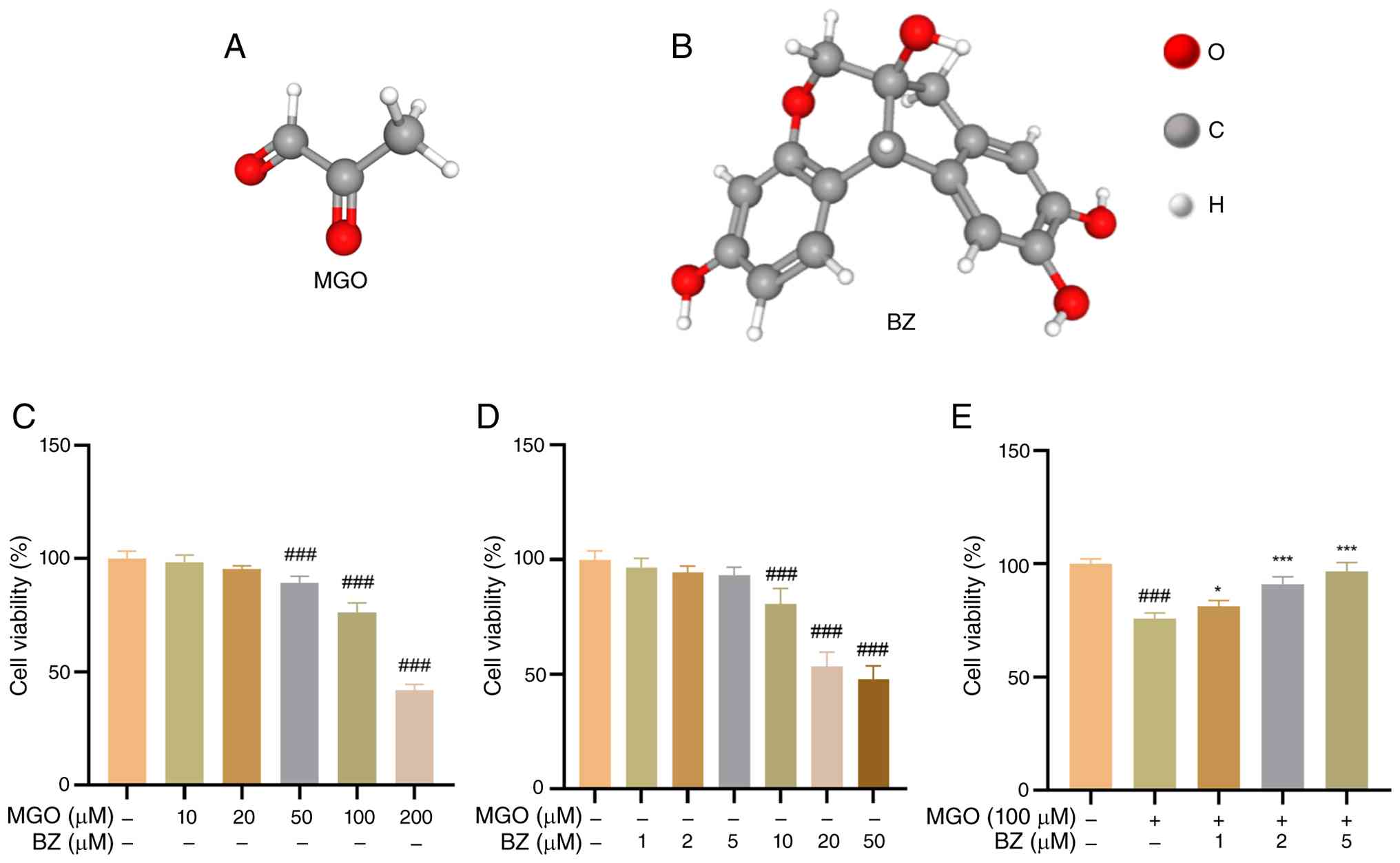
injury, the effects of MGO (Fig.
1A) and BZ (Fig. 1B) on
HUVEC viability were first evaluated using a CCK-8 assay. Following
24 h of exposure to MGO at concentrations of 10, 20, 50, 100, or
200 μM, HUVEC viability decreased significantly, in
agreement with previous findings (Fig. 1C) (19-21). Furthermore, after 2 h of
treatment with BZ at concentrations of 1, 2, and 5 μM, no
marked side effects were observed. However, when the concentration
increased to 10 μM, cell viability began to decrease
(Fig. 1D). Additionally, 2 h of
pretreatment with BZ at 1, 2, or 5 μM dose-dependently
restored HUVEC viability suppressed by MGO (Fig. 1E).
To determine whether BZ modulates MGO-induced
autophagy, HUVECs were exposed to BZ at 1, 2, or 5 μM for 2
h before treatment with 100 μM MGO for 24 h. Changes in the
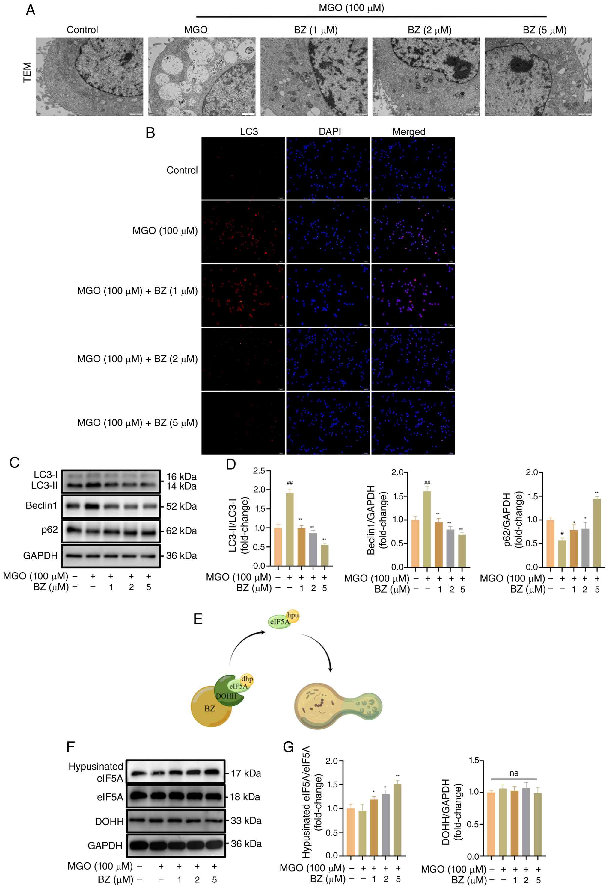
level of intracellular autophagy were detected by TEM. Compared
with controls, exposure to MGO markedly increased autophagic
vesicle and autolysosome formation, whereas BZ supplementation
attenuated these alterations in a dose-dependent manner. In
particular, the number of autophagosomes/autophagolysosomes was
minimized at a BZ concentration of 5 μM (Fig. 2A). Similarly, LC3
immunofluorescence experiments revealed that the fluorescence
intensity was the strongest after the addition of MGO, and the
autophagic flux reached a maximum. However, BZ progressively
attenuated cellular autophagy, with the most pronounced reduction
observed at 5 μM (Fig.
2B). Moreover, immunoblotting experiments were used to further
verify the changes in autophagy-related protein levels. Relative to
the control group, MGO significantly elevated the LC3-II/LC3-I
ratio and Beclin1 levels while reducing p62 expression. By
contrast, BZ treatment dose-dependently reversed these changes by
lowering LC3-II/LC3-I and Beclin1 levels and restoring p62
expression (Fig. 2C and D). As
illustrated in the schematic diagram in Fig. 2E, BZ has been reported to mediate
its protective effects via DOHH, a target protein, and the
downstream eIF5A signaling cascade (39).
The level of DOHH, a BZ target protein, was
subsequently further examined by western blotting. Interestingly,
treatment with either BZ or MGO did not significantly alter DOHH
levels relative to the control group. Hypusinated eIF5A expression
was assessed and the potential mechanism by which BZ exerts its
effects through the DOHH/eIF5A axis was explored. Compared with the
blank group, MGO stimulation did not markedly affect hypusinated
eIF5A levels, whereas BZ pretreatment elevated hypusinated eIF5A,
with a significant increase observed at 5 μM BZ (Fig. 2F and G). Therefore, these
findings suggest that BZ may activate the DOHH/eIF5A signaling
cascade to enhance eIF5A hypusination, thereby regulating
MGO-induced cellular responses.
BZ reverses MGO-induced apoptosis in
HUVECs
These findings confirmed that BZ attenuated
MGO-induced autophagy in ECs. Therefore, it wss further examined
whether BZ modulates MGO-triggered apoptosis. As aforementioned,
HUVECs were preincubated with BZ at 1, 2, or 5 μM for 2 h
and subsequently exposed to 100 μM MGO for 24 h. Annexin
V-FITC/PI double staining was performed to quantify apoptosis by
flow cytometry. MGO exposure significantly increased the apoptotic
rate compared with controls, while BZ pretreatment progressively
reduced apoptosis with increasing concentrations (Fig. 3A and B). Molecular analyses
further corroborated these findings, revealing dose-dependent
reductions in Bax and the cleaved caspase-3/caspase-3 ratio,
alongside a concomitant increase in Bcl-2 expression (Fig. 3C and D). These findings
collectively demonstrate that BZ not only mitigates MGO-induced
autophagy but also effectively blocks the subsequent apoptotic
cascade.
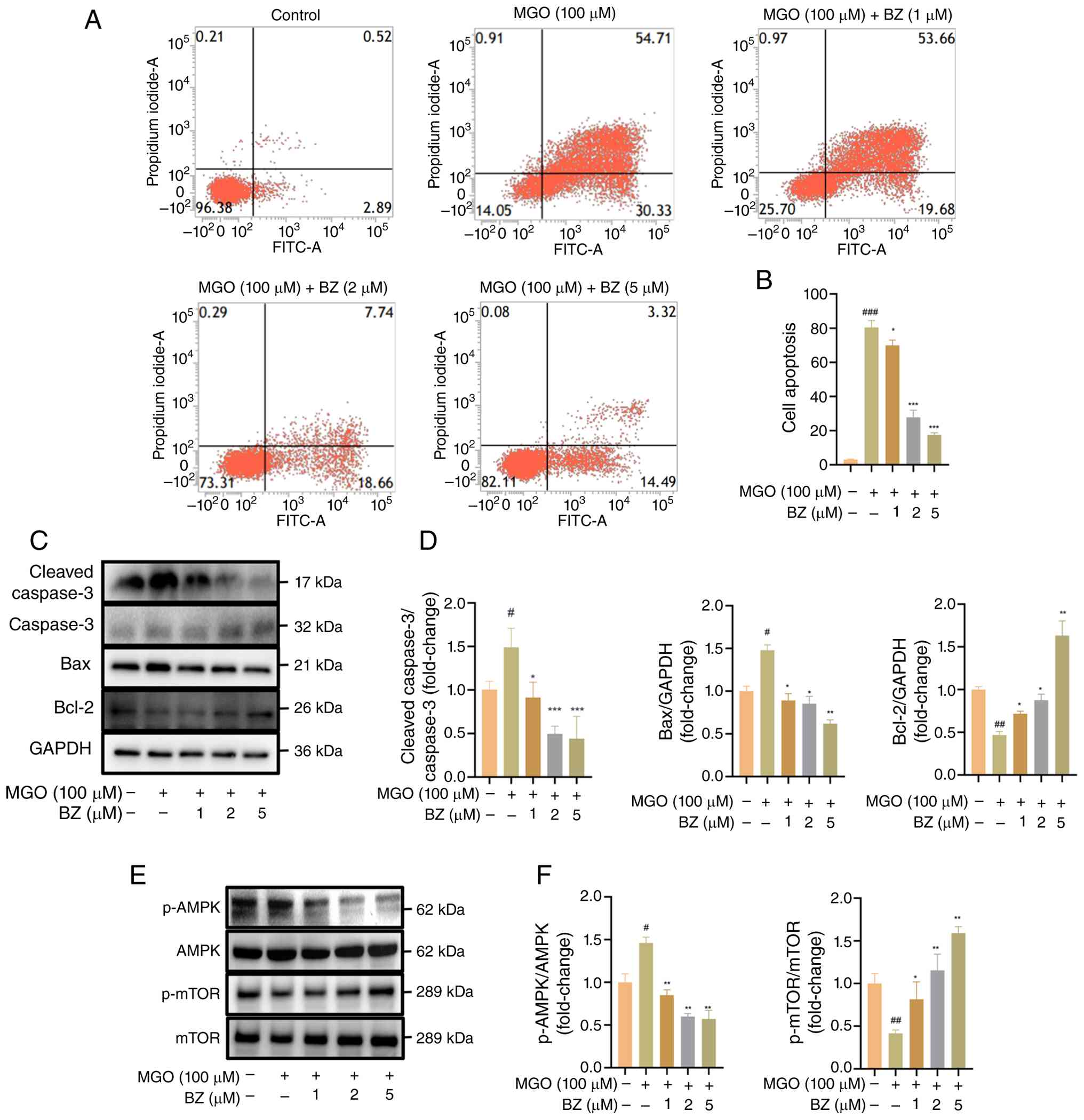 | Figure 3MGO-induced apoptosis is reversed by
BZ. (A and B) After trypsinization, cells were stained with Annexin
V-FITC and PI for 20 min, followed by flow cytometric
quantification of apoptosis. (C-F) Bax, cleaved
caspase-3/caspase-3, Bcl-2, p-mTOR, mTOR, p-AMPK and AMPK protein
expression was assessed by western blot analysis.
#P<0.05 ##P<0.01 and
###P<0.001 vs. Ctrl; *P<0.05
**P<0.01 and ***P<0.001 vs. MGO. MGO,
methylglyoxal; BZ, brazilin; p-, phosphorylated. |
Combined flow cytometry and western blot data
revealed that BZ was able to suppress autophagic and apoptotic
responses elicited by MGO in HUVECs. As the AMPK/mTOR pathway is
pivotal for autophagy regulation (47,48), it was hypothesized that BZ
confers protection by modulating this pathway. Accordingly, it was
first examined how MGO and BZ affected core pathway proteins at the
phosphorylation level. Relative to the control group, MGO treatment
elevated phosphorylated (p-)AMPK levels while reducing p-mTOR
levels. Moreover, BZ pretreatment dose-dependently decreased p-AMPK
levels while increasing p-mTOR levels (Fig. 3E and F). These findings suggest
that MGO induces apoptosis and AMPK/mTOR signaling, which is
modulated by BZ.
Effects of 3-MA on MGO-induced autophagy
and apoptosis in HUVECs
To further determine whether MGO-induced cellular
autophagy and apoptosis are associated with the inhibitory effect
of BZ on autophagy, HUVECs were pretreated with BZ (5 μM) +
3-MA (5 mM, an autophagy inhibitor) for 2 h and co-incubated them
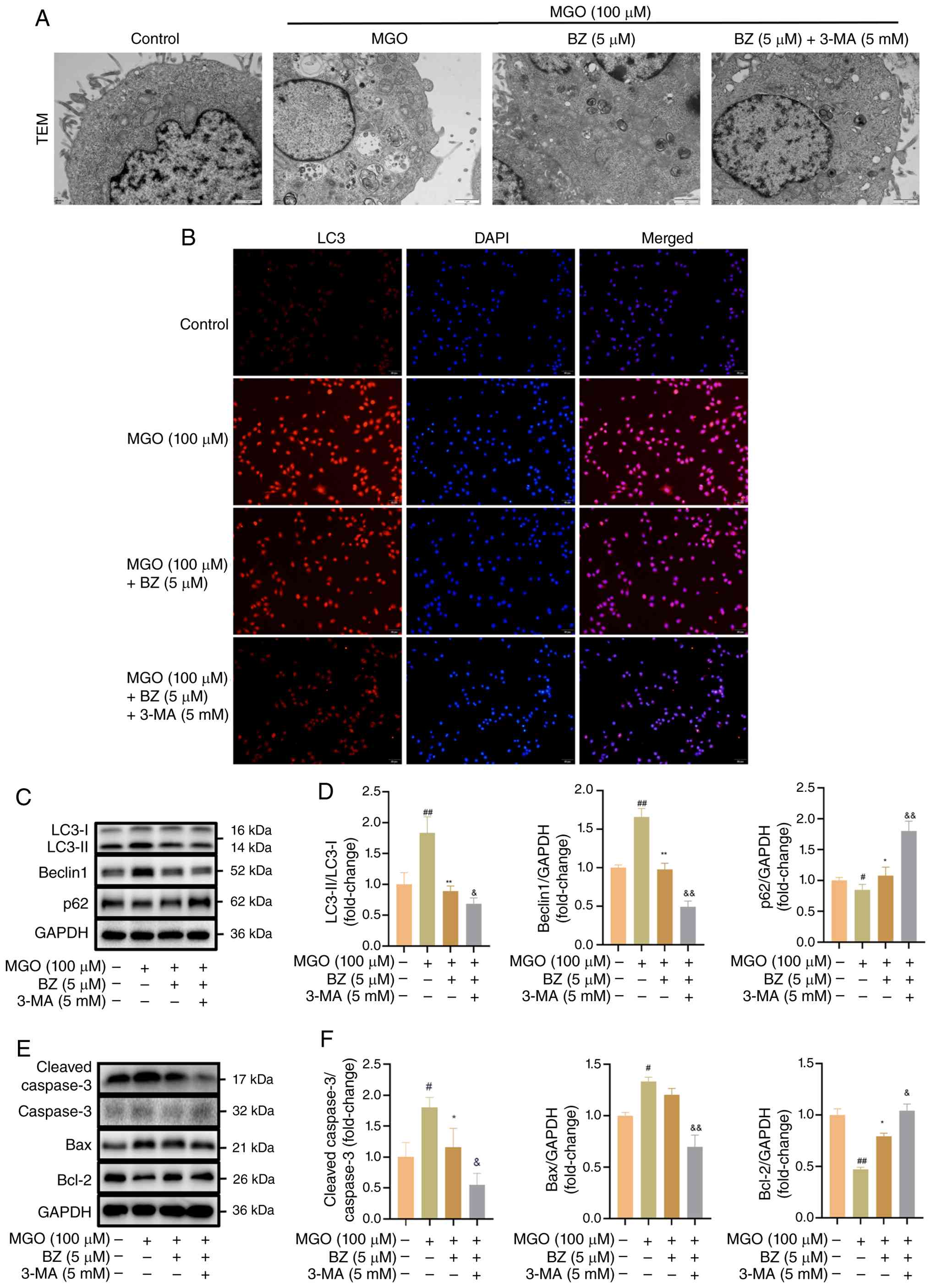
for 24 h with MGO (100 μM) (49). TEM was used to observe the
changes in autophagosomes. BZ clearly reduced MGO-induced
autophagy, while combined BZ and 3-MA pretreatment further
suppressed this response, with reduced autophagosome and
autolysosome formation (Fig.
4A). Further LC3 immunofluorescence analysis was conducted to
determine whether 3-MA suppressed MGO-evoked autophagy in ECs and
enhanced the protective effects of BZ. As observed in
aforementioned results, the red fluorescence was strongest in the
MGO-treated group. After the addition of BZ, autophagic flux was
reduced. In addition, cotreatment with 3-MA and BZ further
potentiated BZ-mediated suppression of MGO-induced autophagy in
HUVECs (Fig. 4B).
Autophagy- and apoptosis-related protein expression
was examined using western blot analysis. Relative to the control
group, MGO significantly enhanced cellular autophagic and apoptotic
responses. However, pretreatment with BZ reversed these effects.
Combined treatment with BZ and 3-MA further enhanced this reversal,
significantly downregulating the LC3-II/LC3-I ratio, the expression
of Beclin1 and Bax, as well as the cleaved caspase-3/caspase-3
ratio, while upregulating the expression levels of Bcl-2 and p62
(Fig. 4C-F). In summary, BZ can
decrease the levels of autophagy and apoptosis and further reduce
MGO-induced autophagy and apoptosis when combined with the
autophagy inhibitor 3-MA. These data suggest that 3-MA and BZ have
synergistic effects on the protection of vascular ECs.
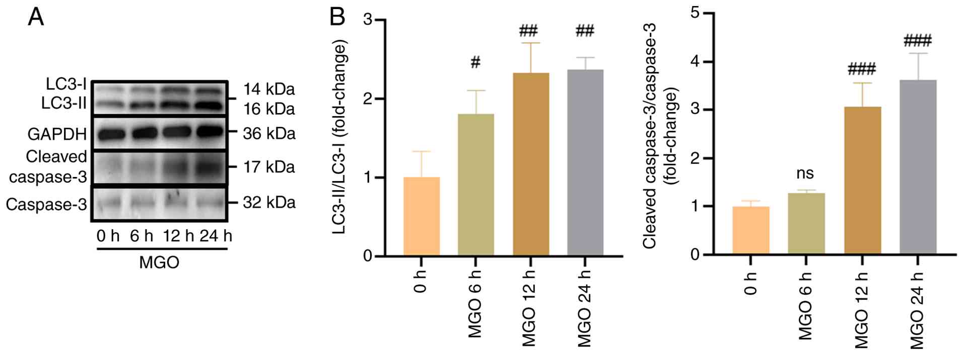
To further demonstrate that MGO-induced autophagy
occurs before apoptosis, the time-course changes in autophagy and
apoptosis markers in HUVECs were examined at 0, 6, 12 and 24 h
after MGO treatment. As shown in Fig. 5A and B, the autophagy marker
LC3-II was significantly upregulated as early as 6 h after MGO
stimulation, whereas the cleaved caspase-3 began to increase
significantly at 12 h and remained elevated at 24 h. In addition, a
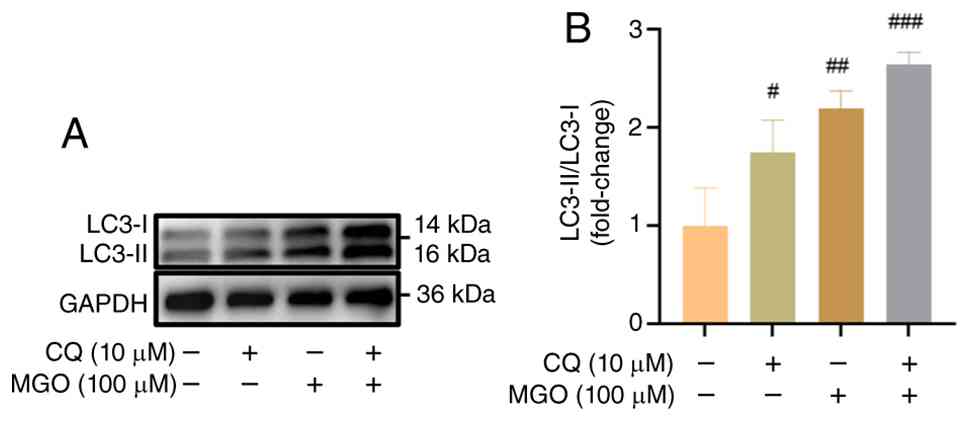
chloroquine (CQ) blockade assay was performed to evaluate
autophagic flux. As revealed in Fig.
6A and B, treatment with either CQ or MGO alone significantly
increased LC3-II levels compared with the control group. Notably,
combined treatment with MGO and CQ further increased LC3-II levels
compared with MGO treatment alone. The results suggest that MGO
enhances autophagic flux, rather than merely causing passive
accumulation of autophagosomes due to impaired lysosomal
degradation.
An AMPK inhibitor enhances the
BZ-mediated suppression of MGO-induced autophagy and apoptosis
Current data indicate that MGO stimulation
significantly increases the levels of p-AMPK while decreasing the
levels of mTOR and that BZ can reverse these effects. Therefore, it
was hypothesized that BZ may regulate MGO-triggered autophagic and
apoptotic responses through AMPK/mTOR signaling. Before exposure to
100 μM MGO for 24 h, HUVECs were preincubated for 2 h with
BZ (5 μM) in the presence or absence of Compound C (50
μM, an AMPK inhibitor) (50). TEM revealed that BZ reduced
MGO-induced autophagy. Furthermore, after cotreatment with BZ and
Compound C, the numbers of autophagosomes and autolysosomes further
decreased (Fig. 7A).
Additionally, flow cytometric analysis was further performed to
assess apoptosis following AMPK blockade. The data showed that BZ
attenuated MGO-induced HUVEC apoptosis. Notably, cotreatment with
BZ and Compound C also attenuated HUVEC apoptosis, which blocked
AMPK signaling (Fig. 7B and C).
Western blot analysis revealed that Compound C, an AMPK inhibitor,
further enhanced the BZ-mediated reversal, as reflected by reduced
p-AMPK levels and increased p-mTOR levels (Fig. 7D and E). Overall, the inhibitory
effects of BZ on MGO-induced autophagy and apoptosis appear to be
further enhanced by Compound C.
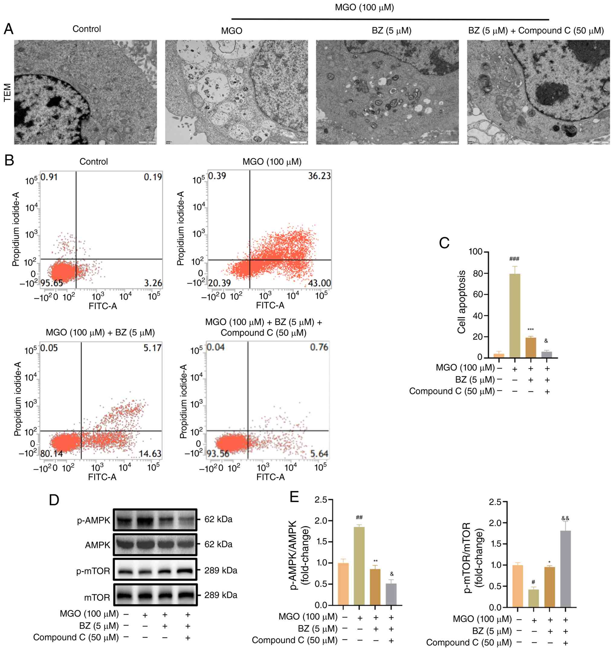 | Figure 7Effect of Compound C on BZ-mediated
inhibition of MGO-induced autophagy and apoptosis in HUVECs. HUVECs
were preincubated with BZ (5 μM) in the presence or absence
of Compound C (50 μM) for 2 h, followed by exposure to MGO
(100 μM) for 24 h. (A) Changes in the level of autophagy
were detected by transmission electron microscopy. Scale bar, 1
μm. (B and C) Apoptotic changes were assessed by flow
cytometry. (D and E) Protein expression of p-AMPK, AMPK, p-mTOR and
mTOR was evaluated by western blotting. #P<0.05,
##P<0.01 and ###P<0.001 vs. Ctrl;
*P<0.05, **P<0.01 and
***P<0.001 vs. MGO; &P<0.05 and
&&P<0.01 vs. MGO + BZ. BZ, brazilin; MGO,
methylglyoxal; HUVECs, human umbilical vein endothelial cells; p-,
phosphorylated. |
BZ activates the DOHH/eIF5A pathway to
regulate MGO-induced autophagy and apoptosis in ECs
The aforementioned results reveal that BZ does not
alter DOHH levels in MGO-treated HUVECs but significantly increases
hypusinated eIF5A levels, suggesting that BZ may inhibit
MGO-triggered autophagy and apoptosis by enhancing DOHH-mediated
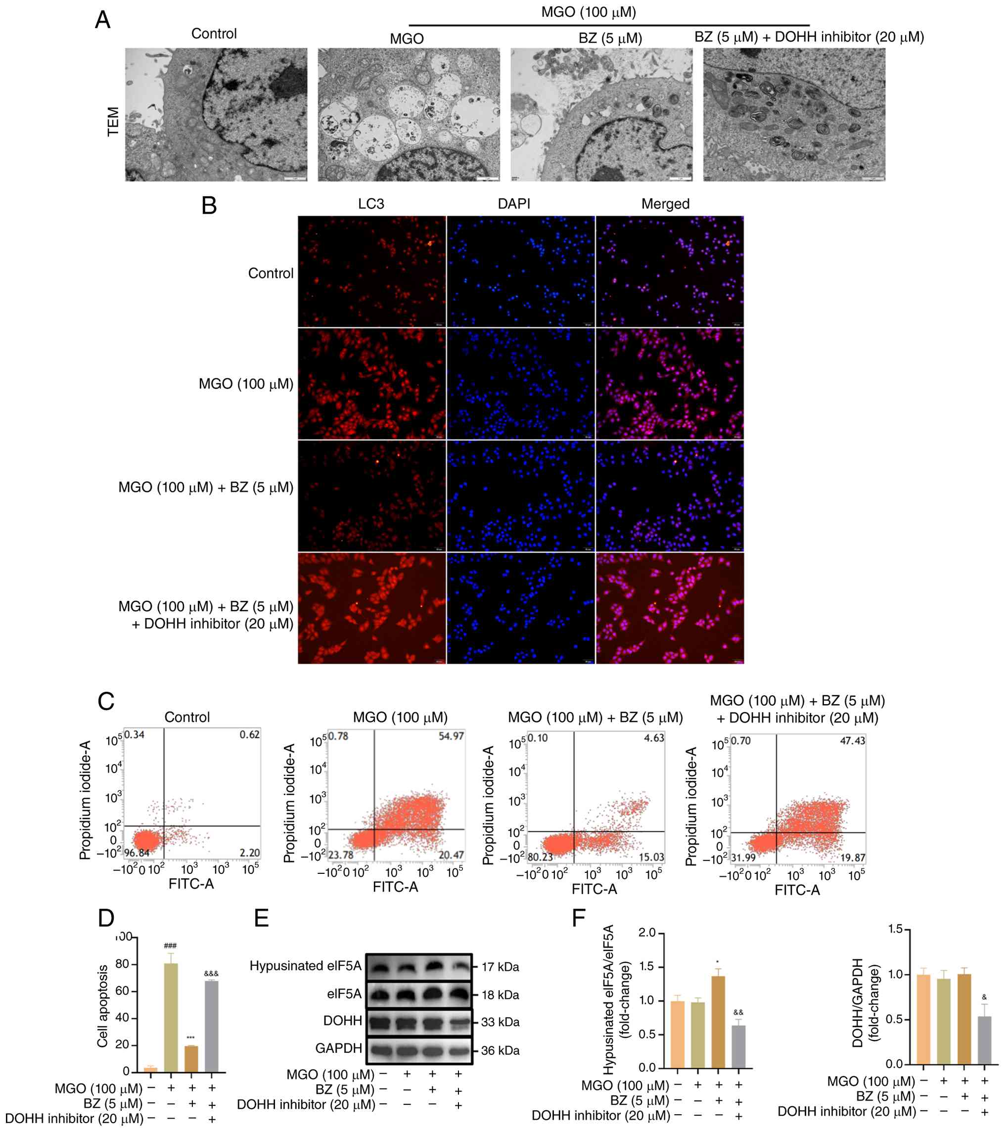
eIF5A hypusination. To verify the aforementioned hypothesis, HUVECs
were pretreated for 2 h with BZ (5 μM) alone or with BZ (5
μM) combined with ciclopirox (20 μM), a DOHH
signaling pathway inhibitor, and were subsequently stimulated with
MGO (100 μM) (51,52).
TEM revealed that BZ inhibited the MGO-induced increases in
autophagic vesicles and autolysosomes, whereas combined treatment
with BZ, ciclopirox and MGO markedly elevated autophagic activity
(Fig. 8A). These results were
further confirmed by LC3 immunofluorescence, which revealed that BZ
reduced the extent of MGO-induced autophagy and decreased
autophagic flux. However, cells receiving combined treatment with
BZ, ciclopirox and MGO exhibited a marked increase in autophagic
fluorescence intensity (Fig.
8B).
More importantly, as demonstrated in Fig. 8C and D, pretreatment with BZ
reversed MGO-induced apoptosis, but the apoptotic rate increased
after ciclopirox treatment. The aforementioned results suggest that
the DOHH inhibitor ciclopirox could resist the protective effect of
BZ and promote apoptosis. Subsequently, western blotting was
performed to examine the BZ-associated downstream DOHH/eIF5A
signaling cascade. DOHH levels were not significantly altered by BZ
pretreatment or MGO exposure relative to the blank group; however,
pretreatment with the DOHH inhibitor ciclopirox significantly
reduced DOHH expression. Hypusinated eIF5A levels were not markedly
altered by MGO treatment relative to the blank control group,
whereas BZ pretreatment increased them; however, this response was
significantly reversed by ciclopirox pretreatment (Fig. 8E and F). These findings indicate
that ciclopirox promotes autophagic and apoptotic responses
elicited by MGO in HUVECs through suppression of the DOHH/eIF5A
signaling cascade and abolishment of the cytoprotective effects of
BZ.
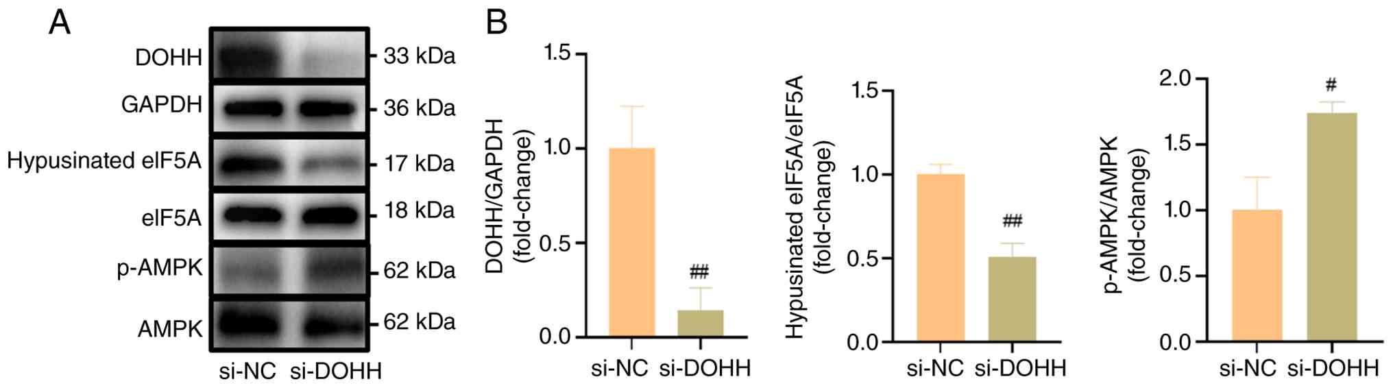
To exclude the potential off-target effects of
ciclopirox, si-DOHH transfection was further performed in the
present study to verify the specificity of DOHH inhibition and to
investigate its relationship with AMPK activation. As revealed in
Fig. 9A and B, si-DOHH
transfection significantly reduced the expression levels of DOHH
and hypusinated eIF5A, while significantly increasing p-AMPK
levels. These results suggest that inhibition of the DOHH/eIF5A
pathway may be closely associated with AMPK activation.
Physiological and pathological changes in
mice
To assess whether BZ modulates MGO-evoked vascular
autophagy and apoptosis in vivo, C57BL/6J mice were
administered MGO and BZ. Body weight, food intake and serum MGO
concentration were measured after intraperitoneal injection of MGO
and intragastric administration of BZ. The experimental design,
including the sampling strategy and treatment regimen for the mouse
study, is presented in Fig.
10A. The results showed a modest increase in body weight across
all mouse groups after treatment, with no statistically significant
difference from pretreatment body weights, which may be
attributable to normal physiological growth during the 7-week
feeding period (Fig. 10B).
Interestingly, the food intake of the mice did not significantly
change after administration compared with that of the
pre-administration mice (Fig.
10C). These data indicate that treatment with BZ and MGO does
not significantly affect the physiological functions of mice. Next,
serum MGO levels were quantified, revealing a significant elevation
in the MGO group and a significant reduction following BZ treatment
(Fig. 10D). Dynamic alterations
in autophagy- and apoptosis-related factors, including LC3, p62 and
cleaved caspase-3, were examined. As shown in Fig. 10E-G, MGO significantly elevated
LC3 and cleaved caspase-3 levels while reducing p62 expression.
However, these effects were significantly suppressed upon the
addition of BZ.
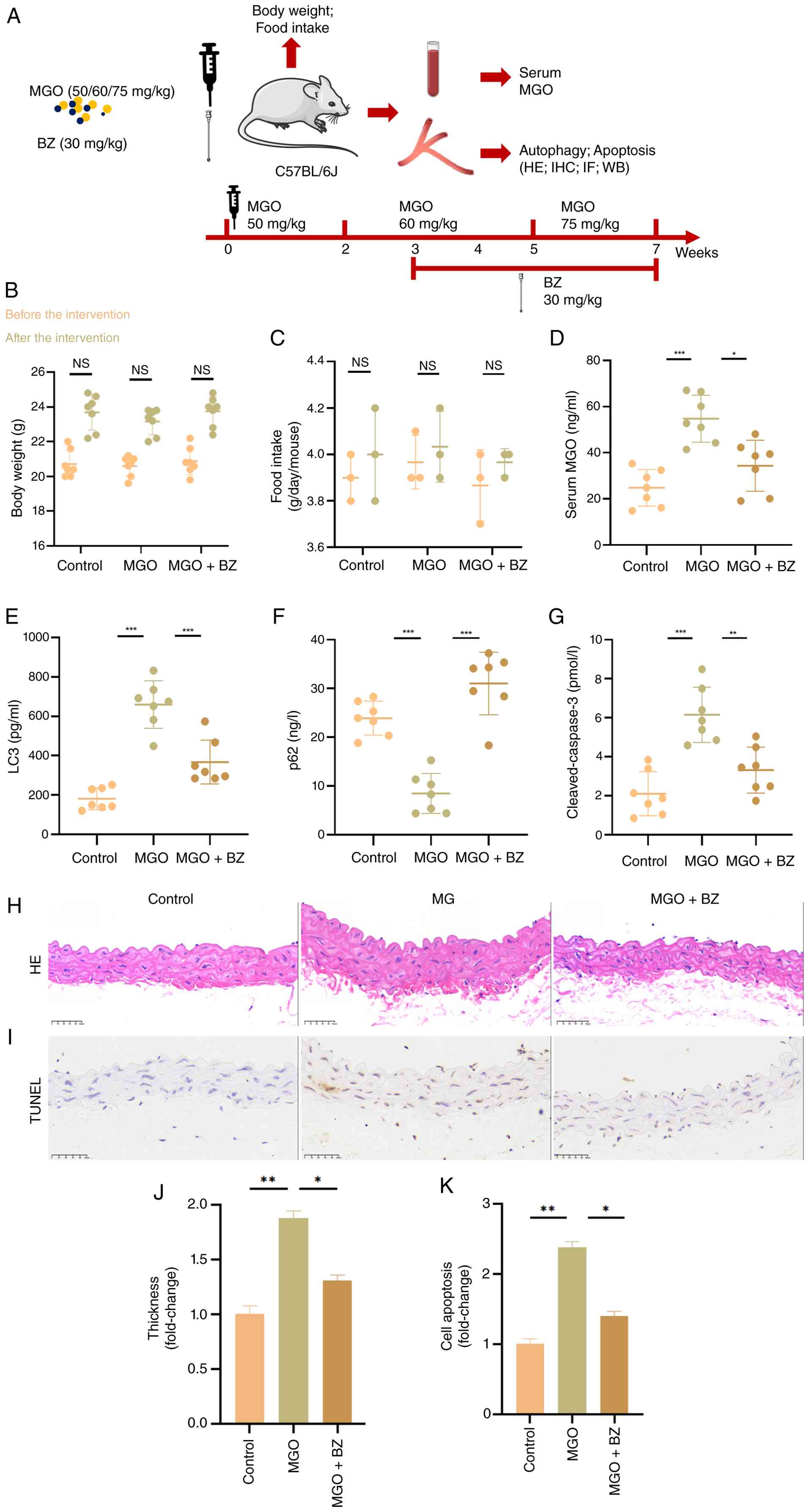 | Figure 10Changes in the physiological features
of mice and the inhibition of MGO-induced aortic vascular injury
and apoptosis by BZ. C57BL/6J mice were intraperitoneally injected
with normal saline or MGO solution (50, 60, or 75 mg/kg/d), and BZ
or normal saline was administered by gavage from the third week.
(A) Experimental design, sampling strategy and treatment schedule
in mice. (B) The weight changes of each mouse were measured before
and after the experiment and recorded statistically. (C) Changes in
food intake before and after the experiment were measured and
statistically recorded. (D) Changes in serum MGO concentration were
determined by extracting serum from mice euthanized after drug
administration. (E-G) Expression changes in
autophagy/apoptosis-related factors, including LC3, p62 and cleaved
caspase-3, were evaluated. (H and J) H&E staining was performed
to assess vascular endothelial morphology. (I and K) TUNEL staining
was performed to evaluate apoptosis in aortic vascular tissues. The
number of mice in each group was ≥6; scale bar, 50 μm.
*P<0.05, **P<0.01 and
***P<0.001. MGO, methylglyoxal; BZ, brazilin; NS, not
significant. |
Moreover, H&E and TUNEL staining were used to
evaluate whether BZ affects MGO-induced vascular injury.
H&E-stained aortic sections from the control group showed an
intact, smooth, and regularly organized intima. Conversely, MGO
treatment led to substantial thickening, irregularity, a darker
color, and a more disordered shape, indicating pronounced vascular
damage. Following BZ treatment, the dilatation and injury of the
aortic intima were significantly ameliorated (Fig. 10H and J). Additionally, TUNEL
staining was performed to further observe changes in EC apoptosis.
MGO treatment significantly increased the apoptotic rate in mouse
aortic vessels, but this effect was significantly reversed after BZ
treatment (Fig. 10I and K).
BZ decreases the MGO-induced autophagy
and apoptosis of blood vessels
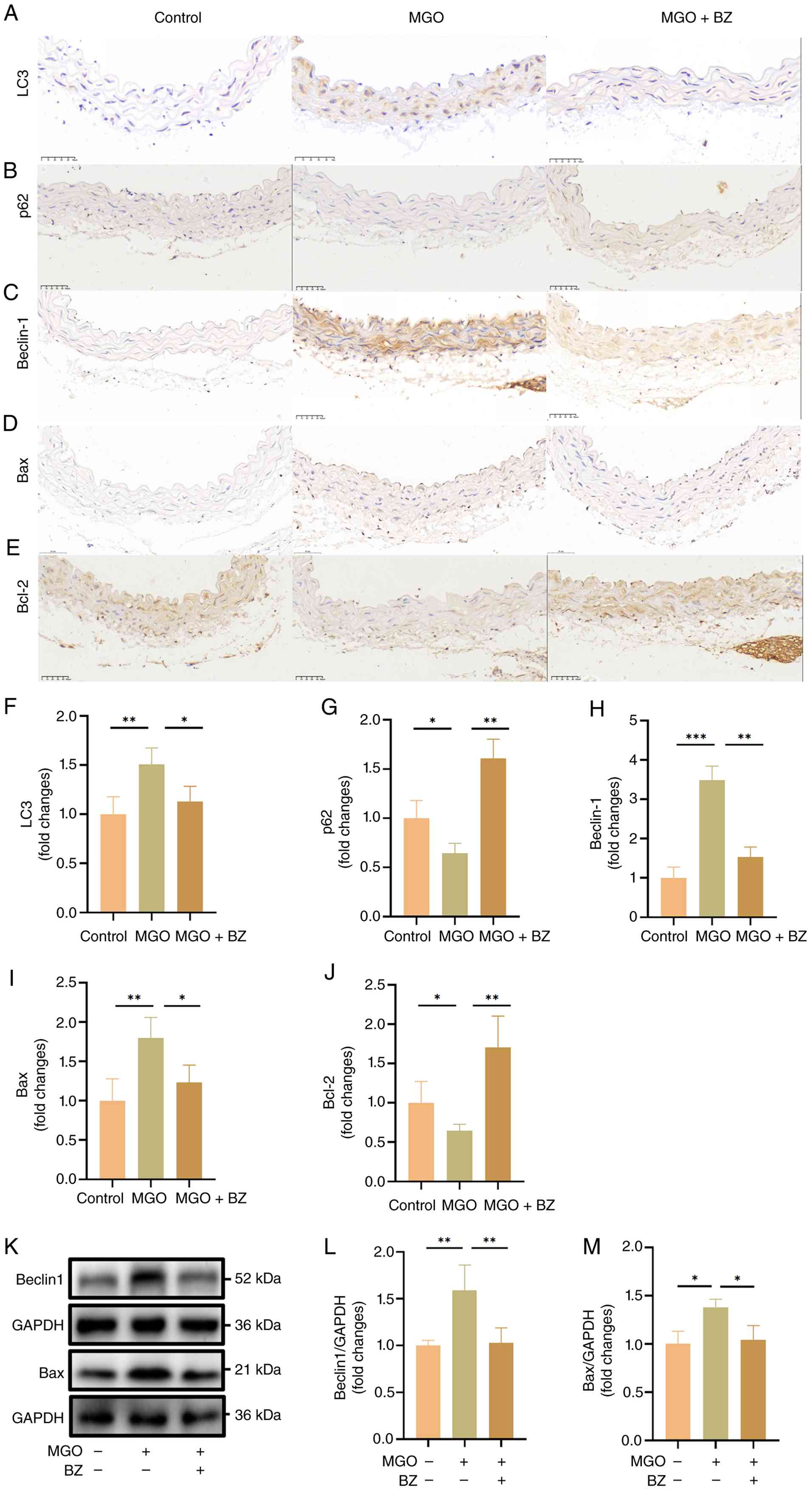
Additionally, to determine whether BZ inhibits
autophagy and apoptosis in vivo, immunohistochemical
experiments were conducted. Relative to the control group, the MGO
group showed elevated vascular autophagy levels, increased
expression of LC3, Beclin1 and Bax, and reduced positivity rates
for p62 and Bcl-2. However, these effects were reversed upon BZ
treatment (Fig. 11A-J).
Harvested mouse vascular tissue was subsequently analyzed by
western blotting to further validate these findings. It was found
that MGO elevated Beclin1 and Bax levels, whereas BZ counteracted
these changes and attenuated autophagic and apoptotic responses
(Fig. 11K-M). These data align
with the cellular findings and indicate that BZ markedly suppresses
vascular cell autophagic and apoptotic responses triggered by MGO,
thereby supporting its vasoprotective effects.
Effects of MGO and BZ on changes in DOHH
and hypusinated eIF5A in vivo
The aforementioned in vitro findings revealed
that BZ suppresses autophagic and apoptotic responses elicited by
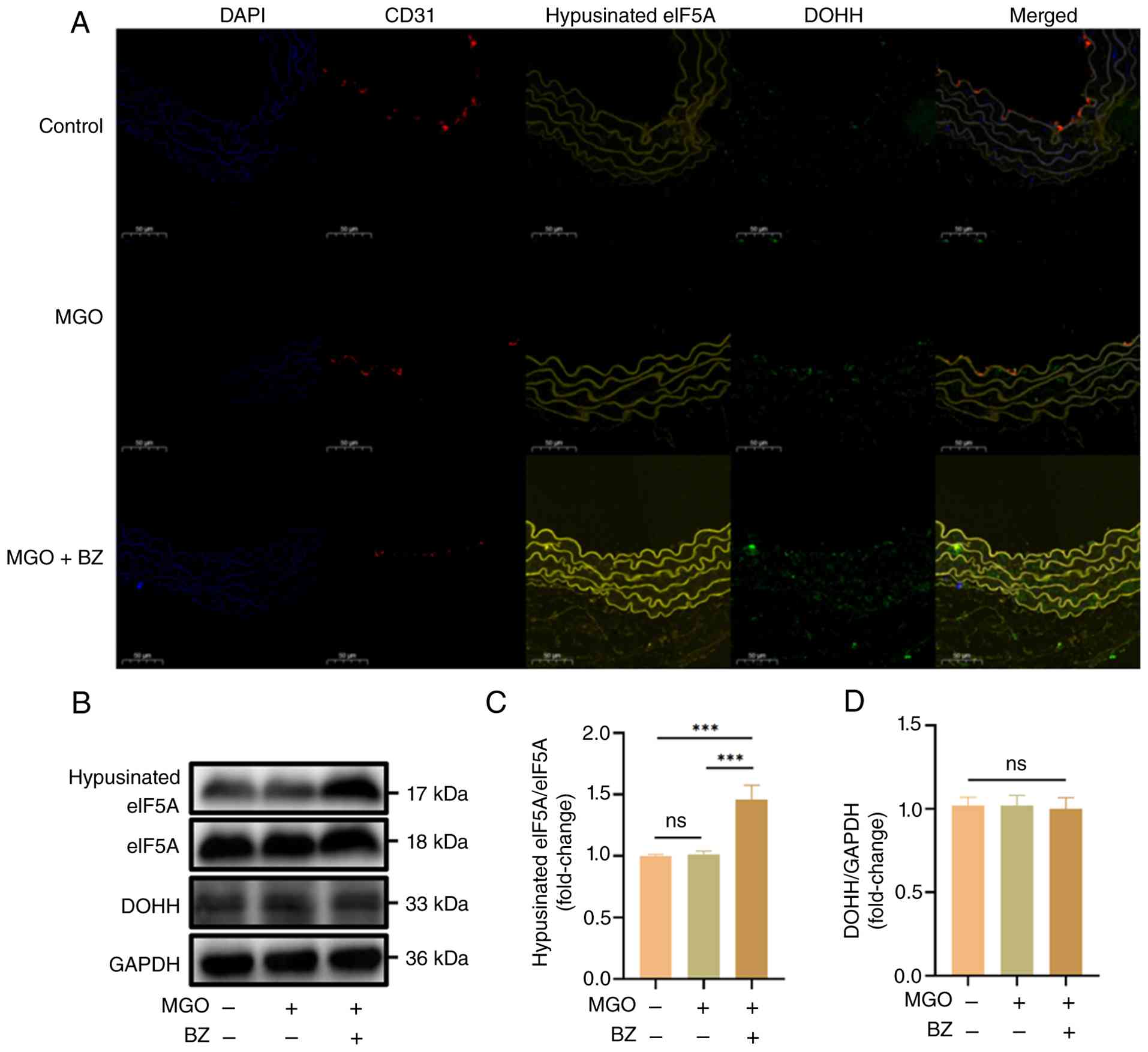
MGO in ECs through the DOHH/eIF5A pathway. Immunofluorescence
localization was used to examine changes in DOHH and hypusinated
eIF5A in vascular ECs. In the control group, ECs (red
fluorescence), DOHH (green fluorescence) and hypusinated eIF5A
(yellow fluorescence) were observed. Furthermore, the results from
MGO treatment alone aligned with the in vitro findings,
showing that MGO did not alter DOHH or hypusinated eIF5A positivity
in ECs. Although DOHH positivity remained unchanged after MGO and
BZ treatment, hypusinated eIF5A positivity was significantly
increased (Fig. 12A).
Consistently, western blot analysis of mouse vascular tissues
showed that MGO did not affect DOHH or hypusinated eIF5A protein
levels relative to the control group, whereas MGO and BZ
cotreatment significantly elevated hypusinated eIF5A levels. These
results further confirmed that BZ enhances the post-translational
modification of the eIF5A protein by hypusine via allosteric
activation of DOHH, thereby exerting its protective effects
(Fig. 12B-D).
Effects of BZ and a DOHH inhibitor on
autophagy and apoptosis in db/db mice
To more accurately mimic the pathophysiological
characteristics of diabetes, db/db mice were used instead of
MGO injection to investigate autophagy and apoptosis in vascular
tissues as well as the involvement of the DOHH/eIF5A signaling
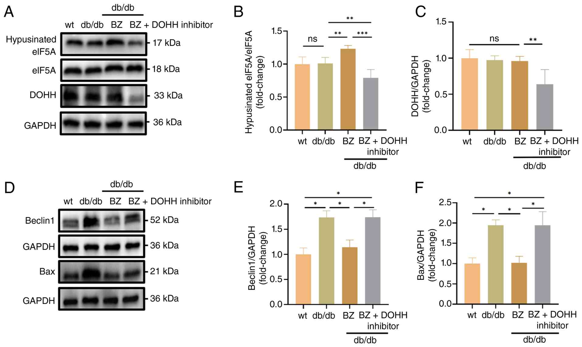
pathway. Relative to the control group, DOHH and hypusinated eIF5A
levels in vascular tissues from db/db mice showed no
significant differences, whereas Beclin1 and Bax levels were
elevated. This pattern was highly consistent with that observed in
the MGO administration model, indicating that both diabetes and
exogenous MGO stimulation increase the levels of autophagy and
apoptosis in vascular tissues. After oral BZ administration to
db/db mice, hypusinated eIF5A protein levels were
significantly elevated, whereas DOHH expression remained unchanged,
and autophagic and apoptotic responses were reduced. By contrast,
DOHH inhibitor treatment significantly reduced DOHH and hypusinated
eIF5A levels, elevated Beclin1 and Bax levels, and enhanced
autophagic and apoptotic responses (Fig. 13A-F). These findings indicate
that autophagic and apoptotic responses triggered by MGO are
aligned with diabetes-related vascular pathological changes, while
BZ may act as a promising therapeutic candidate for diabetic
vascular injury.
Discussion
MGO is a highly reactive glucose-derived metabolic
byproduct that forms stable AGEs through the non-enzymatic chemical
modification of proteins (13,17,53). MGO compromises vascular EC
integrity and homeostasis by enhancing extracellular matrix
collagen cross-linking, remodeling vascular architecture, and
activating inflammatory or oxidative pathways (13,17,53). MGO accumulation is a critical
contributor to diabetic progression and associated vascular
complications (54-56). In previous studies by the
authors, MGO was shown to induce apoptosis and pyroptosis in ECs
through oxidative stress, inflammatory responses and mitochondrial
damage, leading to vascular endothelial injury in mice (19-21). In addition, MGO enhances
autophagy in vascular ECs via AGE receptor- and
peroxynitrite-dependent mechanisms (29). BZ appears to regulate autophagy
and apoptosis and exerts cytoprotective effects (39,57). However, the precise role of BZ in
MGO-triggered HUVEC apoptosis and the associated signaling pathways
remain unclear. Marked reductions in HUVEC viability, together with
enhanced autophagic and apoptotic responses, were observed
following MGO treatment, whereas these effects were
dose-dependently counteracted by BZ. Activation of the DOHH/eIF5A
and AMPK/mTOR signaling pathways may contribute to these effects.
Further in vivo validation using an MGO-induced vascular
injury model and db/db mice confirmed that BZ suppresses
MGO-evoked autophagy and apoptosis. These findings align with
previous studies (28,37,38,53,58), indicating that BZ exerts
protective effects against diabetes-associated vascular injury and
further clarifying the mechanisms underlying its action.
To further investigate the mechanism associated with
autophagic and apoptotic responses triggered by MGO and clarify
their temporal sequence, HUVECs were pretreated with 3-MA, an
autophagy inhibitor. The results showed marked autophagy
inhibition, evidenced by decreased LC3-II/LC3-I and Beclin1 levels
and increased p62 expression, along with reduced Bax and cleaved
caspase-3/caspase-3 and enhanced Bcl-2 expression. These findings
suggest that inhibition of autophagy attenuates MGO-induced
apoptosis, indicating that autophagy may contribute to the
subsequent activation of apoptotic signaling. Consistently,
time-course analysis showed that LC3-II was significantly
upregulated as early as 6 h after MGO stimulation, whereas cleaved
caspase-3/caspase-3 began to increase significantly at 12 h and
remained elevated at 24 h. Taken together, these observations
support the notion that MGO-induced autophagy occurs before
apoptosis and may participate in promoting apoptotic progression in
HUVECs. Furthermore, the CQ blockade assay provided additional
evidence that MGO-induced LC3-II accumulation is not merely
attributable to impaired lysosomal degradation but rather reflects
enhanced autophagosome formation and increased autophagic flux in
HUVECs. The AMPK/mTOR pathway is a central signaling axis that
regulates cellular energy metabolism, nutrient sensing, and growth
and proliferation and is closely associated with autophagy and
apoptosis in ECs (59,60). The use of BZ in the treatment of
vascular inflammation and vascular regulation has been previously
reported (38,61,62). The present findings indicate that
BZ pretreatment dose-dependently mitigated the MGO-evoked elevation
of p-AMPK expression while reversing the reduction in p-mTOR
expression caused by MGO. By inhibiting AMPK/mTOR signaling, BZ
significantly reduced MGO-induced EC autophagy and apoptosis in
vitro and in vivo, as evidenced by reduced LC3-II/LC3-I,
Beclin1, Bax and cleaved caspase-3 levels, together with increased
p62 and Bcl-2 expression. These changes were accompanied by reduced
autophagosome and autolysosome accumulation. Notably, Compound C,
an AMPK inhibitor, further potentiated the BZ-mediated
anti-autophagic and antiapoptotic effects, suppressing HUVEC
autophagic and apoptotic responses elicited by MGO and further
reducing p-AMPK levels while elevating p-mTOR levels. Taken
together, the present findings indicate that MGO-induced autophagy
may precede the onset of apoptosis and be modulated by BZ via
AMPK/mTOR signaling, in agreement with previous studies (59,60). Moreover, the present study
suggests that BZ, as a natural extract, holds considerable
potential for ameliorating autophagy- and apoptosis-related
diabetic vascular injury.
Previously, through protein microarray screening,
Guo et al (39) developed
a biotin-labeled BZ molecular probe and identified DOHH as a direct
target of BZ. Furthermore, BZ exerts its cytoprotective effects by
directly interacting with the Cys232 residue of the DOHH protein
(39). DOHH shows a strong
preference and strict specificity for binding to eIF5A containing
deoxyhypusine and catalyzes its hydroxylation (63). In the present study, BZ
pretreatment was found to leave DOHH abundance largely unchanged
while markedly increasing intracellular hypusinated eIF5A levels.
By contrast, treatment with a DOHH inhibitor significantly reduced
both DOHH and hypusinated eIF5A levels and effectively reversed the
BZ-induced increase in hypusinated eIF5A. In addition, si-DOHH
transfection was used in the present study to specifically knock
down DOHH, thereby further verifying the specificity of ciclopirox
action. The decreases in DOHH expression and hypusinated eIF5A
levels observed after si-DOHH intervention were consistent with the
results of ciclopirox treatment. Collectively, these results
further support that BZ may act on DOHH to catalyze eIF5A, thereby
accelerating the final step of post-translational hypusine
synthesis on eIF5A and modulating cellular autophagy and apoptosis.
However, the present study has several limitations. After si-DOHH
treatment, both DOHH expression and hypusinated eIF5A levels were
decreased, accompanied by an increase in p-AMPK levels, suggesting
that AMPK activation may be associated with DOHH inhibition and
reduced hypusinated eIF5A levels. Nevertheless, changes in mTOR
pathway-related molecules and the precise molecular mechanisms
linking DOHH/eIF5A to the AMPK/mTOR pathway, including whether
direct protein-protein interactions and specific modification sites
are involved, remain to be elucidated through further targeted
mechanistic studies.
Marked elevations in plasma MGO concentrations have
been reported in patients with diabetes (64,65), and MGO has also been implicated
in the initiation and progression of diabetes-associated vascular
diseases (14,35,66,67). Accordingly, in the present study,
MGO was first selected as the primary injurious factor to induce
autophagy, apoptosis, and endothelial dysfunction in ECs in
vitro, thereby mimicking diabetes-related vascular pathology. A
db/db mouse model was subsequently used to establish a
diabetic condition and to validate the in vitro findings
in vivo. The findings of the present study indicate that
MGO-induced vascular injury closely resembles diabetes-associated
vascular disease at the molecular mechanistic level, indicating
that MGO can serve as a reliable experimental surrogate for
diabetic vascular damage. These findings suggest that BZ may
represent a promising therapeutic candidate for diabetic vascular
injury.
In summary, the current findings newly demonstrate
the protective effect of BZ against MGO-evoked autophagic and
apoptotic responses in vitro and in vivo.
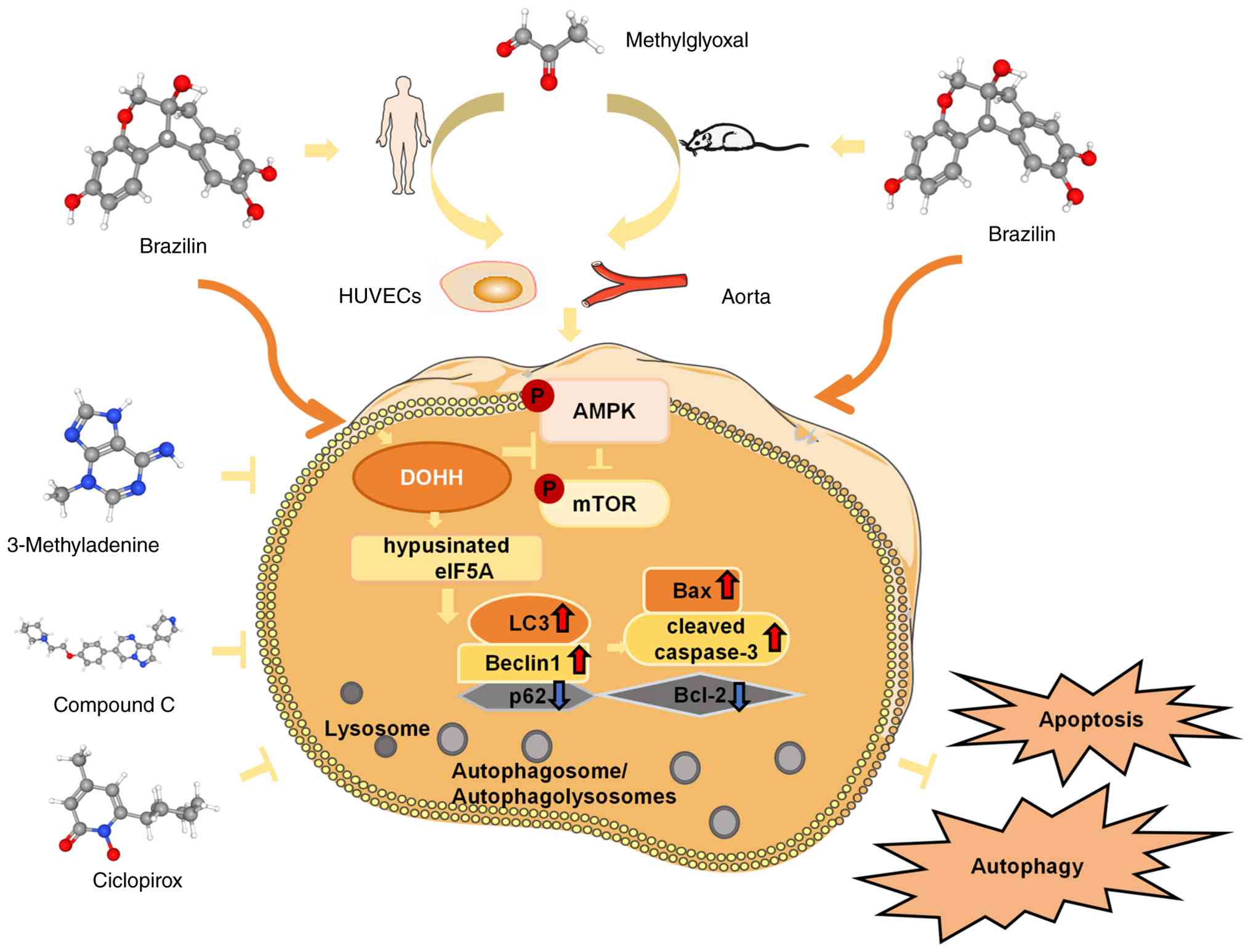
Mechanistically, BZ mitigates MGO-triggered vascular endothelial
damage by enhancing the unique post-translational hypusination of
eIF5A and in turn regulating the DOHH/eIF5A and AMPK/mTOR signaling
cascades (Fig. 14). These
findings further clarify the molecular basis of BZ action and offer
new theoretical support and research perspectives for its potential
therapeutic application in diabetes-associated vascular
diseases.
Availability of data and materials
The data generated in the present study may be
requested from the corresponding author.
Authors' contributions
YL and JC designed the present study and drafted the
manuscript. HL, JX and JY performed the experiments and acquired
the data. LW and JW analyzed the data and conducted the initial
interpretation of the results. YZ and ML contributed to the study
design, critically reviewed the manuscript, and revised it for
important intellectual content. YL and ML confirm the authenticity
of all the raw data. All authors read and approved the final
version of the manuscript.
Ethics approval and consent to
participate
The present study complied with the ARRIVE
guidelines (https://arriveguidelines.org). All animal procedures
were approved by the Animal Ethics Committee of Southwest Medical
University (approval no. 2020895; Luzhou, China).
Patient consent for publication
Not applicable.
Competing interests
The authors declare that they have no competing
interests.
Abbreviations:
|
AGEs
|
advanced glycation end products
|
|
AMPK
|
AMP-activated protein kinase
|
|
BZ
|
brazilin
|
|
CCK-8
|
Cell Counting Kit-8
|
|
CQ
|
chloroquine
|
|
DOHH
|
deoxyhypusine hydroxylase
|
|
ECs
|
endothelial cells
|
|
eIF5A
|
eukaryotic translation initiation
factor 5A
|
|
FITC
|
fluorescein isothiocyanate
|
|
HUVECs
|
human umbilical vein endothelial
cells
|
|
MGO
|
methylglyoxal
|
|
mTOR
|
mammalian target of rapamycin
|
|
PI
|
propidium iodide
|
|
siRNA
|
small interfering RNA
|
|
si-DOHH
|
siRNA targeting DOHH
|
|
si-NC
|
negative control siRNA
|
|
TSA
|
tyramide signal amplification
|
|
WT
|
wild-type
|
|
3-MA
|
3-methyladenine
|
Acknowledgements
The authors would like to thank Dr Michael A. Hill
(Dalton Cardiovascular Research Center, University of
Missouri-Columbia) for technical assistance with professional
English language editing of the manuscript.
Funding
The present study was supported by the National Natural Science
Foundation of China (grant nos. 82030007 and 82370419), the Natural
Science Foundation of Sichuan Province (grant no. 2024NSFSC2104),
and the Sichuan Science and Technology Program (grant no.
2022YFS0627).
References
|
1
|
Bielka W, Przezak A, Molęda P,
Pius-Sadowska E and Machaliński B: Double diabetes-when type 1
diabetes meets type 2 diabetes: Definition, pathogenesis and
recognition. Cardiovasc Diabetol. 23:622024. View Article : Google Scholar : PubMed/NCBI
|
|
2
|
DeFronzo RA, Ferrannini E, Groop L, Henry
RR, Herman WH, Holst JJ, Hu FB, Kahn CR, Raz I, Shulman GI, et al:
Type 2 diabetes mellitus. Nat Rev Dis Primers. 1:150192015.
View Article : Google Scholar : PubMed/NCBI
|
|
3
|
Haythorne E, Lloyd M, Walsby-Tickle J,
Tarasov AI, Sandbrink J, Portillo I, Exposito RT, Sachse G, Cyranka
M, Rohm M, et al: Altered glycolysis triggers impaired
mitochondrial metabolism and mTORC1 activation in diabetic β-cells.
Nat Commun. 13:67542022. View Article : Google Scholar
|
|
4
|
Cheruiyot A, Hollister-Lock J, Sullivan B,
Pan H, Dreyfuss JM, Bonner-Weir S and Schaffer JE: Sustained
hyperglycemia specifically targets translation of mRNAs for insulin
secretion. J Clin Invest. 134:e1732802023. View Article : Google Scholar : PubMed/NCBI
|
|
5
|
Yin Y, Pan Y, He J, Zhong H, Wu Y, Ji C,
Liu L and Cui X: The mitochondrial-derived peptide MOTS-c relieves
hyperglycemia and insulin resistance in gestational diabetes
mellitus. Pharmacol Res. 175:1059872022. View Article : Google Scholar
|
|
6
|
Jia G, Bai H, Mather B, Hill MA, Jia G and
Sowers JR: Diabetic vasculopathy: Molecular mechanisms and clinical
insights. Int J Mol Sci. 25:8042024. View Article : Google Scholar : PubMed/NCBI
|
|
7
|
Jia G, Whaley-Connell A and Sowers JR:
Diabetic cardiomyopathy: A hyperglycaemia- and
insulin-resistance-induced heart disease. Diabetologia. 61:21–28.
2018. View Article : Google Scholar
|
|
8
|
Leiter L: Insulin resistance. Can J
Cardiol. 15(Suppl B): 20B–22B. 1999.PubMed/NCBI
|
|
9
|
Breier G, Chavakis T and Hirsch E:
Angiogenesis in metabolic-vascular disease. Thromb Haemost.
117:1289–1295. 2017. View Article : Google Scholar : PubMed/NCBI
|
|
10
|
Wimmer RA, Leopoldi A, Aichinger M, Wick
N, Hantusch B, Novatchkova M, Taubenschmid J, Hämmerle M, Esk C,
Bagley JA, et al: Human blood vessel organoids as a model of
diabetic vasculopathy. Nature. 565:505–510. 2019. View Article : Google Scholar : PubMed/NCBI
|
|
11
|
Liu Y, Chen J, Liang H, Cai Y, Li X, Yan
L, Zhou L, Shan L and Wang H: Human umbilical cord-derived
mesenchymal stem cells not only ameliorate blood glucose but also
protect vascular endothelium from diabetic damage through a
paracrine mechanism mediated by MAPK/ERK signaling. Stem Cell Res
Ther. 13:2582022. View Article : Google Scholar : PubMed/NCBI
|
|
12
|
Zhang Y, Cao Y, Zhang X, Lin J, Jiang M,
Zhang X, Dai X, Zhang X, Liu Y, Ge W, et al: Single-cell RNA
sequencing uncovers pathological processes and crucial targets for
vascular endothelial injury in diabetic hearts. Adv Sci (Weinh).
11:e24055432024. View Article : Google Scholar : PubMed/NCBI
|
|
13
|
Maessen DEM, Stehouwer CDA and Schalkwijk
CG: The role of methylglyoxal and the glyoxalase system in diabetes
and other age-related diseases. Clin Sci (Lond). 128:839–861. 2015.
View Article : Google Scholar : PubMed/NCBI
|
|
14
|
Schalkwijk CG and Stehouwer CDA:
Methylglyoxal, a highly reactive dicarbonyl compound, in diabetes,
its vascular complications, and other age-related diseases. Physiol
Rev. 100:407–461. 2020. View Article : Google Scholar
|
|
15
|
Vulesevic B, Milne RW and Suuronen EJ:
Reducing methylglyoxal as a therapeutic target for diabetic heart
disease. Biochem Soc Trans. 42:523–527. 2014. View Article : Google Scholar : PubMed/NCBI
|
|
16
|
Hanssen NMJ, Teraa M, Scheijen JLJM, Van
de Waarenburg M, Gremmels H, Stehouwer CDA, Verhaar MC and
Schalkwijk CG: Plasma methylglyoxal levels are associated with
amputations and mortality in severe limb ischemia patients with and
without diabetes. Diabetes Care. 44:157–163. 2021. View Article : Google Scholar
|
|
17
|
van der Bruggen MM, Spronck B, Delhaas T,
Reesink KD and Schalkwijk CG: The putative role of methylglyoxal in
arterial stiffening: A review. Heart Lung Circ. 30:1681–1693. 2021.
View Article : Google Scholar : PubMed/NCBI
|
|
18
|
Navarrete Santos A, Jacobs K, Simm A,
Glaubitz N, Horstkorte R and Hofmann B: Dicarbonyls induce
senescence of human vascular endothelial cells. Mech Ageing Dev.
166:24–32. 2017. View Article : Google Scholar : PubMed/NCBI
|
|
19
|
Wang G, Wang Y, Yang Q, Xu C, Zheng Y,
Wang L, Wu J, Zeng M and Luo M: Metformin prevents
methylglyoxal-induced apoptosis by suppressing oxidative stress in
vitro and in vivo. Cell Death Dis. 13:292022. View Article : Google Scholar : PubMed/NCBI
|
|
20
|
Wang Y, Chen J, Zheng Y, Jiang J, Wang L,
Wu J, Zhang C and Luo M: Glucose metabolite methylglyoxal induces
vascular endothelial cell pyroptosis via NLRP3 inflammasome
activation and oxidative stress in vitro and in vivo. Cell Mol Life
Sci. 81:4012024. View Article : Google Scholar : PubMed/NCBI
|
|
21
|
Chen J, Yang Y, Li H, Xiong J, Wang L,
Zhang C and Luo M: A novel mechanism of berberine targeting EIF2AK2
dimerization attenuates methylglyoxal-induced endothelial
senescence and apoptosis. Phytother Res. 40:887–906. 2026.
View Article : Google Scholar : PubMed/NCBI
|
|
22
|
Engelbertsen D, Anand DV, Fredrikson GN,
Hopkins D, Corder R, Shah PK, Lahiri A, Nilsson J and Bengtsson E:
High levels of IgM against methylglyoxal-modified apolipoprotein
B100 are associated with less coronary artery calcification in
patients with type 2 diabetes. J Intern Med. 271:82–89. 2011.
View Article : Google Scholar : PubMed/NCBI
|
|
23
|
Park SH, Choi HI, Ahn J, Jang YJ, Ha TY,
Seo HD, Kim YS, Lee DH and Jung CH: Autophagy functions to prevent
methylglyoxal-induced apoptosis in HK-2 cells. Oxid Med Cell
Longev. 2020:83406952020. View Article : Google Scholar : PubMed/NCBI
|
|
24
|
Bloom SI, Islam MT, Lesniewski LA and
Donato AJ: Mechanisms and consequences of endothelial cell
senescence. Nat Rev Cardiol. 20:38–51. 2023. View Article : Google Scholar
|
|
25
|
Liu H, Yao Q, Wang X, Xie H, Yang C, Gao H
and Xie C: The research progress of crosstalk mechanism of
autophagy and apoptosis in diabetic vascular endothelial injury.
Biomed Pharmacother. 170:1160722024. View Article : Google Scholar
|
|
26
|
Xi J, Rong Y, Zhao Z, Huang Y, Wang P,
Luan H, Xing Y, Li S, Liao J, Dai Y, et al: Scutellarin ameliorates
high glucose-induced vascular endothelial cells injury by
activating PINK1/Parkin-mediated mitophagy. J Ethnopharmacol.
271:1138552021. View Article : Google Scholar : PubMed/NCBI
|
|
27
|
Wang W, Li H, Shi Y, Zhou J, Khan GJ, Zhu
J, Liu F, Duan H, Li L and Zhai K: Targeted intervention of natural
medicinal active ingredients and traditional Chinese medicine on
epigenetic modification: Possible strategies for prevention and
treatment of atherosclerosis. Phytomedicine. 122:1551392024.
View Article : Google Scholar
|
|
28
|
Lee JH, Parveen A, Do MH, Kang MC, Yumnam
S and Kim SY: Molecular mechanisms of methylglyoxal-induced aortic
endothelial dysfunction in human vascular endothelial cells. Cell
Death Dis. 11:4032020. View Article : Google Scholar : PubMed/NCBI
|
|
29
|
Liu H, Yu S, Zhang H and Xu J:
Angiogenesis impairment in diabetes: Role of methylglyoxal-induced
receptor for advanced glycation endproducts, autophagy and vascular
endothelial growth factor receptor 2. PLoS One. 7:e467202012.
View Article : Google Scholar : PubMed/NCBI
|
|
30
|
Izgilov R, Kislev N, Omari E and Benayahu
D: Advanced glycation end-products accelerate amyloid deposits in
adipocyte's lipid droplets. Cell Death Dis. 15:8462024. View Article : Google Scholar : PubMed/NCBI
|
|
31
|
Sekar P, Hsiao G, Hsu SH, Huang DY, Lin WW
and Chan CM: Metformin inhibits methylglyoxal-induced retinal
pigment epithelial cell death and retinopathy via AMPK-dependent
mechanisms: Reversing mitochondrial dysfunction and upregulating
glyoxalase 1. Redox Biol. 64:1027862023. View Article : Google Scholar : PubMed/NCBI
|
|
32
|
Wan R, Zhou J, Mao R, Zheng Y, Zhou F, Pan
L, Hong Y, Jin L, Li S and Zhu C: Methylglyoxal induces endothelial
cell apoptosis and coronary microvascular dysfunction through
regulating AR-cPLA2 signaling. Biochim Biophys Acta Mol Basis Dis.
1870:1674372024. View Article : Google Scholar
|
|
33
|
Yuan R, Xu H, Wang M, Guo L, Yao Y, Zhang
X and Wang X: Promoting the transition from pyroptosis to apoptosis
in endothelial cells: A novel approach to alleviate
methylglyoxal-induced vascular damage. J Transl Med. 23:1702025.
View Article : Google Scholar : PubMed/NCBI
|
|
34
|
Fang L, Li X, Zhong Y, Yu J, Yu L, Dai H
and Yan M: Autophagy protects human brain microvascular endothelial
cells against methylglyoxal-induced injuries, reproducible in a
cerebral ischemic model in diabetic rats. J Neurochem. 135:431–440.
2015. View Article : Google Scholar : PubMed/NCBI
|
|
35
|
Klochkov V, Chan CM and Lin WW:
Methylglyoxal: A key factor for diabetic retinopathy and its
effects on retinal damage. Biomedicines. 12:25122024. View Article : Google Scholar : PubMed/NCBI
|
|
36
|
Nirmal NP, Rajput MS, Prasad RGSV and
Ahmad M: Brazilin from Caesalpinia sappan heartwood and its
pharmacological activities: A review. Asian Pac J Trop Med.
8:421–430. 2015. View Article : Google Scholar : PubMed/NCBI
|
|
37
|
Nava-Tapia DA, Cayetano-Salazar L,
Herrera-Zúñiga LD, Bello-Martínez J, Mendoza-Catalán MA and
Navarro-Tito N: Brazilin: Biological activities and therapeutic
potential in chronic degenerative diseases and cancer. Pharmacol
Res. 175:1060232022. View Article : Google Scholar
|
|
38
|
Jayakumar T, Chang CC, Lin SL, Huang YK,
Hu CM, Elizebeth AR, Lin SC and Choy CS: Brazilin ameliorates high
glucose-induced vascular inflammation via inhibiting ROS and CAMs
production in human umbilical vein endothelial cells. Biomed Res
Int. 2014:4037032014. View Article : Google Scholar : PubMed/NCBI
|
|
39
|
Guo Q, Zhang YC, Wang W, Wang YQ, Liu Y,
Yang Z, Zhao MM, Feng N, Wang YH, Zhang XW, et al: Deoxyhypusine
hydroxylase as a novel pharmacological target for ischemic stroke
via inducing a unique post-translational hypusination modification.
Pharmacol Res. 176:1060462022. View Article : Google Scholar : PubMed/NCBI
|
|
40
|
Sheng H, Zhang D, Zhang J, Zhang Y, Lu Z,
Mao W, Liu X and Zhang L: Kaempferol attenuated diabetic
nephropathy by reducing apoptosis and promoting autophagy through
AMPK/mTOR pathways. Front Med (Lausanne). 9:9868252022. View Article : Google Scholar : PubMed/NCBI
|
|
41
|
Jia Y, Tong X and Fan J: Effect of
brazilin on apoptosis and autophagy of tongue cancer Tca8113 cells
and its molecular mechanism. Nan Fang Yi Ke Da Xue Xue Bao.
39:351–356. 2019.In Chinese. PubMed/NCBI
|
|
42
|
Zhai K, Deng L, Wu Y, Li H, Zhou J, Shi Y,
Jia J, Wang W, Nian S, Jilany Khan G, et al: Extracellular
vesicle-derived miR-146a as a novel crosstalk mechanism for
high-fat induced atherosclerosis by targeting SMAD4. J Adv Res.
73:729–741. 2025. View Article : Google Scholar :
|
|
43
|
Zhai K, Duan H, Wang W, Zhao S, Khan GJ,
Wang M, Zhang Y, Thakur K, Fang X, Wu C, et al: Ginsenoside Rg1
ameliorates blood-brain barrier disruption and traumatic brain
injury via attenuating macrophages derived exosomes miR-21 release.
Acta Pharm Sin B. 11:3493–3507. 2021. View Article : Google Scholar : PubMed/NCBI
|
|
44
|
Nigro C, Raciti GA, Leone A, Fleming TH,
Longo M, Prevenzano I, Fiory F, Mirra P, D'Esposito V, Ulianich L,
et al: Methylglyoxal impairs endothelial insulin sensitivity both
in vitro and in vivo. Diabetologia. 57:1485–1494. 2014. View Article : Google Scholar : PubMed/NCBI
|
|
45
|
Yan X, Xu L, Qi C, Chang Y, Zhang J, Li N,
Shi B, Guan B, Hu S, Huang C, et al: Brazilin alleviates acute lung
injury via inhibition of ferroptosis through the SIRT3/GPX4
pathway. Apoptosis. 30:768–783. 2024. View Article : Google Scholar : PubMed/NCBI
|
|
46
|
Zhou H, Shen T, Luo Y, Liu L, Chen W, Xu
B, Han X, Pang J, Rivera CA and Huang S: The antitumor activity of
the fungicide ciclopirox. Int J Cancer. 127:2467–2477. 2010.
View Article : Google Scholar : PubMed/NCBI
|
|
47
|
Liu L, An Z, Zhang H, Wan X, Zhao X, Yang
X, Tian J and Song X: Bone marrow mesenchymal stem cell-derived
extracellular vesicles alleviate diabetes-exacerbated
atherosclerosis via AMPK/mTOR pathway-mediated autophagy-related
macrophage polarization. Cardiovasc Diabetol. 24:482025. View Article : Google Scholar : PubMed/NCBI
|
|
48
|
Wang H, Wang L, Hu F, Wang P, Xie Y, Li F
and Guo B: Neuregulin-4 attenuates diabetic cardiomyopathy by
regulating autophagy via the AMPK/mTOR signalling pathway.
Cardiovasc Diabetol. 21:2052022. View Article : Google Scholar : PubMed/NCBI
|
|
49
|
Liang P, Jiang B, Li Y, Liu Z, Zhang P,
Zhang M, Huang X and Xiao X: Autophagy promotes angiogenesis via
AMPK/Akt/mTOR signaling during the recovery of heat-denatured
endothelial cells. Cell Death Dis. 9:11522018. View Article : Google Scholar : PubMed/NCBI
|
|
50
|
Kim JE, Song SE, Kim YW, Kim JY, Park SC,
Park YK, Baek SH, Lee IK and Park SY: Adiponectin inhibits
palmitate-induced apoptosis through suppression of reactive oxygen
species in endothelial cells: Involvement of cAMP/protein kinase A
and AMP-activated protein kinase. J Endocrinol. 207:35–44. 2010.
View Article : Google Scholar : PubMed/NCBI
|
|
51
|
Hoque M, Hanauske-Abel HM, Palumbo P,
Saxena D, Gandolfi DDA, Park MH, Pe'ery T and Mathews MB:
Inhibition of HIV-1 gene expression by ciclopirox and deferiprone,
drugs that prevent hypusination of eukaryotic initiation factor 5A.
Retrovirology. 6:902009. View Article : Google Scholar : PubMed/NCBI
|
|
52
|
Clement PMJ, Hanauske-Abel HM, Wolff EC,
Kleinman HK and Park MH: The antifungal drug ciclopirox inhibits
deoxyhypusine and proline hydroxylation, endothelial cell growth
and angiogenesis in vitro. Int J Cancer. 100:491–498. 2002.
View Article : Google Scholar : PubMed/NCBI
|
|
53
|
Chang YC, Hsieh MC, Wu HJ, Wu WC and Kao
YH: Methylglyoxal, a reactive glucose metabolite, enhances
autophagy flux and suppresses proliferation of human retinal
pigment epithelial ARPE-19 cells. Toxicol In Vitro. 29:1358–1368.
2015. View Article : Google Scholar : PubMed/NCBI
|
|
54
|
Yumnam S, Subedi L and Kim SY: Glyoxalase
system in the progression of skin aging and skin malignancies. Int
J Mol Sci. 22:3102020. View Article : Google Scholar
|
|
55
|
Jiang M, Yakupu A, Guan H, Dong J, Liu Y,
Song F, Tang J, Tian M, Niu Y and Lu S: Pyridoxamine ameliorates
methylglyoxal-induced macrophage dysfunction to facilitate tissue
repair in diabetic wounds. Int Wound J. 19:52–63. 2022. View Article : Google Scholar
|
|
56
|
Schalkwijk CG, Micali LR and Wouters K:
Advanced glycation endproducts in diabetes-related macrovascular
complications: Focus on methylglyoxal. Trends Endocrinol Metab.
34:49–60. 2023. View Article : Google Scholar
|
|
57
|
Qi B, Zhang X, Yu H, Bao Y, Wu N and Jia
D: Brazilin prevents against myocardial ischemia-reperfusion injury
through the modulation of Nrf2 via the PKC signaling pathway. Ann
Transl Med. 9:3122021. View Article : Google Scholar : PubMed/NCBI
|
|
58
|
Nita M and Grzybowski A: Antioxidative
role of heterophagy, autophagy, and mitophagy in the retina and
their association with the age-related macular degeneration (AMD)
etiopathogenesis. Antioxidants (Basel). 12:13682023. View Article : Google Scholar : PubMed/NCBI
|
|
59
|
Zheng XT, Wu ZH, Wei Y, Dai JJ, Yu GF,
Yuan F and Ye LC: Induction of autophagy by salidroside through the
AMPK-mTOR pathway protects vascular endothelial cells from
oxidative stress-induced apoptosis. Mol Cell Biochem. 425:125–138.
2017. View Article : Google Scholar
|
|
60
|
Guo H, Ding H, Yan Y, Chen Q, Zhang J,
Chen B and Cao J: Intermittent hypoxia-induced autophagy via
AMPK/mTOR signaling pathway attenuates endothelial apoptosis and
dysfunction in vitro. Sleep Breath. 25:1859–1865. 2021. View Article : Google Scholar : PubMed/NCBI
|
|
61
|
Balta S: Endothelial dysfunction and
inflammatory markers of vascular disease. Curr Vasc Pharmacol.
19:243–249. 2021. View Article : Google Scholar
|
|
62
|
Guo J, Li L, Wu YJ, Yan Y, Xu XN, Wang SB,
Yuan TY, Fang LH and Du GH: Inhibitory effects of Brazilin on the
vascular smooth muscle cell proliferation and migration induced by
PDGF-BB. Am J Chin Med. 41:1283–1296. 2013. View Article : Google Scholar : PubMed/NCBI
|
|
63
|
Kang KR, Kim YS, Wolff EC and Park MH:
Specificity of the deoxyhypusine hydroxylase-eukaryotic translation
initiation factor (eIF5A) interaction: Identification of amino acid
residues of the enzyme required for binding of its substrate,
deoxyhypusine-containing eIF5A. J Biol Chem. 282:8300–8308. 2007.
View Article : Google Scholar : PubMed/NCBI
|
|
64
|
Stratmann B: Dicarbonyl stress in diabetic
vascular disease. Int J Mol Sci. 23:61862022. View Article : Google Scholar : PubMed/NCBI
|
|
65
|
Rodrigues Oliveira A, Chevalier C, Wargny
M, Pakulska V, Caradeuc C, Cloteau C, Letertre MPM, Giraud N,
Bertho G, Bigot-Corbel E, et al: Methylglyoxal-induced glycation of
plasma albumin: From biomarker discovery to clinical use for
prediction of new-onset diabetes in individuals with prediabetes.
Clin Chem. 71:688–699. 2025. View Article : Google Scholar : PubMed/NCBI
|
|
66
|
Pencheva MG, Berends E, van der Laan KWF,
Giudici A, Niessen P, Scheijen JLJM, Vangrieken P, Leenders P,
Delhaas T, Caiment F, et al: Glyoxalase-1 overexpression attenuates
arterial wall stiffening in diabetic mice. Cardiovasc Diabetol.
24:2832025. View Article : Google Scholar : PubMed/NCBI
|
|
67
|
Wang L, Zhang X, Pang N, Xiao L, Li Y,
Chen N, Ren M, Deng X and Wu J: Glycation of vitronectin inhibits
VEGF-induced angiogenesis by uncoupling VEGF receptor-2-αvβ3
integrin cross-talk. Cell Death Dis. 6:e17962015. View Article : Google Scholar
|