Introduction
Neurodegenerative diseases (NDs), including
Alzheimer's disease (AD), Parkinson's disease (PD) and Huntington's
disease (HD), are characterized by the progressive loss of neuronal
integrity and function, leading to irreversible cognitive and motor
decline (1-7). Despite distinct pathological
hallmarks such as amyloid-β (Aβ) plaques, α-synuclein aggregates
and polyglutamine (polyQ) expansions, these disorders share common
molecular features of oxidative stress, mitochondrial dysfunction
and metabolic imbalance (8-11). Accumulating evidence indicates
that these pathological processes converge to trigger ferroptosis,
an iron-dependent, lipid peroxidation-driven form of regulated cell
death that plays a crucial role in neuronal loss (12). However, the metabolic
determinants that govern ferroptotic susceptibility in
neurodegenerative proteinopathies remain poorly understood.
Ferroptosis is initiated by excessive iron
accumulation and the failure of cellular antioxidant defense
systems, particularly the glutathione peroxidase 4 (GPX4) axis
(13,14). In neurons, ferroptotic damage is
tightly associated with lipid peroxidation, mitochondrial
shrinkage, and depletion of reduced glutathione (15,16). Recent studies have revealed
ferroptotic signatures in postmortem brains and animal models of
AD, PD and HD, suggesting that aberrant activation of this pathway
contributes to neurodegeneration (17,18). Although pharmacological
ferroptosis inhibitors such as liproxstatin-1 (LIP-1) and
ferrostatin-1 (Fer-1) have shown neuroprotective effects, the
upstream mechanisms that dictate neuronal vulnerability to
ferroptosis under pathological metabolic conditions remain elusive
(19). Energy metabolism is a
critical determinant of neuronal survival. Glucose deprivation and
mitochondrial dysfunction trigger energy stress responses that
activate AMP-activated protein kinase (AMPK), a central metabolic
sensor that maintains cellular homeostasis by balancing anabolic
and catabolic pathways. Upon activation, AMPK inhibits
ATP-consuming processes and promotes energy conservation through
phosphorylation of downstream targets such as acetyl-CoA
carboxylase (ACC), thereby modulating lipid synthesis and oxidation
(20). Importantly, energy
stress-mediated AMPK regulation of ferroptosis has been established
previously. Lee et al (21) demonstrated that energy stress
activates AMPK and suppresses ferroptosis through phosphorylation
and inhibition of ACC, thereby limiting polyunsaturated lipid
synthesis and lipid peroxidation. Subsequent studies further
supported the involvement of the liver kinase B1 (LKB1)-AMPK-ACC
axis in ferroptosis regulation (22,23). These findings place AMPK at the
interface between energy sensing and lipid peroxidation control.
However, most previous studies focused on cancer cells or limited
non-neuronal contexts, and it remains unclear whether this
metabolic checkpoint is functionally engaged in neurodegenerative
proteinopathies, where chronic proteotoxic stress, mitochondrial
dysfunction, iron dyshomeostasis and lipid peroxidation coexist.
Moreover, NDs are themselves associated with chronic energy failure
and metabolic dysfunction, conditions that may theoretically engage
endogenous AMPK signaling. Chronic pathological energy stress,
however, is not equivalent to an effective adaptive AMPK response.
In diseased neurons, AMPK activation may be transient,
quantitatively insufficient, spatially compartmentalized, or
uncoupled from downstream effectors required for lipid metabolic
control (24-26). Thus, whether controlled energy
stress-associated interventions can reinforce an inadequate
endogenous AMPK response and restore AMPK-ACC1-dependent restraint
of lipid peroxidation remains unresolved. Addressing this issue is
essential for clarifying the therapeutic rationale of AMPK
activation in neurodegenerative proteinopathies.
In the present study, it was investigated whether
energy stress-associated AMPK activation suppresses
ferroptosis-related neuronal injury in neurodegenerative
proteinopathy models. Using SH-SY5Y cells expressing amyloid
precursor protein (APP), α-synuclein A53T, or mutant huntingtin
Q74, it was examined whether glucose deprivation, 2-deoxy-D-glucose
(2DG), or 5-aminoimidazole-4-carboxamide ribonucleoside (AICAR)
attenuates pathogenic protein-induced lipid peroxidation and cell
death. The functional relevance of this pathway was further tested
in transgenic Caenorhabditis elegans (C. elegans)
models and 3xTg-AD mice. Rather than claiming discovery of a new
AMPK-ACC ferroptosis-regulatory mechanism, the present study aimed
to determine whether this previously described metabolic checkpoint
operates in disease-relevant neuronal proteinopathy contexts. The
current findings support an important contribution of
AMPK-ACC1-associated signaling to the suppression of
ferroptosis-related oxidative damage and behavioral deficits across
cellular and in vivo models.
Materials and methods
Chemicals and reagents
Trypsin-EDTA solution was purchased from Biosharp
Life Sciences. Penicillin-Streptomycin-Gentamicin solution (100x)
and dimethyl sulfoxide (DMSO) were obtained from Beyotime Institute
of Biotechnology and Beijing Solarbio Science & Technology Co.,
Ltd., respectively. The ferroptosis inducers (1S,3R)-RSL3 (RSL3;
cat. no. S815503) and erastin (cat. no. 68450) were purchased from
Selleck Chemicals and MedChemExpress, respectively. The ferroptosis
inhibitors LIP-1 (cat. no. 66621) and Fer-1 were obtained from
MedChemExpress. The AMPK activators AICAR (cat. no. GC10518) and
2DG (cat. no. GC17430) were purchased from GlpBio.
6-hydroxydopamine (6-OHDA; cat. no. H197233) was purchased from
Aladdin Biochemical Technology Co., Ltd. The AMPK inhibitor
Compound C (CC; cat. no. APEB3252) was obtained from APeXBIO
Technology LLC. The ACC inhibitor 5-tetradecyloxy-2-furoic acid
(TOFA; cat. no. T3988) and the reactive oxygen species
(ROS)-sensitive probe dichloro-dihydro-fluorescein diacetate
(DCFDA; cat. no. T15458) were purchased from TargetMol. BODIPY
581/591 C11 (cat. no. 27086) and Phen Green SK (PGSK; cat. no.
25393) were purchased from Cayman Chemical Company. The BODIPY
581/591 C11 lipid peroxidation assay kit used for flow cytometry
was purchased from Beyotime Institute of Biotechnology (cat. no.
S0043S). Dihydroethidium (DHE; cat. no. BN11008) was obtained from
Biorigin (http://www.biorigin.ltd/Show/index/cid/87/id/247.html).
Phospho-AMPK (p-AMPK; cat. no. 2535S) and phospho-ACC (p-ACC; cat.
no. 3661S) were purchased from Cell Signaling Technology, Inc. AMPK
(cat. no. 66536-1-Ig), ACC1 (cat. no. 67373-1-Ig), GPX4 (cat. no.
67763-1-Ig), ACSL4 (cat. no. 22401-1-AP), glial fibrillary acidic
protein (GFAP; cat. no. 16825-1-AP) and ionized calcium-binding
adapter molecule 1 (Iba1; cat. no. 10904-1-AP) were obtained from
Proteintech Group, Inc. The antibody against 4-hydroxynonenal
(4-HNE; cat. no. Ab46545) was purchased from Abcam. The Perls'
Prussian blue staining kit (cat. no. G1428) was purchased from
Beijing Solarbio Science & Technology Co., Ltd. Plasmids
including pCAX-APP-Swe/Ind (cat. no. 30145), pHM6-α-synuclein-A53T
(cat. no. 40825), or pEGFP-Q74 (cat. no. 40262) were obtained from
Addgene, Inc. All reagents were of analytical grade or higher, and
stock solutions were prepared according to the manufacturers'
instructions. Working concentrations were freshly prepared before
each experiment to ensure reagent stability and
reproducibility.
Cell culture
Human neuroblastoma SH-SY5Y cells (American Type
Culture Collection) were cultured in high-glucose Dulbecco's
Modified Eagle Medium (DMEM; Gibco; Thermo Fisher Scientific, Inc.)
containing 25 mM D-glucose, supplemented with 10% fetal bovine
serum (FBS; Gibco; Thermo Fisher Scientific, Inc.), 0.37% sodium
bicarbonate, 100 U/ml penicillin and 100 μg/ml streptomycin.
Cells were maintained in a humidified incubator at 37°C with 5%
CO2. The 25 mM glucose condition was used as the basal
culture control in glucose-related experiments. For glucose
deprivation experiments, cells were incubated in glucose-free DMEM
for the indicated duration. For glucose overload experiments,
additional D-glucose was added to the basal medium to reach the
indicated final concentrations.
Cell transfection
Plasmid transfection was performed to establish
pathogenic protein-overexpressing SH-SY5Y cell models. Briefly,
SH-SY5Y cells were seeded in 6-well plates at a density of
2×10⁵ cells per well and cultured until they reached
70-80% confluence. Cells were transfected with 2.5 μg of
pCAX-APP-Swe/Ind, pHM6-α-synuclein-A53T, or pEGFP-Q74 plasmid DNA
per well using Lipofectamine™ 3000 reagent (Invitrogen; Thermo
Fisher Scientific, Inc.) according to the manufacturer's protocol.
After 6 h of transfection, the medium was replaced with fresh
complete DMEM containing 10% FBS, and the cells were incubated for
an additional 24-48 h before subsequent experiments.
Cell viability assay
Cell viability was measured using the
3-(4,5-dimethylthiazol-2-yl)-2,5-diphenyltetrazolium bromide (MTT)
assay. SH-SY5Y cells were seeded in 96-well plates
(8×10³ cells/well), treated as indicated, and incubated
with MTT (5 mg/ml) for 4 h. The medium was removed, formazan
crystals were dissolved in DMSO, and absorbance was read at 570 nm.
Viability was calculated as a percentage of the untreated
control.
Hoechst/propidium iodide (PI)
staining
Cell death was assessed by Hoechst 33342 and PI dual
staining. SH-SY5Y cells were treated as indicated, incubated with
Hoechst (10 μg/ml) and PI (5 μg/ml) for 15 min at
37°C, washed with phosphate-buffered saline (PBS), and imaged by
fluorescence microscopy. Hoechst-positive nuclei were counted as
total cells, and PI-positive nuclei as dead cells. Cell death (%)
was calculated as PI-positive cells divided by total
Hoechst-positive cells.
AMPK knockdown by small interfering RNA
(siRNA)
siRNA specific to AMPKα1 was synthesized by Chengdu
Bowen Biotechnology Co., Ltd. The siRNA sequences were as follows:
siCtrl, 5'-UUCUCCGAACGUGUCACGUTT-3'; and siAMPK,
5'-UUUCAGGCAUCCUCAUAUAAU-3'. SH-SY5Y cells were seeded in 6-well
plates and transfected with siCtrl or siAMPK at 40 nM siRNA per
well using Lipofectamine™ 3000 according to the manufacturer's
instructions. After transfection, cells were cultured for 24 h and
then subjected to glucose deprivation in the presence of erastin or
RSL3. Cell death was evaluated by Hoechst/PI staining as
aforementioned.
Intracellular ROS measurement
Intracellular ROS levels were assessed using DCFDA
and DHE staining. Treated SH-SY5Y cells were incubated with DCFDA
(10 μM) or DHE (10 μM) for 30 min at 37°C in the
dark, washed with PBS, and imaged under a fluorescence microscope.
DCFDA (green) and DHE (red) fluorescence intensities were
quantified using ImageJ software (version 1.54p; National
Institutes of Health).
Intracellular lipid peroxidation
assay
Lipid peroxidation was measured using BODIPY 581/591
C11. For fluorescence microscopy, treated SH-SY5Y cells were
incubated with 2 μM BODIPY 581/591 C11 for 30 min at 37°C in
the dark, washed three times with PBS, and immediately imaged using
identical acquisition parameters. Oxidized BODIPY C11 was detected
in the green channel, whereas reduced BODIPY C11 was detected in
the red channel. For each sample, at least five randomly selected
fields were analyzed. Background fluorescence was subtracted from
each channel using ImageJ. The green/red fluorescence ratio was
calculated as the oxidized/reduced BODIPY C11 ratio and used as the
lipid peroxidation index. Data were normalized to the corresponding
control group where appropriate.
For flow cytometry-based lipid peroxidation
analysis, cells were stained using the BODIPY 581/591 C11 lipid
peroxidation assay kit (cat. no. S0043S; Beyotime Institute of
Biotechnology) according to the manufacturer's instructions. After
treatment, cells were collected and centrifuged at 600 × g for 3-4
min at 4°C. The cell pellets were washed with PBS, incubated with
the BODIPY C11 working solution in the dark, washed again, and
analyzed by using a BF-730 B5R3V6 flow cytometer (URIT Medical
Electronic Co., Ltd.). Flow cytometry data were analyzed using
FlowJo software (version 10; FlowJo LLC). Single cells were first
selected by forward- and side-scatter gating, and debris was
excluded. Oxidized BODIPY C11-positive cells were quantified using
the fluorescein isothiocyanate (FITC) channel. The threshold for
FITC-positive cells was defined using the untreated control group,
and the percentage of FITC-positive cells was used as the
single-cell lipid peroxidation readout.
C. elegans strains and maintenance
The C. elegans strains used in the present
study were obtained from the Caenorhabditis Genetics Center. The
following strains were employed: Wild-type (WT) N2 worms, CL4176
[smg-1(cc546ts); dvIs27 (myo-3p::Aβ1–42::let-851 3'UTR +
rol-6(su1006))] for Aβ expression, CL2355 [dvIs50
(snb-1p::Aβ1-42::3'UTR + mtl-2:: GFP)], expressing human Aβ1-42 in
neurons, CL2122 [dvIs15 (mtl-2::GFP)], serving as the control
strain for CL2355, NL5901 [pkIs2386 (unc-54p::α-synuclein::YFP)]
for α-synuclein expression, and AM141 [rmIs133 (unc-54p::Q40::YFP)]
for polyQ expansion. Worms were maintained at 20°C on nematode
growth medium (NGM) plates seeded with E. coli OP50. For
pharmacological treatments, 2DG or LIP-1 (200 μM) was added
to NGM before seeding with OP50, and worms were exposed for the
indicated durations prior to behavioral or fluorescence assays.
Paralysis assay
Paralysis was assessed in CL4176 worms expressing
Aβ. Age-synchronized larvae were kept at 16°C until L3-L4 and then
shifted to 25°C to induce Aβ expression. Worms were treated with
AICAR, 2DG, or LIP-1 (200 μM) incorporated into NGM.
Paralysis was scored by gently tapping the nose with a platinum
wire; worms showing head movement without body movement were
counted as paralyzed. At least 100 worms per group were analyzed in
three independent experiments, and paralysis percentage was
calculated according to the following formula: Paralysis rate
(%)=100 × number of paralyzed worms/total number of worms.
Slowing rate assay
The slowing rate assay was used to assess
food-dependent locomotor response in CL2122 (control) and CL2355
(neuronal Aβ) worms. Age-synchronized young adults were treated
with 2DG or LIP-1 (200 μM) incorporated into NGM. For
testing, individual worms were transferred from unseeded NGM to
OP50-seeded plates, and body bends were counted for 20 s before and
after food contact. Slowing rate (%) was calculated as 100 × (bends
off food-bends on food)/bends off food. At least 20 worms per group
were analyzed in three independent experiments.
Body bend assay
Locomotor activity was assessed in NL5901
(α-synuclein) and AM141 (polyQ) worms grown on NGM plates
containing 2DG or LIP-1 (200 μM) from the L1 stage to
adulthood. Individual adults were transferred to unseeded NGM
plates, allowed to acclimate for 1 min, and body bends were counted
for 30 sec under a stereomicroscope. A bend was defined as a
complete change in direction at the posterior pharyngeal bulb. At
least 20 worms per group were analyzed in three independent
experiments, and mean bend counts were used to indicate
neuromuscular function.
RNA interference (RNAi) treatment in C.
elegans
For RNAi experiments, aak-2 knockdown was
performed using the standard feeding RNAi method. Briefly,
HT115(DE3) E. coli bacteria carrying a pL4440-derived RNAi
feeding construct targeting aak-2/T01C8.1 were used to
induce aak-2 knockdown, whereas HT115(DE3) bacteria carrying
the empty pL4440 vector were used as the negative RNAi control. The
vector-specific primers used for the pL4440-based RNAi construct
were pL4440-dest-RNAi-FOR, 5'-GTTTTCCCAGTCACGACGTT-3', and
pL4440-dest-RNAi-REV, 5'-TGGATAACCGTATTACCGCC-3'. The
aak-2-specific primers were as follows: aak-2
forward, 5'-ATGTTTTCTCATCAAGATCGAGACCG-3' and reverse,
5'-CAACTTTCCGCTAATAACCTCAGG-3' (27,28). Synchronized CL4176 worms were
placed on RNAi plates from the larval stage and treated with 2DG as
indicated. For paralysis assays, worms were maintained at 16°C and
then shifted to 25°C to induce Aβ expression. For ROS and lipid
peroxidation assays, worms were collected after treatment and
stained with DHE or BODIPY 581/591 C11 as described below.
ROS detection in C. elegans
ROS levels were assessed by DHE staining.
Age-synchronized adult CL4176, NL5901 and AM141 worms treated with
2DG or LIP-1 (200 μM) were washed and incubated with 10
μM DHE for 30 min at 20°C in the dark. After washing and
anesthetization, worms were mounted on agar pads and imaged using a
fluorescence microscope (Ex 510 nm/Em 580 nm). At least 20 worms
per group were analyzed, and fluorescence intensity was quantified
using ImageJ. For the 6-OHDA-induced N2 worm model, synchronized
adult N2 worms were exposed to 6-OHDA (50 μM) for 24 h with
or without 2DG or LIP-1 (200 μM). Worms were then collected
for DHE staining or BODIPY 581/591 C11 staining as
aforementioned.
Lipid peroxidation detection in C.
elegans
Lipid peroxidation was assessed using the
fluorescent probe BODIPY 581/591 C11. Age-synchronized adult
CL4176, NL5901 and AM141 worms were treated with 2DG or LIP-1 (200
μM) on NGM plates, collected, washed and incubated with 2
μM BODIPY C11 for 30 min at 20°C in the dark. After washing
and anesthetization, worms were mounted on agar pads and imaged
under a fluorescence microscope. Green (oxidized) and red (reduced)
signals were quantified using ImageJ, and the green/red
fluorescence ratio was calculated as an indicator of lipid
peroxidation. At least 20 worms per group were analyzed per
experiment. For the 6-OHDA-induced N2 worm model, synchronized
adult N2 worms were exposed to 6-OHDA (50 μM) for 24 h with
or without 2DG or LIP-1 (200 μM). Worms were then collected
for DHE staining or BODIPY 581/591 C11 staining as
aforementioned.
Detection of dopaminergic neuron
viability in C. elegans
Dopaminergic neuron viability was evaluated using
the BZ555 strain expressing GFP in dat-1-positive neurons.
Synchronized worms were grown to adulthood and exposed for 24 h to
6-OHDA (50 μM) with or without 2DG or LIP-1 (200 μM)
incorporated into NGM. After washing and anesthetization, CEP
dopaminergic neurons were imaged under identical fluorescence
settings. GFP intensity was quantified with ImageJ, analyzing ≥20
worms per group in three independent experiments, and mean
fluorescence intensity was used as an indicator of neuronal
viability.
Animals and administration
All animal care and experimental procedures were
approved by the Institutional Animal Care and Use Committee of
Southwest Medical University (approval no. 20220812-003; Luzhou,
China). The animal experiments were conducted in accordance with
institutional guidelines and with consideration of the ARRIVE
reporting recommendations for in vivo studies (29). Eight-month-old male 3xTg-AD mice
and age-matched wild-type (WT) littermates, weighing approximately
18-23 g at the beginning of the experiment, were bred and
maintained at the Laboratory Animal Center of Southwest Medical
University. Genotypes were confirmed by polymerase chain reaction
(PCR) according to the protocol provided by The Jackson Laboratory
for 3xTg-AD mice (Stock No. 004807). The primer sequences used for
genotyping were as follows: Transgene forward,
5'-AGGACTGACCACTCGACCAG-3' and reverse, 5'-CGGGGGTCTAGTTCTGCAT-3'.
Mice were housed under specific pathogen-free conditions with free
access to food and water under a 12/12-h light/dark cycle at
controlled temperature and humidity. A total of 32 mice were
randomly assigned to four groups (n=8 per group): WT control,
3xTg-AD, 3xTg-AD + 2DG and 3xTg-AD + LIP-1. The treatment period
lasted 8 weeks. Mice in the 3xTg-AD + 2DG group received 2DG at 500
mg/kg/day by intraperitoneal injection. Mice in the 3xTg-AD + LIP-1
group received LIP-1 at 10 mg/kg/day by intraperitoneal injection.
WT control and untreated 3xTg-AD mice received the corresponding
vehicle by the same route. Animals were monitored daily for general
health status, appearance, activity, feeding, drinking, and
behavior throughout the experimental period. Humane endpoints were
predefined and included severe distress, inability to access food
or water, marked reduction in activity, self-injury, or body weight
loss exceeding 20% of baseline body weight. No animals met these
humane endpoint criteria during the study. No animals were excluded
from the final analysis, and no spontaneous or unexpected deaths
occurred during the experimental period. To minimize suffering and
distress, all procedures were performed by trained personnel, and
mice were maintained under appropriate housing and husbandry
conditions. No surgical procedures were performed in the present
study, and no additional analgesic treatment was required. After
the 8-week treatment period, behavioral tests were conducted. At
the planned endpoint, all 32 mice were euthanized for tissue
collection. Mice were euthanized via intraperitoneal injection of
pentobarbital sodium (150 mg/kg body weight) to ensure a rapid and
painless death. Death was confirmed by the absence of respiration,
heartbeat, corneal reflex, and pedal withdrawal reflex, followed by
cervical dislocation as a secondary physical method (30). Brain tissues were rapidly
collected after death confirmation and stored at −80°C for
subsequent analyses.
Morris water maze
Spatial learning and memory were assessed using a
Morris water maze consisting of a circular pool with opaque water
and a submerged escape platform placed in a fixed quadrant. Mice
underwent acquisition training (four trials/day for five days),
during which escape latency and swimming speed were recorded by a
video-tracking system. A total of 24 h after training, the platform
was removed for the probe trial, and the time spent in the target
quadrant, platform crossings, and swimming paths were measured. All
tests were performed under consistent conditions by investigators
blinded to group assignments.
Immunohistochemical and iron staining
analyses
Paraffin-embedded hippocampal sections (4 μm)
were deparaffinized in xylene (cat. no. 10023418; Sinopharm
Chemical Reagent Co., Ltd.) and rehydrated through a descending
ethanol series prepared using ethanol (cat. no. 10009218; Sinopharm
Chemical Reagent Co., Ltd.), and subjected to antigen retrieval in
citrate buffer at 95°C. After endogenous peroxidase activity was
quenched with 3% H2O2 and sections were
blocked with 5% bovine serum albumin (BSA; cat. no. SW3015; Beijing
Solarbio Science & Technology Co., Ltd.), the sections were
incubated overnight at 4°C with anti-4-HNE antibody (1:200). After
washing with PBST, an HRP-conjugated goat anti-rabbit IgG (H+L)
secondary antibody (1:500; cat. no. 32460; Thermo Fisher
Scientific, Inc.) was applied for 1 h, and immunoreactive signals
were developed with DAB and counterstained with hematoxylin.
Whole-slide bright-field images were acquired under identical
scanning settings using a KF-PRO-002 digital pathology slide
scanner (KFBIO, Ningbo Jiangfeng Bio-information Technology Co.,
Ltd.). ImageJ was used to quantify positive staining area from five
randomly selected CA1 fields per section.
For iron staining, hippocampal sections were
processed using a Perls' Prussian blue staining kit according to
the manufacturer's instructions. Briefly, sections were incubated
with freshly prepared potassium ferrocyanide/hydrochloric acid
working solution, counterstained, dehydrated, cleared, and mounted.
Iron-positive signals in the hippocampal CA1 region were imaged
under identical conditions.
Immunofluorescence analysis
Paraffin-embedded hippocampal sections (4 μm)
were deparaffinized, rehydrated, and subjected to antigen retrieval
in citrate buffer at 95°C. After PBS washing, sections were blocked
with 5% BSA/0.3% Triton X-100 for 1 h and incubated overnight at
4°C with primary antibodies against GFAP, Iba1, GPX4, or ACSL4,
each diluted 1:500. After washing, the sections were incubated for
1 h at room temperature in the dark with the appropriate Alexa
Fluor Plus 594-conjugated secondary antibody: donkey anti-mouse IgG
(H+L), highly cross-adsorbed (1:500; cat. no. A32744; Invitrogen,
Thermo Fisher Scientific, Inc.), or donkey anti-rabbit IgG (H+L),
highly cross-adsorbed (1:500; cat. no. A32754; Invitrogen, Thermo
Fisher Scientific, Inc.). Nuclei were counterstained with DAPI
staining solution (cat. no. C1006-50ml; Beyotime Institute of
Biotechnology). Sections were mounted with anti-fade medium and
imaged using a Zeiss LSM880 confocal microscope under identical
settings. ImageJ was used to quantify fluorescence intensity or
positive cell ratios from five randomly selected hippocampal fields
per section.
Nissl staining
Paraffin sections were deparaffinized, rehydrated,
and stained with 0.1% cresyl violet at 37°C, followed by
differentiation, dehydration, clearing, and mounting.
Nissl-positive neurons in the hippocampal CA1 region were examined
and imaged under a bright-field microscope. Viable neurons were
defined by intact soma, clear nuclei and visible Nissl bodies.
Quantification was performed in ImageJ using five randomly selected
fields per section.
Hematoxylin and eosin (H&E)
staining
Paraffin sections were deparaffinized, rehydrated,
stained with hematoxylin, differentiated, blued, and counterstained
with eosin following standard procedures. After dehydration and
mounting, hippocampal CA1 morphology was examined by bright-field
microscopy. Neuronal integrity was evaluated based on cell
morphology, and representative images were acquired at consistent
magnifications.
Western blot analysis
SH-SY5Y cells or mouse brain tissues were lysed in
radioimmunoprecipitation assay buffer containing 1x protease
inhibitor (100x stock; cat. no. GRF101; Epizyme Biomedical
Technology Co., Ltd.) and 1x phosphatase inhibitor (100x stock;
cat. no. GRF102; Epizyme Biomedical Technology Co., Ltd.),
incubated on ice for 30 min, and centrifuged at 12,000 × g for 15
min at 4°C. Protein concentrations were determined by bicinchoninic
acid assay, and equal amounts (20-40 μg) were separated on
7.5%, 10%, or 12.5% sodium dodecyl sulfate-polyacrylamide gel
electrophoresis, depending on the molecular weight of the target
protein, and transferred to polyvinylidene difluoride membranes.
Membranes were blocked with 5% non-fat milk in Tris-buffered saline
with 0.1% Tween-20, incubated with primary antibodies overnight at
4°C, followed by HRP-conjugated secondary antibodies for 1 h.
Protein bands were detected using enhanced chemiluminescence (cat.
no. PK10001; Proteintech Group, Inc.) reagents on a ChemiDoc MP
system and quantified with ImageJ, with GAPDH or β-actin as the
loading control.
Statistical analysis
Data are expressed as the mean ± SEM from at least
three independent experiments. Statistical analyses were performed
using GraphPad Prism 9.0 (Dotmatics). Multiple-group comparisons
were conducted by one-way ANOVA with Tukey's post hoc test, and
Morris water maze training data were analyzed by two-way
repeated-measures ANOVA with Bonferroni correction. P<0.05 was
considered to indicate a statistically significant difference.
Results
LIP-1 attenuates neurodegenerative
pathogenic protein-induced ferroptosis-associated injury
NDs such as AD, PD and HD are characterized by the
accumulation of misfolded proteins that disrupt cellular
homeostasis and promote oxidative injury. To investigate whether
these pathogenic proteins induce ferroptosis-associated neuronal
injury, SH-SY5Y cellular models overexpressing APP, α-synuclein
A53T, or mutant huntingtin Q74 were established. Successful
overexpression of APP, α-synuclein A53T, and GFP-tagged Q74 was
confirmed by Western blotting (Fig.
S1A-F). APP overexpression markedly increased cell death and
oxidative stress, as evidenced by enhanced PI uptake, elevated
DCFDA fluorescence, and increased C11-BODIPY oxidation. Treatment
with the ferroptosis inhibitor LIP-1 attenuated these changes,
reducing oxidative damage and lipid peroxidation-related signals
(Fig. 1A-D). A similar pattern
was observed in α-synuclein A53T-overexpressing cells, in which
A53T increased cell death, ROS accumulation, and C11-BODIPY
oxidation, all of which were reduced by LIP-1 treatment (Fig. 1E-H). Likewise, Q74 overexpression
induced ferroptosis-associated changes, including increased
PI-positive cells, elevated DHE fluorescence intensity, and
increased malondialdehyde (MDA) levels, and these changes were
alleviated by LIP-1 (Fig. 1I-L).
To further strengthen the molecular evidence for
ferroptosis-associated injury, GPX4 and ACSL4 expression was
examined in APP-overexpressing SH-SY5Y cells. APP overexpression
reduced GPX4 expression and increased ACSL4 expression, whereas
LIP-1 restored GPX4 and decreased ACSL4 levels (Fig. 1M-O). In addition, Fer-1
attenuated APP-induced viability loss and cell death in SH-SY5Y
cells (Fig. S2), providing an
additional pharmacological line of evidence beyond LIP-1. Flow
cytometry-based BODIPY 581/591 C11 analysis further showed that
LIP-1 reduced oxidized BODIPY C11-positive cells in APP- and
A53T-overexpressing SH-SY5Y cells (Fig. S3A-D). Together, these findings
indicate that APP, α-synuclein A53T, and Q74 overexpression induce
a ferroptosis-associated injury phenotype in SH-SY5Y cells,
characterized by increased cell death, oxidative stress, lipid
peroxidation, sensitivity to ferroptosis inhibition, GPX4 reduction
and ACSL4 upregulation.
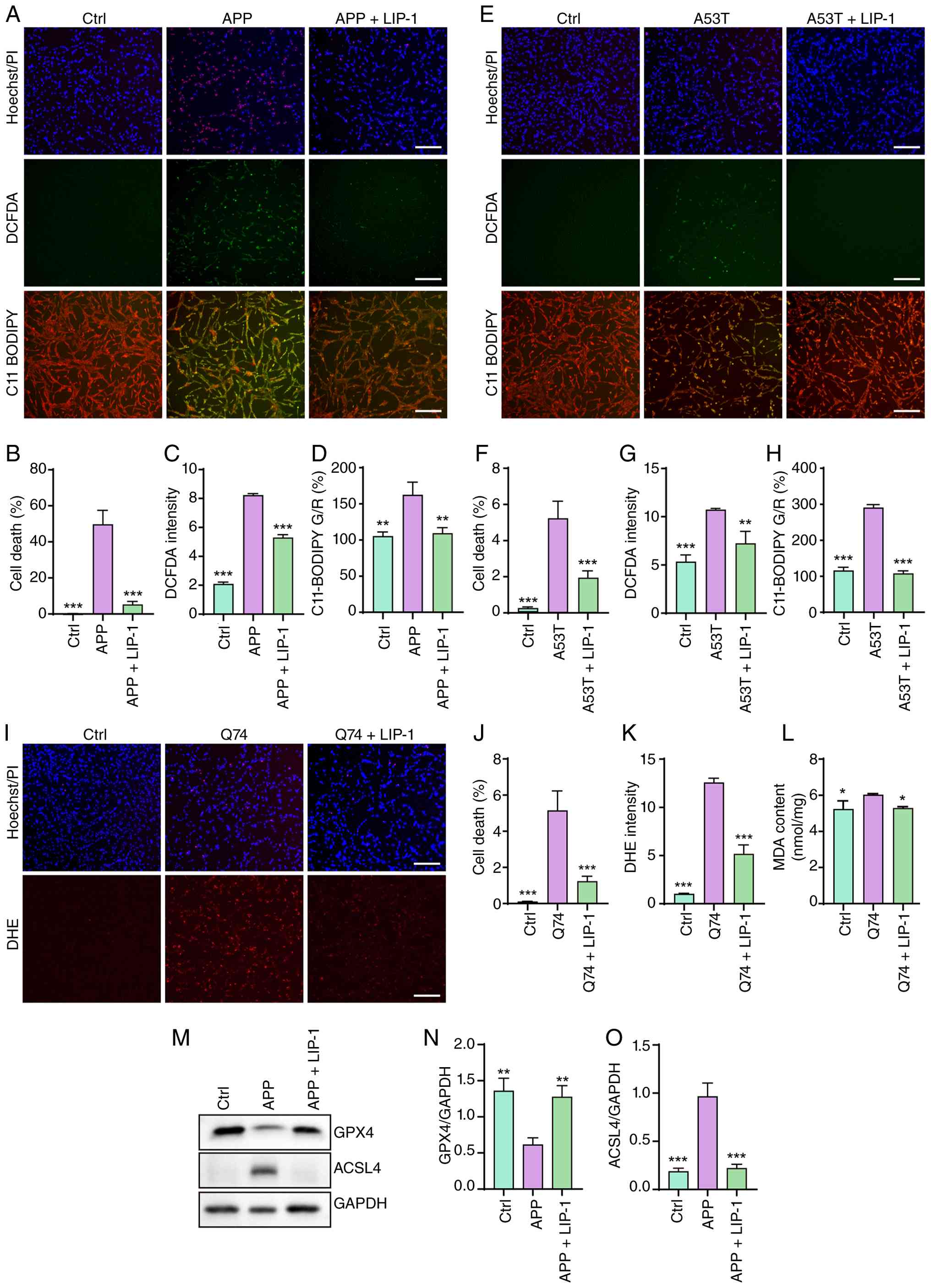 | Figure 1LIP-1 suppresses ferroptotic
phenotypes induced by neurodegenerative pathogenic proteins. (A)
Representative fluorescence images of cell death (Hoechst/PI),
intracellular reactive oxygen species accumulation (DCFDA) and
lipid peroxidation (C11-BODIPY 581/591) in Ctrl,
APP-overexpressing, and APP-overexpressing SH-SY5Y cells with or
without LIP-1 treatment. Magnification, ×10; scale bars, 50
μm. (B-D) Quantification of cell death, DCFDA fluorescence
intensity, and the green-to-red (G/R) fluorescence ratio of
C11-BODIPY 581/591, respectively. The G/R ratio represents the
oxidized-to-reduced fluorescence ratio and was used as an indicator
of lipid peroxidation. (E) Representative fluorescence images of
Hoechst/PI, DCFDA and C11-BODIPY 581/591 staining in control,
α-synuclein A53T-overexpressing, and A53T-overexpressing SH-SY5Y
cells with or without LIP-1 treatment. Magnification, ×10; scale
bars, 50 μm. (F-H) Quantification of cell death, DCFDA
fluorescence intensity, and the green-to-red (G/R) fluorescence
ratio of C11-BODIPY 581/591, respectively. The G/R ratio represents
the oxidized-to-reduced fluorescence ratio and was used as an
indicator of lipid peroxidation. (I) Representative fluorescence
images of Hoechst/PI and DHE staining in control,
Q74-overexpressing, and Q74-overexpressing SH-SY5Y cells with or
without LIP-1 treatment. Magnification, ×10; scale bars, 50
μm. (J and K) Quantification of cell death and DHE
fluorescence intensity, respectively. (L) Quantification of MDA
levels in control, Q74-overexpressing, and Q74-overexpressing
SH-SY5Y cells with or without LIP-1 treatment. (M) Representative
western blots of GPX4 and ACSL4 in control, APP-overexpressing, and
APP-overexpressing SH-SY5Y cells with or without LIP-1 treatment.
(N and O) Densitometric quantification of GPX4 and ACSL4 protein
levels normalized to GAPDH, respectively. Data are presented as the
mean ± SEM. *P<0.05, **P<0.01 and
***P<0.001. LIP-1, liproxstatin-1; APP, amyloid
precursor protein; MDA, malondialdehyde; GPX4, glutathione
peroxidase 4; ACSL4, acyl-CoA synthetase long-chain family member
4. |
Glucose deprivation inhibits ferroptosis
in SH-SY5Y cells
To examine whether changes in extracellular glucose
availability affect ferroptotic susceptibility, SH-SY5Y cells were
exposed to different glucose conditions. Because the basal culture
medium contained 25 mM glucose, this condition was used as the
standard culture control. Exposure to markedly higher glucose
concentrations, ranging from 100 to 800 mM, progressively reduced
cell viability (Fig. 2A). These
concentrations represent supraphysiological glucose overload and
likely reflect non-physiological hyperosmotic stress rather than
clinically relevant hyperglycemia. Treatment with the ferroptosis
inhibitors LIP-1 or Fer-1 partially improved cell viability under
supraphysiological glucose overload (Fig. 2B), suggesting that lipid
peroxidation-related injury may contribute to glucose
overload-associated cytotoxicity. However, because osmotic controls
were not included, these data should not be interpreted as evidence
that glucose overload-induced cytotoxicity is primarily or
specifically mediated by ferroptosis. In RSL3-treated cells,
supraphysiological glucose overload further reduced cell viability,
whereas LIP-1 preserved cell survival (Fig. 2C). The effects of glucose
deprivation-induced energy stress were next examined. When
ferroptosis was induced pharmacologically by erastin or RSL3,
reducing glucose availability significantly protected SH-SY5Y cells
from ferroptosis-associated cell death, as shown by Hoechst/PI
staining and quantification (Figs.
2D-F and S4). Cell death
was extensive under the standard 25 mM glucose condition after
erastin or RSL3 treatment, whereas reducing glucose availability
progressively decreased PI-positive cells. Cell death was nearly
abolished under glucose-free conditions. These findings indicate
that glucose deprivation suppresses erastin- and RSL3-induced
ferroptotic injury in SH-SY5Y cells, supporting the possibility
that energy stress activates an adaptive response that limits lipid
peroxidation-driven cell death.
![Glucose deprivation protects SH-SY5Y
cells from ferroptotic cell death. (A) Cell viability of SH-SY5Y
cells exposed to different extracellular glucose concentrations.
The 25 mM glucose condition represents the standard high-glucose
DMEM control. Glucose concentrations of 100-800 mM were used as a
non-physiological hyperosmotic glucose challenge to assess cellular
tolerance to glucose overload. (B) Cell viability of SH-SY5Y cells
exposed to high glucose, with or without the ferroptosis inhibitors
LIP-1 or Fer-1. (C) Cell viability of SH-SY5Y cells treated with
RSL3 under different glucose concentrations, with or without the
ferroptosis inhibitor LIP-1. (D) Representative Hoechst/PI
fluorescence images showing cell death in SH-SY5Y cells treated
with erastin or RSL3 under normal glucose [Glu (25 mM)] or
glucose-deprived/low-glucose conditions [Glu (6.25, 1.56, or 0
mM)]. Magnification, ×10; scale bars, 50 μm. (E and F)
Quantification of cell death in erastin-treated and RSL3-treated
SH-SY5Y cells under the indicated glucose concentrations,
respectively. Data are presented as the mean ± SEM.
*P<0.05, **P<0.01 and
***P<0.001. LIP-1, liproxstatin-1; Fer-1,
ferrostatin-1.](/article_images/ijmm/58/4/ijmm-58-04-05945-g01.jpg) | Figure 2Glucose deprivation protects SH-SY5Y
cells from ferroptotic cell death. (A) Cell viability of SH-SY5Y
cells exposed to different extracellular glucose concentrations.
The 25 mM glucose condition represents the standard high-glucose
DMEM control. Glucose concentrations of 100-800 mM were used as a
non-physiological hyperosmotic glucose challenge to assess cellular
tolerance to glucose overload. (B) Cell viability of SH-SY5Y cells
exposed to high glucose, with or without the ferroptosis inhibitors
LIP-1 or Fer-1. (C) Cell viability of SH-SY5Y cells treated with
RSL3 under different glucose concentrations, with or without the
ferroptosis inhibitor LIP-1. (D) Representative Hoechst/PI
fluorescence images showing cell death in SH-SY5Y cells treated
with erastin or RSL3 under normal glucose [Glu (25 mM)] or
glucose-deprived/low-glucose conditions [Glu (6.25, 1.56, or 0
mM)]. Magnification, ×10; scale bars, 50 μm. (E and F)
Quantification of cell death in erastin-treated and RSL3-treated
SH-SY5Y cells under the indicated glucose concentrations,
respectively. Data are presented as the mean ± SEM.
*P<0.05, **P<0.01 and
***P<0.001. LIP-1, liproxstatin-1; Fer-1,
ferrostatin-1. |
Glucose deprivation-induced AMPK
activation inhibits ferroptosis in SH-SY5Y cells
Given that glucose deprivation protected SH-SY5Y
cells from ferroptosis, it was next investigated whether this
protective effect was mediated by AMPK activation. Under
glucose-free conditions, erastin- or RSL3-induced cell death was
significantly reduced, whereas co-treatment with the AMPK inhibitor
CC largely abolished this protection. By contrast, the ferroptosis
inhibitor LIP-1 effectively suppressed ferroptotic cell death under
the indicated conditions (Fig.
3A-C). Cell viability analysis further revealed that CC reduced
the protective effect of glucose deprivation, whereas LIP-1
restored cell viability in erastin- or RSL3-treated cells (Fig. S5A and B). These results suggest
that AMPK activity may contribute to the anti-ferroptotic effect of
glucose deprivation, but they do not establish AMPK specificity
because CC can inhibit kinases other than AMPK. To complement the
pharmacological inhibition data, AMPK knockdown was further
performed in SH-SY5Y cells. The knockdown efficiency of siAMPK was
confirmed by western blotting (Fig.
S6A and B). Glucose deprivation significantly reduced erastin-
and RSL3-induced cell death, whereas AMPK knockdown substantially
weakened this protective effect (Fig. S7A-C). By contrast, LIP-1
continued to suppress ferroptotic cell death under normal glucose
conditions. These results provide genetic loss-of-function evidence
that AMPK contributes to glucose deprivation-mediated ferroptosis
suppression, although they do not exclude additional
AMPK-independent metabolic effects. It was next examined whether
pharmacological induction of AMPK-related energy stress produced
similar protection. AICAR or 2DG increased cell viability in
erastin- and RSL3-treated cells (Fig. 3D-G). Consistently, Hoechst/PI
staining showed that AICAR and 2DG reduced erastin-induced cell
death (Fig. S8A and B). Because
2DG alters glycolytic flux and AICAR may affect AMP-sensitive
pathways beyond AMPK, these data should be interpreted as
supporting the involvement of energy stress-associated AMPK
signaling, rather than proving that AMPK activation alone is
sufficient to suppress ferroptosis in SH-SY5Y cells. To further
examine whether ACC-associated lipid metabolism contributes to
ferroptosis regulation downstream of AMPK, TOFA was used as a
pharmacological ACC inhibitor. TOFA reduced DCFDA fluorescence and
C11-BODIPY oxidation in erastin- or RSL3-treated SH-SY5Y cells
(Fig. 3H-M), suggesting that
inhibition of ACC-associated lipid metabolic activity attenuates
ROS accumulation and lipid peroxidation under ferroptotic stress.
In parallel, BODIPY 493/503 staining showed that TOFA reduced
neutral lipid/lipid droplet accumulation in erastin- or
RSL3-treated cells (Fig.
S9A-C). These findings are consistent with the involvement of
ACC-associated lipid metabolic regulation in ferroptosis
suppression. Western blot analysis demonstrated that 2DG increased
the p-AMPK/AMPK and p-ACC1/ACC1 ratios, whereas total AMPK and
total ACC1 protein levels were not markedly altered, suggesting
that 2DG enhances AMPK-ACC1 signaling primarily through
phosphorylation-dependent regulation rather than changes in total
protein abundance (Fig. 3N-R).
Together, these results suggest that energy stress engages an
AMPK-ACC1-associated signaling axis that contributes to the
suppression of lipid peroxidation and ferroptosis-associated cell
death in SH-SY5Y cells.
![Energy stress activates the AMPK-ACC1
axis to suppress ferroptosis in SH-SY5Y cells. (A) Representative
Hoechst/PI fluorescence images showing cell death in SH-SY5Y cells
treated with erastin or RSL3 under normal glucose [Glu (25 mM)] or
glucose-deprived [Glu (0 mM)] conditions, with or without CC or
LIP-1. Magnification, ×10; scale bars, 50 μm. (B and C)
Quantification of cell death in erastin-treated and RSL3-treated
SH-SY5Y cells under the indicated conditions, respectively. (D-G)
Cell viability of SH-SY5Y cells treated with erastin or RSL3 in the
presence or absence of the AMPK activator AICAR or the glycolytic
inhibitor 2DG. (H and I) Representative fluorescence images of
DCFDA and C11-BODIPY 581/591 staining in SH-SY5Y cells treated with
erastin or RSL3, with or without TOFA. Magnification, ×10; scale
bars, 50 μm. (J-M) Quantification of DCFDA fluorescence
intensity and the green-to-red (G/R) fluorescence ratio of
C11-BODIPY 581/591 in erastin-treated and RSL3-treated SH-SY5Y
cells. The G/R ratio represents the oxidized-to-reduced
fluorescence ratio and was used as an indicator of lipid
peroxidation. (N) Representative western blots of p-AMPK, AMPK,
p-ACC, ACC1 and β-actin in SH-SY5Y cells treated with 2DG. (O-R)
Densitometric quantification of p-AMPK/AMPK, total AMPK/β-actin,
p-ACC/ACC1 and ACC1/β-actin, respectively. Data are presented as
the mean ± SEM. *P<0.05, **P<0.01 and
***P<0.001. 2DG, 2-deoxy-D-glucose; AMPK,
AMP-activated protein kinase; AICAR, 5-aminoimidazole-4-carboxamide
ribonucleoside; TOFA, 5-tetradecyloxy-2-furoic acid; p-,
phosphorylated; ACC, acetyl-CoA carboxylase; ns, not
significant.](/article_images/ijmm/58/4/ijmm-58-04-05945-g02.jpg) | Figure 3Energy stress activates the AMPK-ACC1
axis to suppress ferroptosis in SH-SY5Y cells. (A) Representative
Hoechst/PI fluorescence images showing cell death in SH-SY5Y cells
treated with erastin or RSL3 under normal glucose [Glu (25 mM)] or
glucose-deprived [Glu (0 mM)] conditions, with or without CC or
LIP-1. Magnification, ×10; scale bars, 50 μm. (B and C)
Quantification of cell death in erastin-treated and RSL3-treated
SH-SY5Y cells under the indicated conditions, respectively. (D-G)
Cell viability of SH-SY5Y cells treated with erastin or RSL3 in the
presence or absence of the AMPK activator AICAR or the glycolytic
inhibitor 2DG. (H and I) Representative fluorescence images of
DCFDA and C11-BODIPY 581/591 staining in SH-SY5Y cells treated with
erastin or RSL3, with or without TOFA. Magnification, ×10; scale
bars, 50 μm. (J-M) Quantification of DCFDA fluorescence
intensity and the green-to-red (G/R) fluorescence ratio of
C11-BODIPY 581/591 in erastin-treated and RSL3-treated SH-SY5Y
cells. The G/R ratio represents the oxidized-to-reduced
fluorescence ratio and was used as an indicator of lipid
peroxidation. (N) Representative western blots of p-AMPK, AMPK,
p-ACC, ACC1 and β-actin in SH-SY5Y cells treated with 2DG. (O-R)
Densitometric quantification of p-AMPK/AMPK, total AMPK/β-actin,
p-ACC/ACC1 and ACC1/β-actin, respectively. Data are presented as
the mean ± SEM. *P<0.05, **P<0.01 and
***P<0.001. 2DG, 2-deoxy-D-glucose; AMPK,
AMP-activated protein kinase; AICAR, 5-aminoimidazole-4-carboxamide
ribonucleoside; TOFA, 5-tetradecyloxy-2-furoic acid; p-,
phosphorylated; ACC, acetyl-CoA carboxylase; ns, not
significant. |
AMPK activation suppresses ferroptosis
triggered by neurodegenerative pathogenic proteins
To determine whether AMPK activation protects
against ferroptosis caused by neurodegenerative pathogenic
proteins, SH-SY5Y cells overexpressing APP, α-synuclein A53T, or
mutant huntingtin Q74 were exposed to glucose deprivation or AMPK
activators. As a fluorescence control, glucose deprivation did not
significantly alter pEGFP-N1 fluorescence in transfected SH-SY5Y
cells (Fig. S10A and B). In
APP-overexpressing cells, glucose deprivation, AICAR, or 2DG
treatment significantly decreased cell death, ROS accumulation and
lipid peroxidation, as shown by reduced PI uptake, DCFDA
fluorescence and C11 BODIPY oxidation (Fig. 4A-D). Similarly, α-synuclein
A53T-expressing cells displayed strong ferroptotic phenotypes that
were significantly alleviated by glucose deprivation or AMPK
activation (Fig. 4E-H).
Consistent results were observed in Q74-overexpressing cells, where
energy stress or AMPK activation attenuated ferroptosis, as
evidenced by decreased PI-positive cells, DHE fluorescence
intensity, and MDA levels (Figs.
4I-K and S11).
Collectively, these findings support a protective association
between energy stress-associated AMPK activation and reduced
ferroptosis-related injury induced by multiple neurodegenerative
pathogenic proteins.
![AMPK activation suppresses
ferroptosis-like oxidative injury induced by neurodegenerative
pathogenic proteins. (A) Representative fluorescence images of
Hoechst/PI, DCFDA and C11-BODIPY 581/591 staining in Ctrl,
APP-overexpressing, and APP-overexpressing SH-SY5Y cells subjected
to glucose deprivation [Glu (0 mM)] or treated with the AMPK
activator AICAR or 2DG. Magnification, ×10; scale bars, 50
μm. (B-D) Quantification of cell death (B), DCFDA
fluorescence intensity (C), and the green-to-red (G/R) fluorescence
ratio of C11-BODIPY 581/591 (D) in APP-overexpressing SH-SY5Y cells
under the indicated conditions. The G/R ratio represents the
oxidized-to-reduced fluorescence ratio and was used as an indicator
of lipid peroxidation. (E) Representative fluorescence images of
Hoechst/PI, DCFDA and C11-BODIPY 581/591 staining in Ctrl,
α-synuclein A53T-overexpressing, and A53T-overexpressing SH-SY5Y
cells subjected to glucose deprivation [Glu (0 mM)] or treated with
AICAR or 2DG. Magnification, ×10; scale bars, 50 μm. (F-H)
Quantification of cell death (F), DCFDA fluorescence intensity (G),
and the green-to-red (G/R) fluorescence ratio of C11-BODIPY 581/591
(H) in A53T-overexpressing SH-SY5Y cells under the indicated
conditions. The G/R ratio represents the oxidized-to-reduced
fluorescence ratio and was used as an indicator of lipid
peroxidation. (I) Representative fluorescence images of Hoechst/PI
and DHE staining in control, Q74-overexpressing, and
Q74-overexpressing SH-SY5Y cells subjected to glucose deprivation
[Glu (0 mM)] or treated with AICAR or 2DG. Magnification, ×10;
scale bars, 50 μm. (J and K) Quantification of cell death
(J) and DHE fluorescence intensity (K) in Q74-overexpressing cells
under the indicated treatments. Data are presented as the mean ±
SEM. ***P<0.001. 2DG, 2-deoxy-D-glucose; AMPK,
AMP-activated protein kinase; APP, amyloid precursor protein;
AICAR, 5-aminoimidazole-4-carboxamide ribonucleoside.](/article_images/ijmm/58/4/ijmm-58-04-05945-g03.jpg) | Figure 4AMPK activation suppresses
ferroptosis-like oxidative injury induced by neurodegenerative
pathogenic proteins. (A) Representative fluorescence images of
Hoechst/PI, DCFDA and C11-BODIPY 581/591 staining in Ctrl,
APP-overexpressing, and APP-overexpressing SH-SY5Y cells subjected
to glucose deprivation [Glu (0 mM)] or treated with the AMPK
activator AICAR or 2DG. Magnification, ×10; scale bars, 50
μm. (B-D) Quantification of cell death (B), DCFDA
fluorescence intensity (C), and the green-to-red (G/R) fluorescence
ratio of C11-BODIPY 581/591 (D) in APP-overexpressing SH-SY5Y cells
under the indicated conditions. The G/R ratio represents the
oxidized-to-reduced fluorescence ratio and was used as an indicator
of lipid peroxidation. (E) Representative fluorescence images of
Hoechst/PI, DCFDA and C11-BODIPY 581/591 staining in Ctrl,
α-synuclein A53T-overexpressing, and A53T-overexpressing SH-SY5Y
cells subjected to glucose deprivation [Glu (0 mM)] or treated with
AICAR or 2DG. Magnification, ×10; scale bars, 50 μm. (F-H)
Quantification of cell death (F), DCFDA fluorescence intensity (G),
and the green-to-red (G/R) fluorescence ratio of C11-BODIPY 581/591
(H) in A53T-overexpressing SH-SY5Y cells under the indicated
conditions. The G/R ratio represents the oxidized-to-reduced
fluorescence ratio and was used as an indicator of lipid
peroxidation. (I) Representative fluorescence images of Hoechst/PI
and DHE staining in control, Q74-overexpressing, and
Q74-overexpressing SH-SY5Y cells subjected to glucose deprivation
[Glu (0 mM)] or treated with AICAR or 2DG. Magnification, ×10;
scale bars, 50 μm. (J and K) Quantification of cell death
(J) and DHE fluorescence intensity (K) in Q74-overexpressing cells
under the indicated treatments. Data are presented as the mean ±
SEM. ***P<0.001. 2DG, 2-deoxy-D-glucose; AMPK,
AMP-activated protein kinase; APP, amyloid precursor protein;
AICAR, 5-aminoimidazole-4-carboxamide ribonucleoside. |
AMPK activation ameliorates behavioral
impairments in transgenic C. elegans models of neurodegenerative
proteinopathies
To evaluate the functional relevance of AMPK
activation in vivo, behavioral performance was assessed in
transgenic C. elegans models expressing human Aβ1-42,
α-synuclein, or polyQ-expanded proteins. In the Aβ-expressing
CL4176 model, treatment with 2DG or AICAR significantly reduced
paralysis rates in a concentration-dependent manner, with an effect
comparable to that of the ferroptosis inhibitor LIP-1 (Fig. 5A and B). In neuronal
Aβ-expressing CL2355 worms, 2DG and LIP-1 improved the defective
slowing response compared with untreated CL2355 worms, whereas the
control strain CL2122 showed normal behavioral performance
(Fig. 5C). These results
indicate that metabolic activation of AMPK-related signaling
alleviates Aβ-associated behavioral impairment. It was next
examined whether this protective effect extended to other
proteinopathy models. In α-synuclein-expressing NL5901 worms and
polyQ-expanded AM141 worms, both 2DG and LIP-1 increased body bend
frequency, indicating improved locomotor activity and neuromuscular
function (Fig. 5D and E). These
findings suggest that 2DG-mediated metabolic stress and ferroptosis
inhibition confer behavioral protection across AD-, PD- and
HD-related C. elegans models. To further determine whether
the behavioral protection induced by 2DG depends on AMPK-related
signaling, aak-2 RNAi was performed in CL4176 worms.
AAK-2 is the major AMPK orthologue in C. elegans.
Under HT115/pL4440 control RNAi conditions, 2DG significantly
reduced Aβ-induced paralysis. However, aak-2 RNAi largely
abolished the protective effect of 2DG (Fig. 5F and G). These results provide
in vivo genetic evidence that AAK-2/AMPK signaling is
required, at least in part, for 2DG-mediated behavioral protection
in the Aβ-expressing worm model. Collectively, these behavioral
data support a conserved protective role of energy stress-induced
AMPK-related signaling in neurodegenerative proteinopathy
models.
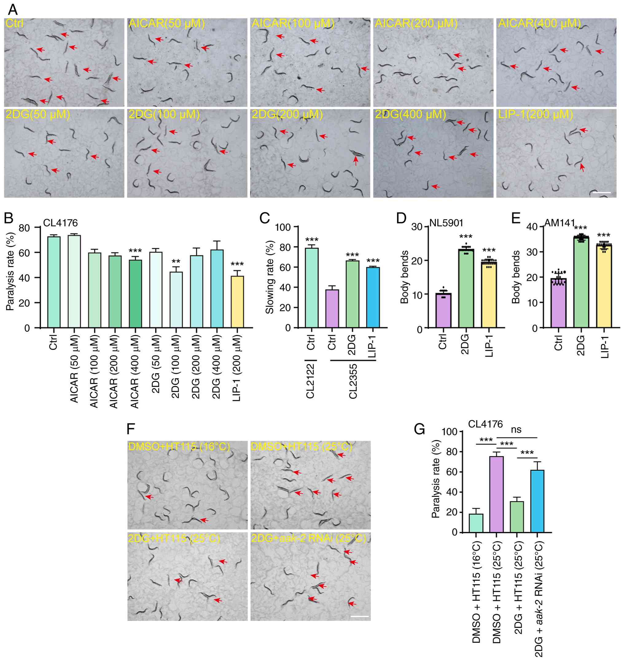 | Figure 5AMPK activation improves
disease-associated behavioral deficits in transgenic C.
elegans models of AD, PD and HD. (A) Representative
bright-field images showing paralysis phenotypes in Aβ-expressing
CL4176 worms treated with different concentrations of AICAR or 2DG,
or with LIP-1. Red arrows indicate paralyzed worms. Magnification,
×24; scale bar, 200 μm. (B) Quantification of paralysis rate
in CL4176 worms after treatment with AICAR, 2DG, or LIP-1. (C)
Slowing response analysis in CL2122 control worms and neuronal
Aβ-expressing CL2355 worms treated with 2DG or LIP-1. (D and E)
Quantification of body bends in α-synuclein-expressing NL5901 worms
(D) and polyglutamine (polyQ)-expanded AM141 worms (E) after
treatment with 2DG or LIP-1. Data are presented as the mean ± SEM.
(F) Representative bright-field images showing paralysis phenotypes
in CL4176 worms cultured on HT115/pL4440 control bacteria or
aak-2 RNAi bacteria, with or without 2DG treatment. Red
arrows indicate paralyzed worms. Magnification, ×24; scale bar, 200
μm. (G) Quantification of paralysis rate in CL4176 worms
under the indicated conditions. Data are presented as the mean ±
SEM. **P<0.01 and ***P<0.001. 2DG,
2-deoxy-D-glucose; AICAR, 5-aminoimidazole-4-carboxamide
ribonucleoside; LIP-1, liproxstatin-1. |
2DG suppresses ferroptosis-related stress
in transgenic C. elegans models of neurodegenerative
proteinopathies
To determine whether 2DG suppresses
ferroptosis-related damage in vivo, ROS accumulation, lipid
peroxidation and MDA levels were examined in transgenic C.
elegans models expressing neurodegeneration-associated
pathogenic proteins. In Aβ-expressing CL4176 worms,
temperature-induced Aβ expression at 25°C caused a significant
increase in oxidative and ferroptosis-associated stress, as
reflected by elevated DHE fluorescence, increased C11-BODIPY
oxidation, and higher MDA levels. Treatment with 2DG or the
ferroptosis inhibitor LIP-1 significantly reduced ROS accumulation
and lipid peroxidation in CL4176 worms (Fig. 6A-D) and also decreased MDA levels
(Fig. S12A). These results
indicate that 2DG suppresses Aβ-associated oxidative stress and
lipid peroxidation in vivo. Similar effects were observed in
α-synuclein-expressing NL5901 worms. NL5901 worms exhibited
increased DHE fluorescence, C11-BODIPY oxidation, and MDA
production, indicating enhanced ROS accumulation and lipid
peroxidation. Both 2DG and LIP-1 significantly reduced these
ferroptosis-related changes (Figs.
6E-H and S12B). In
polyQ-expanded AM141 worms, 2DG also decreased DHE fluorescence,
C11-BODIPY oxidation and MDA levels, with effects comparable to
those of LIP-1 treatment (Fig.
6I-L and Fig. S12C). These
findings suggest that 2DG broadly attenuates oxidative and lipid
peroxidation damage across AD-, PD-, and HD-related worm
proteinopathy models. It was further evaluated whether 2DG and
LIP-1 protect against ferroptosis-related injury in a toxin-induced
dopaminergic neurodegeneration model. In 6-OHDA-exposed N2 worms,
both treatments reduced DHE fluorescence and C11-BODIPY oxidation,
indicating suppression of ROS accumulation and lipid peroxidation
(Fig. S13A-D). Consistently, in
6-OHDA-exposed BZ555 worms, 2DG and LIP-1 preserved GFP
fluorescence in dopaminergic neurons, supporting improved
dopaminergic neuronal viability (Fig. S14A and B). Consistent with the
behavioral data, aak-2 RNAi weakened the ability of 2DG to
suppress oxidative and ferroptosis-associated stress in CL4176
worms. Under HT115/pL4440 control RNAi conditions, 2DG reduced DHE
fluorescence and C11-BODIPY oxidation after Aβ induction. By
contrast, these protective effects were largely lost in worms fed
aak-2 RNAi bacteria (Fig.
6M-P). These results provide in vivo genetic evidence
that AAK-2/AMPK signaling contributes to 2DG-mediated
suppression of ROS accumulation and lipid peroxidation.
Collectively, these findings show that 2DG reduces ROS
accumulation, lipid peroxidation and MDA production in transgenic
C. elegans models of neurodegenerative proteinopathies.
Together with the aak-2 RNAi results, these data support an
important role for AAK-2/AMPK-related signaling in the
anti-ferroptotic effects of energy stress in vivo.
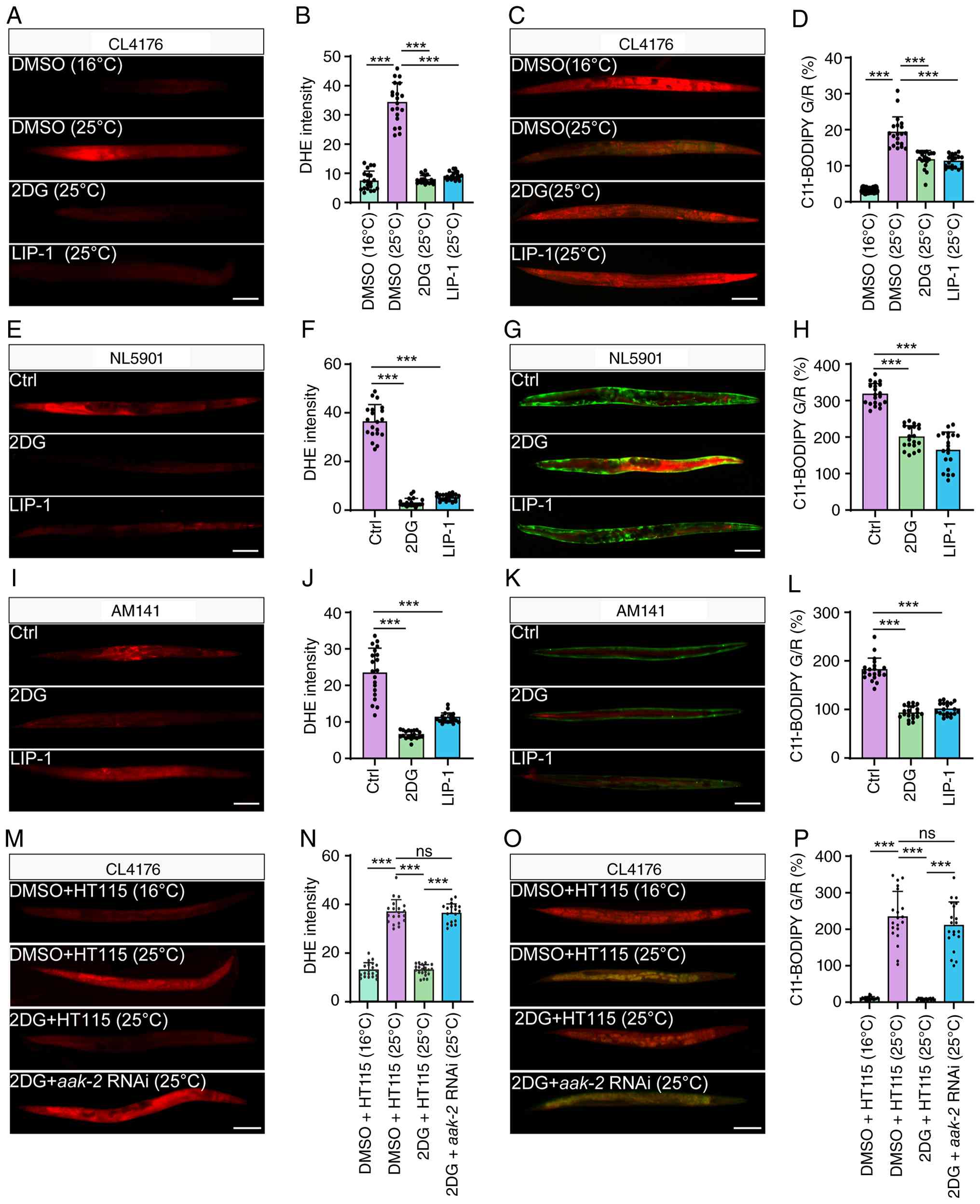 | Figure 62DG suppresses ferroptosis in
transgenic C. elegans models of neurodegenerative
proteinopathies through AAK-2/AMP-activated protein kinase
signaling. (A and C) Representative fluorescence images of DHE
staining (A) and C11-BODIPY 581/591 staining (C) in Aβ-expressing
CL4176 worms maintained at 16°C or shifted to 25°C, with or without
2DG or LIP-1 treatment. Magnification, ×10; scale bars, 50
μm. (B and D) Quantification of DHE fluorescence intensity
(B) and the green-to-red (G/R) fluorescence ratio of C11-BODIPY
581/591 (D) in CL4176 worms under the indicated conditions. The G/R
ratio represents the oxidized-to-reduced fluorescence ratio and was
used as an indicator of lipid peroxidation. (E and G)
Representative fluorescence images of DHE staining (E) and
C11-BODIPY 581/591 staining (G) in α-synuclein-expressing NL5901
worms treated with 2DG or LIP-1. Magnification, ×10; scale bars, 50
μm. (F and H) Quantification of DHE fluorescence intensity
(F) and the green-to-red (G/R) fluorescence ratio of C11-BODIPY
581/591 (H) in NL5901 worms, the G/R ratio represents the
oxidized-to-reduced fluorescence ratio and was used as an indicator
of lipid peroxidation. (I and K) Representative fluorescence images
of DHE staining (I) and C11-BODIPY 581/591 staining (K) in
polyQ-expanded AM141 worms treated with 2DG or LIP-1.
Magnification, ×10; scale bars, 50 μm. (J and L)
Quantification of DHE fluorescence intensity (J) and the
green-to-red (G/R) fluorescence ratio of C11-BODIPY 581/591 (L) in
AM141 worms, the G/R ratio represents the oxidized-to-reduced
fluorescence ratio and was used as an indicator of lipid
peroxidation. (M and O) Representative fluorescence images of DHE
staining (M) and C11-BODIPY 581/591 staining (O) in CL4176 worms
fed HT115/pL4440 control bacteria or aak-2 RNAi bacteria
under the indicated conditions. (N and P) Quantification of DHE
fluorescence intensity (N) and the green-to-red (G/R) fluorescence
ratio of C11-BODIPY 581/591 (P) in CL4176 worms under the indicated
RNAi conditions, the G/R ratio represents the oxidized-to-reduced
fluorescence ratio and was used as an indicator of lipid
peroxidation. Data are presented as the mean ± SEM.
***P<0.001. 2DG, 2-deoxy-D-glucose; LIP-1,
liproxstatin-1; RNAi, RNA interference. |
2DG ameliorates cognitive deficits and
suppresses neuroinflammatory and ferroptosis-associated pathology
in 3xTg-AD mice
To determine whether AMPK activation protects
against neurodegeneration-associated ferroptotic pathology in
vivo, the behavioral and neuropathological effects of 2DG and
LIP-1 in 3xTg-AD mice were evaluated. In the Morris water maze
probe trial, 3xTg-AD mice exhibited significant spatial memory
impairment, as shown by fewer platform crossings, reduced time
spent in the target quadrant, and shorter path length in the target
quadrant compared with control mice. Treatment with either 2DG or
LIP-1 significantly improved these memory-related parameters
(Fig. 7A-D). During the
acquisition phase, swimming speed did not differ substantially
among groups, indicating that the behavioral differences were not
attributable to altered motor ability (Fig. 7E). By contrast, 3xTg-AD mice
showed prolonged escape latency during training, which was markedly
reduced by 2DG or LIP-1 treatment (Fig. 7F). These results indicate that
2DG and LIP-1 improve spatial learning and memory deficits in
3xTg-AD mice. Hippocampal neuroinflammatory changes were next
examined. Immunofluorescence staining showed increased
GFAP-positive astrocytes and Iba1-positive microglia in the
hippocampi of 3xTg-AD mice, indicating pronounced astrocytic and
microglial activation. Both 2DG and LIP-1 significantly reduced the
percentages of GFAP-positive and Iba1-positive cells (Fig. 7G-I), suggesting that these
treatments attenuate glial activation in the AD brain. Nissl
staining further showed obvious neuronal loss and disorganization
in the hippocampal CA1 region of 3xTg-AD mice, whereas 2DG and
LIP-1 preserved neuronal morphology and improved hippocampal
structural integrity (Fig. 7J).
Consistently, H&E staining confirmed that 2DG and LIP-1
alleviated CA1 histopathological damage and reduced the appearance
of damaged or pyknotic neurons (Fig. S15). To strengthen the in
vivo evidence for ferroptosis-associated pathology, multiple
ferroptosis-related markers in hippocampal sections were examined.
Iron staining suggested increased hippocampal iron deposition in
3xTg-AD mice, which appeared to be attenuated by 2DG and LIP-1
treatment (Fig. 7K). 3xTg-AD
mice also exhibited enhanced 4-HNE immunoreactivity, indicating
increased lipid peroxidation, and this increase was attenuated by
2DG or LIP-1 treatment (Figs. 7K
and S16). In parallel,
immunofluorescence staining showed that ACSL4-positive cells were
increased and GPX4-positive cells were decreased in the hippocampi
of 3xTg-AD mice. Treatment with 2DG or LIP-1 significantly reduced
ACSL4 positivity and restored GPX4 expression (Fig. 7K-M). These results provide
convergent evidence that ferroptosis-associated iron accumulation,
lipid peroxidation, ACSL4 upregulation and GPX4 loss occur in the
3xTg-AD hippocampus and are suppressed by 2DG or LIP-1. Moreover,
the restoration of GPX4 expression and reduction of ACSL4
expression suggest that 2DG-mediated AMPK activation is associated
with preservation of GPX4-related anti-ferroptotic defense and
suppression of ACSL4-associated ferroptotic susceptibility in the
AD hippocampus. Finally, western blot analysis demonstrated that
2DG increased hippocampal AMPK and ACC phosphorylation compared
with untreated 3xTg-AD mice, whereas total AMPK and ACC1 levels
were not markedly altered (Fig.
7N-P). These findings indicate that 2DG engages an
AMPK-ACC1-associated signaling axis in the 3xTg-AD hippocampus.
However, because 2DG can also alter glycolytic flux and activate
broader metabolic stress responses, these mouse data should be
interpreted as showing an association between 2DG-mediated
neuroprotection and AMPK-ACC1 activation, rather than as definitive
genetic proof that AMPK is required in mammalian neurons in
vivo. Together with the iron staining, 4-HNE, ACSL4 and GPX4
data, these results support an association between 2DG-associated
AMPK-ACC1 activation and reduced ferroptosis-associated hippocampal
pathology in vivo. Collectively, these results demonstrate
that 2DG ameliorates cognitive impairment and hippocampal pathology
in 3xTg-AD mice, accompanied by suppression of neuroinflammation
and ferroptosis-associated damage, including iron accumulation,
lipid peroxidation, ACSL4 upregulation and GPX4 reduction.
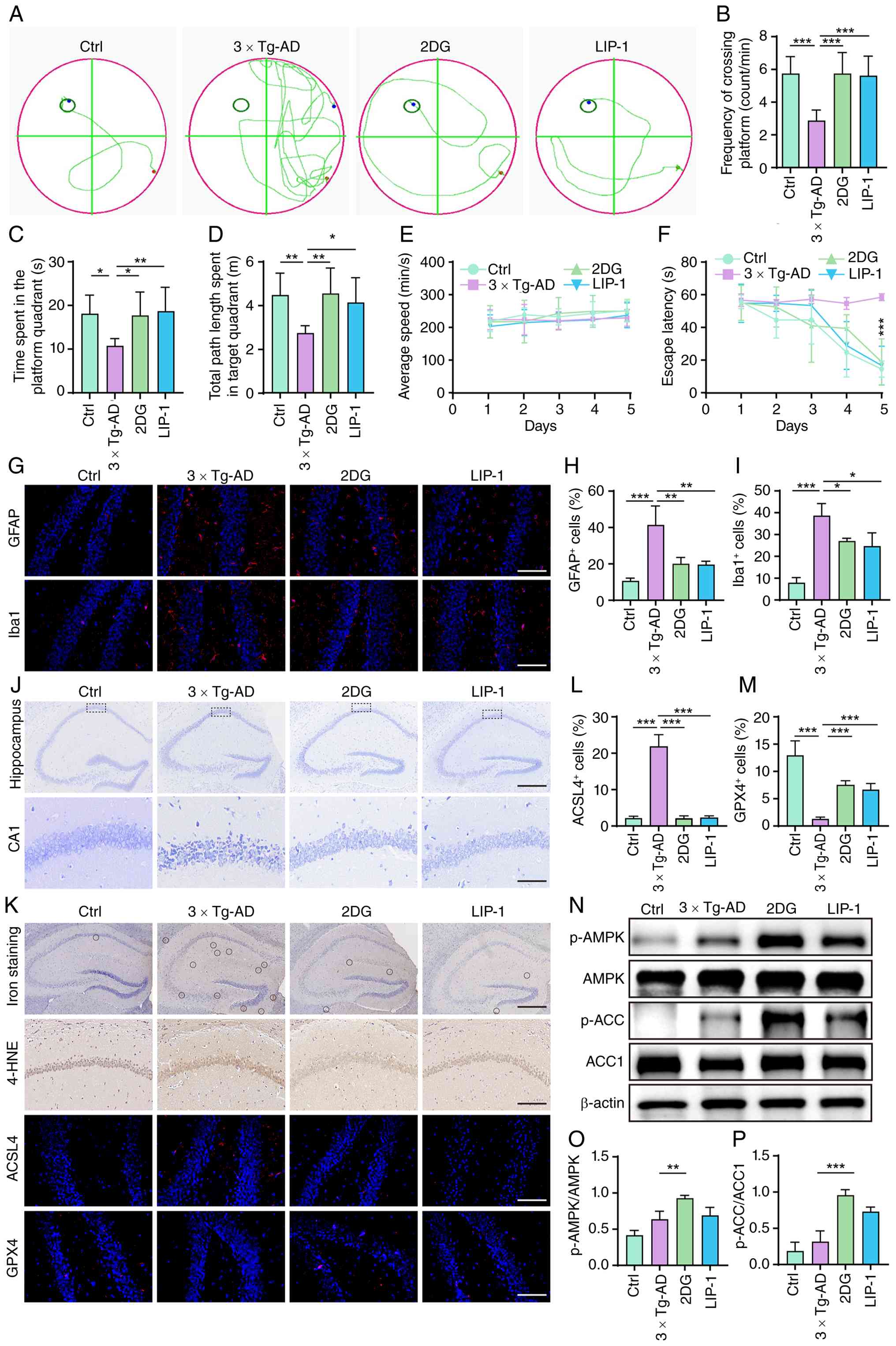 | Figure 72DG-mediated AMPK activation
ameliorates cognitive impairment and attenuates neuroinflammatory
and ferroptotic pathology in 3xTg-AD mice. (A) Representative
swimming trajectories during the Morris water maze probe trial in
WT control, 3xTg-AD, 3xTg-AD + 2DG, and 3xTg-AD + LIP-1 mice. The
2DG and LIP-1 groups throughout the figure refer to 3xTg-AD mice
treated with 2DG or LIP-1, respectively. (B-D) Quantification of
platform crossing frequency (B), time spent in the target quadrant
(C), and total path length in the target quadrant (D) during the
probe trial. (E and F) Average swimming speed (E) and escape
latency (F) during the acquisition phase. (G) Representative
hippocampal immunofluorescence images of GFAP and Iba1 staining in
each group. Magnification, ×40; scale bars, 200 μm. (H and
I) Quantification of GFAP-positive astrocytes (H) and Iba1-positive
microglia (I) in the hippocampus. (J) Representative Nissl-stained
hippocampal sections. Upper panels show the overall hippocampal
region, and lower panels show higher-magnification views of the CA1
region indicated by dashed boxes. Magnification, ×5; scale bars,
300 μm. (K) Representative images of iron staining, 4-HNE
immunohistochemistry, and immunofluorescence staining for ACSL4 and
GPX4 in hippocampal sections from each group. Circles indicate
iron-positive deposits. Magnifications and scale bars were as
follows: ×5 and 300 μm for Prussian blue staining; ×20 and
100 μm for 4-HNE immunohistochemistry; and ×40 and 200
μm for ACSL4 and GPX4 immunofluorescence. (L and M)
Quantification of ACSL4-positive cells and GPX4-positive cells in
the hippocampus. Quantification of 4-HNE staining is shown in
Fig. S16. (N) Representative
Western blots of p-AMPK, AMPK, p-ACC, ACC1 and β-actin in
hippocampal lysates from each group. (O and P) Densitometric
quantification of p-AMPK/AMPK (O) and p-ACC/ACC1 (P). Data are
presented as the mean ± SEM. *P<0.05,
**P<0.01 and ***P<0.001. 2DG,
2-deoxy-D-glucose; 4-HNE, 4-hydroxynonenal; AMPK, AMP-activated
protein kinase; LIP-1, liproxstatin-1; Iba1, ionized
calcium-binding adapter molecule 1; p-, phosphorylated; GPX4,
glutathione peroxidase 4; ACSL4, acyl-CoA synthetase long-chain
family member 4; GFAP, glial fibrillary acidic protein; WT,
wild-type. |
Discussion
The present study demonstrates that controlled
energy stress attenuates ferroptosis-associated neuronal injury in
neurodegenerative proteinopathy models in association with
AMPK-ACC1-mediated metabolic regulation (Fig. 8). Previous studies have
established that energy stress activates AMPK and suppresses
ferroptosis through phosphorylation and inhibition of ACC, thereby
limiting polyunsaturated lipid synthesis and lipid peroxidation
(21-23). Therefore, the present study does
not claim the AMPK-ACC axis as a newly discovered
ferroptosis-regulatory mechanism. Rather, its main contribution is
to show that this metabolic checkpoint is functionally engaged in
disease-relevant neuronal proteinopathy contexts, including APP-,
α-synuclein A53T- and polyQ-associated cellular models, transgenic
C. elegans models, and 3xTg-AD mice. Across these systems,
glucose deprivation, 2DG, or AICAR reduced ferroptosis-associated
cell death and lipid peroxidation, while 2DG and LIP-1 improved
behavioral or cognitive deficits and reduced ferroptosis-related
pathology in vivo. These findings extend a previously
defined metabolic ferroptosis checkpoint to neurodegenerative
proteinopathies, where proteotoxic stress, mitochondrial
dysfunction, iron dyshomeostasis and lipid peroxidation
coexist.
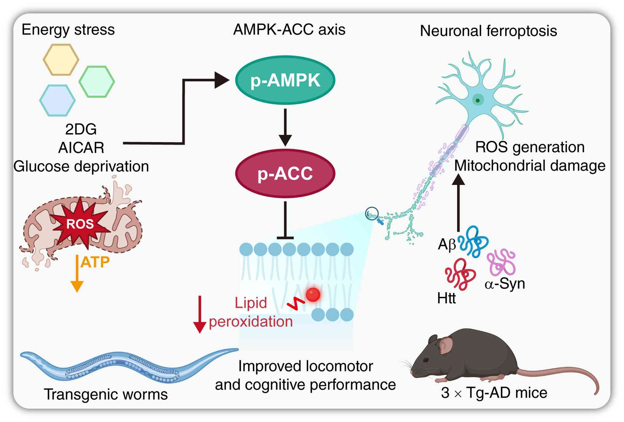 | Figure 8Proposed model by which energy stress
activates the AMPK-ACC1 axis to suppress neuronal ferroptosis in
neurodegenerative proteinopathies. Energy-stress interventions,
including 2DG, AICAR, and glucose deprivation, activate AMPK
signaling and promote ACC phosphorylation. Activation of the
AMPK-ACC1 axis limits ferroptosis-associated lipid peroxidation,
thereby reducing ROS generation and mitochondrial damage induced by
neurodegenerative pathogenic proteins, including Aβ, α-synuclein
and mutant huntingtin. In transgenic C. elegans models and
3xTg-AD mice, this mechanism is associated with reduced oxidative
and ferroptotic injury and improved locomotor or cognitive
performance. 2DG, 2-deoxy-D-glucose; AMPK, AMP-activated protein
kinase; ACC, acetyl-CoA carboxylase; p-, phosphorylated; AICAR,
5-aminoimidazole-4-carboxamide ribonucleoside; ROS, reactive oxygen
species. |
These results also help clarify an apparent
energy-stress paradox in neurodegeneration. AD and related
proteinopathies are characterized by impaired glucose metabolism,
mitochondrial dysfunction and chronic bioenergetic stress,
conditions that could theoretically activate AMPK. However, chronic
disease-associated metabolic failure is not necessarily equivalent
to an effective protective AMPK response. AMPK signaling is highly
context-dependent and may vary according to stimulus intensity,
duration, cell type, subcellular localization, and coupling to
downstream effectors. In the present study, 3xTg-AD mice showed
clear ferroptosis-associated hippocampal pathology and cognitive
impairment, indicating that the endogenous AMPK response at the
examined disease stage was insufficient to prevent
neurodegeneration. By contrast, 2DG induced more evident activation
of the AMPK-ACC1 axis and was accompanied by reduced lipid
peroxidation, decreased ACSL4 expression, restored GPX4 expression,
reduced glial activation, and improved behavioral performance.
Together with AMPK inhibition, siAMPK, and aak-2 RNAi data
in cellular and C. elegans models, these findings support
the idea that controlled metabolic intervention can reinforce an
inadequate endogenous adaptive response and restore downstream
restraint of ferroptotic lipid peroxidation.
Lipid metabolism is central to ferroptotic
vulnerability. Polyunsaturated phospholipids are key substrates for
lipid peroxidation, and limiting their synthesis or incorporation
into membranes can reduce ferroptotic sensitivity (31-33). In the present study, 2DG
increased the p-AMPK/AMPK and p-ACC1/ACC1 ratios in SH-SY5Y cells
and hippocampal tissue, whereas TOFA reduced ROS accumulation,
C11-BODIPY oxidation, and BODIPY 493/503-positive neutral lipid
accumulation. These findings are consistent with an
AMPK-ACC1-associated mechanism that limits lipid metabolic inputs
required for ferroptosis. However, the data should be interpreted
carefully. The detected ACC isoform in the present study was ACC1,
and TOFA is not isoform-selective. Moreover, TOFA may exert
ACC-independent metabolic effects, and de novo lipid
synthesis was not directly measured. Thus, the current data support
the involvement of ACC-associated lipid metabolic regulation, but
do not prove that AMPK suppresses ferroptosis exclusively through
ACC1 inhibition. Future studies using ACC1- and ACC2-specific
genetic manipulation, malonyl-CoA measurement and isotope-tracing
lipidomics will be needed to define the precise lipid metabolic
steps by which AMPK limits ferroptotic lipid peroxidation in
neuronal cells.
The in vivo findings further support the
relevance of ferroptosis-associated pathology in neurodegenerative
proteinopathies. In transgenic C. elegans models expressing
Aβ, α-synuclein, or polyQ-expanded proteins, 2DG and LIP-1 reduced
ROS accumulation and lipid peroxidation and improved locomotor or
behavioral phenotypes. Importantly, aak-2 RNAi weakened the
protective effects of 2DG, providing in vivo genetic
evidence that AMPK-related signaling contributes to the
anti-ferroptotic effect of energy stress. In 3xTg-AD mice, 2DG and
LIP-1 improved spatial learning and memory, reduced GFAP- and
Iba1-positive glial activation, preserved hippocampal neuronal
morphology, and suppressed ferroptosis-associated changes,
including iron accumulation, 4-HNE immunoreactivity, ACSL4
upregulation, and GPX4 reduction. The parallel effects of 2DG and
LIP-1 suggest that suppression of ferroptosis-associated lipid
peroxidation is an important component of the observed
neuroprotection. The restoration of GPX4 and reduction of ACSL4
further indicate that energy stress-associated metabolic regulation
is linked not only to reduced lipid peroxidation products but also
to modulation of ferroptosis susceptibility and defense markers in
the AD hippocampus.
Several limitations should be acknowledged. First,
the causal requirement of AMPK-ACC1 signaling was not genetically
tested in mammalian neurons in vivo. Although AMPK knockdown
in SH-SY5Y cells and aak-2 RNAi in C. elegans reduced
reliance on pharmacological tools, the 3xTg-AD mouse data establish
an association between 2DG-induced AMPK-ACC1 activation and reduced
ferroptosis-associated pathology rather than definitive proof of
AMPK necessity. This distinction is important because 2DG alters
glycolytic flux and can activate broader metabolic stress
responses, AICAR may affect AMP-sensitive pathways beyond AMPK, and
CC can inhibit kinases other than AMPK (34-37). Therefore, the pharmacological
results should be interpreted together with, but not as a
substitute for, genetic validation. In addition, AMPK-ACC1
signaling was assessed mainly at the experimental endpoint in
3xTg-AD mice. Thus, although the data suggest that the endogenous
AMPK response at this stage was insufficient to prevent
ferroptosis-associated pathology, they do not define dynamic
changes in AMPK signaling during disease initiation, progression,
or late-stage neurodegeneration. Future studies using
neuron-specific AMPKα1/AMPKα2 knockout, dominant-negative AMPK,
CRISPR-based AMPK knockout, viral AMPK manipulation, rescue
experiments, and longitudinal analysis of AMPK-ACC1 signaling will
be required to establish the causal, isoform-specific, cell
type-specific, and stage-dependent role of this pathway.
Second, the ferroptosis-related analyses in 3xTg-AD
mice were based mainly on hippocampal histological and
immunostaining evidence. Iron staining, 4-HNE immunohistochemistry,
ACSL4 immunofluorescence, and GPX4 immunofluorescence provide
convergent spatial evidence for a ferroptosis-associated pattern,
but they do not establish neuron-specific localization or
biochemical activity. Quantitative measurements of MDA, total iron,
labile iron, GSH/GSSG ratio, GPX4 enzymatic activity, and
NeuN-based neuronal co-localization would further strengthen the
mechanistic interpretation. In addition, only male mice were used.
This design reduced variability related to sex hormone cycling but
limits generalizability, especially because sex differences have
been reported in AD pathology, glucose metabolism,
neuroinflammation and behavioral phenotypes in 3xTg-AD mice
(38-40). Future studies should include both
male and female mice and perform sex-stratified analyses of
cognitive behavior, ferroptosis-associated pathology, AMPK-ACC1
signaling, and neuroinflammatory responses.
Third, ferroptosis should not be regarded as the
only form of cell death in these models. LIP-1 and Fer-1 rescue,
C11-BODIPY oxidation, MDA accumulation, GPX4 reduction and ACSL4
upregulation support a ferroptosis-associated injury component, but
pathogenic proteins may also induce apoptosis, necroptosis, or
mixed regulated cell death responses. This interpretation is
consistent with our previous observation that Aβ(1-42)-induced PC-12 cell injury could be
partially rescued by LIP-1, Nec-1 and Z-VAD-FMK (41). Therefore, future studies should
combine ferroptosis inhibitors with caspase inhibitors, necroptosis
inhibitors, cleaved caspase-3/PARP analysis, and RIPK1/RIPK3/MLKL
pathway detection to determine the relative contribution of
different cell death modalities in APP-, α-synuclein-, and
polyQ-induced neuronal injury.
Finally, the model systems and translational
implications require caution. SH-SY5Y cells are useful for
mechanistic screening but do not fully recapitulate the metabolic
and electrophysiological properties of mature human neurons.
Primary neurons, human iPSC-derived neurons, brain organoids, and
transgenic animals with neuron-specific modulation of AMPK or ACC
would provide more physiologically relevant validation. Moreover,
AMPK has pleiotropic functions beyond ACC1 regulation, including
effects on autophagy, mitochondrial biogenesis, glucose utilization
and redox adaptation (41-44). Thus, part of the protection
observed in the present study may reflect broader metabolic
remodeling, including improved mitochondrial quality control or
NADPH-dependent redox homeostasis, rather than direct
ACC1-dependent lipid synthesis control alone. As aforementioned,
ACC1- and ACC2-specific genetic validation will be needed to
clarify the relative contribution of each ACC isoform. Although 2DG
and LIP-1 restored GPX4 expression in the 3xTg-AD hippocampus, GPX4
enzymatic activity and GSH/GSSG status were not directly measured,
and whether AMPK directly regulates GPX4 or other ferroptosis
defense proteins remains to be determined. From a translational
perspective, energy stress-based therapy must also be approached
carefully, because sustained AMPK activation or chronic energy
deprivation may impair synaptic plasticity, neuronal
differentiation, or systemic metabolic homeostasis (45-47). More selective and brain-penetrant
AMPK modulators, downstream ACC-targeted approaches, or
intermittent and localized metabolic interventions may be needed to
achieve neuroprotection without compromising metabolic
flexibility.
Together, these findings support energy
stress-associated AMPK-ACC1 signaling as an important metabolic
defense mechanism against ferroptosis-related neuronal injury in
neurodegenerative proteinopathies. At the same time, the pathway
should be viewed as part of a broader metabolic and cell death
regulatory network rather than as a single exclusive mechanism.
Defining its neuron-specific causal role, lipid metabolic outputs,
interaction with other regulated cell death pathways, and
translational safety will be essential for developing
AMPK-ACC1-centered strategies against ferroptosis-associated
neurodegeneration.
In conclusion, the present study supports AMPK as an
important metabolic regulator that contributes to ferroptosis
suppression through modulation of lipid metabolism and oxidative
homeostasis. By extending the previously described AMPK-ACC
ferroptosis checkpoint to neurodegenerative proteinopathy models,
our findings provide disease-relevant evidence that controlled
energy stress-associated AMPK-ACC1 signaling can limit lipid
peroxidation and ferroptosis-associated neuronal injury. These
results suggest that modulation of metabolic signaling may
represent a potential strategy to reduce ferroptosis-related damage
in NDs, although further genetic and mechanistic studies are
required to define the direct targets, isoform specificity, cell
death specificity, and translational feasibility of this
pathway.
Supplementary Data
Availability of data and materials
The data generated in the present study are included
in the figures of this article.
Authors' contributions
YY conducted investigation, data curation, formal
analysis, visualization, and wrote the original draft. YH and QF
conducted investigation, data curation and formal analysis. XZ, LY
and JM conducted data curation and formal analysis. DQ
conceptualized the study and acquired funding. JW conceptualized
the study. JL and AW conceptualized the study, acquired funding and
performed project administration. All authors agree to be
accountable for all aspects of work ensuring integrity and
accuracy. YY and YH confirm the authenticity of all the raw data.
All authors read and approved the final version of the
manuscript.
Ethics approval and consent to
participate
All experimental procedures were reviewed and
approved by the Animal Ethics Committee of Southwest Medical
University (approval no. 20220812-003; Luzhou, China).
Patient consent for publication
Not applicable.
Competing interests
The authors declare that they have no competing
interests.
Abbreviations:
|
2DG
|
2-deoxy-D-glucose
|
|
4-HNE
|
4-hydroxynonenal
|
|
AD
|
Alzheimer's disease
|
|
ACC
|
acetyl-CoA carboxylase
|
|
ACSL4
|
acyl-CoA synthetase long-chain family
member 4
|
|
AICAR
|
5-aminoimidazole-4-carboxamide
ribonucleoside
|
|
APP
|
amyloid precursor protein
|
|
LKB1
|
liver kinase B1
|
|
AMPK
|
AMP-activated protein kinase
|
|
CC
|
Compound C
|
|
Fer-1
|
ferrostatin-1
|
|
GPX4
|
glutathione peroxidase 4
|
|
HD
|
Huntington's disease
|
|
LIP-1
|
liproxstatin-1
|
|
NDs
|
neurodegenerative diseases
|
|
PD
|
Parkinson's disease
|
|
polyQ
|
polyglutamine
|
|
RNAi
|
RNA interference
|
|
TOFA
|
5-tetradecyloxy-2-furoic acid
|
|
ROS
|
reactive oxygen species
|
|
GFAP
|
glial fibrillary acidic protein
|
|
Iba1
|
ionized calcium-binding adapter
molecule 1
|
|
MDA
|
malondialdehyde
|
Acknowledgements
Not applicable.
Funding
The present study was supported by the National Natural Science
Foundation of China (grant no. 81903829 and 82274178), the Hunan
Provincial Natural Science Foundation of China (grant no.
2026JJ30124), the Open Project from the State Key Laboratory of
Traditional Chinese Medicine Syndrome (grant no. SKLKY2024B0006),
the Foundation of Institute of Traditional Chinese Medicine Health
Industry, China Academy of Chinese Medical Sciences (grant no.
ZYTS-2025011), the Ziyang Central Hospital-Southwest Medical
University Science and Technology Strategic Cooperation (grant no.
2025ZYXNYD13), the Open Fund of Innovation Center for Basic
Research on Metabolic Cardiovascular Diseases, Southwest Medical
University (grant no. xnykdxcxzx-2025-10) and the Joint Project of
Luzhou Municipal People's Government and Southwest Medical
University (grant no. 2024LZXNYDJ026).
References
|
1
|
Guo M, Wang Z, Zhou X, Yu C, Wu J, Yu L,
Mi J, Ren F, Law BYK, Pan H, et al: Ciliatoside A attenuates
neuroinflammation in Alzheimer's disease by activating mitophagy
and inhibiting NLRP3 inflammasome activation. Phytomedicine.
145:1569282025. View Article : Google Scholar : PubMed/NCBI
|
|
2
|
Zhang Y, Deng L, Wei J, Huang L, Gao F, Yu
L, Zhu F, Mi J, Wu J, Ren F, et al: Gaultheria leucocarpa inhibits
A Aβ fibrillization and enhances mitophagy-mediated degradation of
pathogenic proteins. Neurotherapeutics. 22:e007212025. View Article : Google Scholar
|
|
3
|
Ren F, Wei J, Chen Q, Hu M, Yu L, Mi J,
Zhou X, Qin D, Wu J and Wu A: Artificial intelligence-driven
multi-omics approaches in Alzheimer's disease: Progress,
challenges, and future directions. Acta Pharm Sin B. 15:4327–4385.
2025. View Article : Google Scholar : PubMed/NCBI
|
|
4
|
Liu W, Wang G, Wang Z, Wang G, Huang J and
Liu B: Repurposing small-molecule drugs for modulating toxic
protein aggregates in neurodegenerative diseases. Drug Discov
Today. 27:1994–2007. 2022. View Article : Google Scholar : PubMed/NCBI
|
|
5
|
Ahmad SS, Khalid M, Kamal MA and Younis K:
Study of nutraceuticals and phytochemicals for the management of
Alzheimer's disease: A review. Curr Neuropharmacol. 19:1884–1895.
2021. View Article : Google Scholar : PubMed/NCBI
|
|
6
|
Jia T, Yang F, Qin F, He Y, Han F and
Zhang C: Identification of common brain protein and genetic loci
between parkinson's disease and lewy body dementia. CNS Neurosci
Ther. 31:e703702025. View Article : Google Scholar : PubMed/NCBI
|
|
7
|
Zhang L, Liu Y, Lu Y and Wang G: Targeting
epigenetics as a promising therapeutic strategy for treatment of
neurodegenerative diseases. Biochem Pharmacol. 206:1152952022.
View Article : Google Scholar : PubMed/NCBI
|
|
8
|
Hu ML, Pan YR, Yong YY, Liu Y, Yu L, Qin
DL, Qiao G, Law BY, Wu JM, Zhou XG and Wu AG: Poly (ADP-ribose)
polymerase 1 and neurodegenerative diseases: Past, present, and
future. Ageing Res Rev. 91:1020782023. View Article : Google Scholar : PubMed/NCBI
|
|
9
|
Rahman MH, Akter R and Kamal MA:
Prospective function of different antioxidant containing natural
products in the treatment of neurodegenerative diseases. CNS Neurol
Disord Drug Targets. 20:694–703. 2021. View Article : Google Scholar
|
|
10
|
Fu Y, Zhang J, Qin R, Ren Y, Zhou T, Han B
and Liu B: Activating autophagy to eliminate toxic protein
aggregates with small molecules in neurodegenerative diseases.
Pharmacol Rev. 77:1000532025. View Article : Google Scholar : PubMed/NCBI
|
|
11
|
Cui D, Chen Y, Ye B, Guo W, Wang D and He
J: Natural products for the treatment of neurodegenerative
diseases. Phytomedicine. 121:1551012023. View Article : Google Scholar : PubMed/NCBI
|
|
12
|
Abdukarimov N, Kokabi K and Kunz J:
Ferroptosis and iron homeostasis: Molecular mechanisms and
neurodegenerative disease implications. Antioxidants (Basel).
14:5272025. View Article : Google Scholar : PubMed/NCBI
|
|
13
|
Dixon SJ, Lemberg KM, Lamprecht MR, Skouta
R, Zaitsev EM, Gleason CE, Patel DN, Bauer AJ, Cantley AM, Yang WS,
et al: Ferroptosis: an iron-dependent form of nonapoptotic cell
death. Cell. 149:1060–1072. 2012. View Article : Google Scholar : PubMed/NCBI
|
|
14
|
Yan H, Wang S, Hu Y, Jing R, Liu L, Liu P,
Yin H and Mu L: AG8 induces apoptosis and ferroptosis through
regulating caspase 3 and SLC7A11/GPX4 signaling pathway in
triple-negative breast cancer. Clin Traditional Med Pharmacol.
6:2002222025. View Article : Google Scholar
|
|
15
|
Hu M, Li X, Fan D, Yu L, Ren F, Wu J, Mi
J, Zheng Y, Zhou X, Qin D and Wu A: NAD+ supplementation
augments the efficacy of the PARP1 inhibitor PJ34 in a
6-OHDA-induced model of Parkinson's disease. Genes Dis.
13:1017832026. View Article : Google Scholar
|
|
16
|
Seiler A, Schneider M, Forster H, Yu L,
Ren F, Wu J, Mi J, Zheng Y, Zhou X, Qin D and Wu A: Glutathione
peroxidase 4 senses and translates oxidative stress into
12/15-lipoxygenase dependent- and AIF-mediated cell death. Cell
Metab. 8:237–248. 2008. View Article : Google Scholar : PubMed/NCBI
|
|
17
|
Fei Y and Ding Y: The role of ferroptosis
in neurodegenerative diseases. Front Cell Neurosci. 18:14759342024.
View Article : Google Scholar : PubMed/NCBI
|
|
18
|
Zhang J, Huang Y, Ying X, Wang R, Zhang K,
Wu L, Han D, Ma R and He K: Integrated single-cell and bulk RNA
sequencing reveals the mechanisms of electroacupuncture in
suppressing ferroptosis after spinal cord injury. Clin Traditional
Med Pharmacol. 6:2002302025. View Article : Google Scholar
|
|
19
|
Zilka O, Shah R, Li B, Friedmann Angeli
JP, Griesser M, Conrad M and Pratt DA: On the mechanism of
cytoprotection by ferrostatin-1 and liproxstatin-1 and the role of
lipid peroxidation in ferroptotic cell death. ACS Cent Sci.
3:232–243. 2017. View Article : Google Scholar : PubMed/NCBI
|
|
20
|
Hardie DG: AMP-activated protein kinase:
An energy sensor that regulates all aspects of cell function. Genes
Dev. 25:1895–1908. 2011. View Article : Google Scholar : PubMed/NCBI
|
|
21
|
Lee H, Zandkarimi F, Zhang Y, Meena JK,
Kim J, Zhuang L, Tyagi S, Ma L, Westbrook TF, Steinberg GR, et al:
Energy-stress-mediated AMPK activation inhibits ferroptosis. Nat
Cell Biol. 22:225–234. 2020. View Article : Google Scholar : PubMed/NCBI
|
|
22
|
Lee H, Zhuang L and Gan B: Energy stress
inhibits ferroptosis via AMPK. Mol Cell Oncol. 7:17612422020.
View Article : Google Scholar : PubMed/NCBI
|
|
23
|
Li C, Dong X, Du W, Shi X, Chen K, Zhang W
and Gao M: LKB1-AMPK axis negatively regulates ferroptosis by
inhibiting fatty acid synthesis. Signal Transduct Target Ther.
5:1872020. View Article : Google Scholar : PubMed/NCBI
|
|
24
|
Kumar A, Karuppagounder SS, Chen Y, Corona
C, Kawaguchi R, Cheng Y, Balkaya M, Sagdullaev BT, Wen Z, Stuart C,
et al: 2-Deoxyglucose drives plasticity via an adaptive ER
stress-ATF4 pathway and elicits stroke recovery and Alzheimer's
resilience. Neuron. 111:2831–2846.e10. 2023. View Article : Google Scholar : PubMed/NCBI
|
|
25
|
Assefa BT, Tafere GG, Wondafrash DZ and
Gidey MT: The bewildering effect of AMPK activators in Alzheimer's
disease: Review of the current evidence. Biomed Res Int.
2020:98951212020. View Article : Google Scholar : PubMed/NCBI
|
|
26
|
Valverde A, Lacas-Gervais S, Bauer C,
Eddarkaoui S, Buée L, Buée-Scherrer V, Checler F and Chami M:
Hampered AMPK-ULK1 cascade in Alzheimer's disease (AD) instigates
mitochondria dysfunctions and AD-related alterations which are
alleviated by metformin. Alzheimers Res Ther. 17:1272025.
View Article : Google Scholar : PubMed/NCBI
|
|
27
|
Ceron J, Rual JF, Chandra A, Dupuy D,
Vidal M and van den Heuvel S: Large-scale RNAi screens identify
novel genes that interact with the C. elegans retinoblastoma
pathway as well as splicing-related components with synMuv B
activity. BMC Dev Biol. 7:302007. View Article : Google Scholar : PubMed/NCBI
|
|
28
|
Chow YL and Sato F: Screening of
isoquinoline alkaloids for potent lipid metabolism modulation with
Caenorhabditis elegans. Biosci Biotechnol Biochem. 77:2405–2412.
2013. View Article : Google Scholar : PubMed/NCBI
|
|
29
|
Percie du Sert N, Hurst V, Ahluwalia A,
Alam S, Avey MT, Baker M, Browne WJ, Clark A, Cuthill IC, Dirnagl
U, et al: The ARRIVE guidelines 2.0: Updated guidelines for
reporting animal research. PLoS Biol. 18:e30004102020. View Article : Google Scholar : PubMed/NCBI
|
|
30
|
Du F, Cao S, Guo S, Hou G, Li K, Zhao C,
Zhang S, Zhao L, Yue Q and Su L: Effects of electroacupuncture on
p-chlorophenylalanine-induced insomnia mice. Innov Acupunct and
Med. 18:252025. View Article : Google Scholar
|
|
31
|
Sun D, Wang L, Wu Y, Yu Y, Yao Y, Yang H
and Hao C: Lipid metabolism in ferroptosis: Mechanistic insights
and therapeutic potential. Front Immunol. 16:15453392025.
View Article : Google Scholar : PubMed/NCBI
|
|
32
|
Han Z, Han S, Fang X, Lu M, Mao Y, Shi L,
Song J, Wang T, Xiao J, Xiang L, et al: Acetyl-CoA carboxylase
activation disrupts iron homeostasis to drive ferroptosis. Free
Radic Biol Med. 237:110–130. 2025. View Article : Google Scholar : PubMed/NCBI
|
|
33
|
Doll S, Proneth B, Tyurina YY, Panzilius
E, Kobayashi S, Ingold I, Irmler M, Beckers J, Aichler M, Walch A,
et al: ACSL4 dictates ferroptosis sensitivity by shaping cellular
lipid composition. Nat Chem Biol. 13:91–98. 2017. View Article : Google Scholar :
|
|
34
|
Yao J, Chen S, Mao Z, Cadenas E and
Brinton RD: 2-Deoxy-D-glucose treatment induces ketogenesis,
sustains mitochondrial function, and reduces pathology in female
mouse model of Alzheimer's disease. PLoS One. 6:e217882011.
View Article : Google Scholar : PubMed/NCBI
|
|
35
|
Xi H, Kurtoglu M, Liu H, Wangpaichitr M,
You M, Liu X, Savaraj N and Lampidis TJ: 2-Deoxy-D-glucose
activates autophagy via endoplasmic reticulum stress rather than
ATP depletion. Cancer Chemother Pharmacol. 67:899–910. 2011.
View Article : Google Scholar :
|
|
36
|
Dite TA, Langendorf CG, Hoque A, Galic S,
Rebello RJ, Ovens AJ, Lindqvist LM, Ngoei KRW, Ling NXY, Furic L,
et al: AMP-activated protein kinase selectively inhibited by the
type II inhibitor SBI-0206965. J Biol Chem. 293:8874–8885. 2018.
View Article : Google Scholar : PubMed/NCBI
|
|
37
|
Dasgupta B and Seibel W: Compound
C/dorsomorphin: Its use and misuse as an AMPK inhibitor. Methods
Mol Biol. 1732:195–202. 2018. View Article : Google Scholar : PubMed/NCBI
|
|
38
|
Carroll JC, Rosario ER, Kreimer S,
Villamagna A, Gentzschein E, Stanczyk FZ and Pike CJ: Sex
differences in β-amyloid accumulation in 3xTg-AD mice: Role of
neonatal sex steroid hormone exposure. Brain Res. 1366:233–245.
2010. View Article : Google Scholar : PubMed/NCBI
|
|
39
|
Dennison JL, Ricciardi NR, Lohse I, Volmar
CH and Wahlestedt C: Sexual dimorphism in the 3xTg-AD mouse model
and its impact on pre-clinical research. J Alzheimers Dis.
80:41–52. 2021. View Article : Google Scholar : PubMed/NCBI
|
|
40
|
Park JC, Lim H, Byun MS, Yi D, Byeon G,
Jung G, Kim YK, Lee DY, Han SH and Mook-Jung I: Sex differences in
the progression of glucose metabolism dysfunction in Alzheimer's
disease. Exp Mol Med. 55:1023–1032. 2023. View Article : Google Scholar : PubMed/NCBI
|
|
41
|
Yong Y, Yan L, Wei J, Feng C, Yu L, Wu J,
Guo M, Fan D, Yu C, Qin D, et al: A novel ferroptosis inhibitor,
Thonningianin A, improves Alzheimer's disease by activating GPX4.
Theranostics. 14:6161–6184. 2024. View Article : Google Scholar : PubMed/NCBI
|
|
42
|
Srivastava RA, Pinkosky SL, Filippov S,
Hanselman JC, Cramer CT and Newton RS: AMP-activated protein
kinase: An emerging drug target to regulate imbalances in lipid and
carbohydrate metabolism to treat cardio-metabolic diseases. J Lipid
Res. 53:2490–2514. 2012. View Article : Google Scholar : PubMed/NCBI
|
|
43
|
Liu J, Kuang F, Kroemer G, Klionsky DJ,
Kang R and Tang D: Autophagy-dependent ferroptosis: Machinery and
regulation. Cell Chem Biol. 27:420–435. 2020. View Article : Google Scholar : PubMed/NCBI
|
|
44
|
Qiu WQ, Yu L, He CL, Wu JM, Law BY, Yu CL,
Qin DL, Zhou XG and Wu AG: Two 18-norspirostane steroidal saponins
as novel mitophagy enhancers improve Alzheimer's disease. Clin
Transl Med. 13:e13902023. View Article : Google Scholar : PubMed/NCBI
|
|
45
|
Duff K, Eckman C, Zehr C, Yu X, Prada CM,
Perez-tur J, Hutton M, Buee L, Harigaya Y, Yager D, et al:
Increased amyloid-beta42(43) in brains of mice expressing mutant
presenilin 1. Nature. 383:710–713. 1996. View Article : Google Scholar : PubMed/NCBI
|
|
46
|
Marinangeli C, Didier S, Ahmed T,
Caillerez R, Domise M, Laloux C, Bégard S, Carrier S, Colin M,
Marchetti P, et al: AMP-activated protein kinase is essential for
the maintenance of energy levels during synaptic activation.
iScience. 9:1–13. 2018. View Article : Google Scholar : PubMed/NCBI
|
|
47
|
Yang AJT, Mohammad A, Tsiani E, Necakov A
and MacPherson REK: Chronic AMPK activation reduces the expression
and alters distribution of synaptic proteins in neuronal SH-SY5Y
cells. Cells. 11:23542022. View Article : Google Scholar : PubMed/NCBI
|

















