Introduction
Uveitis is a group of intraocular inflammatory
diseases that affect the uvea, retina, and its vasculature, as well
as the optic nerve and vitreous (1). It may cause serious complications,
including macular edema, vitreous hemorrhage, retinal detachment,
cataracts and glaucoma, making it one of the leading causes of
blindness, accounting for 10-15% of severe visual impairment.
Uveitis has a notable impact on public health, primarily affecting
individuals aged 20-50 years (2,3).
According to the Standardization of Uveitis Nomenclature (SUN),
uveitis is classified into anterior uveitis (affecting the iris and
anterior ciliary body), intermediate uveitis (involving the
posterior ciliary body, vitreous and pars plana), posterior uveitis
(affecting the retina and choroid) and panuveitis (involving the
entire uveal tract and associated tissues) (4,5).
In addition, uveitis can also be categorized as either infectious
or non-infectious. Specifically, bacterial, viral, fungal, or
parasitic infections, including Mycobacterium tuberculosis,
Treponema pallidum, Toxoplasma gondii and
cytomegalovirus, cause infectious uveitis (6). Non-infectious uveitis is primarily
autoimmune or autoinflammatory and includes Vogt-Koyanagi-Harada
(VKH) disease, Behçet's disease (BD) uveitis, HLA-B27-associated
uveitis, birdshot chorioretinopathy (BSCR), sympathetic ophthalmia,
Fuchs uveitis syndrome, multifocal choroiditis and juvenile
idiopathic arthritis-associated uveitis (3,7).
Idiopathic uveitis refers to uveitis of unknown origin after
considerable investigation. As the cause of idiopathic uveitis
remains unclear, treatment can be challenging. However, the term
'unclassified uveitis' is preferred by modern ophthalmologists, as
some cases of idiopathic uveitis may potentially be linked to
immune system disorders or systemic diseases that are not yet fully
understood (8,9).
Over the past decade, notable progress has been made
in molecular profiling across various diseases, driving more
precise and personalized clinical decision-making. Advances in
multi-omics technologies, including genomics, transcriptomics,
proteomics and metabolomics, have facilitated the identification of
informative biomarkers and their translation into clinical practice
(10,11). By integrating these omics
approaches, researchers have gained deeper insights into disease
mechanisms, ultimately enhancing early diagnosis and treatment
strategies (12). The present
review explores the recent applications of multi-omics strategies
in uveitis research. Key findings are summarized and integrated,
highlighting the contributions of multi-omics technologies in
improving diagnostic accuracy and therapeutic interventions.
Furthermore, the future potential of these approaches in uveitis is
discussed, offering novel perspectives for advancing precision
medicine and optimizing patient care.
Main pathological processes in uveitis
The development of uveitis is driven by complex
immune imbalances, with T cell-mediated autoimmunity recognized as
the central pathological mechanism. The main pathological features
include disruption of the blood-retinal barrier (BRB) and
infiltration of immune cells (13). This section briefly reviews the
current understanding of the underlying immune mechanisms.
Anatomically, the uvea is richly vascularized, characterized by
slow blood flow, and exhibits lymph node-like functions, making it
particularly susceptible to immune cell infiltration and
inflammation. Normal ocular tissues contain several potential
autoantigens, including retinal S-antigen (S-Ag),
interphotoreceptor retinoid-binding protein (IRBP) and
melanin-associated antigens (14,15). Under physiological immune
tolerance, these autoantigens do not elicit immune responses.
However, when immune regulation is disturbed, due to infection,
drugs, or genetic predisposition, tolerance may break down.
Antigen-presenting cells (APCs), including dendritic cells (DCs)
and macrophages, can then capture and process these self-antigens,
presenting them to naïve CD4+ T cells via major
histocompatibility complex (MHC) class II molecules, thereby
initiating autoimmune activation. Under the influence of antigenic
stimulation and the local cytokine milieu, naïve CD4+ T
cells differentiate into Th1 and Th17 effector subsets (16-18). Th1 cells, activated primarily by
interleukin (IL)-12, secrete interferon-γ (IFN-γ), which enhances
macrophage activation and promotes antigen presentation and
cytotoxic responses. By contrast, Th17 cells differentiate under
the combined action of IL-6 and transforming growth factor-β
(TGF-β), producing cytokines such as IL-17 and IL-22 that recruit
neutrophils and amplify inflammation. Both Th1 and Th17 cells can
cross the BRB, infiltrating intraocular tissues, where they disrupt
barrier integrity and promote local inflammatory damage (19,20). Within the inflammatory
microenvironment, monocytes/macrophages and neutrophils are
continuously recruited, releasing reactive oxygen species,
proteases, and additional proinflammatory cytokines that exacerbate
uveal and retinal injury (21,22). Meanwhile, the levels of
anti-inflammatory cytokines such as IL-10 and TGF-β decline,
further aggravating the immune imbalance. Regulatory T cells
(Tregs) normally maintain immune tolerance and intraocular
homeostasis by secreting IL-10 and TGF-β to suppress excessive
effector T-cell activation (15,23). In uveitis, however, Treg numbers
are reduced or their function impaired, disrupting the balance
between Th1/Th17 cells and Tregs. This imbalance results in
sustained immune activation and chronic inflammation. Persistent
activation of Th1/Th17 cells and proinflammatory pathways involving
TNF-α, IFN-γ and IL-17 leads to progressive ocular tissue damage,
while Treg dysfunction prevents effective suppression of
inflammation (24,25). Collectively, these immune
processes form the pathological basis of uveitis and provide a
rationale for therapeutic strategies targeting cytokines and immune
regulatory pathways (Fig.
1).
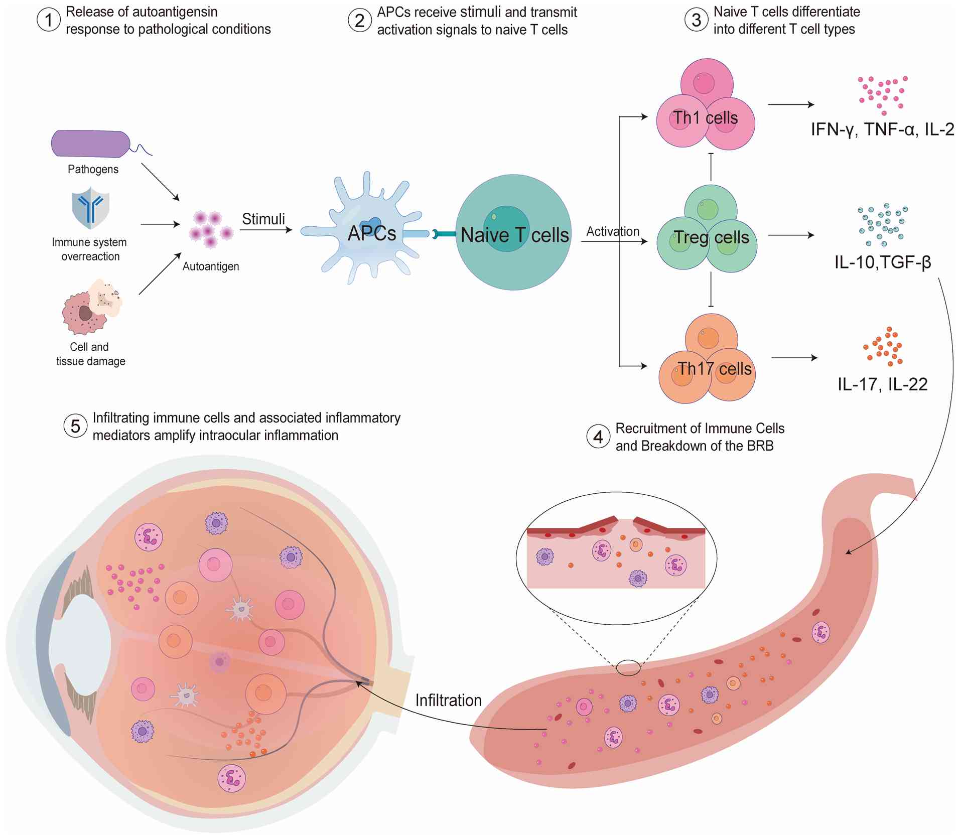 | Figure 1Schematic illustration of the
immunopathogenesis of uveitis. Environmental, infectious, or
autoimmune triggers promote the release and presentation of ocular
autoantigens by APCs. APCs activate naïve CD4+ T cells
through MHC-mediated antigen presentation, leading to their
differentiation into Th1, Th17 and Treg cell subsets. Activated Th1
and Th17 cells produce pro-inflammatory cytokines, IFN-γ, IL-17 and
TNF-α, which disrupt the BRB and promote the recruitment of
additional immune cells into ocular tissues. The resulting
inflammatory cascade amplifies retinal and choroidal inflammation,
ultimately contributing to tissue damage and visual impairment.
APC, antigen-presenting cell; MHC, major histocompatibility
complex; Th, T helper cell; Treg, regulatory T cell; IFN-γ,
interferon-γ; IL-17, interleukin-17; TNF-α, tumor necrosis
factor-α; BRB, blood-retinal barrier. |
Leveraging these immunological insights, researchers
have established experimental uveitis models. In the classic
experimental autoimmune uveitis (EAU) model, mice are immunized
with retinal antigens (including IRBP or its peptide fragments) in
combination with complete Freund's adjuvant, with pertussis toxin
administered to enhance the inflammatory response (26). The EAU model serves as a critical
research platform for investigating the immune mechanisms of
uveitis, providing insights into the role of T cells in disease
pathogenesis (27,28). In addition to EAU,
endotoxin-induced uveitis (EIU) is another widely used experimental
model for studying ocular inflammation. Unlike EAU, which is
primarily driven by antigen-specific adaptive immune responses, EIU
is induced by systemic administration of lipopolysaccharide (LPS)
and predominantly reflects innate immune activation. The model is
characterized by a rapid onset of inflammation, breakdown of the
blood-aqueous barrier, infiltration of neutrophils and macrophages,
and increased production of pro-inflammatory cytokines such as
TNF-α, IL-1β and IL-6. Due to its simplicity, reproducibility and
acute inflammatory nature, EIU has been extensively employed to
investigate early inflammatory responses, innate immune signaling
pathways, and the therapeutic effects of anti-inflammatory agents
(29). Together, EAU and EIU
provide complementary platforms for elucidating the complex
immunopathological mechanisms underlying uveitis. Immunotherapy for
uveitis primarily includes glucocorticoids, immunosuppressants and
biologics. Glucocorticoids are the first-line anti-inflammatory
agents and can be administered either locally or systemically.
Immunosuppressants modulate pathogenic immune responses and are a
cornerstone in the management of non-infectious uveitis,
particularly in severe, chronic, or vision-threatening cases and in
specific entities such as BD or VKH. Major classes include
antimetabolites (methotrexate, azathioprine), calcineurin
inhibitors (cyclosporine A, tacrolimus), and alkylating agents
(cyclophosphamide). These agents serve as essential steroid-sparing
therapies for long-term inflammation control and, in selected
conditions, may be used as first-line treatment. Additionally,
biologics, including TNF-α inhibitors, including adalimumab, and
IL-6 inhibitors, such as tocilizumab, play a crucial role in
managing refractory autoimmune uveitis (30-32). Although significant progress has
been made in the immune pathogenesis of uveitis in recent years and
new immune-targeted drugs have been introduced, the disease still
faces numerous challenges in clinical diagnosis and treatment,
especially in early identification, efficacy evaluation, and the
formulation of individualized treatment strategies (33).
Omics in ophthalmology research
To understand the mechanisms by which multi-omics
technologies uncover the pathogenesis of uveitis, aid in
classification, and support personalized treatment, it is important
to examine both their technical foundations and sample
requirements. In the present review, first, the principles,
focuses, and typical biomedical applications of major omics
approaches are introduced, followed by a discussion of the sample
types and acquisition methods relevant to ophthalmic research. This
dual perspective will provide a basis for exploring the role of
multi-omics in uveitis.
Overview of omics approaches
Omics refers to the systematic acquisition, analysis
and interpretation of large-scale biological data on a specific
class of biomolecules to elucidate the structure, function, and
regulatory mechanisms of complex life systems (34,35). Omics technologies serve as
powerful platforms in modern biomedical research, enabling
comprehensive and high-throughput profiling of biological molecules
across multiple regulatory layers. The foundational concept
underlying most omics approaches is the central dogma of molecular
biology, which describes the directional flow of genetic
information from DNA to RNA to protein (36). Based on this framework, several
major omics disciplines have been developed. Genomics investigates
the complete structure, variation and function of the genome,
helping to identify disease-associated genetic variants and
susceptibility loci (37).
Transcriptomics focuses on the entire set of RNA transcripts,
including coding and non-coding RNAs (ncRNAs), to examine gene
activity in different tissues or disease states (38). Proteomics analyzes protein
abundance, modifications and interactions, offering insights into
signaling pathways and immune responses (38). Metabolomics assesses
low-molecular-weight metabolites, which reflect downstream cellular
processes and metabolic changes associated with disease progression
(39).
However, with advances in molecular biology, the
central dogma has been increasingly expanded and refined. For
example, retroviruses utilize reverse transcriptase to convert RNA
back into DNA, challenging the unidirectional nature of information
flow. Furthermore, an increasing number of ncRNAs, including
microRNAs (miRNAs) and long ncRNAs (lncRNAs), have been found to
play critical roles in gene regulation without encoding proteins.
In addition, processes including RNA splicing, alternative
splicing, and epigenetic modifications are now recognized as
essential contributors to protein synthesis and functional
regulation (40). Epigenomics,
in particular, investigates heritable modifications to DNA or
chromatin that influence gene expression without altering the
underlying DNA sequence (41).
In addition to omics disciplines grounded in the
central dogma, the field has expanded to include non-central
dogma-based omics. For instance, radiomics extracts
high-dimensional quantitative features from medical imaging
modalities, including optical coherence tomography (OCT) and
magnetic resonance imaging (MRI), enabling the identification of
subtle patterns associated with disease phenotypes and therapeutic
responses (42,43). Microbiomics characterizes
microbial communities in environments including the gut, skin or
ocular surface, offering novel insights into host-microbe
interactions and their roles in immune regulation (44).
These diverse omics layers provide complementary
perspectives on disease mechanisms. When integrated, multi-omics
approaches allow for a more complete understanding of complex
conditions, including uveitis, improving biomarker discovery,
therapeutic target identification, and precision medicine
strategies (Fig. 2). Despite
challenges related to sample availability, data integration and
analytical complexity, the continued development and application of
omics technologies are expected to transform uveitis research and
clinical care (45,46). In the subsequent sections, each
omics discipline is systematically presented, and its specific
contributions to understanding the pathogenesis, diagnosis and
treatment of uveitis are explored.
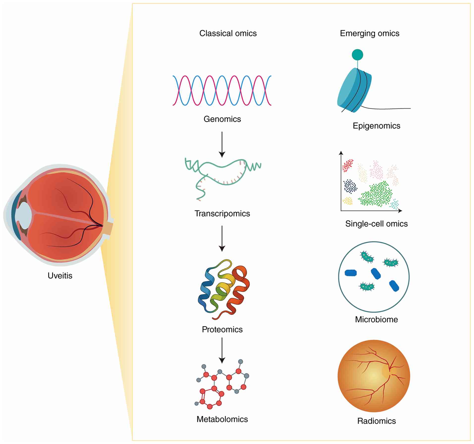 | Figure 2Overview of classical and emerging
omics approaches in uveitis research. The figure summarizes the
major omics technologies currently applied in uveitis research and
their corresponding biological information layers. Classical omics
approaches include genomics, which investigates genetic variants
and disease susceptibility; transcriptomics, which characterizes
gene expression patterns; proteomics, which profiles protein
abundance and signaling pathways; and metabolomics, which analyzes
small-molecule metabolites associated with cellular and immune
functions. Emerging omics technologies include epigenomics,
single-cell omics, spatial omics, lipidomics, microbiome analysis,
and radiomics. These approaches provide complementary insights into
gene regulation, cellular heterogeneity, spatial organization,
host-microbe interactions, metabolic alterations, and
imaging-derived biomarkers. Together, these technologies facilitate
a comprehensive understanding of uveitis pathogenesis, biomarker
discovery, disease stratification, therapeutic target
identification and precision medicine. |
Sample sources for omics study in
uveitis
Biomarkers in ocular and systemic fluids serve as
important indicators of pathological processes associated with
uveitis, reflecting their critical role in disease development
(Fig. 3). Ocular samples include
the retina, choroid, aqueous humor, vitreous, tears and conjunctiva
(47). While retinal and uveal
tissues are most directly relevant to the disease, obtaining such
samples from humans is not feasible. Thus, related omics studies
typically rely on animal models or postmortem donor tissues to
explore disease pathogenesis. In clinical practice, aqueous humor
and vitreous samples are commonly used for omics analyses. Aqueous
humor paracentesis is a well-established and generally safe
procedure when performed under sterile conditions, whereas vitreous
sampling is more invasive and usually reserved for selected cases.
In both settings, sample volumes are carefully limited to minimize
procedure-related complications, including hypotony (48). Optimizing the collection,
storage, processing, and analysis of these limited samples is
essential to maximize their utility (49). Tears and conjunctival samples are
more easily accessible and primarily reflect anterior segment
pathology. However, due to anatomical proximity, intraocular
inflammation can influence tear composition, making tears a
promising, non-invasive source of biomarkers for active intraocular
inflammation (50,51).
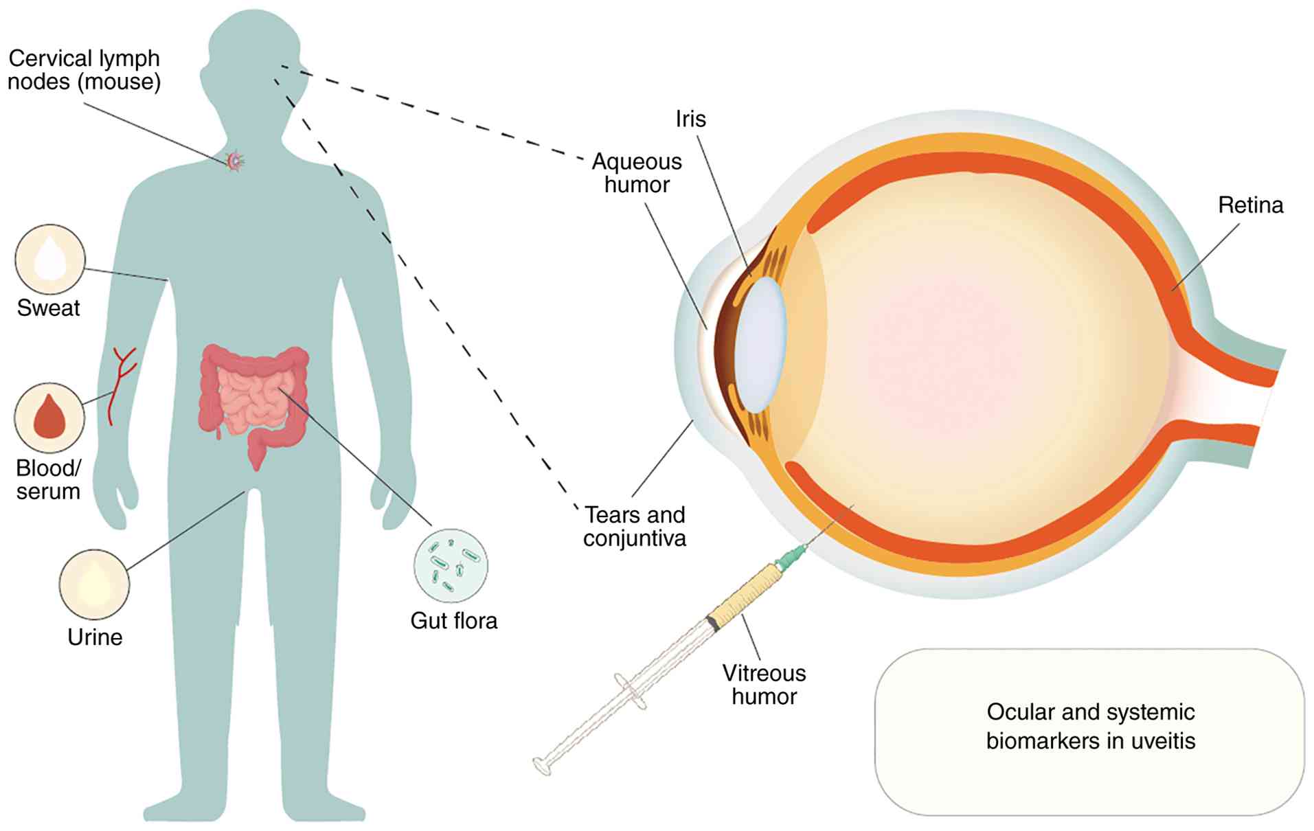 | Figure 3Biological sample sources used in
multi-omics studies of uveitis. Systemic and ocular biological
samples are widely used to investigate both local and systemic
molecular alterations associated with uveitis pathogenesis.
Systemic sample sources include peripheral blood, serum, urine, gut
microbiota, and cervical lymph nodes in animal models, providing
information on systemic immune responses and host-microbiome
interactions. Ocular sample sources include tears, conjunctival
swabs, aqueous humor, vitreous humor obtained through aqueous or
vitreous paracentesis, as well as retinal and iris tissues, which
more directly reflect the local inflammatory microenvironment. Each
sample type presents distinct advantages and limitations.
Peripheral blood and tears are readily accessible and suitable for
longitudinal monitoring but may not fully capture intraocular
disease activity. By contrast, aqueous humor and vitreous humor
provide more disease-relevant molecular information but are
obtained through invasive procedures and are often limited by small
sample volumes. Retinal and iris tissues offer valuable mechanistic
insights but are rarely available in clinical settings. Therefore,
the selection of biological samples represents an important
consideration in study design and influences the translational
applicability of omics-based biomarkers in uveitis. |
Beyond ocular samples, researchers are investigating
systemic biomarkers in blood, lymph nodes, feces, sweat, and other
body fluids using omics approaches. Blood offers several
advantages, including large sample volumes, accessibility,
standardized collection protocols, established analytical
techniques, and the opportunity for repeated measurements (10). Lymph nodes are crucial in immune
cell interactions and in initiating autoimmune responses. Cervical
draining lymph nodes (CDLN), as the primary drainage nodes of the
central nervous system (CNS), play a crucial role in the
pathogenesis of autoimmune diseases of the CNS, effectively
receiving macromolecules and immune cells from the CNS (52). The gut microbiome, assessed
through fecal analysis, has also been increasingly recognized for
its influence on diseases beyond the intestine, including ocular
and systemic autoimmune conditions (53).
Omics in uveitis
The present review summarizes recent advances in
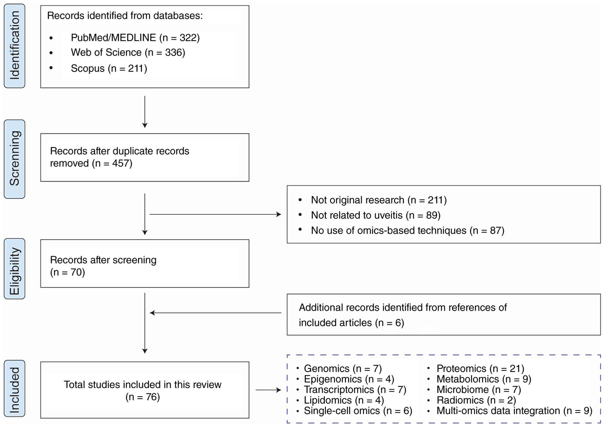
omics research in uveitis. To ensure comprehensive coverage of the
literature, a systematic search was conducted in PubMed (https://pubmed.ncbi.nlm.nih.gov/), Web of Science
(https://clarivate.com/academia-government/scientific-and-academic-research/research-discovery-and-referencing/web-of-science/)
and Scopus (https://www.scopus.com) for articles
published. The search strategy combined relevant keywords and
Medical Subject Headings terms, including 'uveitis', 'multi-omics',
'genomics', 'epigenomics', 'transcriptomics', 'single-cell
sequencing', 'spatial omics', 'proteomics', 'metabolomics',
'lipidomics', 'microbiome', 'radiomics' and 'precision medicine'.
Reference lists of relevant articles were also screened to identify
additional studies. Priority was given to original research
articles, high-quality reviews, and recent publications focusing on
autoimmune and infectious uveitis. Studies lacking sufficient
methodological details, those not directly related to ocular
inflammatory diseases, or publications with limited relevance to
the scope of the present review were excluded (Fig. 4). The selected literature was
synthesized narratively to provide an overview of current omics
technologies, key biological insights, and emerging applications in
precision medicine for uveitis.
Genomics
The exact pathogenesis of uveitis remains unclear.
However, it is widely acknowledged that a combination of genetic or
epigenetic predispositions, environmental risk factors, and
dysregulated immune responses contributes to disease development,
particularly in autoimmune uveitis, including acute anterior
uveitis (AAU), BD, VKH, BSCR, and sarcoid uveitis (54). In addition, several monogenic
autoinflammatory disorders, including but not limited to Blau
syndrome, haploinsufficiency of A20 (HA20), and familial
Mediterranean fever, have been identified as rare but important
causes of uveitis (55,56).
Genomics is the comprehensive study of the entire
genetic material of an organism, encompassing the structure,
function, evolution, mapping and editing of genomes. The term
genomics was first coined by geneticist Thomas H. Roderick in 1986
and gained prominence with advances in molecular biology and the
landmark completion of the Human Genome Project (37). Since then, the field has expanded
rapidly, driven by the advent of high-throughput sequencing
technologies and the development of genome-wide association studies
(GWAS), which have enabled the identification of genetic variants
associated with complex traits and diseases (57). Genomics has broad applications
ranging from the identification of disease-associated genetic
variations, including single-nucleotide polymorphisms (SNPs) and
copy number variations (CNVs), to advancements in personalized
medicine, biotechnology and evolutionary biology (58,59). SNPs are point mutations involving
the substitution of a single base at a specific genomic position,
typically occurring at a frequency of >5% in the population.
SNPs account for >90% of human genetic variation and are a major
determinant of individual differences in drug metabolism and
therapeutic response. CNVs refer to structural alterations in the
genome involving deletions, insertions, duplications, or inversions
of DNA segments ranging from 1 kilobase to 3 megabases in length.
These variations can influence gene dosage and contribute to both
normal phenotypic diversity and disease susceptibility (60). As sequencing becomes faster and
cheaper, genomics continues to deepen the current understanding of
life at a molecular level and revolutionize biomedical research
(61).
Human leukocyte antigen (HLA)
polymorphisms in uveitis
Early genetic studies of uveitis primarily focused
on associations with HLA polymorphisms (54,62). HLAs are a group of highly
polymorphic genes located on the short arm of chromosome 6,
encoding glycoprotein allo-antigens involved in immune regulation.
Identification of specific HLA alleles has proven valuable not only
for disease diagnosis, risk prediction and population-based genetic
epidemiology, but also for uncovering the potential immunogenetic
basis of uveitis (63). Notably,
these associations often exhibit ethnic specificity, underscoring
the importance of population-based studies in understanding
HLA-disease correlations (64).
One of the most well-established examples is AAU, which shows a
strong association with HLA-B27, particularly among patients with
ankylosing spondylitis (AS) (65). In BD, HLA-B51 has been
consistently linked to disease susceptibility across multiple
populations, especially in those of Mediterranean and East Asian
descent (66). VKH syndrome is
associated with HLA-DR4 and specific DRB1/DQA1 haplotypes,
implicating a T cell-mediated autoimmune response; these
associations have been observed in various Asian populations
(67,68). Among all uveitis subtypes, BSCR
demonstrates the strongest HLA linkage, with >95% of patients
carrying the HLA-A29 allele (69).
Polygenic risk score (PRS) in
uveitis
Recent GWAS conducted in China, Japan, Turkey, and
other countries have expanded the current understanding of the
genetic basis of uveitis. These studies have shown that, in
addition to HLA gene variants, polymorphisms in several non-HLA
genes, including IL10, IL23R/IL12RB2, and signal transducer and
activator of transcription 4, are also associated with uveitis,
further supporting the hypothesis that genetic factors contribute
to disease susceptibility (70-72). Hou et al (54,62) provided a comprehensive summary of
these findings. These insights suggest that, beyond uveitis caused
by rare monogenic autoinflammatory disorders with defined
etiologies, including Blau syndrome and HA20, common forms of
non-infectious uveitis may involve multiple genetic susceptibility
loci. In this context, the PRS model has emerged as a valuable
tool, aggregating the effects of numerous genetic variants across
the genome into a single risk score. PRS allows for quantitative
assessment of the genetic susceptibility of an individual and can
be applied to elucidate disease pathogenesis, stratify population
risk, identify disease subtypes, and support early clinical
intervention. Previous studies have supported the potential
application of PRS in uveitis. A large GWAS in the UK involving
2,752 European participants with AS and AAU, and 3,836 patients
with AS without AAU, identified a genome-wide significant
association at the rs9378248 locus near HLA-B
(P=2.69×10⁻⁸; OR=0.78). In addition, several suggestive
loci were reported, including established AS-associated genes
(ERAP1 and NOS2) and novel candidates (MERTK, KIFAP3, CLCN7 and
ACAA2). Notably, the estimated SNP-based heritability of AS with
AAU was 0.7, significantly higher than the 0.5 observed in AS
without uveitis, indicating that PRS may be particularly effective
in identifying high-risk individuals (72). Similarly, a GWAS conducted in
Taiwan through the Taiwan Precision Medicine Program analyzed 468
patients with AS, including 90 with non-infectious anterior
uveitis. This study identified two novel SNPs, rs1736952 and
rs17354984 (P<5×10⁻⁸), and developed an optimal PRS
model incorporating 19 SNPs, achieving an area under the curve
(AUC) of 0.907, demonstrating strong discriminatory power. Pathway
enrichment analysis revealed that these genetic variants were
associated with key biological processes, including antigen
presentation, IFN signaling, immune regulation, ciliary movement
and neurodegeneration (73).
Despite these promising findings, several important
limitations currently restrict the broader clinical application of
genomics in uveitis. First, numerous existing GWAS studies have
been conducted within relatively homogeneous ethnic populations,
which limits the generalizability and universal applicability of
these genetic risk prediction models across diverse populations
(73). Furthermore, although
common susceptibility variants identified through GWAS have
provided valuable insights into immune-related pathways involved in
uveitis, they remain insufficient to explain rare monogenic and
autoinflammatory forms of the disease, including Blau syndrome and
HA20. Therefore, larger multi-center and multi-ethnic studies,
together with standardized analytical pipelines and external
validation cohorts, are still required to improve the robustness
and translational applicability of genomics-based precision
medicine approaches in uveitis.
Epigenomics
Several mechanisms underlying uveitis cannot be
fully explained by genetic variation alone, underscoring the need
for further research into epigenetic modifications to address the
gap in heritability (74).
Epigenetics investigates reversible mechanisms that regulate gene
expression without changing the underlying DNA sequence. It serves
as a vital link between genes and the environment, enabling the
same gene to display distinct expression patterns across tissues,
developmental stages or environmental conditions (75). The primary mechanisms of
epigenetic regulation include DNA methylation and histone
modifications. Additionally, ncRNAs, including miRNAs, lncRNAs and
circular RNAs, regulate gene expression at the post-transcriptional
level by modulating mRNA stability and translation efficiency
(76,77).
DNA methylation
DNA methylation, a stable and heritable epigenetic
mark, typically occurs at cytosine residues in CpG dinucleotides,
forming 5-methylcytosine. This modification plays a key role in
regulating gene expression, maintaining genomic stability and
guiding cellular differentiation. Promoter hypermethylation is
often linked to gene silencing, whereas global hypomethylation can
contribute to genomic instability (78). In a study by Hughes et al
(79), 383 differentially
methylated CpG sites were identified in monocytes and 125 in
CD4+ T cells from patients with BD compared with healthy
controls. These changes were enriched in genes related to
cytoskeletal regulation, implicating altered DNA methylation in BD
pathogenesis. Notably, a number of these aberrant methylation
patterns were reversed following disease remission, suggesting
their dynamic and treatment-responsive nature (79).
ncRNAs
miRNAs are small (18-25 nucleotides), endogenous
ncRNAs that regulate gene expression post-transcriptionally by
binding to target mRNAs, thereby inhibiting translation or
promoting degradation. Due to their stability in body fluids,
resistance to degradation, and ease of detection, miRNAs hold
promise as non-invasive biomarkers for autoimmune diseases
(80). In a study involving
serum samples from 10 patients with BD, 17 with sarcoidosis, 13
with VKH disease and 11 healthy controls, microarray analysis
revealed widespread miRNA dysregulation. Specifically, 281
upregulated and 137 downregulated miRNAs were identified in BD, 35
upregulated and 86 downregulated in sarcoidosis, and 153
upregulated and 35 downregulated in VKH. A number of these miRNAs
were associated with mitogen-activated protein kinase (MAPK)
signaling and inflammatory cytokine pathways. Notably, miR-4708-3p
(BD), miR-4323 (sarcoidosis) and let-7g-3p (VKH) emerged as top
disease-specific biomarkers. A machine learning-based miRNA panel
further demonstrated diagnostic potential across these uveitis
subtypes (81). In addition to
miRNAs, lncRNAs play crucial roles in autoimmune inflammation.
Bioinformatics analysis of peripheral blood from patients with AS,
BD and sarcoidosis by Lu and Lu (82) identified dysregulated lncRNAs and
mRNAs, with XIST and MIAT as potential hub lncRNAs and FCGBP,
CD247, CTSW, AES, NCR3, TIGIT, CASP5, DUSP2 and TBX21 as hub mRNAs.
These genes were enriched in MAPK signaling and inflammatory
cytokine pathways, suggesting that aberrant lncRNA expression may
contribute to noninfectious uveitis and could serve as potential
biomarkers or therapeutic targets (82).
Histone modifications
Histone modifications are among the most extensively
studied epigenetic mechanisms and play a central role in regulating
chromatin structure and gene transcription. Histone proteins form
the core of nucleosomes around which DNA is wrapped, and
post-translational modifications occurring on histone tails can
alter chromatin accessibility and influence the recruitment of
transcription factors and regulatory complexes. Common histone
modifications include acetylation, methylation, phosphorylation,
ubiquitination, and the recently identified lactylation (83). In general, histone acetylation,
such as H3K27ac and H3K18ac, is associated with transcriptional
activation and open chromatin states, whereas certain methylation
marks, including H3K27me3 and H3K9me3, are linked to gene
repression and chromatin compaction (84). Through these mechanisms, histone
modifications dynamically regulate immune cell differentiation,
inflammatory signaling pathways, and cytokine production. Emerging
evidence suggests that aberrant histone modifications contribute to
immune dysregulation (85) and
ocular inflammation (86). Using
data-independent acquisition-based lactyl-proteomics, researchers
systematically profiled the lactylome in acute uveitis and
identified widespread lactylation of nuclear proteins. Among these
modifications, histone H3 lysine 18 lactylation (H3K18la) was found
to be closely associated with chromatin decondensation during
neutrophil extracellular trap (NET) formation. Combined with
metabolic analyses and functional validation experiments, the study
further demonstrated that HIF-1α-driven glycolytic reprogramming
increased intracellular lactate production, thereby promoting
H3K18la modification and NET release. The present study not only
identifies histone lactylation as a novel epigenetic regulator in
uveitis but also highlights the value of integrating
lactyl-proteomics, epigenomics, and immuno-metabolic profiling to
elucidate the molecular mechanisms underlying ocular inflammation
(87).
Chromatin accessibility
Chromatin accessibility refers to the degree to
which genomic DNA is accessible to transcription factors, RNA
polymerases, and other regulatory proteins. As a fundamental
component of epigenetic regulation, chromatin accessibility
reflects the dynamic organization of chromatin and plays a critical
role in controlling gene expression. Regions of open chromatin are
generally associated with active promoters, enhancers, and
transcription factor binding sites, whereas closed chromatin
regions are typically linked to transcriptional repression
(88). Alterations in chromatin
accessibility can influence immune cell differentiation, activation
and inflammatory responses, thereby contributing to the
pathogenesis of immune-mediated diseases. Recent advances in
high-throughput sequencing technologies have enabled genome-wide
profiling of chromatin accessibility. Among these methods, Assay
for Transposase-Accessible Chromatin using Sequencing (ATAC-seq)
has become the most widely used approach due to its high
sensitivity and low sample requirements (89). More recently, single-cell
ATAC-seq (scATAC-seq) has allowed the characterization of chromatin
landscapes at single-cell resolution, providing unprecedented
insights into cellular heterogeneity and gene regulatory networks.
By identifying accessible regulatory elements and transcription
factor binding motifs, chromatin accessibility analyses complement
transcriptomic studies and facilitate the reconstruction of
molecular pathways underlying disease progression. Notably, the
present literature search identified two studies that integrated
single-cell ATAC-seq with single-cell RNA sequencing (scRNA-seq) to
simultaneously characterize chromatin accessibility and
transcriptional programs in uveitis-related immune cells. Given
their relevance to multi-layer molecular profiling and data
integration, these studies will be discussed in detail in Section
5, 'Multi-Omics Data Integration'.
Despite the growing promise of epigenomics, several
important limitations remain. Numerous current studies are based on
peripheral blood samples with relatively small cohort sizes, and
epigenetic signatures may vary substantially across tissues,
disease stages and treatment conditions. In addition, the dynamic
and reversible nature of epigenetic modifications may complicate
reproducibility and longitudinal interpretation (90). Therefore, larger multi-center
studies integrating epigenomics with transcriptomics, proteomics,
and clinical phenotyping are still needed to improve the
translational applicability of epigenetic biomarkers in
uveitis.
Transcriptomics
Transcriptomics refers to the study of all RNA
transcripts within a particular species, including mRNA and ncRNA.
It examines RNA levels across the genome from a qualitative
perspective, including identifying present transcripts, novel
splicing events and RNA editing sites, and a quantitative
perspective, including measuring transcript abundance (91). Unlike the genome, which
statically presents the genetic blueprint of an organism, the
transcriptome is dynamic and varies across different tissues,
developmental stages and disease states. Therefore, transcriptional
analysis plays a crucial role in elucidating genome structure and
function, decoding the genetic networks underlying disease, and
identifying sensitive molecular biomarkers for diseases, drugs and
pathogens (92). The field of
transcriptomics has evolved notably with the development of new
technologies that redefine possibilities approximately every
decade, rendering previous methods largely obsolete. Currently, two
key technologies dominate: i) Microarrays, which quantify a
predefined set of transcripts; and ii) RNA sequencing (RNA-Seq),
which uses high-throughput sequencing to capture the complete range
of RNA transcripts (93,94).
Transcriptomic profiling of idiopathic
uveitis
In the study by Rosenbaum et al (95), RNA-Seq was used to analyze
peripheral blood gene expression profiles from patients with the
following four systemic non-infectious diseases associated with
uveitis: i) Axial spondyloarthritis; ii) inflammatory bowel
disease; iii) sarcoidosis; and iv) tubulointerstitial nephritis
with uveitis. The researchers conducted KEGG and GO pathway
analyses, which identified several shared pathways or GO terms
(P<0.0001). These pathways were primarily related to immune
response and/or infectious response. Based on the analysis, 119
transcripts were upregulated by at least 1.5-fold, while 61
transcripts were downregulated using the same criteria (False
discovery rate <0.05). The following 10 common gene transcripts
were identified: i) ICAM1; ii) IL15RA; iii) IL15; iv) IRF1; v)
IL10RB; vi) GSK3A; vii) TYK2; viii) MEF2A; ix) MEF2B; and x) MEF2D
(95). To explore whether gene
expression profiles could assist in investigating the etiology of
idiopathic uveitis, a classification algorithm was created using a
gradient boosting tree model with 5-fold cross-validation. The mRNA
from patients with idiopathic uveitis was analyzed to investigate
whether any gene expression patterns matched the systemic diseases
previously diagnosed. The gene expression profiling model achieved
an overall accuracy of 85% (P<0.001) in associating uveitis with
a diagnosable systemic disease. Although most patients with
idiopathic uveitis did not have these four systemic diseases, the
gene expression profiling helped reclassify 11 of the 38 patients
(96). This suggests that gene
expression profiling could play a notable role in understanding the
pathogenesis of idiopathic uveitis and may aid in the improvement
of diagnosis and treatment strategies.
Transcriptomic detection of intraocular
infections
RNA-seq offers notable advantages over traditional
targeted PCR testing for detecting intraocular infections, and with
technological advancements, its cost is no longer prohibitive
(97). PCR testing relies on a
predefined list of pathogens and can only detect specified
microorganisms, making it easy to miss atypical or unknown
infections. By contrast, RNA-seq is a hypothesis-free,
high-throughput technology that can identify all established and
potentially novel pathogens present in a sample in a single test,
offering broader diagnostic coverage (98). A study comparing RNA-seq and
targeted PCR testing in samples from 41 patients with suspected
ocular infections found that RNA-seq achieved complete positive
concordance with PCR (positive agreement rate, 100%; 95% confidence
interval, 78.5-100%) and a negative concordance rate of 92.6% (95%
confidence interval, 76.6-97.9%). Notably, RNA-seq also identified
four additional pathogens missed by PCR, including rubella virus
and Pithomyces chartarum, and these findings were confirmed
by orthogonal testing. The study concluded that RNA-seq can
accurately detect both common and rare pathogens in intraocular
fluid samples, expanding the diagnostic capabilities for ocular
infections (99). In the future,
further research is needed to establish its clinical utility
fully.
Transcriptomic insights from EAU
models
The retina is an ideal tissue for investigating the
underlying mechanisms of uveitis, as it directly reflects
pathological changes occurring during disease progression. However,
obtaining retinal tissue from human patients is impractical and
ethically challenging (100).
Therefore, animal models, including EAU, have become essential
tools for studying the pathogenesis of uveitis. EAU closely mimics
numerous key features of human non-infectious uveitis, including
immune-mediated retinal inflammation and tissue damage (101,102). With the advancement of
high-throughput RNA-Seq technologies, transcriptomic profiling of
retinal cells in EAU models has enabled researchers to
systematically explore gene expression changes during disease
progression. These studies have provided critical insights into the
molecular mechanisms underlying uveitis, including inflammatory
responses, immune activation, complement system involvement and
cellular stress pathways. The application of transcriptomics in EAU
thus provides a valuable platform for identifying novel therapeutic
targets and enhancing the current understanding of ocular
autoimmune diseases. The current studies utilizing transcriptomic
approaches to investigate gene expression changes in the retina
during EAU are summarized in Table
I.
 | Table ISummary of transcriptomic studies
investigating retinal and microglial gene expression changes during
uveitis. |
Table I
Summary of transcriptomic studies
investigating retinal and microglial gene expression changes during
uveitis.
| Authors, year | Species | Tissue/cell | Findings | (Refs.) |
|---|
| Busch et al,
2019 | Mouse | Retina | 478 DEGs were
identified (log |FC| >1), among which 406 genes were
upregulated, and 72 genes were downregulated in the EIU group.
GO/KEGG analysis showed that these genes were related to biological
processes, including inflammatory response, complement system
activation, fibrinolytic system changes, and cellular stress. | (212) |
| de Hoog et
al, 2019 | Mouse | Retina | 52 DEGs were
identified (log |FC| >1). 37 genes were upregulated, and 15
genes were downregulated in the EIU group compared with the DEX
treatment group. DEX treatment significantly suppressed the
RIG-I-like receptor signaling pathway as well as several immune-
and inflammation-related genes, including Ifit1, H2-T24, Mx2, and
Eif2ak2. | (213) |
| Eidet et al,
2019 | Mouse | Microglia | During the early
activation phase (4 h), 613 DEGs were identified. A total of 537
DEGs were observed at the peak of cell infiltration (18 h), whereas
no DEGs were detected at 2 weeks. C5AR1 was identified and
validated as a robust marker for distinguishing microglial subsets
during the LPS response. | (214) |
| Liang et al,
2020 | Mouse | Retina | Compared with the
TMP-treated group, 407 DEGs were identified in the untreated
control group (log |FC| > 1), of which 356 were upregulated, and
51 were downregulated. There were 12 upregulated gene ontology
terms enriched and 27 upregulated pathways. Seven DEGs were
validated using quantitative PCR, including inflammation-related,
complement system-related, and interferon-related genes. | (215) |
| Sepah et al,
2020 | Mouse | Retinal cells and
endothelial cells | RNA-Seq analysis of
total retinal cells revealed a predominant upregulation of genes
involved in antigen presentation and T cell activation during EAU.
Targeted transcriptome analysis of retinal endothelial cells
identified 82 genes that were differentially regulated during EAU
development. The protein expression of five of these genes
(Serpina3n, Lcn2, Ackr1, Lrg1, and Lamc3) was validated in cells
located within the BRB. | (216) |
| Eidet et al,
2021 | Zebrafish | Retina | Four gene clusters
were identified via enrichment analysis, which were related to
Toll-like receptor signaling pathway, cytokine-cytokine receptor
interaction, NOD-like receptor signaling pathway, and extracellular
matrix (ECM)-receptor interaction, respectively in EIU model. | (217) |
| Zheng et al,
2021 | Mouse | Retina | KEGG and Reactome
enrichment analysis of RNA-seq data revealed associations with
chemokine signaling pathways and cytokine-cytokine receptor
interactions in EAU model. | (218) |
Despite the substantial advances enabled by
transcriptomic technologies, several important limitations remain
in current uveitis transcriptomics research. First, numerous
transcriptomic studies rely heavily on EAU animal models, which,
although valuable, cannot fully recapitulate the complexity and
heterogeneity of human uveitis. Notable differences in immune
regulation, retinal structure and disease progression between
experimental models and human disease may limit translational
applicability. In addition, human intraocular samples remain
difficult to obtain due to ethical and technical constraints,
resulting in relatively small clinical cohort sizes and limited
validation studies. Transcriptomic profiles may also vary
substantially depending on sample source, including peripheral
blood, aqueous humor, vitreous fluid, tears and retinal tissue,
thereby complicating cross-study comparisons and biomarker
interpretation. Furthermore, technical factors such as batch
effects, RNA degradation, sequencing depth and differences in
bioinformatic pipelines may influence data reproducibility and
contribute to inconsistencies among studies (103). Therefore, larger multi-center
studies integrating transcriptomics with proteomics, metabolomics
and clinical phenotyping are needed to improve the robustness,
reproducibility and clinical translational potential of
transcriptomic biomarkers in uveitis.
Single-cell omics
Single-cell omics aims to profile molecular
information, including genes, transcripts, proteins, metabolites
and epigenetic modifications at the resolution of individual cells,
providing deep insights into cellular functional states,
heterogeneity and developmental trajectories (104). This approach represents a
notable advancement in spatial resolution over traditional bulk
omics technologies. By integrating multidimensional data across
various omics layers, single-cell sequencing enables precise cell
clustering, enhances analytical resolution, and effectively
addresses intercellular heterogeneity (105). Among these technologies,
scRNA-seq is the earliest and most widely adopted method, first
introduced in 2009. Although single-cell approaches have since
expanded to other omics layers and integrative frameworks (106), scRNA-seq remains the primary
tool applied in uveitis research to date, with no reported studies
utilizing other types of single-cell omics. This highlights the
foundational role of transcriptomic profiling in current
single-cell investigations of ocular inflammation. The following
section focuses on the application of scRNA-seq in uveitis.
As a high-throughput technique, scRNA-seq enables
comprehensive analysis of gene expression at the single-cell level,
offering a powerful means to uncover cellular heterogeneity and to
delineate distinct cell types and their dynamic changes (107). Compared with bulk RNA-seq,
which averages gene expression across cell populations and obscures
individual cell variations, scRNA-seq provides a more granular
view. This is particularly important in complex tissues, including
the retina, where multiple cell types coexist in varied
physiological states. The advantages of scRNA-seq extend beyond
structural characterization. It allows for the identification of
transcriptional changes within specific cell subpopulations under
disease conditions or therapeutic interventions, thereby deepening
the current understanding of disease mechanisms and drug responses.
Additionally, it supports an unbiased evaluation of tissue
composition by capturing shifts in the relative abundance of all
cell types under different pathological states (108,109). These features underscore the
value of scRNA-seq as a key tool for dissecting the cellular basis
of uveitis and related ocular disorders.
Applications of scRNA-seq in EAU
models
Heng et al (110) were the first to apply scRNA-seq
to the retina of Aire-deficient mice, a spontaneous uveitis model
characterized by impaired central tolerance due to the failure to
express self-antigens in the thymus. These mice develop chronic and
progressive autoimmune uveitis. The study revealed a significant
increase in immune cell infiltration in the retina of
Aire-deficient mice compared with controls, with T cells, NK cells
and monocyte-derived cells being the predominant populations,
alongside smaller numbers of B cells and plasma cells (PCs).
Further classification of T cells identified Th1 cells as the
principal effector subset. Notably, the Th1 population could be
divided into two distinct clusters, with one expressing Il10 and
the other expressing Cd40lg (CD40 ligand), representing regulatory
(Il10+) and activated (Cd40lg+) states,
respectively. These findings suggest a self-regulatory mechanism
within the Th1 compartment. In addition, enrichment analysis
demonstrated that IFN-γ-responsive genes were upregulated across
all major resident retinal cell types, indicating a widespread
transcriptional response to IFN-γ during the autoimmune process.
Furthermore, in the EAU mouse model, researchers performed clinical
and histopathological assessments at days 7, 14, 21 and 28, in
parallel with scRNA-seq of retinal tissues. Day 14 was identified
as the peak of inflammation and disease severity, corresponding
with the highest number of differentially expressed genes (DEGs).
At this stage, microglia were markedly elevated, comprising 73.9%
of all immune cells. Inflammatory-associated microglia showed
significant enrichment of DEGs involved in antigen processing and
presentation, as well as responses to type II IFN (111). In the study by Quinn et
al (112), scRNA-seq of the
EAU model revealed a widespread upregulation of IFN-γ and IFN-α
response genes. Furthermore, Müller glia and retinal pigment
epithelial cells were found to upregulate the expression of
chemokines, complement factors, leukocyte adhesion molecules and
MHC class II molecules, highlighting their active roles in immune
cell recruitment and antigen presentation both within and beyond
the BRB (112). The study by Li
et al (113) focused on
ocular-infiltrating PCs and found that these cells expressed
multiple pro-inflammatory factors, particularly TNF-α, which
contributed to the induction of α-synuclein-expressing microglia.
Further subtyping of the heterogeneous PC population revealed that
MUC1+ PCs constituted the dominant pathogenic subset.
These cells secreted various cytokines, exhibited long-lived
properties, and produced IgG and IgM antibodies, thereby sustaining
inflammation and contributing to prolonged disease progression.
Clinical translation and future
perspectives of single-cell omics in uveitis
A recent study attempted to classify uveitis using
single-cell omics approaches, which applied scRNA-seq combined with
immune receptor profiling to ocular fluid samples from 23 patients
with various clinical forms of uveitis. The analysis revealed
subtype-specific immune signatures, including HLA-B27-associated
AAU, which was characterized by an enrichment of myeloid cells and
activation of innate immune pathways, and chronic uveitis, which
showed a predominance of adaptive immune cells and related
transcriptional programs. Furthermore, the observation of clonal
expansion among T cells in some patients suggests antigen-driven
pathogenesis. These findings highlight the potential of single-cell
omics in uncovering immune endotypes and improving the molecular
classification of uveitis, paving the way for personalized
therapeutic strategies (114).
In addition, single-cell sequencing holds notable promise for drug
development in uveitis. A single-cell perspective is essential for
advancing the current understanding of cellular and molecular
targets, thereby improving the identification and prioritization of
therapeutically effective candidates. The integration of
single-cell protocols with advanced multiplexing strategies further
enhances the scalability and resolution of these assays, enabling
more precise and comprehensive drug screening (115). Li et al (52) investigated the effects of IL-38
on immune cells in the CDLN of EAU mice using single-cell analysis.
Their findings revealed that IL-38 attenuated the expansion of
effector T cells and the decline of Tregs, while also preventing
the upregulation of numerous DEGs, particularly those associated
with the IL-17 signaling and Th17 differentiation pathways, both of
which are known to play pivotal roles in the pathogenesis of
uveitis.
Spatial omics
Single-cell sequencing has played a pivotal role in
providing high-resolution insights at the individual cell level. By
enabling the detection of cellular heterogeneity, single-cell
approaches allow detailed characterization of cellular behaviors,
molecular mechanisms, and intercellular interactions. The high
resolution of these technologies has greatly facilitated
large-scale exploration and characterization of cellular diversity.
However, despite these advantages, conventional single-cell
sequencing often fails to preserve critical spatial information
within tissues, resulting in the loss of important spatial context
(116). To overcome this
limitation, spatial multi-omics has emerged as a transformative
technology that enables precise spatial localization of cells and
quantitative mapping of gene expression within intact tissue
architecture (117).
Compared with conventional dissociative single-cell
sequencing approaches, spatial omics preserves tissue morphology
and regional cellular organization, thereby enabling simultaneous
characterization of molecular alterations and their anatomical
localization within ocular tissues. This advantage is particularly
important in uveitis, where BRB disruption, inflammatory cell
infiltration and retinal/choroidal immune microenvironment
remodeling often exhibit strong spatial heterogeneity (118). Although only a limited number
of spatial omics studies have been reported in uveitis to date,
recent advances have enabled the integration of spatial
transcriptomics with scRNA-seq. This combined approach preserves
spatial context while providing high-resolution cellular
characterization, allowing a more comprehensive understanding of
tissue architecture, immune-cell localization, and intercellular
interactions within inflamed ocular tissues. A representative study
that applies this strategy to uveitis is discussed in the
'Multi-Omics Data Integration' section.
Despite its substantial promise, several challenges
currently limit the widespread application of spatial omics in
uveitis research, including limited ocular tissue availability,
high technical complexity, elevated costs, and difficulties in
computational integration and data interpretation (119,120). Nevertheless, as spatial
transcriptomics and spatial proteomics technologies continue to
evolve, their integration with single-cell sequencing, proteomics
and imaging modalities is expected to provide deeper mechanistic
insights into ocular inflammation and promote the development of
precision medicine strategies for uveitis.
Proteomics
Proteomics is the study of protein composition,
structure, function and interactions within organisms, and it plays
a vital role in elucidating biological processes and disease
mechanisms (38). Proteomics
encompasses the complete workflow of protein separation,
identification, quantification and analysis of post-translational
modifications. Modern approaches rely on high-performance liquid
chromatography coupled with mass spectrometry (MS) for
high-resolution, high-throughput protein analysis (121). Quantitative strategies include
stable isotope labeling methods, such as SILAC, iTRAQ, TMT and
label-free techniques, enabling comparative analysis of protein
expression across conditions. Post-translational modifications,
including phosphorylation and ubiquitination, are analyzed via
enrichment methods and high-resolution MS. Combined with systems
biology and bioinformatics, proteomics provides a powerful platform
for deciphering complex protein networks and identifying
disease-related biomarkers (122,123).
Proteomics studies can be broadly categorized based
on their objectives into assay-based (analytical) and
discovery-based approaches (124). Assay-based studies focus on the
quantification of a predefined set of proteins or peptides and are
particularly advantageous for investigating specific classes of
proteins. For instance, one study isolated mitochondrial components
from the retina and utilized two-dimensional difference gel
electrophoresis to examine changes in mitochondrial protein levels
in response to oxidative stress during the early stage of EAU
(125). Protein spots with
differential expression were excised and analyzed using
matrix-assisted laser desorption/ionization time-of-flight MS for
peptide identification. The findings revealed the presence of
mitochondrial-specific oxidative stress-related proteins and a
downregulation of ATP synthase, providing early evidence of
stress-induced retinal damage in EAU.
Discovery-based proteomics aims to analyze a broad,
unbiased proteome to identify novel biomarkers or elucidate
underlying biological pathways and has been widely applied in the
study of uveitis (126). In the
EAU model, researchers have examined a variety of samples,
including plasma and granulocytes, to uncover disease-related
targets. Guo et al (127) identified 62 upregulated and 106
downregulated proteins in the plasma of EAU rats using label-free
liquid chromatography-tandem MS (LC-MS/MS). Notably, plasma levels
of complement component 3 increased from 92.32 μg/ml in
saline-treated rats to 168.92 μg/ml in EAU rats, while
levels of IL-1 receptor accessory protein decreased from 1,120.97
to 798.39 pg/ml. In another study, primary granulocytes from
healthy and equine recurrent uveitis (ERU) horses were stimulated
with IL-8, and their responses were analyzed using differential
proteomics. This revealed significant differences in the abundance
of 170 proteins in ERU samples. Subsequent Ingenuity Pathway
Analysis identified three activated canonical pathways: i) PKA
signaling; ii) PTEN signaling; and iii) leukocyte extravasation.
Notably, MMP25, a membrane-type GPI-anchored protease involved in
the leukocyte extravasation pathway, was upregulated in
IL-8-stimulated ERU granulocytes. These findings suggest that MMP25
may play a regulatory role in granulocyte extravasation and
contribute to the disruption of the BRB in uveitis (128). In addition, proteomics has been
increasingly applied to rare forms of uveitis to identify
disease-specific biomarkers that may serve as diagnostic indicators
or potential therapeutic targets (Table II).
| Table IISummary of key findings from
proteomic studies in uveitis. |
Table II
Summary of key findings from
proteomic studies in uveitis.
| Authors, year | System/Disease | Sample type | Methodology | Study design | Key findings | (Refs.) |
|---|
| Isenberg et
al, 2018 | OT | Serum | 2D-GE, SELDI-MS,
SDS-PAGE, MS | Case-control
proteomic biomarker discovery and validation study (first-episode
OT, n=9; recurrent OT, n=9; toxoplasmosis IgG-positive without
uveitis, n=9; non-toxoplasmosis uveitis, n=9) | PPIA was identified
as a serum biomarker for recurrent ocular toxoplasmosis; it can
distinguish active recurrent OT from other uveitis types or
infections. | (200) |
| Busch et al,
2019 | JIA-associated
uveitis | Iris, ciliary body,
retina | 2D gel
electrophoresis, MS | Case-control
immunoproteomic study (JIAU, n=23; JIA without uveitis, n=14;
healthy controls, n=10) | Patients with JIAU
exhibited a broad spectrum of serum autoantibodies targeting ocular
antigens, primarily within the iris proteome. These antibodies were
directed against proteins, including keratin and heat shock
proteins, suggesting that autoantibodies may contribute to iris
inflammation in JIAU. | (212) |
| de Hoog et
al, 2019 | PVRL vs.
uveitis | Vitreous | Flow cytometry,
multiplex immunoassay | Prospective
diagnostic immune-profiling study ((P)VRL, n=10; non-(P)VRL
uveitis/vitritis, n=43) | Combined cellular
and soluble mediator profiles distinguished PVRL from non-PVRL
cases; key markers include IL-10/IL-6 ratio, IL-1RA, and
CD19+ B cells. | (213) |
| Eidet et al,
2020 | Unilateral acute
anterior uveitis | Tear fluid | LC-MS/MS | Paired-eye
proteomic study (unilateral AAU patients, n=5; fellow healthy eyes,
n=5) | Ipsilateral changes
in tear proteome identified; LXR/RXR pathway implicated; APOBEC3A
increased, TGM2 decreased in affected eyes. | (214) |
| Liang et al,
2020 | Unilateral BDU | Tear fluid | DIA proteomics | Paired-eye
proteomic study (unilateral relapsed BDU patients, n=15) | 51 proteins were
differentially expressed between active and quiescent eyes;
Alpha-1-acid glycoprotein 1 increased, and Annexin A1 decreased in
active eyes; potential tear biomarkers for BDU activity. | (215) |
| Sepah et al,
2020 | Intermediate
uveitis | Vitreous | LC-MS/MS | Case-control
vitreous proteomic study (IU, n=4; controls, n=4) | A total of 233
proteins were differentially expressed; IL-23 and MYD88 were
implicated in myeloid cell recruitment, revealing potential
biomarkers and therapeutic targets. | (216) |
| Bansal et
al, 2021 | Tubercular
uveitis | Vitreous | Shotgun
LC-MS/MS | Case-control
vitreous proteomic study (TBU, n=13; non-TBU uveitis, n=7;
non-uveitic controls, n=9) | Compared with
controls, 32 differentially expressed proteins were identified,
related to complement, coagulation, and glycolysis pathways,
suggesting diagnostic potential for tubercular uveitis. | (199) |
| Eidet et al,
2021 | Acute anterior
uveitis | Tear fluid | LC-MS/MS, laser
flare measurement | Case-control tear
proteomic biomarker discovery and validation study (AAU, n=13;
bacterial keratitis, n=7; healthy controls, n=14) | SERPINA3 is a
potential tear biomarker for AAU. SERPINA3 levels were elevated in
unaffected fellow eyes, correlated with intraocular inflammation,
and showed good diagnostic performance (85% sensitivity, 71%
specificity). | (217) |
| Zheng et al,
2021 | VKH disease | Plasma
exosomes | Isobaric tagging,
LC-MS/MS, ELISA | Case-control
exosomal proteomic biomarker discovery and validation study (active
VKH, unstable VKH, stable VKH, and healthy controls) | 43 proteins were
upregulated during the active phase; CA2 and Rap-1b were identified
as potential biomarkers correlating with inflammation
severity. | (218) |
| Choi et al,
2022 | CMV-positive
HAU | Aqueous humor | LC-MS/MS, GO
enrichment, network analysis | Case-control
aqueous humor proteomic study (CMV-HAU, n=10; cataract controls,
n=10) |
Complement-associated inflammation and
immunoglobulin pathways were upregulated; increased GDF-15 and
decreased vasorin suggest inflammatory mechanisms in CMV-HAU. | (219) |
| Komatsu et
al, 2022 | Ocular sarcoidosis
vs. VRL | Vitreous | LC-MS/MS, pathway
analysis | Case-control
vitreous proteomic biomarker discovery and validation study (ocular
sarcoidosis, n=28; VRL, n=25; Behçet's disease-associated uveitis,
n=7; ERM/MH controls, n=30) | Compared with
controls and VRL, 290 and 174 DEPs were detected in the vitreous of
sarcoidosis eyes, respectively. NGAL and JAMB were identified as
auxiliary diagnostic biomarkers | (220) |
| Kuiper et
al, 2022 | Non-infectious
uveitis | Serum | Aptamer-based
proteomics, bioinformatics, and Cox analysis | Multicenter
prospective proteomic prognostic cohort study (discovery cohort,
n=78; validation cohorts, n=111 and n=67; healthy controls,
n=26) | A network of 85
serum proteins predicted the need for systemic immunosuppressive
therapy. Serum protein signatures associated with neutrophil levels
are highly predictive for the use of IMT in non-infectious
uveitis. | (221) |
| Schrijver et
al, 2022 | PVRL vs.
uveitis | Vitreous | TMT, LC-MS/MS |
Discovery-validation vitreous proteomic
stratification study (training cohort, n=47; validation cohort,
n=22) | The diagnostic
value of ISOLD and the IL-10/IL-6 ratio for identifying (P)VRL has
been validated, and CD70 has emerged as a potentially valuable
target for (P)VRL stratification. Additionally, elevated CCL17
levels may aid in distinguishing sarcoid uveitis from
tuberculosis-associated uveitis. | (222) |
| Wu et al,
2023 | Ankylosing
spondylitis-associated uveitis, Behçet's uveitis,
Vogt-Koyanagi-Harada syndrome, and posterior scleritis | EVs | SWATH-MS |
Discovery-validation plasma and
extracellular vesicle proteomic study (AS-associated uveitis, n=16;
BDU, n=16; VKH, n=16; posterior scleritis, n=16; healthy controls,
n=16) | A total of 3,668
proteins were identified, and over 3,000 proteins were
quantitatively profiled across 278 samples. When comparing disease
groups to healthy controls, the proteomic signatures of both EV
subgroups showed stronger disease association than those derived
from plasma. | (223) |
| Achten et
al, 2024 | dupilumab-related
uveitis | Aqueous humor | PEA | Case-control
aqueous humour proteomic profiling study (dupilumab-associated
uveitis, n=3; non-infectious uveitis, n=27; cataract controls,
n=11) | The molecular
characteristics of dupilumab-related uveitis are similar to those
of non-infectious uveitis | (224) |
| Galozzi et
al, 2024 | Blau syndrome | Tear fluid | MS | Case-control tear
proteomic biomarker discovery study (Blau syndrome patients with
NOD2 mutation, n=4; healthy familial controls, n=3; healthy
controls, n=7) | A2M and IGHG4 were
overexpressed. Bioinformatics analysis linked differentially
expressed proteins to the acute phase response, exosome formation,
and protein binding. Notably, neutrophil granule proteins (AZU1,
MPO, DEFA3) were highly expressed in severely affected individuals,
suggesting neutrophil involvement in ocular severity. | (225) |
| Kouwenberg et
al, 2024 | Non-infectious
pediatric uveitis patients diagnosed with nonanterior uveitis,
idiopathic chronic anterior uveitis, or JIA-associated uveitis | Serum | Serum Proteomic
Olink Analysis | Case-control serum
proteomic biomarker discovery study (nonanterior uveitis, n=74;
idiopathic chronic anterior uveitis, n=36; juvenile idiopathic
arthritis-associated uveitis, n=44; noninflammatory pediatric
controls, n=22) | Serum proteins
involved in coagulation and complement cascades are associated with
retinal vascular involvement in pediatric uveitis. | (226) |
| Qin et al,
2024 | BDU | Urine | Label-free
data-dependent acquisition and TMT-labeled quantitative proteomics
methods |
Discovery-validation urinary proteomic
biomarker study for disease activity monitoring in Behçet's
disease-associated uveitis (active BDU and quiescent BDU patients;
discovery cohort analyzed by label-free DDA and TMT proteomics,
followed by DIA validation cohort, n=50) | 79 DEPs were
significantly changed in other active BDU urine samples compared
with quiescent BDU urine samples. GO and PPI analyses showed that
DEPs were associated with multiple functions, including immune and
neutrophil activation responses. Finally, 7 proteins were
identified as candidate biomarkers for BDU monitoring and relapse
prediction, namely CD38, KCRB, DPP4, FUCA2, MTPN, S100A8, and
S100A9. | (227) |
| Zhang et al,
2024 | VH and BDU | aqueous
humor-derived exosomes | label-free
quantitative proteomics | Case-control
aqueous humor-derived exosomal proteomic biomarker discovery study
(VKH, n=45; BDU, n=39; senile cataract controls, n=45) | 65 and 40 DEPs were
detected in the VKH and BU groups, respectively. GO and KEGG
analyses showed that DEPs were mainly enriched in
complement-related pathways. C1QB was identified as a key exosomal
protein | (228) |
Metabolomics
Metabolomics focuses on the identification and
quantification of small-molecule metabolites, typically <1,500
Da, in biological systems. These metabolites, including amino
acids, peptides, sugars, nucleic acids and exogenous compounds,
reflect the metabolic state of the body under various
physiological, pathological or environmental conditions (129). As the end products of cellular
activity, metabolites provide a direct link to phenotypic changes,
giving metabolomics a unique advantage among omics technologies
(130). Depending on the
research aim, metabolomics can be non-targeted, targeted or
broad-targeted. Non-targeted approaches capture a wide range of
metabolites for exploratory analysis and biomarker discovery, while
targeted approaches focus on specific metabolites or pathways with
high sensitivity and quantitative precision. Broad-targeted
metabolomics combines both strategies to enable high-throughput,
quasi-quantitative analysis based on established metabolite
libraries (131). MS and
nuclear magnetic resonance (NMR) are the core analytical platforms.
MS, often combined with chromatography, including GC-MS for
volatile compounds and LC-MS for polar, thermally unstable
metabolites, offers high sensitivity and resolution, especially
when coupled with advanced techniques such as
ultra-high-performance liquid chromatography and high-resolution MS
(including TOF-MS and Orbitrap). NMR, although less sensitive,
provides notable reproducibility and structural information with
minimal sample preparation. Capillary electrophoresis-MS also
complements these methods in analyzing highly polar, charged
metabolites (132). The
integration of multiple platforms enhances metabolite coverage and
improves the depth of metabolic profiling.
Recent advances in metabolomics have notably
contributed to the understanding of the pathogenesis and biomarker
discovery in various forms of uveitis. Chu et al (133) used untargeted metabolomics via
LC-MS/MS to examine the effects of green tea extract (GTE) in a rat
model of EIU. GTE treatment increased systemic phosphorylcholine
lipids and reduced plasma prostaglandin E1 and other inflammatory
lipid metabolites, correlating with improved retinal function, as
shown by enhanced electroretinography responses. In the retina, GTE
suppressed inflammatory mediators, including tetranor-PGAM and
kynuramine, while elevating antioxidative selenopeptides,
suggesting that its anti-inflammatory and antioxidative effects are
mediated through modulation of lipid metabolism both systemically
and locally. Chistyakov et al (134) developed a novel rabbit model of
recoverin-induced EAU, closely mimicking human posterior uveitis.
Metabolomic and targeted lipidomic analysis of aqueous humor in
early EAU revealed elevated lactate and decreased ascorbate levels,
along with increased levels of pro-inflammatory lipid mediators
including PGE2, TXB2, 11-HETE and Lyso-PAF. In the late
stage, levels of the anti-inflammatory fatty acid docosahexaenoic
acid (DHA) were reduced. Additionally, the presence of recoverin in
AH indicated BRB and photoreceptor disruption.
To comprehensively understand the metabolic
alterations associated with various types of uveitis, recent
metabolomics studies focusing on different uveitis subtypes, sample
types, analytical methods and key findings were summarized
(Table III). By integrating
metabolomic profiling from diverse biological fluids, including
aqueous humor, plasma, serum, urine, feces and sweat, researchers
have identified potential diagnostic biomarkers and elucidated
disease-specific metabolic pathways that may offer new insights
into uveitis pathogenesis and therapeutic targets.
| Table IIISummary of metabolomics studies in
uveitis. |
Table III
Summary of metabolomics studies in
uveitis.
| Authors, year | System/Disease | Sample type | Methodology | Study design | Key findings | (Refs.) |
|---|
| Guo et al,
2014 | AAU | Plasma | UPLC-MS, PCA,
PLS-DA | Cross-sectional
aqueous humor metabolomics biomarker discovery study (ocular
sarcoidosis n=28; VRL n=25; Behçet's disease-associated uveitis
n=7; ERM/MH controls n=30) | 33 metabolites
identified; 10 pathways involved; metabolomics effective for AAU
diagnosis. | (229) |
| Wang et al,
2019 | PSS | Aqueous humour | GC-TOF-MS | Aqueous humor
metabolomics case-control study in Posner-Schlossman syndrome (PSS
n=12; control n=12) | Identified 14
differential metabolites; pathways including lysine degradation and
TCA cycle were disturbed; glycine and homogentisic acid were
proposed as biomarkers. | (230) |
| Shimizu et
al, 2020 | BD, Sarcoidosis,
VKH | Serum | LC-TOF-MS | Serum metabolomics
case-control and disease-stratification study in non-infectious
uveitis (BD n=19; sarcoidosis n=20; VKH n=15; healthy controls
n=16) | 24 metabolites
significantly differed; logistic regression models achieved AUCs of
0.72-0.84 for disease discrimination. | (231) |
| Chen et al,
2020 | VKH | Plasma | LC-MS/MS | Plasma metabolomics
case-control study in VKH disease (active VKH n=28; inactive VKH
n=27; healthy controls n=30) | D-mannose, stearic
acid, and L-lysine were identified as diagnostic biomarkers (AUC
>0.91); sarcosine indicated inactive VKH. | (232) |
| Cui et al,
2020 | VKH | Sweat | LC-MS/MS | Sweat proteomics
and metabolomics case-control multi-omics study in VKH disease (VKH
patients n=30; healthy controls n=30) | Amino acid
metabolism abnormalities were identified in the sweat samples of
VKH patients. | (233) |
| Chang et al,
2021 | VKH | Urine | UHPLC-Q-TOF/MS | Case-control urine
metabolomic biomarker discovery and disease activity stratification
study (VKH active vs. inactive vs. controls; VKH active n=28,
inactive n=27, healthy controls n=30) | Urinary
acetylglycine and γ-glutamylalanine differentiated VKH from
controls; active VKH was associated with altered lysine and biotin
metabolism. | (234) |
| Xu et al,
2021 | VKH and BD | Aqueous humor | UHPLC-Q-TOF/MS | Case-control
aqueous humor metabolomic profiling study in Vogt-Koyanagi-Harada
disease and Behçet's disease (VKH n=28 differential metabolites vs.
controls; BD n=29 differential metabolites; controls included) | 28-29 differential
metabolites per group; pathways involved included amino acid and
fatty acid metabolism, aminoacyl-tRNA biosynthesis. | (235) |
Lipidomics
Lipids have traditionally been regarded as the
primary structural components of cell membranes and as reservoirs
for energy storage. However, accumulating evidence indicates that
lipids also serve as bioactive signaling molecules that regulate
diverse cellular processes, including proliferation, apoptosis and
inflammation (135,136). According to the LIPID
MAPS® Structure Database, lipids are categorized into
eight major classes: i) Fatty acyls; ii) glycerolipids; iii)
glycerophospholipids; iv) sphingolipids; v) sterol lipids; vi)
prenol lipids; vii) saccharolipids; and vii) polyketides (137,138). These structurally diverse
molecules not only constitute essential components of cellular
membranes and lipid droplets, but also play critical roles in
signal transduction, molecular transport and the spatial
distribution of biomacromolecules (139,140). In uveitis, multiple lipid
subclasses contribute notably to immune regulation. Phospholipids,
once considered simply structural membrane constituents, are now
recognized as signaling mediators that modulate a broad range of
cellular responses. Various lysophospholipids have been shown to
regulate neutrophil and macrophage activation and to facilitate the
phagocytosis and clearance of apoptotic cells, thereby maintaining
immune homeostasis. Similarly, sphingolipids, key components of
cellular and extracellular membranes, act as signaling molecules
involved in stress responses and inflammatory cascades (135,136). Moreover, several endogenous
lipid mediators, including prostaglandins and leukotrienes, have
attracted increasing attention for their roles in modulating
inflammation and coordinating lipid signaling pathways (141,142). Specialized pro-resolving
mediators (SPMs), including lipoxins, resolvins, protectins and
maresins, have emerged as promising endogenous regulators of
inflammation resolution in uveitis. Unlike conventional
anti-inflammatory therapies that primarily suppress immune
responses, SPMs actively promote the termination of inflammation
and restoration of tissue homeostasis without inducing broad
immunosuppression (143,144).
Experimental studies have demonstrated that SPMs can attenuate
ocular inflammation by reducing leukocyte infiltration, suppressing
Th1/Th17-mediated immune responses, promoting macrophage
polarization toward a pro-resolving phenotype, and enhancing the
clearance of inflammatory cells (145,146). Moreover, eicosanoids are a
diverse family of bioactive lipid mediators derived primarily from
arachidonic acid (AA), a 20-carbon ω-6 polyunsaturated fatty acid
released from membrane phospholipids by phospholipase A2. Once
liberated, AA is metabolized through three major enzymatic
pathways: The cyclooxygenase, lipoxygenase and cytochrome P450
pathways. These pathways generate a wide range of lipid mediators,
including prostaglandins, thromboxanes, leukotrienes, lipoxins,
hydroxy-eicosatetraenoic acids and epoxy-eicosatrienoic acids.
Eicosanoids play pivotal roles in regulating vascular permeability,
leukocyte recruitment, cytokine production, immune cell activation
and tissue repair, thereby orchestrating both the initiation and
resolution phases of inflammation (147-149).
In recent years, lipidomics has rapidly emerged as a
vital branch of metabolomics. Unlike conventional metabolomics,
which encompasses a broad spectrum of small-molecule metabolites,
lipidomics specifically focuses on the comprehensive analysis of
lipid composition, dynamics and functional networks in biological
systems. Given that lipids account for ~70% of plasma metabolites,
lipidomics offers distinct advantages for elucidating mechanisms of
energy metabolism, signal transduction, and inflammatory regulation
(150,151). A recent study identified six
differentially expressed lipid metabolites in the plasma of
patients with active BD, including two triglyceride (TAG) subtypes
that were significantly elevated. TAGs are generally associated
with autoimmune activation and inflammatory processes. In
subsequent in vitro experiments, TAG stimulation of
CD4+ T cells isolated from healthy donors resulted in
increased cytokine production and enhanced proliferation and
differentiation of Th1 and Th17 subsets. These findings suggest
that TAGs exert proinflammatory effects and may contribute to the
immunopathogenesis of active BD (20). Additionally, Wang et al
(152) identified elevated
levels of lysophospholipids and sphingolipids, including ceramides
(Cer240 and Cer241) and sphingomyelins
(SM240), during the acute inflammatory phase. Notably,
the ratio of lysophospholipids to total phospholipids was markedly
increased, accompanied by a 6.4-fold and 3.8-fold rise in 18:0
lysophosphatidylcholine in the aqueous humor and retina,
respectively. Several ceramide-1-phosphate (C-1-P) species,
including C12 C-1-P, C16 C-1-P and C24 C-1-P, were also
upregulated, suggesting their involvement in the regulation of
local inflammatory response. Although C1P is hypothesized to
promote homeostasis by inhibiting NF-κB activation, elevated NF-κB
levels were still observed, suggesting a dysregulated compensatory
mechanism. These findings highlight that distinct lipid subclasses
may exert both pro-inflammatory and protective effects in uveitis.
Modulating these lipid pathways, either by inhibiting pathogenic
lipid mediators or supplementing protective lipid species, may
represent a promising therapeutic strategy for controlling ocular
inflammation. In a mouse model of Staphylococcus aureus
(S. aureus)-induced bacterial endophthalmitis, Ahmad et
al (153) performed
untargeted and temporal lipidomic profiling of retinal tissues,
identifying dynamic alterations across multiple lipid classes. The
levels of sphingolipids, glycerolipids, sterols and non-esterified
fatty acids increased progressively during infection, whereas
phospholipids, particularly phosphatidylcholine, declined markedly.
Oxylipin analysis demonstrated elevated prostaglandin
E2, hydroxy-eicosatetraenoic acids, and polyunsaturated
fatty acids such as DHA and AA, indicating enhanced lipid
peroxidation and proinflammatory lipid mediator synthesis. In
vitro experiments using bone marrow-derived macrophages further
confirmed increased lipid droplet accumulation and lipid-peroxide
formation following S. aureus exposure. Collectively, these
findings suggest that bacterial infection disrupts retinal lipid
metabolism, promoting oxidative stress and inflammatory lipid
signaling that may contribute to the pathogenesis of
endophthalmitis. Additionally, in a targeted LC-MS/MS study,
Ben-Fradj et al (154)
analyzed plasma polyunsaturated fatty acids (PUFAs) and oxylipins
in patients with BD and identified a distinct lipidomic signature
characterized by reduced circulating n-3 and n-6 PUFAs together
with increased levels of both pro-inflammatory mediators (for
example, prostaglandin E2, thromboxane B2 and leukotriene B4) and
pro-resolving lipid mediators (for example, lipoxin A4, resolvin D5
and protectin X). These findings suggest an imbalance between
inflammatory activation and resolution pathways, highlighting
oxylipin metabolism as a potential biomarker source and therapeutic
target in BD (154).
Together, these findings highlight the pivotal role
of lipid metabolism in ocular inflammation. Dysregulated lipid
pathways appear to contribute to both autoimmune and infectious
forms of uveitis by modulating immune activation, cytokine
production, oxidative stress, and inflammatory resolution within
ocular tissues. Different lipid subclasses may exert divergent
biological effects, with certain mediators amplifying inflammatory
cascades while others promote inflammation resolution and tissue
repair. Therefore, characterization of context-dependent lipid
signatures may facilitate the identification of disease-specific
biomarkers and novel therapeutic targets. Further studies are still
needed to clarify the temporal dynamics and cell-specific functions
of lipid mediators in uveitis and to evaluate the therapeutic
potential of lipid-modulating strategies in inflammatory eye
diseases.
Despite the rapid advancement of proteomics,
metabolomics and lipidomics in uveitis research, several important
challenges continue to limit their clinical translation. Molecular
profiles identified through these approaches may vary substantially
depending on sample source, including tears, aqueous humor,
vitreous fluid, serum, plasma and retinal tissues, thereby
complicating cross-study comparisons and biomarker interpretation.
In addition, pre-analytical variables, including sample collection,
storage conditions, extraction procedures, and batch effects, may
influence molecular measurements and reduce reproducibility across
studies. Technical heterogeneity among analytical platforms,
including differences in MS instruments, chromatographic separation
methods, and bioinformatic pipelines, further contributes to
inconsistencies in identified biomarkers and molecular signatures
(155,156). Moreover, numerous currently
available studies remain limited by relatively small sample sizes,
single-center designs, and the lack of independent external
validation cohorts, reducing the robustness and generalizability of
reported findings. Although numerous candidate biomarkers and
dysregulated inflammatory pathways have been identified, relatively
few have progressed to clinical validation or therapeutic
application. Therefore, future studies should prioritize
standardized experimental protocols, larger multi-center cohorts,
and integrated multi-omics approaches combining proteomics,
metabolomics, and lipidomics with genomics, transcriptomics, and
clinical phenotyping to improve reproducibility and facilitate
precision medicine applications in uveitis.
Microbiome
The microbiome, often referred to as the second
human genome, encompasses the composition, functions, and host
interactions of microbial communities and constitutes a vital
component of the immune system (157). Functionally, the microbiome
plays critical roles in metabolic regulation and drug response
modulation. To explore its diversity and functionality, microbiome
research utilizes a suite of high-throughput omics technologies.
Amplicon-based sequencing, including 16S rRNA for bacteria and
internal transcribed spacer for fungi, facilitates taxonomic
profiling (158,159). Shotgun metagenomics offers
species- and strain-level resolution while enabling functional gene
annotation (160).
Meta-transcriptomics captures active gene expression patterns,
providing insights into microbial activity under specific
conditions (161).
Meta-proteomics assesses expressed proteins to uncover functional
phenotypes, whereas metabolomics focuses on small-molecule
metabolites, elucidating microbe-host metabolic interactions
(162).
Gut microbiota in uveitis
Studies have shown that in EAU models, alterations
in intestinal morphology and microbial gene expression occur even
before the onset of ocular inflammation. Notably, multiple studies
have demonstrated that depletion or absence of the gut microbiota
can attenuate disease severity in animal models of uveitis
(163,164). For example, Nakamura et
al (165) reported that
oral administration of broad-spectrum antibiotics significantly
reduced the severity of EAU, whereas intraperitoneal antibiotic
treatment had no protective effect, highlighting the importance of
intestinal microbial modulation rather than direct systemic
antibiotic exposure. This protection was accompanied by an increase
in Tregs within the gut lamina propria and peripheral lymphoid
tissues, along with reduced effector T-cell responses and
inflammatory cytokine production. Additionally, Heissigerova et
al (166) demonstrated that
germ-free mice or conventionally housed mice subjected to
broad-spectrum antibiotic-induced microbiota reduction exhibited
significantly reduced retinal inflammation, characterized by
decreased infiltration of macrophages and T cells, as well as lower
frequencies of IFN-γ- and IL-17-producing T cells in draining lymph
nodes, together with an increase in regulatory T-cell populations,
indicating that the presence of gut microbiota during antigen
priming is a key determinant of autoimmune susceptibility in EAU.
However, more recent studies have highlighted that the relationship
between gut microbiota depletion and uveitis is not strictly linear
but is influenced by treatment duration. Salvador et al
(167) showed that short-term
oral antibiotic treatment before immunization delayed and
attenuated EAU, whereas long-term antibiotic exposure abolished
this protective effect. This paradoxical finding was associated
with a progressive loss of microbiota-dependent
CD4+CD8+ intraepithelial lymphocytes in the
gut, which possess regulatory functions and suppress autoreactive
T-cell activation, suggesting that prolonged microbiota depletion
may disrupt intestinal immune homeostasis and thereby reverse the
beneficial effects of microbial reduction in EAU. Furthermore, 16S
rRNA sequencing revealed distinct microbial community structures
associated with protection from uveitis, suggesting that specific
commensal microbes may either promote or suppress ocular
inflammation. For the eye, peripheral immune activation appears to
be a critical step, as the retinal autoantigens targeted in uveitis
are typically sequestered within the immune-privileged environment
of the healthy eye. To initiate pathology, retinal antigen-specific
lymphocytes must first be activated in the periphery to breach the
BRB. The gut microbiota may function as an 'adjuvant', providing
innate immune signals that enhance and direct the host immune
response, thereby contributing to the initiation and progression of
uveitis (168). A summary of
the current microbiome studies related to uveitis is provided in
Table IV.
| Table IVSummary of microbiome studies in
uveitis. |
Table IV
Summary of microbiome studies in
uveitis.
| Authors, year | System/Disease | Sample type | Methodology | Study design | Key findings | (Refs.) |
|---|
| Ye et al,
2018 | BD | Feces, Saliva | Metagenomics, 16S
rRNA | Case-control gut
metagenomic and fecal microbiome functional profiling study with
experimental fecal microbiota transplantation (BD active n=32;
healthy controls n=74; additional EAU mouse model validation) | Patients with BD
had more sulfate-reducing bacteria and pathogens, including
Bilophila, Parabacteroides, and fewer
butyrate-producing bacteria; fecal transplants exacerbated EAU in
mice. | (236) |
| Ye et al,
2020 | VKH | Feces | Metagenomics | Case-control gut
microbiome metagenomic sequencing study with treatment response
evaluation (VKH patients n=32 active; healthy controls n=74) | Patients with VKH
had lower butyrate/lactate producers and methanogens; microbial
changes were partially reversible by immunosuppressants. | (237) |
| Li et al,
2022 | VKH and
non-infectious anterior scleritis | Feces | 16S rDNA | Case-control 16S
rDNA gut microbiome sequencing study comparing active VKH patients,
non-infectious anterior scleritis patients, and healthy controls
(VKH n=11; scleritis n=20; controls n=11) | At the genus level,
three microbes (Stomatobaculum, Pseudomonas,
Lachnoanaerobaculum) were uniquely enriched, and two
(Gordonibacter, Slackia) depleted in VKH patients.
Additionally, 10 genera were enriched and 12 depleted in both VKH
and non-infectious anterior scleritis. | (238) |
| Wang et al,
2023 | BDU, VKH | Feces | Metagenomics | Case-control
integrated gut metagenomic comparative analysis of Behçet's uveitis
and Vogt-Koyanagi-Harada disease within a multi-disease cohort
framework, including autoimmune and inflammatory controls (BU + VKH
+ AS + RA + CD + UC + healthy controls), with functional
integration analysis. | In BU patients,
Dorea, Blautia, Coprococcus,
Erysipelotrichaceae, and Lachnospiraceae were
depleted, while Bilophila and Stenotrophomonas were
enriched. In VKH patients, Alistipes was enriched and
Dorea was reduced. | (239) |
| Essex et al,
2024 | SpA, AAU, CD | Feces | 16S rRNA
sequencing | Case-control 16S
rRNA stool microbiome profiling study with multi-disease
comparative analysis of immune-mediated conditions, including
spondyloarthritis (SpA, n=102), AAU (n=103), CD (n=72), and back
pain controls without inflammatory disease (n=62). | Shared microbial
dysbiosis was marked by decreased Lachnospiraceae, while
disease-specific changes included increased Collinsella in
SpA and elevated Faecalibacterium in HLA-B27+ individuals,
suggesting overlapping but distinct microbial mechanisms. | (240) |
| Liu et al,
2024 | BU and Fuchs
Syndrome | Feces | 16S rRNA | Case-control
integrated gut microbiome (16S rRNA) and fecal metabolomics
(LC-MS/MS) comparative study of BDU (n=11), Fuchs uveitis syndrome
(n=15), and healthy controls (n=18), aimed at identifying
differential microbial and metabolic signatures associated with
distinct uveitis phenotypes. | Compared with
controls, BU patients showed specific changes in
Fusicatenibacter and Fuchs patients showed specific changes
in Pantoea. | (195) |
| Morandi et
al, 2024 | HLA-B27-associated
AAU | Feces | Shotgun
metagenomics | Case-control whole
metagenome shotgun gut microbiome study of HLA-B27-associated
non-infectious anterior uveitis, including patients with AU (n=20),
HLA-B27-negative healthy controls (n=21), and HLA-B27-positive
healthy controls without uveitis (n=6), with additional subgroup
analysis based on disease activity and MaAsLin-based multivariate
association analysis of taxonomic and functional microbial
features. | In AAU, Lipid IV(A)
biosynthesis was increased, E. ramulus was reduced in
patients, and microbiome alterations were functionally associated
with HLA-B27 status. | (241) |
| Li et al,
2025 | VKH vs. AS-related
AAU | Feces | 16S rDNA +
LC-MS/MS | Case-control
integrated 16S rDNA gut microbiome and LC-MS/MS metabolomics
profiling study of uveitis, including Vogt-Koyanagi-Harada disease
(VKH, n=16), acute anterior uveitis (AAU, n=11), and healthy
controls (n=18), with multi-omics correlation analysis and
biomarker evaluation for disease discrimination. | VKH had more
distinct microbial and metabolic changes; biomarkers (for example,
pyrimidine, Pediococcus) distinguished disease types with
high AUC. | (194) |
Ocular and intraocular microbiomes:
Emerging insights
Microbiome research has largely centered on the gut
due to its rich microbial diversity and notable systemic influence
on metabolism, immunity and disease (169). In comparison, an increasing
number of studies relating to the ocular surface microbiome have
emerged, driven by increasing evidence that resident microbes play
a role in maintaining ocular surface homeostasis and may be
involved in conditions including dry eye, blepharitis and keratitis
(170). Research on intraocular
fluids, including aqueous humor and vitreous humor, remains
limited. Previously considered sterile except during infection,
these compartments were considered to harbor microorganisms only
under pathological conditions. However, advancements in deep
metagenomic sequencing have challenged this theory, revealing the
presence of bacterial DNA even in these traditionally considered
immune-privileged sites, thus opening new avenues for understanding
ocular immunity and disease pathogenesis (171). Advances in sequencing
technologies have enabled more precise characterization of these
low-biomass communities, highlighting their potential relevance in
ocular health and disease.
Despite the growing interest in microbiome research
in uveitis, several important limitations remain. A study relies
primarily on 16S rRNA sequencing, which provides limited taxonomic
resolution and insufficient functional characterization (172). In addition, although microbial
dysbiosis has been associated with uveitis, the causal relationship
between microbiome alterations and intraocular inflammation remains
unclear. Microbiome profiles may also be influenced by geographic
region, diet, ethnicity, disease subtype and immunosuppressive
therapies, contributing to inconsistencies among studies.
Furthermore, the low microbial biomass of ocular samples increases
the risk of contamination and false-positive findings (173). Therefore, larger standardized
multi-center studies integrating microbiomics with other omics
approaches are still needed to improve reproducibility and clarify
the translational potential of microbiome-based biomarkers in
uveitis.
Radiomics
Imaging modalities, including fundus photography,
OCT, OCTA, MRI and CT, are essential tools for the diagnosis,
monitoring and management of a wide range of ophthalmic diseases.
However, inter-observer variability and the potential for human
error due to subjective interpretation remain notable challenges.
Radiomics is an emerging technique that addresses these limitations
by extracting a large number of quantitative features from medical
images. The standard radiomics workflow typically involves feature
extraction, feature selection and data processing (42). By providing objective,
reproducible and quantitative assessments, radiomics helps reduce
subjectivity in ophthalmic image interpretation and enhances
clinical decision-making (174). With the rapid advancement of
artificial intelligence (AI), medical imaging has evolved beyond
traditional diagnostic applications to encompass disease risk
prediction, differential diagnosis, treatment response evaluation
and prognostic assessment (42).
The eye serves as a unique and accessible window into systemic
health, offering insights into disease mechanisms and progression.
Over the past decade, growing evidence has shown that ocular
structure and function closely reflect systemic conditions,
including cardiovascular disease, neurodegenerative disorders and
renal dysfunction. This convergence has led to the emergence of
oculomics, a field dedicated to leveraging ophthalmic imaging
biomarkers to elucidate disease mechanisms, improve early detection
and refine predictive modeling (175,176).
Several recent studies have explored the
application of radiomics in diagnosing and monitoring uveitis and
related posterior segment disorders using OCT-based imaging. In a
post-hoc analysis of the HAWK trial focusing on cases involving
intraocular inflammation, endophthalmitis and retinal vascular
occlusion, Sil Kar et al (177) extracted 481 texture-based
radiomic features from the vitreous compartment of spectral-domain
OCT images and identified early inflammatory signals associated
with intraocular inflammation, achieving AUCs of 0.76 and 0.81 at
pre- and post-event time points, respectively. In another study on
Behçet's uveitis (BU), Lu et al (178) employed OCTA combined with
radiomic analysis to develop a neural network classifier. From 837
extracted features, the top 20 were used to train a model that
achieved an AUC of 0.90, which increased to 0.908 with the
inclusion of clinical variables. These findings underscore the
potential of radiomic signatures in enhancing the diagnostic
accuracy for BU.
Despite these promising findings, several important
limitations currently restrict the broader clinical application of
radiomics in uveitis. Numerous existing studies are based on
relatively small, single-center cohorts with limited external
validation, which may reduce the reproducibility and
generalizability of radiomic models. In addition, variations in
imaging devices, acquisition protocols, image quality, and feature
extraction pipelines may substantially influence radiomic results
across studies. The 'black-box' nature of certain AI algorithms
also limits interpretability and clinical trust (179). Therefore, larger standardized
multi-center studies integrating radiomics with clinical and
multi-omics data are needed to improve model robustness and
facilitate the development of precision medicine approaches in
uveitis.
Multi-omics data integration
Uveitis is a multifactorial disease driven by
complex interactions among immune, genetic, metabolic and
environmental factors. While single-omics studies have
significantly advanced our understanding of its pathogenesis, each
approach offers only a partial view of the disease (180). Abdel-Aziz et al
(181) metaphorically compares
disease to a puzzle: A single biomarker represents just one puzzle
piece, insufficient to reveal the complete picture. Only by
assembling multiple biomarkers can the full image emerge, providing
a clearer understanding of the disease's size, shape and
complexity, which ultimately guides precise clinical decisions
(181). Multi-omics
integration, which combines and analyzes data from genomics,
epigenomics, transcriptomics, proteomics, metabolomics, and other
omics layers, offers a powerful strategy for comprehensively
characterizing biological systems and complex diseases. By
integrating molecular omics data with clinical phenotypic
information, researchers can uncover intricate relationships
between molecular alterations and disease manifestations, identify
disease-driving mechanisms, construct detailed patient profiles,
and improve the accuracy of diagnosis, prognosis and treatment
stratification. Furthermore, multi-omics approaches enable the
identification of novel biomarkers and therapeutic targets while
supporting the development of personalized medicine through
monitoring of disease progression and treatment responses.
Consequently, the transition from single-omics investigations to
integrated multi-omics analyses has become an essential trend in
contemporary biomedical research and holds great promise for
advancing precision medicine in uveitis (182,183). To achieve a more comprehensive
understanding of disease biology, various multi-omics integration
strategies have been developed by combining complementary omics
layers. By integrating these complementary datasets, researchers
can construct molecular networks across different biological
layers, identify key regulatory pathways, and gain a systems-level
understanding of disease pathogenesis.
Integrated genomics and
transcriptomics
The integration of genomic and transcriptomic data
provides a powerful framework for linking genetic susceptibility to
downstream cellular and molecular mechanisms. While GWAS have
identified numerous susceptibility loci for autoimmune and
inflammatory diseases, most disease-associated variants are located
within non-coding regions of the genome, making their functional
interpretation challenging. By combining genomic data with
transcriptomic profiling, researchers can map genetic risk variants
to specific cell populations, identify their downstream target
genes, and elucidate the biological pathways through which genetic
variation contributes to disease pathogenesis (184). Such approaches are particularly
valuable in uveitis, where the relationship between genetic
susceptibility and immune-cell dysfunction remains incompletely
understood.
A study was reported by Chen et al (185), who combined genomic and
single-cell transcriptomic analyses to investigate the pathogenesis
of HLA-B27-associated anterior uveitis. Using plasma protein
quantitative trait loci data, GWAS datasets, Mendelian
randomization, and colocalization analyses, the authors identified
several proteins causally associated with disease risk, including
allograft inflammatory factor-1 (AIF1) and valyl-tRNA synthetase.
Subsequent scRNA-seq analysis revealed that AIF1 was predominantly
expressed in myeloid cells, particularly monocytes, macrophages,
and DCs, and that HLA-B27-positive patients exhibited altered
myeloid-cell differentiation trajectories characterized by
increased DC abundance and reduced AIF1 expression. Cell-cell
communication analyses further identified enhanced DC-mediated
signaling pathways, including APP, SELPLG and CADM pathways,
suggesting a central role of DCs in disease initiation and
propagation (185). By
integrating genetic association signals with cellular
transcriptomic data, the aforementioned study linked
disease-associated molecular markers to specific immune-cell
populations and identified AIF1 as a potential biomarker and
therapeutic target for HLA-B27-associated uveitis.
Integrated transcriptomics and
proteomics
Transcriptomics and proteomics provide
complementary perspectives on biological processes. Transcriptomics
characterizes gene-expression patterns and regulatory programs at
the mRNA level, whereas proteomics directly reflects protein
abundance and post-translational regulation, which more closely
represent cellular function and disease phenotypes (10). Because mRNA abundance does not
always correlate with protein expression due to translational
control, protein degradation, and post-translational modifications,
integration of transcriptomic and proteomic datasets can provide a
more comprehensive understanding of disease mechanisms than either
modality alone. In inflammatory and autoimmune diseases, such
integrative approaches have proven particularly valuable for
identifying dysregulated pathways, validating molecular signatures
across multiple biological layers, and uncovering clinically
relevant therapeutic targets.
Integrated transcriptomic and proteomic analyses
have provided important insights into the local immune
microenvironment and pathogenic mechanisms of juvenile idiopathic
arthritis-associated uveitis (JIAU). Using iris tissues and aqueous
humor samples from patients with JIAU, Wildschütz et al
(186) combined RNA-seq with
proteomic profiling and identified 136 DEGs and 56 differentially
expressed proteins compared with controls. Multi-omics integration
revealed coordinated upregulation of immunoglobulin-related
molecules and B-cell-associated factors, including ID1,
ID3, EBF1 and MZB1, together with elevated
aqueous humor levels of BAFF, APRIL and IL-6, supporting a central
role for B-cell activation and plasma-cell survival in disease
pathogenesis (186). Building
upon this dataset, Baquet-Walscheid et al (187) further investigated
angiogenesis-related mechanisms and found that angiogenesis was the
most significantly enriched biological process among DEGs.
Proteomic analyses additionally identified dysregulated
angiogenesis-associated proteins, including angiopoietin, lumican
and decorin, while aqueous humor profiling demonstrated increased
ANGPT-2 levels. Moreover, multimodal iris angiography revealed
vascular leakage, hypoperfusion and neovascularization in patients
with severe disease. By integrating transcriptomic, proteomic,
aqueous humor and imaging data, these studies demonstrated that
both aberrant B-cell-mediated immunity and angiogenesis-related
pathways contribute to chronic ocular inflammation and tissue
remodeling in JIAU, highlighting potential therapeutic targets and
illustrating the value of integrated multi-omics approaches in
elucidating uveitis pathogenesis (187).
Beyond identifying differentially expressed
molecules, these studies underscore the ability of integrated
transcriptomic-proteomic analyses to connect immune activation with
downstream tissue-level alterations. The coordinated upregulation
of B-cell-related genes and proteins provides convergent evidence
that humoral immune responses may play a more prominent role in
uveitis pathogenesis than previously appreciated. Meanwhile, the
discovery of angiogenesis-associated molecular signatures suggests
that vascular remodeling may represent an important component of
chronic ocular inflammation, potentially contributing to persistent
tissue damage, leukocyte recruitment and disease progression.
Together, these findings highlight the complex interplay between
immune dysregulation, inflammatory signaling, and tissue remodeling
in uveitis.
Despite these advances, several challenges remain.
Most existing studies have been conducted in relatively small
patient cohorts and have primarily focused on advanced or
treatment-refractory disease stages, limiting the ability to
capture dynamic molecular changes throughout disease evolution.
Furthermore, bulk transcriptomic and proteomic analyses may obscure
cellular heterogeneity within ocular tissues. Future studies
integrating transcriptomics and proteomics with single-cell
sequencing, spatial omics, and longitudinal clinical data are
expected to provide a more comprehensive understanding of disease
mechanisms. Such multidimensional approaches may facilitate the
identification of robust biomarkers for disease activity, prognosis
and therapeutic response, while enabling the development of
precision medicine strategies tailored to individual patients with
uveitis.
Integrated single-cell omics
Single-cell omics technologies have revolutionized
the study of complex diseases by enabling the characterization of
cellular heterogeneity at unprecedented resolution. However,
individual single-cell modalities capture only specific aspects of
cellular biology. For instance, scRNA-seq reveals transcriptional
states, whereas scATAC-seq provides insights into chromatin
accessibility and gene regulatory mechanisms. Likewise, spatial
transcriptomics preserves tissue architecture and cellular
localization, while emerging multimodal platforms can
simultaneously profile gene expression, chromatin accessibility,
protein abundance and immune receptor repertoires within the same
cell. The integration of these complementary datasets enables a
more comprehensive characterization of cellular identity,
functional states, developmental trajectories, and intercellular
communication than any single modality alone. In uveitis research,
integrated single-cell omics approaches have begun to uncover
previously unrecognized immune-cell subsets, transcriptional
regulatory networks, and cellular interactions involved in ocular
inflammation. By combining transcriptomic, epigenomic, proteomic
and spatial information at single-cell resolution, these
technologies provide unprecedented insights into disease
pathogenesis while creating new opportunities for biomarker
discovery and precision therapeutics.
In VKH disease, Shi et al (188) integrated transcriptomic and
chromatin accessibility profiles of PBMCs and identified extensive
immune-cell heterogeneity, widespread inflammatory activation, and
disease-specific regulatory programs. Their analyses demonstrated
that conventional DCs exhibited enhanced NF-κB-associated chromatin
accessibility and transcriptional activity, suggesting a central
role for these cells in coordinating immune responses through
antigen presentation, cytokine production and intercellular
communication. Furthermore, the combined analysis of scRNA-seq and
scATAC-seq data enabled the assignment of VKH-associated genetic
risk loci to specific immune-cell populations and identified key
transcription factors, including RELA and HIF1A, that may drive
pathogenic immune activation (188). These findings illustrate how
integrated single-cell multi-omics can bridge gene expression,
epigenetic regulation, genetic susceptibility and cell-cell
communication, thereby providing mechanistic insights into immune
dysregulation and highlighting potential therapeutic targets in
uveitis. The translational value of integrated single-cell
multi-omics was further demonstrated by Huang et al
(189), who combined scRNA-seq
analysis of cervical draining lymph node cells from EAU mice with
scATAC-seq profiling of peripheral blood mononuclear cells from
patients with VKH disease to investigate the role of JAK/STAT
signaling in autoimmune uveitis. Across multiple immune-cell
populations, including B cells, CD4+ T cells,
CD8+ T cells, monocytes and other myeloid cells, both
transcriptomic and chromatin accessibility analyses consistently
revealed activation of JAK/STAT, TNF, NF-κB and Th17-associated
inflammatory pathways. Moreover, cell-type-specific epigenomic
alterations were identified at key signaling genes, including
increased chromatin accessibility of JAK1 in CD4+
T cells, JAK3 in monocytes, STAT4 in CD8+
T cells, and STAT1 in B cells, highlighting widespread
dysregulation of JAK/STAT signaling in uveitis (189).
Beyond transcriptomic and epigenomic integration,
recent advances in spatial transcriptomics have enabled the
investigation of retinal inflammatory responses within their native
tissue context. A recent study integrating scRNA-seq with spatial
transcriptomics in non-human primate retinas following
adeno-associated virus-mediated retinal gene therapy provided
important insights into localized retinal immune responses. Using
the 10X Genomics Visium platform, the authors generated spatially
resolved transcriptomic maps of full-thickness retinal tissues and
integrated these data with single-cell transcriptomic profiles to
characterize inflammatory microenvironments within the retina. This
approach revealed region-specific immune activation characterized
by T-cell infiltration, chemokine upregulation, microglial
activation and glial reactivity in treated retinal regions.
Importantly, integration of spatial and single-cell transcriptomic
datasets enabled simultaneous identification of inflammatory cell
populations and their precise spatial localization, providing a
cell-type- and location-resolved view of retinal inflammation
(190). These findings
underscore the substantial spatial heterogeneity of retinal immune
responses and suggest that inflammatory signaling is concentrated
within localized retinal niches rather than being uniformly
distributed throughout ocular tissues. Furthermore, spatial
transcriptomics preserves retinal layer architecture and local
tissue context, facilitating more precise investigation of
inflammatory signaling pathways and immune-cell interactions within
specific retinal regions. Such approaches may enable the
identification of spatially restricted biomarkers and therapeutic
targets that cannot be fully resolved through bulk tissue analyses
or dissociative single-cell sequencing alone. Collectively,
integrated single-cell and spatial multi-omics technologies are
providing increasingly comprehensive insights into the cellular,
molecular and spatial mechanisms underlying uveitis and are
expected to play an increasingly important role in advancing
precision medicine for ocular inflammatory diseases.
Integrated microbiome and
metabolomics
Microbiome and metabolomic analyses are highly
complementary and are particularly well suited for integrated
investigations. Both approaches can be performed using
non-invasively collected fecal samples, facilitating patient
recruitment and longitudinal monitoring. Moreover, while microbiome
profiling characterizes the composition and diversity of microbial
communities, metabolomics captures the downstream biochemical
products and functional consequences of host-microbe interactions
(191). The integration of
these datasets therefore provides a more comprehensive view of the
intestinal ecosystem, linking microbial alterations with metabolic
changes and their potential effects on host physiology and immune
regulation. Accordingly, the integration of microbiome and
metabolomic data has emerged as a powerful strategy for elucidating
the gut-eye axis in uveitis. By simultaneously characterizing
microbial communities and host metabolic profiles, this approach
enables the identification of complex interactions between
intestinal dysbiosis, metabolic reprogramming and immune-mediated
ocular inflammation.
Recent studies have demonstrated the value of this
strategy in both mechanistic investigations and disease
stratification. Using a vitamin D-deficient EAU model, Chen et
al (192) combined gut
microbiota profiling with untargeted metabolomics and showed that
vitamin D deficiency profoundly altered both microbial composition
and host metabolism. These changes were associated with impaired
intestinal barrier integrity, increased circulating LPS levels,
enhanced inflammatory responses, and aggravated disease severity.
Notably, the vitamin digestion and absorption pathway was
consistently disrupted in both microbiome and metabolomic analyses,
suggesting coordinated dysregulation of vitamin-associated
metabolic processes and highlighting a potential gut
microbiota-metabolism-immune axis involved in uveitis pathogenesis
(192).
One of the earliest studies applying this strategy
in uveitis was conducted by Huang et al (193), who combined 16S rDNA sequencing
with GC-MS-based fecal metabolomics in patients with AAU. Although
only modest alterations in gut microbial composition were observed,
significant differences in fecal metabolic profiles were identified
between AAU patients and healthy controls. Several metabolites,
including linoleic acid, palmitoleic acid and shikimic acid, were
significantly increased in AAU, and correlation analyses revealed
associations between altered metabolites and specific bacterial
genera such as Roseburia and Veillonella. These
findings suggested that metabolic alterations may be more prominent
than taxonomic microbial changes in AAU and highlighted the
importance of integrating microbiome and metabolomic data to better
understand disease-associated host-microbe interactions (193). Microbiome-metabolomics
integration has also provided insights into the heterogeneity of
different uveitis subtypes. Li et al (194) compared patients with active VKH
disease, AS-associated AAU, and healthy controls using 16S rDNA
sequencing and LC-MS/MS-based metabolomics. VKH exhibited more
pronounced alterations in both microbial and metabolic profiles
than AAU, with disease-specific metabolites primarily associated
with nicotinamide and biotin metabolism, whereas AAU-related
metabolites were mainly enriched in AA metabolism. Furthermore,
integrated analyses identified microbial and metabolic biomarkers
capable of distinguishing VKH from AAU and healthy controls with
high diagnostic accuracy, demonstrating the potential of
multi-omics approaches for disease classification and biomarker
discovery (194). Similarly,
Liu et al (195)
integrated gut microbiome and metabolomic analyses to compare BU
and Fuchs syndrome. Distinct microbial and metabolic signatures
were observed between the two disease entities. BU was
characterized by alterations in Fusicatenibacter and
metabolites related to delta-tocopherol, palmitic acid and
serotonin metabolism, whereas Fuchs syndrome showed enrichment of
Pantoea and disturbances in linoleic acid metabolism. In
addition, elevated serum zonulin levels in both groups suggested
impaired intestinal barrier function, further supporting the
involvement of the gut-eye axis in uveitis. Importantly,
metabolite-based biomarkers demonstrated superior discriminatory
performance compared with microbial markers, highlighting the
complementary value of integrating multiple omics layers (195).
Microbiome-metabolomics integration offers several
unique advantages over single-omics approaches. While microbiome
analyses identify alterations in microbial composition and
diversity, metabolomics provides functional information regarding
the biochemical consequences of these changes. The combination of
these two approaches enables researchers to move beyond descriptive
taxonomic profiling and establish links between specific microbial
taxa and their metabolic outputs, thereby providing a more
comprehensive understanding of host-microbe interactions (196). In uveitis, this strategy has
facilitated the identification of disease-specific microbial and
metabolic signatures, improved disease stratification, and
generated novel mechanistic hypotheses regarding the gut-eye axis.
However, several limitations should also be acknowledged. Firstly,
microbial abundance does not necessarily reflect microbial
activity, and correlations between microbial taxa and metabolites
do not prove direct functional relationships. Secondly, metabolomic
profiles are influenced by numerous confounding factors, including
diet, medications, geographic location and lifestyle, which may
obscure disease-specific signals. In addition, methodological
heterogeneity across studies, including differences in sample
collection, sequencing platforms, metabolomic techniques and
bioinformatic pipelines, complicates comparisons and
reproducibility. Therefore, although microbiome-metabolomics
integration provides valuable insights into uveitis pathogenesis,
further validation through larger longitudinal cohorts, functional
experiments and integration with additional omics layers is
required before these findings can be translated into routine
clinical applications.
Omics-guided precision medicine in
uveitis
Recent advances in omics technologies have
significantly expanded the understanding of the molecular
mechanisms underlying uveitis and have created new opportunities
for precision medicine. By characterizing disease-associated
alterations at the genomic, transcriptomic, proteomic, metabolomic,
microbiome and single-cell levels, omics studies have begun to
identify molecular signatures associated with specific disease
subtypes, treatment responses and pathogenic pathways (197). Although most findings remain at
the discovery stage, these approaches provide an important
foundation for improving disease classification, risk
stratification, prognostic assessment and individualized
therapeutic intervention in uveitis.
Precision diagnosis and disease
subtyping
Current classification of uveitis relies primarily
on clinical manifestations, anatomical location and etiological
assessment. However, considerable heterogeneity exists within
clinically defined disease entities, and overlapping clinical
features may complicate differential diagnosis. Omics technologies
provide complementary molecular information that may facilitate
more accurate disease classification and patient
stratification.
Recent studies have demonstrated that omics-based
technologies can also enhance the characterization of infectious
uveitis, complementing clinical and anatomical assessments. For
example, metagenomic deep sequencing of intraocular fluids has
successfully identified fungal, parasitic and viral pathogens,
including Cryptococcus neoformans, Toxoplasma gondii,
Herpes simplex virus 1 and Rubella virus, in cases
where conventional diagnostics were inconclusive (198). These sequencing-based
approaches not only improve pathogen detection but also enable
molecular profiling of host immune responses, providing insights
into disease activity, inflammatory pathways, and potential
therapeutic targets. Emerging applications of transcriptomics,
proteomics and metabolomics further allow evaluation of
host-pathogen interactions and biomarker discovery for monitoring
treatment response (199,200). Collectively, these advances
suggest that integrating multi-omics data may enhance disease
classification and patient stratification for both non-infectious
and infectious uveitis.
Accumulating evidence indicates that distinct
uveitis subtypes exhibit characteristic molecular signatures
revealed by different omics platforms. For example, integrated
single-cell transcriptomic and epigenomic analyses of VKH disease
have demonstrated activation of Th17-associated inflammatory
pathways, dysregulated JAK/STAT signaling, and a disease-specific
transcriptional regulatory network (188,201). In BD-associated uveitis,
transcriptomic, proteomic and metabolomic studies have consistently
identified enhanced neutrophil activation, innate immune
dysregulation and altered inflammatory metabolic pathways. In
HLA-B27-associated anterior uveitis, integrated genomic, proteomic
and single-cell transcriptomic analyses have revealed altered DC
differentiation trajectories and AIF1-related signaling networks
(185,202). Meanwhile, integrated
transcriptomic and proteomic analyses of JIA-associated uveitis
have highlighted the importance of B-cell activation, plasma-cell
survival, and BAFF/APRIL-mediated immune responses (186). Collectively, these findings
suggest that multi-omics-derived molecular endotypes may complement
traditional clinical classifications and facilitate more precise
disease subtyping.
Precision prediction of treatment
response and prognosis
Precision medicine aims to tailor therapeutic
strategies to individual patients based on their molecular,
cellular and clinical profiles. In uveitis, significant
heterogeneity exists in disease mechanisms, severity and response
to therapy, making it challenging to predict which patients will
benefit from a given treatment (203). Traditional clinical parameters
alone are often insufficient to guide personalized therapy,
highlighting the need for molecular biomarkers that can reliably
forecast therapeutic efficacy and disease prognosis.
High-throughput proteomic analyses have begun to
provide such predictive biomarkers in non-infectious uveitis. For
example, Rodríguez-Martínez et al (204) performed tear proteomics in
patients receiving adalimumab therapy and identified 29
differentially expressed proteins distinguishing responders from
non-responders. The proteins upregulated in non-responders were
associated with enhanced neutrophil effector functions and redox
imbalance. Among these, defensin-1/3 (DEF-1,3), biotinidase, and
ATP-binding cassette transporter A1 were highlighted as potential
biomarkers for predicting treatment response. These findings
suggest that molecular signatures in easily accessible biological
fluids, such as tears, may provide non-invasive predictors of
therapeutic efficacy, inform individualized treatment decisions,
and identify alternative therapeutic targets, including IL-6, JAK
signaling, or complement pathway modulation, for patients who do
not respond to anti-TNF therapy (204).
Therapeutic target discovery
Omics technologies have provided a powerful
platform for identifying novel therapeutic targets in uveitis by
interrogating multiple molecular layers simultaneously, uncovering
disease-driving pathways that are often not evident in traditional
hypothesis-driven studies. Integrated genomics and transcriptomics
analyses in VKH disease have implicated RELA and HIF1A as central
regulators of pathogenic immune activation, highlighting potential
upstream nodes for targeted intervention. Single-cell multi-omics
studies further revealed dysregulated JAK1-, JAK3- and
STAT-mediated signaling pathways across multiple immune-cell
populations, suggesting the utility of JAK inhibition in modulating
autoimmune responses. Preclinical and translational studies have
begun to validate these insights. For example, in patients with
VKH, tofacitinib treatment was shown to restore Th17/Treg balance,
inhibit STAT1/3 phosphorylation, and reduce pathogenic
CD4+ T cell proliferation, while also attenuating
inflammatory cell-cell interactions, particularly in dendritic
cells and monocytes (201).
Similarly, upadacitinib, a selective JAK1 inhibitor, effectively
alleviated EAU in mice and modulated pathogenic immune networks in
patients with VKH. Upadacitinib suppressed aberrant CD4+
T cell proliferation, promoted Treg expansion, restored
CD8+/B cell ratios, downregulated inflammatory gene
expression, and inhibited CXCR4-mediated migratory pathways,
revealing both mechanistic targets and translational therapeutic
potential (189).
Although most candidate targets remain at the
preclinical or early translational stage, these multi-omics
investigations collectively illustrate how integrating genomics,
transcriptomics, proteomics and single-cell analyses can uncover
actionable molecular targets, guide mechanism-based therapeutic
strategies, and advance precision medicine approaches in uveitis.
Future studies focusing on independent cohort validation,
longitudinal monitoring, and in vivo functional assessment
are required to translate these discoveries into clinical
practice.
Future perspectives
Despite the rapid advancement of omics technologies
in uveitis research, several important challenges continue to
hinder their clinical translation. A critical concern is the
limited reproducibility of numerous published omics studies. Most
currently available studies are single-center investigations with
relatively small sample sizes, lacking independent external
validation cohorts and standardized experimental or bioinformatic
pipelines. These limitations may contribute to inconsistencies
across studies and reduce the reliability and generalizability of
identified biomarkers and molecular signatures. In addition,
substantial heterogeneity in patient populations, disease subtypes,
sample sources, sequencing platforms and analytical methods further
complicates cross-study comparison and data integration.
To address these limitations, future research
should prioritize large-scale, multi-center and standardized
clinical studies that can generate robust multi-omics datasets
across diverse patient populations. Such initiatives would enhance
reproducibility, facilitate subtype classification and support the
discovery of universally applicable biomarkers. In addition,
establishing harmonized data standards and open-access platforms,
including uveitis-specific ontologies, curated databases and
knowledge graphs, will be critical for cross-study comparability
and global collaboration (183,205). Building upon this, the SUN
Project has provided a robust framework for disease classification
by developing validated, expert-driven criteria for 25 common
uveitic conditions through a rigorous, multi-phase process
involving informatics, case curation and machine learning. This
landmark effort demonstrates the feasibility and necessity of
achieving global consensus on disease definitions, which is
essential for integrating heterogeneous multi-omics datasets and
ensuring consistency across studies (5,206).
The sheer complexity and high dimensionality of
multi-omics data necessitate the development of advanced
computational tools, including machine learning, network analysis
and AI-powered predictive models (207,208). These approaches can accelerate
the identification of diagnostic and prognostic biomarkers,
optimize treatment regimens and support the development of
personalized therapeutic strategies. In the era of data-driven
medicine, future paradigms of uveitis care will increasingly rely
on intelligent, integrative and individualized models capable of
translating multi-omics insights into actionable clinical decisions
(209).
Ultimately, multi-omics integration holds
transformative potential for decoding the immunopathogenesis of
uveitis, enhancing disease monitoring and refining therapeutic
strategies (210). Future
efforts should also aim to translate research findings into routine
clinical practice, bridging the gap between discovery and
application through rigorous validation, regulatory frameworks, and
clinician-friendly decision support systems (211). By embracing the full potential
of omics-driven precision medicine, more effective, personalized
and preventive care may be achieved for patients with uveitis.
Conclusion
Uveitis is a multifactorial disease characterized
by immune dysregulation, genetic predisposition and environmental
triggers. Traditional diagnostic and therapeutic strategies have a
limited capacity to address the heterogeneity and complexity of
this condition. The emergence of multi-omics technologies has
revolutionized the current ability to interrogate the molecular
underpinnings of uveitis. Genomics, transcriptomics, proteomics,
metabolomics and microbiome studies have each provided unique
insights into immune pathways, metabolic shifts and microbial
influences. Furthermore, single-cell omics and radiomics offer
fine-resolution perspectives on cellular behavior and disease
phenotypes. While current research has largely focused on
single-layer omics, the integration of multi-omics data stands to
transform the present understanding and management of uveitis.
Future efforts should prioritize large-scale, standardized and
collaborative studies incorporating multi-dimensional data and AI
to achieve actionable biomarkers and therapeutic targets.
Ultimately, the convergence of omics and clinical practice paves
the way for precision medicine in uveitis, offering the opportunity
for earlier detection, more accurate classification and
individualized treatment strategies.
Availability of data and materials
Not applicable.
Authors' contributions
CL wrote the original draft, conceptualized the
study, visualized and curated data. QW, MY, ML, XW and JS wrote,
reviewed and edited the manuscript. XC wrote, reviewed and edited
the manuscript, acquired funding and conceptualized the study. All
authors read and approved the final version of the manuscript. Data
authentication is not applicable.
Ethics approval and consent to
participate
Not applicable.
Patient consent for publication
Not applicable.
Competing interests
The authors declare that they have no competing
interests.
Acknowledgements
Not applicable.
Funding
The present study was supported by the Beijing Natural Science
Foundation (grant no. Z200014).
References
|
1
|
Dunn JP: Uveitis. Prim Care. 42:305–323.
2015. View Article : Google Scholar : PubMed/NCBI
|
|
2
|
Tsirouki T, Dastiridou A, Symeonidis C,
Tounakaki O, Brazitikou I, Kalogeropoulos C and Androudi S: A focus
on the epidemiology of uveitis. Ocul Immunol Inflamm. 26:2–16.
2018. View Article : Google Scholar
|
|
3
|
Miserocchi E, Fogliato G, Modorati G and
Bandello F: Review on the worldwide epidemiology of uveitis. Eur J
Ophthalmol. 23:705–717. 2013. View Article : Google Scholar : PubMed/NCBI
|
|
4
|
Jabs DA, Nussenblatt RB and Rosenbaum JT;
Standardization of Uveitis Nomenclature (SUN) Working Group:
Standardization of uveitis nomenclature for reporting clinical
data. Results of the First International Workshop. Am J Ophthalmol.
140:509–516. 2005. View Article : Google Scholar : PubMed/NCBI
|
|
5
|
Jabs DA, McCluskey P, Palestine AG and
Thorne JE; Standardization of Uveitis Nomenclature (SUN) Working
Group: The standardisation of uveitis nomenclature (SUN) project.
Clin Exp Ophthalmol. Sep 27–2022.Epub ahead of print. View Article : Google Scholar : PubMed/NCBI
|
|
6
|
Agarwal A, Aggarwal K and Gupta V:
Infectious uveitis: An Asian perspective. Eye (Lond). 33:50–65.
2019. View Article : Google Scholar
|
|
7
|
Forrester JV: Uveitis: Pathogenesis.
Lancet. 338:1498–1501. 1991. View Article : Google Scholar : PubMed/NCBI
|
|
8
|
Choi RY, Rivera-Grana E and Rosenbaum JT:
Reclassifying idiopathic uveitis: Lessons from a tertiary uveitis
center. Am J Ophthalmol. 198:193–199. 2019. View Article : Google Scholar
|
|
9
|
Chua CEL, Rojas-Carabali W,
Cifuentes-González C, Gangaputra S, Smet MD, Yao L, Okada AA,
Biswas J, McCluskey P, Tugal-Tutkun I, et al: Complexities in the
terminology used for describing, diagnosing, and classifying
retinal vasculitis: A scoping review from the International uveitis
study group (IUSG) retinal vasculitis study (ReViSe)-Report 6. Ocul
Immunol Inflamm. 33:2475–2485. 2025. View Article : Google Scholar : PubMed/NCBI
|
|
10
|
Hasin Y, Seldin M and Lusis A: Multi-omics
approaches to disease. Genome Biol. 18:832017. View Article : Google Scholar : PubMed/NCBI
|
|
11
|
Dar MA, Arafah A, Bhat KA, Khan A, Khan
MS, Ali A, Ahmad SM, Rashid SM and Rehman MU: Multiomics
technologies: Role in disease biomarker discoveries and
therapeutics. Brief Funct Genomics. 22:76–96. 2023. View Article : Google Scholar
|
|
12
|
Wei Y, Qian H, Zhang X, Wang J, Yan H,
Xiao N, Zeng S, Chen B, Yang Q, Lu H, et al: Progress in
multi-omics studies of osteoarthritis. Biomark Res. 13:262025.
View Article : Google Scholar : PubMed/NCBI
|
|
13
|
Kim J, Chun J, Ahn M, Jung K, Moon C and
Shin T: Blood-retina barrier dysfunction in experimental autoimmune
uveitis: the pathogenesis and therapeutic targets. Anat Cell Biol.
55:20–27. 2022. View Article : Google Scholar : PubMed/NCBI
|
|
14
|
Zhang M and Zhang X: T cells in ocular
autoimmune uveitis: Pathways and therapeutic approaches. Int
Immunopharmacol. 114:1095652023. View Article : Google Scholar
|
|
15
|
Liu C, Wang X and Cao X: IL-10: A key
regulator and potential therapeutic target in uveitis. Cell
Immunol. 405-406:1048852024. View Article : Google Scholar : PubMed/NCBI
|
|
16
|
Shao H, Kaplan HJ and Sun D: The role of
adenosine in γδ T-cell regulation of Th17 responses in experimental
autoimmune uveitis. Biomolecules. 13:14322023. View Article : Google Scholar
|
|
17
|
Willermain F, Rosenbaum JT, Bodaghi B,
Rosenzweig HL, Childers S, Behrend T, Wildner G and Dick AD:
Interplay between innate and adaptive immunity in the development
of non-infectious uveitis. Prog Retin Eye Res. 31:182–194. 2012.
View Article : Google Scholar
|
|
18
|
Takeuchi M, Mizuki N and Ohno S:
Pathogenesis of non-infectious uveitis elucidated by recent genetic
findings. Front Immunol. 12:6404732021. View Article : Google Scholar : PubMed/NCBI
|
|
19
|
Meng T, Nie L and Wang Y: Role of CD4(+) T
cell-derived cytokines in the pathogenesis of uveitis. Clin Exp
Med. 25:492025. View Article : Google Scholar : PubMed/NCBI
|
|
20
|
Tan S, Feng X, Liu Z, Wang Q, Jiang Q, Ye
X, Li H, Su G, Zhou C, Wang Y and Yang P: The pro-inflammatory
effect of triglyceride on human CD4(+) T cells and experimental
autoimmune uveitis. Clin Immunol. 240:1090562022. View Article : Google Scholar : PubMed/NCBI
|
|
21
|
Chauhan K and Tyagi M: Update on
non-infectious uveitis treatment: Anti-TNF-alpha and beyond. Front
Ophthalmol (Lausanne). 4:14129302024. View Article : Google Scholar : PubMed/NCBI
|
|
22
|
Jiang Q, Li Z, Tao T, Duan R, Wang X and
Su W: TNF-α in uveitis: From bench to clinic. Front Pharmacol.
12:7400572021. View Article : Google Scholar
|
|
23
|
Ke Y, Jiang G, Sun D, Kaplan HJ and Shao
H: Anti-CD3 antibody ameliorates experimental autoimmune uveitis by
inducing both IL-10 and TGF-β dependent regulatory T cells. Clin
Immunol. 138:311–320. 2011. View Article : Google Scholar : PubMed/NCBI
|
|
24
|
Zong Y, Tong X and Chong WP: Th17 response
in uveitis: A double-edged sword in ocular inflammation and immune
regulation. Clin Rev Allergy Immunol. 68:262025. View Article : Google Scholar : PubMed/NCBI
|
|
25
|
Guo K and Zhang X: Cytokines that modulate
the differentiation of Th17 cells in autoimmune uveitis. J Immunol
Res. 2021:66935422021. View Article : Google Scholar : PubMed/NCBI
|
|
26
|
Bansal S, Barathi VA, Iwata D and Agrawal
R: Experimental autoimmune uveitis and other animal models of
uveitis: An update. Indian J Ophthalmol. 63:211–218. 2015.
View Article : Google Scholar : PubMed/NCBI
|
|
27
|
Singh VK, Biswas S, Anand R and Agarwal
SS: Experimental autoimmune uveitis as animal model for human
posterior uveitis. Indian J Med Res. 107:53–67. 1998.PubMed/NCBI
|
|
28
|
Bi HS, Liu ZF and Cui Y: Pathogenesis of
innate immunity and adaptive immunity in the mouse model of
experimental autoimmune uveitis. J Chin Med Assoc. 78:276–282.
2015. View Article : Google Scholar : PubMed/NCBI
|
|
29
|
Yadav UC and Ramana KV: Endotoxin-induced
uveitis in rodents. Methods Mol Biol. 1031:155–162. 2013.
View Article : Google Scholar : PubMed/NCBI
|
|
30
|
Castiblanco C and Foster CS: Review of
systemic immunosuppression for autoimmune uveitis. Ophthalmol Ther.
3:17–36. 2014. View Article : Google Scholar : PubMed/NCBI
|
|
31
|
Papotto PH, Marengo EB, Sardinha LR,
Goldberg AC and Rizzo LV: Immunotherapeutic strategies in
autoimmune uveitis. Autoimmun Rev. 13:909–916. 2014. View Article : Google Scholar : PubMed/NCBI
|
|
32
|
Feng H, Chen W, Yang J, Kong H, Li H, He Y
and Wang H: Predictive factors and adalimumab efficacy in managing
chronic recurrence Vogt-Koyanagi-Harada disease. BMC Ophthalmol.
24:2382024. View Article : Google Scholar : PubMed/NCBI
|
|
33
|
Hassan M, Karkhur S, Bae JH, Halim MS,
Ormaechea MS, Onghanseng N, Nguyen NV, Afridi R, Sepah YJ, Do DV
and Nguyen QD: New therapies in development for the management of
non-infectious uveitis: A review. Clin Exp Ophthalmol. 47:396–417.
2019. View Article : Google Scholar : PubMed/NCBI
|
|
34
|
Vailati-Riboni M, Palombo V and Loor JJ:
What are omics sciences? Periparturient Diseases of Dairy Cows.
Ametaj B: Springer; Cham: pp. 1–7. 2017
|
|
35
|
Schneider MV and Orchard S: Omics
Technologies, Data and Bioinformatics Principles. Bioinformatics
for Omics Data: Methods and Protocols. Mayer B: Humana Press;
Totowa, NJ: pp. 3–30. 2011, View Article : Google Scholar
|
|
36
|
Franklin S and Vondriska TM: Genomes,
proteomes, and the central dogma. Circ Cardiovasc Genet. 4:5762011.
View Article : Google Scholar : PubMed/NCBI
|
|
37
|
Sherman RM and Salzberg SL: Pan-genomics
in the human genome era. Nat Rev Genet. 21:243–254. 2020.
View Article : Google Scholar : PubMed/NCBI
|
|
38
|
Aslam B, Basit M, Nisar MA, Khurshid M and
Rasool MH: Proteomics: Technologies and their applications. J
Chromatogr Sci. 55:182–196. 2017. View Article : Google Scholar : PubMed/NCBI
|
|
39
|
Costa dos Santos G Jr, Renovato-Martins M
and de Brito NM: The remodel of the 'central dogma': A metabolomics
interaction perspective. Metabolomics. 17:482021. View Article : Google Scholar
|
|
40
|
Miller WB Jr, Baluška F and Reber AS: A
revised central dogma for the 21st century: All biology is
cognitive information processing. Prog Biophys Mol Biol. 182:34–48.
2023. View Article : Google Scholar : PubMed/NCBI
|
|
41
|
Wang KC and Chang HY: Epigenomics:
Technologies and applications. Circ Res. 122:1191–1199. 2018.
View Article : Google Scholar : PubMed/NCBI
|
|
42
|
Lambin P, Leijenaar RTH, Deist TM,
Peerlings J, de Jong EEC, van Timmeren J, Sanduleanu S, Larue RTHM,
Even AJG, Jochems A, et al: Radiomics: the bridge between medical
imaging and personalized medicine. Nat Rev Clin Oncol. 14:749–762.
2017. View Article : Google Scholar : PubMed/NCBI
|
|
43
|
Scapicchio C, Gabelloni M, Barucci A,
Cioni D, Saba L and Neri E: A deep look into radiomics. Radiol Med.
126:1296–1311. 2021. View Article : Google Scholar : PubMed/NCBI
|
|
44
|
Davenport ER, Sanders JG, Song SJ, Amato
KR, Clark AG and Knight R: The human microbiome in evolution. BMC
Biol. 15:1272017. View Article : Google Scholar : PubMed/NCBI
|
|
45
|
Dai X and Shen L: Advances and trends in
omics technology development. Front Med (Lausanne). 9:9118612022.
View Article : Google Scholar : PubMed/NCBI
|
|
46
|
Debnath M, Prasad GB and Bisen PS: Omics
Technology. Molecular Diagnostics: Promises and Possibilities.
Springer; Dordrecht: pp. 11–31. 2010
|
|
47
|
Fucito M, Spedicato M, Felletti S, Yu AC,
Busin M, Pasti L, Franchina FA, Cavazzini A, De Luca C and Catani
M: A look into ocular diseases: The pivotal role of omics sciences
in ophthalmology research. ACS Meas Sci Au. 4:247–259. 2024.
View Article : Google Scholar : PubMed/NCBI
|
|
48
|
Correa ZM, Fry MV and Eberhart C:
Pathology of the Vitreous. Albert and Jakobiec's Principles and
Practice of Ophthalmology. Springer; Cham: pp. 6291–6313. 2022,
View Article : Google Scholar
|
|
49
|
Shiju TM and Yuan A: Extracellular vesicle
biomarkers in ocular fluids associated with ophthalmic diseases.
Exp Eye Res. 241:1098312024. View Article : Google Scholar : PubMed/NCBI
|
|
50
|
Jiang B, Li SJ, Wang WL, Hu M, He S, Cao
J, Jiang L and Li Y: Ocular manifestations and SARS-CoV-2 detection
in tears and conjunctival scrape from non-severe COVID-19 patients.
Int J Ophthalmol. 14:1133–1137. 2021. View Article : Google Scholar : PubMed/NCBI
|
|
51
|
Kumar NR, Praveen M, Narasimhan R, Khamar
P, D'Souza S, Sinha-Roy A, Sethu S, Shetty R and Ghosh A: Tear
biomarkers in dry eye disease: Progress in the last decade. Indian
J Ophthalmol. 71:1190–1202. 2023. View Article : Google Scholar : PubMed/NCBI
|
|
52
|
Li H, Zhu L, Wang R, Xie L, Chen Y, Duan
R, Liu X, Huang Z, Chen B, Li Z, et al: Therapeutic effect of IL-38
on experimental autoimmune uveitis: Reprogrammed immune cell
landscape and reduced Th17 cell pathogenicity. Invest Ophthalmol
Vis Sci. 62:312021. View Article : Google Scholar : PubMed/NCBI
|
|
53
|
Mölzer C, Heissigerova J, Wilson HM,
Kuffova L and Forrester JV: Immune privilege: The microbiome and
uveitis. Front Immunol. 11:6083772021. View Article : Google Scholar : PubMed/NCBI
|
|
54
|
Hou S, Li N, Liao X, Kijlstra A and Yang
P: Uveitis genetics. Exp Eye Res. 190:1078532020. View Article : Google Scholar
|
|
55
|
Huang XF and Brown MA: Progress in the
genetics of uveitis. Genes Immun. 23:57–65. 2022. View Article : Google Scholar : PubMed/NCBI
|
|
56
|
Maghsoudlou P, Abraham AR, El-Ashry M,
Chew C, Mohd N, Ramanan AV and Dick AD: Uveitis associated with
monogenic autoinflammatory syndromes in children. Ocul Immunol
Inflamm. 31:1930–1943. 2023. View Article : Google Scholar : PubMed/NCBI
|
|
57
|
Green ED, Gunter C, Biesecker LG, Di
Francesco V, Easter CL, Feingold EA, Felsenfeld AL, Kaufman DJ,
Ostrander EA, Pavan WJ, et al: Strategic vision for improving human
health at The forefront of genomics. Nature. 586:683–692. 2020.
View Article : Google Scholar : PubMed/NCBI
|
|
58
|
Lesk AM: Introduction to genomics. Oxford
University Press; Oxford: 2017, View Article : Google Scholar
|
|
59
|
Pevsner J: Bioinformatics and functional
genomics. John Wiley & Sons; Hoboken, NJ: 2015
|
|
60
|
Brookes AJ: The essence of SNPs. Gene.
234:177–186. 1999. View Article : Google Scholar : PubMed/NCBI
|
|
61
|
Ginsburg GS and Phillips KA: Precision
medicine: From science to value. Health Aff (Millwood). 37:694–701.
2018. View Article : Google Scholar : PubMed/NCBI
|
|
62
|
Hou S, Kijlstra A and Yang P: Molecular
genetic advances in uveitis. Prog Mol Biol Transl Sci. 134:283–298.
2015. View Article : Google Scholar : PubMed/NCBI
|
|
63
|
Du L, Kijlstra A and Yang P: Immune
response genes in uveitis. Ocul Immunol Inflamm. 17:249–256. 2009.
View Article : Google Scholar : PubMed/NCBI
|
|
64
|
Dendrou CA, Petersen J, Rossjohn J and
Fugger L: HLA variation and disease. Nat Rev Immunol. 18:325–339.
2018. View Article : Google Scholar : PubMed/NCBI
|
|
65
|
Wakefield D, Clarke D and McCluskey P:
Recent developments in HLA B27 anterior uveitis. Front Immunol.
11:6081342021. View Article : Google Scholar : PubMed/NCBI
|
|
66
|
Maldini C, Lavalley MP, Cheminant M, de
Menthon M and Mahr A: Relationships of HLA-B51 or B5 genotype with
Behcet's disease clinical characteristics: Systematic review and
meta-analyses of observational studies. Rheumatology (Oxford).
51:887–900. 2012. View Article : Google Scholar : PubMed/NCBI
|
|
67
|
Islam SM, Numaga J, Fujino Y, Hirata R,
Matsuki K, Maeda H and Masuda K: HLA class II genes in
Vogt-Koyanagi-Harada disease. Invest Ophthalmol Vis Sci.
35:3890–3896. 1994.PubMed/NCBI
|
|
68
|
Wang Q, Su G, Tan X, Deng J, Du L, Huang
X, Lv M, Yi S, Hou S, Kijlstra A and Yang P: UVEOGENE: An SNP
database for investigations on genetic factors associated with
uveitis and their relationship with other systemic autoimmune
diseases. Hum Mutat. 40:258–266. 2019. View Article : Google Scholar : PubMed/NCBI
|
|
69
|
Brézin AP, Monnet D, Cohen JH and Levinson
RD: HLA-A29 and birdshot chorioretinopathy. Ocul Immunol Inflamm.
19:397–400. 2011. View Article : Google Scholar : PubMed/NCBI
|
|
70
|
Bai L, Liu Y, Hou S, Liao D, Kijlstra A
and Yang P: Association of T-Bet, GATA-3, RORC, and FOXP3 copy
number variations with acute anterior uveitis with or without
ankylosing spondylitis in Chinese Han. Invest Ophthalmol Vis Sci.
57:1847–1852. 2016. View Article : Google Scholar : PubMed/NCBI
|
|
71
|
Linssen A, Rothova A, Valkenburg HA,
Dekker-Saeys AJ, Luyendijk L, Kijlstra A and Feltkamp TE: The
lifetime cumulative incidence of acute anterior uveitis in a normal
population and its relation to ankylosing spondylitis and
histocompatibility antigen HLA-B27. Invest Ophthalmol Vis Sci.
32:2568–2578. 1991.PubMed/NCBI
|
|
72
|
Yang P, Wan W, Du L, Zhou Q, Qi J, Liang
L, Wang C, Wu L and Kijlstra A: Clinical features of
HLA-B27-positive acute anterior uveitis with or without ankylosing
spondylitis in a Chinese cohort. Br J Ophthalmol. 102:215–219.
2018. View Article : Google Scholar
|
|
73
|
Peterson RE, Kuchenbaecker K, Walters RK,
Chen CY, Popejoy AB, Periyasamy S, Lam M, Iyegbe C, Strawbridge RJ,
Brick L, et al: Genome-wide association studies in ancestrally
diverse populations: Opportunities, methods, pitfalls, and
recommendations. Cell. 179:589–603. 2019. View Article : Google Scholar : PubMed/NCBI
|
|
74
|
He RY, Jiang H, Wang X, Hu P and Liu X:
Unlocking the treatment code for uveitis: The potential role of
epigenetics. Curr Eye Res. 51:3–8. 2025. View Article : Google Scholar : PubMed/NCBI
|
|
75
|
Zhang L, Lu Q and Chang C: Epigenetics in
health and disease. Adv Exp Med Biol. 1253:3–55. 2020. View Article : Google Scholar : PubMed/NCBI
|
|
76
|
Hogg SJ, Beavis PA, Dawson MA and
Johnstone RW: Targeting the epigenetic regulation of antitumour
immunity. Nat Rev Drug Discov. 19:776–800. 2020. View Article : Google Scholar : PubMed/NCBI
|
|
77
|
Wang K, Liu H, Hu Q, Wang L, Liu J, Zheng
Z, Zhang W, Ren J, Zhu F and Liu GH: Epigenetic regulation of
aging: Implications for interventions of aging and diseases. Signal
Transduct Target Ther. 7:3742022. View Article : Google Scholar : PubMed/NCBI
|
|
78
|
Mattei AL, Bailly N and Meissner A: DNA
methylation: A historical perspective. Trends Genet. 38:676–707.
2022. View Article : Google Scholar : PubMed/NCBI
|
|
79
|
Hughes T, Ture-Ozdemir F, Alibaz-Oner F,
Coit P, Direskeneli H and Sawalha AH: Epigenome-wide scan
identifies a treatment-responsive pattern of altered DNA
methylation among cytoskeletal remodeling genes in monocytes and
CD4+ T cells from patients with Behçet's disease. Arthritis
Rheumatol. 66:1648–1658. 2014. View Article : Google Scholar : PubMed/NCBI
|
|
80
|
Ho PTB, Clark IM and Le LTT:
MicroRNA-Based diagnosis and therapy. Int J Mol Sci. 23:71672022.
View Article : Google Scholar : PubMed/NCBI
|
|
81
|
Asakage M, Usui Y, Nezu N, Shimizu H,
Tsubota K, Yamakawa N, Takanashi M, Kuroda M and Goto H:
Comprehensive miRNA analysis using serum from patients with
noninfectious uveitis. Invest Ophthalmol Vis Sci. 61:42020.
View Article : Google Scholar : PubMed/NCBI
|
|
82
|
Lu S and Lu P: Comprehensive LncRNA and
potential molecular mechanism analysis in noninfectious uveitis.
Transl Vis Sci Technol. 12:22023. View Article : Google Scholar : PubMed/NCBI
|
|
83
|
Cheng J, Qian W, Chen F, Liu X, Fu M, Cao
W and Zhou Y: Function of epigenetic modifications in wound healing
and potential therapies (Review). Int J Mol Med. 56:1902025.
View Article : Google Scholar : PubMed/NCBI
|
|
84
|
Lawrence M, Daujat S and Schneider R:
Lateral thinking: How histone modifications regulate gene
expression. Trends Genet. 32:42–56. 2016. View Article : Google Scholar
|
|
85
|
Wu D, Shi Y, Zhang H and Miao C:
Epigenetic mechanisms of Immune remodeling in sepsis: Targeting
histone modification. Cell Death Dis. 14:1122023. View Article : Google Scholar : PubMed/NCBI
|
|
86
|
Jun JH, Kim JS, Palomera LF and Jo DG:
Dysregulation of histone deacetylases in ocular diseases. Arch
Pharm Res. 47:20–39. 2024. View Article : Google Scholar
|
|
87
|
Liu B, Peng Y, Wang C, Wei H, Yin Y, Bi H,
Yin X and Guo D: Baicalin attenuates HIF-1α-driven histone
lactylation and neutrophil extracellular trap (NET) formation in
autoimmune uveitis. Int J Biol Macromol. 341(Pt 1): 1502642026.
View Article : Google Scholar : PubMed/NCBI
|
|
88
|
Klemm SL, Shipony Z and Greenleaf WJ:
Chromatin accessibility and the regulatory epigenome. Nat Rev
Genet. 20:207–220. 2019. View Article : Google Scholar : PubMed/NCBI
|
|
89
|
Grandi FC, Modi H, Kampman L and Corces
MR: Chromatin accessibility profiling by ATAC-seq. Nat Protoc.
17:1518–1552. 2022. View Article : Google Scholar : PubMed/NCBI
|
|
90
|
Dirks RA, Stunnenberg HG and Marks H:
Genome-wide epigenomic profiling for biomarker discovery. Clin
Epigenetics. 8:1222016. View Article : Google Scholar : PubMed/NCBI
|
|
91
|
Dong Z and Chen Y: Transcriptomics:
Advances and approaches. Sci China Life Sci. 56:960–967. 2013.
View Article : Google Scholar : PubMed/NCBI
|
|
92
|
Gomase VS and Tagore S: Transcriptomics.
Curr Drug Metab. 9:245–249. 2008. View Article : Google Scholar : PubMed/NCBI
|
|
93
|
Lowe R, Shirley N, Bleackley M, Dolan S
and Shafee T: Transcriptomics technologies. PLoS Comput Biol.
13:e10054572017. View Article : Google Scholar : PubMed/NCBI
|
|
94
|
Wang Z, Gerstein M and Snyder M: RNA-Seq:
A revolutionary tool for transcriptomics. Nat Rev Genet. 10:57–63.
2009. View Article : Google Scholar
|
|
95
|
Rosenbaum JT, Harrington CA, Searles RP,
Fei SS, Zaki A, Arepalli S, Paley MA, Hassman LM, Vitale AT,
Conrady CD, et al: Identifying RNA biomarkers and molecular
pathways involved in multiple subtypes of uveitis. Am J Ophthalmol.
226:226–234. 2021. View Article : Google Scholar : PubMed/NCBI
|
|
96
|
Rosenbaum JT, Harrington CA, Searles RP,
Fei SS, Zaki A, Arepalli S, Paley MA, Hassman LM, Vitale AT,
Conrady CD, et al: Revising the diagnosis of idiopathic uveitis by
peripheral blood transcriptomics. Am J Ophthalmol. 222:15–23. 2021.
View Article : Google Scholar
|
|
97
|
Birnbaum KD: Power in numbers: Single-cell
RNA-seq strategies to dissect complex tissues. Annu Rev Genet.
52:203–221. 2018. View Article : Google Scholar : PubMed/NCBI
|
|
98
|
Ma L, Jakobiec FA and Dryja TP: A review
of next-generation sequencing (NGS): Applications to the diagnosis
of ocular infectious diseases. Semin Ophthalmol. 34:223–231. 2019.
View Article : Google Scholar : PubMed/NCBI
|
|
99
|
Doan T, Sahoo MK, Ruder K, Huang C, Zhong
L, Chen C, Hinterwirth A, Lin C, Gonzales JA, Pinsky BA and Acharya
NR: Comprehensive pathogen detection for ocular infections. J Clin
Virol. 136:1047592021. View Article : Google Scholar : PubMed/NCBI
|
|
100
|
Stamer WD, Williams AM, Pflugfelder S and
Coupland SE: Accessibility to and quality of human eye tissue for
research: A cross-sectional survey of ARVO members. Invest
Ophthalmol Vis Sci. 59:4783–4792. 2018. View Article : Google Scholar : PubMed/NCBI
|
|
101
|
Nussenblatt RB: Proctor Lecture.
Experimental autoimmune uveitis: Mechanisms of disease and clinical
therapeutic indications. Invest Ophthalmol Vis Sci. 32:3131–3141.
1991.PubMed/NCBI
|
|
102
|
Agarwal RK and Caspi RR: Rodent models of
experimental autoimmune uveitis. Methods Mol Med. 102:395–419.
2004.PubMed/NCBI
|
|
103
|
Gustafsson J, Held F, Robinson JL,
Björnson E, Jörnsten R and Nielsen J: Sources of variation in
cell-type RNA-Seq profiles. PLoS One. 15:e02394952020. View Article : Google Scholar : PubMed/NCBI
|
|
104
|
Szałata A, Hrovatin K, Becker S,
Tejada-Lapuerta A, Cui H, Wang B and Theis FJ: Transformers in
single-cell omics: A review and new perspectives. Nat Methods.
21:1430–1443. 2024. View Article : Google Scholar
|
|
105
|
Wagner DE and Klein AM: Lineage tracing
meets single-cell omics: Opportunities and challenges. Nat Rev
Genet. 21:410–427. 2020. View Article : Google Scholar : PubMed/NCBI
|
|
106
|
Sun F, Li H, Sun D, Fu S, Gu L, Shao X,
Wang Q, Dong X, Duan B, Xing F, et al: Single-cell omics:
Experimental workflow, data analyses and applications. Sci China
Life Sci. 68:5–102. 2025. View Article : Google Scholar
|
|
107
|
Stegle O, Teichmann SA and Marioni JC:
Computational and analytical challenges in single-cell
transcriptomics. Nat Rev Genet. 16:133–145. 2015. View Article : Google Scholar : PubMed/NCBI
|
|
108
|
Aldridge S and Teichmann SA: Single cell
transcriptomics comes of age. Nat Commun. 11:43072020. View Article : Google Scholar : PubMed/NCBI
|
|
109
|
Sandberg R: Entering the era of
single-cell transcriptomics in biology and medicine. Nat Methods.
11:22–24. 2014. View Article : Google Scholar : PubMed/NCBI
|
|
110
|
Heng JS, Hackett SF, Stein-O'Brien GL,
Winer BL, Williams J, Goff LA and Nathans J: Comprehensive analysis
of a mouse model of spontaneous uveoretinitis using single-cell RNA
sequencing. Proc Natl Acad Sci USA. 116:26734–26744. 2019.
View Article : Google Scholar : PubMed/NCBI
|
|
111
|
Liu J, Liao X, Li N, Xu Z, Yang W, Zhou H,
Liu Y, Zhang Z, Wang G and Hou S: Single-cell RNA sequencing
reveals inflammatory retinal microglia in experimental autoimmune
uveitis. MedComm (2020). 5:e5342024.In Italian. View Article : Google Scholar
|
|
112
|
Quinn J, Salman A, Paluch C, Jackson-Wood
M, McClements ME, Luo J, Davis SJ, Cornall RJ, MacLaren RE, Dendrou
CA and Xue K: Single-cell transcriptomic analysis of retinal immune
regulation and blood-retinal barrier function during experimental
autoimmune uveitis. Sci Rep. 14:200332024. View Article : Google Scholar : PubMed/NCBI
|
|
113
|
Li M, Feng M, Liu T, Duan S, Man X, Yuan
X, Wang L, Sun Y, Wei X, Fu Q, et al: Increased ocular plasma cells
induce damaging α-synuclein(+) microglia in autoimmune uveitis.
Mucosal Immunol. 18:668–684. 2025. View Article : Google Scholar : PubMed/NCBI
|
|
114
|
Concepcion C, Xia Y, Korshunova Y, Bligard
GW, Taylor A, Paley MA, Ruzycki PA and Hassman LM: Compositional
variation in eye-infiltrating immune cells distinguishes human
uveitis subtypes. iScience. 28:1119282025. View Article : Google Scholar : PubMed/NCBI
|
|
115
|
Van de Sande B, Lee JS, Mutasa-Gottgens E,
Naughton B, Bacon W, Manning J, Wang Y, Pollard J, Mendez M, Hill
J, et al: Applications of single-cell RNA sequencing in drug
discovery and development. Nat Rev Drug Discov. 22:496–520. 2023.
View Article : Google Scholar : PubMed/NCBI
|
|
116
|
Longo SK, Guo MG, Ji AL and Khavari PA:
Integrating single-cell and spatial transcriptomics to elucidate
intercellular tissue dynamics. Nat Rev Genet. 22:627–644. 2021.
View Article : Google Scholar : PubMed/NCBI
|
|
117
|
Liu X, Peng T, Xu M, Lin S, Hu B, Chu T,
Liu B, Xu Y, Ding W, Li L, et al: Spatial multi-omics: Deciphering
technological landscape of integration of multi-omics and its
applications. J Hematol Oncol. 17:722024. View Article : Google Scholar : PubMed/NCBI
|
|
118
|
Lee RW, Nicholson LB, Sen HN, Chan CC, Wei
L, Nussenblatt RB and Dick AD: Autoimmune and autoinflammatory
mechanisms in uveitis. Semin Immunopathol. 36:581–594. 2014.
View Article : Google Scholar : PubMed/NCBI
|
|
119
|
Wang Y, Liu B, Zhao G, Lee Y, Buzdin A, Mu
X, Zhao J, Chen H and Li X: Spatial transcriptomics: Technologies,
applications and experimental considerations. Genomics.
115:1106712023. View Article : Google Scholar : PubMed/NCBI
|
|
120
|
Tian L, Chen F and Macosko EZ: The
expanding vistas of spatial transcriptomics. Nat Biotechnol.
41:773–782. 2023. View Article : Google Scholar
|
|
121
|
Li X, Wang W and Chen J: Recent progress
in mass spectrometry proteomics for biomedical research. Sci China
Life Sci. 60:1093–1113. 2017. View Article : Google Scholar : PubMed/NCBI
|
|
122
|
Monti C, Zilocchi M, Colugnat I and
Alberio T: Proteomics turns functional. J Proteomics. 198:36–44.
2019. View Article : Google Scholar
|
|
123
|
Zhu H, Bilgin M and Snyder M: Proteomics.
Annu Rev Biochem. 72:783–812. 2003. View Article : Google Scholar : PubMed/NCBI
|
|
124
|
Angi M, Kalirai H, Coupland SE, Damato BE,
Semeraro F and Romano MR: Proteomic analyses of the vitreous
humour. Mediators Inflamm. 2012:1480392012. View Article : Google Scholar : PubMed/NCBI
|
|
125
|
Saraswathy S and Rao NA: Mitochondrial
proteomics in experimental autoimmune uveitis oxidative stress.
Invest Ophthalmol Vis Sci. 50:5559–5566. 2009. View Article : Google Scholar : PubMed/NCBI
|
|
126
|
Bansal R and Gupta A: Protein biomarkers
in uveitis. Front Immunol. 11:6104282020. View Article : Google Scholar : PubMed/NCBI
|
|
127
|
Guo D, Gu P, Liu Z, Tang K, Du Y and Bi H:
Proteomic analysis of rat plasma with experimental autoimmune
uveitis based on label-free liquid chromatography-tandem mass
spectrometry (LC-MS/MS). J Chromatogr B Analyt Technol Biomed Life
Sci. 976-977:84–90. 2015. View Article : Google Scholar
|
|
128
|
Hoffmann ALC, Hauck SM, Deeg CA and
Degroote RL: Pre-Activated granulocytes from an autoimmune uveitis
model show divergent pathway activation profiles upon IL8
stimulation in vitro. Int J Mol Sci. 23:95552022. View Article : Google Scholar : PubMed/NCBI
|
|
129
|
Liu X and Locasale JW: Metabolomics: A
primer. Trends Biochem Sci. 42:274–284. 2017. View Article : Google Scholar : PubMed/NCBI
|
|
130
|
Verma KK, Gaur PK, Gupta SL, Lata K,
Kaushik R and Sharma V: Metabolomics: A new frontier in
neurodegenerative disease biomarker discovery. Metabolomics.
21:672025. View Article : Google Scholar : PubMed/NCBI
|
|
131
|
Fillet M and Frédérich M: The emergence of
metabolomics as a key discipline in the drug discovery process.
Drug Discov Today Technol. 13:19–24. 2015. View Article : Google Scholar : PubMed/NCBI
|
|
132
|
Zhang A, Sun H, Wang P, Han Y and Wang X:
Modern analytical techniques in metabolomics analysis. Analyst.
137:293–300. 2012. View Article : Google Scholar
|
|
133
|
Chu KO, Chan KP, Yip YWY, Chu WK, Wang CC
and Pang CP: Systemic and ocular anti-inflammatory mechanisms of
green tea extract on endotoxin-induced ocular inflammation. Front
Endocrinol (Lausanne). 13:8992712022. View Article : Google Scholar : PubMed/NCBI
|
|
134
|
Chistyakov DV, Tiulina VV, Gancharova OS,
Baksheeva VE, Goriainov SV, Shebardina NG, Ivlev VA, Komarov SV,
Shevelyova MP, Tikhomirova NK, et al: Targeting oxidative stress
and inflammation in the eye: Insights from a new model of
experimental autoimmune uveitis. Int J Mol Sci. 25:129102024.
View Article : Google Scholar : PubMed/NCBI
|
|
135
|
Coras R, Murillo-Saich JD, Singh AG,
Kavanaugh A and Guma M: Lipidomic profiling in synovial tissue.
Front Med (Lausanne). 9:8571352022. View Article : Google Scholar : PubMed/NCBI
|
|
136
|
Castro-Alves V, Orešič M and Hyötyläinen
T: Lipidomics in nutrition research. Curr Opin Clin Nutr Metab
Care. 25:311–318. 2022.PubMed/NCBI
|
|
137
|
Sud M, Fahy E, Cotter D, Brown A, Dennis
EA, Glass CK, Merrill AH Jr, Murphy RC, Raetz CR, Russell DW and
Subramaniam S: LMSD: LIPID MAPS structure database. Nucleic Acids
Res. 35(Database issue): D527–532. 2007. View Article : Google Scholar :
|
|
138
|
Conroy MJ, Andrews RM, Andrews S, Cockayne
L, Dennis EA, Fahy E, Gaud C, Griffiths WJ, Jukes G, Kolchin M, et
al: LIPID MAPS: update to databases and tools for the lipidomics
community. Nucleic Acids Res. 52(D1): D1677–D1682. 2024. View Article : Google Scholar :
|
|
139
|
Zeng Q, Gong Y, Zhu N, Shi Y, Zhang C and
Qin L: Lipids and lipid metabolism in cellular senescence: Emerging
targets for age-related diseases. Ageing Res Rev. 97:1022942024.
View Article : Google Scholar : PubMed/NCBI
|
|
140
|
Segré D, Ben-Eli D, Deamer DW and Lancet
D: The lipid world. Orig Life Evol Biosph. 31:119–145. 2001.
View Article : Google Scholar : PubMed/NCBI
|
|
141
|
Grambergs R, Mondal K and Mandal N:
Inflammatory ocular diseases and sphingolipid signaling. Adv Exp
Med Biol. 1159:139–152. 2019. View Article : Google Scholar : PubMed/NCBI
|
|
142
|
Chen H, Chan AY, Stone DU and Mandal NA:
Beyond the cherry-red spot: Ocular manifestations of
sphingolipid-mediated neurodegenerative and inflammatory disorders.
Surv Ophthalmol. 59:64–76. 2014. View Article : Google Scholar
|
|
143
|
Julliard WA, Myo YPA, Perelas A, Jackson
PD, Thatcher TH and Sime PJ: Specialized pro-resolving mediators as
modulators of immune responses. Semin Immunol. 59:1016052022.
View Article : Google Scholar : PubMed/NCBI
|
|
144
|
Serhan CN: Pro-resolving lipid mediators
are leads for resolution physiology. Nature. 510:92–101. 2014.
View Article : Google Scholar : PubMed/NCBI
|
|
145
|
Settimio R, Clara DF, Franca F, Francesca
S and Michele D: Resolvin D1 reduces the immunoinflammatory
response of the rat eye following uveitis. Mediators Inflamm.
2012:3186212012. View Article : Google Scholar
|
|
146
|
Karim MJ, Bhattacherjee P, Biswas S and
Paterson CA: Anti-inflammatory effects of lipoxins on
lipopolysaccharide-induced uveitis in rats. J Ocul Pharmacol Ther.
25:483–486. 2009. View Article : Google Scholar : PubMed/NCBI
|
|
147
|
Parker JA, Goetzl EJ and Friedlaender MH:
Leukotrienes in the aqueous humor of patients with uveitis. Arch
Ophthalmol. 104:722–724. 1986. View Article : Google Scholar : PubMed/NCBI
|
|
148
|
Hall DW and Bonta IL: Prostaglandins and
ocular inflammation. Doc Ophthalmol. 44:421–434. 1977. View Article : Google Scholar : PubMed/NCBI
|
|
149
|
Park WH: Eicosanoids and Inflammation: A
delicate balance of Pro-Inflammatory and Pro-Resolving mediators.
Biochem Pharmacol. 245:1176622026. View Article : Google Scholar
|
|
150
|
Han X: Lipidomics for studying metabolism.
Nat Rev Endocrinol. 12:668–679. 2016. View Article : Google Scholar : PubMed/NCBI
|
|
151
|
Wenk MR: Lipidomics: New tools and
applications. Cell. 143:888–895. 2010. View Article : Google Scholar : PubMed/NCBI
|
|
152
|
Wang HY, Wang Y, Zhang Y, Wang J, Xiong SY
and Sun Q: Crosslink between lipids and acute uveitis: A lipidomic
analysis. Int J Ophthalmol. 11:736–746. 2018.PubMed/NCBI
|
|
153
|
Ahmad Z, Singh S, Lee TJ, Sharma A, Lydic
TA, Giri S and Kumar A: Untargeted and temporal analysis of retinal
lipidome in bacterial endophthalmitis. Prostaglandins Other Lipid
Mediat. 171:1068062024. View Article : Google Scholar : PubMed/NCBI
|
|
154
|
Ben-Fradj MK, Naceur I, Talbi E, Wada R,
Feki O, Smiti-Khanfir M and Feki M: Altered polyunsaturated fatty
acids and oxylipins profile in Behçet's disease. Korean J Intern
Med. 40:502–511. 2025. View Article : Google Scholar : PubMed/NCBI
|
|
155
|
Yin P, Peter A, Franken H, Zhao X, Neukamm
SS, Rosenbaum L, Lucio M, Zell A, Häring HU, Xu G and Lehmann R:
Preanalytical aspects and sample quality assessment in metabolomics
studies of human blood. Clin Chem. 59:833–845. 2013. View Article : Google Scholar : PubMed/NCBI
|
|
156
|
Li H, Han J, Pan J, Liu T, Parker CE and
Borchers CH: Current trends in quantitative proteomics - an update.
J Mass Spectrom. 52:319–341. 2017. View Article : Google Scholar : PubMed/NCBI
|
|
157
|
Grice EA and Segre JA: The human
microbiome: Our second genome. Annu Rev Genomics Hum Genet.
13:151–170. 2012. View Article : Google Scholar : PubMed/NCBI
|
|
158
|
Grubaugh ND, Gangavarapu K, Quick J,
Matteson NL, De Jesus JG, Main BJ, Tan AL, Paul LM, Brackney DE,
Grewal S, et al: An amplicon-based sequencing framework for
accurately measuring intrahost virus diversity using PrimalSeq and
iVar. Genome Biol. 20:82019. View Article : Google Scholar : PubMed/NCBI
|
|
159
|
Lakshmanan V, Ray P and Craven KD:
Rhizosphere sampling protocols for microbiome (16S/18S/ITS rRNA)
library preparation and enrichment for the isolation of drought
tolerance-promoting microbes. Methods Mol Biol. 1631:349–362. 2017.
View Article : Google Scholar : PubMed/NCBI
|
|
160
|
Jovel J, Patterson J, Wang W, Hotte N,
O'Keefe S, Mitchel T, Perry T, Kao D, Mason AL, Madsen KL and Wong
GK: Characterization of the gut microbiome using 16S or shotgun
metagenomics. Front Microbiol. 7:4592016. View Article : Google Scholar : PubMed/NCBI
|
|
161
|
Zhang Y, Thompson KN, Branck T, Yan Yan,
Nguyen LH, Franzosa EA and Huttenhower C: Metatranscriptomics for
the human microbiome and microbial community functional profiling.
Annu Rev Biomed Data Sci. 4:279–311. 2021. View Article : Google Scholar : PubMed/NCBI
|
|
162
|
Zhang X and Figeys D: Perspective and
guidelines for metaproteomics in microbiome studies. J Proteome
Res. 18:2370–2380. 2019. View Article : Google Scholar : PubMed/NCBI
|
|
163
|
Tîrziu AT, Susan M, Susan R, Sonia T,
Harich OO, Tudora A, Varga NI, Tiberiu-Liviu D, Avram CR, Boru C,
et al: From gut to eye: Exploring the role of microbiome imbalance
in ocular diseases. J Clin Med. 13:56112024. View Article : Google Scholar : PubMed/NCBI
|
|
164
|
Samalia PD, Solanki J, Kam J, Angelo L and
Niederer RL: From dysbiosis to disease: The microbiome's influence
on uveitis pathogenesis. Microorganisms. 13:2712025. View Article : Google Scholar : PubMed/NCBI
|
|
165
|
Nakamura YK, Metea C, Karstens L, Asquith
M, Gruner H, Moscibrocki C, Lee I, Brislawn CJ, Jansson JK,
Rosenbaum JT and Lin P: Gut microbial alterations associated with
protection from autoimmune uveitis. Invest Ophthalmol Vis Sci.
57:3747–3758. 2016. View Article : Google Scholar : PubMed/NCBI
|
|
166
|
Heissigerova J, Seidler Stangova P,
Klimova A, Svozilkova P, Hrncir T, Stepankova R, Kverka M,
Tlaskalova-Hogenova H and Forrester JV: The microbiota determines
susceptibility to experimental autoimmune uveoretinitis. J Immunol
Res. 2016:50657032016. View Article : Google Scholar : PubMed/NCBI
|
|
167
|
Salvador R, Horai R, Zhang A,
Jittayasothorn Y, Tang J, Gupta A, Nagarajan V and Caspi RR: Too
much of a good thing: Extended duration of gut microbiota depletion
reverses protection from experimental autoimmune uveitis. Invest
Ophthalmol Vis Sci. 64:432023. View Article : Google Scholar : PubMed/NCBI
|
|
168
|
Wildner G and Diedrichs-Möhring M:
Molecular mimicry and uveitis. Front Immunol. 11:5806362020.
View Article : Google Scholar : PubMed/NCBI
|
|
169
|
Horai R and Caspi RR: Microbiome and
autoimmune Uveitis. Front Immunol. 10:2322019. View Article : Google Scholar : PubMed/NCBI
|
|
170
|
Ozkan J, Willcox M, Wemheuer B, Wilcsek G,
Coroneo M and Thomas T: Biogeography of the human ocular
microbiota. Ocul Surf. 17:111–118. 2019. View Article : Google Scholar
|
|
171
|
Kirstahler P, Bjerrum SS, Friis-Møller A,
la Cour M, Aarestrup FM, Westh H and Pamp SJ: Genomics-based
identification of microorganisms in human ocular body fluid. Sci
Rep. 8:41262018. View Article : Google Scholar : PubMed/NCBI
|
|
172
|
Pollock J, Glendinning L, Wisedchanwet T
and Watson M: The madness of microbiome: Attempting To find
consensus 'best practice' for 16S microbiome studies. Appl Environ
Microbiol. 84:e02627–17. 2018. View Article : Google Scholar
|
|
173
|
Rodríguez-Fernández CA, Iglesias MB, de
Domingo B, Conde-Pérez K, Vallejo JA, Rodríguez-Martínez L,
González-Barcia M, Llorenç V, Mondelo-Garcia C, Poza M and
Fernández-Ferreiro A: Microbiome in immune-mediated uveitis. Int J
Mol Sci. 23:70202022. View Article : Google Scholar : PubMed/NCBI
|
|
174
|
Zhang H, Zhang H, Jiang M, Li J, Li J,
Zhou H, Song X and Fan X: Radiomics in ophthalmology: A systematic
review. Eur Radiol. 35:542–557. 2025. View Article : Google Scholar
|
|
175
|
Zhu Z, Wang Y, Qi Z, Hu W, Zhang X, Wagner
SK, Wang Y, Ran AR, Ong J, Waisberg E, et al: Oculomics: Current
concepts and evidence. Prog Retin Eye Res. 106:1013502025.
View Article : Google Scholar : PubMed/NCBI
|
|
176
|
Wang J, Wang YX, Zeng D, Zhu Z, Li D, Liu
Y, Sheng B, Grzybowski A and Wong TY: Artificial
intelligence-enhanced retinal imaging as a biomarker for systemic
diseases. Theranostics. 15:3223–3233. 2025. View Article : Google Scholar : PubMed/NCBI
|
|
177
|
Sil Kar S, Cetin H, Srivastava SK,
Madabhushi A and Ehlers JP: Optical coherence tomography-derived
texture-based radiomics features identify eyes with intraocular
inflammation in the HAWK clinical trial. Heliyon. 10:e322322024.
View Article : Google Scholar : PubMed/NCBI
|
|
178
|
Lu A, Li K, Guo S, Zhang X, Su G and Yang
P: Development and validation of novel retina biomarkers and
artificial intelligence models for Behçet disease uveitis
prediction. Biomedical Signal Processing and Control.
94:1062712024. View Article : Google Scholar
|
|
179
|
Chen H, Gomez C, Huang CM and Unberath M:
Explainable medical imaging AI needs human-centered design:
guidelines and evidence from a systematic review. NPJ Digit Med.
5:1562022. View Article : Google Scholar : PubMed/NCBI
|
|
180
|
Xiao Y, Bi M, Guo H and Li M: Multi-omics
approaches for biomarker discovery in early ovarian cancer
diagnosis. EBioMedicine. 79:1040012022. View Article : Google Scholar : PubMed/NCBI
|
|
181
|
Abdel-Aziz MI, Neerincx AH, Vijverberg SJ,
Kraneveld AD and Maitland-van der Zee AH: Omics for the future in
asthma. Semin Immunopathol. 42:111–126. 2020. View Article : Google Scholar : PubMed/NCBI
|
|
182
|
Chen H, Zhang L, Liu M, Li Y and Chi Y:
Multi-omics research on angina pectoris: A novel perspective. Aging
Dis. 16:3381–3399. 2024.
|
|
183
|
Zhan C, Tang T, Wu E, Zhang Y, He M, Wu R,
Bi C, Wang J, Zhang Y and Shen B: From multi-omics approaches to
personalized medicine in myocardial infarction. Front Cardiovasc
Med. 10:12503402023. View Article : Google Scholar : PubMed/NCBI
|
|
184
|
Wainberg M, Sinnott-Armstrong N, Mancuso
N, Barbeira AN, Knowles DA, Golan D, Ermel R, Ruusalepp A,
Quertermous T, Hao K, et al: Opportunities and challenges for
transcriptome-wide association studies. Nat Genet. 51:592–599.
2019. View Article : Google Scholar : PubMed/NCBI
|
|
185
|
Chen S, Huang W, Wan Q, Tang Z, Li X, Zeng
F, Zheng S, Li Z and Liu X: Investigation of the acute pathogenesis
of spondyloarthritis/HLA-B27-associated anterior uveitis based on
genome-wide association analysis and single-cell transcriptomics. J
Transl Med. 22:2712024. View Article : Google Scholar : PubMed/NCBI
|
|
186
|
Wildschütz L, Ackermann D, Witten A,
Kasper M, Busch M, Glander S, Melkonyan H, Walscheid K, Tappeiner
C, Thanos S, et al: Transcriptomic and proteomic analysis of iris
tissue and aqueous humor in juvenile idiopathic
arthritis-associated uveitis. J Autoimmun. 100:75–83. 2019.
View Article : Google Scholar : PubMed/NCBI
|
|
187
|
Baquet-Walscheid K, Wildschütz L, Kasper
M, Busch M, Thanos S, Bauer D, Stoll M, König S and Heiligenhaus A:
Assessment of angiogenesis-related parameters in juvenile
idiopathic arthritis-associated uveitis. Mol Biol Rep.
49:6093–6102. 2022. View Article : Google Scholar : PubMed/NCBI
|
|
188
|
Shi W, Ye J, Shi Z, Pan C, Zhang Q, Lin Y,
Luo Y, Su W, Zheng Y and Liu Y: Chromatin accessibility analysis
reveals regulatory dynamics and therapeutic relevance of
Vogt-Koyanagi-Harada disease. Commun Biol. 5:5062022. View Article : Google Scholar : PubMed/NCBI
|
|
189
|
Huang Z, Jiang Q, Chen J, Liu X, Gu C, Tao
T, Lv J, Li Z, Li Z and Su W: Therapeutic effects of upadacitinib
on experimental autoimmune uveitis: Insights from single-cell
analysis. Invest Ophthalmol Vis Sci. 64:282023. View Article : Google Scholar : PubMed/NCBI
|
|
190
|
Sourd C, Quinn J, John MC,
Martinez-Fernandez de la Camara C, Wickramasinghe LC, Attar M,
Shamsnajafabadi H, Salman A, Cowley SA, Dendrou CA, et al:
Single-cell and spatial transcriptomic analyses of gene
therapy-associated retinal inflammation in non-human primates. Mol
Ther Adv. 34:2017262026. View Article : Google Scholar : PubMed/NCBI
|
|
191
|
Bauermeister A, Mannochio-Russo H,
Costa-Lotufo LV, Jarmusch AK and Dorrestein PC: Mass
spectrometry-based metabolomics in microbiome investigations. Nat
Rev Microbiol. 20:143–160. 2022. View Article : Google Scholar :
|
|
192
|
Chen Z, Zhang W, Deng Y, Zhang Y, Su G,
Wang Y and Yang P: Integrated omics reveal the effects of vitamin D
deficiency on gut microbiota and plasma metabolism in experimental
autoimmune uveitis. J Neuroinflammation. 23:1392026. View Article : Google Scholar : PubMed/NCBI
|
|
193
|
Huang X, Ye Z, Cao Q, Su G, Wang Q, Deng
J, Zhou C, Kijlstra A and Yang P: Gut microbiota composition and
fecal metabolic phenotype in patients with acute anterior uveitis.
Invest Ophthalmol Vis Sci. 59:1523–1531. 2018. View Article : Google Scholar : PubMed/NCBI
|
|
194
|
Li M, Liu M, Wang X, Wei H, Jin S and Liu
X: Comparison of intestinal microbes and metabolites in active VKH
versus acute anterior uveitis associated with ankylosing
spondylitis. Br J Ophthalmol. 109:353–361. 2025. View Article : Google Scholar
|
|
195
|
Liu M, Li M, Jin S, Wang X, Geng J and Liu
X: Differential intestinal microbes and metabolites between
Behcet's uveitis and Fuchs syndrome. Heliyon. 10:e393932024.
View Article : Google Scholar : PubMed/NCBI
|
|
196
|
Bishop SL, Drikic M, Wacker S, Chen YY,
Kozyrskyj AL and Lewis IA: Moving beyond descriptive studies:
Harnessing metabolomics to elucidate the molecular mechanisms
underpinning host-microbiome phenotypes. Mucosal Immunol.
15:1071–1084. 2022. View Article : Google Scholar : PubMed/NCBI
|
|
197
|
Karczewski KJ and Snyder MP: Integrative
omics for health and disease. Nat Rev Genet. 19:299–310. 2018.
View Article : Google Scholar : PubMed/NCBI
|
|
198
|
Doan T, Wilson MR, Crawford ED, Chow ED,
Khan LM, Knopp KA, O'Donovan BD, Xia D, Hacker JK, Stewart JM, et
al: Illuminating uveitis: Metagenomic deep sequencing identifies
common and rare pathogens. Genome Med. 8:902016. View Article : Google Scholar : PubMed/NCBI
|
|
199
|
Bansal R, Khan MM, Dasari S, Verma I,
Goodlett DR, Manes NP, Nita-Lazar A, Sharma SP, Kumar A, Singh N,
et al: Proteomic profile of vitreous in patients with tubercular
uveitis. Tuberculosis (Edinb). 126:1020362021. View Article : Google Scholar
|
|
200
|
Isenberg J, Golizeh M, Belfort RN, da
Silva AJ, Burnier MN and Ndao M: Peptidyl-prolyl cis-trans
isomerase A - A novel biomarker of multi-episodic (recurrent)
ocular toxoplasmosis. Exp Eye Res. 177:104–111. 2018. View Article : Google Scholar : PubMed/NCBI
|
|
201
|
Liu X, Jiang Q, Lv J, Yang S, Huang Z,
Duan R, Tao T, Li Z, Ju R, Zheng Y and Su W: Insights gained from
single-cell analysis of immune cells in tofacitinib treatment of
Vogt-Koyanagi-Harada disease. JCI insight. 7:e1623352022.
View Article : Google Scholar : PubMed/NCBI
|
|
202
|
Liu R, Wang Q, Jiang Q, Chang R, Zhou Y,
Ye X, Luo X, Lai Y, Su G and Yang P: Proteomic profiles of
neutrophils from behcet's uveitis patients and their sex
differences. Inflammation. 48:3975–3985. 2025. View Article : Google Scholar : PubMed/NCBI
|
|
203
|
Wu X, Tao M, Zhu L, Zhang T and Zhang M:
Pathogenesis and current therapies for non-infectious uveitis. Clin
Exp Med. 23:1089–1106. 2023. View Article : Google Scholar :
|
|
204
|
Rodríguez-Martínez L, Rodríguez-Fernández
CA, Rodríguez Lemos O, de Domingo B, García Bru P, Mateos J and
Fernández-Ferreiro A: Potential prognostic protein biomarkers in
tears from noninfectious uveitis patients under biologic treatment
as a prelude to personalized medicine. Invest Ophthalmol Vis Sci.
65:292024. View Article : Google Scholar : PubMed/NCBI
|
|
205
|
Chen C, Wang J, Pan D, Wang X, Xu Y, Yan
J, Wang L, Yang X, Yang M and Liu GP: Applications of multi-omics
analysis in human diseases. MedComm (2020). 4:e3152023. View Article : Google Scholar : PubMed/NCBI
|
|
206
|
Heiligenhaus A, Rothaus K and Pleyer U:
Development of classification criteria for uveitis by the
standardization of uveitis nomenclature (SUN) working group.
Ophthalmologe. 118:913–918. 2021.In German. View Article : Google Scholar : PubMed/NCBI
|
|
207
|
Reel PS, Reel S, Pearson E, Trucco E and
Jefferson E: Using machine learning approaches for multi-omics data
analysis: A review. Biotechnol Adv. 49:1077392021. View Article : Google Scholar : PubMed/NCBI
|
|
208
|
Morabito A, De Simone G, Pastorelli R,
Brunelli L and Ferrario M: Algorithms and tools for data-driven
omics integration to achieve multilayer biological insights: A
narrative review. J Transl Med. 23:4252025. View Article : Google Scholar : PubMed/NCBI
|
|
209
|
Jacquot R, Sève P, Jackson TL, Wang T,
Duclos A and Stanescu-Segall D: Diagnosis, classification, and
assessment of the underlying etiology of uveitis by artificial
intelligence: A systematic review. J Clin Med. 12:37462023.
View Article : Google Scholar : PubMed/NCBI
|
|
210
|
He X, Liu X, Zuo F, Shi H and Jing J:
Artificial intelligence-based multi-omics analysis fuels cancer
precision medicine. Semin Cancer Biol. 88:187–200. 2023. View Article : Google Scholar : PubMed/NCBI
|
|
211
|
Flores JE, Claborne DM, Weller ZD,
Webb-Robertson BM, Waters KM and Bramer LM: Missing data in
multi-omics integration: Recent advances through artificial
intelligence. Front Artif Intell. 6:10983082023. View Article : Google Scholar : PubMed/NCBI
|
|
212
|
Busch M, Wefelmeyer KL, Walscheid K,
Rothaus K, Bauer D, Deeg CA, Degroote RL, Ackermann D, König S,
Thanos S, et al: Identification of ocular autoantigens associated
with juvenile idiopathic arthritis-associated uveitis. Front
Immunol. 10:17932019. View Article : Google Scholar : PubMed/NCBI
|
|
213
|
de Hoog J, Dik WA, Lu L, Heezen KC, Ten
Berge JC, Swagemakers SMA, van der Spek PJ, van Dongen JJM, van der
Velden VHJ, Rothova A and Langerak AW: Combined cellular and
soluble mediator analysis for improved diagnosis of vitreoretinal
lymphoma. Acta Ophthalmol. 97:626–632. 2019. View Article : Google Scholar : PubMed/NCBI
|
|
214
|
Eidet JR, Jørstad ØK, Fostad IG, Olstad
OK, Sørland RØ, Moe MC, Petrovski G and Pepaj M: Unilateral acute
anterior uveitis is associated with ipsilateral changes in the tear
fluid proteome that involves the LXR/RXR pathway. J Ophthalmic
Inflamm Infect. 10:132020. View Article : Google Scholar : PubMed/NCBI
|
|
215
|
Liang A, Qin W, Zhang M, Gao F, Zhao C and
Gao Y: Profiling tear proteomes of patients with unilateral
relapsed Behcet's disease-associated uveitis using data-independent
acquisition proteomics. PeerJ. 8:e92502020. View Article : Google Scholar : PubMed/NCBI
|
|
216
|
Sepah YJ, Velez G, Tang PH, Yang J,
Chemudupati T, Li AS, Nguyen QD, Bassuk AG and Mahajan VB:
Proteomic analysis of intermediate uveitis suggests myeloid cell
recruitment and implicates IL-23 as a therapeutic target. Am J
Ophthalmol Case Rep. 18:1006462020. View Article : Google Scholar : PubMed/NCBI
|
|
217
|
Eidet JR, Akopian M, Olstad OK, Jørstad
ØK, Moe MC, Petrovski G and Pepaj M: The acute phase response
protein SERPINA3 is increased in tear fluid from the unaffected
eyes of patients with unilateral acute anterior uveitis. J
Ophthalmic Inflamm Infect. 11:192021. View Article : Google Scholar : PubMed/NCBI
|
|
218
|
Zheng H, Yang F, Ea V, Zhou L, Wu L, Zhao
G, Shao X, Jiang Y, Huang Y, Li X and Zhang X: Proteomics profiling
of plasma exosomes in VKH Patients. Curr Mol Med. 21:675–689. 2021.
View Article : Google Scholar
|
|
219
|
Choi JA, Ju HH, Lee J, Kim JE, Paik SY,
Skiba NP and Rao PV: Increased complement-associated inflammation
in cytomegalovirus-positive hypertensive anterior uveitis patients
based on the aqueous humor proteomics analysis. J Clin Med.
11:23372022. View Article : Google Scholar : PubMed/NCBI
|
|
220
|
Komatsu H, Usui Y, Tsubota K, Fujii R,
Yamaguchi T, Maruyama K, Wakita R, Asakage M, Shimizu H, Yamakawa
N, et al: Comprehensive proteomic profiling of vitreous humor in
ocular sarcoidosis compared with other vitreoretinal diseases. J
Clin Med. 11:36062022. View Article : Google Scholar : PubMed/NCBI
|
|
221
|
Kuiper JJW, Verhagen FH, Hiddingh S,
Wennink RAW, Hansen AM, Casey KA, Hoefer IE, Haitjema S, Drylewicz
J, Yakin M, et al: A network of serum proteins predict the need for
systemic immunomodulatory therapy at diagnosis in noninfectious
uveitis. Ophthalmol Sci. 2:1001752022. View Article : Google Scholar : PubMed/NCBI
|
|
222
|
Schrijver B, Kolijn PM, Ten Berge JCEM,
Nagtzaam NMA, van Rijswijk ALCT, Swagemakers SMA, van der Spek PJ,
Missotten TOAR, van Velthoven MEJ, de Hoog J, et al: Vitreous
proteomics, a gateway to improved understanding and stratification
of diverse uveitis aetiologies. Acta Ophthalmol. 100:403–413. 2022.
View Article : Google Scholar :
|
|
223
|
Wu L, Zhou L, An J, Shao X, Zhang H, Wang
C, Zhao G, Chen S, Cui X, Zhang X, et al: Comprehensive profiling
of extracellular vesicles in uveitis and scleritis enables
biomarker discovery and mechanism exploration. J Transl Med.
21:3882023. View Article : Google Scholar : PubMed/NCBI
|
|
224
|
Achten R, van Luijk C, Thijs J, Drylewicz
J, Delemarre E, Nierkens S, Bakker D, van Wijk F, de Graaf M, de
Bruin-Weller M, et al: Non-infectious uveitis secondary to
dupilumab treatment in atopic dermatitis patients shows a
pro-inflammatory molecular profile. Ocul Immunol Inflamm.
32:1150–1154. 2024. View Article : Google Scholar
|
|
225
|
Galozzi P, Bindoli S, Baggio C, Battisti
I, Leonardi A, Basso D, Arrigoni G and Sfriso P: Proteomic
profiling of tears in blau syndrome patients in identification of
potential disease biomarkers. Int J Mol Sci. 25:83872024.
View Article : Google Scholar : PubMed/NCBI
|
|
226
|
Kouwenberg CV, Kuiper JJW, de Boer JH and
Kalinina Ayuso V: Serum biomarkers of vascular involvement in
childhood uveitis. Transl Vis Sci Technol. 13:92024. View Article : Google Scholar : PubMed/NCBI
|
|
227
|
Qin W, Liang A, Han X, Zhang M, Gao Y and
Zhao C: Quantitative urinary proteome analysis reveals potential
biomarkers for disease activity of Behcet's disease uveitis. BMC
Ophthalmol. 24:2772024. View Article : Google Scholar : PubMed/NCBI
|
|
228
|
Zhang Y, Deng Y, Jing S, Su G, Li N, Huang
Z, Zhang W, Chen Z and Yang P: Proteomic profiling of aqueous
humor-derived exosomes in Vogt-Koyanagi-Harada disease and Behcet's
uveitis. Clin Immunol. 259:1098952024. View Article : Google Scholar : PubMed/NCBI
|
|
229
|
Guo J, Yan T, Bi H, Xie X, Wang X, Guo D
and Jiang H: Plasma metabonomics study of the patients with acute
anterior uveitis based on ultra-performance liquid
chromatography-mass spectrometry. Graefes Arch Clin Exp Ophthalmol.
252:925–934. 2014. View Article : Google Scholar : PubMed/NCBI
|
|
230
|
Wang H, Zhai R, Sun Q, Wu Y, Wang Z, Fang
J and Kong X: Metabolomic profile of posner-schlossman syndrome: A
gas chromatography time-of-flight mass spectrometry-based approach
using aqueous humor. Front Pharmacol. 10:13222019. View Article : Google Scholar : PubMed/NCBI
|
|
231
|
Shimizu H, Usui Y, Asakage M, Nezu N,
Wakita R, Tsubota K, Sugimoto M and Goto H: Serum metabolomic
profiling of patients with non-infectious uveitis. J Clin Med.
9:39552020. View Article : Google Scholar : PubMed/NCBI
|
|
232
|
Chen L, Chang R, Pan S, Xu J, Cao Q, Su G,
Zhou C, Kijlstra A and Yang P: Plasma metabolomics study of
Vogt-Koyanagi-Harada disease identifies potential diagnostic
biomarkers. Exp Eye Res. 196:1080702020. View Article : Google Scholar : PubMed/NCBI
|
|
233
|
Cui X, Su G, Zhang L, Yi S, Cao Q, Zhou C,
Kijlstra A and Yang P: Integrated omics analysis of sweat reveals
an aberrant amino acid metabolism pathway in Vogt-Koyanagi-Harada
disease. Clin Exp Immunol. 200:250–259. 2020. View Article : Google Scholar : PubMed/NCBI
|
|
234
|
Chang R, Zhu Y, Xu J, Chen L, Su G,
Kijlstra A and Yang P: Identification of urine metabolic biomarkers
for vogt-koyanagi-harada disease. Front Cell Dev Biol.
9:6374892021. View Article : Google Scholar : PubMed/NCBI
|
|
235
|
Xu J, Su G, Huang X, Chang R, Chen Z, Ye
Z, Cao Q, Kijlstra A and Yang P: Metabolomic analysis of aqueous
humor identifies aberrant amino acid and fatty acid metabolism in
vogt-koyanagi-harada and Behcet's disease. Front Immunol.
12:5873932021. View Article : Google Scholar : PubMed/NCBI
|
|
236
|
Ye Z, Zhang N, Wu C, Zhang X, Wang Q,
Huang X, Du L, Cao Q, Tang J, Zhou C, et al: A metagenomic study of
the gut microbiome in Behcet's disease. Microbiome. 6:1352018.
View Article : Google Scholar : PubMed/NCBI
|
|
237
|
Ye Z, Wu C, Zhang N, Du L, Cao Q, Huang X,
Tang J, Wang Q, Li F, Zhou C, et al: Altered gut microbiome
composition in patients with Vogt-Koyanagi-Harada disease. Gut
Microbes. 11:539–555. 2020. View Article : Google Scholar : PubMed/NCBI
|
|
238
|
Li M, Yang L, Cao J, Liu T and Liu X:
Enriched and decreased intestinal microbes in active VKH patients.
Invest Ophthalmol Vis Sci. 63:212022. View Article : Google Scholar
|
|
239
|
Wang Q, Wu S, Ye X, Tan S, Huang F, Su G,
Kijlstra A and Yang P: Gut microbial signatures and their functions
in Behcet's uveitis and Vogt-Koyanagi-Harada disease. J Autoimmun.
137:1030552023. View Article : Google Scholar : PubMed/NCBI
|
|
240
|
Essex M, Rios Rodriguez V, Rademacher J,
Proft F, Löber U, Markó L, Pleyer U, Strowig T, Marchand J, Kirwan
JA, et al: Shared and distinct gut microbiota in spondyloarthritis,
acute anterior uveitis, and crohn's disease. Arthritis Rheumatol.
76:48–58. 2024. View Article : Google Scholar
|
|
241
|
Morandi SC, Herzog EL, Munk M, Kreuzer M,
Largiadèr CR, Wolf S, Zinkernagel M and Zysset-Burri DC: The gut
microbiome and HLA-B27-associated anterior uveitis: A case-control
study. J Neuroinflammation. 21:1202024. View Article : Google Scholar : PubMed/NCBI
|