Introduction
Liver fibrosis is a progressive pathological state
characterized by the aberrant deposition of extracellular matrix
(ECM) constituents, primarily collagen. This process causes
structural damage and impairs liver function, ultimately
progressing toward cirrhosis and subsequent liver failure, even
hepatocellular carcinoma if left unchecked (1). Liver fibrosis develops from
prolonged liver damage, which can result from a range of causes,
including viral hepatitis, excessive alcohol consumption, metabolic
dysfunction-associated fatty liver disease (MAFLD), as well as
other medical conditions. These chronic liver diseases account for
~2 million deaths annually, representing 4% of all deaths
worldwide, and were the 12th leading cause of mortality globally in
2021, responsible for 2.3 million deaths (2,3).
Liver fibrosis is a major clinical concern because it substantially
contributes to global morbidity and mortality (4). There is a need to identify the
molecular mechanisms and essential signaling pathways underlying
liver fibrosis to develop more specific treatments that can halt
and potentially even reverse it. Among the numerous signaling
pathways governing this biological process, the p38
mitogen-activated protein kinase (MAPK) pathway has been identified
as a pivotal component (5). The
present review examines p38 MAPK functions in liver fibrosis by
comprehensively evaluating its molecular mechanisms, pathway
interactions, and therapeutic potential.
The p38 MAPK pathway is a critical signaling cascade
that orchestrates a wide array of fundamental cellular events
(6). The process begins when
cells face various stressors, including elevated levels of growth
factors and pro-inflammatory cytokines, as well as oxidative and
mechanical stressors (7).
Stimulation of p38 MAPK results in the phosphorylation of essential
cellular effectors that control vital cellular processes, including
inflammation, apoptosis, and fibrogenesis (8-10).
p38 MAPK is a multifunctional enzyme that
contributes to the development of liver fibrosis by driving hepatic
stellate cell (HSC) activation, inflammatory cytokine production,
oxidative stress responses, apoptosis, and the regulation of
fibrosis-related gene expression and profibrotic cytokine
production, thereby leading to increased fibrotic damage (11-20). These mechanisms, along with its
complex crosstalk with other signaling pathways, are detailed in a
later section.
The therapeutic potential of modulation of p38 MAPK
activity has become evident in the treatment of liver fibrosis.
Studies have shown that p38 MAPK inhibitors hold promise for
fibrosis treatment by inhibiting HSC activation and reducing
fibrogenic cytokine production and collagen accumulation, resulting
in improved liver function in animal models of liver fibrosis
(12,14). Research using SB203580, a
selective p38 MAPK inhibitor, showed encouraging preclinical
results in liver fibrosis by decreasing HSC activation and collagen
accumulation (21). Wu et
al (22) showed that a
traditional Chinese medicine worked as an antifibrotic agent
through its ability to block the p38 MAPK signaling pathway. These
findings highlight the role of the pathway in driving fibrogenesis
and necessitate further exploration of p38 MAPK inhibitors and
related compounds for clinical applications. A comprehensive
overview of these pathological mechanisms, illustrating how diverse
liver insults converge on p38 MAPK to drive HSC activation and ECM
deposition, while also highlighting potential therapeutic
intervention points, is provided in Fig. 1.
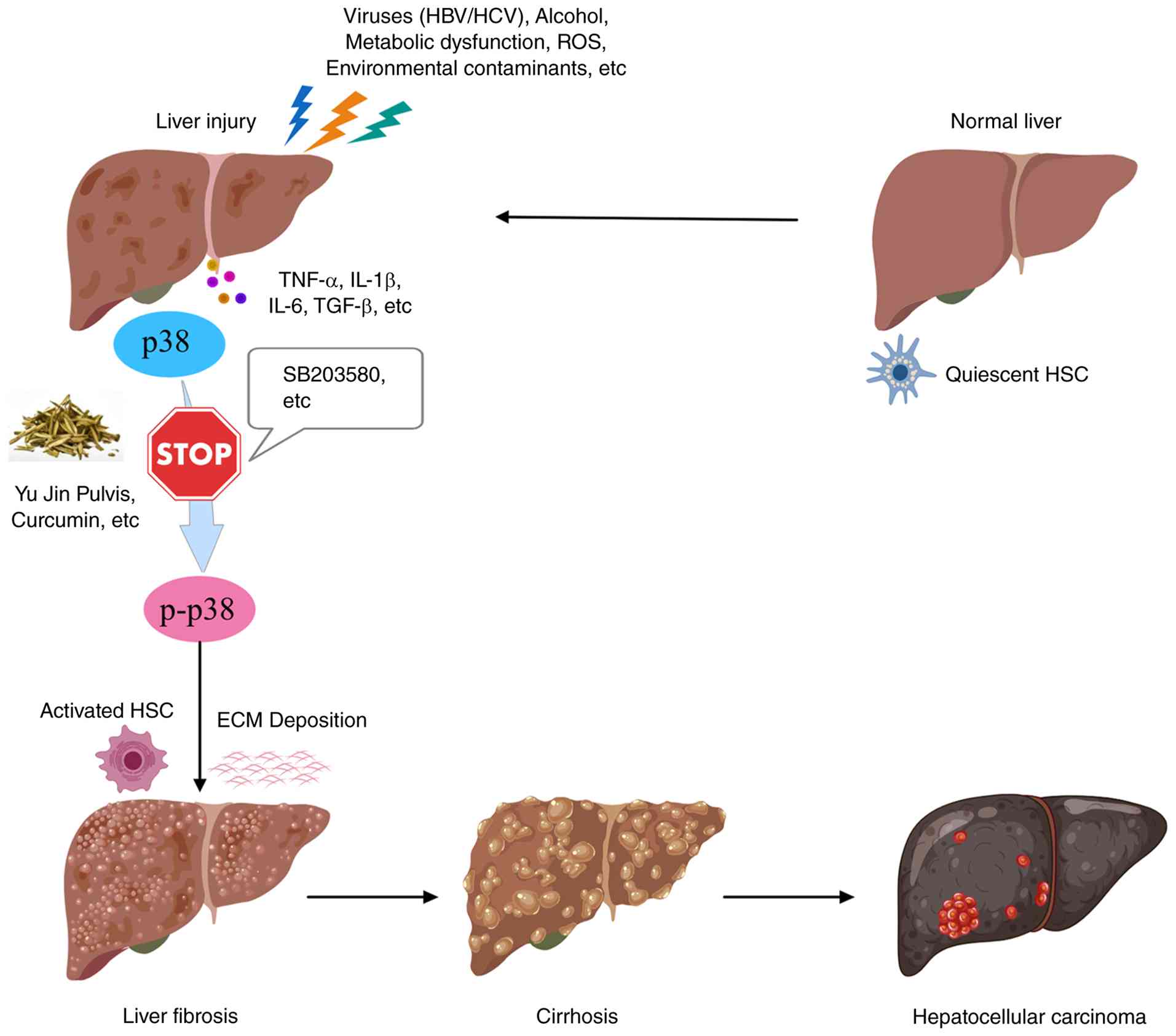 | Figure 1Schematic illustrating the
pathological mechanisms underlying liver fibrosis, including the
involvement of p38 MAPK, is presented. It also highlights potential
points for therapeutic intervention (detailed in Fig. 4). This diagram illustrates the
pivotal function of the p38 MAPK cascade in the progression of
liver fibrosis. The liver becomes injured through various causes,
including hepatitis viruses, alcohol consumption, metabolic
problems, ROS, and environmental contaminants such as DBP and DEHP.
The pro-inflammatory mediators IL-1β, IL-6, TGF-β and TNF-α
function as primary upstream activators of p38 MAPK. Activation of
p38 MAPK leads to the phosphorylation of multiple targets, thereby
promoting HSC activation, myofibroblast differentiation, and cell
proliferation. The process culminates in abnormal growth of ECM
components, including collagen I and α-SMA. The liver develops
fibrosis and cirrhosis through these pathological changes, which
can also result in HCC. Research on p38 MAPK therapy shows promise
through three approaches: Small-molecule inhibitors such as
SB203580 and natural compounds, including curcumin and traditional
Chinese medicines such as Yu Jin Pulvis, for treating liver
fibrosis. p38 MAPK, p38 mitogen-activated protein kinase; ROS,
reactive oxygen species; DBP, dibutyl phthalate; DEHP,
di(2-ethylhexyl) phthalate; IL-1β, interleukin-1β; IL-6,
interleukin-6; TGF-β, transforming growth factor-β; TNF-α, tumor
necrosis factor-α; HSC, hepatic stellate cell; ECM, extracellular
matrix; α-SMA, α-smooth muscle actin; HCC, hepatocellular
carcinoma; HBV, hepatitis B virus; HCV, hepatitis C virus. |
The development of specific treatments for liver
fibrosis requires a complete grasp of p38 MAPK functions, including
HSC activation, ECM remodeling, and its interactions with
inflammatory and oxidative pathways. The present review provides an
overview of p38 MAPK mechanisms that drive fibrogenesis, examines
their relationships with essential signaling pathways, and assesses
current treatment options using specific inhibitors and natural
compounds.
Biological characteristics of p38 MAPK
p38 MAPK constitutes a pivotal component within the
MAPK superfamily. The p38 MAPK signaling pathway exhibits notable
responsiveness to a multitude of stressors, including growth
factors, lineage specification to programmed cell death,
environmental stressors, and pro-inflammatory cytokines, thereby
establishing itself as a pivotal component of the cellular stress
response. This pathway orchestrates the phosphorylation of
downstream effectors, which in turn modulate gene expression and a
range of cellular activities. The p38 MAPK signaling cascade holds
substantial pathological relevance across a diverse spectrum of
disease states, such as liver fibrosis, malignancies,
cardiovascular disorders, and neurodegenerative diseases.
The biological functions attributed to p38 MAPK are
multifaceted and largely context-dependent. A principal role of p38
MAPK involves the modulation of inflammatory reactions (23,24). Empirical research has
demonstrated that p38 MAPK is activated in response to inflammatory
signals, contributing to the upsurge in these cytokines and thereby
amplifying the inflammatory cascade. Notably, inhibition of p38
MAPK signaling has been associated with reduced inflammation and
improved outcomes in various experimental models of inflammatory
diseases (25).
Beyond its role in inflammation, p38 MAPK also
critically regulates cellular homeostasis through modulation of
cell cycle progression, apoptosis, and the differentiation of
diverse cell types, including myoblasts, chondrocytes, and stem
cells (26). It is also pivotal
in mediating cellular responses to diverse stressors, including
oxidative damage, ultraviolet (UV) radiation, and hyperosmotic
stress. Upon activation, p38 MAPK modulates the expression of
antioxidant enzymes and finely balances survival vs. apoptotic
signals, thereby facilitating cellular adaptation (27-29).
The involvement of p38 MAPK in apoptosis is
particularly noteworthy. It can promote apoptosis through several
mechanisms, including the activation of pro-apoptotic factors and
the suppression of anti-apoptotic proteins. At the same time, in
other contexts, it may promote cell survival by activating
protective signaling pathways, highlighting its context-dependent
functions (30).
The signaling dynamics of p38 MAPK are complex, with
multiple feedback mechanisms and crosstalk with other signal
transduction pathways, including the NF-κB and PI3K/Akt axes. This
interplay can determine the ultimate cellular outcomes, such as
survival vs. apoptosis, based on the specific cellular environment
and the nature of the stimuli (23,31).
Beyond its essential roles in managing cellular
stress and homeostasis, dysregulation of p38 MAPK has been
increasingly implicated in various pathological conditions.
Previous investigations have underscored the critical role of p38
MAPK in liver diseases, particularly fibrosis and inflammation.
Furthermore, the therapeutic targeting of p38 MAPK has gained
attention, particularly in diseases characterized by chronic
inflammation and fibrosis, where inhibitors of this pathway may
help mitigate disease progression and improve clinical outcomes
(24,25,32). A diagram of the mechanisms that
activate the p38 MAPK signaling pathway is shown in Fig. 2.
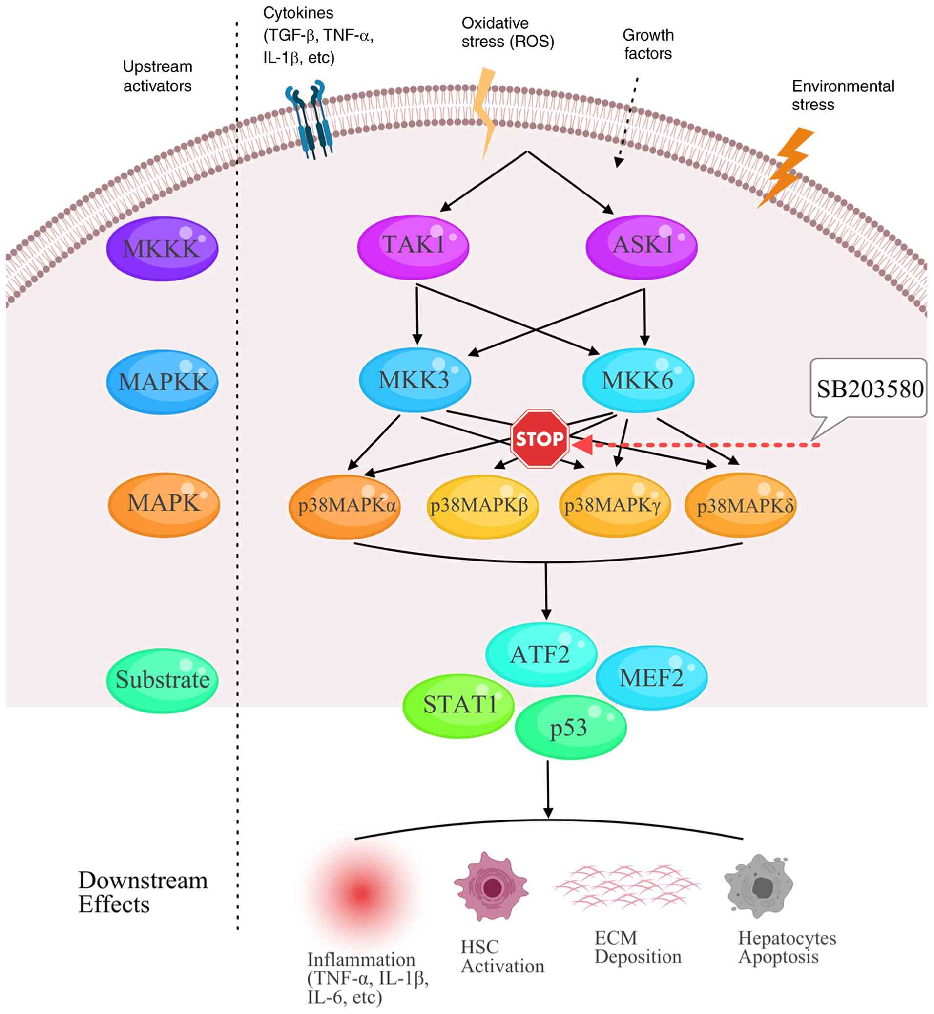 | Figure 2Mechanisms underlying the activation
of the p38 MAPK signaling cascade. The schematic shows the
molecular events that activate the p38 MAPK pathway in response to
various external signals. The pathway is activated by stress
signals, including TGF-β, IL-1β, and TNF-α, pro-inflammatory
mediators, ROS, growth factors, and environmental stressors such as
DBP and LPS. The activation process begins with a series of
sequential kinase reactions, in which MKKKs activate specific MKKs,
including MKK3 and MKK6. p38 MAPK becomes activated due to
phosphorylation events which these signaling molecules trigger. p38
MAPK activation results in phosphorylation of multiple substrates,
which include transcription factors such as ATF2, MEF2, and STAT1,
and apoptotic regulators such as p53. Upon stimulation, p38 MAPK is
activated, thereby triggering phosphorylation cascades involving
ATF2, MEF2, STAT1, and p53 that control inflammation, HSC
activation, ECM deposition, and hepatocyte apoptosis. The diagram
shows the four p38 MAPK isoforms (p38α, p38β, p38γ, p38δ), which
are differentially expressed and perform various cellular functions
through this signaling pathway. TGF-β, transforming growth
factor-β; IL-1β, interleukin-1β; TNF-α, tumor necrosis factor-α;
ROS, reactive oxygen species; DBP, dibutyl phthalate; LPS,
lipopolysaccharide; MKKK, mitogen-activated protein kinase kinase
kinase; MKK, mitogen-activated protein kinase kinase; p38 MAPK, p38
mitogen-activated protein kinase; ATF2, activating transcription
factor 2; MEF2, myocyte enhancer factor 2; STAT1, signal transducer
and activator of transcription 1; p53, tumor protein p53; HSC,
hepatic stellate cell; ECM, extracellular matrix. |
Structure and classification of p38
MAPK
p38 MAPK is a member of the MAPK family of kinases,
which also includes ERKs and JNKs. It is a structurally conserved
serine/threonine kinase characterized by a conserved kinase domain
essential for enzymatic activity, along with regulatory regions
that modulate its activation and substrate specificity (33). The kinase domain comprises a
catalytic core that facilitates the transfer of phosphate moieties
from adenosine triphosphate (ATP) to substrate proteins, a process
known as phosphorylation.
The p38 MAPK family comprises four isoforms: p38α,
p38β, p38γ, and p38δ, each encoded by a distinct gene and
exhibiting unique tissue distribution, regulatory mechanisms, and
functional roles (33). This
classification helps understand how these isoforms contribute to
the complexity of the p38 MAPK signaling pathway in health and
disease (12,14). Among these, p38α is the most
thoroughly characterized isoform and is ubiquitously expressed
across almost all cell types, with predominant expression in the
liver, heart, skeletal muscle, and brain, among others, where it is
implicated in stress responses and apoptosis. By contrast, p38β is
less understood but is hypothesized to have overlapping functions
with p38α in certain contexts. p38γ is predominantly found in
skeletal muscle, where it is hypothesized to facilitate muscle
differentiation, while p38δ is primarily expressed in the brain and
associated with neuronal signaling. Consequently, the differential
activation of these isoforms leads to varied biological outcomes
depending on the cellular context and specific stimuli involved. A
summary of the four p38 MAPK isoforms, their tissue distribution,
and their reported roles in the liver is provided in Table I.
 | Table Ip38 MAPK isoforms and their roles in
the liver/liver fibrosis. |
Table I
p38 MAPK isoforms and their roles in
the liver/liver fibrosis.
| Isoform | Gene | Tissue
distribution | Role in liver/liver
fibrosis | Key
mechanisms/context | (Refs.) |
|---|
| p38α | MAPK14 | Ubiquitous;
predominant expression in liver, heart, skeletal muscle, brain | Most extensively
studied in liver fibrosis; mediates stress responses, inflammation,
and apoptosis | Implicated in HSC
activation, inflammatory cytokine production, and fibrogenic
signaling. Central isoform in most preclinical studies using
inhibitors such as SB203580 (which targets p38α/β). | (12,14,33) |
| p38β | MAPK11 | Less understood;
believed to have overlapping functions with p38α | Less studied in
liver fibrosis; potential functional redundancy with p38α | May compensate for
p38α in certain contexts, but specific role in liver fibrosis
remains unclear. | (33) |
| p38γ | MAPK12 | Predominantly in
skeletal muscle | Not specifically
reported in liver fibrosis | Mainly associated
with muscle differentiation; liver expression low; likely minor
role. | (33) |
| p38δ | MAPK13 | Mainly expressed in
brain, lso present in other tissues | Key role in
MAFLD-associated fibrosis | Activated by free
fatty acids in hepatocytes during MAFLD. Promotes YAP
dephosphorylation and nuclear translocation, driving profibrogenic
gene expression (CTGF, CYR61, ANKRD1, JUB). Inhibition of p38δ
blocks YAP activation and downstream fibrotic effects. | (124,125) |
Beyond its structural diversity, p38 MAPK is
functionally classified into two primary cascades: The inflammatory
and stress response routes. In the context of inflammation, p38
MAPK is triggered by cytokines, including interleukin-1 (IL)-1 and
tumor necrosis factor (TNF)-α, thereby stimulating the subsequent
synthesis of pro-inflammatory mediators and contributing to chronic
inflammatory diseases. Conversely, exposure to environmental
stressors, such as oxidative stress or UV radiation, may activate
p38 MAPK, leading to cell cycle arrest or programmed cell death,
underscoring its essential role in regulating cell fate (34).
Mechanisms of activation of p38 MAPK
The activation of p38 MAPK includes multiple
upstream signaling pathways and stimuli which work together to
activate the enzyme. The activation process begins with
phosphorylation reactions that start when pro-inflammatory
cytokines IL-1 and TNF engage their respective cell-surface
receptors. The receptor-ligand binding process activates multiple
signaling pathways, which produce downstream responses. These
pathways activate MAPK kinase kinases (MKKKs), which then
phosphorylate and activate particular MAPK kinases (MKKs), notably
MKK3 and MKK6. These MKKs subsequently activate p38 MAPK through
phosphorylation at both threonine and tyrosine sites located in the
activation loop (4). The
phosphorylation event triggers a structural transformation which
enables p38 MAPK to phosphorylate its target proteins, including
transcription factors and other kinases, thereby eliciting multiple
cellular responses (12,14).
The activation of p38 MAPK occurs through two
primary mechanisms: Cytokine release and oxidative stress. The
cellular oxidative stress response produces reactive oxygen species
(ROS), which activate p38 MAPK through multiple different pathways.
It is critically implicated in the pathogenesis of diverse
disorders, including neurodegenerative diseases and cancer, by
regulating cellular viability and apoptotic processes (14).
The activation of p38 MAPK is influenced by multiple
signaling pathways, which include NF-κB. The activation level of
p38 MAPK and its downstream effects depend on interactions among
these signaling pathways (12).
The body activates p38 MAPK when it detects
environmental pollutants. Environmental plasticizers cause liver
collagen buildup and elevated fibrosis indicators by activating the
p38 MAPK/NF-κB signaling pathways. These findings underscore the
pivotal role of p38 MAPK in driving environmental toxin-induced
hepatic injury (14,25).
The p38 MAPK activation pathway is present in both
disease states and normal bodily functions. The activation of p38
MAPK during stem cell differentiation enables it to control gene
expression, which determines cell fate. Research has shown that p38
MAPK signaling is indispensable for driving osteogenic
differentiation, as it enables mesenchymal stem cells to
differentiate into osteoblasts during normal development (25). This hierarchical kinase cascade,
from upstream MKKKs to the dual phosphorylation of p38 MAPK is
delineated in Fig. 2, and the
diverse downstream effectors that mediate its pleiotropic cellular
functions are subsequently illustrated.
Downstream effects of p38 MAPK
The p38 MAPK pathway elicits multiple effects that
can result in protective or harmful responses, depending on the
cell type and stimulus characteristics. The phosphorylation and
subsequent activation of p38 MAPK trigger downstream effects that
control inflammatory responses. In the context of liver fibrosis,
p38 MAPK signaling increases the production of IL-6, TNF-α and
IL-1β, thereby accelerating fibrosis progression (35).
The p38 MAPK signaling pathway is directly linked to
apoptotic mechanisms. Phosphorylation of diverse downstream targets
is triggered upon p38 MAPK activation. These substrates include
pro-apoptotic factors such as BAD and caspases, which affect cell
survival and apoptosis. Conversely, the p38 MAPK enzyme also
protects cells from death by activating survival autophagy pathways
during oxidative stress. The dual function of p38 MAPK yields a
complex signaling molecule, as its output depends on both cellular
conditions and the specific activation signals it receives
(12).
In addition to its roles in inflammation and
apoptosis, p38 MAPK participates in mechanisms that govern cellular
differentiation. Research has shown that p38 MAPK signaling
regulates the levels of essential transcription factors and
osteogenic markers that guide osteogenic differentiation.
Inhibition of p38 MAPK activity prevents mesenchymal stem cells
from differentiating into osteoblasts, demonstrating that this
pathway is essential for bone maintenance and repair (36). The regulatory function of p38
MAPK extends to multiple cell types, including immune cells,
fibroblasts, and neurons, thereby affecting multiple biological
processes and disease conditions (37,38).
The p38 MAPK pathway functions through numerous
signaling pathways that activate NF-κB and ERK to control cellular
responses. In inflammatory conditions, p38 MAPK can enhance NF-κB
activation, leading to increased expression of inflammatory
mediators. NF-κB controls p38 MAPK activity through a feedback loop
that amplifies inflammatory responses. The p38 MAPK signaling
pathway interacts with multiple pathways, demonstrating how
cellular networks function as unified systems and creating
opportunities for the development of specific medical interventions
(14). The hierarchical
activation of this pathway, from MKKKs to downstream effectors, is
illustrated in Fig. 2.
Mechanistic insights into the contribution
of p38 MAPK in hepatic fibrogenesis
This section explores the intricate mechanisms
through which p38 MAPK influences the emergence and advancement of
hepatic fibrosis. Research has indicated that p38 MAPK expression
levels are markedly increased in fibrotic liver tissues and are
associated with the extent of disease progression (21). The p38 MAPK pathway has been
identified as a crucial component in the development of liver
fibrosis, affecting a wide range of cellular processes, including
inflammatory mechanisms, apoptosis, and fibroblast activation
(39-42). The activation of this signaling
cascade leads to the upregulation of fibrogenic markers and
promotes HSC proliferation, contributing to fibrosis (43-48). Pro-inflammatory cytokines,
oxidative stress, and other stimuli prevalent during liver injury
often trigger the phosphorylation and subsequent activation of p38
MAPK (49-52).
The p38 MAPK signaling pathway is a pivotal mediator
of the mechanisms underlying liver fibrosis (53). Its roles in HSC activation,
inflammation, regulation of apoptosis, and interactions with other
signaling pathways underscore the complexity of fibrogenesis
(54,55).
Research has demonstrated that the p38 MAPK
signaling cascade exhibits a profound and complex regulatory
association with the transcriptional activation of diverse
profibrotic mediators. Apart from TGF-β, a diverse array of
profibrotic stimuli converges on p38 MAPK. These can be grouped
into several functional categories, including growth factors and
matricellular proteins [such as connective tissue growth factor
(CTGF), and platelet-derived growth factor (PDGF)-D], metabolic and
hormonal signals (such as leptin, angiotensin II, and
15-F2t-isoprostane), immune and inflammatory mediators [such as a
polarized T helper type 2 (Th2) response], redox signaling [such as
NADPH oxidase (NOX)5], components of certain pathogens [such as
lipopolysaccharide (LPS), and human immunodeficiency virus protein
gp120], additional intracellular mediators [such as β-arrestin 1,
Ets domain transcription factor Elk-3 (Net/Sap-2/Erp), slit
guidance ligand 2, and a disintegrin and metalloproteinase 8], the
restructuring of the F-actin cytoskeleton, and even chemical
insults (such as heroin derivatives and Iranian crack) (56-70). Mechanistically, these diverse
activators all trigger the MKK3/6-p38 MAPK axis. For instance,
leptin promotes HSC activation and fibrosis by inhibiting
peroxisome proliferator-activated receptor (PPAR)γ via p38 MAPK,
while LPS-induced p38 MAPK activation enhances Smad2
phosphorylation and collagen synthesis in HSCs (58,64). A comprehensive list of these
stimuli is provided in Table
II.
| Table IIBiochemical stimuli from multiple
categories that converge on p38 MAPK to drive liver
fibrogenesis. |
Table II
Biochemical stimuli from multiple
categories that converge on p38 MAPK to drive liver
fibrogenesis.
| Category |
Stimulus/factor | Mechanism involving
p38 MAPK | (Refs.) |
|---|
| Growth factors and
matricellular proteins | CTGF | Promotes ECM
deposition and inhibits degradation | (56) |
| PDGF-D | Acts through p38
MAPK in HSC activation and ECM remodeling | (57) |
| Metabolic and
hormonal signals | Leptin | Inhibits PPARγ2,
SREBP-1c, and LXR-α via p38 MAPK to drive HSC activation and
fibrosis | (58-60) |
| Angiotensin II | Upregulates CTGF
and ECM via AT1R/PKCα/p38 MAPK, engaging NF-κB and Smad2/3 | (56) |
|
15-F2t-isoprostane | Induces HSC
proliferation and collagen synthesis via p38, ERK, and JNK | (61) |
| Immune and
inflammatory signals Redox signaling | Th2
polarization | Requires active p38
MAPK to sustain profibrogenic response | (62) |
| NOX5 | Silencing reduces
collagen by downregulating p38 MAPK | (63) |
| Components of
certain pathogens | LPS | Promotes Smad2
phosphorylation and α-SMA/collagen I through p38 MAPK | (64) |
| HIV gp120 | Activates p38 MAPK
and NF-κB to promote liver fibrosis | (65) |
| Additional
intracellular mediators | β-arrestin 1 | Upregulates MASP-1,
activating HSCs via the p38/ATF2 axis | (35) |
| Elk-3 | Drives
Egr-1-dependent EMT under p38 MAPK control | (66) |
| Slit2 | Promotes HSC
proliferation through ERK and p38 MAPK | (67) |
| ADAM8 | Accelerates
alcoholic liver fibrosis via p38 MAPK-mediated mechanisms | (68) |
| F-actin
remodeling | Correlates with HSC
activation in a p38 MAPK-dependent manner | (69) |
| Chemical
insults | Iranian crack | Induces liver
damage and fibrosis partly via p38 MAPK stress kinases | (70) |
Moreover, the interplay between p38 MAPK and
additional signaling pathways, including NF-κB, both of which
participate in the inflammatory reaction and fibrogenesis, further
complicates the understanding of its role in liver fibrosis
(14). For example, p38 MAPK can
modulate Smad protein activity, which is vital for mediating TGF-β
signaling, thereby influencing the fibrotic response (71). In addition, hepatocyte growth
factor (HGF) has been demonstrated to confer protective benefits
against liver fibrosis by inhibiting p38 MAPK activation, thereby
promoting HSC apoptosis and reducing collagen production (12). This highlights the prospect of
combinatorial therapeutic strategies designed to simultaneously
inhibit diverse signaling cascades involved in fibrosis, including
p38 MAPK, to effectively manage liver fibrosis (14,20).
Additionally, previous investigations have
identified how diverse environmental stimuli regulate the p38 MAPK
signaling cascade in liver fibrosis. For example, exposure to
environmental pollutants such as dibutyl phthalate (DBP),
di(2-ethylhexyl) phthalate (DEHP) and microcystins has been linked
to increased p38 MAPK activation and subsequent liver fibrosis,
emphasizing the importance of understanding the impact of external
factors on this signaling pathway (14,21,72). A diagram of the mechanisms
underlying the activation of the p38 MAPK signaling cascade is
shown in Fig. 2.
p38 MAPK in HSC activation and ECM
remodeling
The functional activity of HSCs fundamentally drives
the progression of hepatic fibrosis. Studying this process requires
understanding how p38 MAPK activation leads to HSC activation. The
liver contains HSCs, which function as fat-storing cells, and Ito
cells, which play essential roles in liver health and disease
development. The body maintains HSCs in a resting state, which
enables them to store vitamin A and preserve liver ECM stability.
These cells remain inactive until liver damage or inflammation
occurs, which triggers their activation into proliferative cells
that display myofibroblast-like features. The liver then develops
fibrosis through two main changes: Increased α-smooth muscle actin
(α-SMA) protein expression and excessive production of ECM
components, especially collagen in HSCs. This fibrous tissue
accumulation can eventually progress to hepatic fibrosis and
cirrhosis (4,7,20).
Research has shown that p38 MAPK signaling interacts
with HSC activation in response to various stimuli, including
cytokines and growth factors, to induce fibrogenic responses in
HSCs. The activation of p38 MAPK is a critical step, as it triggers
multiple cellular responses, including inflammation, apoptosis, and
fibrosis, then regulates fibrogenic genes, including collagen- and
α-SMA-encoding genes, which are essential for activated HSCs
(73). Inhibiting this pathway
was shown to reduce fibrogenic marker expression, collagen
deposition, and HSC activation in experimental models (4,12,14).
The TGF-β signaling pathway is the primary mechanism
that activates HSCs during the fibrogenic response to liver injury
(74). Research has shown that
activation of the p38 MAPK pathway increases TGF-β1 production
(20,75,76). Elevated TGF-β1 levels activate
p38 MAPK in HSCs, creating a feedback loop. This loop activates
HSCs via TGF-β1 while strengthening p38 MAPK signaling, thereby
increasing fibrotic tissue formation (4,77,78). Additionally, research has
indicated that the p38 MAPK signaling cascade is indispensable for
mediating TGF-β-induced activation of HSCs and for the fibrogenic
response (76,79).
In addition to TGF-β1, other factors, such as ROS
and inflammatory cytokines, can activate p38 MAPK in HSCs, thereby
promoting fibrosis (12).
Research has shown that high glucose levels in diabetes induce
oxidative stress, which activates p38 MAPK, initiating HSC
activation and fibrosis (80).
The study demonstrates that p38 MAPK functions as a key regulator
controlling HSC responses to various activation factors (80).
The process of HSC activation and proliferation is
complicated by the interactions of p38 MAPK with several signaling
pathways. The survival or death of HSCs depends on their signaling
pathway interactions, which determine the extent of fibrosis
development (81-83). As shown in Fig. 2, both TGF-β and oxidative stress
converge on the p38 MAPK activation cascade, ultimately leading to
the phosphorylation of key transcription factors, including myocyte
enhancer factor 2 (MEF2) and activating transcription factor 2
(ATF2), which drive HSC transdifferentiation and fibrogenic gene
expression (84,85).
Beyond initiating activation, p38 MAPK is a master
regulator of the ECM remodeling that characterizes fibrosis.
Notably, the p38 MAPK signaling pathway also contributes to
enhanced stability of α1(I) collagen messenger RNA (mRNA) in HSCs,
which in turn promotes the production and build-up of type I
collagen (86). Supporting the
clinical relevance of this pathway, simultaneous profiling of serum
proteome and hepatic phosphoproteome in patients with metabolic
dysfunction-associated steatohepatitis (MASH) revealed that liver
p38 MAPK activation is independently associated with hepatic
collagen deposition (11).
Moreover, p38 MAPK signaling has been linked to the induction of
other profibrotic factors, creating a feed-forward loop that
exacerbates ECM remodeling in fibrotic tissues. It also regulates
the activity of matrix metalloproteinases (MMPs), enzymes that
degrade ECM components. Several studies performed in fibroblasts
indicated that p38 MAPK and NF-κB signaling cascades are pivotal in
driving the expression of MMPs, specifically MMP-13, MMP-3, and
MMP-1 (87-89). The suppression of the p38 MAPK
signaling cascade can markedly attenuate MMP-13 gene expression in
rat HSCs, thereby preventing MMP-13 from degrading the ECM
components, especially collagen (90). This dual role of p38 MAPK in
promoting ECM deposition while also facilitating its degradation
underscores its importance in maintaining ECM homeostasis. For
example, in a study investigating hepatic alveolar echinococcosis,
increased expression levels of p38 MAPK were associated with
elevated TGF-β1 and bone morphogenetic protein-7 (BMP-7) in liver
specimens from patients, indicating a strong association between
p38 MAPK activation and ECM remodeling in liver fibrosis (4). Similarly, in models of chronic
liver injury induced by carbon tetrachloride (CCl4), p38
MAPK was activated, promoting the expression of ECM components and
contributing to the progression of liver fibrosis (21).
Furthermore, p38 MAPK activity is not confined to
HSCs. p38 MAPK is also a key signaling molecule that mediates the
defenestration process of liver sinusoidal endothelial cells
(LSECs) in response to increased matrix rigidity, thereby
accelerating the advancement of hepatic fibrosis by regulating
cytoskeletal reorganization (91). The activation of the focal
adhesion kinase (FAK)-p38 MAPK signaling pathway in LSECs occurs
when liver matrix stiffness increases, leading to their
pathological defenestration (loss of fenestrae) (91). Stiff substrates promote FAK
phosphorylation, which subsequently activates p38 MAPK and its
downstream effector p38-mitogen-activated protein kinase-activated
protein kinase 2 (MK2) (91).
p38-MK2 activation results in LIM kinase 1 phosphorylation,
ultimately inducing cofilin inactivation through its role as an
actin depolymerization protein. The process results in increased
actin polymerization, leading to stress fiber formation and
reorganization of the actin cytoskeleton (91). These changes cause the collapse
of fenestrae-associated cytoskeletal rings and LSEC defenestration,
which represents the first stages of capillarization and fibrosis
(91). The process is reversed
when p38-MK2 signaling is blocked with SB203580, which restores
fenestrae structures and LSEC phenotypic markers in fibrotic liver
samples across different disease stages (91). Additionally, p38 MAPK functions
as a primary mechano-transducer, causing LSEC dysfunction when
cells experience stiffness (86). As illustrated in Fig. 1, p38 MAPK serves as a central
node that integrates a range of profibrotic signals and transduces
them into the key pathological outcomes of HSC activation and
excessive ECM deposition.
p38 MAPK-mediated inflammatory, oxidative
stress and apoptotic signaling in fibrosis
The liver continues to sustain damage from ongoing
inflammation and oxidative stress, which trigger hepatic fibrosis
through multiple cellular and molecular mechanisms. The p38 MAPK
pathway functions as a central signaling hub which combines
inflammatory, oxidative, and apoptotic signals to control fibrotic
disease progression.
p38 MAPK as a central amplifier of
profibrotic inflammation
Chronic liver injury is marked by increased
concentrations of pro-inflammatory cytokines, including TGF-β,
IL-1β, TNF-α, and IL-17A (4,92). Rather than being a passive
responder, p38 MAPK activation serves as a critical amplifier
within this inflammatory milieu. Upon stimulation by these
cytokines, p38 MAPK enhances the inflammatory response through
multiple mechanisms: It upregulates the expression of inflammatory
enzymes such as cyclo-oxygenase-2, modulates MMP activity, and
potentiates NF-κB signaling (4,93). This leads to sustained production
of key pro-inflammatory mediators, which not only cause direct
tissue damage but also create a microenvironment that perpetuates
HSC activation and survival.
The role of p38 MAPK extends beyond cytokine
signaling to direct mediation of inflammation-related cell death
and environmental responses. For example, in DBP-induced liver
fibrosis, p38 MAPK activation links inflammatory stimuli to
hepatocyte pyroptosis, followed by HSC activation, directly tying
inflammatory signaling to fibrotic tissue remodeling (14). Furthermore, p38 MAPK is
implicated in the inflammatory aspects of MAFLD, contributing to
insulin resistance and subsequent fibrogenic signaling (94). Other factors, such as LPS,
exacerbate inflammation by promoting p38 MAPK-mediated degradation
of autophagy-related proteins, including autophagy-related gene 13
(Atg13), in HSCs, thereby disrupting cellular homeostasis and
favoring a pro-fibrotic state (95). The aging liver also exhibits a
p38 MAPK-mediated inflammatory and mildly fibrotic phenotype, which
can be modulated by interventions such as caloric restriction,
highlighting the role of the pathway in environment- and
age-dependent fibrogenesis (96). Thus, p38 MAPK is not merely
activated by inflammation; it orchestrates a feed-forward loop that
sustains and intensifies the profibrotic inflammatory response.
Oxidative stress and p38 MAPK: A mutually
reinforcing axis in fibrosis
Oxidative stress is a hallmark of chronic liver
injury and a potent activator of p38 MAPK (97). This relationship forms a
critical, self-reinforcing axis in fibrosis. Signals from damaged
hepatocytes, such as ROS directly activate p38 MAPK, which in turn
increases the expression of inflammatory genes and genes associated
with apoptosis, such as Bcl-2-associated X protein (Bax), alongside
a reduction in the levels of the anti-apoptotic protein Bcl-2
(98). Notably, p38 MAPK
activation can further stimulate ROS generation, establishing a
self-amplifying cycle that intensifies oxidative damage and tissue
injury (99).
This cyclical interplay has profound implications
for liver fibrosis. In hepatocytes, activation of p38 MAPK
triggered by oxidative stress promotes cell death and the release
of inflammatory cytokines, which subsequently activate HSCs and
drive fibrosis (7,100). Simultaneously, ROS can activate
NF-κB, which enhances p38 MAPK pathway activity, leading to
increased inflammation and sustaining a pro-fibrotic environment,
linking hepatocyte injury to fibrogenic responses (101,102). This suggests that targeting
oxidative stress and p38 MAPK activation may offer therapeutic
avenues for mitigating liver fibrosis.
The dual nature of p38 MAPK in oxidative stress is
context-dependent: While it can mediate protective responses by
regulating antioxidant defenses such as glutathione and inhibiting
expression of inducible nitric oxide synthase under specific
conditions, its chronic activation under persistent oxidative
stress typically leads to detrimental outcomes, including cell
cycle disruption and apoptosis (27). This functional duality
underscores that p38 MAPK can act as both a stress sensor and a
determinant of cell fate, with its outcomes finely balanced by the
cellular context and cross-talk with other pathways (103). Pharmacological inhibition of
p38 MAPK has shown protective effects by bolstering cellular
antioxidant systems, underscoring its pivotal role as both a
mediator and a potential therapeutic breakpoint in the oxidative
stress-fibrosis cycle (28). How
pro-inflammatory mediators, specifically IL-1β and TNF-α, alongside
oxidative stress, serve as key upstream activators of the p38 MAPK
signaling cascade, highlighting the molecular basis for its central
role in amplifying inflammatory and oxidative responses during
liver injury is illustrated in Fig.
2.
Apoptosis and p38 MAPK crosstalk in liver
fibrosis
The equilibrium between cell survival and apoptosis
is critical in liver injury, as excessive hepatocyte apoptosis can
lead to further fibrogenesis. Research has indicated that the
activation of p38 MAPK can either facilitate or suppress apoptosis,
contingent on the specific cellular environment and stimuli. The
p38 MAPK pathway has been associated with the modulation of
apoptosis across a range of cell types, including hepatocytes
(14). Prolonged oxidative
stress in hepatocytes activates p38 MAPK, leading to apoptosis via
mitochondrial damage and caspase activation, thereby exacerbating
liver damage (12,104). Research findings show that MASH
develops in response to oxidative stress, which activates the MAPK
phosphatase-1-p38 MAPK-liver kinase B1 nuclear signaling pathway.
This pathway suppresses the enzymatic activity of the α catalytic
subunit of adenosine 5'-monophosphate-activated protein kinase
(AMPK), thereby triggering caspase-6 activation and culminating in
cellular demise. The resulting inflammation drives liver fibrosis
and MASH progression (105).
The p38 MAPK pathway functions differently in HSCs because it
generates survival signals that enable these cells to survive and
build the ECM (14). However,
p38 MAPK has been recognized as a crucial mediator of the apoptotic
effect of Ocoxin on HSCs (106). IL-18 works together with
Toll-like receptor 3 ligand stimulation to activate natural killer
(NK) cells, enhancing their cytotoxic functions through the p38
MAPK/PI3K/Akt signaling pathway. These activated NK cells eliminate
HSCs through TNF-related apoptosis-inducing ligand-mediated
degranulation, thereby preventing fibrosis progression (107).
The body regulates p38 MAPK activity in a
cell-type-specific manner, creating challenges for developing
therapeutic strategies targeting this enzyme. p38 MAPK inhibition,
as shown in animal studies of CCl4-induced fibrosis,
decreases collagen accumulation but may impair hepatocyte
restoration processes (12,14). The downstream effectors shown in
Fig. 2, including p53 and other
pro-apoptotic factors, mediate the cell death-promoting functions
of p38 MAPK in hepatocytes, whereas alternative signaling outputs
may support survival in activated HSCs.
p38 MAPK interplay with key signaling
networks in liver fibrosis
The development of hepatic fibrosis depends on
multiple signaling pathways that interact through crosstalk
mechanisms to regulate fibrogenesis. A comprehensive network map of
these interactions is provided in Fig. 3, illustrating how p38 MAPK serves
as a central signaling hub that both receives inputs from and
coordinates outputs to multiple pro-fibrotic pathways. Rather than
viewing each pathway in isolation, these interactions can be
grouped into functional classes: i) Core pro-fibrotic feed-forward
loops that drive disease progression; ii) metabolic and
mechanosensitive signaling axes that link cellular stress to
fibrogenesis; iii) counter-regulatory and pro-resolution pathways
that oppose fibrosis; and iv) parallel MAPK cascades that
collectively determine cell fate. The signaling pathways that drive
hepatic fibrosis progression become less active when patients
undergo lifestyle interventions that reduce liver inflammation and
improve metabolic health, providing clinical evidence for the
reversibility of these network interactions (108,109).
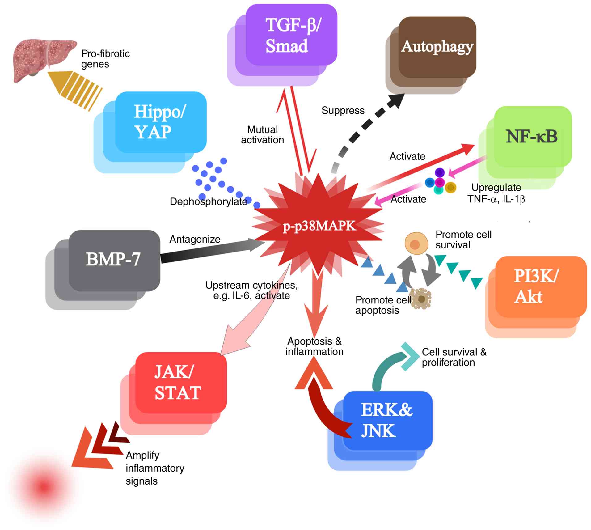 | Figure 3Network diagram of p38 MAPK
interactions with other major signaling pathways. This diagram
illustrates the extensive crosstalk between the p38 MAPK signaling
pathway and other major signaling networks involved in cellular
stress, inflammation, fibrosis, and apoptosis. p38 MAPK serves as a
central hub that receives signals from various pathways and directs
outputs to distinct signaling pathways to control three essential
disease mechanisms, including HSC activation, ECM remodeling, and
inflammatory reactions. The p38 MAPK pathway facilitates NF-κB
activation. NF-κB activates p38 MAPK by increasing the production
of the cytokines TNF-α and IL-1β. The PI3K/Akt signaling cascade is
a pivotal mechanism that regulates survival, migration, apoptosis,
and fibrogenesis in HSCs. The primary interactions occur between
TGF-β/Smad and p38 MAPK, which serves a dual role, operating both
downstream and upstream of the TGF-β cascade to boost
Smad-dependent fibrogenic gene expression and create a
self-reinforcing mechanism that accelerates fibrosis development.
NF-κB: p38 MAPK facilitates the initiation of NF-κB signaling. In
turn, NF-κB can further stimulate p38 MAPK activity via elevated
levels of cytokines, including TNF-α and IL-1β. PI3K/Akt:
Coordinated signaling influences various biological mechanisms,
including survival, apoptosis, migration, and fibrogenesis in HSCs.
JNK/ERK: The MAPK family members JNK and ERK work with p38 MAPK to
activate stress responses that regulate apoptosis, cell
proliferation, and inflammation. YAP: p38δ activation by free fatty
acids results in YAP dephosphorylation and nuclear translocation,
which produces profibrogenic gene expression during MAFLD.
JAK/STAT: p38 MAPK-induced cytokines (such as IL-6) activate
JAK/STAT signaling to maintain ongoing inflammatory and fibrotic
responses. BMP-7 functions as a TGF-β blocking agent, which halts
p38 MAPK activation to stop fibrosis development and trigger cell
death in HSCs. The process of autophagy (AMPK/mTOR) depends on p38
MAPK to block autophagic flux through mTOR-mediated ULK1
suppression, which helps HSCs maintain their fibrogenic state. The
network shows that p38 MAPK operates within a complex signaling
network, making it an optimal target for combination therapies that
block multiple profibrotic pathways. p38 MAPK, p38
mitogen-activated protein kinase; HSC, hepatic stellate cell; ECM,
extracellular matrix; NF-κB, nuclear factor κ-light-chain-enhancer
of activated B cells; TNF-α, tumor necrosis factor-α; IL-1β,
interleukin-1β; PI3K, phosphoinositide 3-kinase; Akt, protein
kinase B; TGF-β, transforming growth factor-β; Smad, small mothers
against decapentaplegic; JNK, c-Jun N-terminal kinase; ERK,
extracellular signal-regulated kinase; YAP, yes-associated protein;
MAFLD, metabolic-associated fatty liver disease; JAK, Janus kinase;
STAT, signal transducer and activator of transcription; IL-6,
interleukin-6; BMP-7, bone morphogenetic protein 7; AMPK, adenosine
monophosphate-activated protein kinase; mTOR, mechanistic target of
rapamycin; ULK1, unc-51-like autophagy activating kinase 1. |
Core pro-fibrotic feed-forward loops:
TGF-β/Smad, NF-κB and JAK/signal transducer and activator of
transcription (STAT) pathway
Among the pathways that intersect with p38 MAPK in
liver fibrosis, TGF-β/Smad and NF-κB signaling form two interlocked
feed-forward loops that are central to disease progression.
One of the most well-studied interactions involving
p38 MAPK is its relationship with the TGF-β signaling cascade. The
TGF-β1 signaling cascade is widely recognized as a principal
mediator of hepatic fibrosis, promoting HSC activation and ECM
deposition. Research has shown that TGF-β1 can initiate multiple
downstream signaling cascades, including the Smad pathway, which is
the main pathway of HSC activation and essential for mediating its
fibrogenic effects (4,12). Additionally, TGF-β1 has been
linked to the initiation of the p38 MAPK signaling cascade, which
further amplifies the fibrogenic response by enhancing inflammation
and apoptosis in hepatocytes (20). These findings indicate that the
p38 MAPK pathway serves as an effector downstream of TGF-β
signaling, mediating the fibrogenic effects of TGF-β1. For example,
in a study investigating hepatic fibrosis, it was found that TGF-β1
induced the phosphorylation of p38 MAPK, which resulted in an
upregulation of fibrogenic markers and an augmentation of collagen
production, correlated with the severity of fibrosis in liver
tissues (4). In another study,
TGF-β was shown to stimulate CTGF expression, which in turn
activated p38 MAPK, creating a feedback loop that exacerbated
fibrosis (110). Upon receptor
binding, TGF-β1 was demonstrated to activate Smad2 and Smad3,
leading to their phosphorylation and nuclear translocation. This
translocation was revealed to activate fibrogenic genes, including
those encoding α-SMA and type I collagen (14,20). At the same time, TGF-β1
facilitated the production of sorting nexin protein-10 (SNX-10)
through the p38 MAPK signaling cascade. In turn, SNX-10 modulated
MMP9 release through direct protein-protein interactions,
ultimately contributing to the progression of hepatic fibrosis
(111). Furthermore, TGF-β1
stimulation triggered p38 MAPK activation, thereby enhancing MEF2C
phosphorylation at the p38-specific residue, inducing activation of
MEF2 and HSC (112). TGF-β1
also caused an increase in aerobic glycolysis in HSCs and
stimulated the expression of glucose transporter 1 (GLUT1) in these
cells via the stimulation of p38 MAPK, Smad, as well as PI3K/Akt
signaling cascades. GLUT1 was shown to promote HSC glycolysis and
activation (79). Research
indicates that upregulation of integrin-β6 expression plays a
pivotal role in the fibrogenic response associated with chronic
cholestasis. p38 MAPK cascade facilitates TGF-β1-driven
transcriptional upregulation of integrin-β6 mRNA within bile duct
epithelial cells, specifically in MMNK-1 cell lines. This process
is facilitated through the modulation of the activation of both
activator protein 1 and Smad transcription factors (113). Notably, both p38 MAPK and Smad
signaling pathways exert independent and additive effects on the
transcriptional upregulation of the gene encoding type I collagen
α1 chain. However, it is noteworthy that only p38 MAPK contributes
to the enhancement of α1(I) collagen mRNA stability (71). Thus, feedback mechanisms
characterize the relationship between p38 MAPK and TGF-β, as p38
MAPK activation can amplify TGF-β1 effects, thereby establishing a
self-reinforcing cycle that intensifies fibrotic progression and
leads to a vicious cycle of HSC activation and collagen deposition
(114). Previous investigations
have explored the potential of targeting the p38 MAPK pathway to
mitigate TGF-β-induced fibrosis. Research involving liver fibrosis
models has demonstrated that inhibiting p38 MAPK reduces TGF-β
expression and its downstream effects, thereby ameliorating
fibrosis (115-117).
The TGF-β/p38 MAPK loop is intimately coupled with
NF-κB signaling. NF-κB functions as a transcription factor that
regulates inflammatory responses and immune activation by
controlling the expression of pro-inflammatory cytokine genes. The
activation of p38 MAPK leads to the phosphorylation of inhibitor of
κBα (IκBα), which removes its inhibitory function on NF-κB. The
process enables NF-κB dimers to exit the cytoplasm and enter the
nucleus, where they activate transcription of inflammatory cytokine
genes, which in turn activate p38 MAPK (118). The two-way activation process
between these cells creates a self-reinforcing cycle which results
in continuous tissue damage and inflammation. Research has shown
that p38 MAPK activation during the development of liver fibrosis
leads to increased NF-κB signaling, resulting in higher production
of the pro-fibrotic factors TGF-β and CTGF (119). This interplay is exemplified by
the fact that inflammatory cytokines, such as TNF-α and IL-1β, can
trigger p38 MAPK activation, which subsequently enhances TGF-β
expression, perpetuating a cycle of inflammation and fibrosis
(21).
The tight coupling of these two feed-forward loops
may explain why single-target interventions often fail. Dual
inhibition of TGF-β and NF-κB signaling, or direct targeting of
their common downstream mediator p38 MAPK, has been proposed as a
more effective strategy.
Moreover, the p38 MAPK and JAK/STAT signaling
pathways also interact extensively during liver fibrosis. The
activation of p38 MAPK leads to increased synthesis of TNF-α and
IL-6, which not only feed back onto NF-κB but also activate the
JAK/STAT pathway, thereby further enhancing inflammatory responses
(120,121). The JAK/STAT signaling pathway
is similarly activated after TGF-β1 cytokine binds to its receptor.
The p38 MAPK and JAK/STAT pathways operate as a complex system that
amplifies the inflammatory-fibrogenic network in the progression of
liver fibrosis. This synergistic interaction creates a feed-forward
loop that amplifies fibrogenic signaling, making co-targeting of
these pathways an attractive therapeutic strategy (Fig. 3).
Metabolic and mechanosensitive signaling
pathways: PI3K/Akt, yes-associated protein (YAP), autophagy, and
bile acids
Beyond classical inflammatory and growth factor
signals, metabolic and mechanical cues are integrated with p38 MAPK
through the PI3K/Akt/mechanistic target of rapamycin (mTOR), YAP,
and autophagy pathways and bile acids. The p38 MAPK and PI3K/Akt
signaling cascades in liver fibrosis function together through
coordinated activation, which strengthens their combined fibrotic
effects. Research has indicated that the p38 MAPK signaling pathway
interacts with the PI3K/Akt pathway, thereby protecting HSCs from
apoptosis. It has been shown that blocking the PI3K/Akt signaling
pathways renders p38 MAPK inhibitors more effective at inhibiting
HSC activation driven by various p38 MAPK-dependent profibrotic
mechanisms. Angiotensin II (AngII) increases the expression of
essential proteins (PI3K, p-Akt, and p-P38 MAPK) in HSCs, thereby
promoting fibrosis progression (12). The pathways show functional
cross-talk because LY294002 treatment of PI3K and SB203580
treatment of p38 MAPK both block AngII-induced effects, including
increased cell viability and migration, and elevated collagen I and
α-SMA expression (12).
HGF-overexpressed exosomes were shown to reduce both pathways,
resulting in lower oxidative stress levels (NOX4 and
malondialdehyde) and decreased p-p38 MAPK and PI3K/p-Akt activity
in vivo, thereby treating CCl4-induced liver
fibrosis in mice (12). However,
the context-dependent nature of this crosstalk is underscored by
the finding that N-acetylglucosamine (GlcNAc)-carrying polymers,
which mimic the multivalent GlcNAc moiety of O-linked
β-GlcNAc-modified proteins, activate both p38 MAPK and PI3K/Akt
signaling cascades in activated stellate cells. This activation
produces anti-fibrotic effects by decreasing α-SMA expression and
reducing collagen production (122). This seemingly paradoxical
outcome underscores the context-dependent nature of p38
MAPK-PI3K/Akt crosstalk, where the net effect on fibrosis is
dictated by factors such as activation kinetics, coincident
signals, and downstream effector engagement. Moreover, it was
demonstrated that PI3K-Akt-mTOR and p38 MAPK signaling cascades
play a crucial role in the motility characteristics of HSCs. By
contrast, the Smad-dependent TGF-β signaling cascade is primarily
involved in their proliferation and contraction (123).
The metabolic stress connection is further
strengthened by the p38 MAPK-YAP axis. Research has demonstrated
that the activation of Hippo signaling transcriptional coactivator
YAP1 in the context of MAFLD could be stimulated by the activation
of p38 MAPK induced by free fatty acids (FFAs) (124). FFAs, which accumulate in
hepatocytes during MAFLD, activate p38 MAPK (specifically the p38δ
isoform MAPK13). This activation promotes the
dephosphorylation/nuclear translocation of the transcriptional
coactivator YAP. Once in the nucleus, the active form of YAP
promotes transcription of profibrogenic target genes, including
cysteine-rich protein 61, CTGF, ankyrin repeat domain 1, and ajuba
homolog. The products of these genes stimulate HSC activation and
subsequent ECM deposition, thereby driving the progression of liver
fibrosis. Inhibition of p38 MAPK (via siRNA knockdown or chemical
inhibitors such as SB203580) was shown to block FFA-induced YAP
activation and its downstream fibrogenic effects. In addition,
activation of HSCs by taurocholic acid was significantly associated
with activation of the YAP signaling pathway, mediated by
sphingosine 1-phosphate receptor 2 via the p38 MAPK pathway
(125). The p38δ-YAP axis,
shown in Fig. 3, represents a
newly identified mechanism linking metabolic stress (FFAs) to
fibrogenic gene expression, providing a rationale for targeting
this specific isoform in MAFLD-associated fibrosis.
Autophagy adds another layer of metabolic
regulation. The lysosomal degradation process of autophagy
functions as a dual mechanism contributing to the progression of
liver fibrosis. Basal autophagy plays a crucial role in maintaining
cellular homeostasis. HSCs with impaired autophagic function may
either survive longer and exacerbate fibrosis or undergo apoptosis,
thereby mitigating fibrosis, depending on the specific context
(126,127). The activation of p38 MAPK
triggers the phosphorylation of the serine residue 757 within
Unc-51-like autophagy activating kinase 1 (ULK1). This modification
disrupts the ULK1-Atg13 complex, thereby blocking autophagic flux
in activated HSCs via mTOR-dependent mechanisms, leading to
increased collagen production and enhanced fibrogenesis (128). The p38 MAPK inhibitor SB203580
activates AMPK, thereby inducing autophagy, which strengthens the
therapeutic effects of anti-fibrotic drugs (128). However, the natural compound
harmine inhibits p38 MAPK and modulates AMPK/mTOR, relieving its
suppression on the mTOR complex 1, reducing excessive autophagy in
CCl4-induced liver injury (129). This indicates that the outcome
of p38 MAPK modulation of autophagy is highly dependent on the
degree and context of pathway activation.
Bile acids further contribute to this metabolic
signaling network. Research has focused on liver fibrogenesis by
studying conjugated bile acids, specifically 12α-hydroxylated bile
acids. Research has shown that liver tissue contains elevated bile
acid levels of conjugated 12α-hydroxylated bile acids, which
activate HSCs and promote liver fibrosis. Activation of the Takeda
G-protein-coupled receptor, also known as G protein-coupled bile
acid receptor 1, leads to phosphorylation-mediated upregulation of
the p38 MAPK and ERK1/2 cascades (31). In a previous study, it was shown
that metabolic factors, including bile acid buildup, affect
fibrosis development by modulating essential signaling pathways.
The study also demonstrated that modulation of bile acid signaling
is a potential treatment approach (31). Collectively, the PI3K/Akt, YAP,
autophagy, and bile acid pathways highlight the extensive
integration of metabolic and mechanical signals at the p38 MAPK
node, although the context-dependent effects, which are sometimes
pro-fibrotic, sometimes anti-fibrotic, call for careful evaluation
of isoform-specific and cell-type-specific therapeutic
strategies.
Counter-regulatory and pro-resolution
pathways: BMP-7 and mesenchymal-to-epithelial transition (MET)
In contrast to the pro-fibrotic circuits previously
described, BMP-7 and the reversal of EMT represent endogenous
counter-regulatory mechanisms that converge on p38 MAPK. BMP-7
functions as an essential factor that inhibits hepatic fibrosis.
The compound blocks the fibrogenic effects of TGF-β1, induces HSC
apoptosis, and reduces their collagen-producing capacity. The
maintenance of liver homeostasis depends on the proper balance
between TGF-β1 and BMP-7 signaling pathways. The body develops
excessive fibrosis when its fibrosis regulation system becomes
unbalanced. Research has shown that BMP-7 acts as a protective
factor through its ability to stop TGF-β1 from activating the p38
MAPK pathway, which prevents fibrogenesis (4). HSC activation depends on EMT, and
the MET of HSCs helps resolve fibrosis. A decrease in the
TGF-β1:BMP-7 ratio was shown to lead to reduced levels of total and
phosphorylated p38 MAPK, allowing cells to revert from EMT to MET.
The transition became apparent through the loss of EMT markers,
including α-SMA and desmin, while MET marker and E-cadherin
reappeared (130). As shown in
Fig. 3, BMP-7 counteracts TGF-β1
signaling by suppressing p38 MAPK activation, representing a
natural counter-regulatory mechanism that could be harnessed
therapeutically to promote fibrosis resolution. The development of
pharmacological agents which activate BMP-7 signaling and block
TGF-β1 and p38 MAPK activation pathways shows promise for treating
and stopping fibrosis progression (7).
Parallel MAPK cascades: JNK and ERK
p38 MAPK also interacts with other MAPK signaling
cascades, such as JNKs and ERKs. The p38 MAPK, JNK, and ERK
pathways work together to regulate essential processes in liver
fibrosis by modulating HSC activation, inflammation, apoptosis, and
ECM deposition.
The JNK signaling pathway is essential for
regulating both cell death and cell growth. The process is
activated when cells face stress and inflammatory signals emerge,
leading to liver fibrosis through hepatocyte apoptosis and an
increased inflammatory response. JNK activation leads to c-Jun
phosphorylation. c-Jun is a crucial transcription factor that
regulates the expression of survival- and apoptosis-related genes
(131). The activation of p38
MAPK and JNK pathways is of particular interest because they
respond to identical triggers, producing a synchronized cellular
response that determines cell survival or death based on activation
timing and environment.
By contrast, the ERK signaling pathway primarily
promotes cell proliferation and differentiation by being activated
by growth factors and mitogens. The activity of p38 MAPK and JNK
depends on ERK, which regulates their downstream effects. Research
shows that the ERK and p38 MAPK pathways function together to
regulate cell-cycle events and stress responses in polyploid, giant
cancer cells, suggesting that HSCs employ similar mechanisms to
develop fibrotic tissue (131).
The outcome of fibrosis results from the interaction
between these two pathways. The activation of ERK promotes cell
survival and proliferation, whereas persistent JNK/p38 MAPK
signaling pathways lead to apoptosis and inflammation (132,133). In liver fibrosis, oxidative
stress and cytokine release of TNF-α and IL-6 may activate all
three MAPK signaling pathways, creating a sustained cycle of HSC
activation and ECM deposition (134). The p38 MAPK and ERK pathways
serve as targets for inhibition because this approach would break
the self-sustaining cycle (135). The complex interplay among the
p38 MAPK, JNK, and ERK pathways, which collectively determine
cell-fate decisions ranging from proliferation to apoptosis in the
context of liver fibrosis is shown in Fig. 3.
Signaling pathways related to
inflammation
The inflammatory response is another critical
component of hepatic fibrosis, and inflammatory signaling pathways
often intersect with pathways involved in fibrogenesis. The p38
MAPK signaling cascade is activated through elevated levels of the
pro-inflammatory mediators TNF-α and IL-6, resulting in a marked
augmentation of fibrogenic response. HSC activation decreases when
anti-inflammatory signals enter the system because these signals
promote HSC death (136). The
regulatory mechanisms of hepatic fibrosis become complex because
pro-inflammatory and anti-inflammatory signals interact with each
other and with p38 MAPK and other essential pathways. Collectively,
the signaling network depicted in Fig. 3 establishes p38 MAPK as a central
hub that integrates a range of fibrotic inputs and coordinates
multi-pathway outputs. The most biologically significant
mechanisms, the interlocked TGF-β/NF-κB/p38 feed-forward loops and
the metabolic-mechanical axes converging on p38 MAPK, represent
priority targets for therapeutic intervention. The complexity and
context dependence of these interactions underscore the rationale
for simultaneously targeting multiple nodes, or its downstream
effectors within the network, in combination therapies for liver
fibrosis. p38 MAPK interactions with key signaling pathways in
liver fibrosis are summarized in Table III.
| Table IIISummary of p38 MAPK interactions with
key signaling pathways in liver fibrosis. |
Table III
Summary of p38 MAPK interactions with
key signaling pathways in liver fibrosis.
| Signaling
pathway | Interaction with
p38 MAPK | Mechanism/Key
points | Functional role in
liver fibrosis | (Refs.) |
|---|
| TGF-β/Smad | Bidirectional
cross-talk, synergistic activation | p38 MAPK acts as
both a downstream effector and an amplifier of TGF-β signaling. It
collaborates with Smads to enhance transcription of fibrogenic
genes (α-SMA, collagen I) and stabilizes collagen mRNA
independently of Smads. | Promotes HSC
activation, ECM deposition, and fibrosis progression. Creates a
positive feedback loop sustaining fibrogenesis. | (4,14,71,79,111-113,115,116,216) |
| NF-κB | Reciprocal
activation, positive feedback | p38 MAPK enhances
NF-κB transcriptional activity;NF-κB promotes pro-inflammatory
cytokine production (IL-6 and TNF-α) that further activates p38
MAPK | Amplifies
inflammatory responses, sustains chronic inflammation, and promotes
HSC activation and fibrosis. | (14,93,101) |
| PI3K/Akt | Coordinated
regulation, functional cross-talk | Both pathways
regulate HSC survival, migration, and fibrogenesis. p38 MAPK
interacts with PI3K/Akt to balance pro-apoptotic and pro-survival
signals. | Protects HSCs from
apoptosis; promotes HSC activation, collagen production, and
fibrosis progression. Combined inhibition yields synergistic
antifibrotic effects. | (12,122,123) |
| YAP | Upstream activation
(p38δ isoform) | FFAs activate p38δ,
which promotes YAP dephosphorylation and nuclear translocation.
Nuclear YAP drives transcription of pro-fibrotic genes (CTGF,
CYR61, ANKRD1, and JUB). | Links metabolic
stress (MAFLD) to HSC activation and ECM deposition. Represents a
novel mechanism in obesity-associated fibrosis. | (124,125) |
| JAK/STAT | Indirect activation
via cytokines | p38 MAPK induces
IL-6 and TNF-α production, which subsequently activate JAK/STAT
signaling. | Maintains ongoing
inflammatory and fibrotic responses; contributes to HSC activation
and disease progression. | (120,121) |
| BMP-7 | Antagonistic | BMP-7 counteracts
TGF-β1 signaling by inhibiting p38 MAPK activation, promoting HSC
apoptosis, and reducing collagen production. | Serves as a
protective, anti-fibrotic factor. Restores balance between EMT and
MET, promoting fibrosis resolution. | (4,130) |
| JNK/ERK | Coordinated MAPK
network | p38 MAPK, JNK, and
ERK pathways interact to regulate cell proliferation, apoptosis,
inflammation, and ECM deposition. ERK promotes
survival/proliferation; JNK/p38 promote stress responses. | Determines cell
fate (survival vs. apoptosis) and net fibrotic outcome. Targeting
multiple MAPKs may break the self-sustaining fibrotic cycle. | (106,131-135,214) |
| Autophagy
(AMPK/mTOR) | Inhibitory
regulation | p38 MAPK
phosphorylates ULK1 (Ser757), disrupting the ULK1-Atg13 complex and
blocking autophagic flux via mTOR-dependent mechanisms. | Suppresses
autophagy in activated HSCs, promoting collagen production and
sustained fibrogenesis. p38 inhibition can restore autophagy and
enhance anti-fibrotic effects. | (128,129) |
| Bile acids
(TGR5) | Activation | Conjugated
12α-hydroxylated bile acids activate TGR5, which in turn activates
ERK1/2 and p38 MAPK signaling. | Links bile acid
metabolism to HSC activation and fibrosis progression. Suggests
bile acid signaling modulation as a potential therapeutic
approach. | (31) |
Epigenetic regulation of p38 MAPK: The
role of microRNAs (miRNAs)
Research has focused on studying how miRNAs interact
with the p38 MAPK pathway to understand cellular processes that
include inflammation, fibrosis, and cancer development. Non-coding
RNAs, such as miRNAs, regulate gene expression
post-transcriptionally as short molecules, influencing biological
processes such as cell proliferation, differentiation, and
apoptosis. The interaction between miRNAs and p38 MAPK depends on
multiple factors, which include the cellular context and the
specific miRNAs involved (137).
A previous study showed that specific miRNAs
directly bind to components of the p38 MAPK pathway, thereby
modulating their activity. It also demonstrated that miRNAs serve
as vital regulators of the p38 MAPK pathway, which controls
cellular responses to external stimuli. The study revealed that
miR-146b-3p reduces p38 MAPK signaling in monocytes, thereby
altering their inflammatory response (138). Notably, miRNAs can act as
crucial upstream regulators of the pathway. For example, research
in Caenorhabditis elegans has shown that elevated intestinal
expression of miR-794 modulates the response to nanoplastic
toxicity by affecting downstream transcription factors in both p38
MAPK- and insulin-mediated signaling cascades (139). Furthermore, in human immune
cells, treatment with inactivated probiotic strains can
significantly downregulate p38 MAPK expression while concurrently
altering the levels of immune-related miRNAs such as miR-146a and
miR-155, providing direct evidence of miRNA-p38 MAPK interplay in
immune regulation (137).
The p38 MAPK pathway interacts with miRNAs through
multiple mechanisms which go beyond direct miRNA targeting. It is
also mediated by various signaling cascades involving other
pathways. Activation of the p38 MAPK pathway by miR-802 leads to
oxidative stress and regulation of insulin resistance in liver
fibrosis, according to research (140). Another study found that
oxidative stress reduced miR-144 levels, leading to decreased
expression of the SIN3 transcription regulator family member A
(SIN3A). The study demonstrated that activation of the p38 MAPK
signaling pathway was mediated by SIN3A downregulation, leading to
HSC activation and worsening liver fibrosis (141). The study showed that miRNAs
regulate p38 MAPK activity by modulating signaling networks that
activate it. This regulation can be part of a complex network. For
example, in lead-induced neurotoxicity, a regulatory network
involving a long non-coding RNA (lncRpa), a circular RNA
(circRar1), and miR-671 was shown to cooperatively promote the
upregulation of apoptosis-associated genes, including p38,
revealing a significant role for non-coding RNA networks in
controlling p38 MAPK expression (142).
Research has shown that miRNA-p38 MAPK interactions
hold therapeutic potential, as specific miRNA inhibitors and mimics
can affect diseases resulting from p38 MAPK dysfunction. Such
findings suggest that targeting miRNAs that regulate p38 MAPK could
provide novel therapeutic avenues for treating conditions
associated with chronic inflammation and fibrosis. Additionally, in
the context of viral infections such as COVID-19, virus-induced
immune-epigenetic reprogramming may affect MAPK signaling,
suggesting that targeting the relevant miRNA-p38 MAPK axis could be
a strategy to modulate dysregulated immune responses, such as
macrophage activation syndrome (143). Furthermore, under other stress
conditions, such as viral infection and cardiovascular dysfunction,
the expression of transcription factors such as nuclear factor of
activated T cells 5, which interacts with the p38 MAPK pathway, is
also regulated by miRNAs and epigenetic modifications, expanding
the significance of the miRNA-p38 MAPK regulatory network in
disease (144).
Prospects of p38 MAPK as a therapeutic
target for the management of liver fibrosis
The therapeutic potential of p38 MAPK as a target
emerges from its role in regulating cellular functions, including
apoptosis, proliferation, and differentiation, that are commonly
disrupted in disease. The development of liver fibrosis depends on
p38 MAPK, a vital mediator of cellular responses to stress and
inflammation. The p38 MAPK signaling cascade shows promise as a
potential intervention point for treating various medical
conditions, including liver fibrosis and associated hepatic
diseases. Research has shown that p38 MAPK inhibition reduces
fibrosis symptoms across various experimental models, supporting
its potential for human use (20). A comprehensive overview of the
therapeutic intervention points along the p38 MAPK signaling
cascade, from upstream stimuli to fibrotic outcomes is provided in
Fig. 4. Studies have shown that
SB203580 and other pharmacological agents which block this pathway
can lower the transcriptional levels of genes associated with
fibrotic processes, such as heat shock protein 47, α-SMA, and
procollagen type I α1. These preclinical studies demonstrate that
the inhibitors reduce fibrotic changes while improving liver
function (14,21,31,76,85,145,146). In a previous study, the first
stage of HSC activation resulted in decreased α-SMA protein
expression when p38 MAPK was blocked, but blocking this kinase in
activated HSCs did not have a similar effect. The suppression of
p38 activity led to increased HSC proliferation, independent of HSC
activation status (85). This
pathogenic cascade, schematically summarized in Fig. 1, highlights the multiple levels
at which therapeutic intervention, ranging from synthetic
inhibitors to natural compounds, can potentially disrupt p38
MAPK-driven fibrogenesis.
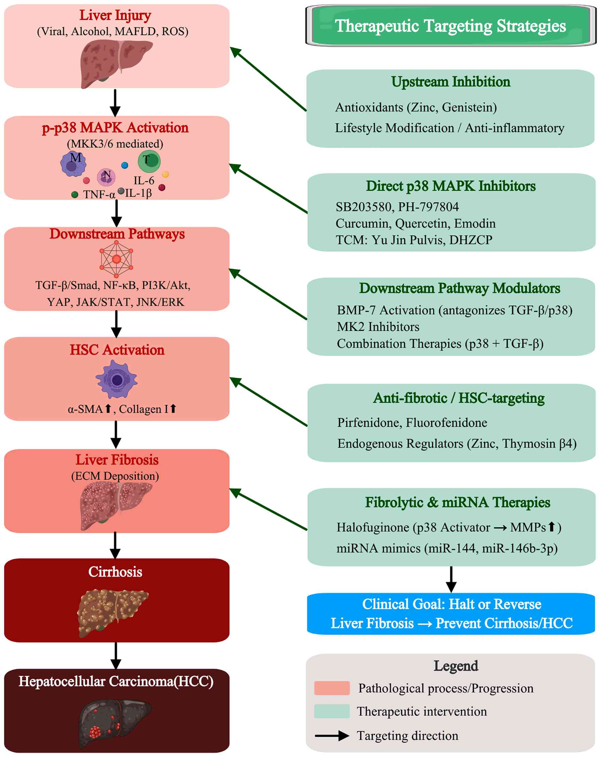 | Figure 4Therapeutic targeting of the p38 MAPK
cascade in liver fibrosis. The schematic presents a comprehensive
overview of the pathological progression (left column) and
corresponding intervention strategies (right column) targeting
different nodes of the p38 MAPK cascade. Left column, pathological
progression: The cascade begins with liver injury (viral hepatitis,
alcohol, MAFLD, ROS, environmental toxins), followed by p-p38 MAPK
activation (via MKK3/6, with associated inflammatory cells and
pro-inflammatory mediators, specifically IL-1β, IL-6 and TNF-α).
Downstream signaling pathways (NF-κB, TGF-β/Smad, PI3K/Akt, YAP,
JNK/ERK, JAK/STAT) then drive HSC activation (α-SMA↑, collagen I↑),
resulting in hepatic fibrosis (ECM deposition), progression to
cirrhosis, and ultimately HCC. Red boxes indicate pathological
states; downward arrows denote disease progression. Right column,
therapeutic targeting strategies: Intervention options are
categorized by their point of action. Upstream inhibition (via
antioxidants, lifestyle modifications) targets liver injury. Direct
p38 MAPK inhibitors (SB203580, PH-797804; natural compounds such as
curcumin, quercetin, emodin; TCM formulations such as Yu Jin Pulvis
and Dahuang Zhechong Pill) block p-p38 activation. Downstream
pathway modulators (BMP-7 activation, MK2 inhibitors, combination
therapies) interfere with TGF-β/Smad and other signaling networks.
Anti-fibrotic/HSC-targeting agents (pirfenidone, fluorofenidone,
zinc, thymosin β4) inhibit HSC activation. Fibrolytic and
miRNA-based therapies (halofuginone, a p38 activator that increases
MMP-3/-13; miRNA mimics such as miR-144 and miR-146b-3p that
indirectly suppress p38) promote ECM degradation or reduce p38
activity. Dashed green arrows indicate the therapeutic targeting
direction toward the corresponding pathological step. Bottom right
box, clinical goal: The ultimate objective is to halt or reverse
liver fibrosis and prevent progression to cirrhosis or HCC. p38
MAPK, p38 mitogen-activated protein kinase; MAFLD,
metabolic-associated fatty liver disease; ROS, reactive oxygen
species; MKK, mitogen-activated protein kinase kinase; IL-1β,
interleukin-1β; IL-6, interleukin-6; TNF-α, tumor necrosis
factor-α; NF-κB, nuclear factor β-light-chain-enhancer of activated
B cells; TGF-β, transforming growth factor-β; Smad, small mothers
against decapentaplegic; PI3K, phosphoinositide 3-kinase; Akt,
protein kinase B; YAP, yes-associated protein; JNK, c-Jun
N-terminal kinase; ERK, extracellular signal-regulated kinase; JAK,
Janus kinase; STAT, signal transducer and activator of
transcription; HSC, hepatic stellate cell; α-SMA, α-smooth muscle
actin; ECM, extracellular matrix; HCC, hepatocellular carcinoma;
TCM, traditional Chinese medicine; BMP-7, bone morphogenetic
protein 7; MK2, mitogen-activated protein kinase-activated protein
kinase 2; MMP, matrix metalloproteinase; miR, microRNA. |
Notably, mechanistic studies reveal that p38 MAPK
blockade reduces collagen deposition and suppresses inflammatory
cytokine production (76,147).
Exosomes from adipose-derived mesenchymal stem cells elevated HGF
expression to reduce oxidative stress and fibrosis markers via
PI3K/Akt/p38 MAPK pathway inhibition in CCL4-induced
liver fibrosis models (12).
The development of novel p38 MAPK inhibitors has
also been a significant area of research. Research in preclinical
models shows that PH-797804 and similar selective inhibitors have
favorable pharmacological properties, leading to better treatment
outcomes (148). The various
classes of p38 MAPK-targeting agents, including synthetic
inhibitors, natural compounds, traditional Chinese medicine
(TCM)-based formulations, repurposed drugs, and miRNA-based
therapies, are schematically summarized in Fig. 4, which illustrates their points
of intervention along the fibrotic signaling pathway. The compounds
block p38 MAPK kinase activity while exhibiting reduced binding to
other targets, thereby improving their potential for medical use
(149). The particular
structure of these inhibitors helps patients avoid side effects,
which can happen when kinase inhibitors interact with multiple
targets.
Research into existing medications to modulate the
p38 MAPK pathway has yielded promising results. For example,
several natural compounds and traditional herbal medicines have
been identified that can modulate the p38 MAPK pathway, providing
additional avenues for therapeutic intervention (22).
TCMs include Yu Jin Pulvis as one of several
effective treatments that show promise for managing hepatic
fibrosis by modulating key signaling pathways. Research has shown
that Jiawei Taohe Chengqi Decoction can reverse hepatic fibrosis in
animal models by modulating the p38 MAPK pathway (7). Similarly, Dahuang Zhechong Pill was
demonstrated to inhibit HSC fibrosis by downregulating the p38
MAPK/NF-κB/TGF-β1 signaling axis (150,151). Fuzheng Huayu Recipe has been
shown to improve liver fibrosis by restoring the TGF-β1/BMP-7
balance, thereby suppressing p38 MAPK phosphorylation and promoting
MET in HSCs (130). The core
bioactive components of Yinchenhao Decoction, including benzyl
acetate, vanillic acid, and polydatin, were found to work together
to block the PI3K-Akt and p38 MAPK pathways in HSCs, resulting in
anti-fibrotic effects (152).
There are also some plant extracts, such as both
Thunbergia laurifolia (TL) and Ginkgo biloba extract
(GBE), that demonstrate anti-fibrotic effects by targeting the p38
MAPK pathway. TL prevents fibrosis by inhibiting the Erk1/2 kinase
and p38 MAPK activation pathways. The anti-fibrotic effects of GBE
occur through inhibition of p38 MAPK, thereby preventing HSC
activation and inflammation (54,153).
Natural compounds and their derivatives exhibit
anti-fibrotic properties in liver tissue via p38 MAPK signaling
inhibition in HSCs, except for harmine. They can be classified into
three groups: i) Antioxidants that indirectly suppress p38
activation: Astragalus-derived astragaloside IV was shown to work
with ferulic acid to block HSC activation through reduced p38 MAPK
activity, which results from oxidative stress (102,154). ii) Direct p38 phosphorylation
inhibitors: Short-term gossypetin treatment was demonstrated to
block MKK3/6-p38 MAPK and p53 activation, thereby decreasing
activation of HSCs and Kupffer cells (155). Likewise, compounds such as
baicalin, quercetin, hydroxysafflor yellow A, oxymatrine, and a
novel synthetic derivative of oleanolic acid (CPU-II2) were all
reported to reduce liver fibrosis by directly inhibiting p38 MAPK
phosphorylation (39,53,119,156,157). iii) Multi-target agents that
simultaneously block p38 and other pro-fibrotic pathways: Compounds
such as emodin, dioscin, curcumin, isorhamnetin, schisandrin B,
spinosin, betulin, ankaflavin/monascin, salvianolic acid B,
salvianolic acid A, butein, neferine, total C-21 steroidal
glycosides, dilinoleoylphosphatidylcholine, γ-linolenic acid, and
cannabidiol achieve broader pathway inhibition, targeting p38 MAPK
alongside key partners such as TGF-β/Smad, sirtuin 1/nuclear
erythroid factor 2-related factor 2 (Nrf2), methionine
adenosyltransferase 2B, cannabinoid 2 receptor, NF-κB/IκBα,
neuron-derived clone 77/apoptosis signal-regulating kinase 1
(ASK1), JNK, Akt/NF-κB, ERK, Nrf2/heme oxygenase-1, JAK1/STAT3,
plasminogen activator inhibitor 1, NOX4, adenosine A2A receptor,
and PPAR-α (40,76,77,117,158-171). Among these, schisandrin B and
spinosin have shown promising in vivo efficacy with
favorable safety profiles, whereas curcumin and quercetin suffer
from poor bioavailability that limits clinical translation
(39,117,159,161). A full list is provided in
Table IV.
| Table IVSummary of representative p38 MAPK
inhibitors and activators with their antifibrotic profiles in liver
fibrosis. |
Table IV
Summary of representative p38 MAPK
inhibitors and activators with their antifibrotic profiles in liver
fibrosis.
A, Inhibitors
|
|---|
| Category | Representative
Agent(s) | Mechanism of action
(p38 MAPK-related) | Key efficacy in
preclinical models | Research stage | Potential
advantages |
Limitations/challenges | Model/evidence | (Refs.) |
|---|
| Synthetic
small-molecule inhibitors | SB203580 | Competitive ATP
binding inhibitor selective for p38α/β; prevents phosphorylation of
downstream substrates (ATF2 and MK2) | ↓ HSC activation, ↓
α-SMA, ↓ Col I, ↓ inflammatory cytokines (TNF-α and IL-1β) in
CCl4 and DBP models | Preclinical
(best-characterized tool compound) | Best-characterized
tool compound; extensive in vitro and in vivo
validation | Off-target effects
(such as RIPK2 and CK1); lacks isoform absolute selectivity;
limited clinical translation | CCl4 and
DBP-induced liver fibrosis models (rodent) | (14,84,85,91) |
| PH-797804 | Potent, selective
ATP-competitive p38α/β inhibitor with favorable pharmacokinetic
profile | ↓ fibrosis markers,
↓ collagen deposition in rodent fibrosis models | Advanced
preclinical | Improved
selectivity; favorable drug-like properties | Limited data
specifically in liver fibrosis models; safety in chronic liver
disease unknown | Rodent fibrosis
models | (148) |
| Natural compounds
(plant-derived) | Curcumin | Inhibits p38 MAPK
phosphorylation; disrupts TGF-β1/p38 MAPK/Smad crosstalk | ↓ HSC activation, ↓
ECM deposition, ↓ oxidative stress | Preclinical | Multi-targeted
(anti-inflammatory, antioxidant); oral bioavailability | Poor systemic
bioavailability; requires high doses; limited clinical efficacy
data | HSC in vitro
models | (159) |
| Quercetin | Suppresses p38 MAPK
and NF-κB phosphorylation; modulates Bcl-2/Bax balance | ↓ HSC
proliferation, ↓ collagen synthesis, ↓ apoptosis in
hepatocytes | Preclinical | Broad bioactivity;
well-tolerated; dietary source | Non-specific;
pleiotropic effects complicate mechanistic interpretation | Liver fibrosis
animal models | (39) |
| Schisandrin B | Targets CB2
receptor in Kupffer cells; indirectly suppresses p38 MAPK/NF-κB
signaling | ↓ hepatic
inflammation, ↓ HSC activation, ↓ collagen deposition | Preclinical | Dual anti-inflam
matory and antifibrotic; favorable safety profile | Mechanism indirect;
p38 nhibition may tbe secondary to CB2 activation |
CCl4-induced liver fibrosis
mouse model | (117) |
| Emodin | Inhibits p38 MAPK
and Smad signaling pathways | ↓ α-SMA, ↓ Col I, ↓
HSC proliferation in vitro | Preclinical | Multi-pathway
inhibition (p38 + Smad) | Limited in
vivo pharmacokinetic data; potential hepatotoxicity at high
doses | HSCs in
vitro | (77) |
| Dioscin | Activates
Sirt1/Nrf2 axis; suppresses p38 MAPK phosphorylation | ↓ oxidative stress,
↓ HSC activation, ↓ ECM accumulation | Preclinical | Combines
antioxidant and anti-p38 effects | Insufficient
large-animal validation | BDL and DMN-induced
liver fibrosis models | (158) |
| Astragaloside IV,
gossypetin, baicalin, hydroxysafflor yellow A, oxymatrine, and
oleanolic acid derivative (CPU-II2) | Directly inhibit
p38 MAPK phosphorylation or block upstream activation | ↓ HSC activation, ↓
fibrosis markers in various models | Preclinical | Diverse structural
scaffolds; potential for optimization | Variable potency;
mechanism details often incomplete | Various liver
fibrosis models (CCl4, HSCs) | (53,102,119,154-157) |
| Isorhamnetin,
spinosin, betulin, ankaflavin/monascin, salvianolic acid A/B,
butein, neferine, Total C-21 steroidal glycosides, DLPC,
γ-linolenic acid, cannabidiol, ursolic acids, and caffeine | Achieve broader
pathway inhibition targeting p38 MAPK alongside TGF-β/Smad, NF-κB,
Nrf2, JNK and ERK, PI3K/Akt | ↓ HSC activation, ↓
ECM deposition, ↓ inflammation | Preclinical | Multi-target
effects may enhance efficacy | Pleiotropic
effects; primary target often unclear | Multiple liver
fibrosis models (CCl4, BDL, HSCs) | (40,76,160-171) |
| Harmine | Inhibits p38 MAPK
phosphorylation; modulates AMPK/mTOR signaling | ↓ excessive
autophagy, ↓ inflammation, ameliorates CCl4-induced
acute liver injury | Preclinical | Natural β-carboline
alkaloid; unique autophagy-modulating mechanism | Context-dependent;
opposite effect compared with SB203580 on autophagy; requires
further validation in fibrosis models |
CCl4-induced acute liver injury
model (mice) | (129) |
| TCM
formulations | Yu Jin Pulvis | Blocks p38 MAPK and
PI3K/Akt signaling pathways | ↓
CCl4-induced fibrosis, ↓ HSC activation, ↓ inflammatory
infiltration | Preclinical | Multi-compound,
multi-target synergy; historical clinical use | Complex
composition; active components undefined; quality control
challenges |
CCl4-induced liver fibrosis in
rats | (22) |
| Dahuang Zhechong
pill | Downregulates the
p38 MAPK/NF-κB/TGF-β1 pathway | ↓ HSC activation, ↓
collagen deposition, ↓ pro-inflammatory cytokines | Preclinical | Classic TCM formula
with demonstrated antifibrotic efficacy | Mechanism not fully
deconvoluted; standardization issues | Rat models of
hepatic fibrosis | (150,151) |
| Fuzheng Huayu
Recipe | Restores
TGF-β1/BMP-7 balance; suppresses p38 MAPK phosphorylation; promotes
MET in HSCs | ↓ HSC activation, ↓
EMT, ↑ MET, ↓ fibrosis stage | Clinically used in
China for hepatitis fibrosis; multiple RCTs published | Clinically
validated efficacy; restores EMT/MET balance and promotes fibrosis
resolution | Western validation
limited; mechanistic complexity | Clinical trials
(RCTs) in hepatitis fibrosis | (130) |
| Jiawei Taohe
Chengqi decoction, Yinchenhao decoction | Regulates p38 MAPK
and PI3K/Akt pathways | Reverses hepatic
fibrosis in animal models | Preclinical | Traditional use;
multi-component synergy | Active components
often undefined; quality control challenges |
CCl4-induced liver fibrosis
animal models | (7,152) |
| Drug repurposing
candidates | Empagliflozin +
Metformin | Reduces p38 MAPKα
and ERK1/2 activity; enhances AMPK-mediated NF-κB inactivation | ↓
CCl4-induced fibrosis, ↓ oxidative stress, ↓
inflammation | Clinically approved
for diabetes | Clinically
approved; known safety profiles; synergistic potential | Not a direct p38
inhibitor; p38 modulation is indirect |
CCl4-induced liver fibrosis in
mice | (172) |
| Pirfenidone | Inhibits p38 MAPK
signaling; restricts Th2 differentiation | ↓ Th2 response, ↓
HSC activation, ↓ collagen deposition | Approved for
IPF | Approved for IPF;
repurposing opportunity | Moderate potency;
multiple mechanisms beyond p38 inhibition | Experimental liver
fibrosis (Th2-driven) | (62) |
| Fluorofenidone
(AKF-PD) | Inhibits
TGF-β1-induced p38 MAPK, ERK1/2, JNK, and Smad3
phosphorylation | ↓ HSC activation, ↓
ECM production, ↓ fibrotic markers | Preclinical/early
clinical | Novel pyridone
derivative; broad anti-fibrotic activity | Still in
preclinical/early clinical stages | Hepatic fibrosis
models | (173) |
| Endogenous/metal
ion-based | Zinc
supplementation | Antioxidant;
directly inhibits p38 MAPK signaling in HSCs | ↓
ethanol/acetaldehyde-induced HSC activation, ↓ ROS, ↓ collagen | Physiologic
nutrient | Physiologic
nutrient; excellent safety profile; low cost | Indirect; p38
inhibition may be partial; efficacy in advanced fibrosis
unclear |
Ethanol/acetaldehyde-induced HSCs | (115) |
| Thymosin β4
(Tβ4) | Blocks NF-κB and
JNK/p38 MAPK signaling; inhibits NLRP3 inflammasome | ↓ LPS/ATP-induced
HSC activation, ↓ inflammation | Preclinical | Endogenous peptide;
pleiotropic protective effects cost | Protein
therapeutic; delivery challenges; | LPS/ATP-induced
HSCs | (179) |
|
| B, Activators |
|
| Category | Representative
Agent(s) | Mechanism of action
(p38 MAPK-related) | Key efficacy in
preclinical models | Research stage | Potential
advantages |
Limitations/challenges | Model/evidence | (Refs.) |
|
| Drug repurposing
candidate | Halofuginone | Activates p38 MAPK
signaling (fibrolytic effect) | Increases MMP-3 and
MMP-13 expression in rat HSCs | Preclinical | Distinct fibrolytic
mechanism; promotes ECM degradation | Opposite to most
p38 inhibitors; mechanism requires further validation; potential
safety concerns | Rat HSCs in
vitro | (174) |
In addition, several drugs in clinical use have
therapeutic potential against hepatic fibrosis by modulating the
p38 MAPK signaling cascade. Abdelhamid et al (172) showed that empagliflozin
enhances the anti-fibrotic effects of metformin by reducing p38
MAPKα and ERK1/2 activity, resulting in stronger AMPK-mediated
NF-κB inactivation. Similarly, fluorofenidone was demonstrated to
attenuate hepatic fibrosis by suppressing HSC activation through
inhibition of phosphorylation of p38 MAPK, Smad3, ERK1/2 and JNK
triggered by TGF-β1 stimulation (173). Similarly, pirfenidone was shown
to function as an antifibrotic agent which prevents Th2 cell
formation and decreases fibrosis caused by Th2 cells through its
mechanism of blocking p38 MAPK signaling (62). Halofuginone was found to exert
fibrolytic effects by activating p38 MAPK, leading to increased
MMP-3 and MMP-13 expression in rat HSCs, indicating that this
pathway may operate differently for fibrosis treatment (174). Research has shown that current
medications can be repurposed to treat various medical conditions
when p38 MAPK pathways are involved in disease development
(175-177).
Furthermore, other endogenous compounds can also
exert anti-liver fibrotic effects through p38 MAPK.
Cyclin-dependent kinase inhibitor 2A/p16 deficiency was shown to
lead to increased ROS production in HSCs via p38 MAPK activation,
whereas thymosin β4 stopped HSC activation by blocking NF-κB and
JNK/p38 MAPK signaling pathways, which are activated by NLRP3 after
LPS exposure (178,179). The antioxidant enzyme
glutathione S-transferase α3 was found to function as an HSC
activation blocker, as it prevents activation of both glycogen
synthase kinase 3β and p38 MAPK signaling pathways (180). Taurine was demonstrated to
induce HSC apoptosis through increased TGF-β1 expression, which
activates the p38 MAPK-JNK-caspase-9/8/3 signaling pathway
(181).
Moreover, supplementation with metal ions,
particularly zinc, has been observed to suppress ethanol- and
acetaldehyde-induced HSC activation via various mechanisms. This
effect is attributed to its role as an antioxidant and as an
inhibitor of the p38 MAPK signaling pathway (115) (representative p38 MAPK
inhibitors/activators and their anti-fibrotic effects in liver
fibrosis are listed in Table
IV).
Future studies should determine how different
signaling pathways interact, informed by emerging research on liver
fibrosis development. It has been shown that the TGF-β and p38 MAPK
pathways work together to regulate fibrogenesis. The discovery
shows that treatment of liver fibrosis requires a comprehensive
approach that targets multiple disease mechanisms simultaneously to
achieve improved treatment outcomes (31).
Despite promising data supporting p38 MAPK as a
therapeutic target, challenges remain in the clinical translation
of p38 MAPK inhibitors. The therapeutic efficacy and safety
profiles of compounds in clinical trials remain under
investigation. The specific characteristics of these inhibitors
matter because p38 MAPK participates in various bodily processes so
that broad inhibition would lead to unwanted side effects. The
development of p38 MAPK therapies requires researchers to address
two essential factors: Off-target effects and the need for specific
inhibitors to reduce harmful side effects. The development of
selective inhibitors targeting particular p38 MAPK isoforms or
their downstream effectors may yield improved therapeutic outcomes
with fewer side effects (182).
The distinct expression patterns and functional characteristics of
each p38 MAPK isoform, highlighting p38δ as a promising target in
MAFLD-associated fibrosis are summarized in Table I.
The clinical development of p38 inhibitors has been
marked by significant pharmacokinetic and therapeutic challenges,
underscored by the dose-dependent exposure of agents such as the
oral drug ralimetinib in patients with cancer, yet is plagued by
off-target effects typical of conventional ATP-competitive
inhibitors that disrupt physiological processes such as regulatory
T-cell proliferation and cause serious adverse effects,
discouraging clinical application (183-185). This challenge is further
highlighted by research showing divergent therapeutic outcomes
between the p38α-specific inhibitor VCP979 and the dual p38α/β
inhibitor SB203580 in cancer-associated cachexia, emphasizing the
need for improved selectivity (186). Lessons from failed trials in
inflammatory diseases are instructive: In chronic obstructive
pulmonary disease (COPD), losmapimod failed to improve physical
capacity or pulmonary function notwithstanding favorable
tolerability, and in atherosclerosis, BIRB 796 (doramapimod, an
efficient and a specific inhibitor targeting p38 MAPK) and
BMS-582949 (an orally active and potent inhibitor exhibiting high
selectivity for p38α MAPK) did not reduce arterial inflammation
when compared with the placebo, suggesting that p38 inhibition
alone may be insufficient and may require combination strategies
(187-189). In rheumatoid arthritis (RA),
despite p38 MAPK being a central inflammatory mediator, numerous
inhibitors have failed in advanced trials due to limited efficacy
and tachyphylaxis, where initial anti-inflammatory effects wane
over time (190,191). Selectivity and toxicity remain
critical barriers, as non-selective inhibition of this master
kinase disrupts fundamental physiological pathways, compromising
the therapeutic window (190).
Consequently, an emerging strategy is to target downstream
effectors such as MAPK-MK2; early data show that MK2 inhibition can
produce sustained anti-inflammatory effects in RA without the
tachyphylaxis observed with direct p38 inhibitors (191). This approach represents a
valuable lesson: Modulating the pathway at a more downstream node
may preserve regulatory functions while improving efficacy and
tolerability (190,191).
Future research should determine how p38 MAPK
induces disease progression, develop targeted inhibitors, and test
combination therapies that leverage the multiple signaling routes
of p38 MAPK for medical applications. An integrated view of these
therapeutic strategies, highlighting the hierarchical relationship
between signaling nodes and intervention points, is presented in
Fig. 4. Future research should
determine how these inhibitors work and assess their potential
medical benefits and safety. Advances in p38 MAPK signaling
research will lead to improved treatment outcomes for patients with
various chronic diseases through the development of specific,
potent inhibitors. Given the extensive crosstalk illustrated in
Fig. 3, combination therapies
that simultaneously target p38 MAPK and its interacting pathways
(such as TGF-β/Smad, NF-κB, or PI3K/Akt) may offer greater efficacy
than a single-agent approach.
Current controversies and future
directions
Despite the substantial evidence establishing p38
MAPK as a key driver of liver fibrosis, the literature reveals
several apparent contradictions that warrant careful consideration.
These discrepancies likely stem from the context-dependent nature
of p38 MAPK signaling, in which outcomes vary by cell type,
activation stage, upstream regulators, and the specific stimuli
involved. Resolving these controversies will be essential for
translating p38 MAPK-targeted therapies into clinical practice.
Recent studies across different tissues and disease models have
further illuminated these complexities and opened new avenues for
therapeutic intervention (192-197).
Cell and context-specific functions
One of the most significant controversies concerns
the dual role of p38 MAPK in cell survival and death. Research has
shown that p38 MAPK activation can either promote or suppress
apoptosis depending on the cellular context. For example, fibulin-1
was shown to promote HSC activation via the p38 MAPK pathway,
suggesting a pro-survival, pro-fibrogenic role in these cells
(198). Conversely, in
cardiomyocytes, upregulation of dual specificity protein
phosphatase 4 was found to mitigate doxorubicin-induced
cardiotoxicity by inhibiting the p38 MAPK/MK2 signaling axis,
thereby underscoring the protective effect of p38 inhibition in
parenchymal cells (199). In
hepatocytes exposed to toxic agents, p38 MAPK activation under
oxidative stress was demonstrated to lead to cell death, while in
activated HSCs, it promoted survival and fibrogenic activity
(14). The differential
engagement of downstream substrates, such as p53 and Bax in
hepatocytes vs. survival factors in HSCs, may explain these
opposing outcomes (200-202).
These opposing outcomes can be reconciled by conceptualizing p38
MAPK as a 'context-dependent rheostat': In mesenchymal cells such
as HSCs and osteoblasts, its activation supports survival and
differentiation, whereas in parenchymal cells such as hepatocytes
and cardiomyocytes, it predominantly mediates stress-induced
apoptosis (203,204). This cell-type-specific duality
highlights that therapeutic targeting must consider not only the
kinase itself but also the cell-type-specific downstream effector
landscape. Given this duality, systemic p38 MAPK inhibitors are
unlikely to succeed in liver fibrosis; instead, cell-type-specific
delivery strategies (such as HSC-targeting nanoparticles) or
isoform-selective inhibitors that spare hepatocyte p38α should be
prioritized.
Stage-dependent effects and upstream
regulation in HSC activation
Further complicating the picture, p38 MAPK appears
to exert stage-dependent effects during HSC activation. Research
has demonstrated that blocking p38 MAPK during the early stages of
HSC activation reduces α-SMA expression, whereas inhibiting this
kinase in already-activated HSCs fails to produce similar effects.
Moreover, p38 suppression in activated HSCs paradoxically increased
cell proliferation, suggesting that p38 MAPK may exert
anti-proliferative functions once HSCs have fully
transdifferentiated (85). These
stage-dependent effects can be conceptualized through a 'temporal
switch' model: In early HSC activation or acute inflammation, p38
MAPK drives a pro-fibrogenic and pro-inflammatory program, whereas
in established disease, its sustained activation may shift toward
growth-suppressive or tissue-remodeling functions. This implies
that the therapeutic window for p38 MAPK inhibitors may be limited
to early fibrotic stages, with potential risks if administered
during established disease.
Emerging evidence suggests that targeting
regulators upstream of p38 MAPK activation, rather than the kinase
itself, may offer a more precise therapeutic window. In MASH,
ubiquilin 1 (UBQLN1) was found to initiate the p38 MAPK pathway by
mediating the degradation of its suppressor, suppressor of IKKε
(SIKE). Inhibition of this UBQLN1-SIKE-p38 MAPK axis mitigated
hepatic steatosis and fibrosis (196). This indicates that the
pathological activation of p38 MAPK is tightly controlled by
specific upstream modulators, and interventions at this level could
be more effective and potentially stage-specific than direct kinase
inhibition.
Dual roles in ECM remodeling and
inflammation
p38 MAPK also displays opposing functions in ECM
turnover. While it promotes ECM deposition by stabilizing collagen
mRNA (86), it can also induce
MMPs, including MMP-1, MMP-3, and MMP-13 (87-89). In intervertebral disc
degeneration, baicalin exerted protective effects by suppressing
the p38 MAPK signaling cascade, leading to downregulation of
catabolic factors such as MMP-3 and MMP-13 (205). Conversely, its well-established
role in driving the synthesis of pro-inflammatory cytokines,
specifically IL-6, IL-1β, and TNF-α, has been confirmed in models
of shoulder adhesive capsulitis and gastric ulcers, where p38
inhibition reduced inflammation and tissue fibrosis (197,206). Similarly, in the context of
inflammation, p38 MAPK acts primarily as a pro-inflammatory
mediator, yet it can also participate in protective antioxidant
responses under certain conditions, such as genistein-mediated
cardioprotection (27). This
functional dichotomy can be understood through a 'temporal-spatial
switch' model: In early acute responses, p38 MAPK drives
pro-inflammatory cytokine synthesis and ECM remodeling, whereas in
chronic settings, its sustained activation may shift toward
catabolic MMP expression (such as MMP-3 and MMP-13) or tissue
degradation. The net effect, pro-fibrotic or fibrolytic, is
dictated by the balance between these outputs, which in turn
depends on cell type, disease stage, and the duration of pathway
engagement (103).
Autophagy and novel cell death
mechanisms
The relationship between p38 MAPK and cellular
degradation pathways extends beyond classical apoptosis to include
autophagy and ferroptosis. Research indicates that inhibiting p38
MAPK with SB203580 restores the autophagic flux by blocking the
inhibitory phosphorylation of p38 on ULK1 (128). Conversely, the natural compound
harmine was shown to reduce excessive autophagy in
CCl4-induced liver injury by inhibiting p38 MAPK and
modulating AMPK/mTOR signaling (129). In acute kidney injury induced
by depleted uranium, downregulation of ethylmalonic encephalopathy
1 led to ROS-mediated activation of the p38 MAPK pathway, which
subsequently promoted nuclear receptor coactivator 4-mediated
ferritinophagy, ultimately triggering renal cell ferroptosis
(207). In neutrophils,
constitutive p38 MAPK phosphorylation was found to be involved in
spontaneous apoptosis, as SB203580 and antisense oligonucleotides
delayed apoptosis by ~24 h (208). These findings indicate that p38
MAPK serves as a 'stress-responsive hub' that integrates signals
toward apoptosis, autophagy, or ferroptosis depending on the
cellular context and upstream stimuli. For instance, in renal
cells, p38 activation has been shown to lead to ferroptotic cell
death, while in neutrophils, it has been found to promote
spontaneous apoptosis as a physiological clearance mechanism
(207,209). The final outcome is determined
by the balance of ROS, AMPK/mTOR status, and the availability of
iron-dependent pathways.
Challenges in clinical translation
The clinical translation of p38 MAPK inhibitors
continues to face significant hurdles, primarily owing to adverse
toxic effects and insufficient therapeutic benefit in complex
diseases (210). While
preclinical studies demonstrate promising anti-fibrotic efficacy,
clinical trials in other inflammatory diseases have revealed issues
with dose-dependent toxicity, off-target effects, and
tachyphylaxis, where initial anti-inflammatory effects wane over
time (190,191). For example, in COPD, losmapimod
failed to improve exercise tolerance despite good tolerability
(187). In atherosclerosis,
BIRB 796 (doramapimod) and BMS-582949 did not reduce arterial
inflammation vs. placebo (188). These failures suggest that p38
MAPK inhibition alone may be insufficient and may require
combination strategies (189).
Moreover, the lack of isoform selectivity in early inhibitors
likely contributed to adverse effects by disrupting physiological
p38 MAPK functions in non-target tissues (184,185). These divergent therapeutic
outcomes between p38α-specific inhibitors and dual p38α/β
inhibitors in conditions such as cancer-associated cachexia further
underscore the need for isoform-selective approaches (186).
Emerging strategies and future
directions
To overcome current challenges, several strategies
should be prioritized. Recognizing that direct p38 MAPK inhibition
has faced clinical hurdles due to tachyphylaxis and toxicity, a
shift toward more upstream or downstream nodes within the pathway
is warranted. First, targeting downstream effectors such as MK2 may
offer sustained anti-inflammatory effects without the tachyphylaxis
observed with direct p38 MAPK inhibitors (191). Furthermore, exploiting upstream
regulators, such as the UBQLN1-SIKE axis, or inducing inhibitory
post-translational modifications, offers novel avenues for precise
intervention that may avoid the toxicity associated with direct
kinase inhibition. This upstream-downstream dichotomy can be framed
as a 'hierarchical targeting strategy': Intervening at the level of
a single, context-specific upstream regulator may yield more
selective effects than blocking the pleiotropic p38 MAPK kinase
itself, and targeting a downstream effector could circumvent
compensatory feedback loops that cause tachyphylaxis.
Second, cell-type-specific targeting is being
explored. For example, the myeloid-specific p38α inhibitor MPL-5821
was shown to potently augment the inflammatory and phagocytic
responses of human macrophages against Staphylococcus aureus
without causing systemic toxicity, thereby presenting a novel
antibacterial approach (211).
Hence isoform- and cell-type-selective inhibitors, such as
MPL-5821, for specific cell populations (for example, myeloid
cells) can minimize on-target, off-tissue effects (196,211,212). The distinct tissue distribution
and functional roles of p38α, p38β, p38γ, and p38δ make
isoform-selective inhibitors a promising avenue, particularly for
p38δ, in MAFLD-associated fibrosis (33,124,125). This approach is supported by
evidence that while p38α is ubiquitously expressed and drives
canonical inflammatory responses, p38δ is enriched in myeloid and
hepatic cells and is linked to steatohepatitis and fibrosis. Thus,
isoform selectivity offers a 'cellular precision' strategy that
could preserve the protective functions of other p38 isoforms in
non-target tissues.
Third, the success of combination regimens in SMA
models should be rigorously tested in fibrotic diseases,
potentially pairing p38 pathway modulators with anti-fibrotic,
anti-inflammatory, or metabolic agents (213). In spinal muscular atrophy
(SMA), pharmacological inhibition of p38 MAPK synergized with
survival motor neuron-upregulating drugs, enhancing motor neuron
survival and synaptic rewiring, demonstrating a powerful
combinatorial neuroprotective strategy (213). Given the extensive crosstalk
illustrated in Fig. 3,
co-targeting p38 MAPK with TGF-β/Smad, NF-κB, or PI3K/Akt may yield
synergistic effects while allowing lower doses of individual
agents, thereby minimizing toxicity (14,20). This combination approach is
conceptually supported by the observation that p38 MAPK functions
not in isolation but as a 'signaling hub' that integrates with the
MAPK family (ERK and JNK) and the TGF-β1 pathway to drive HSC
activation and ECM remodeling (106,214). A rational combination, for
instance, pairing a p38 inhibitor with a TGF-β receptor kinase
inhibitor, could disrupt both the initiation and the maintenance
phases of fibrosis. Fourth, elucidating the precise molecular
determinants that dictate the pro-fibrotic vs. the protective
outcomes of p38 MAPK, whether they are post-translational modifica-
tions, interacting partners, or spatial signaling dynamics, will be
essential for developing context-aware therapeutics. Emerging
technologies, including single-cell transcriptomics and spatial
proteomics, offer powerful tools to dissect p38 MAPK signaling
heterogeneity across hepatic cell populations during fibrosis
progression and regression.
Fifth, novel approaches to target the pathway are
being discovered. A vitamin C-based nanostructure was found to
inhibit hepatic stellate cell proliferation and fibrosis by
covalently modifying p38 MAPK (vitcylation), blocking its nuclear
translocation and inducing cell cycle arrest (212). This represents a move beyond
traditional ATP-competitive inhibitors and potentially enabling
context-specific modulation.
In summary, the contradictions in the p38 MAPK
literature do not diminish its importance as a therapeutic target
but rather underscore the need for more nuanced approaches. The
apparent contradictions in cell survival, ECM turnover, and
autophagy regulation can be reconciled by viewing p38 MAPK as a
'context-dependent signaling rheostat,' where its output is
determined by a 'temporal-spatial switch' (pro-fibrotic early,
anti-proliferative late) and a 'bidirectional modulator' of
autophagy and cell death pathways (175,177,215). Previous studies reinforce p38
MAPK as a master regulator in fibrosis and related pathologies
within a highly contextual signaling network (124,216-219). The apparent contradictions
highlight its pleiotropic functions rather than invalidating it as
a target. The future lies in developing sophisticated intervention
strategies that account for cellular context, disease stage, and
pathway crosstalk to harness their therapeutic potential safely.
Moving forward, the field must prioritize isoform-specific
targeting, stage-dependent interventions, and rational combination
regimens to translate the promise of p38 MAPK modulation into
effective clinical treatments for patients with progressive liver
fibrosis.
Conclusions
The convergence of molecular, pharmacological, and
clinical evidence positions p38 MAPK as a nodal signaling hub in
the pathogenesis of liver fibrosis. Rather than acting as a linear
mediator, p38 MAPK integrates diverse fibrotic inputs, including
inflammatory cytokines, oxidative stress, and mechanical cues, and
translates them into coordinated cellular outputs through extensive
crosstalk with signaling cascades such as NF-κB, TGF-β/Smad,
PI3K/Akt, and YAP. This central role, underscored by consistent
preclinical efficacy of p38 MAPK inhibition across multiple injury
models, establishes the pathway as a compelling therapeutic
target.
However, several critical challenges must be
addressed to translate these findings into clinical practice.
First, the p38 MAPK pathway serves as a highly context-dependent
signaling hub, with its often-opposing functions, such as promoting
injury in some cell types, such as hepatocytes, while supporting
survival in others, such as activated HSCs, demanding
cell-type-specific or temporally controlled targeting strategies.
The ultimate role of p38 is critically determined by the inherent
function of the cell, divergent upstream activators and contexts,
differential engagement of downstream substrates, and integration
with other signaling networks. For example, the ASK1-p38 axis is a
key driver of hepatocyte death in injury models. By contrast, its
activity in the hepatic milieu also supports processes such as
fibrosis that depend on the survival of activated HSCs (41,220). In hepatocytes, pro-inflammatory
mediators, specifically IL-1β, TNF-α, oxidative stress, and DNA
damage, typically activate p38 (especially p38α), signals often
tightly coupled with death receptor pathways and endoplasmic
reticulum stress, steering p38 toward pro-apoptotic outcomes.
Conversely, in activated HSCs, p38 activation is frequently driven
by pro-fibrotic growth factors, including PDGF and TGF-β, which
primarily promote cell proliferation, migration, and ECM production
within a signaling context that favors activation of
survival-related downstream substrates. This cell-type-specific
duality underscores the therapeutic challenge of targeting p38 in
complex liver diseases, as systemic inhibition might simultaneously
protect parenchymal cells while potentially mitigating
fibrogenesis. Second, the four p38 MAPK isoforms exhibit distinct
tissue distribution and functional roles; developing
isoform-selective inhibitors (particularly p38α vs. p38δ) may
improve efficacy and reduce off-target toxicity. Third, the complex
crosstalk between p38 MAPK and other pro-fibrotic networks argues
against single-agent approaches. Future therapeutic efforts should
prioritize rational combination regimens that simultaneously
disrupt multiple nodes, for example, co-targeting p38 MAPK and
TGF-β signaling or integrating p38 inhibitors with antioxidants and
senolytic agents.
Beyond pharmacology, key mechanistic questions
remain unanswered. The precise molecular switches that dictate the
pro-fibrotic vs. protective outcomes of p38 MAPK are poorly
defined. Elucidating these determinants, whether post-translational
modifications, interacting partners, or spatial signaling dynamics,
will be essential for developing context-aware therapeutics.
Emerging technologies, including single-cell transcriptomics and
spatial proteomics, offer powerful tools to dissect p38 MAPK
signaling heterogeneity across hepatic cell populations during
fibrosis progression and regression.
In summary, p38 MAPK represents not merely a
biomarker of liver injury but a functional linchpin of the fibrotic
cascade. Advancing this target toward clinical application will
require a paradigm shift from broad kinase inhibition to
precision-oriented strategies that account for isoform specificity,
cellular context, and pathway interdependence. By addressing these
challenges, the field can transform mechanistic insights into
durable therapies for patients with progressive liver fibrosis.
Particularly, isoform-selective inhibitors targeting p38δ in MAFLD,
or combination therapies co-targeting p38 MAPK and TGF-β receptors,
represent the most promising near-term translational avenues.
Availability of data and materials
Not applicable.
Authors' contributions
HL and PY performed conceived and designed the
review. XY, HL, TW, FX, CH and CY wrote the first draft of the
manuscript. XY, HY and PY edited the manuscript. XY, and PY were
responsible for critical revisions of the article. HL and PY
contributed to the acquisition of funds. All authors read and
approved the final manuscript. Data authentication is not
applicable.
Ethics approval and consent to
participate
Not applicable.
Patient consent for publication
Not applicable.
Competing interests
The authors declare that they have no competing
interests.
Abbreviations:
|
AMPK
|
adenosine 5'-monophosphate-activated
protein kinase
|
|
BMP-7
|
bone morphogenetic protein-7
|
|
CCl4
|
carbon tetrachloride
|
|
ECM
|
extracellular matrix
|
|
EMT
|
epithelial-to-mesenchymal
transition
|
|
ERK
|
extracellular signal-regulated
kinase
|
|
HSCs
|
hepatic stellate cells
|
|
IL-
|
interleukin
|
|
JAK
|
Janus kinase
|
|
JNK
|
c-Jun N-terminal kinase
|
|
MAFLD
|
metabolic dysfunction-associated
fatty liver disease
|
|
MET
|
mesenchymal-to-epithelial
transition
|
|
MKK3/6
|
MAPK kinase 3/6
|
|
MMPs
|
matrix metalloproteinases
|
|
mTOR
|
mechanistic target of rapamycin
|
|
NF-κB
|
nuclear factor-κB
|
|
p38 MAPK
|
p38 mitogen-activated protein
kinase
|
|
PI3K/Akt
|
phosphoinositide 3-kinase/protein
kinase B
|
|
ROS
|
reactive oxygen species
|
|
Smad
|
mothers against decapentaplegic
homolog
|
|
α-SMA
|
α-smooth muscle actin
|
|
STAT
|
signal transducer and activator of
transcription
|
|
TGF-β
|
transforming growth factor-β
|
|
TNF
|
tumor necrosis factor
|
|
YAP
|
yes-associated protein
|
Acknowledgements
Not applicable.
Funding
The present review was supported by the Scientific Research
Cooperation Project between Wuhan Union Hospital and Jingshan Union
Hospital, Huazhong University of Science and Technology (grant no.
900005601).
References
|
1
|
Mohammed OS, Attia HG, Mohamed BMSA,
Elbaset MA and Fayed HM: Current investigations for liver fibrosis
treatment: Between repurposing the FDA-approved drugs and the other
emerging approaches. J Pharm Pharm Sci. 26:118082023. View Article : Google Scholar : PubMed/NCBI
|
|
2
|
Castera L, Rinella ME and Tsochatzis EA:
Noninvasive assessment of liver fibrosis. N Engl J Med.
393:1715–1729. 2025. View Article : Google Scholar : PubMed/NCBI
|
|
3
|
Graupera I, Thiele M, Castera L, Pera G,
Piano S, Sòria A, Fabrellas N, Toran P, Chacon C, Bech KT, et al:
Prevalence of liver fibrosis in the general population (the
LiverScreen project): A multinational European cohort study.
Lancet. 407:1448–1458. 2026. View Article : Google Scholar : PubMed/NCBI
|
|
4
|
Cao T, Yang YW, Yang X and Zhu HH:
Expression and significance of TGF-β1, p38MAPK and BMP-7 protein in
liver specimens of patients with alveolar hepatic echinococcosis.
Zhongguo Xue Xi Chong Bing Fang Zhi Za Zhi. 33:353–358. 2021.In
Chinese. PubMed/NCBI
|
|
5
|
Almalki RS, Gazzaz M, Miski SF, Fayed HM,
Mohamed BMSA, Hessin AF, Afifi SM, Esatbeyoglu T, Korany RMS and
Elbaset MA: Repositioning gemigliptin for the alleviation of
thioacetamide-induced liver fibrosis in rats: Targeting TLR4/MAPK,
SIRT1/AMPK/Nrf2, PI3K/AKT/mTOR axis and apoptosis. Eur J Pharm Sci.
212:1071922025. View Article : Google Scholar : PubMed/NCBI
|
|
6
|
Kim HY, Yu JH, Chon YE, Kim SU, Kim MN,
Han JW, Lee HA, Jin YJ, An J, Choi M and Jun DW: Prevalence of
clinically significant liver fibrosis in the general population: A
systematic review and meta-analysis. Clin Mol Hepatol. 30(Suppl):
S199–S213. 2024. View Article : Google Scholar : PubMed/NCBI
|
|
7
|
Ye L, Huang J, Liang X, Guo W, Sun X, Shao
C, He Y and Zhang J: Jiawei taohe chengqi decoction attenuates
CCl4 induced hepatic fibrosis by inhibiting HSCs
activation via TGF-β1/CUGBP1 and IFN-γ/Smad7 pathway.
Phytomedicine. 133:1559162024. View Article : Google Scholar
|
|
8
|
Ma FY, Tesch GH, Flavell RA, Davis RJ and
Nikolic-Paterson DJ: MKK3-p38 signaling promotes apoptosis and the
early inflammatory response in the obstructed mouse kidney. Am J
Physiol Renal Physiol. 293:F1556–F1563. 2007. View Article : Google Scholar : PubMed/NCBI
|
|
9
|
Amos LA, Ma FY, Tesch GH, Liles JT,
Breckenridge DG, Nikolic-Paterson DJ and Han Y: ASK1 inhibitor
treatment suppresses p38/JNK signalling with reduced kidney
inflammation and fibrosis in rat crescentic glomerulonephritis. J
Cell Mol Med. 22:4522–4533. 2018. View Article : Google Scholar : PubMed/NCBI
|
|
10
|
Koga Y, Tsurumaki H, Aoki-Saito H, Sato M,
Yatomi M, Takehara K and Hisada T: Roles of cyclic AMP response
element binding activation in the ERK1/2 and p38 MAPK signalling
pathway in central nervous system, cardiovascular system,
osteoclast differentiation and mucin and cytokine production. Int J
Mol Sci. 20:13462019. View Article : Google Scholar : PubMed/NCBI
|
|
11
|
Younossi ZM, Karrar A, Pierobon M,
Birerdinc A, Stepanova M, Abdelatif D, Younoszai Z, Jeffers T,
Felix S, Jeiran K, et al: An exploratory study examining how
nano-liquid chromatography-mass spectrometry and phosphoproteomics
can differentiate patients with advanced fibrosis and higher
percentage collagen in non-alcoholic fatty liver disease. BMC Med.
16:1702018. View Article : Google Scholar : PubMed/NCBI
|
|
12
|
Zhou H, Wu Y, Xue J and Yu L: Ameliorative
effects of HGF-overexpressed exosomes derived from ADMSCs on
oxidative stress in hepatic fibrosis. Histol Histopathol.
40:757–772. 2025.
|
|
13
|
Mou WL, Chen SR, Wu ZT, Hu LH, Zhang JY,
Chang HJ, Zhou H and Liu Y: LPS-TLR4/MD-2-TNF-α signaling mediates
alcohol-induced liver fibrosis in rats. J Toxicol Pathol.
35:193–203. 2022. View Article : Google Scholar : PubMed/NCBI
|
|
14
|
Huo S, Li B, Du J, Zhang X, Zhang J, Wang
Q, Song M and Li Y: Dibutyl phthalate induces liver fibrosis via
p38MAPK/NF-κB/NLRP3-mediated pyroptosis. Sci Total Environ.
897:1655002023. View Article : Google Scholar
|
|
15
|
Klintman D, Li X, Santen S, Schramm R,
Jeppsson B and Thorlacius H: p38 mitogen-activated protein
kinase-dependent chemokine production, leukocyte recruitment, and
hepatocellular apoptosis in endotoxemic liver injury. Ann Surg.
242:830–839. 2005. View Article : Google Scholar : PubMed/NCBI
|
|
16
|
Sato H, Tanaka T and Tanaka N: The effect
of p38 mitogen-activated protein kinase activation on inflammatory
liver damage following hemorrhagic shock in rats. PLoS One.
7:e301242012. View Article : Google Scholar : PubMed/NCBI
|
|
17
|
Ma X, Jia YT and Qiu DK: Inhibition of p38
mitogen-activated protein kinase attenuates experimental autoimmune
hepatitis: Involvement of nuclear factor kappa B. World J
Gastroenterol. 13:4249–4254. 2007. View Article : Google Scholar : PubMed/NCBI
|
|
18
|
Kobayashi M, Takeyoshi I, Yoshinari D,
Matsumoto K and Morishita Y: P38 mitogen-activated protein kinase
inhibition attenuates ischemia-reperfusion injury of the rat liver.
Surgery. 131:344–349. 2002. View Article : Google Scholar : PubMed/NCBI
|
|
19
|
Huang Y, Xiong K, Wang A, Wang Z, Cui Q,
Xie H, Yang T, Fan X, Jiang W, Tan X and Huang Q: Cold stress
causes liver damage by inducing ferroptosis through the p38
MAPK/Drp1 pathway. Cryobiology. 113:1045632023. View Article : Google Scholar : PubMed/NCBI
|
|
20
|
Kan Z, Zhao KX, Jiang C, Liu DY, Guo Y,
Liu LY, Wang WJ, He ZQ, Zhang ZF and Wang SY: Respiratory exposure
to graphene oxide induces pulmonary fibrosis and organ damages in
rats involving caspase-1/p38MAPK/TGF-β1 signaling pathways.
Chemosphere. 303:1351812022. View Article : Google Scholar
|
|
21
|
Zhao ZB, Ji K, Shen XY, Zhang WW, Wang R,
Xu WP and Wei W: Di(2-ethylhexyl) phthalate promotes hepatic
fibrosis by regulation of oxidative stress and inflammation
responses in rats. Environ Toxicol Pharmacol. 68:109–119. 2019.
View Article : Google Scholar : PubMed/NCBI
|
|
22
|
Wu W, Piao H, Wu F, Han Y, An D, Wu Y, Lin
Z, Piao Y and Jin J: Yu Jin Pulvis inhibits carbon
tetrachloride-induced liver fibrosis by blocking the MAPK and
PI3K/Akt signaling pathways. Am J Transl Res. 11:5998–6006.
2019.PubMed/NCBI
|
|
23
|
Tingting WU, Xin Y, Huiping Z, Jinwei G,
Hui Z, Peipei Z, Meng W, Guoqiang L and Hongwen S: Regulatory
effects of the p38 mitogen-activated protein kinase-myosin light
chain kinase pathway on the intestinal epithelial mechanical
barrier and the mechanism of modified Pulsatilla decoction in the
treatment of ulcerative colitis. J Tradit Chin Med. 44:885–895.
2024.PubMed/NCBI
|
|
24
|
Tran QTN, Gan PXL, Liao W, Mok YK, Chai
CLL and Wong WSF: Degradation of MK2 with natural compound
andrographolide: A new modality for anti-inflammatory therapy.
Pharmacol Res. 194:1068612023. View Article : Google Scholar : PubMed/NCBI
|
|
25
|
Nie B, Jiang H, Chen H and Liu Q:
Dexmedetomidine alleviates hyperalgesia in arthritis rats through
inhibition of the p38MAPK signaling pathway. Immunopharmacol
Immunotoxicol. 44:586–593. 2022. View Article : Google Scholar : PubMed/NCBI
|
|
26
|
Zhang Y, Zhang R, Lu L, Zhou N, Lv X, Wang
X and Feng Z: Knockdown of lectin-like oxidized low-density
lipoprotein-1 ameliorates alcoholic cardiomyopathy via inactivating
the p38 mitogen-activated protein kinase pathway. Bioengineered.
13:8926–8936. 2022. View Article : Google Scholar : PubMed/NCBI
|
|
27
|
Farruggio S, Raina G, Cocomazzi G, Librasi
C, Mary D, Gentilli S and Grossini E: Genistein improves viability,
proliferation and mitochondrial function of cardiomyoblasts
cultured in physiologic and peroxidative conditions. Int J Mol Med.
44:2298–2310. 2019.PubMed/NCBI
|
|
28
|
Li X, Xu F, Zhang P, Mao L, Guo Y, Li H,
Xie Y, Li Y, Liao Y, Chen J, et al: Overexpression of PRDM16
attenuates acute kidney injury progression: Genetic and
pharmacological approaches. MedComm (2020). 5:e7372024. View Article : Google Scholar : PubMed/NCBI
|
|
29
|
Lavu N, Richardson L, Radnaa E, Kechichian
T, Urrabaz-Garza R, Sheller-Miller S, Bonney E and Menon R:
Oxidative stress-induced downregulation of glycogen synthase kinase
3 beta in fetal membranes promotes cellular senescence†. Biol
Reprod. 101:1018–1030. 2019. View Article : Google Scholar : PubMed/NCBI
|
|
30
|
Gu Y, Hou T, Qin Y and Dong W: Zoledronate
promotes osteoblast differentiation in high-glucose conditions via
the p38MAPK pathway. Cell Biol Int. 47:216–227. 2023. View Article : Google Scholar
|
|
31
|
Xie G, Jiang R, Wang X, Liu P, Zhao A, Wu
Y, Huang F, Liu Z, Rajani C, Zheng X, et al: Conjugated secondary
12α-hydroxylated bile acids promote liver fibrogenesis.
EBioMedicine. 66:1032902021. View Article : Google Scholar
|
|
32
|
Maruyama H, Sakai S and Ieda M:
Endothelin-1 alters BMP signaling to promote proliferation of
pulmonary artery smooth muscle cells. Can J Physiol Pharmacol.
100:1018–1027. 2022. View Article : Google Scholar : PubMed/NCBI
|
|
33
|
Coulthard LR, White DE, Jones DL,
McDermott MF and Burchill SA: p38(MAPK): Stress responses from
molecular mechanisms to therapeutics. Trends Mol Med. 15:369–379.
2009. View Article : Google Scholar : PubMed/NCBI
|
|
34
|
Salminen A, Kauppinen A and Kaarniranta K:
Emerging role of NF-κB signaling in the induction of
senescence-associated secretory phenotype (SASP). Cell Signal.
24:835–845. 2012. View Article : Google Scholar
|
|
35
|
Liu X, Tan S, Liu H, Jiang J, Wang X, Li L
and Wu B: Hepatocyte-derived MASP1-enriched small extracellular
vesicles activate HSCs to promote liver fibrosis. Hepatology.
77:1181–1197. 2023. View Article : Google Scholar
|
|
36
|
Liang J, Bao D, Ye Z, Cao B, Lu Z and Chen
J: Neferine alleviates ovariectomy-induced osteoporosis by
enhancing osteogenic differentiation of bone marrow mesenchymal
stem cells via regulation of the p38MAPK pathway. Connect Tissue
Res. 65:253–264. 2024. View Article : Google Scholar : PubMed/NCBI
|
|
37
|
Barakat A, Doncheva NT, Prado J, Moll L,
Jensen JL, Díaz-delCastillo M, Novak I, Eijkelkamp N, Jensen LJ and
Heegaard AM: A ligand-receptor interactome of the bone tumor
microenvironment in multiple myeloma bone pain. Pain.
166:2831–2850. 2025. View Article : Google Scholar : PubMed/NCBI
|
|
38
|
Hui P, Zhou S, Cao C, Zhao W, Zeng L and
Rong X: The elucidation of the anti-inflammatory mechanism of EMO
in rheumatoid arthritis through an integrative approach combining
bioinformatics and experimental verification. Front Pharmacol.
14:11955672023. View Article : Google Scholar : PubMed/NCBI
|
|
39
|
Wang R, Zhang H, Wang Y, Song F and Yuan
Y: Inhibitory effects of quercetin on the progression of liver
fibrosis through the regulation of NF-кB/IкBα, p38 MAPK, and
Bcl-2/Bax signaling. Int Immunopharmacol. 47:126–133. 2017.
View Article : Google Scholar : PubMed/NCBI
|
|
40
|
Park JH, Lee MK and Yoon J:
Gamma-linolenic acid inhibits hepatic PAI-1 expression by
inhibiting p38 MAPK-dependent activator protein and
mitochondria-mediated apoptosis pathway. Apoptosis. 20:336–347.
2015. View Article : Google Scholar
|
|
41
|
Schuster-Gaul S, Geisler LJ, McGeough MD,
Johnson CD, Zagorska A, Li L, Wree A, Barry V, Mikaelian I, Jih LJ,
et al: ASK1 inhibition reduces cell death and hepatic fibrosis in
an Nlrp3 mutant liver injury model. JCI Insight. 5:e1232942020.
View Article : Google Scholar : PubMed/NCBI
|
|
42
|
Li Y, Gao M, Xu LN, Yin LH, Qi Y and Peng
JY: MicroRNA-142-3p attenuates hepatic ischemia/reperfusion injury
via targeting of myristoylated alanine-rich C-kinase substrate.
Pharmacol Res. 156:1047832020. View Article : Google Scholar : PubMed/NCBI
|
|
43
|
McKee C, Sigala B, Soeda J, Mouralidarane
A, Morgan M, Mazzoccoli G, Rappa F, Cappello F, Cabibi D, Pazienza
V, et al: Amphiregulin activates human hepatic stellate cells and
is upregulated in non alcoholic steatohepatitis. Sci Rep.
5:88122015. View Article : Google Scholar : PubMed/NCBI
|
|
44
|
Ping J, Li JT, Liao ZX, Shang L and Wang
H: Indole-3-carbinol inhibits hepatic stellate cells proliferation
by blocking NADPH oxidase/reactive oxygen species/p38 MAPK pathway.
Eur J Pharmacol. 650:656–662. 2011. View Article : Google Scholar
|
|
45
|
Peng Y, Yang H, Zhu T, Zhao M, Deng Y, Liu
B, Shen H, Hu G, Wang Z and Tao L: The antihepatic fibrotic effects
of fluorofenidone via MAPK signalling pathways. Eur J Clin Invest.
43:358–368. 2013. View Article : Google Scholar : PubMed/NCBI
|
|
46
|
Zhang Y, Chen R, Yang X, Qian L, Shen B
and Shuai Z: HSC-derived exosomal miR-122-5p inhibits EMT and
fibrosis of intrahepatic biliary epithelial cells to alleviate
primary biliary cholangitis. Front Immunol. 16:16840642025.
View Article : Google Scholar : PubMed/NCBI
|
|
47
|
Kawamura H, Naiki-Ito A, Kuang X, Murakami
A, Komura M, Suzuki T, Kato H, Nagayasu Y, Matsuura K, Fujiwara K,
et al: Sacubitril/valsartan attenuates inflammation and fibrosis
associated with decreased integrin α8 and inhibits
hepatocarcinogenesis in a rat model of metabolic
dysfunction-associated steatohepatitis. Hepatol Res. 56:420–435.
2026. View Article : Google Scholar
|
|
48
|
Zhang F, Ni C, Kong D, Zhang X, Zhu X,
Chen L, Lu Y and Zheng S: Ligustrazine attenuates oxidative
stress-induced activation of hepatic stellate cells by interrupting
platelet-derived growth factor-β receptor-mediat and p38 pathways.
Toxicol Appl Pharmacol. 265:51–60. 2012. View Article : Google Scholar : PubMed/NCBI
|
|
49
|
El-Kholy AA, Elkablawy MA and El-Agamy DS:
Lutein mitigates cyclophosphamide induced lung and liver injury via
NF-κB/MAPK dependent mechanism. Biomed Pharmacother. 92:519–527.
2017. View Article : Google Scholar : PubMed/NCBI
|
|
50
|
Usta M, Çiğremiş Y and Özen H: Nerve
growth factor acts as a modulator on the p38 MAPK pathway in
copper-induced liver damage. J Trace Elem Med Biol. 90:1276942025.
View Article : Google Scholar : PubMed/NCBI
|
|
51
|
Wang M, Zhang F, Zhou J, Gong K, Chen S,
Zhu X, Zhang M, Duan Y, Liao C, Han J and Yin Z: Glabridin
ameliorates alcohol-caused liver damage by reducing oxidative
stress and inflammation via p38 MAPK/Nrf2/NF-κB pathway. Nutrients.
15:21572023. View Article : Google Scholar
|
|
52
|
Arumugam S, Girish Subbiah K, Kemparaju K
and Thirunavukkarasu C: Neutrophil extracellular traps in acrolein
promoted hepatic ischemia reperfusion injury: Therapeutic potential
of NOX2 and p38MAPK inhibitors. J Cell Physiol. 233:3244–3261.
2018. View Article : Google Scholar
|
|
53
|
Deng ZY, Li J, Jin Y, Chen XL and Lu XW:
Effect of oxymatrine on the p38 mitogen-activated protein kinases
signalling pathway in rats with CCl4 induced hepatic fibrosis. Chin
Med J (Engl). 122:1449–1454. 2009.PubMed/NCBI
|
|
54
|
Wang Y, Wang R, Wang Y, Peng R, Wu Y and
Yuan Y: Ginkgo biloba extract mitigates liver fibrosis and
apoptosis by regulating p38 MAPK, NF-κB/IκBα, and Bcl-2/Bax
signaling. Drug Des Devel Ther. 9:6303–6317. 2015.
|
|
55
|
Sjøgaard-Frich LM, Henriksen MS, Lam SM,
Birkbak FJ, Czaplinska D, Flinck M and Pedersen SF: NHE1 regulation
in NAFLD in vitro contributes to hepatocyte injury and HSC
crosstalk. J Endocrinol. 263:e2400992024. View Article : Google Scholar : PubMed/NCBI
|
|
56
|
Li A, Zhang J, Zhang X, Wang J, Wang S,
Xiao X, Wang R, Li P and Wang Y: Angiotensin II induces connective
tissue growth factor expression in human hepatic stellate cells by
a transforming growth factor β-independent mechanism. Sci Rep.
7:78412017. View Article : Google Scholar
|
|
57
|
Borkham-Kamphorst E, van Roeyen CRC,
Ostendorf T, Floege J, Gressner AM and Weiskirchen R:
Pro-fibrogenic potential of PDGF-D in liver fibrosis. J Hepatol.
46:1064–1074. 2007. View Article : Google Scholar : PubMed/NCBI
|
|
58
|
Cheng Y, Zhu X, Cheng F, Ji L and Zhou Y:
Delta-like homolog1/GATA binding protein 2 axis mediates leptin
inhibition of PPARγ2 expression in hepatic stellate cells in vitro.
Life Sci. 192:183–189. 2018. View Article : Google Scholar
|
|
59
|
Yan K, Deng X, Zhai X, Zhou M, Jia X, Luo
L, Niu M, Zhu H, Qiang H and Zhou Y: p38 mitogen-activated protein
kinase and liver X receptor-α mediate the leptin effect on sterol
regulatory element binding protein-1c expression in hepatic
stellate cells. Mol Med. 18:10–18. 2012. View Article : Google Scholar :
|
|
60
|
Zhai X, Yan K, Fan J, Niu M, Zhou Q and
Zhou Y, Chen H and Zhou Y: The β-catenin pathway contributes to the
effects of leptin on SREBP-1c expression in rat hepatic stellate
cells and liver fibrosis. Br J Pharmacol. 169:197–212. 2013.
View Article : Google Scholar : PubMed/NCBI
|
|
61
|
Acquaviva A, Vecchio D, Arezzini B,
Comporti M and Gardi C: Signaling pathways involved in
isoprostane-mediated fibrogenic effects in rat hepatic stellate
cells. Free Radic Biol Med. 65:201–207. 2013. View Article : Google Scholar : PubMed/NCBI
|
|
62
|
Navarro-Partida J, Martinez-Rizo AB,
Gonzalez-Cuevas J, Arrevillaga-Boni G, Ortiz-Navarrete V and
Armendariz-Borunda J: Pirfenidone restricts Th2 differentiation in
vitro and limits Th2 response in experimental liver fibrosis. Eur J
Pharmacol. 678:71–77. 2012. View Article : Google Scholar : PubMed/NCBI
|
|
63
|
Andueza A, Garde N, García-Garzón A,
Ansorena E, López-Zabalza MJ, Iraburu MJ, Zalba G and
Martínez-Irujo JJ: NADPH oxidase 5 promotes proliferation and
fibrosis in human hepatic stellate cells. Free Radic Biol Med.
126:15–26. 2018. View Article : Google Scholar : PubMed/NCBI
|
|
64
|
Kao YH, Chen PH, Wu TY, Lin YC, Tsai MS,
Lee PH, Tai TS, Chang HR and Sun CK: Lipopolysaccharides induce
Smad2 phosphorylation through PI3K/Akt and MAPK cascades in HSC-T6
hepatic stellate cells. Life Sci. 184:37–46. 2017. View Article : Google Scholar : PubMed/NCBI
|
|
65
|
Bruno R, Galastri S, Sacchi P, Cima S,
Caligiuri A, DeFranco R, Milani S, Gessani S, Fantuzzi L, Liotta F,
et al: gp120 modulates the biology of human hepatic stellate cells:
A link between HIV infection and liver fibrogenesis. Gut.
59:513–520. 2010. View Article : Google Scholar
|
|
66
|
Li TZ, Kim SM, Hur W, Choi JE, Kim JH,
Hong SW, Lee EB, Lee JH and Yoon SK: Elk-3 contributes to the
progression of liver fibrosis by regulating the
epithelial-mesenchymal transition. Gut Liver. 11:102–111. 2017.
View Article : Google Scholar :
|
|
67
|
Li C, Yang G, Lin L, Xuan Y, Yan S, Ji X,
Song F, Lu M and Lan T: Slit2 signaling contributes to cholestatic
fibrosis in mice by activation of hepatic stellate cells. Exp Cell
Res. 385:1116262019. View Article : Google Scholar : PubMed/NCBI
|
|
68
|
Yang M, Li S, Luo R, Zhao Y, Sun Y, Li H,
Cui Q, Wu J and Mao L: ADAM8 promotes alcoholic liver fibrosis
through the MAPK signaling pathway. J Physiol Sci. 74:522024.
View Article : Google Scholar : PubMed/NCBI
|
|
69
|
Cui X, Zhang X, Yin Q, Meng A, Su S, Jing
X, Li H, Guan X, Li X, Liu S and Cheng M: F-actin cytoskeleton
reorganization is associated with hepatic stellate cell activation.
Mol Med Rep. 9:1641–1647. 2014. View Article : Google Scholar : PubMed/NCBI
|
|
70
|
Parvaresh Anbar A, Piran T, Farhadi M and
Karimi P: Iranian crack induces hepatic injury through
mitogen-activated protein kinase pathway in the liver of Wistar
rat. Iran J Basic Med Sci. 21:1179–1185. 2018.PubMed/NCBI
|
|
71
|
Tsukada S, Westwick JK, Ikejima K, Sato N
and Rippe RA: SMAD and p38 MAPK signaling pathways independently
regulate alpha1(I) collagen gene expression in unstimulated and
transforming growth factor-beta-stimulated hepatic stellate cells.
J Biol Chem. 280:10055–10064. 2005. View Article : Google Scholar : PubMed/NCBI
|
|
72
|
Li T, Fan X, Cai M, Jiang Y, Wang Y, He P,
Ni J, Mo A, Peng C and Liu J: Advances in investigating
microcystin-induced liver toxicity and underlying mechanisms. Sci
Total Environ. 905:1671672023. View Article : Google Scholar : PubMed/NCBI
|
|
73
|
Wang X, Tang X, Gong X, Albanis E,
Friedman SL and Mao Z: Regulation of hepatic stellate cell
activation and growth by transcription factor myocyte enhancer
factor 2. Gastroenterology. 127:1174–1188. 2004. View Article : Google Scholar : PubMed/NCBI
|
|
74
|
Hassan NH, Kamel GM, Fayed HM, Korany RMS
and Ramadan A: Dapagliflozin alleviates thioacetamide induced-liver
fibrosis in rats via controlling the Nrf2/HO-1 and TLR4/TGF-β1/PI3K
signaling pathways. Immunopharmacol Immunotoxicol. 47:392–405.
2025. View Article : Google Scholar : PubMed/NCBI
|
|
75
|
Lin W, Tsai WL, Shao RX, Wu G, Peng LF,
Barlow LL, Chung WJ, Zhang L, Zhao H, Jang JY and Chung RT:
Hepatitis C virus regulates transforming growth factor beta1
production through the generation of reactive oxygen species in a
nuclear factor kappaB-dependent manner. Gastroenterology.
138:2509–2518. 2518.e12010. View Article : Google Scholar : PubMed/NCBI
|
|
76
|
Cao Q, Mak KM and Lieber CS: DLPC
decreases TGF-beta1-induced collagen mRNA by inhibiting p38 MAPK in
hepatic stellate cells. Am J Physiol Gastrointest Liver Physiol.
283:G1051–G1061. 2002. View Article : Google Scholar : PubMed/NCBI
|
|
77
|
Wang X, Niu C, Zhang X and Dong M: Emodin
suppresses activation of hepatic stellate cells through p38
mitogen-activated protein kinase and Smad signaling pathways in
vitro. Phytother Res. 32:2436–2446. 2018. View Article : Google Scholar : PubMed/NCBI
|
|
78
|
Wu X, Wu X, Ma Y, Shao F, Tan Y, Tan T, Gu
L, Zhou Y, Sun B, Sun Y, et al: CUG-binding protein 1 regulates HSC
activation and liver fibrogenesis. Nat Commun. 7:134982016.
View Article : Google Scholar : PubMed/NCBI
|
|
79
|
Zhou MY, Cheng ML, Huang T, Hu RH, Zou GL,
Li H, Zhang BF, Zhu JJ, Liu YM, Liu Y and Zhao XK: Transforming
growth factor beta-1 upregulates glucose transporter 1 and
glycolysis through canonical and noncanonical pathways in hepatic
stellate cells. World J Gastroenterol. 27:6908–6926. 2021.
View Article : Google Scholar : PubMed/NCBI
|
|
80
|
Wang S, Ning J, Huai J and Yang H:
Hyperglycemia in pregnancy-associated oxidative stress augments
altered placental glucose transporter 1 trafficking via
AMPKα/p38MAPK signaling cascade. Int J Mol Sci. 23:85722022.
View Article : Google Scholar
|
|
81
|
Kuo LM, Kuo CY, Lin CY, Hung MF, Shen JJ
and Hwang TL: Intracellular glutathione depletion by oridonin leads
to apoptosis in hepatic stellate cells. Molecules. 19:3327–3344.
2014. View Article : Google Scholar : PubMed/NCBI
|
|
82
|
Dunning S, Hannivoort RA, de Boer JF,
Buist-Homan M, Faber KN and Moshage H: Superoxide anions and
hydrogen peroxide inhibit proliferation of activated rat stellate
cells and induce different modes of cell death. Liver Int.
29:922–932. 2009. View Article : Google Scholar : PubMed/NCBI
|
|
83
|
Yang X, Zhu L, Zhao W, Shi Y, He C and Xu
C: Comparative analysis of regulatory roles of P38 signaling
pathway in 8 types liver cell during liver regeneration. Gene.
594:66–73. 2016. View Article : Google Scholar : PubMed/NCBI
|
|
84
|
Zhang Y, Luo Y, Che X, Dai Z and Sun X and
Sun X: Forchlorfenuron exposure induces hepatocyte apoptosis via
MKK3/P38/ATF2 pathway. Int J Mol Sci. 27:21732026. View Article : Google Scholar : PubMed/NCBI
|
|
85
|
Schnabl B, Bradham CA, Bennett BL, Manning
AM, Stefanovic B and Brenner DA: TAK1/JNK and p38 have opposite
effects on rat hepatic stellate cells. Hepatology. 34:953–963.
2001. View Article : Google Scholar : PubMed/NCBI
|
|
86
|
Parsons CJ, Takashima M and Rippe RA:
Molecular mechanisms of hepatic fibrogenesis. J Gastroenterol
Hepatol. 22(Suppl 1): S79–S84. 2007. View Article : Google Scholar : PubMed/NCBI
|
|
87
|
Reunanen N, Li SP, Ahonen M, Foschi M, Han
J and Kähäri VM: Activation of p38 alpha MAPK enhances
collagenase-1 (matrix metalloproteinase (MMP)-1) and stromelysin-1
(MMP-3) expression by mRNA stabilization. J Biol Chem.
277:32360–32368. 2002. View Article : Google Scholar : PubMed/NCBI
|
|
88
|
Cortez DM, Feldman MD, Mummidi S, Valente
AJ, Steffensen B, Vincenti M, Barnes JL and Chandrasekar B: IL-17
stimulates MMP-1 expression in primary human cardiac fibroblasts
via p38 MAPK- and ERK1/2-dependent C/EBP-beta, NF-kappaB, and AP-1
activation. Am J Physiol Heart Circ Physiol. 293:H3356–H3365. 2007.
View Article : Google Scholar : PubMed/NCBI
|
|
89
|
Ravanti L, Heino J, López-Otín C and
Kähäri VM: Induction of collagenase-3 (MMP-13) expression in human
skin fibroblasts by three-dimensional collagen is mediated by p38
mitogen-activated protein kinase. J Biol Chem. 274:2446–2455. 1999.
View Article : Google Scholar : PubMed/NCBI
|
|
90
|
Tang N, Zhang YP, Ying W and Yao XX:
Interleukin-1β upregulates matrix metalloproteinase-13 gene
expression via c-Jun N-terminal kinase and p38 MAPK pathways in rat
hepatic stellate cells. Mol Med Rep. 8:1861–1865. 2013. View Article : Google Scholar : PubMed/NCBI
|
|
91
|
Zhang X, Li P, Zhou J, Zhang Z, Wu H, Shu
X, Li W, Wu Y, Du Y, Lü D, et al: FAK-p38 signaling serves as a
potential target for reverting matrix stiffness-modulated liver
sinusoidal endothelial cell defenestration. Biomaterials.
305:1224622024. View Article : Google Scholar : PubMed/NCBI
|
|
92
|
Zhang SC, Zheng YH, Yu PP, Min TH, Yu FX,
Ye C, Xie YK and Zhang QY: Lentiviral vector-mediated
down-regulation of IL-17A receptor in hepatic stellate cells
results in decreased secretion of IL-6. World J Gastroenterol.
18:3696–3704. 2012. View Article : Google Scholar : PubMed/NCBI
|
|
93
|
Zhang Y, Ren L, Tian Y, Guo X, Wei F and
Zhang Y: Signaling pathways that activate hepatic stellate cells
during liver fibrosis. Front Med (Lausanne). 11:14549802024.
View Article : Google Scholar : PubMed/NCBI
|
|
94
|
Ji YX, Huang Z, Yang X, Wang X, Zhao LP,
Wang PX, Zhang XJ, Alves-Bezerra M, Cai L, Zhang P, et al: The
deubiquitinating enzyme cylindromatosis mitigates nonalcoholic
steatohepatitis. Nat Med. 24:213–223. 2018. View Article : Google Scholar : PubMed/NCBI
|
|
95
|
Wu Y, He Y, Wang F, Yao N, Zhao Y and Tian
Z: Lipopolysaccharide inhibits autophagy and promotes inflammatory
responses via p38 MAPK-induced proteasomal degradation of Atg13 in
hepatic stellate cells. Mediators Inflamm. 2022:96039892022.
View Article : Google Scholar : PubMed/NCBI
|
|
96
|
Horrillo D, Gallardo N, Lauzurica N,
Barrus MT, San Frutos MG, Andres A, Ros M and Fernandez-Agullo T:
Development of liver fibrosis during aging: Effects of caloric
restriction. J Biol Regul Homeost Agents. 27:377–388.
2013.PubMed/NCBI
|
|
97
|
Adachi T, Togashi H, Suzuki A, Kasai S,
Ito J, Sugahara K and Kawata S: NAD(P)H oxidase plays a crucial
role in PDGF-induced proliferation of hepatic stellate cells.
Hepatology. 41:1272–1281. 2005. View Article : Google Scholar : PubMed/NCBI
|
|
98
|
Wang Q, Xian M, Li C, Mi L, Ji J, Wu Z,
Shi X and Han X: Protective effect of liquiritin against
cisplatin-induced liver injury in mice through reduction of
inflammation, oxidative stress, apoptosis and inhibition of p38
MAPK/p53 pathway based on network pharmacology and in experimental
validation. J Ethnopharmacol. 354:1205362026. View Article : Google Scholar
|
|
99
|
Abidi P, Zhang H, Zaidi SM, Shen WJ,
Leers-Sucheta S, Cortez Y, Han J and Azhar S: Oxidative
stress-induced inhibition of adrenal steroidogenesis requires
participation of p38 mitogen-activated protein kinase signaling
pathway. J Endocrinol. 198:193–207. 2008. View Article : Google Scholar : PubMed/NCBI
|
|
100
|
Li D, Guo X, Zhao W, Jingyu J, Xia C and
Yu G: Genome-wide DNA methylation dynamics in carbon
tetrachloride-induced mice liver fibrosis. Iran J Basic Med Sci.
26:85–92. 2023.PubMed/NCBI
|
|
101
|
Blas-García A and Apostolova N: Novel
therapeutic approaches to liver fibrosis based on targeting
oxidative stress. Antioxidants (Basel). 12:15672023. View Article : Google Scholar : PubMed/NCBI
|
|
102
|
Li X, Wang X, Han C, Wang X, Xing G, Zhou
L, Li G and Niu Y: Astragaloside IV suppresses collagen production
of activated hepatic stellate cells via oxidative stress-mediated
p38 MAPK pathway. Free Radic Biol Med. 60:168–176. 2013. View Article : Google Scholar : PubMed/NCBI
|
|
103
|
Wu H, He D, Biswas S, Shafiquzzaman M,
Zhou X, Charron J, Wang Y, Nayak BK, Habib SL, Liu H and Li B: mTOR
activation initiates renal cell carcinoma development by
coordinating ERK and p38MAPK. Cancer Res. 81:3174–3186. 2021.
View Article : Google Scholar : PubMed/NCBI
|
|
104
|
Zhang L, Zhang Y, Chang X and Zhang X:
Imbalance in mitochondrial dynamics induced by low PGC-1α
expression contributes to hepatocyte EMT and liver fibrosis. Cell
Death Dis. 11:2262020. View Article : Google Scholar
|
|
105
|
Qiu B, Lawan A, Xirouchaki CE, Yi JS,
Robert M, Zhang L, Brown W, Fernández-Hernando C, Yang X, Tiganis T
and Bennett AM: MKP1 promotes nonalcoholic steatohepatitis by
suppressing AMPK activity through LKB1 nuclear retention. Nat
Commun. 14:54052023. View Article : Google Scholar : PubMed/NCBI
|
|
106
|
Ruiz de Galarreta M, Arriazu E, Pérez de
Obanos MP, Ansorena E and Iraburu MJ: Antifibrogenic and apoptotic
effects of Ocoxin in cultured rat hepatic stellate cells. J Physiol
Biochem. 79:881–890. 2023. View Article : Google Scholar :
|
|
107
|
Li T, Yang Y, Song H, Li H, Cui A, Liu Y,
Su L, Crispe IN and Tu Z: Activated NK cells kill hepatic stellate
cells via p38/PI3K signaling in a TRAIL-involved degranulation
manner. J Leukoc Biol. 105:695–704. 2019. View Article : Google Scholar : PubMed/NCBI
|
|
108
|
Hannah WN Jr and Harrison SA: Lifestyle
and dietary interventions in the management of nonalcoholic fatty
liver disease. Dig Dis Sci. 61:1365–1374. 2016. View Article : Google Scholar : PubMed/NCBI
|
|
109
|
Vilar-Gomez E, Calzadilla-Bertot L,
Friedman SL, Gra-Oramas B, Gonzalez-Fabian L, Lazo-Del Vallin S,
Diago M and Adams LA: Serum biomarkers can predict a change in
liver fibrosis 1 year after lifestyle intervention for
biopsy-proven NASH. Liver Int. 37:1887–1896. 2017. View Article : Google Scholar : PubMed/NCBI
|
|
110
|
Perng DW, Wu YC, Tsai CC, Su KC, Liu LY,
Hsu WH and Lee YC: Bile acids induce CCN2 production through p38
MAP kinase activation in human bronchial epithelial cells: A factor
contributing to airway fibrosis. Respirology. 13:983–989. 2008.
View Article : Google Scholar : PubMed/NCBI
|
|
111
|
Xie J, Chen S, Chen Y, Tong J, Huang H,
Liao J, Sun J, Cong L and Zeng Y: FFA intervention on LO2 cells
mediates SNX-10 synthesis and regulates MMP9 secretion in LX2 cells
via TGF-β1. Arch Biochem Biophys. 764:1102552025. View Article : Google Scholar
|
|
112
|
Zhang W, Ping J, Zhou Y, Chen G and Xu L:
Salvianolic acid B inhibits activation of human primary hepatic
stellate cells through downregulation of the myocyte enhancer
factor 2 signaling pathway. Front Pharmacol. 10:3222019. View Article : Google Scholar : PubMed/NCBI
|
|
113
|
Sullivan BP, Kassel KM, Manley S, Baker AK
and Luyendyk JP: Regulation of transforming growth
factor-β1-dependent integrin β6 expression by p38 mitogen-activated
protein kinase in bile duct epithelial cells. J Pharmacol Exp Ther.
337:471–478. 2011. View Article : Google Scholar : PubMed/NCBI
|
|
114
|
Qi R, Chang Z, Zhao W, Qiu Y, Chang W,
Zhang Y, Hu X and Li Z: TEAD4-mediated upregulation of LPAR3
augments hepatic stellate cell activation in portal hypertension.
Cell Biol Toxicol. 41:1102025. View Article : Google Scholar : PubMed/NCBI
|
|
115
|
Szuster-Ciesielska A, Plewka K, Daniluk J
and Kandefer-Szerszen M: Zinc supplementation attenuates ethanol-
and acetaldehyde-induced liver stellate cell activation by
inhibiting reactive oxygen species (ROS) production and by
influencing intracellular signaling. Biochem Pharmacol. 78:301–314.
2009. View Article : Google Scholar : PubMed/NCBI
|
|
116
|
Gan L, Zheng L, Yao L, Lei L, Huang Y,
Zeng Z and Fang N: Exosomes from adipose-derived mesenchymal stem
cells improve liver fibrosis by regulating the miR-20a-5p/TGFBR2
axis to affect the p38 MAPK/NF-κB pathway. Cytokine.
172:1563862023. View Article : Google Scholar
|
|
117
|
Wang HQ, Wan Z, Zhang Q, Su T, Yu D, Wang
F, Zhang C, Li W, Xu D and Zhang H: Schisandrin B targets
cannabinoid 2 receptor in Kupffer cell to ameliorate
CCl4-induced liver fibrosis by suppressing NF-κB and p38
MAPK pathway. Phytomedicine. 98:1539602022. View Article : Google Scholar
|
|
118
|
Watanabe T, Jono H, Han J, Lim DJ and Li
JD: Synergistic activation of NF-kappaB by nontypeable Haemophilus
influenzae and tumor necrosis factor alpha. Proc Natl Acad Sci USA.
101:3563–3568. 2004. View Article : Google Scholar : PubMed/NCBI
|
|
119
|
Ganguly R, Gupta A and Pandey AK: Role of
baicalin as a potential therapeutic agent in hepatobiliary and
gastrointestinal disorders: A review. World J Gastroenterol.
28:3047–3062. 2022. View Article : Google Scholar : PubMed/NCBI
|
|
120
|
Sheikh MY, Choi J, Qadri I, Friedman JE
and Sanyal AJ: Hepatitis C virus infection: Molecular pathways to
metabolic syndrome. Hepatology. 47:2127–2133. 2008. View Article : Google Scholar : PubMed/NCBI
|
|
121
|
Feng H, Chen J, Wang H, Cheng Y, Zou Z,
Zhong Q and Xu J: Roflumilast reverses polymicrobial sepsis-induced
liver damage by inhibiting inflammation in mice. Lab Invest.
97:1008–1019. 2017. View Article : Google Scholar : PubMed/NCBI
|
|
122
|
Ise H, Araki Y, Song I and Akatsuka G:
N-acetylglucosamine-bearing polymers mimicking O-GlcNAc-modified
proteins elicit anti-fibrotic activities in myofibroblasts and
activated stellate cells. Glycobiology. 33:17–37. 2023. View Article : Google Scholar
|
|
123
|
Xie X, Lv H, Liu C, Su X, Yu Z, Song S,
Bian H, Tian M, Qin C, Qi J and Zhu Q: HBeAg mediates inflammatory
functions of macrophages by TLR2 contributing to hepatic fibrosis.
BMC Med. 19:2472021. View Article : Google Scholar : PubMed/NCBI
|
|
124
|
Salloum S, Jeyarajan AJ, Kruger AJ, Holmes
JA, Shao T, Sojoodi M, Kim MH, Zhuo Z, Shroff SG, Kassa A, et al:
Fatty acids activate the transcriptional coactivator YAP1 to
promote liver fibrosis via p38 mitogen-activated protein kinase.
Cell Mol Gastroenterol Hepatol. 12:1297–1310. 2021. View Article : Google Scholar : PubMed/NCBI
|
|
125
|
Yang J, Tang X, Liang Z, Chen M and Sun L:
Taurocholic acid promotes hepatic stellate cell activation via
S1PR2/p38 MAPK/YAP signaling under cholestatic conditions. Clin Mol
Hepatol. 29:465–481. 2023. View Article : Google Scholar : PubMed/NCBI
|
|
126
|
Hernández-Gea V and Friedman SL: Autophagy
fuels tissue fibrogenesis. Autophagy. 8:849–850. 2012. View Article : Google Scholar : PubMed/NCBI
|
|
127
|
Allaire M, Rautou PE, Codogno P and
Lotersztajn S: Autophagy in liver diseases: Time for translation? J
Hepatol. 70:985–998. 2019. View Article : Google Scholar : PubMed/NCBI
|
|
128
|
Jin L, Li J, Yang S, Zhang R, Hu C, Chen
Y, Tian Z, Ma W, Feng Y, Liu N, et al: MAPK p38/Ulk1 pathway
inhibits autophagy and induces IL-1β expression in hepatic stellate
cells. Am J Physiol Gastrointest Liver Physiol. 322:G360–G367.
2022. View Article : Google Scholar
|
|
129
|
Ma Y, Li W, Yao Q, Liu Y, Yu J, Zang L,
Wang S, Zhou L, Wen S, Luo Y, et al: Harmine ameliorates
CCl4-induced acute liver injury through suppression of
autophagy and inflammation. Int Immunopharmacol. 129:1115382024.
View Article : Google Scholar
|
|
130
|
Pan Q, Wang YQ, Li GM, Duan XY and Fan JG:
Fuzheng huayu recipe ameliorates liver fibrosis by restoring
balance between epithelial-to-mesenchymal transition and
mesenchymal-to-epithelial transition in hepatic stellate cells.
Biomed Res Int. 2015:9359032015. View Article : Google Scholar
|
|
131
|
Liu K, Lu R, Zhao Q, Du J, Li Y, Zheng M
and Zhang S: Association and clinicopathologic significance of
p38MAPK-ERK-JNK-CDC25C with polyploid giant cancer cell formation.
Med Oncol. 37:62019. View Article : Google Scholar : PubMed/NCBI
|
|
132
|
Iwayama H, Sakamoto T, Nawa A and Ueda N:
Crosstalk between smad and mitogen-activated protein kinases for
the regulation of apoptosis in cyclosporine A-induced renal tubular
injury. Nephron Extra. 1:178–189. 2011. View Article : Google Scholar : PubMed/NCBI
|
|
133
|
Giovannini MG, Cerbai F, Bellucci A,
Melani C, Grossi C, Bartolozzi C, Nosi D and Casamenti F:
Differential activation of mitogen-activated protein kinase
signalling pathways in the hippocampus of CRND8 transgenic mouse, a
model of Alzheimer's disease. Neuroscience. 153:618–633. 2008.
View Article : Google Scholar : PubMed/NCBI
|
|
134
|
Zhang XH, Li FF, Qi Y, Ming CR, Li Y, Pan
ST, Liu SJ and Ma TM: Electroacupuncture improves cutaneous
allergic reaction by inhibiting degranulation of intrape-ritoneal
mast cells, MAPK signaling and inflammatory factor levels in
urticaria rats. Zhen Ci Yan Jiu. 45:299–304. 2020.In Chinese.
PubMed/NCBI
|
|
135
|
Liu ZZ, Zhao YX, Lin JN and Zhou GP:
Effect of dihydroartemisinin on multidrug resistance of human oral
squamous cell carcinoma cell line KBV200 by regulating ROS-MAPK
pathway. Shanghai Kou Qiang Yi Xue. 28:586–590. 2019.In
Chinese.
|
|
136
|
Ren G, Bai C, Yi S, Cong Q and Zhu Y:
Mechanisms and therapeutic strategies for MAFLD targeting TLR4
signaling pathways. J Innate Immun. 16:45–55. 2024. View Article : Google Scholar :
|
|
137
|
Giahi L, Aumueller E, Elmadfa I and
Haslberger AG: Regulation of TLR4, p38 MAPkinase, IκB and miRNAs by
inactivated strains of lactobacilli in human dendritic cells. Benef
Microbes. 3:91–98. 2012. View Article : Google Scholar : PubMed/NCBI
|
|
138
|
Su Z and Wu F: Inflammatory factors induce
thrombosis through the miR-146b-3p/p38MAPK/COX-2 pathway. Biomed
Res Int. 2020:87183212020. View Article : Google Scholar : PubMed/NCBI
|
|
139
|
Qiu Y, Liu Y, Li Y and Wang D: Intestinal
mir-794 responds to nanopolystyrene by linking insulin and p38 MAPK
signaling pathways in nematode Caenorhabditis elegans. Ecotoxicol
Environ Saf. 201:1108572020. View Article : Google Scholar : PubMed/NCBI
|
|
140
|
Yang X, Xing H, Liu J, Yang L and Ma H and
Ma H: MicroRNA-802 increases hepatic oxidative stress and induces
insulin resistance in high-fat fed mice. Mol Med Rep. 20:1230–1240.
2019.PubMed/NCBI
|
|
141
|
Hao Y, Song S, Li T, Zai Q, Ma N, Li Y,
Yang L, Xiao P, Xu T, Ji L, et al: Oxidative stress promotes liver
fibrosis by modulating the microRNA-144 and SIN3A-p38 pathways in
hepatic stellate cells. Int J Biol Sci. 20:2422–2439. 2024.
View Article : Google Scholar : PubMed/NCBI
|
|
142
|
Nan A, Chen L, Zhang N, Liu Z, Yang T,
Wang Z, Yang C and Jiang Y: A novel regulatory network among
LncRpa, CircRar1, MiR-671 and apoptotic genes promotes lead-induced
neuronal cell apoptosis. Arch Toxicol. 91:1671–1684. 2017.
View Article : Google Scholar :
|
|
143
|
Roy RK, Sharma U, Wasson MK, Jain A,
Hassan MI and Prakash H: Macrophage activation syndrome and COVID
19: Impact of MAPK driven immune-epigenetic programming by
SARS-Cov-2. Front Immunol. 12:7633132021. View Article : Google Scholar : PubMed/NCBI
|
|
144
|
Zhao G, Aghakeshmiri S, Chen YT, Zhang HM,
Yip F and Yang D: NFAT5-mediated signalling pathways in viral
infection and cardiovascular dysfunction. Int J Mol Sci.
22:48722021. View Article : Google Scholar : PubMed/NCBI
|
|
145
|
Westra IM, Mutsaers HAM, Luangmonkong T,
Hadi M, Oosterhuis D, de Jong KP, Groothuis GMM and Olinga P: Human
precision-cut liver slices as a model to test antifibrotic drugs in
the early onset of liver fibrosis. Toxicol In Vitro. 35:77–85.
2016. View Article : Google Scholar : PubMed/NCBI
|
|
146
|
Guan Y, Fang Z, Hu A, Roberts S, Wang M,
Ren W, Johansson PK, Heilshorn SC, Enejder A and Peltz G: Live-cell
imaging of human liver fibrosis using hepatic micro-organoids. JCI
Insight. 10:e1870992024. View Article : Google Scholar : PubMed/NCBI
|
|
147
|
Vayalil PK, Iles KE, Choi J, Yi AK,
Postlethwait EM and Liu RM: Glutathione suppresses TGF-beta-induced
PAI-1 expression by inhibiting p38 and JNK MAPK and the binding of
AP-1, SP-1, and Smad to the PAI-1 promoter. Am J Physiol Lung Cell
Mol Physiol. 293:L1281–L1292. 2007. View Article : Google Scholar : PubMed/NCBI
|
|
148
|
Revuelta M, Elicegui A, Scheuer T,
Endesfelder S, Bührer C, Moreno-Cugnon L, Matheu A and Schmitz T:
In vitro P38MAPK inhibition in aged astrocytes decreases reactive
astrocytes, inflammation and increases nutritive capacity after
oxygen-glucose deprivation. Aging (Albany NY). 13:6346–6358. 2021.
View Article : Google Scholar : PubMed/NCBI
|
|
149
|
Ito S, Kuromiya K, Sekai M, Sako H, Sai K,
Morikawa R, Mukai Y, Ida Y, Anzai M, Ishikawa S, et al:
Accumulation of annexin A2 and S100A10 prevents apoptosis of
apically delaminated, transformed epithelial cells. Proc Natl Acad
Sci USA. 120:e23071181202023. View Article : Google Scholar : PubMed/NCBI
|
|
150
|
He XY, Xiong XJ, Liu MJ, Liang JT, Liu FY,
Xiao JY and Wu LJ: Dahuang zhechong pill alleviates liver fibrosis
progression by regulating p38 MAPK/NF-κ B/TGF-β1 pathway. Chin J
Integr Med. 30:1113–1120. 2024. View Article : Google Scholar : PubMed/NCBI
|
|
151
|
Cai HB, Sun XG, Liu ZF, Liu YW, Tang J,
Liu Q, Ji BM, Song YH, Zhou YC, Yang MH and Lv ZP: Effects of
dahuangzhechong pills on cytokines and mitogen activated protein
kinase activation in rats with hepatic fibrosis. J Ethnopharmacol.
132:157–164. 2010. View Article : Google Scholar : PubMed/NCBI
|
|
152
|
Chen X, Liu Q, Li Y, Zhong X, Fan Q, Ma K,
Luo L, Guan D and Zhu Z: Analysis of core functional components in
Yinchenhao Decoction and their pathways for treating liver
fibrosis. Nan Fang Yi Ke Da Xue Xue Bao. 44:1508–1517. 2024.In
Chinese. PubMed/NCBI
|
|
153
|
Namsen R, Rojanasthien N, Sireeratawong S,
Rojsanga P, Nimlamool W and Potikanond S: Thunbergia laurifolia
exhibits antifibrotic effects in human hepatic stellate cells. Evid
Based Complement Alternat Med. 2017:35085692017. View Article : Google Scholar
|
|
154
|
Dong H, Guo H, Liang Y, Wang X and Niu Y:
Astragaloside IV synergizes with ferulic acid to suppress hepatic
stellate cells activation in vitro. Free Radic Res. 51:167–178.
2017. View Article : Google Scholar : PubMed/NCBI
|
|
155
|
Xu C, Tai H, Chu Y, Liu Y, He J, Wang Y,
Su B and Li S: Gossypetin targets the liver-brain axis to alleviate
pre-existing liver fibrosis and hippocampal neuroinflammation in
mice. Front Pharmacol. 15:13853302024. View Article : Google Scholar : PubMed/NCBI
|
|
156
|
Liu Q, Wang CY, Liu Z, Ma XS, He YH, Chen
SS and Bai XY: Hydroxysafflor yellow A suppresses liver fibrosis
induced by carbon tetrachloride with high-fat diet by regulating
PPAR-γ/p38 MAPK signaling. Pharm Biol. 52:1085–1093. 2014.
View Article : Google Scholar : PubMed/NCBI
|
|
157
|
Wu LM, Wu XX, Sun Y, Kong XW, Zhang YH and
Xu Q: A novel synthetic oleanolic acid derivative (CPU-II2)
attenuates liver fibrosis in mice through regulating the function
of hepatic stellate cells. J Biomed Sci. 15:251–259. 2008.
View Article : Google Scholar
|
|
158
|
Gu L, Tao X, Xu Y, Han X, Qi Y, Xu L, Yin
L and Peng J: Dioscin alleviates BDL- and DMN-induced hepatic
fibrosis via Sirt1/Nrf2-mediated inhibition of p38 MAPK pathway.
Toxicol Appl Pharmacol. 292:19–29. 2016. View Article : Google Scholar : PubMed/NCBI
|
|
159
|
Hu X and Zhou Y: Curcumin reduces
methionine adenosyltransferase 2B expression by interrupting
phosphorylation of p38 MAPK in hepatic stellate cells. Eur J
Pharmacol. 886:1734242020. View Article : Google Scholar : PubMed/NCBI
|
|
160
|
Liu N, Feng J, Lu X, Yao Z, Liu Q, Lv Y,
Han Y, Deng J and Zhou Y: Isorhamnetin inhibits liver fibrosis by
reducing autophagy and inhibiting extracellular matrix formation
via the TGF-β1/Smad3 and TGF-β1/p38 MAPK pathways. Mediators
Inflamm. 2019:61750912019. View Article : Google Scholar
|
|
161
|
Lin G, Li W, Hong W, Zhu D, Hu H, Fu J,
Gao Y, Chen S, Chai D and Zeng JZ: Spinosin inhibits activated
hepatic stellate cell to attenuate liver fibrosis by targeting
Nur77/ASK1/p38 MAPK signaling pathway. Eur J Pharmacol.
966:1762702024. View Article : Google Scholar
|
|
162
|
Szuster-Ciesielska A, Plewka K, Daniluk J
and Kandefer-Szerszen M: Betulin and betulinic acid attenuate
ethanol-induced liver stellate cell activation by inhibiting
reactive oxygen species (ROS), cytokine (TNF-α, TGF-β) production
and by influencing intracellular signaling. Toxicology.
280:152–163. 2011. View Article : Google Scholar
|
|
163
|
Cheng CF and Pan TM: Ankaflavin and
monascin induce apoptosis in activated hepatic stellate cells
through suppression of the Akt/NF-κB/p38 signaling pathway. J Agric
Food Chem. 64:9326–9334. 2016. View Article : Google Scholar : PubMed/NCBI
|
|
164
|
Lv Z, Song Y, Xue D, Zhang W, Cheng Y and
Xu L: Effect of salvianolic-acid B on inhibiting MAPK signaling
induced by transforming growth factor-β1 in activated rat hepatic
stellate cells. J Ethnopharmacol. 132:384–392. 2010. View Article : Google Scholar : PubMed/NCBI
|
|
165
|
Li S, Wang R, Song F, Chen P, Gu Y, Chen C
and Yuan Y: Salvianolic acid A suppresses CCl -induced liver
fibrosis through regulating the Nrf2/HO-1, NF-4 κB/IκBα, p38 MAPK,
and JAK1/STAT3 signaling pathways. Drug Chem Toxicol. 46:304–313.
2023. View Article : Google Scholar
|
|
166
|
Szuster-Ciesielska A, Mizerska-Dudka M,
Daniluk J and Kandefer-Szerszen M: Butein inhibits ethanol-induced
activation of liver stellate cells through TGF-β, NFκB, p38, and
JNK signaling pathways and inhibition of oxidative stress. J
Gastroenterol. 48:222–237. 2013. View Article : Google Scholar :
|
|
167
|
Wang Y, Wang S, Wang R, Li S and Yuan Y:
Neferine exerts antioxidant and anti-inflammatory effects on carbon
tetrachloride-induced liver fibrosis by inhibiting the MAPK and
NF-κB/IκBα pathways. Evid Based Complement Alternat Med.
2021:41360192021.
|
|
168
|
Qin T, Wang M, Zhang T, Wang Y, Zhang Y,
Hasnat M, Zhuang Z, Ding Y and Peng Y: Total C-21 steroidal
glycosides from baishouwu ameliorate hepatic and renal fibrosis by
regulating IL-1β/MyD88 inflammation signaling. Front Pharmacol.
12:7757302021. View Article : Google Scholar
|
|
169
|
He W, Shi F, Zhou ZW, Li B, Zhang K, Zhang
X, Ouyang C, Zhou SF and Zhu X: A bioinformatic and mechanistic
study elicits the antifibrotic effect of ursolic acid through the
attenuation of oxidative stress with the involvement of ERK,
PI3K/Akt, and p38 MAPK signaling pathways in human hepatic stellate
cells and rat liver. Drug Des Devel Ther. 9:3989–4104.
2015.PubMed/NCBI
|
|
170
|
Wang H, Guan W, Yang W, Wang Q, Zhao H,
Yang F, Lv X and Li J: Caffeine inhibits the activation of hepatic
stellate cells induced by acetaldehyde via adenosine A2A receptor
mediated by the cAMP/PKA/SRC/ERK1/2/P38 MAPK signal pathway. PLoS
One. 9:e924822014. View Article : Google Scholar : PubMed/NCBI
|
|
171
|
Ma R, Xie N, Shu Y, Wu Y, He P, Xiang Y,
Zhou Y and Wang Y: Cannabidiol alleviates carbon
tetrachloride-induced liver fibrosis in mice by regulating NF-κB
and PPAR-α pathways. Exp Biol Med (Maywood). 249:101412024.
View Article : Google Scholar
|
|
172
|
Abdelhamid AM, Youssef ME, Abd El-Fattah
EE, Gobba NA, Gaafar AGA, Girgis S, Shata A, Hafez AM, El-Ahwany E,
Amin NA, et al: Blunting p38 MAPKα and ERK1/2 activities by
empagliflozin enhances the antifibrotic effect of metformin and
augments its AMPK-induced NF-κB inactivation in mice intoxicated
with carbon tetrachloride. Life Sci. 286:1200702021. View Article : Google Scholar
|
|
173
|
Peng Y, Li L, Zhang X, Xie M, Yang C, Tu
S, Shen H, Hu G, Tao L and Yang H: Fluorofenidone affects hepatic
stellate cell activation in hepatic fibrosis by targeting the
TGF-β1/Smad and MAPK signaling pathways. Exp Ther Med. 18:41–48.
2019.PubMed/NCBI
|
|
174
|
Popov Y, Patsenker E, Bauer M, Niedobitek
E, Schulze-Krebs A and Schuppan D: Halofuginone induces matrix
metalloproteinases in rat hepatic stellate cells via activation of
p38 and NFkappaB. J Biol Chem. 281:15090–15098. 2006. View Article : Google Scholar : PubMed/NCBI
|
|
175
|
Arabacilar P and Marber M: The case for
inhibiting p38 mitogen-activated protein kinase in heart failure.
Front Pharmacol. 6:1022015. View Article : Google Scholar : PubMed/NCBI
|
|
176
|
Gaffey K, Reynolds S, Plumb J, Kaur M and
Singh D: Increased phosphorylated p38 mitogen-activated protein
kinase in COPD lungs. Eur Respir J. 42:28–41. 2013. View Article : Google Scholar
|
|
177
|
Mai L, Zhu X, Huang F, He H and Fan W: p38
mitogen-activated protein kinase and pain. Life Sci.
256:1178852020. View Article : Google Scholar : PubMed/NCBI
|
|
178
|
Lv F, Li N, Kong M, Wu J, Fan Z, Miao D,
Xu Y, Ye Q and Wang Y: CDKN2a/p16 antagonizes hepatic stellate cell
activation and liver fibrosis by modulating ROS levels. Front Cell
Dev Biol. 8:1762020. View Article : Google Scholar : PubMed/NCBI
|
|
179
|
Choi J, Cho Y, Choi H, Lee S, Han H, Lee J
and Kwon J: Thymosin beta 4 inhibits LPS and ATP-induced hepatic
stellate cells via the regulation of multiple signaling pathways.
Int J Mol Sci. 24:34392023. View Article : Google Scholar : PubMed/NCBI
|
|
180
|
Chen H, Gan Q, Yang C, Peng X, Qin J, Qiu
S, Jiang Y, Tu S, He Y, Li S, et al: A novel role of glutathione
S-transferase A3 in inhibiting hepatic stellate cell activation and
rat hepatic fibrosis. J Transl Med. 17:2802019. View Article : Google Scholar : PubMed/NCBI
|
|
181
|
Liang XQ, Liang J, Zhao XF, Wang XY and
Deng X: Integrated network analysis of transcriptomic and
protein-protein interaction data in taurine-treated hepatic
stellate cells. World J Gastroenterol. 25:1067–1079. 2019.
View Article : Google Scholar : PubMed/NCBI
|
|
182
|
Chen CH, Lin YJ, Cheng LT, Lin CH and Ke
GM: Poloxamer-188 adjuvant efficiently maintains adaptive immunity
of SARS-CoV-2 RBD subunit vaccination through repressing p38MAPK
signaling. Vaccines (Basel). 10:7152022. View Article : Google Scholar : PubMed/NCBI
|
|
183
|
Patnaik A, Haluska P, Tolcher AW,
Erlichman C, Papadopoulos KP, Lensing JL, Beeram M, Molina JR,
Rasco DW, Arcos RR, et al: A first-in-human phase I study of the
Oral p38 MAPK inhibitor, ralimetinib (LY2228820 dimesylate), in
patients with advanced cancer. Clin Cancer Res. 22:1095–1102. 2016.
View Article : Google Scholar
|
|
184
|
Noguchi H, Miyagi-Shiohira C, Nakashima Y,
Saitoh I and Watanabe M: Novel cell-permeable p38-MAPK inhibitor
efficiently prevents porcine islet apoptosis and improves islet
graft function. Am J Transplant. 20:1296–1308. 2020. View Article : Google Scholar
|
|
185
|
He T, Liu S, Chen S, Ye J, Wu X, Bian Z
and Chen X: The p38 MAPK inhibitor SB203580 abrogates tumor
necrosis factor-induced proliferative expansion of mouse
CD4+Foxp3+ regulatory T cells. Front Immunol.
9:15562018. View Article : Google Scholar
|
|
186
|
Zhao Y, Dai J, Jiang Y, Wu H, Cui Y, Li X,
Mao H, Wang B, Ju S and Peng XG: Reducing white adipose tissue
browning using p38α MAPK inhibitors ameliorates cancer-associated
cachexia as assessed by magnetic resonance imaging. Nutrients.
14:30132022. View Article : Google Scholar
|
|
187
|
Watz H, Barnacle H, Hartley BF and Chan R:
Efficacy and safety of the p38 MAPK inhibitor losmapimod for
patients with chronic obstructive pulmonary disease: A randomised,
double-blind, placebo-controlled trial. Lancet Respir Med. 2:63–72.
2014. View Article : Google Scholar : PubMed/NCBI
|
|
188
|
Emami H, Vucic E, Subramanian S, Abdelbaky
A, Fayad ZA, Du S, Roth E, Ballantyne CM, Mohler ER, Farkouh ME, et
al: The effect of BMS-582949, a P38 mitogen-activated protein
kinase (P38 MAPK) inhibitor on arterial inflammation: A multicenter
FDG-PET trial. Atherosclerosis. 240:490–496. 2015. View Article : Google Scholar : PubMed/NCBI
|
|
189
|
Ahmadi A, Ahrari S, Salimian J, Salehi Z,
Karimi M, Emamvirdizadeh A, Jamalkandi SA and Ghanei M: p38 MAPK
signaling in chronic obstructive pulmonary disease pathogenesis and
inhibitor therapeutics. Cell Commun Signal. 21:3142023. View Article : Google Scholar : PubMed/NCBI
|
|
190
|
Wang J, Liu Y, Guo Y, Liu C, Yang Y, Fan
X, Yang H, Liu Y and Ma T: Function and inhibition of P38 MAP
kinase signaling: Targeting multiple inflammation diseases. Biochem
Pharmacol. 220:1159732024. View Article : Google Scholar
|
|
191
|
Gordon D, Kivitz A, Singhal A, Burt D,
Bangs MC, Huff EE, Hope HR and Monahan JB: Selective inhibition of
the MK2 pathway: Data from a phase IIa randomized clinical trial in
rheumatoid arthritis. ACR Open Rheumatol. 5:63–70. 2023. View Article : Google Scholar : PubMed/NCBI
|
|
192
|
González-Osuna L, Fukada SY,
Hernández-Cáceres MP, Luz-Crawford P, Cortez C, Rojas C, Carvajal
P, Sierra-Cristancho A and Vernal R: p38 mitogen-activated protein
kinase drives senescence in CD4+ T lymphocytes and
increases their pathological potential. Immun Ageing. 22:302025.
View Article : Google Scholar
|
|
193
|
Zhou J, Xia T, Zhang S, He Z, Li Z and
Chen Z: Interaction of vernodalin with p38 mitogen-activated
protein kinase (p38 MAPK) and subsequent effects in lung cancer
cell model. Int J Biol Macromol. 311:1434132025. View Article : Google Scholar : PubMed/NCBI
|
|
194
|
Fuchs M, Möchel M, Radeva MY, Schmitt T,
Yazdi AS, Hashimoto T and Waschke J: Direct inhibition precedes p38
mitogen activated protein kinase-mediated uncoupling in desmosomes
to reduce desmoglein 3 adhesion by pemphigus autoantibodies. Br J
Dermatol. 193:468–479. 2025. View Article : Google Scholar : PubMed/NCBI
|
|
195
|
Li W, Ma B, Tian X, Xiong Q, Zhou M, Liu W
and Shu X: MUSASHI1 promotes tau phosphorylation by activating the
p38 MAPK pathway. Neurochem Int. 190:1060662025. View Article : Google Scholar : PubMed/NCBI
|
|
196
|
Chen Y, Yang F, Zheng G, Wang Y, Liu L and
Yan Y: UBQLN1 Inhibition reduces MASH progression through
downregulating SIKE/p38 MAPK pathway in hepatocyte. J
Nanobiotechnology. 24:3732026. View Article : Google Scholar : PubMed/NCBI
|
|
197
|
Qi B, Chen Z, Lin H, Sun Y, Yi C and Chen
J: Antioxidant RuCo nanosheet attenuates capsule fibrosis in
adhesive capsulitis of shoulder by p38 MAPK signaling pathway
inhibition. Mater Today Bio. 33:1019792025. View Article : Google Scholar : PubMed/NCBI
|
|
198
|
Zhao W, Zhang J, Guo Q, Wang Q, Zhao H,
Xiao F, Han M, Cao Y, Ding R, Yang A and Xie W: Fibulin-1
deficiency alleviates liver fibrosis by inhibiting hepatic stellate
cell activation via the p38 MAPK pathway. Cell Mol Life Sci.
82:1922025. View Article : Google Scholar : PubMed/NCBI
|
|
199
|
Deng L, Zha Y, Zhu C, Li Y, He M and Zhang
S: DUSP4 mitigates doxorubicin-induced cardiotoxicity via the p38
MAPK/MK2 signaling pathway. Biochim Biophys Acta Mol Cell Res.
1873:1201412026. View Article : Google Scholar : PubMed/NCBI
|
|
200
|
Li Y, Chen J, Chen Y, Dong Q, Jin X, Zhao
P and Dong Z: Pyraclostrobin induces brain oxidative stress,
apoptosis and inflammation through mitochondrial
phosphorylation-induced ROS activation of the p38 MAPK pathway.
Pestic Biochem Physiol. 216:1067872026. View Article : Google Scholar
|
|
201
|
Zhou JS, Zhu Z, Wu F, Zhou Y, Sheng R, Wu
JC and Qin ZH: NADPH ameliorates MPTP-induced dopaminergic
neurodegeneration through inhibiting p38MAPK activation. Acta
Pharmacol Sin. 40:180–191. 2019. View Article : Google Scholar
|
|
202
|
Nakahara M, Okumura N, Nakano S and
Koizumi N: Effect of a p38 mitogen-activated protein kinase
inhibitor on corneal endothelial cell proliferation. Invest
Ophthalmol Vis Sci. 59:4218–4227. 2018. View Article : Google Scholar : PubMed/NCBI
|
|
203
|
Hu Y, Chan E, Wang SX and Li B: Activation
of p38 mitogen-activated protein kinase is required for osteoblast
differentiation. Endocrinology. 144:2068–2074. 2003. View Article : Google Scholar : PubMed/NCBI
|
|
204
|
Dodeller F and Schulze-Koops H: The p38
mitogen-activated protein kinase signaling cascade in CD4 T cells.
Arthritis Res Ther. 8:2052006. View
Article : Google Scholar : PubMed/NCBI
|
|
205
|
Wu Y, Li F, Shu S, Feng Z, Qiu Y, Li S and
Zhu Z: Baicalin alleviates intervertebral disc degeneration by
inhibiting the p38 MAPK signaling pathway. Exp Gerontol.
204:1127432025. View Article : Google Scholar : PubMed/NCBI
|
|
206
|
Chen YT, Lin YJ, Tsai CH and Lu YC: The
role of p38 MAPK in acteoside-mediated gastric ulcer protection.
Int J Med Sci. 23:916–926. 2026. View Article : Google Scholar : PubMed/NCBI
|
|
207
|
Huang F, Li W, Li J, Ran Y, Fu S, Shen L,
Li Q, Yang X, Zhao Y and Hao Y: Depleted uranium exposure induced
ferroptosis in renal cells via the ETHE1/P38-MAPK pathway. Arch
Toxicol. 100:465–474. 2026. View Article : Google Scholar
|
|
208
|
Ding N, Dahlke K, Janze AK, Mailer PC,
Maus R, Bohling J, Welte T, Bauer M, Riedemann NC and Maus UA: Role
of p38 mitogen-activated protein kinase in posttraumatic
immunosuppression in mice. J Trauma Acute Care Surg. 73:861–868.
2012. View Article : Google Scholar : PubMed/NCBI
|
|
209
|
Li Y, Liu J, Sun Y, Hu Y, Cong C, Chen Y
and Fang Y: Targeting p38 MAPK signaling pathway and neutrophil
extracellular traps: An important anti-inflammatory mechanism of
Huangqin Qingre Chubi Capsule in rheumatoid arthritis. Int
Immunopharmacol. 148:1141122025. View Article : Google Scholar : PubMed/NCBI
|
|
210
|
Bhole RP, Kadam N, Karwa PN, Labhade SD,
Kapare HS and Gurav S: Advances in targeting p38 MAPK for cancer
therapy: Insights from molecular pharmacology and medicinal
chemistry. Mol Divers. Jul 21–2025.Epub ahead of print. View Article : Google Scholar : PubMed/NCBI
|
|
211
|
Lund A, Moffat D, Jepsen SD, Desler C,
Andresen L, Bregenholt S, Reddy V and Skov S: Targeted p38
mitogen-activated protein kinase inhibition potently augment
inflammatory responses of human macrophages toward Staphylococcus
aureus. Mol Immunol. 183:145–155. 2025. View Article : Google Scholar : PubMed/NCBI
|
|
212
|
Xiang G, Zhang J, Lu Y, Wu F, Yang C, Shi
R, Wu Y, Liu Y and Jiang X: Magnesium-chelating vitamin C
nanostructures induce p38 MAPK vitcylation for hepatic fibrosis
therapy. ACS Appl Mater Interfaces. 18:18781–18792. 2026.
View Article : Google Scholar : PubMed/NCBI
|
|
213
|
Carlini MJ, Espinoza-Derout J, Van Alstyne
M, Tisdale S, Workman E, Triplett MK, Tattoli I, Yadav S, Henderson
CE, Watterson DM and Pellizzoni L: Identification of p38 MAPK
inhibition as a neuroprotective strategy for combinatorial SMA
therapy. EMBO Mol Med. 17:2762–2786. 2025. View Article : Google Scholar : PubMed/NCBI
|
|
214
|
Yang Y, He L, Yang W, Feng Y, Li R, Wu Y,
Yang R, Xiao W and He Y: Hydroxy safflower yellow A: A natural
compound from carthamus tinctorius L. with potent activity against
liver fibrosis and cancer. Phytomedicine. 153:1578582026.
View Article : Google Scholar : PubMed/NCBI
|
|
215
|
Bell JR, Eaton P and Shattock MJ: Role of
p38-mitogen-activated protein kinase in ischaemic preconditioning
in rat heart. Clin Exp Pharmacol Physiol. 35:126–134. 2008.
View Article : Google Scholar
|
|
216
|
Li J, Campanale NV, Liang RJ, Deane JA,
Bertram JF and Ricardo SD: Inhibition of p38 mitogen-activated
protein kinase and transforming growth factor-beta1/Smad signaling
pathways modulates the development of fibrosis in
adriamycin-induced nephropathy. Am J Pathol. 169:1527–1540. 2006.
View Article : Google Scholar : PubMed/NCBI
|
|
217
|
Liu C, Sun ZR, Wang MM, Yang ZZ, Zhang W,
Ren Y, Han XQ, Liu R, Li Q and Nie SN: Arctigenin attenuates
paraquat-induced human lung epithelial A549 cell injury by
suppressing ROS/p38 mitogen-activated protein kinases-mediated
apoptosis. World J Emerg Med. 13:373–378. 2022. View Article : Google Scholar : PubMed/NCBI
|
|
218
|
Gao F, Wang Y, Li S, Wang Z, Liu C and Sun
D: Inhibition of p38 mitogen-activated protein kinases attenuates
renal interstitial fibrosis in a murine unilateral ureteral
occlusion model. Life Sci. 167:78–84. 2016. View Article : Google Scholar : PubMed/NCBI
|
|
219
|
Zhu Q, Li K, Li H, Han F, Tang Z and Wang
Z: Ketamine induced bladder fibrosis through MTDH/P38 MAPK/EMT
pathway. Front Pharmacol. 12:7436822021. View Article : Google Scholar
|
|
220
|
Qin JJ, Mao W, Wang X, Sun P, Cheng D,
Tian S, Zhu XY, Yang L, Huang Z and Li H: Caspase recruitment
domain 6 protects against hepatic ischemia/reperfusion injury by
suppressing ASK1. J Hepatol. 69:1110–1122. 2018. View Article : Google Scholar : PubMed/NCBI
|

















