Introduction
G protein-coupled receptor kinase 4 (GRK4) is one of
seven kinases originally identified for their regulatory activity
at ligand-stimulated G protein-coupled receptors (GPCRs) (1). GRK4, which belongs to the GRK4-6
subfamily, is the least investigated GRK, as it was initially only
detected in a small number of tissues and due to the fact that
whole-body knockout mice are viable (2,3).
With the aid of more sensitive methods than those originally used,
it is currently known that GRK4 is almost ubiquitously expressed
(4). Moreover, the other GRKs of
its subfamily appear to compensate for the loss of GRK4, rendering
knockout mice apparently indistinguishable from wild-type mice
(2,4). To date, only a limited amount of
research has been performed on the abilities of GRK4 to modulate
cellular signaling, apart from certain functions during renal
sodium reabsorption. The identification of genetic variants
associated with salt-sensitive hypertension sparked research on
GRK4 and revealed a substantial involvement of GRK4 in the
regulation of renal dopaminergic receptors, besides others.
Recently, the authors expanded on this knowledge and developed
loss-of-function (LOF) models of Grk4 in zebrafish, which revealed
that Grk4 has functions in the developing kidney. The loss of Grk4
in zebrafish embryos results in uncontrolled cilium elongation,
pronephric tubule dilatation and glomerular cyst formation.
Together, these disrupt the functionality of the developing kidney,
as evidenced by proteinuria and edema formation, such as
hydrocephalus (4). These
phenotypes further mimic human ciliopathies characterized by
polycystic kidneys (5).
Notably, the early embryonic functions of Grk4
appear to be largely independent of dopamine receptor regulation.
Instead, it has been demonstrated that GRK4 limits mTOR complex 1
activation, which has similarly been shown for GRK5 (6,7).
The pharmacological inhibition of mTOR activity, however, only
partially rescued the phenotypic spectrum suggesting that Grk4
impacts on cellular signaling via additional mechanisms (4).
The present study expanded on the previous study by
the authors (4) and demonstrates
that the loss of Grk4 function disrupts normal pronephric
patterning and increases expression of the sodium-hydrogen
exchanger 3a (Nhe3a) independently of dopaminergic receptors. The
decrease in the expression of Nhe3a ameliorates the phenotypes
induced by Grk4 loss-of-function (LOF). Grk4 induces nhe3a
expression in a receptor-independent manner through the activation
of signal transducer and activator of transcription 3 (STAT3), a
protein known to augment NHE3 expression in renal epithelial cells
(8). Interference with increased
Stat3 signaling in zebrafish normalizes nhe3a expression,
hydrocephalus and cyst formation, suggesting that a Stat3-Nhe3a
axis contributes to the development of phenotypes induced by Grk4
LOF.
Materials and methods
Reagents
Dopamine HCl (cat. no. H8502), fenoldopam (cat. no.
SML0198), phenylthiourea (cat. no. P7629) and proteinase K (cat.
no. 3115836001) were obtained from Merck KGaA, SCH39166
hydrobromide was purchased from Biomol (cat. no. Cay25331), and
tenapanor was purchased from BIOZOL Diagnostica Vertrieb GmbH (cat.
no. ADQ-A14011-2). Poly-D-lysine was obtained from Merck KGaA.
Veterinarian-grade tricaine mesylate was obtained from PharmaQ.
Zebrafish handling and manipulation
The following fish lines were used: AB and EK
wild-type lines, Grk4mut (4) and Tg(wt1b:GFP) (9). Adult zebrafish were housed in a
Zebtec zebrafish holding rack system with removable tanks,
permanent water cleanup and recycling and automatic monitoring and
adjustment of water parameters such as pH, temperature and
conductivity (Tecniplast). The fish were kept under a 14-h light
and 10-h dark cycle and were fed three times a day with pelleted
dry food (Sparos) and once a day with living artemia. Zebrafish
husbandry and propagation were approved by local authorities at Ulm
University and University of Tuebingen, as well as the regional
board for scientific animal experiments. Experiments performed
herein did not require an additional permit and were carried out in
agreement with the European Communities Council Directive of
September 22, 2010 (2010/63/UE).
Fertilized eggs were generated by natural mating and
embryos were incubated at 28.5°C in embryo medium (13.7 mM NaCl,
0.5 mM KCl, 0.025 mM HNa2PO4, 0.044 mM
KH2PO4, 1.3 mM CaCl2, 1 mM
MgSO4 × 7 H2O, 0.42 mM NaHCO3, pH
7.2) until the desired stage. Manipulation by microinjection was
performed at the 1-cell stage using an antisense morpholino
oligonucleotide (MO) targeting the translation start site of
zebrafish Grk4 as previously characterized and published (4): Grk4 MO: 5'-GTC AGA AGA AAG ATC CCA
TGC AGT C. All injected embryos were controlled against embryos
injected with a control MO (CTRL MO: 5'-GTG ACA ACA AAC ATC CCA TCC
AGT C) harboring 5 mismatches compared to the Grk4 MO (4). In addition, uninjected wild-type
embryos of the same clutch were incubated along to assess clutch
quality. All MOs were custom-synthetized by Gene Tools.
Zebrafish embryos homozygous for a hypomorphic
allele of Grk4 (named Grk4mut) were generated by
heterozygous mating. Embryos were raised until the desired stage,
which was either 20 somite stage or 48 hpf, processed for analysis
and subsequently genotyped (please see below).
Manipulations with small chemical compounds were
performed from the tailbud stage until the final analysis was
completed. All compound treatments were controlled against the
vehicle DMSO, in which the compounds were dissolved. Fenoldopam,
SCH39166 and tenapanor were dissolved in DMSO and used at 5
μM (tenapanor) and 10 μM (fenoldopam, SCH39166) final
concentration.
Embryos undergoing subsequent fluorescent analyses
were furthermore treated from tailbud stage with phenylthiourea to
block pigmentation. Analyses (i.e. image analysis or phenotypic
scoring) did not include the blinding of the experimenter.
Genotyping of Grk4mut
embryos
Embryos were digested in lysis buffer (100 mM Tris
HCl pH 8.5, 5 mM EDTA, 0.2% SDS, 200 mM NaCl) containing 1 mg/ml
proteinase K at 55°C overnight. An equal amount of isopropanol was
added and the DNA was pelleted by centrifugation at room
temperature, 11,000 × g for 15 min. The DNA pellet was dissolved in
ultrapure water and directly used for polymerase chain reaction
(PCR). PCR was performed with the following primers amplifying exon
7 along with parts of the flanking introns selected based on the
Ensembl sequence ENSDART00000066638.7: WT forward,
5'-TGGGATTTACGGTGCATTGTTATTAGTGAA and Mut forward,
5'-GACATTACAGGGTATTGGGCGGAT and reverse,
5'-ACCACTGATAACGCCCATTCAATCAAATG, and Taq polymerase from NEB (cat.
no. M0267). Following an initial denaturation at 94°C for 1 min, 35
cycles of 12 sec 94°C, 15 sec 68°C and 30 sec elongation at 68°C
were run followed by an additional 3 min at 68°C. Bands (WT: 493
bp, mutant: 487 and 385 bp) were resolved on a 2% agarose gel.
Generation of capped RNA
Capped RNA encoding dnSTAT3 was generated by the
linearization of pcDNA3-STAT3-Y705F (Addgene no. 74434), which has
been previously validated as dominant negative (10) using DraIII and subsequent
in vitro transcription using the T7 mMessage mMachine Kit of
Ambion (cat. no. AM1344; Thermo Fisher Scientific, Inc.) following
the manufacturer's instructions.
Cloning
For the generation of new in situ probes,
TOPO TA cloning was performed (cat. no. K460001; Thermo Fisher
Scientific, Inc.). As inserts, coding sequence fragments were
generated using PCR using the Expand™ Long Template PCR-System
(cat. no. 11681834001; Merck KGaA) as polymerase. Following an
initial denaturation at 94°C for 1 min, 35 cycles of 12 sec at
94°C, 15 sec at 56°C and 30-60 sec elongation at 68°C were run
followed by an additional 3 min at 68°C. Primers and GenBank
accession numbers are listed in Table I. A 1400 fragment of trpm7
from clone cb495 provided by ZIRC was subcloned via BamHI
and EcoRI into pCS2+. In situ probes were then
generated by the in vitro transcription of linearized
plasmids using SP6, T3 or T7 polymerase (NEB cat. nos. M0207, M0378
and M0251), respectively and the RNA DIG labeling mix (cat. no.
11277073910; Merck KGaA).
 | Table IGenBank IDs, fragment sizes and
primer sequences used for TOPO TA cloning. |
Table I
GenBank IDs, fragment sizes and
primer sequences used for TOPO TA cloning.
| Gene | GenBank ID | Size (bp) | Forward primer (5'
to 3') | Reverse primer (5'
to 3') |
|---|
| nhe3a | NM_001113473 | 900 |
GTGGACCAGAGGCTGGAA |
GAGTCTGTGGAGCAGGTGC |
| rfx2 | NM_001013278.1 | 1,392 |
GCTCTGTCTTCATGGGCCTT |
GGCTTCTCTCGCTCATCCTC |
| wt1a | NM_131046 | 1,038 |
ACCCTCATGAAGACCCTCACT |
GATTCCTCTGGTGCATGTTGT |
A plasmid encoding human GRK4 ΔNLS, in which amino
acids 224-226 were mutated from KKR to LLE (Table II), was generated by
site-directed mutagenesis from pCS2+Flag-hGRK4 (4).
| Table IIPrimers (5' to 3') for the
site-directed mutagenesis of NLS. |
Table II
Primers (5' to 3') for the
site-directed mutagenesis of NLS.
| Forward |
CAAAAAAAAAGAATATTACTGGAGAAAGGTGAAGCTATG |
| Reverse |
CATAGCTTCACCTTTCTCCAGTAATATTCTTTTTTTTTG |
In situ hybridization
Embryos were fixed in 4% buffered paraformaldehyde
(cat. no. P6148; Merck KGaA) at 4°C overnight, dehydrated in a
graded methanol series and stored at −20°C for at least 1 h before
processing by in situ hybridization. In situ
hybridization was performed with DIG-labeled in situ probes
essentially as described in great detail in the study by Thisse and
Thisse (11). Plasmids not
listed above were kindly provided by Professor Jeroen Bakkers
(tbx2b) (12) and ZIRC
(slc20a1a/clone cb1011).
Cells, cell culture and transfection
293T cells were cultured in high-glucose DMEM
containing 10% heat-inactivated fetal bovine serum and antibiotics
(all from Gibco; Thermo Fisher Scientific, Inc.). Transfections
with a plasmid encoding C-terminally GFP-tagged GRK4 variants were
performed in six-well plates using Lipofectamine 3000 according to
the manufacturer's recommendations (cat. no. L3000008; Thermo
Fisher Scientific, Inc.). After 1 day, the cells were transferred
to poly-D-lysine-coated coverslips. At 2 days following
transfection, the cells were processed for immunofluorescence.
The cells were routinely and regularly examined for
mycoplasma contamination using the LookOut Mycoplasma PCR Detection
kit and according to the manufacturer's recommendations (cat. no.
MP0035; Merck KGaA). Cells were authenticated and matched to the
Cellosaurus databases.
Immunofluorescence
For the visualization of cilia and apical cell
borders of the proximal tubule, embryos at 48 h post-fertilization
(hpf) were fixed in 4% buffered paraformaldehyde at 4°C overnight.
Subsequently, the staining protocol provided by Jaffe et al
(13) was followed. The primary
antibodies used were mouse anti-acetylated tubulin (1:500, cat. no.
T6793; Merck KGaA) and rabbit anti-PKCz (1:500, cat. no. sc-216;
Santa Cruz Biotechnology, Inc.). Alexa-coupled secondary antibodies
were used for detection (1:1,000, cat. nos. A10037 and A11008;
Invitrogen, Thermo Fisher Scientific, Inc.). Following
immunostaining, the tail of the embryos was cut off using forceps
and embedded between two coverslips using Vectashield with DAPI
(cat. no. VEC-H-1200; Biozol). The same staining protocol was used
to detect p-Stat3 (1:100, clone no. PS3/1; MBL International
Corporation) and total STAT3 (1:500, cat. no. 10253-2-AP,
Proteintech Group, Inc.; or 1:500, cat. no. ab226942, Abcam).
Transiently transfected 293T cells expressing
GFP-tagged GRK4 variants were fixed for 10 min using 4% buffered
paraformaldehyde and mounted using Vectashield with DAPI.
Imaging
Living embryos were anesthetized by immersion in
0.02% (m/v) buffered tricaine and imaged with a Leica M125
stereomicroscope and a Leica IC80 HD camera or a Flexacam C1 color
camera. The same setup was used for imaging embryos processed by
in situ hybridization. A Leica M205FA and a K8 sCMOS camera
were used for epifluorescence analyses. Confocal z-stacks were
acquired with a Leica SP5 or a Leica Stellaris 5 system,
respectively and maximum projections were generated.
Measurements
FIJI (14) was
used to perform all measurements: The line tool was used to measure
the length of distinct pronephric tubule parts in in situ
hybridizations and for the measurement of the proximal tubule
diameter in maximum projections of z-stacks. The freehand selection
tool was used to measure the area of the glomerulus in in
situ hybridizations. Cilia length was determined using the
Simple Neurite Tracer within the neuroanatomy tool set. In detail,
the beginning and the end of a given cilium was selected in 3D and
the whole length was traced and measured.
To assess p-Stat3 levels, all embryos processed by
antibody staining were imaged and images were compared side-by-side
for staining being stronger than the average control-injected
embryo in each experiment.
Statistical analysis
Statistical analyses were performed with the aid of
Prism 10 (Dotmatics). First, data were analyzed for normal
distribution using a Shapiro-Wilk-test before the respective
parametric or non-parametric test was applied. An α level of 0.05
was considered to indicate a statistically significant difference.
The specific tests applied, as well as P-values and experimental
repeats are described in the respective figure legends. In detail,
the following tests were applied for data with normal distribution:
A two-tailed t-test with Welch's correction, one-way ANOVA with
Sidak's multiple comparison test and two-way ANOVA with Sidak's
multiple comparison post-test. Data not following a normal
distribution were analyzed using the following tests: A two-tailed
Mann-Whitney test or the Kruskal-Wallis test with Dunn's multiple
comparison post-test. All Fisher's exact tests were two-sided and
the α levels were Bonferroni-corrected for the number of
comparisons. Values of P<0.025 were considered to indicate
statistically significant differences in the case of two
comparisons.
Results
Loss of Grk4 disrupts normal pronephros
patterning
Zebrafish serve as a simple, yet elegant model of
human kidney development, as they develop only two nephrons, one on
each side of the midline. The zebrafish pronephros furthermore has
a very similar composition as the mammalian nephron and displays a
comparable functionality during embryonic development (15). In the present study, in order to
assess Grk4-dependent kidney development in greater detail, two
models were used, which were previously validated and analyzed by
the authors: A knockdown approach by an antisense MO injection into
the yolk of fertilized eggs and a mutant, in which two amino acids
at position 194 and 195 are deleted producing a hypomorphic allele
(4). Whole mount in situ
hybridization for commonly used marker genes was performed to
visualize distinct regions of the pronephros (Fig. 1A): Using a probe against
wt1a revealed an enlargement of the glomerular area in
Grk4-deficient embryos compared to control embryos (Fig. 1B), whereas the length of the
proximal convoluted tubule, as detected by slc20a1a
expression, was reduced (Fig.
1C). By contrast, the depletion of Grk4 increased the length of
the proximal straight tubule, which corresponds to the proximal
tubule in the mammalian nephron (Fig. 1D). A stronger signal of
rfx2 (Fig. 1E) was also
observed at the expense of the distal late tubule visualized by
tbx2b expression (Fig.
1F). Taken together, these results suggest that Grk4 is
involved in pronephros patterning.
 | Figure 1Loss of Grk4 disrupts normal
pronephros patterning. (A) Cartoon illustrating the segments of the
developing zebrafish pronephros and marker genes used to visualize
the segments. (B) Loss of Grk4 increased the area of the glomeruli,
as shown by wt1a expression. n=6 experiments with 150-192
embryos in total. ****P<0.0001 (data were analyzed
using a two-tailed Welch's test). n=64 WT and 52 homozygous mutant
embryos, respectively. **P=0.0012 (data were analyzed
using a two-tailed Welch's test). (C) Grk4 depletion limited the
abundance of cells positive for the PCT marker slc20a1a. n=7
experiments with 118-143 embryos in total. **P=0.0029
(data were analyzed using a two-tailed Welch's test). (D) Grk4
knockdown resulted in the elongation of the PST as shown by
trpm7 expression. n=3 experiments with 73-74 embryos in
total. ****P<0.0001 (data were analyzed using a
two-tailed Welch's test). (E) The DE segment, as detected by
rfx2 expression, was enlarged in Grk4 knockdown and mutant
embryos compared to control embryos. n=3 experiments with 51-128
embryos injected with MOs in total. ****P<0.0001
(data were analyzed using a two-tailed Welch's test). n=13 WT and
15 homozygous mutant embryos, respectively. *P=0.0451
(data were analyzed using a two-tailed Welch's test). (F) The DL
segment was shortened upon Grk4 loss-of-function. n=3 experiments
with 56-77 embryos in total. ****P<0.0001 (data were
analyzed using a two-tailed Welch's test). All images: Whole mount
in situ hybridization at 20-22 somite stage (ss). Arrows and
brackets indicate the expression domain. Scale bars, 200 μm.
Glom, glomerulus; PCT, proximal convoluted tubule; PST, proximal
straight tubule; DE, distal early tubule; DL, distal straight
tubule; Grk4, G protein-coupled receptor kinase 4; CTRL, control;
MO, morpholino oligonucleotide; WT, wild-type. |
Grk4 LOF results in the upregulation of
Nhe3a
In mammals, GRK4 modulates renal sodium reabsorption
through dopamine receptor desensitization in the proximal tubule
(16,17). As an extension of the proximal
straight tubule was observed in Grk4-knockdown embryos, the authors
wished to determine whether Nhe3a, the sodium transport protein,
controlled by dopamine receptors in the mammalian proximal tubule
(18), was affected by Grk4 LOF.
Hence, in situ hybridization was performed at several
developmental stages for nhe3a, the close zebrafish homolog
of mammalian NHE3. It was observed that nhe3a is maternally
provided and ubiquitously expressed, as shown for the 256 cell
stage (Fig. 2A), but appears to
be downregulated when embryonic gene expression begins; thus, at
the shield stage, almost no expression remains. Towards the end of
gastrulation (tailbud stage), and with the beginning of
somitogenesis (as shown for the 16 somites stage), nhe3a
begins being expressed again. From these stages on, when the
pronephric mesoderm has formed, nhe3a can be exclusively
detected in pronephric cells with the highest abundance in a region
corresponding to the proximal straight tubule in wild-type embryos
and also in the distal late segment or the cloaca, respectively
(Fig. 2A). In embryos lacking
functional Grk4, however, the expression domain of nhe3a was
elevated at the 20 somites stage (Fig. 2B). At 48 hpf, when the pronephros
has become functional, a strong nhe3a expression and a
larger expression domain along the pronephric tubule could be
detected (Fig. 2C). Hence, Grk4
appears to regulate Nhe3a expression during kidney development.
Nhe3 inhibition rescues kidney patterning
and ciliopathy phenotypes
To examine whether the observed nhe3a
upregulation contributes to the pronephros phenotypes evoked by
Grk4 LOF, experiments using the specific NHE3 inhibitor, tenapanor,
were performed. Zebrafish eggs were injected with either a control
or the Grk4 MO and subsequently treated during organogenesis with 5
μM tenapanor or the vehicle. Of note, tenapanor partially,
yet significantly, rescued the disrupted kidney patterning, as
shown for the expression domains of wt1a, trpm7 and
rfx2 (Fig. 3A-C).
Furthermore, significantly fewer Grk4-knockdown embryos developing
cystic glomeruli were observed compared to vehicle-treated
counterparts (Fig. 3D), with
less dilated pronephric tubules (Fig. 3E) and the significant shortening
of pronephric cilia (Fig. 3F).
In addition, hydrocephalus, which develops as a consequence of the
pronephric dysfunction (4),
occurred to a lesser extent when Grk4 knockdown embryos were
treated with tenapanor (Fig.
3G). By contrast, in the control-injected embryos, tenapanor
had no or only very minor effects on the assessed phenotypes. Taken
together, these data suggest a Grk4-Nhe3a axis contributing to the
Grk4 LOF phenotype in zebrafish embryos.
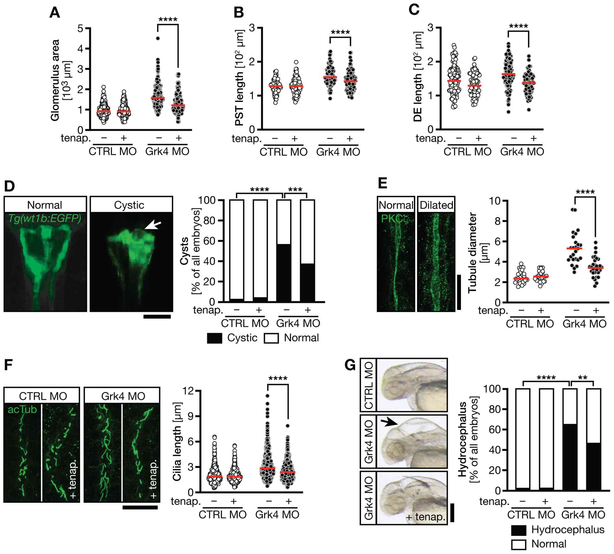 | Figure 3Nhe3 inhibition rescues kidney
patterning and ciliopathy phenotypes. (A) Tenapanor treatment
reduced the size of the wt1a expression domain in Grk4
knockdown embryos indicating a smaller glomerulus size. n=4
experiments with 218-242 embryos in total.
****P<0.0001 (data were analyzed using two-way ANOVA
with Sidak's multiple comparison post-test). (B) The length of the
PST, as detected by trpm7 expression, was reduced in Grk4
morphants by tenapanor. n=3 experiments with 90-100 embryos in
total. ****P<0.0001 (data were analyzed using two-way
ANOVA with Sidak's multiple comparison post-test). (C) Tenapanor
also decreased the DE length of Grk4 knockdown embryos. n=3
experiments with 74-123 embryos in total.
****P<0.0001 (data were analyzed using two-way ANOVA
with Sidak's multiple comparison post-test). (D) Fluorescence image
illustrating the glomerulus and parts of the proximal tubules in
Tg(wt1b:GFP) transgenic embryos. Arrows indicate cystic
glomeruli, which occur more often in Grk4 MO embryos than in
control-injected embryos. Tenapanor reduced the percentage of
embryos developing cysts. n=5 experiments with 144-166 embryos in
total. ***P=0.0003 and ****P<0.0001 (data
were analyzed using a two-sided Fisher's exact test with the
Bonferroni correction applied). (E) Confocal z-stacks of pronephric
tubules stained using anti-PKCz antibody. Grk4 knockdown produces
dilated pronephric tubules. Tenapanor reduces the extent of
dilatation. n=3 experiments with 25-29 embryos in total.
****P<0.0001 (data were analyzed using two-way ANOVA
with Sidak's multiple comparison post-test). (F) Confocal stacks of
cilia visualized by immunofluorescence using an acetylated tubulin
(acTub) antibody. Tenapanor also reduced the length of cilia in the
distal pronephric tubules in Grk4-depleted embryos. n=3 experiments
with 446-639 cilia in total. ****P<0.0001 (data were
analyzed using the Kruskal-Wallis test with Dunn's multiple
comparison post-test). (G) Hydrocephalus (arrow) formation in
Grk4-depleted embryos was significantly reduced upon treatment with
tenapanor. n=5 experiments with 150-167 embryos in total.
**P=0.0011 and ****P<0.0001 (data were
analyzed using a two-sided Fisher's exact test with the Bonferroni
correction applied). Analyses were performed at (A-C) 20 ss and
(D-G) 48 hpf. Scale bars: (D and G) 200 μm, and (E and F) 20
μm. Nhe3, sodium-hydrogen exchanger 3; Grk4, G
protein-coupled receptor kinase 4; CTRL, control; MO, morpholino
oligonucleotide; ss, somite stage; hpf, hours post-fertilization;
tenap., tenapanor. |
Nhe3 upregulation in the absence of Grk4
is not mediated by dopaminergic receptor signaling
Subsequently, the present study assessed whether the
observed phenotypes were coupled to dopaminergic receptor activity
as Grk4 LOF may prevent receptor desensitization and cause elevated
dopaminergic receptor signaling. Fertilized eggs were injected with
control or Grk4 MO and treated with the vehicle or 10 μM of
the dopamine receptor antagonist, SCH39166, from the tailbud stage
on until analysis at 48 hpf. Notably, no significant change was
detected in the expression domain of nhe3a in either the
control-injected or Grk4 MO-injected embryos (Fig. 4A). SCH39166 also failed to reduce
the percentage of embryos developing glomerular cysts (Fig. 4B). To further exclude unleashed
dopaminergic receptor signaling as a driver of aberrant pronephric
development, wild-type zebrafish embryos were treated with
dopamine. Yet again, the length of the nhe3a+
tubule was not altered (Fig. 4C and
D). In addition, dopaminergic receptor stimulation using
fenoldopam did not induce glomerular cysts or hydrocephalus
(Fig. 4E and F). Hence, defects
in pronephric development and function in Grk4-depleted embryos are
unlikely due to abnormal dopaminergic receptor signaling.
 | Figure 4Modulation of dopaminergic receptor
activity does not rescue Grk4 LOF kidney phenotypes. (A) Antagonism
of dopaminergic receptor by SCH39166 failed to shorten the
nhe3a expression domain. n=3 experiments with 54-67 embryos
in total. ns; P>0.9999 data were analyzed using the
Kruskal-Wallis test with Dunn's multiple comparison post-test). (B)
The dopamine receptor antagonist, SCH39166, did not reduce the
percentage of Grk4 LOF embryos developing glomerular cysts. n=4
experiments with 134-155 embryos in total.
****P<0.0001 ns, P=0.6176 (data were analyzed using a
two-sided Fisher's exact test with the Bonferroni correction
applied). (C) Nhe3a expression in embryos treated with the
vehicle (CTRL) or dopamine. Scale bar, 200 μm. (D) Dopamine
administration did not alter the length of the nhe3a
expression domain in Grk4-depleted embryos. n=3 experiments with
64-72 embryos in total. ns, P=0.3242 (data were analyzed using a
two-tailed Welch's test). (E) Fenoldopam treatment did not induce
cyst formation. n=4 experiments with 94-96 embryos in total. (F)
Stimulation of dopaminergic receptor using fenoldopam did not
result in hydrocephalus formation. n=6 experiments with 134-135
embryos in total. ns, not significant; DA, dopamine; Grk4, G
protein-coupled receptor kinase 4; LOF, loss-of-function; Nhe3,
sodium-hydrogen exchanger 3; Grk4, G protein-coupled receptor
kinase 4; CTRL, control; MO, morpholino oligonucleotide; fenold.,
fenoldopam. |
Nuclear localization of GRK4 is not
required for normal pronephros development
Previously and consistent with the data shown herein
for dopaminergic receptor signaling, the authors found that the
function of GRK4 during pronephros development does not depend on
its kinase activity (4). In
addition to the kinase domain, however, GRK4 also contains a
nuclear localization sequence (NLS), which entails a low, yet
reproducible GRK4 expression in the nucleus of transfected 293T
cells (Fig. 5A). Mutation of the
NLS (GRK4 ΔNLS) results in the exclusion of GRK4 from the nucleus
and sole cytosolic distribution (Fig. 5A). Of note, rescue experiments
with GRK4 ΔNLS normalized the expression of nhe3a and
reduced both hydrocephalus, as well as cyst formation in Grk4 LOF
embryos, suggesting an improvement in kidney function (Fig. 5B-E). Therefore, it can be
concluded that the location of Grk4 action resides in the cytosol
and does not depend on the nuclear localization of Grk4.
Loss of Grk4 results in Stat3
activation
In addition to dopaminergic receptors, NHE3 has been
reported to be controlled by other means. The transcriptional
activity of STAT3 directs the RNA synthesis of NHE3 in
kidney epithelial cells by binding to its promoter (8). Moreover, STAT3 activity based on
the phosphorylation of tyrosine 705 (corresponding to Y708 in
zebrafish Stat3) is higher in cystic than in the healthy kidneys of
patients diagnosed with autosomal polycystic kidney disease
(19). Mutation of that same
tyrosine 705 to phenylalanine abrogates the transcriptional
activity of human STAT3 (20).
Hence, the present study assessed STAT3 activity using an antibody
specific for phosphorylated Stat3 in zebrafish and observed indeed
a brighter fluorescence signal in Grk4-depleted embryos than in
control embryos, while total Stat3 levels appeared to be the same
(Fig. 6A and B). Co-injection of
the Grk4 MO with RNA encoding for dominant negative STAT3 (dnSTAT3)
revealed a significant reduction of the expression domain of
nhe3a compared to embryos injected only with the Grk4 MO
(Fig. 6C and D) and consistent
with the transcriptional control of NHE3 by STAT3, many more
embryos displaying a very weak nhe3a signal were counted
(Fig. 6E). Last, but not least,
the expression of dnSTAT3 partially, yet significantly reduced the
percentage of embryos displaying kidney dysfunction as detected by
hydrocephalus development (Fig.
6F) and glomerular cysts (Fig.
6G). Taken together, these findings suggest that the loss of
Grk4 induces Stat3 activation in zebrafish embryos, which in turn
results in higher rates of nhe3a transcription and
subsequently the ciliopathy-like malformation of the developing
pronephros, which was previously reported by the authors (4) and shown again herein.
 | Figure 6Pronephric Grk4 phenotypes are
mediated by STAT3. (A) Representative immunofluorescence images of
24 hpf embryos stained for p-Stat3 and total Stat3. Scale bar, 200
μm. (B) Loss of Grk4 increased the percentage of embryos
exhibiting stronger p-Stat3 staining than the average
control-injected embryo. n=3 experiments with 55-87 embryos in
total. **P=0.0022 (data were analyzed using a two-sided
Welch's test). (C) In situ hybridization demonstrating
nhe3a expression at 48 hpf. Scale bar, 200 μm. (D)
Expression of dnSTAT3 rescued the length of nhe3a expression
in Grk4-depleted embryos. n=3 experiments with 59-89 embryos in
total. ns, P=0.0588; *P=0.0253 and **P=0.0043
(data were analyzed using the Kruskal-Wallis-test with Dunn's
multiple comparison test). (E) Embryos expressing dnSTAT3 displayed
a weaker nhe3a signal. n=3 experiments with 59-89 embryos in
total. *P=0.0147 (data were analyzed usig one-way ANOVA
with Sidak's multiple comparison test). (F) dnSTAT3 prevented
hydrocephalus development upon Grk4 knockdown. n=4 experiments with
167-188 embryos in total. ****P<0.0001 (data were
analyzed using a two-sided Fisher's exact test with the Bonferroni
correction applied). Scale bar, 200 μm. (G) Cyst formation
was reduced upon dnSTAT3 expression. n=4 experiments with 167-188
embryos in total. ***P=0.0001 and
****P<0.0001 (data were analyzed using a two-sided
Fisher's exact test with the Bonferroni correction applied). Scale
bar, 100 μm. ns, not significant; Grk4, G protein-coupled
receptor kinase 4; STAT3, signal transducer and activator of
transcription 3; dnSTAT3, dominant negative STAT3; CTRL, control;
MO, morpholino oligonucleotide. |
Discussion
The present study describes a novel function of GRK4
in facilitating normal nephron architecture. In the absence of
functional Grk4, zebrafish embryos display alterations along the
whole pronephros starting from the glomerulus, which becomes cystic
thereafter. In addition, there is an enlargement of the segment,
which corresponds to the proximal tubule in mammals. This is the
region, where GRK4 desensitizes dopaminergic receptors in rodents
and humans (21,22). The loss of Grk4 also increased
the segment expressing rfx2, a gene, which is required for
the development of multiciliated cells and the formation and
extension of cilia (12,23). Hence, the elevated expression of
rfx2 may at least partially explain the expansion of
pronephric cilia in number and length, which we have already
previously reported in the absence of Grk4 (4). Furthermore, the present study
observed the expression of the close homolog of mammalian NHE3
along the whole pronephric tubule instead of being concentrated in
the proximal tubule. Similar to Rfx2, NHE3 proteins have been
associated with cilia. In Xenopus epithelia, NHE3 proteins
facilitate coordinated cilium formation and function of
multiciliated cells (24).
Interestingly, increased NHE3 levels have also been found in a
mouse model of cilium dysfunction resulting in polycystic disease
(25). Apart from its connection
to cilia, NHE3 has been described as one of several sodium
transport proteins regulated by dopaminergic receptors in the
proximal tubule of mammals (16,18). Dopaminergic receptors inhibit
NHE3 activity and prevent plasma membrane localization (26,27). Dopaminergic receptors further
promote the proteasome-dependent degradation of NHE3 (28). The authors tried to investigate
this in zebrafish. Notably though, both in the present study and in
a previous study by the authors on Grk4, a dependency on
dopaminergic receptors was not observed, which may also be a
limitation of the model organism used. Consistent with the
observation, however, a requirement of the kinase activity of Grk4
was also not found (4). As the
authors unfortunately were unable to find an antibody reliably
detecting zebrafish Nhe3a, the authors could not follow-up further
on this and assess protein levels or subcellular localization,
which would have added additional important insight into the
regulatory impact of Grk4 on Nhe3. Since the upregulation of Nhe3a
was detected on the level of mRNA, however, the authors considered
a novel mechanism by which GRK4 regulates NHE3 proteins. GRK4 as
with other GRKs of this subfamily translocates to the nucleus
(29), which was also observed
in 293T cells. While GRK4 does not appear to bind to DNA directly
(29), it could influence RNA
levels also by other means, such as modulating transcription
factors. However, as GRK4 abrogated of its nuclear localization was
as efficient in rescuing the Grk4 LOF phenotypes as wild-type GRK4,
this hypothesis was dismissed and the authors searched for other
mechanisms of NHE3 induction. STAT3, which is a gene required for
cell survival (8), can be
activated independent of receptor activation (8). It is highly active in the
developing rat kidney (30) and
displays an elevated activity in epithelial cells derived from
polycystic kidneys (19,31). The transcriptional activity of
STAT3 results specifically in the upregulation of NHE3 in
epithelial cells on the RNA level by binding to the NHE3 promoter
(8). Consistent with this, the
present study observed an elevated Stat3 activity, as detected by
phosphorylation at tyrosine 708 using immunofluorescence. The
authors also tried to corroborate these findings further using
western blotting, which however failed to produce quantifiable
bands. The overexpression of a dominant negative version of STAT3
did not only normalize Nhe3a expression in the model used herein,
but also rescued the cyst phenotype and general pronephros
function. Most likely, GRK4 does not regulate STAT3 directly
through phosphorylation, as previous experiments by the authors
using kinase-dead GRK4 demonstrated that GRK4 does not rely on its
kinase function in order to limit cilium elongation and to
facilitate normal kidney phenotypes (4). One possible mechanism may be that
other GRKs of the same subfamily become activated in the absence of
GRK4 and compensate. Hence, mice deficient in GRK4, 5 or 6 are
viable, while double homozygous embryos are at least partially
lethal (4,6). GRK5 is in fact expressed in the
zebrafish pronephros (6) and it
inhibits the mineralocorticoid receptor by phosphorylation
(32), which has been shown to
cause STAT3 phosphorylation at serine 727 (33). While the canonical activation of
STAT3 resulting in nuclear translocation and transcriptional
activity relies on tyrosine phosphorylation at position 705 in
mammals and 708 in zebrafish, which was tested herein, there is
also a non-canonical mechanism via the dephosphorylation of serine
727. When STAT3 is dephosphorylated at this position, it can also
enter the nucleus and act as transcription factor (34). A closer analysis of the STAT3
phosphorylation state in future studies would be of interest, as it
could dissect which residue would be regulated by which subtype of
GRK.
Another avenue of STAT3 regulation may be via mTOR,
which is negatively regulated by GRK4 (4). In zebrafish oocytes, mTOR
phosphorylates STAT3 to control centrosome position and microtubule
stability, which are both essential components of cilia (35). Apart from mTOR, GRK4 appears to
also interact with Fyn-related kinase (FRK), a member of the
SRC-like kinase family (36).
SRC-like kinases, which are non-receptor tyrosine kinases, are
typical upstream activators of STAT3 (37). Gain-of-function variants of FRK
have been associated with increased phosphorylation of STAT3 at the
same tyrosine as we tested in here (38) and SRC kinase inhibitors, such as
bosutinib, which also blocks FRK activity, can limit cyst growth in
a murine model of polycystic kidney disease (39). Last but not least, cilia on their
own have been reported to control STAT3 activity so that it is also
possible that Grk4 loss indirectly leads to STAT3 activation
through induction of cilium elongation, which is known to cause
cilium dysfunction.
Taken together, the present study expands the
previous characterization of kidney phenotypes upon Grk4 abrogation
in zebrafish embryos and provides evidence that early kidney
development is impaired in the absence of Grk4. In addition, the
present study identified a novel regulatory mechanism of NHE3
expression by GRK4, which is independent of the well-known function
of GRK4 in dopaminergic receptor desensitization (Fig. 7). To date, to the best of our
knowledge, no such observations have been reported in Grk4 KO mice,
which may be explained by known compensatory actions of other GRKs
(4). Moreover, as cysts, which
can be already observed in zebrafish in the embryo, appear in
patients suffering from autosomal dominant polycystic kidney
disease mostly at an advanced age, it may have escaped detection on
the studies done so far on GRK4 KO mice.
Translational impact for and beyond
polycystic diseases
The previously identified regulatory action of GRK4
on NHE3 subcellular localization through the phosphorylation of
dopaminergic receptors has ranked GRK4 as a potential therapeutic
target in hypertension. The data presented herein classify GRK4
additionally as a candidate gene in polycystic kidney diseases,
such as nephronophtisis or autosomal dominant polycystic kidney
disease. It further suggests that the mechanistic impact of GRK4 on
NHE3 is considerably beyond G protein-coupled receptors and places
GRK4 in the center of a much more complex signaling network, which
includes STAT3. STAT3 is involved in a rather large variety of
pathologies ranging from immune disorders and inflammation over
cardiovascular diseases and neurodegenerative conditions to several
types of cancer (37). Hence,
potential therapeutic strategies targeting GRK4 may require careful
design in order to avoid interference with the full GRK4 signaling
network.
The present study has certain limitations, which
should be mentioned. The data presented herein solely rely on
zebrafish embryos. While the authors demonstrated in a previous
study that the abrogation of GRK4 in human fibroblasts, as well as
kidney organoids derived from murine inner medullary collecting
duct cells results in the same cilia defects as in zebrafish
embryos (4), further studies
using NHE3-expressiong mammalian cells or animal models are
warranted to test the conservation of the newly identified
Grk4-Stat3-Nhe3 axis across species.
Availability of data and materials
The data generated in the present study may be
requested from the corresponding author.
Authors' contributions
LATV, JG, LDM, MDB and MP performed experiments,
analyzed data and maintained the animal facility. MP conceived the
study and analyzed the data. MDB and MP wrote the manuscript. All
authors have read and approved the final manuscript. LATV, MDB and
MP confirm the authenticity of all the raw data.
Ethics approval and consent to
participate
Zebrafish husbandry and experiments in the presented
study were approved by local authorities (Veterinary Care Units at
the University of Ulm and Tübingen and the local animal welfare
commissioner of the regional board for scientific animal
experiments): registry nos. 0140 and 35/9185.46/Uni TÜ. Experiments
were conducted in agreement with the European Communities Council
Directive of September 22, 2010 (2010/63/UE).
Patient consent for publication
Not applicable.
Competing interests
The authors declare that they have no competing
interests.
Abbreviations:
|
hpf
|
hours post-fertilization
|
|
GPCR
|
G protein-coupled receptor
|
|
GRK4
|
G protein-coupled receptor kinase
4
|
|
Nhe3
|
sodium-hydrogen exchanger 3
|
|
NLS
|
nuclear localization sequence
|
|
STAT3
|
signal transducer and activator of
transcription 3
|
|
dnSTAT3
|
dominant negative signal transducer
and activator of transcription 3
|
Acknowledgements
The authors would like to thank the animal care
takers at the University of Tübingen (Tübingen, Germany) for
providing excellent fish care, Mrs. Cornelia Donow for providing
technical assistance, BSc Monika Häußler for pilot experiments
(both from Ulm University, Ulm, Germany). Professor Jeroen Bakkers
(University Medical Center Utrecht, Utrecht, The Netherlands),
Addgene and ZIRC kindly provided the plasmids for the in
situ probe and capped RNA generation.
Funding
The authors are grateful for grant support by the German
Research organization (grant nos. PH144/4-1, PH144/6-1, PH144/6-3
and 467868420). LDM was a fellow of the International Graduate
School in Molecular Medicine at Ulm University (funded by Deutsche
Forschungsgemeinschaft).
References
|
1
|
Gurevich VV and Gurevich EV: GPCR
signaling regulation: The role of GRKs and arrestins. Front
Pharmacol. 10:1252019. View Article : Google Scholar : PubMed/NCBI
|
|
2
|
Gainetdinov RR, Premont RT, Bohn LM,
Lefkowitz RJ and Caron MG: Desensitization of G protein-coupled
receptors and neuronal functions. Annu Rev Neurosci. 27:107–144.
2004. View Article : Google Scholar : PubMed/NCBI
|
|
3
|
Premont RT, Macrae AD, Stoffel RH, Chung
N, Pitcher JA, Ambrose C, Inglese J, MacDonald ME and Lefkowitz RJ:
Characterization of the G protein-coupled receptor kinase GRK4.
Identification of four splice variants. J Biol Chem. 271:6403–6410.
1996. View Article : Google Scholar : PubMed/NCBI
|
|
4
|
Gerhards J, Maerz LD, Matthees ESF, Donow
C, Moepps B, Premont RT, Burkhalter MD, Hoffmann C and Philipp M:
Kinase activity is not required for G protein-coupled receptor
kinase 4 restraining mTOR signaling during cilia and kidney
development. J Am Soc Nephrol. 34:590–606. 2023. View Article : Google Scholar : PubMed/NCBI
|
|
5
|
Duong Phu M, Bross S, Burkhalter MD and
Philipp M: Limitations and opportunities in the pharmacotherapy of
ciliopathies. Pharmacol Ther. 225:1078412021. View Article : Google Scholar : PubMed/NCBI
|
|
6
|
Burkhalter MD, Fralish GB, Premont RT,
Caron MG and Philipp M: Grk5l controls heart development by
limiting mTOR signaling during symmetry breaking. Cell Rep.
4:625–632. 2013. View Article : Google Scholar : PubMed/NCBI
|
|
7
|
Niu B, Liu P, Shen M, Liu C, Wang L, Wang
F and Ma L: GRK5 regulates social behavior via suppression of
mTORC1 signaling in medial prefrontal cortex. Cereb Cortex.
28:421–432. 2018. View Article : Google Scholar
|
|
8
|
Su HW, Yeh HH, Wang SW, Shen MR, Chen TL,
Kiela PR, Ghishan FK and Tang MJ: Cell confluence-induced
activation of signal transducer and activator of transcription-3
(Stat3) triggers epithelial dome formation via augmentation of
sodium hydrogen exchanger-3 (NHE3) expression. J Biol Chem.
282:9883–9894. 2007. View Article : Google Scholar : PubMed/NCBI
|
|
9
|
Bollig F, Perner B, Besenbeck B, Köthe S,
Ebert C, Taudien S and Englert C: A highly conserved retinoic acid
responsive element controls wt1a expression in the zebrafish
pronephros. Development. 136:2883–2892. 2009. View Article : Google Scholar : PubMed/NCBI
|
|
10
|
Kim JH, Yoon MS and Chen J: Signal
transducer and activator of transcription 3 (STAT3) mediates amino
acid inhibition of insulin signaling through serine 727
phosphorylation. J Biol Chem. 284:35425–35432. 2009. View Article : Google Scholar : PubMed/NCBI
|
|
11
|
Thisse C and Thisse B: High-resolution in
situ hybridization to whole-mount zebrafish embryos. Nat Protoc.
3:59–69. 2008. View Article : Google Scholar : PubMed/NCBI
|
|
12
|
Verhoeven MC, Haase C, Christoffels VM,
Weidinger G and Bakkers J: Wnt signaling regulates atrioventricular
canal formation upstream of BMP and Tbx2. Birth Defects Res A Clin
Mol Teratol. 91:435–440. 2011. View Article : Google Scholar : PubMed/NCBI
|
|
13
|
Jaffe KM, Thiberge SY, Bisher ME and
Burdine RD: Imaging cilia in zebrafish. Methods Cell Biol.
97:415–435. 2010. View Article : Google Scholar : PubMed/NCBI
|
|
14
|
Schneider CA, Rasband WS and Eliceiri KW:
NIH image to ImageJ: 25 years of image analysis. Nat Methods.
9:671–675. 2012. View Article : Google Scholar : PubMed/NCBI
|
|
15
|
Nguyen TK, Petrikas M, Chambers BE and
Wingert RA: Principles of zebrafish nephron segment development. J
Dev Biol. 11:142023. View Article : Google Scholar : PubMed/NCBI
|
|
16
|
Gildea JJ, Shah IT, Van Sciver RE, Israel
JA, Enzensperger C, McGrath HE, Jose PA and Felder RA: The
cooperative roles of the dopamine receptors, D1R and D5R, on the
regulation of renal sodium transport. Kidney Int. 86:118–126. 2014.
View Article : Google Scholar : PubMed/NCBI
|
|
17
|
Watanabe H, Xu J, Bengra C, Jose PA and
Felder RA: Desensitization of human renal D1 dopamine receptors by
G protein-coupled receptor kinase 4. Kidney Int. 62:790–798. 2002.
View Article : Google Scholar : PubMed/NCBI
|
|
18
|
Wang X, Luo Y, Escano CS, Yang Z, Asico L,
Li H, Jones JE, Armando I, Lu Q, Sibley DR, et al: Upregulation of
renal sodium transporters in D5 dopamine receptor-deficient mice.
Hypertension. 55:1431–1437. 2010. View Article : Google Scholar : PubMed/NCBI
|
|
19
|
Talbot JJ, Shillingford JM, Vasanth S,
Doerr N, Mukherjee S, Kinter MT, Watnick T and Weimbs T:
Polycystin-1 regulates STAT activity by a dual mechanism. Proc Natl
Acad Sci USA. 108:7985–7990. 2011. View Article : Google Scholar : PubMed/NCBI
|
|
20
|
Kaptein A, Paillard V and Saunders M:
Dominant negative stat3 mutant inhibits interleukin-6-induced
Jak-STAT signal transduction. J Biol Chem. 271:5961–5964. 1996.
View Article : Google Scholar : PubMed/NCBI
|
|
21
|
Fraga S, Jose PA and Soares-da-Silva P:
Involvement of G protein-coupled receptor kinase 4 and 6 in rapid
desensitization of dopamine D1 receptor in rat IEC-6 intestinal
epithelial cells. Am J Physiol Regul Integr Comp Physiol.
287:R772–R779. 2004. View Article : Google Scholar : PubMed/NCBI
|
|
22
|
Sanada H, Yatabe J, Midorikawa S, Katoh T,
Hashimoto S, Watanabe T, Xu J, Luo Y, Wang X, Zeng C, et al:
Amelioration of genetic hypertension by suppression of renal G
protein-coupled receptor kinase type 4 expression. Hypertension.
47:1131–1139. 2006. View Article : Google Scholar : PubMed/NCBI
|
|
23
|
Chung MI, Peyrot SM, LeBoeuf S, Park TJ,
McGary KL, Marcotte EM and Wallingford JB: RFX2 is broadly required
for ciliogenesis during vertebrate development. Dev Biol.
363:155–165. 2012. View Article : Google Scholar : PubMed/NCBI
|
|
24
|
Sun DI, Tasca A, Haas M, Baltazar G,
Harland RM, Finkbeiner WE and Walentek P: Na+/H+ exchangers are
required for the development and function of vertebrate mucociliary
epithelia. Cells Tissues Organs. 205:279–292. 2018. View Article : Google Scholar : PubMed/NCBI
|
|
25
|
Hu C, Lakshmipathi J, Stuart D and Kohan
DE: Profiling renal sodium transporters in mice with nephron Ift88
disruption: Association with sex, cysts, and blood pressure.
Physiol Rep. 10:e152062022. View Article : Google Scholar : PubMed/NCBI
|
|
26
|
Bacic D, Kaissling B, McLeroy P, Zou L,
Baum M and Moe OW: Dopamine acutely decreases apical membrane Na/H
exchanger NHE3 protein in mouse renal proximal tubule. Kidney Int.
64:2133–2141. 2003. View Article : Google Scholar : PubMed/NCBI
|
|
27
|
Hu MC, Fan L, Crowder LA, Karim-Jimenez Z,
Murer H and Moe OW: Dopamine acutely stimulates Na+/H+ exchanger
(NHE3) endocytosis via clathrin-coated vesicles: Dependence on
protein kinase A-mediated NHE3 phosphorylation. J Biol Chem.
276:26906–26915. 2001. View Article : Google Scholar : PubMed/NCBI
|
|
28
|
Armando I, Villar VA, Jones JE, Lee H,
Wang X, Asico LD, Yu P, Yang J, Escano CS Jr, Pascua-Crusan AM, et
al: Dopamine D3 receptor inhibits the ubiquitin-specific peptidase
48 to promote NHE3 degradation. FASEB J. 28:1422–1434. 2014.
View Article : Google Scholar
|
|
29
|
Johnson LR, Robinson JD, Lester KN and
Pitcher JA: Distinct structural features of G protein-coupled
receptor kinase 5 (GRK5) regulate its nuclear localization and
DNA-binding ability. PLoS One. 8:e625082013. View Article : Google Scholar : PubMed/NCBI
|
|
30
|
Wang H, Yang Y, Sharma N, Tarasova NI,
Timofeeva OA, Winkler-Pickett RT, Tanigawa S and Perantoni AO:
STAT1 activation regulates proliferation and differentiation of
renal progenitors. Cell Signal. 22:1717–1726. 2010. View Article : Google Scholar : PubMed/NCBI
|
|
31
|
Li J, Lu D, Liu H, Williams BO, Overbeek
PA, Lee B, Zheng L and Yang T: Sclt1 deficiency causes cystic
kidney by activating ERK and STAT3 signaling. Hum Mol Genet.
26:2949–2960. 2017. View Article : Google Scholar : PubMed/NCBI
|
|
32
|
Maning J, McCrink KA, Pollard CM, Desimine
VL, Ghandour J, Perez A, Cora N, Ferraino KE, Parker BM, Brill AR,
et al: Antagonistic roles of GRK2 and GRK5 in cardiac aldosterone
signaling reveal GRK5-mediated cardioprotection via
mineralocorticoid receptor inhibition. Int J Mol Sci. 21:28682020.
View Article : Google Scholar : PubMed/NCBI
|
|
33
|
Queisser N, Schupp N, Schwarz E, Hartmann
C, Mackenzie GG and Oteiza PI: Aldosterone activates the oncogenic
signals ERK1/2 and STAT3 via redox-regulated mechanisms. Mol
Carcinog. 56:1868–1883. 2017. View Article : Google Scholar : PubMed/NCBI
|
|
34
|
Diallo M, Pimenta C, Murtinheira F,
Martins-Alves D, Pinto FR, da Costa AA, Letra-Vilela R, Martin V,
Rodriguez C, Rodrigues MS and Herrera F: Asymmetric
post-translational modifications regulate the nuclear translocation
of STAT3 homodimers in response to leukemia inhibitory factor. Cell
Oncol (Dordr). 47:1065–1070. 2024. View Article : Google Scholar
|
|
35
|
Shawahny A, Bogoch Y, Hart N and Elkouby
YM: An mTOR-Stat3-Stathmin pathway controls centrosome and
microtubule dynamics for oocyte polarization. Curr Biol.
35:6054–6069.e4. 2025. View Article : Google Scholar : PubMed/NCBI
|
|
36
|
Stark C, Breitkreutz BJ, Reguly T, Boucher
L, Breitkreutz A and Tyers M: BioGRID: A general repository for
interaction datasets. Nucleic Acids Res. 34(Database Issue):
D535–D539. 2006. View Article : Google Scholar :
|
|
37
|
Samad MA, Ahmad I, Hasan A, Alhashmi MH,
Ayub A, Al-Abbasi FA, Kumer A and Tabrez S: STAT3 signaling pathway
in health and disease. MedComm (2020). 6:e701522025. View Article : Google Scholar : PubMed/NCBI
|
|
38
|
Pilati C, Letouzé E, Nault JC, Imbeaud S,
Boulai A, Calderaro J, Poussin K, Franconi A, Couchy G, Morcrette
G, et al: Genomic profiling of hepatocellular adenomas reveals
recurrent FRK-activating mutations and the mechanisms of malignant
transformation. Cancer Cell. 25:428–441. 2014. View Article : Google Scholar : PubMed/NCBI
|
|
39
|
Sweeney WE Jr, von Vigier RO, Frost P and
Avner ED: Src inhibition ameliorates polycystic kidney disease. J
Am Soc Nephrol. 19:1331–1341. 2008. View Article : Google Scholar : PubMed/NCBI
|

















