According to the International Diabetes Federation
(IDF), an estimated 537 million adults aged 20-79 years worldwide
had diabetes in 2021. Projections indicate this number will rise to
783 million by 2045 (1).
Notably, over 90% of these cases are type 2 diabetes mellitus
(T2DM) (2). Inadequately managed
T2DM can lead to numerous severe complications. Within the
musculoskeletal system, T2DM induces multiple pathological
alterations affecting bone, joints, muscles and the nervous system.
These impairments not only directly compromise mobility but are
also intrinsically linked to metabolic dysregulation, chronic
inflammation and microvascular dysfunction. Physical exercise is an
important intervention for diabetes management. Exercise activates
adenosine monophosphate-activated protein kinase (AMPK) signaling,
promotes glucose transport and lipid metabolism, and enhances
mitochondrial function in skeletal muscle. It also improves insulin
sensitivity and blood sugar control (3-8).
However, a scientific exercise plan fundamentally relies on a
stable and functional musculoskeletal system. Because of the
detrimental impact of T2DM on the musculoskeletal system, early
intervention is critical. Identifying effective treatment targets
and preventive strategies may reduce the need for surgery and
significantly improve the quality of life for patients. This
approach aligns with current priorities in the prevention and
management of major diseases, emphasizing the integration of
traditional Chinese and Western medicine, as well as
multidisciplinary collaboration. The present review synthesizes
current research to explain how T2DM affects the musculoskeletal
system and identifies potential intervention targets, thereby
laying a solid foundation for future investigations.
Hyperglycemia and insulin resistance represent the
earliest pathological events that start musculoskeletal
degeneration in T2DM. Persistent metabolic dysregulation disrupts
glucose utilization, energy metabolism and cellular anabolic
signaling. This creates a common pathological basis for
abnormalities in skeletal muscle, tendon, tendon-bone interface
(TBI) and bone. Although these tissues perform distinct
physiological functions, they all exhibit impaired cellular
metabolism and tissue homeostasis under chronic hyperglycemic and
insulin-resistant conditions (Table
I).
Skeletal muscle is the primary site for
post-prandial glucose uptake and disposal, playing a central role
in maintaining glucose homeostasis (9). Peripheral insulin resistance
originating in skeletal muscle is a key driver in the development
and progression of T2DM (10).
Under normal physiological conditions, insulin promotes glucose
uptake by activating glucose transporter type 4 (GLUT4). However,
in T2DM, defects in the insulin signaling pathways lead to impaired
translocation of GLUT4 to the cell membrane, which represents a
core mechanism of insulin resistance (11,12). The resistance results in
decreased activity of glycogen synthase in skeletal muscle and
reduced glycogen storage. Reduced glycogen synthesis forces cells
to rely on lipolysis for energy production. At the same time,
impaired mitochondrial function decreases fatty acid oxidation,
which exacerbates the accumulation of lipid intermediates (13,14). Among these metabolites,
diacylglycerol (DAG) activates protein kinase C (PKC), attenuating
insulin-stimulated insulin receptor substrate-phosphorylation and
phosphoinositide 3-kinase (PI3K) activity. As a result, GLUT4
movement is further reduced, worsening insulin resistance (15,16). Moreover, prolonged exposure to
free fatty acids or a high-fat diet increases DAG accumulation
inside muscle cells. A high blood sugar environment further
promotes fat synthesis and buildup, forming a vicious cycle of
metabolic imbalance that progressively worsens energy metabolism
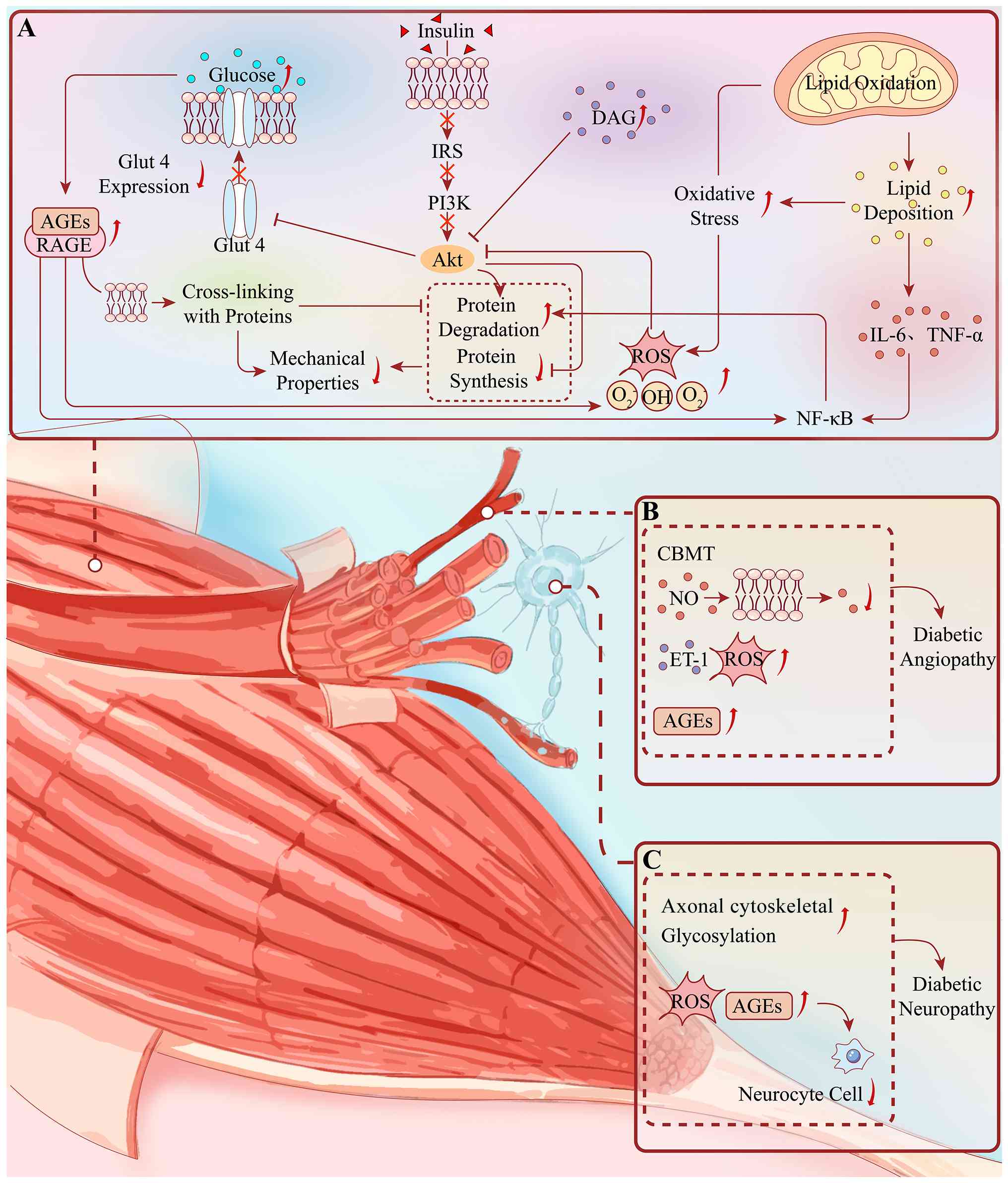
(Fig. 1A) (16).
The metabolic disturbances initiated by
hyperglycemia and insulin resistance also extend to tendon tissue.
Tendons are highly organized connective tissues composed primarily
of type I collagen, together with proteoglycans and glycoproteins
(17). Chronic hyperglycemia
suppresses the function of tendon stem/progenitor cell (TSPC). It
also disrupts the balance between matrix metalloproteinases (MMPs)
and tissue inhibitors of metalloproteinases (TIMPs), promoting
collagen degradation and preventing effective matrix remodeling
(18-20). Consequently, collagen fibers
become disorganized, losing elasticity and changing their
mechanical properties. Experimental studies have demonstrated
reduced elasticity and impaired mechanical performance in diabetes
rats' tendons, highlighting the harmful effects of chronic
hyperglycemia on tendon function (21,22). Similarly, the TBI is continuously
exposed to the diabetic metabolic environment. As a specialized
tissue responsible for transferring loads between tendon and bone,
the TBI depends on coordinated cellular differentiation and
extracellular matrix (ECM) homeostasis. Persistent hyperglycemia
promotes abnormal collagen cross-linking and chronic low-grade
inflammation, leading to a progressive worsening of the interface
structure and function (23-25). Following an injury, the diabetic
TBI heals mostly by forming fibrotic scar tissue rather than
regenerating the normal fibrocartilage transition zone (26). The newly formed scar tissue is
characterized by disorganized collagen fibers and poor
biomechanical properties, leading to reduced load-bearing capacity
and compromised interface function.
Bone tissue is likewise profoundly affected by
chronic hyperglycemia and insulin resistance. Under diabetic
conditions, hyperglycemia directly suppresses the expression of
bone-forming markers, including osteocalcin, osteopontin,
runt-related transcription factor 2 (Runx2) and alkaline
phosphatase. Hyperglycemia also promotes the death of bone-forming
cells, resulting in reduced bone formation and poor bone quality
(27-30). In addition, insulin deficiency or
resistance diminishes the anabolic effects of insulin and
insulin-like growth factor-1 on osteoblasts, further impairing
matrix synthesis and mineralization (31-33). Elevated glucose levels also
induce aberrant differentiation of mesenchymal stem cells,
resulting in increased bone marrow adiposity and impaired
osteoblast formation (34-37). Furthermore, chronic hyperglycemia
enhances the sensitivity of osteoclast precursors to macrophage
colony-stimulating factor and receptor activator of nuclear factor
κB ligand (RANKL), thereby facilitating osteoclast differentiation
and fusion, and consequently increasing bone resorptive activity
(38). These findings indicate
that hyperglycemia and insulin resistance constitute the primary
early drivers of diabetic musculoskeletal disorders. By disrupting
glucose utilization, lipid metabolism, anabolic signaling and
lineage commitment, they establish the pathological foundation for
subsequent oxidative stress (OS), chronic inflammation, ECM
remodeling, vascular dysfunction, and impaired tissue regeneration
across skeletal muscle, tendon, the TBI and bone.
Following chronic hyperglycemia and insulin
resistance, the excessive accumulation of advanced glycation end
products (AGEs), persistent OS, and chronic inflammation constitute
a connected pathogenic network that drives musculoskeletal
degeneration in T2DM. Despite the tissue-specific signs of
diabetes-related damage, these pathological mechanisms are
remarkably similar across skeletal muscle, tendon, the TBI and bone
(Table I). Under normal
physiological conditions, the formation of AGEs is minimal and
efficiently cleared from the body (39). However, chronic hyperglycemia
greatly accelerates the non-enzymatic glycation of proteins and
lipids, resulting in excessive AGEs generation and progressive
accumulation within multiple tissues (40,41). Excessive AGEs alter the
structural and biochemical properties of the ECM, disrupt collagen
integrity through non-enzymatic cross-linking, and trigger
persistent signaling through the receptor for AGEs (RAGE) (42-45). The sustained activation of this
pathway amplifies OS and inflammatory cascades, leading to
mitochondrial dysfunction, impaired cellular differentiation, and
cell death in multiple tissue types (46,47). These connected events ultimately
compromise the balance between tissue formation and degradation,
weaken the biomechanical properties of muscles, tendons and bone,
and destroy the coordinated function of the TBI.
AGE accumulation profoundly alters the mechanical
properties of collagen-rich tissues throughout the musculoskeletal
system. In skeletal muscle, AGEs accumulate within the muscle ECM
and collagen networks, increasing collagen cross-linking, tissue
stiffness, fibrosis and functional impairment (42,48-50). In tendon, AGEs impair
biomechanics in multiple species by reducing tendon fibers sliding,
resulting in increased passive muscle tension, reduced elasticity
and decreased flexibility (51-53). In addition, AGEs directly inhibit
the expression of collagen-related genes. Glycosylated collagen
accumulates in connective tissues due to its insolubility and
increased resistance to collagenase, leading to further AGE
deposition (54-56). At the TBI, the persistent
accumulation of AGEs, along with hyperglycemia-induced OS,
continuously activates AGEs-RAGE signaling and promotes chronic
inflammatory immune responses (23-25). This pathological cascade
activates the TGF-β1/Smad3 pathway, causing excessive fibroblast
growth and fibrotic tissue remodeling. This is accompanied by
abnormal cross-linking of collagen and proteoglycans, increased ECM
stiffness, and loss of tissue elasticity, which impairs mechanical
buffering and load transmission across the enthesis (24,53,57,58). In bone, excessive accumulation of
AGEs within the bone matrix harms collagen structure through
non-enzymatic cross-linking, reduces matrix elasticity and
mechanical strength, and directly impairs osteoblast
differentiation through activating RAGE-dependent signaling
pathways (59-63). Major axonal cytoskeletal
proteins, such as tubulin, neurofilaments and actin, are vulnerable
to glycation under diabetic conditions. As essential components for
maintaining axonal structure and function, their modification via
glycation may alter structural integrity and functional properties
of axons, leading to axonal atrophy, degeneration and slowed axonal
transport (64-67). Studies indicate that AGE-modified
myelin sheaths in peripheral nerves are more vulnerable to being
consumed by macrophages. This process stimulates the release of
enzymes and likely contributes to demyelination in diabetic
neuropathy (68).
Hyperglycemia-induced OS may accelerate the accumulation of AGEs,
thereby exacerbating oxidative damage in peripheral nerves
associated with neuronal disturbance and impaired Schwann cell
signaling (67). In this manner,
diabetic neuropathy promotes progressive nerve fiber loss and poor
regeneration. Additionally, increased activity in the polyol
pathway promotes the formation of 3-deoxyglucosone and
methylglyoxal, which are precursors that contribute to the
accumulation of AGEs. These AGEs alter neural cell components and,
through engagement with the RAGE expressed on neural cells, trigger
the production of various cytokines implicated in nerve injury
(38,67). Thus, both hyperglycemia and
polyol pathway-mediated AGE accumulation, along with AGE-RAGE
interactions, are likely to occur within diabetic nervous and
vascular systems. This highlights a cooperative relationship
between the polyol pathway and AGE/RAGE-dependent signaling in
promoting OS and nuclear factor kappa B (NF-κB) activation, which
aggravates neurovascular dysfunction in experimental T2DM
neuropathy (67).
The binding of AGEs to RAGE represents the central
molecular event linking metabolic disturbance with OS and
inflammation. By activating nicotinamide adenine dinucleotide
phosphate (NADPH) oxidase and the PI3K/protein kinase B (Akt)
pathway, AGEs stimulate excessive reactive oxygen species (ROS)
generation and activate NF-κB signaling, thereby establishing a
self-amplifying cycle between OS and inflammation (41,43-45,69). Simultaneously, AGEs formation,
glucose autoxidation, and inflammatory activation collectively
induce excessive ROS production. This disrupts intracellular
metabolic homeostasis, impairs insulin signaling, and promotes
catabolic processes (70). ROS
further attack mitochondrial DNA, respiratory chain complexes, and
membrane phospholipids, leading to mitochondrial dysfunction
(71). As the primary source of
cellular energy, dysfunctional mitochondria fail to meet tissue
energy demands, resulting in muscle atrophy, fatigue, impaired
cellular differentiation and reduced exercise tolerance (72). Meanwhile, OS suppresses anabolic
pathways, including PI3K/Akt/mammalian target of rapamycin (mTOR)
signaling, and compromises tissue structural integrity. It also
impairs angiogenesis and tissue repair, aggravates hypoxia, and
ultimately contributes to delayed or failed healing across
musculoskeletal tissues (73-77).
Chronic inflammation is another essential part of
this pathogenic network. While T2DM has traditionally been defined
by insulin resistance and progressive β-cell dysfunction,
increasing evidence highlights chronic low-grade inflammation and
immune dysregulation as equally central to disease progression
(78). Persistent hyperglycemia,
lipid overload, and the metabolic stress caused by compensatory
insulin hypersecretion activate innate immune pathways. This
environment releases signaling molecules that recruit monocytes to
eliminate apoptotic β-cells. Under the regulation of NF-κB
signaling, these recruited monocytes primarily differentiate into
pro-inflammatory M1 and anti-inflammatory M2 macrophages. This
process contributes to β-cell dysfunction and apoptosis. In
particular, tumor necrosis factor α (TNF-α) impairs insulin
signaling and diminishes insulin sensitivity (79,80). A vicious inflammatory cycle
subsequently emerges. The IkappaB kinase β (IKKβ)/NF-κB signaling
axis serves as a critical mediator in this process. It drives the
systemic accumulation of pro-inflammatory mediators such as TNF-α,
interleukin-6 (IL-6) and C-reactive protein (81-83). In turn, these cytokines further
activate the IKK/NF-κB cascade, impair insulin signaling, and
exacerbate β-cell dysfunction and systemic insulin resistance. They
also promote the excessive breakdown of structural proteins and the
ECM (79,84). Together with persistent ROS
production, this inflammatory microenvironment progressively shifts
tissue homeostasis toward catabolism, inhibits tissue regeneration,
and amplifies structural degeneration throughout skeletal muscle,
tendon, the TBI and bone.
Collectively, AGEs-RAGE activation, OS and chronic
inflammation form a tightly connected pathological axis linking
metabolic abnormalities to musculoskeletal degeneration. Rather
than acting independently, these mechanisms reinforce one another
through positive feedback, continuously exacerbating mitochondrial
dysfunction, ECM abnormalities, impaired cellular differentiation,
cell death and biomechanical deterioration. This sustained
inflammatory and oxidative microenvironment provides the foundation
for the subsequent development of microvascular dysfunction and
defective tissue regeneration.
Microvascular dysfunction is a critical pathological
consequence of persistent hyperglycemia and chronic metabolic
stress. It acts as a tissue-level amplifier that accelerates
musculoskeletal degeneration in T2DM (Table I). Under physiological
conditions, an intact microvascular network ensures adequate oxygen
and nutrient delivery while facilitating metabolite removal to
maintain tissue homeostasis. However, prolonged exposure to
hyperglycemia, insulin resistance, OS and chronic inflammation
progressively impairs endothelial function, resulting in reduced
nitric oxide (NO) bioavailability, excessive ROS production,
endothelial cell dysfunction, basement membrane thickening and poor
blood vessel formation (76,85-87). Consequently, tissue blood flow
declines, chronic hypoxia develops, and regenerative capacity is
progressively weakened across skeletal muscle, tendon, the TBI and
bone. Rather than representing an isolated pathological process,
microvascular dysfunction reinforces metabolic stress, inflammation
and ECM abnormalities, thereby establishing a vicious cycle that
accelerates tissue degeneration.
Skeletal muscle is highly dependent on an extensive
capillary network to support its substantial metabolic demand
(88). In T2DM, diabetic
microangiopathy features capillary rarefaction, basement membrane
thickening, endothelial dysfunction, and reduced skeletal muscle
blood flow. The consequent reduction in oxygen and nutrient
delivery aggravates mitochondrial dysfunction and energy
deficiency, which further impairs glucose utilization and insulin
sensitivity (85,89-91). In addition, diabetic
microvascular injury extends to the blood vessels of nerves,
producing neural ischemia, axonal degeneration and peripheral
neuropathy, which disrupt nerve-to-muscle communication (Fig. 1B and C) (92,93).
The detrimental effects of microvascular dysfunction
extend to tendon tissue, which naturally has limited blood supply
and therefore possesses a relatively poor intrinsic healing
capacity (94). Diabetic tendons
exhibit a significant increase in blood vessel density, with
vessels extending into the central tendon region, leading to an
enlarged vascular cross-sectional area (95). This aberrant vascularization
disrupts the normal blood supply pattern and microcirculatory
balance in tendons, leading to local tissue hypoxia, accumulation
of metabolic waste and ROS generation. Ultimately, this compromises
collagen fiber stability, increases tissue brittleness, and
contributes to tenocyte damage and collagen degradation (21,74,95-97). Concurrently, this pathological
environment triggers an inflammatory response characterized by
immune cell activation and the release of pro-inflammatory
cytokines. These cytokines disrupt the organized architecture of
collagen fibers, leading to a decrease in fiber diameter, a
reduction in density, fiber separation and impaired biomechanical
properties (74). Furthermore,
the impaired microenvironment hinders stem cell migration and
differentiation and delays post-injury angiogenesis, collectively
weakening the tendon's self-repair capacity (74,94,98,99). Besides, the hypoxic environment
also directly impairs the proliferation, differentiation and
metabolic function of tenocytes (100-102). Although local hypoxia
upregulates vascular endothelial growth factor (VEGF) expression to
stimulate angiogenesis, endothelial progenitor cell (EPC)
dysfunction, reduced VEGF signaling, and the accumulation of ROS
and AGEs result in the formation of aberrant, immature blood
vessels that fail to meet the mechanical demands of the tendon
(103-108). Moreover, hypoxia promotes M2
macrophage polarization, which accelerates fibrosis and scar
formation, disrupting the balance of ECM turnover. This imbalance
is reflected in aberrant protease activity, such as elevated
MMP-13, leading to tendon structural degeneration and functional
impairment (95,109). In summary,
microangiopathy-induced local hypoxia and inadequate nutrient
supply suppress tenocytes metabolic activity and impair the
tendon's self-repair capacity and biomechanical properties. Hypoxia
further exacerbates vascular endothelial damage through AGE
accumulation and OS, increasing vascular permeability and promoting
the release of inflammatory factors, thereby establishing a vicious
cycle (Fig. 2).
Successful tendon-bone healing (TBH) proceeds
through sequential inflammatory, proliferative and remodeling
phases, all of which depend on adequate vascularization (110-112). Newly formed vessels deliver
oxygen, nutrients, growth factors, and reparative cells to the
healing interface, while insufficient blood supply delays tissue
regeneration and compromises biomechanical recovery (77,113,114). Hyperglycemia disrupts vascular
homeostasis through persistent OS and endothelial dysfunction.
Excessive ROS damage endothelial cells and impair new blood vessel
responses, creating a vicious cycle of inflammation and hypoxia
(76,115). Demirag et al (116) reported that the degree of
vascularization positively correlates with histological maturity of
the TBI, particularly with the formation of Sharpey's fibers.
Likewise, inhibition of VEGF expression at the TBI suppresses blood
vessel formation, thereby reducing tendon maturation and
biomechanical strength (117).
Although VEGF expression is often elevated in diabetes, endothelial
responsiveness to VEGF is diminished because of the reduced
expression of VEGF receptors, particularly VEGFR2 (115,118,119). Consequently, angiogenic
signaling becomes ineffective, resulting in defective
neovascularization and delayed healing. Additionally, both CD31 and
endomucin have been identified as critical markers involved in
vascular development and TBH (120,121). Exosomes derived from bone
marrow mesenchymal stem cells (BMSC-Exos) have been shown to
markedly enhance the expression of CD31 and endomucin within the
interface microenvironment (26,77). By contrast, diabetic conditions
significantly impair the function of BMSCs and suppress CD31 and
endomucin expression (122).
Accumulating evidence also suggests that angiogenic progenitor
cells (APCs) are dysfunctional in diabetes. Insulin resistance
impairs the PI3K/Akt pathway and increases intracellular ROS
levels, reducing APC mobilization, migration and homing capacity
(123-129). Hypoxia further alters cell fate
at the TBI. The fibrocartilage region is naturally hypovascular and
maintains physiological expression of hypoxia-inducible factors
(HIFs) (130). At this
interface, progenitor cells expressing SRY-box transcription factor
9 (Sox9) and Scleraxis (Scx) give rise to TSPCs and fibrocartilage
cells during early musculoskeletal development (131,132). Under diabetic conditions,
chronic inflammation and elevated IL-1β drive excessive HIF-2α
activation (79,131). This aberrant upregulation
directly suppresses the tendon-specific transcription factor Scx,
impairing the tenogenic differentiation of TSPCs and shifting their
fate toward osteogenic and chondrogenic lineages, thereby promoting
ectopic calcification or ossification at the TBI (131).
Bone remodeling is likewise highly dependent on an
intact vascular network, as osteoblasts, osteoclasts, osteocytes,
and skeletal stem cells require continuous vascular support to
maintain normal bone turnover. Histological and morphological
studies demonstrate that diabetes decreases hematopoietic tissue,
increases marrow adiposity, and reduces microvascular density
(133). Reduced bone marrow
capillary density and microangiopathy further limit blood
perfusion, leading to local hypoxia that suppresses osteoblast
differentiation while stimulating osteoclast activity, ultimately
resulting in direct disruption of the bone structure (133,134). Not only can hypoxia affect
osteoblast development at early stages, failing to provide proper
signals for matrix maturation, but under hypoxic conditions,
osteocytes can also increase RANKL expression via the HIF-1α
signaling pathway (135,136).
This promotes the differentiation of osteoclast precursors into
mature osteoclasts and enhances bone resorption activity (136). The dual effects exacerbate the
imbalance of bone remodeling, contributing to trabecular thinning
and sparsity.
ECM dysregulation and impaired regenerative capacity
represent the converging pathways through which chronic metabolic
disturbances ultimately compromise musculoskeletal integrity in
T2DM (Table I). Following
persistent hyperglycemia, AGEs-RAGE activation, OS, chronic
inflammation and microvascular dysfunction, the coordinated balance
between matrix synthesis and degradation is progressively
disrupted. These pathological alterations not only impair the
structural organization and biomechanical properties of the ECM but
also compromise the regenerative potential of resident
stem/progenitor cells, resulting in defective tissue remodeling and
incomplete functional recovery.
Hyperglycemia suppresses collagen synthesis through
multiple pathways. Inhibition of the AMPK and early growth response
factor 1 (EGR1) pathway reduces the expression of collagen-related
genes, including collagen type 1 alpha 1 (Col1a1), Col1a2, and
biglycan (137,138). AGE deposition not only
compromises cell viability but also disrupts the structure and
function of the ECM, interferes with normal collagen cross-linking,
and additionally suppresses collagen synthesis. Collectively,
insulin resistance, suppression of AMPK/EGR1 signaling, and AGE
accumulation reduce collagen synthesis and contribute to tendon
degeneration. Collagen degradation is primarily mediated by MMPs,
particularly MMP-9 and MMP-13 (139,140). Hyperglycemia increases their
expression and activity, leading to excessive collagen breakdown
and disruption of collagen fiber organization (141-143). Under physiological conditions,
TIMPs maintain a balance with MMPs to preserve ECM integrity
(144). However, T2DM reduces
TIMP activity, resulting in excessive MMP-mediated collagen
degradation (20). This systemic
imbalance between synthetic and degradation drives tissue-specific
structural disorder. In tendon, T2DM disrupts collagen homeostasis
by reducing collagen synthesis while enhancing degradation,
resulting in collagen disorganization, altered fiber architecture,
and impaired biomechanical properties that predispose tendons to
degeneration and injury (15).
At the TBI, the ECM of the fibrocartilage region is primarily
composed of collagens and proteoglycans, whose balance is tightly
regulated by MMPs and TIMPs (131,145,146). Under diabetic conditions,
hyperglycemia, hypoxia and chronic inflammation disrupt the
MMP/TIMP balance, resulting in excessive matrix degradation
(146-153). Simultaneously, AGEs accumulate
within the TBI and form abnormal cross-links with collagen and
proteoglycans, reducing tissue elasticity and impairing cell-matrix
interactions (50,58,70,154). In bone, osteoclasts degrade the
bone matrix by creating an acidic microenvironment and secreting
collagenases and other proteolytic enzymes (155). Additionally, the structural
integrity of collagen fibers within the bone matrix is disrupted,
with compromised fiber architecture and reduced matrix ductility
and strength (59,156). These ECM-level abnormalities
collectively undermine bone matrix quality and mechanical
performance.
Simultaneously, the regenerative capacity of
tissue-resident stem cells is severely compromised across all four
tissues. Skeletal muscle satellite cells (MuSCs) are essential for
muscle repair and regeneration, but increased inflammation and
excessive OS impair MuSC function and suppress myogenic
differentiation (157-160). Inflammatory mediators such as
IL-6 and TNF-α inhibit PI3K/AKT/mTOR signaling while activating
NF-κB and forkhead box O (FoxO) pathways, thereby impairing muscle
regeneration (159,161,162). The reduction of myogenic
regulatory factors, including MyoD1 and myogenin, further impairs
muscle regenerative capacity, leading to reduced cross-sectional
area of muscle fibers and atrophy (163). In tendon, TSPCs play a central
role in maintaining tendon homeostasis and promoting tissue repair
(164-166). Under T2DM conditions, TSPC
proliferation is reduced while cell death is increased.
Hyperglycemia and AGE accumulation alter TSPC fate, promoting
abnormal osteogenic and chondrogenic differentiation at the expense
of tenogenic differentiation, which contributes to tendon
calcification, structural abnormalities and functional impairment
(18,19,167,168). Impaired self-renewal,
proliferation and differentiation of TSPCs directly compromise
tendon regeneration, contributing to collagen disorganization,
excessive vascularization, persistent inflammation, fibrosis and
reduced biomechanical performance (19). Reduced collagen synthesis and
enhanced degradation lead to impaired structural and mechanical
properties, while TSPC dysfunction compromises the tendon's
self-repair capacity and increases the risk of rupture. At the TBI,
tendon-derived stem cells (TDSCs) are critical for fibrocartilage
regeneration during tendon-bone healing (111,169-173). However, hyperglycemic
conditions directly induce TDSC apoptosis, inhibit their
proliferation, and deplete the available TDSC pool for repair
(18,174). Consequently, fibrocartilage
exposed to repetitive mechanical loading becomes increasingly
vulnerable to thinning, structural disorganization and
degeneration. Fibrotic scar formation replaces the native
fibrocartilage transition zone, resulting in disorganized collagen
fibers, poor biomechanical properties, and compromised load-bearing
capacity (26). In bone, the
osteogenic potential of MSCs is disrupted. Elevated glucose levels
promote adipogenic differentiation through activation of adipogenic
transcription factors, including peroxisome proliferator-activated
receptor gamma (PPARγ) and adipocyte protein 2 (aP2), while
suppressing osteogenic regulators such as Runx2 and Osterix
(34,35). Consequently, MSCs preferentially
differentiate into adipocytes rather than osteoblasts, resulting in
increased bone marrow adiposity and reduced osteoblast numbers
(36,37). The imbalance between
osteoblast-mediated formation and osteoclast-mediated resorption,
coupled with preferential adipogenic differentiation of MSCs, leads
to reduced bone formation, trabecular deterioration, cortical
thinning and increased skeletal fragility (175). Together, these tissue-specific
stem/progenitor cell dysfunctions, coupled with systemic ECM
remodeling abnormalities, establish a self-perpetuating cycle of
structural degradation and failed repair across the entire
musculoskeletal system. These findings highlight the critical need
for therapeutic strategies that simultaneously target ECM
homeostasis and endogenous regenerative mechanisms.
Although hyperglycemia, AGEs-RAGE signaling, OS,
chronic inflammation, microvascular dysfunction and ECM
dysregulation collectively drive musculoskeletal degeneration in
T2DM, each tissue exhibits distinct biological characteristics.
These characteristics determine their unique pathological response
to chronic metabolic stress. These tissue-specific mechanisms
further contribute to the heterogeneity of diabetic musculoskeletal
complications and represent potential targets for precision
treatment interventions (Table
II).
Unlike other musculoskeletal tissues, skeletal
muscle serves as the principal organ responsible for post-prandial
glucose disposal and whole-body energy metabolism (9). A characteristic feature of diabetic
skeletal muscle is the progressive disruption of intracellular
energy metabolism. Defective insulin signaling impairs GLUT4
translocation and glucose uptake, forcing muscle fibers to rely
increasingly on lipid oxidation (15,16). However, incomplete fatty acid
oxidation results in the accumulation of toxic lipid intermediates,
particularly DAG, which activates PKC and further suppresses
insulin signaling, forming a vicious cycle of metabolic dysfunction
(16). In parallel,
mitochondrial dysfunction serves as a fundamental mechanism
underlying insulin resistance and muscle atrophy (72,176). In patients with T2DM, skeletal
muscle exhibits reduced mitochondrial density, impaired oxidative
phosphorylation, and abnormal mitochondrial morphology, resulting
in diminished adenosine triphosphate (ATP) production and
compromised energy metabolism (14). These alterations contribute to
the loss of muscle mass, elevated fatigue and reduced exercise
tolerance (72). Mitochondrial
dynamics are regulated by fusion proteins, including mitofusin-1
(Mfn1), Mfn2 and optic atrophy 1 (Opa1) (177). Reduced expression of Mfn2 and
Opa1 in T2DM is associated with impaired mitochondrial fusion and
decreased mitochondrial function (178-180). Hyperglycemia promotes excessive
mitochondrial fission and ROS generation, further exacerbating
mitochondrial damage and OS (97,178,181-184). In addition, mitochondrial
creatine kinase 2 (CKMT2), an important regulator of oxidative
phosphorylation and mitochondrial respiration (185,186). Decreased CKMT2 impairs
phosphocreatine formation and ATP generation, leading to reduced
glucose oxidation and worsening energy deficits (186). Together, mitochondrial
dysfunction, OS, and impaired substrate utilization contribute to
progressive muscle weakness and atrophy.
Skeletal muscle loss in T2DM results from both
enhanced protein degradation and impaired protein synthesis
(187). T2DM is associated with
a shift from oxidative type I fibers toward glycolytic type II
fibers, reducing oxidative capacity and insulin sensitivity while
increasing susceptibility to muscle wasting (84,188-193). Insulin resistance suppresses
the anabolic IRS/PI3K/Akt/mTOR pathway while concurrently
activating the ubiquitin-proteasome system (UPS), which is the
principal pathway responsible for protein degradation in skeletal
muscle (72,194,195). The muscle-specific enzymes
muscle RING-finger protein-1 (MuRF1) and muscle atrophy F-box
(MAFbx/atrogin-1) are markedly upregulated during muscle atrophy
(196). The FoxO transcription
factors regulate the expression of MuRF1 and MAFbx (196,197). Reduced insulin signaling
decreases FoxO phosphorylation, thereby activating the UPS and
further amplifying proteolysis (84,198). Furthermore, inflammatory
cytokines further enhance UPS activity through NF-κB and janus
kinase/signal transducers and activators of transcription
(JAK/STAT) signaling pathways, accelerating proteolysis and muscle
loss (38,72,84,199-202). Under healthy conditions,
insulin activates the PI3K/Akt signaling to promote glucose uptake
and stimulate mTORC1-dependent protein synthesis (203,204). In T2DM, impaired signaling
suppresses these pathways, resulting in diminished protein
synthesis (73,205,206). Combined with diabetic
peripheral neuropathy (DPN), neuromuscular junction degeneration
and denervation-induced muscle atrophy, these metabolic
abnormalities contribute to the progressive decline in muscle
strength, endurance, and physical performance characteristic of
diabetic sarcopenia.
Compared with skeletal muscle, tendon pathology in
T2DM is characterized primarily by disruption of collagen
homeostasis and deterioration of biomechanical properties. Diabetes
profoundly alters tendon mechanical behavior, reducing its ability
to withstand and transmit mechanical loads. Structural
abnormalities such as collagen disorganization, fibrosis and
calcification compromise tendon integrity and increase
susceptibility to injury (22,207,208). As mentioned previously,
collagen cross-linking is essential for maintaining tendon
mechanical properties. Enzymatic cross-linking enhances tendon
stiffness and fatigue resistance by stabilizing collagen fibril
arrangement. By contrast, non-enzymatic cross-linking, particularly
AGEs, are minimal under physiological conditions but accumulate
markedly in pathological states such as diabetes (209). Hyperglycemia accelerates
non-enzymatic glycosylation, leading to AGE deposition between
collagen fibers. This promotes abnormal intermolecular
cross-linking that impede fibrillar sliding, increasing tissue
stiffness, reducing overall elasticity and ductility (40,210-212). Transglutaminase (TGase) is an
enzyme that catalyzes acyl-transfer reactions to form covalent
cross-links between proteins, normally enhancing the mechanical
strength and fatigue resistance of tissues (213). However, hyperglycemia elevates
TGase activity within tenocytes, which exacerbates non-enzymatic
cross-linking and impairs dynamic collagen sliding (74,214,215). These aberrant cross-links
disrupt normal fibrillar organization and helical structure,
ultimately reducing the elastic modulus of the tendon (216-219).
Hyperglycemia also activates endothelial NO
synthase and inducible NO synthase, resulting in a short-term
increase in the absolute production of NO (220,221). Elevated NO levels inhibit
fibroblast proliferation and collagen synthesis, thereby reducing
tendon elasticity (222). In
the short term, excessive NO impairs vascular endothelial barrier
function. The endothelial dysfunction further induces OS via
activation of NADPH oxidase and glycolytic pathways, leading to a
marked rise in ROS. The overproduction of ROS reduces NO
bioavailability through both the degradation of tetrahydrobiopterin
and direct oxidation of NO, thereby exacerbating endothelial
dysfunction (223-227). Such impairment diminishes the
regenerative potential of MSCs, compromising the tendon's repair
capacity. Tendon elasticity relies on the organized arrangement of
collagen and the homeostasis of elastin. Impaired repair results in
abnormal collagen cross-linking and elastin degradation,
collectively reducing elasticity and increasing brittleness
(18,228). Concurrently, aberrant M2
macrophage polarization promotes the secretion of pro-fibrotic
factors (for example, TGF-β and VEGF), which stimulate pathological
scarring (109,229,230). These changes lead to stiffened
tendon fibers, diminished tensile strength, and an elevated risk of
rupture (Fig. 2).
The TBI is a specialized transitional tissue that
comprises two distinct insertion types, which are direct and
indirect insertions, enabling efficient load transfer between
tendon and bone (231,232). It consists of tendon,
unmineralized fibrocartilage, mineralized fibrocartilage, and bone,
forming a graded structure characterized by progressive changes in
cellular phenotype, collagen composition, and mineral content
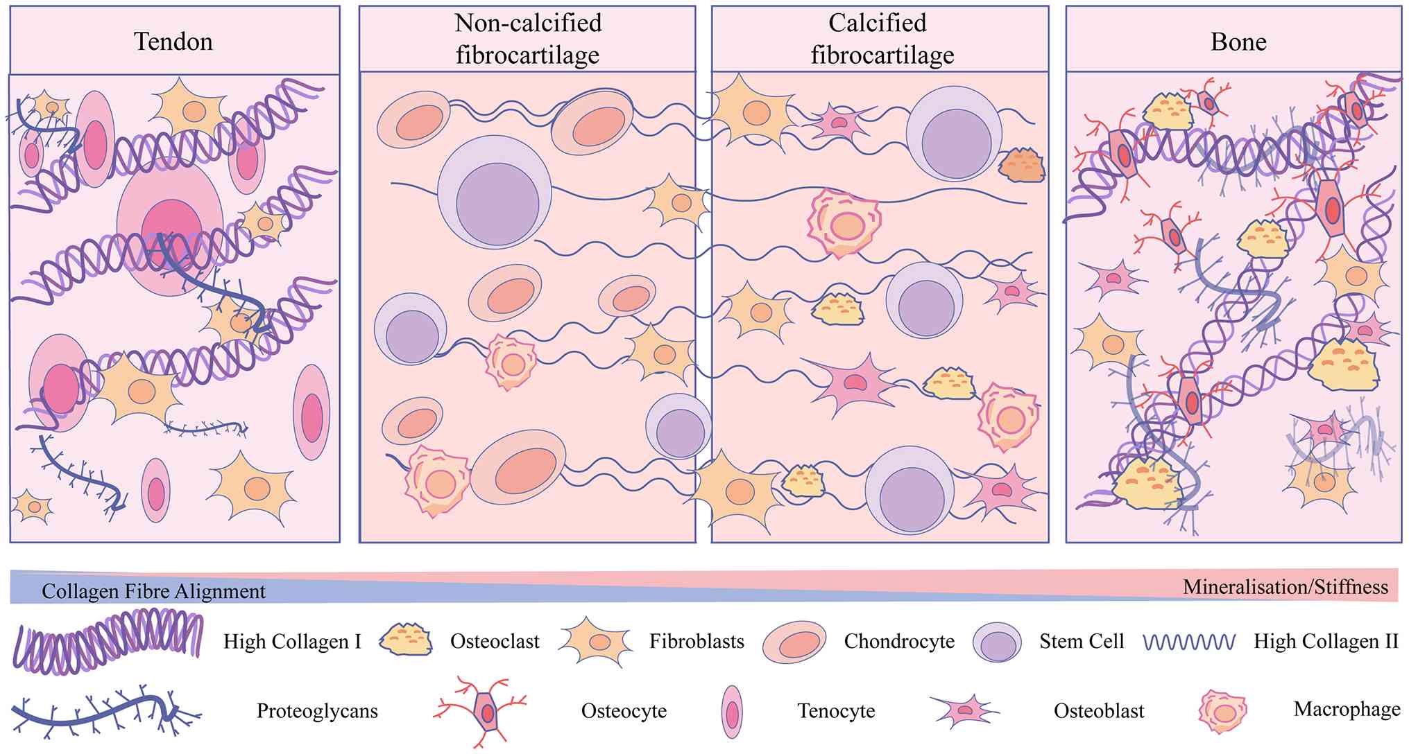
(Fig. 3) (112,233-238). The indirect insertion is
characterized by Sharpey's fibers, which anchor tendon directly
into bone (239). The two
insertion types differ primarily in collagen maturity and extent of
new bone formation. Biomechanically and biologically, indirect
insertions exhibit inferior properties compared with direct
insertions (26).
By providing a gradual transition in stiffness,
this native fibrocartilage interface minimizes stress concentration
between tissues with vastly different mechanical properties,
ensuring effective force transmission (240-244). However, this transmission
relies heavily on both the structural integrity of the interface
and the capacity of resident cells to sense and respond to
mechanical stimuli. Biomechanical studies have shown that diabetes
significantly reduces the Young's modulus, stiffness and ultimate
failure load of the TBI (22,154). Diabetes-induced fibrosis
disrupts the hierarchical architecture by replacing fibrocartilage
with scar tissue. This loss of fibrocartilage, alongside reduced
vascularization and collagen disorganization, severely compromises
the interface's stress-buffering capacity. Consequently, these
alterations impair force transmission and contribute to a higher
incidence of rotator cuff tears, Achilles tendinopathy,
postoperative retears, and delayed healing in diabetic patients
(244). Experimental studies
further demonstrate that diabetic animals exhibit reduced
fibrocartilage formation, poorer collagen organization, and lower
biomechanical strength following rotator cuff repair compared with
healthy controls (154).
Beyond structural degradation, cells within the TBI
depend on mechanotransduction pathways to continuously sense
mechanical cues from the extracellular environment (245-246). Integrins, which are
heterodimeric transmembrane receptors composed of α and β subunits,
are key mediators of this process. By linking the ECM to the actin
cytoskeleton, integrins convert mechanical forces into
intracellular biochemical signals that regulate tissue remodeling
and repair (247,248). Multiple studies indicate that
hyperglycemia impairs this integrin-mediated signaling. AGEs can
modify the Arg-Gly-Asp (RGD) domain of fibronectin, disrupting its
interaction with integrins, thereby altering ECM stiffness and
attenuating mechanotransduction efficiency (249). Furthermore, in human proximal
tubular epithelial (HK-2) cells cultured under high-glucose
conditions, the expression of specific integrin subunits is
significantly reduced (250).
Chronic hyperglycemia has also been reported to delay fibronectin
and integrin expression, impairing the integration of implants into
bone (251). These findings
suggest that diabetes downregulates integrin expression, disrupts
interactions between integrins and ECM components, and consequently
impairs mechanosensation, leading to abnormal mechanical stress
transmission at the TBI (Fig.
4A).
Bone exhibits a distinct pathological response to
diabetes because its homeostasis depends on the dynamic coupling of
osteoblast-mediated bone formation, osteoclast-mediated bone
resorption, and osteocyte-mediated mechanosensation (252-255). A hallmark of diabetic bone
disease is uncoupled bone remodeling. As noted previously,
hyperglycemia suppresses osteoblast differentiation while
simultaneously promoting excessive osteoclast development and bone
resorption. Another distinctive mechanism involves osteocyte
dysfunction. Osteocytes are the most abundant and essential
cellular component of the skeletal system, acting as the primary
sensors of mechanical and biochemical stimuli and playing an
irreplaceable role in bone remodeling (256). They are derived from
osteoblasts, and a subset of osteoblasts becomes embedded within
the mineralized bone matrix to differentiate into osteocytes. These
cells are interconnected through a complex network and produce a
variety of signaling molecules that regulate osteoblast and
osteoclast activity (255).
Hyperglycemia impairs the mechanosensory function of osteocytes,
accelerates cellular aging, and compromises bone strength (37,257). It can also directly induce
osteocyte death, disrupting the integrity of the osteocyte network
and impairing skeletal mechanical adaptability (258,259). Cellular aging is a
stress-responsive cell fate characterized by durable growth arrest,
extensive chromatin remodeling, and acquisition of a
proinflammatory secretory program known as the
senescence-associated secretory phenotype (SASP) (260). The accumulation of AGEs and
senescent cells compromises osteocyte viability and function
through the activation of RAGE signaling and the secretion of
proinflammatory SASP factors, ultimately enhancing the production
of sclerostin by osteocytes. When sclerostin binds to Wnt
co-receptors, it inhibits bone formation and reduces osteocyte
mechanosensitivity, thereby contributing to poor bone quality in
diabetic patients (261-263).
Meanwhile, hyperglycemia drives BMSCs toward a metabolically
stressed adipogenic lineage rather than osteoblasts. This shift not
only causes marrow fat accumulation and impaired trabecular
formation, but it also triggers inflammation mediated by
hyaluronan, ultimately exacerbating trabecular demineralization
(37). These alterations
collectively impair fracture healing, reduce bone quality, and
increase skeletal fragility despite relatively preserved bone
mineral density in numerous patients (Fig. 4B).
Although the preceding sections summarize the
proposed mechanisms underlying diabetes-related musculoskeletal
disorders, the strength of evidence supporting each pathway
warrants critical evaluation. Much of the available evidence,
particularly for AGEs-RAGE signaling and chronic inflammation,
comes from in vitro and animal studies. Therefore, its
direct translation to human pathophysiology should be interpreted
with caution. For instance, AGE accumulation is consistently
associated with increased tendon stiffness and reduced elasticity
in rodent models. However, evidence from human studies is largely
correlative and is often confounded by disease duration and
comorbidities (22,52,216). Likewise, mitochondrial
dysfunction is considered a central feature of numerous proposed
mechanisms. However, current evidence mainly comes from associative
studies showing reduced oxidative capacity and abnormal
mitochondrial morphology in diabetic muscle (14,178). Whether these mitochondrial
alterations initiate insulin resistance or arise as a consequence
of the diabetic metabolic milieu remains unresolved (264).
Notable contradictions exist within literature. For
example, although chronic hyperglycemia is widely accepted to
impair angiogenesis, some studies report elevated VEGF levels in
diabetic tissues such as the retina. This finding suggests a
paradoxical upregulation in response to hypoxia under certain
pathological conditions (103,104). By contrast, peripheral tissues
such as skeletal muscle and tendon exhibit reduced VEGFR2 signaling
and poor neovascularization (115,118,119). These observations suggest that
the overall angiogenic response depends on the complex interplay of
multiple dysregulated pathways and may be tissue dependent. A
significant knowledge gap remains in the understanding of the TBI.
While the role of TGF-β1 in fibrosis is well-established, the
precise molecular triggers that shift tissue repair from
regenerative fibrocartilage formation to pathological scarring in
diabetes remain largely unknown (244,265). In addition, the specific
contributions of different immune cell subpopulations, particularly
the dynamic phenotypic switching of macrophages, remain
incompletely understood. Most available evidence comes from a
limited number of studies. Finally, most current evidence is
derived from single-tissue or single-mechanism studies, making it
difficult to understand the integrated, system-level response to
diabetes. Future research should prioritize longitudinal human
studies, apply multi-omics approaches to capture the complexity of
the disease, and develop experimental models that simultaneously
investigate interactions among skeletal muscle, tendon, bone and
their interfaces. Such integrative approaches will facilitate the
identification of robust therapeutic targets and their translation
into effective interventions for diabetes-related musculoskeletal
complications.
Conventional antidiabetic medications and
anti-osteoporotic therapies not only improve systemic metabolic
homeostasis but also exert pleiotropic effects on musculoskeletal
tissues by targeting shared pathological mechanisms underlying
T2DM-associated musculoskeletal disorders. Rather than acting
solely on glucose metabolism or bone remodeling alone, these
therapies serve as an important bridge between metabolic regulation
and musculoskeletal health through coordinated modulation of
inflammation, OS, AGEs, and stem cell function.
Among conventional antidiabetic agents, metformin
has been the cornerstone of first-line pharmacological therapy for
T2DM for over six decades because of its well-established efficacy,
excellent safety profile, minimal risk of hypoglycemia, favorable
effects on body weight, cost-effectiveness, robust long-term
clinical evidence, and pleiotropic metabolic benefits (266). Beyond its glucose-lowering
effect, metformin activates AMPK, thereby suppressing
NF-κB-mediated inflammatory signaling and reducing the production
of pro-inflammatory cytokines such as TNF-α and IL-6 (267). It also inhibits the formation
and accumulation of AGEs, consequently attenuating AGEs-RAGE
signaling, OS, apoptosis, and tissue fibrosis (267-269). Since excessive AGEs
accumulation disrupts the stem cell microenvironment and impairs
proliferation and lineage commitment, metformin may indirectly
preserve stem cell function by reducing glycation stress (270-272). Moreover, accumulating evidence
suggests that metformin promotes osteogenic differentiation while
suppressing osteoclast activity through multiple signaling
pathways, including AMPK, Nrf2/HO-1, SIRT1 and mTOR (273,274). Combination therapy with
metformin and anti-osteoporotic agents has also been shown to
improve both glycemic control and diabetes-induced bone loss in
experimental models (275).
Anti-osteoporotic therapies primarily restore skeletal integrity by
re-establishing the balance between bone resorption and bone
formation. Antiresorptive agents, including RANKL-neutralizing
antibody denosumab, suppress osteoclast differentiation and induce
osteoclast apoptosis, thereby reducing excessive bone resorption
(276). By contrast, anabolic
agents such as sclerostin-neutralizing antibody romosozumab
directly stimulate osteoblast differentiation and bone formation
(277). Beyond increasing bone
mineral density and reducing fracture risk, these therapies improve
the musculoskeletal microenvironment by preserving bone remodeling
homeostasis, thereby providing a favorable niche for stem
cell-mediated tissue regeneration.
Conventional antidiabetic and anti-osteoporotic
therapies share complementary mechanisms that extend beyond their
primary therapeutic indications. By simultaneously improving
metabolic control, suppressing chronic inflammation and OS,
limiting AGEs accumulation, and preserving stem cell activity,
these agents help maintain musculoskeletal tissue homeostasis and
strengthen the link between systemic metabolism and the
musculoskeletal system.
In recent years, rapid advances in emerging
technologies have transformed the prevention, diagnosis, monitoring
and management of diabetes. Continuous glucose monitoring,
artificial intelligence-assisted decision support systems, digital
health tools, biosensors, and advanced drug delivery approaches are
reshaping current models of diabetes care. Meanwhile, progress in
omics research, big data analytics, and regenerative medicine has
provided new insights into disease mechanisms and facilitated the
development of personalized therapeutic strategies. The integration
of biotechnology, data science and clinical practice marks the
beginning of a new stage in precision diabetology, with an emphasis
on improving glycemic control, preventing complications, and
enhancing the overall quality of life for individuals with
diabetes.
With the rapid development of high-throughput
sequencing technologies, the genetic and transcriptomic mechanisms
underlying diabetes have been progressively uncovered. The precise
regulation of glucose homeostasis depends on the coordinated
activity of multiple endocrine cell types within the pancreatic
islets (278). Human pancreatic
islets are composed mainly of β cells (~54%), α cells (35%) and δ
cells (11%), along with smaller populations of γ/PP and ε cells
(279,280). Increasing evidence suggests
that alterations in the proportion or function of these endocrine
cells are closely associated with the pathophysiological processes
of monogenic diabetes, T1DM and T2DM (278). However, because pancreatic
islets account for only ~1% of pancreatic tissue and contain rare
cell populations such as δ and ε cells, whole-genome sequencing at
the single-islet level has long been technically challenging
(281). The emergence of
scRNA-seq has overcome numerous of these limitations. It enables
detailed investigation of the molecular mechanisms underlying
diabetes and provides new opportunities for clinical diagnosis and
treatment (278,282).
Although the widespread application of scRNA-seq in
both basic research and clinical studies continues to expand,
several technical challenges remain. The single-cell capture
procedure often compromises cellular integrity and viability,
highlighting the need for further optimization to improve
throughput and precision. In addition, scRNA-seq produces
high-dimensional datasets that are inherently noisy, and reducing
this noise remains a major challenge for reliable data analysis.
Moreover, cell differentiation and proliferation are inherently
dynamic processes, making it difficult for current scRNA-seq
methods to accurately distinguish between cell types (292). Beyond these technical
limitations, the relatively high cost of scRNA-seq also limits its
broader adoption as a routine research tool. Clinical translation
of scRNA-seq is further challenged by the limited accessibility of
disease-relevant tissues, the lack of standardized analytical
workflows, and the need for large-scale prospective validation to
ensure the reproducibility and clinical interpretability of
single-cell findings across diverse patient populations. Continued
improvements in sequencing technologies, bioinformatics algorithms,
analytical standardization and cost efficiency will be essential
for advancing the application of scRNA-seq in diabetes research and
clinical practice.
Precision medicine has become a key strategy for
managing a wide range of diseases, and targeted drug delivery
systems (TDDS) are now emerging as one of its central technologies
in diabetes treatment. Insulin remains the fundamental therapy for
maintaining glucose homeostasis in patients with T1DM and advanced
T2DM. However, conventional insulin therapy relies primarily on
frequent subcutaneous injections, such as rapid-acting insulin
before meals and long-acting insulin at bedtime, to mimic
physiological secretion patterns (293). Repeated injections often lead
to pain, local tissue injury, infection and neuropathy, resulting
in poor patient compliance (294). In addition, factors such as
injection site, depth and dosage can significantly affect insulin
absorption kinetics, further increasing the uncertainty of glycemic
control (295). To overcome the
limitations of injection-based therapy, research has increasingly
focused on non-invasive and intelligent TDDS strategies. These
systems enable targeted drug delivery to diseased sites and achieve
rhythm-synchronized release in response to physiological demands.
By improving local therapeutic efficacy while minimizing systemic
side effects and reducing dosage requirements, TDDS provide new
opportunities for personalized diabetes management. They also
optimize pharmacokinetics, regulate drug biodistribution and
enhance patient adherence, makes them highly promising for the
treatment of complex chronic diseases (296).
A typical TDDS consists of three essential
components: The therapeutic target, the active pharmaceutical
ingredient (API), and the carrier or delivery vehicle. The
therapeutic target refers to the specific organ, tissue, or cell
that requires intervention, whereas the API provides the
therapeutic effect. The carrier transports the API to the target
site with spatial and temporal precision. An ideal carrier should
exhibit favorable biocompatibility, biodegradability, low toxicity
and specific recognition of the target site (297). Various carrier systems,
including liposomes, aptamers and tetrahedral framework nucleic
acids, have been extensively investigated. These carriers can
improve the bioavailability and tissue penetration of therapeutic
molecules, thereby enhancing efficacy. According to their delivery
mechanisms, TDDS can be classified as active or passive targeting
systems. Active targeting relies on ligand-receptor interactions to
achieve precise drug localization, whereas passive take advantage
of pathological features such as increased permeability to enhance
drug accumulation in diseased tissues (297). The combination of
micro-nanotechnology with TDDS represents a major advancement in
diabetes therapy. Encapsulating drugs within micro- or nanoscale
carriers improves drug stability, bioavailability and local
concentration, enhancing therapeutic efficacy and reducing adverse
reactions (293,298). For example, polymer-based
nanoparticle systems for insulin delivery, constructed from
biocompatible and biodegradable materials, such as chitosan,
poly(lactic-co-glycolic acid) (PLGA) and polycaprolacton, can
effectively overcome the limitations of traditional injection
therapy. These nanocarriers protect insulin from enzymatic
degradation and acidic conditions during oral administration,
significantly increasing its bioavailability and enabling
controlled and targeted release (299). Similar benefits have been
observed with other oral hypoglycemic agents. A PLGA-based
metformin delivery system demonstrated remarkable changes in
pharmacokinetic properties. Compared with conventional
formulations, PLGA-loaded metformin achieved more than a 10-fold
increase in mean residence time and a substantially larger apparent
distribution volume, even at 1/10 of the standard dose (300). These findings support PLGA as
an efficient sustained-release delivery platform with great
potential in diabetes treatment.
As β cells play a central role in the onset and
progression of diabetes, designing delivery systems capable of
specifically targeting the islet microenvironment is crucial for
effective therapy. Ghosh et al (301) developed functionalized
nanoparticles by conjugating the targeting peptide CHVLWSTRKC to a
PLGA-b-PEG-COOH polymer and loading it with genistein. These
nanoparticles bound to islet endothelial cells with 3-fold greater
affinity through specific interaction with the EphA4 receptor,
resulting in a 200-fold enhancement in the immunosuppressive
activity of genistein (301).
These results demonstrate the feasibility and potential of
islet-specific drug delivery. In addition to targeting peptides,
antibodies and aptamers have been widely used to construct
nanoscale delivery systems for gene therapy, demonstrating
considerable potential for precise genetic modulation in diabetes.
These systems use antibody- or aptamer-mediated targeting to
deliver therapeutic genes into specific cells and employ
disease-responsive promoters to achieve controlled gene expression.
This approach helps restore disrupted gene networks and recover
cellular function (302). Among
these ligands, aptamers have received growing attention for their
unique advantages as single-stranded DNA or RNA oligonucleotides
with high affinity and selectivity (303). Compared with antibodies,
aptamers are smaller, enabling deeper tissue penetration, and they
exhibit low immunogenicity and high stability in vivo,
making them suitable for long-term treatment. Their easy synthesis
and chemical flexibility also facilitate the development of
personalized therapeutic platforms for diabetes (304,305). Yamada et al (306) provided strong experimental
evidence for the concept by designing a β-cell-targeted nanocarrier
system known as β-MEND. This carrier efficiently delivered nucleic
acids into pancreatic β cells. β-MEND successfully internalized
into MIN6 cells and delivered antisense RNA targeting microRNA
(miR)-375, leading to a marked reduction in miR-375 expression. As
a result, Pdk1 and Mtpn were upregulated, significantly enhancing
insulin secretion (306). These
findings demonstrated that aptamer-based nanocarriers can
effectively regulate β-cell function and provide a viable route
toward gene therapy in diabetes. Collectively, these advances
indicate that TDDS are gradually evolving from experimental
delivery platforms toward clinically applicable therapeutic
strategies. Recent advances in oral nanoplatforms have achieved
efficient insulin delivery, glucose-responsive drug release and
sustained glycemic control in preclinical models, highlighting the
promising translational potential of precision drug delivery. For
example, a milk-derived nanovesicle-based oral insulin delivery
system achieved ~20% oral bioavailability with sustained
glucose-lowering efficacy in diabetic pigs, highlighting the
feasibility of non-invasive insulin replacement therapy (307). Similarly, the recently
developed the MOP@T@D (MON-Proteins@Transferrin@Deoxycholic acid)
oral nanoplatform in a T2DM rat model achieved an oral
bioavailability of 10.6%, increased islet area by 26.7% after
treatment, restored insulin secretion to 74.6% of the physiological
level, and maintained normal blood glucose levels for up to two
weeks after treatment cessation (308). By enabling sustained drug
release and selective delivery to diseased tissues or cells, these
strategies may facilitate individualized therapeutic interventions
while reducing systemic adverse effects, thereby providing a
feasible route for future clinical translation.
Despite these encouraging advances, TDDS still face
significant challenges in clinical translation. Each type of
carrier has inherent limitations. Nanoparticles often exhibit poor
physical stability and tend to aggregate or undergo oxidative
degradation. In addition, their manufacturing processes are complex
(309). Polymer-based also face
challenges such as complicated synthesis routes, high production
costs and limited structural stability. These limitations may lead
to aggregation and, in some cases, inflammatory reactions in
vivo (310).
As a systemic metabolic disorder, diabetes is not
only inherently complex but also closely associated with numerous
severe complications, which substantially increase patient
morbidity and mortality (311).
Therefore, treatment alone is insufficient to address the full
spectrum of health challenges associated with diabetes. Early
intervention through prevention and timely diagnosis is equally
important. Consequently, the integration of artificial intelligence
(AI) into diabetes management, through machine learning, deep
learning and other AI-based analytical approaches, has the
potential to transform diabetes care. By leveraging advanced data
analysis, feature extraction and predictive modeling, AI provides a
data-driven framework for screening, diagnosis, treatment,
prognostic assessment and preventive interventions, thereby opening
new avenues for disease management (312).
AI applications in diabetes primarily focus on four
key domains: Screening for diabetes and its complications, clinical
decision support systems (CDSS), population risk prediction and
patient self-management tools (313,314). In the field of diabetes
screening, AI offers the potential for early, non-invasive, rapid,
and cost-effective detection of pathological changes (314). Shu et al (315) utilized facial texture analysis
combined with a support vector machine model, achieving an accuracy
of 99.02%, sensitivity of 99.64% and specificity of 98.26% in
diabetes detection. Similarly, Li et al (316) integrated tongue color and
texture features with machine learning models to predict diabetes
risk, reporting an average accuracy of 0.821. However, neither
study included external validation, limiting the generalizability
of the findings. AI-based early detection of diabetic retinopathy
(DR) has emerged as a cost-effective alternative, potentially
reducing diabetes-related ocular complications and preventable
blindness (317). The American
Diabetes Association formally recognizes the clinical value of
autonomous AI systems in detecting DR and macular edema (318). Keel et al (319) demonstrated that an AI-based DR
screening model is highly feasible in endocrinology outpatient
settings, showing a sensitivity of 92.3% and specificity of 93.7%,
and a patient satisfaction rate of 96%, providing strong evidence
for integrating AI screening into routine clinical practice. DPN is
another common yet often overlooked complication, largely because
of limited screening tools and insufficient diagnostic sensitivity
(320). Deep learning
algorithms trained on corneal confocal microscopy images have shown
promise in detecting DPN. Williams et al (321) developed a deep learning
algorithm that demonstrated rapid and accurate localization for
quantifying corneal nerve biomarkers. Preston et al
(322) developed a
ResNet-50-based model that achieved precisions value of 0.83 in
healthy volunteers, with 0.92 and 1.0 for non-neuropathic and
neuropathic individuals, respectively.
AI can also assist in optimizing treatment
strategies for patients with complex diabetes by identifying
optimal therapeutic targets and supporting clinical
decision-making. Murphree et al (323) conducted a retrospective study
of 12,147 patients initiating metformin therapy, using a machine
learning model to predict 1-year glycemic control outcomes.
Baseline HbA1c, initial metformin dose and complications emerged as
key predictors of treatment failure. The best-performing model
achieved an area under the curve of 0.75, providing evidence for
early identification of high-risk patients and personalized
treatment planning (323).
Additionally, AI-driven CDSS based on treatment pathway modeling
can simulate HbA1c responses under different therapeutic strategies
in T2DM, offering critical guidance for individualized care
(324). By analyzing patients'
lifestyle behaviors, physical and mental health parameters, and
social network activities, machine learning-based health
recommendation systems can predict disease risks (324). Decision tree models have also
been applied to predict the risk of T2DM development in women with
gestational diabetes, achieving discrimination performance of 83.0%
in training datasets and 76.9% in independent test sets,
outperforming traditional fasting glucose monitoring (325). AI models integrating genetic
and medical imaging data have demonstrated strong predictive
performance for T2DM risk assessment. Huang et al (326) employed XGBoost to combine
polygenic risk scores, multi-modality imaging risk scores and
demographic variables (for example, age, sex and family history of
diabetes) in 68,911 participants, achieving an Area Under the
Receiver Operating Curve (AUC) of 0.94. This model effectively
identified high-risk populations with specific genetic and imaging
features, enabling precision prevention and early intervention for
T2DM (326). These advances
suggest that AI is gradually transitioning from a research-oriented
analytical tool to an integrated component of precision diabetes
care. In clinical practice, AI-based systems may facilitate earlier
identification of individuals at risk, improve screening efficiency
for diabetes complications, and support individualized therapeutic
decision-making through integration with continuous glucose
monitoring, electronic health records, and digital health
platforms. Among current AI-enabled therapeutic strategies,
personalized insulin adjustment based on reinforcement learning,
prediction of treatment responses to glucose-lowering medications,
and AI-guided lifestyle interventions have shown promising
potential for improving glycemic control and optimizing long-term
disease management.
Self-management, particularly maintaining blood
glucose within recommended ranges, remains a cornerstone of
diabetes care. This involves active glucose monitoring, regulation
of physical activity and diet, and appropriate insulin
administration (327). Dietary
and exercise interventions are primary strategies for preventing
T2DM in high-risk populations (328). Personalized applications
provide tailored dietary plans, while wearable devices track daily
activity and use behavioral incentives to promote healthy habits,
thereby helping prevent chronic diseases including T2DM (314). In addition, accurate prediction
of blood glucose fluctuations in patients with T2DM facilitates
improved glycemic control, reduces hypoglycemic events, and lowers
diabetes-related morbidity and mortality, ultimately enhancing
quality of life. Deng et al (329) developed a predictive model
using deep transfer learning and data augmentation on continuous
glucose monitoring data recorded every 30 min, forecasting blood
glucose 5 min to 1 h ahead with over 95% accuracy and 90%
sensitivity. Wang et al (330) proposed a reinforcement
learning-based dynamic insulin titration regimen, improving
glycemic control without increasing hypoglycemia risk, achieving
personalized insulin dose optimization for patients with T2DM.
These technological advances enable both healthcare providers and
patients to proactively manage diabetes, adjusting insulin doses
and lifestyle behaviors to prevent hyperglycemia or hypoglycemia
and associated complications. Despite the promise of AI in
research, its real-world application remains challenging,
particularly across diverse populations (331). Training datasets lacking
balanced representation in terms of age, sex, ethnicity, or risk
factors may exacerbate biases, leading to misdiagnosis or
inappropriate treatment for underrepresented groups (332). Therefore, inclusive data
collection strategies are essential. Given the potential for AI
errors, ethical challenges arise, underscoring the need for robust
supervision and clear accountability guidelines in AI-driven
diabetes care. Furthermore, ensuring data privacy is critical.
Securing data during development and deployment remains
challenging, and global-scale implementation intensifies these
risks, necessitating stringent protection measures.
Not applicable.
ZH conceptualized the study. ZH, HL, YL and YY
conducted investigation and data visualization, wrote the original
draft, and wrote, reviewed and edited the manuscript. SL conducted
investigation, and wrote, reviewed and edited the manuscript. TZ
supervised the study, conducted project administration, and wrote,
reviewed and edited the manuscript. DB conceptualized and
supervised the study, conducted project administration, acquired
funding, and wrote, reviewed and edited the manuscript. All authors
read and approved the final version of the manuscript. Data
authentication is not applicable.
Not applicable.
Not applicable.
The authors declare that they have no competing
interests.
Not applicable.
The present study was supported by Sichuan Provincial
Administration of Traditional Chinese Medicine (grant no.
25ZDIZX028) and the Science and Technology Strategic Cooperation
Programs of Luzhou Municipal People's Government and Southwest
Medical University (grant no. 2025LZXNYDJC06).
|
1
|
Liu H, Wu Y, Zhu H, Wang P, Chen T, Xia A,
Zhao Z, He D, Chen X, Xu J and Ji L: Association between napping
and type 2 diabetes mellitus. Front Endocrinol (Lausanne).
15:12946382024. View Article : Google Scholar : PubMed/NCBI
|
|
2
|
Chatterjee S, Khunti K and Davies MJ: Type
2 diabetes. Lancet. 389:2239–2251. 2017. View Article : Google Scholar : PubMed/NCBI
|
|
3
|
Hawley JA, Hargreaves M and Zierath JR:
Signalling mechanisms in skeletal muscle: Role in substrate
selection and muscle adaptation. Essays Biochem. 42:1–12.
2006.PubMed/NCBI
|
|
4
|
Ruderman NB, Carling D, Prentki M and
Cacicedo JM: AMPK, insulin resistance, and the metabolic syndrome.
J Clin Invest. 123:2764–2772. 2013. View Article : Google Scholar : PubMed/NCBI
|
|
5
|
Garber CE, Blissmer B, Deschenes MR,
Franklin BA, Lamonte MJ, Lee IM, Nieman DC and Swain DP; American
College of Sports Medicine: Quantity and quality of exercise for
developing and maintaining cardiorespiratory, musculoskeletal, and
neuromotor fitness in apparently healthy adults. Med Sci Sports
Exerc. 43:1334–1359. 2011. View Article : Google Scholar : PubMed/NCBI
|
|
6
|
Mulya A, Haus JM, Solomon TP, Kelly KR,
Malin SK, Rocco M, Barkoukis H and Kirwan JP: Exercise
training-induced improvement in skeletal muscle PGC-1α-mediated fat
metabolism is independent of dietary glycemic index. Obesity
(Silver Spring). 25:721–729. 2017. View Article : Google Scholar : PubMed/NCBI
|
|
7
|
Kirwan JP, Sacks J and Nieuwoudt S: The
essential role of exercise in the management of type 2 diabetes.
Cleve Clin J Med. 84(7 Suppl 1): S15–S21. 2017. View Article : Google Scholar : PubMed/NCBI
|
|
8
|
Boul NG, Kenny GP, Haddad E, Wells GA and
Sigal RJ: Meta-analysis of the effect of structured exercise
training on cardiorespiratory fitness in type 2 diabetes mellitus.
Diabetologia. 46:1071–1081. 2003. View Article : Google Scholar
|
|
9
|
Evans PL, McMillin SL, Weyrauch LA and
Witczak CA: Regulation of skeletal muscle glucose transport and
glucose metabolism by exercise training. Nutrients. 11:24322019.
View Article : Google Scholar : PubMed/NCBI
|
|
10
|
DeFronzo RA and Tripathy D: Skeletal
muscle insulin resistance is the primary defect in type 2 diabetes.
Diabetes Care. 32(Suppl 2): S157–S163. 2009. View Article : Google Scholar : PubMed/NCBI
|
|
11
|
Abdul-Ghani MA and DeFronzo RA:
Pathogenesis of insulin resistance in skeletal muscle. J Biomed
Biotechnol. 2010:4762792010. View Article : Google Scholar : PubMed/NCBI
|
|
12
|
Shepherd PR and Kahn BB: Glucose
transporters and insulin action-implications for insulin resistance
and diabetes mellitus. N Engl J Med. 341:248–257. 1999. View Article : Google Scholar : PubMed/NCBI
|
|
13
|
Razak F and Anand SS: Impaired
mitochondrial activity in the insulin-resistant offspring of
patients with type 2 diabetes. N Engl J Med. 350:664–671. 2004.
View Article : Google Scholar
Petersen KF, Dufour S, Befroy D, Garcia R
and Shulman GI: Vasc Med. 9:223–224. 2004. View Article : Google Scholar
|
|
14
|
Phielix E and Mensink M: Type 2 diabetes
mellitus and skeletal muscle metabolic function. Physiol Behav.
94:252–258. 2008. View Article : Google Scholar : PubMed/NCBI
|
|
15
|
Timmers S, Schrauwen P and de Vogel J:
Muscular diacylglycerol metabolism and insulin resistance. Physiol
Behav. 94:242–251. 2008. View Article : Google Scholar : PubMed/NCBI
|
|
16
|
Chow L, From A and Seaquist E: Skeletal
muscle insulin resistance: The interplay of local lipid excess and
mitochondrial dysfunction. Metabolism. 59:70–85. 2010. View Article : Google Scholar
|
|
17
|
Ahmed AS, Schizas N, Li J, Ahmed M,
Östenson CG, Salo P, Hewitt C, Hart DA and Ackermann PW: Type 2
diabetes impairs tendon repair after injury in a rat model. J Appl
Physiol (1985). 113:1784–1791. 2012. View Article : Google Scholar : PubMed/NCBI
|
|
18
|
Lin YC, Li YJ, Rui YF, Dai GC, Shi L, Xu
HL, Ni M, Zhao S, Chen H, Wang C, et al: The effects of high
glucose on tendon-derived stem cells: Implications of the
pathogenesis of diabetic tendon disorders. Oncotarget.
8:17518–17528. 2017. View Article : Google Scholar : PubMed/NCBI
|
|
19
|
Lu P, Dai G, Shi L, Li Y, Zhang M, Wang H
and Rui Y: HMGB1 modulates high glucose-induced erroneous
differentiation of tendon stem/progenitor cells through
RAGE/β-catenin pathway. Stem Cells Int. 2024:23352702024.
View Article : Google Scholar
|
|
20
|
Huo JL, Feng Q, Pan S, Fu WJ and Liu Z and
Liu Z: Diabetic cardiomyopathy: Early diagnostic biomarkers,
pathogenetic mechanisms, and therapeutic interventions. Cell Death
Discov. 9:2562023. View Article : Google Scholar : PubMed/NCBI
|
|
21
|
Connizzo BK, Bhatt PR, Liechty KW and
Soslowsky LJ: Diabetes alters mechanical properties and collagen
fiber re-alignment in multiple mouse tendons. Ann Biomed Eng.
42:1880–1888. 2014. View Article : Google Scholar : PubMed/NCBI
|
|
22
|
Fox AJ, Bedi A, Deng XH, Ying L, Harris
PE, Warren RF and Rodeo SA: Diabetes mellitus alters the mechanical
properties of the native tendon in an experimental rat model. J
Orthop Res. 29:880–885. 2011. View Article : Google Scholar : PubMed/NCBI
|
|
23
|
Lee JM and Veres SP: Advanced glycation
end-product cross-linking inhibits biomechanical plasticity and
characteristic failure morphology of native tendon. J Appl Physiol
(1985). 126:832–841. 2019. View Article : Google Scholar : PubMed/NCBI
|
|
24
|
Kamml J, Acevedo C and Kammer DS:
Advanced-Glycation Endproducts: How cross-linking properties affect
the collagen fibril behavior. J Mech Behav Biomed Mater.
148:1061982023. View Article : Google Scholar : PubMed/NCBI
|
|
25
|
Kayal RA, Siqueira M, Alblowi J, McLean J,
Krothapalli N, Faibish D, Einhorn TA, Gerstenfeld LC and Graves DT:
TNF-α mediates diabetes-enhanced chondrocyte apoptosis during
fracture healing and stimulates chondrocyte apoptosis through
FOXO1. J Bone Miner Res. 25:1604–1615. 2010. View Article : Google Scholar : PubMed/NCBI
|
|
26
|
Qin B, Bao D, Liu Y, Zeng S, Deng K, Liu H
and Fu S: Engineered exosomes: A promising strategy for tendon-bone
healing. J Adv Res. 64:155–169. 2024. View Article : Google Scholar :
|
|
27
|
Wongdee K and Charoenphandhu N:
Osteoporosis in diabetes mellitus: Possible cellular and molecular
mechanisms. World J Diabetes. 2:41–48. 2011. View Article : Google Scholar : PubMed/NCBI
|
|
28
|
Medeiros C and Wallace JM: High
glucose-induced inhibition of osteoblast like MC3T3-E1
differentiation promotes mitochondrial perturbations. PLoS One.
17:e02700012022. View Article : Google Scholar : PubMed/NCBI
|
|
29
|
Sheng N, Xing F, Wang J, Zhang QY, Nie R,
Li-Ling J, Duan X and Xie HQ: Recent progress in bone-repair
strategies in diabetic conditions. Mater Today Bio. 23:1008352023.
View Article : Google Scholar : PubMed/NCBI
|
|
30
|
Zhen D, Chen Y and Tang X: Metformin
reverses the deleterious effects of high glucose on osteoblast
function. J Diabetes Complications. 24:334–344. 2010. View Article : Google Scholar
|
|
31
|
Sanches CP, Vianna AGD and Barreto FC: The
impact of type 2 diabetes on bone metabolism. Diabetol Metab Syndr.
9:852017. View Article : Google Scholar : PubMed/NCBI
|
|
32
|
Jiao H, Xiao E and Graves DT: Diabetes and
its effect on bone and fracture healing. Curr Osteoporos Rep.
13:327–335. 2015. View Article : Google Scholar : PubMed/NCBI
|
|
33
|
Wu B, Fu Z, Wang X, Zhou P, Yang Q, Jiang
Y and Zhu D: A narrative review of diabetic bone disease:
Characteristics, pathogenesis, and treatment. Front Endocrinol
(Lausanne). 13:10525922022. View Article : Google Scholar :
|
|
34
|
Lecka-Czernik B, Gubrij I, Moerman EJ,
Kajkenova O, Lipschitz DA, Manolagas SC and Jilka RL: Inhibition of
Osf2/Cbfa1 expression and terminal osteoblast differentiation by
PPARgamma2. J Cell Biochem. 74:357–371. 1999. View Article : Google Scholar : PubMed/NCBI
|
|
35
|
Keats E and Khan ZA: Unique responses of
stem cell-derived vascular endothelial and mesenchymal cells to
high levels of glucose. PLoS One. 7:e387522012. View Article : Google Scholar : PubMed/NCBI
|
|
36
|
Piccinin MA and Khan ZA:
Pathophysiological role of enhanced bone marrow adipogenesis in
diabetic complications. Adipocyte. 3:263–272. 2014. View Article : Google Scholar
|
|
37
|
Wang A, Midura RJ, Vasanji A, Wang AJ and
Hascall VC: Hyperglycemia diverts dividing osteoblastic precursor
cells to an adipogenic pathway and induces synthesis of a
hyaluronan matrix that is adhesive for monocytes. J Biol Chem.
289:11410–11420. 2014. View Article : Google Scholar : PubMed/NCBI
|
|
38
|
Roy B: Biomolecular basis of the role of
diabetes mellitus in osteoporosis and bone fractures. World J
Diabetes. 4:101–113. 2013. View Article : Google Scholar : PubMed/NCBI
|
|
39
|
Bansal S, Burman A and Tripathi AK:
Advanced glycation end products: Key mediator and therapeutic
target of cardiovascular complications in diabetes. World J
Diabetes. 14:1146–1162. 2023. View Article : Google Scholar : PubMed/NCBI
|
|
40
|
Shi L, Lu PP, Dai GC, Li YJ and Rui YF:
Advanced glycation end productions and tendon stem/progenitor cells
in pathogenesis of diabetic tendinopathy. World J Stem Cells.
13:1338–1348. 2021. View Article : Google Scholar : PubMed/NCBI
|
|
41
|
Baig MH, Jan AT, Rabbani G, Ahmad K,
Ashraf JM, Kim T, Min HS, Lee YH, Cho WK, Ma JY, et al:
Methylglyoxal and Advanced Glycation End products: Insight of the
regulatory machinery affecting the myogenic program and of its
modulation by natural compounds. Sci Rep. 7:59162017. View Article : Google Scholar : PubMed/NCBI
|
|
42
|
Mori H, Kuroda A, Ishizu M, Ohishi M,
Takashi Y, Otsuka Y, Taniguchi S, Tamaki M, Kurahashi K, Yoshida S,
et al: Association of accumulated advanced glycation end-products
with a high prevalence of sarcopenia and dynapenia in patients with
type 2 diabetes. J Diabetes Investig. 10:1332–1340. 2019.
View Article : Google Scholar : PubMed/NCBI
|
|
43
|
Yan SD, Schmidt AM, Anderson GM, Zhang J,
Brett J, Zou YS, Pinsky D and Stern D: Enhanced cellular oxidant
stress by the interaction of advanced glycation end products with
their receptors/binding proteins. J Biol Chem. 269:9889–9897. 1994.
View Article : Google Scholar : PubMed/NCBI
|
|
44
|
Lander HM, Tauras JM, Ogiste JS, Hori O,
Moss RA and Schmidt AM: Activation of the receptor for advanced
glycation end products triggers a p21 -dependent mitogen-activated
protein kinase pathway regulated by oxidant stress. J Biol Chem.
272:17810–17814. 1997. View Article : Google Scholar : PubMed/NCBI
|
|
45
|
Wautier MP, Chappey O, Corda S, Stern DM,
Schmidt AM and Wautier JL: Activation of NADPH oxidase by AGE links
oxidant stress to altered gene expression via RAGE. Am J Physiol
Endocrinol Metab. 280:E685–E694. 2001. View Article : Google Scholar : PubMed/NCBI
|
|
46
|
Chen J, Song M, Yu S, Gao P, Yu Y, Wang H
and Huang L: Advanced glycation endproducts alter functions and
promote apoptosis in endothelial progenitor cells through receptor
for advanced glycation endproducts mediate overpression of cell
oxidant stress. Mol Cell Biochem. 335:137–146. 2009. View Article : Google Scholar : PubMed/NCBI
|
|
47
|
Nowotny K, Jung T, Höhn A, Weber D and
Grune T: Advanced glycation end products and oxidative stress in
type 2 diabetes mellitus. Biomolecules. 5:194–222. 2015. View Article : Google Scholar : PubMed/NCBI
|
|
48
|
Mori H, Kuroda A, Araki M, Suzuki R,
Taniguchi S, Tamaki M, Akehi Y and Matsuhisa M: Advanced glycation
end-products are a risk for muscle weakness in Japanese patients
with type 1 diabetes. J Diabetes Investig. 8:377–382. 2017.
View Article : Google Scholar :
|
|
49
|
Ramamurthy B, Höök P, Jones AD and Larsson
L: Changes in myosin structure and function in response to
glycation. FASEB J. 15:2415–2422. 2001. View Article : Google Scholar : PubMed/NCBI
|
|
50
|
Avery NC and Bailey AJ: The effects of the
Maillard reaction on the physical properties and cell interactions
of collagen. Pathol Biol (Paris). 54:387–395. 2006. View Article : Google Scholar : PubMed/NCBI
|
|
51
|
Fessel G, Li Y, Diederich V,
Guizar-Sicairos M, Schneider P, Sell DR, Monnier VM and Snedeker
JG: Advanced glycation end-products reduce collagen molecular
sliding to affect collagen fibril damage mechanisms but not
stiffness. PLoS One. 9:e1109482014. View Article : Google Scholar : PubMed/NCBI
|
|
52
|
Li Y, Fessel G, Georgiadis M and Snedeker
JG: Advanced glycation end-products diminish tendon collagen fiber
sliding. Matrix Biol. 32:169–177. 2013. View Article : Google Scholar : PubMed/NCBI
|
|
53
|
Gautieri A, Passini FS, Silván U,
Guizar-Sicairos M, Carimati G, Volpi P, Moretti M, Schoenhuber H,
Redaelli A, Berli M and Snedeker JG: Advanced glycation
end-products: Mechanics of aged collagen from molecule to tissue.
Matrix Biol. 59:95–108. 2017. View Article : Google Scholar
|
|
54
|
Kudsi M and Labban L: The prevalence of
musculoskeletal complications in type 2 diabetes mellitus. Open
Access Library J. 7:1–10. 2020.
|
|
55
|
Cagliero E: Rheumatic manifestations of
diabetes mellitus. Curr Rheumatol Rep. 5:189–194. 2003. View Article : Google Scholar : PubMed/NCBI
|
|
56
|
DeGroot J, Verzijl N, Budde M, Bijlsma
JWJ, Lafeber FP and TeKoppele JM: Accumulation of advanced
glycation end products decreases collagen turnover by bovine
chondrocytes. Exp Cell Res. 266:303–310. 2001. View Article : Google Scholar : PubMed/NCBI
|
|
57
|
Chang J, Thunder R, Most D, Longaker MT
and Lineaweaver WC: Studies in flexor tendon wound healing:
Neutralizing antibody to TGF-β1 increases postoperative range of
motion. Plast Reconstr Surg. 105:148–155. 2000. View Article : Google Scholar : PubMed/NCBI
|
|
58
|
Khalid M, Petroianu G and Adem A: Advanced
glycation end products and diabetes mellitus: Mechanisms and
perspectives. Biomolecules. 12:5422022. View Article : Google Scholar : PubMed/NCBI
|
|
59
|
Valcourt U, Merle B, Gineyts E,
Viguet-Carrin S, Delmas PD and Garnero P: Non-enzymatic glycation
of bone collagen modifies osteoclastic activity and
differentiation. J Biol Chem. 282:5691–5703. 2007. View Article : Google Scholar
|
|
60
|
Aoki C, Uto K, Honda K, Kato Y and Oda H:
Advanced glycation end products suppress lysyl oxidase and induce
bone collagen degradation in a rat model of renal osteodystrophy.
Lab Invest. 93:1170–1183. 2013. View Article : Google Scholar : PubMed/NCBI
|
|
61
|
Cavati G, Pirrotta F, Merlotti D,
Ceccarelli E, Calabrese M, Gennari L and Mingiano C: Role of
advanced glycation end-products and oxidative stress in
type-2-diabetes-induced bone fragility and implications on fracture
risk stratification. Antioxidants (Basel). 12:9282023. View Article : Google Scholar : PubMed/NCBI
|
|
62
|
Miranda C, Giner M, Montoya MJ, Vázquez
MA, Miranda MJ and Pérez-Cano R: Influence of high glucose and
advanced glycation end-products (ages) levels in human
osteoblast-like cells gene expression. BMC Musculoskelet Disord.
17:3772016. View Article : Google Scholar : PubMed/NCBI
|
|
63
|
Li Y, Wang L, Zhang M, Huang K, Yao Z, Rao
P, Cai X and Xiao J: Advanced glycation end products inhibit the
osteogenic differentiation potential of adipose-derived stem cells
by modulating Wnt/β-catenin signalling pathway via DNA methylation.
Cell Prolif. 53:e128342020. View Article : Google Scholar
|
|
64
|
Ryle C, Leow CK and Donaghy M:
Nonenzymatic glycation of peripheral and central nervous system
proteins in experimental diabetes mellitus. Muscle Nerve.
20:577–584. 1997. View Article : Google Scholar : PubMed/NCBI
|
|
65
|
Cullum NA, Mahon J, Stringer K and McLean
WG: Glycation of rat sciatic nerve tubulin in experimental diabetes
mellitus. Diabetologia. 34:387–389. 1991. View Article : Google Scholar : PubMed/NCBI
|
|
66
|
Williams SK, Howarth NL, Devenny JJ and
Bitensky MW: Structural and functional consequences of increased
tubulin glycosylation in diabetes mellitus. Proc Natl Acad Sci USA.
79:6546–6550. 1982. View Article : Google Scholar : PubMed/NCBI
|
|
67
|
Sugimoto K, Yasujima M and Yagihashi S:
Role of advanced glycation end products in diabetic neuropathy.
Curr Pharm Des. 14:953–961. 2008. View Article : Google Scholar : PubMed/NCBI
|
|
68
|
Vlassara H, Brownlee M and Cerami A:
Recognition and uptake of human diabetic peripheral nerve myelin by
macrophages. Diabetes. 34:553–557. 1985. View Article : Google Scholar : PubMed/NCBI
|
|
69
|
Bierhaus A, Chevion S, Chevion M, Hofmann
M, Quehenberger P, Illmer T, Luther T, Berentshtein E, Tritschler
H, Müller M, et al: Advanced glycation end product-induced
activation of NF-kappaB is suppressed by alpha-lipoic acid in
cultured endothelial cells. Diabetes. 46:1481–1490. 1997.
View Article : Google Scholar : PubMed/NCBI
|
|
70
|
Li P, Alenazi KKK, Dally J, Woods EL,
Waddington RJ and Moseley R: Role of oxidative stress in impaired
type II diabetic bone repair: Scope for antioxidant therapy
intervention? Front Dent Med. 5:14640092025. View Article : Google Scholar : PubMed/NCBI
|
|
71
|
Belosludtsev KN, Belosludtseva NV and
Dubinin MV: Diabetes mellitus, mitochondrial dysfunction and
Ca2+-Dependent permeability transition pore. Int J Mol Sci.
21:65592020. View Article : Google Scholar
|
|
72
|
Lopez-Pedrosa JM, Camprubi-Robles M,
Guzman-Rolo G, Lopez-Gonzalez A, Garcia-Almeida JM, Sanz-Paris A
and Rueda R: The vicious cycle of type 2 diabetes mellitus and
skeletal muscle atrophy: Clinical, biochemical, and nutritional
bases. Nutrients. 16:1722024. View Article : Google Scholar : PubMed/NCBI
|
|
73
|
Bitar MS and Al-Mulla F: ROS constitute a
convergence nexus in the development of IGF1 resistance and
impaired wound healing in a rat model of type 2 diabetes. Dis Model
Mech. 5:375–388. 2012.PubMed/NCBI
|
|
74
|
Lu PP, Chen MH, Dai GC, Li YJ, Shi L and
Rui YF: Understanding cellular and molecular mechanisms of
pathogenesis of diabetic tendinopathy. World J Stem Cells.
12:1255–1275. 2020. View Article : Google Scholar : PubMed/NCBI
|
|
75
|
Kwan CK, Fu SC and Yung PS: A high glucose
level stimulate inflammation and weaken pro-resolving response in
tendon cells-A possible factor contributing to tendinopathy in
diabetic patients. Asia Pac J Sports Med Arthrosc Rehabil Technol.
19:1–6. 2019.
|
|
76
|
Bakker W, Eringa EC, Sipkema P and van
Hinsbergh VW: Endothelial dysfunction and diabetes: roles of
hyperglycemia, impaired insulin signaling and obesity. Cell Tissue
Res. 335:165–189. 2009. View Article : Google Scholar
|
|
77
|
Huang Y, He B, Wang L, Yuan B, Shu H,
Zhang F and Sun L: Bone marrow mesenchymal stem cell-derived
exosomes promote rotator cuff tendon-bone healing by promoting
angiogenesis and regulating M1 macrophages in rats. Stem Cell Res
Ther. 11:4962020. View Article : Google Scholar : PubMed/NCBI
|
|
78
|
Pinheiro-Machado E, Gurgul-Convey E and
Marzec M: Immunometabolism in type 2 diabetes mellitus:
Tissue-specific interactions. Arch Med Sci. 19:895–911.
2020.PubMed/NCBI
|
|
79
|
Baker RG, Hayden MS and Ghosh S: NF-κB,
inflammation, and metabolic disease. Cell Metab. 13:11–22. 2011.
View Article : Google Scholar : PubMed/NCBI
|
|
80
|
Hotamisligil GS, Shargill NS and
Spiegelman BM: Adipose expression of tumor necrosis factor-alpha:
Direct role in obesity-linked insulin resistance. Science.
259:87–91. 1993. View Article : Google Scholar : PubMed/NCBI
|
|
81
|
Yin MJ, Yamamoto Y and Gaynor RB: The
anti-inflammatory agents aspirin and salicylate inhibit the
activity of I(kappa)B kinase-beta. Nature. 396:77–80. 1998.
View Article : Google Scholar : PubMed/NCBI
|
|
82
|
Yuan M, Konstantopoulos N, Lee J, Hansen
L, Li ZW, Karin M and Shoelson SE: Reversal of obesity- and
diet-induced insulin resistance with salicylates or targeted
disruption of Ikkbeta. Science. 293:1673–1677. 2001. View Article : Google Scholar : PubMed/NCBI
|
|
83
|
Mulder AH, de Langen CDJ, de Regt V and
Hogenboom F: Alpha-receptor-mediated modulation of 3H-noradrenaline
release from rat brain cortex synaptosomes. Naunyn Schmiedebergs
Arch Pharmacol. 303:193–196. 1978. View Article : Google Scholar : PubMed/NCBI
|
|
84
|
Liu Z, Guo Y and Zheng C: Type 2 diabetes
mellitus related sarcopenia: A type of muscle loss distinct from
sarcopenia and disuse muscle atrophy. Front Endocrinol (Lausanne).
15:13756102024. View Article : Google Scholar : PubMed/NCBI
|
|
85
|
Hulett NA, Scalzo RL and Reusch JEB:
Glucose uptake by skeletal muscle within the contexts of type 2
diabetes and exercise: An integrated approach. Nutrients.
14:6472022. View Article : Google Scholar : PubMed/NCBI
|
|
86
|
Mason RM and Wahab NA: Extracellular
matrix metabolism in diabetic nephropathy. J Am Soc Nephrol.
14:1358–1373. 2003. View Article : Google Scholar : PubMed/NCBI
|
|
87
|
Brownlee M: Biochemistry and molecular
cell biology of diabetic complications. Nature. 414:813–820. 2001.
View Article : Google Scholar : PubMed/NCBI
|
|
88
|
Hepple RT: Skeletal muscle:
Microcirculatory adaptation to metabolic demand. Med Sci Sports
Exerc. 32:117–123. 2000. View Article : Google Scholar : PubMed/NCBI
|
|
89
|
Baum O and Bigler M: Pericapillary
basement membrane thickening in human skeletal muscles. Am J
Physiol Heart Circ Physiol. 311:H654–H666. 2016. View Article : Google Scholar : PubMed/NCBI
|
|
90
|
Siperstein MD, Unger RH and Madison LL:
Studies of muscle capillary basement membranes in normal subjects,
diabetic, and prediabetic patients. J Clin Invest. 47:1973–1999.
1968. View Article : Google Scholar : PubMed/NCBI
|
|
91
|
D'Souza DM, Al-Sajee D and Hawke TJ:
Diabetic myopathy: Impact of diabetes mellitus on skeletal muscle
progenitor cells. Front Physiol. 4:3792013. View Article : Google Scholar
|
|
92
|
Mesinovic J, Zengin A, De Courten B,
Ebeling PR and Scott D: Sarcopenia and type 2 diabetes mellitus: A
bidirectional relationship. Diabetes Metab Syndr Obes.
12:1057–1072. 2019. View Article : Google Scholar : PubMed/NCBI
|
|
93
|
Sharma KR, Cross J, Farronay O, Ayyar DR,
Shebert RT and Bradley WG: Demyelinating neuropathy in diabetes
mellitus. Arch Neurol. 59:758–765. 2002. View Article : Google Scholar : PubMed/NCBI
|
|
94
|
Tempfer H and Traweger A: Tendon
vasculature in health and disease. Front Physiol. 6:3302015.
View Article : Google Scholar : PubMed/NCBI
|
|
95
|
de Oliveira RR, Martins CS, Rocha YR,
Braga AB, Mattos RM, Hecht F, Brito GA and Nasciutti LE:
Experimental diabetes induces structural, inflammatory and vascular
changes of achilles tendons. PLoS One. 8:e749422013. View Article : Google Scholar : PubMed/NCBI
|
|
96
|
Ziobro A, Duchnowicz P, Mulik A,
Koter-Michalak M and Broncel M: Oxidative damages in erythrocytes
of patients with metabolic syndrome. Mol Cell Biochem. 378:267–273.
2013. View Article : Google Scholar : PubMed/NCBI
|
|
97
|
Rehman K and Akash MSH: Mechanism of
generation of oxidative stress and pathophysiology of type 2
diabetes mellitus: How are they interlinked? J Cell Biochem.
118:3577–3585. 2017. View Article : Google Scholar : PubMed/NCBI
|
|
98
|
Kim DH, Noh SU, Chae SW, Kim SJ and Lee
YT: Altered differentiation of tendon-derived stem cells in
diabetic conditions mediated by macrophage migration inhibitory
factor. Int J Mol Sci. 22:89832021. View Article : Google Scholar : PubMed/NCBI
|
|
99
|
Knobloch K: The role of tendon
microcirculation in Achilles and patellar tendinopathy. J Orthop
Surg Res. 3:182008. View Article : Google Scholar : PubMed/NCBI
|
|
100
|
Yu Y, Lin L, Zhou Y, Lu X, Shao X, Lin C,
Yu K, Zhang X, Hong J and Chen Y: Effect of hypoxia on self-renewal
capacity and differentiation in human tendon-derived stem cells.
Med Sci Monit. 23:1334–1339. 2017. View Article : Google Scholar : PubMed/NCBI
|
|
101
|
Lavagnino M, Oslapas AN, Gardner KL and
Arnoczky SP: Hypoxia inhibits primary cilia formation and reduces
cell-mediated contraction in stress-deprived rat tail tendon
fascicles. Muscles Ligaments Tendons J. 6:193–197. 2016. View Article : Google Scholar : PubMed/NCBI
|
|
102
|
Mahjoor M, Fakouri A, Farokhi S, Nazari H,
Afkhami H and Heidari F: Regenerative potential of mesenchymal
stromal cells in wound healing: Unveiling the influence of normoxic
and hypoxic environments. Front Cell Dev Biol. 11:12458722023.
View Article : Google Scholar : PubMed/NCBI
|
|
103
|
Gupta N, Mansoor S, Sharma A, Sapkal A,
Sheth J, Falatoonzadeh P, Kuppermann B and Kenney M: Diabetic
retinopathy and VEGF. Open Ophthalmol J. 7:4–10. 2013. View Article : Google Scholar : PubMed/NCBI
|
|
104
|
Murata T, Nagai R, Ishibashi T, Inomata H,
Ikeda K and Horiuchi S: The relationship between accumulation of
advanced glycation end products and expression of vascular
endothelial growth factor in human diabetic retinas. Diabetologia.
40:764–769. 1997. View Article : Google Scholar : PubMed/NCBI
|
|
105
|
Van Dyken P and Lacoste B: Impact of
metabolic syndrome on neuroinflammation and the blood-brain
barrier. Front Neurosci. 12:9302018. View Article : Google Scholar
|
|
106
|
Spinetti G, Kraenkel N, Emanueli C and
Madeddu P: Diabetes and vessel wall remodelling: From mechanistic
insights to regenerative therapies. Cardiovasc Res. 78:265–273.
2008. View Article : Google Scholar : PubMed/NCBI
|
|
107
|
Hazarika S, Dokun AO, Li Y, Popel AS,
Kontos CD and Annex BH: Impaired angiogenesis after hindlimb
ischemia in type 2 diabetes mellitus: Differential regulation of
vascular endothelial growth factor receptor 1 and soluble vascular
endothelial growth factor receptor 1. Circ Res. 101:948–956. 2007.
View Article : Google Scholar : PubMed/NCBI
|
|
108
|
Kolluru GK, Bir SC and Kevil CG:
Endothelial dysfunction and diabetes: Effects on angiogenesis,
vascular remodeling, and wound healing. Int J Vasc Med.
2012:9182672012.PubMed/NCBI
|
|
109
|
Zhao C, Ackerman JE, Geary MB, Orner CA,
Bawany F and Loiselle AE: Obesity/Type II diabetes alters
macrophage polarization resulting in a fibrotic tendon healing
response. PLoS One. 12:e01811272017. View Article : Google Scholar
|
|
110
|
Bunker DL, Ilie V, Ilie V and Nicklin S:
Tendon to bone healing and its implications for surgery. Muscles
Ligaments Tendons J. 4:343–350. 2014. View Article : Google Scholar : PubMed/NCBI
|
|
111
|
Chen J, Jiang C, Yin L, Liu Y, He Y, Li S
and Shen H: A review of the role of tendon stem cells in
tendon-bone regeneration. Med Sci Monit. 29:e9408052023. View Article : Google Scholar : PubMed/NCBI
|
|
112
|
Lei T, Zhang T, Ju W, Chen X, Heng BC,
Shen W and Yin Z: Biomimetic strategies for tendon/ligament-to-bone
interface regeneration. Bioact Mater. 6:2491–2510. 2021.PubMed/NCBI
|
|
113
|
Cheng P, Han P, Zhao C, Zhang S, Wu H, Ni
J, Hou P, Zhang Y, Liu J, Xu H, et al: High-purity magnesium
interference screws promote fibrocartilaginous entheses
regeneration in the anterior cruciate ligament reconstruction
rabbit model via accumulation of BMP-2 and VEGF. Biomaterials.
81:14–26. 2016. View Article : Google Scholar
|
|
114
|
Yoshikawa T, Tohyama H, Enomoto H,
Matsumoto H, Toyama Y and Yasuda K: Expression of vascular
endothelial growth factor and angiogenesis in patellar tendon
grafts in the early phase after anterior cruciate ligament
reconstruction. Knee Surg Sports Traumatol Arthrosc. 14:804–810.
2006. View Article : Google Scholar : PubMed/NCBI
|
|
115
|
Fadini GP, Albiero M, Bonora BM and
Avogaro A: Angiogenic abnormalities in diabetes mellitus:
Mechanistic and clinical aspects. J Clin Endocrinol Metab.
104:5431–5444. 2019. View Article : Google Scholar : PubMed/NCBI
|
|
116
|
Demirag B, Sarisozen B, Ozer O, Kaplan T
and Ozturk C: Enhancement of tendon-bone healing of anterior
cruciate ligament grafts by blockage of matrix metalloproteinases.
J Bone Joint Surg Am. 87:2401–2410. 2005.PubMed/NCBI
|
|
117
|
Takayama K, Kawakami Y, Mifune Y,
Matsumoto T, Tang Y, Cummins JH, Greco N, Kuroda R, Kurosaka M,
Wang B, et al: The effect of blocking angiogenesis on anterior
cruciate ligament healing following stem cell transplantation.
Biomaterials. 60:9–19. 2015. View Article : Google Scholar : PubMed/NCBI
|
|
118
|
Zafar N, Krishnasamy SS, Shah J, Rai SN,
Riggs DW, Bhatnagar A and O'Toole TE: Circulating angiogenic stem
cells in type 2 diabetes are associated with glycemic control and
endothelial dysfunction. PLoS One. 13:e02058512018. View Article : Google Scholar : PubMed/NCBI
|
|
119
|
Paine SK, Mondal LK, Borah PK,
Bhattacharya CK and Mahanta J: Pro- and antiangiogenic VEGF and its
receptor status for the severity of diabetic retinopathy. Mol Vis.
23:356–363. 2017.PubMed/NCBI
|
|
120
|
Kusumbe AP, Ramasamy SK and Adams RH:
Coupling of angiogenesis and osteogenesis by a specific vessel
subtype in bone. Nature. 507:323–328. 2014. View Article : Google Scholar : PubMed/NCBI
|
|
121
|
Ji G, Xu R, Niu Y, Li N, Ivashkiv L,
Bostrom MPG, Greenblatt MB and Yang X: Vascular endothelial growth
factor pathway promotes osseointegration and CD31hiEMCNhi
endothelium expansion in a mouse tibial implant model. Bone Joint
J. 101-B(7_Supple_C): 108–114. 2019. View Article : Google Scholar : PubMed/NCBI
|
|
122
|
Fijany A, Sayadi LR, Khoshab N, Banyard
DA, Shaterian A, Alexander M, Lakey JRT, Paydar KZ, Evans GRD and
Widgerow AD: Mesenchymal stem cell dysfunction in diabetes. Mol
Biol Rep. 46:1459–1475. 2018. View Article : Google Scholar : PubMed/NCBI
|
|
123
|
Kahn MB, Yuldasheva NY, Cubbon RM, Smith
J, Rashid ST, Viswambharan H, Imrie H, Abbas A, Rajwani A, Aziz A,
et al: Insulin resistance impairs circulating angiogenic progenitor
cell function and delays endothelial regeneration. Diabetes.
60:1295–1303. 2011. View Article : Google Scholar : PubMed/NCBI
|
|
124
|
Sorrentino SA, Bahlmann FH, Besler C,
Müller M, Schulz S, Kirchhoff N, Doerries C, Horváth T, Limbourg A,
Limbourg F, et al: Oxidant stress impairs in vivo
reendothelialization capacity of endothelial progenitor cells from
patients with type 2 diabetes mellitus. Circulation. 116:163–173.
2007. View Article : Google Scholar : PubMed/NCBI
|
|
125
|
Ceradini DJ, Yao D, Grogan RH, Callaghan
MJ, Edelstein D, Brownlee M and Gurtner GC: Decreasing
intracellular superoxide corrects defective ischemia-induced new
vessel formation in diabetic mice. J Biol Chem. 283:10930–10938.
2008. View Article : Google Scholar : PubMed/NCBI
|
|
126
|
Wheatcroft SB, Shah AM, Li JM, Duncan E,
Noronha BT, Crossey PA and Kearney MT: Preserved glucoregulation
but attenuation of the vascular actions of insulin in mice
heterozygous for knockout of the insulin receptor. Diabetes.
53:2645–2652. 2004. View Article : Google Scholar : PubMed/NCBI
|
|
127
|
Duncan ER, Walker SJ, Ezzat VA, Wheatcroft
SB, Li JM, Shah AM and Kearney MT: Accelerated endothelial
dysfunction in mild prediabetic insulin resistance: The early role
of reactive oxygen species. Am J Physiol Endocrinol Metab.
293:E1311–E1319. 2007. View Article : Google Scholar : PubMed/NCBI
|
|
128
|
Duncan ER, Crossey PA, Walker S, Anilkumar
N, Poston L, Douglas G, Ezzat VA, Wheatcroft SB, Shah AM and
Kearney MT: Effect of endothelium-specific insulin resistance on
endothelial function in vivo. Diabetes. 57:3307–3314. 2008.
View Article : Google Scholar : PubMed/NCBI
|
|
129
|
Everaert BR, Van Craenenbroeck EM, Hoymans
VY, Haine SE, Van Nassauw L, Conraads VM, Timmermans JP and Vrints
CJ: Current perspective of pathophysiological and interventional
effects on endothelial progenitor cell biology: Focus on
Pi3K/AKT/eNOS pathway. Int J Cardiol. 144:350–366. 2010. View Article : Google Scholar : PubMed/NCBI
|
|
130
|
Friese N, Gierschner MB, Schadzek P, Roger
Y and Hoffmann A: Regeneration of damaged tendon-bone junctions
(Entheses)-TAK1 as a potential node factor. Int J Mol Sci.
21:51772020. View Article : Google Scholar : PubMed/NCBI
|
|
131
|
Hu JJ, Yin Z, Shen WL, Xie YB, Zhu T, Lu
P, Cai YZ, Kong MJ, Heng BC, Zhou YT, et al: Pharmacological
regulation of in situ tissue stem cells differentiation for soft
tissue calcification treatment. Stem Cells. 34:1083–1096. 2016.
View Article : Google Scholar : PubMed/NCBI
|
|
132
|
Sugimoto Y, Takimoto A, Akiyama H, Kist R,
Scherer G, Nakamura T, Hiraki Y and Shukunami C: Scx+/Sox9+
progenitors contribute to the establishment of the junction between
cartilage and tendon/ligament. Development. 140:2280–2288. 2013.
View Article : Google Scholar : PubMed/NCBI
|
|
133
|
Spinetti G, Cordella D, Fortunato O,
Sangalli E, Losa S, Gotti A, Carnelli F, Rosa F, Riboldi S, Sessa
F, et al: Global remodeling of the vascular stem cell niche in bone
marrow of diabetic patients. Circ Res. 112:510–522. 2013.
View Article : Google Scholar :
|
|
134
|
Semeraro F, Cancarini A, dell'Omo R,
Rezzola S, Romano MR and Costagliola C: Diabetic retinopathy:
Vascular and inflammatory disease. J Diabetes Res. 2015:5820602015.
View Article : Google Scholar : PubMed/NCBI
|
|
135
|
Xu N, Liu H, Qu F, Fan J, Mao K, Yin Y,
Liu J, Geng Z and Wang Y: Hypoxia inhibits the differentiation of
mesenchymal stem cells into osteoblasts by activation of Notch
signaling. Exp Mol Pathol. 94:33–39. 2013. View Article : Google Scholar
|
|
136
|
Hinoi E, Ochi H, Takarada T, Nakatani E,
Iezaki T, Nakajima H, Fujita H, Takahata Y, Hidano S, Kobayashi T,
et al: Positive regulation of osteoclastic differentiation by
growth differentiation factor 15 upregulated in osteocytic cells
under hypoxia. J Bone Miner Res. 27:938–949. 2012. View Article : Google Scholar
|
|
137
|
Wu YF, Wang HK, Chang HW, Sun J, Sun JS
and Chao YH: High glucose alters tendon homeostasis through
downregulation of the AMPK/Egr1 pathway. Sci Rep. 7:441992017.
View Article : Google Scholar : PubMed/NCBI
|
|
138
|
Guerquin MJ, Charvet B, Nourissat G, Havis
E, Ronsin O, Bonnin MA, Ruggiu M, Olivera-Martinez I, Robert N, Lu
Y, et al: Transcription factor EGR1 directs tendon differentiation
and promotes tendon repair. J Clin Invest. 123:3564–3576. 2013.
View Article : Google Scholar : PubMed/NCBI
|
|
139
|
Veidal SS, Karsdal MA, Vassiliadis E,
Nawrocki A, Larsen MR, Nguyen QH, Hägglund P, Luo Y, Zheng Q,
Vainer B and Leeming DJ: MMP mediated degradation of type VI
collagen is highly associated with liver fibrosis-identification
and validation of a novel biochemical marker assay. PLoS One.
6:e247532011. View Article : Google Scholar
|
|
140
|
Cui N, Hu M and Khalil RA: Biochemical and
biological attributes of matrix metalloproteinases. Prog Mol Biol
Transl Sci. 147:1–73. 2017. View Article : Google Scholar
|
|
141
|
Tsai WC, Liang FC, Cheng JW, Lin LP, Chang
SC, Chen HH and Pang JH: High glucose concentration up-regulates
the expression of matrix metalloproteinase-9 and -13 in tendon
cells. BMC Musculoskeletal Disorders. 14:2552013. View Article : Google Scholar : PubMed/NCBI
|
|
142
|
Sharma P and Maffulli N: Tendon injury and
tendinopathy. J Bone Joint Surg Am. 87:187–202. 2005.PubMed/NCBI
|
|
143
|
Oshiro W, Lou J, Xing X, Tu Y and Manske
PR: Flexor tendon healing in the rat: A histologic and gene
expression study. J Hand Surg Am. 28:814–823. 2003. View Article : Google Scholar : PubMed/NCBI
|
|
144
|
Cabral-Pacheco GA, Garza-Veloz I,
Castruita-De la Rosa C, Ramirez-Acuña JM, Perez-Romero BA,
Guerrero-Rodriguez JF, Martinez-Avila N and Martinez-Fierro ML: The
roles of matrix metalloproteinases and their inhibitors in human
diseases. Int J Mol Sci. 21:97392020. View Article : Google Scholar :
|
|
145
|
Zhang J, Liu Z, Li Y, You Q, Yang J, Jin
Y, Zou G, Tang J, Ge Z and Liu Y: FGF2: A key regulator augmenting
tendon-to-bone healing and cartilage repair. Regen Med.
15:2129–2142. 2020. View Article : Google Scholar : PubMed/NCBI
|
|
146
|
Bedi A, Kovacevic D, Hettrich C, Gulotta
LV, Ehteshami JR, Warren RF and Rodeo SA: The effect of matrix
metalloproteinase inhibition on tendon-to-bone healing in a rotator
cuff repair model. J Shoulder Elbow Surg. 19:384–391. 2010.
View Article : Google Scholar
|
|
147
|
Lo IK, Marchuk LL, Hollinshead R, Hart DA
and Frank CB: Matrix metalloproteinase and tissue inhibitor of
matrix metalloproteinase mRNA levels are specifically altered in
torn rotator cuff tendons. Am J Sports Med. 32:1223–1229. 2004.
View Article : Google Scholar : PubMed/NCBI
|
|
148
|
Choi HR, Kondo S, Hirose K, Ishiguro N,
Hasegawa Y and Iwata H: Expression and enzymatic activity of MMP-2
during healing process of the acute supraspinatus tendon tear in
rabbits. J Orthop Res. 20:927–933. 2002. View Article : Google Scholar : PubMed/NCBI
|
|
149
|
Yoshihara Y, Hamada K, Nakajima T,
Fujikawa K and Fukuda H: Biochemical markers in the synovial fluid
of glenohumeral joints from patients with rotator cuff tear. J
Orthop Res. 19:573–579. 2001. View Article : Google Scholar : PubMed/NCBI
|
|
150
|
Zhen EY, Brittain IJ, Laska DA, Mitchell
PG, Sumer EU, Karsdal MA and Duffin KL: Characterization of
metalloprotease cleavage products of human articular cartilage.
Arthritis Rheum. 58:2420–2431. 2008. View Article : Google Scholar : PubMed/NCBI
|
|
151
|
Huang Y and Kyriakides TR: The role of
extracellular matrix in the pathophysiology of diabetic wounds.
Matrix Biol Plus. 6-7:1000372020. View Article : Google Scholar
|
|
152
|
Uemura S, Matsushita H, Li W, Glassford
AJ, Asagami T, Lee KH, Harrison DG and Tsao PS: Diabetes mellitus
enhances vascular matrix metalloproteinase activity. Circ Res.
88:1291–1298. 2001. View Article : Google Scholar : PubMed/NCBI
|
|
153
|
Morey M, O'Gaora P, Pandit A and Hélary C:
Hyperglycemia acts in synergy with hypoxia to maintain the
pro-inflammatory phenotype of macrophages. PLoS One.
14:e02205772019. View Article : Google Scholar : PubMed/NCBI
|
|
154
|
Bedi A, Fox AJ, Harris PE, Deng XH, Ying
L, Warren RF and Rodeo SA: Diabetes mellitus impairs tendon-bone
healing after rotator cuff repair. J Shoulder Elbow Surg.
19:978–988. 2010. View Article : Google Scholar : PubMed/NCBI
|
|
155
|
Boyle WJ, Simonet WS and Lacey DL:
Osteoclast differentiation and activation. Nature. 423:337–342.
2003. View Article : Google Scholar : PubMed/NCBI
|
|
156
|
Teissier T, Temkin V, Pollak RD and Cox
LS: Crosstalk between senescent bone cells and the bone tissue
microenvironment influences bone fragility during chronological age
and in diabetes. Front Physiol. 13:8121572022. View Article : Google Scholar : PubMed/NCBI
|
|
157
|
Relaix F and Zammit PS: Satellite cells
are essential for skeletal muscle regeneration: The cell on the
edge returns centre stage. Development. 139:2845–2856. 2012.
View Article : Google Scholar : PubMed/NCBI
|
|
158
|
Hawke TJ and Garry DJ: Myogenic satellite
cells: Physiology to molecular biology. J Appl Physiol (1985).
91:534–551. 2001. View Article : Google Scholar : PubMed/NCBI
|
|
159
|
Espino-Gonzalez E, Dalbram E, Mounier R,
Gondin J, Farup J, Jessen N and Treebak JT: Impaired skeletal
muscle regeneration in diabetes: From cellular and molecular
mechanisms to novel treatments. Cell Metab. 36:1204–1236. 2024.
View Article : Google Scholar : PubMed/NCBI
|
|
160
|
Karla EM and Debbie CT: Role of skeletal
muscle in insulin resistance and glucose uptake. Compr Physiol.
10:785–809. 2020. View Article : Google Scholar
|
|
161
|
Chen H, Huang X, Dong M, Wen S, Zhou L and
Yuan X: The association between sarcopenia and diabetes: From
pathophysiology mechanism to therapeutic strategy. Diabetes Metab
Syndr Obes. 16:1541–1554. 2023. View Article : Google Scholar : PubMed/NCBI
|
|
162
|
Perry BD, Caldow MK, Brennan-Speranza TC,
Sbaraglia M, Jerums G, Garnham A, Wong C, Levinger P, Asrar Ul Haq
M, Hare DL, et al: Muscle atrophy in patients with type 2 diabetes
mellitus: Roles of inflammatory pathways, physical activity and
exercise. Exerc Immunol Rev. 22:94–109. 2016.PubMed/NCBI
|
|
163
|
Tipbunjong C, Khimmaktong W, Pholpramool C
and Surinlert P: Hyperglycaemia attenuated C2C12 myoblast
proliferation and induced skeletal muscle atrophy via modulating
myogenic regulatory factors genes expression in diabetic rats.
Sains Malaysiana. 52:47–56. 2023. View Article : Google Scholar
|
|
164
|
Lu J, Chen H, Lyu K, Jiang L, Chen Y, Long
L, Wang X, Shi H and Li S: The functions and mechanisms of tendon
stem/progenitor cells in tendon healing. Stem Cells Int.
2023:12580242023. View Article : Google Scholar : PubMed/NCBI
|
|
165
|
Walia B and Huang AH: Tendon stem
progenitor cells: Understanding the biology to inform therapeutic
strategies for tendon repair. J Orthop Res. 37:1270–1280. 2019.
View Article : Google Scholar
|
|
166
|
Kohler J, Popov C, Klotz B, Alberton P,
Prall WC, Haasters F, Müller-Deubert S, Ebert R, Klein-Hitpass L,
Jakob F, et al: Uncovering the cellular and molecular changes in
tendon stem/progenitor cells attributed to tendon aging and
degeneration. Aging Cell. 12:988–999. 2013. View Article : Google Scholar : PubMed/NCBI
|
|
167
|
Shi L, Li YJ, Dai GC, Lin YC, Li G, Wang
C, Chen H and Rui YF: Impaired function of tendon-derived stem
cells in experimental diabetes mellitus rat tendons: Implications
for cellular mechanism of diabetic tendon disorder. Stem Cell Res
Ther. 10:272019. View Article : Google Scholar : PubMed/NCBI
|
|
168
|
Zhang X, Lin YC, Rui YF, Xu HL, Chen H,
Wang C and Teng GJ: Therapeutic roles of tendon stem/progenitor
cells in tendinopathy. Stem Cells Int. 2016:40765782016. View Article : Google Scholar : PubMed/NCBI
|
|
169
|
He Z, Zeng S, Qin B, Liu G, Liu H and Bao
D: Investigation on the role of Icariin in tendon injury repair:
Focusing on tendon stem cell differentiation. J Orthop Surg Res.
20:3792025. View Article : Google Scholar : PubMed/NCBI
|
|
170
|
Guo J, Chan KM, Zhang JF and Li G:
Tendon-derived stem cells undergo spontaneous tenogenic
differentiation. Exp Cell Res. 341:1–7. 2016. View Article : Google Scholar : PubMed/NCBI
|
|
171
|
Qin S, Wang W, Liu Z, Hua X, Fu S, Dong F,
Li A, Liu Z, Wang P, Dai L, et al: Fibrochondrogenic
differentiation potential of tendon-derived stem/progenitor cells
from human patellar tendon. J Orthop Translat. 22:101–108. 2019.
View Article : Google Scholar
|
|
172
|
Shengnan Q, Fei D and Wen W: Mechanism
research progress of tendon-derived stem cells in reconstruction of
fibrocartilage zone at bone-tendon junction. Zhongguo Xiu Fu Chong
Jian Wai Ke Za Zhi. 31:1006–1009. 2018.In Chinese.
|
|
173
|
Shen S, Lin Y, Sun J, Liu Y, Chen Y and Lu
J: A New tissue engineering strategy to promote tendon-bone
healing: Regulation of osteogenic and chondrogenic differentiation
of tendon-derived stem cells. Orthop Surg. 16:2311–2325. 2024.
View Article : Google Scholar : PubMed/NCBI
|
|
174
|
Xu L, Xu K, Wu Z, Chen Z, He Y, Ma C,
Moqbel SAA, Ran J, Zhang C, Wu L and Xiong Y: Pioglitazone
attenuates advanced glycation end products-induced apoptosis and
calcification by modulating autophagy in tendon-derived stem cells.
J Cell Mol Med. 24:2240–2251. 2020. View Article : Google Scholar : PubMed/NCBI
|
|
175
|
Wongdee K and Charoenphandhu N: Update on
type 2 diabetes-related osteoporosis. World J Diabetes. 6:673–678.
2015. View Article : Google Scholar : PubMed/NCBI
|
|
176
|
San-Millán I: The key role of
mitochondrial function in health and disease. Antioxidants.
12:7822023. View Article : Google Scholar : PubMed/NCBI
|
|
177
|
Gao S and Hu J: Mitochondrial fusion: The
machineries in and out. Trends Cell Biol. 31:62–74. 2021.
View Article : Google Scholar
|
|
178
|
Rovira-Llopis S, Bañuls C, Diaz-Morales N,
Hernandez-Mijares A, Rocha M and Victor VM: Mitochondrial dynamics
in type 2 diabetes: Pathophysiological implications. Redox Biol.
11:637–645. 2017. View Article : Google Scholar : PubMed/NCBI
|
|
179
|
van der Bliek AM, Shen Q and Kawajiri S:
Mechanisms of mitochondrial fission and fusion. Cold Spring Harb
Perspect Biol. 5:a0110722013. View Article : Google Scholar : PubMed/NCBI
|
|
180
|
Van Huynh T, Rethi L, Rethi L, Chen CH,
Chen YJ and Kao YH: The complex interplay between imbalanced
mitochondrial dynamics and metabolic disorders in type 2 diabetes.
Cells. 12:12232023. View Article : Google Scholar : PubMed/NCBI
|
|
181
|
Yu T, Robotham JL and Yoon Y: Increased
production of reactive oxygen species in hyperglycemic conditions
requires dynamic change of mitochondrial morphology. Proc Natl Acad
Sci USA. 103:2653–2658. 2006. View Article : Google Scholar : PubMed/NCBI
|
|
182
|
Shan Z, Fa WH, Tian CR, Yuan CS and Jie N:
Mitophagy and mitochondrial dynamics in type 2 diabetes mellitus
treatment. Aging (Albany NY). 14:2902–2919. 2022. View Article : Google Scholar : PubMed/NCBI
|
|
183
|
Wang S, Zhao H, Lin S, Lv Y, Lin Y, Liu Y,
Peng R and Jin H: New therapeutic directions in type II diabetes
and its complications: mitochondrial dynamics. Front Endocrinol
(Lausanne). 14:12301682023. View Article : Google Scholar : PubMed/NCBI
|
|
184
|
Wu MY, Yiang GT, Lai TT and Li CJ: The
oxidative stress and mitochondrial dysfunction during the
pathogenesis of diabetic retinopathy. Oxid Med Cell Longev.
2018:34201872018. View Article : Google Scholar : PubMed/NCBI
|
|
185
|
Kay L, Nicolay K, Wieringa B, Saks V and
Wallimann T: Direct evidence for the control of mitochondrial
respiration by mitochondrial creatine kinase in oxidative muscle
cells in situ. J Biol Chem. 275:6937–6944. 2000. View Article : Google Scholar : PubMed/NCBI
|
|
186
|
Rizo-Roca D, Guimarães DSPSF, Pendergrast
LA, Di LeoN, Chibalin AV, Maqdasy S, Rydén M, Näslund E, Zierath JR
and Krook A: Decreased mitochondrial creatine kinase 2 impairs
skeletal muscle mitochondrial function independently of insulin in
type 2 diabetes. Sci Transl Med. 16:eado30222024. View Article : Google Scholar : PubMed/NCBI
|
|
187
|
Mogi M and Horiuchi M: Diabetes and
sarcopenia. Diabetes and Aging-related Complications. Si Yamagishi:
Springer; Singapore: pp. 141–151. 2018, View Article : Google Scholar
|
|
188
|
Hickey MS, Carey JO, Azevedo JL, Houmard
JA, Pories WJ, Israel RG and Dohm GL: Skeletal muscle fiber
composition is related to adiposity and in vitro glucose transport
rate in humans. Am J Physiol. 268(3 Pt 1): E453–E457.
1995.PubMed/NCBI
|
|
189
|
Tanner CJ, Barakat HA, Dohm GL, Pories WJ,
MacDonald KG, Cunningham PR, Swanson MS and Houmard JA: Muscle
fiber type is associated with obesity and weight loss. Am J Physiol
Endocrinol Metab. 282:E1191–E1196. 2002. View Article : Google Scholar : PubMed/NCBI
|
|
190
|
Oberbach A, Bossenz Y, Lehmann S, Niebauer
J, Adams V, Paschke R, Schön MR, Blüher M and Punkt K: Altered
fiber distribution and fiber-specific glycolytic and oxidative
enzyme activity in skeletal muscle of patients with type 2
diabetes. Diabetes Care. 29:895–900. 2006. View Article : Google Scholar : PubMed/NCBI
|
|
191
|
Giebelstein J, Poschmann G, Højlund K,
Schechinger W, Dietrich JW, Levin K, Beck-Nielsen H, Podwojski K,
Stühler K, Meyer HE and Klein HH: The proteomic signature of
insulin-resistant human skeletal muscle reveals increased
glycolytic and decreased mitochondrial enzymes. Diabetologia.
55:1114–1127. 2012. View Article : Google Scholar : PubMed/NCBI
|
|
192
|
Simoneau JA and Kelley DE: Altered
glycolytic and oxidative capacities of skeletal muscle contribute
to insulin resistance in NIDDM. J Appl Physiol (1985). 83:166–171.
1997. View Article : Google Scholar : PubMed/NCBI
|
|
193
|
Albers PH, Pedersen AJT, Birk JB,
Kristensen DE, Vind BF, Baba O, Nøhr J, Højlund K and Wojtaszewski
JF: Human muscle fiber type-specific insulin signaling: impact of
obesity and type 2 diabetes. Diabetes. 64:485–497. 2015. View Article : Google Scholar
|
|
194
|
Al Mamun A, Rahman MM, Zaman S, Munira MS,
Uddin MS, Rauf A, Banu N and Ashraf GM: Molecular insight into the
crosstalk of UPS components and Alzheimer's disease. Curr Protein
Pept Sci. 21:1193–1201. 2020. View Article : Google Scholar : PubMed/NCBI
|
|
195
|
Wang W, Li M, Chen Z, Xu L, Chang M, Wang
K, Deng C, Gu Y, Zhou S, Shen Y, et al: Biogenesis and function of
extracellular vesicles in pathophysiological processes of skeletal
muscle atrophy. Biochem Pharmacol. 198:1149542022. View Article : Google Scholar : PubMed/NCBI
|
|
196
|
Shen Y, Li M, Wang K, Qi G, Liu H, Wang W,
Ji Y, Chang M, Deng C, Xu F, et al: Diabetic muscular atrophy:
Molecular mechanisms and promising therapies. Front Endocrinol
(Lausanne). 13:9171132022. View Article : Google Scholar : PubMed/NCBI
|
|
197
|
Yin L, Li N, Jia W, Wang N, Liang M, Yang
X and Du G: Skeletal muscle atrophy: From mechanisms to treatments.
Pharmacol Res. 172:1058072021. View Article : Google Scholar : PubMed/NCBI
|
|
198
|
Teaney NA and Cyr NE: FoxO1 as a
tissue-specific therapeutic target for type 2 diabetes. Front
Endocrinol (Lausanne). 14:12868382023. View Article : Google Scholar : PubMed/NCBI
|
|
199
|
Kahn D, Macias E, Zarini S, Garfield A,
Zemski Berry K, Gerszten R, Schoen J, Cree-Green M and Bergman BC:
Quantifying the inflammatory secretome of human intermuscular
adipose tissue. Physiol Rep. 10:e154242022. View Article : Google Scholar : PubMed/NCBI
|
|
200
|
Yang B, Yang X, Sun X, Shi J, Shen Y, Chen
R and Sakuma K: IL-6 deficiency attenuates skeletal muscle atrophy
by inhibiting mitochondrial ROS production through the upregulation
of PGC-1α in septic mice. Oxid Med Cell Longev. 2022:91482462022.
View Article : Google Scholar
|
|
201
|
Hardee JP, Fix DK, Wang X, Goldsmith EC,
Koh HJ and Carson JA: Systemic IL-6 regulation of eccentric
contraction-induced muscle protein synthesis. Am J Physiol Cell
Physiol. 315:C91–C103. 2018. View Article : Google Scholar : PubMed/NCBI
|
|
202
|
Haberecht-Müller S, Krüger E and Fielitz
J: Out of control: The role of the ubiquitin proteasome system in
skeletal muscle during inflammation. Biomolecules. 11:13272021.
View Article : Google Scholar : PubMed/NCBI
|
|
203
|
Taniguchi CM, Emanuelli B and Kahn CR:
Critical nodes in signalling pathways: Insights into insulin
action. Nat Rev Mol Cell Biol. 7:85–96. 2006. View Article : Google Scholar : PubMed/NCBI
|
|
204
|
Mahadev K, Motoshima H, Wu X, Ruddy JM,
Arnold RS, Cheng G, Lambeth JD and Goldstein BJ: The NAD(P)H
oxidase homolog nox4 modulates insulin-stimulated generation of
H2O2 and plays an integral role in insulin signal transduction. Mol
Cell Biol. 24:1844–1854. 2004. View Article : Google Scholar : PubMed/NCBI
|
|
205
|
Zuo J, Zhang Z, Luo M, Zhou L, Nice EC,
Zhang W, Wang C and Huang C: Redox signaling at the crossroads of
human health and disease. MedComm (2020). 3:e1272022. View Article : Google Scholar
|
|
206
|
O'Reilly CL, Uranga S and Fluckey JD:
Culprits or consequences: Understanding the metabolic dysregulation
of muscle in diabetes. World J Biol Chem. 12:70–86. 2021.
View Article : Google Scholar : PubMed/NCBI
|
|
207
|
Zellers JA, Eekhoff J, Walk R, Tang SY,
Hastings MK and Lake S: Effects of advanced glycation endproducts
on the mechanical properties of posterior tibialis in individuals
with diabetes mellitus. Foot Ankle Orthop. 4:2473011419S004542019.
View Article : Google Scholar
|
|
208
|
Yammine K, Daccache E and Assi C:
Morphological changes and pathological findings in the achilles
tendons of diabetic patients: A meta-analysis of comparative
clinical studies. Muscles Ligaments Tendons J. 13:201–210.
2026.
|
|
209
|
Muramatsu S, Nakamura W and Etô H:
Chromosomal aberrations persisting in hematopoietic tissue of mice
having been subjected to lethl x-ray doses following thrombocyte
transfusion. C R Seances Soc Biol Fil. 163:243–248. 1969.In
French.
|
|
210
|
Khor BYC, Woodburn J, Newcombe L and Barn
R: Plantar soft tissues and Achilles tendon thickness and stiffness
in people with diabetes: A systematic review. J Foot Ankle Res.
14:352021. View Article : Google Scholar : PubMed/NCBI
|
|
211
|
Reddy GK: Cross-linking in collagen by
nonenzymatic glycation increases the matrix stiffness in rabbit
achilles tendon. Exp Diabesity Res. 5:143–153. 2004.PubMed/NCBI
|
|
212
|
Xu J, Wang J, Ji Y, Liu Y, Jiang J, Wang
Y, Cui X, Wan Y, Guo B and Yu H: The impact of diabetes mellitus on
tendon pathology: A review. Front Pharmacol. 15:14916332024.
View Article : Google Scholar : PubMed/NCBI
|
|
213
|
Luciano FB and Arntfield S: Use of
transglutaminases in foods and potential utilization of plants as a
transglutaminase source - Review. Biotemas. 25:1–11. 2012.
|
|
214
|
Rosenthal AK, Gohr CM, Mitton E, Monnier V
and Burner T: Advanced glycation end products increase
transglutaminase activity in primary porcine tenocytes. J Investig
Med. 57:460–466. 2009. View Article : Google Scholar : PubMed/NCBI
|
|
215
|
Heinkel D, Gohr CM, Uzuki M and Rosenthal
AK: Transglutaminase contributes to CPPD crystal formation in
osteoarthritis. Front Biosci. 9:3257–3261. 2004. View Article : Google Scholar : PubMed/NCBI
|
|
216
|
Zellers JA, Eekhoff JD, Walk RE, Hastings
MK, Tang SY and Lake SP: Human Achilles tendon mechanical behavior
is more strongly related to collagen disorganization than advanced
glycation end-products content. Sci Rep. 11:241472021. View Article : Google Scholar : PubMed/NCBI
|
|
217
|
Hammond MA, Gallant MA, Burr DB and
Wallace JM: Nanoscale changes in collagen are reflected in physical
and mechanical properties of bone at the microscale in diabetic
rats. Bone. 60:26–32. 2014. View Article : Google Scholar
|
|
218
|
Choi JH, Kim HR and Song KH:
Musculoskeletal complications in patients with diabetes mellitus.
Korean J Intern Med. 37:1099–1110. 2022. View Article : Google Scholar : PubMed/NCBI
|
|
219
|
Patel SH and Carroll CC: Impact of
elevated serum advanced glycation end products and exercise on
intact and injured murine tendons. Connect Tissue Res. 64:161–174.
2023. View Article : Google Scholar
|
|
220
|
Bayraktutan U: Free radicals, diabetes and
endothelial dysfunction. Diabetes Obes Metab. 4:224–238. 2002.
View Article : Google Scholar : PubMed/NCBI
|
|
221
|
Adela R, Nethi SK, Bagul PK, Barui AK,
Mattapally S, Kuncha M, Patra CR, Reddy PN and Banerjee SK:
Hyperglycaemia enhances nitric oxide production in diabetes: A
study from South Indian patients. PLos One. 10:e01252702015.
View Article : Google Scholar : PubMed/NCBI
|
|
222
|
Xia W, Szomor Z, Wang Y and Murrell GA:
Nitric oxide enhances collagen synthesis in cultured human tendon
cells. J Orthop Res. 24:159–172. 2006. View Article : Google Scholar : PubMed/NCBI
|
|
223
|
Cazzaniga A, Scrimieri R, Giani E,
Zuccotti GV and Maier JAM: Endothelial hyper-permeability induced
by T1D sera can be reversed by iNOS inactivation. Int J Mol Sci.
21:27982020. View Article : Google Scholar : PubMed/NCBI
|
|
224
|
Hwang MH and Kim S: Type 2 diabetes:
Endothelial dysfunction and exercise. J Exerc Nutrition Biochem.
18:239–247. 2014. View Article : Google Scholar
|
|
225
|
Pandolfi A and De Filippis EA: Chronic
hyperglicemia and nitric oxide bioavailability play a pivotal role
in pro-atherogenic vascular modifications. Genes Nutr. 2:195–208.
2007. View Article : Google Scholar
|
|
226
|
Pitocco D, Tesauro M, Alessandro R,
Ghirlanda G and Cardillo C: Oxidative Stress in Diabetes:
Implications for Vascular and Other Complications. Int J Mol Sci.
14:21525–21550. 2013. View Article : Google Scholar : PubMed/NCBI
|
|
227
|
Mawhinney AE, Bertolet M, Minni M,
Barsotti R, Young L and Chen Q: Effects of NOX-1 on Real-Time Blood
Nitric Oxide and Hydrogen Peroxide in Acute Hyperglycemia. FASEB J.
30:734.102016. View Article : Google Scholar
|
|
228
|
Singh R, Barden A, Mori T and Beilin L:
Advanced glycation end-products: A review. Diabetologia.
44:129–146. 2001. View Article : Google Scholar : PubMed/NCBI
|
|
229
|
Nichols AEC, Best KT and Loiselle AE: The
cellular basis of fibrotic tendon healing: Challenges and
opportunities. Transl Res. 209:156–168. 2019. View Article : Google Scholar : PubMed/NCBI
|
|
230
|
Yang L, Song Y, Wang T, Cui Z, Wu J, Shi
Y, Yu Z and Song B: Transcription factor c-Maf drives macrophages
to promote hypertrophic scar formation. J Cosmet Dermatol.
23:639–647. 2023. View Article : Google Scholar : PubMed/NCBI
|
|
231
|
Teng C, Zhou C, Xu D and Bi F: Combination
of platelet-rich plasma and bone marrow mesenchymal stem cells
enhances tendon-bone healing in a rabbit model of anterior cruciate
ligament reconstruction. J Orthop Surg Res. 11:962016. View Article : Google Scholar : PubMed/NCBI
|
|
232
|
Nakase J, Kitaoka K, Matsumoto K and
Tomita K: Facilitated tendon-bone healing by local delivery of
recombinant hepatocyte growth factor in rabbits. Arthroscopy.
26:84–90. 2010. View Article : Google Scholar : PubMed/NCBI
|
|
233
|
Lu H, Liu F, Chen C, Wang Z, Chen H, Qu J,
Zhang T, Xu D and Hu J: Low-Intensity pulsed ultrasound stimulation
for tendon-bone healing. Am J Phys Med Rehabil. 97:270–277. 2018.
View Article : Google Scholar
|
|
234
|
Font Tellado S, Balmayor ER and Van
Griensven M: Strategies to engineer tendon/ligament-to-bone
interface: Biomaterials, cells and growth factors. Adv Drug Deliv
Rev. 94:126–140. 2015. View Article : Google Scholar : PubMed/NCBI
|
|
235
|
Qu D, Subramony SD, Boskey AL, Pleshko N,
Doty SB and Lu HH: Compositional mapping of the mature anterior
cruciate ligament-to-bone insertion. J Orthop Res. 35:2513–2523.
2017. View Article : Google Scholar : PubMed/NCBI
|
|
236
|
Spalazzi JP, Boskey AL, Pleshko N and Lu
HH: Quantitative mapping of matrix content and distribution across
the ligament-to-bone insertion. PLoS One. 8:e743492013. View Article : Google Scholar : PubMed/NCBI
|
|
237
|
Zhong S, Lan Y, Liu J, Seng Tam M, Hou Z,
Zheng Q, Fu S and Bao D: Advances focusing on the application of
decellularization methods in tendon-bone healing. J Adv Res.
67:361–372. 2025. View Article : Google Scholar :
|
|
238
|
Patel S, Caldwell JM, Doty SB, Levine WN,
Rodeo S, Soslowsky LJ, Thomopoulos S and Lu HH: Integrating soft
and hard tissues via interface tissue engineering. J Orthop Res.
36:1069–1077. 2018. View Article : Google Scholar
|
|
239
|
Yang PJ and Temenoff JS: Engineering
orthopedic tissue interfaces. Tissue Eng Part B Rev. 15:127–141.
2009. View Article : Google Scholar : PubMed/NCBI
|
|
240
|
Benjamin M, Toumi H, Ralphs JR, Bydder G,
Best TM and Milz S: Where tendons and ligaments meet bone:
attachment sites ('entheses') in relation to exercise and/or
mechanical load. J Anat. 208:471–490. 2006. View Article : Google Scholar : PubMed/NCBI
|
|
241
|
Zitnay JL and Weiss JA: Load transfer,
damage, and failure in ligaments and tendons. J Orthop Res.
36:3093–3104. 2018. View Article : Google Scholar : PubMed/NCBI
|
|
242
|
Killian ML: Growth and mechanobiology of
the tendon-bone enthesis. Semin Cell Dev Biol. 123:64–73. 2022.
View Article : Google Scholar
|
|
243
|
Barajaa MA, Nair LS and Laurencin CT:
Bioinspired scaffold designs for regenerating musculoskeletal
tissue interfaces. Regen Eng Transl Med. 6:451–483. 2020.
View Article : Google Scholar : PubMed/NCBI
|
|
244
|
Apostolakos J, Durant TJ, Dwyer CR,
Russell RP, Weinreb JH, Alaee F, Beitzel K, McCarthy MB, Cote MP
and Mazzocca AD: The enthesis: A review of the tendon-to-bone
insertion. Muscles Ligaments Tendons J. 4:333–342. 2014. View Article : Google Scholar : PubMed/NCBI
|
|
245
|
Wang N, Tytell JD and Ingber DE:
Mechanotransduction at a distance: mechanically coupling the
extracellular matrix with the nucleus. Nat Rev Mol Cell Biol.
10:75–82. 2009. View Article : Google Scholar : PubMed/NCBI
|
|
246
|
Humphrey JD, Dufresne ER and Schwartz MA:
Mechanotransduction and extracellular matrix homeostasis. Nat Rev
Mol Cell Biol. 15:802–812. 2014. View Article : Google Scholar : PubMed/NCBI
|
|
247
|
Xie W, Wei X, Kang H, Jiang H, Chu Z, Lin
Y, Hou Y and Wei Q: Static and dynamic: Evolving biomaterial
mechanical properties to control cellular mechanotransduction. Adv
Sci (Weinh). 10:e22045942023. View Article : Google Scholar : PubMed/NCBI
|
|
248
|
Xu Z, Xu W, Zhang T and Luo L: Mechanisms
of tendon-bone interface healing: biomechanics, cell mechanics, and
tissue engineering approaches. J Orthop Surg Res. 19:8172024.
View Article : Google Scholar : PubMed/NCBI
|
|
249
|
Pastino AK, Greco TM, Mathias RA, Cristea
IM and Schwarzbauer JE: Stimulatory effects of advanced glycation
endproducts (AGEs) on fibronectin matrix assembly. Matrix Biol.
59:39–53. 2017. View Article : Google Scholar
|
|
250
|
Karamessinis PM, Tzinia AK, Kitsiou PV,
Stetler-Stevenson WG, Michael AF, Fan WW, Zhou B, Margaritis LH and
Tsilibary EC: Proximal tubular epithelial cell integrins respond to
high glucose by altered cell-matrix interactions and differentially
regulate matrixin expression. Lab Invest. 82:1081–1093. 2002.
View Article : Google Scholar : PubMed/NCBI
|
|
251
|
Javed F and Romanos GE: Chronic
hyperglycemia as a risk factor in implant therapy. Periodontol
2000. 81:57–63. 2019. View Article : Google Scholar : PubMed/NCBI
|
|
252
|
Wang S, Deng Z, Ma Y, Jin J, Qi F, Li S,
Liu C, Lyu FJ and Zheng Q: The role of autophagy and mitophagy in
bone metabolic disorders. Int J Biol Sci. 16:2675–2691. 2020.
View Article : Google Scholar : PubMed/NCBI
|
|
253
|
Forner P and Sheu A: Bone health in
patients with type 2 diabetes. J Endocr Soc. 8:bvae1122024.
View Article : Google Scholar : PubMed/NCBI
|
|
254
|
Bonewald LF: The amazing osteocyte. J Bone
Miner Res. 26:229–238. 2011. View Article : Google Scholar : PubMed/NCBI
|
|
255
|
Pathak JL, Bravenboer N and Klein-Nulend
J: The osteocyte as the new discovery of therapeutic options in
rare bone diseases. Front Endocrinol (Lausanne). 11:4052020.
View Article : Google Scholar : PubMed/NCBI
|
|
256
|
Zhu J, Tang Y, Wu Q, Ji YC and Kang FW:
Mechanism of participation of osteocytes in the formation of
osteoclasts under hypoxia. Hua Xi Kou Qiang Yi Xue Za Zhi.
37:463–468. 2019.In Chinese. PubMed/NCBI
|
|
257
|
Eckhardt BA, Rowsey JL, Thicke BS, Fraser
DG, O'Grady KL, Bondar OP, Hines JM, Singh RJ, Thoreson AR, Rakshit
K, et al: Accelerated osteocyte senescence and skeletal fragility
in mice with type 2 diabetes. JCI Insight. 5:e1352362020.
View Article : Google Scholar : PubMed/NCBI
|
|
258
|
Villarino ME, Sánchez LM, Bozal CB and
Ubios AM: Influence of short-term diabetes on osteocytic lacunae of
alveolar bone. A histomorphometric study. Acta Odontol Latinoam.
19:23–28. 2006.PubMed/NCBI
|
|
259
|
Tanaka K, Yamaguchi T, Kanazawa I and
Sugimoto T: Effects of high glucose and advanced glycation end
products on the expressions of sclerostin and RANKL as well as
apoptosis in osteocyte-like MLO-Y4-A2 cells. Biochem Biophys Res
Commun. 461:193–199. 2015. View Article : Google Scholar : PubMed/NCBI
|
|
260
|
Tchkonia T, Zhu Y, van Deursen J, Campisi
J and Kirkland JL: Cellular senescence and the senescent secretory
phenotype: Therapeutic opportunities. J Clin Invest. 123:966–972.
2013. View Article : Google Scholar : PubMed/NCBI
|
|
261
|
Moiseeva V, Cisneros A, Cobos AC, Tarrega
AB, Oñate CS, Perdiguero E, Serrano AL and Muñoz-Cánoves P:
Context-dependent roles of cellular senescence in normal, aged, and
disease states. FEBS J. 290:1161–1185. 2023. View Article : Google Scholar
|
|
262
|
Birch J and Gil J: Senescence and the
SASP: Many therapeutic avenues. Genes Dev. 34:1565–1576. 2020.
View Article : Google Scholar : PubMed/NCBI
|
|
263
|
Canalis E: Wnt signalling in osteoporosis:
Mechanisms and novel therapeutic approaches. Nat Rev Endocrinol.
9:575–583. 2013. View Article : Google Scholar : PubMed/NCBI
|
|
264
|
Montgomery MK and Turner N: Mitochondrial
dysfunction and insulin resistance: An update. Endocr Connect.
4:R1–R15. 2015. View Article : Google Scholar
|
|
265
|
Galatz LM, Sandell LJ, Rothermich SY, Das
R, Mastny A, Havlioglu N, Silva MJ and Thomopoulos S:
Characteristics of the rat supraspinatus tendon during
tendon-to-bone healing after acute injury. J Orthop Res.
24:541–550. 2006. View Article : Google Scholar : PubMed/NCBI
|
|
266
|
Rojas LB and Gomes MB: Metformin: An old
but still the best treatment for type 2 diabetes. Diabetol Metab
Syndr. 5:62013. View Article : Google Scholar : PubMed/NCBI
|
|
267
|
Zhou Z, Tang Y, Jin X, Chen C, Lu Y, Liu L
and Shen C: Metformin inhibits advanced glycation end
products-induced inflammatory response in murine macrophages partly
through AMPK activation and RAGE/NFκB pathway suppression. J
Diabetes Res. 2016:48478122016. View Article : Google Scholar
|
|
268
|
Ishibashi Y, Matsui T, Takeuchi M and
Yamagishi S: Metformin inhibits advanced glycation end products
(AGEs)-induced renal tubular cell injury by suppressing reactive
oxygen species generation via reducing receptor for AGEs (RAGE)
expression. Horm Metab Res. 44:891–895. 2012. View Article : Google Scholar : PubMed/NCBI
|
|
269
|
Pang R, Guan M, Zheng Z and Xue Y: Effects
of metformin on apoptosis induced by advanced glycation
end-products and expressions of caspase-3, Bax and Bcl-2 in human
dermal fibroblasts in vitro. Nan Fang Yi Ke Da Xue Xue Bao.
35:898–902. 2015.In Chinese. PubMed/NCBI
|
|
270
|
Alikhani M, Alikhani Z, Boyd C, MacLellan
CM, Raptis M, Liu R, Pischon N, Trackman PC, Gerstenfeld L and
Graves DT: Advanced glycation end products stimulate osteoblast
apoptosis via the MAP kinase and cytosolic apoptotic pathways.
Bone. 40:345–353. 2007. View Article : Google Scholar
|
|
271
|
Neumann B, Baror R, Zhao C, Segel M,
Dietmann S, Rawji KS, Foerster S, McClain CR, Chalut K, van
Wijngaarden P and Franklin RJM: Metformin restores CNS
remyelination capacity by rejuvenating aged stem cells. Cell Stem
Cell. 25:473–485.e8. 2019. View Article : Google Scholar : PubMed/NCBI
|
|
272
|
Ma J, Zhang ZL, Hu XT, Wang XT and Chen
AM: Metformin promotes differentiation of human bone marrow derived
mesenchymal stem cells into osteoblast via GSK3β inhibition. Eur
Rev Med Pharmacol Sci. 22:7962–7968. 2018.PubMed/NCBI
|
|
273
|
Yang K, Cao F, Qiu S, Jiang W, Tao L and
Zhu Y: Metformin promotes differentiation and attenuates
H2O2-Induced oxidative damage of osteoblasts via the
PI3K/AKT/Nrf2/HO-1 pathway. Front Pharmacol. 13:8298302022.
View Article : Google Scholar
|
|
274
|
Liu Y, Wang X, Wu J, Wu A and Yu X:
Metformin and bone metabolism: Unraveling their direct and indirect
effects. Ther Adv Endocrinol Metab. 16:204201882513972072025.
View Article : Google Scholar :
|
|
275
|
Zhou Q and Guan Z, Liu S, Xuan Y, Han G,
Chen H, Jin X, Tao K and Guan Z: The effects of metformin and
alendronate in attenuating bone loss and improving glucose
metabolism in diabetes mellitus mice. Aging (Albany NY).
14:272–285. 2022. View Article : Google Scholar : PubMed/NCBI
|
|
276
|
Cummings SR, San Martin J, McClung MR,
Siris ES, Eastell R, Reid IR, Delmas P, Zoog HB, Austin M, Wang A,
et al: Denosumab for prevention of fractures in postmenopausal
women with osteoporosis. N Engl J Med. 361:756–765. 2009.
View Article : Google Scholar : PubMed/NCBI
|
|
277
|
Cosman F, Crittenden DB, Adachi JD,
Binkley N, Czerwinski E, Ferrari S, Hofbauer LC, Lau E, Lewiecki
EM, Miyauchi A, et al: Romosozumab treatment in postmenopausal
women with osteoporosis. N Engl J Med. 375:1532–1543. 2016.
View Article : Google Scholar : PubMed/NCBI
|
|
278
|
Lawlor N, George J, Bolisetty M, Kursawe
R, Sun L, Sivakamasundari V, Kycia I, Robson P and Stitzel ML:
Single-cell transcriptomes identify human islet cell signatures and
reveal cell-type-specific expression changes in type 2 diabetes.
Genome Res. 27:208–222. 2017. View Article : Google Scholar :
|
|
279
|
Brissova M, Fowler MJ, Nicholson WE, Chu
A, Hirshberg B, Harlan DM and Powers AC: Assessment of human
pancreatic islet architecture and composition by laser scanning
confocal microscopy. J Histochem Cytochem. 53:1087–1097. 2005.
View Article : Google Scholar : PubMed/NCBI
|
|
280
|
Blodgett DM, Nowosielska A, Afik S,
Pechhold S, Cura AJ, Kennedy NJ, Kim S, Kucukural A, Davis RJ, Kent
SC, et al: Novel observations from next-generation RNA sequencing
of highly purified human adult and fetal islet cell subsets.
Diabetes. 64:3172–3181. 2015. View Article : Google Scholar : PubMed/NCBI
|
|
281
|
Geach T: Transcriptomes reveal specific
islet cell signatures. Nat Rev Endocrinol. 12:6862016. View Article : Google Scholar
|
|
282
|
Zadian SS, Jahanbin K, Nikooei S,
Rostaminejad M, Rahimi A, Abdizadeh P and Alipoor B: Current trends
in single-cell RNA sequencing applications in diabetes mellitus.
FEBS Open Bio. 15:1738–1761. 2025. View Article : Google Scholar : PubMed/NCBI
|
|
283
|
Yang H, Luo J, Liu X, Luo Y, Lai X and Zou
F: Unveiling cell subpopulations in T1D mouse islets using
single-cell RNA sequencing. Am J Physiol Endocrinol Metab.
326:E723–E734. 2024. View Article : Google Scholar : PubMed/NCBI
|
|
284
|
Honardoost MA, Adinatha A, Schmidt F,
Ranjan B, Ghaeidamini M, Arul Rayan N, Gek Liang Lim M, Joanito I,
Xiao Xuan Lin Q, Rajagopalan D, et al: Systematic immune cell
dysregulation and molecular subtypes revealed by single-cell
RNA-seq of subjects with type 1 diabetes. Genome Med. 16:452024.
View Article : Google Scholar : PubMed/NCBI
|
|
285
|
Segerstolpe Å, Palasantza A, Eliasson P,
Andersson EM, Andréasson AC, Sun X, Picelli S, Sabirsh A, Clausen
M, Bjursell MK, et al: Single-cell transcriptome profiling of human
pancreatic islets in health and type 2 diabetes. Cell Metab.
24:593–607. 2016. View Article : Google Scholar : PubMed/NCBI
|
|
286
|
Armstrong DG, Ingelfinger JR, Boulton AJM
and Bus SA: Diabetic foot ulcers and their recurrence. N Engl J
Med. 376:2367–2375. 2017. View Article : Google Scholar : PubMed/NCBI
|
|
287
|
Wong R, Geyer S, Weninger W, Guimberteau
JC and Wong JK: The dynamic anatomy and patterning of skin. Exp
Dermatol. 25:92–98. 2016. View Article : Google Scholar
|
|
288
|
Theocharidis G, Tekkela S, Veves A,
McGrath JA and Onoufriadis A: Single-cell transcriptomics in human
skin research: Available technologies, technical considerations and
disease applications. Exp Dermatol. 31:655–673. 2022. View Article : Google Scholar : PubMed/NCBI
|
|
289
|
Patel S, Srivastava S, Singh MR and Singh
D: Mechanistic insight into diabetic wounds: Pathogenesis,
molecular targets and treatment strategies to pace wound healing.
Biomed Pharmacother. 112:1086152019. View Article : Google Scholar : PubMed/NCBI
|
|
290
|
Luo W, Li N, Liu J, Li D, Li Y, Ma Q, Lin
C, Lu L and Lin S: Bulk and single-cell transcriptome analyses
unravel gene signatures of mitochondria-associated programmed cell
death in diabetic foot ulcer. J Cell Mol Med. 28:e703192024.
View Article : Google Scholar : PubMed/NCBI
|
|
291
|
Wang Z, Wei D, Li S, Tang Q, Lu G, Gu S,
Lu L, Liang F, Teng J, Lin J, et al: Healing mechanism of diabetic
foot ulcers using single-cell RNA-sequencing. Ann Transl Med.
11:2102023. View Article : Google Scholar : PubMed/NCBI
|
|
292
|
Wang S, Sun ST, Zhang XY, Ding HR, Yuan Y,
He JJ, Wang MS, Yang B and Li YB: The Evolution of Single-Cell RNA
Sequencing Technology and Application: Progress and Perspectives.
Int J Mol Sci. 24:29432023. View Article : Google Scholar : PubMed/NCBI
|
|
293
|
Mo R, Jiang T, Di J, Tai W and Gu Z:
Emerging micro- and nanotechnology based synthetic approaches for
insulin delivery. Chem Soc Rev. 43:3595–3629. 2014. View Article : Google Scholar : PubMed/NCBI
|
|
294
|
Azevedo C, Pinto S, Benjakul S, Nilsen J,
Santos HA, Traverso G, Andersen JT and Sarmento B: Prevention of
diabetes-associated fibrosis: Strategies in FcRn-targeted
nanosystems for oral drug delivery. Adv Drug Deliv Rev.
175:1137782021. View Article : Google Scholar : PubMed/NCBI
|
|
295
|
Gualandi-Signorini AM and Giorgi G:
Insulin formulations-a review. Eur Rev Med Pharmacol Sci. 5:73–83.
2001.
|
|
296
|
Kumar A, Zhang X and Liang XJ: Gold
nanoparticles: Emerging paradigm for targeted drug delivery system.
Biotechnol Adv. 31:593–606. 2013. View Article : Google Scholar
|
|
297
|
Ashique S, Sandhu NK, Chawla V and Chawla
PA: Targeted drug delivery: Trends and perspectives. Curr Drug
Deliv. 18:1435–1455. 2021. View Article : Google Scholar : PubMed/NCBI
|
|
298
|
Abbasi M, Heath B and McGinness L:
Advances in metformin-delivery systems for diabetes and obesity
management. Diabetes Obes Metab. 26:3513–3529. 2024. View Article : Google Scholar : PubMed/NCBI
|
|
299
|
Mansoor S, Kondiah PPD, Choonara YE and
Pillay V: Polymer-based nanoparticle strategies for insulin
delivery. Polymers (Basel). 11:13802019. View Article : Google Scholar : PubMed/NCBI
|
|
300
|
Pereira ASBF, de Souza Lima ML, da
Silva-Junior AA, dos Santos Silva E, de Araújo Júnior RF, Martins
AA, Alves JSF, Oliveira AS, De Santis Ferreira L, de Araújo Costa
ECT, et al: In vitro-in vivo availability of metformin
hydrochloride-PLGA nanoparticles in diabetic rats in a periodontal
disease experimental model. Pharm Biol. 59:1574–1582. 2021.
View Article : Google Scholar
|
|
301
|
Ghosh K, Kanapathipillai M, Korin N,
McCarthy JR and Ingber DE: Polymeric nanomaterials for islet
targeting and immunotherapeutic delivery. Nano Lett. 12:203–208.
2012. View Article : Google Scholar
|
|
302
|
He Z, Peng Q, Bin W, Zhao L, Chen Y, Zhang
Y, Yang W, Yan X and Liu H: Nucleic acid aptamers in orthopedic
diseases: promising therapeutic agents for bone disorders. Bone
Res. 13:712025. View Article : Google Scholar : PubMed/NCBI
|
|
303
|
Wu Z, Tang LJ, Zhang XB, Jiang JH and Tan
W: Aptamer-modified nanodrug delivery systems. ACS Nano.
5:7696–7699. 2011. View Article : Google Scholar : PubMed/NCBI
|
|
304
|
Yan L, Zhang J, Lee CS and Chen X: Micro-
and nanotechnologies for intracellular delivery. Small.
10:4487–4504. 2014. View Article : Google Scholar : PubMed/NCBI
|
|
305
|
Xiao Z and Farokhzad OC:
Aptamer-Functionalized nanoparticles for medical applications:
Challenges and opportunities. ACS Nano. 6:3670–3676. 2012.
View Article : Google Scholar : PubMed/NCBI
|
|
306
|
Yamada Y, Tabata M, Yasuzaki Y, Nomura M,
Shibata A, Ibayashi Y, Taniguchi Y, Sasaki S and Harashima H: A
nanocarrier system for the delivery of nucleic acids targeted to a
pancreatic beta cell line. Biomaterials. 35:6430–6438. 2014.
View Article : Google Scholar : PubMed/NCBI
|
|
307
|
Xia B, Xu F, Chen J, Shan S, Shen J, Zhang
Y, Wang J, Zhou Z, Sun W, Jia Y, et al: Site-specific adaptive
nanovesicles for oral insulin delivery. Sci Adv. 11:eady63862025.
View Article : Google Scholar : PubMed/NCBI
|
|
308
|
Chu C, Wei M, Bian C, Bi X, Deng Y, Xiao
P, Zhao J, Wang Y, He H, Gou J, et al: Oral dosed organo-silica
nanoparticles restore glucose homeostasis and β-cell function in
diabetes rats. Adv Funct Mater. 36:e196282025. View Article : Google Scholar
|
|
309
|
Ghasemiyeh P and Mohammadi-Samani S:
Potential of nanoparticles as permeation enhancers and targeted
delivery options for skin: Advantages and disadvantages. Drug Des
Devel Ther. 14:3271–3289. 2020. View Article : Google Scholar : PubMed/NCBI
|
|
310
|
Montanheiro TLD, Schatkoski VM, de Menezes
BRC, Pereira RM, Ribas RG, de Freitas ADM, Lemes AP, Fernandes MHFV
and Thim GP: Recent progress on polymer scaffolds production:
Methods, main results, advantages and disadvantages. Express Polym
Lett. 16:197–219. 2022. View Article : Google Scholar
|
|
311
|
Papatheodorou K, Papanas N, Banach M,
Papazoglou D and Edmonds M: Complications of diabetes 2016. J
Diabetes Res. 2016:69894532016. View Article : Google Scholar : PubMed/NCBI
|
|
312
|
Alowais SA, Alghamdi SS, Alsuhebany N,
Alqahtani T, Alshaya AI, Almohareb SN, Aldairem A, Alrashed M, Bin
Saleh K, Badreldin HA, et al: Revolutionizing healthcare: The role
of artificial intelligence in clinical practice. BMC Med Educ.
23:6892023. View Article : Google Scholar : PubMed/NCBI
|
|
313
|
Dankwa-Mullan I, Rivo M, Sepulveda M, Park
Y, Snowdon J and Rhee K: Transforming diabetes care through
artificial intelligence: The future is here. Popul Health Manag.
22:229–242. 2019. View Article : Google Scholar :
|
|
314
|
Ellahham S: Artificial intelligence: The
future for diabetes care. Am J Med. 133:895–900. 2020. View Article : Google Scholar : PubMed/NCBI
|
|
315
|
Shu T, Zhang B and Yan Tang Y: An
extensive analysis of various texture feature extractors to detect
diabetes mellitus using facial specific regions. Comput Biol Med.
83:69–83. 2017. View Article : Google Scholar : PubMed/NCBI
|
|
316
|
Li J, Yuan P, Hu X, Huang J, Cui L, Cui J,
Ma X, Jiang T, Yao X, Li J, et al: A tongue features fusion
approach to predicting prediabetes and diabetes with machine
learning. J Biomed Inform. 115:1036932021. View Article : Google Scholar : PubMed/NCBI
|
|
317
|
Bellemo V, Lim G, Rim TH, Tan GSW, Cheung
CY, Sadda S, He MG, Tufail A, Lee ML, Hsu W, et al: Artificial
intelligence screening for diabetic retinopathy: the real-world
emerging application. Curr Diab Rep. 19:722019. View Article : Google Scholar : PubMed/NCBI
|
|
318
|
American Diabetes Association: 11.
Microvascular complications and foot care: Standards of medical
care in diabetes-2020. Diabetes Care. 43(Suppl 1): S135–S151. 2020.
View Article : Google Scholar
|
|
319
|
Keel S, Lee PY, Scheetz J, Li Z, Kotowicz
MA, MacIsaac RJ and He M: Feasibility and patient acceptability of
a novel artificial intelligence-based screening model for diabetic
retinopathy at endocrinology outpatient services: A pilot study.
Sci Rep. 8:43302018. View Article : Google Scholar : PubMed/NCBI
|
|
320
|
Elafros MA, Andersen H, Bennett DL,
Savelieff MG, Viswanathan V, Callaghan BC and Feldman EL: Towards
prevention of diabetic peripheral neuropathy: Clinical
presentation, pathogenesis, and new treatments. Lancet Neurol.
21:922–936. 2022. View Article : Google Scholar : PubMed/NCBI
|
|
321
|
Williams BM, Borroni D, Liu R, Zhao Y,
Zhang J, Lim J, Ma B, Romano V, Qi H, Ferdousi M, et al: An
artificial intelligence-based deep learning algorithm for the
diagnosis of diabetic neuropathy using corneal confocal microscopy:
A development and validation study. Diabetologia. 63:419–430. 2020.
View Article : Google Scholar :
|
|
322
|
Preston FG, Meng Y, Burgess J, Ferdousi M,
Azmi S, Petropoulos IN, Kaye S, Malik RA, Zheng Y and Alam U:
Artificial intelligence utilising corneal confocal microscopy for
the diagnosis of peripheral neuropathy in diabetes mellitus and
prediabetes. Diabetologia. 65:457–466. 2022. View Article : Google Scholar :
|
|
323
|
Murphree DH, Arabmakki E, Ngufor C,
Storlie CB and McCoy RG: Stacked classifiers for individualized
prediction of glycemic control following initiation of metformin
therapy in type 2 diabetes. Comput Biol Med. 103:109–115. 2018.
View Article : Google Scholar : PubMed/NCBI
|
|
324
|
Tarumi S, Takeuchi W, Chalkidis G,
Rodriguez-Loya S, Kuwata J, Flynn M, Turner KM, Sakaguchi FH, Weir
C, Kramer H, et al: Leveraging artificial intelligence to improve
chronic disease care: Methods and application to pharmacotherapy
decision support for type-2 diabetes mellitus. Methods Inf Med.
60(S01): e32–e43. 2021. View Article : Google Scholar : PubMed/NCBI
|
|
325
|
Allalou A, Nalla A, Prentice KJ, Liu Y,
Zhang M, Dai FF, Ning X, Osborne LR, Cox BJ, Gunderson EP and
Wheeler MB: A predictive metabolic signature for the transition
from gestational diabetes mellitus to type 2 diabetes. Diabetes.
65:2529–2539. 2016. View Article : Google Scholar : PubMed/NCBI
|
|
326
|
Huang YJ, Chen CH and Yang HC: AI-enhanced
integration of genetic and medical imaging data for risk assessment
of type 2 diabetes. Nat Commun. 15:42302024. View Article : Google Scholar : PubMed/NCBI
|
|
327
|
Woldaregay AZ, Årsand E, Walderhaug S,
Albers D, Mamykina L, Botsis T and Hartvigsen G: Data-driven
modeling and prediction of blood glucose dynamics: Machine learning
applications in type 1 diabetes. Artif Intell Med. 98:109–134.
2019. View Article : Google Scholar : PubMed/NCBI
|
|
328
|
Zheng Y, Ley SH and Hu FB: Global
aetiology and epidemiology of type 2 diabetes mellitus and its
complications. Nat Rev Endocrinol. 14:88–98. 2018. View Article : Google Scholar
|
|
329
|
Deng Y, Lu L, Aponte L, Angelidi AM, Novak
V, Karniadakis GE and Mantzoros CS: Deep transfer learning and data
augmentation improve glucose levels prediction in type 2 diabetes
patients. NPJ Digit Med. 4:1092021. View Article : Google Scholar : PubMed/NCBI
|
|
330
|
Wang G, Liu X, Ying Z, Yang G, Chen Z, Liu
Z, Zhang M, Yan H, Lu Y, Gao Y, et al: Optimized glycemic control
of type 2 diabetes with reinforcement learning: A proof-of-concept
trial. Nat Med. 29:2633–2642. 2023. View Article : Google Scholar : PubMed/NCBI
|
|
331
|
Sheng B, Pushpanathan K, Guan Z, Lim QH,
Lim ZW, Yew SME, Goh JHL, Bee YM, Sabanayagam C, Sevdalis N, et al:
Artificial intelligence for diabetes care: Current and future
prospects. Lancet Diabetes Endocrinol. 12:569–595. 2024. View Article : Google Scholar : PubMed/NCBI
|
|
332
|
The Lancet Diabetes Endocrinology:
Diabetes care and AI: A looming threat or a necessary advancement?
Lancet Diabetes Endocrinol. 11:4412023. View Article : Google Scholar : PubMed/NCBI
|