Fibrosis is a core pathological process in the
progression of a number of chronic diseases to end-stage organ
failure, characterized by abnormal deposition of extracellular
matrix (ECM), reduction of parenchymal cells and destruction of
tissue structure. Fibrosis affects key organs such as the liver,
lungs and kidneys, posing a serious threat to human health
(1,2). Globally, ~25% of deaths are
directly attributed to fibrotic disorders. China is facing a
particularly heavy burden of hepatic fibrosis, with 20-40% of its
70 million chronic hepatitis B virus (HBV) carriers projected to
develop liver fibrosis (3,4).
Additionally, idiopathic pulmonary fibrosis in the Asia-Pacific
region exhibits an incidence rate of 0.35-1.30 per 10,000
individuals, with a median survival period of merely 3-5 years
post-diagnosis (5). At the
molecular level, fibrosis fundamentally represents a pathological
scarring process arising from dysregulated tissue repair; its core
components include a persistent chronic inflammatory
microenvironment, fibroblast activation and proliferation, and
organ-specific remodeling (6-9).
Based on this three-stage pathological evolution, the design of
specific intervention strategies for key molecular events at each
stage is expected to achieve precise inhibition of the fibrotic
process and restoration of tissue function.
In recent years, with the discovery of novel cell
death pathways such as ferroptosis, pyroptosis and autophagy,
researchers' understanding of the pathogenesis of fibrosis has
broken through the traditional binary cognitive framework of
apoptosis and necrosis (10-12). Ferroptosis distinguishes the
classical apoptotic pathway by its unique iron-dependent lipid
peroxidation cascade. Under pathological conditions, intracellular
iron overload triggers the Fenton reaction, resulting in a burst of
reactive oxygen species (ROS). This oxidative stress not only
triggers structural disintegration of the mitochondrial cristae and
depolarization of the membrane potential, but also forms a positive
feedback loop through the accumulation of lipid peroxidation
products, ultimately leading to irreversible damage to the cell
membrane (13,14). At the level of molecular
mechanisms, ferroptosis is dominated by a core regulatory network
involving glutathione peroxidase 4 (GPX4) inactivation, aberrant
activation of lipoxygenases (LOXs) and iron dyshomeostasis
(15,16).
Fibrosis progression initiates from an inflammatory
microenvironment triggered by parenchymal cell death (6,17,18). Subsequently, macrophages and
other immune cells secrete pro-fibrotic factors that activate
resident effector cells (for example, fibroblasts and stellate
cells). Following activation, these cells undergo proliferation and
a phenotypic transition into α-smooth muscle actin (α-SMA)-positive
myofibroblasts (18,19). Ultimately, excessive secretion of
ECM leads to tissue stiffening and organ dysfunction (19). Through modulation of the
ferroptosis pathway, the targeted protection of parenchymal cells
or the elimination of activated effector cells has become a core
strategy for anti-fibrotic therapy. Additionally,
macrophage-targeted intervention represents a viable therapeutic
alternative.
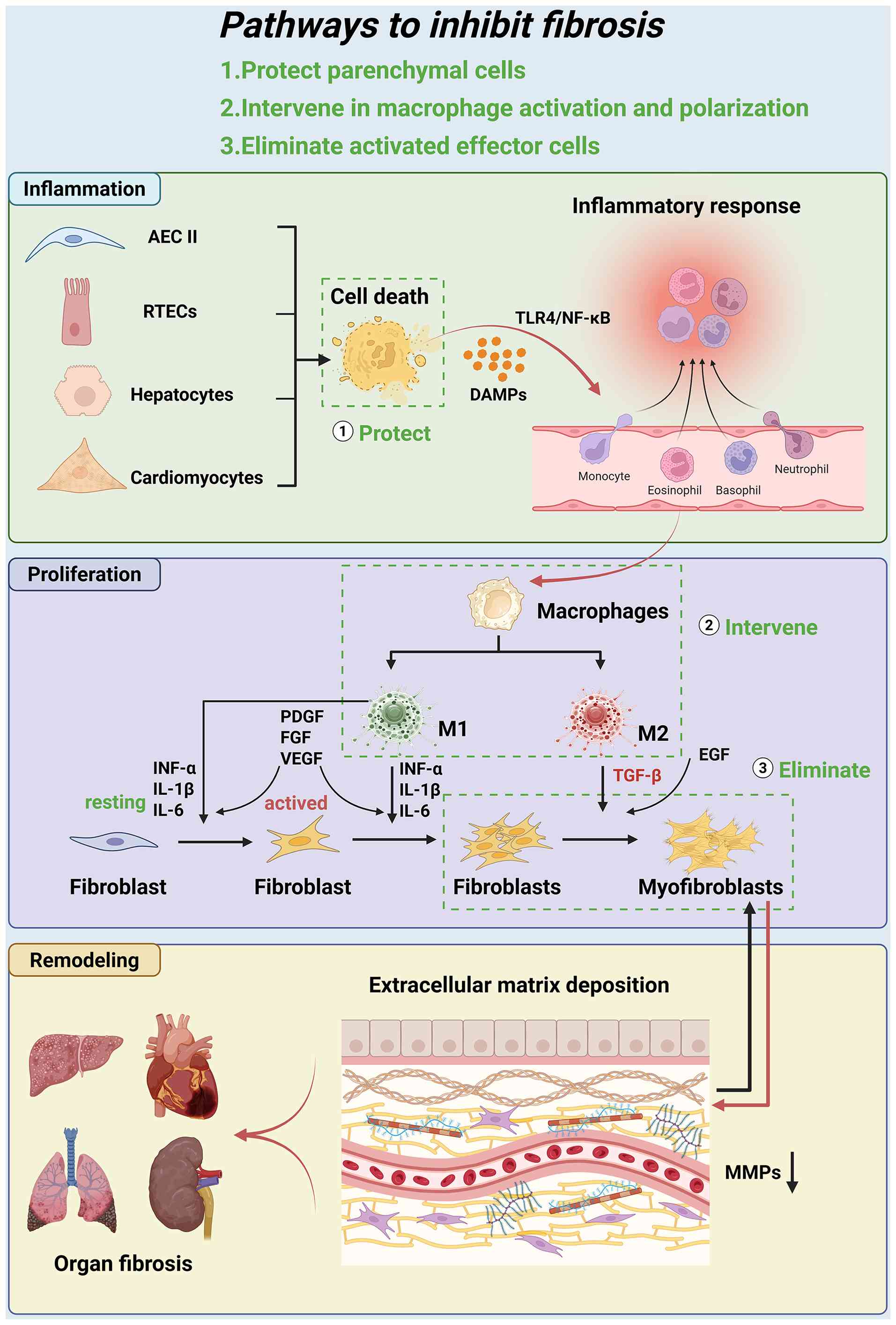
Fibrosis is a pathological remodeling process that
occurs following tissue injury. This process advances through three
distinct phases: Inflammation, proliferation and remodeling
(20). The molecular mechanisms
involve multiple signaling pathways, including TGF-β/Smad,
Wnt/β-catenin and yes-associated protein (YAP)/TAZ signaling axes.
These pathways have a cascading amplification effect and
self-reinforcing regulatory properties (21-24).
The pathological process of fibrosis is initiated by
the death of parenchymal cells, which is triggered by mechanical,
chemical or pathogenic injury. The subsequent release of
damage-associated molecular patterns (DAMPs) activates the local
innate immune response, which in turn leads to the activation of
NOD-like receptor family pyrin domain containing 3 inflammatory
vesicles via the Toll-like receptor 4 (TLR4)/NF-κB signaling axis
(25-29). This process involves dual immune
recruitment. On one hand, the monocyte-macrophage system polarizes
into pro-inflammatory M1 phenotypes (secreting TNF-α, IL-1β and
IL-6) and pro-fibrotic M2 phenotypes (secreting TGF-β) (7,30). On the other hand, neutrophils
infiltrate the injured area through the CXCL8/CXCR2 chemotaxis
pathway (28). Pro-inflammatory
cytokines (for example, TNF-α and IL-1β) released by these immune
cells, interact with ROS generated by NADPH oxidase to create an
oxidative stress microenvironment, thereby sustaining a persistent
inflammatory state (31)
(Fig. 1).
The hallmark of the proliferative phase is the
activation and differentiation of fibroblasts into α-SMA-positive
myofibroblasts, stimulated by growth factors such as TGF-β and
platelet-derived growth factor (18,32-34). These cells not only exhibit
abnormal proliferation and migration characteristics but also
overproduce ECM components, including type I and III collagen, and
fibronectin (9,35). Concurrently, the upregulation of
matrix metalloproteinase inhibitors suppresses matrix
metalloproteinase (MMP) activity, resulting in an imbalance in ECM
metabolism (9,35,36). At this stage, the TGF-β/Smad
signaling pathway fulfills a central regulatory role (37,38). In addition, mechanical stress
further sustains myofibroblast activation via the integrin-YAP/TAZ
axis, ultimately leading to the formation of early fibrotic lesions
characterized by disorganized tissue architecture (39,40) (Fig. 1).
The remodeling phase is characterized by tissue
stiffening and destruction of organ structures due to excessive ECM
deposition. Disruption of the tissue structure leads to
upregulation of hypoxia-inducible factor-1α (HIF-1α), which in turn
promotes an increase in pro-fibrotic factors such as connective
tissue growth factor and vascular endothelial growth factor. In
addition, the mechanical stress generated by the stiffening of the
ECM continuously activates myofibroblasts through a positive
feedback loop (41-43). Meanwhile, ECM degradation
products act as DAMPs that reactivate the TLR/NF-κB signaling
pathway, establishing a cycle of
'inflammation-fibrosis-reinflammation' (44,45). This stage is accompanied by
abnormal angiogenesis and the replacement of functional parenchymal
cells, ultimately leading to irreversible organ dysfunction
(46) (Fig. 1).
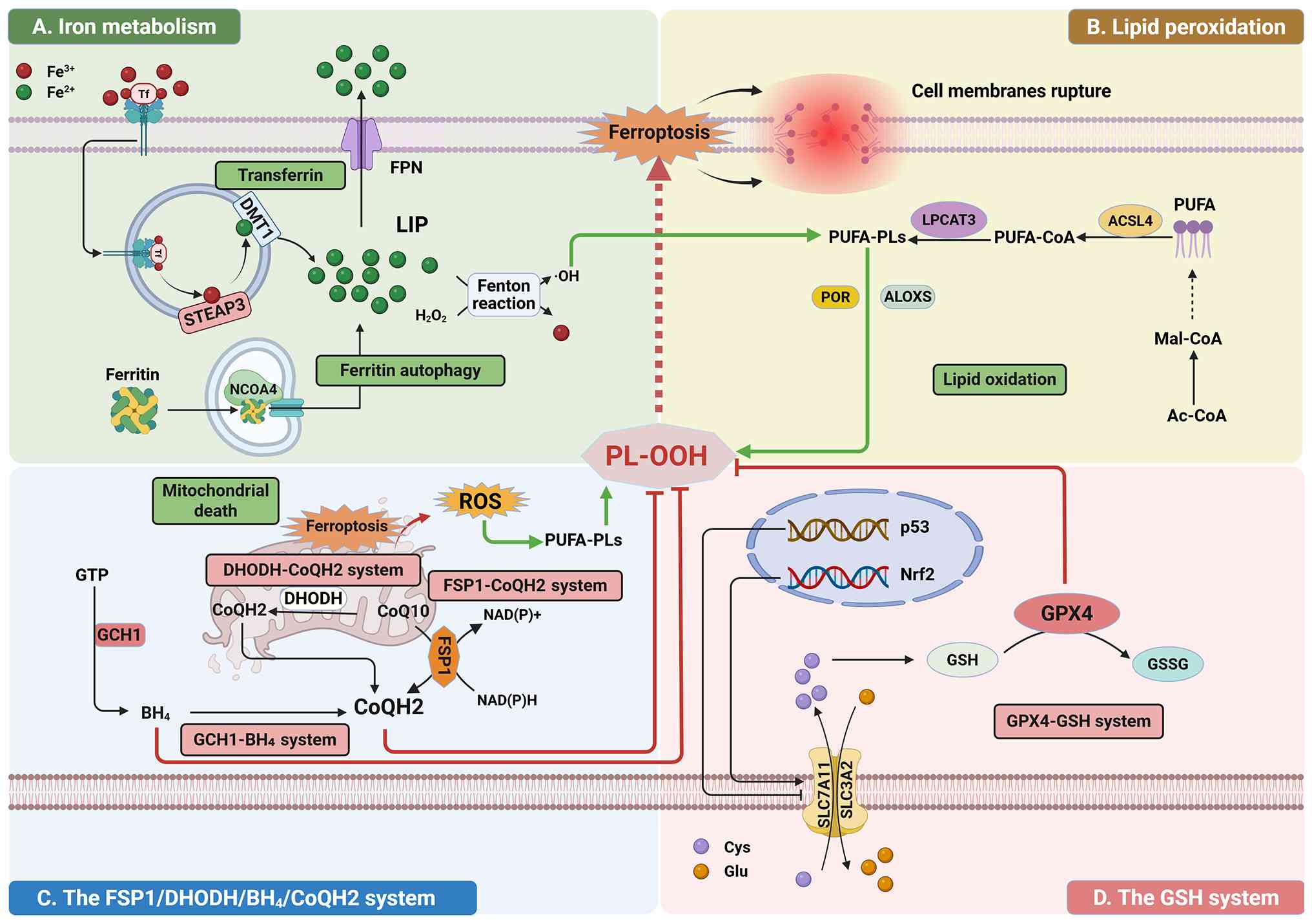
Within cells, ROS refer to a general category of
highly reactive oxygen-containing molecules, mainly consisting of
superoxide anion, hydroxyl radical, hydrogen peroxide, singlet
oxygen and lipid hydroperoxide (LOOH). The accumulation of ROS is
the primary driver of the ferroptosis process (47-50). ROS generation is mainly caused by
the dysregulation of iron metabolism and lipid peroxidation
cascades, with mitochondrial structural disruption as a secondary
contributing factor (51-53).
Under chronic injury conditions, the upregulation of
transferrin receptor 1 (TFR1) facilitates the endocytosis of
extracellular ferric iron, which is subsequently reduced to ferrous
iron (Fe2+) by six-transmembrane epithelial antigen of
the prostate 3 (54-56). Simultaneously, the autophagy
pathway mediated by nuclear receptor coactivator 4 (NCOA4)
accelerates the degradation of ferritin, thereby releasing free
Fe2+ into the cytoplasm (57-59). Subsequently, Fe2+
undergoes the Fenton reaction with hydrogen peroxide to release
highly reactive hydroxyl radicals. These radicals attack
polyunsaturated fatty acids (PUFAs), thereby triggering lipid
peroxidation (60,61) (Fig. 2A).
Acyl-CoA synthetase long chain family member 4
(ACSL4) catalyzes the enzymatic reaction that links PUFAs, such as
arachidonic acid and linolenic acid, to CoA, producing acyl-CoA
variants such as arachidonoyl-CoA and adrenoyl-CoA (62-65). These acyl-CoAs are subsequently
esterified into phospholipids by lysophosphatidylcholine
acyltransferase 3 (66,67). These phospholipids, enriched with
PUFAs, are highly susceptible to peroxidation. A large number of
hydroxyl radicals generated via the Fenton reaction attack PUFAs in
phospholipids, generating lipid radicals. These radicals react with
oxygen to form lipid peroxyl radicals, which further propagate the
chain reaction by abstracting hydrogen from adjacent lipid
molecules, generating LOOH (62,66,68). Accumulation of lipid peroxides
leads to decreased cell membrane fluidity and increased
permeability, ultimately triggering leakage of cell contents and
cell death (Fig. 2B).
Furthermore, studies have shown that ferroptosis can
also occur within the mitochondria (69-71). Mitochondria serve as a critical
hub for intracellular iron metabolism, participating in the
synthesis of heme, iron-sulfur clusters (Fe-S), and iron storage.
Mitochondrial membranes are rich in PUFAs, making them highly
susceptible to attack by hydroxyl radicals, which in turn triggers
mitochondrial rupture and the release of ROS (72). Ruptured mitochondria release a
substantial amount of iron ions, which subsequently catalyze the
generation of hydroxyl radicals via the Fenton reaction (73). These newly generated hydroxyl
radicals, together with ROS, further assault the mitochondrial
membrane, thereby establishing a self-amplifying vicious cycle
(65,74,75) (Fig. 2C).
Cells contain multiple antioxidant systems capable
of eliminating ROS and lipid peroxides, thereby inhibiting the
onset of ferroptosis. These systems are primarily divided into four
pathways, with the GPX4-glutathione (GSH) system playing a leading
role in eliminating cytotoxic LOOH. Furthermore, the ferroptosis
suppressor protein 1 (FSP1)-ubiquinol (CoQH2) system, the
dihydroorotate dehydrogenase (DHODH)-CoQH2 system and the GTP
cyclase hydrolase 1 (GCH1)-biosynthesis of tetrahydrobiopterin
(BH4) system have been shown to inhibit ferroptosis by eliminating
ROS within the cell (14,15,54,76).
The GPX4-GSH system serves as the central
antioxidant system, dynamically regulating lipid peroxidation via
GPX4 and its associated metabolic network (77,78). GPX4 utilizes reduced GSH as an
electron donor to reduce cytotoxic LOOH into non-toxic lipid
alcohols, thereby halting lipid peroxidation chain reactions and
preserving membrane integrity (79,80). The biosynthesis of GSH begins
with the uptake of cystine, which is mediated by the solute carrier
family 7 member 11 (SLC7A11)-dependent cystine/glutamate
antiporter. Intracellular cystine is reduced to cysteine, which is
then combined with glutamate by glutamate-cysteine ligase to form
γ-glutamylcysteine, and finally converted to GSH via glutathione
synthetase (81-83). This metabolic pathway provides
essential reducing equivalents for GPX4 activity. In terms of
transcription factors, nuclear factor erythroid 2-related factor 2
(Nrf2) enhances antioxidant capacity by binding to antioxidant
response elements to upregulate GPX4 and SLC7A11 expression
(84-86). Conversely, tumor protein p53
(p53) promotes ferroptosis by suppressing SLC7A11 transcription and
depleting GSH (87-89) (Fig. 2D).
The FSP1-CoQH2 pathway, as a GPX4-independent
pathway, also plays a crucial role in the antioxidant process.
FSP1, an NADH-dependent oxidoreductase, reduces coenzyme Q10
(CoQ10) to CoQH2, which directly neutralizes lipid peroxidation
radicals and terminates chain reactions (90-92). FSP1 expression is positively
regulated by Nrf2 and negatively regulated by p53 (93-95) (Fig. 2C).
The DHODH-CoQH2 system safeguards mitochondrial
redox homeostasis by coupling pyrimidine biosynthesis with electron
transport. DHODH is an enzyme localized to the inner mitochondrial
membrane; it catalyzes the oxidation of dihydro-lactate to lactate,
transferring electrons to CoQ10 in the process and generating CoQH2
(96,97). Under conditions of elevated ROS,
this system enhances CoQH2 production to eliminate the burst of ROS
and preserve mitochondrial function (98,99) (Fig. 2C).
The GCH1-BH4 axis is another intracellular
ferroptosis defense pathway that operates independently of GPX4.
GCH1 is the rate-limiting enzyme in the biosynthesis of BH4; it
catalyzes the conversion of GTP to dihydropterin triphosphate,
thereby initiating the de novo synthesis of BH4 (100,101). BH4 blocks lipid peroxidation
chain reactions by directly scavenging lipid radicals, while also
promoting the reduction of CoQ10 to CoQH2, thereby enhancing
membrane antioxidant capacity and synergistically inhibiting the
onset of ferroptosis (102-104) (Fig. 2C).
Since ROS burst is a central driver of ferroptosis,
the following sections will systematically discuss strategies for
regulating intracellular ferroptosis at three levels: Modulating
iron metabolism pathways, modulating lipid peroxidation pathways
and enhancing antioxidant systems.
Ferroptosis, a distinct form of cell death, is
present throughout the entire pathological process of organ
fibrosis and primarily affects parenchymal cells, macrophages and
effector cells (105,106). In the early stages of tissue
injury, an imbalance in redox homeostasis and disrupted iron
metabolism within parenchymal cells are the primary triggers of
ferroptosis. As lipid peroxidation occurs, the cell membranes of
damaged cells rupture and release DAMPs, which in turn activate the
immune response and amplify the inflammatory cascade (106,107). Concurrently, ferroptosis in the
inflammatory microenvironment drives polarization of macrophages
(108). M1 macrophages secrete
pro-inflammatory factors such as TNF-α and IL-1β, further
exacerbating the inflammatory response (108). M2 macrophages, on the other
hand, secrete TGF-β, which induces fibroblast proliferation,
activation and myofibroblast differentiation, ultimately promoting
fibrosis (109,110). The following sections will
provide a detailed explanation of the specific mechanisms
underlying cellular ferroptosis during the fibrotic process.
The fibrotic microenvironment can also exert
positive feedback regulation on ferroptosis, a process that occurs
primarily during the remodeling phase of fibrosis. During the
remodeling phase, excessive deposition of the ECM can cause tissue
stiffening and structural damage to organs, leading to tissue
hypoxia and subsequent upregulation of HIF-1α (41). Under hypoxic conditions, lactate
accumulation upregulates GPX4 expression, endowing activated
fibroblasts with ferroptosis resistance and thereby maintaining
their profibrotic phenotype. Conversely, reducing lactate levels
restores the sensitivity of the cells to ferroptosis and alleviates
fibrosis (111). Previous
studies have shown that during hypoxia caused by acute ischemia or
injury, HIF-1α plays a tissue-protective role by reducing tissue
damage and mediating a moderate inflammatory response to prevent
abnormal repair (112-114). However, in the context of
long-term chronic hypoxia caused by fibrosis, HIF-1α acts as a
promoter of fibrosis (106,115). For example, under conditions of
chronic hypoxia in the liver and kidneys, HIF-1α promotes iron
release by upregulating heme oxygenase-1 (HO-1). This is
accompanied by downregulation of GPX4 and SLC7A11, which
exacerbates ferroptosis in parenchymal cells, thereby accelerating
organ fibrosis (116-118).
Liver fibrosis is a condition in which the liver
undergoes abnormal repair and progresses toward sclerosis due to
persistent stimulation from various forms of chronic liver injury.
The core pathological mechanism involves hepatocyte death and the
activation and transdifferentiation of hepatic stellate cells
(HSCs). Recent research on ferroptosis has primarily focused on the
two types of cells (119,120). Under pathological conditions,
iron overload, accumulation of lipid peroxides and depletion of GSH
occur in the liver, ultimately triggering ferroptosis in
hepatocytes (121,122). For example, in alcohol-related
liver disease, TFR1 expression in hepatocytes is markedly
upregulated, leading to intracellular iron overload. Furthermore,
microRNA (miRNA/miR)-214 can exacerbate the accumulation of lipid
oxidation products by upregulating the expression of ACSL4
(123). In metabolic fatty
liver disease, elevated levels of TFR1 and reduced levels of
ferroportin (FPN) jointly lead to a significant expansion of the
labile iron pool (124). At the
same time, impaired function of the GPX4-GSH system further
exacerbates ferroptosis (125).
Subsequently, liver cells undergoing ferroptosis
release DAMPs, which activate Kupffer cells and induce their
polarization toward the M1 and M2 phenotypes, leading to the
secretion of cytokines such as TGF-β1 and IL-6 (108). These factors further drive the
activation of quiescent HSCs into myofibroblasts, which secrete
large amounts of ECM, ultimately leading to the development of
liver fibrosis (105). In viral
hepatitis, miR-222 generated by hepatocytes infected with HBV can
inhibit ferroptosis in HSCs by suppressing the expression of the
TFR1 in HSCs, thereby promoting their activation (126). In the context of metabolic
fatty liver disease, the accumulation of cholesterol within HSCs
not only activates the HSCs but also confers resistance to
ferroptosis by maintaining high levels of GPX4 expression (127).
Pulmonary fibrosis is often triggered by
environmental dust, infections, medications, autoimmune diseases or
radiation (128,129). When alveolar epithelial cells
are damaged, they release DAMPs, which trigger a series of immune
and inflammatory responses (130). This process further drives the
transdifferentiation of fibroblasts into myofibroblasts, induces
epithelial-mesenchymal transition (EMT) in alveolar epithelial
cells, and promotes massive deposition of ECM, ultimately
disrupting the alveolar-capillary membrane structure and forming
honeycomb-like scars (131).
The specific pathological processes driven by
ferroptosis in pulmonary fibrosis are similar to those in liver
fibrosis. Among these, alveolar epithelial type II cells (AEC II)
play a key role in ferroptosis. In models of exposure to bleomycin,
silica or PM2.5, AEC II can undergo ferroptosis, which is
characterized by iron accumulation, decreased GPX4 expression and
mitochondrial dysfunction (132,133). Furthermore, ficolin B carried
by alveolar macrophage exosomes can promote ferroptosis in AEC II
by activating the cyclic GMP-AMP synthasestimulator of interferon
genes (cGAS-STING) signaling pathway, thereby exacerbating
pulmonary fibrosis (134).
Renal fibrosis is a common pathological change
observed in various chronic kidney diseases as they progress to end
stage; it essentially represents an abnormal repair process that
occurs following repeated damage to the kidneys (135,136). This process begins in damaged
renal tubular cells, triggering a vicious cycle of inflammation and
oxidative stress, which leads to the abnormal activation of the key
factor TGF-β. This process subsequently drives fibroblast
activation and EMT, ultimately leading to ECM deposition and scar
formation, gradually replacing normal nephrons (8,21,137,138).
Ferroptosis of renal tubular epithelial cells is one
of the core mechanisms underlying renal fibrosis. Under
pathological conditions such as diabetic nephropathy and a high-fat
diet, renal tubular epithelial cells undergo ferroptosis,
characterized by downregulation of GPX4 and upregulation of ACSL4,
which induces lipid peroxidation (139-141). Among these, the diamine
acetyltransferase 1 (SAT1)/Nrf2 axis is a key pathway regulating
ferroptosis in renal tubular epithelial cells, and SAT1 silencing
can alleviate fibrosis (142).
Furthermore, sigma-1 receptor (S1R) promotes ferroptosis in renal
tubular epithelial cells and leads to renal fibrosis by inhibiting
Nrf2 (143).
Cardiac fibrosis can be caused by factors such as
hypertension, myocardial infarction and diabetes. Under the
influence of mechanical stress, ischemia, inflammation and
angiotensin II, damaged cardiac muscle cells release cytokines that
activate fibroblasts and promote their transformation into
myofibroblasts (144,145). Myofibroblasts excessively
synthesize ECM while its degradation is suppressed, leading to
aberrant collagen deposition and increased interstitial stiffness,
ultimately impairing cardiac diastolic and systolic functions
(146,147).
Ferroptosis plays a key regulatory role in cardiac
fibrosis and occurs primarily within cardiomyocytes. At the level
of iron metabolism, TFR1-mediated iron uptake and NCOA4-mediated
ferritin autophagy constitute the sources of iron accumulation
(148,149). In lipid metabolism, ACSL4 and
arachidonate 15-LOX (ALOX15) act at the substrate end of
synthesizing PUFA-containing phospholipids and the initiation end
of lipid peroxidation, respectively, thereby determining cellular
sensitivity to ferroptosis (150,151). Regarding glutathione
metabolism, GPX4 and SLC7A11 are key factors in inhibiting
ferroptosis, and their downregulation exacerbates oxidative damage
(152). Furthermore, the
acetylation of p53 and the ubiquitination of Nrf2 suppress the
expression levels of GPX4 and SLC7A11 (153,154).
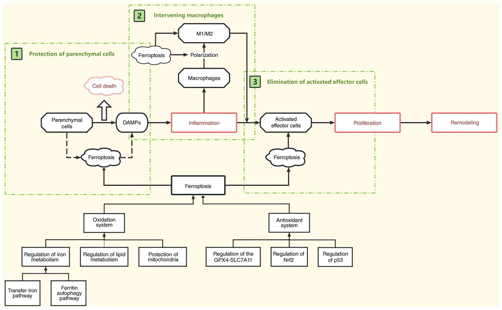
Targeting ferroptosis to mitigate fibrosis has
become a prominent research focus, with intervention strategies
primarily focused on three key areas. Firstly, during the
inflammatory stage, the death of parenchymal cells releases DAMPs,
which trigger an inflammatory response and drive the fibrotic
process (25,26). Inhibiting ferroptosis in these
cells by modulating the oxidative or antioxidant systems can halt
inflammation at its source, thereby impeding fibrosis progression
(106,155).
Secondly, targeting macrophages is also a key focus
of current research. Upon receiving inflammatory signals from
upstream, macrophages can polarize into two phenotypes:
Pro-inflammatory (M1) and pro-fibrotic (M2). These two polarization
pathways are key drivers of the fibrotic process (156). Therefore, macrophages can be
targeted in two ways to effectively inhibit the fibrotic process.
On the one hand, inhibiting ferroptosis in macrophages can prevent
their phenotypic transition. On the other hand, inducing
ferroptosis can eliminate already polarized macrophages.
Additionally, inducing ferroptosis in activated
effector cells, such as HSCs and fibroblasts, is a promising
strategy (157). However, this
strategy is currently primarily applied to liver fibrosis, with
relatively limited research on fibrosis in other organs. The
present review further elaborates on anti-fibrotic interventions
centered on these three core strategies (Fig. 3).
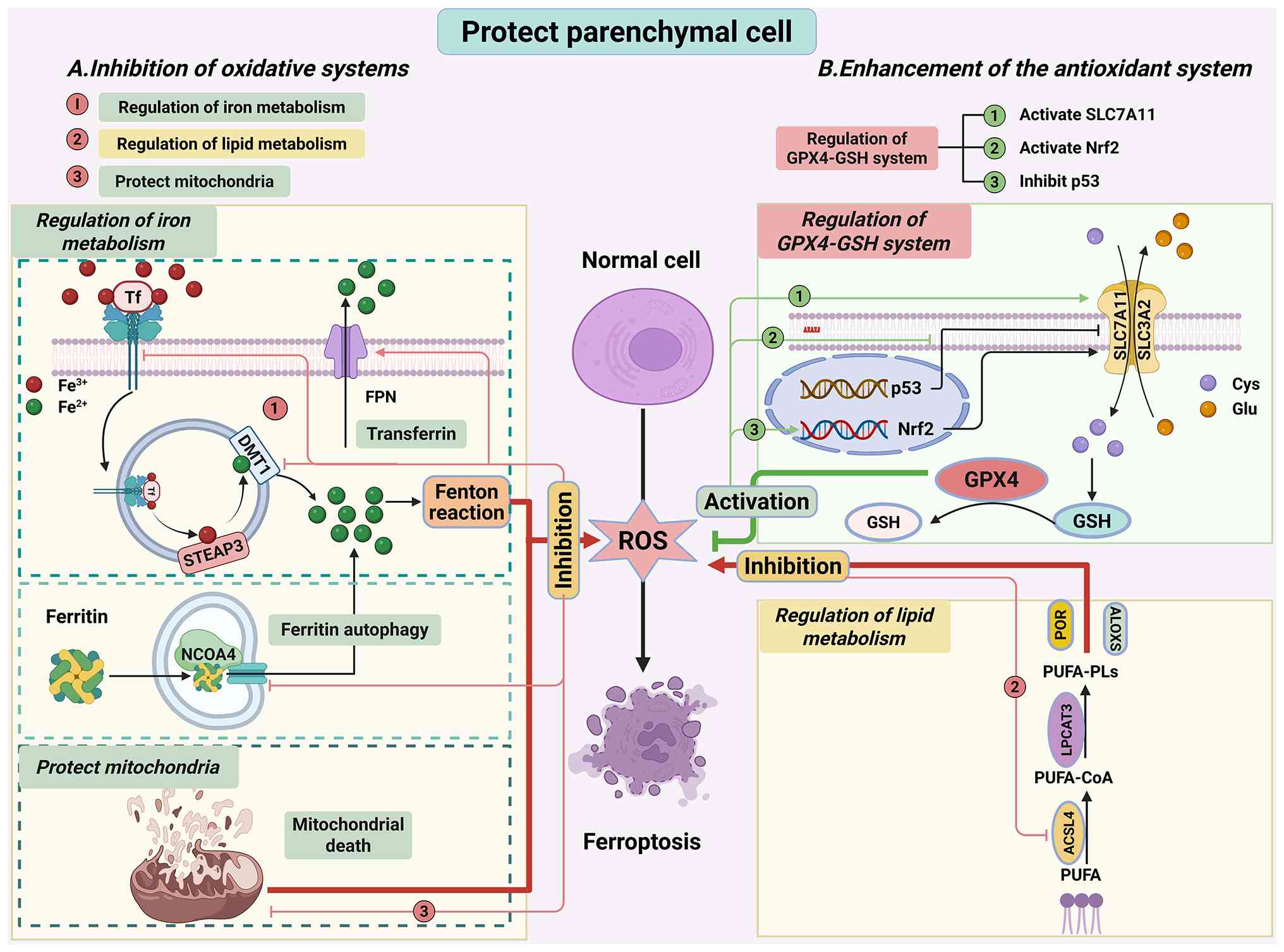
Transfer iron pathway. Research indicates that the
core strategy for regulating iron metabolism lies in modulating
intracellular iron homeostasis through the transferrin pathway,
thereby maintaining a dynamic balance of iron ions and ultimately
inhibiting ferroptosis in parenchymal cells (106,158). In pulmonary fibrosis,
upregulated TFR1 and divalent metal transporter 1 (DMT1) is a
critical factor that drives ferroptosis in alveolar epithelial
cells (132,159,160). The iron chelator deferoxamine
markedly alleviates fibrosis by reducing intracellular iron
accumulation via the downregulation of TFR1/DMT1 expression
(159) (Fig. 4A-1). FPN functions to export
intracellular iron into the extracellular space; its downregulation
consequently results in hepatocellular iron accumulation (103). Rifaximin alleviates iron
overload-induced ferroptosis in hepatocytes by restoring FPN
function, ultimately ameliorating liver fibrosis (161,162). Iron regulatory protein 2 (IRP2)
binds to iron-responsive elements in the mRNAs of TFR1 and FPN,
thereby upregulating TFR1 protein expression and repressing FPN
protein expression. These two proteins work together to promote the
uptake of iron ions by cells (163). Therefore, inhibiting IRP2 can
correct the imbalance of iron transport, thus alleviating fibrosis
(164) (Fig. 4A-1).
Ferritin autophagy pathway. Previous studies have
shown that inhibiting ferritin autophagy can also prevent
ferroptosis in parenchymal cells. Intracellular ferritin can bind
to NCOA4, thereby triggering ferritin autophagy and releasing large
amounts of iron ions (165-167). Therefore, upregulation of NCOA4
often leads to ferroptosis in parenchymal cells, thereby
exacerbating fibrosis (59,168). For instance, the transcription
factor Yin Yang 1 markedly upregulates NCOA4 expression, thereby
inducing ferritin autophagy, which ultimately drives myocardial
fibrosis and remodeling (169).
Similarly, silica nanoparticle-induced hepatic fibrosis is driven
by ferritin autophagy, and silencing the NCOA4 can suppress
ferritin degradation and reverse the fibrotic phenotype (170). Therefore, targeting NCOA4 has
become a strategic approach to restore iron homeostasis. Drugs such
as dihydroquercetin and fraxetin can inhibit NCOA4 expression,
thereby blocking the ferroptosis pathway and ultimately suppressing
pulmonary fibrosis (171,172) (Fig. 4A).
ACSL4 is a central hub in lipid metabolism.
Upregulation of ACSL4 exacerbates the pathological accumulation of
lipid peroxides, thereby triggering ferroptosis. In pulmonary
fibrosis models, ACSL4 expression is markedly upregulated,
exacerbating lipid peroxidation and thereby inducing alveolar
epithelial cell death (173,174). In liver fibrosis, activation of
the gp78-ACSL4 axis exacerbates ferroptosis in hepatocytes, and
specific inhibitors targeting this axis have demonstrated
therapeutic potential (175).
In renal fibrosis, calcium oxalate crystals induce ferroptosis in
renal tubular epithelial cells by activating the YAP-ACSL4 pathway,
thereby accelerating interstitial fibrosis. Accordingly, silencing
YAP or inhibiting ACSL4 can alleviate the progression of fibrosis
(176,177). Currently, intervention
strategies for ACSL4 include small-molecule inhibitors (such as
rosiglitazone), epigenetic modulators (such as DNA methylation
inhibitors), flavonoid derivatives (such as fisetin) and gene
silencing technologies (for example, small interfering RNA), which
provide a new direction in the treatment of fibrotic diseases
(177-179) (Fig. 4A).
Mitochondria maintain cellular iron homeostasis
through iron-sulfur cluster biosynthesis and heme metabolism.
Previous reports have indicated that the mitochondrial membrane is
susceptible to attack by hydroxyl radicals, which triggers
mitochondrial rupture and leads to the leakage of large amounts of
ROS from the mitochondria (69,180). Doxorubicin can bind to
Fe2+ to form a complex, which inhibits GPX4 activity,
leading to the massive accumulation of lipid peroxides within
mitochondria. This subsequently triggers mitochondrial-dependent
ferroptosis in cardiomyocytes, ultimately exacerbating myocardial
fibrosis and cardiac dysfunction (150,181). FUN14 domain-containing protein
1 (FUNDC1) is a mitophagy receptor primarily located on the outer
mitochondrial membrane, which mediates the entry of GPX4 into
mitochondria. During mitophagy, GPX4 is degraded, thereby
triggering cellular ferroptosis and ultimately leading to organ
fibrosis. Knockout of FUNDC1 markedly alleviates this process
(150,182). These findings suggest that
modulating mitochondrial ferroptosis holds potential value in the
treatment of fibrosis. For example, forsythiaside-A and melatonin
enhance GPX4 function by activating the Nrf2 pathway, thereby
alleviating mitochondrial ferroptosis (183,184). Additionally, the botanical
extract WGX50 and salidroside can alleviate cardiac fibrosis by
restoring the activity of mitochondrial GPX4 (185,186). The hydrogen sulfide donor AP39
inhibits mitochondrial ferroptosis via the PTEN-induced kinase
1/parkin pathway, thereby alleviating myocardial fibrosis (187) (Fig. 4A).
The GPX4-SLC7A11 system serves as the primary
antioxidant barrier; its inactivation leads to substantial
accumulation of lipid peroxides and accelerates fibrosis
progression (188-191). Therefore, modulating the
GPX4-SLC7A11 axis has become an important strategy for combating
fibrosis. For instance, elabela peptide inhibits ferroptosis in
cardiomyocytes by upregulating SLC7A11 expression through
antagonism of the IL-6/STAT3 signaling pathway, thereby effectively
suppressing myocardial fibrosis (192). The plant alkaloid
tuberostemonine enhances the activity of the SLC7A11/SLC3A2
heterotetramer, thereby markedly increasing GPX4 activity in
alveolar epithelial cells and inhibiting ferroptosis (193). Furthermore, Salvia
miltiorrhiza injection promotes the deacetylation of SLC7A11 by
activating sirtuin 1 (Sirt1), thereby enhancing the stability of
the SLC7A11 protein, protecting renal tubular epithelial cells from
ferroptosis and inhibiting renal interstitial fibrosis (194). Luteolin can also reverse carbon
tetrachloride-induced liver fibrosis by modulating the GPX4-SLC7A11
axis (195) (Fig. 4B).
Nrf2 is a key regulator of the oxidative stress
defense system; it activates downstream antioxidant systems (such
as GPX4), thereby effectively scavenging lipid peroxides and
maintaining redox homeostasis (196-198). In a renal fibrosis model
induced by high glucose and high fat, the S1R protein in renal
tubular epithelial cells was shown to bind to Nrf2 and promote its
phosphorylation, thereby inhibiting GPX4 and subsequently
exacerbating ferroptosis (143). Similar phenomena are also
observed in liver fibrosis and pulmonary fibrosis. In these
conditions, Nrf2 is suppressed, the expression of ferroptosis
markers is markedly elevated, and fibrosis is aggravated
accordingly (198,199) (Fig. 4B).
Drug development targeting the Nrf2 signaling
network has evolved into three major categories: Herbal extracts,
clinical drugs and small-molecule proteins. Herbal extracts (for
example, fucoxanthin, ginkgolide B, cinnamaldehyde and triptolide)
or traditional Chinese formulations (such as Taohongsiwu decoction
and LuQi formula) alleviate organ fibrosis by modulating pathways
(for example, Nrf2/GPX4, AKT/mTOR/Nrf2 and Nrf2/HO-1) to inhibit
ferroptosis in parenchymal cells (200-204). Among clinical drugs,
empagliflozin mitigates pulmonary fibrosis through the sestrin 2
(Sesn2)/AMPK/Nrf2 signaling (205). Meanwhile, melatonin
concurrently suppresses mitophagy and ferroptosis via the
AKT/mTOR/Nrf2 pathway to improve hepatic and renal fibrosis
(183,206). Additionally, small-molecule
proteins interact with Nrf2. Sirt7 ameliorates hypertensive renal
fibrosis via the KLF15/Nrf2 axis (207). Sesn2 reduces ferroptosis
through the Nrf2/activating transcription factor 4 (ATF4) pathway
to alleviate idiopathic pulmonary fibrosis (208).
Studies have shown that p53 negatively regulates
the GPX4-SLC7A11 system. In addition, p53 can activate ALOX12,
thereby exacerbating lipid peroxidation. Therefore, targeting p53
can inhibit ferroptosis in parenchymal cells, thereby offering a
potential therapeutic approach for fibrosis (209,210). For instance, SIRT1 deacetylates
p53 to restore the antioxidant defense system, thereby markedly
ameliorating the fibrotic phenotype (211). By contrast, SIRT3 knockout
upregulates the acetylation level of p53 in cardiac fibrosis, which
further induces ferroptosis in cardiomyocytes (212,213) (Fig. 4B-3).
Studies have revealed that macrophage ferroptosis
plays a vital role in the progression of fibrosis. For instance,
the expression of ferroptosis-related genes in macrophages is
markedly upregulated in both pulmonary fibrosis and systemic
sclerosis models (214,215). Moreover, Fei et al
(216) performed single-cell
RNA sequencing analysis on hypertrophic ligamentum flavum tissues
and identified a population of enriched secreted phosphoprotein
1-positive macrophages. These cells are not only involved in the
fibrotic process but also exhibit highly activated metabolic
pathways associated with ferroptosis.
Ferroptosis drives macrophage polarization into
pro-inflammatory (M1) and pro-fibrotic (M2) phenotypes by
modulating classical signaling pathways (156,217). Specifically, the formation of
the pro-inflammatory M1 phenotype depends on the activation of the
cGAS-STING pathway, which exacerbates chronic inflammatory
microenvironments through the sustained secretion of inflammatory
factors, such as IL-6 and TNF-α (218,219). By contrast, the pro-fibrotic M2
phenotype is synergistically driven by the coordinated activation
of the Wnt/β-catenin and JAK-STAT pathways, which subsequently
activate fibroblasts via the TGF-β/Smad signaling axis to induce
pathological ECM deposition (220,221).
Interventions targeting macrophage ferroptosis can
be implemented through two approaches: Suppressing phenotypic
polarization and eliminating polarized cells. In terms of
inhibiting polarization, targeted blockade of the ATF3-CD36 pathway
can activate the Nrf2/GPX4 signaling axis, thereby suppressing
macrophage ferroptosis (222,223). Additionally, inhibition of
calpain downregulates ACSL4 expression, thereby reducing macrophage
susceptibility to ferroptosis (179,215). In polarized cell elimination,
current research focuses on pro-inflammatory M1 macrophages. For
example, upregulating ACSL4 or inhibiting SLC7A11 expression can
specifically increase intracellular lipid peroxidation levels,
thereby inducing ferroptosis in M1 macrophages (179,224). Drugs represented by
glyceraldehyde 3-O-mono-β-D-glucuronide can selectively eliminate
inflammatory macrophages by activating the interferon regulatory
factor 1/SLC7A11 signaling pathway, which provides new insights
into reversing the progression of fibrosis (224).
During the progression of fibrosis, the
inflammatory microenvironment activates tissue-resident effector
cells, such as fibroblasts and HSCs, leading to their abnormal
proliferation and differentiation into myofibroblasts. Targeting
the elimination of activated effector cells has emerged as a
pivotal strategy in anti-fibrotic therapy. Currently, this strategy
is mainly applied to hepatic fibrosis; it induces ferroptosis of
activated HSCs via multiple pathways to alleviate fibrosis
(157,225-227).
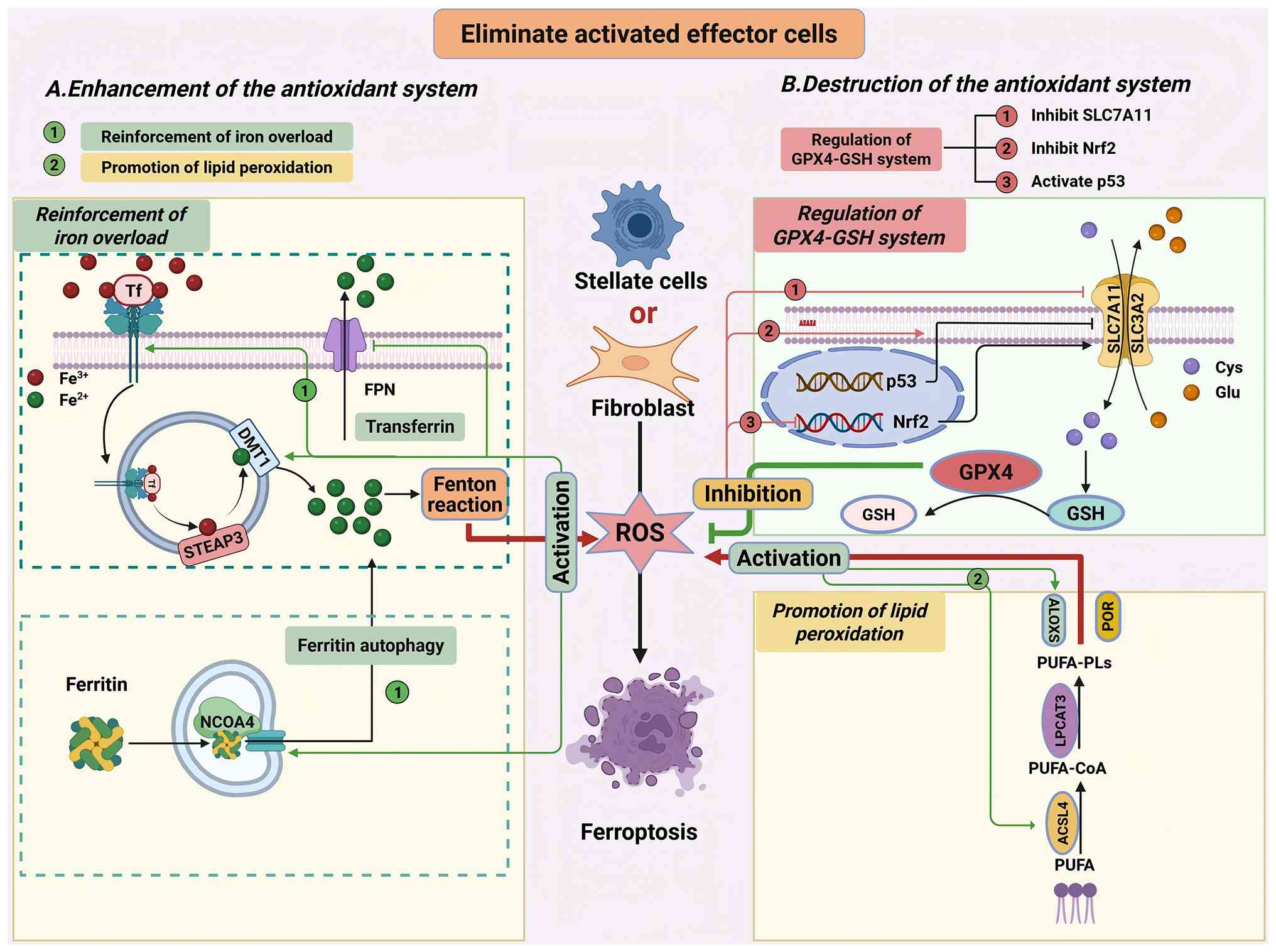
Transfer pathway. Studies have demonstrated that
modulating transferrin-related pathways to induce ferroptosis in
HSCs and fibroblasts represents an effective strategy for
alleviating fibrosis. YAP modulates the iron metabolic network by
coordinately regulating iron uptake, storage and efflux. Mechanism
analysis reveals that YAP promotes iron influx through the
activation of TFR1 and DMT1, while suppressing the expression of
FPN. This dysregulation leads to the abnormal accumulation of the
iron ions in activated HSCs (228-230). Therefore, upregulation of YAP
expression can trigger ferroptosis in HSCs, thereby alleviating
fibrosis.
Furthermore, several drugs have been proven to
inhibit fibrosis via this strategy. Artemether suppresses the
ubiquitination of IRP2, thereby upregulating the expression of
TFR/DMT1 and inhibiting FPN, and ultimately inducing ferroptosis in
HSCs (164). Liquiritigenin
upregulates the expression of TFR/DMT1, thereby activating
ferroptosis in HSCs (227).
Ellagic acid inhibits the transport function of FPN, leading to
iron overload and ROS accumulation, and ultimately triggering
ferroptosis in HSCs (231)
(Fig. 5A-1).
Ferritin autophagy. In addition, disrupting
intracellular iron homeostasis via NCOA4-mediated ferritin
autophagy also serves as a strategy for targeted elimination of
effector cells (59,165,232). NCOA4 specifically recognizes
and binds to the ferritin heavy chain (FTH1), forming an NCOA4-FTH1
complex, which subsequently mediates the transport of ferritin to
lysosomes. Upon degradation of this complex within lysosomes, a
large amount of iron ions is released. Natural compounds such as
curcumin, artemisinin derivatives, taurine, naringin and berberine
can accelerate the degradation of the NCOA4-FTH1 complex, releasing
iron ions, which in turn induce ferroptosis in HSCs, thereby
exerting an anti-fibrotic effect (233-241). In addition, the RNA-binding
proteins embryonic lethal and zinc finger protein 36 (ZFP36) have
been identified as regulators of the ferritin autophagy signaling
pathway in HSCs, and their dysfunction is closely associated with
the progression of fibrosis. These findings offer new insights for
the development of targeted therapies (242,243) (Fig. 5A).
The enhancement of lipid metabolic pathways can
also induce ferroptosis in activated effector cells (106,244). A recent study has shown that
YTH N6-methyladenosine RNA-binding protein F2 can upregulate ACSL4
expression, enhance lipid oxidation pathways and thereby induce
ferroptosis in activated HSCs (245). Ginsenoside Rg3 can restore
ACSL4 expression by promoting its demethylation, thereby inducing
ferroptosis in HSCs (246).
ALOX15 directly activates the ferroptosis process by catalyzing the
oxidation of PUFAs to form lipid peroxides (247,248). Dihydrotanshinone I promotes the
demethylation of ALOX15 by downregulating the expression of DNA
methyltransferase 1, thereby inducing ferroptosis in HSCs (249) (Fig. 5A).
In addition to enhancing oxidative pathways,
inhibiting the antioxidant system is another potential therapeutic
strategy (227,250). Studies have shown that various
plant extracts can induce ferroptosis in activated HSCs by
regulating the GPX4-SLC7A11 system. Ginkgolic acid and fig extract
can downregulate GPX4 expression, thereby inducing ferroptosis in
HSCs and ultimately improving liver fibrosis (251,252). Ginsenoside Rb1 and ginsenoside
Rh2 inhibit SLC7A11, disrupting the cysteine-glutamate reverse
transport system and thereby inducing ferroptosis in HSCs (253,254). Furthermore, simvastatin
selectively downregulates GPX4 expression in HSCs, achieving
anti-fibrotic effects while avoiding hepatotoxicity, demonstrating
significant translational medical value (255).
At the transcriptional level, HIF-1α can upregulate
the expression of SLC7A11. Sorafenib alleviates liver fibrosis by
inhibiting the HIF-1α/SLC7A11 pathway, thereby triggering
ferrocytosis in HSCs (225).
Conversely, the tumor suppressor protein p53 promotes ferroptosis
by inhibiting SLC7A11 transcription, thereby reducing the
biosynthesis of GSH (256). For
example, vogonolide and artemisinin can activate p53, disrupt the
antioxidant system and consequently induce ferroptosis in HSCs
(257,258) (Fig. 5B).
Exosome therapy, a novel therapeutic strategy based
on extracellular vesicles (exosomes), regulates intercellular
communication by delivering bioactive molecules (for example,
proteins, miRNAs and mRNAs) and demonstrates potential in treating
diverse diseases (259,260). As critical mediators of
cell-cell communication, exosomes play a role in fibrotic processes
by modulating ferroptosis signaling pathways. In pulmonary
fibrosis, ficolin B secreted by alveolar macrophages can promote
ferroptosis in pulmonary epithelial cells via the cGAS-STING
signaling pathway, thereby exacerbating the progression of
pulmonary fibrosis (134).
Following HBV infection, miR-222 in exosomes secreted by
hepatocytes can suppress ferroptosis in HSCs by inhibiting TFR1
expression (126).
Additionally, exosomes derived from renal tubular epithelial cells
can activate ATF3, which both inhibits the Nrf2/GPX4 signaling
pathway and promotes M2 macrophage polarization, thereby
synergistically driving renal fibrosis (261).
Exosome-engineered therapies have opened up a new
dimension in fibrosis treatment through a two-way precision
regulation strategy of promoting effector cell death and protecting
parenchymal cells. In targeting effector cell ferroptosis,
mesenchymal stem cell-derived exosomes (MSC-Exos) exhibit specific
therapeutic advantages. For instance, human umbilical cord MSC-Exos
(hucMSC-Exos) selectively induce HSC ferroptosis by delivering the
Beclin 1 (BECN1) protein to suppress GPX4 expression (262). The miR-499a-5p carried by
hucMSC-Exos interacts with the transcription factor ETS
proto-oncogene 1 to downregulate SLC7A11 expression, thereby
disrupting GSH synthesis and facilitating the precise elimination
of HSCs (263,264). Similarly, bone marrow MSC-Exos
utilize miR-144-3p to silence SLC7A11, thereby effectively
inhibiting HSC activation (265).
In parenchymal cytoprotective strategies,
engineered exosomes exhibit multifunctional regulatory properties.
Spike receptor-binding domain (S-RBD)-modified MSC-Exos inhibit
ferroptosis of alveolar epithelial cells by delivering miR-486-5p,
while inhibiting SMAD2 signaling and activating the Akt pathway to
achieve dual 'antifibrotic and anti-inflammatory' effects (266). Menstrual blood-derived SC-Exos
deliver miR-let-7 to suppress Sp3 transcription factor expression
and reduce recruitment to histone deacetylase 2, thereby
alleviating the inhibition of Nrf2 and ultimately suppressing
alveolar epithelial cell ferroptosis (267).
Nanodelivery platforms are drug delivery systems
engineered using nanotechnology to enhance drug targeting,
stability and therapeutic efficacy (268,269). Nanoparticles, owing to their
unique targeting capabilities and controllable drug delivery
properties, have emerged as cutting-edge strategies for modulating
ferroptosis to alleviate fibrosis.
As innovative carriers for regulating ferroptosis,
nanoparticles can protect parenchymal cells or eliminate effector
cells through targeted drug delivery, thereby intervening precisely
in fibrosis. In parenchymal cell protection, silica nanoparticles
loaded with allicin are able to inhibit ferroptosis in cardiac
microvascular endothelial cells, significantly improving myocardial
fibrosis (270). Selenium-doped
silica nanoparticles mitigate myocardial ischemia-reperfusion
injury by synergistically alleviating ferroptosis and mitochondrial
dysfunction (271).
In the elimination of effector cells,
naringenin-loaded nanoparticles selectively remove HSCs by
activating autophagy-dependent ferroptosis pathways (272). Carbon nitride-based hybrid
nanoparticles induce HSC ferroptosis via downregulation of the
HIF-1α/SLC7A11 signaling axis (273). Beyond that, taurine-conjugated
lipid nanoparticles and fluorinated peptide-lipid hybrid
nanoparticles enhance HSC ferroptosis sensitivity through
multi-dimensional regulation (274,275).
While nanoparticles show promise in
ferroptosis-targeted therapy, their design requires optimization to
improve targeting specificity and reduce off-target toxicity.
Future development of multifunctional platforms (for example,
stimuli-responsive release and multi-target modulation) may offer
more efficient solutions for fibrosis treatment.
Fibrosis, a core pathological process that drives
chronic diseases toward organ failure, is characterized by the
abnormal deposition of ECM and involves three interconnected
phases: Inflammation, proliferation and remodeling. Recent studies
have shown that ferroptosis plays a key role in the fibrotic
process. The present review systematically summarizes the process
of fibrosis, the major mechanisms of ferroptosis and its role in
fibrosis, and discusses therapeutic strategies and novel treatment
modalities that target ferroptosis for the intervention of
fibrosis.
Current therapeutic approaches primarily focus on
two strategies: Protecting parenchymal cells during the
inflammatory phase and eliminating activated effector cells during
the proliferative phase. Both strategies target three core
regulatory nodes: Iron metabolism pathways, lipid peroxidation
pathways and antioxidant systems. Additionally, interventions
targeting macrophage represent a currently prominent strategy.
Inhibiting macrophage polarization or eliminating polarized
macrophages via the mechanism of ferroptosis can also alleviate
fibrosis.
Novel ferroptosis-modulating therapies are evolving
in multi-dimensional directions. In the field of delivery system
innovation, engineered exosomes offer unique advantages. MSC-Exos
achieve precise regulation of HSCs ferroptosis by delivering
functional RNAs (such as miR-499a-5p and miR-144-3p) and proteins
(such as BECN1). S-RBD-modified exosomes exert dual regulatory
effects in pulmonary fibrosis by suppressing SMAD2/Akt signaling
via miR-486-5p. Concurrently, nanodelivery platforms are advancing
precision medicine. For instance, selenium-doped silica
nanoparticles inhibit myocardial ferroptosis by restoring
mitochondrial function, while naringenin-loaded nanoparticles
eliminate activated HSCs via autophagy-dependent ferroptosis
pathways. These innovations are steering ferroptosis-targeted
therapies toward greater specificity and efficacy.
Despite the promise of ferroptosis modulation in
fibrosis treatment, clinical translation faces challenges. First,
off-target effects of existing delivery systems risk damaging
healthy parenchymal cells, necessitating improved targeting through
surface ligand modifications or organ-specific promoters. Second,
dense ECM barriers impede drug penetration, driving the development
of MMP-responsive nanocarriers or ultrasound microbubble-mediated
delivery systems. Future research should prioritize intelligent
nanoplatforms, gene-editing technologies and the integration of
single-cell sequencing with spatial transcriptomics to map
organ-specific regulatory networks. By bridging foundational
discoveries with clinical precision medicine, these efforts may
unlock transformative therapies for fibrosis.
Not applicable.
XF was primarily responsible for the writing,
review and revision of the article. JZ participated in the
literature review and provided revisions for this review. QM and CS
provided guidance throughout the preparation of this manuscript and
made revisions to the text. All authors have read and approved the
manuscript. Data authentication is not applicable.
Not applicable.
Not applicable.
The authors declare that they have no competing
interests.
Not applicable.
This review was supported by the National Natural Science
Foundation of China (grant no. 81802198) and the Natural Science
Foundation of Jiangsu Province (grant no. BK20221176).
|
1
|
Santos A and Lagares D: Matrix stiffness:
The conductor of organ fibrosis. Curr Rheumatol Rep. 20:22018.
View Article : Google Scholar : PubMed/NCBI
|
|
2
|
Lagares D, Ghassemi-Kakroodi P, Tremblay
C, Santos A, Probst CK, Franklin A, Santos DM, Grasberger P,
Ahluwalia N, Montesi SB, et al: ADAM10-mediated ephrin-B2 shedding
promotes myofibroblast activation and organ fibrosis. Nat Med.
23:1405–1415. 2017. View Article : Google Scholar : PubMed/NCBI
|
|
3
|
Qin XJ, Zhang JX and Wang RL: Exosomes as
mediators and biomarkers in fibrosis. Biomark Med. 14:697–712.
2020. View Article : Google Scholar : PubMed/NCBI
|
|
4
|
Somnay K, Wadgaonkar P, Sridhar N, Roshni
P, Rao N and Wadgaonkar R: Liver Fibrosis leading to cirrhosis:
Basic mechanisms and clinical perspectives. Biomedicines.
12:22292024. View Article : Google Scholar : PubMed/NCBI
|
|
5
|
Hu Y, Huang Y, Zong L, Lin J, Liu X and
Ning S: Emerging roles of ferroptosis in pulmonary fibrosis:
Current perspectives, opportunities and challenges. Cell Death
Discov. 10:3012024. View Article : Google Scholar : PubMed/NCBI
|
|
6
|
Hammerich L and Tacke F: Hepatic
inflammatory responses in liver fibrosis. Nat Rev Gastroenterol
Hepatol. 20:633–646. 2023. View Article : Google Scholar : PubMed/NCBI
|
|
7
|
Wen JH, Li DY, Liang S, Yang C, Tang JX
and Liu HF: Macrophage autophagy in macrophage polarization,
chronic inflammation and organ fibrosis. Front Immunol.
13:9468322022. View Article : Google Scholar : PubMed/NCBI
|
|
8
|
Henderson NC, Rieder F and Wynn TA:
Fibrosis: From mechanisms to medicines. Nature. 587:555–566. 2020.
View Article : Google Scholar : PubMed/NCBI
|
|
9
|
Wynn TA and Ramalingam TR: Mechanisms of
fibrosis: Therapeutic translation for fibrotic disease. Nat Med.
18:1028–1040. 2012. View Article : Google Scholar : PubMed/NCBI
|
|
10
|
Stockwell BR, Jiang X and Gu W: Emerging
mechanisms and disease relevance of ferroptosis. Trends Cell Biol.
30:478–490. 2020. View Article : Google Scholar : PubMed/NCBI
|
|
11
|
Yu P, Zhang X, Liu N, Tang L, Peng C and
Chen X: Pyroptosis: Mechanisms and diseases. Signal Transduct
Target Ther. 6:1282021. View Article : Google Scholar : PubMed/NCBI
|
|
12
|
Liu S, Yao S, Yang H, Liu S and Wang Y:
Autophagy: Regulator of cell death. Cell Death Dis. 14:6482023.
View Article : Google Scholar : PubMed/NCBI
|
|
13
|
Liang D, Minikes AM and Jiang X:
Ferroptosis at the intersection of lipid metabolism and cellular
signaling. Mol Cell. 82:2215–2227. 2022. View Article : Google Scholar : PubMed/NCBI
|
|
14
|
Li J, Cao F, Yin HL, Huang ZJ, Lin ZT, Mao
N, Sun B and Wang G: Ferroptosis: Past, present and future. Cell
Death Dis. 11:882020. View Article : Google Scholar : PubMed/NCBI
|
|
15
|
Liu J, Kang R and Tang D: Signaling
pathways and defense mechanisms of ferroptosis. FEBS J.
289:7038–7050. 2022. View Article : Google Scholar
|
|
16
|
Pope LE and Dixon SJ: Regulation of
ferroptosis by lipid metabolism. Trends Cell Biol. 33:1077–1087.
2023. View Article : Google Scholar : PubMed/NCBI
|
|
17
|
Rockey DC, Bell PD and Hill JA: Fibrosis-a
common pathway to organ injury and failure. N Engl J Med.
372:1138–1149. 2015. View Article : Google Scholar : PubMed/NCBI
|
|
18
|
Kisseleva T and Brenner D: Molecular and
cellular mechanisms of liver fibrosis and its regression. Nat Rev
Gastroenterol Hepatol. 18:151–166. 2021. View Article : Google Scholar
|
|
19
|
Zhang M, Serna-Salas S, Damba T, Borghesan
M, Demaria M and Moshage H: Hepatic stellate cell senescence in
liver fibrosis: Characteristics, mechanisms and perspectives. Mech
Ageing Dev. 199:1115722021. View Article : Google Scholar : PubMed/NCBI
|
|
20
|
Pei Z, Fan J, Tang M and Li Y:
Ferroptosis: A new strategy for the treatment of fibrotic diseases.
Adv Biol (Weinh). 9:e24003832025. View Article : Google Scholar
|
|
21
|
Hu HH, Chen DQ, Wang YN, Feng YL, Cao G,
Vaziri ND and Zhao YY: New insights into TGF-β/Smad signaling in
tissue fibrosis. Chem Biol Interact. 292:76–83. 2018. View Article : Google Scholar : PubMed/NCBI
|
|
22
|
Xu F, Liu C, Zhou D and Zhang L:
TGF-β/SMAD pathway and its regulation in hepatic fibrosis. J
Histochem Cytochem. 64:157–167. 2016. View Article : Google Scholar : PubMed/NCBI
|
|
23
|
Liu J, Xiao Q, Xiao J, Niu C, Li Y, Zhang
X, Zhou Z, Shu G and Yin G: Wnt/β-catenin signalling: Function,
biological mechanisms, and therapeutic opportunities. Signal
Transduct Target Ther. 7:32022. View Article : Google Scholar
|
|
24
|
Wei Y, Hui VLZ, Chen Y, Han R, Han X and
Guo Y: YAP/TAZ: Molecular pathway and disease therapy. MedComm
(2020). 4:e3402023. View Article : Google Scholar : PubMed/NCBI
|
|
25
|
Leuti A, Fazio D, Fava M, Piccoli A, Oddi
S and Maccarrone M: Bioactive lipids, inflammation and chronic
diseases. Adv Drug Deliv Rev. 159:133–169. 2020. View Article : Google Scholar : PubMed/NCBI
|
|
26
|
Ma M, Jiang W and Zhou R: DAMPs and
DAMP-sensing receptors in inflammation and diseases. Immunity.
57:752–771. 2024. View Article : Google Scholar : PubMed/NCBI
|
|
27
|
Taru V, Szabo G, Mehal W and Reiberger T:
Inflammasomes in chronic liver disease: Hepatic injury, fibrosis
progression and systemic inflammation. J Hepatol. 81:895–910. 2024.
View Article : Google Scholar : PubMed/NCBI
|
|
28
|
Mack M: Inflammation and fibrosis. Matrix
Biol. 68-69:106–121. 2018. View Article : Google Scholar
|
|
29
|
Vringer E and Tait SWG: Mitochondria and
cell death-associated inflammation. Cell Death Differ. 30:304–312.
2023. View Article : Google Scholar :
|
|
30
|
Lafuse WP, Wozniak DJ and Rajaram MVS:
Role of cardiac macrophages on cardiac inflammation, fibrosis and
tissue repair. Cells. 10:512020. View Article : Google Scholar
|
|
31
|
Antar SA, Ashour NA, Marawan ME and
Al-Karmalawy AA: Fibrosis: Types, effects, markers, mechanisms for
disease progression, and its relation with oxidative stress,
immunity, and inflammation. Int J Mol Sci. 24:40042023. View Article : Google Scholar : PubMed/NCBI
|
|
32
|
Gibb AA, Lazaropoulos MP and Elrod JW:
Myofibroblasts and fibrosis: Mitochondrial and metabolic control of
cellular differentiation. Circ Res. 127:427–447. 2020. View Article : Google Scholar : PubMed/NCBI
|
|
33
|
Younesi FS, Miller AE, Barker TH, Rossi
FMV and Hinz B: Fibroblast and myofibroblast activation in normal
tissue repair and fibrosis. Nat Rev Mol Cell Biol. 25:617–638.
2024. View Article : Google Scholar : PubMed/NCBI
|
|
34
|
Yang H, Cheng H, Dai R, Shang L, Zhang X
and Wen H: Macrophage polarization in tissue fibrosis. PeerJ.
11:e160922023. View Article : Google Scholar : PubMed/NCBI
|
|
35
|
Pei Q, Yi Q and Tang L: Liver fibrosis
resolution: From molecular mechanisms to therapeutic opportunities.
Int J Mol Sci. 24:96712023. View Article : Google Scholar : PubMed/NCBI
|
|
36
|
Yamashita N and Kramann R: Mechanisms of
kidney fibrosis and routes towards therapy. Trends Endocrinol
Metab. 35:31–48. 2024. View Article : Google Scholar
|
|
37
|
Peng D, Fu M, Wang M, Wei Y and Wei X:
Targeting TGF-β signal transduction for fibrosis and cancer
therapy. Mol Cancer. 21:1042022. View Article : Google Scholar
|
|
38
|
Meng XM, Nikolic-Paterson DJ and Lan HY:
TGF-β: The master regulator of fibrosis. Nat Rev Nephrol.
12:325–338. 2016. View Article : Google Scholar : PubMed/NCBI
|
|
39
|
Chu CQ and Quan T: Fibroblast Yap/Taz
signaling in extracellular matrix homeostasis and tissue fibrosis.
J Clin Med. 13:33582024. View Article : Google Scholar : PubMed/NCBI
|
|
40
|
Papavassiliou KA, Sofianidi AA,
Spiliopoulos FG, Gogou VA, Gargalionis AN and Papavassiliou AG:
YAP/TAZ signaling in the pathobiology of pulmonary fibrosis. Cells.
13:15192024. View Article : Google Scholar : PubMed/NCBI
|
|
41
|
Liu D, Wang L, Ha W, Li K, Shen R and Wang
D: HIF-1α: A potential therapeutic opportunity in renal fibrosis.
Chem Biol Interact. 387:1108082024. View Article : Google Scholar
|
|
42
|
Wei X, Hou Y, Long M, Jiang L and Du Y:
Molecular mechanisms underlying the role of hypoxia-inducible
factor-1 α in metabolic reprogramming in renal fibrosis. Front
Endocrinol (Lausanne). 13:9273292022. View Article : Google Scholar
|
|
43
|
Steiner CA, Cartwright IM, Taylor CT and
Colgan SP: Hypoxia-inducible factor as a bridge between healthy
barrier function, wound healing, and fibrosis. Am J Physiol Cell
Physiol. 323:C866–C878. 2022. View Article : Google Scholar : PubMed/NCBI
|
|
44
|
McQuitty CE, Williams R, Chokshi S and
Urbani L: Immunomodulatory role of the extracellular matrix within
the liver disease microenvironment. Front Immunol. 11:5742762020.
View Article : Google Scholar : PubMed/NCBI
|
|
45
|
Liu X, Lu F and Chen X: Examination of the
role of necroptotic damage-associated molecular patterns in tissue
fibrosis. Front Immunol. 13:8863742022. View Article : Google Scholar : PubMed/NCBI
|
|
46
|
Wang S, Tang C, Zhao H, Shen P, Lin C, Zhu
Y and Han D: Network pharmacological analysis and experimental
validation of the mechanisms of action of Si-Ni-San against liver
fibrosis. Front Pharmacol. 12:6561152021. View Article : Google Scholar : PubMed/NCBI
|
|
47
|
Zhang X, Ma Y, Lv G and Wang H:
Ferroptosis as a therapeutic target for inflammation-related
intestinal diseases. Front Pharmacol. 14:10953662023. View Article : Google Scholar : PubMed/NCBI
|
|
48
|
Chen Y, Fang ZM, Yi X, Wei X and Jiang DS:
The interaction between ferroptosis and inflammatory signaling
pathways. Cell Death Dis. 14:2052023. View Article : Google Scholar : PubMed/NCBI
|
|
49
|
Liu H, Xue H, Guo Q, Xue X, Yang L, Zhao K
and Liu Y: Ferroptosis meets inflammation: A new frontier in cancer
therapy. Cancer Lett. 620:2176962025. View Article : Google Scholar : PubMed/NCBI
|
|
50
|
Han H, Zhang G, Zhang X and Zhao Q:
Nrf2-mediated ferroptosis inhibition: A novel approach for managing
inflammatory diseases. Inflammopharmacology. 32:2961–2986. 2024.
View Article : Google Scholar : PubMed/NCBI
|
|
51
|
Rochette L, Dogon G, Rigal E, Zeller M,
Cottin Y and Vergely C: Lipid peroxidation and iron metabolism: Two
corner stones in the homeostasis control of ferroptosis. Int J Mol
Sci. 24:4492022. View Article : Google Scholar
|
|
52
|
Zhang X, Wu L, Zhen W, Li S and Jiang X:
Generation of singlet oxygen via iron-dependent lipid peroxidation
and its role in ferroptosis. Fundam Res. 2:66–73. 2021. View Article : Google Scholar : PubMed/NCBI
|
|
53
|
Nakamura T, Naguro I and Ichijo H: Iron
homeostasis and iron-regulated ROS in cell death, senescence and
human diseases. Biochim Biophys Acta Gen Subj. 1863:1398–1409.
2019. View Article : Google Scholar : PubMed/NCBI
|
|
54
|
Jiang X, Stockwell BR and Conrad M:
Ferroptosis: Mechanisms, biology and role in disease. Nat Rev Mol
Cell Biol. 22:266–282. 2021. View Article : Google Scholar : PubMed/NCBI
|
|
55
|
Gao M, Monian P, Quadri N, Ramasamy R and
Jiang X: Glutaminolysis and transferrin regulate ferroptosis. Mol
Cell. 59:298–308. 2015. View Article : Google Scholar : PubMed/NCBI
|
|
56
|
Mou Y, Wang J, Wu J, He D, Zhang C, Duan C
and Li B: Ferroptosis, a new form of cell death: Opportunities and
challenges in cancer. J Hematol Oncol. 12:342019. View Article : Google Scholar : PubMed/NCBI
|
|
57
|
He J, Li Z, Xia P, Shi A, FuChen X, Zhang
J and Yu P: Ferroptosis and ferritinophagy in diabetes
complications. Mol Metab. 60:1014702022. View Article : Google Scholar : PubMed/NCBI
|
|
58
|
Santana-Codina N, Gikandi A and Mancias
JD: The role of NCOA4-mediated ferritinophagy in ferroptosis. Adv
Exp Med Biol. 1301:41–57. 2021. View Article : Google Scholar : PubMed/NCBI
|
|
59
|
Hou W, Xie Y, Song X, Sun X, Lotze MT, Zeh
HJ III, Kang R and Tang D: Autophagy promotes ferroptosis by
degradation of ferritin. Autophagy. 12:1425–1428. 2016. View Article : Google Scholar : PubMed/NCBI
|
|
60
|
Ayala A, Muñoz MF and Argüelles S: Lipid
peroxidation: Production, metabolism, and signaling mechanisms of
malondialdehyde and 4-hydroxy-2-nonenal. Oxid Med Cell Longev.
2014:3604382014. View Article : Google Scholar : PubMed/NCBI
|
|
61
|
Babu KR and Muckenthaler MU: miR-20a
regulates expression of the iron exporter ferroportin in lung
cancer. J Mol Med (Berl). 94:347–359. 2016. View Article : Google Scholar :
|
|
62
|
Ding K, Liu C, Li L, Yang M, Jiang N, Luo
S and Sun L: Acyl-CoA synthase ACSL4: An essential target in
ferroptosis and fatty acid metabolism. Chin Med J (Engl).
136:2521–2537. 2023. View Article : Google Scholar : PubMed/NCBI
|
|
63
|
Kuwata H, Nakatani E, Tomitsuka Y, Ochiai
T, Sasaki Y, Yoda E and Hara S: Deficiency of long-chain acyl-CoA
synthetase 4 leads to lipopolysaccharide-induced mortality in a
mouse model of septic shock. FASEB J. 37:e233302023. View Article : Google Scholar : PubMed/NCBI
|
|
64
|
Lee H and Gan B: Ferroptosis execution: Is
it all about ACSL4? Cell Chem Biol. 29:1363–1365. 2022. View Article : Google Scholar : PubMed/NCBI
|
|
65
|
Samovich SN, Mikulska-Ruminska K, Dar HH,
Tyurina YY, Tyurin VA, Souryavong AB, Kapralov AA, Amoscato AA,
Beharier O, Karumanchi SA, et al: Strikingly high activity of
15-lipoxygenase towards Di-polyunsaturated
arachidonoyl/adrenoyl-phosphatidylethanolamines generates
peroxidation signals of ferroptotic cell death. Angew Chem Int Ed
Engl. 63:e2023147102024. View Article : Google Scholar : PubMed/NCBI
|
|
66
|
Li Z, Hu Y, Zheng H, Li M, Liu Y, Feng R,
Li X, Zhang S, Tang M, Yang M, et al: LPCAT1-mediated membrane
phospholipid remodelling promotes ferroptosis evasion and tumour
growth. Nat Cell Biol. 26:811–824. 2024. View Article : Google Scholar : PubMed/NCBI
|
|
67
|
Merkel M, Goebel B, Boll M, Adhikari A,
Maurer V, Steinhilber D and Culmsee C: Mitochondrial reactive
oxygen species formation determines ACSL4/LPCAT2-mediated
ferroptosis. Antioxidants (Basel). 12:15902023. View Article : Google Scholar : PubMed/NCBI
|
|
68
|
Liu W, Zhu Y, Ye W, Xiong J, Wang H, Gao
Y, Huang S, Zhang Y, Zhou X, Zhou X, et al: Redox regulation of
TRIM28 facilitates neuronal ferroptosis by promoting SUMOylation
and inhibiting OPTN-selective autophagic degradation of ACSL4. Cell
Death Differ. 32:1041–1057. 2025. View Article : Google Scholar : PubMed/NCBI
|
|
69
|
Cong L, Shen Y, Wang J, Meng F, Xu W, Sun
W and Xu S: Revealing mitochondrial microenvironmental changes
triggered by ferroptosis regulation. Anal Bioanal Chem.
417:219–228. 2025. View Article : Google Scholar
|
|
70
|
Wang X, Wei T, Luo J, Lang K, Song Y, Ning
X, Chao Y, Gu Z, Wang L, Chen C, et al: Iron overload-dependent
ferroptosis aggravates LPS-induced acute lung injury by impairing
mitochondrial function. Inflammation. 47:2013–2026. 2024.
View Article : Google Scholar : PubMed/NCBI
|
|
71
|
Ahola S: Mitochondria setting the stage
for ferroptosis. Trends Endocrinol Metab.
S1043-2760(26)00150-52026.Epub ahead of print. PubMed/NCBI
|
|
72
|
Yuan H, Li X, Zhang X, Kang R and Tang D:
CISD1 inhibits ferroptosis by protection against mitochondrial
lipid peroxidation. Biochem Biophys Res Commun. 478:838–844. 2016.
View Article : Google Scholar : PubMed/NCBI
|
|
73
|
Jiang Y, Liu X and Sun M: The mist of
ferroptosis: The orpheus journey of mitochondria-exploring the
symphony of cell fate. Int J Biol Macromol. 319:1454722025.
View Article : Google Scholar
|
|
74
|
Ye H, Hu H, Zhou X, Dong M and Ren J:
Targeting ferroptosis in the maintenance of mitochondrial
homeostasis in the realm of septic cardiomyopathy. Curr Opin
Pharmacol. 74:1024302024. View Article : Google Scholar : PubMed/NCBI
|
|
75
|
Gong C, Fu X, Ma Q, He M, Zhu X, Liu L,
Zhou D and Yan S: Gastrodin: Modulating the xCT/GPX4 and
ACSL4/LPCAT3 pathways to inhibit ferroptosis after ischemic stroke.
Phytomedicine. 136:1563312025. View Article : Google Scholar
|
|
76
|
Zeng F, Nijiati S, Tang L, Ye J, Zhou Z
and Chen X: Ferroptosis detection: From approaches to applications.
Angew Chem Int Ed Engl. 62:e2023003792023. View Article : Google Scholar : PubMed/NCBI
|
|
77
|
Liang D, Feng Y, Zandkarimi F, Wang H,
Zhang Z, Kim J, Cai Y, Gu W, Stockwell BR and Jiang X: Ferroptosis
surveillance independent of GPX4 and differentially regulated by
sex hormones. Cell. 186:2748–2764.e22. 2023. View Article : Google Scholar : PubMed/NCBI
|
|
78
|
Liu Y, Wan Y, Jiang Y, Zhang L and Cheng
W: GPX4: The hub of lipid oxidation, ferroptosis, disease and
treatment. Biochim Biophys Acta Rev Cancer. 1878:1888902023.
View Article : Google Scholar : PubMed/NCBI
|
|
79
|
Xue Q, Yan D, Chen X, Li X, Kang R,
Klionsky DJ, Kroemer G, Chen X, Tang D and Liu J: Copper-dependent
autophagic degradation of GPX4 drives ferroptosis. Autophagy.
19:1982–1996. 2023. View Article : Google Scholar : PubMed/NCBI
|
|
80
|
Zhang W, Liu Y, Liao Y, Zhu C and Zou Z:
GPX4, ferroptosis, and diseases. Biomed Pharmacother.
174:1165122024. View Article : Google Scholar : PubMed/NCBI
|
|
81
|
Koppula P, Zhang Y, Zhuang L and Gan B:
Amino acid transporter SLC7A11/xCT at the crossroads of regulating
redox homeostasis and nutrient dependency of cancer. Cancer Commun
(Lond). 38:122018. View Article : Google Scholar : PubMed/NCBI
|
|
82
|
Koppula P, Zhuang L and Gan B: Cystine
transporter SLC7A11/xCT in cancer: Ferroptosis, nutrient
dependency, and cancer therapy. Protein Cell. 12:599–620. 2021.
View Article : Google Scholar :
|
|
83
|
Lei G, Mao C, Yan Y, Zhuang L and Gan B:
Ferroptosis, radiotherapy, and combination therapeutic strategies.
Protein Cell. 12:836–857. 2021. View Article : Google Scholar : PubMed/NCBI
|
|
84
|
Anandhan A, Dodson M, Shakya A, Chen J,
Liu P, Wei Y, Tan H, Wang Q, Jiang Z, Yang K, et al: NRF2 controls
iron homeostasis and ferroptosis through HERC2 and VAMP8. Sci Adv.
9:eade95852023. View Article : Google Scholar : PubMed/NCBI
|
|
85
|
Dodson M, Castro-Portuguez R and Zhang DD:
NRF2 plays a critical role in mitigating lipid peroxidation and
ferroptosis. Redox Biol. 23:1011072019. View Article : Google Scholar : PubMed/NCBI
|
|
86
|
Yuan Y, Zhai Y, Chen J, Xu X and Wang H:
Kaempferol ameliorates oxygen-glucose
deprivation/reoxygenation-induced neuronal ferroptosis by
activating Nrf2/SLC7A11/GPX4 axis. Biomolecules. 11:9232021.
View Article : Google Scholar : PubMed/NCBI
|
|
87
|
Liu Y and Gu W: p53 in ferroptosis
regulation: The new weapon for the old guardian. Cell Death Differ.
29:895–910. 2022. View Article : Google Scholar : PubMed/NCBI
|
|
88
|
Jiang L, Kon N, Li T, Wang SJ, Su T,
Hibshoosh H, Baer R and Gu W: Ferroptosis as a p53-mediated
activity during tumour suppression. Nature. 520:57–62. 2015.
View Article : Google Scholar : PubMed/NCBI
|
|
89
|
Wang H, Guo M, Wei H and Chen Y: Targeting
p53 pathways: Mechanisms, structures, and advances in therapy.
Signal Transduct Target Ther. 8:922023. View Article : Google Scholar : PubMed/NCBI
|
|
90
|
Bersuker K, Hendricks JM, Li Z, Magtanong
L, Ford B, Tang PH, Roberts MA, Tong B, Maimone TJ, Zoncu R, et al:
The CoQ oxidoreductase FSP1 acts parallel to GPX4 to inhibit
ferroptosis. Nature. 575:688–692. 2019. View Article : Google Scholar : PubMed/NCBI
|
|
91
|
Doll S, Freitas FP, Shah R, Aldrovandi M,
da Silva MC, Ingold I, Goya Grocin A, Xavier da Silva TN, Panzilius
E, Scheel CH, et al: FSP1 is a glutathione-independent ferroptosis
suppressor. Nature. 575:693–698. 2019. View Article : Google Scholar : PubMed/NCBI
|
|
92
|
Li W, Liang L, Liu S, Yi H and Zhou Y:
FSP1: A key regulator of ferroptosis. Trends Mol Med. 29:753–764.
2023. View Article : Google Scholar : PubMed/NCBI
|
|
93
|
Yang M, Shen Z, Zhang X, Song Z, Zhang Y,
Lin Z and Chen L: Ferroptosis of macrophages facilitates bone loss
in apical periodontitis via NRF2/FSP1/ROS pathway. Free Radic Biol
Med. 208:334–347. 2023. View Article : Google Scholar : PubMed/NCBI
|
|
94
|
Shi Z, Zhang L, Zheng J, Sun H and Shao C:
Ferroptosis: Biochemistry and biology in cancers. Front Oncol.
11:5792862021. View Article : Google Scholar : PubMed/NCBI
|
|
95
|
Zhang RF, Zeng M, Lv N, Wang LM, Yang QY,
Gan JL, Li HH, Yu B, Jiang XJ and Yang L: Ferroptosis in
neurodegenerative diseases: Inhibitors as promising candidate
mitigators. Eur Rev Med Pharmacol Sci. 27:46–65. 2023.PubMed/NCBI
|
|
96
|
Mao C, Liu X, Zhang Y, Lei G, Yan Y, Lee
H, Koppula P, Wu S, Zhuang L, Fang B, et al: DHODH-mediated
ferroptosis defence is a targetable vulnerability in cancer.
Nature. 593:586–590. 2021. View Article : Google Scholar : PubMed/NCBI
|
|
97
|
Mishima E, Nakamura T, Zheng J, Zhang W,
Mourão ASD, Sennhenn P and Conrad M: DHODH inhibitors sensitize to
ferroptosis by FSP1 inhibition. Nature. 619:E9–E18. 2023.
View Article : Google Scholar : PubMed/NCBI
|
|
98
|
Liu Y, Lu S, Wu LL, Yang L, Yang L and
Wang J: The diversified role of mitochondria in ferroptosis in
cancer. Cell Death Dis. 14:5192023. View Article : Google Scholar : PubMed/NCBI
|
|
99
|
Xie LH, Fefelova N, Pamarthi SH and
Gwathmey JK: Molecular mechanisms of ferroptosis and relevance to
cardiovascular disease. Cells. 11:27262022. View Article : Google Scholar : PubMed/NCBI
|
|
100
|
Costa I, Barbosa DJ, Benfeito S, Silva V,
Chavarria D, Borges F, Remião F and Silva R: Molecular mechanisms
of ferroptosis and their involvement in brain diseases. Pharmacol
Ther. 244:1083732023. View Article : Google Scholar : PubMed/NCBI
|
|
101
|
Kraft VAN, Bezjian CT, Pfeiffer S,
Ringelstetter L, Müller C, Zandkarimi F, Merl-Pham J, Bao X,
Anastasov N, Kössl J, et al: GTP cyclohydrolase
1/tetrahydrobiopterin counteract ferroptosis through lipid
remodeling. ACS Cent Sci. 6:41–53. 2020. View Article : Google Scholar : PubMed/NCBI
|
|
102
|
Gao R, Wang J, Huang J, Wang T, Guo L, Liu
W, Guan J, Liang D, Meng Q and Pan H: FSP1-mediated ferroptosis in
cancer: From mechanisms to therapeutic applications. Apoptosis.
29:1019–1037. 2024. View Article : Google Scholar : PubMed/NCBI
|
|
103
|
Wang D, Liang W, Huo D, Wang H, Wang Y,
Cong C, Zhang C, Yan S, Gao M, Su X, et al: SPY1 inhibits neuronal
ferroptosis in amyotrophic lateral sclerosis by reducing lipid
peroxidation through regulation of GCH1 and TFR1. Cell Death
Differ. 30:369–382. 2023. View Article : Google Scholar :
|
|
104
|
Wei X, Yi X, Zhu XH and Jiang DS:
Posttranslational modifications in ferroptosis. Oxid Med Cell
Longev. 2020:88320432020. View Article : Google Scholar : PubMed/NCBI
|
|
105
|
Zhou J, Tan Y, Wang R and Li X: Role of
ferroptosis in fibrotic diseases. J Inflamm Res. 15:3689–3708.
2022. View Article : Google Scholar : PubMed/NCBI
|
|
106
|
Ning ZH, Wang XH, Zhao Y, Ou Y, Yang JY,
Tang HF and Hu HJ: Ferroptosis in organ fibrosis: Mechanisms and
therapeutic approaches. Int Immunopharmacol. 151:1143412025.
View Article : Google Scholar : PubMed/NCBI
|
|
107
|
Song L, Gao F and Man J: Ferroptosis: The
potential key roles in idiopathic pulmonary fibrosis. Eur J Med
Res. 30:3412025. View Article : Google Scholar : PubMed/NCBI
|
|
108
|
Purnama JN, Ghozali M, Malini DM and
Safitri R: The role of macrophage polarization and ferroptosis in
the progression of liver fibrosis. Can J Gastroenterol Hepatol.
2025:12000732025. View Article : Google Scholar : PubMed/NCBI
|
|
109
|
Chen Y, Dai Y, Huang Y, Zhang L, Zhang C,
Gao H and Yan Q: Inhibition of tubular epithelial cells ferroptosis
alleviates renal interstitial fibrosis by reducing lipid
hydroperoxides and TGF-β/Smad signaling. Cell Commun Signal.
23:812025. View Article : Google Scholar
|
|
110
|
Xiang J, Yang G, Li L, Liao T, Li Y, Liu
X, Kang L, Wang X, Yang S and Liang Z: Lactate orchestrates the
TGFβ pathway and ferroptosis nexus in organ fibrosis via USP2
lactylation. Commun Biol. 8:18552025. View Article : Google Scholar
|
|
111
|
Gong C, Liu S, Zhao J, Zhu J, Xing C, Li
K, Xie C, Wu N and Chen R: Hypoxia-induced ferroptosis resistance
drives orbital fibrosis in thyroid eye disease. Invest Ophthalmol
Vis Sci. 67:152026. View Article : Google Scholar : PubMed/NCBI
|
|
112
|
Helton R, Cui J, Scheel JR, Ellison JA,
Ames C, Gibson C, Blouw B, Ouyang L, Dragatsis I, Zeitlin S, et al:
Brain-specific knock-out of hypoxia-inducible factor-1alpha reduces
rather than increases hypoxic-ischemic damage. J Neurosci.
25:4099–4107. 2005. View Article : Google Scholar : PubMed/NCBI
|
|
113
|
Heck-Swain KL, Li J, Ruan W, Yuan X, Wang
Y, Koeppen M and Eltzschig HK: Myeloid hypoxia-inducible factor
HIF1A provides cardio-protection during ischemia and reperfusion
via induction of netrin-1. Front Cardiovasc Med. 9:9704152022.
View Article : Google Scholar : PubMed/NCBI
|
|
114
|
Liu J, Piao M, Li Y, Yang C and Zhao B:
The key role of hyaluronic acid molecular weight in HIF-1α
pathway-mediated hypoxic microenvironment reprogramming. Int J Biol
Macromol. 321:1465062025. View Article : Google Scholar
|
|
115
|
Liu XQ, Shi MZ, Bai YT, Su XL, Liu YM, Wu
JC and Chen LR: Hypoxia and ferroptosis. Cell Signal.
122:1113282024. View Article : Google Scholar : PubMed/NCBI
|
|
116
|
Zhao X, Wang Q, Jin JY and Xiang R: The
key regulator of ferroptosis: HIF-1α and its complex roles and
treatment strategies in related diseases. Life Sci. 397:1244422026.
View Article : Google Scholar
|
|
117
|
Gao D, Wu Y, Zhan Y, Peng L, Zhao L, Cao
S, Xue Z and Wang W: Chronic hypoxia drives the occurrence of
ferroptosis in liver of fat greening (Hexagrammos otakii) by
activating HIF-1α and promoting iron production. Ecotoxicol Environ
Saf. 285:1171352024. View Article : Google Scholar
|
|
118
|
Liu J, Ren J, Zhou L, Tan K, Du D, Xu L,
Cao W and Zhang Y: Proteomic and lipidomic analysis of the
mechanism underlying astragaloside IV in mitigating ferroptosis
through hypoxia-inducible factor 1α/heme oxygenase 1 pathway in
renal tubular epithelial cells in diabetic kidney disease. J
Ethnopharmacol. 334:1185172024. View Article : Google Scholar
|
|
119
|
Xu L, Zhang M, Pan J, Xu X, Zhang Y, Han
X, Yin L, Chen L, Ren J, Yu J, et al: Doxofylline ameliorates liver
fibrosis by regulating the ferroptosis signaling pathway. Front
Pharmacol. 14:11353662023. View Article : Google Scholar : PubMed/NCBI
|
|
120
|
Jia M, Zhang H, Qin Q, Hou Y, Zhang X,
Chen D, Zhang H and Chen Y: Ferroptosis as a new therapeutic
opportunity for nonviral liver disease. Eur J Pharmacol.
908:1743192021. View Article : Google Scholar : PubMed/NCBI
|
|
121
|
Zhu W, Fu L, Cui Y, Tang Y, Liu K, Shi L,
Gao Y, Li M and Huang L: Ferroptosis in liver fibrosis and its
potential intervention strategy. Cell Biol Toxicol. 41:1522025.
View Article : Google Scholar : PubMed/NCBI
|
|
122
|
Li L and Zhu Z: Pharmacological modulation
of ferroptosis as a therapeutic target for liver fibrosis. Front
Pharmacol. 13:10718442023. View Article : Google Scholar : PubMed/NCBI
|
|
123
|
Luo J, Song G, Chen N, Xie M, Niu X, Zhou
S, Ji Y, Zhu X, Ma W, Zhang Q and Yu D: Ferroptosis contributes to
ethanol-induced hepatic cell death via labile iron accumulation and
GPx4 inactivation. Cell Death Discov. 9:3112023. View Article : Google Scholar : PubMed/NCBI
|
|
124
|
Drakesmith H, Nemeth E and Ganz T: Ironing
out ferroportin. Cell Metab. 22:777–787. 2015. View Article : Google Scholar : PubMed/NCBI
|
|
125
|
Song JX, An JR, Chen Q, Yang XY, Jia CL,
Xu S, Zhao YS and Ji ES: Liraglutide attenuates hepatic iron levels
and ferroptosis in db/db mice. Bioengineered. 13:8334–8348. 2022.
View Article : Google Scholar : PubMed/NCBI
|
|
126
|
Zhang Q, Qu Y, Zhang Q, Li F, Li B, Li Z,
Dong Y, Lu L and Cai X: Exosomes derived from hepatitis B
virus-infected hepatocytes promote liver fibrosis via miR-222/TFRC
axis. Cell Biol Toxicol. 39:467–481. 2023. View Article : Google Scholar
|
|
127
|
Liu W, Chakraborty B, Safi R, Kazmin D,
Chang CY and McDonnell DP: Dysregulated cholesterol homeostasis
results in resistance to ferroptosis increasing tumorigenicity and
metastasis in cancer. Nat Commun. 12:51032021. View Article : Google Scholar : PubMed/NCBI
|
|
128
|
O'Reilly S: Pulmonary fibrosis in
COVID-19: Mechanisms, consequences and targets. QJM. 116:750–754.
2023. View Article : Google Scholar : PubMed/NCBI
|
|
129
|
Zhu J, Zhou D, Yu M and Li Y: Appraising
the causal role of smoking in idiopathic pulmonary fibrosis: A
Mendelian randomization study. Thorax. 79:179–181. 2024. View Article : Google Scholar
|
|
130
|
Koudstaal T, Funke-Chambour M, Kreuter M,
Molyneaux PL and Wijsenbeek MS: Pulmonary fibrosis: From
pathogenesis to clinical decision-making. Trends Mol Med.
29:1076–1087. 2023. View Article : Google Scholar : PubMed/NCBI
|
|
131
|
Moss BJ, Ryter SW and Rosas IO: Pathogenic
mechanisms underlying idiopathic pulmonary fibrosis. Annu Rev
Pathol. 17:515–546. 2022. View Article : Google Scholar
|
|
132
|
Pei Z, Qin Y, Fu X, Yang F, Huo F, Liang
X, Wang S, Cui H, Lin P, Zhou G, et al: Inhibition of ferroptosis
and iron accumulation alleviates pulmonary fibrosis in a bleomycin
model. Redox Biol. 57:1025092022. View Article : Google Scholar : PubMed/NCBI
|
|
133
|
Tsubouchi K, Araya J, Yoshida M, Sakamoto
T, Koumura T, Minagawa S, Hara H, Hosaka Y, Ichikawa A, Saito N, et
al: Involvement of GPx4-regulated lipid peroxidation in idiopathic
pulmonary fibrosis pathogenesis. J Immunol. 203:2076–2087. 2019.
View Article : Google Scholar : PubMed/NCBI
|
|
134
|
Wu X, Jiang Y, Li R, Xia Y, Li F, Zhao M,
Li G and Tan X: Ficolin B secreted by alveolar macrophage exosomes
exacerbates bleomycin-induced lung injury via ferroptosis through
the cGAS-STING signaling pathway. Cell Death Dis. 14:5772023.
View Article : Google Scholar : PubMed/NCBI
|
|
135
|
Nastase MV, Zeng-Brouwers J, Wygrecka M
and Schaefer L: Targeting renal fibrosis: Mechanisms and drug
delivery systems. Adv Drug Deliv Rev. 129:295–307. 2018. View Article : Google Scholar
|
|
136
|
Song L, Zhang W, Tang SY, Luo SM, Xiong
PY, Liu JY, Hu HC, Chen YQ, Jia B, Yan QH, et al: Natural products
in traditional Chinese medicine: Molecular mechanisms and
therapeutic targets of renal fibrosis and state-of-the-art drug
delivery systems. Biomed Pharmacother. 170:1160392024. View Article : Google Scholar : PubMed/NCBI
|
|
137
|
Ma TT and Meng XM: TGF-β/Smad and renal
fibrosis. Adv Exp Med Biol. 1165:347–364. 2019. View Article : Google Scholar
|
|
138
|
Tang PMK, Nikolic-Paterson DJ and Lan HY:
Macrophages: Versatile players in renal inflammation and fibrosis.
Nat Rev Nephrol. 15:144–158. 2019. View Article : Google Scholar : PubMed/NCBI
|
|
139
|
Zhang HY, Cheng M, Zhang L and Wang YP:
Ferroptosis and renal fibrosis: A new target for the future
(review). Exp Ther Med. 25:132022. View Article : Google Scholar : PubMed/NCBI
|
|
140
|
Zhang M, Tong Z, Wang Y, Fu W, Meng Y,
Huang J and Sun L: Relationship between ferroptosis and mitophagy
in renal fibrosis: A systematic review. J Drug Target. 31:858–866.
2023. View Article : Google Scholar : PubMed/NCBI
|
|
141
|
Lyu G, Liao H and Li R: Ferroptosis and
renal fibrosis: Mechanistic insights and emerging therapeutic
targets. Ren Fail. 47:24986292025. View Article : Google Scholar : PubMed/NCBI
|
|
142
|
Tang Y, Wang S, Wang Y, Zhang T, Liu Y and
Di Y: Targeting SAT1 alleviates high glucose-induced tubular
ferroptosis and fibrosis: Implications for diabetic kidney disease.
Histochem Cell Biol. 164:192026. View Article : Google Scholar : PubMed/NCBI
|
|
143
|
Yuan C, Chang F, Zhou Q, Chen F, Gao X,
Yusufu A, Chen J, Liao Z, Wu X and Ni L: S1R mediates NRF2
dependent ferroptosis of renal tubular epithelial cells to promote
renal fibrosis in diabetic nephropathy. Int J Med Sci. 22:955–970.
2025. View Article : Google Scholar : PubMed/NCBI
|
|
144
|
Liu M, López de Juan Abad B and Cheng K:
Cardiac fibrosis: Myofibroblast-mediated pathological regulation
and drug delivery strategies. Adv Drug Deliv Rev. 173:504–519.
2021. View Article : Google Scholar : PubMed/NCBI
|
|
145
|
Frangogiannis NG: Cardiac fibrosis.
Cardiovasc Res. 117:1450–1488. 2021. View Article : Google Scholar :
|
|
146
|
Kong P, Christia P and Frangogiannis NG:
The pathogenesis of cardiac fibrosis. Cell Mol Life Sci.
71:549–574. 2014. View Article : Google Scholar
|
|
147
|
Ghazal R, Wang M, Liu D, Tschumperlin DJ
and Pereira NL: Cardiac fibrosis in the multi-omics era:
Implications for heart failure. Circ Res. 136:773–802. 2025.
View Article : Google Scholar : PubMed/NCBI
|
|
148
|
Jiang J, Li Y, Chen Y, Wu Q and Ding S:
Ambient fine particulate matter induces cardiac fibrosis through
triggering ferroptosis by heme degradation induced-iron overload.
Ecotoxicol Environ Saf. 297:1182272025. View Article : Google Scholar : PubMed/NCBI
|
|
149
|
Wu H, Liu Q, Shan X, Gao W and Chen Q: ATM
orchestrates ferritinophagy and ferroptosis by phosphorylating
NCOA4. Autophagy. 19:2062–2077. 2023. View Article : Google Scholar : PubMed/NCBI
|
|
150
|
Li FJ, Hu H, Wu L, Luo B, Zhou Y, Ren J,
Lin J, Reiter RJ, Wang S, Dong M, et al: Ablation of mitophagy
receptor FUNDC1 accentuates septic cardiomyopathy through
ACSL4-dependent regulation of ferroptosis and mitochondrial
integrity. Free Radic Biol Med. 225:75–86. 2024. View Article : Google Scholar : PubMed/NCBI
|
|
151
|
Sun Q, Lv M and Wang Z: Delphinidin
inhibits the ALOX15-mediated ferroptosis in rats to alleviate
myocardial ischemia and reperfusion injury. Biochim Biophys Acta
Mol Cell Res. 1872:1200062025. View Article : Google Scholar : PubMed/NCBI
|
|
152
|
Zhang X, Zheng C, Gao Z, Chen H, Li K,
Wang L, Zheng Y, Li C, Zhang H, Gong M, et al: SLC7A11/xCT prevents
cardiac hypertrophy by inhibiting ferroptosis. Cardiovasc Drugs
Ther. 36:437–447. 2022. View Article : Google Scholar
|
|
153
|
Jiang W, Yu L, Mu N, Zhang Z and Ma H:
MG53 inhibits ferroptosis by targeting the p53/SLC7A11/GPX4 pathway
to alleviate doxorubicin-induced cardiotoxicity. Free Radic Biol
Med. 223:224–236. 2024. View Article : Google Scholar : PubMed/NCBI
|
|
154
|
Lv Z, Wang F, Zhang X, Zhang X, Zhang J
and Liu R: Etomidate attenuates the ferroptosis in myocardial
ischemia/reperfusion rat model via Nrf2/HO-1 pathway. Shock.
56:440–449. 2021. View Article : Google Scholar : PubMed/NCBI
|
|
155
|
Du X, Dong R, Wu Y and Ni B: Physiological
effects of ferroptosis on organ fibrosis. Oxid Med Cell Longev.
2022:52954342022. View Article : Google Scholar : PubMed/NCBI
|
|
156
|
Wu L, Lin H, Li S, Huang Y, Sun Y, Shu S,
Luo T, Liang T, Lai W, Rao J, et al: Macrophage iron dyshomeostasis
promotes aging-related renal fibrosis. Aging Cell. 23:e142752024.
View Article : Google Scholar : PubMed/NCBI
|
|
157
|
Xu S, Chen Y, Miao J, Li Y, Liu J, Zhang
J, Liang J, Chen S and Hou S: Esculin inhibits hepatic stellate
cell activation and CCl4-induced liver fibrosis by
activating the Nrf2/GPX4 signaling pathway. Phytomedicine.
128:1554652024. View Article : Google Scholar
|
|
158
|
Huang X, Song Y, Wei L, Guo J, Xu W and Li
M: The emerging roles of ferroptosis in organ fibrosis and its
potential therapeutic effect. Int Immunopharmacol. 116:1098122023.
View Article : Google Scholar : PubMed/NCBI
|
|
159
|
Cheng H, Feng D, Li X, Gao L, Tang S, Liu
W, Wu X, Yue S, Li C and Luo Z: Iron deposition-induced ferroptosis
in alveolar type II cells promotes the development of pulmonary
fibrosis. Biochim Biophys Acta Mol Basis Dis. 1867:1662042021.
View Article : Google Scholar : PubMed/NCBI
|
|
160
|
Cheng HP, Feng DD, Li XH, Gao LH, Qiu YJ,
Liang XY, Zhou Y, Huang P, Shao M, Zhang YN, et al: NMDA receptor
activation induces damage of alveolar type II cells and lung
fibrogenesis through ferroptosis. Biochim Biophys Acta Mol Cell
Res. 1870:1195352023. View Article : Google Scholar : PubMed/NCBI
|
|
161
|
Yuan Q, Fang Y, Guo J, Zhang Z, Liao J and
Kuang J: Therapeutic potential and mechanisms of Rifaximin in
ameliorating iron overload-induced ferroptosis and liver fibrosis
in vivo and in vitro. Toxicol Appl Pharmacol. 484:1168452024.
View Article : Google Scholar : PubMed/NCBI
|
|
162
|
Yu Y, Jiang L, Wang H, Shen Z, Cheng Q,
Zhang P, Wang J, Wu Q, Fang X, Duan L, et al: Hepatic transferrin
plays a role in systemic iron homeostasis and liver ferroptosis.
Blood. 136:726–739. 2020. View Article : Google Scholar : PubMed/NCBI
|
|
163
|
Li QM, Xu T, Zha XQ, Feng XW, Zhang FY and
Luo JP: Buddlejasaponin IVb ameliorates ferroptosis of dopaminergic
neuron by suppressing IRP2-mediated iron overload in Parkinson's
disease. J Ethnopharmacol. 319:1171962024. View Article : Google Scholar
|
|
164
|
Li Y, Jin C, Shen M, Wang Z, Tan S, Chen
A, Wang S, Shao J, Zhang F, Zhang Z and Zheng S: Iron regulatory
protein 2 is required for artemether-mediated anti-hepatic fibrosis
through ferroptosis pathway. Free Radic Biol Med. 160:845–859.
2020. View Article : Google Scholar : PubMed/NCBI
|
|
165
|
Gao M, Monian P, Pan Q, Zhang W, Xiang J
and Jiang X: Ferroptosis is an autophagic cell death process. Cell
Res. 26:1021–1032. 2016. View Article : Google Scholar : PubMed/NCBI
|
|
166
|
Liang N, Song W and Li J: BPA promotes
lung fibrosis in mice by regulating autophagy-dependent ferroptosis
in alveolar epithelial cells. Ecotoxicol Environ Saf.
278:1164122024. View Article : Google Scholar : PubMed/NCBI
|
|
167
|
Yue D, Zhang Q, Zhang J, Liu W, Chen L,
Wang M, Li R, Qin S, Song X and Ji Y: Diesel exhaust PM2.5 greatly
deteriorates fibrosis process in pre-existing pulmonary fibrosis
via ferroptosis. Environ Int. 171:1077062023. View Article : Google Scholar
|
|
168
|
Zhu M, Peng L, Huo S, Peng D, Gou J, Shi
W, Tao J, Jiang T, Jiang Y, Wang Q, et al: STAT3 signaling promotes
cardiac injury by upregulating NCOA4-mediated ferritinophagy and
ferroptosis in high-fat-diet fed mice. Free Radic Biol Med.
201:111–125. 2023. View Article : Google Scholar : PubMed/NCBI
|
|
169
|
Hu H, Li L, Zhang H, Zhang Y, Liu Q, Chen
M, Ning J, Pang Y, Hu W, Niu Y and Zhang R: Mechanism of YY1
mediating autophagy dependent ferroptosis in PM2.5 induced cardiac
fibrosis. Chemosphere. 315:1377492023. View Article : Google Scholar : PubMed/NCBI
|
|
170
|
Liang Q, Ma Y, Wang F, Sun M, Lin L, Li T,
Duan J and Sun Z: Ferritinophagy was involved in long-term SiNPs
exposure induced ferroptosis and liver fibrosis. Nanotoxicology.
17:157–175. 2023. View Article : Google Scholar : PubMed/NCBI
|
|
171
|
Yuan L, Sun Y, Zhou N, Wu W, Zheng W and
Wang Y: Dihydroquercetin attenuates silica-induced pulmonary
fibrosis by inhibiting ferroptosis signaling pathway. Front
Pharmacol. 13:8456002022. View Article : Google Scholar : PubMed/NCBI
|
|
172
|
Zhai X, Zhu J, Li J, Wang Z, Zhang G and
Nie Y: Fraxetin alleviates BLM-induced idiopathic pulmonary
fibrosis by inhibiting NCOA4-mediated epithelial cell ferroptosis.
Inflamm Res. 72:1999–2012. 2023. View Article : Google Scholar : PubMed/NCBI
|
|
173
|
Wang Y and Xia S: Relationship between
ACSL4-mediated ferroptosis and chronic obstructive pulmonary
disease. Int J Chron Obstruct Pulmon Dis. 18:99–111. 2023.
View Article : Google Scholar : PubMed/NCBI
|
|
174
|
Tomitsuka Y, Imaeda H, Ito H, Asou I,
Ohbayashi M, Ishikawa F, Kuwata H and Hara S: Gene deletion of
long-chain acyl-CoA synthetase 4 attenuates xenobiotic
chemical-induced lung injury via the suppression of lipid
peroxidation. Redox Biol. 66:1028502023. View Article : Google Scholar : PubMed/NCBI
|
|
175
|
Li C, Wu Y, Chen K, Chen R, Xu S, Yang B,
Lian Z, Wang X, Wang K, Xie H, et al: Gp78 deficiency in
hepatocytes alleviates hepatic ischemia-reperfusion injury via
suppressing ACSL4-mediated ferroptosis. Cell Death Dis. 14:8102023.
View Article : Google Scholar : PubMed/NCBI
|
|
176
|
Li L, Ye Z, Xia Y, Li B, Chen L, Yan X,
Yuan T, Song B, Yu W, Rao T, et al: YAP/ACSL4 pathway-mediated
ferroptosis promotes renal fibrosis in the presence of kidney
stones. Biomedicines. 11:26922023. View Article : Google Scholar : PubMed/NCBI
|
|
177
|
Wang B, Yang LN, Yang LT, Liang Y, Guo F,
Fu P and Ma L: Fisetin ameliorates fibrotic kidney disease in mice
via inhibiting ACSL4-mediated tubular ferroptosis. Acta Pharmacol
Sin. 45:150–165. 2024. View Article : Google Scholar
|
|
178
|
Shi L, Song Z, Li Y, Huang J, Zhao F, Luo
Y, Wang J, Deng F, Shadekejiang H, Zhang M, et al: MiR-20a-5p
alleviates kidney ischemia/reperfusion injury by targeting
ACSL4-dependent ferroptosis. Am J Transplant. 23:11–25. 2023.
View Article : Google Scholar : PubMed/NCBI
|
|
179
|
Gao L, Zhang J, Yang T, Jiang L, Liu X,
Wang S, Wang X, Huang Y, Wang H, Zhang M, et al: STING/ACSL4
axis-dependent ferroptosis and inflammation promote
hypertension-associated chronic kidney disease. Mol Ther.
31:3084–3103. 2023. View Article : Google Scholar : PubMed/NCBI
|
|
180
|
Dong X, Li Y, Sheng X, Zhou W, Sun A and
Dai H: Mitochondria-related signaling pathways involved in breast
cancer regulate ferroptosis. Genes Dis. 11:358–366. 2023.
View Article : Google Scholar : PubMed/NCBI
|
|
181
|
Tadokoro T, Ikeda M, Ide T, Deguchi H,
Ikeda S, Okabe K, Ishikita A, Matsushima S, Koumura T, Yamada KI,
et al: Mitochondria-dependent ferroptosis plays a pivotal role in
doxorubicin cardiotoxicity. JCI Insight. 5:e1327472020. View Article : Google Scholar : PubMed/NCBI
|
|
182
|
Bi Y, Liu S, Qin X, Abudureyimu M, Wang L,
Zou R, Ajoolabady A, Zhang W, Peng H, Ren J and Zhang Y: FUNDC1
interacts with GPx4 to govern hepatic ferroptosis and fibrotic
injury through a mitophagy-dependent manner. J Adv Res. 55:45–60.
2024. View Article : Google Scholar :
|
|
183
|
Zhu L, Zhang Q, Hua C and Ci X: Melatonin
alleviates particulate matter-induced liver fibrosis by inhibiting
ROS-mediated mitophagy and inflammation via Nrf2 activation.
Ecotoxicol Environ Saf. 268:1157172023. View Article : Google Scholar : PubMed/NCBI
|
|
184
|
Guo Q, Wu Z, Wang K, Shi J, Wei M, Lu B,
Huang Z and Ji L: Forsythiaside-A improved
bile-duct-ligation-induced liver fibrosis in mice: The involvement
of alleviating mitochondrial damage and ferroptosis in hepatocytes
via activating Nrf2. Free Radic Biol Med. 222:27–40. 2024.
View Article : Google Scholar : PubMed/NCBI
|
|
185
|
Chen H, Zhu J, Le Y, Pan J, Liu Y, Liu Z,
Wang C, Dou X and Lu D: Salidroside inhibits doxorubicin-induced
cardiomyopathy by modulating a ferroptosis-dependent pathway.
Phytomedicine. 99:1539642022. View Article : Google Scholar : PubMed/NCBI
|
|
186
|
Tai P, Chen X, Jia G, Chen G, Gong L,
Cheng Y, Li Z, Wang H, Chen A, Zhang G, et al: WGX50 mitigates
doxorubicin-induced cardiotoxicity through inhibition of
mitochondrial ROS and ferroptosis. J Transl Med. 21:8232023.
View Article : Google Scholar : PubMed/NCBI
|
|
187
|
Yang T, Yang Q, Lai Q, Zhao J, Nie L, Liu
S, Yang J and Chu C: AP39 inhibits ferroptosis by inhibiting
mitochondrial autophagy through the PINK1/parkin pathway to improve
myocardial fibrosis with myocardial infarction. Biomed
Pharmacother. 165:1151952023. View Article : Google Scholar : PubMed/NCBI
|
|
188
|
Zhang L, Chen F, Dong J, Wang R, Bi G, Xu
D, Zhang Y, Deng Y, Lin W, Yang Z and Cao W: HDAC3
aberration-incurred GPX4 suppression drives renal ferroptosis and
AKI-CKD progression. Redox Biol. 68:1029392023. View Article : Google Scholar : PubMed/NCBI
|
|
189
|
Zhang W, Sun Z, Cheng W, Li X, Zhang J, Li
Y, Tan H, Ji X, Zhang L and Tang J: Impaired GPX4 activity elicits
ferroptosis in alveolar type II cells promoting PHMG-induced
pulmonary fibrosis development. Ecotoxicol Environ Saf.
281:1166802024. View Article : Google Scholar : PubMed/NCBI
|
|
190
|
Yan X, Xia Y, Li B, Ye Z, Li L, Yuan T,
Song B, Yu W, Rao T, Ning J, et al: The SOX4/EZH2/SLC7A11 signaling
axis mediates ferroptosis in calcium oxalate crystal
deposition-induced kidney injury. J Transl Med. 22:92024.
View Article : Google Scholar : PubMed/NCBI
|
|
191
|
Zhang Y, Zhang J, Feng D, Zhou H, Gui Z,
Zheng M, Hang Z, Wang Z, Wang Z, Gu M and Tan R:
IRF1/ZNF350/GPX4-mediated ferroptosis of renal tubular epithelial
cells promote chronic renal allograft interstitial fibrosis. Free
Radic Biol Med. 193:579–594. 2022. View Article : Google Scholar : PubMed/NCBI
|
|
192
|
Zhang Z, Tang J, Song J, Xie M, Liu Y,
Dong Z, Liu X, Li X, Zhang M, Chen Y, et al: Elabela alleviates
ferroptosis, myocardial remodeling, fibrosis and heart dysfunction
in hypertensive mice by modulating the IL-6/STAT3/GPX4 signaling.
Free Radic Biol Med. 181:130–142. 2022. View Article : Google Scholar : PubMed/NCBI
|
|
193
|
Liu Y, Tang A, Liu M, Xu C, Cao F and Yang
C: Tuberostemonine may enhance the function of the
SLC7A11/glutamate antiporter to restrain the ferroptosis to
alleviate pulmonary fibrosis. J Ethnopharmacol. 318:1169832024.
View Article : Google Scholar
|
|
194
|
Cao Y, Zhao H, Lin S, Chen J, Xiong J,
Zeng Z, Long Z, Su Y, Zhong Y, Zhao L, et al: Danshen injection
ameliorates unilateral ureteral obstruction-induced renal fibrosis
by inhibiting ferroptosis via activating SIRT1/GPX4 pathway. Front
Pharmacol. 15:15036282025. View Article : Google Scholar : PubMed/NCBI
|
|
195
|
Han Z, Batudeligen, Chen H, Narisu Anda,
Xu Y and Xue L: Luteolin attenuates CCl4-induced hepatic injury by
inhibiting ferroptosis via SLC7A11. BMC Complement Med Ther.
24:1932024. View Article : Google Scholar : PubMed/NCBI
|
|
196
|
Su X, Liang F, Zeng Y, Yang ZR, Deng YZ,
Xu YH and Cai XW: Radiation-induced endothelial ferroptosis
accelerates atherosclerosis via the DDHD2-mediated Nrf2/GPX4
pathway. Biomolecules. 14:8792024. View Article : Google Scholar : PubMed/NCBI
|
|
197
|
Du L, Guo C, Zeng S, Yu K, Liu M and Li Y:
Sirt6 overexpression relieves ferroptosis and delays the
progression of diabetic nephropathy via Nrf2/GPX4 pathway. Ren
Fail. 46:23777852024. View Article : Google Scholar : PubMed/NCBI
|
|
198
|
Yang X, Xiao P and Shi X: Molecular
mechanism of paraquat-induced ferroptosis leading to pulmonary
fibrosis mediated by Keap1/Nrf2 signaling pathway. Mol Biol Rep.
50:9249–9261. 2023. View Article : Google Scholar : PubMed/NCBI
|
|
199
|
Yin X, Liu Z, Li C and Wang J: Hinokitiol
ameliorates MASH in mice by therapeutic targeting of hepatic Nrf2
and inhibiting hepatocyte ferroptosis. Phytomedicine.
139:1564722025. View Article : Google Scholar : PubMed/NCBI
|
|
200
|
Yan J, Li Z, Liang Y, Yang C, Ou W, Mo H,
Tang M, Chen D, Zhong C, Que D, et al: Fucoxanthin alleviated
myocardial ischemia and reperfusion injury through inhibition of
ferroptosis via the NRF2 signaling pathway. Food Funct.
14:10052–10068. 2023. View Article : Google Scholar : PubMed/NCBI
|
|
201
|
Cheng P, Wang X, Liu Q, Yang T, Dai E, Sha
W, Qu H and Zhou H: LuQi formula attenuates cardiomyocyte
ferroptosis via activating Nrf2/GPX4 signaling axis in heart
failure. Phytomedicine. 125:1553572024. View Article : Google Scholar : PubMed/NCBI
|
|
202
|
Xie T, Bai Z, Chen Z, Liang H, Liu T, Lam
LK, Xu P, Xie P, Chen L and Xiao Y: Inhibition of ferroptosis
ameliorates hypertensive nephropathy through p53/Nrf2/p21 pathway
by Taohongsiwu decoction: Based on network pharmacology and
experimental validation. J Ethnopharmacol. 312:1165062023.
View Article : Google Scholar : PubMed/NCBI
|
|
203
|
Zhang T, Wang C, Song A, Lei X, Li G, Sun
H, Wang X, Geng Z, Shu G and Deng X: Water extract of earthworms
mitigates mouse liver fibrosis by potentiating hepatic LKB1/Nrf2
axis to inhibit HSC activation and hepatocyte death. J
Ethnopharmacol. 321:1174952024. View Article : Google Scholar
|
|
204
|
Chen J, Ou Z, Gao T, Yang Y, Shu A, Xu H,
Chen Y and Lv Z: Ginkgolide B alleviates oxidative stress and
ferroptosis by inhibiting GPX4 ubiquitination to improve diabetic
nephropathy. Biomed Pharmacother. 156:1139532022. View Article : Google Scholar : PubMed/NCBI
|
|
205
|
El-Horany HES, Atef MM, Abdel Ghafar MTA,
Fouda MH, Nasef NA, Hegab II, Helal DS, Elseady W, Hafez YM, Hagag
RY, et al: Empagliflozin ameliorates bleomycin-induced pulmonary
fibrosis in rats by modulating Sesn2/AMPK/Nrf2 signaling and
targeting ferroptosis and autophagy. Int J Mol Sci. 24:94812023.
View Article : Google Scholar : PubMed/NCBI
|
|
206
|
Jung KH, Kim SE, Go HG, Lee YJ, Park MS,
Ko S, Han BS, Yoon YC, Cho YJ, Lee P, et al: Synergistic
renoprotective effect of melatonin and zileuton by inhibition of
ferroptosis via the AKT/mTOR/NRF2 signaling in kidney injury and
fibrosis. Biomol Ther (Seoul). 31:599–610. 2023. View Article : Google Scholar : PubMed/NCBI
|
|
207
|
Li XT, Song JW, Zhang ZZ, Zhang MW, Liang
LR, Miao R, Liu Y, Chen YH, Liu XY and Zhong JC: Sirtuin 7
mitigates renal ferroptosis, fibrosis and injury in hypertensive
mice by facilitating the KLF15/Nrf2 signaling. Free Radic Biol Med.
193:459–473. 2022. View Article : Google Scholar : PubMed/NCBI
|
|
208
|
Dong Z, Li T, Wang C, Zhou Y, Tong Z and
Du X: Sestrin2 regulates endoplasmic reticulum stress-dependent
ferroptosis to engage pulmonary fibrosis by nuclear factor
erythroid 2-related factor 2/activating transcription factor 4
(NRF2/ATF4). J Immunol Res. 2023:94395362023. View Article : Google Scholar : PubMed/NCBI
|
|
209
|
Kang R, Kroemer G and Tang D: The tumor
suppressor protein p53 and the ferroptosis network. Free Radic Biol
Med. 133:162–168. 2019. View Article : Google Scholar
|
|
210
|
Zhang W, Gai C, Ding D, Wang F and Li W:
Targeted p53 on small-molecules-induced ferroptosis in cancers.
Front Oncol. 8:5072018. View Article : Google Scholar : PubMed/NCBI
|
|
211
|
Ye Z, Xia Y, Li L, Li B, Chen L, Yu W,
Ruan Y, Rao T, Zhou X and Cheng F: p53 deacetylation alleviates
calcium oxalate deposition-induced renal fibrosis by inhibiting
ferroptosis. Biomed Pharmacother. 164:1149252023. View Article : Google Scholar : PubMed/NCBI
|
|
212
|
Su H, Cantrell AC, Chen JX, Gu W and Zeng
H: SIRT3 deficiency enhances ferroptosis and promotes cardiac
fibrosis via p53 acetylation. Cells. 12:14282023. View Article : Google Scholar : PubMed/NCBI
|
|
213
|
Zhang W, Qian S, Tang B, Kang P, Zhang H
and Shi C: Resveratrol inhibits ferroptosis and decelerates heart
failure progression via Sirt1/p53 pathway activation. J Cell Mol
Med. 27:3075–3089. 2023. View Article : Google Scholar : PubMed/NCBI
|
|
214
|
Liu T, Bao R, Wang Q, Hao W, Liu Y, Chang
S, Wang M, Li Y, Liu Z and Sun Y: SiO2-induced
ferroptosis in macrophages promotes the development of pulmonary
fibrosis in silicosis models. Toxicol Res (Camb). 11:42–51. 2021.
View Article : Google Scholar
|
|
215
|
Cao D, Zheng J, Li Z, Yu Y, Chen Z and
Wang Q: ACSL4 inhibition prevents macrophage ferroptosis and
alleviates fibrosis in bleomycin-induced systemic sclerosis model.
Arthritis Res Ther. 25:2122023. View Article : Google Scholar : PubMed/NCBI
|
|
216
|
Fei C, Chen Y, Tan R, Yang X, Wu G, Li C,
Shi J, Le S, Yang W, Xu J, et al: Single-cell multi-omics analysis
identifies SPP1+ macrophages as key drivers of
ferroptosis-mediated fibrosis in ligamentum flavum hypertrophy.
Biomark Res. 13:332025. View Article : Google Scholar
|
|
217
|
Cai C, Zeng D, Gao Q, Ma L, Zeng B, Zhou Y
and Wang H: Decreased ferroportin in hepatocytes promotes
macrophages polarize towards an M2-like phenotype and liver
fibrosis. Sci Rep. 11:133862021. View Article : Google Scholar : PubMed/NCBI
|
|
218
|
Zhou Y, Que KT, Zhang Z, Yi ZJ, Zhao PX,
You Y, Gong JP and Liu ZJ: Iron overloaded polarizes macrophage to
proinflammation phenotype through ROS/acetyl-p53 pathway. Cancer
Med. 7:4012–4022. 2018. View Article : Google Scholar : PubMed/NCBI
|
|
219
|
Su W, Gao W, Zhang R, Wang Q, Li L, Bu Q,
Xu Z, Liu Z, Wang M, Zhu Y, et al: TAK1 deficiency promotes liver
injury and tumorigenesis via ferroptosis and macrophage cGAS-STING
signalling. JHEP Rep. 5:1006952023. View Article : Google Scholar : PubMed/NCBI
|
|
220
|
Li Y, Du X, Hu Y, Wang D, Duan L, Zhang H,
Zhang R, Xu Y, Zhou R, Zhang X, et al: Iron-laden
macrophage-mediated paracrine profibrotic signaling induces lung
fibroblast activation. Am J Physiol Cell Physiol. 327:C979–C993.
2024. View Article : Google Scholar : PubMed/NCBI
|
|
221
|
Ma J, Wang J, Ma C, Cai Q, Wu S, Hu W,
Yang J, Xue J, Chen J and Liu X: Wnt5a/Ca2+ signaling
regulates silica-induced ferroptosis in mouse macrophages by
altering ER stress-mediated redox balance. Toxicology.
490:1535142023. View Article : Google Scholar
|
|
222
|
Wang J, Li J, Wang S, Pan Y, Yang J, Yin
L, Dou H and Hou Y: Amphiregulin secreted by umbilical cord
multipotent stromal cells protects against ferroptosis of
macrophages via the activating transcription factor 3-CD36 axis to
alleviate endometrial fibrosis. Stem Cells. 42:763–776. 2024.
View Article : Google Scholar : PubMed/NCBI
|
|
223
|
Tang Y, Chu C, Bu S, Sun Q, Liu A, Xie J,
Qiao S, Huang L and Wang H: Integrated multi-omics profiling
landscape of organising pneumonia. Clin Transl Med. 14:e17822024.
View Article : Google Scholar : PubMed/NCBI
|
|
224
|
Pang Q, Zhou S, Wang Y, Pan H, Wang Z, Qin
X, Zhu C, Chen S, Liu H, Hu X and Jin H: GAMG alleviates liver
fibrosis through inducing ferroptosis in inflammatory macrophages
via the IRF1/SLC7A11 signaling pathway. Redox Biol. 80:1035092025.
View Article : Google Scholar : PubMed/NCBI
|
|
225
|
Yuan S, Wei C, Liu G, Zhang L, Li J, Li L,
Cai S and Fang L: Sorafenib attenuates liver fibrosis by triggering
hepatic stellate cell ferroptosis via HIF-1α/SLC7A11 pathway. Cell
Prolif. 55:e131582022. View Article : Google Scholar
|
|
226
|
Zheng Y, Wang L and Wang J, Zhao T and
Wang J: Modulation of the HIF-1α-NCOA4-FTH1 signaling axis
regulating ferroptosis-induced hepatic stellate cell senescence to
explore the anti-hepatic fibrosis mechanism of curcumol. Curr Med
Chem. 31:2821–2837. 2024. View Article : Google Scholar
|
|
227
|
Huang S, Wang Y, Xie S, Lai Y, Mo C, Zeng
T, Kuang S, Zhou C, Zeng Z, Chen Y, et al: Isoliquiritigenin
alleviates liver fibrosis through caveolin-1-mediated hepatic
stellate cells ferroptosis in zebrafish and mice. Phytomedicine.
101:1541172022. View Article : Google Scholar : PubMed/NCBI
|
|
228
|
Wang Y, Qiu S, Wang H, Cui J, Tian X, Miao
Y, Zhang C, Cao L, Ma L, Xu X, et al: Transcriptional repression of
ferritin light chain increases ferroptosis sensitivity in lung
adenocarcinoma. Front Cell Dev Biol. 9:7191872021. View Article : Google Scholar : PubMed/NCBI
|
|
229
|
Zhu G, Murshed A, Li H, Ma J, Zhen N, Ding
M, Zhu J, Mao S, Tang X, Liu L, et al: O-GlcNAcylation enhances
sensitivity to RSL3-induced ferroptosis via the YAP/TFRC pathway in
liver cancer. Cell Death Discov. 7:832021. View Article : Google Scholar : PubMed/NCBI
|
|
230
|
Du K, Maeso-Díaz R, Oh SH, Wang E, Chen T,
Pan C, Xiang K, Dutta RK, Wang XF, Chi JT and Diehl AM: Targeting
YAP-mediated HSC death susceptibility and senescence for treatment
of liver fibrosis. Hepatology. 77:1998–2015. 2023. View Article : Google Scholar : PubMed/NCBI
|
|
231
|
Li L, Wang K, Jia R, Xie J, Ma L, Hao Z,
Zhang W, Mo J and Ren F: Ferroportin-dependent ferroptosis induced
by ellagic acid retards liver fibrosis by impairing the SNARE
complexes formation. Redox Biol. 56:1024352022. View Article : Google Scholar : PubMed/NCBI
|
|
232
|
Ajoolabady A, Aslkhodapasandhokmabad H,
Libby P, Tuomilehto J, Lip GYH, Penninger JM, Richardson DR, Tang
D, Zhou H, Wang S, et al: Ferritinophagy and ferroptosis in the
management of metabolic diseases. Trends Endocrinol Metab.
32:444–462. 2021. View Article : Google Scholar : PubMed/NCBI
|
|
233
|
Liu J, Pan Z, Tong B, Wang C, Yang J, Zou
J, Jiang J, Zhang L and Jiang B: Artesunate protects against ocular
fibrosis by suppressing fibroblast activation and inducing
mitochondria-dependent ferroptosis. FASEB J. 37:e229542023.
View Article : Google Scholar : PubMed/NCBI
|
|
234
|
Yu N, Wang N, Zhang W, Xue J, Zhou Q, Hu
F, Bai X and Liu N: Dihydroartemisinin (DHA) inhibits myofibroblast
differentiation through inducing ferroptosis mediated by
ferritinophagy. Heliyon. 10:e272762024. View Article : Google Scholar : PubMed/NCBI
|
|
235
|
Kong Z, Liu R and Cheng Y: Artesunate
alleviates liver fibrosis by regulating ferroptosis signaling
pathway. Biomed Pharmacother. 109:2043–2053. 2019. View Article : Google Scholar
|
|
236
|
Shen M, Guo M, Li Y, Wang Y, Qiu Y, Shao
J, Zhang F, Xu X, Yin G, Wang S, et al: m6A methylation
is required for dihydroartemisinin to alleviate liver fibrosis by
inducing ferroptosis in hepatic stellate cells. Free Radic Biol
Med. 182:246–259. 2022. View Article : Google Scholar : PubMed/NCBI
|
|
237
|
Wang Y, Li Y, Qiu Y, Shen M, Wang L, Shao
J, Zhang F, Xu X, Zhang Z, Guo M and Zheng S: Artesunate induces
ferroptosis in hepatic stellate cells and alleviates liver fibrosis
via the ROCK1/ATF3 axis. J Clin Transl Hepatol. 12:36–51. 2024.
View Article : Google Scholar : PubMed/NCBI
|
|
238
|
Zheng Y, Zhao T and Wang J, Jiang R, Huang
J, Li W and Wang J: Curcumol alleviates liver fibrosis through
inducing autophagy and ferroptosis in hepatic stellate cells. FASEB
J. 36:e226652022. View Article : Google Scholar : PubMed/NCBI
|
|
239
|
Li S, Ren QJ, Xie CH, Cui Y, Xu LT, Wang
YD, Li S, Liang XQ, Wen B, Liang MK and Zhao XF: Taurine attenuates
activation of hepatic stellate cells by inhibiting autophagy and
inducing ferroptosis. World J Gastroenterol. 30:2143–2154. 2024.
View Article : Google Scholar : PubMed/NCBI
|
|
240
|
Yu T, Lu X, Liang Y, Yang L, Yin Y and
Chen H: Naringenin alleviates liver fibrosis by triggering
autophagy-dependent ferroptosis in hepatic stellate cells. Heliyon.
10:e288652024. View Article : Google Scholar
|
|
241
|
Yi J, Wu S, Tan S, Qin Y, Wang X, Jiang J,
Liu H and Wu B: Berberine alleviates liver fibrosis through
inducing ferrous redox to activate ROS-mediated hepatic stellate
cells ferroptosis. Cell Death Discov. 7:3742021. View Article : Google Scholar : PubMed/NCBI
|
|
242
|
Zhang Z, Yao Z, Wang L, Ding H, Shao J,
Chen A, Zhang F and Zheng S: Activation of ferritinophagy is
required for the RNA-binding protein ELAVL1/HuR to regulate
ferroptosis in hepatic stellate cells. Autophagy. 14:2083–2103.
2018. View Article : Google Scholar : PubMed/NCBI
|
|
243
|
Zhang Z, Guo M, Li Y, Shen M, Kong D, Shao
J, Ding H, Tan S, Chen A, Zhang F and Zheng S: RNA-binding protein
ZFP36/TTP protects against ferroptosis by regulating autophagy
signaling pathway in hepatic stellate cells. Autophagy.
16:1482–1505. 2020. View Article : Google Scholar :
|
|
244
|
Hao M, Han X, Yao Z, Zhang H, Zhao M, Peng
M, Wang K, Shan Q, Sang X, Wu X, et al: The pathogenesis of organ
fibrosis: Focus on necroptosis. Br J Pharmacol. 180:2862–2879.
2023. View Article : Google Scholar
|
|
245
|
Liu W, He Y, Chen K, Ye J, Yu L, Zhou C
and Zhai W: YTHDF2 influences hepatic fibrosis by regulating
ferroptosis in hepatic stellate cells by mediating the expression
of ACSL4 in an m 6A-dependent manner. Acta Biochim
Biophys Sin (Shanghai). 57:521–528. 2024. View Article : Google Scholar : PubMed/NCBI
|
|
246
|
Hu Y, Lang Z, Li X, Lin L, Li Y, Zhang R,
Zheng J and Yu Z: Ginsenoside Rg3 promotes hepatic stellate cell
ferroptosis by epigenetically regulating ACSL4 to suppress liver
fibrosis progression. Phytomedicine. 124:1552892024. View Article : Google Scholar : PubMed/NCBI
|
|
247
|
Cai W, Liu L, Shi X, Liu Y, Wang J, Fang
X, Chen Z, Ai D, Zhu Y and Zhang X: Alox15/15-HpETE aggravates
myocardial ischemia-reperfusion injury by promoting cardiomyocyte
ferroptosis. Circulation. 147:1444–1460. 2023. View Article : Google Scholar : PubMed/NCBI
|
|
248
|
Ye H, Wu L, Liu YM, Zhang JX, Hu HT, Dong
ML and Ren J: Wogonin attenuates septic cardiomyopathy by
suppressing ALOX15-mediated ferroptosis. Acta Pharmacol Sin.
46:2407–2422. 2025. View Article : Google Scholar : PubMed/NCBI
|
|
249
|
Shen N, Li M, Fang B, Li X, Jiang F, Zhu
T, Zheng J and Zhang W: ALOX15-driven ferroptosis: The key target
in dihydrotanshinone I's epigenetic battle in hepatic stellate
cells against liver fibrosis. Int Immunopharmacol. 146:1138272025.
View Article : Google Scholar
|
|
250
|
Cho SS, Yang JH, Lee JH, Baek JS, Ku SK,
Cho IJ, Kim KM and Ki SH: Ferroptosis contribute to hepatic
stellate cell activation and liver fibrogenesis. Free Radic Biol
Med. 193:620–637. 2022. View Article : Google Scholar : PubMed/NCBI
|
|
251
|
Zhang S, Liu Z, Xia T, Hao W, Yang R, Li
J, Du G, Xu Q, Jiang Z, Liu M, et al: Ginkgolic acid inhibits the
expression of SAE1 and induces ferroptosis to exert an anti-hepatic
fibrosis effect. Phytomedicine. 126:1551482024. View Article : Google Scholar : PubMed/NCBI
|
|
252
|
Yang Y, Chen Y, Feng D, Wu H, Long C,
Zhang J, Wang J, Zhou B, Li S and Xiang S: Ficus hirta Vahl.
ameliorates liver fibrosis by triggering hepatic stellate cell
ferroptosis through GSH/GPX4 pathway. J Ethnopharmacol.
334:1185572024. View Article : Google Scholar : PubMed/NCBI
|
|
253
|
Lin L, Li X, Li Y, Lang Z, Li Y and Zheng
J: Ginsenoside Rb1 induces hepatic stellate cell ferroptosis to
alleviate liver fibrosis via the BECN1/SLC7A11 axis. J Pharm Anal.
14:1009022024. View Article : Google Scholar : PubMed/NCBI
|
|
254
|
Lang Z, Yu S, Hu Y, Tao Q, Zhang J, Wang
H, Zheng L, Yu Z and Zheng J: Ginsenoside Rh2 promotes hepatic
stellate cell ferroptosis and inactivation via regulation of
IRF1-inhibited SLC7A11. Phytomedicine. 118:1549502023. View Article : Google Scholar : PubMed/NCBI
|
|
255
|
Kitsugi K, Noritake H, Matsumoto M,
Hanaoka T, Umemura M, Yamashita M, Takatori S, Ito J, Ohta K, Chida
T, et al: Simvastatin inhibits hepatic stellate cells activation by
regulating the ferroptosis signaling pathway. Biochim Biophys Acta
Mol Basis Dis. 1869:1667502023. View Article : Google Scholar : PubMed/NCBI
|
|
256
|
Chen J, Zhang R, Li F, Lin S and Wang J:
Integrated analysis and validation of TRIM23/p53 signaling pathway
in hepatic stellate cells ferroptosis and liver fibrosis. Dig Liver
Dis. 56:281–290. 2024. View Article : Google Scholar
|
|
257
|
Liu G, Wei C, Yuan S, Zhang Z, Li J, Zhang
L, Wang G and Fang L: Wogonoside attenuates liver fibrosis by
triggering hepatic stellate cell ferroptosis through
SOCS1/P53/SLC7A11 pathway. Phytother Res. 36:4230–4243. 2022.
View Article : Google Scholar : PubMed/NCBI
|
|
258
|
Wang L, Zhang Z, Li M, Wang F, Jia Y,
Zhang F, Shao J, Chen A and Zheng S: P53-dependent induction of
ferroptosis is required for artemether to alleviate carbon
tetrachloride-induced liver fibrosis and hepatic stellate cell
activation. IUBMB Life. 71:45–56. 2019. View Article : Google Scholar
|
|
259
|
Zhang J, Li S, Li L, Li M, Guo C, Yao J
and Mi S: Exosome and exosomal microRNA: Trafficking, sorting, and
function. Genomics Proteomics Bioinformatics. 13:17–24. 2015.
View Article : Google Scholar : PubMed/NCBI
|
|
260
|
Zhang F, Jiang J, Qian H, Yan Y and Xu W:
Exosomal circRNA: Emerging insights into cancer progression and
clinical application potential. J Hematol Oncol. 16:672023.
View Article : Google Scholar : PubMed/NCBI
|
|
261
|
Tang Q, Xie J, Wang Y, Dong C and Sun Q:
Exosomes secreted by ATF3/Nrf2-mediated ferroptotic renal tubular
epithelial cells promote M1/M2 ratio imbalance inducing renal
interstitial fibrosis following ischemia and reperfusion injury.
Front Immunol. 16:15105002025. View Article : Google Scholar : PubMed/NCBI
|
|
262
|
Tan Y, Huang Y, Mei R, Mao F, Yang D, Liu
J, Xu W, Qian H and Yan Y: HucMSC-derived exosomes delivered BECN1
induces ferroptosis of hepatic stellate cells via regulating the
xCT/GPX4 axis. Cell Death Dis. 13:3192022. View Article : Google Scholar : PubMed/NCBI
|
|
263
|
Wang Z, Yuan M, Yao L, Xiong Z, Dai K, Liu
P, Chen P, Sun M, Shu K, Xia Y and Jiang Y: Exosomal miR-499a-5p
from human umbilical cord mesenchymal stem cells attenuates liver
fibrosis via targeting ETS1/GPX4-mediated ferroptosis in hepatic
stellate cells. J Nanobiotechnology. 23:2222025. View Article : Google Scholar : PubMed/NCBI
|
|
264
|
Liu S: Aryl hydrocarbon receptor
alleviates hepatic fibrosis by inducing hepatic stellate cell
ferroptosis. J Cell Mol Med. 28:e702782024. View Article : Google Scholar : PubMed/NCBI
|
|
265
|
Hao Y, Wang R, Zhou Q and Ren J: Bone
marrow mesenchymal stem cell-originated exosomes suppress
activation of hepatic stellate cells through the miR-144-3p/SLC7A11
axis. Clin Exp Hepatol. 10:197–210. 2024. View Article : Google Scholar : PubMed/NCBI
|
|
266
|
Zhang WY, Wen L, Du L, Liu TT, Sun Y, Chen
YZ, Lu YX, Cheng XC, Sun HY, Xiao FJ and Wang LS: S-RBD-modified
and miR-486-5p-engineered exosomes derived from mesenchymal stem
cells suppress ferroptosis and alleviate radiation-induced lung
injury and long-term pulmonary fibrosis. J Nanobiotechnology.
22:6622024. View Article : Google Scholar : PubMed/NCBI
|
|
267
|
Sun L, He X, Kong J, Yu H and Wang Y:
Menstrual blood-derived stem cells exosomal miR-let-7 to ameliorate
pulmonary fibrosis through inhibiting ferroptosis by Sp3/HDAC2/Nrf2
signaling pathway. Int Immunopharmacol. 126:1113162024. View Article : Google Scholar
|
|
268
|
Hosseini SM, Mohammadnejad J, Najafi-Taher
R, Zadeh ZB, Tanhaei M and Ramakrishna S: Multifunctional
carbon-based nanoparticles: Theranostic applications in cancer
therapy and diagnosis. ACS Appl Bio Mater. 6:1323–1338. 2023.
View Article : Google Scholar : PubMed/NCBI
|
|
269
|
Selmani A, Kovačević D and Bohinc K:
Nanoparticles: From synthesis to applications and beyond. Adv
Colloid Interface Sci. 303:1026402022. View Article : Google Scholar : PubMed/NCBI
|
|
270
|
Li M, Wu J, Yang T, Zhao Y, Ren P, Chang
L, Shi P, Yang J, Liu Y, Li X, et al: Engineered biomimetic
nanoparticles-mediated targeting delivery of allicin against
myocardial ischemia-reperfusion injury by inhibiting ferroptosis.
Int J Nanomedicine. 19:11275–11292. 2024. View Article : Google Scholar : PubMed/NCBI
|
|
271
|
Li T, Yang B, Liu X, Shi D, Wang Z, Chen Y
and Shen C: Silica nanoparticles loaded with selenium quantum dots
reduce myocardial ischemia-reperfusion injury by alleviating
ferroptosis and mitochondrial dysfunction. Int J Nanomedicine.
20:1843–1864. 2025. View Article : Google Scholar : PubMed/NCBI
|
|
272
|
Yang F, Hu S, Sheng X and Liu Y:
Naringenin loaded multifunctional nanoparticles to enhance the
chemotherapeutic efficacy in hepatic fibrosis. Biomed Microdevices.
22:682020. View Article : Google Scholar : PubMed/NCBI
|
|
273
|
Liu MX, Cai YT, Wang RJ, Zhu PF, Liu YC,
Sun H, Ling Y, Zhu WZ, Chen J and Zhang XL: Aggregation-induced
emission CN-based nanoparticles to alleviate hypoxic liver fibrosis
via triggering HSC ferroptosis and enhancing photodynamic therapy.
ACS Appl Mater Interfaces. 16:33021–33037. 2024. View Article : Google Scholar : PubMed/NCBI
|
|
274
|
Zhang XJ, Jiang XY, Ma YL, Huang FY and
Huang ZW: Encapsulating taurine into liposomes: A promising
therapeutic for liver fibrosis. World J Gastroenterol.
30:4509–4513. 2024. View Article : Google Scholar : PubMed/NCBI
|
|
275
|
Lai Q, Li W, Hu D, Huang Z, Wu M, Feng S
and Wan Y: Hepatic stellate cell-targeted chemo-gene therapy for
liver fibrosis using fluorinated peptide-lipid hybrid
nanoparticles. J Control Release. 376:601–617. 2024. View Article : Google Scholar : PubMed/NCBI
|