Introduction
Sepsis-associated acute kidney injury (SA-AKI)
remains one of the most frequent and lethal complications
encountered among critically ill patients with sepsis in the
intensive care unit (ICU) (1).
Large cohort studies have indicated that AKI occurs in up to 40-70%
of patients suffering from sepsis, with SA-AKI accounting for
nearly half of all AKI diagnoses in the ICU (2,3).
Beyond its association with short-term mortality, SA-AKI is
associated with an increased need for prolonged mechanical
ventilation, greater dependence on renal replacement therapy (RRT)
and extended ICU stays (4).
Survivors are also at a heightened risk of incomplete renal
recovery and subsequent chronic kidney dysfunction. Sepsis research
highlights the significance of systemic inflammatory dysregulation,
immune-metabolic disturbances, endothelial injury and organ
crosstalk as central determinants of sepsis progression and organ
failure (5). For instance, the
metabolic reprogramming of macrophages has been shown to exacerbate
cytokine storm amplification through glycolysis-related
inflammatory activation and stimulator of interferon genes
(STING)-associated signaling, thereby reinforcing the perspective
that sepsis is not merely an inflammatory disorder but also an
immune-metabolic syndrome (6).
Concurrently, studies focusing on nanomaterial-based delivery of
bioactive therapeutics have shed light on the intricate
pathophysiology of sepsis, characterized by excessive inflammation,
oxidative stress, immune imbalances and multi-organ failure, as
well as the necessity to address limitations regarding stability,
bioavailability, tissue delivery and immune modulation (7,8).
These findings provide a comprehensive context for understanding
SA-AKI as a manifestation of systemic septic immune-metabolic and
inflammatory injury, rather than as an isolated renal event.
Despite advancements in antimicrobial therapy,
hemodynamic management and renal replacement support, effective
interventions aimed at directly halting septic renal parenchymal
injury remain elusive. The absence of effective kidney-targeted
therapies in SA-AKI likely reflects the multifaceted nature of the
injury, wherein inflammatory dysregulation, microvascular
disturbances, metabolic stress and organelle dysfunction operate
concurrently (9). Unlike
conventional forms of AKI, SA-AKI is often characterized by
endothelial activation, microvascular flow heterogeneity and
mitochondrial structural damage accompanied by impaired
bioenergetics, along with inflammatory signaling that persists even
in the absence of significant tubular necrosis (10). These abnormalities are not
restricted to a single compartment; rather, tubular epithelial
stress, endothelial dysfunction, immune activation and metabolic
failure frequently develop concurrently, which may elucidate why
interventions targeting a singular pathway have exhibited only
partial protective effects in complex sepsis injury models
(11).
Programmed cell death (PCD) has emerged as a
critical factor in renal injury associated with SA-AKI. Previous
research has demonstrated that SA-AKI encompasses not only
caspase-mediated apoptosis, but also pyroptosis linked to the
NOD-like receptor family pyrin domain-containing 3 (NLRP3)
inflammasome, ferroptotic injury associated with lipid peroxidation
and compromised antioxidant defense, and disturbances in autophagic
flux (12-15). Historically, apoptosis,
pyroptosis, ferroptosis, necroptosis and autophagy have been
characterized as distinct pathways (16,17). However, emerging evidence from
SA-AKI and sepsis models indicates that these responses frequently
intersect rather than operate in isolation (18,19). Factors such as inflammatory
cytokine signaling, mitochondrial dysfunction, accumulation of
reactive oxygen species (ROS), iron dysregulation and inflammasome
priming can lower the activation threshold for multiple death
programs concurrently (20).
Impaired autophagic control may further heighten susceptibility to
apoptosis, pyroptosis, necroptosis or ferroptosis, contingent upon
the stage of the disease and the cellular context (19,21). Investigations have suggested that
mitochondrial injury may act as a pivotal convergence point between
immune activation and cell death, while microvascular dysfunction,
persistent hypoxia and dysregulated iron handling may promote lipid
peroxidation and ferroptotic damage (22,23). This overlapping pathology may
elucidate why the inhibition of a single death pathway typically
yields only limited protective effects in preclinical sepsis
models. Under conditions of severe inflammatory stress, apoptotic,
pyroptotic and necroptotic signals may be activated simultaneously
rather than through isolated mechanisms. The term 'PANoptosis' has
been proposed to describe this coordinated response (24). Although the specific role of
PANoptosis in SA-AKI remains to be elucidated, current evidence
indicates extensive crosstalk among the regulated cell death
pathways involved in SA-AKI (25).
The complexity and redundancy of PCD in SA-AKI have
heightened interest in interventions capable of targeting multiple
pathogenic processes concurrently. In this context, diet-related
small molecules have garnered increasing attention, as a number of
these compounds influence biological processes that are frequently
implicated in SA-AKI (26-28). For the purposes of the present
review, diet-related small molecules are defined as food-derived,
naturally occurring or diet-associated bioactive small molecules
that may have relevance to renal injury biology (29). This primary category primarily
includes food-derived phytochemicals and related naturally
occurring compounds. To enhance translational interpretation, a
distinction is made between these molecules and other categories,
including nutritional or essential small molecules,
microbiota-derived metabolites, pharmacological derivatives or
formulations, and ICU-based antioxidant or metabolic regimens
(30). This differentiation is
crucial, as evidence from one category should not be directly
extrapolated to another. For instance, hydrocortisone, while
relevant, is not classified as a diet-related small molecule and is
discussed solely within the context of ICU combination regimens
involving vitamin C and thiamine (31). Recent studies have indicated that
food-derived or naturally occurring small molecules can modulate
inflammatory signaling, oxidative stress, mitochondrial
dysfunction, impaired antioxidant defenses and dysregulated cell
death responses in SA-AKI (32,33). In experimental models of sepsis-
or endotoxin-induced kidney injury, compounds such as resveratrol,
morin and reduced glutathione have been shown to mitigate renal
dysfunction and tubular injury while simultaneously decreasing the
inflammatory burden, oxidative damage and signaling related to cell
death (34-36). These protective effects often
stem not from the selective inhibition of a single pathway, but
rather from broader regulation of shared upstream injury
mechanisms, including redox imbalance, stress signaling and
organelle dysfunction (37).
However, most existing evidence remains preclinical, accompanied by
significant uncertainty regarding active species, bioavailability,
timing of treatment, renal target exposure and clinical
applicability (38).
Despite the growing interest in diet-related small
molecules for addressing septic organ injury, current discussions
often remain descriptive and are commonly organized around
individual compounds or isolated signaling pathways (39-41). Such an approach can obscure the
interconnected nature of apoptosis, pyroptosis, ferroptosis,
necroptosis and autophagy-related responses in SA-AKI, which are
regularly regulated through shared upstream stress programs
(20).
Consequently, the present review uses an integrated
cell death network framework to analyze diet-related small
molecules. Within this framework, these molecules are evaluated
based on the shared regulatory nodes they influence, the strength
and category of supporting evidence, the translational barriers
that hinder development, and precision-oriented strategies that
could enhance the biological and clinical coherence of future
interventions.
PCD as an integrated network in SA-AKI
In the present review, 'integrated' refers to the
dynamic interaction among multiple regulated cell death pathways
through shared upstream drivers, overlapping signaling mechanisms
and context-dependent crosstalk, rather than implying that these
pathways function as a single unified mechanism. This section
analyzes PCD in SA-AKI as a collection of interconnected injury
responses rather than as distinct pathways. The focus shifts from
reiterating the defining features of apoptosis, ferroptosis,
pyroptosis, necroptosis and autophagy-related responses to the
common stress conditions and overlapping signals that influence
cell death patterns in SA-AKI. To facilitate mechanistic
interpretation of the network concept, a distinction is made among
upstream drivers, amplifiers or permissive states,
context-dependent modifiers and terminal executors. This integrated
regulated cell death network is summarized in Fig. 1 (19-21). At the receptor and proximal
signaling levels, pathogen-associated molecular patterns (PAMPs)
and damage-associated molecular patterns (DAMPs) activate
pattern-recognition receptors, including toll like receptor 4
(TLR4), whereas TNF-tumor necrosis factor receptor (TNFR) signaling
converges with these inputs on NF-κB- and receptor-interacting
serine/threonine-protein kinase (RIPK)1-dependent pathways
(42-48). Mitochondrial dysfunction,
mitochondrial ROS and oxidized mitochondrial DNA (mtDNA), together
with endoplasmic reticulum (ER) stress and STING-protein kinase
R-like endoplasmic reticulum kinase (PERK) signaling, further
promote inflammasome activation and lower the activation thresholds
for apoptosis, pyroptosis, necroptosis and ferroptosis (48-53). Autophagy and mitophagy act as
context-dependent modifiers: Early adaptive flux may limit the
accumulation of damaged organelles, whereas defective or
insufficient flux allows mitochondrial and inflammatory stress to
accumulate and increases susceptibility to other cell death
programs (19,54). The relative contributions of
these pathways may vary across renal compartments, cell types and
disease stages, resulting in spatiotemporal heterogeneity in the
dominant cell death signatures (46,55-63).
 | Figure 1Hierarchical integrated regulated
cell death network in SA-AKI. In SA-AKI, PAMPs and DAMPs,
inflammatory cytokines, microcirculatory dysfunction and regional
hypoxia, metabolic stress, mitochondrial dysfunction, ROS/RNS
accumulation, ER stress, and iron dysregulation collectively lower
the activation thresholds for multiple regulated cell death
programs. These upstream stressors converge on shared signaling
nodes, including pattern-recognition receptor signaling involving
TLR4, TNFR signaling, NF-κB-dependent inflammatory transcription,
STING-PERK/ER stress signaling, mitochondrial ROS and oxidized
mtDNA, RIPK1-dependent signaling, and inflammasome priming and
activation. Downstream apoptosis, pyroptosis, necroptosis and
ferroptosis are interconnected rather than operating as entirely
isolated pathways. PANoptosis is presented as an emerging concept
restricted to the coordinated overlap among pyroptotic, apoptotic
and necroptotic signaling. Autophagy and mitophagy are presented as
context-dependent modifiers: Early adaptive responses may limit
organelle damage, whereas defective autophagic flux may increase
susceptibility to other cell death pathways. The relative
contributions of these mechanisms may vary across renal
compartments, cell types and disease stages. Collectively, this
network promotes tubular epithelial injury, DAMP release,
endothelial activation, inflammatory amplification and worsening
renal dysfunction. ACSL4, acyl-CoA synthetase long-chain family
member 4; ASC, apoptosis-associated speck-like protein containing a
caspase recruitment domain; DAMP, damage-associated molecular
pattern; ER, endoplasmic reticulum; GPX4, glutathione peroxidase 4;
GSDMD, gasdermin D; MLKL, mixed lineage kinase domain-like
pseudokinase; mtDNA, mitochondrial DNA; NLRP3, NOD-like receptor
family pyrin domain-containing 3; PAMP, pathogen-associated
molecular pattern; PERK, protein kinase R-like endoplasmic
reticulum kinase; RIPK, receptor-interacting
serine/threonine-protein kinase; RNS, reactive nitrogen species;
ROS, reactive oxygen species; SA-AKI, sepsis-associated acute
kidney injury; STING, stimulator of interferon genes; TLR4,
toll-like receptor 4; TNFR, tumor necrosis factor receptor. |
In this hierarchy, mitochondrial dysfunction,
microcirculatory-metabolic stress and inflammatory transcriptional
activation function as upstream drivers that reduce the threshold
for injury. Activation of NF-κB, inflammasome priming, iron
dysregulation, oxidative stress and ER stress primarily serve as
amplifiers or permissive states (64). Autophagy and mitophagy influence
cell fate in a stage-dependent manner, either by limiting the
accumulation of damaged organelles or, when insufficient, allowing
mitochondrial and inflammatory stress to accumulate (65). Downstream execution mechanisms
include caspase activation, gasdermin D (GSDMD) cleavage, lipid
peroxidation-dependent ferroptotic execution and the machinery
associated with necroptosis (42). This hierarchy is intended as a
conceptual framework rather than a rigid classification. It
emphasizes that the shared regulatory network is not a flat
structure in which every node occupies the same mechanistic
position or represents an equally suitable therapeutic target.
Pathophysiological drivers of cell death
in SA-AKI
SA-AKI develops within a pathophysiological
environment characterized by inflammatory dysregulation,
microcirculatory disturbances, metabolic reprogramming and
organelle stress (21). These
processes interact continuously, shaping the susceptibility of
renal cells to distinct but overlapping PCD pathways. Unlike forms
of AKI primarily driven by ischemia-reperfusion or direct
nephrotoxic injury, SA-AKI is marked by persistent exposure to
signals from pathogens, endogenous danger molecules, endothelial
dysfunction and maladaptive immune activation (21). Consequently, renal injury is
influenced not only by the magnitude of the initial insult but also
by the capacity to maintain immunometabolic homeostasis under
septic stress.
Microcirculatory dysfunction emerges as one of the
earliest determinants in this process. Sepsis disrupts intrarenal
perfusion through mechanisms such as endothelial activation,
capillary leakage, vasomotor dysregulation and regional blood flow
redistribution. Even when global hemodynamic parameters appear
normalized, the renal microenvironment may remain hypoxic and
metabolically unstable (66).
Tubular epithelial cells subsequently adapt by altering substrate
utilization and compromising oxidative phosphorylation (49). However, this adaptive response is
limited. Once the mitochondrial reserve is overwhelmed, energetic
failure and redox imbalance act together to promote the activation
of cell death signaling. Under these conditions, mitochondrial
injury transcends being merely a downstream consequence of
sepsis-induced stress; it acts as a central amplifier of renal
vulnerability (43).
Inflammatory signaling exacerbates this
vulnerability. PAMPs and DAMPs activate pattern recognition
receptors and sustain the transcription of pro-inflammatory
mediators through NF-κB signaling. Cytokines such as TNF-α and IL-6
not only recruit and activate immune cells but also directly alter
the survival threshold of renal parenchymal cells (67). In parallel, inflammatory
activation stimulates the production of reactive oxygen and
nitrogen species, impairing mitochondrial integrity, and disrupting
membrane lipid stability (21).
Oxidative stress therefore serves as a critical junction linking
inflammation, metabolic failure and cell death execution. Once the
antioxidant buffering capacity is depleted, the renal epithelium
becomes susceptible to apoptosis, ferroptosis, pyroptosis and other
regulated death responses (68).
An additional layer of complexity arises from the
significant biological heterogeneity of SA-AKI. Not all patients
exhibit the same dominant pattern of injury; some present with
pronounced inflammatory activation, whereas others show stronger
evidence of mitochondrial dysfunction, oxidative damage or tubular
stress (55). Variability in
biomarkers such as neutrophil gelatinase-associated lipocalin
(NGAL) and kidney injury molecule-1 supports the view that SA-AKI
involves heterogeneous tubular stress and injury patterns rather
than a uniform disease process (69,70). This heterogeneity has major
implications for the interpretation of PCD in sepsis. Different
death programs are unlikely to contribute equally across all
patients, disease stages or renal compartments. Any mechanistic
framework attempting to explain SA-AKI must take into account the
context, timing and biological diversity.
Crosstalk among major regulated cell
death pathways in SA-AKI
In SA-AKI, apoptosis, pyroptosis, ferroptosis,
necroptosis and autophagy-related responses are unlikely to
function as entirely separate processes. Available evidence
frequently indicates convergence at the level of shared upstream
stressors rather than strict delineation into independent pathways.
Factors such as inflammatory cytokine signaling, pattern
recognition receptor activation, mitochondrial dysfunction, ROS
accumulation, ER stress and disturbed iron handling can each lower
the threshold for multiple forms of regulated cell death (20,44,50,51). This perspective is reinforced by
a review of PCD in SA-AKI, which characterized these pathways as
mechanistically interconnected rather than discrete. The authors
noted that autophagic activity might transiently mitigate injury in
early sepsis, while progressive stress is accompanied by the
activation of alternative death programs (19).
Current renal studies also support the notion of
crosstalk through shared signaling nodes. In sepsis, renal tubular
epithelial cells have been shown to express acyl-CoA synthetase
short chain family member 2, which promotes pyroptosis and
inflammation through the KLF transcription factor 5/NF-κB axis,
indicating that inflammatory transcriptional reprogramming can
directly favor pyroptotic injury (44). Similarly, lipopolysaccharide
(LPS)-induced AKI has been linked to ER stress, PERK activation and
Bax/Bcl2 imbalance, linking stress-adaptive failure to apoptotic
signaling (71). Additionally,
another SA-AKI study demonstrated that STING-PERK activation was
accompanied by inflammation, apoptosis and cellular senescence,
again suggesting that upstream danger signaling does not engage
only a single terminal pathway (51). These findings highlight NF-κB, ER
stress, PERK and mitochondrial injury as pivotal intersections
where inflammatory and death-related signals converge rather than
remain distinct.
The relationship between ferroptosis and other death
programs in SA-AKI mirrors this complexity. In cecal ligation and
puncture (CLP)- and/or LPS-based models, Guo et al (52) demonstrated that ginsenoside Rg1
restored ferroptosis suppressor protein 1 (FSP1)- and glutathione
peroxidase 4 (GPX4)-associated ferroptosis defenses, and reduced
iron accumulation and lipid peroxidation in renal tubular
epithelial cells. Zhang et al (72) reported that andrographolide
protected septic rats and LPS-treated HK-2 cells by activating the
nuclear factor erythroid 2-related factor 2 (Nrf2)/FSP1 pathway,
increasing solute carrier family 7 member 11 (SLC7A11) and GPX4
expression, and reducing iron accumulation and lipid peroxidation.
Xiao et al (73) further
demonstrated that NFIL3 knockdown attenuated acyl-CoA synthetase
long chain family member 4 (ACSL4)-dependent ferroptosis and
inflammation in models of SA-AKI. In addition, curcumin reduced
renal iron overload, ACSL4 expression and 4-hydroxynonenal (4-HNE)
accumulation while preserving GPX4 expression and attenuating
inflammation and tubular injury in CLP- and LPS-based models
(74). Because the
anti-ferroptotic effects in these studies were accompanied by
improvements in renal function, histological injury, oxidative
stress and/or inflammation, these findings are consistent with
modulation of shared upstream redox-inflammatory programs rather
than an entirely isolated terminal death module (52,72-74). Evidence from non-septic AKI
models further supports the context-dependence of cell death
regulation. In ischemia-reperfusion- and folic acid-induced AKI,
quercetin inhibited ferroptosis and ferroptosis-associated
macrophage recruitment without inhibiting apoptosis, necrosis or
autophagy (75). Collectively,
these findings suggest that the predominant form of cell death and
its downstream inflammatory consequences may vary according to the
renal injury context.
Mechanistic overlap is particularly pronounced among
apoptosis, pyroptosis and necroptosis. RIPK1 occupies a major
signaling intersection downstream of TNF receptor pathways and can
influence inflammatory activation as well as apoptosis, necroptosis
and pyroptosis depending on its post-translational modifications
and molecular partners (60). In
addition, the caspase-3/gasdermin E axis provides a direct
molecular pathway through which an apoptotic caspase can trigger
pyroptotic membrane rupture (61). These observations elucidate why
severe inflammatory injury may manifest as mixed death phenotypes
rather than being confined to a single pathway. The term PANoptosis
has been proposed to describe this coordinated activation of
pyroptotic, apoptotic and necroptotic signaling (76). However, in the context of SA-AKI,
direct evidence for a fully assembled PANoptotic process in renal
cells remains limited. Therefore, PANoptosis should currently be
regarded as a conceptual framework for interpreting coordinated
cell death responses rather than as a firmly established mechanism
in SA-AKI.
Autophagy should also be interpreted with care in
this context. In SA-AKI, it is more appropriately considered to be
a context-dependent modifier of cell fate rather than a uniformly
terminal death program. Early autophagic activation can facilitate
the removal of damaged mitochondria and mitigate inflammatory or
oxidative injury, whereas defective or insufficient autophagic flux
may allow the accumulation of mitochondrial damage, increased
inflammasome activation and enhanced lipid peroxidation, ultimately
heightening susceptibility to apoptosis, pyroptosis, necroptosis or
ferroptosis (54,77). This may explain why studies
concentrated on individual death pathways often converge on common
upstream abnormalities and only partially elucidate the overall
pattern of SA-AKI.
PANoptosis and integrated death signaling
in SA-AKI
The concept of PANoptosis helps explain why
inflammatory cell death in SA-AKI does not consistently follow a
single canonical pathway. Rather than simply representing a
coexistence of pyroptosis, apoptosis and necroptosis, PANoptosis
signifies a coordinated form of inflammatory cell death in which
components of these pathways are engaged in an integrated manner
(78). For clarity, evidence
related to PANoptosis in SA-AKI should be evaluated at three
levels: The co-expression or parallel activation of death markers,
functional crosstalk among pathways and the formation of a defined
PANoptotic complex (62). This
distinction is crucial because the co-activation of markers alone
does not substantiate the existence of a fully assembled PANoptotic
mechanism (79). Recent
mechanistic studies have indicated that PANoptosis is driven by
interactions among caspases, receptor-interacting protein kinases
and innate immune sensors, reinforcing the notion that severe
inflammatory injury may prompt mixed death phenotypes rather than
strictly segregated programs (80,81).
In SA-AKI, direct evidence for PANoptosis is still
limited but is beginning to surface. A recent study demonstrated
that eukaryotic translation initiation factor 2 α kinase 2 promoted
SA-AKI by upregulating absent in melanoma 2 and activating
signaling associated with PANoptosis in both CLP mice and
LPS-treated HK-2 cells, providing direct support for the
involvement of integrated death signaling in the kidney during
sepsis (18). This finding
suggests that innate immune sensing may connect pyroptotic,
apoptotic and necroptotic machinery under severe septic stress.
However, most available evidence regarding SA-AKI currently
supports marker co-activation and pathway crosstalk more robustly
than the definitive formation of PANoptotic complexes (63). Consequently, PANoptosis in SA-AKI
should be understood as a cautious mechanistic concept rather than
a solid conclusion. Additional studies are necessary to define the
responsible sensors, molecular complexes, cell-specific
characteristics and temporal sequence of PANoptosis-related
signaling during the progression of SA-AKI.
Accumulating evidence also supports the notion of
integrated death signaling in SA-AKI. RIPK1, RIPK3 and mixed
lineage kinase domain like pseudokinase have increasingly been
implicated in SA-AKI and occupy a central role at the intersection
of inflammatory signaling, apoptosis and necroptosis (45,46). Concurrently, mechanistic studies
outside the kidney have demonstrated that caspase-8 and RIPK3 can
cooperate in lytic inflammatory death responses with PANoptotic
characteristics, further reinforcing the idea that traditional
boundaries between apoptosis and inflammatory cell death may become
destabilized under conditions of intense stress (82,83). Although these findings do not yet
establish PANoptosis as a fully defined and uniformly validated
mechanism in SA-AKI, they suggest that the complex interactions
among regulated cell death pathways cannot be fully explained by
considering apoptosis, pyroptosis or necroptosis in isolation.
At present, PANoptosis should be interpreted as a
cautious mechanistic concept rather than a definitive conclusion in
SA-AKI. This perspective provides a useful framework for
understanding how innate immune sensing, inflammatory cytokine
signaling and stress-activated kinase pathways converge on shared
cell death regulatory mechanisms in SA-AKI (76). At the same time, the current
evidence base is still narrow. Additional studies are needed to
define the responsible sensors, molecular complexes, cell-specific
features and temporal sequence of PANoptosis-related signaling
during the progression of SA-AKI.
Spatiotemporal heterogeneity of cell
death programs
The activation of regulated cell death programs in
SA-AKI is unlikely to be uniform in space or time. Clinical and
biomarker-based research increasingly suggests that SA-AKI
comprises biologically distinct subphenotypes, each exhibiting
differing trajectories, pathophysiological features and outcomes
(56,57). This heterogeneity is further
observable at the bedside (84).
In a large multicenter ICU cohort, SA-AKI was typically identified
within the first day post-ICU admission; however, the severity,
diagnostic patterns and subsequent courses varied among patients
(85,86). These observations do not support
the premise that a single dominant cell death pathway operates
uniformly throughout AKI.
Spatial heterogeneity is also evident within the
kidney itself. Spatial and single-cell transcriptomics analyses
have revealed that AKI does not uniformly affect all renal regions
and that epithelial immune interactions differ across local
microenvironments (58,87). In murine models of CLP and
ischemia reperfusion injury, regional expression differences were
detectable even in areas with minimal histologic injury, and immune
cell signatures localized preferentially to specific cortical and
outer medullary regions (88).
This spatial pattern is consistent with the microvascular
abnormalities observed in SA-AKI, where glycocalyx disruption,
endothelial dysfunction and disturbed microcirculatory homeostasis
may lead to uneven exposure to hypoxia, inflammatory mediators and
cell death triggers across renal compartments (89).
Temporal heterogeneity is similarly significant. The
cellular response observed shortly after injury may differ from
that during persistent injury or incomplete recovery (59). Single-nucleus profiling of AKI
has identified a distinct failed-repair state in proximal tubular
cells during recovery, and experimental work has further
demonstrated that different forms of regulated necrosis may
manifest sequentially rather than at a fixed stage (90). In this context, the dominant
death signature in SA-AKI is unlikely to remain constant across
time, cell type or renal region. Findings derived from a
single-marker panel or a single sampling time point may therefore
capture only a partial aspect of the overall injury pattern.
Diet-related small molecules as network
modulators in SA-AKI
Functional classes and pharmacological
characteristics
Current evidence suggests that diet-related small
molecules in SA-AKI should not be treated as a chemically uniform
group (91). Most compounds
examined in preclinical sepsis and SA-AKI studies can be classified
into several broad functional categories, including polyphenols
(comprising flavonoids, stilbenes and phenolic acids), terpenoids,
alkaloids and quinones (84).
Microbiota-derived short-chain fatty acids (SCFAs) and related
metabolites represent a distinct diet-associated category rather
than food-derived phytochemicals. Among these metabolites, butyrate
has been shown to attenuate SA-AKI by suppressing NLRP3-associated
pyroptosis and STING/GSDMD signaling, while broader SCFA
supplementation reduces systemic inflammation and organ injury in
experimental sepsis (92,93).
These categories and their convergence on shared regulatory modules
are summarized in Fig. 2. For
the purposes of translational interpretation, these compounds
should be distinctly separated from pharmacological derivatives and
ICU-based antioxidant or vitamin regimens such as vitamin C,
thiamine or combination therapies involving hydrocortisone
(94,95). Despite considerable structural
diversity, these molecules frequently exhibit similar
pharmacological patterns. They typically target upstream stress
programs that recur across AKI rather than engaging solely with a
single terminal death pathway (96). Consequently, factors such as
redox imbalance, inflammatory transcriptional activation,
mitochondrial dysfunction, disturbed iron handling and stress
response signaling provide more informative bases for functional
classification than the sources of the compounds (97).
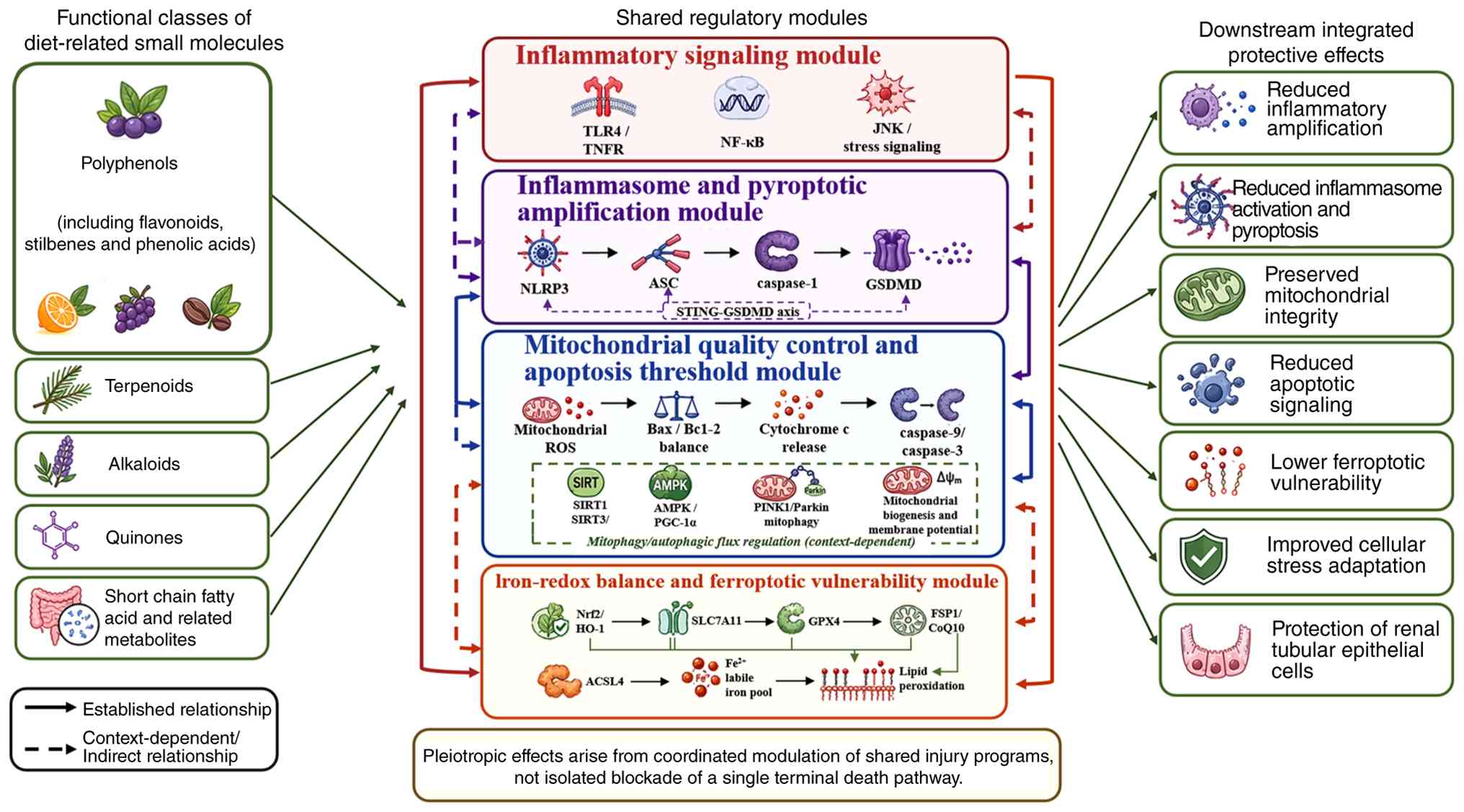 | Figure 2Diet-related small molecules as
modulators of hierarchical shared regulatory nodes in SA-AKI. The
functional classes shown include polyphenols (including flavonoids,
stilbenes and phenolic acids), terpenoids, alkaloids and quinones,
together with microbiota-derived short-chain fatty acids and
related metabolites. Although chemically and pharmacologically
diverse, these compounds and metabolites may converge on shared
regulatory modules rather than selectively blocking a single
terminal cell death pathway. The modules shown comprise
inflammatory signaling involving TLR4/TNFR, NF-κB and JNK/stress
signaling; NLRP3/caspase-1/GSDMD- and STING/GSDMD-associated
inflammasome and pyroptotic signaling; SIRT1/SIRT3-, AMPK/PGC-1α-
and PINK1/Parkin-related mitochondrial quality control, together
with Bax/Bcl-2-, cytochrome c- and
caspase-9/caspase-3-associated apoptotic signaling; and Nrf2/HO-1-,
SLC7A11-, GPX4-, FSP1/CoQ10- and ACSL4-related regulation of
iron-redox balance and ferroptotic vulnerability. Autophagy and
mitophagy are presented as context-dependent modifiers. The
resulting integrated protective effects include reduced
inflammatory amplification and pyroptosis, preserved mitochondrial
integrity, reduced apoptotic signaling and ferroptotic
vulnerability, improved cellular stress adaptation, and protection
of renal tubular epithelial cells. ACSL4, acyl-CoA synthetase
long-chain family member 4; AMPK, AMP-activated protein kinase;
ASC, apoptosis-associated speck-like protein containing a caspase
recruitment domain; CoQ10, coenzyme Q10; FSP1, ferroptosis
suppressor protein 1; GPX4, glutathione peroxidase 4; GSDMD,
gasdermin D; HO-1, heme oxygenase 1; NLRP3, NOD-like receptor
family pyrin domain-containing 3; Nrf2, nuclear factor erythroid
2-related factor 2; PGC-1α, peroxisome proliferator-activated
receptor-γ coactivator 1α; PINK1, PTEN-induced kinase 1; ROS,
reactive oxygen species; SA-AKI, sepsis-associated acute kidney
injury; SIRT, sirtuin; SLC7A11, solute carrier family 7 member 11;
STING, stimulator of interferon genes; TLR4, toll-like receptor 4;
TNFR, tumor necrosis factor receptor. |
Among these categories, polyphenolic compounds have
received the most attention. This increased focus is attributable
not only to their prevalence in plant-based foods, but also to
their extensive metabolic and signaling effects following digestion
and biotransformation (98). The
biological activity of these compounds is influenced by their
chemical forms; glycosides, aglycones and low-molecular-weight
phenolic metabolites differ markedly in solubility, stability and
intestinal absorption (32).
Additionally, microbial transformations occurring during
fermentation or within the gastrointestinal tract may produce
metabolites with distinct bioactivity compared with their parent
compounds (99). This
consideration is directly relevant to SA-AKI. For instance,
resveratrol has been reported to mitigate early polymicrobial
SA-AKI by inhibiting ER stress-activated NF-κB signaling, while
curcumin has been shown to alleviate SA-AKI with concomitant
suppression of ferroptosis-related ACSL4/GPX4 signaling (74,100). These findings imply that
representative diet-related small molecules often exert pleiotropic
effects across inflammatory, oxidative, metabolic and death-related
pathways rather than selectively blocking a single isolated
target.
Nonetheless, the pharmacological potential of these
compounds does not eliminate translational constraints. Numerous
diet-related small molecules exhibit low oral bioavailability,
extensive host and microbial metabolism, rapid elimination, and
significant dependence on the food matrix, formulation and dose
exposure (101). As a result,
the species present in circulation or tissues may differ
substantially from the parent compounds utilized in cell-based
experiments, and compounds within the same chemical class may
demonstrate markedly different pharmacokinetic behaviors. These
characteristics help explain why seemingly similar molecules can
exhibit inconsistent efficacy across models and why experimental
benefits may not readily translate to clinically achievable
exposures (102). Hence, the
evaluation of diet-related small molecules in SA-AKI should
incorporate both chemical class and metabolic fate, formulation
properties, and achievable exposure rather than rely solely on
nominal compound identity. The functional and pharmacological
features of the primary diet-related small molecule groups relevant
to SA-AKI are summarized in Table
SI.
Shared regulatory nodes targeted by
diet-related small molecules
Despite the chemical diversity of diet-related small
molecules studied in SA-AKI, their reported protective effects
converge on a limited number of shared regulatory nodes rather than
targeting individual terminal cell death pathways (103).
As illustrated in Fig. 2, these nodes can be categorized
by their mechanistic positions: Upstream inflammatory
transcriptional activation focused on TLR4-, TNFR- and
NF-κB-related signaling; amplifying or permissive states such as
inflammasome priming, mitochondrial ROS generation, iron
dysregulation and lipid peroxidation; context-dependent modifiers,
including autophagy and mitophagy; and terminal execution
mechanisms such as GSDMD cleavage, caspase activation, ferroptotic
lipid membrane damage and necroptotic signaling (47,104). Puerarin has been shown to
reduce systemic inflammation and organ injury in experimental
sepsis by suppressing NF-κB/JNK signaling, while resveratrol
mitigates early polymicrobial septic AKI through inhibition of ER
stress-associated inositol-requiring enzyme 1-NF-κB activation
(100,105). Similarly, luteolin exhibits a
comparable effect in endotoxin-induced renal injury, as evidenced
by reduced NF-κB activation, decreased inflammatory mediator
levels, attenuated oxidative stress and reduced apoptosis (92). These findings suggest that
certain diet-related small molecules act by initially lowering the
inflammatory background, thereby limiting the activation of
multiple downstream cell death programs (106).
A second recurring node is the Nrf2-centered
antioxidant defense system (107). In the context of SA-AKI,
allicin has been reported to promote Nrf2 nuclear translocation and
heme oxygenase-1 (HO-1) expression, resulting in reduced ROS
accumulation, inflammatory mediator release, mitochondrial
dysfunction and apoptosis (108). More broadly, the literature on
AKI positions Nrf2 at the intersection of antioxidant enzyme
induction, mitochondrial protection, lipid homeostasis and
suppression of inflammatory signaling (109). In this regard, the activation
of Nrf2/HO-1 is not merely an antioxidant response; it likely
reflects regulation of a more extensive stress-adaptation program
that impacts the redox balance, organelle integrity and
vulnerability to regulated cell death (110).
Inflammasome-related pyroptotic signaling also
emerges as a shared target. Recent research has indicated that
butyrate mitigates SA-AKI by suppressing the STING/GSDMD axis, with
parallel reductions in NLRP3-associated pyroptotic signaling and
renal injury (53). This aligns
with recent reviews that position NLRP3-caspase-1-GSDMD signaling
at the core of inflammatory cell death in SA-AKI (48,111). These observations indicate that
at least some diet-related small molecules, including resveratrol,
curcumin and cichoric acid, may interrupt pyroptotic injury by
modulating upstream innate immune sensors and inflammasome
activation rather than solely targeting terminal membrane rupture
(27,28,112-114).
Nodes associated with mitochondrial quality control
and metabolic adaptation are also consistently targeted.
Resveratrol improves SA-AKI in rats through activation of sirtuin
(SIRT)1 and SIRT3, accompanied by attenuation of oxidative stress
and restoration of autophagic activity (36). This pattern is significant
because it situates certain dietary molecules upstream of organelle
stress, rather than solely downstream of cytokine release. Studies
focusing on ferroptosis further corroborate this view. Ginsenoside
Rg1 ameliorates SA-AKI by regulating ferroptosis in tubular
epithelial cells, andrographolide inhibits ferroptosis in renal
tubular epithelial cells via Nrf2/FSP1-related signaling and
curcumin reduces SA-AKI with concurrent regulation of the
ACSL4/GPX4 axis (52,72,74). Together, these findings indicate
that, in SA-AKI, diet-related small molecules repeatedly converge
on a restricted set of shared regulatory nodes, particularly NF-κB,
Nrf2, inflammasome-related pyroptotic signaling, SIRT-dependent
autophagic or mitochondrial control, and ferroptosis-related lipid
redox pathways. Thus, their apparent broad actions reflect the
coordinated modulation of upstream injury programs rather than a
selective blockade of a single terminal mode of cell death
(41,115). The key shared regulatory nodes
linking the actions of dietary small molecules with the integrated
cell death network in SA-AKI are summarized in Table SII.
Bioavailability and variation in
biological effects under septic conditions
The biological effects of diet-related small
molecules in SA-AKI cannot be interpreted independently of
bioavailability. For numerous compounds investigated in this
domain, the parent molecule utilized in cell-based experiments may
only be transiently present in circulation following oral
administration or might not be detectable at all. For instance,
while resveratrol is absorbed efficiently, it undergoes extensive
first-pass conjugation, resulting in very low systemic exposure to
the unconjugated parent compound (116). Similarly, curcumin exhibits
limited systemic availability due to poor aqueous solubility and
rapid metabolism, while quercetin absorption varies considerably
based on its structural form and formulation (117,118). Intact ginsenosides also exhibit
low plasma exposure after oral intake due to poor permeability and
extensive presystemic transformation, and allicin is characterized
by high instability, with rapid degradation limiting the
persistence of the parent compound in vivo (119,120). These observations indicate that
the biological effects observed in experimental settings may not
directly translate to clinical settings, because in vivo
efficacy depends more on metabolic fate and actual tissue exposure
than on the administered parent compound alone.
This issue becomes increasingly critical under
septic conditions. Sepsis alters the pharmacokinetic landscape
through mechanisms such as splanchnic hypoperfusion, impaired
intestinal absorption, reduced hepatic metabolic capacity, modified
protein binding, capillary leak and disturbed microcirculatory
delivery (121,122). Consequently, plasma
concentrations may not accurately reflect renal tissue exposure,
leading to an increasingly unstable relationship between the
administered dose and biological effect during septic organ
dysfunction (123).
Furthermore, sepsis-related disruptions of the intestinal barrier
and acute shifts in gut microbiota may further influence the
generation, absorption and systemic persistence of secondary
metabolites derived from dietary compounds (124). Under such conditions, the
active species that reach the injured kidney may differ from the
parent compounds typically examined in mechanistic in vitro
studies (125).
Variation in biological effects across studies is
therefore expected. For certain compounds, substantial protective
effects observed in pretreatment scenarios may not be consistently
replicated when administration is delayed until after the onset of
sepsis (126,127). In other instances,
intraperitoneal administration appears to provide more stable
protection compared with oral dosing, particularly when prominent
gut dysfunction is present. Furthermore, even when renal biomarkers
demonstrate improvement, histological recovery and survival
benefits do not always exhibit parallel changes (128). The apparent efficacy of the
same compound may also fluctuate based on factors such as model
severity, administration route and the underlying metabolic state
(1,122). These discrepancies imply that
inconsistent outcomes across studies do not necessarily indicate
target irrelevance; rather, they may arise from unstable exposure,
altered metabolism and marked context dependence under septic
conditions.
Metabolic transformation provides an additional
source of complexity. In vivo, the dominant circulating
forms of numerous polyphenols are glucuronidated, sulfated, reduced
or microbiota-derived metabolites rather than the parent compounds
themselves (102,129). Some of these metabolites may
exhibit biological activities that differ from, or even exceed,
those of the original molecules. Inflammatory tissues may also
locally deconjugate circulating metabolites, resulting in
site-specific exposure to active aglycones (93,130). This phenomenon is particularly
relevant in sepsis, where intestinal dysbiosis, altered
fermentation and disrupted host-microbiota co-metabolism can alter
the metabolite pool available to the kidney (131). Consequently, the in vivo
protective species in SA-AKI may not align with the compounds
directly applied in reductionist cell systems.
These pharmacokinetic and metabolic constraints have
important translational implications. A number of promising
diet-related small molecules are supported by preclinical studies
that utilize doses, formulations or administration routes not
easily replicable through realistic nutritional exposure or routine
clinical treatment (132). The
gap between experimental dosing and clinically attainable exposure
is further widened by the lack of dedicated pharmacokinetic data in
sepsis and critical illness. Delivery strategies such as liposomal
encapsulation, nanoparticle formulation or kidney-targeted carrier
systems mitigate this issue by enhancing local exposure and
limiting rapid systemic loss (133,134). Variability in biological
effects under septic conditions arises not only from target
selection but also from issues of absorption, metabolism, delivery
and context-dependent exposure (135). Addressing these challenges is
essential for the evaluation of diet-related small molecules as
clinically meaningful interventions in SA-AKI.
Mechanistic basis for dietary small molecule
intervention
The subsequent sections focus on three mechanistic
axes that represent recurrent upstream convergence points in the
current SA-AKI literature and are most directly linked to the
reported actions of diet-related small molecules. Necroptosis,
autophagy-related responses and PANoptosis-like signaling are
discussed as interacting components of the integrated cell death
network rather than as independent therapeutic targets, because
direct evidence supporting diet-related small molecule modulation
of these processes in SA-AKI remains limited.
Mitochondrial dysfunction and apoptotic
signaling
Mitochondrial dysfunction serves as a critical
determinant of apoptotic injury in SA-AKI. Experimental studies
have indicated that SA-AKI frequently involves tubular epithelial
apoptosis, along with caspase-3 activation and Bax upregulation
even in the absence of extensive tubular necrosis (136,137). Ultrastructural analyses have
revealed that mitochondrial swelling, cristae disruption and loss
of membrane potential occur early in septic kidneys, subsequently
leading to cytochrome c release and caspase-9-dependent
apoptotic signaling (138,139). Concurrently, reductions in
intracellular ATP levels are closely associated with tubular TUNEL
positivity, suggesting that mitochondrial energetic failure and
apoptotic execution occur in parallel rather than as distinct
events (140).
The mitochondrial apoptotic response is driven by
multiple upstream stressors that converge on organelle instability.
Excessive ROS can induce pathological mitochondrial fission through
dynamin-related protein 1 translocation, thereby exacerbating
membrane damage and increasing apoptotic susceptibility. Prolonged
ER stress may further propagate injury via the C/EBP homologous
protein-Bax axis, linking calcium dysregulation to mitochondrial
calcium overload and outer membrane permeabilization (141-143). Additionally, inflammatory
cytokines such as TNF-α and IL-1β disturb mitochondrial homeostasis
directly and may facilitate the opening of the mitochondrial
permeability transition pore under septic conditions (144-146). Collectively, these processes
drive the transition of renal tubular cells from adaptive stress
responses to intrinsic apoptosis through Bax/Bcl-2 imbalance,
cytochrome c release and downstream caspase activation
(136).
An expanding body of evidence indicates that
diet-related small molecules can modulate the mitochondria-mediated
intrinsic apoptotic axis at multiple levels. In septic models,
curcumin has been shown to preserve mitochondrial ultrastructure,
reduce mitochondrial swelling and lower Bax expression in
conjunction with decreased tubular apoptosis (74). Resveratrol effectively maintains
the mitochondrial membrane potential and reduces both cleaved
caspase-9 and caspase-3 levels (147). Furthermore, quercetin has been
reported to attenuate mitochondrial apoptotic signaling involving
Bax/Bcl-2 balance, cytochrome c and caspase activation in
related renal injury models (148-150). Procyanidin B2 has also been
shown to restore disturbed mitochondrial fusion-fission dynamics,
reduce ROS accumulation and attenuate mitochondria-mediated
apoptosis through Nrf2-related mechanisms (151). Although these compounds exhibit
structural differences, their protective effects repeatedly
converge on preservation of mitochondrial integrity rather than on
blockade of apoptosis alone.
This pattern is further substantiated by studies
focusing on mitochondrial quality control. In experimental sepsis,
defective PTEN induced kinase 1/recombinant Parkinson disease
protein 2-dependent mitophagy exacerbates apoptosis, reinforcing
the notion that the clearance of damaged mitochondria represents an
essential adaptive response rather than a secondary bystander event
(152). Activation of
AMP-activated protein kinase/SIRT1/peroxisome
proliferator-activated receptor γ coactivator 1-α signaling
promotes mitochondrial biogenesis and improves energy homeostasis,
whereas SIRT3-dependent regulation of mitochondrial protein
acetylation supports ATP production and limits apoptosis under
septic stress (153). These
findings imply that the anti-apoptotic effects of diet-related
small molecules are frequently intertwined with broader impacts on
mitochondrial turnover, metabolic adaptation and oxidative
balance.
Mitochondrial protection should not be viewed
solely as an anti-apoptotic mechanism. In SA-AKI, interventions
aimed at reducing apoptosis often concurrently suppress
inflammatory signaling, oxidative stress or inflammasome
activation, and caspase inhibition alone does not fully restore
renal function in severe sepsis models (154). Mitochondrial preservation may
therefore exert broader protective effects by reducing both
intrinsic apoptotic signaling and ROS-driven inflammatory
amplification (155). In this
context, the mechanistic value of diet-related small molecules lies
not simply in lowering apoptotic readouts, but in stabilizing a
central organelle system that links energy failure, oxidative
injury, inflammatory stress and multiple downstream cell death
responses.
Iron-redox disequilibrium and ferroptotic
vulnerability
Iron-redox disequilibrium has emerged as a
significant factor underlying ferroptotic vulnerability in SA-AKI.
Experimental studies have demonstrated that tubular damage in
SA-AKI is frequently characterized by classical ferroptotic
features, including marked lipid peroxidation, glutathione
depletion and loss of GPX4 expression (156,157). Concurrently, renal iron
deposition, altered SLC7A11-associated cystine transport and
accumulation of lipid peroxidation products, such as 4-HNE,
malondialdehyde (MDA) and prostaglandin-endoperoxide synthase 2,
are observed as renal injury progresses. These findings suggest
that ferroptosis in SA-AKI is not solely defined by iron excess;
rather, it signifies a broader collapse of redox buffering
capacity, phospholipid homeostasis and lipid peroxide
detoxification in stressed tubular cells (73,158).
Several upstream processes appear to drive this
state of vulnerability. Systemic inflammatory stress can increase
the intracellular labile iron pool through mechanisms such as
nuclear receptor coactivator 4-dependent ferritinophagy, thus
enhancing the availability of redox-active iron for Fenton
reactions (40). Simultaneously,
excessive mitochondrial ROS generation rapidly depletes
intracellular glutathione and weakens the GPX4-dependent repair
system (159). This depletion
is exacerbated by the upregulation of ACSL4, which causes membrane
phospholipids to become increasingly enriched with polyunsaturated
fatty acids that are highly susceptible to peroxidation (40). The role of HO-1 also appears to
be context-dependent. Although HO-1 is often discussed as a
protective antioxidant response, excessive heme degradation under
severe septic stress may release additional free iron and thereby
increase ferroptotic susceptibility rather than suppress it
(160).
These observations clarify why ferroptosis is best
understood as a state of increased vulnerability rather than as an
isolated terminal event in SA-AKI. The septic kidney simultaneously
endures oxidative stress, mitochondrial dysfunction, disturbed iron
handling and impaired glutathione metabolism (10). Under such conditions, even modest
increases in lipid peroxide generation may become challenging to
neutralize. Nrf2 serves a particularly important role in this
context by transcriptionally regulating key components of the
anti-ferroptotic defense system, including SLC7A11, which maintains
cystine uptake and glutathione synthesis, and GPX4, which
detoxifies phospholipid hydroperoxides (161). Additionally, FSP1 provides a
parallel glutathione-independent system for detoxifying lipid
peroxides through coenzyme Q10 regeneration (162). Thus, ferroptotic susceptibility
in SA-AKI hinges on the balance between ongoing oxidative and
iron-driven injury, and the remaining capacity of Nrf2-, GPX4-,
SLC7A11- and FSP1-related defense systems (14,163).
A growing number of diet-related small molecules
appear to modulate this axis. In septic models, curcumin has been
shown to reduce renal iron overload, suppress ACSL4 expression and
limit 4-HNE accumulation, while preserving renal function (74). Quercetin alleviates AKI by
inhibiting ferroptosis in renal tubular epithelial cells (75). Baicalein attenuates cisplatin-
and folic acid-induced AKI by suppressing ALOX12-dependent
ferroptosis, highlighting the broader relevance of
flavonoid-mediated control of lipid peroxidation in renal tubular
injury (164). In LPS-induced
AKI, polydatin has been reported to reduce renal dysfunction,
inflammatory cytokine production, MDA accumulation and NF-κB
activation, while increasing Nrf2 and HO-1 expression (165). Resveratrol ameliorates
sepsis-induced AKI through Nrf2-related antioxidant signaling
(166). Collectively, these
findings suggest that diet-related small molecules may stabilize
the iron-redox environment through partially overlapping
mechanisms, which include preservation of antioxidant defenses,
attenuation of lipid peroxidation and reduction of inflammatory
oxidative stress.
Ferroptosis should not be regarded as an isolated
mechanism. Previous studies have indicated that lipid peroxidation
products generated during ferroptotic stress can amplify
inflammatory signaling and promote NLRP3-associated pyroptotic
responses (167,168). Anti-ferroptotic interventions
may also lead to a reduction in both apoptotic and pyroptotic
markers, suggesting that protection extends beyond a single death
program (169). Impaired
autophagic clearance of damaged mitochondria further exacerbates
intracellular iron overload, linking ferroptotic susceptibility to
organelle quality control (170). In this context, the mechanistic
value of diet-related small molecules lies not simply in reducing
lipid peroxidation, but in stabilizing a broader iron-redox
environment that influences inflammation, mitochondrial integrity
and multiple downstream cell death responses in SA-AKI.
Inflammasome activation and pyroptotic
amplification
Inflammasome activation has emerged as a
significant mechanism of tissue injury amplification in SA-AKI
(171). Experimental studies
have demonstrated that renal dysfunction is associated with
increased NLRP3 expression, apoptosis-associated speck-like protein
containing a CARD assembly, caspase-1 activation, GSDMD cleavage,
and IL-1β/IL-18 release in both renal tubular epithelial cells and
kidney tissues from experimental models of SA-AKI (48,172). Beyond the canonical
caspase-1-dependent pathway, non-canonical pyroptotic signaling
mediated by caspase-11 has also been implicated in septic tubular
injury, indicating that cytosolic endotoxin sensing can directly
contribute to epithelial cell death in Gram-negative sepsis
(173). These findings position
pyroptosis at the intersection of innate immune sensing and
structural renal injury rather than as a mere secondary
inflammatory bystander.
The amplification of pyroptotic signaling in SA-AKI
appears to be dependent on both priming and activation signals.
Priming is facilitated by inflammatory pathways, such as the
TLR4/NF-κB pathway, which increase transcription of
inflammasome-related components and proinflammatory cytokines under
septic stress (174).
Activation is then reinforced by a second wave of danger signals,
which includes mitochondrial ROS, oxidized mtDNA, thioredoxin
interacting protein-dependent coupling and other damage-associated
signals originating from stressed renal cells and infiltrating
immune cells (48,175). This two-step process helps
explain why inflammasome signaling in SA-AKI is often
self-amplifying. Once GSDMD-mediated pore formation is initiated,
inflammatory mediator release, ionic imbalance and cell lysis
further intensify local tissue stress and propagate additional
inflammasome activity in neighboring cells (176).
Increasing studies have indicated that diet-related
small molecules can disrupt this amplification axis at multiple
levels (177,178). In a recent study on SA-AKI,
butyrate was shown to reduce renal injury by suppressing the
STING/GSDMD pathway, with concurrent reductions in pyroptotic
signaling and the overall inflammatory burden (53). This finding is crucial as it
suggests that certain diet-related small molecules may not merely
diminish downstream cytokine release but can act upstream of GSDMD
cleavage by modulating inflammatory signal integration. More
broadly, studies in related renal and inflammatory injury models
suggest that natural compounds may restrain pyroptotic
amplification through distinct upstream mechanisms. In experimental
folic acid nephropathy, quercetin reduced macrophage caspase-1
expression and the expression of proinflammatory cytokines,
including IL-1β, IL-18, TNF-α and IL-6 (100). In LPS-induced acute lung
injury, resveratrol reduced ROS accumulation and inhibited
ROS-dependent thioredoxin interacting protein/NLRP3 inflammasome
activation (101). In
experimental hyperlipidemic acute pancreatitis, baicalein
suppressed macrophage M1 polarization and high mobility group box
1/TLR4/NLRP3 signaling, thereby reducing caspase-1 activation,
GSDMD cleavage and inflammatory cytokine release (102). Although these studies were
conducted outside the specific context of SA-AKI, they illustrate
how natural compounds may modulate upstream redox and inflammatory
signals that amplify pyroptotic injury.
These findings suggest that the mechanistic
importance of pyroptosis in SA-AKI lies not only in terminal
membrane rupture, but also in its role as an inflammatory
amplifier. GSDMD pore formation facilitates the release of IL-1β,
IL-18 and other intracellular danger signals, thereby intensifying
leukocyte recruitment, endothelial activation and further
epithelial injury (179). In
this context, inhibition of inflammasome signaling may reduce both
direct tubular cell loss and the local inflammatory environment
sustaining ongoing renal dysfunction. However, pyroptosis should
not be interpreted as an isolated process. Available evidence
indicates substantial overlap with oxidative stress, mitochondrial
injury and other regulated cell death pathways, including apoptosis
and ferroptosis (180,181). Mitochondrial protection, for
example, may reduce pyroptotic signaling by limiting mitochondrial
ROS and mtDNA release, while lipid peroxidation and iron-redox
disequilibrium may further intensify inflammasome activation
(19).
In summary, these observations support a model
whereby inflammasome activation and pyroptotic amplification
contribute to the progression of SA-AKI through the conversion of
local danger sensing into sustained inflammatory tissue damage
(182). The relevance of
diet-related small molecules in this context lies not merely in the
reduction of a single pyroptotic marker but in their capacity to
weaken the broader upstream conditions that foster inflammasome
overactivation, including NF-κB-dependent priming, mitochondrial
damage, oxidative stress and impaired cellular stress control
(183). Therefore, their
apparent anti-pyroptotic effects should be interpreted as part of a
wider network-level regulation of AKI rather than as selective
suppression of a single terminal pathway.
Translational barriers to clinical
application
Altered pharmacokinetics in SA-AKI
Pharmacokinetic behavior in SA-AKI exhibits
significant instability, diverging considerably from that observed
in healthy individuals or simplified preclinical models (184). Sepsis can impair enteral
absorption due to gastrointestinal dysfunction and splanchnic
hypoperfusion, while factors such as capillary leak, tissue edema
and hypoalbuminemia alter the volume of distribution and protein
binding (185). Simultaneously,
hepatic metabolism may be diminished by inflammation, altered blood
flow and downregulation of metabolic enzymes. These alterations are
particularly critical for diet-related small molecules, a number of
which already exhibit limited oral bioavailability and extensive
presystemic metabolism under baseline conditions (186).
AKI introduces an additional layer of variability.
In SA-AKI, changes in glomerular filtration rates are often
accompanied by altered tubular secretion, reabsorption and
transporter functionality, meaning that plasma concentrations may
not accurately reflect intrarenal exposure (187,188). This discrepancy may be further
amplified by septic microcirculatory dysfunction, which limits drug
delivery to injured tubular regions despite measurable systemic
levels. As a result, the biologically active concentration at the
site of renal injury may differ markedly from what is predicted by
circulating exposure alone (43).
Pharmacokinetic interpretation in SA-AKI is further
complicated by intensive care interventions such as fluid
resuscitation, vasopressor therapy and RRT (189). In certain patients, augmented
renal clearance may lead to reduced exposure early in the disease
process, whereas later organ dysfunction may favor accumulation
(190). For orally delivered
diet-related small molecules, sepsis-associated dysbiosis and
intestinal barrier disruption may additionally alter microbial
biotransformation and metabolite generation (32,191,192). These factors create a major
translational barrier, as the experimental dosing regimens and
exposure patterns are difficult to reproduce in patients with
established SA-AKI (193).
In line with previous discussions regarding
sepsis-targeted therapeutic delivery, the future development of
diet-related small molecules for SA-AKI should not only focus on
identifying biologically relevant targets, but also address
formulation stability, bioavailability, renal tissue exposure and
context-dependent delivery in the context of septic conditions
(194,195).
Therapeutic timing and disease-stage
specificity
Therapeutic timing is a critical determinant of
efficacy in SA-AKI. Several interventions, including necrostatin-1
pretreatment, curcumin administered immediately after CLP,
post-treatment with irisin and delayed administration of AP214,
have shown renoprotective effects in experimental models of SA-AKI.
However, these protective effects may be attenuated or lost when
treatment initiation is delayed until after sepsis has been
established (196-200). This pattern has been
consistently observed in rescue-design studies, where early
administration suppresses inflammatory escalation and preserves
organ function, whereas delayed treatment results in only modest
improvements in biomarkers or fails to enhance survival. For
diet-related small molecules, this issue is especially important
because numerous reported benefits have been generated under
pretreatment or very early post-insult conditions that do not
reflect the usual timing of clinical recognition (2,197-201).
The biological plausibility of this timing
dependence arises from the fact that SA-AKI is not a static state.
Early injury is frequently characterized by inflammatory
amplification, oxidative stress, microvascular disturbances and
rapid activation of regulated cell death pathways. As the disease
progresses, the renal response transitions toward metabolic
suppression, persistent organelle dysfunction, maladaptive repair
and changing immune cell states (9). Available time-course studies have
further indicated that the relative contributions of pyroptosis,
apoptosis, ferroptosis and autophagy-related responses may change
across disease stages rather than remain constant (2,202). A compound that is effective
during early inflammatory priming may exhibit diminished effects
once tissue injury, metabolic failure or defective repair
mechanisms have taken root.
Disease-stage specificity carries important
translational implications as well. By the time SA-AKI is
clinically recognized through indicators such as rising creatinine
levels or reduced urine output, the underlying molecular injury may
already be advanced (203,204). This scenario narrows the
therapeutic window for compounds primarily aimed at suppressing
early inflammatory or oxidative triggers. In addition, patients
with SA-AKI do not adhere to a uniform trajectory (55). Some individuals experience rapid
recovery, while others endure persistent injury, and
biomarker-based studies have indicated that the inflammatory burden
also varies across subphenotypes (205,206). These differences make it
unlikely that one uniform timing strategy will be effective for all
patients.
For diet-related small molecules, the primary
implication is that apparent preclinical efficacy cannot be
evaluated without consideration of treatment timing and disease
stage. The protective effects observed in early or prophylactic
models do not necessarily predict benefits after clinically
manifest SA-AKI has developed. Future translational studies should
therefore prioritize rescue designs, stage-specific intervention
windows and patient stratification based on trajectory or
inflammatory phenotype. Without this adjustment, promising
mechanistic effects are likely to remain disconnected from
clinically relevant treatment conditions.
Patient heterogeneity and mechanistic
endotypes
SA-AKI is not a biologically uniform syndrome, and
this heterogeneity presents a significant challenge for clinical
translation. Patients with similar increases in serum creatinine
may experience vastly different trajectories, including rapid
reversal, persistent injury or recurrent deterioration (207,208). These patterns are not
clinically trivial. They likely reflect differences in inflammatory
burden, microvascular dysfunction, tubular stress and the extent of
structural renal injury rather than variations in severity
alone.
This variability is increasingly captured by
biomarker-based and molecular subphenotypes. Research involving
critically ill patients with AKI has identified distinct subgroups
characterized by varying levels of inflammatory activation,
endothelial injury and mortality risk (55,209). In this context, mechanistic
endotypes are likely to have greater importance than the diagnostic
label alone. For example, patients with predominant inflammasome
activation, endothelial dysfunction and hyperinflammatory signaling
may respond differently to treatment than those whose injury is
primarily driven by metabolic shutdown, mitochondrial dysfunction
or persistent redox imbalance (210).
This distinction holds particular relevance for
diet-related small molecules. While a number of these compounds
function through anti-inflammatory, antioxidant or
organelle-protective mechanisms, their effects are unlikely to be
uniformly beneficial across all endotypes (211). Agents that suppress NF-κB,
NLRP3 or pyroptotic amplification may be more relevant in early
hyperinflammatory states, whereas compounds that support
mitochondrial quality control or limit ferroptotic vulnerability
may be more rational in patients with persistent metabolic and
redox stress (212,213). Thus, the therapeutic effects of
the same intervention may appear inconsistent in unselected patient
populations, not due to invalid mechanisms, but because the
individuals treated do not share the same predominant biological
profiles.
Patient heterogeneity is further amplified by age,
chronic kidney disease, diabetes, cardiovascular comorbidity,
pathogen burden, shock severity and exposure to organ support
interventions such as vasopressors or RRT (207). These elements influence the
trajectory of SA-AKI and shape the biological context in which an
intervention is administered. Consequently, future clinical
development of diet-related small molecules will likely require
biomarker-guided or endotype-informed trial designs rather than
one-size-fits-all treatment strategies. Without stratification,
even mechanistically promising compounds are likely to show diluted
or misleading effects in clinical studies.
Current clinical evidence and its
limitations
Current clinical evidence for diet-related small
molecules in sepsis and SA-AKI remains limited and uneven. Most
human studies have focused on vitamins and antioxidant compounds,
especially vitamin C, thiamine and combination regimens that
include hydrocortisone, rather than exploring a diverse range of
mechanistically defined diet-related small molecules (214,215). Among the data pertinent to
kidney outcomes, secondary and post hoc analyses of randomized
trials have suggested potential renal benefits of thiamine in
septic shock, using the need for RRT and alive and RRT-free status
as endpoints, while a recent pilot trial has indicated that vitamin
C might enhance renal function in the short term for patients with
SA-AKI (216-218).
Despite these findings, the overall clinical
picture is inconclusive. Large sepsis trials assessing vitamin C
strategies have produced conflicting results. For instance, in the
ACTS trial, combined ascorbic acid, corticosteroids and thiamine
did not lead to an improvement in the primary organ dysfunction
outcome (219). Similarly, the
CITRIS-ALI trial found that vitamin C did not significantly impact
the prespecified biomarker or organ failure endpoints (220). However, in the LOVIT trial,
high-dose intravenous vitamin C was associated with a higher rate
of death or persistent organ dysfunction by day 28 (221). A recent meta-analysis has
likewise shown substantial heterogeneity across trials, with
inconsistent effects on mortality, vasopressor duration and organ
failure scores (222).
The limitations of these studies extend beyond
categorizing trials as positive or negative, which does not account
for important differences in study design, patient heterogeneity,
pharmacokinetics and endpoint selection. Kidney-specific data
remain scarce, with numerous studies not designed to prioritize
SA-AKI as a primary target. Sample sizes frequently remain small,
patient populations are biologically heterogeneous and renal
endpoints are often secondary, indirect or absent. Additionally,
the rationale for dosing, treatment timing and formulation is often
poorly aligned with the pharmacokinetic instability of sepsis. Even
in cases where inflammatory biomarkers improve, this is not
consistently associated with enhanced renal recovery, reduced need
for RRT or improved survival outcomes (223,224). Future trials should aim to
connect molecular target engagement with kidney-centered decision
points. For interventions aimed at ferroptosis, linking
preservation of GPX4 or SLC7A11, suppression of ACSL4, improvements
in iron-redox indices, and reductions in lipid peroxidation
products such as 4-HNE or MDA to patient selection, treatment
windows, AKI progression, renal recovery and RRT-free days is
essential (225). Similarly,
for inflammasome- or pyroptosis-oriented treatments, assessments of
NLRP3, caspase-1 activity, GSDMD cleavage, IL-1β and IL-18 should
be contextualized with early hyperinflammatory endotypes and
kidney-specific outcomes rather than viewed as isolated
inflammatory metrics (226).
Mitochondria-directed strategies should also align mitochondrial
stress markers, mitophagy-related signals, oxidative injury markers
and tubular stress biomarkers with clinically relevant endpoints
such as renal recovery, avoidance or duration of RRT, RRT-free
days, major adverse kidney events-type composite outcomes and
survival (227). Establishing
this connection is crucial because an improvement in a molecular
biomarker does not necessarily signify renal recovery unless it is
tied to relevant clinical decision points.
In conclusion, current clinical evidence does not
yet support routine use of diet-related small molecules as an
established therapy for SA-AKI. Nevertheless, it indicates that
biological activity in humans is plausible, but highly
context-dependent (228).
Future trials will need clearer kidney-specific endpoints, improved
control of treatment timing, direct assessments of pharmacokinetics
and target engagement, and robust patient stratification. This
emphasis on stratification may be particularly important in light
of previous analyses suggesting that differential treatment
responses in sepsis could be obscured when mechanistically distinct
patients are grouped within the same trial (229,230). The available clinical evidence
and major sources of translational failure are summarized in
Table SIII. The complete
translational framework is summarized in Fig. 3. Dietary intake, parent compound
properties, food matrix effects and formulation determine compound
entry, while intestinal absorption, host first-pass metabolism and
gut microbiota-dependent biotransformation determine whether parent
compounds, conjugated metabolites or microbiota-derived metabolites
reach the systemic circulation (32,116-122,124,186,191,192). In sepsis and SA-AKI, splanchnic
hypoperfusion, impaired intestinal absorption, capillary leak,
hypoalbuminemia, reduced hepatic metabolism, altered tubular
handling and disturbed renal microcirculatory delivery may further
uncouple the administered dose and circulating concentration from
the biologically active exposure achieved within the kidney
(185,187,188,190). These exposures may influence
inflammatory amplification, mitochondrial dysfunction, iron-redox
imbalance, inflammasome activation and context-dependent autophagic
or adaptive stress responses, which converge on an integrated
network of apoptotic, necroptotic, pyroptotic and ferroptotic
injury (73,138,152,155,173,212,213). Accordingly, precision
translation will require biomarker-guided enrichment using the
urinary [TIMP metallopeptidase inhibitor 2 (TIMP-2)] x
[insulin-like growth factor-binding protein 7] product and
circulating markers such as NGAL, soluble urokinase plasminogen
activator receptor (suPAR) and bioactive adrenomedullin (bio-ADM),
together with mechanistic endotyping, transcriptomic, proteomic and
metabolomic profiling, and spatial or single-cell analysis
(84,231-237). These data should subsequently
inform stage-specific rescue designs, pharmacokinetic and exposure
assessment, target-engagement analysis, exposure monitoring,
optimized formulation or kidney-targeted delivery,
mechanism-informed adjunctive therapy in biologically selected
patients, and kidney-specific outcome assessment (132-134,196,207-209).
![From metabolic fate to precision
intervention: A translational framework for diet-related small
molecules in SA-AKI. The framework links five sequential domains
that determine the translational potential of diet-related small
molecules and diet-associated metabolites. First, dietary intake,
parent compound properties, representative compound classes, food
matrix effects and formulation determine the context in which a
compound enters the body. Second, intestinal absorption, host
first-pass metabolism and gut microbiota-dependent transformation
determine whether parent compounds, host-conjugated metabolites or
microbiota-derived metabolites reach the systemic circulation. In
sepsis and SA-AKI, exposure may be further altered by splanchnic
hypoperfusion, impaired intestinal absorption, capillary leak,
hypoalbuminemia, reduced hepatic metabolism, altered renal handling
and impaired renal microcirculatory delivery. Third,
kidney-relevant mechanistic targets include inflammatory
amplification, mitochondrial dysfunction, iron-redox imbalance,
inflammasome activation, and context-dependent autophagic and
adaptive stress control, which converge on an integrated network
involving apoptosis, necroptosis, pyroptosis and ferroptosis.
Fourth, precision stratification may combine biomarker-guided
enrichment using the urinary [TIMP-2] x [IGFBP7] product and
circulating NGAL, suPAR and bio-ADM with putative mechanistic
endotypes, transcriptomic, proteomic and metabolomic phenotyping,
and spatial or single-cell information. Finally, clinical
translation requires stage-specific intervention windows, rescue
rather than exclusively pretreatment paradigms, pharmacokinetic and
target-engagement assessment, exposure monitoring with therapeutic
drug monitoring where applicable, optimized formulation or
kidney-targeted delivery, mechanism-informed adjunctive therapy in
biologically selected patients, and kidney-specific outcome
assessment. Overall, successful translation depends on aligning
achievable exposure, treatment timing, mechanistic endotype and
target organ delivery to support precision adjunctive intervention
in selected patients with SA-AKI. bio-ADM, bioactive
adrenomedullin; IGFBP7, insulin like growth factor binding protein
7; NGAL, neutrophil gelatinase-associated lipocalin; SA-AKI,
sepsis-associated acute kidney injury; suPAR, soluble urokinase
plasminogen activator receptor; TIMP-2, TIMP metallopeptidase
inhibitor 2.](/article_images/ijmm/58/5/ijmm-58-05-05974-g02.jpg) | Figure 3From metabolic fate to precision
intervention: A translational framework for diet-related small
molecules in SA-AKI. The framework links five sequential domains
that determine the translational potential of diet-related small
molecules and diet-associated metabolites. First, dietary intake,
parent compound properties, representative compound classes, food
matrix effects and formulation determine the context in which a
compound enters the body. Second, intestinal absorption, host
first-pass metabolism and gut microbiota-dependent transformation
determine whether parent compounds, host-conjugated metabolites or
microbiota-derived metabolites reach the systemic circulation. In
sepsis and SA-AKI, exposure may be further altered by splanchnic
hypoperfusion, impaired intestinal absorption, capillary leak,
hypoalbuminemia, reduced hepatic metabolism, altered renal handling
and impaired renal microcirculatory delivery. Third,
kidney-relevant mechanistic targets include inflammatory
amplification, mitochondrial dysfunction, iron-redox imbalance,
inflammasome activation, and context-dependent autophagic and
adaptive stress control, which converge on an integrated network
involving apoptosis, necroptosis, pyroptosis and ferroptosis.
Fourth, precision stratification may combine biomarker-guided
enrichment using the urinary [TIMP-2] x [IGFBP7] product and
circulating NGAL, suPAR and bio-ADM with putative mechanistic
endotypes, transcriptomic, proteomic and metabolomic phenotyping,
and spatial or single-cell information. Finally, clinical
translation requires stage-specific intervention windows, rescue
rather than exclusively pretreatment paradigms, pharmacokinetic and
target-engagement assessment, exposure monitoring with therapeutic
drug monitoring where applicable, optimized formulation or
kidney-targeted delivery, mechanism-informed adjunctive therapy in
biologically selected patients, and kidney-specific outcome
assessment. Overall, successful translation depends on aligning
achievable exposure, treatment timing, mechanistic endotype and
target organ delivery to support precision adjunctive intervention
in selected patients with SA-AKI. bio-ADM, bioactive
adrenomedullin; IGFBP7, insulin like growth factor binding protein
7; NGAL, neutrophil gelatinase-associated lipocalin; SA-AKI,
sepsis-associated acute kidney injury; suPAR, soluble urokinase
plasminogen activator receptor; TIMP-2, TIMP metallopeptidase
inhibitor 2. |
Toward precision intervention in SA-AKI
Biomarker-guided stratification
Biomarker-guided stratification is likely essential
for achieving precision intervention in SA-AKI. While serum
creatinine and urine output remain critical for diagnosis, they are
often too delayed and nonspecific to accurately define the dominant
biology of injury (238). By
contrast, urinary TIMP-2 and insulin like growth factor binding
protein 7 reflect early tubular stress and cell cycle arrest,
potentially identifying high-risk patients before overt functional
decline becomes evident (231).
In these biomarker-based stratification models, markers of
inflammation and endothelial dysfunction often contribute more to
subgroup assignment than serum creatinine alone.
The value of biomarkers becomes greater when they
are used as panels rather than in isolation. In a multicenter
cohort of 769 hospitalized adults with AKI, Bhatraju et al
(84,239) applied latent class analysis and
k-means clustering to 29 clinical, plasma and urinary biomarker
variables and identified two molecularly distinct AKI subphenotypes
with different long-term risks of major adverse kidney events. In
both clustering analyses, plasma inflammatory markers and urinary
epithelial injury markers were among the variables that most
strongly differentiated the two subphenotypes, whereas serum
creatinine ranked only 20th among the 29 discriminatory variables
(240). These findings
highlight the value of biomarker-guided stratification not only for
risk assessment but also for distinguishing the biological context
in which kidney injury occurs.
Recent investigations into SA-AKI have reinforced
this approach. Transcriptomic endotypes combined with plasma
biomarkers such as NGAL, suPAR and bio-ADM have identified
clinically distinct groups characterized by different severity,
mortality risk and underlying mechanisms of injury (233). Practically, this indicates that
not all patients with SA-AKI are likely to benefit from the same
intervention. A patient exhibiting a hyperinflammatory profile with
predominant endothelial injury may be more appropriately treated
with compounds targeting NF-κB, NLRP3 or pyroptotic amplification,
whereas another patient demonstrating stronger metabolic stress or
tubular injury signatures may be better suited for interventions
directed at mitochondrial dysfunction or redox instability
(241,242).
Stratification guided by biomarkers should
therefore serve as both a prognostic and a mechanistic tool. The
goal is not simply to detect AKI earlier, but to identify which
patients are most likely to respond to a given class of
diet-related small molecules and at what stage of disease. Future
trials will likely require repeated biomarker assessment,
panel-based rather than single-marker selection, and closer
alignment between circulating readouts and the dominant injury
mechanism. Candidate target engagement readouts should be paired
with kidney-specific outcomes, encompassing AKI progression, renal
recovery, duration or avoidance of RRT, RRT-free days and survival.
Without this step, precision intervention in SA-AKI will remain
largely theoretical.
Multi-omics approaches for mechanistic
phenotyping
Multi-omics approaches offer a broader framework
for mechanistic phenotyping in SA-AKI than clinical variables or
single biomarkers alone (234).
Transcriptomic, proteomic and metabolomic data capture different
layers of the host response and facilitate the distinction of
patients who share a diagnosis of SA-AKI but differ substantially
in terms of the underlying biology (235). This differentiation is
particularly important in sepsis, where inflammatory activation,
endothelial dysfunction, metabolic disturbance and tubular stress
often coexist but do not dominate to the same extent in every
patient (243).
Previous studies support the value of this
approach. An integrated analysis combining transcriptomic and
protein-based data has identified AKI subphenotypes characterized
by distinct molecular signatures, outcome risks and even
differential responses to vasopressor therapy (84). In SA-AKI, the integration of
transcriptomic endotyping with circulating protein biomarkers such
as NGAL, suPAR and bio-ADM has further improved risk stratification
beyond conventional clinical assessment by identifying
mechanistically distinct patient subgroups (233). These findings suggest that
mechanistic phenotyping is more informative when molecular layers
are interpreted together rather than separately.
Multi-omics data also facilitate mapping back to
specific injury mechanisms. Inflammatory endotypes may align with
stronger inflammasome activity, endothelial injury and pyroptotic
amplification, whereas other signatures may indicate metabolic
shutdown, mitochondrial dysfunction or persistent tubular stress.
Metabolomic studies similarly suggest that altered energy
metabolism, amino acid handling and lipid remodeling could
distinguish biologically different forms of septic organ injury
(84,236). In this context, mechanistic
phenotyping transcends mere descriptions and connects circulating
signals with the dominant pathophysiology that diet-related small
molecules are expected to modulate.
Single-cell and spatial transcriptomic approaches
further extend this idea by elucidating the origins of these
biological programs within specific cellular contexts. Studies
integrating single-cell and spatial data have localized
epithelial-immune crosstalk, injury-specific microenvironments and
segment-specific tubular injury states in AKI (244,245). Such approaches may eventually
help explain why the same circulating biomarker profile can reflect
different local tissue patterns (58,237). At present, the main challenge
is translation. Multi-omics signatures remain difficult to
standardize, validate and implement in routine care. Their
immediate utility may therefore lie in identifying mechanistically
defined patient populations for clinical trials and refining
biomarker panels for bedside patient stratification (244).
Future directions for precision
pharmaconutrition
Future progress in SA-AKI will likely depend on
moving beyond the assumption that diet-related small molecules can
be applied as broadly protective interventions. A more realistic
direction is precision pharmaconutrition, in which compound
selection, timing, formulation and target populations are aligned
with the dominant biology of injury. In this context, the objective
shifts from matching a single compound to a specific pathway to
identifying which patients are most likely to benefit from
interventions targeting inflammatory amplification, mitochondrial
dysfunction, iron-redox imbalance or maladaptive stress responses
at particular stages of the disease.
This transition necessitates closer integration of
mechanistic endotyping, biomarker-guided patient stratification and
pharmacological intervention. The utilization of biomarker panels,
mechanistic endotypes and multi-omics profiling may aid in
delineating patients exhibiting hyperinflammatory, endothelial
injury predominant, mitochondrial stress dominant or
ferroptosis-prone forms of SA-AKI. Concurrently, forthcoming
research must adopt more clinically realistic dosing strategies,
conduct direct pharmacokinetic assessments under septic conditions,
and focus more on active metabolites, feasible routes of
administration and target organ exposure. Without these
translational measures, even mechanistically promising compounds
will remain difficult to translate beyond preclinical
proof-of-concept into clinical practice.
The subsequent phase of research should prioritize
biomarker-enriched trial designs, in which biomarkers are used to
identify patients most likely to benefit from a given intervention,
together with rescue paradigms rather than pretreatment approaches
and combination strategies that acknowledge the network nature of
SA-AKI. Diet-related small molecules may be most effective as
mechanism-informed adjuncts rather than as standalone universal
therapies, and may be added to standard sepsis care for
biologically selected patients. In this framework, precision
pharmaconutrition should be understood as selecting diet-related
small molecules according to disease stage, mechanistic endotype
and expected pharmacokinetic exposure rather than assuming that
nutrition-derived compounds are uniformly beneficial in SA-AKI. A
proposed endotype-guided framework for precision pharmaconutrition
in SA-AKI is shown in Table
SIV.
Conclusions
SA-AKI represents a biologically complex syndrome
featuring the concurrent development of inflammatory dysregulation,
microvascular disturbance, metabolic failure, mitochondrial injury,
iron-redox imbalance and multiple forms of regulated cell death.
The processes of apoptosis, ferroptosis, pyroptosis, necroptosis
and autophagy-related responses should not be viewed in isolation,
as they are interconnected by shared upstream stress signals and
notable mechanistic overlap. This complexity helps explain why
single pathway interventions have shown limited and inconsistent
benefits.
The primary value of diet-related small molecules
in this context does not stem from their characterization as
broadly protective natural products. Instead, it lies in the
potential for certain compounds to modulate common regulatory nodes
within the integrated death network associated with SA-AKI.
However, the current literature is still dominated by preclinical
studies, and the apparent protective effects of these compounds are
strongly shaped by bioavailability, metabolic conversion, treatment
timing and disease-stage specificity. Clinical translation is
further complicated by the considerable heterogeneity of SA-AKI,
which includes variations in inflammatory burden, endothelial
injury, metabolic stress and injury trajectories across
patients.
Future progress will hinge less on expanding
compound catalogs and more on mechanism-based stratification.
Essential developments will include biomarker-guided enrichment, in
which biomarkers are used to identify patients most likely to
benefit from a given intervention, together with multi-omics
phenotyping, clinically realistic pharmacokinetic evaluations, and
stage-specific intervention designs to determine when, in whom and
under what exposure conditions these agents may prove beneficial.
At present, diet-related small molecules should not be perceived as
routine clinical treatments for SA-AKI. Instead, they are best
understood as candidate adjuncts within a precision
pharmaconutrition strategy that necessitates further validation
through well-designed preclinical rescue studies and kidney-focused
clinical trials.
Supplementary Data
Availability of data and materials
Not applicable.
Authors' contributions
LS, AH, WJ, JY and RZ designed and conceived the
review. KS, JW and LX generated the figures. Data authentication is
not applicable. All authors have read and approved the final
version of the manuscript.
Ethics approval and consent to
participate
Not applicable.
Patient consent for publication
Not applicable.
Competing interests
The authors declare that they have no competing
interests.
Acknowledgements
Not applicable.
Funding
The present study was supported by grants from the Postgraduate
Research & Practice Innovation Program of Jiangsu Province
(grant no. SJCX24_2348), National Key Clinical Specialty, Financial
Appropriations of National [grant no. 176 (2022)], Flagship
Institution of Chinese and Western Medicine Coordination [grant no.
60 (2023)], Jiangsu Provincial Medical Key Discipline Cultivation
Unit (grant no. JSDW20221), Yangzhou Commission of Health Key
Research Project (grant no. 2023-1-02), Management Project of
Northern Jiangsu People's Hospital (grant no. YYGL202306), and Key
Projects of Jiangsu Province Traditional Chinese Medicine Science
and Technology Development Plan (grant no. ZD202427).
References
|
1
|
Peerapornratana S, Manrique-Caballero CL,
Gómez H and Kellum JA: Acute kidney injury from sepsis: Current
concepts, epidemiology, pathophysiology, prevention and treatment.
Kidney Int. 96:1083–1099. 2019. View Article : Google Scholar : PubMed/NCBI
|
|
2
|
Takeuchi T, Flannery AH, Liu LJ, Ghazi L,
Cama-Olivares A, Fushimi K, Chen J, Huen SC, Tolwani AJ and Neyra
JA: Epidemiology of sepsis-associated acute kidney injury in the
ICU with contemporary consensus definitions. Crit Care. 29:1282025.
View Article : Google Scholar : PubMed/NCBI
|
|
3
|
Zarbock A, Koyner JL, Gomez H, Pickkers P
and Forni L; Acute Disease Quality Initiative group:
Sepsis-associated acute kidney injury-treatment standard. Nephrol
Dial Transplant. 39:26–35. 2023. View Article : Google Scholar : PubMed/NCBI
|
|
4
|
Agapito Fonseca J, Gameiro J, Marques F
and Lopes JA: Timing of initiation of renal replacement therapy in
sepsis-associated acute kidney injury. J Clin Med. 9:14132020.
View Article : Google Scholar : PubMed/NCBI
|
|
5
|
Peerapornratana S, Priyanka P, Wang S,
Smith A, Singbartl K, Palevsky PM, Chawla LS, Yealy DM, Angus DC
and Kellum JA; ProCESS and ProGReSS-AKI Investigators:
Sepsis-associated acute kidney disease. Kidney Int Rep. 5:839–850.
2020. View Article : Google Scholar : PubMed/NCBI
|
|
6
|
Odum JD, Standage S, Alder M, Zingarelli
B, Devarajan P and Wong HR: Candidate biomarkers for
sepsis-associated acute kidney injury mechanistic studies. Shock.
57:687–693. 2022. View Article : Google Scholar : PubMed/NCBI
|
|
7
|
Li Z, Luo B, Chen Y, Wang L, Liu Y, Jia J,
Chen M, Yang S, Shi H, Dai L, et al: Nanomaterial-based
encapsulation of biochemicals for targeted sepsis therapy. Mater
Today Bio. 33:1020542025. View Article : Google Scholar : PubMed/NCBI
|
|
8
|
Zhuang J, Hai Y, Lu X, Sun B, Fan R, Zhang
B, Wang W, Han B, Luo L, Yang L, et al: A Self-assembled metabolic
regulator reprograms macrophages to combat cytokine storm and boost
sepsis immunotherapy. Research (Wash D C). 8:06632025.PubMed/NCBI
|
|
9
|
Zarbock A, Nadim MK, Pickkers P, Gomez H,
Bell S, Joannidis M, Kashani K, Koyner JL, Pannu N, Meersch M, et
al: Sepsis-associated acute kidney injury: Consensus report of the
28th Acute Disease Quality Initiative workgroup. Nat Rev Nephrol.
19:401–417. 2023. View Article : Google Scholar : PubMed/NCBI
|
|
10
|
Poston JT and Koyner JL: Sepsis associated
acute kidney injury. BMJ. 364:k48912019. View Article : Google Scholar : PubMed/NCBI
|
|
11
|
Hu Q, Zhang Y, Xu H, Zhu L, Chen L and Hao
C: Association between admission serum procalcitonin and the
occurrence of acute kidney injury in patients with septic shock: A
retrospective cohort study. Sci Prog. 104:3685042110437682021.
View Article : Google Scholar : PubMed/NCBI
|
|
12
|
Xia W, Li Y, Wu M, Jin Q, Wang Q, Li S,
Huang S, Zhang A, Zhang Y and Jia Z: Gasdermin E deficiency
attenuates acute kidney injury by inhibiting pyroptosis and
inflammation. Cell Death Dis. 12:1392021. View Article : Google Scholar : PubMed/NCBI
|
|
13
|
Yang Q, Ren G, Wei B, Jin J, Huang XR,
Shao W, Li J, Meng XM and Lan HY: Conditional knockout of
TGF-βRII/Smad2 signals protects against acute renal injury by
alleviating cell necroptosis, apoptosis and inflammation.
Theranostics. 9:8277–8293. 2019. View Article : Google Scholar
|
|
14
|
Huang J, Zhao Y, Luo X, Luo Y, Ji J, Li J,
Lai J, Liu Z, Chen Y, Lin Y and Liu J: Dexmedetomidine inhibits
ferroptosis and attenuates sepsis-induced acute kidney injury via
activating the Nrf2/SLC7A11/FSP1/CoQ10 pathway. Redox Rep.
29:24309292024. View Article : Google Scholar : PubMed/NCBI
|
|
15
|
Li T, Sun H, Li Y, Su L, Jiang J, Liu Y,
Jiang N, Huang R, Zhang J and Peng Z: Downregulation of macrophage
migration inhibitory factor attenuates NLRP3 inflammasome mediated
pyroptosis in sepsis-induced AKI. Cell Death Discov. 8:612022.
View Article : Google Scholar : PubMed/NCBI
|
|
16
|
Galluzzi L, Vitale I, Aaronson SA, Abrams
JM, Adam D, Agostinis P, Alnemri ES, Altucci L, Amelio I, Andrews
DW, et al: Molecular mechanisms of cell death: Recommendations of
the Nomenclature Committee on Cell Death 2018. Cell Death Differ.
25:486–541. 2018. View Article : Google Scholar : PubMed/NCBI
|
|
17
|
Cui J, Zhao S, Li Y, Zhang D, Wang B, Xie
J and Wang J: Regulated cell death: Discovery, features and
implications for neurodegenerative diseases. Cell Commun Signal.
19:1202021. View Article : Google Scholar : PubMed/NCBI
|
|
18
|
Wei S, Wu L, Xiang Z, Yang X, Pei D, Jiang
L and Du Z: EIF2AK2 protein targeted activation of AIM2-mediated
PANoptosis promotes sepsis-induced acute kidney injury. Ren Fail.
46:24036492024. View Article : Google Scholar : PubMed/NCBI
|
|
19
|
Wu Z, Deng J, Zhou H, Tan W, Lin L and
Yang J: Programmed cell death in sepsis associated acute kidney
injury. Front Med (Lausanne). 9:8830282022. View Article : Google Scholar : PubMed/NCBI
|
|
20
|
Kounatidis D, Vallianou NG, Psallida S,
Panagopoulos F, Margellou E, Tsilingiris D, Karampela I, Stratigou
T and Dalamaga M: Sepsis-associated acute kidney injury: Where are
we now? Medicina (Kaunas). 60:4342024. View Article : Google Scholar : PubMed/NCBI
|
|
21
|
Ren D, Liu Q, Liao H, Wang M, Wang Y, Guo
W, Duan C, Zhou Y and Luo Z: Inflammation, cell death, and lncRNAs:
Unraveling the mechanisms of sepsis-associated acute kidney injury.
Front Immunol. 17:17106242026. View Article : Google Scholar : PubMed/NCBI
|
|
22
|
Zhou F, Ju RJ, Kang C, Li J, Yang A,
Libert A, Sun Y, Liang L, Ai Y, Hu X, et al: Mechanical confinement
induces ferroptosis through mitochondrial dysfunction. Nat Commun.
16:117172025. View Article : Google Scholar : PubMed/NCBI
|
|
23
|
Yang J, Guan R, Tang Z, Yang S, Zhao H and
Yang F: Mitochondrial dysfunction and its role in ferroptosis:
Molecular mechanisms and therapeutic targets. Compr Physiol.
16:e702022026. View Article : Google Scholar : PubMed/NCBI
|
|
24
|
Jiang Y, Gao S, Chen Z, Zhao X, Gu J, Wu
H, Liao Y, Wang J and Chen W: Pyroptosis in septic lung injury:
Interactions with other types of cell death. Biomed Pharmacother.
169:1159142023. View Article : Google Scholar : PubMed/NCBI
|
|
25
|
Xiang Q, Geng ZX, Yi X, Wei X, Zhu XH and
Jiang DS: PANoptosis: A novel target for cardiovascular diseases.
Trends Pharmacol Sci. 45:739–756. 2024. View Article : Google Scholar : PubMed/NCBI
|
|
26
|
Fang CY, Lou DY, Zhou LQ, Wang JC, Yang B,
He QJ, Wang JJ and Weng QJ: Natural products: Potential treatments
for cisplatin-induced nephrotoxicity. Acta Pharmacol Sin.
42:1951–1969. 2021. View Article : Google Scholar : PubMed/NCBI
|
|
27
|
Wang B, Wang Y, Xu K, Zeng Z, Xu Z, Yue D,
Li T, Luo J, Liu J and Yuan J: Resveratrol alleviates
sepsis-induced acute kidney injury by deactivating the lncRNA
MALAT1/MiR-205 axis. Cent Eur J Immunol. 46:295–304. 2021.
View Article : Google Scholar : PubMed/NCBI
|
|
28
|
Li H, Sun H, Xu Y, Xing G and Wang X:
Curcumin plays a protective role against septic acute kidney injury
by regulating the TLR9 signaling pathway. Transl Androl Urol.
10:2103–2112. 2021. View Article : Google Scholar : PubMed/NCBI
|
|
29
|
Falah K, Zhang P, Nigam AK, Maity K, Chang
G, Granados JC, Momper JD and Nigam SK: In vivo regulation of small
molecule natural products, antioxidants, and nutrients by OAT1 and
OAT3. Nutrients. 16:22422024. View Article : Google Scholar : PubMed/NCBI
|
|
30
|
Restivo I, Attanzio A, Tesoriere L,
Allegra M, Garcia-Llatas G and Cilla A: Anti-eryptotic activity of
food-derived phytochemicals and natural compounds. Int J Mol Sci.
23:30192022. View Article : Google Scholar : PubMed/NCBI
|
|
31
|
Ammar MA, Ammar AA, Condeni MS and Bell
CM: Vitamin C for sepsis and septic shock. Am J Ther. 28:e649–e679.
2021. View Article : Google Scholar : PubMed/NCBI
|
|
32
|
Alqudah S and Claesen J: Mechanisms of gut
bacterial metabolism of dietary polyphenols into bioactive
compounds. Gut Microbes. 16:24266142024. View Article : Google Scholar : PubMed/NCBI
|
|
33
|
Su J, Zhou F, Wu S and Tong Z: Research
progress on natural small-molecule compounds for the prevention and
treatment of sepsis. Int J Mol Sci. 24:127322023. View Article : Google Scholar : PubMed/NCBI
|
|
34
|
Wang L, Jiang L, Wang J, Tang W, Wang Z,
Guo R and Zhang D: Reduced glutathione ameliorates sepsis-induced
acute kidney injury in mouse model: Involvement of cuproptosis. Ren
Fail. 47:25182272025. View Article : Google Scholar : PubMed/NCBI
|
|
35
|
Shehata AM, Fares NH, Amin BH, Mahmoud AA
and Mahmoud YI: Morin attenuates sepsis-induced acute kidney injury
by regulating inflammatory responses, oxidative stress and tubular
regeneration (morin and sepsis-induced acute kidney injury).
Environ Toxicol Pharmacol. 111:1045432024. View Article : Google Scholar : PubMed/NCBI
|
|
36
|
Xu S, Gao Y, Zhang Q, Wei S, Chen Z, Dai
X, Zeng Z and Zhao KS: SIRT1/3 activation by resveratrol attenuates
acute kidney injury in a septic rat model. Oxid Med Cell Longev.
2016:72960922016. View Article : Google Scholar : PubMed/NCBI
|
|
37
|
Nhung TTM, Phat NK, Anh TT, Nghi TD, Thu
NQ, Lee A, Nam Tien NT, Anh NK, Nguyen HT, Kim K, et al:
Endoplasmic reticulum stress inhibition preserves mitochondrial
function and cell survival during the early onset of
isoniazid-induced oxidative stress. Chem Biol Interact.
411:1114482025. View Article : Google Scholar : PubMed/NCBI
|
|
38
|
Patwekar F, Patwekar M, Wei LS, Rather GA,
Mohammed A, Hussain MS, Gupta G, Fuloria S and Fuloria NK:
Marine-derived bioactive compounds from Aquaculture:
Receptor-mediated neuroprotection in neurodegenerative disorders.
Brain Res. 1879:1502082026. View Article : Google Scholar : PubMed/NCBI
|
|
39
|
He FF, Wang YM, Chen YY, Huang W, Li ZQ
and Zhang C: Sepsis-induced AKI: From pathogenesis to therapeutic
approaches. Front Pharmacol. 13:9815782022. View Article : Google Scholar : PubMed/NCBI
|
|
40
|
Göring J, Schwarz C, Unger E, Quaas R and
Hilger I: The long-term impact of polysaccharide-coated iron oxide
nanoparticles on inflammatory-stressed mice. J Xenobiot.
14:1711–1728. 2024. View Article : Google Scholar : PubMed/NCBI
|
|
41
|
Balkrishna A, Sinha S, Kumar A, Arya V,
Gautam AK, Valis M, Kuca K, Kumar D and Amarowicz R:
Sepsis-mediated renal dysfunction: Pathophysiology, biomarkers and
role of phytoconstituents in its management. Biomed Pharmacother.
165:1151832023. View Article : Google Scholar : PubMed/NCBI
|
|
42
|
Yuan J and Ofengeim D: A guide to cell
death pathways. Nat Rev Mol Cell Biol. 25:379–395. 2024. View Article : Google Scholar
|
|
43
|
Pais T, Jorge S and Lopes JA: Acute kidney
injury in sepsis. Int J Mol Sci. 25:59242024. View Article : Google Scholar : PubMed/NCBI
|
|
44
|
Lu J, Hou Y, Liu SX, Jin B, Liu J, Li N,
Zhu Y, Zhang QY, Wan C, Feng Y, et al: Acetyl-CoA synthetase 2
induces pyroptosis and inflammation of renal epithelial tubular
cells in sepsis-induced acute kidney injury by upregulating the
KLF5/NF-κB pathway. Cell Commun Signal. 22:1872024. View Article : Google Scholar
|
|
45
|
Tunctan B, Elosman MA-R, Senol SP, Ikiz E
and Kara T: RIPK1/RIPK3/MLKL Necrosome contributes to the
sepsis-induced cardiorenal necroptotic inflammatory injury and
mortality. Curr Mol Pharmacol. 17:e187614293745742024.
|
|
46
|
Yin H, Wang J and Han H: Role of
RIPK1/RIPK3/MLKL signalling pathway in sepsis-associated acute
kidney injury. Exp Physiol. 10:2025. View Article : Google Scholar
|
|
47
|
Vallés PG, Gil Lorenzo AF, Garcia RD,
Cacciamani V, Benardon ME and Costantino VV: Toll-like receptor 4
in acute kidney injury. Int J Mol Sci. 24:14152023. View Article : Google Scholar : PubMed/NCBI
|
|
48
|
Wu W, Lan W, Jiao X, Wang K, Deng Y, Chen
R, Zeng R and Li J: Pyroptosis in sepsis-associated acute kidney
injury: Mechanisms and therapeutic perspectives. Crit Care.
29:1682025. View Article : Google Scholar : PubMed/NCBI
|
|
49
|
Liu J, Zheng J, Xu Y and Jiang Y: The role
of mitochondrial dysfunction in sepsis-associated acute kidney
injury: A narrative review. Drug Des Devel Ther. 19:9545–9558.
2025. View Article : Google Scholar : PubMed/NCBI
|
|
50
|
Song C, Chen Q, Xu J, He K, Guo Q, Teng X,
Xue H, Xiao L, Tian D, Jin S, et al: H2S alleviated sepsis-induced
acute kidney injury by inhibiting PERK/bax-Bcl2 pathway. Nitric
Oxide. 152:11–18. 2024. View Article : Google Scholar : PubMed/NCBI
|
|
51
|
Dong Y, Liu G, Situ X, Xia L, Zhang T, Zhu
X, Jin H, Liu Y and Shou S: Non-canonical STING-PERK pathway
modulation of cellular senescence and therapeutic response in
sepsis-associated acute kidney injury. Inflammation. 48:696–712.
2025. View Article : Google Scholar
|
|
52
|
Guo J, Wang R and Min F: Ginsenoside Rg1
ameliorates sepsis-induced acute kidney injury by inhibiting
ferroptosis in renal tubular epithelial cells. J Leukoc Biol.
112:1065–1077. 2022. View Article : Google Scholar : PubMed/NCBI
|
|
53
|
Tian X, Yuan L and Zeng Y: Butyrate
attenuates SA-AKI by inhibiting pyroptosis via the STING-GSDMD
axis. Biochem Biophys Res Commun. 743:1511432025. View Article : Google Scholar
|
|
54
|
Tan C, Gu J, Li T, Chen H, Liu K, Liu M,
Zhang H and Xiao X: Inhibition of aerobic glycolysis alleviates
sepsis-induced acute kidney injury by promoting lactate/Sirtuin
3/AMPK-regulated autophagy. Int J Mol Med. 47:192021. View Article : Google Scholar
|
|
55
|
Vaara ST, Bhatraju PK, Stanski NL, McMahon
BA, Liu K, Joannidis M and Bagshaw SM: Subphenotypes in acute
kidney injury: A narrative review. Crit Care. 26:2512022.
View Article : Google Scholar : PubMed/NCBI
|
|
56
|
Titeca-Beauport D, Diouf M, Daubin D, Vong
LV, Belliard G, Bruel C, Zerbib Y, Vinsonneau C, Klouche K and
Maizel J: The combination of kidney function variables with cell
cycle arrest biomarkers identifies distinct subphenotypes of
sepsis-associated acute kidney injury: A post-hoc analysis (the
PHENAKI study). Ren Fail. 46:23256402024. View Article : Google Scholar : PubMed/NCBI
|
|
57
|
Wiersema R, Jukarainen S, Vaara ST,
Poukkanen M, Lakkisto P, Wong H, Linder A, van der Horst ICC and
Pettilä V: Two subphenotypes of septic acute kidney injury are
associated with different 90-day mortality and renal recovery. Crit
Care. 24:1502020. View Article : Google Scholar : PubMed/NCBI
|
|
58
|
Ferreira RM, Sabo AR, Winfree S, Collins
KS, Janosevic D, Gulbronson CJ, Cheng YH, Casbon L, Barwinska D,
Ferkowicz MJ, et al: Integration of spatial and single-cell
transcriptomics localizes epithelial cell-immune cross-talk in
kidney injury. JCI Insight. 6:e1477032021. View Article : Google Scholar
|
|
59
|
Kirita Y, Wu H, Uchimura K, Wilson PC and
Humphreys BD: Cell profiling of mouse acute kidney injury reveals
conserved cellular responses to injury. Proc Natl Acad Sci USA.
117:15874–15883. 2020. View Article : Google Scholar : PubMed/NCBI
|
|
60
|
Kondo T, Macdonald S, Engelmann C,
Habtesion A, Macnaughtan J, Mehta G, Mookerjee RP, Davies N, Pavesi
M, Moreau R, et al: The role of RIPK1 mediated cell death in acute
on chronic liver failure. Cell Death Dis. 13:52021. View Article : Google Scholar : PubMed/NCBI
|
|
61
|
Jiang M, Qi L, Li L and Li Y: The
caspase-3/GSDME signal pathway as a switch between apoptosis and
pyroptosis in cancer. Cell Death Discov. 6:1122020. View Article : Google Scholar : PubMed/NCBI
|
|
62
|
Guo Y, Zhao Y, Qiao Y, Xing Y, Fang Y,
Zhao Y, Yang H, Chen Y and Yang B: Targeting panoptosis: A
narrative review of its therapeutic potential in kidney disease.
BMC Nephrol. 26:5452025. View Article : Google Scholar : PubMed/NCBI
|
|
63
|
Zheng J, Lan J, Zhao ZY, Luo L, Zhang XJ
and Hu QY: Immune molecular mechanisms of PANoptosis in
sepsis-induced acute kidney injury. Inflamm Regen. 46:132026.
View Article : Google Scholar : PubMed/NCBI
|
|
64
|
Rong YS, Zhu J, Wang CC, Zhang J and Chen
YC: Aberrant mitochondria in endometriosis: From pathogenic
mechanisms to therapeutic opportunities. iScience. 29:1159162026.
View Article : Google Scholar : PubMed/NCBI
|
|
65
|
Huang ZB, Lin JY, Cheng LJ, Tan HWS, Shen
HM and Lu G: STING1 senses mitochondrial damage to promote
mitophagy. Autophagy. 1–3. 2026. View Article : Google Scholar : Epub ahead of
print.
|
|
66
|
Beurskens DM, Bol ME, Delhaas T, van de
Poll MC, Reutelingsperger CP, Nicolaes GA and Sels JWE: Decreased
endothelial glycocalyx thickness is an early predictor of mortality
in sepsis. Anaesth Intensive Care. 48:221–228. 2020. View Article : Google Scholar : PubMed/NCBI
|
|
67
|
Coutinho-Wolino KS, Almeida PP, Mafra D
and Stockler-Pinto MB: Bioactive compounds modulating Toll-like 4
receptor (TLR4)-mediated inflammation: Pathways involved and future
perspectives. Nutr Res. 107:96–116. 2022. View Article : Google Scholar : PubMed/NCBI
|
|
68
|
Bakhasha J, Saxena V, Arya N, Kumar P and
Trivedi A: Metalloplastic interaction triggers renal oxeiptosis:
Novel insights into KEAP1/PGAM5/AIFM1 pathway in snakeheaded fish
Channa punctatus. Environ Toxicol Pharmacol. 118:1047602025.
View Article : Google Scholar : PubMed/NCBI
|
|
69
|
Hu B, Li D, Lu Y, Ling Y, Zhang L, Ling Y,
Chen H, Tan M, Jiang H, Xu X and Shi W: Early diagnostic value of
urinary NGAL in acute kidney injury in septic patients. Altern Ther
Health Med. 28:120–124. 2022.PubMed/NCBI
|
|
70
|
Brozat JF, Harbalioğlu N, Hohlstein P, Abu
Jhaisha S, Pollmanns MR, Adams JK, Wirtz TH, Hamesch K, Yagmur E,
Weiskirchen R, et al: Elevated Serum KIM-1 in sepsis correlates
with kidney dysfunction and the severity of Multi-organ critical
illness. Int J Mol Sci. 25:58192024. View Article : Google Scholar : PubMed/NCBI
|
|
71
|
Zhang W, Guan Y, Bayliss G and Zhuang S:
Class IIa HDAC inhibitor TMP195 alleviates
lipopolysaccharide-induced acute kidney injury. Am J Physiol Renal
Physiol. 319:F1015–F1026. 2020. View Article : Google Scholar : PubMed/NCBI
|
|
72
|
Zhang Y, Zeng Y, Huang M, Cao G, Lin L,
Wang X and Cheng Q: Andrographolide attenuates sepsis-induced acute
kidney injury by inhibiting ferroptosis through the Nrf2/FSP1
pathway. Free Radic Res. 58:156–169. 2024. View Article : Google Scholar : PubMed/NCBI
|
|
73
|
Xiao Z, Zhang J, Qiu Z, Liu H, Ding H, Li
H, Liu Y, Zou X and Long J: Ferroptosis and inflammation are
modulated by the NFIL3-ACSL4 axis in sepsis associated-acute kidney
injury. Cell Death Discov. 10:3492024. View Article : Google Scholar : PubMed/NCBI
|
|
74
|
Wang L, Wu N, Zhang W, Huang D and Li Y:
Curcumin alleviates sepsis-associated acute kidney injury
potentially by inhibiting ferroptosis through the ACSL4/GPX4
signaling pathway. Drug Dev Res. 86:e701812025. View Article : Google Scholar : PubMed/NCBI
|
|
75
|
Wang Y, Quan F, Cao Q, Lin Y, Yue C, Bi R,
Cui X, Yang H, Yang Y, Birnbaumer L, et al: Quercetin alleviates
acute kidney injury by inhibiting ferroptosis. J Adv Res.
28:231–243. 2021. View Article : Google Scholar
|
|
76
|
Sun X, Yang Y, Meng X, Li J, Liu X and Liu
H: PANoptosis: Mechanisms, biology, and role in disease. Immunol
Rev. 321:246–262. 2024. View Article : Google Scholar
|
|
77
|
Miller DR, Cramer SD and Thorburn A: The
interplay of autophagy and non-apoptotic cell death pathways. Int
Rev Cell Mol Biol. 352:159–187. 2020. View Article : Google Scholar : PubMed/NCBI
|
|
78
|
Casagrande Raffi G, Chen J, Feng X, Chen
Z, Lieftink C, Deng S, Mo J, Zeng C, Steur M, Wang J, et al: An
antibiotic that mediates immune destruction of senescent cancer
cells. Proc Natl Acad Sci USA. 121:e24177241212024. View Article : Google Scholar : PubMed/NCBI
|
|
79
|
Yan WT, Zhao WJ, Hu XM, Ban XX, Ning WY,
Wan H, Zhang Q and Xiong K: PANoptosis-like cell death in
ischemia/reperfusion injury of retinal neurons. Neural Regen Res.
18:357–363. 2023. View Article : Google Scholar
|
|
80
|
Wu Q, Qi S, Kang Z, Bai X, Li Z, Cheng J
and Dong X: PANoptosis in Sepsis: A central role and emerging
therapeutic target. J Inflamm Res. 18:6245–6261. 2025. View Article : Google Scholar : PubMed/NCBI
|
|
81
|
Qi Z, Zhu L, Wang K and Wang N:
PANoptosis: Emerging mechanisms and disease implications. Life Sci.
333:1221582023. View Article : Google Scholar : PubMed/NCBI
|
|
82
|
Samir P, Malireddi RKS and Kanneganti TD:
The PANoptosome: A deadly protein complex driving pyroptosis,
apoptosis, and necroptosis (PANoptosis). Front Cell Infect
Microbiol. 10:2382020. View Article : Google Scholar : PubMed/NCBI
|
|
83
|
Dillon CP, Weinlich R, Rodriguez DA,
Cripps JG, Quarato G, Gurung P, Verbist KC, Brewer TL, Llambi F,
Gong YN, et al: RIPK1 blocks early postnatal lethality mediated by
caspase-8 and RIPK3. Cell. 157:1189–1202. 2014. View Article : Google Scholar : PubMed/NCBI
|
|
84
|
Bhatraju PK, Zelnick LR, Herting J, Katz
R, Mikacenic C, Kosamo S, Morrell ED, Robinson-Cohen C, Calfee CS,
Christie JD, et al: Identification of acute kidney injury
subphenotypes with differing molecular signatures and responses to
vasopressin therapy. Am J Respir Crit Care Med. 199:863–872. 2019.
View Article : Google Scholar
|
|
85
|
Song MJ, Jang Y, Legrand M, Park S, Ko R,
Suh GY, Oh DK, Lee SY, Park MH, Lim CM, et al: Epidemiology of
sepsis-associated acute kidney injury in critically ill patients: A
multicenter, prospective, observational cohort study in South
Korea. Crit Care. 28:3832024. View Article : Google Scholar : PubMed/NCBI
|
|
86
|
Gómez H, Chen X, Minturn JS, Saraiva IE,
Hamahata N, Zaidi A, Sakhuja A, Chang CH, Clermont G, Zarbock A and
Kellum JA: Persistent severe acute kidney injury is a major
modifiable determinant of outcome during critical illness.
Intensive Care Med. 51:542–555. 2025. View Article : Google Scholar : PubMed/NCBI
|
|
87
|
Ho KM and Morgan DJR: The Proximal tubule
as the pathogenic and therapeutic target in acute kidney injury.
Nephron. 146:494–502. 2022. View Article : Google Scholar : PubMed/NCBI
|
|
88
|
Li Z, Ludwig N, Thomas K, Mersmann S,
Lehmann M, Vestweber D, Pittet JF, Gomez H, Kellum JA, Rossaint J
and Zarbock A: The pathogenesis of ischemia-reperfusion induced
acute kidney injury depends on renal neutrophil recruitment whereas
sepsis-induced AKI does not. Front Immunol. 13:8437822022.
View Article : Google Scholar : PubMed/NCBI
|
|
89
|
Wang Y, Zhang Z, Qu X and Zhou G: Role of
the endothelial cell glycocalyx in sepsis-induced acute kidney
injury. Front Med. 12:15356732025. View Article : Google Scholar
|
|
90
|
Guerrero-Mauvecin J, Villar-Gómez N,
Rayego-Mateos S, Ramos AM, Ruiz-Ortega M, Ortiz A and Sanz AB:
Regulated necrosis role in inflammation and repair in acute kidney
injury. Front Immunol. 14:13249962023. View Article : Google Scholar : PubMed/NCBI
|
|
91
|
Liu J, Zhou F, Tang Y and Li L and Li L:
Progress in lactate metabolism and its regulation via small
molecule drugs. Molecules. 29:56562024. View Article : Google Scholar : PubMed/NCBI
|
|
92
|
Xin SB, Yan H, Ma J, Sun Q and Shen L:
Protective effects of luteolin on Lipopolysaccharide-Induced acute
renal injury in mice. Med Sci Monit. 22:5173–5180. 2016. View Article : Google Scholar : PubMed/NCBI
|
|
93
|
Yang C, Lan X, Zhong H, Geng J and Wang W:
Enteric nervous system in microbiota-associated gut inflammation.
Front Immunol. 17:17357272026. View Article : Google Scholar : PubMed/NCBI
|
|
94
|
Wang J, Song Q, Yang S, Wang H, Meng S,
Huang L, Li Q, Xu J, Xie J and Huang Y: Effects of hydrocortisone
combined with vitamin C and vitamin B1 versus hydrocortisone alone
on microcirculation in septic shock patients: A pilot study. Clin
Hemorheol Microcirc. 84:111–123. 2023. View Article : Google Scholar : PubMed/NCBI
|
|
95
|
Chang P, Liao Y, Guan J, Guo Y, Zhao M, Hu
J, Zhou J, Wang H, Cen Z, Tang Y and Liu Z: Combined treatment with
hydrocortisone, vitamin C, and thiamine for sepsis and septic
shock: A randomized controlled trial. Chest. 158:174–182. 2020.
View Article : Google Scholar : PubMed/NCBI
|
|
96
|
Kim JY, Bai Y, Jayne LA, Abdulkader F,
Gandhi M, Perreau T, Parikh SV, Gardner DS, Davidson AJ, Sander V,
et al: SOX9 promotes stress-responsive transcription of VGF nerve
growth factor inducible gene in renal tubular epithelial cells. J
Biol Chem. 295:16328–16341. 2020. View Article : Google Scholar : PubMed/NCBI
|
|
97
|
Zhao M, Liu S, Wang C, Wang Y, Wan M, Liu
F, Gong M, Yuan Y, Chen Y, Cheng J, et al: Correction to
'Mesenchymal stem cell-derived extracellular vesicles attenuate
mitochondrial damage and inflammation by stabilizing mitochondrial
DNA'. ACS Nano. 15:206922021. View Article : Google Scholar
|
|
98
|
Sun Y, Wang Q, Wu X, Li F, Song M, Wang M,
Cai X, Li Z, Gao Z, Zheng J, et al: Gastrointestinal
biotransformation and tissue distribution of pterostilbene after
long-term dietary administration in mice. Food Chem.
372:1312132022. View Article : Google Scholar
|
|
99
|
Alharbi NA: Polyphenol metabolites in
fermented foods: Biotransformation, bioavailability, and functional
roles. Front Nutr. 13:17674532026. View Article : Google Scholar : PubMed/NCBI
|
|
100
|
Wang N, Mao L, Yang L, Zou J, Liu K, Liu
M, Zhang H, Xiao X and Wang K: Resveratrol protects against early
polymicrobial sepsis-induced acute kidney injury through inhibiting
endoplasmic reticulum stress-activated NF-κB pathway. Oncotarget.
8:36449–36461. 2017. View Article : Google Scholar : PubMed/NCBI
|
|
101
|
Wang J, Cai X and Liang Q:
Structure-activity relationships and molecular mechanisms of
natural products in sepsis-associated organ dysfunction: A
narrative review. Front Pharmacol. 17:18087722026. View Article : Google Scholar : PubMed/NCBI
|
|
102
|
Liu Z and Hu M: Natural polyphenol
disposition via coupled metabolic pathways. Expert Opin Drug Metab
Toxicol. 3:389–406. 2007. View Article : Google Scholar : PubMed/NCBI
|
|
103
|
Kang HG, Lee HK, Cho KB and Park SI: A
review of natural products for prevention of acute kidney injury.
Medicina (Kaunas). 57:12662021. View Article : Google Scholar : PubMed/NCBI
|
|
104
|
Verma S and Sowdhamini R: Toll-like
receptor 4 pathway evolutionary trajectory and functional
emergence. Front Immunol. 15:14940172024. View Article : Google Scholar
|
|
105
|
Wang L, Liang Q, Lin A, Chen X, Wu Y,
Zhang B, Zhang Y, Min H, Wen Y, Song S and Gao Q: Puerarin
increases survival and protects against organ injury by suppressing
NF-κB/JNK signaling in experimental sepsis. Front Pharmacol.
11:5602020. View Article : Google Scholar
|
|
106
|
Rafiq T, Ma J, Joshi D, Mayhew A, Griffith
L, van den Heuvel E and Raina P: Dietary metabolomic determinants
of frailty through inflammation in the Canadian Longitudinal Study
on Aging. NPJ Aging. 12:572026. View Article : Google Scholar : PubMed/NCBI
|
|
107
|
Kumar A and Ramanarayanan S: Redox
homeostasis and therapeutic modulation: The central role of
oxidative stress and nuclear factor erythroid 2-related factor 2
(Nrf2) activation in systemic diseases and reproductive
dysfunction. Can J Physiol Pharmacol. 104:1–29. 2026. View Article : Google Scholar
|
|
108
|
Li XJ, Liu T and Wang Y: Allicin
ameliorates sepsis-induced acute kidney injury through Nrf2/HO-1
signaling pathway. J Nat Med. 78:53–67. 2024. View Article : Google Scholar :
|
|
109
|
Liu J, Wang Y, Qiao P, Ying Y, Lin S, Lu
F, Gao C, Li M, Yang B and Zhou H: Mechanisms of cisplatin-induced
acute kidney injury: The role of NRF2 in mitochondrial dysfunction
and metabolic reprogramming. Antioxidants (Basel). 14:7752025.
View Article : Google Scholar : PubMed/NCBI
|
|
110
|
Park JH, Kim DY, Na HH, Kwon TH, Park JS,
Oh YT and Kim KC: Cannabigerol induces endoplasmic reticulum
stress-mediated apoptosis and ferroptosis via the IRE1α-XBP1 axis
in human pancreatic cancer cells. Free Radic Biol Med. 250:504–516.
2026. View Article : Google Scholar : PubMed/NCBI
|
|
111
|
Feng S, Wierzbowski MC, Hrovat-Schaale K,
Dumortier A, Zhang Y, Zyulina M, Baker PJ, Reygaerts T, Steiner A,
De Nardo D, et al: Mechanisms of NLRP3 activation and inhibition
elucidated by functional analysis of disease-associated variants.
Nat Immunol. 26:511–523. 2025. View Article : Google Scholar : PubMed/NCBI
|
|
112
|
Guan C, Ryu S, Dong M, Youm YH, Mohanty S,
Maeda R, Orliaguet L, Kim HH and Dlugos T: Hormetic elevation of
taurine restrains inflammaging by deactivating the NLRP3
inflammasome. bioRxiv. May 31–2025. View Article : Google Scholar
|
|
113
|
Maeder C, Baumann R, Gaul S, Fikenzer S,
Schaefer M, Kalwa H, Laufs U and Kogel A: Nitroxoline is a novel
inhibitor of NLRP3-dependent pyroptosis. Cell Death Discov.
11:3942025. View Article : Google Scholar : PubMed/NCBI
|
|
114
|
Zhu XX, Zheng GL, Lu QB, Su JB, Liu Y,
Wang M, Sun QY, Hu JY, Bao N, Xiao PX, et al: Cichoric acid
ameliorates sepsis-induced acute kidney injury by inhibiting M1
macrophage polarization. Eur J Pharmacol. 976:1766962024.
View Article : Google Scholar : PubMed/NCBI
|
|
115
|
Cai J, Pan J, Zeng Y, Wei J, Yi M, Xiang
Y, Liu Z, Duan S, Wang C and Dong Z: SUMOylation protects against
sepsis-associated acute kidney injury by stabilizing IκBα. Mol
Ther. 34:1277–1291. 2026. View Article : Google Scholar :
|
|
116
|
Walle T, Hsieh F, DeLegge MH, Oatis JE and
Walle UK: High absorption but very low bioavailability of oral
resveratrol in humans. Drug Metab Dispos. 32:1377–1382. 2004.
View Article : Google Scholar : PubMed/NCBI
|
|
117
|
Anand P, Kunnumakkara AB, Newman RA and
Aggarwal BB: Bioavailability of curcumin: Problems and Promises.
Mol Pharm. 4:807–818. 2007. View Article : Google Scholar : PubMed/NCBI
|
|
118
|
Solnier J, Zhang Y, Roh K, Kuo YC, Du M,
Wood S, Hardy M, Gahler RJ and Chang C: A pharmacokinetic study of
different quercetin formulations in healthy participants: A
diet-controlled, crossover, single-and multiple-dose pilot study.
Evid Based Complement Alternat Med. 2023:97275392023. View Article : Google Scholar
|
|
119
|
Yao ZW and Zhu H: Pharmacological
mechanisms and drug delivery systems of ginsenoside Rg3: A
comprehensive review. Pharmacol Res. 216:1077992025. View Article : Google Scholar : PubMed/NCBI
|
|
120
|
Fujisawa H, Suma K, Origuchi K, Kumagai H,
Seki T and Ariga T: Biological and chemical stability of
garlic-derived allicin. J Agric Food Chem. 56:4229–4235. 2008.
View Article : Google Scholar : PubMed/NCBI
|
|
121
|
Ewoldt TM, Abdulla A, Hunfeld N, Li L,
Smeets TJL, Gommers D, Koch BCP and Endeman H: The impact of sepsis
on hepatic drug metabolism in critically ill patients: A narrative
review. Expert Opin Drug Metab Toxicol. 18:413–421. 2022.
View Article : Google Scholar : PubMed/NCBI
|
|
122
|
Roberts JA and Lipman J: Pharmacokinetic
issues for antibiotics in the critically ill patient. Crit Care
Med. 37:840–851. 2009. View Article : Google Scholar : PubMed/NCBI
|
|
123
|
Sanz Codina M and Zeitlinger M: Biomarkers
predicting tissue pharmacokinetics of antimicrobials in sepsis: A
review. Clin Pharmacokinet. 61:593–617. 2022. View Article : Google Scholar : PubMed/NCBI
|
|
124
|
Cardona F, Andrés-Lacueva C, Tulipani S,
Tinahones FJ and Queipo-Ortuño MI: Benefits of polyphenols on gut
microbiota and implications in human health. J Nutr Biochem.
24:1415–1422. 2013. View Article : Google Scholar : PubMed/NCBI
|
|
125
|
Tang W, Panja S, Jogdeo CM, Tang S, Ding
L, Yu A, Foster KW, Dsouza DL, Chhonker YS, Jensen-Smith H, et al:
Modified chitosan for effective renal delivery of siRNA to treat
acute kidney injury. Biomaterials. 285:1215622022. View Article : Google Scholar : PubMed/NCBI
|
|
126
|
Canovai E, Farré R, Accarie A, Lauriola M,
De Hertogh G, Vanuytsel T, Pirenne J and Ceulemans LJ: INT-767-A
dual farnesoid-X receptor (FXR) and Takeda G protein-coupled
receptor-5 (TGR5) agonist improves survival in rats and attenuates
intestinal ischemia reperfusion injury. Int J Mol Sci.
24:148812023. View Article : Google Scholar : PubMed/NCBI
|
|
127
|
Mohammad S, O'Riordan CE, Verra C,
Aimaretti E, Alves GF, Dreisch K, Evenäs J, Gena P, Tesse A,
Rützler M, et al: RG100204, a novel aquaporin-9 inhibitor, reduces
septic cardiomyopathy and multiple organ failure in murine sepsis.
Front Immunol. 13:9009062022. View Article : Google Scholar : PubMed/NCBI
|
|
128
|
Bagheri Y, Barati A, Nouraei S, Jalili
Namini N, Bakhshi M, Fathi E and Montazersaheb S: Comparative study
of gavage and intraperitoneal administration of gamma-oryzanol in
alleviation/attenuation in a rat animal model of renal
ischemia/reperfusion-induced injury. Iran J Basic Med Sci.
24:175–183. 2021.PubMed/NCBI
|
|
129
|
Scott MB, Styring AK and McCullagh JSO:
Polyphenols: Bioavailability, microbiome interactions and cellular
effects on health in humans and animals. Pathogens. 11:7702022.
View Article : Google Scholar : PubMed/NCBI
|
|
130
|
Zeng L, Qian Y, Cui X, Zhao J, Ning Z, Cha
J, Wang K, Ge C, Jia J, Dou T, et al: Immunomodulatory role of gut
microbial metabolites: Mechanistic insights and therapeutic
frontiers. Front Microbiol. 16:16750652025. View Article : Google Scholar : PubMed/NCBI
|
|
131
|
Lou X, Xue J, Shao R, Yang Y, Ning D, Mo
C, Wang F and Chen G: Fecal microbiota transplantation and
short-chain fatty acids reduce sepsis mortality by remodeling
antibiotic-induced gut microbiota disturbances. Front Immunol.
13:10635432023. View Article : Google Scholar : PubMed/NCBI
|
|
132
|
Atanasov AG, Zotchev SB, Dirsch VM;
International Natural Product Sciences Taskforce; Supuran CT:
Natural products in drug discovery: Advances and opportunities. Nat
Rev Drug Discov. 20:200–216. 2021. View Article : Google Scholar : PubMed/NCBI
|
|
133
|
Qin W, Huang J, Zhang M, Xu M, He J and
Liu Q: Nanotechnology-based drug delivery systems for treating
acute kidney injury. ACS Biomater Sci Eng. 10:6078–6096. 2024.
View Article : Google Scholar : PubMed/NCBI
|
|
134
|
Huang J, Guo J, Dong Y, Xiao H, Yang P,
Liu Y, Liu S, Cheng S, Song J, Su Y and Wang S: Self-assembled
hyaluronic acid-coated nanocomplexes for targeted delivery of
curcumin alleviate acute kidney injury. Int J Biol Macromol.
226:1192–1202. 2023. View Article : Google Scholar
|
|
135
|
Ito T, Kakuuchi M and Maruyama I:
Endotheliopathy in septic conditions: Mechanistic insight into
intravascular coagulation. Crit Care. 25:952021. View Article : Google Scholar : PubMed/NCBI
|
|
136
|
Nežić L, Škrbić R, Amidžić L, Gajanin R,
Milovanović Z, Nepovimova E, Kuča K and Jaćević V: Protective
effects of simvastatin on endotoxin-induced acute kidney injury
through activation of tubular epithelial cells' survival and
hindering cytochrome C-mediated apoptosis. Int J Mol Sci.
21:72362020. View Article : Google Scholar
|
|
137
|
Stoyanoff TR, Todaro JS, Aguirre MV,
Zimmermann MC and Brandan NC: Amelioration of
lipopolysaccharide-induced acute kidney injury by erythropoietin:
Involvement of mitochondria-regulated apoptosis. Toxicology.
318:13–21. 2014. View Article : Google Scholar : PubMed/NCBI
|
|
138
|
Sureshbabu A, Patino E, Ma KC, Laursen K,
Finkelsztein EJ, Akchurin O, Muthukumar T, Ryter SW, Gudas L, Choi
AMK and Choi ME: RIPK3 promotes sepsis-induced acute kidney injury
via mitochondrial dysfunction. JCI Insight. 3:e984112018.
View Article : Google Scholar : PubMed/NCBI
|
|
139
|
Yuan Y, Xu X, Zhao C, Zhao M, Wang H,
Zhang B, Wang N, Mao H, Zhang A and Xing C: The roles of oxidative
stress, endoplasmic reticulum stress, and autophagy in
aldosterone/mineralocorticoid receptor-induced podocyte injury. Lab
Invest. 95:1374–1386. 2015. View Article : Google Scholar : PubMed/NCBI
|
|
140
|
Bock FJ and Tait SWG: Mitochondria as
multifaceted regulators of cell death. Nat Rev Mol Cell Biol.
21:85–100. 2020. View Article : Google Scholar
|
|
141
|
Modi J, Menzie-Suderam J, Xu H, Trujillo
P, Medley K, Marshall ML, Tao R, Prentice H and Wu JY: Mode of
action of granulocyte-colony stimulating factor (G-CSF) as a novel
therapy for stroke in a mouse model. J Biomed Sci. 27:192020.
View Article : Google Scholar : PubMed/NCBI
|
|
142
|
Bussi C, Mangiarotti A, Vanhille-Campos C,
Aylan B, Pellegrino E, Athanasiadi N, Fearns A, Rodgers A,
Franzmann TM, Šarić A, et al: Stress granules plug and stabilize
damaged endolysosomal membranes. Nature. 623:1062–1069. 2023.
View Article : Google Scholar : PubMed/NCBI
|
|
143
|
Wu JY, Han B, Yang T, Zheng L, Guo YX, Li
JY, Guo XY, Yin HH and Xie RJ: CHOP aggravates hepatocyte apoptosis
upon endoplasmic reticulum stress by downregulating autophagy. Cell
Stress Chaperones. 30:109–118. 2025. View Article : Google Scholar : PubMed/NCBI
|
|
144
|
Song Z, Xia Y, Shi L, Zha H, Huang J,
Xiang X, Li H, Huang H, Yue R, Wang H and Zhu J: Inhibition of
Drp1-Fis1 interaction alleviates aberrant mitochondrial
fragmentation and acute kidney injury. Cell Mol Biol Lett.
29:312024. View Article : Google Scholar
|
|
145
|
Marchi S, Guilbaud E, Tait SWG, Yamazaki T
and Galluzzi L: Mitochondrial control of inflammation. Nat Rev
Immunol. 23:159–173. 2023. View Article : Google Scholar
|
|
146
|
Li H, Chen H, Li R, Xin J, Wu S, Lan J,
Xue K, Li X, Zuo C, Jiang W and Zhu L: Cucurbitacin I induces
cancer cell death through the endoplasmic reticulum stress pathway.
J Cell Biochem. 120:2391–2403. 2019. View Article : Google Scholar
|
|
147
|
Ye MJ and Meng N: Resveratrol acts via the
mitogen-activated protein kinase (MAPK) pathway to protect retinal
ganglion cells from apoptosis induced by hydrogen peroxide.
Bioengineered. 12:4878–4886. 2021. View Article : Google Scholar : PubMed/NCBI
|
|
148
|
Wang Y, Wang B, Qi X, Zhang X and Ren K:
Resveratrol protects against Post-contrast acute kidney injury in
rabbits with diabetic nephropathy. Front Pharmacol. 10:8332019.
View Article : Google Scholar : PubMed/NCBI
|
|
149
|
Albrakati A: Protective effects of
quercetin against glyphosate-induced nephrotoxicity in rats: Role
of oxidative stress, inflammatory response, and apoptotic pathways.
Front Vet Sci. 12:16247632025. View Article : Google Scholar : PubMed/NCBI
|
|
150
|
Huang R, Ding L, Ye Y, Wang K, Yu W, Yan
B, Liu Z and Wang J: Protective effect of quercetin on
cadmium-induced renal apoptosis through cyt-c/caspase-9/caspase-3
signaling pathway. Front Pharmacol. 13:9909932022. View Article : Google Scholar : PubMed/NCBI
|
|
151
|
Liu JX, Yang C, Liu ZJ, Su HY, Zhang WH,
Pan Q and Liu HF: Protection of procyanidin B2 on mitochondrial
dynamics in sepsis associated acute kidney injury via promoting
Nrf2 nuclear translocation. Aging. 12:15638–15655. 2020. View Article : Google Scholar : PubMed/NCBI
|
|
152
|
Wang Y, Zhu J, Liu Z, Shu S, Fu Y, Liu Y,
Cai J, Tang C, Liu Y, Yin X and Dong Z: The PINK1/PARK2/optineurin
pathway of mitophagy is activated for protection in septic acute
kidney injury. Redox Biol. 38:1017672021. View Article : Google Scholar
|
|
153
|
Ahmet B, Halil A, Oznur K, Yusuf TM, Sinan
K, Ilter I, Gulsum BT and Ozlem O: Omega-3 fatty acids attenuate
LPS-induced acute kidney injury via activation of
AMPK/SIRT1/PGC-1α/NRF2/FOXO3 signaling and suppression of
NF-κB-mediated inflammation. Nutrients. 18:6182026. View Article : Google Scholar
|
|
154
|
Li C, Wang W, Xie SS, Ma WX, Fan QW, Chen
Y, He Y, Wang JN, Yang Q, Li HD, et al: The Programmed cell death
of macrophages, endothelial cells, and tubular epithelial cells in
sepsis-AKI. Front Med (Lausanne). 8:7967242021. View Article : Google Scholar : PubMed/NCBI
|
|
155
|
Sanz AB, Sanchez-Niño MD, Ramos AM and
Ortiz A: Regulated cell death pathways in kidney disease. Nat Rev
Nephrol. 19:281–299. 2023. View Article : Google Scholar : PubMed/NCBI
|
|
156
|
Tang Y, Luo H, Xiao Q, Li L, Zhong X,
Zhang J, Wang F, Li G, Wang L and Li Y: Isoliquiritigenin
attenuates septic acute kidney injury by regulating
ferritinophagy-mediated ferroptosis. Ren Fail. 43:1551–1560. 2021.
View Article : Google Scholar : PubMed/NCBI
|
|
157
|
Wang T, Zhang C, Gao L, Guo W, Zhou L, Li
H, Zhang H and Liu H: Targeting PIM1 ameliorates sepsis-associated
acute kidney injury through inhibiting ferroptosis. Biochem
Pharmacol. 243:1175362026. View Article : Google Scholar
|
|
158
|
Yang Y, Deng X and Li W, Leng Y, Xiong Y,
Wang B, Gong S, Wang Y, Yang B and Li W: Targeting the epigenetic
regulation of ferroptosis: A potential therapeutic approach for
sepsis-associated acute kidney injury. Clin Epigenetics. 17:572025.
View Article : Google Scholar : PubMed/NCBI
|
|
159
|
Zhou HT, Huang J, Liu YK, Wang JH and Wang
J: The emerging role of ferroptosis in the pathological development
and progression of sepsis. Mil Med Res. 12:812025.PubMed/NCBI
|
|
160
|
Xu Y and Bu G: Identification of two novel
ferroptosis-associated targets in sepsis-induced cardiac injury:
Hmox1 and Slc7a11. Front Cardiovasc Med. 10:11859242023. View Article : Google Scholar : PubMed/NCBI
|
|
161
|
Khan SU, Halawi MH, Almehmadi M, Ibrahim
EH, Taha R, Alsyaad KM, Ahmed AE, Al-Doaiss AA and Thornbury W:
NRF2-mediated anti-ferroptotic pathways in diabetic cardiomyopathy:
Mechanistic insights, therapeutic advances, and challenges in
cardiovascular protection. Curr Probl Cardiol. 51:1032512026.
View Article : Google Scholar
|
|
162
|
Bersuker K, Hendricks JM, Li Z, Magtanong
L, Ford B, Tang PH, Roberts MA, Tong B, Maimone TJ, Zoncu R, et al:
The CoQ oxidoreductase FSP1 acts parallel to GPX4 to inhibit
ferroptosis. Nature. 575:688–692. 2019. View Article : Google Scholar : PubMed/NCBI
|
|
163
|
Zhong W, Xie Z, Wei S, Zhong T and Jiang
T: Regulatory mechanism of ferroptosis in acute kidney injury and
potential therapeutic strategies. Life Sci. 378:1238282025.
View Article : Google Scholar : PubMed/NCBI
|
|
164
|
Guo S, Zhou L, Liu X, Gao L, Li Y and Wu
Y: Baicalein alleviates cisplatin-induced acute kidney injury by
inhibiting ALOX12-dependent ferroptosis. Phytomedicine.
130:1557572024. View Article : Google Scholar : PubMed/NCBI
|
|
165
|
Gu L, Liu J, Xu D and Lu Y: Polydatin
prevents LPS-induced acute kidney injury through inhibiting
inflammatory and oxidative responses. Microb Pathog.
137:1036882019. View Article : Google Scholar : PubMed/NCBI
|
|
166
|
Wang Y, Feng F, Liu M, Xue J and Huang H:
Resveratrol ameliorates sepsis-induced acute kidney injury in a
pediatric rat model via Nrf2 signaling pathway. Exp Ther Med.
16:3233–3240. 2018.PubMed/NCBI
|
|
167
|
Liu S, Wu Y, Chen L, Hu L and Yin J:
OxLDL-induced ferroptosis and pyroptosis in atherosclerosis: A mini
review. Front Immunol. 17:18314572026. View Article : Google Scholar : PubMed/NCBI
|
|
168
|
Zhang W, Zhang K, Xu W, Kan C, Sun X and
Xue Y: Targeting ferroptosis and pyroptosis in cardiovascular
diseases: Mechanisms and therapeutic implication. J Cardiovasc
Transl Res. 19:712026. View Article : Google Scholar : PubMed/NCBI
|
|
169
|
Safari MH, Rahimzadeh P, Alaei E,
Alimohammadi M, Esfandiari N, Daneshi S, Malgard N, Farahani N,
Taheriazam A and Hashemi M: Targeting ferroptosis in
gastrointestinal tumors: Interplay of iron-dependent cell death and
autophagy. Mol Cell Probes. 79:1020132025. View Article : Google Scholar : PubMed/NCBI
|
|
170
|
Feng Q, Yu X, Qiao Y, Pan S, Wang R, Zheng
B, Wang H, Ren KD, Liu H and Yang Y: Ferroptosis and acute kidney
injury (AKI): Molecular mechanisms and therapeutic potentials.
Front Pharmacol. 13:8586762022. View Article : Google Scholar : PubMed/NCBI
|
|
171
|
Luo M, Mi Z, Yu B, Zhang S, Wu P, Shi Y,
Fang H and Shi Q: LDHB K156 lactylation links cGAS-STING-mediated
metabolic reprogramming to NLRP3 inflammasome activation in
sepsis-associated acute kidney injury. Life Sci. 392:1243112026.
View Article : Google Scholar : PubMed/NCBI
|
|
172
|
Liu X, Chen Q, Chen X, Wang Y, Fan J, Tian
X, Xu X and Su L: TREM1-NLRP3-driven pyroptosis in
sepsis-associated acute kidney injury (AKI) with parallel autophagy
changes. Sci Rep. 16:142392026. View Article : Google Scholar : PubMed/NCBI
|
|
173
|
Ye Z, Zhang L, Li R, Dong W, Liu S, Li Z,
Liang H, Wang L, Shi W, Malik AB, Cheng KT and Liang X: Caspase-11
mediates pyroptosis of tubular epithelial cells and septic acute
kidney injury. Kidney Blood Press Res. 44:465–478. 2019. View Article : Google Scholar : PubMed/NCBI
|
|
174
|
Zhao Y, Qu Y, Huang C, Li C, Zhang W, Wang
X, Duan W, He Q, Zhang Y, Jiao J and Zheng R: Neutrophils loaded
NAD+ impede TLR4/NF-κB/NLRP3 pathway for sepsis treatment. Mater
Today Bio. 34:1021682025. View Article : Google Scholar
|
|
175
|
Zhang Y, Jin S, Tian Y and Qi J: 'Zombie
virus' Like pyroptosis: Extracellular vesicles spread pyroptosis by
transferring functional N-GSDMD pore. J Transl Int Med. 14:10–13.
2026. View Article : Google Scholar : PubMed/NCBI
|
|
176
|
Fender AC, Kleeschulte S, Stolte S,
Leineweber K, Kamler M, Bode J, Li N and Dobrev D: Thrombin
receptor PAR4 drives canonical NLRP3 inflammasome signaling in the
heart. Basic Res Cardiol. 115:102020. View Article : Google Scholar : PubMed/NCBI
|
|
177
|
Gonçalves B, Vilela A, Aires A, Oliveira
I, Gonçalves C, Pinto T and Cosme F: Probiotic and bioactive
compounds in foods: From antioxidant properties to gut microbiota
modulation. Molecules. 31:3452026. View Article : Google Scholar : PubMed/NCBI
|
|
178
|
Xue D, Hu X, Li R, Sun T, Qian S, Chu F,
Gao H, Li F and Cai B: Plant-derived bioactive compounds modulate
the gut microbiota in Alzheimer's disease: Metabolite signaling,
neuroimmune circuits, and systems-level regulation. Phytomedicine.
153:1579192026. View Article : Google Scholar : PubMed/NCBI
|
|
179
|
Wang H, Sun J, Luo Y, Li X and Li J: The
role of ferroptosis in acute kidney injury: The preemptive mode of
cell death and the bridging effect. Renal Failure. 47:25367322025.
View Article : Google Scholar : PubMed/NCBI
|
|
180
|
Su L, Zhang J, Gomez H, Kellum JA and Peng
Z: Mitochondria ROS and mitophagy in acute kidney injury.
Autophagy. 19:401–414. 2023. View Article : Google Scholar :
|
|
181
|
Martínez-Rojas MÁ and Bobadilla NA:
Sodium-glucose cotransporter 2 inhibitors: A novel approach to
prevent the transition from acute kidney injury to chronic kidney
disease. Curr Opin Nephrol Hypertens. 34:433–439. 2025. View Article : Google Scholar : PubMed/NCBI
|
|
182
|
Xu D, Jiang J, Liu Y, Pang J, Suo J, Li Y
and Peng Z: TIMP2 protects against sepsis-associated acute kidney
injury by cAMP/NLRP3 axis-mediated pyroptosis. Am J Physiol Cell
Physiol. 326:C1353–C1366. 2024. View Article : Google Scholar : PubMed/NCBI
|
|
183
|
Bermúdez MA, Pello OM, Sánchez-Escamilla
M, Vallejo-Díez S, Fernández-Martínez M, Lavín-Alconero L, Colorado
M, Romón I, Méndez-Navarro GA, Vega P, et al: Feasibility and
interim safety and efficacy analysis of a fast automated production
of prophylactic cytomegalovirus-specific cytotoxic T lymphocytes
after allogeneic hematopoietic stem cell transplantation.
Transplant Cell Ther. Jul 9–2026. View Article : Google Scholar : Epub ahead of
print. PubMed/NCBI
|
|
184
|
Zamri PJ, Lim SMS, Sime FB, Roberts JA and
Abdul-Aziz MH: A systematic review of pharmacokinetic studies of
colistin and polymyxin B in adult populations. Clin Pharmacokinet.
64:655–689. 2025. View Article : Google Scholar : PubMed/NCBI
|
|
185
|
Blot SI, Pea F and Lipman J: The effect of
pathophysiology on pharmacokinetics in the critically ill
patient-concepts appraised by the example of antimicrobial agents.
Adv Drug Deliv Rev. 77:3–11. 2014. View Article : Google Scholar : PubMed/NCBI
|
|
186
|
Williamson G: Bioavailability of food
polyphenols: Current state of knowledge. Annu Rev Food Sci Technol.
16:315–332. 2025. View Article : Google Scholar : PubMed/NCBI
|
|
187
|
Vallon V: Tubular transport in acute
kidney injury: Relevance for diagnosis, prognosis and intervention.
Nephron. 134:160–166. 2016. View Article : Google Scholar : PubMed/NCBI
|
|
188
|
Blanco VE, Hernandorena CV, Scibona P,
Belloso W and Musso CG: Acute kidney injury pharmacokinetic changes
and its impact on drug prescription. Healthcare (Basel). 7:102019.
View Article : Google Scholar : PubMed/NCBI
|
|
189
|
Tee C, Ngai M and See KC: Antimicrobial
dosing considerations in critically ill patients with acute kidney
injury: A review. World J Nephrol. 14:1026672025. View Article : Google Scholar : PubMed/NCBI
|
|
190
|
Bilbao-Meseguer I, Rodríguez-Gascón A,
Barrasa H, Isla A and Solinís MÁ: Augmented renal clearance in
critically Ill patients: A systematic review. Clin Pharmacokinet.
57:1107–1121. 2018. View Article : Google Scholar : PubMed/NCBI
|
|
191
|
Dickson RP: The microbiome and critical
illness. Lancet Respir Med. 4:59–72. 2016. View Article : Google Scholar
|
|
192
|
Haak BW and Wiersinga WJ: The role of the
gut microbiota in sepsis. Lancet Gastroenterol Hepatol. 2:135–143.
2017. View Article : Google Scholar : PubMed/NCBI
|
|
193
|
Zhang H, Fan B, Ma R, Jiang R, Qin Z, Qu
X, Wang J, Xue J, Wang C, Liu X and Guo L: Gut microbiota and
sepsis-associated acute kidney injury: A narrative review. Front
Immunol. 17:17242662026. View Article : Google Scholar : PubMed/NCBI
|
|
194
|
Wen D, Deng Q, Zeng Y, Li L, Yang X, Chen
Q, Yan Y and Lv J: Tanshinone IIA prevents septicemia acute kidney
injury via regulating the DUSP10/JNK/P38/NLRP3 pathway. Am J Transl
Res. 18:190–206. 2026. View Article : Google Scholar : PubMed/NCBI
|
|
195
|
Kounatidis D, Tzivaki I, Daskalopoulou S,
Daskou A, Adamou A, Rigatou A, Sdogkos E, Karampela I, Dalamaga M
and Vallianou NG: Sepsis-associated acute kidney injury: What's new
regarding its diagnostics and therapeutics? Diagnostics (Basel).
14:28452024. View Article : Google Scholar :
|
|
196
|
Ulloa L, Ochani M, Yang H, Tanovic M,
Halperin D, Yang R, Czura CJ, Fink MP and Tracey KJ: Ethyl pyruvate
prevents lethality in mice with established lethal sepsis and
systemic inflammation. Proc Natl Acad Sci USA. 99:12351–12356.
2002. View Article : Google Scholar : PubMed/NCBI
|
|
197
|
Doi K, Hu X, Yuen PS, Leelahavanichkul A,
Yasuda H, Kim SM, Schnermann J, Jonassen TE, Frøkiaer J, Nielsen S
and Star RA: AP214, an analogue of alpha-melanocyte-stimulating
hormone, ameliorates sepsis-induced acute kidney injury and
mortality. Kidney Int. 73:1266–1274. 2008. View Article : Google Scholar : PubMed/NCBI
|
|
198
|
Dong W, Li Z, Chen Y, Zhang L, Ye Z, Liang
H, Li R, Xu L, Zhang B, Liu S, et al: Necrostatin-1 attenuates
sepsis-associated acute kidney injury by promoting autophagosome
elimination in renal tubular epithelial cells. Mol Med Rep.
17:3194–3199. 2018.
|
|
199
|
Qiongyue Z, Xin Y, Meng P, Sulin M, Yanlin
W, Xinyi L and Xuemin S: Post-treatment with irisin attenuates
acute kidney injury in sepsis mice through anti-ferroptosis via the
SIRT1/Nrf2 pathway. Front Pharmacol. 13:8570672022. View Article : Google Scholar : PubMed/NCBI
|
|
200
|
Wang S, Zhao P, Zhang Y, Zhu L, Zhu J, Luo
Y and Li Q: The therapeutic effects of curcumin in early septic
acute kidney injury: An experimental study. Drug Des Devel Ther.
15:4243–4255. 2021. View Article : Google Scholar : PubMed/NCBI
|
|
201
|
Liu K, Wang L and Pang T: Research
progress of small-molecule natural medicines for the treatment of
ischemic stroke. Chin J Nat Med. 23:21–30. 2025.PubMed/NCBI
|
|
202
|
Del Re DP, Amgalan D, Linkermann A, Liu Q
and Kitsis RN: Fundamental mechanisms of regulated cell death and
implications for heart disease. Physiol Rev. 99:1765–1817. 2019.
View Article : Google Scholar : PubMed/NCBI
|
|
203
|
Yu WK, McNeil JB, Wickersham NE, Shaver
CM, Bastarache JA and Ware LB: Angiopoietin-2 outperforms other
endothelial biomarkers associated with severe acute kidney injury
in patients with severe sepsis and respiratory failure. Crit Care.
25:482021. View Article : Google Scholar : PubMed/NCBI
|
|
204
|
Chaudhary K, Vaid A, Duffy Á, Paranjpe I,
Jaladanki S, Paranjpe M, Johnson K, Gokhale A, Pattharanitima P,
Chauhan K, et al: Utilization of deep learning for subphenotype
identification in sepsis-associated acute kidney injury. Clin J Am
Soc Nephrol. 15:1557–1565. 2020. View Article : Google Scholar : PubMed/NCBI
|
|
205
|
Fiorentino M, Philippe R, Palumbo CA,
Prenna S, Cantaluppi V and Rosa SD: Epigenetic mechanisms in
sepsis-associated acute kidney injury. Semin Respir Crit Care Med.
45:491–502. 2024. View Article : Google Scholar : PubMed/NCBI
|
|
206
|
Li X, Ouyang B, Zhang M, Sun H, Ye H, You
L, Yuan W, Wang H, Yang H, Xu K and Lei M: Chrysophanol is
associated with reduced inflammation and oxidative stress in
sepsis-associated acute kidney injury. Naunyn Schmiedebergs Arch
Pharmacol. Jun 27–2026. View Article : Google Scholar : Epub ahead of
print.
|
|
207
|
White KC, Serpa-Neto A, Hurford R, Clement
P, Laupland KB, See E, McCullough J, White H, Shekar K, Tabah A, et
al: Sepsis-associated acute kidney injury in the intensive care
unit: Incidence, patient characteristics, timing, trajectory,
treatment, and associated outcomes. A multicenter, observational
study. Intensive Care Med. 49:1079–1089. 2023. View Article : Google Scholar : PubMed/NCBI
|
|
208
|
Takkavatakarn K, Oh W, Chan L, Hofer I,
Shawwa K, Kraft M, Shah N, Kohli-Seth R, Nadkarni GN and Sakhuja A:
Machine learning derived serum creatinine trajectories in acute
kidney injury in critically ill patients with sepsis. Crit Care.
28:1562024. View Article : Google Scholar : PubMed/NCBI
|
|
209
|
Wan DY, Luo XY, He M and Zhang ZW:
Exploring trajectories of acute kidney injury in the intensive care
unit: A population-based cohort study. Sci Rep. 15:330562025.
View Article : Google Scholar :
|
|
210
|
Stanaway IB, Morrell ED, Mabrey FL, Sathe
NA, Bailey Z, Speckmaier S, Lo J, Zelnick LR, Himmelfarb J,
Mikacenic C, et al: Urinary proteomics identifies distinct
immunological profiles of sepsis associated AKI sub-phenotypes.
Crit Care. 28:4192024. View Article : Google Scholar : PubMed/NCBI
|
|
211
|
Balakrishnan R, Jannat K and Choi DK:
Development of dietary small molecules as multi-targeting treatment
strategies for Alzheimer's disease. Redox Biol. 71:1031052024.
View Article : Google Scholar : PubMed/NCBI
|
|
212
|
Li S, Wang R, Wang Y, Liu Y, Qiao Y, Li P,
Chen J, Pan S, Feng Q, Liu Z and Liu D: Ferroptosis: A new insight
for treatment of acute kidney injury. Front Pharmacol.
13:10658672022. View Article : Google Scholar : PubMed/NCBI
|
|
213
|
Xiong J and Zhao J: Pyroptosis: The
determinator of cell death and fate in acute kidney injury. Kidney
Dis (Basel). 10:118–131. 2024. View Article : Google Scholar : PubMed/NCBI
|
|
214
|
Fujii T, Luethi N, Young PJ, Frei DR,
Eastwood GM, French CJ, Deane AM, Shehabi Y, Hajjar LA, Oliveira G,
et al: Effect of Vitamin C, hydrocortisone, and thiamine vs
hydrocortisone alone on time alive and free of vasopressor support
among patients with septic shock: The vitamins randomized clinical
trial. JAMA. 323:423–431. 2020. View Article : Google Scholar : PubMed/NCBI
|
|
215
|
Sevransky JE, Rothman RE, Hager DN,
Bernard GR, Brown SM, Buchman TG, Busse LW, Coopersmith CM, DeWilde
C, Ely EW, et al: Effect of vitamin C, thiamine, and hydrocortisone
on ventilator- and vasopressor-free days in patients with sepsis:
The VICTAS randomized clinical trial. JAMA. 325:742–750. 2021.
View Article : Google Scholar : PubMed/NCBI
|
|
216
|
Vine J, Lee JH, Kravitz MS, Grossestreuer
AV, Balaji L, Leland SB, Berlin N, Moskowitz A and Donnino MW:
Thiamine administration in septic shock: A post hoc analysis of two
randomized trials. Crit Care. 28:412024. View Article : Google Scholar : PubMed/NCBI
|
|
217
|
Chen Y, Lu M, Shi X, Feng F and Zhou J:
The protective effect of vitamin C targeting oxidative stress on
renal function in patients with septic acute kidney injury: A
prospective, randomized controlled, pilot study. Int J Vitam Nutr
Res. 96:387702026. View Article : Google Scholar : PubMed/NCBI
|
|
218
|
Moskowitz A, Andersen LW, Cocchi MN,
Karlsson M, Patel PV and Donnino MW: Thiamine as a renal protective
agent in septic shock. A secondary analysis of a randomized,
double-blind, placebo-controlled trial. Ann Am Thorac Soc.
14:737–741. 2017. View Article : Google Scholar : PubMed/NCBI
|
|
219
|
Moskowitz A, Huang DT, Hou PC, Gong J,
Doshi PB, Grossestreuer AV, Andersen LW, Ngo L, Sherwin RL, Berg
KM, et al: Effect of ascorbic acid, corticosteroids, and thiamine
on organ injury in septic shock: The ACTS randomized clinical
trial. JAMA. 324:642–650. 2020. View Article : Google Scholar : PubMed/NCBI
|
|
220
|
Fowler AA III, Truwit JD, Hite RD, Morris
PE, DeWilde C, Priday A, Fisher B, Thacker LR II, Natarajan R,
Brophy DF, et al: Effect of vitamin C infusion on organ failure and
biomarkers of inflammation and vascular injury in patients with
sepsis and severe acute respiratory failure: The CITRIS-ALI
randomized clinical trial. JAMA. 322:1261–1270. 2019. View Article : Google Scholar : PubMed/NCBI
|
|
221
|
Lamontagne F, Masse MH, Menard J, Sprague
S, Pinto R, Heyland DK, Cook DJ, Battista MC, Day AG, Guyatt GH, et
al: Intravenous vitamin C in adults with sepsis in the intensive
care unit. N Engl J Med. 386:2387–2398. 2022. View Article : Google Scholar : PubMed/NCBI
|
|
222
|
Ahmed R, Shetty S, Aziz Khan MQ, Kalsoom
I, Qasim S, Khan SB and Abdullah S: Intravenous vitamin C in severe
sepsis: A systematic review of evidence from 2020 to 2025. Cureus.
18:e1008672026.PubMed/NCBI
|
|
223
|
Dyer CJ, De Waele JJ and Roberts JA:
Antibiotic dose optimisation in the critically ill: Targets,
evidence and future strategies. Curr Opin Crit Care. 30:439–447.
2024.PubMed/NCBI
|
|
224
|
Vega Harwood AW, Fernández MM, Ezquer
Garin C, Álvarez FJ, López Herrero R, Tamayo E and Aguilar G:
Antimicrobial dosing during continuous venovenous hemodiafiltration
in septic shock patients: A prospective, multicenter study
protocol. Antibiotics (Basel). 14:4202025. View Article : Google Scholar : PubMed/NCBI
|
|
225
|
Yang L, Xiao JJ, Zhang L, Lu Q, Hu BB, Liu
Y, Pu JX, Hu JW, Yu H, Wu X and Zhang BF: Methionine sulfoxide
reductase A deficiency aggravated ferroptosis in LPS-induced acute
kidney injury by inhibiting the AMPK/NRF2 axis and activating the
CaMKII/HIF-1α pathway. Free Radic Biol Med. 234:248–263. 2025.
View Article : Google Scholar : PubMed/NCBI
|
|
226
|
Kalkan ÖF, Erdem Ş, Erdem M, Ateşer Kalkan
S, Şahin Z, Uzun AY, Kurt AK and Karahan SC: Dual role of
kisspeptin-10 in modulating neuroinflammation: Downregulation of
NLRP3 inflammasome activation and caspase-1-mediated pyroptosis,
and activation of BAG3-dependent aggrephagy in microglial cells.
Neuropeptides. 115:1025802026. View Article : Google Scholar
|
|
227
|
Thompson AD, Victor Santiago Raj P,
Scholpa NE and Schnellmann RG: Repurposing mitochondria-targeted
therapeutics for kidney diseases. Kidney Int. 107:617–627. 2025.
View Article : Google Scholar : PubMed/NCBI
|
|
228
|
Zhang S, Hu R, Geng Y, Chen K, Wang L and
Imam MU: The regulatory effects and the signaling pathways of
natural bioactive compounds on ferroptosis. Foods. 10:29522021.
View Article : Google Scholar : PubMed/NCBI
|
|
229
|
Molinari L, Rio-Pertuz GD, Priyanka P,
Smith A, Maggiore JC, Kennedy J, Gomez H, Seymour CW and Kellum JA;
ProCESS and ProGReSS-AKI Investigators: Distribution of acute and
chronic kidney disease across clinical phenotypes for sepsis.
Chest. 166:480–490. 2024. View Article : Google Scholar : PubMed/NCBI
|
|
230
|
Liu C, Wei W, Huang Y, Fu P, Zhang L and
Zhao Y: Metabolic reprogramming in septic acute kidney injury:
Pathogenesis and therapeutic implications. Metabolism.
158:1559742024. View Article : Google Scholar : PubMed/NCBI
|
|
231
|
Jiang C, Yang C, Chen H, Jiang X, Zhang J,
Alvarez JF, Yu H, Zhu Y, Su L and Peng Z: Predictive value of
urinary [TIMP-2]•[IGFBP7] for AKI among sepsis, stroke, and cardiac
surgery cohorts: A prospective study. PLoS One. 20:e03322722025.
View Article : Google Scholar
|
|
232
|
Shi K, Jiang W, Song L, Li X, Zhang C, Li
L, Feng Y, Yang J, Wang T, Wang H, et al: Persistent acute kidney
injury biomarkers: A systematic review and meta-analysis. Clin Chim
Acta. 564:1199072025. View Article : Google Scholar
|
|
233
|
Tavris BS, Morath C, Rupp C, Szudarek R,
Uhle F, Sweeney TE, Liesenfeld O, Fiedler-Kalenka MO, Dubler S,
Zeier M, et al: Complementary role of transcriptomic endotyping and
protein-based biomarkers for risk stratification in
sepsis-associated acute kidney injury. Crit Care. 29:1362025.
View Article : Google Scholar : PubMed/NCBI
|
|
234
|
Zheng J, Lai P, Wu J, Li Y, Chen F and Zhu
D: A multi-omics landscape of programmed cell death in
acetaminophen-induced acute kidney injury. Ren Fail.
47:25800642025. View Article : Google Scholar : PubMed/NCBI
|
|
235
|
Garduno A, Cusack R, Leone M, Einav S and
Martin-Loeches I: Multi-omics endotypes in ICU Sepsis-induced
immunosuppression. Microorganisms. 11:11192023. View Article : Google Scholar : PubMed/NCBI
|
|
236
|
Hussain H, Vutipongsatorn K, Jiménez B and
Antcliffe DB: Patient stratification in sepsis: Using metabolomics
to detect clinical phenotypes, sub-phenotypes and therapeutic
response. Metabolites. 12:3762022. View Article : Google Scholar : PubMed/NCBI
|
|
237
|
Polonsky M, Gerhardt LMS, Yun J, Koppitch
K, Colón KL, Amrhein H, Wold B, Zheng S, Yuan GC, Thomson M, et al:
Spatial transcriptomics defines injury specific microenvironments
and cellular interactions in kidney regeneration and disease. Nat
Commun. 15:70102024. View Article : Google Scholar : PubMed/NCBI
|
|
238
|
Li N, Tang TT, Gu M, Fu YQ, Qian WW, Ma
NN, Shen AR, Zhu T, Huo RB, Zhang T, et al: Single urinary
extracellular vesicle proteomics identifies complement receptor
CD35 as a biomarker for sepsis-associated acute kidney injury. Nat
Commun. 16:69602025. View Article : Google Scholar : PubMed/NCBI
|
|
239
|
Bhatraju PK, Prince DK, Mansour S, Ikizler
TA, Siew ED, Chinchilli VM, Garg AX, Go AS, Kaufman JS, Kimmel PL,
et al: Integrated analysis of blood and urine biomarkers to
identify acute kidney injury subphenotypes and associations with
long-term outcomes. Am J Kidney Dis. 82:311–321.e1. 2023.
View Article : Google Scholar : PubMed/NCBI
|
|
240
|
Suzuki Y, Yoshida M and Sakamoto Y:
Response to the letter to the editor: 'Development and validation
of a nomogram to predict the need for a bailout procedure in
laparoscopic cholecystectomy: A multicenter study of 1,898 cases'.
Surgery. 184:1094852025. View Article : Google Scholar
|
|
241
|
Su E, Song X, Wei L, Xue J, Cheng X, Xie
S, Jiang H and Liu M: Endothelial GSDMD underlies LPS-induced
systemic vascular injury and lethality. JCI Insight.
10:e1823982025. View Article : Google Scholar : PubMed/NCBI
|
|
242
|
Wang YT, Moura AK, Zuo R, Guan Y, Lee EA,
Hu JZ, Roudbari K, Wang Z, Wang M, Li PL, et al: Contribution of
NLRP3-GSDMD axis to PDGF-BB-induced vascular smooth muscle cell
phenotypic transition. Am J Physiol Cell Physiol. 329:C682–C698.
2025. View Article : Google Scholar : PubMed/NCBI
|
|
243
|
Xing M, Liang S, Cao W, Guo Q and Zou W:
Annexin A3 represses endothelial permeability and inflammation
during sepsis via actin cytoskeleton modulation. Adv Sci (Weinh).
12:e24169042025. View Article : Google Scholar : PubMed/NCBI
|
|
244
|
Cui C, Cui F, Zou Q, Zhang Z and Jia L:
Progress and applications of single-cell RNA sequencing and spatial
transcriptome technology in acute kidney injury research. Mol Ther
Nucleic Acids. 36:1025832025. View Article : Google Scholar : PubMed/NCBI
|
|
245
|
Yin J, Mei Q, Paust HJ, Song N, Zhao Y,
Klaus D, Eichler M, Hua Y, Qin J, Cheng W, et al: Predicting
macrophage spatial localization from single-cell transcriptomes to
uncover disease mechanisms. Adv Sci (Weinh). 13:e109242026.
View Article : Google Scholar : PubMed/NCBI
|

















