Introduction
Metabolic syndrome (MetS) is a complex cluster of
metabolic disorders primarily comprising central obesity, insulin
resistance (IR), hypertension, hyperglycemia and dyslipidemia.
These factors markedly increase the risk of severe complications,
including type 2 diabetes mellitus (T2DM), non-alcoholic fatty
liver disease (NAFLD) and atherosclerotic cardiovascular disease
(1). Currently, the global
prevalence of MetS is rising sharply. According to the 2011-2018
National Health and Nutrition Examination Survey (2), ~39.8% of U.S. adults met the
criteria for MetS, with prevalence reaching up to 56.4% among
adults aged 60 years and older. In China, the estimated prevalence
of MetS has reached 24.2% and increases with age (3). Notably, global mortalities
attributable to MetS continue to rise, posing a major public health
challenge (4). Research
indicates that MetS is closely linked to multiple factors,
including genetic predisposition, lifestyle and environmental
influences, which collectively contribute to IR and chronic
low-grade inflammation, ultimately progressing to MetS (5). However, current treatments for MetS
remain primarily focused on lifestyle modifications, including a
balanced diet, increased physical activity, weight management and
symptomatic control of blood pressure and blood glucose. A
comprehensive, systematic treatment protocol has not yet been
established. Therefore, an improved understanding of the underlying
pathophysiological mechanisms of MetS is crucial for developing
effective prevention and treatment strategies.
In recent years, the pathogenic role of programmed
cell death (PCD) in MetS has attracted considerable attention
(6). PCD refers to a highly
regulated, genetically controlled process of cell self-elimination,
including multiple forms such as apoptosis, necroptosis,
pyroptosis, autophagy and ferroptosis. It serves a crucial role in
various biological processes, including cell renewal, development,
tissue homeostasis and immunity (7). Historically, apoptosis was
considered the primary mode of cell death in MetS, involved in
processes such as pancreatic β-cell depletion and atherosclerotic
plaque formation (8). Recently,
other PCD pathways with strong pro-inflammatory characteristics
have been identified. Pyroptosis, a lytic cell death mediated by
inflammasomes, induces metabolic stress and systemic inflammation
by releasing large amounts of pro-inflammatory factors (9). Necroptosis is typically triggered
by metabolic stress, with receptor-interacting protein kinases
[receptor-interacting serine/threonine-protein kinase (RIPK) 1 and
RIPK3] recognized as key mediators in fatty liver disease and IR
development. Ferroptosis, characterized by iron-dependent lipid
peroxidation, contributes notably to hepatocyte loss and
endothelial dysfunction in non-alcoholic steatohepatitis (NASH).
Autophagy typically exerts protective cellular quality-control
functions in insulin-sensitive tissues, but its dysfunction
markedly accelerates metabolic deterioration. Similarly, novel
inflammatory death pathways such as PANoptosis and disulfidptosis
have been increasingly identified in MetS-associated organ damage
(10). Although current research
has preliminarily elucidated individual roles of PCD pathways in
MetS, systematic discussions on their core interactive mechanisms
and therapeutic targeting remain inadequate. Therefore, the present
review aimed to comprehensively discuss the mechanisms of
apoptosis, necroptosis, pyroptosis, autophagy, ferroptosis,
PANoptosis and disulfidptosis in MetS. Furthermore, it explores
therapeutic strategies targeting PCD to provide theoretical
foundations and directions for developing novel interventions
against MetS and its complications.
Methods
Literature search strategy
The present review followed the reporting principles
of the Preferred Reporting Items for Systematic Reviews and
Meta-Analyses (PRISMA) 2020 guidelines (11). A comprehensive literature search
was performed using three electronic databases: PubMed (https://pubmed.ncbi.nlm.nih.gov/), Web of Science
(https://www.webofscience.com/),
ScienceDirect (https://www.sciencedirect.com/) and Google Scholar
(https://scholar.google.com/). The search
period covered from database inception to December 2025. A search
strategy combining subject headings and free-text terms was
adopted. The main keywords included 'metabolic syndrome',
'obesity', 'insulin resistance', 'type 2 diabetes mellitus',
'metabolic dysfunction-associated steatotic liver disease',
'atherosclerosis', 'programmed cell death', 'apoptosis',
'pyroptosis', 'autophagy', 'ferroptosis', 'necroptosis',
'PANoptosis', 'disulfidptosis' and 'therapeutic strategies'.
Duplicate records were removed before subsequent screening. Two
independent reviewers screened the retrieved records and assessed
the eligibility of full-text articles according to predefined
criteria. The PRISMA flow diagram of the literature retrieval
process is presented in Fig.
1.
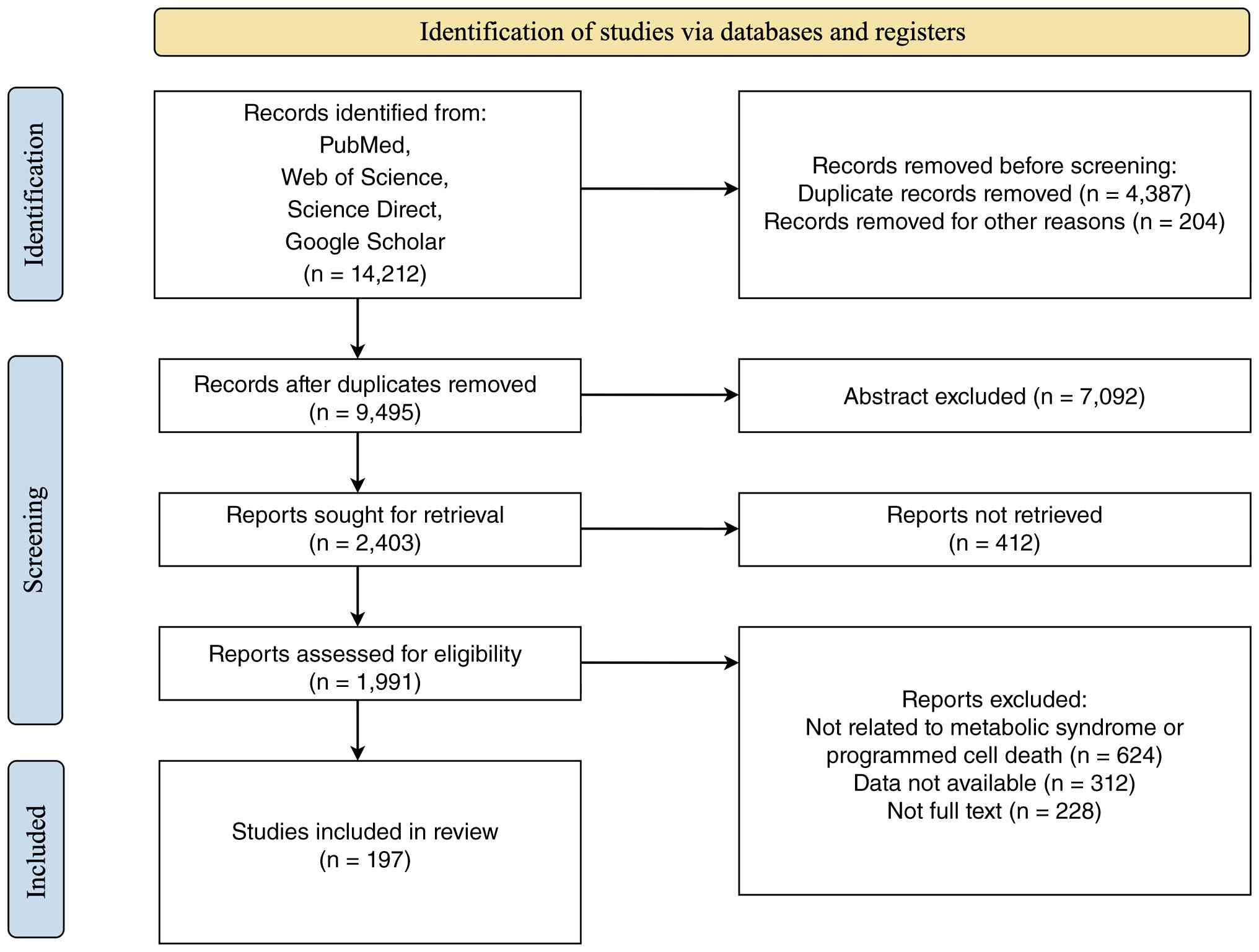 | Figure 1Flow diagram of the literature search
and study selection process. The flow diagram illustrates the
process of literature identification, screening, eligibility
assessment and inclusion of studies according to the Preferred
Reporting Items for Systematic Reviews and Meta-Analyses
guidelines. A total of 14,212 records were initially identified
from PubMed, Web of Science, ScienceDirect and Google Scholar.
After removal of duplicates and irrelevant records, the remaining
articles were screened based on titles, abstracts and full texts.
Studies were excluded according to predefined criteria, including
lack of relevance to metabolic syndrome or programmed cell death,
unavailable data and absence of full-text access. Finally, 197
studies were included in the present review. The figure was created
using Figdraw (www.figdraw.com). |
Inclusion criteria
Studies were included according to the following
criteria: i) Studies investigating the involvement of PCD pathways
in metabolic syndrome or its related metabolic disorders, including
obesity, IR, T2DM, metabolic dysfunction-associated steatotic liver
disease (MASLD), cardiovascular complications and related metabolic
abnormalities; ii) studies focusing on classical or emerging PCD
pathways, including apoptosis, pyroptosis, autophagy, ferroptosis,
necroptosis, PANoptosis and disulfidptosis; iii) clinical studies,
animal models and in vitro studies exploring the molecular
mechanisms, pathological roles, biomarkers or therapeutic potential
of PCD in metabolic disorders; iv) studies evaluating
pharmacological agents, natural compounds, lifestyle interventions
or molecular targets that modulate PCD pathways for metabolic
disease treatment; v) full-text articles published in English.
Exclusion criteria
The following types of publications were excluded:
i) Reviews, meta-analyses, editorials, letters, conference
abstracts and other secondary literature; ii) duplicate
publications or studies with insufficient experimental information;
iii) studies unrelated to MetS, metabolic disorders or PCD
mechanisms; iv) studies lacking detailed information regarding
molecular mechanisms, pathological effects, therapeutic
interventions or clinical relevance; v) articles without available
full text or published in languages other than English.
Data extraction and literature
selection
After duplicate removal, two-stage screening was
performed based on titles/abstracts and full-text assessment.
Eligible studies were systematically reviewed, and relevant
information was extracted, including publication characteristics,
study model (clinical population, animal model or cellular model),
metabolic disease phenotype, PCD pathway involved, molecular
mechanisms, regulatory targets and therapeutic interventions. The
included studies were categorized according to different PCD
modalities and their roles in MetS-associated pathological
processes. Particular attention was given to the molecular
interactions among different cell death pathways, their
contribution to metabolic inflammation, mitochondrial dysfunction,
oxidative stress and tissue injury, as well as their potential
therapeutic implications.
Evidence quality assessment and
grading
To evaluate the strength and translational relevance
of evidence supporting the involvement of each PCD pathway in MetS,
included studies were assessed according to predefined criteria,
including study design, methodological rigor, mechanistic
validation, reproducibility and relevance to human metabolic
diseases. Studies were categorized according to a modified evidence
hierarchy adapted for preclinical and clinical research. Level A
(strong evidence): Findings supported by human clinical studies,
including prospective cohort studies, large-scale case-control
studies or clinical investigations with mechanistic validation,
together with consistent evidence from experimental models. Level B
(moderate evidence): Findings derived from ex vivo human
tissue studies (such as adipose tissue, liver biopsies or
pancreatic islets) with mechanistic analyses, or from well-designed
animal studies (including genetic knockout/knock-in models) showing
reproducible effects. Level C (preliminary evidence): Findings
mainly obtained from cellular models or single animal studies
without independent replication, particularly studies relying
primarily on pharmacological intervention without genetic
validation. Level D (emerging evidence): Findings from recently
identified PCD pathways (suhch as PANoptosis and disulfidptosis)
with limited direct evidence in MetS, mainly supported by metabolic
stress-related models or extrapolation from other disease
contexts.
Pathophysiological mechanisms of MetS
MetS represents a self-sustaining cycle of central
obesity and IR, triggering downstream consequences such as chronic
low-grade inflammation, lipotoxicity and oxidative stress. These
factors collectively establish the foundation for developing
cardiovascular disease and T2DM (Fig. 2) (4). During the compensatory stage,
pancreatic β-cells secrete insulin in excess to maintain glucose
homeostasis, resulting in hyperinsulinemia (12). Defective signaling downstream of
insulin receptors impairs the tyrosine phosphorylation of insulin
receptor substrates (IRS), thereby weakening the essential
phosphoinositide 3-kinase (PI3K)/protein kinase B (AKT) signaling
pathway responsible for insulin's metabolic effects (13). Conversely, the mitogen-activated
protein kinase (MAPK) signaling pathway, associated with cell
proliferation and inflammation, becomes relatively hyperactivated.
In obesity, increased lipolysis in adipose tissue leads to
excessive free fatty acid (FFA) release. Elevated FFAs enter the
liver and muscle tissues, inhibit glucose oxidation through the
Randle cycle, and activate multiple kinases, including protein
kinase Cθ (PKCθ), JNK and inhibitor of NF-κB kinase subunit β
(IKKβ). These kinases subsequently phosphorylate IRS-1, further
impairing insulin signaling (14). Additionally, adipose-derived
inflammatory cytokines, particularly tumor necrosis factor-α
(TNF-α) and interleukin-6 (IL-6), activate JNK and the IKKβ/NF-κB
pathways, exacerbating disruptions in insulin signaling (15).
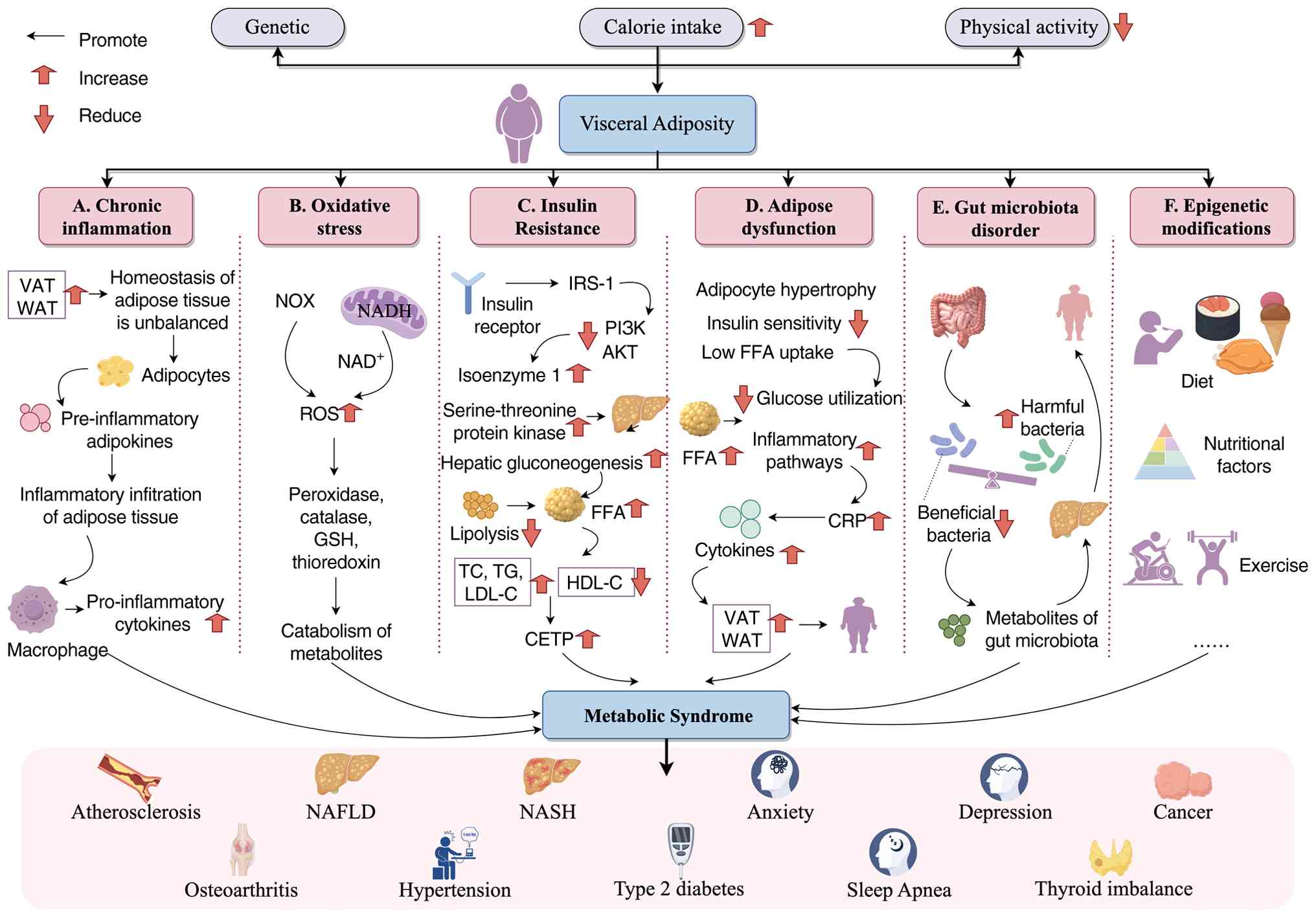 | Figure 2Schematic diagram of the
pathophysiological mechanisms of MetS. (A) Chronic inflammation:
Imbalance between VAT and WAT homeostasis leads to adipocyte
release of pro-inflammatory adipokines, attracting macrophage
infiltration and secretion of pro-inflammatory cytokines. This
triggers chronic inflammation in adipose tissue, ultimately driving
diseases such as atherosclerosis. (B) Oxidative stress: Visceral
fat accumulation causes excessive reactive ROS production via
NADH-mediated NOX enzymes; simultaneously, insufficient
antioxidants such as peroxidase and GSH fail to neutralize ROS,
leading to abnormal metabolite degradation and exacerbated
oxidative damage. (C) Insulin resistance: Inhibition of insulin
receptor signaling pathways (IRS-1, PI3K, AKT) leads to enhanced
hepatic gluconeogenesis and lipolysis, elevated FFA, TC, TG and
LDL-C, along with reduced HDL-C. This ultimately induces type 2
diabetes, hypertension and other conditions. (D) Adipose tissue
dysfunction: Adipocytes undergo excessive hypertrophy, leading to
reduced insulin sensitivity and diminished FFA uptake/glucose
utilization. Massive FFA release activates inflammatory pathways,
elevating CRP and cytokines. This further exacerbates visceral fat
accumulation, creating a vicious cycle. (E) Gut microbiota
dysbiosis: Visceral fat accumulation disrupts gut microbiota
balance, increasing harmful bacteria while reducing beneficial
ones. Abnormal microbial metabolites further exacerbate
inflammation and metabolic disorders, acting as a contributing
factor to MetS. (F) Epigenetic modifications: Environmental factors
such as diet, nutrition and exercise influence gene expression
through epigenetic modifications, indirectly regulating visceral
fat accumulation and subsequent metabolic abnormalities. The figure
was created using Figdraw (www.figdraw.com). MetS, metabolic syndrome; VAT,
visceral adipose tissue; WAT, white adipose tissue; ROS, reactive
oxygen species; NOX, NADPH oxidase; GSH, glutathione; IRS-1,
insulin receptor substrate 1; FFA, free fatty acid; TC, total
cholesterol; TG, triglyceride; LDL-C, low-density lipoprotein
cholesterol; HDL-C, high-density lipoprotein cholesterol; CRP, C
reactive protein; NAFLD, non-alcoholic fatty liver disease; NASH,
non-alcoholic steatohepatitis AKT, protein kinase B; PI3K,
phosphoinositide 3-kinase. |
Compared with subcutaneous adipose tissue, visceral
adipose tissue (VAT) demonstrates greater metabolic activity but
has limited capacity for lipid storage. Secretion of adiponectin, a
beneficial adipokine promoting insulin sensitivity and fatty acid
oxidation, is substantially reduced. By contrast, multiple
pro-inflammatory adipokines, including leptin, resistin, TNF-α and
IL-6, exhibit increased secretion, thereby leading to adipose
tissue dysfunction (16). When
the adipocyte lipid storage capacity is exceeded, fat deposition
occurs ectopically. Enlarged, hypoxic adipocytes recruit
pro-inflammatory M1 macrophages, forming characteristic 'crown-like
structures' (17). Activated
immune cells, together with adipocytes, release extensive
inflammatory cytokines and chemokines into systemic circulation,
disrupting insulin signaling and promoting atherogenesis.
Recently, the gut microbiota has emerged as a
pivotal environmental factor influencing MetS (18). Specific microbial communities
enhance energy extraction from food, contributing to obesity.
Increased intestinal permeability facilitates entry of
lipopolysaccharides (LPS) into the portal circulation, inducing
metabolic endotoxemia and systemic inflammatory responses.
Additionally, certain microbial metabolites such as short-chain
fatty acids (SCFAs) and trimethylamine N-oxide markedly influence
host metabolism. While SCFAs generally confer health benefits,
bacterial cell wall components such as LPS and peptidoglycan
trigger host immune responses, elevating cardiovascular disease
risk (19). Epigenetic
modifications, influenced indirectly by lifestyle factors such as
diet and exercise, have recently been identified as notable
regulators of visceral fat accumulation and associated metabolic
disorders.
PCD in MetS
Multiple forms of PCD contribute to the pathogenesis
of MetS, but their roles differ substantially in terms of
biological functions, tissue distribution and the strength of
available evidence. Established pathways, including apoptosis,
pyroptosis, autophagy and ferroptosis, have been extensively
investigated and represent major mechanisms linking metabolic
stress to organ dysfunction. By contrast, emerging pathways such as
necroptosis, PANoptosis and disulfidptosis remain under
investigation and require further validation in metabolic
disorders.
Apoptosis
Mechanisms of apoptosis
Apoptosis is characterized by cell shrinkage,
chromatin condensation, DNA fragmentation and apoptotic body
formation via membrane blebbing. These apoptotic bodies are
subsequently cleared by macrophages or neighboring cells without
eliciting inflammation (20).
The apoptosis cascade primarily involves two pathways: Extrinsic
and intrinsic. In the extrinsic pathway, extracellular ligands such
as TNF-α, Fas ligand and TRAIL bind to corresponding receptors
(DR4, DR5 and Fas), transmitting death signals into the cell
(21). Following this binding,
proteins including TNFR-associated death domain (TRADD),
Fas-associated death domain (FADD) and caspase-8 form complexes,
thereby activating caspase-8. Activated caspase-8 initiates
apoptosis by cleaving downstream effectors caspase-3, caspase-6 and
caspase-7 (22). By contrast,
the intrinsic mitochondrial pathway activates BH3-only proteins
under cellular stress conditions. BH3-only proteins inhibit
anti-apoptotic B-cell lymphoma-2 (Bcl-2) family proteins,
activating pro-apoptotic molecules such as Bcl-2-associated X
protein (Bax) and Bcl-2 homologous antagonist/killer. This process
induces mitochondrial outer membrane permeabilization, causing
cytochrome c and other pro-apoptotic factors to be released
into the cytoplasm. Subsequently, cytochrome C binds to apoptotic
protease activating factor-1, forming apoptosomes that activate
caspase-9, ultimately triggering caspase-3, -6 and -7 activation
and apoptosis (Fig. 3) (23).
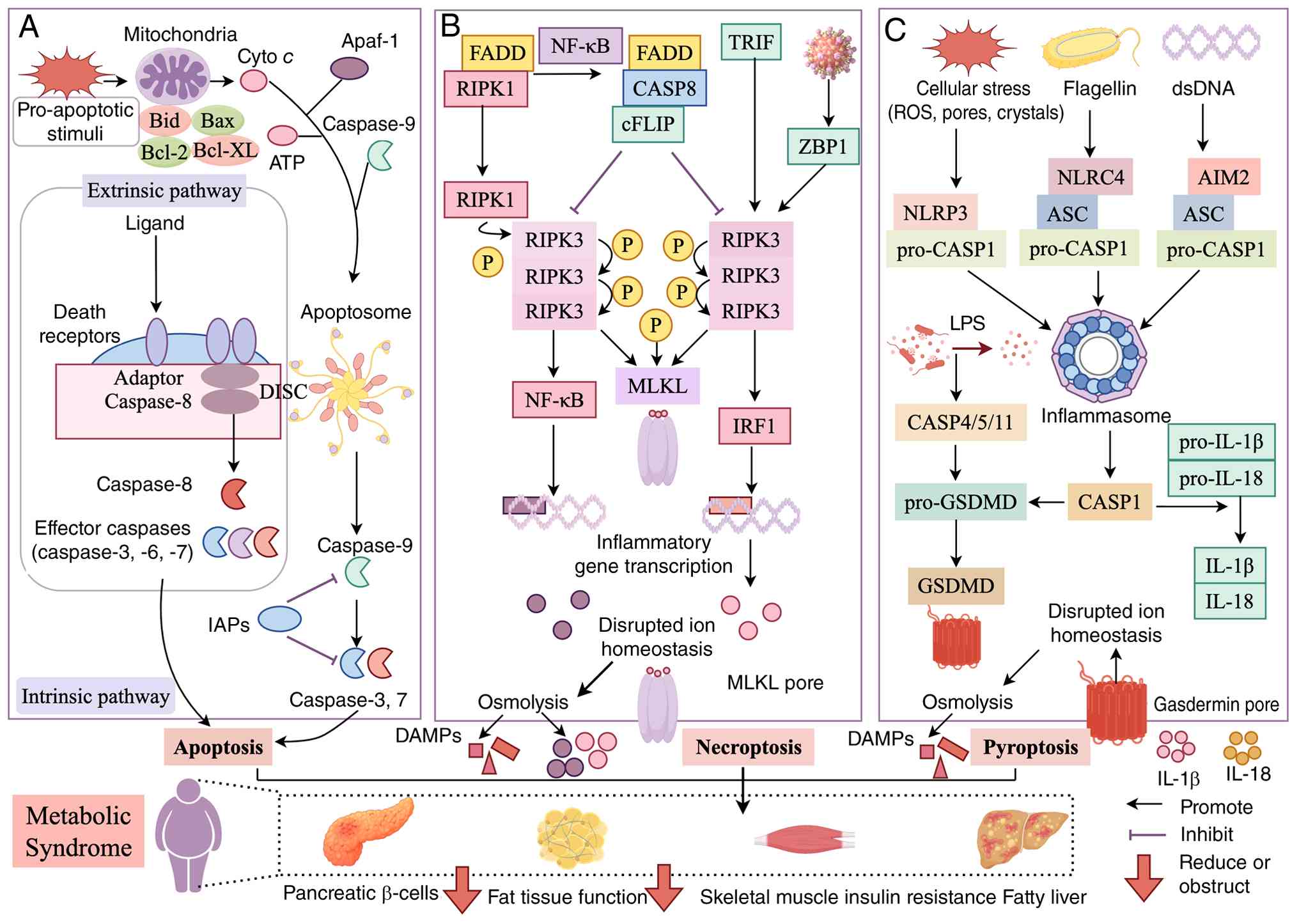 | Figure 3Schematic diagram of apoptosis,
necroptosis, and pyroptosis mechanisms in MetS. (A) Exogenous
apoptosis pathway: Death receptor binds ligand, recruits adaptor
proteins to form a complex with caspase-8 (DISC), activating
caspase-3. Intrinsic apoptosis pathway: Metabolic stress induces
mitochondria to release cyto c, which binds Apaf-1 to form
an apoptosome, activating caspase-9, which ultimately activates
effector caspases. In MetS, this pathway can cause tissue damage,
such as to pancreatic β-cells. (B) Necroptosis: Regulated by
proteins such as RIPK1 and RIPK3. When caspase-8 is inhibited (such
as by cFLIP), RIPK1 and RIPK3 bind and become phosphorylated,
further activating the MLKL protein. MLKL aggregates and forms
pores in the cell membrane, disrupting ion homeostasis and
ultimately leading to osmotic lysis. This process activates
pathways such as NF-κB to trigger inflammatory gene transcription
while releasing large amounts of DAMPs. In MetS, this can damage
adipose tissue, skeletal muscle and other tissues, exacerbating
insulin resistance. (C) Pyroptosis: Different stimuli, such as
cellular stress, bacterial flagella and double-stranded DNA,
activate corresponding inflammasomes NLRP3, NLRC4 and AIM2,
respectively. These complexes activate caspase-1; caspase-1 cleaves
the GSDMD protein to form pores that disrupt the cell membrane,
while simultaneously processing pro-IL-1β and pro-IL-18 into active
cytokines IL-1β and IL-18. Ultimately, cells lyse and release
pro-inflammatory factors, exacerbating pathological processes such
as fatty liver disease and adipose tissue inflammation in MetS. The
figure was created using Figdraw (www.figdraw.com). MetS, metabolic syndrome; DISC,
death-inducing signaling complex; RIPK, receptor-interacting
serine/threonine-protein kinase; MLKL, mixed lineage kinase domain
like pseudokinase; DAMPs, damage-associated molecular patterns;
NLRP3, NLR family pyrin domain containing 3; GSDMD, gasdermin
D. |
Apoptosis in MetS
Among various PCD modalities, apoptosis is one of
the most extensively characterized forms of PCD contributing to
MetS progression, particularly in pancreatic β-cell dysfunction,
adipose tissue remodeling and hepatic injury. In MetS, apoptosis is
not merely a consequence of metabolic stress but also an active
driver of disease progression through tissue remodeling and
amplification of inflammatory responses. In obesity, adipose tissue
expansion is not limitless. When adipocytes enlarge beyond their
vascular support and metabolic capabilities, hypoxia, lipotoxicity
and endoplasmic reticulum stress (ERS) occur (24). Nutrient overload and elevated
FFAs stimulate excessive lipid and protein synthesis in the ER,
activating the unfolded protein response (UPR) (25). Severe, prolonged ERS shifts the
UPR from promoting cell survival towards apoptosis, primarily
mediated by protein kinase R-like ER kinase-eukaryotic translation
initiation factor 2 subunit 1-activating transcription factor 4
(ATF4) and inositol-requiring enzyme 1α-apoptosis signal-regulating
kinase 1-JNK signaling pathways (26). ATF4 upregulates the pro-apoptotic
protein C/EBP homologous protein, while JNK pathway activation
phosphorylates and inhibits anti-apoptotic Bcl-2, simultaneously
activating pro-apoptotic proteins Bad and Bim. This disrupts
cellular balance between survival and apoptosis (27). Together with elevated FFAs, these
signals promote mitochondrial membrane potential collapse,
resulting in cytochrome C release. Macrophages recognize, engulf
and become activated by apoptotic adipocytes, subsequently
secreting abundant pro-inflammatory cytokines. This exacerbates
local inflammation and induces apoptosis in neighboring adipocytes
through paracrine signaling (28).
Although apoptotic hepatocytes can no longer perform
their normal metabolic functions, including glucose metabolism,
their death process releases 'find-me' and 'eat-me' signals. These
signals recruit and activate Kupffer cells, resident hepatic
macrophages (29). Activated
Kupffer cells secrete inflammatory cytokines such as TNF-α,
impairing insulin signaling in adjacent hepatocytes. A previous
study indicated that inhibiting adipocyte apoptosis enhances
systemic insulin sensitivity, suggesting apoptosis as a pathogenic
contributor to IR (30). In
T2DM, glucotoxicity and lipotoxicity synergistically drive
pancreatic β-cell apoptosis. Sustained hyperglycemia increases
mitochondrial oxidative phosphorylation, producing excessive
reactive oxygen species (ROS) that exceed cellular antioxidant
capacity, causing oxidative stress and cellular damage (31). Moreover, inflammatory mediators
secreted by insulin-resistant tissues, and cytokines (IL-1β, TNF-α
and IFN-γ) from immune cells infiltrating pancreatic islets,
directly activate β-cell apoptotic mechanisms (32). Although β-cell apoptosis is
'silent' and non-inflammatory, its consequences are profound. It
leads to irreversible loss of functional β-cell mass, absolute
insulin deficiency and uncontrolled hyperglycemia.
Hypertension induces apoptosis in vascular smooth
muscle cells (VSMCs) and vascular endothelial cells (VECs) due to
stress from the vascular wall and shear forces from blood flow.
Prolonged pressure overload alters the expression of proteins such
as p53, Bax and Bcl-2, thus affecting the regulatory balance
between proliferation and apoptosis in VSMCs (33). Studies have demonstrated that
caspase-3 is involved in apoptosis of hypoxic VECs (34). During the progression of
hypertension, accumulated apoptosis results in thinning of the
myocardial microvascular network, compromising blood supply and
exacerbating myocardial cell death (35). Notably, sustained
vasoconstriction simultaneously promotes apoptosis, vascular wall
thickening, atherosclerosis, luminal narrowing and increased vessel
stiffness, consequently aggravating hypertensive complications
(36). Hyperlipidemia,
particularly elevated FFAs and oxidized low-density lipoprotein
(ox-LDL), acts as a critical stressor triggering various apoptotic
pathways. Palmitic acid serves as a precursor for ceramide
synthesis, inducing mitochondrial dysfunction, increased ROS
production and cytochrome C release, thereby strongly activating
mitochondrial apoptosis (37).
In macrophages, apoptosis triggered by ox-LDL is essential for
necrotic core formation within atherosclerotic plaques. If
apoptotic macrophages are not cleared efficiently, they undergo
secondary necrosis, releasing pro-inflammatory components (38). Current evidence supporting the
involvement of apoptosis in MetS is relatively strong, based on
consistent findings from animal models, human pancreatic tissue
studies and clinical observations linking β-cell loss to metabolic
deterioration.
Pyroptosis
Mechanism of pyroptosis
The classical pyroptosis pathway primarily involves
activation of intracellular multiprotein complexes known as
inflammasomes. Inflammasomes detect and respond to various
endogenous and exogenous danger signals, including
pathogen-associated molecular patterns (PAMPs) and
damage-associated molecular patterns (DAMPs) (39). Upon activation, inflammasome
sensors recruit the adaptor protein apoptosis-associated speck-like
protein containing a CARD (ASC), primarily via pyrin domain
interactions. ASC aggregation subsequently exposes its caspase
recruitment domain (CARD), facilitating interaction with
procaspase-1 and linking inflammasome sensors with procaspase-1
(40). Additionally, certain
CARD-containing sensors can directly recruit procaspase-1. Elevated
local concentrations of procaspase-1 drive its heterodimerization,
self-cleavage and activation to caspase-1. Activated caspase-1 then
cleaves pro-IL-1β/pro-IL-18 and gasdermin D (GSDMD), generating an
N-terminal fragment (NT). GSDMD-NT integrates into the cell
membrane lipid bilayer, forming pores that induce pyroptotic cell
death and facilitate the release of mature IL-1β and IL-18 into the
extracellular space (41). The
non-canonical pathway involves human caspase-4/5 or murine
caspase-11 as direct intracellular receptors for LPS. Upon sensing
LPS, these caspases cleave GSDMD, initiating pyroptosis (42). Recent findings also suggest
alternative pyroptosis pathways mediated by caspase-3, caspase-8
and granzyme (Fig. 3) (43).
Pyroptosis in MetS
In obesity, excessive nutrient intake induces
adipose tissue dysfunction, ultimately impairing insulin
sensitivity. A previous study demonstrated increased NLR family
pyrin domain containing 3 (NLRP3), IL-1β and IL-18 expression in
adipose tissues of high-fat diet-fed mice, associating with reduced
insulin sensitivity. Conversely, NLRP3 knockout mice exhibited
improved insulin sensitivity under identical dietary conditions
(44). In 10 male patients with
T2DM, weight reduction achieved through caloric restriction and
exercise led to decreased adipose tissue expression of NLRP3 and
IL-18, accompanied by enhanced insulin sensitivity (45). Sehgal et al (46) proposed that adipose tissue
inflammation mediated by the NLRP3 inflammasome markedly reduces
insulin sensitivity, a hypothesis supported by other research. In a
study involving 98 subjects, targeted NLRP3 inhibition via small
interfering RNA substantially decreased LPS-induced expression of
fibrosis-associated molecules in adipocytes, alleviating
inflammation and fibrosis (47).
Additionally, melatonin has been reported to attenuate
inflammasome-mediated pyroptosis in murine adipose tissue by
inhibiting NF-κB/GSDMD signaling (48).
Inflammation is intrinsically associated with
diabetes. Research has demonstrated that hyperglycemia increases
levels of advanced glycation end products (AGEs) and ROS, thereby
activating inflammasomes and triggering pyroptosis (49). Ma et al (50) confirmed that pyroptosis
exacerbates β-cell destruction, ultimately impairing glucose
metabolism. Furthermore, Carlos et al (51) observed an increased proportion of
macrophages expressing caspase-1 in pancreatic lymph nodes of
streptozotocin-induced diabetic mice, whereas this proportion
decreased in NLRP3-knockout mice. Furthermore, knockout of NLRP3
and IL-1R genes markedly elevated insulin levels and reduced
hyperglycemia. Luo et al (52) separately reported increased
expression of NLRP3, caspase-1 and IL-1β as critical factors
driving diabetic cardiomyopathy progression. Experimental
NLRP3-microRNA (miRNA) therapy effectively reduced myocardial
inflammation by inhibiting IL-1β, IL-18, TNF-α and IL-6 release.
Moreover, NLRP3 gene silencing attenuated myocardial inflammation,
cardiac pyroptosis and left ventricular fibrosis, thereby improving
diabetic cardiomyopathy progression in T2DM rat models.
Low-grade or persistent inflammation is crucial in
hypertension progression. For example, Dalekos et al
(53) proposed that inflammasome
activation and pyroptosis notably contribute to elevated blood
pressure. Inflammasome activation leads to caspase-1 activation,
which subsequently cleaves GSDMD, mediates pyroptosis and releases
additional inflammatory mediators into the extracellular space,
further exacerbating inflammation and hypertension. NF-κB is a
primary activator of the pyroptosis-related inflammasome NLRP3.
NF-κB knockout markedly attenuated NLRP3 and caspase-1 activation,
thereby inhibiting salt-sensitive hypertension progression in rats
(54). Research has further
confirmed that after 25 days of treatment with the NLRP3 inhibitor
MCC950, hypertensive mice exhibited marked improvements in blood
pressure, urine volume, osmolarity and proteinuria (55). Levels of NLRP3 inflammasome
components, including apoptosis-associated speck-like protein,
pro-caspase-1 and inflammatory markers (pro-IL-18, pro-IL-1β and
TNF-α), were markedly reduced compared with the baseline. These
findings indicate that pharmacological inhibition of the NLRP3
inflammasome represents a promising therapeutic approach for
hypertension. Another animal study demonstrated that NLRP3
inflammasome inhibition effectively alleviates kidney injury
resulting from pyroptosis (56).
Current evidence implicating pyroptosis in MetS is primarily
derived from experimental models and human tissue studies,
particularly in adipose inflammation and NAFLD, while large-scale
clinical validation remains limited.
Autophagy
Mechanisms of autophagy
Autophagy is a critical metabolic recycling process
in eukaryotic cells, mediated by lysosomes or vacuoles, involving
the degradation and reuse of intracellular components. Autophagy
regulation involves several signaling pathways, among which
adenosine monophosphate-activated protein kinase (AMPK) and
mammalian target of rapamycin (mTOR) pathways are most prominent
(57). AMPK functions as a
conserved energy sensor, activating autophagy initiation under
metabolic stress conditions, such as energy deprivation (58). Conversely, mTOR acts as a major
negative regulator of autophagy, integrating upstream signals
including PI3K, insulin-like growth factor (IGF-1/IGF-2) and AMPK
to inhibit autophagic activity (59). Specifically, mTOR kinase
activates signaling pathways involving AKT and MAPK, thereby
suppressing autophagy. The AMPK and p53 signaling pathways
negatively regulate mTOR activity, thus promoting autophagy
(60). Under normal
physiological conditions, mTOR inhibits autophagy; however, during
stress states such as nutrient deprivation or starvation, autophagy
initiation occurs via the mTOR/autophagy-related gene (Atg) 1/Atg13
signaling axis (61). During
autophagy, the selective adaptor protein p62 specifically targets
certain proteins and organelles for sorting and degradation.
Mitophagy exemplifies selective autophagy and is essential for
maintaining cardiomyocyte homeostasis. In this process, damaged or
dysfunctional mitochondria are selectively recognized via the
Pink1-Parkin signaling pathway or other mitochondrial autophagy
receptors and subsequently degraded by acidic lysosomal hydrolases
(Fig. 4) (62).
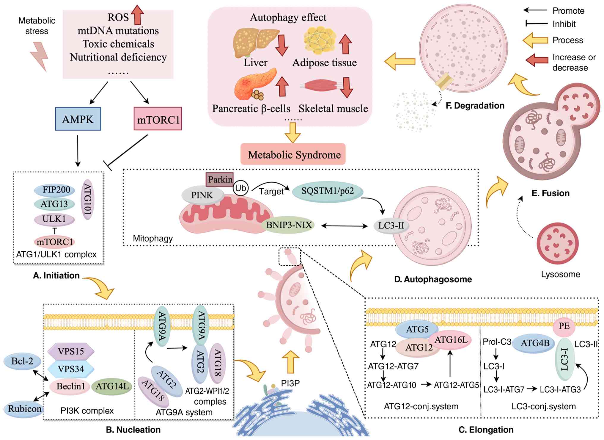 | Figure 4Schematic diagram of autophagy
mechanisms in MetS. When metabolic stress occurs, such as increased
ROS, mtDNA mutations or toxin/nutrient deprivation, autophagy is
regulated through two core pathways: AMPK pathway activation
promotes autophagy initiation by forming the ATG1/ULK1 complex;
mTORC1 pathway inhibition releases its suppression of autophagy,
jointly driving the process. Complete autophagy process: (A)
Initiation: ATG1/ULK1 complex formation prepares for autophagy; (B)
nucleation: PI3K complex, ATG9A system and others form the
autophagosome precursor membrane; (C) elongation: Proteins
including ATG12 and LC3 systems assist in extending the precursor
membrane, forming a double-layered structure enveloping 'substances
awaiting clearance'; (D) autophagosome (E) fusion and (F)
degradation: Autophagosomes fuse with lysosomes, degrading and
recycling their contents. Among these, mitophagy represents a
specialized form of autophagy. Damaged mitochondria are marked
through pathways such as PINK/Parkin and BNIP3-NIX, ultimately
engulfed by autophagosomes for degradation, thereby preventing
metabolic disorders triggered by mitochondrial dysfunction.
Ultimately, this affects the function of the liver, skeletal
muscle, adipose tissue and pancreatic β-cells, contributing to the
onset and progression of MetS. The figure was created using Figdraw
(www.figdraw.com). MetS, metabolic syndrome; ROS,
reactive oxygen species; ULK1, autophagy-light chain kinase 1; ATG,
autophagy-related gene; mtDNA, mitochondrial DNA. |
Autophagy dysfunction: A double-edged
regulator of MetS
Although autophagy is generally regarded as a
protective mechanism maintaining cellular homeostasis, accumulating
evidence indicates that its role in MetS is highly
context-dependent and involves dual regulatory effects (63). The metabolic effects of autophagy
are influenced by multiple factors, including tissue type, disease
stage, nutritional status and autophagic flux efficiency (64). Physiological activation of
autophagy maintains metabolic homeostasis by eliminating damaged
mitochondria and regulating lipid metabolism (65). In insulin-sensitive tissues,
basal autophagy is essential for metabolic regulation. For example,
a previous study involving tissue-specific deletion of
autophagy-related genes demonstrated the indispensable role of
autophagy in preventing metabolic deterioration (66). Liver-specific Atg7 knockout mice
exhibit impaired autophagic flux, hepatic lipid accumulation,
increased oxidative stress and spontaneous steatosis, whereas
restoration of autophagic activity ameliorates metabolic
abnormalities (67). Similarly,
loss of autophagy-related gene function in pancreatic β-cells
results in mitochondrial dysfunction and impaired insulin
secretion, highlighting the protective role of autophagy in
maintaining β-cell function (68). However, excessive or dysregulated
autophagy may become maladaptive under certain pathological
conditions. Persistent nutrient stress or chronic inflammation can
disrupt the balance between autophagic degradation and cellular
regeneration, leading to excessive organelle turnover and cellular
dysfunction (69).
In adipose tissue, enhanced autophagic activity may
facilitate adipocyte remodeling and lipid mobilization, while in
advanced metabolic disorders, sustained autophagic activity may
fail to restore cellular homeostasis due to impaired lysosomal
function (70) Autophagy serves
an essential role in adipocyte differentiation and maturation, with
obesity further influencing autophagic activity during MetS.
Dyslipidemia, hypertension and hyperglycemia primarily contribute
to altered autophagy in obese individuals. Conversely, impaired
autophagy, especially its suppression, accelerates obesity and
metabolic disorders (71). Mice
with systemic or tissue-specific knockouts of autophagy-related
proteins, including Beclin 2, Atg7, lysosomal membrane-associated
protein 2, transcription factor EB (TFEB) and Bif-1, exhibit
increased susceptibility to obesity phenotypes (72). For example, liver-specific
deletion of Atg7 or TFEB causes hepatic steatosis and increased
body weight in mice, whereas their overexpression prevents weight
gain and MetS (73).
Furthermore, autophagy regulation in the central nervous system is
closely linked to obesity; inhibiting autophagy in specific
neuronal populations stimulates appetite and promotes obesity
(74). This creates a vicious
cycle wherein defective autophagy accelerates lifestyle-induced
obesity, further suppressing autophagy in liver, muscle, and
adipose tissues and exacerbating MetS symptoms.
Elevated blood glucose activates the IGF-1R pathway,
promoting PI3K translocation to membranes and subsequent activation
of AKT/PKB signaling. This inhibits the tuberous sclerosis complex
(TSC), activating mTOR, which impairs autophagic flux in cardiac
microvascular endothelial cells (75). A previous study showed reduced
levels of hepatic autophagy markers (LC3II) and increased p62
expression in hyperinsulinemic conditions, high-fat diet-induced
obesity and ob/ob mouse models, indicating impaired autophagy
(76). Under glucose
deprivation, decreased ATP levels activate AMPK. Activated AMPK
suppresses mTORC1 by stimulating the TSC1-TSC2 complex and directly
initiates autophagy via the autophagy-light chain kinase 1 complex
(77). Zhao et al
(78) demonstrated that treating
HUVECs with ox-LDL or AGEs altered endothelial autophagic flux,
while AGE-induced autophagy blockade increased mitochondrial ROS
production. Stimulation of autophagy by the mTOR inhibitor
rapamycin or TFEB overexpression prevented AGE/ox-LDL-induced
mitochondrial ROS accumulation, enhanced eNOS dimerization and
ameliorated diabetic endothelial dysfunction (79). Furthermore, autophagy-deficient
mice fed a high-fat diet showed increased susceptibility to
diabetic symptoms, mitochondrial injury and pancreatic β-cell
dysfunction (80).
Under normal conditions, hyperlipidemia reduces
basal autophagy levels. Enhanced lipolysis in adipocytes and IR
elevate blood lipid levels, potentially impairing autophagy by
blocking autophagosome-lysosome fusion, lysosomal acidification and
lysosomal enzyme activities. A previous study demonstrated that in
high-fat diet-induced obese mouse models, elevated blood lipids
suppressed autophagosome-lysosome fusion, disrupting intracellular
Ca2+ regulation, damaging mitochondria and inducing
apoptosis (81). Intracellular
fatty acid derivatives, such as ceramides, diacylglycerols and
fatty acyl-CoA, further disrupt autophagic homeostasis, thereby
promoting IR and steatohepatitis (82). Knockout of protein tyrosine
phosphatase 1B (PTP1B) alleviates cardiomyocyte damage induced by a
high-fat diet in mice by restoring cardiac autophagy through an
AMPK-dependent mechanism (83).
Although autophagy inhibition alone is insufficient for lipid
accumulation and steatosis development, it promotes lipid
deposition when combined with other factors, such as prolonged
high-fat diet exposure or obesity-related genetic mutations
(84). Therefore, autophagy
should not be simply considered either beneficial or detrimental in
MetS. Instead, its biological effects depend on whether autophagic
flux is maintained at an appropriate level. Therapeutic strategies
should aim to restore physiological autophagy rather than
indiscriminately activate or inhibit this pathway.
Ferroptosis
Mechanism of ferroptosis
Ferroptosis is closely linked to intracellular iron
accumulation. Under normal physiological conditions,
Fe3+ binds to transferrin (TF) and enters cells via the
TF receptor 1 (TFR1) on the cell membrane (85). Upon entering the cell,
Fe3+ is reduced to Fe2+, subsequently
entering the labile iron pool (LIP). Disturbances in iron
metabolism lead to abnormal Fe2+ accumulation within the
LIP. This excess Fe2+ initiates lipid peroxidation
through the Fenton reaction, generating abundant lipid-ROS that
damage cell membranes and induce ferroptosis (86). During ferroptosis,
Fe3+-catalyzed free radicals primarily target membrane
polyunsaturated fatty acids (PUFAs), such as arachidonic acid (AA)
and adrenocortical acid (AdA) (87). Under catalysis by ACSL4, these
fatty acids bind to coenzyme A (CoA) to form AA-CoA or AdA-CoA
intermediates. Subsequently, these intermediates are esterified by
lysophospholipid acyltransferase 5 (LPCAT3) into
phosphatidylethanolamine-linked forms (PE-AA or PE-AdA).
Lipoxygenases (LOXs) then enzymatically convert these forms into
peroxidized products (PE-AA/AdA-OOH), ultimately causing
ferroptosis (88). Glutathione
peroxidase 4 (GPX4) serves a crucial role in cellular antioxidant
defense by catalyzing the reduction of toxic lipid hydroperoxides
(LOOH) into non-toxic phospholipids, while converting reduced
glutathione (GSH) to oxidized GSH (89). When GSH availability decreases,
GPX4 activity declines, causing ROS accumulation and ferroptotic
cell death (Fig. 5).
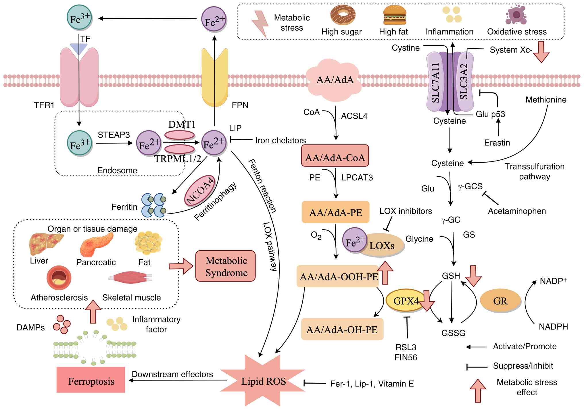 | Figure 5Schematic diagram of ferroptosis
mechanisms in MetS. Iron enters cells via the TFR1 receptor, is
reduced to Fe2+ by STEAP3 in endosomes and then
transported to the cytoplasm by DMT1. Cytoplasmic Fe2+
is partially stored by ferritin, while the remainder exists as an
LIP. Under metabolic stress, ferritin is degraded via
ferritinophagy, releasing large amounts of Fe2+.
Concurrently, impaired function of the iron export protein FPN
leads to intracellular Fe2+ accumulation. Excess
Fe2+ catalyzes lipid peroxidation through the Fenton
reaction, ultimately triggering ferroptosis. Metabolic stress also
inhibits System Xc-, causing intracellular cysteine deficiency and
reduced GSH synthesis. This GSH depletion prevents GPX4 from
scavenging lipid ROS. Concurrently, AA/AdA undergoes enzymatic
modification by ACSL4, LPCAT3 and others into lipid substrates.
Under the influence of Fe2+ and LOXs, these substrates
generate substantial lipid peroxides, ultimately damaging cell
membranes and triggering ferroptosis. Following ferroptosis, DAMPs
and inflammatory cytokines are released, exacerbating tissue damage
in the liver, pancreas and vasculature, thereby driving the
progression of MetS. Conversely, iron chelators and GPX4 activators
(such as vitamin E) can mitigate ferroptosis by reducing
Fe2+ or inhibiting lipid peroxidation. The figure was
created using Figdraw (www.figdraw.com). MetS, metabolic syndrome; ROS,
reactive oxygen species; DAMPs, damage-associated molecular
patterns; GPX4, glutathione peroxidase 4; FPN, ferroportin; TFR1,
transferrin receptor 1; STEAP3, six-transmembrane epithelial
antigen of prostate 3; LIP, labile iron pool; GSH, glutathione;
AA/AdA, arachidonic acid/adrenic acid; LOXs, lipoxygenases; ACSL4,
acyl-CoA synthetase long-chain family member 4; LPCAT3,
lysophosphatidylcholine acyltransferase 3; DMT1, divalent metal
transporter 1. |
Ferroptosis in MetS
High intracellular iron levels disrupt adipocyte
differentiation, contributing to metabolic dysfunction. Liposomes,
which store triglycerides (TGs) as neutral lipids, are enveloped by
Fas-associated factor 1 (FAF1) to protect against Fe2+
exposure (90). TGs within
liposomes undergo hydrolysis of ester bonds to form glycerol and
FFAs. Under conditions of Fe2+ overload, cytoplasmic
FFAs trigger lipid peroxidation through the Fenton reaction,
rapidly generating PUFA-OOH (91). In the presence of excess
Fe2+, PUFA-OOH readily oxidizes further, initiating
chain reactions of lipid oxidation by extracting hydrogen atoms
from neighboring PUFAs. This disrupts membrane integrity,
ultimately leading to cell death (92). A previous study demonstrated that
reducing iron accumulation in white adipose tissue limits
intestinal lipid absorption, thus protecting against metabolic
disturbances induced by high-fat diets (93). Further research indicates
(94) that lowering
intracellular iron inhibits adipogenic gene expression and reduces
lipid synthesis. Elevated iron levels also promote persistent
adipokine secretion, creating an inflammatory environment that
attracts macrophage infiltration and drives obesity
progression.
A previous epidemiological study suggested a
possible relationship between excess body iron storage and the risk
of T2DM (95). Iron overload
reduces insulin sensitivity, lowers adiponectin secretion and
increases leptin levels, thus promoting IR (96). A previous prospective study
reported a positive association between IR and elevated serum
ferritin concentrations (97).
Disrupted iron metabolism contributes to mitochondrial dysfunction,
thereby impairing insulin signaling pathways. Additionally,
GPX4-deficient mice exhibit adipocyte hypertrophy and IR,
indicating that iron metabolism disturbances induce IR by impairing
mitochondrial function and altering adipocyte cytokine production
(98). Research findings also
show that increased body iron stores elevate T2DM risk (99). For instance, mouse models of
hereditary hemochromatosis, characterized by systemic iron
overload, exhibit iron accumulation in skeletal muscles, enhanced
fatty acid oxidation and diminished glucose oxidation, collectively
aggravating IR (100). A
previous study aiming to improve diabetes outcomes demonstrated
enhanced insulin secretion and sensitivity following reductions in
systemic iron stores (101).
However, it is noteworthy that serum ferritin-based assessments of
body iron storage are not entirely reliable indicators, as ferritin
levels also increase in conditions such as inflammation, cancer and
liver diseases (102).
A previous study demonstrated that iron overload is
closely linked to atherosclerosis risk factors, including abnormal
iron metabolism, which is frequently observed in obese and
hypertensive individuals (103). Excessive iron intake aggravates
dyslipidemia, particularly by increasing TG levels. Moreover, iron
overload and lipid metabolic disturbances synergistically
accelerate atherosclerosis progression. Iron chelation therapy and
dietary iron restriction stabilize atherosclerotic plaques and
protect against endothelial injury (104). In addition, inhibition of GPX4
promotes lipid peroxide accumulation, induces ferroptosis and
contributes to atherosclerotic plaque formation.
Ferroptosis-related LOXs accelerate atherosclerosis development,
whereas inhibition of 12/15-LOX reduces LDL oxidation and
attenuates atherosclerosis progression (105). Iron overload also serves a
critical role in the progression of metabolic-associated fatty
liver disease (MAFLD). Excessive systemic iron induces
intracellular lipid peroxidation, damages hepatocytes and promotes
liver fibrosis. Ferroptosis in this context is accompanied by
collapse of intracellular antioxidant defenses, particularly
depletion of GSH and GPX4, rendering hepatocytes more vulnerable to
oxidative stress and further exacerbating MAFLD progression
(106). Although ferroptosis
has been increasingly implicated in NAFLD/MASH progression, most
available evidence is derived from cellular and animal models, and
its clinical relevance requires further validation.
Necroptosis
Mechanisms of necroptosis
Necroptosis is a form of regulated cellular death
occurring when apoptosis pathways are inhibited. While its
underlying mechanism shares similarities with apoptosis, its
morphological features closely resemble necrosis. Dysregulated
necroptosis contributes to multiple inflammatory diseases,
infections and neurodegenerative conditions (107). Numerous stimuli can initiate
necroptosis, but the classical pathway involves the
TNF-α/TNFR-induced RIPK1-RIPK3-mixed lineage kinase domain like
pseudokinase (MLKL) cascade (108). Upon TNF-α binding, TNFR1
undergoes conformational changes and recruits multiple proteins,
including RIPK1, TRADD, cellular inhibitor of apoptosis proteins
(cIAP1, cIAP2), TNFR2 and TNFR5, forming Complex I at the cell
membrane. Following internalization and modification, TNFR1
dissociates from Complex I, and RIPK1 subsequently assembles
Complex II together with TRADD, FADD and caspase-8. When caspase-8
activity is suppressed, RIPK1 phosphorylation initiates the
necroptosis pathway, interacting with RIPK3 to form a necrosome
complex. RIPK3 autophosphorylation then activates MLKL, which
oligomerizes, relocates to the plasma membrane and forms pores.
This pore formation causes an influx of Na+ and
Ca2+, elevating intracellular osmotic pressure and
resulting in plasma membrane rupture. This event releases DAMPs,
causing inflammation and tissue injury (Fig. 3) (109).
Necroptosis in MetS
In obese adipose tissue, hypertrophic adipocytes are
among the first cells to undergo necroptosis. Consequently,
intracellular contents, including DAMPs such as ATP, DNA, HMGB1 and
IL-33, are extensively released into the extracellular matrix
(110). These DAMPs act as
'danger signals', activating macrophages via classical inflammatory
pathways, including NF-κB and the NLRP3 inflammasome. This
activation leads to abundant cytokine secretion (TNF-α, IL-1β, IL-6
and MCP-1), further amplifying adipocyte death (111). Experimental evidence
demonstrates marked upregulation of RIPK3 and MLKL at both protein
and mRNA levels in adipose tissues from diet-induced obese and
ob/ob mice, associating strongly with inflammatory severity
(112). In skeletal muscle and
liver tissues, inflammatory signals increase the expression of key
gluconeogenic enzymes. Consequently, hepatic glucose production
continues despite insulin presence, exacerbating fasting
hyperglycemia. Additionally, these inflammatory signals can induce
damage and death in healthy β-cells, triggering a cascade effect
and accelerating β-cell mass depletion (113). Furthermore, sublethal
activation of RIPK1/RIPK3 may impair β-cell insulin secretion,
leading to defective insulin release. Notably, human pancreatic
tissues affected by T2DM frequently exhibit islet amyloid
polypeptide (IAPP) deposits. These oligomerized IAPP aggregates are
internalized by phagocytes, inducing lysosomal membrane rupture and
content leakage, which strongly activates the NLRP3 inflammasome
and subsequently triggers necroptosis (114). When LDL levels increase, LDL
enters the vascular endothelium and oxidizes into ox-LDL, which
induces ERS and mitochondrial dysfunction, activating RIPK3
(115). When cholesterol
reverse transport is compromised, free cholesterol accumulates
intracellularly, forming cholesterol crystals (CCs). These
crystals, upon phagocytosis into lysosomes, puncture the lysosomal
membrane due to their rigidity, releasing cathepsins into the
cytoplasm. This directly activates the NLRP3 inflammasome and
downstream caspase-1 in a RIPK3-dependent manner, potentially
triggering MLKL activation (116).
Emerging PCD mechanisms in MetS
PANoptosis in MetS
PANoptosis represents an emerging form of regulated
cell death implicated in metabolic inflammation (117). Unlike the simultaneous
activation of multiple independent cell death pathways, PANoptosis
is mediated by the formation of a supramolecular complex termed the
PANoptosome, which coordinates key regulators from different death
programs, including Z-DNA binding protein 1 (ZBP1), RIPK3,
caspase-8 and inflammasome components (118,119). Although PANoptosis has
attracted increasing attention in inflammatory diseases, its direct
contribution to MetS remains largely unexplored. Current evidence
linking PANoptosis to MetS-associated disorders is mainly based on
observations of shared inflammatory pathways, including NLRP3
inflammasome activation, caspase signaling and RIPK-dependent
inflammatory responses (120,121). In obesity and MASLD, chronic
metabolic stress induces persistent activation of inflammatory
signaling pathways that overlap with known PANoptosis-related
mechanisms (122). For example,
saturated fatty acids, mitochondrial dysfunction and oxidative
stress can activate inflammasome pathways and inflammatory caspase
signaling, thereby theoretically providing a favorable environment
for PANoptotic responses (123,124). However, direct evidence
demonstrating a causal role of PANoptosis in MetS remains limited.
It remains unclear whether functional PANoptosome complexes are
assembled in metabolic tissues such as adipose tissue, the liver or
the pancreas. Future studies using genetic models and single-cell
approaches are required to determine the precise contribution of
PANoptosis to MetS progression. (Fig. 6).
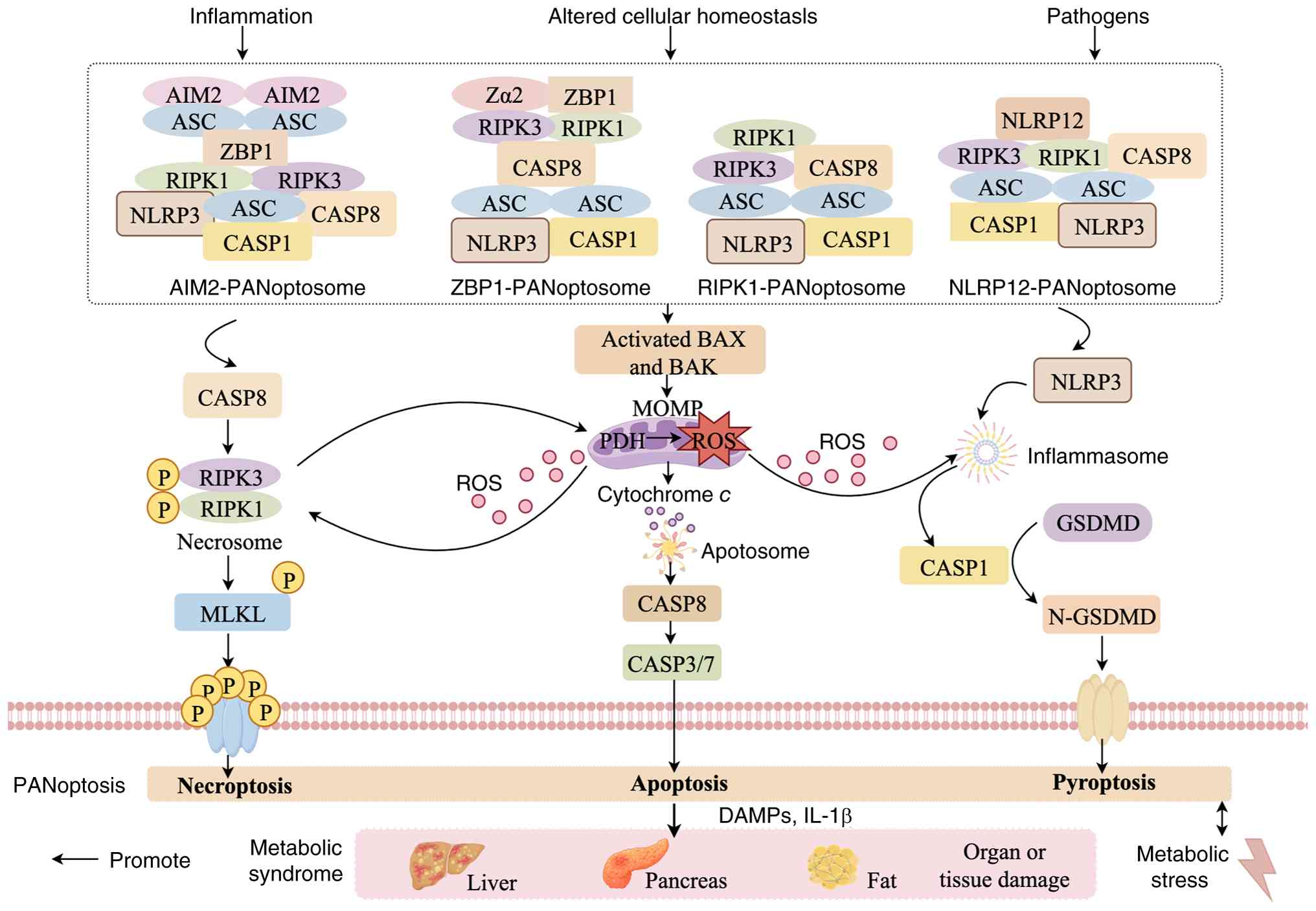 | Figure 6Schematic of PANoptosis assembly
mechanisms in MetS. Different stimuli induce distinct PANoptosome
complex assembly: Inflammatory stimuli form AIM2-PANoptosomes,
cellular homeostasis disruption forms ZBP1-PANoptosomes and
RIPK1-PANoptosomes, while pathogen stimulation forms
NLRP12-PANoptosomes. PANoptosis may concurrently promote the
release of DAMPs and proinflammatory factors, exacerbating tissue
damage in the liver, pancreas, and adipose tissue, thereby
contributing to the initiation and progression of MetS. Conversely,
metabolic stress further enhances PANoptotic activation,
establishing a vicious cycle. The figure was created using Figdraw
(www.figdraw.com). MetS, metabolic syndrome;
AIM2, absent in melanoma 2; ZBP1, Z-DNA-binding protein 1; RIPK,
receptor-interacting serine/threonine-protein kinase; NLRP12, NLR
family pyrin domain containing 12; DAMPs, damage-associated
molecular patterns; GSDMD, gasdermin D; CASP, caspase; NLRP3, NLR
family pyrin domain containing 3; ROS, reactive oxygen species. |
Disulfidptosis in MetS
Disulfidptosis is a novel cell death mechanism
recently identified by Liu et al (125) in 2023. It is characterized by
the disruption of cytoskeletal integrity triggered by abnormal
intracellular accumulation of disulfide bonds. Under conditions of
glucose deprivation or inhibition of glucose transporters, the
pentose phosphate pathway becomes impaired, reducing NADPH
generation and inhibiting the reduction of cystine to cysteine.
This metabolic dysfunction leads to the abnormal accumulation of
intracellular cystine and excessive disulfide bond formation. These
disulfide bonds aberrantly cross-link with free cysteine residues
on cytoskeletal proteins such as actin, disrupting the filamentous
actin network, impairing cellular contractility, and ultimately
causing plasma membrane rupture (126). Notably, overexpression of the
cystine/glutamate antiporter solute carrier family 7 member 11
(SLC7A11) exacerbates disulfidptosis by enhancing cystine uptake
and further consuming NADPH reserves (Fig. 7) (127). Compared with classical PCD
pathways such as apoptosis, pyroptosis and ferroptosis, evidence
supporting the direct involvement of disulfidptosis in MetS remains
extremely limited. A previous study primarily focused on cancer
metabolism and cellular stress-related models rather than metabolic
diseases (128). Nevertheless,
several metabolic features of MetS, including glucose imbalance,
oxidative stress and redox dysregulation, share biological
similarities with cellular stress conditions known to induce
disulfidptosis (129).
Therefore, disulfidptosis may represent a potential but currently
unvalidated mechanism linking metabolic stress to cellular injury.
Future investigations should determine whether disulfidptosis is
activated in metabolic tissues affected by MetS, including adipose
tissue, the liver and vascular endothelium, and whether modulation
of SLC7A11-dependent cystine metabolism influences metabolic
disease progression.
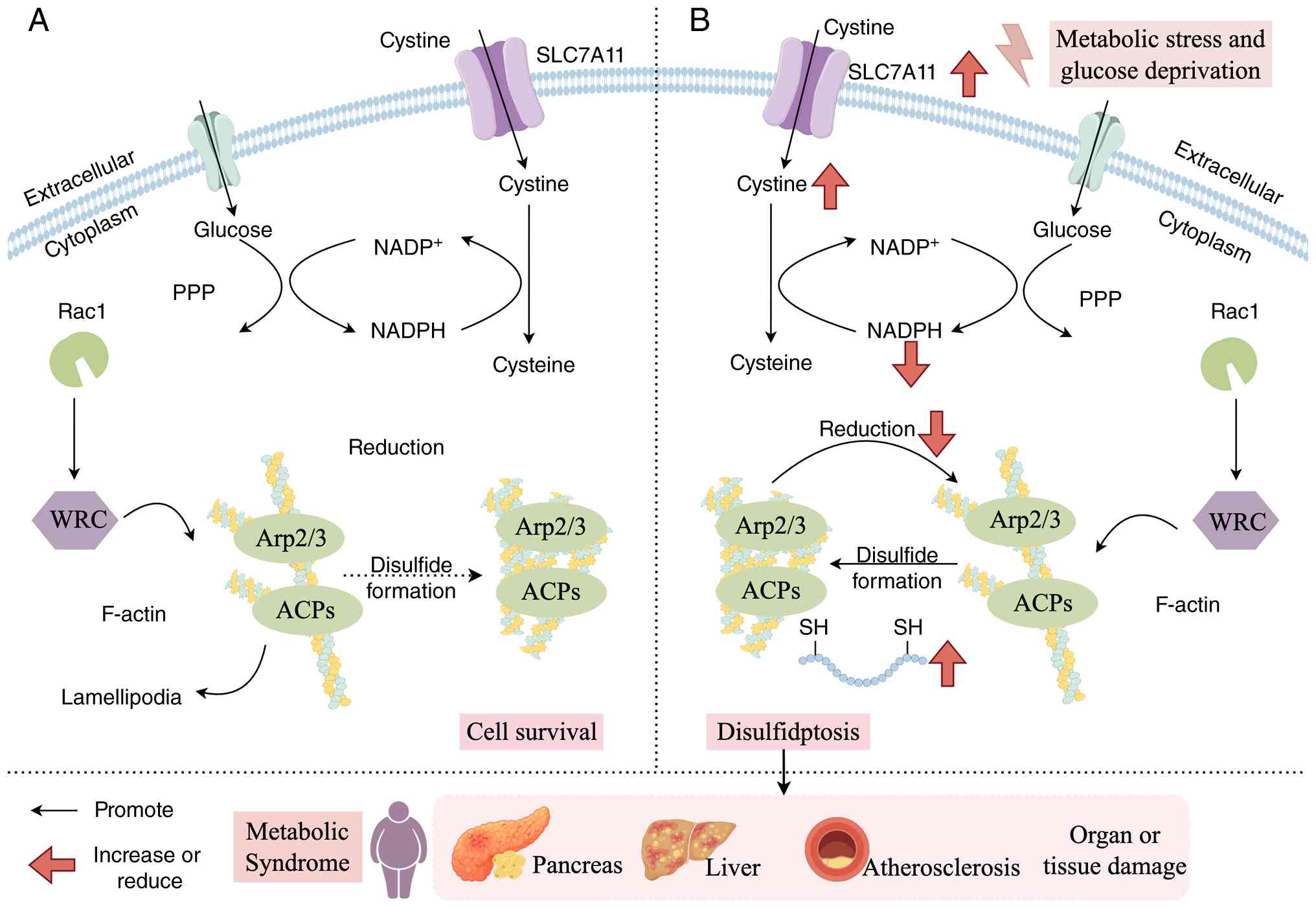 | Figure 7Schematic diagram of disulfidptosis
mechanisms in MetS. (A) Under normal conditions glucose metabolism
is balanced, the pentose phosphate pathway functions normally,
generating sufficient NADPH to maintain GSH levels and ensure
intracellular redox homeostasis. Cysteine entering cells via the
transporter SLC7A11 is effectively reduced and utilized, preserving
cytoskeletal stability. (B) During disulfidptosis, under the
high-glucose and insulin-resistant conditions of MetS, cellular
glucose metabolism becomes imbalanced. NADPH is extensively
depleted with insufficient regeneration, leading to GSH exhaustion.
Despite continued SLC7A11-mediated cystine influx, the loss of
reducing capacity causes abnormal intracellular cystine
accumulation. Cystine then forms extensive abnormal disulfide bonds
with intracellular proteins, directly attacking and cross-linking
cytoskeletal proteins (such as actin). This triggers cytoskeletal
network disintegration, ultimately inducing cell death. This
mechanism drives metabolic vascular complications and tissue
fibrosis. The figure was created using Figdraw (www.figdraw.com). MetS, metabolic syndrome; SLC7A11,
solute carrier family 7 member 11; GSH, glutathione. |
In summary, emerging PCD pathways provide new
perspectives for understanding metabolic disorders. However, their
relevance to MetS remains at an early stage. Several key questions
remain unresolved: First, whether PANoptosome formation occurs in
metabolic tissues under physiological and pathological conditions;
second, whether disulfidptosis represents a genuine metabolic
disease-associated death mechanism or merely a cellular stress
response; and third, how these emerging pathways interact with
established PCD programs during disease progression. The key
characteristics, regulatory molecules, biological consequences and
representative evidence for the major PCD pathways involved in MetS
are summarized in Table I
(20,43,57,85,107,120,127).
 | Table IBiochemical characteristics of cell
death pathways, key regulatory factors, pathogenesis in MetS,
representative tissues, evidence source and evidence level in
MetS. |
Table I
Biochemical characteristics of cell
death pathways, key regulatory factors, pathogenesis in MetS,
representative tissues, evidence source and evidence level in
MetS.
| Mode of cell
death | Biochemical
features | Key regulatory
factors | Pathogenesis in
MetS | Representative
tissues | Evidence
source | Evidence level | (Refs.) |
|---|
| Apoptosis | Release cytochrome
c, activate pro-apoptotic proteins of the Bcl-2 family to form
apoptosomes, initiate the caspase processing cascade, leading to
DNA fragmentation | 1. Caspase-8, -9,
-3
2. Bcl-2 family proteins
3. p53
4. MAPK signaling pathway | 1. Reduction in
pancreatic β-cells
2. Endothelial cell dysfunction
3. Adipose tissue remodeling | Pancreas, adipose
tissue, liver | Human studies and
animal models | A/B | (20) |
| Pyroptosis | Caspase-dependent
cleavage of gasdermin triggers inflammation and releases IL-18 and
IL-1β | 1. NLRP3,
caspase-1/4/5/11
2. GSDMD
3. IL-1β, IL-18 | 1. The core link
connecting metabolic inflammation
2. Insulin resistance
3. Adipose tissue inflammation | Liver, muscle,
adipose tissue | Animal models and
mechanistic studies | B | (43) |
| Autophagy | Increased lysosomal
activity, degradation of p62 protein, autophagosome formation, no
inflammatory response | 1. ATG
2. mTOR pathway (inhibits autophagy), AMPK pathway (activates
autophagy)
3. Beclin-1, LC3-II | 1. Protective:
Basal levels of autophagy maintain β-cell function and insulin
sensitivity
2. Destructive: Excessive autophagy activation or dysfunction
contributes to β-cell exhaustion and tissue damage | Liver,
cardiovascular system | Cell/animal
studies | A/B | (57) |
| Ferroptosis | Intracellular
Fe2+ levels increase, generating large amounts of ROS,
depleting GSH, and reducing GPX4 activity, leading to the
accumulation of lipid peroxides | 1. GPX4, SLC7A11,
GSH, Nrf2
2. ACSL4, free iron, LOXs | 1. Lipid
peroxidation-driven
2. Liver injury
3. Pancreatic β-cell damage
4. Cardiomyopathy | Adipose tissue,
liver | Animal models | B | (85) |
| Necroptosis | Decreased ATP
levels activate RIP1 and RIP3, and MLKL releases DAMP, triggering a
secondary inflammatory response | 1. RIPK1, RIPK3,
MLKL
2. Caspase-8 | 1. Exacerbate
tissue inflammation
2. Damage insulin target organs
3. Promote atherosclerosis | Adipose tissue,
liver, pancreas | Animal and human
tissue | C | (107) |
| PANoptosis | Activation of
caspase-8 cleaves GSDMD/GSDME during pyroptosis; activation of MLKL
perforates the plasma membrane during necroptosis; activation of
caspase-3/7 cleaves substrates during apoptosis | 1. RIPK1,
RIPK3
2. Caspase-8
3. Caspase-3/7
4. MLKL
5. GSDME | This phenomenon may
occur when multiple pathways such as apoptosis and necroptosis are
simultaneously activated within the complex stress environment of
MetS | Immune cells,
metabolic tissues | Preclinical | D | (120) |
| Disulfidptosis | Glucose deprivation
induces high expression of SLC7A11, leading to excessive cystine
uptake by cells. This depletes NADPH and forms numerous abnormal
disulfide bonds within cytoskeletal proteins such as actin.
Consequently, the cytoskeletal network undergoes contraction,
aggregation, and fragmentation | 1. NADPH
levels
2. SLC7A11
3. Actin cytoskeletal proteins
4. Oxidative-reductive imbalance | Glucose-deprivation
stress | Unknown/under
investigation | Limited
studies | D | (127) |
Molecular crosstalk and network regulation
among PCD pathways in MetS
In MetS, the interaction between distinct cell death
pathways forms a highly intricate and dynamically regulated
network. This crosstalk markedly amplifies tissue inflammation and
damage, functioning as a crucial mechanism driving the progression
of MetS toward end-organ complications. Different PCD pathways
share common upstream signals, regulatory molecules and cellular
stress responses, forming a complex cell death regulatory network
that determines cell fate under metabolic stress conditions.
Importantly, the biological outcome of PCD activation is highly
context-dependent. The predominant cell death pathway varies
depending on tissue type, metabolic environment, disease stage and
stress intensity. Furthermore, inhibition of one PCD pathway may
induce compensatory activation of alternative cell death pathways,
thereby limiting the therapeutic efficacy of single-pathway
interventions. Therefore, understanding the molecular crosstalk and
hierarchical regulation among PCD pathways is essential for
developing more precise therapeutic strategies for MetS.
Shared molecular nodes controlling the
transition among PCD pathways
RIPK1/RIPK3 signaling as a molecular
switch
The RIPK1/RIPK3 signaling axis represents one of the
best-characterized molecular switches regulating the transition
between different forms of PCD. Under physiological conditions,
activation of death receptors such as TNFR1 can induce apoptosis
through the activation of caspase-8 (130). However, when caspase-8 activity
is inhibited or impaired, RIPK1 interacts with RIPK3 to form the
necrosome complex, resulting in RIPK3-mediated phosphorylation of
MLKL and subsequent necroptosis (131). In MetS, chronic inflammatory
signals and metabolic stress may disrupt the balance between
apoptosis and necroptosis. For example, obesity-associated
inflammatory cytokines and excessive FFAs can activate TNF-α
signaling, thereby enhancing RIPK1/RIPK3 signaling in adipose
tissue and liver (132).
Although apoptosis may initially function as a controlled mechanism
for eliminating damaged cells, persistent metabolic stress may
shift cell fate from apoptosis toward necroptosis, resulting in
membrane rupture and DAMP release, thereby amplifying metabolic
inflammation (133). Moreover,
a previous study suggested that RIPK1/RIPK3 signaling may interact
with inflammasome activation, thereby establishing a potential link
between necroptosis and pyroptosis (134). Therefore, the RIPK1/RIPK3
signaling axis does not simply represent a necroptotic pathway but
functions as a molecular switch that integrates inflammatory and
metabolic signals to determine cell fate.
NLRP3 inflammasome as a convergence
hub
The NLRP3 inflammasome represents another critical
regulatory hub linking metabolic stress to inflammatory PCD. In
response to metabolic stimuli and cellular stress signals,
including saturated fatty acids, CCs, mitochondrial ROS and ERS,
NLRP3 activation promotes caspase-1 cleavage and subsequent
GSDMD-mediated pyroptosis (135). However, the role of NLRP3
extends beyond pyroptosis induction. Inflammasome activation can
also influence other PCD pathways through inflammatory
amplification, mitochondrial dysfunction and metabolic stress
responses. For example, excessive NLRP3 activation promotes IL-1β
and IL-18 secretion, which further aggravates IR, hepatic
inflammation and adipose tissue dysfunction (136). Meanwhile, metabolic
stress-induced mitochondrial damage enhances ROS production,
creating a positive feedback loop among mitochondrial dysfunction,
pyroptosis and other forms of cell death. In adipose tissue,
NLRP3-mediated macrophage pyroptosis contributes to chronic
inflammation and IR (137). In
the liver, Kupffer cell pyroptosis promotes inflammatory responses
and accelerates steatohepatitis progression (29). In pancreatic β-cells,
inflammasome activation contributes to inflammatory injury and
impaired insulin secretion (31). These findings indicate that
pyroptosis functions as an important inflammatory amplifier within
the broader PCD network.
Mitochondria as a central regulator
linking apoptosis, autophagy and ferroptosis
Mitochondria serve as a central regulatory platform
connecting multiple PCD pathways in MetS. As metabolic organs are
highly dependent on mitochondrial function, nutrient overload and
lipid accumulation can induce mitochondrial dysfunction,
characterized by impaired oxidative phosphorylation, excessive ROS
generation, mitochondrial membrane potential disruption and
defective mitochondrial quality control (138,139). Mitochondrial dysfunction
directly activates apoptosis through cytochrome c release
and subsequent activation of the caspase cascade (140). Meanwhile, defective mitophagy
impairs mitochondrial clearance, resulting in the accumulation of
damaged mitochondria and further enhancing oxidative stress and
inflammatory signaling (141).
Thus, mitophagy dysfunction may increase cellular susceptibility to
apoptosis and other stress-induced death pathways. In addition,
mitochondria-mediated oxidative imbalance contributes to
ferroptosis by promoting iron-dependent lipid peroxidation
(142). Excessive mitochondrial
ROS production facilitates the oxidation of polyunsaturated fatty
acids and compromises antioxidant defense systems, particularly the
GPX4-dependent pathway (143).
Therefore, mitochondrial homeostasis represents a critical
determinant of whether cells undergo adaptive survival responses or
irreversible death under metabolic stress.
GPX4/SLC7A11 axis connecting
ferroptosis and disulfidptosis
Redox regulation represents another important
regulatory interface between ferroptosis and the emerging
disulfidptosis pathway. Ferroptosis is primarily driven by
iron-dependent lipid peroxidation, whereas disulfidptosis is
associated with abnormal disulfide stress caused by impaired
cystine metabolism. The SLC7A11/GSH system represents a shared
regulatory axis connecting these pathways. SLC7A11 promotes cystine
uptake, maintaining intracellular GSH synthesis and supporting GPX4
activity, thereby preventing the accumulation of lipid peroxides
and ferroptotic cell death (144,145). Under conditions of severe
metabolic stress, particularly glucose deprivation, impaired
cystine utilization may disrupt redox homeostasis, leading to
excessive disulfide accumulation and cytoskeletal collapse
(146). Although direct
evidence linking disulfidptosis to MetS remains limited, the
metabolic regulation of SLC7A11 suggests potential interactions
between ferroptosis and newly recognized forms of PCD. Further
studies are required to determine whether modulation of this axis
contributes to metabolic disease progression.
Tissue-specific dominance and
compensation of PCD pathways in MetS
Although multiple PCD pathways coexist within
metabolic tissues, their relative contributions vary according to
tissue characteristics and disease stage. Therapeutic inhibition of
one pathway may result in compensatory activation of alternative
cell death pathways. Therefore, identifying dominant PCD patterns
in different organs is essential for understanding MetS
pathogenesis and designing targeted interventions. Adipose tissue
dysfunction is a central feature of obesity-associated MetS. Under
conditions of excessive nutrient accumulation, adipocyte
hypertrophy induces hypoxia, ERS and mitochondrial dysfunction,
resulting in increased apoptosis (29). Dying adipocytes release
inflammatory signals that recruit macrophages and promote chronic
adipose inflammation. Meanwhile, macrophage activation stimulates
NLRP3 inflammasome formation and pyroptosis, leading to enhanced
production of IL-1β and IL-18 (51). Thus, apoptosis and pyroptosis
cooperate to establish an inflammatory microenvironment that
promotes IR.
The liver is one of the major target organs
affected by MetS. In MASLD, excessive lipid accumulation induces
mitochondrial dysfunction, oxidative stress and iron imbalance
(147). Although hepatocyte
apoptosis contributes to liver injury, increasing evidence suggests
that ferroptosis serves a critical role in lipid
peroxidation-associated hepatic damage. Impaired antioxidant
defense, particularly reduced GPX4 activity, increases
susceptibility to ferroptosis (148). Meanwhile, defective mitophagy
further aggravates mitochondrial injury and lipid accumulation,
creating a vicious cycle among mitophagy impairment, ferroptosis
and inflammation.
Pancreatic β-cells are particularly vulnerable to
metabolic stress due to their limited antioxidant capacity. Chronic
hyperglycemia, inflammatory cytokines and ERS induce β-cell
apoptosis, leading to progressive loss of insulin-producing cells
(30). In addition, inflammasome
activation promotes β-cell pyroptosis, further impairing insulin
secretion. Therefore, apoptosis and pyroptosis represent
complementary mechanisms contributing to pancreatic dysfunction in
MetS. Vascular endothelial dysfunction is a major complication of
MetS. Hyperglycemia, dyslipidemia and oxidative stress impair
endothelial mitochondrial function and activate apoptotic pathways
(149). Emerging evidence also
suggests that ferroptosis contributes to endothelial lipid
peroxidation and vascular inflammation (150). Therefore, endothelial PCD
regulation may represent an important therapeutic target for
preventing cardiovascular complications associated with MetS.
Therapeutic importance of targeting
multiple PCD pathways
Due to the extensive crosstalk and compensatory
mechanisms among PCD pathways, targeting a single cell death
modality may offer limited therapeutic efficacy. For example,
inhibition of pyroptosis may reduce inflammatory injury but cannot
fully ameliorate mitochondrial dysfunction or oxidative stress.
Similarly, ferroptosis inhibition may protect hepatocytes from
lipid peroxidation-induced injury but may not completely resolve
metabolic inflammation. Therefore, future therapeutic strategies
should consider coordinated modulation of multiple PCD pathways
rather than selective targeting of individual death programs.
Pharmacological approaches targeting common upstream events, such
as mitochondrial dysfunction, oxidative stress, inflammasome
activation and metabolic dysregulation, may provide broader
protective effects. For instance, AMPK activation can
simultaneously enhance autophagy, improve mitochondrial function
and suppress inflammatory signaling. Similarly, modulation of Nrf2
signaling may simultaneously enhance antioxidant defense, regulate
ferroptosis susceptibility and modulate inflammatory responses.
However, multi-target regulation also presents challenges,
including tissue specificity, potential interference with
physiological cell death processes, and difficulties in determining
optimal timing and dosage. Future studies integrating single-cell
sequencing, multi-omics approaches and data from human clinical
cohorts are needed to identify precise PCD regulatory networks and
develop individualized therapeutic strategies for MetS.
Research on novel regulatory mechanisms in
MetS
In MetS, epigenetic modifications, exosomes, and
PCD form a dynamic and mutually regulated network that drives
chronic inflammation and tissue injury. The high-glucose, high-fat,
inflammatory environment associated with MetS induces epigenetic
alterations, affecting the expression of genes involved in PCD.
These epigenetic changes can be encapsulated into exosomes,
released into the circulation and subsequently delivered to distant
tissues. Exosomes transfer these aberrant signals, altering PCD
processes in recipient cells, thus exacerbating metabolic
dysfunction and organ damage (151). Therefore, targeting epigenetic
mechanisms or using engineered exosomes to modulate PCD represents
an innovative strategy for disrupting this detrimental cycle.
Epigenetic regulation of PCD in MetS
Epigenetic modifications precisely control the
initiation and execution of PCD by regulating gene expression via
DNA methylation, histone modifications and non-coding RNAs, without
altering the underlying DNA sequence (152). Concurrently, epigenetic
mechanisms regulate autophagy during nutritional excess or
deficiency. A previous study revealed that coactivator-associated
arginine methyltransferase 1-mediated methylation of histone
arginine residues critically regulates autophagy, particularly
activating autophagic processes during starvation (153). Experimental evidence indicates
that obesity-induced epigenetic changes in adipocytes establish
cellular 'memory', increasing susceptibility to weight regain
following caloric restriction and activating hepatic inflammatory
pathways (154).
Hypermethylation at cg02814054 promotes obesity by reducing
microtubule associated serine/threonine kinase 3 expression,
whereas hypomethylation at cg06028605 elevates obesity risk by
downregulating SLC5A11 (155).
Despite substantial evidence linking epigenetic modifications to
obesity onset and progression, mechanisms governing epigenetic
regulation of autophagy in response to drastic nutritional changes
remain unclear. Laha et al (156) demonstrated that homocysteine
modifies the epigenome, promoting lipogenesis and impairing fatty
acid metabolism through Zfp407-mediated pyroptosis and peroxisome
proliferator-activated receptor γ-dependent pathways, contributing
notably to obesity and related metabolic dysfunctions. Extensive
research has also examined epigenetic abnormalities, including DNA
methylation, histone modifications and miRNAs, in type 1 diabetes.
DNA methylation and miRNAs are proposed biomarkers predicting
pancreatic β-cell death (157).
Abnormal histone acetylation and deacetylation mechanisms are
involved in hypertension; A previous study confirmed that elevated
histone H3 acetylation associates with primary hypertension
progression (158).
Exosome-mediated regulation of PCD in
MetS
Exosomes are nanoscale extracellular vesicles
secreted by cells, carrying proteins, lipids, DNA and abundant
non-coding RNAs. They function as crucial mediators of inter-organ
communication (159). DNA
methyltransferases or histone-modifying enzymes carried by exosomes
can enter the nucleus of recipient cells, directly altering the
epigenetic state of genes associated with PCD. Under MetS
conditions, the composition of exosomes secreted by adipose tissue,
liver and muscle cells undergoes notable alterations. Persistent
hyperglycemia activates monocytes/macrophages, causing an imbalance
in M1/M2 polarization. These activated cells release exosomes
enriched with miR-30a, interacting with renal tubular epithelial
cells to activate the transient receptor potential canonical
6-calcineurin A-nuclear factor of activated T cells pathway, thus
triggering PCD (160).
Adipocyte-derived exosomes in MetS contain elevated
pro-inflammatory miRNAs (miR-27a and miR-155) and pro-PCD miRNAs,
whereas protective miRNAs (miR-30a and miR-124) are decreased. For
instance, a previous study demonstrated that extracellular
adipocyte-derived miR-1224 suppresses M2 macrophage polarization in
obesity-induced inflammation via the Musashi RNA-binding protein
2-regulated Wnt/β-catenin signaling axis (161). Additionally,
exosome-transported long non-coding RNAs (lncRNAs) and circular
RNAs function as competitive endogenous RNAs (ceRNAs) by
sequestering miRNAs in recipient cells, thus releasing their
suppression of PCD-related gene expression. For instance, lncRNAs
from adipocyte-derived exosomes are taken up by hepatocytes,
promoting pyroptosis and hepatic steatosis through the
miR-34a/sirtuin 1 pathway. Exosomes may also carry inflammasome
components (such as NLRP3) or DAMPs, directly seeding inflammation
and pyroptosis in recipient cells. Xiong et al (162) reported that astragaloside IV
inhibits diabetic endothelial cell pyroptosis by enhancing exosome
release from endothelial progenitor cells and modulating
PI3KR2/SPRED1 signaling. Yan et al (163) demonstrated that a high-fat diet
reduces adipocyte AMPKα1 activity, decreasing exosome secretion and
inhibiting NAFLD progression both in vivo and in
vitro, highlighting the potential of blocking exosome release
from white adipose tissue as a novel therapeutic approach for
NAFLD.
Therapeutic strategies targeting PCD: From
experimental evidence to clinical translation
Pharmacological modulation
Due to the contribution of multiple PCD pathways to
MetS progression, pharmacological modulation of specific cell death
pathways has emerged as a potential therapeutic strategy. Several
small-molecule regulators targeting apoptosis, pyroptosis,
ferroptosis, necroptosis and autophagy-associated pathways have
demonstrated protective effects in experimental models of metabolic
diseases. However, most evidence is derived from preclinical
studies, and clinical translation remains challenging. Apoptosis is
the most extensively studied form of PCD. The serine protease
inhibitor camostat mesilate was shown to alleviate hypertension and
renal injury in MetS rats by inhibiting podocyte apoptosis and
reducing podocyte injury through both dependent and independent
mechanisms (164).
Methyl-β-cyclodextrin, a cyclic oligosaccharide commonly used to
lower membrane cholesterol, induces apoptosis in mesenchymal
stromal cells, improves glucose transport and endoplasmic reticulum
function, enhances antioxidant capacity, and effectively treats
metabolic disorders such as IR, MetS and diabetes (165). Epigallocatechin-3-gallate
(EGCG), a compound extracted from green tea known for its
antioxidant and anti-inflammatory effects, alleviates bladder
apoptosis and ovarian hormone deficiency in rat models of MetS and
ovarian dysfunction (166).
Strategies targeting pyroptosis have mainly focused on inhibiting
inflammasome activation or gasdermin-mediated membrane pore
formation. MCC950 is a selective NLRP3 inflammasome inhibitor that
exerts anti-inflammatory effects in experimental models of obesity,
IR and MASLD by reducing caspase-1 activation and IL-1β production;
however, caution is warranted due to the reported hepatotoxicity of
MCC950 (55). Disulfiram,
traditionally used for alcohol dependence, has been reported to
inhibit GSDMD pore formation and suppress pyroptosis-related
inflammatory responses in preclinical studies (167,168). However, the safety profile and
long-term efficacy of inflammasome inhibition require further
evaluation because inflammasomes also contribute to host defense
against infection. Exercise can notably enhance metabolic health by
suppressing inflammatory pyroptosis, representing a novel
therapeutic target for chronic disorders such as age-related
obesity, muscle atrophy and IR/T2DM (169). Therefore, exercise constitutes
a promising strategy to inhibit pyroptosis, reduce inflammation and
promote metabolic health.
Unlike numerous classical PCD pathways, autophagy
exhibits a dual role in MetS, and therapeutic modulation requires
careful consideration of disease stage and tissue context.
Rapamycin-induced autophagy activation has demonstrated beneficial
effects in experimental models by improving mitochondrial quality
control and lipid metabolism (79). Conversely, excessive inhibition
of autophagy may aggravate metabolic dysfunction, highlighting the
importance of precise regulation rather than indiscriminate
activation or inhibition. Peng et al (170) demonstrated that EGCG inhibits
obesity and glucose intolerance induced by a high-fat diet in
C57BL/6 mice by modulating autophagy and lipolysis. Furthermore,
compounds including luteolin (171), berberine (172), ferulic acid (173), MSI-1436 (174), elamipretide (175) and melatonin (176) exhibit therapeutic potential in
MetS by regulating autophagy and ferroptosis pathways. Lim et
al (177) identified a
small molecule named MSL through chemical library screening, which
enhances autophagic flux and improves metabolic profiles in obese
and MetS mice, thus representing a promising therapeutic candidate.
Drugs such as memantine (178)
and moxonidine (179) have
shown beneficial effects in MetS model rats by modulating
autophagy. Cai (180) conducted
a randomized controlled trial involving 84 patients with MetS who
were classified according to the Traditional Chinese Medicine (TCM)
syndrome differentiation framework as having Qi stagnation and
phlegm-stasis syndrome, a TCM diagnostic pattern characterized by
impaired Qi circulation and the accumulation of phlegm and blood
stasis. Participants were randomized into a control group receiving
conventional Western medical treatment and a treatment group
receiving conventional treatment combined with Shugan Wendan
Decoction, a traditional Chinese herbal formula. After 12 weeks,
the results indicated that Shugan Wendan Decoction improves IR,
glucose and lipid metabolism, and oxidative stress in patients with
MetS by upregulating liver X receptor α and mediating
autophagy/apoptosis pathways. Additionally, the PTP1B inhibitor
MSI-1436 has entered Phase 1 and 1b clinical trials, although
currently only for obesity and T2DM treatment (NCT00606112, 2008;
NCT00806338, 2009) (174).
Regarding non-pharmacological interventions, a previous study
demonstrated that intermittent fasting and ketogenic diets enhance
insulin sensitivity, promote autophagy, reduce inflammation and
improve metabolic indicators in patients with MetS and T2DM,
including reductions in HbA1c, fasting blood glucose, body weight
and TGs (181). Additionally,
chronic intermittent hypobaric hypoxia protects vascular
endothelium by modulating autophagy in MetS rat models (182).
Ferroptosis has attracted increasing attention as a
potential therapeutic target for metabolic liver diseases due to
its close association with lipid peroxidation and oxidative stress.
Ferrostatin-1, a classical ferroptosis inhibitor, suppresses lipid
peroxidation by preventing iron-dependent oxidative damage
(183). In cellular and animal
models of metabolic liver injury, inhibition of ferroptosis has
been shown to preserve hepatocyte viability, reduce oxidative
stress and alleviate inflammatory responses (184). Similar protective effects have
been reported for liproxstatin-1, another ferroptosis inhibitor,
particularly in experimental models of hepatic injury (185). Nevertheless, whether
ferroptosis inhibition provides long-term metabolic benefits
remains uncertain because ferroptosis may also participate in
physiological iron metabolism and immune regulation. In addition,
although GPX4 activators can alleviate ferroptosis by inhibiting
lipid peroxidation, they also exhibit limited target specificity.
Therapeutic inhibition of necroptosis has mainly focused on
targeting the RIPK1/RIPK3/MLKL signaling axis. Necrostatin-1
(Nec-1), a selective RIPK1 inhibitor, has been widely used as a
pharmacological tool to suppress necroptosis. In experimental
models of metabolic disorders, inhibition of RIPK1 signaling has
been shown to reduce inflammatory responses, improve insulin
sensitivity and attenuate tissue injury (186). For example, Nec-1 treatment
reduced inflammatory signaling activation and cell death in models
of obesity-associated metabolic dysfunction and hepatic injury
(187). However, the
application of Nec-1 in clinical settings is limited by concerns
regarding target specificity, pharmacokinetic properties and the
physiological roles of necroptosis in immune regulation. Additional
compounds, including orientin (188), dihydroquercetin (189), clofibrate (190), astaxanthin (191), longan flower water extract
(192),
5-azacytidine/resveratrol (193) and clomiphene (194), have also been investigated for
their potential effects on PCD-related processes in experimental
models. Representative compounds and drugs targeting PCD pathways
for the treatment of MetS are summarized in Table II (164-166,170-176,179,180,188-197).
| Table IIRepresentative compounds/drugs
targeting programmed cell death for MetS treatment. |
Table II
Representative compounds/drugs
targeting programmed cell death for MetS treatment.
|
Compounds/drugs | Research model | Target
molecule | Mechanism | (Refs.) |
|---|
| Camostat
mesilate | MetS model
rats | TG ↓, TC ↓,
Bax/Bcl-2 ↓, pro-caspase-3 ↓ | Apoptosis ↓ | (164) |
|
Methyl-β-cyclodextrin | Horse MetS
model | ROS ↓, SOD ↑, iNOS
↑, Bcl-2 ↓, p53 ↑, Bax ↑ | Apoptosis ↑ | (165) |
|
Epigallocatechin-3-gallate | MetS model
rats | Apoptotic cells ↓,
Bcl-2 ↑, GRP78 ↓, CHOP ↓, caspase-12 ↓ | Apoptosis ↓ | (166) |
| MetS mouse
model | PPARγ ↓, C/EBPα ↓,
FASN ↓, HSL ↑, ATGL ↑, LC3 ↓, p62 ↓ | Autophagy ↑ | (170) |
| Luteolin | Rat model of
MetS-induced cardiac injury | BCL-2 ↓, CASP-3 ↑,
CASP-9 ↑, NR4A2 ↓, p53 ↓, IL-6 ↓, IL-1β ↓, TNF-α ↓ | Apoptosis ↓,
autophagy ↑ | (171) |
| Berberine | Rat model of MetS
combined with carotid stenosis |
Wnt/GSK-3β/β-catenin pathway activity
↓ | Apoptosis ↓ | (172) |
| Ferulic acid | MetS hepatocyte
model | MMP ↓, ROS ↓, ATP
↑, AMPK ↑, SIRT1 ↑ | Autophagy ↑ | (173) |
| MSI-1436 | Horse MetS
model | IL-1β ↓, TNF-α ↓,
TGF-β ↓, hs70 ↑, Beclin-1 ↑, MFN2 ↑, PINK1 ↑, CHOP ↓, ATF6 ↓, HSPA5
↓, XBP1 ↓, LC3A ↑, LC3B-I ↓, LC3B-II ↓ | Autophagy ↑ | (174) |
| Elamipretide | MetS pig kidney
cell model | p62 ↑, ATG5-12 ↑,
mTOR ↑, AMPK ↑, ULK-1 ↑, cytochrome c ↑ | Autophagy ↑,
apoptosis ↓ | (175) |
| Melatonin | MetS hamster
model | mTOR ↑, LC3-II ↑,
LC3-II/LC3-I ↑, Beclin1 ↑ | Autophagy ↑ | (176) |
| Moxonidine | MetS model
rats | IL-6 ↓, c-TnI ↓,
caspase-3 ↓, TGF-β1 ↓, MMP-9 ↓, NR4A2 ↓, p53 ↑, LC3A/B ↑, Beclin-1
↑, SQSTM1 ↓, p62 ↓ | Autophagy ↑,
apoptosis ↓ | (179) |
| Shugan Wendan
decoction | Patients with
MetS | FPG ↓, HbA1c ↓,
FINS ↓, HOMA-IR ↓, TC ↓, TG ↓, LDL-C ↓, HOMA-IR ↓, MDA ↓, HDL-C ↑,
SOD ↑, GSH-PX ↑ | Autophagy ↑,
apoptosis ↓ | (180) |
| Orientin | Horse MetS
model | Bax/Bcl-2 ↓, p21 ↓,
TP53, ROS ↓, SOD1 ↑ | Apoptosis ↑ | (188) |
|
Dihydroquercetin | MetS model
rats | GSH-Px ↑, GSH ↑,
TNF-α ↓, IL-1β ↓, BDNF ↑, ROS, apoptotic cells ↓ | Apoptosis ↓ | (189) |
| Clofibrate | MetS model
rats | IL-1β ↓, IL-6 ↓,
TNF-α ↓, MMP-2 ↓, apoptotic cells ↓ | Apoptosis ↓ | (190) |
| Astaxanthin | Horse MetS
model | p21 ↓, p53 ↓, Bax
↓, caspase-3 ↓, caspase-8 ↓, caspase-9 ↓, Bcl-2 ↑, SOD1 ↑, SOD2
↑ | Apoptosis ↓ | (191) |
| Longan flower water
extract | MetS model
rats | Fas ↓, FADD ↓,
caspase-8 ↓, caspase-3 ↓, Bax ↓, Bak ↓, Bax/Bcl-2 ↓, Bak/Bcl-xL ↓,
IGF-1R ↑, Bcl-2 ↑ | Apoptosis ↓ | (192) |
| 5-azacytidine,
resveratrol | Horse MetS
model | p21 ↓, Bax ↑, Bcl-2
↑, Beclin-3 ↓, LC3 ↓, LAMP-2 ↑, MMP ↑, SOD ↑, p62 ↑, LAMP-2 ↑, mTOR
↓ | Autophagy ↑ | (193) |
| Clomiphene
citrate | MetS mouse
model | LC3-II ↑, p62 ↑,
LAMP2 ↑, CTSB ↑, CTSD ↑, ATGL ↑, HSL ↑, MGL ↑, LAL ↑ | Autophagy ↑ | (194) |
| Pioglitazone | MetS model
rats | Apoptotic cells ↓,
p62 ↑, LC3 ↑ | Autophagy ↑,
apoptosis ↓ | (195) |
| Chaiqi
decoction | MetS model
rats | LC3 ↑, LC3II/I ↑,
Beclin-1 ↑ | Autophagy ↑ | (196) |
| Metformin | MetS mouse
model | ROS ↓, Nrf2 ↑,
SLC7A11 ↑, GPX4 ↑, MDA ↓, GSH ↓ | Ferroptosis ↓ | (197) |
Current clinical status and challenges
for translation
Due to the extensive crosstalk among PCD pathways,
simultaneous modulation of multiple cell death programs may provide
superior therapeutic effects compared with single-pathway
inhibition. Targeting PCD represents a promising therapeutic
strategy for MetS. However, translating mechanistic discoveries
into clinical applications remains challenging. Current therapeutic
strategies are mainly supported by preclinical studies, with most
approaches focusing on pharmacological modulation of specific PCD
pathways. Therefore, beyond the identification of potential
therapeutic agents, it is essential to critically evaluate the
translational barriers, including target specificity, tissue
selectivity, biomarker availability and long-term safety. Despite
promising preclinical findings, no PCD-targeting agent has yet been
approved specifically for the treatment of MetS. Most available
evidence is derived from cellular and animal studies, and several
barriers limit clinical translation: i) Limited clinical evidence:
Although several metabolic interventions indirectly influence PCD
pathways, including lifestyle interventions and existing metabolic
therapies, direct clinical trials targeting specific PCD pathways
in MetS or MASLD remain scarce; ii) off-target effects: Numerous
PCD regulators participate in physiological processes beyond cell
death regulation; for example, inhibition of apoptosis or
inflammasome signaling may interfere with immune surveillance and
tissue homeostasis; iii) tissue specificity: A major challenge is
achieving tissue-specific modulation; a PCD pathway that is harmful
in one tissue may be protective in another, therefore systemic
inhibition may produce unpredictable outcomes; and iv) long-term
safety: Because PCD pathways are essential for normal development,
immune regulation and tissue renewal, long-term safety evaluation
is required before clinical application.
Conclusions and outlook
MetS is a complex metabolic disorder characterized
by a markedly high global prevalence. The primary drivers of MetS
include IR, chronic inflammation and adipose tissue dysfunction,
which collectively pose substantial challenges for effective
management. Current evidence indicates that PCD is not only a
consequence but also a critical pathological factor influencing the
onset and progression of MetS. Various forms of PCD interact with
metabolic tissue injury, such as in adipose tissue, liver,
pancreatic islets and blood vessels, triggering cascading reactions
leading ultimately to systemic metabolic dysfunction. Specifically,
apoptosis of pancreatic β-cells reduces insulin secretion, while
adipocyte apoptosis intensifies adipose dysfunction and promotes
pro-inflammatory adipokine release. Metabolic stress-induced
necroptosis exacerbates inflammation and strongly associates with
fatty liver disease and IR. Lipotoxicity, hyperglycemia and related
stressors activate the NLRP3 inflammasome, inducing pyroptosis and
amplifying chronic, low-grade inflammation in MetS. Ferroptosis,
marked by iron-dependent lipid peroxidation, correlates closely
with hepatocyte loss and endothelial injury in NASH. Autophagy
exerts protective functions in insulin-sensitive tissues, whereas
impaired autophagy accelerates metabolic deterioration. Recently
discovered mechanisms such as PANoptosis and disulfidptosis provide
novel insights into the complex pathophysiology of MetS.
Furthermore, innovative regulatory mechanisms involving epigenetics
and exosomes notably influence chronic inflammation and tissue
damage in MetS by modulating PCD, highlighting their potential as
therapeutic targets.
Although substantial progress has been achieved in
understanding the role of PCD in MetS, several fundamental
questions remain unresolved. First, the precise mechanisms
determining cell fate selection among different PCD pathways remain
unclear. Under metabolic stress, cells may undergo apoptosis,
pyroptosis, ferroptosis or interconnected forms of cell death;
however, the molecular switches controlling these transitions
require further investigation. Second, the tissue-specific
regulation of PCD networks remains poorly understood. Different
metabolic organs exhibit distinct metabolic environments and stress
responses, suggesting that universal PCD-targeting strategies may
not be optimal. Third, clinically applicable biomarkers for
monitoring PCD activity are urgently needed. Current assessment of
PCD activity mainly relies on experimental markers, whereas
circulating indicators capable of predicting disease progression
and therapeutic response remain limited. Fourth, future therapeutic
development should move beyond single-pathway interventions toward
precision regulation of PCD networks. Emerging approaches,
including targeted nanoparticles, tissue-specific delivery systems
and multi-omics-guided treatment strategies, may provide new
opportunities for personalized management of MetS. Fifth, the
current understanding of PCD in MetS is largely based on
experimental models, and differences in disease models, tissue
specificity and methodological approaches limit direct clinical
translation.
To address these limitations, future research
should focus on several key areas. At the mechanistic level,
multi-omics techniques, including single-cell sequencing, spatial
transcriptomics and proteomics, should be utilized to
systematically decipher tissue-specific activation patterns of
distinct forms of PCD across various MetS-related tissues. Such
analyses will clarify the complex interactions between PCD pathways
and factors such as metabolic reprogramming, gut microbiome
dysbiosis and epigenetic modifications, thus identifying crucial
regulatory molecules and signaling cascades. These insights will
enable more precise therapeutic targeting. Additionally, greater
emphasis should be placed on exploring the interactions between
different PCD subtypes, examining their synergistic or antagonistic
roles in disease progression, thereby revealing multidimensional
regulatory mechanisms driving MetS development. Regarding model
optimization, more clinically relevant models, such as gene-edited
animal models and patient-derived organoid systems, should be
developed to improve replication of human disease features. These
advanced models will provide robust platforms for validating
molecular mechanisms and screening potential therapeutics.
Furthermore, establishing a comprehensive clinical database of PCD
characteristics through multicenter cohort studies involving
patients with MetS will be beneficial. Integrating clinical
phenotypes with prognostic data will help clarify the clinical
relevance of PCD-related biomarkers. In the context of clinical
translation, accelerating the development and clinical trials of
PCD-targeted therapies should become a priority, emphasizing
combination treatment strategies to enhance therapeutic efficacy
and minimize adverse effects. Concurrently, research into
personalized treatments based on genetic backgrounds, metabolic
profiles and individual patterns of PCD activation should be
intensified. Precision medicine methodologies should be adopted to
identify populations that would most benefit, enabling targeted
prevention and intervention in MetS. Additionally, efforts should
focus on developing standardized, highly specific and sensitive
biomarker detection systems for PCD, facilitating their clinical
adoption for early diagnosis, monitoring therapeutic efficacy and
prognostic evaluation.
In summary, the importance of PCD in MetS is
becoming increasingly clear, and numerous therapeutic targets are
actively being investigated. Future studies should prioritize
elucidating intricate interactions among these pathways and
accelerating their translation into clinical practice. These steps
are crucial for developing precise, effective therapeutics for
MetS, ultimately reducing the substantial global burden posed by
this widespread health issue.
Availability of data and materials
Not applicable.
Authors' contributions
JZ and QC designed and conceptualized the article;
JZ and MZ prepared and wrote the article; YZ, CG and YD conducted
literature searching and initial reviewing; LS, DZ and QZ did the
study selection and assessment; QC reviewed the draft and provided
suggestions for revision. All authors read and approved the final
version of the manuscript. Data authentication is not
applicable.
Ethics approval and consent to
participate
Not applicable.
Patient consent for publication
Not applicable.
Competing interests
The authors declare that they have no competing
interests.
Abbreviations:
|
MetS
|
metabolic syndrome
|
|
IR
|
insulin resistance
|
|
T2DM
|
type 2 diabetes mellitus
|
|
PCD
|
programmed cell death
|
|
NAFLD
|
non-alcoholic fatty liver disease
|
|
RIPK
|
receptor-interacting protein
kinase
|
|
NASH
|
non-alcoholic steatohepatitis
|
|
IRS
|
insulin receptor substrates
|
|
PI3K
|
phosphoinositide 3-kinase
|
|
AKT
|
protein kinase B
|
|
MAPK
|
mitogen-activated protein kinase
|
|
FFA
|
free fatty acid
|
|
TNF-α
|
tumor necrosis factor-α
|
|
IL-6
|
interleukin-6
|
|
VAT
|
visceral adipose tissue
|
|
LPS
|
lipopolysaccharides
|
|
SCFAs
|
short-chain fatty acids
|
|
TRADD
|
TNFR-associated death domain
|
|
FADD
|
Fas-associated death domain
|
|
Bcl-2
|
B-cell lymphoma-2
|
|
Bax
|
Bcl-2-associated X protein
|
|
ERS
|
endoplasmic reticulum stress
|
|
UPR
|
unfolded protein response
|
|
ROS
|
reactive oxygen species
|
|
VSMCs
|
vascular smooth muscle cells
|
|
VECs
|
vascular endothelial cells
|
|
ox-LDL
|
oxidized low-density lipoprotein
|
|
cIAP
|
cellular inhibitor of apoptosis
protein
|
|
DAMPs
|
damage-associated molecular
patterns
|
|
IAPP
|
islet amyloid polypeptide
|
|
LDL
|
low-density lipoprotein
|
|
CCs
|
cholesterol crystals
|
|
PAMPs
|
pathogen-associated molecular
patterns
|
|
GSDMD
|
gasdermin D
|
|
AGEs
|
advanced glycation end products
|
|
AMPK
|
adenosine monophosphate-activated
protein kinase
|
|
mTOR
|
mammalian target of rapamycin
|
|
IGF
|
insulin-like growth factor
|
|
Atg
|
autophagy-related gene
|
|
LIP
|
labile iron pool
|
|
PUFAs
|
polyunsaturated fatty acids
|
|
LOXs
|
lipoxygenases
|
|
GPX4
|
glutathione peroxidase 4
|
|
GSH
|
glutathione; TG, triglyceride; MAFLD,
metabolic-associated fatty liver disease
|
Acknowledgements
Not applicable.
Funding
The present work was supported by the Noncommunicable Chronic
Diseases-National Science and Technology Major Project (grant no.
2023ZD0509400) and The National Natural Science Foundation of China
(grant no. 82574805).
References
|
1
|
Eckel RH, Grundy SM and Zimmet PZ: The
metabolic syndrome. Lancet. 365:1415–1428. 2005. View Article : Google Scholar : PubMed/NCBI
|
|
2
|
Liang X, Or B, Tsoi MF, Cheung CL and
Cheung BMY: Prevalence of metabolic syndrome in the United States
National health and nutrition examination survey 2011-18. Postgrad
Med J. 99:985–992. 2023. View Article : Google Scholar : PubMed/NCBI
|
|
3
|
Li Y, Zhao L, Yu D, Wang Z and Ding G:
Metabolic syndrome prevalence and its risk factors among adults in
China: A nationally representative cross-sectional study. PLoS One.
13:e01992932018. View Article : Google Scholar : PubMed/NCBI
|
|
4
|
Hu Y, Li W, Nie J, Zhang C, Zhou J, Hu Z,
Zhou H and Lei X: Association between the atherogenic index of
plasma and major adverse cardiovascular events in individuals with
metabolic syndrome: findings from the UK biobank. Cardiovasc
Diabetol. 24:4442025. View Article : Google Scholar : PubMed/NCBI
|
|
5
|
Kassi E, Pervanidou P, Kaltsas G and
Chrousos G: Metabolic syndrome: definitions and controversies. BMC
Med. 9:482011. View Article : Google Scholar : PubMed/NCBI
|
|
6
|
Hsu CN, Hou CY, Hsu WH and Tain YL:
Early-life origins of metabolic syndrome: Mechanisms and preventive
aspects. Int J Mol Sci. 22:118722021. View Article : Google Scholar : PubMed/NCBI
|
|
7
|
Wang M, Yu F, Zhang Y and Li P: Programmed
cell death in tumor immunity: Mechanistic insights and clinical
implications. Front Immunol. 14:13096352024. View Article : Google Scholar : PubMed/NCBI
|
|
8
|
Guicciardi ME, Malhi H, Mott JL and Gores
GJ: Apoptosis and necrosis in the liver. Compr Physiol. 3:977–1010.
2013. View Article : Google Scholar : PubMed/NCBI
|
|
9
|
Zhao X, Zhang H, Ding X, Wu H, Li M, Yin
N, Gao J, Xu Y, Wang R, Qi Z, et al: ATP6AP2 ameliorates
inflammation and pyroptosis in heart failure by promoting
lysosome-dependent STING degradation. Apoptosis. 31:2022026.
View Article : Google Scholar : PubMed/NCBI
|
|
10
|
Liu P, Anandhan A, Chen J, Shakya A,
Dodson M, Ooi A, Chapman E, White E, Garcia JG and Zhang DD:
Decreased autophagosome biogenesis, reduced NRF2, and enhanced
ferroptotic cell death are underlying molecular mechanisms of
non-alcoholic fatty liver disease. Redox Biol. 59:1025702023.
View Article : Google Scholar
|
|
11
|
Parums DV: Editorial: Review articles,
systematic reviews, meta-analysis, and the updated preferred
reporting items for systematic reviews and meta-analyses (PRISMA)
2020 Guidelines. Med Sci Monit. 27:e9344752021. View Article : Google Scholar : PubMed/NCBI
|
|
12
|
Gluvic Z, Zaric B, Resanovic I, Obradovic
M, Mitrovic A, Radak D and Isenovic ER: Link between metabolic
syndrome and insulin resistance. Curr Vasc Pharmacol. 15:30–39.
2017. View Article : Google Scholar
|
|
13
|
Ramasubbu K and Devi Rajeswari V:
Impairment of insulin signaling pathway PI3K/Akt/mTOR and insulin
resistance induced AGEs on diabetes mellitus and neurodegenerative
diseases: A perspective review. Mol Cell Biochem. 478:1307–1324.
2023. View Article : Google Scholar
|
|
14
|
Tripathy D, Mohanty P, Dhindsa S, Syed T,
Ghanim H, Aljada A and Dandona P: Elevation of free fatty acids
induces inflammation and impairs vascular reactivity in healthy
subjects. Diabetes. 52:2882–2887. 2003. View Article : Google Scholar : PubMed/NCBI
|
|
15
|
Murakami T, Michelagnoli S, Longhi R,
Gianfranceschi G, Pazzucconi F, Calabresi L, Sirtori CR and
Franceschini G: Triglycerides are major determinants of cholesterol
esterification/transfer and HDL remodeling in human plasma.
Arterioscler Thromb Vasc Biol. 15:1819–1828. 1995. View Article : Google Scholar : PubMed/NCBI
|
|
16
|
Esteve E, Ricart W and Fernandez-Real JM:
Adipocytokines and insulin resistance: The possible role of
lipocalin-2, retinol binding protein-4, and adiponectin. Diabetes
Care. 32(Suppl 2): S362–S367. 2009. View Article : Google Scholar : PubMed/NCBI
|
|
17
|
Bellingan GJ, Xu P, Cooksley H, Cauldwell
H, Shock A, Bottoms S, Haslett C, Mutsaers SE and Laurent GJ:
Adhesion molecule-dependent mechanisms regulate the rate of
macrophage clearance during the resolution of peritoneal
inflammation. J Exp Med. 196:1515–1521. 2002. View Article : Google Scholar : PubMed/NCBI
|
|
18
|
Burcelin R, Luche E, Serino M and Amar J:
The gut microbiota ecology: A new opportunity for the treatment of
metabolic diseases? Front Biosci (Landmark Ed). 14:5107–5117. 2009.
View Article : Google Scholar : PubMed/NCBI
|
|
19
|
Armougom F, Henry M, Vialettes B, Raccah D
and Raoult D: Monitoring bacterial community of human gut
microbiota reveals an increase in Lactobacillus in obese patients
and Methanogens in anorexic patients. PLoS One. 4:e71252009.
View Article : Google Scholar : PubMed/NCBI
|
|
20
|
Obeng E: Apoptosis (programmed cell death)
and its signals-A review. Braz J Biol. 81:1133–1143. 2020.
View Article : Google Scholar
|
|
21
|
Zhu M, Liu D, Liu G, Zhang M and Pan F:
Caspase-linked programmed cell death in prostate cancer: From
apoptosis, necroptosis, and pyroptosis to PANoptosis. Biomolecules.
13:17152023. View Article : Google Scholar : PubMed/NCBI
|
|
22
|
Rajesh Y and Kanneganti TD: Innate immune
cell death in neuroinflammation and Alzheimer's disease. Cells.
11:18852022. View Article : Google Scholar : PubMed/NCBI
|
|
23
|
Zhou Z, Arroum T, Luo X, Kang R, Lee YJ,
Tang D, Hüttemann M and Song X: Diverse functions of cytochrome c
in cell death and disease. Cell Death Differ. 31:387–404. 2024.
View Article : Google Scholar : PubMed/NCBI
|
|
24
|
Ma J, Liu Y, Valladolid-Acebes I,
Recio-López P, Peng G, Li J, Berggren PO, Juntti-Berggren L and
Tong N: ATF5 is a regulator of ER stress and β-cell apoptosis in
different mouse models of genetic- and diet-induced obesity and
diabetes mellitus. Cell Signal. 102:1105352023. View Article : Google Scholar
|
|
25
|
Liu Y, Yang Y, Xu C, Liu J, Chen J, Li G,
Huang B, Pan Y, Zhang Y, Wei Q, et al: Circular RNA circGlis3
protects against islet β-cell dysfunction and apoptosis in obesity.
Nat Commun. 14:3512023. View Article : Google Scholar
|
|
26
|
Liu Y, Li Q, Tian G, Zhou X, Chen P, Chen
B, Shan Z and Qi B: Neuronal PRDX-2-mediated ROS signaling
regulates food digestion via peripheral UPRmt activation. Nat
Commun. 15:105822024. View Article : Google Scholar
|
|
27
|
Uddandrao VVS, Eraniappan S, Balakrishnan
Ramajayam A, Singaravel S, Roy A, Parim BN, Ponnusamy C, Ganapathy
S, Ponnusamy P and Sasikumar V: Hydroxycitric acid and capsaicin
combination alleviates obesity-induced testicular apoptosis,
oxidative stress and inflammation. Syst Biol Reprod Med. 70:20–37.
2024. View Article : Google Scholar : PubMed/NCBI
|
|
28
|
Nelson-Dooley C, Della-Fera MA, Hamrick M
and Baile CA: Novel treatments for obesity and osteoporosis:
Targeting apoptotic pathways in adipocytes. Curr Med Chem.
12:2215–2225. 2005. View Article : Google Scholar : PubMed/NCBI
|
|
29
|
Jamalat Y, Gamallat Y, Jaceline Gislaine
PS, Meyiah A, Shopit A, Li H, Ahmed B, Chu P, Wang H, Li X, et al:
Phosphocreatine attenuates endoplasmic reticulum stress-mediated
hepatocellular apoptosis ameliorates insulin resistance in diabetes
model. Biochem Biophys Res Commun. 506:611–618. 2018. View Article : Google Scholar : PubMed/NCBI
|
|
30
|
Osmanlıoğlu HÖ and Nazıroğlu M:
Resveratrol modulates diabetes-induced neuropathic pain, apoptosis,
and oxidative neurotoxicity in mice through TRPV4 channel
inhibition. Mol Neurobiol. 61:7269–7286. 2024. View Article : Google Scholar
|
|
31
|
Guo Z, Wan X, Luo Y, Liang F, Jiang S,
Yuan X and Mo Z: The vicious circle of UHRF1 down-regulation and
KEAP1/NRF2/HO-1 pathway impairment promotes oxidative
stress-induced endothelial cell apoptosis in diabetes. Diabet Med.
40:e150262023. View Article : Google Scholar
|
|
32
|
Eizirik DL and Darville MI: Beta-cell
apoptosis and defense mechanisms: Lessons from type 1 diabetes.
Diabetes. 50(Suppl 1): S64–S69. 2001. View Article : Google Scholar : PubMed/NCBI
|
|
33
|
Intengan HD and Schiffrin EL: Vascular
remodeling in hypertension: Roles of apoptosis, inflammation, and
fibrosis. Hypertension. 38(3 Pt 2): 581–587. 2001. View Article : Google Scholar : PubMed/NCBI
|
|
34
|
Wang Q, Cui Y, Lin N and Pang S:
Correlation of cardiomyocyte apoptosis with duration of
hypertension, severity of hypertension and caspase-3 expression in
hypertensive rats. Exp Ther Med. 17:2741–2745. 2019.PubMed/NCBI
|
|
35
|
Zhang F, Guo Z, Wu M, Lin G, Chen H, Zheng
H, Zhang D, Jiang M, Xie Y, Chen Y, et al: Trifolin attenuates
hypertension-mediated cardiac injury by inhibiting cardiomyocyte
apoptosis: Mechanistic insights and therapeutic potential. Eur J
Pharmacol. 985:1771252024. View Article : Google Scholar : PubMed/NCBI
|
|
36
|
Sakao S, Tatsumi K and Voelkel NF:
Endothelial cells and pulmonary arterial hypertension: Apoptosis,
proliferation, interaction and transdifferentiation. Respir Res.
10:952009. View Article : Google Scholar : PubMed/NCBI
|
|
37
|
Mu YM, Yanase T, Nishi Y, Tanaka A, Saito
M, Jin CH, Mukasa C, Okabe T, Nomura M, Goto K and Nawata H:
Saturated FFAs, palmitic acid and stearic acid, induce apoptosis in
human granulosa cells. Endocrinology. 142:3590–3597. 2001.
View Article : Google Scholar : PubMed/NCBI
|
|
38
|
Lin F, Pei L, Zhang Q, Han W, Jiang S, Lin
Y, Dong B, Cui L and Li M: Ox-LDL induces endothelial cell
apoptosis and macrophage migration by regulating caveolin-1
phosphorylation. J Cell Physiol. 233:6683–6692. 2018. View Article : Google Scholar : PubMed/NCBI
|
|
39
|
Broz P and Dixit VM: Inflammasomes:
Mechanism of assembly, regulation and signalling. Nat Rev Immunol.
16:407–420. 2016. View Article : Google Scholar : PubMed/NCBI
|
|
40
|
Lu A, Li Y, Schmidt FI, Yin Q, Chen S, Fu
TM, Tong AB, Ploegh HL, Mao Y and Wu H: Molecular basis of
caspase-1 polymerization and its inhibition by a new capping
mechanism. Nat Struct Mol Biol. 23:416–425. 2016. View Article : Google Scholar : PubMed/NCBI
|
|
41
|
Heilig R, Dick MS, Sborgi L, Meunier E,
Hiller S and Broz P: The Gasdermin-D pore acts as a conduit for
IL-1β secretion in mice. Eur J Immunol. 48:584–592. 2018.
View Article : Google Scholar
|
|
42
|
Man SM, Karki R and Kanneganti TD:
Molecular mechanisms and functions of pyroptosis, inflammatory
caspases and inflammasomes in infectious diseases. Immunol Rev.
277:61–75. 2017. View Article : Google Scholar : PubMed/NCBI
|
|
43
|
Zhou Z, He H, Wang K, Shi X, Wang Y, Su Y,
Wang Y, Li D, Liu W, Zhang Y, et al: Granzyme A from cytotoxic
lymphocytes cleaves GSDMB to trigger pyroptosis in target cells.
Science. 368:eaaz75482020. View Article : Google Scholar : PubMed/NCBI
|
|
44
|
Yuan X, Chen P, Luan X, Yu C, Miao L, Zuo
Y, Liu A, Sun T and Di G: NLRP3 deficiency protects against
acetaminophen-induced liver injury by inhibiting hepatocyte
pyroptosis. Mol Med Rep. 29:612024. View Article : Google Scholar
|
|
45
|
Vandanmagsar B, Youm YH, Ravussin A,
Galgani JE, Stadler K, Mynatt RL, Ravussin E, Stephens JM and Dixit
VD: The NLRP3 inflammasome instigates obesity-induced inflammation
and insulin resistance. Nat Med. 17:179–188. 2011. View Article : Google Scholar : PubMed/NCBI
|
|
46
|
Sehgal A, Behl T, Kaur I, Singh S, Sharma
N and Aleya L: Targeting NLRP3 inflammasome as a chief instigator
of obesity, contributing to local adipose tissue inflammation and
insulin resistance. Environ Sci Pollut Res Int. 28:43102–43113.
2021. View Article : Google Scholar : PubMed/NCBI
|
|
47
|
Unamuno X, Gómez-Ambrosi J, Ramírez B,
Rodríguez A, Becerril S, Valentí V, Moncada R, Silva C, Salvador J,
Frühbeck G and Catalán V: NLRP3 inflammasome blockade reduces
adipose tissue inflammation and extracellular matrix remodeling.
Cell Mol Immunol. 18:1045–1057. 2021. View Article : Google Scholar
|
|
48
|
Liu Z, Gan L, Xu Y, Luo D, Ren Q, Wu S and
Sun C: Melatonin alleviates inflammasome-induced pyroptosis through
inhibiting NF-κB/GSDMD signal in mice adipose tissue. J Pineal Res.
63:c124142017. View Article : Google Scholar
|
|
49
|
Khalid M, Petroianu G and Adem A: Advanced
glycation end products and diabetes mellitus: mechanisms and
perspectives. Biomolecules. 12:5422022. View Article : Google Scholar : PubMed/NCBI
|
|
50
|
Ma H, Jeppesen JF and Jaenisch R: Human T
cells expressing a CD19 CAR-T receptor provide insights into
mechanisms of human CD19-positive B cell destruction. Cell Rep Med.
1:1000972020. View Article : Google Scholar
|
|
51
|
Carlos D, Costa FR, Pereira CA, Rocha FA,
Yaochite JN, Oliveira GG, Carneiro FS, Tostes RC, Ramos SG, Zamboni
DS, et al: Mitochondrial DNA activates the NLRP3 inflammasome and
predisposes to type 1 diabetes in murine model. Front Immunol.
8:1642017. View Article : Google Scholar : PubMed/NCBI
|
|
52
|
Luo B, Li B, Wang W, Liu X, Liu X, Xia Y,
Zhang C, Zhang Y, Zhang M and An F: Rosuvastatin alleviates
diabetic cardiomyopathy by inhibiting NLRP3 inflammasome and MAPK
pathways in a type 2 diabetes rat model. Cardiovasc Drugs Ther.
28:33–43. 2014. View Article : Google Scholar
|
|
53
|
Dalekos GN, Elisaf M, Bairaktari E, Tsolas
O and Siamopoulos KC: Increased serum levels of interleukin-1beta
in the systemic circulation of patients with essential
hypertension: Additional risk factor for atherogenesis in
hypertensive patients? J Lab Clin Med. 129:300–308. 1997.
View Article : Google Scholar : PubMed/NCBI
|
|
54
|
Qi J, Yu XJ, Shi XL, Gao HL, Yi QY, Tan H,
Fan XY, Zhang Y, Song XA, Cui W, et al: NF-κB blockade in
hypothalamic paraventricular nucleus inhibits high-salt-induced
hypertension through NLRP3 and caspase-1. Cardiovasc Toxicol.
16:345–354. 2016. View Article : Google Scholar
|
|
55
|
Krishnan SM, Ling YH, Huuskes BM, Ferens
DM, Saini N, Chan CT, Diep H, Kett MM, Samuel CS, Kemp-Harper BK,
et al: Pharmacological inhibition of the NLRP3 inflammasome reduces
blood pressure, renal damage, and dysfunction in salt-sensitive
hypertension. Cardiovasc Res. 115:776–787. 2019. View Article : Google Scholar :
|
|
56
|
Chi K, Geng X, Liu C, Cai G and Hong Q:
Research progress on the role of inflammasomes in kidney disease.
Mediators Inflamm. 2020:80327972020. View Article : Google Scholar : PubMed/NCBI
|
|
57
|
Glick D, Barth S and Macleod KF:
Autophagy: Cellular and molecular mechanisms. J Pathol. 221:3–12.
2010. View Article : Google Scholar : PubMed/NCBI
|
|
58
|
Hu Y, Chen H, Zhang L, Lin X, Li X, Zhuang
H, Fan H, Meng T, He Z, Huang H, et al: The AMPK-MFN2 axis
regulates MAM dynamics and autophagy induced by energy stresses.
Autophagy. 17:1142–1156. 2021. View Article : Google Scholar :
|
|
59
|
Kim YC and Guan KL: mTOR: A pharmacologic
target for autophagy regulation. J Clin Invest. 125:25–32. 2015.
View Article : Google Scholar : PubMed/NCBI
|
|
60
|
Kim J, Kundu M, Viollet B and Guan KL:
AMPK and mTOR regulate autophagy through direct phosphorylation of
Ulk1. Nat Cell Biol. 13:132–141. 2011. View Article : Google Scholar : PubMed/NCBI
|
|
61
|
Fu L, Wu W, Sun X and Zhang P:
Glucocorticoids enhanced osteoclast autophagy through the
PI3K/Akt/mTOR signaling pathway. Calcif Tissue Int. 107:60–71.
2020. View Article : Google Scholar : PubMed/NCBI
|
|
62
|
Eiyama A and Okamoto K:
PINK1/Parkin-mediated mitophagy in mammalian cells. Curr Opin Cell
Biol. 33:95–101. 2015. View Article : Google Scholar : PubMed/NCBI
|
|
63
|
Eldakhakhny B, Bima A, Alamoudi AA, Alnami
A, Abo-Elkhair SM, Sakr H, Almoghrabi Y, Ghoneim FM, Nagib RM and
Elsamanoudy A: The role of low-carbohydrate, high-fat diet in
modulating autophagy and endoplasmic reticulum stress in aortic
endothelial dysfunction of metabolic syndrome animal model. Front
Nutr. 11:14677192024. View Article : Google Scholar : PubMed/NCBI
|
|
64
|
Li H, Ye Z, Zheng G and Su Z:
Polysaccharides targeting autophagy to alleviate metabolic
syndrome. Int J Biol Macromol. 283(Pt 1): 1373932024. View Article : Google Scholar : PubMed/NCBI
|
|
65
|
Zhao J, Duan L, Li J, Yao C, Wang G, Mi J,
Yu Y, Ding L, Zhao Y, Yan G, et al: New insights into the interplay
between autophagy, gut microbiota and insulin resistance in
metabolic syndrome. Biomed Pharmacother. 176:1168072024. View Article : Google Scholar : PubMed/NCBI
|
|
66
|
Rodrigues SF, Stokes KY, Festuccia WT and
Martins JO: Editorial: Interplay between autophagy and metabolic
syndrome: Causes, consequences and therapeutic challenges. Front
Cell Dev Biol. 9:7657782021. View Article : Google Scholar : PubMed/NCBI
|
|
67
|
Sciarretta S, Zhai P, Shao D, Maejima Y,
Robbins J, Volpe M, Condorelli G and Sadoshima J: Rheb is a
critical regulator of autophagy during myocardial ischemia:
Pathophysiological implications in obesity and metabolic syndrome.
Circulation. 125:1134–1146. 2012. View Article : Google Scholar : PubMed/NCBI
|
|
68
|
Álvarez-Mercado AI, Rojano-Alfonso C,
Micó-Carnero M, Caballeria-Casals A, Peralta C and Casillas-Ramírez
A: New insights into the role of autophagy in liver surgery in the
setting of metabolic syndrome and related diseases. Front Cell Dev
Biol. 9:6702732021. View Article : Google Scholar : PubMed/NCBI
|
|
69
|
Xu J, Kitada M, Ogura Y and Koya D:
Relationship between autophagy and metabolic syndrome
characteristics in the pathogenesis of atherosclerosis. Front Cell
Dev Biol. 9:6418522021. View Article : Google Scholar : PubMed/NCBI
|
|
70
|
Kornicka K, Houston J and Marycz K:
Dysfunction of mesenchymal stem cells isolated from metabolic
syndrome and type 2 diabetic patients as result of oxidative stress
and autophagy may limit their potential therapeutic use. Stem Cell
Rev Rep. 14:337–345. 2018. View Article : Google Scholar : PubMed/NCBI
|
|
71
|
Kim KH and Lee MS: Autophagy-a key player
in cellular and body metabolism. Nat Rev Endocrinol. 10:322–337.
2014. View Article : Google Scholar : PubMed/NCBI
|
|
72
|
He C, Wei Y, Sun K, Li B, Dong X, Zou Z,
Liu Y, Kinch LN, Khan S, Sinha S, et al: Beclin 2 functions in
autophagy, degradation of G protein-coupled receptors, and
metabolism. Cell. 154:1085–1099. 2013. View Article : Google Scholar : PubMed/NCBI
|
|
73
|
Settembre C, De Cegli R, Mansueto G, Saha
PK, Vetrini F, Visvikis O, Huynh T, Carissimo A, Palmer D, Klisch
TJ, et al: TFEB controls cellular lipid metabolism through a
starvation-induced autoregulatory loop. Nat Cell Biol. 15:647–658.
2013. View Article : Google Scholar : PubMed/NCBI
|
|
74
|
Quan W, Kim HK, Moon EY, Kim SS, Choi CS,
Komatsu M, Jeong YT, Lee MK, Kim KW, Kim MS and Lee MS: Role of
hypothalamic proopiomelanocortin neuron autophagy in the control of
appetite and leptin response. Endocrinology. 153:1817–1826. 2012.
View Article : Google Scholar : PubMed/NCBI
|
|
75
|
Kumariya S, Ubba V, Jha RK and Gayen JR:
Autophagy in ovary and polycystic ovary syndrome: role, dispute and
future perspective. Autophagy. 17:2706–2733. 2021. View Article : Google Scholar : PubMed/NCBI
|
|
76
|
Song YM, Lee YH, Kim JW, Ham DS, Kang ES,
Cha BS, Lee HC and Lee BW: Metformin alleviates hepatosteatosis by
restoring SIRT1-mediated autophagy induction via an AMP-activated
protein kinase-independent pathway. Autophagy. 11:46–59. 2015.
View Article : Google Scholar :
|
|
77
|
Maiese K: Novel nervous and multi-system
regenerative therapeutic strategies for diabetes mellitus with
mTOR. Neural Regen Res. 11:372–385. 2016. View Article : Google Scholar : PubMed/NCBI
|
|
78
|
Zhao L, Zhang CL, He L, Chen Q, Liu L,
Kang L, Liu J, Luo JY, Gou L, Qu D, et al: Restoration of
autophagic flux improves endothelial function in diabetes through
lowering mitochondrial ROS-Mediated eNOS monomerization. Diabetes.
71:1099–1114. 2022. View Article : Google Scholar : PubMed/NCBI
|
|
79
|
Zhang Z, Zhang S, Wang Y, Yang M, Zhang N,
Jin Z, Ding L, Jiang W, Yang J, Sun Z, et al: Autophagy inhibits
high glucose induced cardiac microvascular endothelial cells
apoptosis by MTOR signal pathway. Apoptosis. 22:1510–1523. 2017.
View Article : Google Scholar : PubMed/NCBI
|
|
80
|
Da Cruz LL, Vesentini G, Sinzato YK,
Villaverde AISB, Volpato GT and Damasceno DC: Effects of high-fat
diet-induced diabetes on autophagy in the murine liver: A
systematic review and meta-analysis. Life Sci. 309:1210122022.
View Article : Google Scholar : PubMed/NCBI
|
|
81
|
Koga H, Kaushik S and Cuervo AM: Altered
lipid content inhibits autophagic vesicular fusion. FASEB J.
24:3052–3065. 2010. View Article : Google Scholar : PubMed/NCBI
|
|
82
|
Engin A: Non-alcoholic fatty liver
disease. Adv Exp Med Biol. 960:443–467. 2017. View Article : Google Scholar : PubMed/NCBI
|
|
83
|
Kandadi MR, Panzhinskiy E, Roe ND, Nair S,
Hu D and Sun A: Deletion of protein tyrosine phosphatase 1B rescues
against myocardial anomalies in high fat diet-induced obesity: Role
of AMPK-dependent autophagy. Biochim Biophys Acta. 1852:299–309.
2015. View Article : Google Scholar
|
|
84
|
Zheng P, Ma W, Gu Y, Wu H, Bian Z, Liu N,
Yang D and Chen X: High-fat diet causes mitochondrial damage and
downregulation of mitofusin-2 and optic atrophy-1 in multiple
organs. J Clin Biochem Nutr. 73:61–76. 2023. View Article : Google Scholar : PubMed/NCBI
|
|
85
|
Wang Q, Sun J, Chen T, Song S, Hou Y, Feng
L, Fan C and Li M: Ferroptosis, pyroptosis, and cuproptosis in
Alzheimer's disease. ACS Chem Neurosci. 14:3564–3587. 2023.
View Article : Google Scholar : PubMed/NCBI
|
|
86
|
Chavoshinezhad S, Beirami E, Izadpanah E,
Feligioni M and Hassanzadeh K: Molecular mechanism and potential
therapeutic targets of necroptosis and ferroptosis in Alzheimer's
disease. Biomed Pharmacother. 168:1156562023. View Article : Google Scholar : PubMed/NCBI
|
|
87
|
Liang D, Minikes AM and Jiang X:
Ferroptosis at the intersection of lipid metabolism and cellular
signaling. Mol Cell. 82:2215–2227. 2022. View Article : Google Scholar : PubMed/NCBI
|
|
88
|
Tang D, Chen X, Kang R and Kroemer G:
Ferroptosis: Molecular mechanisms and health implications. Cell
Res. 31:107–125. 2021. View Article : Google Scholar :
|
|
89
|
Liu J, Kang R and Tang D: Signaling
pathways and defense mechanisms of ferroptosis. FEBS J.
289:7038–7050. 2022. View Article : Google Scholar
|
|
90
|
Ursini F and Maiorino M: Lipid
peroxidation and ferroptosis: The role of GSH and GPx4. Free Radic
Biol Med. 152:175–185. 2020. View Article : Google Scholar : PubMed/NCBI
|
|
91
|
Mandal PK, Seiler A, Perisic T, Kölle P,
Banjac Canak A, Förster H, Weiss N, Kremmer E, Lieberman MW, Bannai
S, et al: System x(c)- and thioredoxin reductase 1 cooperatively
rescue glutathione deficiency. J Biol Chem. 285:22244–22253. 2010.
View Article : Google Scholar : PubMed/NCBI
|
|
92
|
Abdelmagid SA, Clarke SE, Nielsen DE,
Badawi A, El-Sohemy A, Mutch DM and Ma DW: Comprehensive profiling
of plasma fatty acid concentrations in young healthy Canadian
adults. PLoS One. 10:e01161952015. View Article : Google Scholar : PubMed/NCBI
|
|
93
|
Zhang Z, Funcke JB, Zi Z, Zhao S, Straub
LG, Zhu Y, Zhu Q, Crewe C, An YA, Chen S, et al: Adipocyte iron
levels impinge on a fat-gut crosstalk to regulate intestinal lipid
absorption and mediate protection from obesity. Cell Metab.
33:1624–1639.e9. 2021. View Article : Google Scholar : PubMed/NCBI
|
|
94
|
Wang X, Wu Q, Zhong M, Chen Y, Wang Y, Li
X, Zhao W, Ge C, Wang X, Yu Y, et al: Adipocyte-derived ferroptotic
signaling mitigates obesity. Cell Metab. 37:673–691.e7. 2025.
View Article : Google Scholar
|
|
95
|
Kose T, Vera-Aviles M, Sharp PA and
Latunde-Dada GO: Curcumin and (-)- epigallocatechin-3-gallate
protect murine MIN6 pancreatic beta-cells against iron toxicity and
erastin-induced ferroptosis. Pharmaceuticals (Basel). 12:262019.
View Article : Google Scholar : PubMed/NCBI
|
|
96
|
Suárez-Ortegón MF, Ensaldo-Carrasco E, Shi
T, McLachlan S, Fernández-Real JM and Wild SH: Ferritin, metabolic
syndrome and its components: A systematic review and meta-analysis.
Atherosclerosis. 275:97–106. 2018. View Article : Google Scholar : PubMed/NCBI
|
|
97
|
Fernández-Real JM, Ricart-Engel W, Arroyo
E, Balançá R, Casamitjana-Abella R, Cabrero D, Fernández-Castañer M
and Soler J: Serum ferritin as a component of the insulin
resistance syndrome. Diabetes Care. 21:62–68. 1998. View Article : Google Scholar : PubMed/NCBI
|
|
98
|
Schwärzler J, Mayr L, Radlinger B,
Grabherr F, Philipp M, Texler B, Grander C, Ritsch A, Hunjadi M,
Enrich B, et al: Adipocyte GPX4 protects against inflammation,
hepatic insulin resistance and metabolic dysregulation. Int J Obes
(Lond). 46:951–959. 2022. View Article : Google Scholar : PubMed/NCBI
|
|
99
|
Jehn M, Clark JM and Guallar E: Serum
ferritin and risk of the metabolic syndrome in U.S. adults.
Diabetes Care. 27:2422–2428. 2004. View Article : Google Scholar : PubMed/NCBI
|
|
100
|
Huang J, Jones D, Luo B, Sanderson M, Soto
J, Abel ED, Cooksey RC and McClain DA: Iron overload and diabetes
risk: A shift from glucose to Fatty Acid oxidation and increased
hepatic glucose production in a mouse model of hereditary
hemochromatosis. Diabetes. 60:80–87. 2011. View Article : Google Scholar
|
|
101
|
Houschyar KS, Lüdtke R, Dobos GJ, Kalus U,
Broecker-Preuss M, Rampp T, Brinkhaus B and Michalsen A: Effects of
phlebotomy-induced reduction of body iron stores on metabolic
syndrome: Results from a randomized clinical trial. BMC Med.
10:542012. View Article : Google Scholar : PubMed/NCBI
|
|
102
|
Wang W, Knovich MA, Coffman LG, Torti FM
and Torti SV: Serum ferritin: Past, present and future. Biochim
Biophys Acta. 1800:760–769. 2010. View Article : Google Scholar : PubMed/NCBI
|
|
103
|
Vinchi F, Porto G, Simmelbauer A, Altamura
S, Passos ST, Garbowski M, Silva AMN, Spaich S, Seide SE, Sparla R,
et al: Atherosclerosis is aggravated by iron overload and
ameliorated by dietary and pharmacological iron restriction. Eur
Heart J. 41:2681–2695. 2020. View Article : Google Scholar
|
|
104
|
Ito F, Sono Y and Ito T: Measurement and
clinical significance of lipid peroxidation as a biomarker of
oxidative stress: Oxidative stress in diabetes, atherosclerosis,
and chronic inflammation. Antioxidants (Basel). 8:722019.
View Article : Google Scholar : PubMed/NCBI
|
|
105
|
Li C, Chen JW, Liu ZH, Shen Y, Ding FH, Gu
G, Liu J, Qiu JP, Gao J, Zhang RY, et al: CTRP5 promotes
transcytosis and oxidative modification of low-density lipoprotein
and the development of atherosclerosis. Atherosclerosis.
278:197–209. 2018. View Article : Google Scholar : PubMed/NCBI
|
|
106
|
Tong J, Li D, Meng H, Sun D, Lan X, Ni M,
Ma J, Zeng F, Sun S, Fu J, et al: Targeting a novel inducible GPX4
alternative isoform to alleviate ferroptosis and treat
metabolic-associated fatty liver disease. Acta Pharm Sin B.
12:3650–3666. 2022. View Article : Google Scholar : PubMed/NCBI
|
|
107
|
Ma D, Wang X, Liu J, Cui Y, Luo S and Wang
F: The development of necroptosis: What we can learn. Cell Stress
Chaperones. 28:969–987. 2023. View Article : Google Scholar : PubMed/NCBI
|
|
108
|
Ai Y, Meng Y, Yan B, Zhou Q and Wang X:
The biochemical pathways of apoptotic, necroptotic, pyroptotic, and
ferroptotic cell death. Mol Cell. 84:170–179. 2024. View Article : Google Scholar : PubMed/NCBI
|
|
109
|
He R, Wang Z, Dong S, Chen Z and Zhou W:
Understanding necroptosis in pancreatic diseases. Biomolecules.
12:8282022. View Article : Google Scholar : PubMed/NCBI
|
|
110
|
Chen R, Kang R and Tang D: The mechanism
of HMGB1 secretion and release. Exp Mol Med. 54:91–102. 2022.
View Article : Google Scholar : PubMed/NCBI
|
|
111
|
Lo CH and Zeng J: TNF as a mediator of
metabolic inflammation and body-brain interaction in obesity-driven
neuroinflammation and neurodegeneration. Ageing Res Rev.
112:1028912025. View Article : Google Scholar : PubMed/NCBI
|
|
112
|
Tye H, Conos SA, Djajawi TM, Gottschalk
TA, Abdoulkader N, Kong IY, Kammoun HL, Narayana VK, Kratina T,
Speir M, et al: Divergent roles of RIPK3 and MLKL in high-fat
diet-induced obesity and MAFLD in mice. Life Sci Alliance.
8:e2023024462024. View Article : Google Scholar : PubMed/NCBI
|
|
113
|
Vankayalapati A, Durojaye O, Mukherjee T,
Paidipally P, Owusu-Afriyie B, Vankayalapati R and Radhakrishnan
RK: Metabolic changes enhance necroptosis of type 2 diabetes
mellitus mice infected with Mycobacterium tuberculosis. PLoS
Pathog. 20:e10121482024. View Article : Google Scholar : PubMed/NCBI
|
|
114
|
Rojas J, Bermudez V, Palmar J, Martínez
MS, Olivar LC, Nava M, Tomey D, Rojas M, Salazar J, Garicano C and
Velasco M: Pancreatic beta cell death: Novel potential mechanisms
in diabetes therapy. J Diabetes Res. 2018:96018012018. View Article : Google Scholar : PubMed/NCBI
|
|
115
|
Wu H, Gao W, Ma Y, Zhong X, Qian J, Huang
D and Ge J: TRIM25-mediated XRCC1 ubiquitination accelerates
atherosclerosis by inducing macrophage M1 polarization and
programmed death. Inflamm Res. 73:1445–1458. 2024. View Article : Google Scholar : PubMed/NCBI
|
|
116
|
Chtourou Y, Slima AB, Makni M, Gdoura R
and Fetoui H: Naringenin protects cardiac
hypercholesterolemia-induced oxidative stress and subsequent
necroptosis in rats. Pharmacol Rep. 67:1090–1097. 2015. View Article : Google Scholar : PubMed/NCBI
|
|
117
|
Malireddi RKS, Kesavardhana S and
Kanneganti TD: ZBP1 and TAK1: Master regulators of NLRP3
inflammasome/pyroptosis, apoptosis, and necroptosis (PAN-optosis).
Front Cell Infect Microbiol. 9:4062019. View Article : Google Scholar : PubMed/NCBI
|
|
118
|
Karki R, Sharma BR, Lee E, Banoth B,
Malireddi RKS, Samir P, Tuladhar S, Mummareddy H, Burton AR, Vogel
P and Kanneganti TD: Interferon regulatory factor 1 regulates
PANoptosis to prevent colorectal cancer. JCI Insight.
5:e1367202020. View Article : Google Scholar : PubMed/NCBI
|
|
119
|
Liu Y, Hu C, Luo S, Han Y, Li L, Wei L,
Chen W and Yang M: S14G-humanin (HNG) ameliorates diabetic
nephropathy tubular injury by inhibiting Z-DNA/ZBP1-mediated
necroptosis. Eur J Pharmacol. 1031:1791752026. View Article : Google Scholar : PubMed/NCBI
|
|
120
|
Hou Y, Feng Q, Wei C, Cao F, Liu D, Pan S,
Shi Y, Liu Z and Liu F: Emerging role of PANoptosis in kidney
diseases: Molecular mechanisms and therapeutic opportunities.
Apoptosis. 30:579–596. 2025. View Article : Google Scholar : PubMed/NCBI
|
|
121
|
Zhang X, Tang B, Luo J, Yang Y, Weng Q,
Fang S, Zhao Z, Tu J, Chen M and Ji J: Cuproptosis, ferroptosis and
PANoptosis in tumor immune microenvironment remodeling and
immunotherapy: Culprits or new hope. Mol Cancer. 23:2552024.
View Article : Google Scholar : PubMed/NCBI
|
|
122
|
Neira G, Becerril S, Valentí V, Moncada R,
Catalán V, Gómez-Ambrosi J, Colina I, Silva C, Escalada J, Frühbeck
G and Rodríguez A: FNDC4 reduces hepatocyte inflammatory cell death
via AMPKα in metabolic dysfunction-associated steatotic liver
disease. Clin Nutr. 43:2221–2233. 2024. View Article : Google Scholar : PubMed/NCBI
|
|
123
|
Wang Y, Tao Y, Zhang S, Wang Q, Liu Z, Lin
S, Wang Y, Huang Y, Zhang T, Mayala HA, et al: Pseudostellaria
heterophylla polysaccharides attenuate diabetic lower limb ischemia
via CYP2E1-mediated mitochondrial homeostasis and PANoptosis
regulation. Phytomedicine. 148:1572522025. View Article : Google Scholar : PubMed/NCBI
|
|
124
|
Lee S, Karki R, Wang Y, Nguyen LN,
Kalathur RC and Kanneganti TD: AIM2 forms a complex with pyrin and
ZBP1 to drive PANoptosis and host defence. Nature. 597:415–419.
2021. View Article : Google Scholar : PubMed/NCBI
|
|
125
|
Liu X, Nie L, Zhang Y, Yan Y, Wang C,
Colic M, Olszewski K, Horbath A, Chen X, Lei G, et al: Actin
cytoskeleton vulnerability to disulfide stress mediates
disulfidptosis. Nat Cell Biol. 25:404–414. 2023. View Article : Google Scholar : PubMed/NCBI
|
|
126
|
Xiao F, Li HL, Yang B, Che H, Xu F, Li G,
Zhou CH and Wang S: Disulfidptosis: A new type of cell death.
Apoptosis. 29:1309–1329. 2024. View Article : Google Scholar : PubMed/NCBI
|
|
127
|
Qiu H, Liu J, Shao N, Zhao J, Chen C,
Jiang Y, Zhao X and Xu L: SLC7A11 as a bridge between ferroptosis
and disulfidptosis: A promising target for tumor treatment. Cell
Commun Signal. 23:4602025. View Article : Google Scholar : PubMed/NCBI
|
|
128
|
Sastre J, Pérez S, Sabater L and
Rius-Pérez S: Redox signaling in the pancreas in health and
disease. Physiol Rev. 105:593–650. 2025. View Article : Google Scholar
|
|
129
|
Li F: Study on the correlation between
crucial disulfidptosis regulators and diabetic nephropathy.
Master's thesis. Nanchang University; 2024, In Chinese.
|
|
130
|
Tu Y, Liu W, Sang T, Pan L, Wu W, Fang Q,
Liu Y, Liu B and Wan Y: Rhein protects against renal aging and
fibrotic injury by multiple targets through inhibition of
TNF-α-mediated autophagy and necroptosis crosstalk. Front
Pharmacol. 17:16930002026. View Article : Google Scholar
|
|
131
|
Lei X, Chen Y, Lien E and Fitzgerald KA:
MLKL-driven inflammasome activation and caspase-8 mediate
inflammatory cell death in influenza A virus infection. mBio.
14:e00110232023. View Article : Google Scholar : PubMed/NCBI
|
|
132
|
Ma S, Sun C, Wang Y, Zhang X and Kuang H:
Interconnected cell death pathways: central mechanisms and
therapeutic targets in impaired follicular development of
polycystic ovary syndrome. J Ovarian Res. 19:1832026. View Article : Google Scholar : PubMed/NCBI
|
|
133
|
Espín E, Yang C, Shannon CP, Checkervarty
AK, Kim S, Lapp L, Assadian S, Grunau B, Goldfarb DM, Hutton J, et
al: Pilot longitudinal integrated transcriptomic-metabolomic study
reveals immune and metabolic signatures in non-hospitalized
healthcare workers with long COVID. Front Cell Infect Microbiol.
16:18085642026. View Article : Google Scholar : PubMed/NCBI
|
|
134
|
Iba T, Okada H, Nagaoka I, Ferrer R and
Levy JH: Regulated cell death in sepsis: Reframing NETosis within
the spectrum of apoptosis and inflammatory lytic death. Inflamm
Res. 75:1062026. View Article : Google Scholar : PubMed/NCBI
|
|
135
|
Ji L and Gu H: The anti-obesity effects of
rhein on improving insulin resistance (IR) and blood lipid levels
are involved in endoplasmic reticulum stress (ERs), inflammation,
and oxidative stress in vivo and vitro. Bioengineered.
12:5797–5813. 2021. View Article : Google Scholar : PubMed/NCBI
|
|
136
|
Shen K, Wang P, Zhang H, Wang X, Fu H, Li
J, Chen J, Zhao P, Cheng W, Wang Y, et al: MiR-24-3p delivered by
adipose derived stem cells exosomes alleviate macrophage pyroptosis
via NLRP3/Caspase1/GSDMD in sepsis ALI. Stem Cell Res Ther.
16:5822025. View Article : Google Scholar : PubMed/NCBI
|
|
137
|
Karunakaran D, Turner AW, Duchez AC,
Soubeyrand S, Rasheed A, Smyth D, Cook DP, Nikpay M, Kandiah JW,
Pan C, et al: RIPK1 gene variants associate with obesity in humans
and can be therapeutically silenced to reduce obesity in mice. Nat
Metab. 2:1113–1125. 2020. View Article : Google Scholar : PubMed/NCBI
|
|
138
|
Saller BS, Wöhrle S, Fischer L, Dufossez
C, Ingerl IL, Kessler S, Mateo-Tortola M, Gorka O, Lange F, Cheng
Y, et al: Acute suppression of mitochondrial ATP production
prevents apoptosis and provides an essential signal for NLRP3
inflammasome activation. Immunity. 58:90–107.e11. 2025. View Article : Google Scholar
|
|
139
|
Albright JM and Holian A: Contribution of
particle-induced lysosome membrane permeabilization to NLRP3
inflammasome activation and mitochondrial ROS production. Toxicol
Sci. 208:357–368. 2025. View Article : Google Scholar : PubMed/NCBI
|
|
140
|
Thakkar AB, Subramanian RB, Thakkar VR,
Thakkar SS, Prajapati J, Goswami D and Thakor P: Biochanin A, an
isoflavone isolated from Dalbergia sissoo Roxb. ex DC., leaves
promote ROS-mediated and caspase-dependent apoptosis in lung
adenocarcinoma cells. J Biomol Struct Dyn. 44:2328–2352. 2026.
View Article : Google Scholar
|
|
141
|
Wan H, Wei J, Wang Y, Ge L, Hu Q, Li Y,
Zhou B and Zeng X: A novel non-nad-based PARP1 inhibitor,
Japoflavone B, triggered Caspase-3/GSDME-mediated pyroptosis
through ROS/p38/p53 pathway in NSCLC. Chem Biol Interact.
420:1116932025. View Article : Google Scholar : PubMed/NCBI
|
|
142
|
Cevallos C, Jarmoluk P, Sviercz F,
Freiberger RN, López CAM, Delpino MV and Quarleri J: Ferroptosis
and mitochondrial ROS are central to SARS-CoV-2-induced hepatocyte
death. Front Cell Infect Microbiol. 15:16259282025. View Article : Google Scholar : PubMed/NCBI
|
|
143
|
Dohl J, Burns G and Singh M: The
intersection of mitochondria, lipids, and ferroptosis: A new avenue
for dry age-related macular degeneration. Apoptosis. 30:2526–2546.
2025. View Article : Google Scholar : PubMed/NCBI
|
|
144
|
Song Z, Yao Q, Huang L, Cui D, Xie J, Wu
L, Huang J, Zhai B, Liu D and Xu X: Glucose deprivation-induced
disulfidptosis via the SLC7A11-INF2 axis: Pan-cancer prognostic
exploration and therapeutic validation. Adv Sci (Weinh).
12:e085562025. View Article : Google Scholar : PubMed/NCBI
|
|
145
|
Ni X, Duan L, Bao Y, Li J, Zhang X, Jia D
and Wu N: Circ_005077 accelerates myocardial lipotoxicity induced
by high-fat diet via CyPA/p47PHOX mediated ferroptosis. Cardiovasc
Diabetol. 23:1292024. View Article : Google Scholar : PubMed/NCBI
|
|
146
|
Li R, Wang J, Li W and Qian L:
Disulfidptosis: Mechanisms, evidence boundaries, and translational
opportunities. Redox Biol. 95:1043062026. View Article : Google Scholar : PubMed/NCBI
|
|
147
|
Li ZY, Zhao YB, Wu ZY, Wang HJ, Hu YZ,
Zhang XF, Kang X, Su C, Zhang T and Liu AD: Exposure to
endocrine-disrupting chemicals, iron metabolism, and risk of
metabolic dysfunction-associated steatotic liver disease: A
nationwide cross-sectional study in China. Biomed Environ Sci.
39:677–689. 2026.PubMed/NCBI
|
|
148
|
Wang K, An G, Park J, Cunha E, Rocha K,
Zeng Z, Qin H, Farrell A, Schlotthauer A, Lim W and Ying W: Labile
iron starvation in embryonic Kupffer cells aggravates MASH via
mitochondrial failure and macrophage dysfunction. Cell Death Dis.
Jun 18–2026.Epub ahead of print.
|
|
149
|
Zhang C, Huang P, Pak M, Korchak JA and
Zubair AC: Extracellular vesicles derived from VEGF mRNA-Engineered
mesenchymal stem cells promote endothelial cell survival. Cells.
15:7172026. View Article : Google Scholar : PubMed/NCBI
|
|
150
|
Du Y, Shen Y, Wang R, Ren X and Ye S: Role
of lipid droplet-mediated lipid peroxidation by GPX4 in
docosahexaenoic acid-induced EA.hy926 endothelial cell ferroptosis.
Biochim Biophys Acta Mol Cell Biol Lipids. 1871:1597482026.
View Article : Google Scholar : PubMed/NCBI
|
|
151
|
Adil M, Kanwal S, Rasheed S, Iqbal M and
Abbas G: Cancer chemoresistance; Recent challenges and future
considerations. Cancer Treat Res. 185:237–253. 2023. View Article : Google Scholar : PubMed/NCBI
|
|
152
|
Yang X, Liu Y, Cao J, Wu C, Tang L, Bian
W, Chen Y, Yu L, Wu Y, Li S, et al: Targeting epigenetic and
post-translational modifications of NRF2: Key regulatory factors in
disease treatment. Cell Death Discov. 11:1892025. View Article : Google Scholar : PubMed/NCBI
|
|
153
|
Shin HJ, Kim H, Oh S, Lee JG, Kee M, Ko
HJ, Kweon MN, Won KJ and Baek SH: AMPK-SKP2-CARM1 signalling
cascade in transcriptional regulation of autophagy. Nature.
534:553–557. 2016. View Article : Google Scholar : PubMed/NCBI
|
|
154
|
Fischer IP, Irmler M, Meyer CW, Sachs SJ,
Neff F, Hrabě de Angelis M, Beckers J, Tschöp MH, Hofmann SM and
Ussar S: A history of obesity leaves an inflammatory fingerprint in
liver and adipose tissue. Int J Obes (Lond). 42:507–517. 2018.
View Article : Google Scholar :
|
|
155
|
Nikpay M, Ravati S, Dent R and McPherson
R: Epigenome-wide study identified methylation sites associated
with the risk of obesity. Nutrients. 13:19842021. View Article : Google Scholar : PubMed/NCBI
|
|
156
|
Laha A, Majumder A, Singh M and Tyagi SC:
Connecting homocysteine and obesity through pyroptosis, gut
microbiome, epigenetics, peroxisome proliferator-activated receptor
γ, and zinc finger protein 407. Can J Physiol Pharmacol.
96:971–976. 2018. View Article : Google Scholar : PubMed/NCBI
|
|
157
|
Xie Z, Chang C, Huang G and Zhou Z: The
role of epigenetics in type 1 diabetes. Adv Exp Med Biol.
1253:223–257. 2020. View Article : Google Scholar : PubMed/NCBI
|
|
158
|
Millis RM: Epigenetics and hypertension.
Curr Hypertens Rep. 13:21–28. 2011. View Article : Google Scholar
|
|
159
|
Pedrosa MA, Labandeira CM, Lago-Baameiro
N, Valenzuela R, Pardo M, Labandeira-Garcia JL and Rodriguez-Perez
AI: Extracellular vesicles and their renin-angiotensin cargo as a
link between metabolic syndrome and Parkinson's disease.
Antioxidants (Basel). 12:20452023. View Article : Google Scholar : PubMed/NCBI
|
|
160
|
Hao J, Wang S, Guo X, Guo X and Hao L: The
axis of miR-30a/AIF-1/TRPC6/calcineurin A/NFAT2 regulated the death
modalities and inflammation of renal tubular epithelial cells in
diabetic kidney disease via exosome. Life Sci. 377:1237602025.
View Article : Google Scholar : PubMed/NCBI
|
|
161
|
Zhang D, Yao X, Teng Y, Zhao T, Lin L, Li
Y, Shang H, Jin Y and Jin Q: Adipocytes-derived Exosomal
microRNA-1224 inhibits M2 macrophage polarization in
obesity-induced adipose tissue inflammation via MSI2-mediated
Wnt/β-catenin Axis. Mol Nutr Food Res. 66:21008892022. View Article : Google Scholar
|
|
162
|
Xiong W, Zhang X, Zhou J, Chen J, Liu Y,
Yan Y, Tan M, Huang H, Si Y and Wei Y: Astragaloside IV promotes
exosome secretion of endothelial progenitor cells to regulate
PI3KR2/SPRED1 signaling and inhibit pyroptosis of diabetic
endothelial cells. Cytotherapy. 26:36–50. 2024. View Article : Google Scholar
|
|
163
|
Yan C, Tian X, Li J, Liu D, Ye D, Xie Z,
Han Y and Zou MH: A high-fat diet attenuates AMPK α1 in adipocytes
to induce exosome shedding and nonalcoholic fatty liver development
in vivo. Diabetes. 70:577–588. 2021. View Article : Google Scholar :
|
|
164
|
Mizumoto T, Kakizoe Y, Nakagawa T, Iwata
Y, Miyasato Y, Uchimura K, Adachi M, Deng Q, Hayata M, Morinaga J,
et al: A serine protease inhibitor camostat mesilate prevents
podocyte apoptosis and attenuates podocyte injury in metabolic
syndrome model rats. J Pharmacol Sci. 146:192–199. 2021. View Article : Google Scholar : PubMed/NCBI
|
|
165
|
Szydlarska J, Weiss C and Marycz K: The
Effect of Methyl-β-cyclodextrin on apoptosis, proliferative
activity, and oxidative stress in adipose-derived mesenchymal
stromal cells of horses suffering from metabolic syndrome (EMS).
Molecules. 23:2872018. View Article : Google Scholar
|
|
166
|
Lee YL, Lin KL, Wu BN, Chuang SM, Wu WJ,
Lee YC, Ho WT and Juan YS: Epigallocatechin-3-gallate alleviates
bladder overactivity in a rat model with metabolic syndrome and
ovarian hormone deficiency through mitochondria apoptosis pathways.
Sci Rep. 8:53582018. View Article : Google Scholar : PubMed/NCBI
|
|
167
|
Li WD, Shen XY, Jiang XL, Wen HF, Shen Y,
Zhang MQ and Tan WT: Disulfiram alleviates cardiac hypertrophic
injury by inhibiting TAK1-mediated PANoptosis. Sheng Li Xue Bao.
77:222–230. 2025.In Chinese. PubMed/NCBI
|
|
168
|
Liu Z, Zhao J, Ding Y, Yan Z, An H, Yu L,
Kang Y, Ye X, Cai Y and Ye Q: Quinone-amine sonosensitizer
nanoplatform loaded with a GSDMD-activating agent for dual-pathway
pyroptosis-augmented sonodynamic immunotherapy against
glioblastoma. Acta Biomater. S1742-7061(26)00499-X2026.Epub ahead
of print. PubMed/NCBI
|
|
169
|
Gong L, Tong T and Zhang E: Editorial:
Exercise intervention on metabolic syndrome: Focus on pyroptosis.
Front Endocrinol (Lausanne). 15:14612832024. View Article : Google Scholar : PubMed/NCBI
|
|
170
|
Peng H, Cheng Q, Chen J, Wang Y, Du M, Lin
X, Zhao Q, Chen S, Zhang J and Wang X: Green tea
epigallocatechin-3-gallate ameliorates lipid accumulation and
obesity-associated metabolic syndrome via regulating autophagy and
lipolysis in preadipocytes and adipose tissue. J Agric Food Chem.
73:12272–12291. 2025. View Article : Google Scholar : PubMed/NCBI
|
|
171
|
Dai X, Liang B and Sun Y: Luteolin
ameliorates rat model of metabolic syndrome-induced cardiac injury
by apoptosis suppression and autophagy promotion via NR4A2/p53
regulation. BMC Complement Med Ther. 25:142025. View Article : Google Scholar : PubMed/NCBI
|
|
172
|
Zhang JJ: Effect of ferberine on carotid
artery stenosis in rats with metabolic syndrome. Doctoral
dissertation. Tianjin Medical University; 2021, In Chinese.
|
|
173
|
Li Y, Zhao W, Sair AT, Li T and Liu RH:
Ferulic acid restores mitochondrial dynamics and autophagy via AMPK
signaling pathway in a palmitate-induced hepatocyte model of
metabolic syndrome. Sci Rep. 14:189702024. View Article : Google Scholar : PubMed/NCBI
|
|
174
|
Bourebaba L, Serwotka-Suszczak A, Pielok
A, Sikora M, Mularczyk M and Marycz K: The PTP1B inhibitor MSI-1436
ameliorates liver insulin sensitivity by modulating autophagy, ER
stress and systemic inflammation in Equine metabolic syndrome
affected horses. Front Endocrinol (Lausanne). 14:11496102023.
View Article : Google Scholar : PubMed/NCBI
|
|
175
|
Hong S, Ghandriz R, Siddiqi S, Zhu XY,
Saadiq IM, Jordan KL, Tang H, Ali KA, Lerman A, Eirin A and Lerman
LO: Effects of elamipretide on autophagy in renal cells of pigs
with metabolic syndrome. Cells. 11:28912022. View Article : Google Scholar : PubMed/NCBI
|
|
176
|
Santos-Ledo A, Luxán-Delgado B, Caballero
B, Potes Y, Rodríguez-González S, Boga JA, Coto-Montes A and
García-Macia M: Melatonin ameliorates autophagy impairment in a
metabolic syndrome model. Antioxidants (Basel). 10:7962021.
View Article : Google Scholar : PubMed/NCBI
|
|
177
|
Lim H, Lim YM, Kim KH, Jeon YE, Park K,
Kim J, Hwang HY, Lee DJ, Pagire H, Kwon HJ, et al: A novel
autophagy enhancer as a therapeutic agent against metabolic
syndrome and diabetes. Nat Commun. 9:14382018. View Article : Google Scholar : PubMed/NCBI
|
|
178
|
Zakaria EM, Abdel-Ghany RH, Elgharbawy AS,
Alsemeh AE and Metwally SS: A novel approach to repositioning
memantine for metabolic syndrome-induced steatohepatitis:
Modulation of hepatic autophagy, inflammation, and fibrosis. Life
Sci. 319:1215092023. View Article : Google Scholar : PubMed/NCBI
|
|
179
|
El-Sayed SS, Rezq S, Alsemeh AE and
Mahmoud MF: Moxonidine ameliorates cardiac injury in rats with
metabolic syndrome by regulating autophagy. Life Sci.
312:1212102023. View Article : Google Scholar
|
|
180
|
Cai HR: The study of effects and mechanism
of Shugan Wendan decoction in improving insulin resistance in
metabolic syndrome. Doctoral dissertation. Guangzhou University of
Chinese Medicine; 2021, In Chinese.
|
|
181
|
Delrue C, Speeckaert R and Speeckaert MM:
The role of intermittent fasting and ketogenic diet in metabolic
syndrome and type 2 diabetes. Acta Clin Belg. 80:100–114. 2025.
View Article : Google Scholar : PubMed/NCBI
|
|
182
|
Cui F, Guan Y, Guo J, Tian YM, Hu HF,
Zhang XJ and Zhang Y: Chronic intermittent hypobaric hypoxia
protects vascular endothelium by ameliorating autophagy in
metabolic syndrome rats. Life Sci. 205:145–154. 2018. View Article : Google Scholar : PubMed/NCBI
|
|
183
|
Shao Q, Xu X, Han X, Liang Z, Zhang Q, Hu
J and Qian X: CSF1 inhibits ferroptosis in colorectal cancer cells
by autocrine activation of the JAK/STAT3 signaling pathway. Biochem
Pharmacol. 253(Pt 1): 1182982026. View Article : Google Scholar : PubMed/NCBI
|
|
184
|
Li ZC, Xu FF, Zhao Y, Shen CR, Song XY, Xu
YP, Guo H, Fu JT, Tao XY, Ren J, et al: Exercise suppresses IP6K3
to modulate BCAA metabolism and ferroptosis in MASLD. Cell Mol Biol
Lett. Jul 8–2026.Epub ahead of print. View Article : Google Scholar
|
|
185
|
Rokop ZP, Zhang W, Ghosh N, Biswas N, Das
A, Lin J, Sen CK and Kubal C: Exacerbated ischemia-reperfusion
injury in fatty livers is mediated by lipid peroxidation stress and
ferroptosis. Surgery. 175:1539–1546. 2024. View Article : Google Scholar : PubMed/NCBI
|
|
186
|
Al-Farhan A, Nayal OS and Pratt DA:
Suppression of lipid peroxidation by necrostatins and their
potential for dual targeting of ferroptosis and necroptosis. J Med
Chem. 69:17508–17523. 2026. View Article : Google Scholar : PubMed/NCBI
|
|
187
|
Kondo T, Macdonald S, Engelmann C,
Habtesion A, Macnaughtan J, Mehta G, Mookerjee RP, Davies N, Pavesi
M, Moreau R, et al: The role of RIPK1 mediated cell death in acute
on chronic liver failure. Cell Death Dis. 13:52021. View Article : Google Scholar : PubMed/NCBI
|
|
188
|
Orzoł D, Kępska M and Zyzak M: Orientin
reverses premature senescence in equine adipose stromal cells
affected by equine metabolic syndrome through oxidative stress
modulation. Int J Mol Sci. 26:68672025. View Article : Google Scholar
|
|
189
|
Fu Y, Yuan P, Zeng M, Zhang Q, Hou Y, Gao
L, Wei Y, Zheng Y, Feng W and Zheng X: Dihydroquercetin regulates
HIF-1α/AKT/NR2B signalling to improve impaired brain function in
rats with metabolic syndrome. Heliyon. 10:e298072024. View Article : Google Scholar
|
|
190
|
Sánchez-Aguilar M, Ibarra-Lara L,
Cano-Martínez A, Soria-Castro E, Castrejón-Téllez V, Pavón N,
Osorio-Yáñez C, Díaz-Díaz E and Rubio-Ruíz ME: PPAR alpha
activation by clofibrate alleviates ischemia/reperfusion injury in
metabolic syndrome rats by decreasing cardiac inflammation and
remodeling and by regulating the atrial natriuretic peptide
compensatory response. Int J Mol Sci. 24:53212023. View Article : Google Scholar : PubMed/NCBI
|
|
191
|
Mularczyk M, Bourebaba N, Marycz K and
Bourebaba L: Astaxanthin carotenoid modulates oxidative stress in
adipose-derived stromal cells isolated from equine metabolic
syndrome affected horses by targeting mitochondrial biogenesis.
Biomolecules. 12:10392022. View Article : Google Scholar : PubMed/NCBI
|
|
192
|
Cheng SM, Kumar VB, Wu LY, Chang HC, Kuo
CH, Wei LS, Lin YM, Padma VV, Lee SD and Huang CY: Anti-apoptotic
and pro-survival effects of longan flower extracts on rat hearts
with fructose-induced metabolic syndrome. Environ Toxicol.
36:1021–1030. 2021. View Article : Google Scholar : PubMed/NCBI
|
|
193
|
Marycz K, Houston JMI, Weiss C, Röcken M
and Kornicka K: 5-Azacytidine and resveratrol enhance chondrogenic
differentiation of metabolic syndrome-derived mesenchymal stem
cells by modulating autophagy. Oxid Med Cell Longev.
2019:15231402019. View Article : Google Scholar : PubMed/NCBI
|
|
194
|
Li L, Lin J, Huang C, Liu J, Yuan Y, Liu
Z, Li Y, Li W and Diao A: The TFEB activator clomiphene citrate
ameliorates lipid metabolic syndrome pathology by activating
lipophagy and lipolysis. Biochem Pharmacol. 232:1166942025.
View Article : Google Scholar
|
|
195
|
Cayli S, Alimogullari E, Piskin I,
Bilginoglu A and Nakkas H: Effect of pioglitazone on the expression
of ubiquitin proteasome system and autophagic proteins in rat
pancreas with metabolic syndrome. J Mol Histol. 52:929–942. 2021.
View Article : Google Scholar : PubMed/NCBI
|
|
196
|
Chen X, Yan XR, Liu J and Zhang LP: Chaiqi
decoction ameliorates vascular endothelial injury in metabolic
syndrome by upregulating autophagy. Am J Transl Res. 12:4902–4922.
2020.PubMed/NCBI
|
|
197
|
Xu L, Li W, Chen Y, Liu S, Liu G, Luo W,
Cao G and Wang S: Metformin regulates cardiac ferroptosis to reduce
metabolic syndrome-induced cardiac dysfunction. Appl Biochem
Biotechnol. 197:179–193. 2025. View Article : Google Scholar
|

















