Introduction
When encountering infection or inflammatory signals,
immune cells face a dilemma: They must precisely balance effective
pathogen clearance against the prevention of excessive inflammatory
damage. The ultimate manifestation of this balance is the cell's
'fate decision' -whether to survive and differentiate into effector
cells to perform defense functions or to undergo programmed cell
death (PCD) to terminate the response in a timely manner and
prevent pathological damage. Studies have shown that in fatal
influenza infections, the inflammatory damage caused by innate
immunity is the main factor responsible for mortality, and merely
limiting this immune damage is insufficient to maintain
physiological functions (1).
This finding indicates that when infectious or inflammatory signals
exceed a critical threshold, the body must re-establish a balance
between controlling pathogen spread and promoting tissue repair.
Macrophages are highly plastic innate immune cells whose functional
phenotypes are regulated by signals from the tissue
microenvironment, allowing them to alternate between
pro-inflammatory (M1) and anti-inflammatory (M2) polarization
states. Following antigen encounter, T cells undergo metabolic
reprogramming and subsequently differentiate into effector cells,
memory cells or an exhausted state. Their fate decisions are also
precisely regulated by metabolic signals and inflammatory
environments (2). Studies on
cell death programs have shifted the perspective on the fate of
immune cell survival. Upon completing their tasks, neutrophils must
exit the inflammatory phase in a timely and orderly manner via
apoptosis, after which they are recognized and cleared by
macrophages, triggering the release of anti-inflammatory signals
and terminating the inflammatory response (3). Conversely, if neutrophil death is
excessive, such as through necroptosis, pyroptosis or NETosis,
danger signals and pro-inflammatory factors will be released,
leading to uncontrolled inflammation (4).
Traditionally, autophagy is considered a cellular
'scavenger' that maintains survival during nutrient deprivation by
degrading cytoplasmic components in a non-selective manner.
However, this function extends far beyond simple material
recycling, as autophagy is also deeply involved in signal
transduction and cell fate regulation. Contrary to this traditional
view of indiscriminate degradation, autophagy can selectively
eliminate ubiquitinated substrates through autophagy receptors.
Specifically, selective autophagy recognizes ubiquitin signals on
substrates via autophagy receptors such as p62/sequestosome-1
(SQSTM1) and nuclear dot protein 52 (NDP52), which then binds to
light chain (LC)3 on the autophagosome membrane (5). Selective autophagy exerts diverse
functions in governing cell fate: Mitophagy removes damaged
mitochondria and regulates reactive oxygen species (ROS) levels,
thereby determining cell survival or apoptosis (6); endoplasmic reticulum (ER)-phagy
alleviates ER stress and maintains secretory homeostasis (7); and xenophagy removes invading
pathogens, representing the first line of cellular autonomous
immunity (8). Notably, autophagy
also participates in non-degradative functions. In fibroblasts
associated with head and neck squamous cell carcinoma, secretory
autophagy mediated by tripartite motif-containing protein 16
releases cytokines such as IL-6 into the extracellular space,
thereby promoting tumor progression (9). This secretory autophagy challenges
the traditional view of autophagy merely as a lysosomal degradative
process, revealing a novel role for autophagy in intercellular
communication.
The development of molecular biology has gradually
revealed that apoptosis, a non-inflammatory form of PCD mediated by
the caspase family, plays a central role in the development and the
maintenance of homeostasis. Its unique morphological features
include cell shrinkage, nuclear pyknosis and apoptotic body
formation. Subsequently, the discovery of necroptosis challenged
the traditional view that necrosis is merely a passive form of cell
death. Studies have confirmed that the cell death program executed
by the receptor-interacting serine/threonine-protein kinase
(RIPK)1, RIPK3, and mixed lineage kinase domain like pseudokinase
(MLKL) signaling axis exhibits necrotic morphology but is precisely
regulated. This process is characterized by plasma membrane
disruption, leakage of intracellular contents and consequent
induction of an inflammatory response (10). Pyroptosis is another form of
regulated inflammatory cell death, initiated by inflammatory
caspases (caspase-1/4/5/11)-mediated cleavage of gasdermin D. Its
hallmarks include pore formation in the cell membrane, cell
swelling and lysis, and secretion of inflammatory cytokines such as
IL-1β and IL-18 (11).
As the understanding of PCD processes has deepened,
PANoptosis integrates pyroptosis, apoptosis and necroptosis into a
unified inflammatory cell death program. It coordinates diverse
cell death pathways through the PANoptosome complex. In infections,
cancer and cytokine storm syndrome, this program can not only serve
as a powerful host defense mechanism but also become a driving
force of pathological damage. Autophagy not only safeguards immune
cell survival under stress conditions but is also essential for
their differentiation and activation. Importantly, autophagy
precisely determines whether immune cells commit to PANoptosis by
eliminating danger signals and key protein complexes. The present
review provides a systematic overview of the role of autophagy in
immune cell survival, differentiation, development and metabolism.
It aims to clarify the specific mechanisms through which autophagy
functions as a core regulator of cell fate decisions, thereby
supporting the development of improved therapeutic strategies for
immune-mediated diseases.
Metabolic reprogramming during the
activation of different immune cells
Immune cell activation involves complex metabolic
reprogramming, a central tenet of immunometabolism. In the resting
state, immune cells mainly depend on oxidative phosphorylation
(OXPHOS) to produce ATP, thereby maintaining basal energy demands
and cellular homeostasis. However, upon exposure to infectious or
inflammatory stimuli, their metabolism shifts from OXPHOS to
glycolysis, mirroring the Warburg effect (12).
T-cell receptor (TCR) stimulation can directly drive
metabolic reprogramming. The traditional view holds that activated
T cells shift away from OXPHOS toward glycolytic metabolism, which
sustains their rapid proliferation and functional responses
(13). However, recent evidence
indicates that this model is overly simplistic. Activated T cells
actually run two metabolic programs, glycolysis and OXPHOS,
simultaneously, allocating distinct nutrients to specific anabolic
and energetic demands (14).
Following TCR activation, the PI3K/Akt/mTOR cascade induces Myc and
hypoxia inducible factor (HIF)1α, which in turn prompts glucose
transporter 1/solute carrier family 2 member 1 expression and
facilitates glucose uptake (Fig.
1) (15). Concurrently, TCR
signaling rapidly induces glycolysis via pyruvate dehydrogenase
kinase 1 within minutes-a process that occurs independently of
transcription and translation, yet directly supports cytokine
synthesis (16). This metabolic
remodeling provides biosynthetic precursors for T cells,
underpinning the material demands required for their clonal
expansion and acquisition of effector functions.
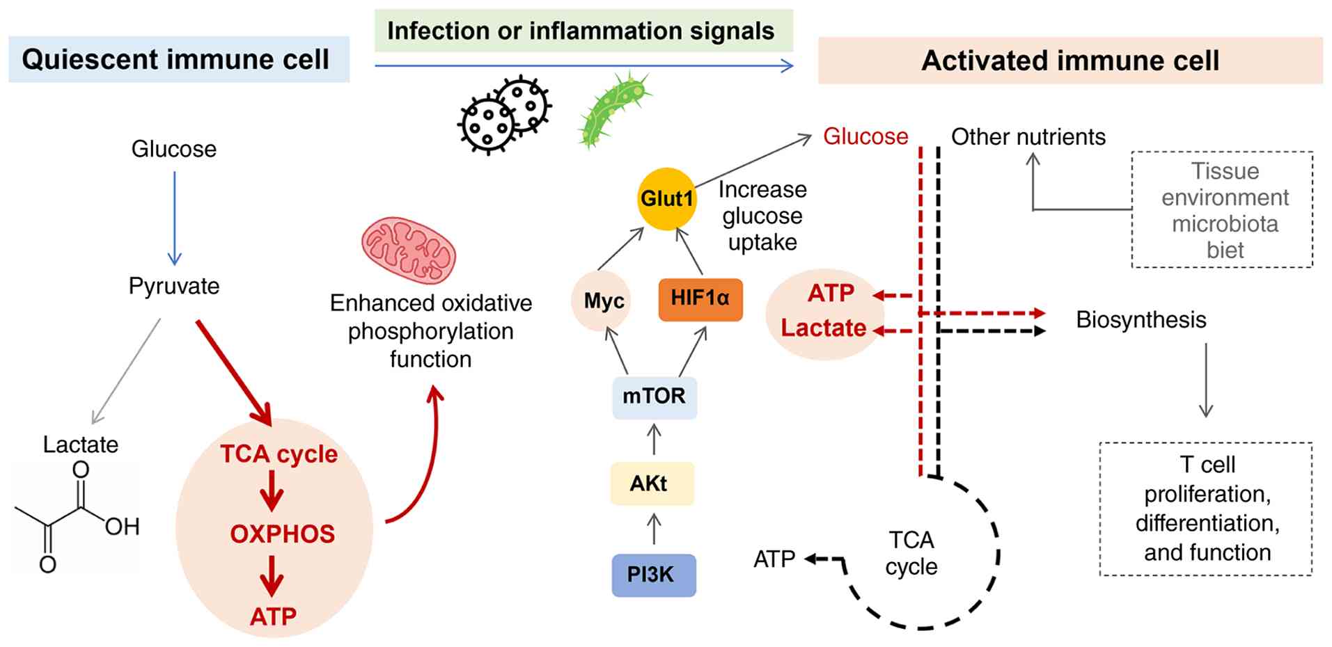 | Figure 1Metabolic reprogramming of T cells
before and after activation. Quiescent T cells rely primarily on
mitochondrial OXPHOS for energy supply; most glucose-derived
pyruvate enters the TCA cycle to fuel OXPHOS for ATP production.
Upon T-cell activation triggered by infection or inflammatory
signals, the PI3K-Akt-mTOR signaling cascade upregulates Myc, HIF1α
and Glut1 to boost glucose uptake. Cells then switch metabolism
toward aerobic glycolysis, where the majority of pyruvate is
converted into lactate. Glycolytic intermediates are diverted to
biosynthetic pathways to support T-cell proliferation,
differentiation and effector functions. TCA, tricarboxylic acid;
HIF1α, hypoxia-inducible factor 1α; OXPHOS, oxidative
phosphorylation; ATP, adenosine triphosphate; PI3K,
phosphoinositide 3-kinase; Akt, protein kinase B; mTOR, mechanistic
target of rapamycin; Myc, MYC proto-oncogene, bHLH transcription
factor; Glut1, glucose transporter 1/solute carrier family 2 member
1. |
A study combining RNA sequencing and glucose isotope
tracing confirmed that activation stimulation substantially
upregulates OXPHOS, the tricarboxylic acid (TCA) cycle and
nucleotide biosynthesis in B cells, but does not enhance the
glycolytic flux (17). Metabolic
regulation in B2 cells is primarily governed by the c-Myc and mTOR
complex (mTORC) signaling pathways, with mTORC promoting protein
synthesis and cell proliferation through glycolysis and OXPHOS
(18). Resting naïve B cells
maintain a hypometabolic homeostatic state that supports their
long-term survival. These cells rely extensively on fatty acid
oxidation for bioenergetic support, a metabolic pattern that is
downregulated upon activation. B-cell receptor engagement mediates
rapid glucose uptake via PI3K signaling (19). B-cell activation centers on the
upregulation of mitochondrial metabolism, whereas glycolysis is not
a key pathway for these cells. Glucose restriction does not impair
B-cell physiological functions, whereas inhibition of OXPHOS or
glutamine limitation markedly impairs B-cell growth and
differentiation (17). Glutamine
serves as the primary substrate for mitochondrial respiration after
B-cell activation, with the AMPK signaling pathway maintaining
mitochondrial respiratory function and homeostatic balance in
activated B cells (17).
B cells exhibit remarkable metabolic heterogeneity
across developmental stages, as summarized in Table I. Pre-B cells primarily rely on
glycolysis, regulated by Fnip1, AMPK, and PI3K (20). B1a cells display a metabolically
active phenotype engaging glycolysis, the pentose phosphate
pathway, OXPHOS, and fatty acid metabolism, supported by Myc,
perilipin-3, acetyl-CoA carboxylase 1, Acacb, Acsl1, Plin3, Srebp2,
and Atg7 (21). Conventional B2
cells utilize both glycolysis and OXPHOS under the regulation of
Myc and mTORC (18,22), whereas naïve B cells depend on
glycolysis and fatty acid oxidation, driven by BCR, BAFF, PI3K and
IL-4 (19,23). Germinal center B cells are highly
glycolytic, with upregulated glycolytic enzymes (GLUT1, HK1, PFKM,
GAPDH, LDHB, PDH) and transcriptional regulation by Myc and HIF1α
(24). Regulatory B cells also
rely on glycolysis, modulated by vitamin D3, aryl hydrocarbon
receptor, SCFA butyrate, HIF1α, STAT3 and valerate (25). In contrast, memory B cells
preferentially utilize OXPHOS for long-term survival, regulated by
Bach2, mTORC1 and AMPK (26,27).
 | Table IMetabolic pathways and regulatory
factors of different types of B cells. |
Table I
Metabolic pathways and regulatory
factors of different types of B cells.
| B cells | Metabolic
pathways | Regulatory
factors | (Refs.) |
|---|
| Pre-B | Glycolysis | Fnip1, AMPK,
PI3K | (20) |
| B1a | Glycolysis, pentose
phosphate pathway, OXPHOS, fatty acid metabolism | Myc, Perilipin-3,
acetyl-CoA carboxylase 1, Acacb, Acsl1, Plin3, Srebf2, Atg7 | (21) |
| B2 | Glycolysis,
OXPHOS | Myc, mTORC | (18,22) |
| Naïve B | Glycolysis, fatty
acid oxidation | BCR, BAFF, PI3K,
IL-4 | (19,23) |
| GC B | Glycolysis | GLUT1, HK1, PFKM,
GAPDH, LDHB, PDH, Myc, HIF1α | (24) |
| Breg | Glycolysis | Vitamin D3, aryl
hydrocarbon receptor, SCFA butyrate, HIF1α, STAT3, short-chain
fatty acid valerate | (25) |
| Memory B | OXPHOS | Bach2, mTORC1,
AMPK | (26,27) |
The metabolic differences between resting and
activated T cells vs. B cells reflect the molecular adaptations
that distinct lymphocyte lineages have evolved to execute their
unique immunological functions. T-cell metabolism is geared toward
supporting migration, proliferation and cytotoxic effector
functions, whereas B-cell metabolism is preferentially directed
toward antigen recognition, processing and presentation, as well as
fulfilling the high biosynthetic demands required for
immunoglobulin production. These distinct metabolic programs are
integral to their functional specialization.
Autophagy and PANoptosis: The regulatory
boundary of inflammatory death
Autophagy as a brake on PANoptosis
Inflammatory cell death is a form of PCD governed by
precise molecular mechanisms and characterized by a robust
inflammatory response. Unlike the immunologically silent clearance
mediated by classical apoptosis, inflammatory cell death releases
large amounts of damage-associated molecular patterns (DAMPs) and
pro-inflammatory cytokines via membrane pore formation or rupture,
thereby triggering immune activation and subsequent immune cell
recruitment (28). The major
forms of inflammatory cell death currently recognized include
pyroptosis, necroptosis and PANoptosis-the latter integrating
features of both pyroptosis and necroptosis. These cell death
modalities serve as a cornerstone of anti-infection immunity and
tumor immune regulation; however, their excessive activation can
also precipitate autoimmune diseases and pathological tissue
damage. Autophagy counteracts the initiation and progression of
inflammatory cell death by eliminating intracellular danger
signals, thereby constituting an important negative regulatory
mechanism. This regulation operates through two interrelated
pathways: First, mitophagy removes damaged mitochondria, preventing
the release of pro-inflammatory mediators; second,
autophagy-receptor-mediated selective autophagy degrades
ubiquitinated protein aggregates and components of inflammatory
complexes, thereby blocking the activation of PANoptosis signaling
(29).
Selective autophagic degradation of ubiquitinated
substrates is mediated by autophagy receptors, such as p62/SQSTM1,
NDP52 and optineurin, a process instrumental in preventing
PANoptosis initiation. Among these, p62-a canonical autophagy
receptor-contains both the LC3-interaction domain and the
ubiquitin-binding domain, enabling it to recognize ubiquitinated
protein aggregates and deliver them to autophagosomes for
degradation (30). This process
not only maintains protein homeostasis but also clears aberrant
proteins associated with inflammatory signaling activation, thereby
indirectly constraining excessive inflammatory responses. Autophagy
can selectively degrade the activated NLR family pyrin domain
containing 3 (NLRP3) inflammasome, thereby limiting caspase-1
activation and its mediated pyroptosis (31). NLRP3 inflammasome activity is
negatively regulated by autophagy, which mediates its lysosomal
delivery and degradation (32).
Furthermore, mitophagy deficiency-induced mitochondrial dysfunction
markedly facilitates NLRP3 inflammasome assembly and activation,
whereas restoration of mitophagy effectively alleviates the
inflammatory response in vivo (33).
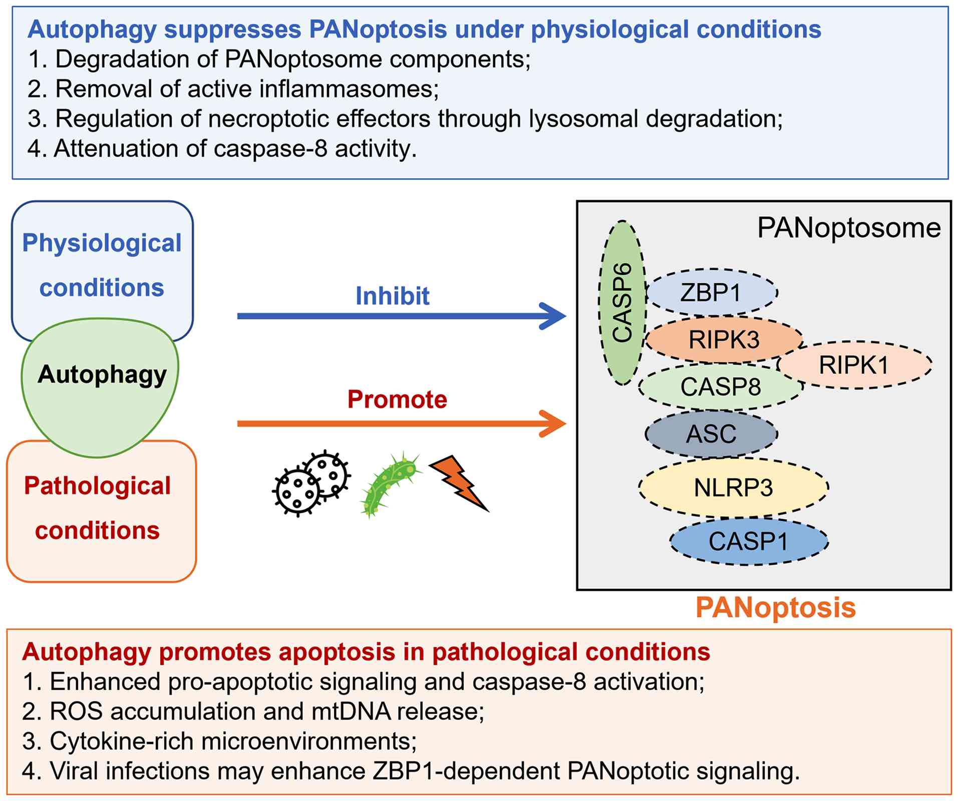
Autophagy and PANoptosis are functionally
interdependent, forming a bidirectional regulatory network that
involves both promotion and counterbalance (Fig. 2). Recent evidence indicates that
autophagy can curb the initiation of PANoptosis at its source by
degrading core components of PANoptosome, such as absent in
melanoma 2 (AIM2), Z-DNA binding protein 1 (ZBP1) and pyrin. As a
key PANoptosome component, absent in AIM2 is subject to
p62-mediated selective autophagy. p62 recognizes ubiquitinated AIM2
and restricts its interaction with apoptosis-associated speck-like
protein containing a CARD (ASC), thereby inhibiting PANoptosome
assembly and activation (29).
Additionally, autophagy suppresses PANoptosome formation by
regulating the degradation of necroptosis effector molecules RIPK1,
RIPK3 and MLKL, further restraining the inflammatory death process
(34). Notably, the NLRP3
inflammasome-a critical upstream activation complex of
PANoptosis-can be targeted for degradation by selective autophagy
after activation, forming a negative feedback loop that prevents
inflammatory response (35). The
mitochondrial-associated ER membrane (MAM) serves as an important
signaling integration platform and is pivotal for NLRP3
inflammasome assembly. Translocation of NLRP3 from the ER to the
mitochondria and the MAM is a prerequisite for its activation
(34). Cardiolipin, a unique
phospholipid of the inner mitochondrial membrane, is exposed to the
outer mitochondrial membrane under mitochondrial stress. Through a
specific binding interface, cardiolipin couples NLRP3 with
pro-caspase-1, thereby orchestrating NLRP3 inflammasome assembly.
Furthermore, mitochondrial antiviral signaling protein and
mitofusin 2 can form a complex that recruits NLRP3 to mitochondria
during RNA virus infection, accelerating inflammasome activation
(36). Autophagy also attenuates
caspase-8 activity, indirectly inhibiting apoptosis (37). Collectively, autophagy functions
as a homeostatic safeguard that suppresses aberrant PANoptosis
activation under physiological conditions-an intrinsic brake that
maintains immune equilibrium.
In aged macrophages, mitophagy decline causes
accumulation of damaged mitochondria and increased mitochondrial
DNA (mtDNA) leakage, leading to persistent overactivation of the
NLRP3 inflammasome and the cyclic GMP-AMP synthase
(cGAS)-stimulator of interferon response cGAMP interactor 1 (STING)
pathway, ultimately disrupting inflammatory homeostasis (38). Autophagy indirectly inhibits
PANoptosis by preserving mitochondrial functional integrity. In
chondrocytes, sirtuin 3 restores mitochondrial function and
suppresses excessive mitophagy, thereby blocking acidosis-induced
PANoptosis (39). In a
Parkinson's disease model, Parkin targets the NLRP3 inflammasomes
for degradation via chaperone-mediated autophagy at the K353 site,
effectively suppressing PANoptosis and protecting dopaminergic
neurons (40). As a key upstream
activator of PANoptosis, autophagic degradation of the NLRP3
inflammasomes potently blocks downstream inflammatory signaling
cascades. Furthermore, ribophagy inhibits ZBP1-mediated PANoptosome
formation by clearing damaged ribosomes, thereby alleviating
PANoptosis in CD4+ T lymphocytes during sepsis (41). Through clearing danger signals,
degrading PANoptosome components and maintaining organelle
homeostasis, autophagy acts as a negative regulator of PANoptosis
across diverse physiological and pathological conditions,
preserving immune homeostasis and preventing excessive inflammatory
damage.
Interplay between autophagic cell death
and PANoptosis
Autophagy plays a dual role in cell fate
determination: On one hand, it acts as a core mechanism that
maintains cellular homeostasis and ensures cell survival under
stress conditions; on the other hand, under inflammatory or
infectious conditions or upon dysregulation of autophagic flux, it
can facilitate the propagation of PANoptotic signals by amplifying
cellular stress responses and modulating cytokine secretion.
Deficiency of autophagy genes, lysosomal dysfunction
or impaired autophagosome-lysosome fusion can all lead to abnormal
accumulation of intracellular danger signals, thereby lowering the
activation threshold of PANoptosis and creating favourable
conditions for its initiation. Studies have shown that loss of
autophagy-related genes such as autophagy-related 5 (ATG5), ATG7 or
ATG16L1 triggers excessive inflammasome activation and aberrantly
enhanced PANoptosis, accompanied by elevated release of
pro-inflammatory cytokines including IL-1β and IL-18 (42-45). Mitophagy deficiency results in
massive accumulation of damaged mitochondria, which subsequently
releases mtDNA and mitochondrial ROS (mtROS), concurrently
activating the NLRP3 inflammasome and the cGAS-STING pathway, and
ultimately triggering PANoptotic inflammatory death (46,47). During viral infection, pathogens
can exploit the autophagy pathway to enhance ZBP1-dependent
PANoptotic signal transduction, accelerating the inflammatory death
of host cells. Furthermore, autophagy dysfunction leads to
substantially increased ZBP1 expression, which in turn potentiates
PANoptosis sensitivity by activating RIPK3, caspase-8 and gasdermin
D (GSDMD) (48,49).
Under certain stress conditions, autophagy can be
aberrantly activated to excessive levels, leading to
over-degradation of key organelles and metabolic enzymes, thereby
triggering an energy crisis and metabolic collapse. When this
'self-digestion' exceeds cellular tolerance, cells fail to maintain
basic functions and may initiate inflammatory death programs,
including PANoptosis. Shared regulatory molecules, including NLRP3,
STING, RIPK, glutathione peroxidase 4 (GPX4) and nuclear receptor
coactivator 4 (NCOA4), orchestrate a synergistic cell death network
that connects autophagy-dependent ferroptosis and PANoptosis in
myocardial and cerebral ischemia-reperfusion injury. These factors
not only activate their respective pathways but also converge to
drive PANoptosome assembly and consolidate death signals, thereby
promoting signal propagation and cross-regulation (50). Induction of autophagy following
ischemia-reperfusion aggravates oxidative injury and elicits marked
ROS elevation. In parallel, ferritin is delivered to lysosomes via
the autophagy adaptor NCOA4 for degradation, thereby releasing
labile iron and precipitating autophagy-driven ferroptosis
(51).
Persistent IFN signaling upregulates ZBP1
expression, whereas STING overactivation induces excessive
autophagy, leading to massive autophagosome formation coupled with
saturated lysosomal degradation capacity (52). This autophagic flux blockade
shifts autophagy toward a pro-PANoptotic state through three
mechanisms: i) Substantial accumulation of the autophagy substrate
p62, which serves as a molecular scaffold that binds RIPK3 and
caspase-8, stabilizing the PANoptosome complex; ii) direct binding
of Beclin-1 to RIPK3, which enhances RHIM domain interactions and
amplifies necroptotic signaling; and iii) release of cathepsins
from damaged autolysosomes, which synergize with caspases to
amplify pyroptotic and apoptotic signals. Together, these events
establish an autophagy-PANoptosis positive feedback loop that
drives extensive inflammatory cell death and a cytokine storm
cascade (29).
Autophagy exhibits prominent cell-type specificity
in regulating inflammatory cell death, with the
autophagy-PANoptosis axis manifesting distinct patterns across
diverse immune cells, including macrophages, dendritic cells (DCs),
neutrophils and T cells. Macrophages represent the most extensively
investigated cell type for this axis. In these cells, autophagy
generally restrains PANoptosis through multiple mechanisms: i)
Elimination of DAMPs and pathogen-associated molecular patterns
(PAMPs), such as lipopolysaccharide (LPS), mtROS and cytoplasmic
DNA; ii) mitophagy curbs NLRP3 inflammasome activation through
removing impaired mitochondria; iii) macroautophagy promotes the
clearance of the NLRP3 inflammasome; and iv) suppression of the
secretion of pro-inflammatory mediators such as IL-1β. Conversely,
knockout of autophagy genes in macrophages triggers excessive
inflammasome activation and PANoptosis, accompanied by elevated
IL-1β and IL-18 levels. During influenza A virus infection, when
autophagy fails to clear damaged organelles or viral components,
macrophages assemble a ZBP1-NLRP3-caspase-8-RIPK3 complex to form
the PANoptosome (29). In DCs,
autophagy indirectly modulates susceptibility to PANoptosis by
regulating Toll like receptor (TLR)-dependent priming and
inflammasome activity. Notably, thyroid-resident DCs from patients
with Hashimoto's thyroiditis exhibit concomitant activation of
necroptosis, ferroptosis, pyroptosis, autophagy and PANoptosis,
alongside differential expression of key PCD-related genes,
including TNF-α-induced protein 3, cytochrome b-245 β chain,
protein tyrosine phosphatase non-receptor type 6, signal transducer
and enhancer of transcription (STAT)1, transforming growth factor
β1 and NLRP3 (53). In
neutrophils with high S100 calcium binding protein A8/A9
expression, nuclear respiratory factor 1 is downregulated, leading
to repression of Nicotinamide Adenine Dinucleotide (NADH):
ubiquinone oxidoreductase subunit A3-a component of mitochondrial
complex I. This instigates mitochondrial dysfunction, characterized
by hyper-fission and deficient mitophagy. Liberated mtDNA from
compromised mitochondria ultimately drives ZBP1-mediated
pan-pyroptosis in endothelial cells (54). In CD4+ T lymphocytes
during sepsis, nuclear FMR1 interacting protein 1
(NUFIP1)-dependent ribophagy eliminates damaged ribosomes through
the cGAS-STING pathway and hinders ZBP1-driven PANoptosome
assembly. Furthermore, mTOR depletion ameliorates pyroptosis in
CD4+ T cells from septic mice by boosting autophagic
activity (55).
In summary, autophagy acts as a bidirectional
regulator of PANoptosis. When autophagy effectively clears DAMPs
and preserves mitochondrial homeostasis, it suppresses PANoptosis.
Conversely, autophagy deficiency or pathological hyperactivation
tilts the balance toward PANoptosome assembly, culminating in
inflammatory cell death.
Interplay between autophagy, metabolic
reprogramming and PANoptosis
Temporal and causal relationships among
autophagy, metabolic reprogramming and PANoptosis
When immune cells encounter infection or
inflammatory signals, they initially perceive shifts in metabolic
status through energy sensors such as AMPK and mTOR. Under
nutrient-sufficient conditions, mTORC1 is activated, which
suppresses autophagy and promotes anabolic metabolism, thereby
supporting cell proliferation and effector differentiation.
Conversely, under nutrient deprivation or energy stress, AMPK
becomes activated and phosphorylates unc-51 like autophagy
activating kinase 1 (ULK1) to initiate autophagy (56). Upon initiation, autophagy
eliminates damaged mitochondria via mitophagy, curbing mtROS and
mtDNA release, while also degrading the NLRP3 inflammasomes and
PANoptosome components through selective autophagy. This maintains
inflammatory signals below the lethal threshold (57). At this stage, autophagy acts as a
molecular brake to sustain cell survival and support the
differentiation and effector functions of immune cells.
When inflammatory signals persist or exceed a
critical threshold, autolysosomal function becomes saturated,
resulting in autophagic flux blockade. Damaged mitochondria
continuously release mtDNA, activating the cGAS-STING pathway and
interferon signaling further upregulates ZBP1 expression. At this
stage, autophagy transitions from a protective to a pro-death role:
p62 accumulates extensively and serves as a molecular scaffold to
stabilize the PANoptosome complex, whereas the autophagy protein
Beclin-1 directly binds RIPK3 to enhance RHIM domain interactions,
ultimately triggering the coordinated activation of pyroptosis,
apoptosis and necroptosis (51).
Following cell lysis, PANoptotic cells release large amounts of
DAMPs and pro-inflammatory cytokines, which further activate
neighboring immune cells and amplify inflammatory signals. This
positive feedback loop sustains and escalates inflammation,
eventually leading to tissue destruction and systemic organ
dysfunction.
Immune cells undergo profound metabolic
reprogramming during activation, differentiation and execution of
effector functions. T cells shift from OXPHOS dependence in the
resting state to glycolysis dependence upon activation, while
macrophages switch their metabolic profiles between M1 and M2
polarization (58). This
metabolic plasticity far exceeds that of most non-immune cells,
rendering autophagy regulation in immune cells more dynamic and
precise.
Immune cells, ordinary somatic cells and tumor cells
exhibit fundamentally distinct metabolic plasticity, reflecting
their divergent biological objectives and survival strategies
(Table II). Immune cells
dynamically reprogram their metabolism in response to external
signals (antigens, cytokines and microenvironmental cues), enabling
reversible switching among resting, effector and memory states to
support specific immune functions such as killing and cytokine
secretion (59). By contrast,
ordinary somatic cells maintain relatively stable metabolism to
preserve homeostasis, with limited plasticity driven by systemic
hormonal and nutritional changes, allowing only slow transitions
between quiescence and proliferation. Tumor cells, however, adopt a
'selfish' metabolic reprogramming, driven by genetic mutations and
microenvironmental stresses (hypoxia, acidosis), which
unidirectionally locks them into the Warburg effect-aerobic
glycolysis-to support unlimited proliferation and survival
(60).
| Table IIDifferences in metabolic plasticity
between immune cells and other cells (59,60). |
Table II
Differences in metabolic plasticity
between immune cells and other cells (59,60).
| Item | Immune cells | Ordinary somatic
cells | Tumor cells |
|---|
| Core objective | Carrying out
specific immune functions (such as killing, secreting
cytokines) | Maintenance of its
own homeostasis and specific physiological functions. Metabolism is
relatively stable. | Unlimited
proliferation and survival. Metabolic reprogramming is 'selfish'
and aims to hijack resources. |
| Plasticity driving
factors | External signals
(antigens, cytokines, microenvironment). The metabolic state is a
reflection and driver of the functional state. | Systemic regulation
such as hormones and nutritional status. The changes are relatively
slow. | Genetic mutations
and microenvironmental stress (such as hypoxia, acidosis). |
| Reprogramming
direction | Dynamic and
reversible switching among various functional states, such as from
rest to effect, and then to memory. | Switching between
different physiological states (such as rest and proliferation);
the plasticity is limited. | Unidirectional
locking occurs in the metabolic pattern that supports unlimited
proliferation (the Warburg effect). |
| Adaptation to the
environment method | Active integration
of environmental signals (such as nutrients and oxygen) to adjust
functions and being able to utilize the locally available
fuels. | Passive adaptation
to environmental changes in order to maintain survival. | Active modification
of the microenvironment (such as producing lactic acid) to
facilitate its own survival. |
The coupling of autophagy with programmed cell death
in immune cells exhibits high cell-type specificity. Autophagy
exerts a dual influence on necroptosis, ferroptosis and pyroptosis.
On the protective side, it eliminates RIPK proteins or the
necrosome to prevent cell death; conversely, by targeting cellular
inhibitor of apoptosis proteins (cIAPs) for degradation, it may
potentiate necroptotic signaling (61). Autophagy-mediated cell death
involves interaction between the autophagic machinery and other
cell death molecules; alternatively, autophagy can directly
activate apoptosis, ferroptosis or necroptosis to promote cell
death. Recent studies have proposed that immune cells harbor a
'master molecular timer' composed of the ubiquitin-proteasome
system, autophagy and translational control, which licenses or
restricts the occurrence of pyroptosis, apoptosis, necroptosis and
PANoptosis. Post-translational modifications provide a decisional
framework that is co-opted by bacterial pathogens and viruses
through effector deployment, enabling threshold modulation via host
shutoff, ubiquitin/ISG15 ubiquitin like modifier editing and
autophagy subversion. This dynamic proteostasis competition
mechanism is particularly prominent in immune cells (62).
Metabolic reprogramming regulates
autophagic-dependent cell death
When immune cells operate under nutrient-sufficient
conditions with normal mitochondrial function, basal autophagy
maintains cellular homeostasis through mitochondrial quality
control, protecting cells from stress-induced damage. However, when
metabolic reprogramming leads to mitochondrial dysfunction-such as
enhanced glycolysis accompanied by impaired OXPHOS-damaged
mitochondria release mtDNA and ROS. These danger signals not only
activate the NLRP3 inflammasome and the cGAS-STING pathways, but
also further disrupt autophagic flux via feedback loops, converting
autophagy from a protector to a promoter of cell death. Meanwhile,
autophagy deficiency (e.g., ATG5 deletion) impairs mitophagy,
exacerbates mitochondrial damage and enhances GSDMD-mediated pore
formation, promoting extracellular release of mtDNA, which in turn
activates the cGAS-STING-NLRP3 signaling axis in macrophages
(63). In myocardial and
cerebral ischemia-reperfusion injury, autophagy-coupled ferroptosis
and PANoptosis converge into a complex cell death circuitry
governed by shared regulators, including NLRP3, STING, RIPK, GPX4
and NCOA4 (50).
TCA cycle intermediates such as succinate and
itaconate not only function as energy metabolites but also act as
signaling molecules that regulate inflammasome activation and cell
death. Succinate primarily acts by inhibiting prolyl hydroxylases,
thereby stabilizing HIF-1α, which in turn induces glycolysis and
production of the pro-inflammatory cytokine IL-1β. Succinate
accumulation also affects immune cells within the tumor
microenvironment, participating in lymphocyte-mediated immunity and
cytokine production. Furthermore, succinyl-CoA can modify key
metabolic enzymes such as PKM2 through succinylation, enhancing
HIF-1α activity and further promoting IL-1β production (64). Itaconate inhibits the NLRP3
inflammasome through multiple mechanisms: i) Alkylating cysteine
residues of kelch-like ECH-associated protein 1 to activate the
NRF2 antioxidant pathway; ii) inhibiting succinate dehydrogenase to
reduce mtROS production; and iii) limiting oxidative stress and
glycolytic flux. The itaconate derivative dimethyl itaconate
inhibits LPS-induced NLRP3-dependent pyroptosis by inducing
autophagy and regulating the Nrf2/heme oxygenase-1 signaling
pathway (65). Another
derivative, 4-octyl itaconate, enhances autophagy by inhibiting the
PI3K/AKT/mTOR signaling pathway, thereby protecting chondrocytes
from IL-1β-induced inflammatory degradation and apoptosis (66). Changes in the concentrations of
these metabolites directly influence autophagic flux and the
threshold for PANoptosome assembly.
Mild metabolic stress induces adaptive autophagy
that protects cell survival. However, under sustained or severe
metabolic stress-such as in the tumor microenvironment or during
chronic infection-autophagic function may become suppressed or
overloaded. This leads to failure of mitochondrial quality control
and continuous accumulation of danger signals, ultimately breaching
the threshold for PANoptosis. Autophagy dictates the fate of
CD8+ T-cell progeny through asymmetric mitochondrial
partitioning. Daughter cells that inherit aged mitochondria exhibit
impaired memory potential, while those without aged mitochondria
persist longer and mount robust expansion (67). These findings suggest that
enhancing autophagy before or during T-cell division may promote
the generation of daughter cells that avoid inheriting aged
mitochondria, thereby augmenting the production of memory T
cells-the cornerstone of long-term immunity and vaccine efficacy.
This provides a novel framework for modulating autophagy to
'rejuvenate' memory T cells, with potential to improve vaccine
responses and cancer immunotherapy.
Metabolic checkpoints that determine the
survival and death of immune cells
Liver kinase B1 (LKB1) is a critical metabolic
checkpoint in natural killer (NK) cells. LKB1 deficiency leads to
mitochondrial dysfunction and impaired autophagic flux, triggering
ROS-dependent cell death. LKB1 ablation also perturbs iron
homeostasis, provoking iron deposition and cytotoxic lipid ROS
production. Notably, these phenotypic changes are refractory to
both AMPK agonism and mTORC1 antagonism, positioning LKB1 as a
metabolic rheostat that operates independently of the canonical
AMPK-mTOR circuitry. Furthermore, LKB1 deficiency upregulates
programmed cell death-1 and T-cell immunoreceptor with Ig and ITIM
domains expression, further compromising NK-cell immunosurveillance
(68).
AMPK and mTOR are core metabolic checkpoints that
respond to cellular energy and nutrient status. AMPK is activated
under energy stress, promoting catabolism and autophagy, whereas
mTOR is activated under nutrient-sufficient conditions, suppressing
autophagy and promoting anabolism. The balance between these two
kinases governs the direction and intensity of autophagic flux. The
generation and maintenance of memory T cells are profoundly
influenced by AMPKα1, which regulates autophagy and mitochondrial
respiration-associated gene expression. Additionally, T-cell memory
is sustained by low-grade IL-15/mTORC1 signals that activate the
AMPKα1-ULK1-ATG7 metabolic axis (69). AMPKα1 stimulates mitochondrial
biogenesis and fatty acid oxidation to support memory T-cell
differentiation. Conversely, AMPKα1 deficiency abrogates these
downstream responses, resulting in upregulation of mTORC1 and
HIF-1α, which shifts metabolism from fatty acid oxidation toward
glycolysis and reduces cell survival (70). When the AMPK-mTOR balance is
disrupted-for example, by mTOR hyperactivation-autophagy is
suppressed, damaged mitochondria accumulate and danger signals
build up, rendering cells more susceptible to PANoptosis (59).
Mitochondrial health status constitutes a critical
'structural' metabolic checkpoint. When mitophagy efficiency is
insufficient to clear damaged mitochondria, mtDNA leakage and
excessive ROS production activate the NLRP3 inflammasome and the
cGAS-STING pathways. The outcome of this checkpoint-'pass' or
'fail'-directly determines whether cells maintain homeostasis or
proceed toward inflammatory cell death. In B cells, FIP200 governs
cell fate by controlling mitophagy and metabolic reprogramming
(71). In CD8+ T
cells, autophagy-regulated asymmetric mitochondrial inheritance
dictates the fate divergence of daughter cells (67). Lipid metabolism represents
another important metabolic checkpoint. Lipophagy, the selective
autophagy of lipid droplets, modulates immune cell function and
inflammatory responses by degrading these droplets. In the
pancreatic cancer microenvironment, the ubiquitin-specific
peptidase 20 (USP20)-regulated metabolic-autophagy axis directly
drives CD8+ T-cell exhaustion via cholesterol metabolic
control (72). Thus, disrupting
this USP20-cholesterol-autophagy circuit constitutes a rational
strategy to reinvigorate antitumor immunity and enhance the
efficacy of KRASG12D inhibitor therapy in pancreatic ductal
adenocarcinoma.
Molecular switch connecting metabolism,
autophagy and inflammatory death
The AMPK-mTOR pathway serves as the core hub linking
cellular energy metabolism and autophagy. Dysregulation of this
pathway, such as mTOR hyperactivation, leads to autophagy
suppression, metabolic disruption and a lowered threshold for
PANoptosis. The underlying mechanisms include the following: i)
Autophagy suppression and danger signal accumulation are driven by
the following mechanisms: mTORC1 hyperactivation directly inhibits
autophagy, preventing clearance of damaged mitochondria and protein
aggregates; accumulation of these substrates triggers release of
danger signals such as mtDNA and ROS; ii) activation of
inflammatory pathways: Accumulated danger signals (e.g., mtDNA and
ROS) activate the NLRP3 inflammasome and the cGAS-STING pathway;
and iii) lowered PANoptosis threshold: Persistent inflammatory
signals render cells more susceptible to PANoptosis, ultimately
leading to inflammatory cell death (73). In autoimmune diseases such as
systemic lupus erythematosus (SLE), mTORC1 hyperactivation is a key
mechanism underlying T and B lymphocyte dysfunction. In SLE, T
cells exhibit aberrant metabolic reprogramming. Activation of AMPK
along with concomitant inhibition of mTOR signaling can effectively
suppress glycolysis, restore the type 17 T-helper cell/regulatory T
cell (Treg) balance, and thereby alleviate SLE symptoms (74). Aberrant activation of the
AMPK-mTOR-STAT3 axis is associated with B-cell commitment to plasma
cells and germinal center B cells, which in turn triggers
autoantibody production. Pharmacological activation of AMPK, for
instance with metformin, can inhibit this pathway, reduce B-cell
differentiation and exert therapeutic benefits (75).
The cGAS-STING pathway represents a critical
molecular connection between mitochondrial damage and inflammatory
cell death. When mitophagy deficiency leads to mtDNA leakage into
the cytosol, cGAS recognizes mtDNA and catalyzes cGAMP production,
which activates STING and subsequently induces type I interferons
and pro-inflammatory cytokines. Excessive STING activation further
induces hyperautophagy, establishing a positive feedback loop. The
cGAS-STING axis is also activated by sepsis-induced ribosome
collision, which facilitates the recruitment of NUFIP1 to the STING
complex. NUFIP1-mediated ribophagy clears damaged ribosomes,
thereby inhibiting ZBP1-driven PANoptosome formation and
alleviating PANoptosis in CD4+ T lymphocytes (41). Blocking the cGAS pathway or
deleting STING exerts protective effects, including suppression of
PANoptosis and mitigation of subsequent pathological damage
(52). At the metabolic level,
STING hyperactivation induces excessive autophagy, while autophagic
flux blockade further exacerbates mitochondrial damage and mtDNA
release, forming a STING-autophagy-metabolism positive feedback
amplification loop. Furthermore, ER cholesterol levels regulate
ER-phagy and STING innate immunity through the complex comprising
SREBF chaperone (SCAP) and reticulophagy regulator 1 (RETREG1)
(76). Collectively, these
findings establish the cGAS-STING pathway as a key molecular bridge
connecting metabolism, autophagy and PANoptosis.
The ZBP1-RIPK3-caspase-8 axis serves as the core
molecular switch for PANoptosome assembly and execution of
inflammatory cell death. As a nucleic acid sensor, ZBP1 recruits
RIPK3 and caspase-8 via its RHIM domain upon recognition of Z-form
nucleic acids, thereby assembling the PANoptosome. Autophagy
tightly regulates this axis by clearing mitochondrial DAMPs and
cytosolic nucleic acids, thereby limiting ZBP1 activation. When
autophagic function is impaired, ZBP1 expression is amplified and
the threshold for PANoptosome assembly is lowered (77). In sepsis, ZBP1 promotes
mitochondrial damage and glycolysis, thereby driving macrophage
polarization toward a pro-inflammatory phenotype and enhancing
NLRP3 inflammasome-mediated pyroptosis. ZBP1 knockout alleviates
mitochondrial damage and suppresses glycolysis, shifting macrophage
metabolism away from the pro-inflammatory state (78). In rheumatoid arthritis (RA),
RIPK3 promotes the migration and invasion of fibroblast-like
synoviocytes through malate shuttle-driven mitochondrial
respiration (79). Thus, the
ZBP1-RIPK3-caspase-8 axis not only serves as the core molecular
switch for PANoptosome assembly and inflammatory death execution,
but also functions as a critical node that integrates metabolic
signals with cell fate determination. The crosstalk among
autophagy, metabolic reprogramming and PANoptosis is illustrated in
Fig. 3, which provides a
framework for exploring the involvement of this axis in disease
contexts.
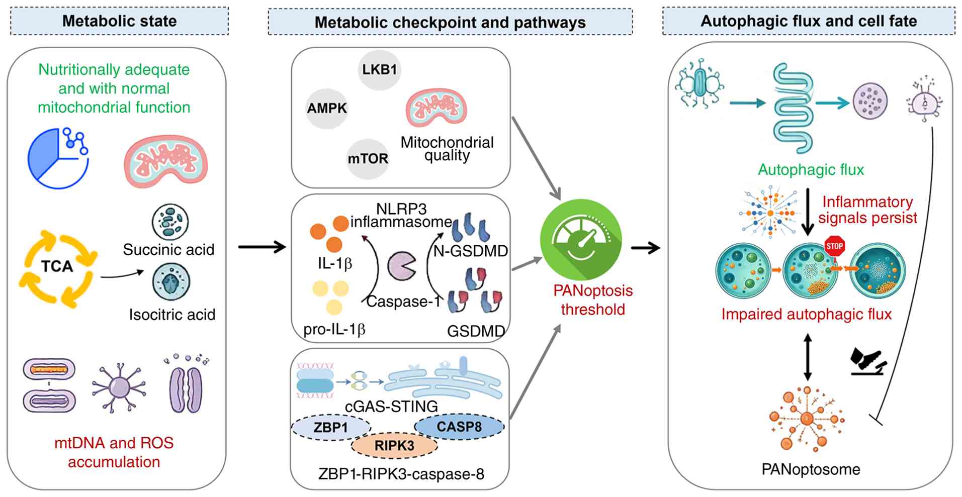 | Figure 3Metabolic status, metabolic
checkpoints and associated pathways cooperatively regulate the
activation threshold of PANoptosis. Cells shift from a
nutritionally sufficient state with intact mitochondrial function
to a state characterized by accumulation of succinic acid and
isocitric acid, as well as extensive release of mtDNA and ROS. This
metabolic transition modulates the activation of metabolic
checkpoints and relevant signaling pathways, thus determining the
threshold for PANoptosis induction. Persistent inflammatory signals
block autophagic flux and defective autophagy further facilitates
PANoptosome assembly, ultimately initiating PANoptosis. ROS,
reactive oxygen species; mtDNA, mitochondrial DNA; TCA,
tricarboxylic acid; LKB1, serine/threonine kinase 11; AMPK,
adenosine 5'-monophosphate-activated protein kinase; mTOR,
mechanistic target of rapamycin; NLRP3, NLR family pyrin domain
containing 3; IL-1β, interleukin-1β; N-GSDMD, gasdermin D,
N-terminal; cGAS, cyclic GMP-AMP synthase; STING, stimulator of
interferon response cGAMP interactor 1; CASP8, caspase 8; ZBP1,
Z-DNA binding protein 1; RIPK3, receptor interacting
serine/threonine kinase 3. |
Autophagy supports the metabolic
reprogramming and survival of immune cells
Autophagy regulates the metabolic
reprogramming and survival of T cells
Autophagy is a key regulator of T-cell survival,
maintaining mitochondrial homeostasis and limiting accumulation of
cytotoxic ROS. After specifically knocking out Atg3 in T cells,
progressive expansion of mitochondria and ER occurs, and cell death
is triggered within several weeks. This indicates that autophagy
sustains T-cell longevity by modulating organellar balance
(80). Upon TCR activation, T
cells transition from a resting state to an activated stage and
initiate a synthetic metabolic program, manifested as accelerated
proliferation, increased mitochondrial biosynthesis and enhanced
bioenergetic support for effector functions. During this process,
PI3K-AKT-mTORC signaling drives metabolic rewiring in T cells,
characterized by a core shift from fatty acid and pyruvate
oxidation toward glycolysis and glutamine utilization, with
concomitant generation of anabolic substrates, including
nucleotides, amino acids and fatty acids (Fig. 4A) (81). T-cell functional specification is
tightly coupled to glucose uptake and the ensuing metabolic
rewiring is pivotal for orchestrating lineage polarization. The
present review summarizes the energy metabolic pathways of T cells
in distinct states (Fig.
4B).
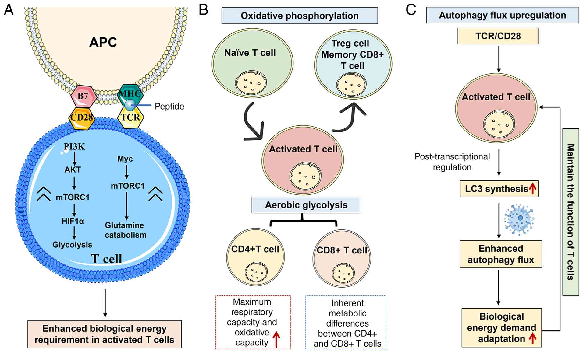 | Figure 4Differences in metabolic pathways
utilized by T cells at different stages. (A) Metabolic pathways
utilized by activated T cells. (B) Energy sources utilized by
different types of T cells. (C) Upregulated autophagy flux upon
T-cell activation. APC, antigen-presenting cell; MHC, major
histocompatibility complex; TCR, T cell receptor; PI3K,
phosphoinositide 3-kinase; Akt, protein kinase B; mTORC1,
mechanistic target of rapamycin complex 1; HIF1α, hypoxia-inducible
factor 1α; Myc, MYC proto-oncogene, bHLH transcription factor; Treg
cell, T-regulatory cell. |
After T-cell activation, autophagic activity
increases substantially, as evidenced by rapid upregulation of LC3
synthesis accompanied by an accelerated degradation rate. This
process is regulated at the post-transcriptional level (82) and is essential for accommodating
the elevated bioenergetic demands of activated T cells (Fig. 4C). Loss of autophagy genes
markedly impairs T-cell expansion and viability. Autophagy
deficiency, whether induced by transplantation of
autophagy-deficient fetal liver hematopoietic progenitor cells into
lethally irradiated wild-type syngeneic recipient mice or by
conditional knockout of autophagy-related genes in T cells, leads
to severe disruption of T-cell homeostasis (83). Furthermore, autophagy selectively
degrades the Bcl10 protein, thereby modulating the intensity of
TCR-mediated NF-κB signaling and preventing excessive signal
activation that would otherwise drive senescence. This mechanism is
crucial for the generation of CD8+ memory T cells
(84). Failure of T cells to
adequately upregulate their metabolic rate results in
immunosuppressive consequences, including reduced immune function,
diminished cytokine secretion and impaired proliferation.
Autophagy-related genes play critical and diverse
roles in modulating T-cell function, with their dysregulation being
implicated in a wide spectrum of autoimmune, inflammatory and
neurodegenerative diseases (Table
III). Vacuolar protein sorting-associated protein 34 is
essential for maintaining peripheral CD4+forkhead box P3+ Treg
cells, and its T cell-specific deletion leads to intestinal
inflammation and anemia (85).
Similarly, phosphatidylinositol 3-kinase catalytic subunit type 3
regulates T-cell activation and promotes CD4+ T-cell
differentiation into Th1 cells; its deficiency impairs cellular
metabolism and Th1 differentiation, contributing to autoimmune
diseases such as experimental autoimmune encephalomyelitis
(86). Atg5 influences Th2 cell
and cytokine levels, with polymorphisms linked to asthma, SLE and
RA (87-90). Atg7 is crucial for CD8+ T-cell
survival and memory formation, and its deficiency reduces CD4+ and
CD8+ T-cell numbers while impairing secondary immune responses and
atherosclerosis development (91,92). Atg16L1 regulates caspase-3
cleavage and Th2 responses while limiting Treg cell expansion in
the intestine; its T cell-specific deletion triggers spontaneous
intestinal inflammation and is associated with Crohn's disease and
inflammatory bowel disease (93,94). Immunity-related GTPase family M
member 1 (IRGM1), an IFN-γ-inducible GTPase, affects CD4+ T-cell
survival and proliferation, and its human homolog IRGM is strongly
linked to Crohn's disease (95).
LC3-mediated endocytosis promotes β-amyloid clearance and
alleviates neurodegeneration in Alzheimer's disease (96). Beclin-1 deficiency increases the
expression of the pro-apoptotic protein Bim in Th1 cells, affecting
T-cell survival and contributing to multiple sclerosis (97). Finally, C-type lectin domain
containing 16A (CLEC16A) deficiency in thymic epithelial cells
impairs autophagosome maturation and alters T-cell selection,
leading to the generation of self-reactive lymphocytes associated
with multiple sclerosis, SLE, rheumatoid arthritis, Crohn's disease
and type 1 diabetes (98).
| Table IIIAutophagy genes that affect T-cell
function and association with disease. |
Table III
Autophagy genes that affect T-cell
function and association with disease.
| Gene name | Function | Diseases or related
symptoms | (Refs.) |
|---|
| Vps34 | Maintenance of the
function of CD4+FoxP3+ regulatory T cells at the periphery;
influence on the development of NK T cells in the thymus. | Intestinal
inflammation; anemia | (85) |
| Pik3c3 | Regulation of the
activation of T cells; promotion of the differentiation of CD4+ T
cells into Th1 cells. | Autoimmune
diseases | (86) |
| Atg5 | Influence on the
levels of Th2 cells and cytokines; reduction of autophagy and
causing of ataxia. | Asthma, systemic
lupus erythematosus, rheumatoid arthritis, ataxia | (87-90) |
| Atg7 | Affecting the
survival of CD8+ T cells and their differentiation into memory T
cells; affecting the quantities of CD4+ T cells, CD8+ T cells and
NK T cells, thereby reducing the occurrence of
atherosclerosis. | The secondary
immune response is reduced; atherosclerosis and hepatic
steatosis | (91,92) |
| Atg16L1 | Regulation of the
cleavage of caspase-3, reduction of autophagy, inhibition of
bacterial clearance and increase of the production of
pro-inflammatory cytokines; regulation of the Th2 response and the
number of Treg cells in the intestine. | Crohn's disease;
inflammatory bowel disease | (93,94) |
| IRGM1 | By regulating
IFN-γ, the survival of CD4+ T cells and their proliferation in the
peripheral circulation pool can be affected. | Crohn's
disease | (95) |
| LC3 | The LC3-mediated
endocytosis promotes the clearance of β-amyloid protein and
alleviates neurodegeneration in mice with Alzheimer's disease. | Alzheimer's
disease | (96) |
| Beclin-1 | The absence of
Beclin-1 increases the expression of death proteins in Th1 cells,
and affects the deletion of the Bim anti-apoptotic gene and the
inhibition of caspases. | Multiple
sclerosis | (97) |
| CLEC16A | The absence of
CLEC16A in thymic epithelial cells inhibits the maturation of
autophagosomes and alters the selection of T cells, resulting in
the generation of self-reactive lymphocytes. | Multiple sclerosis,
systemic lupus erythematosus, rheumatoid arthritis, Crohn's
disease, type 1 diabetes | (98) |
Autophagy regulates metabolic
reprogramming and survival of B cells
The metabolic development and functional execution
of B cells also depend on precise regulation by the autophagic
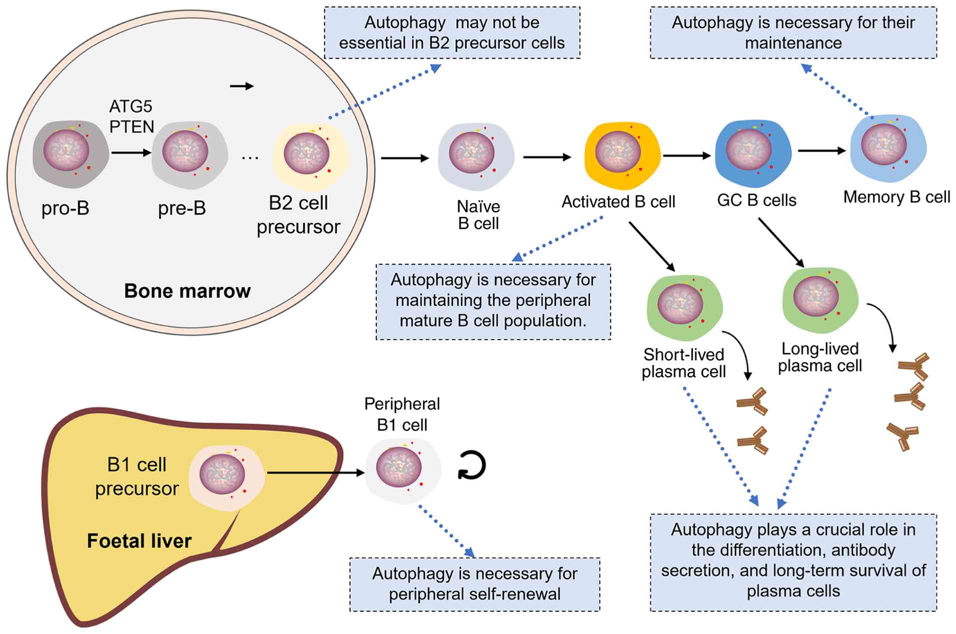
pathway. During early B-cell development in the bone marrow,
autophagy promotes the transition from progenitor B cells to pre-B
lymphocytes. Fetal liver progenitor cells lacking Atg5 exhibit a
notable developmental block at the pro-B to pre-B transition stage,
with only a small number of pre-B cells surviving and entering
peripheral lymphoid organs (99). In addition, phosphatase and
tensin homolog serves as a key regulator of progenitor B-cell
development, reducing their sensitivity to apoptosis (100). Autophagy also exerts specific
regulatory functions in B1 cell subsets within mucosal-associated
lymphoid tissues such as the peritoneal cavity. Loss of autophagic
flux in mature B cells substantially reduces B1a cell counts but
exerts minimal impact on B1b populations (99). Furthermore, early B1 progenitor
development remains intact in autophagy-deficient mice, indicating
that autophagy is not a prerequisite for B1-cell differentiation.
These findings suggest a selective requirement for autophagy in
peripheral self-renewal rather than in B1a cell differentiation.
The role of autophagy in B2 cell development has also been
investigated (101).
Transplanting of Atg5-deficient fetal liver cells into
recombination activating 1−/− mice causes B-cell
development to arrest at the pre-B stage. By contrast, specific
knockout of Atg5 in mature B cells does not affect B-cell numbers
in the spleen or lymph nodes. These observations indicate that
autophagy is essential for early B2-cell development but
dispensable for maintaining peripheral homeostasis of mature B2
cells (99).
In B cells that have matured and settled in
peripheral lymphoid organs, autophagy orchestrates multiple aspects
of plasma cell biology, including differentiation, antibody
secretory capacity and long-term survival (Fig. 5). Loss of Atg5 leads to excessive
ER expansion and heightened ER stress in differentiating plasma
cells. Although PR/SET domain 1 expression and immunoglobulin
secretion increase through a compensatory mechanism, intracellular
ATP levels substantially decrease and cell death rises, suggesting
that autophagy sustains plasma cell homeostasis via protective
mechanisms (102). During
influenza virus infection, memory B-cell generation does not rely
on autophagy, whereas their persistence does. Loss of Atg7 causes
abnormal accumulation of mitochondrial ROS, which drives
progressive loss of memory B cells and ultimately impairs the
establishment of effective antiviral immune memory and protective
responses (103). Notably,
during murine gammaherpesvirus 68 infection, germinal center B
cells (GCB) exhibit the highest autophagy level among all B-cell
populations, and high LC3-II expression correlates positively with
enhanced autophagic flux. Further studies have confirmed that
autophagy in GCB cells does not rely on the canonical
mTORC1-mediated pathway but instead proceeds via non-canonical
autophagy (104). B-cell
receptor activation transiently inhibits canonical autophagy while
initiating non-canonical autophagy. Conditional knockout of WD
repeat domain phosphoinositide-interacting protein 2 (WIPI2) in B
cells further enhances non-canonical autophagy, leading to
mitochondrial homeostasis disruption, aberrant germinal center
responses and altered antibody secretion (104). These findings suggest that B
cells harbor a form of unconventional autophagy that is independent
of ATG16L1 but dependent on WIPI2, which participates in regulating
B-cell fate and function.
Involvement of the autophagy-PANoptosis axis
in autoimmune disorders
Autoimmune diseases comprise a spectrum of disorders
characterized by immune dysregulation, chronic inflammation and
tissue damage, with pathogenesis involving aberrant innate immune
sensing, cytokine imbalance and breakdown of immune tolerance. RA
and SLE have been linked to the autophagy-PANoptosis axis.
The autophagy-PANoptosis axis is critically involved
in the pathogenesis of SLE and RA, with distinct gene signatures
identified for each disease (Table
IV). In SLE, five PANoptosis-related genes-ZBP1, MEFV, LCN2,
IFI27 and HSP90AB1-have been identified as key dysregulated genes,
while multiple autophagy-related susceptibility loci, including
ATG5, ATG16L2, CDKN1B, DRAM1, CLEC16A, S100A8, MyD88, NCR3, DDIT3,
GNB2L1, CTSD, HSPA8, ULK1, DNAJB1 and CANX, have been linked to
disease risk (105-108). In RA, the PANoptosis-associated
gene SPP1 serves as a biomarker to distinguish disease subtypes.
Additionally, GWAS and Mendelian randomization studies have
identified ATG16L1, ATG16L2, BCL2L1, RAF1 and MAPK3 as
autophagy-related risk loci, with BCL2L1 and RAF1 showing causal
relationships with RA risk (109,110).
| Table IVRole of the autophagy-PANoptosis axis
in autoimmune diseases. |
Table IV
Role of the autophagy-PANoptosis axis
in autoimmune diseases.
| Autoimmune
diseases | Disease-related
core PANoptosis genes | Disease-related
core autophagy genes | (Refs.) |
|---|
| SLE | ZBP1, MEFV, LCN2,
IFI27, HSP90AB1 | ATG5, ATG16L2,
CDKN1B, DRAM1, CLEC16A, S100A8, MyD88, NCR3, DDIT3, GNB2L1, CTSD,
HSPA8, ULK1, DNAJB1, CANX | (105-108) |
| RA | Identified the
PANoptosis biomarker SPP1, distinguished between two different
subtypes of RA and established a scoring model with the potential
to differentiate the subtypes. | GWAS and Mendelian
randomization studies have identified ATG16L1, ATG16L2, BCL2L1,
RAF1 and MAPK3 as risk loci associated with rheumatoid
arthritis. | (109,110) |
SLE
SLE is a heterogeneous autoimmune disease
characterized by breakdown of self-tolerance mechanisms. Its
pathological process involves abnormal autoantibody production,
excessive immune activation and immune complex deposition and
damage in multiple tissues and organs. The pathogenesis of SLE
arises from interactions among genetic, sex-related and
environmental determinants. Among these, type I and type II IFN
signaling pathways play a central regulatory role in disease
development. Plasmacytoid DCs are the main source of type I IFN and
a key driver of chronic inflammation, whereas macrophages and
fibroblasts primarily produce IFN-β (111,112). Type II IFN also exerts a
notable impact on SLE pathology. IFN-γ and IFN regulatory factor 1
expression is significantly elevated in peripheral blood
mononuclear cells (PBMCs) from patients with SLE compared with
healthy controls. Importantly, IFN-γ accumulation is already
detectable at early disease stages, preceding autoantibody
production or type I IFN responses (113). Sustained IFN production further
amplifies the immune inflammatory cascade: It promotes immune cell
proliferation and differentiation, while also accelerating immune
complex formation and substantially increasing the risk of
complications such as lupus nephritis and skin damage (114). Existing evidence indicates
aberrant activation of multiple types of PCD in patients with SLE.
NLRP3 inflammasome expression is upregulated in SLE macrophages and
correlates with disease activity (115,116); neutrophil apoptosis is elevated
and MLKL expression in PBMCs is also increased (117,118). Notably, multiple PCD modalities
can coexist and act synergistically within the same cell. For
instance, necroptosis and pyroptosis are concurrently activated in
podocytes from patients with lupus nephritis and in renal tissue of
lupus mice, as reported by Guo et al (119), suggesting a complex regulatory
network among various cell death pathways.
As noted above, IFN signaling is a key regulator of
PANoptosome assembly. In the context of SLE, chronic inflammation
drives sustained mtROS accumulation, which promotes mtDNA
translocation to the cytosol, where it is recognized as a DAMP. DNA
sensors such as cGAS and ZBP1 detect these danger signals and
trigger substantial IFN production. The released IFNs further
upregulate ZBP1 transcription, thereby instigating PANoptosome
assembly and culminating in PANoptosis. Transcriptomic analyses
have identified PANoptosis-related gene signatures in PBMCs from
patients with SLE, with ZBP1, MEFV innate immunity regulator,
pyrin, lipocalin 2, IFN-α-inducible protein 27 and heat shock
protein 90α family class B member 1 all showing elevated expression
(108,120-123). At the cellular level,
neutrophils and plasmacytoid DCs provided strong experimental
evidence for PANoptosis activation, manifested as concurrent
activation of key effector molecules including caspase-1, caspase-8
and RIPK3 (124). The
autophagy-PANoptosis axis holds particular importance in SLE
pathogenesis. Impairment of autophagic function amplifies type I
IFN-mediated inflammation, increases mtROS production and
upregulates ZBP1 expression, thereby lowering the threshold for
PANoptotic activation in neutrophils and plasmacytoid dendritic
cells.
RA
RA is a chronic autoimmune disease that can
progressively affect multiple organs, including the heart, liver,
kidneys and skin (125). RA
onset is influenced by environmental, sex-related and genetic
factors, with the core pathological mechanism involving aberrant
recognition of self-antigens and mistaken attack on joint tissues,
thereby triggering persistent inflammation and tissue damage
(126,127). Emerging evidence has revealed a
complex interplay between PCD and RA pathogenesis. Notably, RA
fibroblast-like synoviocytes (FLSs) exhibit an intrinsic
apoptosis-defective status, which leads to synovial hyperplasia and
a concomitant inflammatory milieu (128). A cohort study demonstrated that
the proportion of apoptotic and primary necrotic granulocytes in
patients with RA is substantially higher than in healthy controls
(129). Pyroptosis has also
been detected in macrophages, monocytes and FLSs from patients with
RA (130). Of note, RA involves
the interaction and coordinated regulation of multiple PCD types
across different cell populations, jointly modulating inflammation
and immune function. Indeed, the pathological progression of RA
cannot be fully explained by any single form of PCD.
The PANoptosis-autophagy axis in RA exhibits
considerable complexity, with its functional outcome likely shaped
by disease phase, inflammatory milieu and the recruited immune
effector populations. In RA pathogenesis, PANoptosis occurs
primarily in synovial macrophages and FLS, where TNF and IL-1β can
trigger caspase-8-RIPK3-NLRP3-dependent inflammatory cell death
(131,132). By contrast, PANoptosis in
lymphocytes remains inferential and has yet to be unequivocally
confirmed. Under physiological conditions, autophagy in RA synovial
cells limits mitochondrial stress and inflammasome activation.
However, protracted TNF-α stimulation suppresses autophagic flux,
exacerbates ROS production, and sensitizes macrophages and FLS to
PANoptosis (133). Thus,
although autophagy restrains inflammation under homeostatic
conditions, persistent inflammatory signaling drives cells towards
PANoptotic death, forming a distinct 'autophagy
exhaustion-PANoptosis amplification' pathological pattern in RA.
Unlike the IFN-dominated environment in SLE, RA is characterized by
TNF-α/IL-17-driven metabolic and mitochondrial stress. This unique
inflammatory context modulates the autophagy-PANoptosis axis in
synovial tissue in a disease-specific manner, generating a
pathological feedback loop in which cytokine-driven autophagic
dysfunction and PANoptosis synergistically exacerbate inflammation
and tissue damage, thereby promoting RA progression.
Potential value of the autophagy-PANoptosis
axis in tumor immunity
In the tumor microenvironment, the
autophagy-PANoptosis axis also exerts dual and context-dependent
functions. PCD can exert both pro- and anti-tumor effects in tumor
immunity by modulating the recruitment of effector vs. regulatory
immune cells, depending in part on the contents released upon cell
death.
Dual role of autophagy in tumors
Autophagy serves as a multifaceted accomplice in
tumor immune evasion. It sustains immunosuppressive cell functions
while attenuating effector immune cell activity, collectively
erecting a barrier to antitumor immunity. The function of Treg
cells relies on autophagy to restrain tumor immunity. For example,
Treg cells infiltrating human melanoma express high levels of
arginase 2, which depletes intracellular arginine and inhibits
arginine-mediated mTOR activation-a process that may induce
autophagy (134). In
autophagy-deficient Treg cells, the mTOR-MYC pathway is activated,
resulting in enhanced glycolytic and loss of the lineage-specific
transcription factor forkhead box protein P3, ultimately rendering
cells more susceptible to apoptosis (135,136). Consistently, silencing key
autophagy regulators (such as Beclin1, ATG5 or class III PI3K)
substantially impairs Treg-cell function (85,137). Additionally, in hepatocellular
carcinoma, TLR2 signaling selectively degrades NF-κB through an
autophagy-dependent pathway, thereby inhibiting NF-κB signaling and
driving macrophage polarization toward the M2 phenotype (138). Inhibition of autophagy restores
NF-κB activity and induces polarized M2 macrophages to produce high
levels of M1-like cytokines. The survival and development of
myeloid-derived suppressor cells (MDSCs) also rely on autophagy. In
triple-negative breast cancer, glycolytic metabolism attenuates
liver-enriched activator protein expression via disruption of
AMPK-ULK1 signaling and autophagosome biogenesis, consequently
diminishing the release of granulocyte colony stimulating factor
(CSF) and granulocyte-macrophage CSF and abrogating MDSC
mobilization (139).
Autophagy plays an essential role in supporting the
effector function and sustained survival of both T lymphocytes and
NK cells. In CD8+ tumor-infiltrating lymphocytes (TILs),
mitophagy deficiency leads to abnormal accumulation of
dysfunctional depolarized mitochondria, which predisposes cells to
terminal exhaustion. Exogenous NAD+ precursors, such as
nicotinamide riboside, reduce the accumulation of dysfunctional
mitochondria and ROS in T lymphocytes via a DNM1L (dynamin 1L, also
known as Drp1)-mediated mechanism, thereby enhancing the
immunosurveillance capacity of TILs (29). These findings suggest that
enhancing autophagy in immune cells, especially mitophagy, may
represent an effective strategy to restore TIL function and improve
antitumor immune responses.
DAMPs and PAMPs present in autophagy substrates can
trigger innate immunity. Clearance of these substances through
autophagy is essential for maintaining immune homeostasis and
protecting cells from membrane damage and organellar stress
(140). As an immunogenic form
of cell death, autophagy enhances ATP secretion by promoting the
migration of ATP-containing lysosomes to the plasma membrane
(141). Given that ATP is a key
chemoattractant for immune cells, loss of autophagic function in
tumor cells substantially reduces the ability of chemotherapeutic
agents to elicit a robust antitumor immune response in vivo
(142).
Beyond ATP, the autophagy machinery can deliver a
range of signaling molecules to professional antigen-presenting
cells (APCs) (143). Following
autophagic capture, pathogen-derived peptides are transported to
APCs for major histocompatibility complex (MHC)II loading, thereby
instigating CD4+ T-cell responses. ATG5 ablation delays
lysosomal-phagosomal fusion, which in turn impairs DC-mediated
antigen cross-presentation and CD4+ T-cell activation
via the MHC I axis (144,145). Autophagy may also promote
presentation of extracellular antigens to MHC I through
ATG8/LC3-associated phagocytosis, a non-canonical autophagy pathway
(146,147). This process is involved in
macrophage clearance of dead cells and subsequent antigen
presentation to immune effector cells. In the absence of
ATG8/LC3-associated phagocytosis, dysregulation of pro- and
anti-inflammatory cytokines can trigger inflammatory reactions
(146,147). Furthermore, autophagy
contributes to MHC I-mediated antigen presentation, providing
important support for the initiation of antitumor immune responses
(148,149).
PANoptosis serves as the core executor of
immunogenic cell death
As a core PANoptosis adaptor, Fas-associated death
domain protein (FADD) undergoes nucleocytoplasmic shuttling closely
linked to lung cancer pathogenesis. Wei et al (150) reported that FADD is upregulated
in lung adenocarcinoma relative to adjacent normal tissues and
correlated this upregulation with worse survival. In vitro
FADD knockdown not only suppressed cancer cell proliferation but
also altered apoptosis/pyroptosis marker profiles, suggesting that
targeting PANoptosis-associated factors may represent a promising
therapeutic strategy. Gastric cancer is also intimately linked to
PANoptosis, with aberrantly expressed long non-coding RNAs
(lncRNAs) contributing to its pathogenesis. Hong et al
(151) developed a prognostic
signature based on PANoptosis-associated lncRNAs and identified
candidate molecules correlated with patient prognosis,
chemosensitivity and tumor immune infiltration, thereby offering
potential biomarkers and actionable targets for clinical
translation. In hepatocellular carcinoma, Shu et al
(152) constructed a prognostic
model using PANoptosis-related genes and found that lncRNA
AC026412.3 exhibited superior predictive ability. Knockdown of
AC026412.3 upregulated caspase-3, pro-apoptotic Bax, NLRP3
inflammasome and p-MLKL, mechanistically confirming the association
between PANoptosis activation and tumor suppression. Adrenocortical
carcinoma (ACC) is a rare, highly invasive malignancy closely
associated with PANoptosis. Ren et al (153) identified cyclin-dependent
kinase 1 (CDK1) as a predictor of poor prognosis in ACC. In
vitro experiments showed that the CDK1 inhibitor cucurbitacin E
upregulated markers of apoptosis, pyroptosis and necroptosis in ACC
cells. Knockdown of ZBP1 abolished these effects, confirming that
cucurbitacin E induces ZBP1-dependent PANoptosis.
The impact of PANoptosis on cancer is multifaceted
and subject to diverse regulatory influences. On the one hand, it
promotes organismal homeostasis by eliminating aberrant or
neoplastic cells, thereby restraining malignant transformation.
Mechanistically, the PANoptosome complex integrates sensors such as
ZBP1, AIM2 and NLRP3, and can simultaneously activate the caspase-1
(pyroptosis), caspase-8 (apoptosis) and the RIPK3-MLKL (necroptotic
apoptosis) signaling pathways. This confers a multi-pronged attack
on tumor cells, effectively preventing escape via defects in any
single death pathway (154).
Such synergistic killing renders PANoptosis a potent antitumor
weapon. On the other hand, excessive PANoptosis activation may lead
to immune cell death, amplify inflammatory cascades and promote an
immunosuppressive microenvironment, thereby facilitating tumor
immune evasion and progression. In the tumor immune
microenvironment, dysregulated PANoptosis can drive exhaustion and
death of effector immune cells (such as CD8+ T cells and
NK cells), while releasing a large amount of pro-inflammatory
factors (including IL-1β and IL-18). This creates a chronic
inflammatory milieu that recruits and expands immunosuppressive
populations including Tregs and MDSCs, establishing a
pro-tumorigenic niche (28).
This dual effect makes PANoptosis a double-edged sword. Moderate
PANoptosis activation can remodel the tumor microenvironment,
change immunologically 'cold' tumors into 'hot' tumors and enhance
sensitivity to immunotherapy (155). Conversely, excessive PANoptosis
may provoke immune exhaustion and tissue damage, accelerating tumor
progression.
Microenvironmental metabolic signals
regulate autophagy and PANoptosis in immune cells
The functional status and fate of immune cells are
not determined autonomously, but are profoundly shaped by the
surrounding microenvironment. Diverse pathological niches-including
the tumor microenvironment, inflammatory lesions and autoimmune
target organs-constitute a dynamic 'metabolic-immune regulatory
network' through local gradients of metabolites, oxygen tension,
cytokine profiles and intercellular contact signals, continuously
transmitting fate cues to infiltrating immune cells (156).
In the tumor microenvironment, immune cells
encounter a metabolically hostile niche characterized by hypoxia,
nutrient deprivation, acidification and accumulation of
immunosuppressive metabolites such as lactate and adenosine
(157). The intense competition
between tumor cells and immune cells for essential
resources-including glucose and glutamine-profoundly affects the
activation, differentiation and effector functions of infiltrating
immune cells. Critically, microenvironmental metabolites not only
serve as energy sources but also function as key signaling
molecules that directly regulate PANoptosis. Lactate enters
macrophages via the MCT1 transporter, enhances ZBP1 lactylation and
reduces ZBP1 interaction with RIPK1 and TRIF, thereby inhibiting
ZBP1-mediated PANoptosis (158). This finding reveals a novel
role for lactate-traditionally viewed as a metabolic waste
product-as a brake signal for PANoptosis within the inflammatory
microenvironment. Meanwhile, other metabolic intermediates such as
succinate can upregulate PANoptosis-related gene expression,
forming a bidirectional regulatory network.
In inflammatory and autoimmune diseases,
microenvironmental metabolic signals also critically regulate cell
fate. For instance, in acute lung injury, lactate exerts a
protective effect by inhibiting ZBP1-mediated PANoptosis (158). In sepsis, PANoptosis-induced
immunopathology directly contributes to T-cell exhaustion and
cytokine storm, and targeted inhibition of PANoptosis signaling may
represent a novel approach to restore T-cell immunity (41). These findings suggest that the
same metabolite can exert opposing regulatory effects on PANoptosis
in different pathological microenvironments through distinct
mechanisms, including lactylation modification.
In autoimmune diseases, dysregulated
microenvironmental metabolic signals similarly contribute to
pathogenic immune cell transformation. In conditions such as RA,
stromal cells sustain inflammatory responses by maintaining the
metabolic niche of pathogenic immune cells (159). Furthermore, microenvironmental
metabolites can act as epigenetic regulators, modulating
immune-related gene expression through post-translational
modifications such as lactylation and succinylation, thereby
influencing immune cell activation, differentiation and propensity
for PANoptosis.
Conclusions and prospects
Autophagy serves as a core molecular switch that
determines immune cell fate, delineating a precise boundary between
survival-differentiation and inflammatory death by eliminating
intracellular danger signals and key protein complexes, thereby
maintaining immune homeostasis. Under normal physiological
conditions, autophagy (especially mitophagy) suppresses
inflammatory signals below the lethal threshold by removing damaged
mitochondria, controlling ROS levels and degrading PANoptosome
components, thereby ensuring proper immune cell differentiation and
functional execution. When autophagy is insufficient or
pathologically over-activated, this 'braking' mechanism fails,
leading to danger signal accumulation and reduced PANoptosome
assembly threshold, ultimately triggering PANoptotic inflammatory
death. Insufficient autophagy-resulting in danger signal
accumulation and excessive PANoptosis activation-has emerged as a
common pathological basis for various immune-related diseases,
including autoimmune disorders, chronic inflammation and
cancer.
Although notable progress has been made in
understanding the autophagy-PANoptosis axis in recent years, this
field still harbors many unanswered questions that merit further
investigation: i) Technologies capable of monitoring the dynamic
interplay between autophagy and PANoptosis in immune cells in
vivo and in real time should be developed, to reveal
spatiotemporal changes during disease initiation and progression;
ii) the heterogeneous regulatory mechanisms of the
autophagy-PANoptosis axis across distinct tissue microenvironments
should be dissected to elucidate its functional divergence in
specific cell types and pathological contexts; and iii) based on
the 'autophagy-cell fate axis', drug screening may be conducted and
precision intervention strategies may be developed to provide novel
targeting opportunities for autoimmune diseases, chronic
inflammation and tumor immunotherapy. A systematic understanding of
the molecular mechanisms by which autophagy governs immune cell
fate provides a theoretical basis for precise intervention of the
autophagy-PANoptosis axis and for optimizing therapeutic strategies
for related diseases.
Availability of data and materials
Not applicable.
Authors' contributions
YG was involved in investigation, writing-original
draft and writing-review and editing. XZ and YixT performed
visualization and conceptualization. DC, XX and BZ contributed to
writing-review and editing and visualization. YinT and YZ were
involved in conceptualization and supervision. Data authentication
is not applicable. All authors have read and approved the final
manuscript.
Ethics approval and consent to
participate
Not applicable.
Patient consent for publication
Not applicable.
Competing interests
The authors declare that they have no competing
interests.
Acknowledgements
Not applicable.
Funding
This work was financially supported by the Introducing Talent
Research Project of Northwest Minzu University (grant no.
Xbmuyjrc202645), Fundamental Research Funds for the Central
Universities (grant nos. 31920260148 and 31920240065), the
Outstanding Doctoral Students Program of Gansu Province (grant no.
26JRRA261) and the Hospital fund of the First Hospital of Lanzhou
University (grant no. ldyyyn2025-75).
References
|
1
|
Ichise H, Speranza E, La Russa F, Veres
TZ, Chu CJ, Gola A, Clark BH and Germain RN: Rebalancing viral and
immune damage versus repair prevents death from lethal influenza
infection. Science. 390:eadr46352025. View Article : Google Scholar : PubMed/NCBI
|
|
2
|
Chimen M, Apta BHR and Mcgettrick HM:
Introduction: T cell trafficking in inflammation and immunity.
Methods Mol Biol. 1591:73–84. 2017. View Article : Google Scholar : PubMed/NCBI
|
|
3
|
Dejas L, Santoni K, Meunier E and Lamkanfi
M: Regulated cell death in neutrophils: From apoptosis to NETosis
and pyroptosis. Semin Immunol. 70:1018492023. View Article : Google Scholar : PubMed/NCBI
|
|
4
|
Liang J, Wang J, Fang H, Tang X, Xue K,
Liu W, Shao S and Wang G: Life towards death: Neutrophils in
immune-mediated inflammatory diseases. Clin Immunol.
284:1106672026. View Article : Google Scholar : PubMed/NCBI
|
|
5
|
Randow F and Youle RJ: Self and nonself:
How autophagy targets mitochondria and bacteria. Cell Host Microbe.
15:403–411. 2014. View Article : Google Scholar : PubMed/NCBI
|
|
6
|
Picca A, Faitg J, Auwerx J, Ferrucci L and
D'Amico D: Mitophagy in human health, ageing and disease. Nat
Metab. 5:2047–2061. 2023. View Article : Google Scholar : PubMed/NCBI
|
|
7
|
Mochida K and Nakatogawa H: ER-phagy:
Selective autophagy of the endoplasmic reticulum. EMBO Rep.
23:e551922022. View Article : Google Scholar : PubMed/NCBI
|
|
8
|
Ammanathan V, Vats S, Abraham IM and
Manjithaya R: Xenophagy in cancer. Semin Cancer Biol. 66:163–170.
2020. View Article : Google Scholar : PubMed/NCBI
|
|
9
|
Ly T, Pickard B, Pandey A, Yap M, Opara J,
Arnold L, Martinez-Rivera N, Rosa-Molinar E, New J, Werner L, et
al: TRIM16 mediates secretory autophagy in head and neck
cancer-associated fibroblasts. Autophagy. 21:2473–2496. 2025.
View Article : Google Scholar : PubMed/NCBI
|
|
10
|
Peng L: Necroptosis and autoimmunity. Clin
Immunol. 266:1103132024. View Article : Google Scholar : PubMed/NCBI
|
|
11
|
Bertheloot D, Latz E and Franklin BS:
Necroptosis, pyroptosis and apoptosis: An intricate game of cell
death. Cell Mol Immunol. 18:1106–1121. 2021. View Article : Google Scholar : PubMed/NCBI
|
|
12
|
Kelly B and O'Neill LAJ: Metabolic
reprogramming in macrophages and dendritic cells in innate
immunity. Cell Res. 25:771–784. 2015. View Article : Google Scholar : PubMed/NCBI
|
|
13
|
Pearce EL, Poffenberger MC, Chang CH and
Jones RG: Fueling immunity: Insights into metabolism and lymphocyte
function. Science. 342:12424542013. View Article : Google Scholar : PubMed/NCBI
|
|
14
|
Longo J, Watson MJ, Williams KS, Sheldon
RD and Jones RG: Nutrient allocation fuels T cell-mediated
immunity. Cell Metab. 37:2311–2322. 2025. View Article : Google Scholar : PubMed/NCBI
|
|
15
|
Sauer S, Bruno L, Hertweck A, Finlay D,
Leleu M, Spivakov M, Knight ZA, Cobb BS, Cantrell D, O'Connor E, et
al: T cell receptor signaling controls Foxp3 expression via PI3K,
Akt, and mTOR. Proc Natl Acad Sci USA. 105:7797–7802. 2008.
View Article : Google Scholar : PubMed/NCBI
|
|
16
|
Menk AV, Scharping NE, Moreci RS, Zeng X,
Guy C, Salvatore S, Bae H, Xie J, Young HA, Wendell SG and Delgoffe
GM: Early TCR signaling induces rapid aerobic glycolysis enabling
distinct acute T cell effector functions. Cell Rep. 22:1509–1521.
2018. View Article : Google Scholar : PubMed/NCBI
|
|
17
|
Waters LR, Ahsan FM, Wolf DM, Shirihai O
and Teitell MA: Initial B cell activation induces metabolic
reprogramming and mitochondrial remodeling. iScience. 5:99–109.
2018. View Article : Google Scholar : PubMed/NCBI
|
|
18
|
Zhang S, Readinger JA, DuBois W,
Janka-Junttila M, Robinson R, Pruitt M, Bliskovsky V, Wu JZ,
Sakakibara K, Patel J, et al: Constitutive reductions in mTOR alter
cell size, immune cell development, and antibody production. Blood.
117:1228–1238. 2011. View Article : Google Scholar
|
|
19
|
Caro-Maldonado A, Wang R, Nichols AG,
Kuraoka M, Milasta S, Sun LD, Gavin AL, Abel ED, Kelsoe G, Green DR
and Rathmell JC: Metabolic reprogramming is required for antibody
production that is suppressed in anergic but exaggerated in
chronically BAFF-exposed B cells. J Immunol. 192:3626–3636. 2014.
View Article : Google Scholar : PubMed/NCBI
|
|
20
|
Stein M, Dütting S, Mougiakakos D, Bösl M,
Fritsch K, Reimer D, Urbanczyk S, Steinmetz T, Schuh W, Bozec A, et
al: A defined metabolic state in pre B cells governs B-cell
development and is counterbalanced by Swiprosin-2/EFhd1. Cell Death
Differ. 24:1239–1252. 2017. View Article : Google Scholar : PubMed/NCBI
|
|
21
|
Clarke AJ, Riffelmacher T, Braas D,
Cornall RJ and Simon AK: B1a B cells require autophagy for
metabolic homeostasis and self-renewal. J Exp Med. 215:399–413.
2018. View Article : Google Scholar : PubMed/NCBI
|
|
22
|
Zeng H, Yu M, Tan H, Li Y, Su W, Shi H,
Dhungana Y, Guy C, Neale G, Cloer C, et al: Discrete roles and
bifurcation of PTEN signaling and mTORC1-mediated anabolic
metabolism underlie IL-7-driven B lymphopoiesis. Sci Adv.
4:eaar57012018. View Article : Google Scholar : PubMed/NCBI
|
|
23
|
Dufort FJ, Bleiman BF, Gumina MR, Blair D,
Wagner DJ, Roberts MF, Abu-Amer Y and Chiles TC: Cutting edge:
IL-4-mediated protection of primary B lymphocytes from apoptosis
via Stat6-dependent regulation of glycolytic metabolism. J Immunol.
179:4953–4957. 2007. View Article : Google Scholar : PubMed/NCBI
|
|
24
|
Sharma R, Smolkin RM, Chowdhury P,
Fernandez KC, Kim Y, Cols M, Alread W, Yen WF, Hu W, Wang ZM, et
al: Distinct metabolic requirements regulate B cell activation and
germinal center responses. Nat Immunol. 24:1358–1369. 2023.
View Article : Google Scholar : PubMed/NCBI
|
|
25
|
Rosser EC and Mauri C: The emerging field
of regulatory B cell immunometabolism. Cell Metab. 33:1088–1097.
2021. View Article : Google Scholar : PubMed/NCBI
|
|
26
|
Inoue T, Shinnakasu R, Kawai C, Ise W,
Kawakami E, Sax N, Oki T, Kitamura T, Yamashita K, Fukuyama H and
Kurosaki T: Exit from germinal center to become quiescent memory B
cells depends on metabolic reprograming and provision of a survival
signal. J Exp Med. 218:e202008662021. View Article : Google Scholar :
|
|
27
|
Brookens SK, Cho SH, Basso PJ and Boothby
MR: AMPKα1 in B cells dampens primary antibody responses yet
promotes mitochondrial homeostasis and persistence of B cell
memory. J Immunol. 205:3011–3022. 2020. View Article : Google Scholar : PubMed/NCBI
|
|
28
|
Wu S, Tian B, Pang X and Sui B: A
potential strategy to rebuild the tumor immune microenvironment:
PANoptosis. Front Immunol. 16:16264112025. View Article : Google Scholar : PubMed/NCBI
|
|
29
|
Sipos F and Műzes G: Emerging insights
into the interplay between PANoptosis and autophagy in immune
regulation and immune-mediated inflammatory diseases. Med Sci
(Basel). 13:3102025. View Article : Google Scholar : PubMed/NCBI
|
|
30
|
Yamada T, Dawson TM, Yanagawa T, Iijima M
and Sesaki H: SQSTM1/p62 promotes mitochondrial ubiquitination
independently of PINK1 and PRKN/parkin in mitophagy. Autophagy.
15:2012–2018. 2019. View Article : Google Scholar : PubMed/NCBI
|
|
31
|
Biasizzo M and Kopitar-Jerala N: Interplay
between NLRP3 inflammasome and autophagy. Front Immunol.
11:5918032020. View Article : Google Scholar : PubMed/NCBI
|
|
32
|
Gupta S, Cassel SL, Sutterwala FS and
Dagvadorj J: Regulation of the NLRP3 inflammasome by autophagy and
mitophagy. Immunol Rev. 329:e134102025. View Article : Google Scholar :
|
|
33
|
Cao Z, Wang Y, Long Z and He G:
Interaction between autophagy and the NLRP3 inflammasome. Acta
Biochim Biophys Sin (Shanghai). 51:1087–1095. 2019. View Article : Google Scholar : PubMed/NCBI
|
|
34
|
Chen S, Liao Z and Xu P: Mitochondrial
control of innate immune responses. Front Immunol. 14:11662142023.
View Article : Google Scholar : PubMed/NCBI
|
|
35
|
Zheng M and Kanneganti TD: The regulation
of the ZBP1-NLRP3 inflammasome and its implications in pyroptosis,
apoptosis, and necroptosis (PANoptosis). Immunol Rev. 297:26–38.
2020. View Article : Google Scholar : PubMed/NCBI
|
|
36
|
Onoguchi K, Onomoto K, Takamatsu S, Jogi
M, Takemura A, Morimoto S, Julkunen I, Namiki H, Yoneyama M and
Fujita T: Virus-infection or 5'ppp-RNA activates antiviral signal
through redistribution of IPS-1 mediated by MFN1. PLoS Pathog.
6:e10010122010. View Article : Google Scholar : PubMed/NCBI
|
|
37
|
Bata N and Cosford NDP: Cell survival and
cell death at the intersection of autophagy and apoptosis:
Implications for current and future cancer therapeutics. ACS
Pharmacol Transl Sci. 4:1728–1746. 2021. View Article : Google Scholar : PubMed/NCBI
|
|
38
|
Algieri C, Nesci S and Oppedisano F:
Mitochondrial dysfunction acts as a modulator of the
immunometabolic route for activating the cytosolic DNA sensor
pathway in triggering innate immunosurveillance. J Transl Med.
23:13212025. View Article : Google Scholar : PubMed/NCBI
|
|
39
|
Zai Z, Qian X, Xu Y, Lv H, Hao M, Tao Y,
Rui L, Zhang X, Peng X, Zhang Y and Chen F: ASIC1a induces
excessive mitophagy and PANoptosis of chondrocyte by the inhibition
of SIRT3 mitochondrial translocation. Theranostics. 15:9623–9642.
2025. View Article : Google Scholar : PubMed/NCBI
|
|
40
|
Zheng D, Zhang H, Xie A, Xiao H, Guan D,
Xie Z, Luo J, Cao J, Lin T, Xing L, et al: Parkin regulates NLRP3
degradation through chaperone-mediated autophagy to suppress
PANoptosis and protect dopaminergic neurons in Parkinson's disease.
J Neuroinflammation. 23:1882026. View Article : Google Scholar : PubMed/NCBI
|
|
41
|
Zhao P, Li J, He P, Wu Y, Zheng L, Yang X,
Yang J, Fu Z, Xia Y, Chen N, et al: NUFIP1-mediated ribophagy
alleviates PANoptosis of CD4+ T lymphocytes in sepsis via the
cGAS-STING pathway. Research (Wash DC). 8:08952025.
|
|
42
|
Pu Q, Gan C, Li R, Li Y, Tan S, Li X, Wei
Y, Lan L, Deng X, Liang H, et al: Atg7 Deficiency intensifies
inflammasome activation and pyroptosis in Pseudomonas sepsis. J
Immunol. 198:3205–3213. 2017. View Article : Google Scholar : PubMed/NCBI
|
|
43
|
Wang D, Yuan T, Liu J, Wen Z, Shen Y, Tang
J, Wang Z and Wu X: ATG16L2 inhibits NLRP3 inflammasome activation
through promoting ATG5-12-16L1 complex assembly and autophagy. Eur
J Immunol. 52:1321–1334. 2022. View Article : Google Scholar : PubMed/NCBI
|
|
44
|
Pang Y, Wu L, Tang C, Wang H and Wei Y:
Autophagy-inflammation interplay during infection: Balancing
pathogen clearance and host inflammation. Front Pharmacol.
13:8327502022. View Article : Google Scholar : PubMed/NCBI
|
|
45
|
Chen W, Gullett JM, Tweedell RE and
Kanneganti TD: Innate immune inflammatory cell death: PANoptosis
and PANoptosomes in host defense and disease. Eur J Immunol.
53:e22502352023. View Article : Google Scholar : PubMed/NCBI
|
|
46
|
Frank M, Duvezin-Caubet S, Koob S,
Occhipinti A, Jagasia R, Petcherski A, Ruonala MO, Priault M, Salin
B and Reichert AS: Mitophagy is triggered by mild oxidative stress
in a mitochondrial fission dependent manner. Biochim Biophys Acta.
1823:2297–2310. 2012. View Article : Google Scholar : PubMed/NCBI
|
|
47
|
Dewaele M, Maes H and Agostinis P:
ROS-mediated mechanisms of autophagy stimulation and their
relevance in cancer therapy. Autophagy. 6:838–854. 2010. View Article : Google Scholar : PubMed/NCBI
|
|
48
|
Lamsira HK, Sabatini A, Ciolfi S,
Ciccosanti F, Sacchi A, Piacentini M and Nardacci R: Autophagy and
programmed cell death modalities interplay in HIV pathogenesis.
Cells. 14:3512025. View Article : Google Scholar : PubMed/NCBI
|
|
49
|
Chung Y, Kim Y, Yun N and Oh YJ:
Dysregulated autophagy is linked to BAX oligomerization and
subsequent cytochrome c release in 6-hydroxydopmaine-treated
neuronal cells. Biochem Biophys Res Commun. 548:20–26. 2021.
View Article : Google Scholar : PubMed/NCBI
|
|
50
|
Fang X, Fu Y, Ma Y, Li B, Zhang Z, Mo T,
Li A, Xie Y and Fu Q: Crosstalk between autophagy-dependent
ferroptosis and PANoptosis in myocardial and cerebral
ischemia-reperfusion injury: Mechanisms and therapeutic
implications. Front Pharmacol. 17:17448842026. View Article : Google Scholar : PubMed/NCBI
|
|
51
|
Sharma A, Raut SS, Shukla A, Singh A and
Mishra A: Therapeutic targeting of the mitochondrial
dysfunction-PANoptosis axis: Mechanistic insights and emerging
strategies. Transl Res. 291:1–39. 2026. View Article : Google Scholar : PubMed/NCBI
|
|
52
|
Yang B, Hu A, Wang T, Chen X, Ma C, Yang X
and Deng K: SARS-CoV-2 infection induces ZBP1-dependent PANoptosis
in bystander cells. Proc Natl Acad Sci USA. 122:e25002081222025.
View Article : Google Scholar : PubMed/NCBI
|
|
53
|
Yang D, Wang X, Sun Y, Shao Y and Shi X:
Identification and experimental validation of genes associated with
programmed cell death in dendritic cells of the thyroid tissue in
Hashimoto's thyroiditis. Int Immunopharmacol. 142:1130832024.
View Article : Google Scholar : PubMed/NCBI
|
|
54
|
Wang Y, Shi Y, Shao Y, Lu X, Zhang H and
Miao C: S100A8/A9hi neutrophils induce mitochondrial dysfunction
and PANoptosis in endothelial cells via mitochondrial complex I
deficiency during sepsis. Cell Death Dis. 15:4622024. View Article : Google Scholar
|
|
55
|
Guo R, Zhao G, Bai G, Chen J, Han W, Cui N
and Wang H: Depletion of mTOR ameliorates CD4+ T cell pyroptosis by
promoting autophagy activity in septic mice. Int Immunopharmacol.
124:1109642023. View Article : Google Scholar
|
|
56
|
Sanchez-Garrido J and Shenoy AR:
Regulation and repurposing of nutrient sensing and autophagy in
innate immunity. Autophagy. 17:1571–1591. 2021. View Article : Google Scholar :
|
|
57
|
Kim MJ, Yoon JH and Ryu JH: Mitophagy: A
balance regulator of NLRP3 inflammasome activation. BMB Rep.
49:529–535. 2016. View Article : Google Scholar : PubMed/NCBI
|
|
58
|
Fenouille N, Lobry C, Benajiba L and
Puissant A: Metabolic modulation of immune cell function:
Mechanisms and therapeutic implications in cancer immunotherapy.
Oncogenesis. 15:332026. View Article : Google Scholar : PubMed/NCBI
|
|
59
|
Lv Y, Li Z, Liu S, Zhou Z, Song J, Ba Y,
Weng S, Zuo A, Xu H, Luo P, et al: Metabolic checkpoints in immune
cell reprogramming: Rewiring immunometabolism for cancer therapy.
Mol Cancer. 24:2102025. View Article : Google Scholar : PubMed/NCBI
|
|
60
|
Ahuja S and Zaheer S: Molecular mediators
of metabolic reprogramming in cancer: Mechanisms, regulatory
networks, and therapeutic strategies. Immunology. 177:1–43. 2026.
View Article : Google Scholar
|
|
61
|
Miller DR, Cramer SD and Thorburn A: The
interplay of autophagy and non-apoptotic cell death pathways. Int
Rev Cell Mol Biol. 352:159–187. 2020. View Article : Google Scholar : PubMed/NCBI
|
|
62
|
Saha S, Verma R and Nita-Lazar A: Kinetic
control of macrophage death by PTM-turnover crosstalk in infection
and inflammation. Cell Chem Biol. 33:S1038–S1057. 2026. View Article : Google Scholar
|
|
63
|
Wang J, Zhang L, Liu Y, Liu Y, Xiong A,
Ran Q, He X, Wong VKW, Combs C, Li G and Wu M: Epithelial Atg5
deficiency intensifies Caspase-11 activation, fueling extracellular
mtDNA release to activate cGAS-STING-NLRP3 axis in macrophages
during Pseudomonas infection. MedComm (2020). 6:e702392025.
View Article : Google Scholar : PubMed/NCBI
|
|
64
|
Tannahill GM, Curtis AM, Adamik J,
Palsson-McDermott EM, McGettrick AF, Goel G, Frezza C, Bernard NJ,
Kelly B, Foley NH, et al: Succinate is an inflammatory signal that
induces IL-1β through HIF-1α. Nature. 496:238–242. 2013. View Article : Google Scholar : PubMed/NCBI
|
|
65
|
Yang S, Zhang X, Zhang H, Lin X, Chen X,
Zhang Y, Lin X, Huang L and Zhuge Q: Dimethyl itaconate inhibits
LPS-induced microglia inflammation and inflammasome-mediated
pyroptosis via inducing autophagy and regulating the Nrf-2/HO-1
signaling pathway. Mol Med Rep. 24:6722021. View Article : Google Scholar
|
|
66
|
Pan X, Shan H, Bai J, Gao T, Chen B, Shen
Z, Zhou H, Lu H, Sheng L and Zhou X: Four-octyl itaconate improves
osteoarthritis by enhancing autophagy in chondrocytes via
PI3K/AKT/mTOR signalling pathway inhibition. Commun Biol.
5:6412022. View Article : Google Scholar : PubMed/NCBI
|
|
67
|
Borsa M, Lechuga-Vieco AV, Kayvanjoo AH,
Jenkins E, Yazicioglu Y, Compeer EB, Richter FC, Rapp S, Mitchell
R, Youdale T, et al: Autophagy-regulated mitochondrial inheritance
controls early CD8+ T cell fate commitment. Nat Cell Biol.
28:66–81. 2026. View Article : Google Scholar :
|
|
68
|
Meng W, Luo L, Xiao Z, Huang J, Huang Y,
Zhao M, Lv W, Xin B, Feng P, He J, et al: Liver kinase B1 maintains
natural killer cell survival by regulating redox homeostasis. Cell
Death Dis. 17:4132026. View Article : Google Scholar : PubMed/NCBI
|
|
69
|
Ara A, Wu Z, Xu A, Ahmed KA, Leary SC,
Islam MF, Chibbar R, Wu Y and Xiang J: The critical role of AMPKα1
in regulating autophagy and mitochondrial respiration in
IL-15-stimulated mTORC1Weak signal-induced T cell
memory: An interplay between Yin (AMPKα1) and Yang (mTORC1) energy
sensors in T cell differentiation. Int J Mol Sci. 23:95342022.
View Article : Google Scholar
|
|
70
|
Huang J, Leary S and Xiang J: Distinct
strengths of mTORC1 control T-cell memory via transcriptional FOXO1
and metabolic AMPKα1 pathways in linear cell differentiation and
asymmetric cell division models. Cell Mol Immunol. 19:1073–1076.
2022. View Article : Google Scholar : PubMed/NCBI
|
|
71
|
Li W, Cai P, Xu Y, Tian W, Jing L, Lv Q,
Zhao Y, Wang H and Shao Q: Mitochondrial quality control
orchestrates the symphony of B cells and plays critical roles in B
cell-related diseases. J Immunol Res. 2024:55775062024. View Article : Google Scholar : PubMed/NCBI
|
|
72
|
Yu Z, Qing H, Wang T, Xing Z, Hou Y, Ma Y,
Wang T, Chen S, Shi W, Qin L, et al: USP20 promotes CD8+
T cell exhaustion and impairs KRASG12D inhibitor
efficacy by orchestrating cholesterol metabolism and autophagy in
pancreatic cancer. Gut. Jul 2–2026. View Article : Google Scholar : Epub ahead of
print.
|
|
73
|
Niu X, You Q, Hou K, Tian Y, Wei P, Zhu Y,
Gao B, Ashrafizadeh M, Aref AR, Kalbasi A, et al: Autophagy in
cancer development, immune evasion, and drug resistance. Drug
Resist Updat. 78:1011702025. View Article : Google Scholar
|
|
74
|
Zhu Q, Han Y, Li X, Huang S, Zhao K, Xie
Z, Fan Y and Zhao T: Study on the mechanism of Jieduquyuziyin
prescription improving the condition of MRL/lpr mice by regulating
T cell metabolic reprogramming through the AMPK/mTOR pathway. J
Ethnopharmacol. 345:1195842025. View Article : Google Scholar : PubMed/NCBI
|
|
75
|
Lee SY, Moon SJ, Kim EK, Seo HB, Yang EJ,
Son HJ, Kim JK, Min JK, Park SH and Cho ML: Metformin suppresses
systemic autoimmunity in Roquinsan/san mice through
inhibiting B cell differentiation into plasma cells via regulation
of AMPK/mTOR/STAT3. J Immunol. 198:2661–2670. 2017. View Article : Google Scholar : PubMed/NCBI
|
|
76
|
Liu S, Wang A, Chen C and Xu P:
Organelle-specific signaling of cGAS-STING. Trends Cell Biol.
36:355–376. 2026. View Article : Google Scholar
|
|
77
|
Guo Y, Zhao Y, Qiao Y, Xing Y, Fang Y,
Zhao Y, Yang H, Chen Y and Yang B: Targeting panoptosis: A
narrative review of its therapeutic potential in kidney disease.
BMC Nephrol. 26:5452025. View Article : Google Scholar : PubMed/NCBI
|
|
78
|
Gong T, Fu Y, Wang Q, Loughran PA, Li Y,
Billiar TR, Wen Z, Liu Y and Fan J: Decoding the multiple functions
of ZBP1 in the mechanism of sepsis-induced acute lung injury.
Commun Biol. 7:13612024. View Article : Google Scholar : PubMed/NCBI
|
|
79
|
Hu W, Wang K, Dong Y, Xu Y, Xing J, Zhu J,
Ding J, Zhao Y, Xu Y, Cheng Y, et al: RIPK3 promotes
ASIC1a-mediated fibroblast-like synoviocyte migration and invasion
via malate shuttle-driven mitochondrial respiration in rheumatoid
arthritis. Theranostics. 15:8719–8737. 2025. View Article : Google Scholar : PubMed/NCBI
|
|
80
|
Jia W and He YW: Temporal regulation of
intracellular organelle homeostasis in T lymphocytes by autophagy.
J Immunol. 186:5313–5322. 2011. View Article : Google Scholar : PubMed/NCBI
|
|
81
|
Sharabi A and Tsokos GC: T cell
metabolism: New insights in systemic lupus erythematosus
pathogenesis and therapy. Nat Rev Rheumatol. 16:100–112. 2020.
View Article : Google Scholar : PubMed/NCBI
|
|
82
|
Botbol Y, Patel B and Macian F: Common
γ-chain cytokine signaling is required for macroautophagy induction
during CD4+ T-cell activation. Autophagy. 11:1864–1877. 2015.
View Article : Google Scholar
|
|
83
|
Pua HH, Dzhagalov I, Chuck M, Mizushima N
and He YW: A critical role for the autophagy gene Atg5 in T cell
survival and proliferation. J Exp Med. 204:25–31. 2007. View Article : Google Scholar :
|
|
84
|
Paul S, Kashyap AK, Jia W, He YW and
Schaefer BC: Selective autophagy of the adaptor protein Bcl10
modulates T cell receptor activation of NF-κB. Immunity.
36:947–958. 2012. View Article : Google Scholar : PubMed/NCBI
|
|
85
|
Parekh VV, Wu L, Boyd KL, Williams JA,
Gaddy JA, Olivares-Villagómez D, Cover TL, Zong WX, Zhang J and Van
Kaer L: Impaired autophagy, defective T cell homeostasis, and a
wasting syndrome in mice with a T cell-specific deletion of Vps34.
J Immunol. 190:5086–5101. 2013. View Article : Google Scholar : PubMed/NCBI
|
|
86
|
Yang G, Song W, Postoak JL, Chen J,
Martinez J, Zhang J, Wu L and Van Kaer L: Autophagy-related protein
PIK3C3/VPS34 controls T cell metabolism and function. Autophagy.
17:1193–1204. 2021. View Article : Google Scholar :
|
|
87
|
Zheng R, Du M, Tian M, Zhu Z, Wei C, Chu
H, Gan C, Liang J, Xue R, Gao F, et al: Fine particulate matter
induces childhood asthma attacks via extracellular vesicle-packaged
Let-7i-5p-mediated modulation of the MAPK signaling pathway. Adv
Sci (Weinh). 9:e21024602022. View Article : Google Scholar :
|
|
88
|
Kim M, Sandford E, Gatica D, Qiu Y, Liu X,
Zheng Y, Schulman BA, Xu J, Semple I, Ro SH, et al: Mutation in
ATG5 reduces autophagy and leads to ataxia with developmental
delay. Elife. 5:e122452016. View Article : Google Scholar : PubMed/NCBI
|
|
89
|
Orozco G, Eyre S, Hinks A, Bowes J, Morgan
AW, Wilson AG, Wordsworth P, Steer S, Hocking L; UKRAG consortium;
et al: Study of the common genetic background for rheumatoid
arthritis and systemic lupus erythematosus. Ann Rheum Dis.
70:463–468. 2011. View Article : Google Scholar :
|
|
90
|
Han JW, Zheng HF, Cui Y, Sun LD, Ye DQ, Hu
Z, Xu JH, Cai ZM, Huang W, Zhao GP, et al: Genome-wide association
study in a Chinese Han population identifies nine new
susceptibility loci for systemic lupus erythematosus. Nat Genet.
41:1234–1237. 2009. View
Article : Google Scholar : PubMed/NCBI
|
|
91
|
Amersfoort J, Douna H, Schaftenaar FH,
Foks AC, Kröner MJ, van Santbrink PJ, van Puijvelde GHM, Bot I and
Kuiper J: Defective autophagy in T cells impairs the development of
diet-induced hepatic steatosis and atherosclerosis. Front Immunol.
9:29372018. View Article : Google Scholar
|
|
92
|
Puleston DJ, Zhang H, Powell TJ, Lipina E,
Sims S, Panse I, Watson AS, Cerundolo V, Townsend AR, Klenerman P
and Simon AK: Autophagy is a critical regulator of memory CD8(+) T
cell formation. Elife. 3:e037062014. View Article : Google Scholar : PubMed/NCBI
|
|
93
|
Lassen KG, Kuballa P, Conway KL, Patel KK,
Becker CE, Peloquin JM, Villablanca EJ, Norman JM, Liu TC, Heath
RJ, et al: Atg16L1 T300A variant decreases selective autophagy
resulting in altered cytokine signaling and decreased antibacterial
defense. Proc Natl Acad Sci USA. 111:7741–7746. 2014. View Article : Google Scholar : PubMed/NCBI
|
|
94
|
Kabat AM, Harrison OJ, Riffelmacher T,
Moghaddam AE, Pearson CF, Laing A, Abeler-Dörner L, Forman SP,
Grencis RK, Sattentau Q, et al: The autophagy gene Atg16l1
differentially regulates Treg and TH2 cells to control intestinal
inflammation. Elife. 5:e124442016. View Article : Google Scholar : PubMed/NCBI
|
|
95
|
Alwarawrah Y, Danzaki K, Nichols AG, Fee
BE, Bock C, Kucera G, Hale LP, Taylor GA and MacIver NJ: Irgm1
regulates metabolism and function in T cell subsets. Sci Rep.
12:8502022. View Article : Google Scholar : PubMed/NCBI
|
|
96
|
Heckmann BL, Teubner BJW, Tummers B,
Boada-Romero E, Harris L, Yang M, Guy CS, Zakharenko SS and Green
DR: LC3-associated endocytosis facilitates β-amyloid clearance and
mitigates neurodegeneration in murine Alzheimer's disease. Cell.
178:536–551.e14. 2019. View Article : Google Scholar
|
|
97
|
Kovacs JR, Li C, Yang Q, Li G, Garcia IG,
Ju S, Roodman DG, Windle JJ, Zhang X and Lu B: Autophagy promotes
T-cell survival through degradation of proteins of the cell death
machinery. Cell Death Differ. 19:144–152. 2012. View Article : Google Scholar
|
|
98
|
Schuster C, Gerold KD, Schober K, Probst
L, Boerner K, Kim MJ, Ruckdeschel A, Serwold T and Kissler S: The
Autoimmunity-associated gene CLEC16A modulates thymic epithelial
cell autophagy and alters T cell selection. Immunity. 42:942–952.
2015. View Article : Google Scholar : PubMed/NCBI
|
|
99
|
Miller BC, Zhao Z, Stephenson LM, Cadwell
K, Pua HH, Lee HK, Mizushima NN, Iwasaki A, He YW, Swat W and
Virgin HW IV: The autophagy gene ATG5 plays an essential role in B
lymphocyte development. Autophagy. 4:309–314. 2008. View Article : Google Scholar : PubMed/NCBI
|
|
100
|
Suzuki A, Kaisho T, Ohishi M,
Tsukio-Yamaguchi M, Tsubata T, Koni PA, Sasaki T, Mak TW and Nakano
T: Critical roles of Pten in B cell homeostasis and immunoglobulin
class switch recombination. J Exp Med. 197:657–667. 2003.
View Article : Google Scholar : PubMed/NCBI
|
|
101
|
Arnold J, Murera D, Arbogast F, Fauny JD,
Muller S and Gros F: Autophagy is dispensable for B-cell
development but essential for humoral autoimmune responses. Cell
Death Differ. 23:853–864. 2016. View Article : Google Scholar
|
|
102
|
Pengo N, Scolari M, Oliva L, Milan E,
Mainoldi F, Raimondi A, Fagioli C, Merlini A, Mariani E,
Pasqualetto E, et al: Plasma cells require autophagy for
sustainable immunoglobulin production. Nat Immunol. 14:298–305.
2013. View Article : Google Scholar : PubMed/NCBI
|
|
103
|
Chen M, Hong MJ, Sun H, Wang L, Shi X,
Gilbert BE, Corry DB, Kheradmand F and Wang J: Essential role for
autophagy in the maintenance of immunological memory against
influenza infection. Nat Med. 20:503–510. 2014. View Article : Google Scholar : PubMed/NCBI
|
|
104
|
Martinez-Martin N, Maldonado P, Gasparrini
F, Frederico B, Aggarwal S, Gaya M, Tsui C, Burbage M, Keppler SJ,
Montaner B, et al: A switch from canonical to noncanonical
autophagy shapes B cell responses. Science. 355:641–647. 2017.
View Article : Google Scholar : PubMed/NCBI
|
|
105
|
Gao B: Identification of feature
autophagy-related genes and DNA methylation profiles in systemic
lupus Erythematosus patients. Med Sci Monit. 27:e9334252021.
View Article : Google Scholar : PubMed/NCBI
|
|
106
|
Zhou XJ, Lu XL, Lv JC, Yang HZ, Qin LX,
Zhao MH, Su Y, Li ZG and Zhang H: Genetic association of PRDM1-ATG5
intergenic region and autophagy with systemic lupus erythematosus
in a Chinese population. Ann Rheum Dis. 70:1330–1337. 2011.
View Article : Google Scholar : PubMed/NCBI
|
|
107
|
Molineros JE, Yang W, Zhou XJ, Sun C,
Okada Y, Zhang H, Heng Chua K, Lau YL, Kochi Y, Suzuki A, et al:
Confirmation of five novel susceptibility loci for systemic lupus
erythematosus (SLE) and integrated network analysis of 82 SLE
susceptibility loci. Hum Mol Genet. 26:1205–1216. 2017.PubMed/NCBI
|
|
108
|
Sun W, Li P, Wang M, Xu Y, Shen D, Zhang X
and Liu Y: Molecular characterization of PANoptosis-related genes
with features of immune dysregulation in systemic lupus
erythematosus. Clin Immunol. 253:1096602023. View Article : Google Scholar : PubMed/NCBI
|
|
109
|
Jiang P, Zhao Y, Jia Y, Ma H, Guo Y, Yan W
and Xi X: Multi-omics study on autophagic dysfunction molecular
network in the pathogenesis of rheumatoid arthritis. J Transl Med.
23:2742025. View Article : Google Scholar : PubMed/NCBI
|
|
110
|
Li J, Cui J, Wu L, Liu YB and Wang Q:
Machine learning and molecular subtype analyses provide insights
into PANoptosis-associated genes in rheumatoid arthritis. Arthritis
Res Ther. 25:2332023. View Article : Google Scholar : PubMed/NCBI
|
|
111
|
Gallucci S, Meka S and Gamero AM:
Abnormalities of the type I interferon signaling pathway in lupus
autoimmunity. Cytokine. 146:1556332021. View Article : Google Scholar : PubMed/NCBI
|
|
112
|
Postal M, Vivaldo JF, Fernandez-Ruiz R,
Paredes JL, Appenzeller S and Niewold TB: Type I interferon in the
pathogenesis of systemic lupus erythematosus. Curr Opin Immunol.
67:87–94. 2020. View Article : Google Scholar : PubMed/NCBI
|
|
113
|
Munroe ME, Lu R, Zhao YD, Fife DA,
Robertson JM, Guthridge JM, Niewold TB, Tsokos GC, Keith MP, Harley
JB and James JA: Altered type II interferon precedes autoantibody
accrual and elevated type I interferon activity prior to systemic
lupus erythematosus classification. Ann Rheum Dis. 75:2014–2021.
2016. View Article : Google Scholar : PubMed/NCBI
|
|
114
|
Sirobhushanam S, Lazar S and Kahlenberg
JM: Interferons in systemic lupus Erythematosus. Rheum Dis Clin
North Am. 47:297–315. 2021. View Article : Google Scholar : PubMed/NCBI
|
|
115
|
Zhang Y, Yang W, Li W and Zhao Y: NLRP3
Inflammasome: Checkpoint connecting innate and adaptive immunity in
autoimmune diseases. Front Immunol. 12:7329332021. View Article : Google Scholar : PubMed/NCBI
|
|
116
|
Li Z, Guo J and Bi L: Role of the NLRP3
inflammasome in autoimmune diseases. Biomed Pharmacother.
130:1105422020. View Article : Google Scholar : PubMed/NCBI
|
|
117
|
Fresneda Alarcon M, McLaren Z and Wright
HL: Neutrophils in the pathogenesis of rheumatoid arthritis and
systemic lupus erythematosus: Same foe different M.O. Front
Immunol. 12:6496932021. View Article : Google Scholar : PubMed/NCBI
|
|
118
|
Zhang M, Jie H, Wu Y, Han X, Li X, He Y,
Shi X, Luo Y, Sun Y, Yang J, et al: Increased MLKL mRNA level in
the PBMCs is correlated with autoantibody production, renal
involvement, and SLE disease activity. Arthritis Res Ther.
22:2392020. View Article : Google Scholar : PubMed/NCBI
|
|
119
|
Guo C, Fu R, Zhou M, Wang S, Huang Y, Hu
H, Zhao J, Gaskin F, Yang N and Fu SM: Pathogenesis of lupus
nephritis: RIP3 dependent necroptosis and NLRP3 inflammasome
activation. J Autoimmun. 103:1022862019. View Article : Google Scholar : PubMed/NCBI
|
|
120
|
Qi YY, Zhou XJ and Zhang H: Autophagy and
immunological aberrations in systemic lupus erythematosus. Eur J
Immunol. 49:523–533. 2019. View Article : Google Scholar : PubMed/NCBI
|
|
121
|
Li R, Li F, Liu Q, Wu X and Tan X: HNRNPH1
promotes autophagy to inhibit the development of lung
adenocarcinoma via the HSP90AB1/MAP1LC3B axis. Respir Res.
26:2062025. View Article : Google Scholar : PubMed/NCBI
|
|
122
|
Gupta U, Ghosh S, Wallace CT, Shang P, Xin
Y, Nair AP, Yazdankhah M, Strizhakova A, Ross MA, Liu H, et al:
Increased LCN2 (lipocalin 2) in the RPE decreases autophagy and
activates inflammasome-ferroptosis processes in a mouse model of
dry AMD. Autophagy. 19:92–111. 2023. View Article : Google Scholar :
|
|
123
|
Kimura T, Jain A, Choi SW, Mandell MA,
Johansen T and Deretic V: TRIM-directed selective autophagy
regulates immune activation. Autophagy. 13:989–990. 2017.
View Article : Google Scholar :
|
|
124
|
Cahilog Z, Zhao H, Wu L, Alam A, Eguchi S,
Weng H and Ma D: The role of neutrophil NETosis in organ injury:
Novel inflammatory cell death mechanisms. Inflammation.
43:2021–2032. 2020. View Article : Google Scholar : PubMed/NCBI
|
|
125
|
Ding Q, Hu W, Wang R, Yang Q, Zhu M, Li M,
Cai J, Rose P, Mao J and Zhu YZ: Signaling pathways in rheumatoid
arthritis: Implications for targeted therapy. Signal Transduct
Target Ther. 8:682023. View Article : Google Scholar : PubMed/NCBI
|
|
126
|
Zhu M, Ding Q, Lin Z, Fu R, Zhang F, Li Z,
Zhang M and Zhu Y: New targets and strategies for rheumatoid
arthritis: From signal transduction to epigenetic aspect.
Biomolecules. 13:7662023. View Article : Google Scholar : PubMed/NCBI
|
|
127
|
Zhou P, Meng X, Nie Z, Wang H, Wang K, Du
A and Lei Y: PTEN: An emerging target in rheumatoid arthritis? Cell
Commun Signal. 22:2462024. View Article : Google Scholar : PubMed/NCBI
|
|
128
|
Zafari P, Rafiei A, Esmaeili SA, Moonesi M
and Taghadosi M: Survivin a pivotal antiapoptotic protein in
rheumatoid arthritis. J Cell Physiol. 234:21575–21587. 2019.
View Article : Google Scholar : PubMed/NCBI
|
|
129
|
Kril I, Havrylyuk A, Potomkina H and
Chopyak V: Apoptosis and secondary necrosis of neutrophils and
monocytes in the immunopathogenesis of rheumatoid arthritis: A
cohort study. Rheumatol Int. 40:1449–1454. 2020. View Article : Google Scholar : PubMed/NCBI
|
|
130
|
Wu D, Li Y and Xu R: Can pyroptosis be a
new target in rheumatoid arthritis treatment? Front Immunol.
14:11556062023. View Article : Google Scholar : PubMed/NCBI
|
|
131
|
Zhao J, Jiang P, Guo S, Schrodi SJ and He
D: Apoptosis, Autophagy, NETosis, necroptosis, and pyroptosis
mediated programmed cell death as targets for innovative therapy in
rheumatoid arthritis. Front Immunol. 12:8098062021. View Article : Google Scholar
|
|
132
|
Tong L, Qiu J, Xu Y, Lian S, Xu Y and Wu
X: Programmed cell death in rheumatoid arthritis. J Inflamm Res.
18:2377–2393. 2025. View Article : Google Scholar : PubMed/NCBI
|
|
133
|
Vomero M, Barbati C, Colasanti T,
Perricone C, Novelli L, Ceccarelli F, Spinelli FR, Di Franco M,
Conti F, Valesini G and Alessandri C: Autophagy and rheumatoid
arthritis: Current knowledges and future perspectives. Front
Immunol. 9:15772018. View Article : Google Scholar : PubMed/NCBI
|
|
134
|
Lowe MM, Boothby I, Clancy S, Ahn RS, Liao
W, Nguyen DN, Schumann K, Marson A, Mahuron KM, Kingsbury GA, et
al: Regulatory T cells use arginase 2 to enhance their metabolic
fitness in tissues. JCI Insight. 4:e1297561297562019. View Article : Google Scholar : PubMed/NCBI
|
|
135
|
Wei J, Long L, Yang K, Guy C, Shrestha S,
Chen Z, Wu C, Vogel P, Neale G, Green DR and Chi H: Autophagy
enforces functional integrity of regulatory T cells by coupling
environmental cues and metabolic homeostasis. Nat Immunol.
17:277–285. 2016. View Article : Google Scholar : PubMed/NCBI
|
|
136
|
Zeng H, Yang K, Cloer C, Neale G, Vogel P
and Chi H: mTORC1 couples immune signals and metabolic programming
to establish T(reg)-cell function. Nature. 499:485–490. 2013.
View Article : Google Scholar : PubMed/NCBI
|
|
137
|
Chen L, Liu D, Zhang Y, Zhang H and Cheng
H: Foxp3-dependent transformation of human primary CD4+ T
lymphocytes by the retroviral protein tax. Biochem Biophys Res
Commun. 466:523–529. 2015. View Article : Google Scholar : PubMed/NCBI
|
|
138
|
Chang CP, Su YC, Hu CW and Lei HY:
TLR2-dependent selective autophagy regulates NF-κB lysosomal
degradation in hepatoma-derived M2 macrophage differentiation. Cell
Death Differ. 20:515–523. 2013. View Article : Google Scholar
|
|
139
|
Li W, Tanikawa T, Kryczek I, Xia H, Li G,
Wu K, Wei S, Zhao L, Vatan L, Wen B, et al: Aerobic glycolysis
controls Myeloid-derived suppressor cells and tumor immunity via a
specific CEBPB isoform in Triple-negative breast cancer. Cell
Metab. 28:87–103.e6. 2018. View Article : Google Scholar : PubMed/NCBI
|
|
140
|
Deretic V and Levine B: Autophagy balances
inflammation in innate immunity. Autophagy. 14:243–251. 2018.
View Article : Google Scholar
|
|
141
|
Martins I, Wang Y, Michaud M, Ma Y,
Sukkurwala AQ, Shen S, Kepp O, Métivier D, Galluzzi L, Perfettini
JL, et al: Molecular mechanisms of ATP secretion during immunogenic
cell death. Cell Death Differ. 21:79–91. 2014. View Article : Google Scholar
|
|
142
|
Michaud M, Martins I, Sukkurwala AQ,
Adjemian S, Ma Y, Pellegatti P, Shen S, Kepp O, Scoazec M, Mignot
G, et al: Autophagy-dependent anticancer immune responses induced
by chemotherapeutic agents in mice. Science. 334:1573–1577. 2011.
View Article : Google Scholar : PubMed/NCBI
|
|
143
|
Zhong Z, Sanchez-Lopez E and Karin M:
Autophagy, inflammation, and immunity: A troika governing cancer
and its treatment. Cell. 166:288–298. 2016. View Article : Google Scholar : PubMed/NCBI
|
|
144
|
Seto S, Tsujimura K, Horii T and Koide Y:
Autophagy adaptor protein p62/SQSTM1 and autophagy-related gene
Atg5 mediate autophagosome formation in response to Mycobacterium
tuberculosis infection in dendritic cells. PLoS One. 8:e860172013.
View Article : Google Scholar :
|
|
145
|
Lee HK, Mattei LM, Steinberg BE, Alberts
P, Lee YH, Chervonsky A, Mizushima N, Grinstein S and Iwasaki A: In
vivo requirement for Atg5 in antigen presentation by dendritic
cells. Immunity. 32:227–239. 2010. View Article : Google Scholar : PubMed/NCBI
|
|
146
|
Münz C: Non-canonical roles of autophagy
proteins in endocytosis and exocytosis. Biochem Soc Trans.
49:2841–2851. 2021. View Article : Google Scholar : PubMed/NCBI
|
|
147
|
Florey O, Kim SE, Sandoval CP, Haynes CM
and Overholtzer M: Autophagy machinery mediates macroendocytic
processing and entotic cell death by targeting single membranes.
Nat Cell Biol. 13:1335–1343. 2011. View Article : Google Scholar : PubMed/NCBI
|
|
148
|
Hahn T and Akporiaye ET: α-TEA as a
stimulator of tumor autophagy and enhancer of antigen
cross-presentation. Autophagy. 9:429–431. 2013. View Article : Google Scholar
|
|
149
|
Li Y, Hahn T, Garrison K, Cui ZH, Thorburn
A, Thorburn J, Hu HM and Akporiaye ET: The vitamin E analogue α-TEA
stimulates tumor autophagy and enhances antigen cross-presentation.
Cancer Res. 72:3535–3545. 2012.PubMed/NCBI
|
|
150
|
Wei S, Chen Z, Ling X, Zhang W and Jiang
L: Comprehensive analysis illustrating the role of
PANoptosis-related genes in lung cancer based on bioinformatic
algorithms and experiments. Front Pharmacol. 14:11152212023.
View Article : Google Scholar : PubMed/NCBI
|
|
151
|
Hong Y, Luo C, Liu Y, Wang Z, Shen H, Niu
W, Ge J, Xuan J, Hu G, Li B, et al: Development of a
PANoptosis-related LncRNAs for prognosis predicting and immune
infiltration characterization of gastric Cancer. Sci Rep.
15:76992025. View Article : Google Scholar : PubMed/NCBI
|
|
152
|
Shu Q, Zhu J, Mo J, Wei X, Zhu Z, Chen X,
He F and Zhong L: Identification and validation of
PANoptosis-related LncRNAs prognosis system in hepatocellular
carcinoma. Sci Rep. 15:60302025. View Article : Google Scholar : PubMed/NCBI
|
|
153
|
Ren L, Yang Y, Li W, Zheng X, Liu J, Li S,
Yang H, Zhang Y, Ge B, Zhang S, et al: CDK1 serves as a therapeutic
target of adrenocortical carcinoma via regulating
epithelial-mesenchymal transition, G2/M phase transition, and
PANoptosis. J Transl Med. 20:4442022. View Article : Google Scholar : PubMed/NCBI
|
|
154
|
McGrath JC and Klein-Schwartz W:
Epidemiology and toxicity of pediatric guanfacine exposures. Ann
Pharmacother. 36:1698–1703. 2002. View Article : Google Scholar : PubMed/NCBI
|
|
155
|
Cai Y, Xiao H, Xue S, Li P, Zhan Z, Lin J,
Song Z, Liu J, Xu W, Zhou Q, et al: Integrative analysis of
immunogenic PANoptosis and experimental validation of
cinobufagin-induced activation to enhance glioma immunotherapy. J
Exp Clin Cancer Res. 44:352025. View Article : Google Scholar : PubMed/NCBI
|
|
156
|
Zhu W, Chu Y and Gao P: Metabolic-immune
nexus in tumor microenvironment: From mechanistic insights to
therapeutic opportunities. Chin Med J (Engl). 138:3317–3331. 2025.
View Article : Google Scholar : PubMed/NCBI
|
|
157
|
Ma Z, Li S, Zhao W, Liu B, Wang X, Zhao Y,
Song Y, Jiao J, Zhang Y, Zhang G and Duan X: Carrier-free
PANoptosis nanoinducers synergize with immunometabolic
reprogramming for enhanced immunotherapy. J Control Release.
392:1147322026. View Article : Google Scholar : PubMed/NCBI
|
|
158
|
Wang D, Gong Y, Chen Y, Wang Z, Yin J,
Wang Z, Yang X, Zhang Y, Huang J and Jiang Z: Lactate attenuates
PANoptosis and enhances ZBP-1 lactylation in macrophages in acute
lung injury. Front Immunol. 16:16483032025. View Article : Google Scholar
|
|
159
|
Saeki N and Imai Y: Reprogramming of
synovial macrophage metabolism by synovial fibroblasts under
inflammatory conditions. Cell Commun Signal. 18:1882020. View Article : Google Scholar : PubMed/NCBI
|