Introduction
Osteoarthritis (OA) is one of the most prevalent
chronic joint disorders and a major contributor to pain and
disability in the aging population (1). It is characterized by progressive
cartilage degradation, synovial inflammation, subchondral bone
remodeling and osteophyte formation, and it seriously affects both
physical and mental health (2).
Statistics indicate that OA affects 250 million people globally.
Among individuals >60 years of age, 10% of men and 18% of women
suffer from knee OA (3). Current
treatment mainly involves drugs and surgical treatments. Drug
therapies include systemic agents [such as non-steroidal
anti-inflammatory drugs (NSAIDs) and analgesics] and local
treatments [such as intra-articular injections of hyaluronic acid
(HA) or corticosteroids]. Although these approaches can, to a
certain extent, delay OA progression and alleviate patients'
discomfort, long-term systemic medication use is associated with
several adverse reactions (4).
Surgical options, including arthroscopic debridement, osteotomy,
joint replacement and total joint arthroplasty, are generally
reserved for patients with moderate to severe conditions who have
failed conservative treatment. Although surgery can significantly
restore joint function, it also comes with substantial trauma, high
costs, long recovery times and limited prosthesis longevity
(5).
Currently, stem cell therapy has become a hot topic
of interest for researchers, but it faces numerous challenges, such
as immune rejection, tumorigenicity and unclear cell
differentiation, all of which may affect its efficacy (6). Compared to stem cells, small
extracellular vesicles (sEVs) possess low immunogenicity, high
physicochemical stability and inherent biocompatibility, which have
drawn considerable attention (7). As small vesicles secreted by cells,
sEVs can carry and deliver bioactive molecules to regulate various
cellular activities. It is worth noting that in the context of OA,
sEVs derived from chondrocytes, synovial cells, immune cells and
mesenchymal stem cells (MSCs) can exhibit either pathogenic or
protective effects, depending on their cell sources and the
molecular cargo they carry (8).
This review systematically summarizes the current research progress
on the mechanism of sEVs as mediators in the progression and
treatment of OA diseases. It particularly focuses on the
preclinical evidence from in vitro and in vivo
experiments. By integrating these preclinical research results, the
study aimed to construct a framework system to provide a basis and
guidance for the clinical translation of OA treatment in the
future.
sEVs
Structure and composition
EVs are a class of nano-sized particles released by
cells with a lipid bilayer structure. Their diameters typically
range from 10 to 10,000 nm and they are widely distributed in
various biological fluids and cell culture supernatants.
Conventionally, EVs are classified into three main subtypes: sEVs,
microvesicles and apoptotic bodies (9). Of note, the International Society
for EVs explicitly recommended in its latest position paper
(MISEV2023) that the term 'exosome' should not be used unless the
item being referred to can be rigorously experimentally proven to
have an endosomal origin (10).
Based on this, current scientific literature tends to use 'sEVs'
(particle size, <200 nm) to refer to what was previously called
'exosomes'. In this review, spherical vesicles of 30 to 150 nm in
diameter were explicitly defined as 'sEVs'. sEVs consist of
'membrane' and 'contents'. The 'membrane' is a lipid bilayer whose
surface carries various receptors, allowing targeted recognition
and uptake by other cells. Research indicates that sEVs contain
approximately four times as much lipid as their parent cells, which
facilitates signal transmission and uptake by other cells (11). As for contents, sEVs carry
various bioactive molecules such as proteins, lipids and nucleic
acids, which can be transported to target cells and exert specific
functions (12). Researchers
have identified >8,000 proteins and 194 lipids within sEVs,
confirming them as crucial carriers and signaling molecules in
intercellular communication (13).
Biogenesis, release and uptake
The formation of sEVs involves the following steps
(Fig. 1): Phase 1, invagination
of the maternal membrane forms early endosomes (EEs). The EEs act
as 'sorting center', receiving various 'cargo' from the parent cell
membrane and cytoplasm (e.g., nucleic acids and proteins). Phase 2:
The EEs gradually mature into the late endosomes, whose membranes
invaginate to form multivesicular bodies (MVBs). Phase 3: MVBs face
two possible fates: Fusion with lysosomes for degradation or fusion
with the plasma membrane for release into the extracellular fluid
(14). Subsequently, they are
taken up by target cells through paracrine pathways. There are
three ways for target cells to acquire them: i) Ligand-receptor
binding: Ligands on sEVs bind to receptors on the target cell
membrane, directly stimulating downstream signaling pathways; ii)
endocytosis: Target cells internalize whole sEVs via phagocytosis
or other mechanisms; and iii) direct fusion: The sEV's membrane
fuses directly with the target cell membrane, releasing its
contents (15).
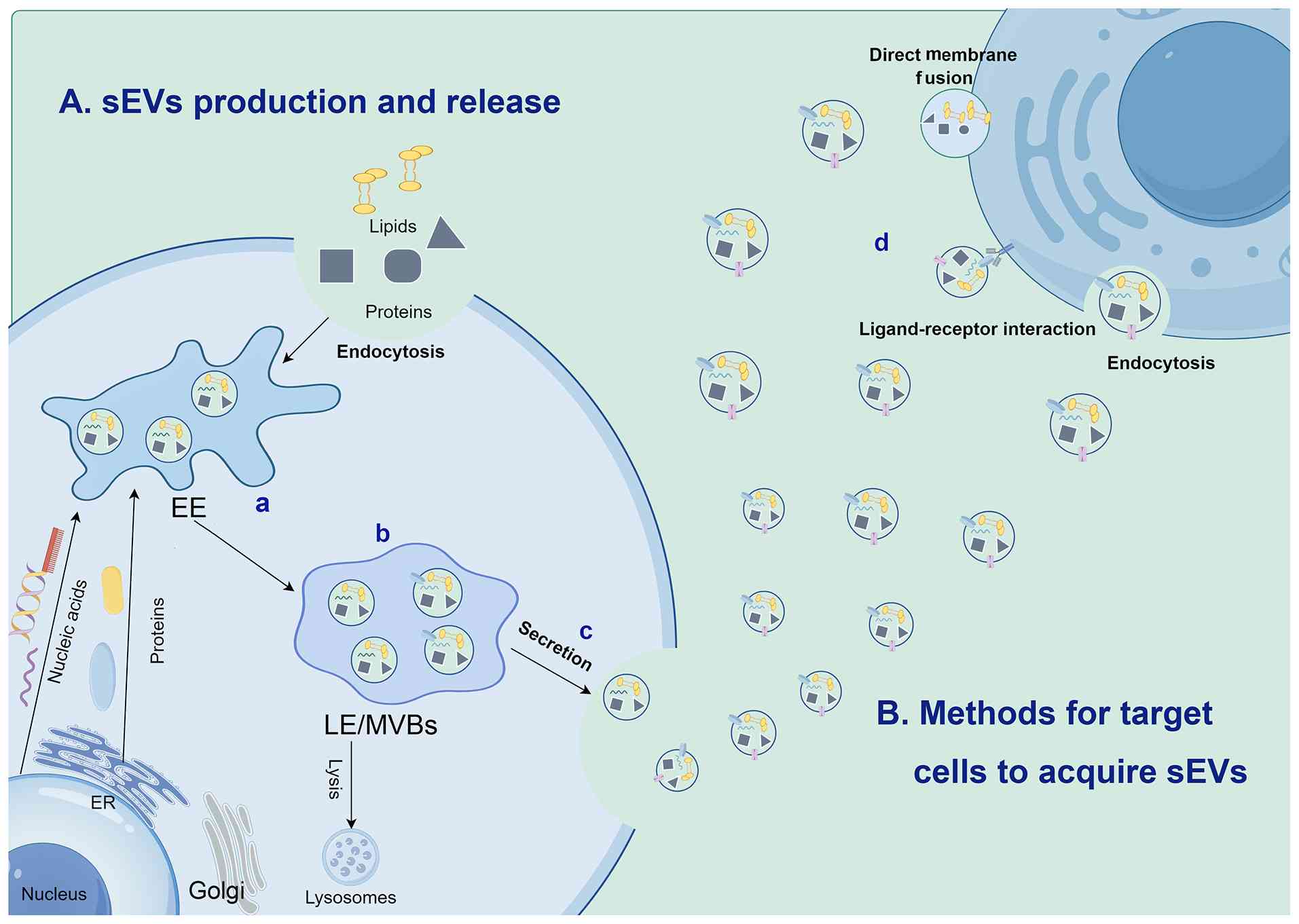 | Figure 1Formation, release and uptake of
sEVs. (A) Formation and release of sEVs. a) The cell membrane and
associated lipids and proteins invaginate together to form EEs,
which contain nucleic acids from the cell nucleus and proteins
synthesized by the ER. b) The early endosomes gradually mature into
LEs or MVBs. c) MVBs have two fates: They are either degraded by
lysosomes or secreted from the cell. When sEVs are secreted, they
bind to target cells through paracrine pathways. (B) Mechanisms for
target cells to acquire sEVs. d) There are three uptake mechanisms:
i) Endocytosis (phagocytosis or other internalization pathways);
ii) ligand-receptor binding, which triggers intracellular signal
transduction and may subsequently initiate internalization; iii)
direct membrane fusion, that is, the sEV membrane fuses with the
target cell plasma membrane, releasing the contents into the
cytoplasm. MVBs, multivesicular bodies; sEV, small extracellular
vesicle; EE, early endosome; LE, late endosome; ER, endoplasmic
reticulum. |
Extraction and separation
Isolation and extraction are the prerequisites for
obtaining highly pure and highly active sEVs, which directly
determine the reliability of subsequent experimental results and
transformation effects. Currently, commonly used sEVs extraction
methods mainly include ultracentrifugation, density gradient
centrifugation, immunomagnetic bead separation, polymer
precipitation, size exclusion chromatography (SEC) and sEVs
kit-based extraction (16).
Ultracentrifugation separates sEVs based on density and size. By
adjusting rotational speed and duration, impurities are effectively
removed to yield highly purified sEVs. Ultracentrifugation is still
the most widely used technique for isolating of sEVs from
biological fluids and culture supernatants. Although suitable for
processing large sample volumes, it has inherent drawbacks: High
shear force may disrupt the structural integrity of sEVs.
Furthermore, it is time-consuming, labor-intensive and costly
(17). Compared with
ultracentrifugation, density gradient centrifugation can further
enhance purity, but it involves complex operations and limited
sample throughput, making it suitable for experiments requiring
high purity (18). Polymer
precipitation is a simple technique that requires no special
equipment. It works by reducing the solubility of target entities
to facilitate sEV isolation. Although this method can produce
relatively high concentrations of sEVs, it introduces a
considerable amount of polymer impurities. This method is easy to
operate and suitable for large-scale extraction (19). SEC utilizes a porous packing
column to separate sEVs and retain sEV activity. Compared with
traditional methods, SEC not only enhances isolation efficiency and
purity but also preserves structural integrity of sEVs by obviating
physical damage. As a result, SEC is widely regarded as a preferred
or benchmark technique in various laboratory settings, and is
particularly suitable for small-volume samples. Nevertheless, its
relatively low yield may constrain its practicality in large-scale
experimental and clinical applications (20). Commercial kit-based extraction
procedures, distinguished by their ease of use and high throughput,
have been widely adopted across diverse research settings. These
kits generally function by altering the solubility of sEVs through
polymer-mediated means, enabling isolation under low-speed
centrifugation. This method is especially advantageous for
processing samples with limited volume (such as synovial fluid
aspirates obtained in routine clinical practice). While kit-based
extraction approaches can achieve higher yields of sEVs, their
purity and specificity are relatively low. Furthermore, they are
prone to co-precipitation of non-vesicular impurities (such as
proteins and polymeric aggregates), which may significantly
interfere with experimental results (21). The benefits and drawbacks of the
various separation methods are summarized in Table I (17-22).
 | Table ICommon sEVs isolation methods and
their benefits and drawbacks. |
Table I
Common sEVs isolation methods and
their benefits and drawbacks.
| Strategy | Principle | Advantages | Disadvantages | (Refs.) |
|---|
| Ultracentrifugation
method | Separation based on
density and size of sEVs | Low cost, high
purity | Time-consuming,
labor-intensive, high-quality equipment, low recovery rate | (17,21, 22) |
| Density gradient
centrifugation method | Utilizes sucrose or
iodixanol to form density gradients, enriching sEVs through
ultracentrifugation | High purity | Time-consuming
operation, small sample processing capacity | (18,22) |
| Kit extraction
method | Modifies the
solubility of sEVs using polymers to precipitate sEVs during
low-speed centrifugation | Convenient and
rapid, high recovery rate | High cost, reagent
residues, lower specificity and purity | (21) |
| Polymer
precipitation method | Utilizes
polyethylene glycol to competitively bind water molecules | Simplifying sEVs
precipitation | Low purity,
potential impurities | (19) |
| Size exclusion
chromatography | Uses a porous
packing column to separate | High efficiency,
high purity, good sEV integrity | Limited sample
processing capacity, lower production volume | (20) |
Of course, the methods for separating sEVs from
different samples vary. Synovial fluid samples are usually
collected in very small quantities and have high viscosity, which
may interfere with the polymer precipitation process (resulting in
excessive co-precipitation) or affect SEC analysis (causing column
blockage or co-elution of large molecular aggregates). Therefore,
pre-treatment steps are usually required before sample separation,
such as dilution, centrifugal removal of cell debris or enzymatic
digestion with hyaluronidase (23). Blood contains various proteins,
which may contaminate sEVs during separation. In addition, platelet
activation in plasma releases a large number of additional
vesicles, so special attention should be paid to the selection of
anticoagulants and centrifugal conditions during collection to
minimize platelet contamination (24). Urine contains Tamm-Horsfall
protein, which can form aggregates and capture sEVs. Therefore,
reducing-reductive pre-treatment is usually required. Brain spinal
fluid samples have small volumes and a low yield of sEVs, and
accordingly, high-recovery-rate separation methods, such as
immunoprecipitation chromatography or microfluidic devices, should
be used (25). Saliva contains a
large amount of mucin and bacterial vesicles, and additional steps
of low-speed centrifugation and density gradient centrifugation are
required (26).
Furthermore, the optimal separation method not only
depends on the sample type but also on the downstream research
goals: In the field of biomarker discovery (such as spectral
analysis of low-abundance proteins or nucleic acids in cohort
studies), purity and reproducibility are of utmost importance, as
contaminating non-vesicular substances may produce false-positive
signals. Therefore, even at the expense of yield and throughput,
high-resolution techniques such as density gradient
ultracentrifugation, immunoprecipitation or sequential SEC-density
gradient ultracentrifugation, are often preferred (27). Conversely, for therapeutic
applications, maintaining biological activity and safety is the
primary consideration, and therefore, size exclusion chromatography
is more recommended (28).
In conclusion, there is no single method that is
applicable to all detection scenarios. It is necessary to
comprehensively consider the sample characteristics (viscosity,
volume, interfering substances) and research goals, and adopt
complementary detection techniques and strict identification
measures, which is crucial for ensuring the reproducibility of
experimental results.
Identification
Current methods for characterizing sEVs include
nanoparticle tracking analysis (NTA), transmission electron
microscopy (TEM) and western blotting (WB) (29). NTA is based on laser scattering
and Brownian motion tracking technology, which enables precise
measurement of sEV size distribution and particle concentration,
providing crucial data on basic properties of sEVs. However, NTA
cannot distinguish non-vesicular particles with sizes similar to
sEVs, and accordingly, complementary methods are needed for
validation (30). TEM allows
direct visualization of sEV size, bilayer membranes and cup-like
structures, which aids in gaining a deeper understanding of their
physical properties. However, TEM provides only semi-quantitative
assessments (31). WB enables
qualitative and quantitative analysis of specific proteins on the
sEV surface. These include positive markers such as CD9/CD63/CD81
(tetraspanins) and (tumor susceptibility 101) TSG101/programmed
cell death 6 interacting protein (PDCD6IP) (ESCRT complex), along
with negative markers such as Calnexin (an endoplasmic reticulum
protein) and LaminB (a nuclear protein), thereby elucidating
biochemical characteristics. However, WB frequently produces
false-positive results due to contamination by cellular debris, and
it fails to provide information on the total number or size
distribution of sEVs (32).
Therefore, a combination of multiple complementary methods is
routinely employed in experimental practice for comprehensive
identification and characterization of sEVs.
Role of sEVs in OA inflammation and
cartilage damage
sEVs and inflammation
Synovial inflammation is a typical pathological
feature of OA. By disrupting the microenvironment within the joint,
it triggers a series of pathological and physiological changes,
thereby becoming a key driving factor for the disease (33). As essential mediators of
intercellular communication, sEVs play a pivotal role in initiating
and perpetuating synovial inflammation (34). sEVs do not function as random
messengers. Instead, they regulate their pro-inflammatory effects
by acting on several mechanistic pathways within the receptor
cells, including the inflammatory signal transduction cascade and
macrophage polarization (35-38).
Inflammatory signal transduction
cascade reaction
sEVs can actively regulate the classical
inflammatory signaling pathways within the OA joint. The synovial
fluid of patients with OA contains high levels of pro-inflammatory
cytokines, such as interleukin-1β (IL-1β) and tumor necrosis
factor-α (TNF-α), which can be transported and delivered to
receptor cells by sEVs (39).
These sEVs exert immunostimulatory effects by activating the
nuclear factor κB (NF-κB) pathway and the
phosphatidylinositol-3-kinase (PI3K)/AKT signaling axis. For
instance, the long non-coding RNA (lncRNA) OANCT derived from
chondrocytes is packaged into sEVs, targets and binds to the fat
mass and obesity-associated protein (FTO protein) and subsequently
promotes M1-type macrophage polarization through the PI3K/AKT
pathway, thereby amplifying the inflammatory response (40). At the same time, proteins related
to sEVs, including IL-1β, IL-6 and various chemokines released by
activated synovial macrophages, further drive the inflammatory
cascade through paracrine signal transduction (41). It is worth noting that different
sEVs carriers can act on the same signaling pathway: Although
microRNAs (miRNAs) and lncRNAs usually regulate signal transduction
indirectly through target gene inhibition or protein isolation, sEV
proteins exert more direct effects on these pathways (42).
Furthermore, different miRNAs, even the same miRNA
molecule, can produce different biological effects under different
conditions. Although miR-146a and miR-146b-5p have highly similar
sequences, they play opposite roles in OA: miR-146a is generally
considered to have an anti-inflammatory effect, while
miR-146b-5p-especially secreted by M1-type macrophages-exacerbates
the inflammatory response through the ubiquitin specific peptidase
3 (Usp3)/SRY-related high-mobility group box protein B5 (Sox5)
pathway (43,44). Remarkably, the elevated level of
miR-146a in OA synovial fluid sEVs is often positively correlated
with the severity of the disease (45), suggesting the failure of a
compensatory anti-inflammatory mechanism. This context-dependent
duality reveals a key principle: The functional outcome of
sEV-miRNAs is not only determined by their molecular
characteristics, but also by the polarization state of the parent
cells and the local microenvironment.
Macrophage polarization and immune
regulation
The polarization of macrophages towards the
pro-inflammatory M1 phenotype is a key event in the pathogenesis of
OA, and sEVs play a central role in this process. sEVs from the
synovial fluid of patients with OA can be efficiently internalized
by macrophages, thereby stimulating the polarization of the M1
phenotype and promoting the release of various inflammatory
mediators, including IL-1β, TNF-α and matrix metalloproteinases
(MMPs) (46). These findings
reveal the important role of sEVs in shaping the immune response
associated with OA (47,48). It is notable that sEVs derived
from inflamed synovial tissue also carry proteins such as
apolipoprotein A1 (a protein closely associated with disease
activity), which can act on monocytes and stimulate their
production of inflammatory cytokines and exacerbate local
inflammatory responses (49).
In conclusion, the pathogenic role of sEVs in OA is
not mediated by a single molecule, but is achieved through the
synergistic action of multiple interrelated signaling pathways.
sEVs form a self-reinforcing vicious cycle: Inflammatory signal
transduction promotes the polarization of macrophages to the M1
type, and this polarization further enhances the release of
pro-inflammatory sEVs. This comprehensive research perspective
emphasizes that the final pathological outcome results from the
cumulative effect of multiple RNA and protein cargoes on the common
downstream signaling pathways.
sEVs and cartilage damage
In addition to regulating synovial inflammation,
sEVs also directly participate in the processes of cartilage damage
and matrix degradation during the pathogenesis of OA through
multiple interrelated mechanisms. These mechanisms can be broadly
classified into three major pathways: i) Extracellular matrix (ECM)
degradation and cartilage destruction; ii) regulation of cell
survival, autophagy and apoptosis; iii) oxidative stress and
ferroptosis; and iv) the regulation of the tissues surrounding the
cartilage and neurogenic regulation.
ECM degradation and cartilage
destruction
sEVs directly participate in the degradation of ECM
and cartilage destruction through two pathways: Indirect regulation
and direct enzymatic action. In the indirect regulation aspect,
sEVs carry non-coding RNAs, thereby regulating the expression of
catabolic enzymes. For instance, treating chondrocytes with sEVs
derived from OA synovial fluid can reduce cell viability, decrease
the expression of catabolic-related genes (such as type II collagen
and aggrecan) and increase the expression of catabolic-related
genes [such as MMPs and a disintegrin and metalloproteinase with
thrombospondin motifs (ADAMTS)] (39,50). Among the molecules carried by
these sEVs, the non-coding RNAs encapsulated in sEVs are key
factors driving the inherent inflammatory response and catabolic
process in the OA pathological process (51).
At the direct action level, sEV-related enzymes
(including MMPs and cathepsins) are transported to the cartilage
surface, where they degrade type II collagen and other ECM
components (52). This mechanism
that directly leads to structural destruction is likely to play a
dominant role in the acute phase of OA. Additionally, during the
progression of OA, the increase in low density lipoprotein receptor
related protein 1 shedding from articular cartilage mediated by
endocytosis mechanisms promotes the occurrence of inflammatory
responses and damages cartilage integrity (53).
It is noteworthy that certain sEV-carrying molecules
can exert protective effects, thereby counteracting their
degradation effects. For instance, lncRNA H19 inhibits ECM
degradation induced by IL-1β through regulating miR-106b-5p, and
promotes chondrocyte proliferation and migration (54). This phenomenon indicates that the
effect on ECM integrity reflects the dynamic balance between
pathogenic and protective signals transmitted by sEVs.
Regulation of cell survival, autophagy
and apoptosis
In addition to affecting the cytoskeleton, sEVs also
regulate chondrocytes and synovial cells by modulating autophagy
and apoptosis. These processes are closely related to the
progression of OA and cartilage loss. The sEV-miR-449a-5p derived
from chondrocytes can inhibit the expression of autophagy-related
protein 4 homolog B (ATG4B) by targeting its mRNA, thereby blocking
the autophagy process of macrophages and promoting the secretion of
IL-1β (55). Similarly, in the
OA rat model, the overexpression of miR-182 in synovial tissue can
target forkhead box O3 (FOXO3)-related mRNAs, suppressing FOXO3
expression and promoting cell apoptosis and inflammatory responses
(56). These research results
indicate that sEVs can regulate the balance between cell survival
and death by targeting the core components of autophagy and
apoptosis pathways.
At the functional level, sEV-miRNAs can act on
various signaling pathways, such as the autophagy pathway mediated
by Usp3, Sox5, FOXP3 and FOXO3 to regulate key biological
processes, including inflammatory responses and cell survival
(57). It is worth noting that
the pathological process of OA is not driven by a single miRNA, but
rather results from the synergistic action of multiple molecules.
Different miRNAs and even the same miRNA can produce different
biological effects under different conditions, which fully
demonstrates their 'environmental dependence' (58,59). However, there is still a lack of
comprehensive understanding of how sEVs regulate these interacting
cellular pathways.
Oxidative stress and ferroptosis
sEVs can reduce the antioxidant stress capacity of
chondrocytes in OA animal models and exacerbate cell apoptosis,
thereby directly damaging the cartilage (60). However, the effects of sEVs on
cartilage are not limited to classical cell apoptosis; they also
involve specific cell death programs driven by metabolic disorders,
among which ferroptosis has attracted considerable attention.
Ferroptosis is a unique cell death mode triggered by intracellular
iron accumulation, lipid peroxidation and plasma membrane damage
(61). Ferroptosis releases
various small molecule substances (such as MMPs), which
subsequently cause damage to the cartilage and matrix (62). Recent evidence indicates that
ferroptosis is an important mechanism by which pathological sEVs
induce chondrocyte death. Kong et al (63) demonstrated that sEVs derived from
fibroblast-like synoviocytes would disrupt the antioxidant defense
system of chondrocytes, thereby promoting iron overload and lipid
peroxidation. This observation suggests that chondrocyte death is
not solely attributed to the classical apoptotic pathway but also
involves the participation of metabolic regulatory mechanisms
centered on iron homeostasis and oxidative stress.
Regulation of the tissues surrounding
the cartilage and neurogenic regulation
In addition to the regulatory effects of
inflammatory cells and synovial cells on cartilage changes,
subchondral osteoblasts can also secrete sEVs that promote
catabolic metabolism of chondrocytes. These sEVs inhibit the energy
metabolism of chondrocytes and thereby induce cartilage degradation
(64). This finding suggests
that sEVs derived from subchondral bone actively participate in the
pathogenesis of OA by targeting the metabolic adaptability of
chondrocytes, rather than simply acting on actinomorphin, and by
regulating the metabolism of chondrocytes.
Furthermore, nerve stimulation can affect the
process of cartilage destruction mediated by sEVs, revealing a new
type of neural-immune-bone axis. Guan et al (65) confirmed that in the context of
OA, sympathetic nerve activity is enhanced, leading to an increase
in norepinephrine levels. This can activate the β1-adrenergic
receptor signaling pathway on chondrocytes. This activation process
promotes the release of exosomes rich in miR-125 from chondrocytes,
and subsequently, these sEVs are transported to the subchondral
bone, where they promote osteoblast differentiation, disrupt the
homeostasis of the subchondral bone and ultimately exacerbate
cartilage damage in aged mice (65). It is worth noting that changes in
the expression level of sirtuin 6 were also observed in
chondrocytes, which is related to the aforementioned process. These
observations indicate that the OA pathological process is not
solely dominated by internal joint mechanisms; instead, sEVs from
surrounding tissues (including bone and nerves) also play a
non-negligible promoting role. Furthermore, these findings also
imply that the metabolic communication between bone, nerves and
cartilage is partially achieved through intercellular signal
transduction mediated by sEVs.
In summary, the cartilage damage in OA is driven by
four interrelated sEV-mediated mechanisms: i) ECM degradation
caused by both indirect genetic regulation and direct enzymatic
action; ii) cartilage cell survival imbalance triggered by
inhibition of autophagy and induction of apoptosis; iii) oxidative
damage ultimately leading to ferroptosis; and iv) neurogenic
regulation. These signaling pathways do not function independently
but are likely to amplify the cartilage degradation process through
synergistic effects. For instance, cartilage cells that undergo
ferroptosis may release damage-associated molecular patterns, which
can then stimulate synovial inflammation; and inflammatory sEVs
can, in turn, make cartilage cells more sensitive to oxidative
stress (66). The identification
of this multi-dimensional regulatory network highlights the need
for future research to deeply analyze the relative contributions of
each pathway and to explore whether sEVs from different cell
sources exhibit different characteristics of cartilage damage.
Interaction between inflammation and bone
changes in OA
OA is a systemic joint disease involving
interactions between cartilage and synovium, which collectively
drives its progression (67). In
the early stages of OA, inflammation initiates and develops, but
the cartilage surface remains relatively intact due to the body's
compensatory mechanisms. In the late stages, intra-articular
decompensation occurs, inflammation worsens, cartilage is destroyed
and even bone changes occur, simultaneously releasing large amounts
of pro-inflammatory mediators that further exacerbate inflammation.
This vicious cycle ultimately leads to joint disruption, causing
pain, swelling and stiffness. Inflammation persists throughout the
process. Degradation products from joint cartilage can trigger
inflammation, leading to synovial fibrosis, vascularization and
immune cell infiltration (68).
Subsequently, immune cells, particularly M1-polarized macrophages,
secrete pro-inflammatory cytokines, accumulating these factors and
accelerating cartilage destruction (69). Concurrently, subchondral bone
thickens and hardens, thereby impairing cartilage nutrient supply.
Additionally, subchondral bone cells secrete pro-inflammatory
cytokines, driving cyclic degradation and destruction of cartilage
(70). Nevertheless, the
'vicious cycle' is mainly based on causal inferences and temporal
correlations. To date, limited investigation has adopted a
longitudinal approach to monitor the evolution of sEV signatures
spanning the transition from initial inflammatory events to
advanced structural deterioration within an identical animal cohort
(71). This lack of such
longitudinal research data is the greatest deficiency in supporting
this theoretical evidence.
In summary, sEVs participate in the cartilage damage
process of OA through multiple molecular mechanisms: They can not
only directly degrade and disrupt ECM structure by regulating the
expression of genes related to matrix metabolism, but also regulate
the balance of autophagy and apoptosis, mediate oxidative stress
and ferroptosis to directly induce the death of chondrocytes, and
receive pathological signals from surrounding tissues such as
subchondral bone and nerves to amplify the effect of cartilage
damage. Furthermore, the inflammatory response in OA and the
apoptosis of chondrocytes can mutually reinforce each other,
forming a vicious cycle, ultimately leading to irreversible disease
progression (Fig. 2).
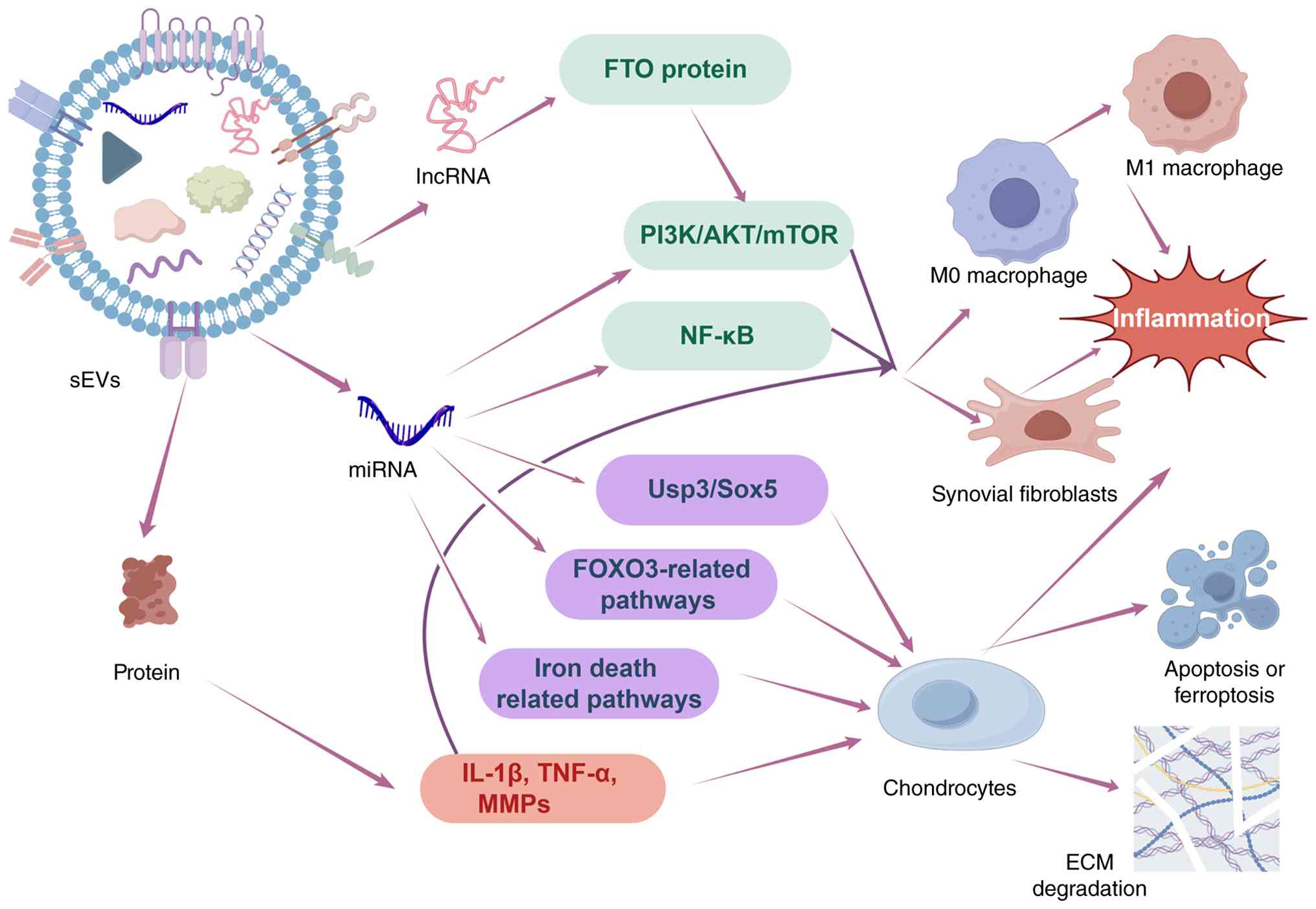 | Figure 2sEVs promote inflammation and
cartilage damage. Under the condition of OA, various small
molecules released by tissues and cells, such as miRNA, lncRNA and
proteins (such as IL-1β, TNF-α and MMPs), can activate NF-κB,
PI3K/AKT, Usp3/Sox5 and ferroptosis-related signal pathways, and
target intracellular molecules and pathways such as FTO protein and
FOXO3 mRNA, thereby promoting the transformation of M0 macrophages
into M1 type (pro-inflammatory), and inducing the secretion of
pro-inflammatory cytokines by synovial fibroblasts, apoptosis or
ferroptosis of chondrocytes, and degradation of ECM, in order to
promote the development of OA. OA, osteoarthritis; sEV, small
extracellular vesicle; ECM, extracellular matrix; miRNA, microRNA;
lncRNA, long non-coding RNA; FOXO3, forkhead box O3; FTO, fat mass
and obesity-associated; Usp3, ubiquitin specific peptidase 3. |
Applications of sEVs in anti-inflammation
and cartilage regeneration
Application in anti-inflammatory
therapy
Inflammation is known to permeate the entire process
of OA development. Therefore, suppressing inflammation is a major
therapeutic measure to counteract OA and protect joints from
damage. The primary clinical approach to suppressing OA
inflammation currently involves NSAIDs. These drugs exert
anti-inflammatory and analgesic effects by inhibiting
cyclooxygenase activity, thereby reducing prostaglandin synthesis
(4). However, NSAIDs may bring
side effects, such as gastrointestinal discomfort and adverse
cardiovascular reactions (72).
In addition, as the disease progresses, NSAIDs may fail to sustain
their initial efficacy (73). In
contrast, sEVs are small, immunogenicity-low and relatively stable
cargo carriers. They can be taken up by target cells and, through
various regulatory mechanisms, avoid the toxic side effects of
drugs. Consequently, sEVs have unique advantages and potential in
anti-inflammatory treatment of OA (74). The following table summarizes
experimental findings on the anti-inflammatory effects of sEVs over
the last 5 years (Table II)
(75-98).
| Table IIApplication of sEVs in
anti-inflammatory therapy for OA. |
Table II
Application of sEVs in
anti-inflammatory therapy for OA.
| Authors, year | Source | In vivo
model | Mechanism of
action | (Refs.) |
|---|
| Song et al,
2024 | M2 macrophages | Surgically induced
rat knee OA | Enhancing lymphatic
endothelial cell activity, proliferation, migration and capillary
formation and reducing apoptosis | (75) |
| Liang et al,
2025 | ADSCs | NR | Promoting FOXC1
deubiquitination to induce macrophage M2 polarization | (76) |
| Zhang et al,
2025 | BMSCs | Sodium
iodate-induced rat knee OA model | Inhibiting NF-κB to
control inflammatory mediators | (77) |
| Zhang et al,
2025 | M2 macrophages | Sodium
monoiodoacetate-induced rat knee OA model | Reducing the
expression of inflammatory factors and promoting the transformation
of M1 type macrophages into M2 type | (78) |
| Tao et al,
2021 | BMSCs | Rat OA model | Inhibiting NF-κB
signaling pathway | (79) |
| Liao et al,
2021 | BMSCs | Rat OA model | Inhibiting the
activation of the NF-κB signaling pathway | (80) |
| Chen et al,
2025 | hUC-MSCs | Sodium
iodacetate-induced rat OA model | Inhibiting the
MAPK4/NF-κB signaling pathway | (81) |
| Kim et al,
2021 | MSCs | NR | Inhibiting the
miR-147b-mediated NF-κB pathway to suppress inflammatory mediator
expression | (82) |
| Lou et al,
2023 | BMSCs | Rat OA model | Inhibiting the
expression of TRAF6 to suppress inflammation | (83) |
| Liao et al,
2025 | Platelet-rich
plasma | Medial
meniscus-induced mouse OA model | Activating PI3K/Akt
signaling pathway to enhance synovial lymphocyte function,
promoting clearance of inflammatory cells and associated
cytokines | (84) |
| Cheng et al,
2025 | Milk | NR | Inhibiting the
TLR4/NF-κB signaling pathway and the PI3K/AKT signaling
pathway | (85) |
| Li et al,
2024 | MSCs | Surgically induced
rat knee OA | Modulating PI3K-Akt
pathway to inhibit degradative enzymes MMP3 and MMP9 expression and
activate antioxidant enzymes | (86) |
| Li et al,
2025 | M2 macrophages | Surgically induced
rat knee OA | Inhibition of
miR-124-3p/STAT3 axis | (87) |
| Karaturk et
al, 2025 | DPSCs | NR | NR | (88) |
| Kang et al,
2025 | ADSCs | Surgically induced
rat knee OA | Inhibiting
chondrocyte hypertrophy and inflammatory response via CRIM1/BMP
signaling pathway | (89) |
| Chen et al,
2024 | Cartilage-derived
stem cells | Sodium
acetate-induced rat knee OA | Reducing
inflammatory cytokine production and improving chondrocyte
microenvironment | (90) |
| Cao et al,
2024 | Synovial
fibroblasts | Surgically induced
mouse knee OA | SOD3 in EVs
prevents ROS accumulation and reduces proinflammatory
cytokines | (91) |
| Zhou et al,
2021 | Synovial
fibroblasts | Surgically induced
rat knee OA | Reducing
inflammatory cytokine production | (92) |
| Wan et al,
2023 | BMSCs | Surgically induced
mouse knee OA | Suppression of
OA-associated inflammatory and gene expression, rescue of
transcriptional responses | (93) |
| Qin et al,
2025 | hUC-MSCs | Surgically induced
mouse knee OA | Inhibiting
Wnt/β-catenin pathway, enhancing autophagy | (94) |
| Liu et al,
2025 | Garlic | Surgically induced
rat knee OA | Inhibiting MAPK
pathway activation and suppressing expression of degradative
enzymes MMP3 and MMP9 | (95) |
| Klyucherev et
al, 2024 | hUC-MSCs rat knee
OA | Surgically
induced | Downregulating
inflammatory markers and increasing Arg-1 expression | (96) |
| Jiang et al,
2025 | BMSCs | NR | Inhibiting the
expression of pro-inflammatory and anabolic genes | (97) |
| El-Din et
al, 2024 | BMSCs | Rat OA model | Regulation of
circYAP1/miRNA-21/TLR7 axis | (98) |
Regulation of intracellular signal
pathways
sEVs, as natural nanocarriers, can carry various
small molecules and bind to target cells to regulate multiple
intracellular singaling pathways (such as PI3K/AKT/mTOR and NF/κB
pathways), inhibiting the release of inflammatory mediators such as
IL-1β, TNF-α and inducible nitric oxide synthase, thereby
alleviating the inflammatory response in OA joints (99).
The NF-κB signaling pathway is a key pathway
associated with inflammatory progression and tissue destruction in
OA (100). Proinflammatory
cytokines can activate the NF-κB pathway, thereby suppressing
chondrocyte anabolic metabolism and promoting the secretion of
various MMPs to accelerate articular cartilage destruction.
Furthermore, activation of this pathway induces chondrocyte release
of inflammatory cytokines and chemokines, including TNF-α, IL-1β,
IL-6, IL-8 and receptor activator of nuclear factor-κB ligand,
thereby amplifying inflammatory responses (101). Consequently, sEVs can target
NF-κB pathway inhibition to suppress host inflammation. For
instance, sEVs derived from bone marrow mesenchymal stem cells
(BMSCs) have been proven to significantly inhibit the NF-κB
pathway, and the physical pre-treatment can enhance this inhibitory
effect (80,82). Nevertheless, it must be
acknowledged that the NF-κB pathway is a fundamental signaling axis
for host immune defense and inflammatory resolution. Systemic
blockade of this pathway has potential safety risks, such as
increased susceptibility to pathogens and impaired immune
surveillance function (102).
By comparison, the PI3K/Akt/mTOR signaling pathway
presents a more favorable and selective intervention window. It
inhibits chondrocyte apoptosis by regulating downstream pathways
(including Bad, Caspase-3 and NF-κB) and is therefore crucial for
regulating chondrocyte proliferation, apoptosis and matrix
homeostasis (103). In the
early stage of OA, the PI3K/AKT/mTOR pathway can be activated by
TNF-related apoptosis-inducing ligand, IL-15, IL-22 and various
growth factors, including AKT and mTOR phosphorylation to
exacerbate the inflammatory response and promote chondrocyte
apoptosis (104). sEVs can act
as various inhibitors within this signaling pathway, such as
negative regulators of mTOR and TNF receptor-associated factor 6
(TRAF6), thereby suppressing OA inflammation (105). For instance, Lou et al
(83) utilized engineered sEVs
to target TRAF6 and inhibit the PI3K/AKT/mTOR pathway, which not
only reduced inflammatory mediators but also promoted autophagy.
This observation implies that, relative to mere suppression of
inflammatory signaling, activation of endogenous pathways such as
autophagy may confer superior therapeutic benefits.
It is worth noting that most current studies on the
regulation of the NF-κB and PI3K/AKT/mTOR pathways have used
pathway inhibitors or agonists to indirectly infer the role of
these signaling cascades. To date, only a few studies have verified
the necessity of these signaling pathways through gene knockout
techniques. For instance, conditional gene knockout of the
phosphatase and tensin homolog in chondrocytes has been shown to
constitutively activate the PI3K/AKT signaling pathway and promote
the development of OA (106).
Other studies have used alternative genetic approaches, such as
gene knockdown, overexpression or drug regulation, to assess the
involvement of NF-κB-related molecules (107,108). However, it should be noted that
these studies explored the role of these signaling pathways in the
pathogenesis of OA independently of sEV-mediated effects.
Therefore, although these studies have provided valuable insights
into the functional significance of these signaling pathways in OA,
they do not constitute direct genetic evidence for the specific
signal regulation of sEV, and their applicability to sEV-mediated
therapy remains to be established. Furthermore, there is complex
cross-regulation between the NF-κB and PI3K/AKT/mTOR pathways.
However, to date, to the best of our knowledge, relatively few
studies have evaluated the synergistic or antagonistic effects of
sEVs when simultaneously acting on both pathways, which limits a
comprehensive understanding of the therapeutic mechanisms (109).
Modulating the polarization of immune
cells
sEVs can also modulate immune cell polarization,
promoting the transition of M0 or M1 macrophages into M2
macrophages, thereby improving the intra-articular immune
microenvironment and alleviating inflammatory responses (110). Under normal conditions,
synovial macrophages within the joints remain quiescent; however,
during OA inflammation, they polarize toward the pro-inflammatory
M1 phenotype, thereby initiating or amplifying the release of a
series of pro-inflammatory cytokines (111). By contrast, the
anti-inflammatory M2 phenotype secretes IL-10, arginase-1 and
transforming growth factor (TGF)-β, suppressing inflammation and
promoting tissue repair (110).
Therefore, using sEVs to polarize macrophages from M0 or M1 to M2
type is expected to alleviate synovial inflammation and mitigate
cartilage degeneration, presenting a promising therapeutic approach
for OA (112). To enhance this
polarization effect, sEVs can be engineered to carry specific
cytokines or proteins. For instance, Liang et al (76) successfully modified
adipose-derived MSC-sEVs with ubiquitin-specific protease 15
(USP15). This enhanced the release of anti-inflammatory and
chemotactic factors by inducing macrophage polarization toward the
M2 phenotype through promoting FOXC1 protein deubiquitination
(76). Qian et al
(112) found that sEVs from M2
macrophages could target Toll-like receptor 3 (TLR3) and collagen
type X α 1 chain (COL10A1) through miR-26-5p, also driving the M1
to M2 phenotypic transformation. However, it is important to note
that although these studies have confirmed the feasibility of the
sEV-induced M2 polarization mechanism, it remains to be further
investigated whether the polarized M2 macrophages will revert to
the M1 phenotype.
Application of engineered sEVs in
anti-inflammatory therapy
Although sEVs possess strong barrier penetration and
high circulatory stability, their entry into joints is rapidly
cleared by intracellular cells or enzymatic reactions, thereby
limiting their clinical application (35). To address these obstacles, two
principal engineering approaches have been developed in recent
years: Engineered sEVs and hydrogel delivery systems. Comparative
evaluations have revealed that both modalities markedly enhance
treatment outcomes (Table III)
(113-118). Among them, engineering sEVs
with exogenous materials can effectively increase their retention
time and targeting accuracy in the joint cavity, thereby amplifying
the anti-inflammatory effect (119). Hydrogels, with their unique
biocompatibility, stability and degradability, are widely employed
to enhance sEV retention and adhesion in OA joints (120). For instance, encapsulating M2
macrophage-derived sEVs within a thermosensitive Pluronic hydrogel
significantly prolonged sustained release up to 12 days. This
formulation effectively promotes synovial lymphatic vessel
formation and enhances synovial lymph drainage, thereby suppressing
inflammation (75). Another
study demonstrated that, compared with the pure sEVs group,
HA-alginate hydrogel-loaded sEVs (ALG-M2-sEVs hydrogel) could
adhere to cartilage surfaces, enabling sustained delivery of
M2-sEVs and extending its therapeutic duration. Rat models treated
with ALG-M2-sEVs hydrogel exhibited significantly enhanced
cartilage damage repair, highlighting the potential of this
hydrogel composite system for anti-inflammatory and cartilage
repair applications (78). In
contrast to free sEVs, this hydrogel-based delivery platform
substantially extends the articular retention duration of sEVs,
thus facilitating the maintenance of elevated local concentrations
and subsequently attenuating inflammation. It is worth noting that
recent research has combined these two strategies to address the
two challenges of prolonged presence and organizational
infiltration. Wan et al (93) developed spherical gelatin
methacrylate (GelMA) hydrogels encapsulating collagen II-targeted
peptide WYRGRL and small inhibitor leucine-rich repeat kinase 2
(LRRK2)-IN-1-modified engineered sEVs. Unlike the passive release
of conventional hydrogels, this design overcomes dense collagen
barriers and rapid sEVs clearance, enhancing targeting to
chondrocytes and enabling delivery of the encapsulated LRRK2-IN-1
deep into cartilage. By inhibiting LRRK2, this approach regulates
mitochondrial homeostasis, inflammation and autophagy, thereby
suppressing inflammation and promoting cartilage repair. Through
the integration of hydrogel-mediated sustained release with the
active targeting capacity of engineered sEVs, this approach enables
effective modulation of mitochondrial homeostasis and autophagic
flux, thereby conferring superior therapeutic benefits relative to
non-targeting delivery systems (93). This integrated approach
epitomizes the advanced development in sEV-based therapeutic
strategies for OA to date. In conclusion, sEVs inhibit the
progression of inflammatory response by regulating a variety of
signaling pathways and macrophage polarization processes, thereby
inhibiting the expression of pro-inflammatory cytokines.
Furthermore, engineered sEVs can prolong their retention time in
local tissues and enhance anti-inflammatory effects; when coupled
with targeting peptides, these sEVs can achieve better targeting
specificity, which represents a more advanced technological
improvement in the development of OA treatment strategies based on
sEVs.
| Table IIIComparative analysis of delivery
strategies for sEVs-based osteoarthritis therapy. |
Table III
Comparative analysis of delivery
strategies for sEVs-based osteoarthritis therapy.
| Therapeutic
strategy | Modification
method | Pharmacokinetic
feature | Advantages | Limitations | (Refs.) |
|---|
| Native sEVs | Unmodified | Passive targeting,
rapid clearance | High
biocompatibility, simple preparation, low immunogenicity | Poor stability, low
bioavailability, lack of targeting | (113) |
| Engineered
sEVs | Surface
modification (e.g. targeting peptides, antibodies) or cargo
loading | Active targeting,
moderately prolonged retention | Enhanced precision,
increased cellular internalization, capability for drug
loading | Complex engineering
process, stability issues | (114,115) |
| Hydrogel-based
delivery systems | Physical
encapsulation of sEVs within hydrogel matrices | Passive retention,
sustained release | Biocompatibility,
biodegradability, significantly prolongs intra-articular time | Lacking active
targeting, limited penetration into dense cartilage matrix | (116,117) |
| Engineered
sEVs-hydrogel composite systems | Combination of
hydrogel encapsulation technology with the surface/content material
engineering of sEVs | Dual-targeting,
controlled release | Synergistically
addresses retention (hydrogel), specificity (engineering),
bioactivity | Most complex
fabrication, higher cost, regulatory challenges for clinical
translation | (115,118) |
Applications in promoting cartilage
repair
Beyond the occurrence and progression of
inflammation, another crucial pathological feature of OA is the
persistent degradation of articular cartilage accompanied by
concomitant bone changes (68).
The regenerative capacity of cartilage and bone tissue is limited.
Furthermore, articular cartilage lacks vascularization and the
supply of essential nutrients for oxygenation, and this condition
is further exacerbated by continuous inflammatory damage caused by
OA, thereby accelerating cartilage degeneration and bone spur
formation (2). Currently,
surgical interventions such as arthroscopic procedures and joint
replacement remain the primary clinical approaches (5). However, high costs and
postoperative complications continue to impose significant burdens
on patients (121). The
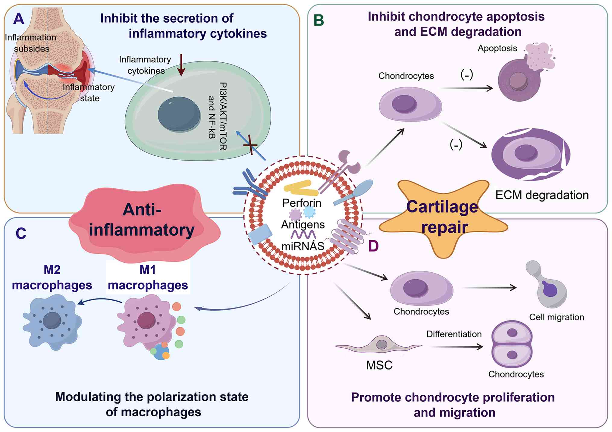
repair-promoting sEVs can inhibit the apoptosis of chondrocytes and
the degradation of ECM, and also promote the proliferation and
migration of chondrocytes as well as the differentiation of stem
cells into chondrocytes. Therefore, as a means of cartilage repair,
it is expected to play an immeasurable role in clinical practice in
the future (122).
Inhibition of chondrocyte apoptosis
and matrix degradation
On the one hand, sEVs can inhibit chondrocyte
apoptosis, promote autophagy and suppress cartilage matrix
degradation to inhibit cartilage degeneration (Table IV) (88,123-140).
| Table IVApplication of sEVs in inhibiting
chondrocyte apoptosis. |
Table IV
Application of sEVs in inhibiting
chondrocyte apoptosis.
| Authors, year | EVs source | Processing | Animal model | Mechanism | (Refs.) |
|---|
| Mao et al,
2021 | BMSCs | No special
treatment | Surgically induced
mouse knee OA | Enhancing Col2a1
and Sox9 expression, suppressing MMP13 expression | (123) |
| Zhao et al,
2023 | ADSCs | Hypoxia
treatment | Lumbar arthritis in
mice induced by spinal instability | Enhancing sEVs'
promotion of chondrogenic anabolism and prevention of
catabolism | (124) |
| Zhang et al,
2024 | Expi293F cells |
CAP-EVs/siMMP13 | Surgically induced
rat knee OA | Reducing MMP13
levels, increasing cartilage collagen COL2A1 and proteoglycans | (125) |
| Wang et al,
2022 | ADSCs | miR-486-5p
transfection | Surgically induced
rat knee OA | Suppressing
endoplasmic reticulum stress | (126) |
| Shi et al,
2023 | Plasma | No special
treatment | NR | Inhibiting
chondrocyte apoptosis by suppressing BAX protein, downregulating
TNF-α and upregulating TGF-β1, IL-4 and IL-10 | (127) |
| Lu et al,
2025 | BMSCs | Quercetin
treatment | Surgically induced
mouse knee OA | Reducing expression
of proinflammatory genes and iNOS, TNF-α, MMPs | (128) |
| Karaturk et
al, 2025 | DPSCs | Hypoxia
treatment | NR | Enhancing sEVs'
suppression of inflammatory factors | (88) |
| Li et al,
2023 | ADSCs | miR-376c-3p
transfection | Sodium
monoiodoacetate-induced rat knee OA | Targeting WNT3 or
WNT9a to inhibit WNT/β-catenin pathway | (129) |
| Wang et al,
2025 | Human amniotic
epithelial stem cells | No special
treatment | Surgically induced
rat knee OA | Delivery of
ACTA2-AS1 downregulates ACSL4, inhibiting lipid peroxidation and
ferroptosis | (130) |
| Li et al,
2022 | hUC-MSCs | No special
treatment | Surgically induced
rat knee OA | Proliferation and
migration, inhibiting apoptosis, suppressing proinflammatory factor
secretion, promoting anti-inflammatory factor expression,
modulating macrophage polarization | (131) |
| Xu et al,
2024 | Osteoblasts | No special
treatment | Surgically induced
mouse knee OA | Promoting
chondrocyte migration and ECM production, inhibiting apoptosis by
upregulating DLX2 to inactivate the Wnt pathway | (132) |
| Meng et al,
2023 | ADSCs | No special
treatment | Sodium
iodacetate-induced rat knee OA | miR-429 promotes
autophagy by targeting FEZ2 | (133) |
| Long et al,
2023 | Synovial
mesenchymal stem cells | MATN3
treatment | Surgically induced
mouse knee OA | MATN3 inhibits
IL-17A/PI3K/AKT/mTOR pathway | (134) |
| Xu et al,
2023 | Platelets | No special
treatment | Surgically induced
mouse knee OA | Modulating immune
and inflammatory responses, promoting chondrocyte proliferation and
migration | (135) |
| Liu et al,
2025 | Human urinary stem
cells | miR140
transfection | Surgically induced
rat knee OA | Downregulating
CAPN1 expression, improving mitochondrial morphology, reducing ROS
accumulation, enhancing mitochondrial membrane potential, promoting
mitochondrial autophagy | (136) |
| Chen et al,
2025 | BMSCs | No special
treatment | MIA-induced TMJOA
rats | Inhibiting
apoptosis and activating the PI3K/AKT pathway | (137) |
| Cao et al,
2023 | hUC-MSCs | CAP-EVs
hydrogel | Surgically induced
rat knee OA | Negative regulation
of p53 signaling pathway inhibits OA chondrocyte senescence and
inflammation | (138) |
| Yang et al,
2025 | hUC-MSCs | miR-148a-3p
transfection, CAP loading | Surgically induced
mouse knee OA | miR-148a targets
ROBO2 to maintain cartilage homeostasis | (139) |
| Xu et al,
2021 | BMSCs
transfection | miR-326
mimetic | Sodium
iodate-induced knee OA in rats | Inhibiting
pyroptosis in chondrocytes by targeting HDAC3 and activating
STAT1/NF-κB p65 signaling pathways | (140) |
Apoptosis is a cell death pathway triggered by
external environmental stimuli [e.g., inflammatory cytokines,
reactive oxygen species (ROS) and TNF-α]. It is executed through
caspase-dependent signaling cascades, in which caspase-8 activates
downstream executioner caspases (such as caspase-3 and caspase-7)
to orchestrate programmed cell death (141). Stem cell sEVs can effectively
inhibit chondrocyte apoptosis and subsequent cartilage degeneration
by suppressing of multiple key genes or proteins in the apoptotic
pathway and blocking the activation of NF-κB and other signaling
cascades (140,142). Therefore, targeting these
signaling pathways to prevent chondrodegeneration by modulating
this pathway may become a future treatment approach for OA. At the
apoptotic level, the pro-apoptotic effector BAX acts on the
mitochondrial outer membrane, promoting its permeabilization and
facilitating the release of cytochrome C and other pro-apoptotic
factors into the cytoplasm. These factors, in turn, activate the
Caspase family (caspase-3/7-9), triggering apoptosis and cell death
(143). Accordingly, inhibiting
BAX activation may be crucial for reducing chondrocyte apoptosis
following injury. Studies have indicated that treatment of inflamed
chondrocytes with human plasma sEVs downregulates BAX expression,
thereby suppressing caspase-3 activation and inhibiting chondrocyte
apoptosis. Concurrently, it enhances SOX9 activity, promoting
chondrogenesis (127).
Nevertheless, it should be noted that the human plasma sEVs
employed in this investigation were obtained from healthy donors.
In clinical practice, however, patients with OA often have multiple
comorbid conditions, including diabetes, hypertension and
hyperlipidemia. Whether such systemic metabolic disturbances modify
the compositional profile and functional properties of circulating
sEVs remains to be evaluated (144). Consequently, the true
therapeutic effect of healthy donor-derived sEVs in the OA patient
population may be subject to overestimation.
Autophagy serves as a protective
mechanism in normal chondrocytes
Through autophagy, chondrocytes can self-clear
defective organelles and macromolecules, enabling maintenance of
nutrition and metabolism (145). This process promotes cell death
induced by aging and trauma, thereby sustaining cellular
homeostasis. sEVs can intrinsically activate autophagy programs in
chondrocytes or serve as delivery carriers to promote senescent or
injured chondrocyte autophagy (146). For instance, Matrilin-3
(MATN3), a non-collagenous chondrospecific ECM protein, alleviates
autophagy defects in osteoblasts by binding to IL-17 and activating
the PI3K/AKT/mTOR pathway, as demonstrated by Long et al
(134). Their team successfully
delivered MATN3 to OA models using sEVs extracted from synovial
MSCs as carriers, thereby promoting chondrocyte autophagy and
maintaining cellular homeostasis. However, autophagy is a
double-edged sword, as excessive activation of autophagy can also
lead to cell death, namely autophagic cell death. The study did not
investigate whether the level of autophagy induced by MATN3 falls
within a safe and beneficial range, nor did it establish the
relationship between autophagic activity and cell survival.
Furthermore, mitochondrial autophagy, a crucial form of cellular
autophagy, participates in the pathogenesis of OA. Deficiency in
mitochondrial autophagy results in chondrocyte death, ECM
homeostasis imbalance and cartilage degeneration. Therefore, it has
increasingly become a research hotspot. BMSCs-sEVs have been
confirmed to promote mitochondrial autophagy in chondrocytes,
thereby inhibiting chondrocyte apoptosis (147). Additionally, Liu et al
(136) engineered human urinary
stem cells (hUSCs)-140-sEVs by transfecting hUSC sEVs with miR-140.
These engineered sEVs delivered miRNA-140 to target and
downregulate calpain 1 expression, thereby decreasing ROS
accumulation, strengthening mitochondrial membrane potential and
promoting mitochondrial autophagy (136). However, the detection methods
used to assess mitochondrial autophagy in current studies vary
significantly. Certain studies rely solely on immunoblotting
analysis of microtubule-associated protein 1A/1B light chain 3 and
p62, while others utilize the mito-Keima transgenic reporter
system. The former cannot distinguish between mitochondrial
autophagy and classical autophagy, while the latter faces
considerable technical challenges in in vivo applications.
This methodological discordance markedly undermines the
comparability and reproducibility of conclusions drawn from
different studies.
The ECM of articular chondrocytes is primarily
composed of type II collagen synthesized and secreted by
chondrocytes. It provides cushioning, wear resistance and
lubrication; transports nutrients and metabolic waste to
chondrocytes; and maintains joint structural integrity to prevent
degradation. Following arthritis onset, various intra-articular
cells secrete chondromatrix-degrading enzymes such as MMPs and
ADAMTS5 to degrade type II collagen and disrupt cartilage
integrity. Therefore, sEVs control of ECM degradation to preserve
its integrity and stability represents another crucial therapeutic
approach for OA. For example, sEVs derived from human umbilical
cord MSCs can reduce the expression of MMP-13 and ADAMTS5 in
chondrocytes induced by IL-1β (131), while miR-7704-modified sEVs can
enhance the expression of type II collagen and reduce the level if
MMP-13, which is helpful for maintaining ECM homeostasis (148).
In conclusion, sEVs exhibit multiple synergistic
effects in cartilage repair, but their inherent repair capabilities
still have clear boundaries. Although sEVs can inhibit apoptosis
(for instance, through the BAX/Caspase-3 axis) and promote ECM
synthesis, relying solely on sEVs to induce endogenous repair is
often insufficient for large-area cartilage defects in advanced OA.
Hence, future research priorities may shift toward sEV-based
combination therapies, such as integrating sEVs with bioengineering
strategies or combining sEVs with pharmaceutical agents.
Promoting the proliferation,
migration, and differentiation of chondrocytes
On the other hand, sEVs can accelerate cartilage
repair by promoting chondrocyte proliferation and migration.
Chondrocyte proliferation and migration are crucial pathways for
repairing damaged cartilage, which is essential for restoring the
morphology and function of joints damaged by OA. sEVs can directly
promote chondrocyte proliferation and migration (35,149). For instance, M2-sEVs have been
proven to induce M1 macrophages to polarize towards the M2
phenotype, thereby alleviating the inhibition of chondrocyte
proliferation by OA and reducing apoptosis (78). Furthermore, sEVs also stabilize
the chondral microenvironment by promoting chondrocyte secretion of
ECM. Human umbilical cord MSC sEVs secrete various immunomodulatory
and reparative factors. They were shown to suppress the
upregulation of MMP-13 and ADAMTS-5 while promoting increased type
II collagen expression. Concurrently, they were observed to enhance
type II collagen synthesis, thereby promoting ECM synthesis of
chondrocytes (150).
Nevertheless, that study did not evaluate the ultrastructure or
mechanical modulus of the newly formed matrix, and thus could not
confirm whether its quality is equivalent to that of native hyaline
cartilage.
Additionally, sEVs can promote the differentiation
of MSCs into chondrocytes to facilitate cartilage repair. sEVs are
rich in or loaded with various growth factors, including TGF-β1,
vascular endothelial growth factor and stromal cell-derived
factor-1. These factors can induce various MSCs to differentiate
into chondrocytes, thereby promoting cartilage repair (151). For example, TGF-β1 in sEVs
suppressed mothers against decapentaplegic homolog 2/3 (Smad2/3),
extracellular signal-regulated kinase 1/2 (ERK1/2) and p38
signaling pathways, thereby promoting BMSC differentiation
(152). Wu et al
(153) successfully engineered
pH-responsive sEVs loaded with VSV-G and hyaluronan synthase 2
(HAS2) (V-H-sEVs). Under acidic conditions, the low pH
responsiveness of the VSV-G protein not only effectively promoted
sEVs fusion with the cell membrane but also increased the amount of
HAS2 delivered to cells via sEVs. Chondrocytes induced by V-H-sEVs
produced HA and promoted BMSC chondrogenic differentiation while
preserving the chondrocyte phenotype (153). Special caution is warranted
because during the differentiation of MSCs into chondrocytes,
improper microenvironmental regulation, such as excessively high
TGF-β concentrations or abnormal mechanical stimulation, can
readily drive cells toward a hypertrophic chondrocyte phenotype, as
indicated by high COL10A1 expression, rather than a stable hyaline
chondrocyte phenotype. The latter may ultimately lead to
endochondral ossification and rapid degeneration of the graft
(154).
In summary, sEVs can prevent chondrocyte apoptosis
and ECM degradation while promoting chondrocyte proliferation and
migration, and facilitating MSCs to differentiate into
chondrocytes. This offers tremendous potential for future cartilage
repair and regeneration in OA (Fig.
3). However, a fundamental paradox still exists: The
characteristic of end stage OA is severe disruption of the
microenvironment, including persistent inflammation, oxidative
stress, acidic pH value and excessive proteolytic activity
(155). Whether exogenous sEVs
can preserve their biological functionality stability in such harsh
environments remains uncertain. Notably, most studies on sEVs
promoting cartilage repair to date have adopted an experimental
model in which inflammation is pretreated before sEVs
administration. This experimental design, to a certain extent,
avoids the most severe clinical reality, that is, the destructive
impact that the microenvironment of advanced OA may have on the
sEVs themselves that are administered. Therefore, more research is
still needed in the future to evaluate the therapeutic effect of
sEVs in cartilage repair.
The double-edged sword effect of sEVs:
Causing disease or providing treatment
sEVs play a dual role in the occurrence, progression
and treatment of OA, and this role depends on their cellular origin
and the surrounding environmental background (156). Furthermore, the bioactive
molecules carried by sEVs do not act independently but rather work
together through specific signaling pathways to mediate complex
interactions between cells in the joint microenvironment. Their
effects are bidirectional: They can either exacerbate the
inflammatory response or participate in tissue repair, and the
specific direction depends on the integration of the
microenvironmental state and regulatory signals (56). This phenomenon is not random, but
is governed by sophisticated regulatory mechanisms.
Causing disease: Disrupting homeostasis
through inflammatory and catabolic pathways
In the pathological microenvironment of OA, sEVs
derived from senescent chondrocytes, inflammatory synovial
fibroblasts and M1 macrophages serve as important carriers for
transmitting 'decompositional metabolic signals', markedly
exacerbating local inflammation and cartilage degeneration
(44,157-160). These sEVs not only contain
abundant pro-inflammatory miRNAs (such as miR-146a and miR-155) and
degradation enzymes like MMP-13, but also reduce or lack some
protective anabolic miRNAs, such as miR-140-5p and matrix proteins.
This imbalance lays the foundation for their pathogenic effects
(161). Specifically, their
pathogenic mechanism involves cross-regulation of multiple
signaling pathways. First, sEVs derived from M1 macrophages can
deliver specific miRNAs to chondrocytes, activating the NF-κB
signaling pathway and weakening autophagy by targeting the
Usp3/Sox5 axis or suppressing ATG4B, thereby disrupting chondrocyte
metabolic homeostasis and promoting matrix degradation (44). Second, sEVs secreted by synovial
cells carry lncRNA OANCT, which can bind to FTO protein and amplify
the inflammatory response through the PI3K/AKT/mTOR pathway, while
promoting the polarization of synovial macrophages to the M1
phenotype (40). At the same
time, pro-inflammatory factors released by M1 macrophages and
various enzymes (such as MMPs and collagenases) directly act on
chondrocytes (41,52); and sEVs derived from synovial
cells can also induce chondrocytes to undergo ferroptosis, further
exacerbating tissue damage (63). These pathways ultimately
synergistically activate NF-κB, inhibit autophagy and induce
ferroptosis, forming a malignant microenvironment characterized by
excessive catabolism and chronic inflammation at the joint site,
continuously accelerating the degradation of ECM (162).
Providing treatment: Restoring
homeostasis via anti-inflammatory and anabolic pathways
In contrast, 'therapeutic sEVs' derived from MSCs
exert 'reparative effects'. They are capable of reprogramming
recipient cells toward a state conducive to cellular well-being.
These sEVs are primarily derived from MSCs or M2 macrophages and
possess distinctive molecular signatures: They are enriched with
anti-inflammatory miRNAs, such as miR-140-5p and miR-23a, and
growth factors, while simultaneously suppressing pro-inflammatory
cytokines, such as miR-155, and catabolic enzyme activity (52). Their therapeutic mechanisms are
primarily achieved through the following pathways. First,
MSC-derived EVs carrying 'therapeutic' miRNAs such as miR-26b-5p
can target and inhibit the TLR3/NF-κB axis, promoting the
polarization of M1 macrophages toward the anti-inflammatory M2
phenotype, thereby secreting IL-10 and TGF-β and reversing the
inflammatory microenvironment (112). This accomplishes
immunomodulation. Second, engineered sEVs, such as those carrying
miR-149 or miR-140-5p, can target the PI3K/AKT/mTOR and RAS/ERK
pathways within chondrocytes, suppressing apoptosis through
downregulation of the BAX/Caspase-3 signaling cascade while
simultaneously activating autophagic pathways to clear damaged
organelles (163,164). This protects chondrocytes and
eliminates damaged cellular components. Furthermore, these sEVs can
modulate Smad signaling pathways to upregulate the expression of
SOX9 and type II collagen, thereby promoting ECM synthesis and
repair (165,166). This indicates that therapeutic
sEVs can effectively reverse the deleterious microenvironment and
restore joint homeostasis. The differences between these two types
of sEVs are presented in Table V
(40, 44,52,63,112,129,157-166) and Fig. 4. This distinction highlights the
importance of developing sEV-based therapeutic strategies in
research.
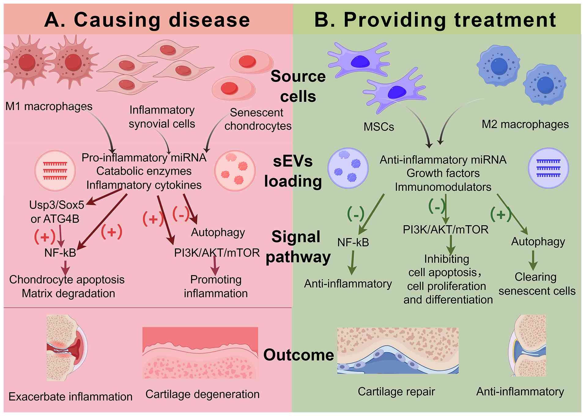 | Figure 4The dual roles of sEVs in OA:
Promotion or treatment. (A) Pathogenic sEVs derived from M1
macrophages and inflammatory synovial cells carry pro-inflammatory
miRNAs, proteins and cytokines. These sEVs target Usp3/Sox5 or
ATG4B to indirectly activate NF-κB, or directly activate NF-κB and
PI3K/AKT/mTOR, while suppressing autophagy, ultimately promoting
inflammation, apoptosis and matrix degradation. (B) Therapeutic
sEVs derived from MSCs and M2 macrophages carry anti-inflammatory
miRNAs, growth factors and immunomodulators. These sEVs inhibit
NF-κB and PI3K/AKT/mTOR or promote autophagy, thereby exerting
anti-inflammatory effects, inhibiting apoptosis, promoting cell
proliferation and migration, and inducing MSC differentiation into
chondrocytes, culminating in cartilage repair. sEV, small
extracellular vesicle; miRNA, microRNA; MSC, mesenchymal stem cell;
ATG4B, autophagy related 4B cysteine peptidase; Usp3, ubiquitin
specific peptidase 3. |
| Table VComparative analysis of pathogenic
and therapeutic sEVs in OA. |
Table V
Comparative analysis of pathogenic
and therapeutic sEVs in OA.
| Feature | Pathogenic sEVs
(drivers of OA) | Therapeutic sEVs
(inhibitors of OA) | (Refs.) |
|---|
| Primary
sources | Senescent
chondrocytes, synovial fibroblasts (inflamed), M1 macrophages | MSCs, M2
macrophages, engineered cells | (44,52,157-160) |
| Key molecular
cargo | Upregulated:
Pro-inflammatory miRNAs (miR-146a, miR-155), catabolic enzymes
(MMP-13, ADAMTS-5), inflammatory cytokines (IL-1β, TNF-α);
Downregulated: Anabolic miRNAs (miR-140-5p, miR-23a), matrix
proteins (collagen II, aggrecan) | Upregulated:
Anti-inflammatory miRNAs (miR-140-5p, miR-23a), growth factors
(TGF-β, IGF-1), immunomodulators (PD-L1, TSG-6); Downregulated:
Pro-inflammatory miRNAs (miR-155), catabolic enzymes (low levels of
MMP-13) | (44,52,157-161) |
| Target
pathways | Activation: NF-κB
MAPK/ERK; Promotion: Inflammation and catabolism | Inhibition: NF-κB,
Wnt/β-catenin; Activation: PI3K/Akt, autophagy; Promotion:
Anabolism and survival | (40,44,63,112, 129,162-166) |
| Biological
effects | Degenerative:
Inducing chondrocyte apoptosis, accelerating ECM degradation,
spreading inflammation (SASP) | Regenerative:
Promoting cell proliferation, enhancing matrix synthesis,
modulation of immune response (M2) | (44,112, 157-160, 162-166) |
In summary, the fundamental distinction between
pathogenic sEVs and therapeutic sEVs lies in the differential
regulation of key signaling pathways, such as NF-κB and
PI3K/AKT/mTOR, by their respective cargo compositions, as well as
their distinct cellular origins. This difference highlights the
critical importance of in-depth elucidation of their molecular
profiles and downstream mechanisms in the development of sEV-based
therapeutic approaches.
Limitations
Limitations of current in vitro
models
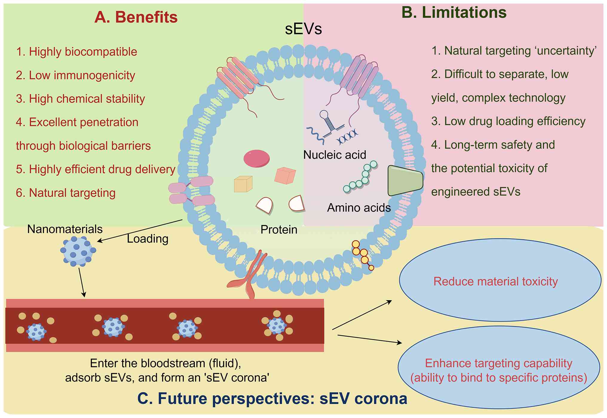
sEVs have excelled in the treatment of a wide range
of diseases due to their low immunogenicity, high stability and
good biocompatibility (7).
However, the clinical application of sEVs in treating OA still
faces various challenges (Fig. 5A
and B). First, most research is based on in vivo rat or
mouse OA models. These physiological structures of rats or mice
differ significantly from those of large animals or even clinical
patients, and most animal models are artificially constructed,
making it challenging to simulate the natural progression of OA in
clinical patients (167).
Second, most experiments focus exclusively on the
knee joint. While the knee is the most common site for OA,
clinically, OA can occur in other joints as well. Although joint
anatomy and physiology are broadly consistent, the human body's
complex environment and physiological functions confer unique
characteristics to each joint site. For example, the
temporomandibular joint (TMJ) is the only bilateral connecting
joint with a complex structure, precise regulation and intricate
functions. This characteristic dictates that any stimulus affecting
the TMJ will trigger a 'chain reaction' throughout the rest of the
oromandibular system (168).
Another distinction lies in neural distribution: The 3 cm sphere
centered on the knee joint meniscus contains no sensory structures.
Conversely, a 3 cm sphere centered on the TMJ disc encompasses
multiple crucial anatomical structures and sensory nerves,
rendering patients with TMJ-OA susceptible to neurological symptoms
(169). Consequently, the
clinical application of sEVs in OA treatment remains a distant
prospect.
Current challenges in manufacturing and
therapeutic delivery
Although the sEV-based treatment approach has
opened up new horizons for the treatment of OA, its manufacturing
and delivery still face numerous interrelated challenges. These
challenges include the following: A scalable production process
that meets Good Manufacturing Practices (GMP) regulations and
ensures consistency between batches, validated potency
determination methods and product release standards, poor
biological distribution characteristics and the rapid clearance
phenomenon within the joint, as well as issues related to
immunogenicity. Therefore, this section mainly focuses on these
several challenges to promote the standardization of the practical
application of sEVs.
Extensible and GMP-compliant
manufacturing process with batch-to-batch consistency
The transition from laboratory-scale,
research-grade materials to large-scale, clinical-grade production
scale highlights a key bottleneck. This process requires
optimization of both the upstream process (such as cell source
selection, culture conditions, bioreactor systems) and the
downstream purification steps (such as separation, concentration
and formulation preparation) (170). Automated closed bioreactors
have emerged as a promising platform for preparing GMP-grade sEVs
derived from MSCs, enabling validation of each process step,
selection of appropriate release criteria and ensuring consistency
among batches (171). However,
the inherent heterogeneity of sEVs makes the purification and
scale-up production process complex. Key bottlenecks include donor
cell source control, medium composition, culture system design,
separation methods, batch yield, sterilization or biological load
control, and storage stability (172). Due to the lack of standardized
detection protocols, the quality of sEVs from different batches
varies, which not only increases the difficulty of obtaining
regulatory approval but may also affect the verification of
clinical efficacy reproducibility (173). Therefore, it is necessary to
strictly determine product characteristics to ensure that the sEVs
used in clinical applications meet the minimum MISEV 2023 standards
in terms of production, separation and characterization.
Potency testing and product-release
criteria
Efficacy testing, as a crucial step in product
release, currently lacks standardized testing methods. The basic
batch release standards typically include sterility, endotoxin
levels, total protein content and the size and concentration of
sEVs particles, but these indicators cannot fully reflect the
biological efficacy. Functional testing is increasingly regarded as
a core component of the manufacturing strategy, shifting from
post-event remedial measures to the cornerstone of early process
development stages (174).
Storage and product release standards present additional
challenges: sEV therapeutic drugs must be transported under
controlled conditions (typically 2-8°C) and administered within a
strict time window. For most sEV products, long-term stability
studies under different storage conditions have not been fully
characterized (175).
Biological distribution and joint
retention
The rapid clearance rate of sEVs in the body and
their ability to achieve precise targeted delivery are also key
issues that need to be addressed urgently (119). A preclinical biodistribution
study showed that after intra-articular injection, sEVs can remain
in the joint cavity for at least 72 h. This characteristic, due to
the absence of significant leakage to other organs, further
confirms their safety (170).
However, sEVs administered systemically or accidentally distributed
are prone to accumulate in the liver, spleen and lungs, thereby
increasing the possibility of triggering unexpected tissue
reactions (176). Furthermore,
the effusion clearance effect and lymphatic drainage can rapidly
reduce the vesicle concentration in the joint cavity, particularly
in large joints. A single injection may not maintain a sustained
and effective therapeutic concentration, and these drawbacks
severely limit the clinical application of sEVs (177).
Immunogenicity and engineering-related
safety
Although sEVs have relatively low immunogenicity on
their own, through engineering modifications such as cross-linking,
targeted peptide conjugation or surface modification, new toxicity
or antigenicity that does not exist in the natural state may be
introduced, especially in repeated exposure scenarios (178). Allogeneic donor-derived sEVs,
due to the possibility of carrying membrane proteins from the donor
source, may trigger immune recognition reactions (179). Minor changes in the production
process, especially when introducing new surface groups or
payloads, may affect their immunogenicity and must be strictly
evaluated. For example, the photoinitiator used in gel synthesis
(such as Irgacure 2959) may have cytotoxicity and can induce DNA
damage and metabolic inhibition in chondrocytes, thereby converting
potentially therapeutic materials into cytotoxic preparations
(180). Therefore, the
biological safety of engineered sEVs needs to be strictly
evaluated.
Long-term safety and regulatory
environment
The long-term safety profile of sEV-based therapies
has not been fully elucidated, yet. Most published studies have
employed single-dose or short-term dosing regimens in small animal
models, which has led to a significant lack of in-depth exploration
of the long-term persistence and safety of sEV-based interventions
(178). Numerous key issues
(including the immune response that may be triggered by repeated
administration of sEV, the risk of off-target effects and the
chronic toxicity characteristics of engineered sEV) remain
unclarified (175). Currently,
the US Food and Drug Administration has explicitly stipulated under
the Public Health Service Act that engineered EVs are regarded as
biological products, requiring submission of an Investigational New
Drug (IND) application, complete chemical, manufacturing and
control (CMC) documents, potency determination and strict virus
safety testing before conducting clinical trials (181,182). The regulatory classification of
sEV-based products remains rather complex, and comprehensive
guidance documents and standardized production process
specifications need to be formulated urgently.
Given the aforementioned challenges, future
research should focus on developing sEV delivery strategies
suitable for different joint sites, improving their in vivo
utilization efficiency, continuously optimizing separation and
purification techniques, establishing unified quality control
standards in accordance with GMP requirements and promoting the
conduct of more high-quality clinical studies to verify their
feasibility.
Clinical translation and current research
landscape
Although many preclinical studies have confirmed
the potential of MSC-derived sEVs in the treatment of OA, their
translation into clinical practice is still at an early stage,
albeit with rapid development momentum. A systematic review and
meta-analysis that included 28 animal experiments demonstrated that
injecting such sEVs into the joint cavity can effectively alleviate
cartilage damage and inflammatory responses, manifested by
significant decreases in Osteoarthritis Research Society
International and Mankin scores, thereby providing strong
preclinical evidence for subsequent human trials (183).
Encouragingly, several clinical studies have been
conducted in recent years. A landmark Phase I clinical trial
conducted by Wang et al (150) in 2025 evaluated the safety and
efficacy of human umbilical cord (hUC)-MSC-sEVs in 41 patients with
knee OA. The study confirmed that the intra-articular injection of
these sEVs did not cause systemic toxicity or serious adverse
events. Notably, patients receiving high-dose treatment exhibited a
significant improvement in the Western Ontario and McMaster
Universities OA Index pain scores at 21 days after injection.
During the 270-day follow-up period, the joint function of these
patients improved, and magnetic resonance imaging showed that joint
effusion and bone marrow edema were both alleviated. This trial was
the first systematic clinical evidence that hUC-MSC-sEVs can
effectively improve OA pathology by modulation of the
IL-6/MMP13/COL2A1 signaling pathway, while also demonstrating the
short-term safety of sEVs. Table
VI summarizes the currently ongoing and registered clinical
trials related to this. These clinical trials will mark a new era
of clinical translation for the treatment of OA using sEVs.
| Table VIOngoing and completed registered
clinical trials of sEV-based therapies for OA. |
Table VI
Ongoing and completed registered
clinical trials of sEV-based therapies for OA.
| Registration
no. | Title | Phase | Status | Estimated enrolled
subjects, n | Intervention | Injection method
and frequency |
|---|
| NCT06466850 | Mesenchymal Stem
Cells Derived Exosomes in Osteoarthritis Patients | NA | Recruiting | 20 | Allogenic
MSC-derived sEVs | Intra-articular
injection, two times (day 1 and day 90) |
| NCT06431152 | Intra-articular
Injection of UC-MSC Exosome in Knee Osteoarthritis (EXO-OA01) | Early Phase 1 | Recruiting | 12 | UC-MSC sEV | Intra-articular
injection, single dose |
| NCT06463132 | Phase 1b Clinical
Trial to Evaluate PEP and EUFLEXXA for Knee Osteoarthritis
(KOA) | Phase 1 | Not yet
recruiting | 24 | PEP,
PEP-Eufflexxa | Intra-articular
injection, single dose |
| NCT06937528 | Use of
Extracellular Vesicles (EV) for Knee Osteoarthrosis | Phase 1 | Recruiting | 50 | EV | Intra-articular
injection, two doses |
| NCT05060107 | Intra-articular
Injection of MSC-derived Exosomes in Knee Osteoarthritis
(ExoOA-1) | Phase 1 | Not recruiting | 10 | Allogeneic MSC
sEVs | Intra-articular
injection, single dose |
However, based on the current experimental
research, the dose optimization and administration barrier for sEVs
in the treatment of OA remain a challenge for true clinical
translation. Although various administration protocols have been
used in current preclinical studies, simply increasing the dosage
does not always lead to improved efficacy (184). In the sequential design of
clinical trials, further exploration of the administration
frequency and dose response is still needed. Additionally, the
dense cartilage matrix constitutes a significant structural barrier
in the drug delivery process: The pore size of healthy cartilage is
only ~5-6 nm, while the diameter range of sEVs is 30 to 150 nm.
This size mismatch results in most sEVs being unable to penetrate
the cartilage matrix, significantly reducing their therapeutic
effect (185). These challenges
should not be overlooked in real clinical applications.
In conclusion, future research should focus on
establishing unified quality control standards that comply with GMP
and regulatory requirements, designing more rigorous clinical trial
protocols with longer follow-up periods, exploring delivery
strategies that can cross physiological barriers and systematically
evaluating biodistribution, immunogenicity and long-term safety.
The aim is to promote sEV therapy to truly become a safe and
effective cell-free regenerative treatment method.
Future perspectives
Although sEVs show great promise in the treatment
research of OA, their rapid clearance in the body, poor targeting
ability and the challenges in personalized therapy still hinder
clinical translation (119). To
overcome these obstacles, future research needs to integrate the
advantages of interdisciplinary fields such as bioengineering and
materials science to explore comprehensive solutions.
In the field of targeted delivery, genetic
engineering provides new ideas for enhancing the functions of sEVs.
By loading specific miRNAs, small molecule drugs or targeting
peptides into sEVs, they can be endowed with diverse therapeutic
functions. For instance, Zhao et al (186) used sEVs derived from
subcutaneous adipose stem cells and genetically engineered them to
enable specific delivery of miR-199a-3p. After intra-articular
injection in the OA model, this approach effectively improved
cartilage damage by regulating the mTOR autophagy pathway. This
strategy transforms natural sEVs into engineered tools with
targeting capabilities and potent therapeutic activity, opening up
new pathways for precise intervention in the key pathological
processes of OA.
In terms of prolonging the retention time within
the joint, the gel-based sEV delivery system demonstrates
significant advantages. Pei et al (187) developed a biomimetic gel
scaffold, integrating sEVs into a cartilage-like ECM composed of
GelMA, chondroitin sulfate methacrylate and HA methacrylate. In the
rat OA model, this scaffold significantly promoted cartilage repair
by creating a regenerative microenvironment conducive to the
recruitment of endogenous MSCs and cartilage differentiation
(187). Future research can
further explore intelligent responsive hydrogel systems, such as
designing materials sensitive to pH changes or enzyme activity,
enabling sEVs to release on demand according to the requirements of
the inflammatory microenvironment, thereby achieving a balance
between targeting and timing (153,188). Additionally, targeting ligands,
such as cartilage-affinity peptides, could be installed on the
surface of such hydrogels (125,136,189). These strategies collectively
demonstrate that controlled release and targeting capabilities of
sEVs can be achieved through rational design.
Third, personalized medical approaches, including
the concept of the 'sEV corona', represent a promising direction
for optimizing therapeutic outcomes by tailoring delivery systems
to match patient-specific pathological characteristics. The sEV
corona refers to a protein crown wrapped around the sEV lipid
bilayer and formed by electrostatic interactions and protein
aggregation (Fig. 5C). When
combined with nanomaterials, it forms a layer of sEV corona at the
periphery of nanomaterials, which is expected to change the
physicochemical properties of nanomaterials (190). Önal Acet et al (191) showed that binding of
peptide-based Fmoc-lysine nanomaterials to cancer cell-derived sEVs
formed a layer of sEV corona, which reduced the toxicity of the
nanomaterials. In addition, sEVs can interact with specific
endogenous proteins to form 'individual' sEV coronas, thus
improving their targeting and bringing great potential for future
breakthroughs in the field of individualized diagnosis and
treatment of clinical OA (192).
Finally, the deep integration of multi-omics
technologies and artificial intelligence (AI) is expected to open
up new paths for the discovery of biomarkers for OA.
High-throughput multi-omics analysis (covering transcriptomics,
proteomics and metabolomics) can systematically interpret the
complex molecular load of sEVs, helping identify key molecular
events in the progression and treatment response of OA. At the same
time, AI algorithms have the ability to handle large-scale data and
can extract biomarkers with early diagnostic value. For instance,
Shin et al (193)
combined surface-enhanced Raman spectroscopy with deep neural
networks to evaluate the severity of OA using type II collagen in
sEVs from synovial fluid as the detection indicator, and the
results showed that the accuracy of disease severity prediction
reached 95.3%, significantly superior to traditional detection
methods. You et al (194) integrated four OA-related
transcriptome datasets and combined LASSO, random forest and
XGBoost algorithms to screen out 10 key genes closely related to
immune-metabolic disorders. These findings underscore the immense
potential of computers in identifying sEV-related biomarkers and
potential therapeutic targets. Furthermore, AI technology can also
be applied to the risk assessment of OA, automatic image scoring
and the discovery of multiple biomarkers (195). Overall, these studies fully
demonstrate the great promise of AI in enabling personalized OA
management and therapy.
In summary, the aforementioned research directions
are jointly driving the OA therapeutic strategies based on sEVs
from an empirical exploration towards a more rigorous and
personalized management paradigm. In the future, the effective
integration of genetic engineering modification, optimization of
sEVs delivery systems and AI-assisted analysis is expected to
translate into practical and feasible individualized treatment
plans, providing more precise treatment options for patients with
OA.
Conclusion
Overall, sEVs hold significant research and
clinical application value in the progression and treatment of OA.
sEV RNA and proteins promote OA inflammation. Furthermore, sEVs
secreted by various intra-articular cells can degrade the ECM and
induce chondrocyte necrosis, thereby accelerating cartilage damage
(35). However, sEVs derived
from diverse cellular and humoral sources, either via intrinsic
bioactivity or as delivery carrier, exert anti-inflammatory effects
through multiple mechanisms. These include inhibiting or regulating
immune cell polarization via various signaling pathways (196). They can also promote
chondrocyte proliferation and migration, inhibit chondrocyte
necrosis and ECM degradation, and even induce various MSCs to
differentiate into chondrocytes, thereby effectively facilitating
cartilage repair and regeneration (152,197).
This paper provides a brief overview of the
composition, generation, isolation and identification of sEVs,
primarily elucidating the mechanisms by which sEVs mediate
inflammation and cartilage damage in OA, as well as their various
pathways for anti-inflammatory effects and the promotion of
cartilage regeneration. This provides a clearer direction for
potential future sEVs-based therapies for OA.
Availability of data and materials
Not applicable.
Authors' contributions
YY participated in writing-original draft, design
of the review search strategy and framework, literature screening
and selection, conceptualization and data curation. JL contributed
to writing-original draft, literature screening and selection, and
data curation. XYS and AN performed literature screening, data
extraction and synthesis of evidence, formal analysis and data
curation. RG and XW contributed to writing-review & editing and
literature screening and validation. XG, XTS and KC performed
writing-review & editing and data curation. BL and RL
participated in writing-review & editing, validation,
supervision, resources, development of the review methodology and
analytical framework, funding acquisition, visualization and
project administration. Data authentication is not applicable. All
authors have read and approved the final manuscript.
Ethics approval and consent to
participate
Not applicable.
Patient consent for publication
Not applicable.
Competing interests
The authors declare that they have no competing
interests.
Acknowledgments
The figures were generated with Figdraw (www.figdraw.com).
Funding
This work was supported by the Fundamental Research Program of
Shanxi Province (grant no. 202503021211129); the Central Guidance
on Local Science and Technology Development Fund Projects (grant
no. YDZJSX2024C029); 2025 Shanxi Province Graduate Education
Innovation Program: Graduate Student Academic Innovation Projects
(grant no. 2025XS331); and College Students' Innovative
Entrepreneurial Training Plan Program (grant no. 20250389).
References
|
1
|
Giorgino R, Albano D, Fusco S, Peretti GM,
Mangiavini L and Messina C: Knee osteoarthritis: Epidemiology,
pathogenesis, and mesenchymal stem cells: What else is new? An
update. Int J Mol Sci. 24:64052023. View Article : Google Scholar : PubMed/NCBI
|
|
2
|
Luo J, Li L, Deng Q, Ji Z, Wang C and Dai
B: Systemic implications of osteoarthritis: From local degeneration
to systemic metabolic dysregulation. J Transl Med. 24:8082026.
View Article : Google Scholar : PubMed/NCBI
|
|
3
|
Thoene M, Bejer-Olenska E and Wojtkiewicz
J: The current state of osteoarthritis treatment options using stem
cells for regenerative therapy: A review. Int J Mol Sci.
24:89252023. View Article : Google Scholar : PubMed/NCBI
|
|
4
|
Richard MJ, Driban JB and McAlindon TE:
Pharmaceutical treatment of osteoarthritis. Osteoarthritis
Cartilage. 31:458–466. 2023. View Article : Google Scholar
|
|
5
|
Lee DH, Kim SJ, Kim SA and Ju G: Past,
present, and future of cartilage restoration: From localized defect
to arthritis. Knee Surg Relat Res. 34:12022. View Article : Google Scholar : PubMed/NCBI
|
|
6
|
Hussen BM, Taheri M, Yashooa RK, Abdullah
GH, Abdullah SR, Kheder RK and Mustafa SA: Revolutionizing
medicine: Recent developments and future prospects in stem-cell
therapy. Int J Surg. 110:8002–8024. 2024. View Article : Google Scholar : PubMed/NCBI
|
|
7
|
Zhang J, Tian X, Li Y, Fang C, Yang F,
Dong L, Shen Y, Pu S, Li J, Chang D, et al: Stem cell-derived
exosomes: A comprehensive review of biomedical applications,
challenges, and future directions. Int J Nanomedicine.
20:10857–10905. 2025. View Article : Google Scholar : PubMed/NCBI
|
|
8
|
Lan S and Zhang C: Roles of exosomes in
immune regulation of osteoarthritis and their applications in
inflammation repair. Front Immunol. 16:6117182025. View Article : Google Scholar
|
|
9
|
Yáñez-Mó M, Siljander PRM, Andreu Z,
Bedina Zavec A, Borràs FE, Buzas EI, Buzas K, Casal E, Cappello F,
Carvalho J, et al: Biological properties of extracellular vesicles
and their physiological functions. J Extracell Vesicles.
4:270662015. View Article : Google Scholar : PubMed/NCBI
|
|
10
|
Welsh JA, Goberdhan DCI, O'Driscoll L,
Buzas EI, Blenkiron C, Bussolati B, Cai H, Di Vizio D, Driedonks
TAP, Erdbrügger U, et al: Minimal information for studies of
extracellular vesicles (MISEV2023): From basic to advanced
approaches. J Extracell Vesicles. 13:e124042024. View Article : Google Scholar : PubMed/NCBI
|
|
11
|
Théry C, Zitvogel L and Amigorena S:
Exosomes: composition, biogenesis and function. Nat Rev Immunol.
2:569–579. 2002. View
Article : Google Scholar : PubMed/NCBI
|
|
12
|
Waqas MY, Javid MA, Nazir MM, Niaz N,
Nisar MF, Manzoor Z, Bhatti SA, Hameed S and Khaliq MH:
Extracellular vesicles and exosome: Insight from physiological
regulatory perspectives. J Physiol Biochem. 78:573–580. 2022.
View Article : Google Scholar : PubMed/NCBI
|
|
13
|
Vlassov AV, Magdaleno S, Setterquist R and
Conrad R: Exosomes: Current knowledge of their composition,
biological functions, and diagnostic and therapeutic potentials.
Biochim Biophys Acta. 1820:940–948. 2012. View Article : Google Scholar : PubMed/NCBI
|
|
14
|
Quadri Z, Elsherbini A and Bieberich E:
Extracellular vesicles in pharmacology: Novel approaches in
diagnostics and therapy. Pharmacol Res. 175:1059802022. View Article : Google Scholar
|
|
15
|
Rezaie J, Feghhi M and Etemadi T: A review
on exosomes application in clinical trials: Perspective, questions,
and challenges. Cell Commun Signal. 20:1452022. View Article : Google Scholar : PubMed/NCBI
|
|
16
|
Yakubovich EI, Polischouk AG and
Evtushenko VI: Principles and problems of exosome isolation from
biological fluids. Biochem (Mosc) Suppl Ser A Membr Cell Biol.
16:115–126. 2022.PubMed/NCBI
|
|
17
|
Ansari FJ, Tafti HA, Amanzadeh A, Rabbani
S, Shokrgozar MA, Heidari R, Behroozi J, Eyni H, Uversky VN and
Ghanbari H: Comparison of the efficiency of ultrafiltration,
precipitation, and ultracentrifugation methods for exosome
isolation. Biochem Biophys Rep. 38:1016682024.PubMed/NCBI
|
|
18
|
Byts N, Makieieva O, Zhyvolozhnyi A, Bart
G, Korvala J, Hekkala J, Salmi S, Samoylenko A and Reunanen J:
Purification of bacterial-enriched extracellular vesicle samples
from feces by density gradient ultracentrifugation. Cell-Secreted
Vesicles: Methods and Protocols. Vainio S: Springer US; New York,
NY: pp. 211–226. 2023, View Article : Google Scholar
|
|
19
|
Gao M, Cai J, Zitkovsky HS, Chen B and Guo
L: Comparison of yield, purity, and functional properties of
large-volume exosome isolation using ultrafiltration and
polymer-based precipitation. Plast Reconstr Surg. 149:638–649.
2022. View Article : Google Scholar : PubMed/NCBI
|
|
20
|
Thomas S, Kaur J, Kamboj R, Thangariyal S,
Yadav R, Kumar K and Dhania NK: Investigate the efficacy of size
exclusion chromatography for the isolation of extracellular
vesicles from C. elegans. J Chromatogr B Analyt Technol Biomed Life
Sci. 1233:1239822024. View Article : Google Scholar : PubMed/NCBI
|
|
21
|
Wang N, Yin M, Yu J, Zhang J and Pan X:
Comparative investigation of exosome extraction from rat bone
marrow mesenchymal stem cells using three different methodologies.
Electrophoresis. 45:2045–2053. 2024. View Article : Google Scholar : PubMed/NCBI
|
|
22
|
Greening DW, Xu R, Ji H, Tauro BJ and
Simpson RJ: A protocol for exosome isolation and characterization:
evaluation of ultracentrifugation, density-gradient separation, and
immunoaffinity capture methods. Proteomic Profiling: Methods and
Protocols. Posch A: Springer; New York, NY: pp. 179–209. 2015,
View Article : Google Scholar
|
|
23
|
Foers AD, Chatfield S, Dagley LF, Scicluna
BJ, Webb AI, Cheng L, Hill AF, Wicks IP and Pang KC: Enrichment of
extracellular vesicles from human synovial fluid using size
exclusion chromatography. J Extracell Vesicles. 7:14901452018.
View Article : Google Scholar : PubMed/NCBI
|
|
24
|
Tzaridis T, Bachurski D, Liu S, Surmann K,
Babatz F, Gesell Salazar M, Völker U, Hallek M, Herrlinger U,
Vorberg I, et al: Extracellular vesicle separation techniques
impact results from human blood samples: Considerations for
diagnostic applications. Int J Mol Sci. 22:92112021. View Article : Google Scholar : PubMed/NCBI
|
|
25
|
Martins TS, Vaz M and Henriques AG: A
review on comparative studies addressing exosome isolation methods
from body fluids. Anal Bioanal Chem. 415:1239–1263. 2023.
View Article : Google Scholar
|
|
26
|
Cheshmi B and Cheshomi H: Salivary
exosomes: Properties, medical applications, and isolation methods.
Mol Biol Rep. 47:6295–6307. 2020. View Article : Google Scholar : PubMed/NCBI
|
|
27
|
Kong F, Upadya M, Wong AS, Dalan R and Dao
M: Isolation of small extracellular vesicles from small volumes of
blood plasma using size exclusion chromatography and density
gradient ultracentrifugation. Elife. 13:RP927962026. View Article : Google Scholar : PubMed/NCBI
|
|
28
|
Dilsiz N: A comprehensive review on recent
advances in exosome isolation and characterization: Toward clinical
applications. Transl Oncol. 50:1021212024. View Article : Google Scholar : PubMed/NCBI
|
|
29
|
Lai JJ, Chau ZL, Chen SY, Hill JJ, Korpany
KV, Liang NW, Lin LH, Lin YH, Liu JK, Liu YC, et al: Exosome
processing and characterization approaches for research and
technology development. Adv Sci (Weinh). 9:21032222022. View Article : Google Scholar : PubMed/NCBI
|
|
30
|
Mladenović D, Brealey J, Peacock B, Koort
K and Zarovni N: Quantitative fluorescent nanoparticle tracking
analysis and nano-flow cytometry enable advanced characterization
of single extracellular vesicles. J Extracell Biol. 4:e700312025.
View Article : Google Scholar
|
|
31
|
Ishii N, Noguchi K, Ikemoto MJ, Yohda M
and Odahara T: Optimizing exosome preparation based on size and
morphology: Insights from electron microscopy. Microsc Microanal.
29:2068–2079. 2023. View Article : Google Scholar : PubMed/NCBI
|
|
32
|
Yamauchi M, Shimizu K, Rahman M, Ishikawa
H, Takase H, Ugawa S, Okada A and Inoshima Y: Efficient method for
isolation of exosomes from raw bovine milk. Drug Dev Ind Pharm.
45:359–364. 2019. View Article : Google Scholar
|
|
33
|
Batarfi WA, Yunus MHM, Hamid AA, Maarof M
and Abdul Rani R: Breaking down osteoarthritis: Exploring
inflammatory and mechanical signaling pathways. Life (Basel).
15:12382025.PubMed/NCBI
|
|
34
|
Wu Y, Li J, Zeng Y, Pu W, Mu X, Sun K,
Peng Y and Shen B: Exosomes rewire the cartilage microenvironment
in osteoarthritis: From intercellular communication to therapeutic
strategies. Int J Oral Sci. 14:402022. View Article : Google Scholar : PubMed/NCBI
|
|
35
|
Ni Z, Zhou S, Li S, Kuang L, Chen H, Luo
X, Ouyang J, He M, Du X and Chen L: Exosomes: Roles and therapeutic
potential in osteoarthritis. Bone Res. 8:252020. View Article : Google Scholar : PubMed/NCBI
|
|
36
|
Wang S, Lei B, Zhang E, Gong P, Gu J, He
L, Han L and Yuan Z: Targeted therapy for inflammatory diseases
with mesenchymal stem cells and their derived exosomes: From basic
to clinics. Int J Nanomedicine. 17:1757–1781. 2022. View Article : Google Scholar : PubMed/NCBI
|
|
37
|
Pakdaman Kolour SS, Nematollahi S,
Dehbozorgi M, Fattahi F, Movahed F, Esfandiari N, Kahrizi MS,
Ghavamikia N and Hajiagha BS: Extracecellulr vesicles (EVs)
microRNAs (miRNAs) derived from mesenchymal stem cells (MSCs) in
osteoarthritis (OA); detailed role in pathogenesis and possible
therapeutics. Heliyon. 11:e422582025. View Article : Google Scholar : PubMed/NCBI
|
|
38
|
Yin X, Wang Q, Tang Y, Wang T, Zhang Y and
Yu T: Research progress on macrophage polarization during
osteoarthritis disease progression: A review. J Orthop Surg Res.
19:5842024. View Article : Google Scholar : PubMed/NCBI
|
|
39
|
Gao K, Zhu W, Li H, Ma D, Liu W, Yu W,
Wang L, Cao Y and Jiang Y: Association between cytokines and
exosomes in synovial fluid of individuals with knee osteoarthritis.
Mod Rheumatol. 30:758–764. 2020. View Article : Google Scholar
|
|
40
|
Lv G, Wang B, Li L, Li Y, Li X, He H and
Kuang L: Exosomes from dysfunctional chondrocytes affect
osteoarthritis in Sprague-Dawley rats through FTO-dependent
regulation of PIK3R5 mRNA stability. Bone Jt Res. 11:652–668. 2022.
View Article : Google Scholar
|
|
41
|
Zhao K, Ruan J, Nie L, Ye X and Li J:
Effects of synovial macrophages in osteoarthritis. Front Immunol.
14:11641372023. View Article : Google Scholar : PubMed/NCBI
|
|
42
|
Burroughs AM and Aravind L: RNA damage in
biological conflicts and the diversity of responding RNA repair
systems. Nucleic Acids Res. 44:8525–8555. 2016. View Article : Google Scholar : PubMed/NCBI
|
|
43
|
Wang H, Zhang Y, Zhang C, Zhao Y, Shu J
and Tang X: Exosomes derived from miR-146a-overexpressing
fibroblast-like synoviocytes in cartilage degradation and
macrophage M1 polarization: A novel protective agent for
osteoarthritis? Front Immunol. 15:13616062024. View Article : Google Scholar : PubMed/NCBI
|
|
44
|
Jia H, Duan L, Yu P, Zhou Y, Liu R and
Wang H: Digoxin ameliorates joint inflammatory microenvironment by
downregulating synovial macrophage M1-like-polarization and
its-derived exosomal miR-146b-5p/Usp3&Sox5 axis. Int
Immunopharmacol. 111:1091352022. View Article : Google Scholar
|
|
45
|
Anderson JR, Jacobsen S, Walters M,
Bundgaard L, Diendorfer A, Hackl M, Clarke EJ, James V and Peffers
MJ: Small non-coding RNA landscape of extracellular vesicles from a
post-traumatic model of equine osteoarthritis. Front Vet Sci.
9:9012692022. View Article : Google Scholar : PubMed/NCBI
|
|
46
|
Asghar S, Litherland GJ, Lockhart JC,
Goodyear CS and Crilly A: Exosomes in intercellular communication
and implications for osteoarthritis. Rheumatology (Oxford).
59:57–68. 2020.
|
|
47
|
Hussain MWA, Jahangir S, Ghosh B, Yesmin
F, Anis A, Satil SN, Anwar F and Rashid MH: Exosomes for regulation
of immune responses and immunotherapy. J Nanotheranostics. 3:55–85.
2022. View Article : Google Scholar
|
|
48
|
Zheng L, Wang Y, Qiu P, Xia C, Fang Y, Mei
S, Fang C, Shi Y, Wu K, Chen Z, et al: Primary chondrocyte exosomes
mediate osteoarthritis progression by regulating mitochondrion and
immune reactivity. Nanomedicine (Lond). 14:3193–3212. 2019.
View Article : Google Scholar : PubMed/NCBI
|
|
49
|
Brenis Gómez CM, Plasencia-Rodríguez C,
Novella-Navarro M, Martínez-Feito A, Balsa A, Calvo-Aranda E and
Hernández-Breijo B: Exosomal protein biomarkers in arthritis:
Deciphering the inflammatory profiles of RA and OA. Biomedicines.
13:12832025. View Article : Google Scholar : PubMed/NCBI
|
|
50
|
Ragni E and de Girolamo L: Extracellular
Vesicles in Synovial Fluid: Their role in joint homeostasis and
pathophysiology. Extracellular Vesicles as Matrix Messengers. Rilla
K: Springer Nature; Switzerland, Cham: pp. 149–170. 2024,
View Article : Google Scholar
|
|
51
|
Li Z, Bi R and Zhu S: The dual role of
small extracellular vesicles in joint osteoarthritis: Their global
and non-coding regulatory RNA molecule-based pathogenic and
therapeutic effects. Biomolecules. 13:16062023. View Article : Google Scholar : PubMed/NCBI
|
|
52
|
González-Rodríguez A, De Toro FJ,
Jorge-Mora A, Fernandez-Pernas P, Rivadulla CP, Fraga M,
Fafián-Labora JA and Arufe MC: Targeting osteoarthritis with small
extracellular vesicle therapy: Potential and perspectives. Front
Bioeng Biotechnol. 13:15705262025. View Article : Google Scholar : PubMed/NCBI
|
|
53
|
Wu Y, Sun K, Li M, Yang Y, Liu Y, Wu L,
Ding Y, Zeng Y and Shen B: LRP1 mediates endocytosis activity and
is a potential therapeutic target in osteoarthritis. Orthop Surg.
17:1604–1619. 2025. View Article : Google Scholar : PubMed/NCBI
|
|
54
|
Tan F, Wang D and Yuan Z: The
fibroblast-like synoviocyte derived exosomal long non-coding rna
h19 alleviates osteoarthritis progression through the
miR-106b-5p/TIMP2 axis. Inflammation. 43:1498–1509. 2020.
View Article : Google Scholar : PubMed/NCBI
|
|
55
|
Ni Z, Kuang L, Chen H, Xie Y, Zhang B,
Ouyang J, Wu J, Zhou S, Chen L, Su N, et al: The exosome-like
vesicles from osteoarthritic chondrocyte enhanced mature IL-1β
production of macrophages and aggravated synovitis in
osteoarthritis. Cell Death Dis. 10:5222019. View Article : Google Scholar
|
|
56
|
Wu S, Luo J, Zhang X, Wang L, Cai L and Xu
J: Synovia tissue-specific exosomes participate in the dual
variation of the osteoarthritis microenvironment via miR-182. Exp
Cell Res. 436:1139812024. View Article : Google Scholar : PubMed/NCBI
|
|
57
|
Roy SG: Regulation of autophagy by miRNAs
in human diseases. Nucleus (Calcutta). 64:317–329. 2021. View Article : Google Scholar : PubMed/NCBI
|
|
58
|
Miyaki S and Asahara H: Macro view of
microRNA function in osteoarthritis. Nat Rev Rheumatol. 8:543–552.
2012. View Article : Google Scholar : PubMed/NCBI
|
|
59
|
Guelfi G, Capaccia C, Ratto VF,
Ciancabilla F, Forti D, Valeri F, Caivano D, Bufalari A and
Maranesi M: MiRNA-mediated regulation in osteoarthritis across
joint tissues, including translational perspectives in dogs.
Animals (Basel). 16:9042026. View Article : Google Scholar : PubMed/NCBI
|
|
60
|
Yang RZ, Zheng HL, Xu WN, Zheng XF, Li B,
Jiang LS and Jiang SD: Vascular endothelial cell-secreted exosomes
facilitate osteoarthritis pathogenesis by promoting chondrocyte
apoptosis. Aging (Albany NY). 13:4647–4662. 2021. View Article : Google Scholar : PubMed/NCBI
|
|
61
|
Wang B, Wang Y, Zhang J, Hu C, Jiang J, Li
Y and Peng Z: ROS-induced lipid peroxidation modulates cell death
outcome: Mechanisms behind apoptosis, autophagy, and ferroptosis.
Arch Toxicol. 97:1439–1451. 2023. View Article : Google Scholar : PubMed/NCBI
|
|
62
|
Huang J, Wu L, Zhao Y and Zhao H:
Programmed cell death of chondrocytes, synovial cells, osteoclasts,
and subchondral bone cells in osteoarthritis. J Inflamm Res.
18:12323–12360. 2025. View Article : Google Scholar : PubMed/NCBI
|
|
63
|
Kong R, Ji L, Pang Y, Zhao D and Gao J:
Exosomes from osteoarthritic fibroblast-like synoviocytes promote
cartilage ferroptosis and damage via delivering microRNA-19b-3p to
target SLC7A11 in osteoarthritis. Front Immunol. 14:11811562023.
View Article : Google Scholar : PubMed/NCBI
|
|
64
|
Wu X, Crawford R, Xiao Y, Mao X and
Prasadam I: Osteoarthritic subchondral bone release exosomes that
promote cartilage degeneration. Cells. 10:2512021. View Article : Google Scholar : PubMed/NCBI
|
|
65
|
Guan Z, Liu Y, Luo L, Jin X, Guan Z, Yang
J, Liu S, Tao K and Pan J: Sympathetic innervation induces exosomal
miR-125 transfer from osteoarthritic chondrocytes, disrupting
subchondral bone homeostasis and aggravating cartilage damage in
aging mice. J Adv Res. 69:245–260. 2025. View Article : Google Scholar :
|
|
66
|
Hossain R, Lee HJ, Hossain MS, Jeong J,
Lee CJ and Hwang SC: Ferroptosis-driven senescence loop as a
central amplifier of osteoarthritis progression. Biomol Ther
(Seoul). 34:506–515. 2026. View Article : Google Scholar : PubMed/NCBI
|
|
67
|
Vonk LA: Key insights and implications of
cartilage degradation in osteoarthritis. Connect Tissue Res.
66:393–398. 2025. View Article : Google Scholar : PubMed/NCBI
|
|
68
|
Motta F, Barone E, Sica A and Selmi C:
Inflammaging and osteoarthritis. Clin Rev Allergy Immunol.
64:222–238. 2023. View Article : Google Scholar
|
|
69
|
Liu S, Deng Z, Chen K, Jian S, Zhou F,
Yang Y, Fu Z, Xie H, Xiong J and Zhu W: Cartilage tissue
engineering: From proinflammatory and anti-inflammatory cytokines
to osteoarthritis treatments (Review). Mol Med Rep. 25:992022.
View Article : Google Scholar
|
|
70
|
Azzini GOM, Santos GS, Visoni SBC, Azzini
VOM, Santos RGD, Huber SC and Lana JF: Metabolic syndrome and
subchondral bone alterations: The rise of osteoarthritis-a review.
J Clin Orthop Trauma. 11(Suppl 5): S849–S855. 2020. View Article : Google Scholar : PubMed/NCBI
|
|
71
|
Anderson JR, Johnson E, Jenkins R,
Jacobsen S, Green D, Walters M, Bundgaard L, Hausmans BAC, van den
Akker G, Welting TJM, et al: Multi-omic temporal landscape of
plasma and synovial fluid-derived extracellular vesicles using an
experimental model of equine osteoarthritis. Int J Mol Sci.
24:148882023. View Article : Google Scholar : PubMed/NCBI
|
|
72
|
Magni A, Agostoni P, Bonezzi C, Massazza
G, Menè P, Savarino V and Fornasari D: Management of
osteoarthritis: Expert opinion on NSAIDs. Pain Ther. 10:783–808.
2021. View Article : Google Scholar : PubMed/NCBI
|
|
73
|
Osani MC, Vaysbrot EE, Zhou M, McAlindon
TE and Bannuru RR: Duration of symptom relief and early trajectory
of adverse events for oral nonsteroidal antiinflammatory drugs in
knee osteoarthritis: A systematic review and meta-analysis.
Arthritis Care Res (Hoboken). 72:641–651. 2020. View Article : Google Scholar
|
|
74
|
Che Shaffi S, Hairuddin ON, Mansor SF,
Syafiq TMF and Yahaya BH: Unlocking the potential of extracellular
vesicles as the next generation therapy: Challenges and
opportunities. Tissue Eng Regen Med. 21:513–527. 2024. View Article : Google Scholar : PubMed/NCBI
|
|
75
|
Song X, Xiao J, Ai X, Li Y, Sun L and Chen
L: An injectable thermosensitive hydrogel delivering M2
macrophage-derived exosomes alleviates osteoarthritis by promoting
synovial lymphangiogenesis. Acta Biomater. 189:130–142. 2024.
View Article : Google Scholar : PubMed/NCBI
|
|
76
|
Liang Q, Ding Q, Zhao L, Tan J and Niu W:
USP15-modified ADMSCs-Exo alleviates chondrocyte damage and
effectively relieved osteoarthritis by inducing M2 polarization of
macrophages through deubiquitinating FOXC1. J Orthop Surg.
20:3362025. View Article : Google Scholar
|
|
77
|
Zhang Y, Liu K, Ma X, Su X, Zhao L, Wu Y
and Shi Y: Therapeutic effects of puerarin loaded bone marrow
mesenchymal stem cell-derived exosomes in a rat model of
osteoarthritis. Chem Biodivers. 22:e2024020952025. View Article : Google Scholar
|
|
78
|
Zhang W, Luo M, Xing Y, Wang M, Dong W, Su
Y, Sun X, Ma X, Yang Q and Zhao Y and Zhao Y: M2 macrophage-derived
extracellular vehicles-loaded hyaluronic acid-alginate hydrogel for
treatment of osteoarthritis. Orthop Surg. 17:1867–1881. 2025.
View Article : Google Scholar : PubMed/NCBI
|
|
79
|
Tao Y, Zhou J, Wang Z, Tao H, Bai J, Ge G,
Li W, Zhang W, Hao Y, Yang X and Geng D: Human bone mesenchymal
stem cells-derived exosomal miRNA-361-5p alleviates osteoarthritis
by downregulating DDX20 and inactivating the NF-κB signaling
pathway. Bioorganic Chem. 113:1049782021. View Article : Google Scholar
|
|
80
|
Liao Q, Li BJ, Li Y, Xiao Y, Zeng H, Liu
JM, Yuan LX and Liu G: Low-intensity pulsed ultrasound promotes
osteoarthritic cartilage regeneration by BMSC-derived exosomes via
modulating the NF-κB signaling pathway. Int Immunopharmacol.
97:1078242021. View Article : Google Scholar
|
|
81
|
Chen LQ, Ma S, Yu J, Zuo DC, Yin ZJ, Li
FY, He X, Peng HT, Shi XQ, Huang WJ, et al: Human umbilical cord
mesenchymal stem cell-derived exosomal miR-199a-3p inhibits the
MAPK4/NF-κB signaling pathway to relieve osteoarthritis. World J
Stem Cells. 17:1039192025. View Article : Google Scholar
|
|
82
|
Kim M, Shin DI, Choi BH and Min BH:
Exosomes from IL-1β-primed mesenchymal stem cells inhibited IL-1β-
and TNF-α-mediated inflammatory responses in osteoarthritic SW982
cells. Tissue Eng Regen Med. 18:525–536. 2021. View Article : Google Scholar : PubMed/NCBI
|
|
83
|
Lou C, Jiang H, Lin Z, Xia T, Wang W, Lin
C, Zhang Z, Fu H, Iqbal S, Liu H, et al: MiR-146b-5p enriched
bioinspired exosomes derived from fucoidan-directed induction
mesenchymal stem cells protect chondrocytes in osteoarthritis by
targeting TRAF6. J Nanobiotechnology. 21:4862023. View Article : Google Scholar : PubMed/NCBI
|
|
84
|
Liao B, Tian Y, Guan M, Han W, Yi W, Li K,
Yang X, Niu Y, Zhang B, Teng P, et al: Exosomes derived from
platelet-rich plasma alleviate synovial inflammation by enhancing
synovial lymphatic function. J Nanobiotechnology. 23:5222025.
View Article : Google Scholar : PubMed/NCBI
|
|
85
|
Cheng X, Sun Q, Zheng R, Sun Y, Liang Z,
Li N and Deng C: Milk-derived exosomes exert anti-inflammatory
activity in lipopolysaccharide-induced RAW264.7 cells by modulating
the TLR4/NF-κB and PI3K/AKT signaling pathways. Exp Ther Med.
30:1492025. View Article : Google Scholar
|
|
86
|
Li Z, Lu H, Fan L, Ma X, Duan Z, Zhang Y,
Fu Y, Wang S, Guan Y, Yang D, et al: Microneedle-delivered PDA@Exo
for multifaceted osteoarthritis treatment via PI3K-Akt-mTOR
pathway. Adv Sci (Weinh). 11:24069422024. View Article : Google Scholar : PubMed/NCBI
|
|
87
|
Li H, Yang Y, Gao Y, Li B, Yang J, Liu P,
Zhang M and Ning G: Exosomes derived from hypoxia-preconditioned M2
macrophages alleviate degeneration in knee osteoarthritis through
the miR-124-3p/STAT3 axis. J Transl Med. 23:7722025. View Article : Google Scholar
|
|
88
|
Karaturk H, Gonen ZB, Saraymen R, Gokdemir
NS and Salkin H: Effects of dental pulp-derived mesenchymal stem
cell exosomes produced under hypoxia conditions on osteoarthritic
chondrocytes. Connect Tissue Res. 66:521–533. 2025. View Article : Google Scholar : PubMed/NCBI
|
|
89
|
Kang J, Zhang L, Zhang L, Nan N, Liu Y and
Hao H: Exosomal miR-574-3p from adipose-derived mesenchymal stem
modulates CRIM1/BMPs signaling to restrain chondrocytes hypertrophy
and inflammatory response in knee osteoarthritis. Int
Immunopharmacol. 159:1149162025. View Article : Google Scholar : PubMed/NCBI
|
|
90
|
Chen J, Tan Y, Chen Z, Yang H, Li X, Long
X, Han Y and Yang J: Exosomes derived from primary cartilage
stem/progenitor cells promote the repair of osteoarthritic
chondrocytes by modulating immune responses. Int Immunopharmacol.
143:1133972024. View Article : Google Scholar : PubMed/NCBI
|
|
91
|
Cao H, Li W, Zhang H, Hong L, Feng X, Gao
X, Li H, Lv N and Liu M: Bio-nanoparticles loaded with
synovial-derived exosomes ameliorate osteoarthritis progression by
modifying the oxidative microenvironment. J Nanobiotechnology.
22:2712024. View Article : Google Scholar : PubMed/NCBI
|
|
92
|
Zhou Y, Ming J, Li Y, Li B, Deng M, Ma Y,
Chen Z, Zhang Y, Li J and Liu S: Exosomes derived from
miR-126-3p-overexpressing synovial fibroblasts suppress chondrocyte
inflammation and cartilage degradation in a rat model of
osteoarthritis. Cell Death Discov. 7:372021. View Article : Google Scholar : PubMed/NCBI
|
|
93
|
Wan J, He Z, Peng R, Wu X, Zhu Z, Cui J,
Hao X, Chen A, Zhang J and Cheng P: Injectable photocrosslinking
spherical hydrogel-encapsulated targeting peptide-modified
engineered exosomes for osteoarthritis therapy. J
Nanobiotechnology. 21:2842023. View Article : Google Scholar : PubMed/NCBI
|
|
94
|
Qin S, Zhang A, Duan L, Lin F and Zhao M:
Exosomes extracted from human umbilical Cord MSCs contribute to
osteoarthritic cartilage and chondrocytes repair through enhancing
autophagy while suppressing the Wnt/β-catenin pathway. Tissue Eng
Regen Med. 22:719–733. 2025. View Article : Google Scholar : PubMed/NCBI
|
|
95
|
Liu Y, Nie M, Li X, Wang H, Ren S, Zou D,
Liu J and Li R: Garlic-derived exosomes alleviate osteoarthritis
through inhibiting the MAPK signaling pathway. Appl Biochem
Biotechnol. 197:518–533. 2025. View Article : Google Scholar
|
|
96
|
Klyucherev TO, Peshkova MA, Revokatova DP,
Serejnikova NB, Fayzullina NM, Fayzullin AL, Ershov BP, Khristidis
YI, Vlasova II, Kosheleva NV, et al: The therapeutic potential of
exosomes vs matrix-bound nanovesicles from human umbilical cord
mesenchymal stromal cells in osteoarthritis Treatment. Int J Mol
Sci. 25:115642024. View Article : Google Scholar
|
|
97
|
Jiang N, Yang S, Sun Y, Zhang C, Liu K,
Huang Y and Li F: The effect of exosomes from canine bone
mesenchymal stem cells on IL-1β-mediated inflammatory responses in
chondrocytes. Cytotechnology. 77:272025. View Article : Google Scholar
|
|
98
|
El-Din SS, Aboulhoda BE, Hassouna A,
Shakweer MM, Alghamdi MA, Essam D, Essam M, AlRakaf NA, Elzahed HM,
Selmy A, et al: The role of intra-articular delivery of
BM-MSCs-derived exosomes in improving osteoarthritis: Implication
of circYAP1/miRNA-21/TLR7 axis. Discov Med. 36:14202024. View Article : Google Scholar : PubMed/NCBI
|
|
99
|
Yao Q, Wu X, Tao C, Gong W, Chen M, Qu M,
Zhong Y, He T, Chen S and Xiao G: Osteoarthritis: Pathogenic
signaling pathways and therapeutic targets. Signal Transduct Target
Ther. 8:562023. View Article : Google Scholar : PubMed/NCBI
|
|
100
|
Tian B, Zhang L, Zheng J and Kang X: The
role of NF-κB-SOX9 signalling pathway in osteoarthritis. Heliyon.
10:e371912024. View Article : Google Scholar
|
|
101
|
Kapoor M, Martel-Pelletier J, Lajeunesse
D, Pelletier JP and Fahmi H: Role of proinflammatory cytokines in
the pathophysiology of osteoarthritis. Nat Rev Rheumatol. 7:33–42.
2011. View Article : Google Scholar
|
|
102
|
Guo Q, Jin Y, Chen X, Ye X, Shen X, Lin M,
Zeng C, Zhou T and Zhang J: NF-κB in biology and targeted therapy:
New insights and translational implications. Signal Transduct
Target Ther. 9:532024. View Article : Google Scholar
|
|
103
|
Zhang R, Deng X, Liu Q, Zhang X, Bai X,
Weng S and Chen M: Global research trends and hotspots of PI3K/Akt
signaling pathway in the field of osteoarthritis: A bibliometric
study. Medicine (Baltimore). 102:e334892023. View Article : Google Scholar : PubMed/NCBI
|
|
104
|
Guo X, Pan X, Wu J, Li Y and Nie N:
Calycosin prevents IL-1β-induced articular chondrocyte damage in
osteoarthritis through regulating the PI3K/AKT/FoxO1 pathway. In
Vitro Cell Dev Biol Anim. 58:491–502. 2022. View Article : Google Scholar : PubMed/NCBI
|
|
105
|
Da-Wa ZX, Jun M, Chao-Zheng L, Sen-Lin Y,
Chuan L, De-Chun L, Zu-Nan D, Hong-Tao Z, Shu-Qing W, Xian-Wei P,
et al: Exosomes derived from M2 macrophages exert a therapeutic
effect via inhibition of the PI3K/AKT/mTOR pathway in rats with
knee osteoarthritic. BioMed Res Int. 2021:72180672021. View Article : Google Scholar : PubMed/NCBI
|
|
106
|
Xie J, Lin J, Wei M, Teng Y, He Q, Yang G
and Yang X: Sustained Akt signaling in articular chondrocytes
causes osteoarthritis via oxidative stress-induced senescence in
mice. Bone Res. 7:232019. View Article : Google Scholar : PubMed/NCBI
|
|
107
|
Liu Z, Qiu L, Zhang Y, Zhao G, Sun X and
Luo W: Kat7 accelerates osteoarthritis disease progression through
the TLR4/NF-κB signaling pathway. J Mol Med (Berl). 103:273–284.
2025. View Article : Google Scholar : PubMed/NCBI
|
|
108
|
Lu Q, Liu P, Miao Z, Luo D, Li S and Lu M:
SIRT1 restoration enhances chondrocyte autophagy in osteoarthritis
through PTEN-mediated EGFR ubiquitination. Cell Death Discov.
8:2032022. View Article : Google Scholar : PubMed/NCBI
|
|
109
|
Li J, Yu G, He Z, Huang H, Yang J and Nie
M: Kojic acid alleviates osteoarthritis by attenuating inflammation
and restoring impaired autophagy through regulating NF-κB and
PI3K/AKT/mTOR signaling pathways: An in vivo and in vitro study.
Int Immunopharmacol. 147:1140222025. View Article : Google Scholar
|
|
110
|
Deng Z, Yang W, Zhao B, Yang Z, Li D and
Yang F: Advances in research on M1/M2 macrophage polarization in
the pathogenesis and treatment of osteoarthritis. Heliyon.
11:e428812025. View Article : Google Scholar
|
|
111
|
Zou X, Xu H and Qian W: Macrophage
polarization in the osteoarthritis pathogenesis and treatment.
Orthop Surg. 17:22–35. 2025. View Article : Google Scholar :
|
|
112
|
Qian Y, Chu G, Zhang L, Wu Z, Wang Q, Guo
JJ and Zhou F: M2 macrophage-derived exosomal miR-26b-5p regulates
macrophage polarization and chondrocyte hypertrophy by targeting
TLR3 and COL10A1 to alleviate osteoarthritis. J Nanobiotechnology.
22:722024. View Article : Google Scholar : PubMed/NCBI
|
|
113
|
Sahoo RK, Tripathi SK, Biswal S, Panda M,
Mathapati SS and Biswal BK: Transforming native exosomes to
engineered drug vehicles: A smart solution to modern cancer
theranostics. Biotechnol J. 19:23003702024. View Article : Google Scholar
|
|
114
|
Singh D, Singh S and Tandon N:
Nanoengineering of exosomal surfaces for precision targeting and
payload delivery at the molecular level. Assay Drug Dev Technol.
24:113–125. 2026. View Article : Google Scholar
|
|
115
|
Mendonca SR, Bangera PD, Keerikkadu M,
Tippavajhala VK and Rathnanand M: Multifunctional engineering of
exosomes for precision therapeutics: Strategies for targeted
delivery, barrier evasion, and clinical translation. Pharm Res.
42:1931–1952. 2025. View Article : Google Scholar : PubMed/NCBI
|
|
116
|
Singhmar R, Lamba JK, Panwar A and Hong
CA: Exploring exosomes in osteoarthritis: Biogenesis, functional
roles, and recent hydrogel-based delivery strategies. Biomater Sci.
13:6525–6544. 2025. View Article : Google Scholar : PubMed/NCBI
|
|
117
|
van de Looij SM, de Jong OG, Vermonden T
and Lorenowicz MJ: Injectable hydrogels for sustained delivery of
extracellular vesicles in cartilage regeneration. J Controlled
Release. 355:685–708. 2023. View Article : Google Scholar
|
|
118
|
Zhang C, Liu J, Chen S, Zhao D, Chen C,
Geng B and Xia Y: Methods and strategies of bioengineered cell
exosomes for the treatment of osteoarthritis. Joint Bone Spine.
93:1060302026. View Article : Google Scholar
|
|
119
|
Zhu X, Ma D, Yang B, An Q, Zhao J, Gao X
and Zhang L: Research progress of engineered mesenchymal stem cells
and their derived exosomes and their application in
autoimmune/inflammatory diseases. Stem Cell Res Ther. 14:712023.
View Article : Google Scholar : PubMed/NCBI
|
|
120
|
Zhao T, Wei Z, Zhu W and Weng X: Recent
developments and current applications of hydrogels in
osteoarthritis. Bioengineering (Basel). 9:1322022. View Article : Google Scholar : PubMed/NCBI
|
|
121
|
Anastasio AT, Lau B and Adams S: Ankle
osteoarthritis. J Am Acad Orthop Surg. 32:738–746. 2024. View Article : Google Scholar : PubMed/NCBI
|
|
122
|
Longfei H, Wenyuan H, Weihua F, Peng P,
Sun L, Kun L, Mincong H, Fan Y, Wei H and Qiushi W: Exosomes in
cartilage microenvironment regulation and cartilage repair. Front
Cell Dev Biol. 13:14604162025. View Article : Google Scholar : PubMed/NCBI
|
|
123
|
Mao G, Xu Y, Long D, Sun H, Li H, Xin R,
Zhang Z, Li Z, Yang Z and Kang Y: Exosome-transported
circRNA_0001236 enhances chondrogenesis and suppress cartilage
degradation via the miR-3677-3p/Sox9 axis. Stem Cell Res Ther.
12:3892021. View Article : Google Scholar : PubMed/NCBI
|
|
124
|
Zhao J, Sun Y, Sheng X, Xu J, Dai G, He R,
Jin Y, Liu Z, Xie Y, Wu T, et al: Hypoxia-treated adipose
mesenchymal stem cell-derived exosomes attenuate lumbar facet joint
osteoarthritis. Mol Med. 29:1202023. View Article : Google Scholar : PubMed/NCBI
|
|
125
|
Zhang H, Yan W, Wang J, Xie S, Tao WA, Lee
CW, Zhang X, Zhang G, Liu Y, Wei D, et al: Surface
functionalization of exosomes for chondrocyte-targeted siRNA
delivery and cartilage regeneration. J Control Release.
369:493–505. 2024. View Article : Google Scholar : PubMed/NCBI
|
|
126
|
Wang Y, Fan A, Lu L, Pan Z, Ma M, Luo S,
Liu Z, Yang L, Cai J and Yin F: Exosome modification to better
alleviates endoplasmic reticulum stress induced chondrocyte
apoptosis and osteoarthritis. Biochem Pharmacol. 206:1153432022.
View Article : Google Scholar : PubMed/NCBI
|
|
127
|
Shi G, Long Z, De La Vega RE, Behfar A,
Moran SL, Evans C and Zhao C: Purified exosome product enhances
chondrocyte survival and regeneration by modulating inflammation
and promoting chondrogenesis. Regen Med. 18:55–71. 2023. View Article : Google Scholar
|
|
128
|
Lu M, Lou A, Gao J, Li S, He L, Fan W and
Zhao L: Quercetin-primed MSC exosomes synergistically attenuate
osteoarthritis progression. J Orthop Surg Res. 20:3732025.
View Article : Google Scholar : PubMed/NCBI
|
|
129
|
Li F, Xu Z, Xie Z, Sun X, Li C, Chen Y, Xu
J and Pi G: Adipose mesenchymal stem cells-derived exosomes
alleviate osteoarthritis by transporting microRNA-376c-3p and
targeting the WNT-beta-catenin signaling axis. Apoptosis.
28:362–378. 2023. View Article : Google Scholar
|
|
130
|
Wang X, Wu Z, Xu L, Lou L, Yang Y, Zhang
J, Miao H, Xia C, Peng Z, Yang D, et al: Human amniotic epithelial
stem cell exosomes regulate chondrocyte ferroptosis through
ACTA2-AS1-targeted binding to ACSL4 for osteoarthritis
intervention. Research (Wash D C). 8:08142025.PubMed/NCBI
|
|
131
|
Li P, Lv S, Jiang W, Si L, Liao B, Zhao G,
Xu Z, Wang L, Zhang J, Wu H, et al: Exosomes derived from umbilical
cord mesenchymal stem cells protect cartilage and regulate the
polarization of macrophages in osteoarthritis. Ann Transl Med.
10:9762022. View Article : Google Scholar : PubMed/NCBI
|
|
132
|
Xu W, Zhang Y, Li L, Pan L, Lu L, Zhi S
and Li W: Osteocyte-derived exosomes regulate the DLX2/wnt pathway
to alleviate osteoarthritis by mediating cartilage repair.
Autoimmunity. 57:23646862024. View Article : Google Scholar : PubMed/NCBI
|
|
133
|
Meng C, Na Y, Han C, Ren Y, Liu M, Ma P
and Bai R: Exosomal miR-429 derived from adipose-derived stem cells
ameliorated chondral injury in osteoarthritis via autophagy by
targeting FEZ2. Int Immunopharmacol. 120:1103152023. View Article : Google Scholar : PubMed/NCBI
|
|
134
|
Long L, Zou G, Cheng Y, Li F, Wu H and
Shen Y: MATN3 delivered by exosome from synovial mesenchymal stem
cells relieves knee osteoarthritis: Evidence from in vitro and in
vivo studies. J Orthop Transl. 41:20–32. 2023.
|
|
135
|
Xu C, Mi Z, Dong Z, Chen X, Ji G, Kang H,
Li K, Zhao B and Wang F: Platelet-derived exosomes alleviate knee
osteoarthritis by attenuating cartilage degeneration and
subchondral bone loss. Am J Sports Med. 51:2975–2985. 2023.
View Article : Google Scholar : PubMed/NCBI
|
|
136
|
Liu Y, Huang K, Zhou SL, Li S, Si HB, Zeng
Y, Xie HQ and Shen B: Engineered exosomal miR140 modulates
mitophagy of chondrocytes through targeting CAPN1 to alleviate
osteoarthritis. Sci China Life Sci. 68:2617–2634. 2025. View Article : Google Scholar : PubMed/NCBI
|
|
137
|
Chen W, Huang F, Chen B, Lin H, Luo G,
Zhang W, Zhang X, Zheng B, Wang Z, Wei S, et al: BMSC derived
exosomes attenuate apoptosis of temporomandibular joint disc
chondrocytes in TMJOA via PI3K/AKT pathway. Stem Cell Rev Rep.
21:491–508. 2025. View Article : Google Scholar
|
|
138
|
Cao H, Chen M, Cui X, Liu Y, Liu Y, Deng
S, Yuan T, Fan Y, Wang Q and Zhang X: Cell-free osteoarthritis
treatment with sustained-release of chondrocyte-targeting exosomes
from umbilical cord-derived mesenchymal stem cells to rejuvenate
aging chondrocytes. ACS Nano. 17:13358–13376. 2023. View Article : Google Scholar : PubMed/NCBI
|
|
139
|
Yang T, Ma H, Li K, Xiang J, Qiu J, Ding
Y, Wang C, Lai H, Hu X, Liu S, et al: A microsphere loaded with
chondrocyte-targeting exosomes continuously deliver miR-148a for
osteoarthritis therapy. Mater Today Bio. 33:1019442025. View Article : Google Scholar : PubMed/NCBI
|
|
140
|
Xu H and Xu B: BMSC-derived exosomes
ameliorate osteoarthritis by inhibiting pyroptosis of cartilage via
delivering miR-326 targeting HDAC3 and STAT1//NF-κB p65 to
chondrocytes. Mediators Inflamm. 2021:99728052021. View Article : Google Scholar
|
|
141
|
Mustafa M, Ahmad R, Tantry IQ, Ahmad W,
Siddiqui S, Alam M, Abbas K, Moinuddin, Hassan MI, Habib S and
Islam S: Apoptosis: A comprehensive overview of signaling pathways,
morphological changes, and physiological significance and
therapeutic implications. Cells. 13:18382024. View Article : Google Scholar : PubMed/NCBI
|
|
142
|
Rizzo MG, Best TM, Huard J, Philippon M,
Hornicek F, Duan Z, Griswold AJ, Kaplan LD, Hare JM and Kouroupis
D: Therapeutic perspectives for inflammation and senescence in
osteoarthritis using mesenchymal stem cells, mesenchymal stem
cell-derived extracellular vesicles and senolytic agents. Cells.
12:14212023. View Article : Google Scholar : PubMed/NCBI
|
|
143
|
Xiao SQ, Cheng M, Wang L, Cao J, Fang L,
Zhou XP, He XJ and Hu YF: The role of apoptosis in the pathogenesis
of osteoarthritis. Int Orthop. 47:1895–1919. 2023. View Article : Google Scholar : PubMed/NCBI
|
|
144
|
Wang HR, Lv XH, Sun Y, Gu ZC, Lin M, Guan
L, Wang QF and Wang SM: Extracellular vesicle-mediated
immune-metabolic crosstalk: Research advances in inflammatory
regulation and the pathogenesis of metabolic diseases. Front
Immunol. 17:18846062026. View Article : Google Scholar : PubMed/NCBI
|
|
145
|
Zhang K, Zeng L, Min H, Jiang X, Li Y and
Hu K: Crosstalk between programmed cell death in chondrocytes-a
molecular mechanism of osteoarthritis. Tissue Cell. 98:1031612026.
View Article : Google Scholar
|
|
146
|
Li L, Li J, Li JJ, Zhou H, Zhu XW, Zhang
PH, Huang B, Zhao WT, Zhao XF and Chen ES: Chondrocyte autophagy
mechanism and therapeutic prospects in osteoarthritis. Front Cell
Dev Biol. 12:14726132024. View Article : Google Scholar : PubMed/NCBI
|
|
147
|
Tang S, Tang T, Gao G, Wei Q, Sun K and
Huang W: Bone marrow mesenchymal stem cell-derived exosomes inhibit
chondrocyte apoptosis and the expression of MMPs by regulating
Drp1-mediated mitophagy. Acta Histochem. 123:1517962021. View Article : Google Scholar : PubMed/NCBI
|
|
148
|
Wu KC, Yang HI, Chang YH, Chiang RY and
Ding DC: Extracellular vesicles derived from human umbilical
mesenchymal stem cells transfected with miR-7704 improved damaged
cartilage and reduced matrix metallopeptidase 13. Cells. 14:822025.
View Article : Google Scholar : PubMed/NCBI
|
|
149
|
Sun K and Fan M: Effects of BMSC-exosomes
on the proliferation and migration of chondrocytes. J Med Biol Eng.
45:47–54. 2025. View Article : Google Scholar
|
|
150
|
Wang Y, Kong Y, Du J, Qi L, Liu M, Xie S,
Hao J, Li M, Cao S, Cui H, et al: Injection of human umbilical cord
mesenchymal stem cells exosomes for the treatment of knee
osteoarthritis: From preclinical to clinical research. J Transl
Med. 23:6412025. View Article : Google Scholar : PubMed/NCBI
|
|
151
|
Hassanzadeh A, Rahman HS, Markov A, Endjun
JJ, Zekiy AO, Chartrand MS, Beheshtkhoo N, Kouhbanani MAJ, Marofi
F, Nikoo M and Jarahian M: Mesenchymal stem/stromal cell-derived
exosomes in regenerative medicine and cancer; overview of
development, challenges, and opportunities. Stem Cell Res Ther.
12:2972021. View Article : Google Scholar : PubMed/NCBI
|
|
152
|
Zhang Y, Wang X, Chen J, Qian D, Gao P,
Qin T, Jiang T, Yi J, Xu T, Huang Y, et al: Exosomes derived from
platelet-rich plasma administration in site mediate cartilage
protection in subtalar osteoarthritis. J Nanobiotechnology.
20:562022. View Article : Google Scholar : PubMed/NCBI
|
|
153
|
Wu X, Tao W, Lan Z, Tian Y, Zhong Z, Wang
J, Li J, Liu X, Zhang X, Wang Y, et al: pH-responsive engineered
exosomes enhance endogenous hyaluronan production by reprogramming
chondrocytes for cartilage repair. Adv Healthc Mater.
14:24051262025. View Article : Google Scholar
|
|
154
|
Fahy N, Alini M and Stoddart MJ:
Mechanical stimulation of mesenchymal stem cells: Implications for
cartilage tissue engineering. J Orthop Res. 36:52–63. 2018.
View Article : Google Scholar
|
|
155
|
Meng Y, Wu S, Liu S and Yang Y:
Microenvironmental response-based treatment of osteoarthritis is a
highly effective and durable program: A review. J Orthop Surg Res.
20:9662025. View Article : Google Scholar : PubMed/NCBI
|
|
156
|
Mustonen AM and Nieminen P: Extracellular
vesicles and their potential significance in the pathogenesis and
treatment of osteoarthritis. Pharmaceuticals (Basel). 14:3152021.
View Article : Google Scholar : PubMed/NCBI
|
|
157
|
Shen X, Qin J, Wei Z and Liu F: Bone
marrow mesenchymal stem cell exosome-derived lncRNA TUC339
influences the progression of osteoarthritis by regulating synovial
macrophage polarization and chondrocyte apoptosis. Biomed
Pharmacother. 167:1154882023. View Article : Google Scholar : PubMed/NCBI
|
|
158
|
Si W, Wei H, Chen W, Chen B, Zhou Y and
Zhang H: Exosomal microRNA-363 mediates the destructive effect of
M1 macrophages on chondrocytes by repressing G3BP2. Exp Cell Res.
442:1142762024. View Article : Google Scholar : PubMed/NCBI
|
|
159
|
Li Y, Duan J, Lin W and Liu J: Exosomal
miR-93-5p regulated the progression of osteoarthritis by targeting
ADAMTS9. Open Med (Wars). 18:202306682023. View Article : Google Scholar : PubMed/NCBI
|
|
160
|
Ding Y, Wang L, Wu H, Zhao Q and Wu S:
Exosomes derived from synovial fibroblasts under hypoxia aggravate
rheumatoid arthritis by regulating Treg/Th17 balance. Exp Biol Med
(Maywood). 245:1177–1186. 2020. View Article : Google Scholar : PubMed/NCBI
|
|
161
|
Mu SH, Li R, Wang PF, Shi YS, Zhou YW, Jin
Y, Jin F and Sui BD: Tissue regeneration strategies based on
mesenchymal stem cell-derived extracellular vesicles: From bench to
bedside. Burns Trauma. 14:tkag0302026. View Article : Google Scholar : PubMed/NCBI
|
|
162
|
Li Y, Zhong W, Li L, Zhang F, Duan X and
Si H: Mitochondrial quality control modulating chondrocyte behavior
and fate in knee osteoarthritis: Mechanistic insights and
therapeutic prospects. Front Immunol. 17:16715022026. View Article : Google Scholar : PubMed/NCBI
|
|
163
|
Chen Q, Wu S, Wu Y, Chen L and Pang Q:
MiR-149 suppresses the inflammatory response of chondrocytes in
osteoarthritis by down-regulating the activation of TAK1/NF-κB.
Biomed Pharmacother. 101:763–768. 2018. View Article : Google Scholar : PubMed/NCBI
|
|
164
|
Tao SC, Yuan T, Zhang YL, Yin WJ, Guo SC
and Zhang CQ: Exosomes derived from miR-140-5p-overexpressing human
synovial mesenchymal stem cells enhance cartilage tissue
regeneration and prevent osteoarthritis of the knee in a rat model.
Theranostics. 7:180–195. 2017. View Article : Google Scholar : PubMed/NCBI
|
|
165
|
Yoo KH, Thapa N, Chwae YJ, Yoon SH, Kim
BJ, Lee JO, Jang YN and Kim J: Transforming growth factor-β family
and stem cell-derived exosome therapeutic treatment in
osteoarthritis (Review). Int J Mol Med. 49:622022. View Article : Google Scholar
|
|
166
|
Wu T, Liu Y, Wang S and Shi C: MSC-derived
extracellular vesicles: Roles and molecular mechanisms for tissue
repair. Int J Nanomedicine. 20:7953–7974. 2025. View Article : Google Scholar : PubMed/NCBI
|
|
167
|
Vadhan A, Gupta T and Hsu WL: Mesenchymal
stem cell-derived exosomes as a treatment option for
osteoarthritis. Int J Mol Sci. 25:91492024. View Article : Google Scholar : PubMed/NCBI
|
|
168
|
Lee YH, Park HK, Auh QS, Nah H, Lee JS,
Moon HJ, Heo DN, Kim IS and Kwon IK: Emerging potential of exosomes
in regenerative medicine for temporomandibular joint
osteoarthritis. Int J Mol Sci. 21:15412020. View Article : Google Scholar : PubMed/NCBI
|
|
169
|
Yang B, Li X, Fu C, Cai W, Meng B, Qu Y,
Kou X and Zhang Q: Extracellular vesicles in osteoarthritis of
peripheral joint and temporomandibular joint. Front Endocrinol
(Lausanne). 14:11587442023. View Article : Google Scholar : PubMed/NCBI
|
|
170
|
Figueroa-Valdés AI, Luz-Crawford P,
Herrera-Luna Y, Georges-Calderón N, García C, Tobar HE, Araya MJ,
Matas J, Donoso-Meneses D, de la Fuente C, et al: Clinical-grade
extracellular vesicles derived from umbilical cord mesenchymal
stromal cells: preclinical development and first-in-human
intra-articular validation as therapeutics for knee osteoarthritis.
J Nanobiotechnology. 23:132025. View Article : Google Scholar : PubMed/NCBI
|
|
171
|
Wiest EF and Zubair AC: Generation of
current good manufacturing practices-grade mesenchymal stromal
cell-derived extracellular vesicles using automated bioreactors.
Biology (Basel). 14:3132025.PubMed/NCBI
|
|
172
|
Zheng X, Ai H, Qian K, Li G, Zhang S, Zou
Y, Lei C, Fu W and Hu S: Small extracellular vesicles purification
and scale-up. Front Immunol. 15:13446812024. View Article : Google Scholar : PubMed/NCBI
|
|
173
|
Gimona M, Brizzi MF, Choo ABH, Dominici M,
Davidson SM, Grillari J, Hermann DM, Hill AF, de Kleijn D, Lai RC,
et al: Critical considerations for the development of potency tests
for therapeutic applications of mesenchymal stromal cell-derived
small extracellular vesicles. Cytotherapy. 23:373–380. 2021.
View Article : Google Scholar : PubMed/NCBI
|
|
174
|
Chuah BH, Law JX, Leong XF, Pang KL, Farm
YR, Razali M and Ng SL: Safety assessment of extracellular
vesicle-based therapy in regenerative dentistry. Int J Mol Sci.
27:7982026. View Article : Google Scholar : PubMed/NCBI
|
|
175
|
Takakura Y, Hanayama R, Akiyoshi K, Futaki
S, Hida K, Ichiki T, Ishii-Watabe A, Kuroda M, Maki K, Miura Y, et
al: Quality and safety considerations for therapeutic products
based on extracellular vesicles. Pharm Res. 41:1573–1594. 2024.
View Article : Google Scholar : PubMed/NCBI
|
|
176
|
Su X, Wang H, Li Q and Chen Z:
Extracellular vesicles: A review of their therapeutic potentials,
sources, biodistribution, and administration routes. Int J
Nanomedicine. 20:3175–3199. 2025. View Article : Google Scholar : PubMed/NCBI
|
|
177
|
Bach DH, Bui VG and Nguyen TL:
Extracellular vesicles in osteoarthritis: From pathogenic mediators
to engineered therapeutics in a precision medicine roadmap. J
Extracell Biol. 5:e701312026. View Article : Google Scholar : PubMed/NCBI
|
|
178
|
Zhang X and Zhang X: Engineering the
future: Strategic advances in extracellular vesicle-mediated drug
delivery systems. Int J Nanomedicine. 21:5832422026.PubMed/NCBI
|
|
179
|
Gołębiewska JE, Wardowska A, Pietrowska M,
Wojakowska A and Dębska-Ślizień A: Small extracellular vesicles in
transplant rejection. Cells. 10:29892021. View Article : Google Scholar
|
|
180
|
Tan G, Xu J, Yu Q, Zhang J, Hu X, Sun C
and Zhang H: Photo-crosslinkable hydrogels for 3D bioprinting in
the repair of osteochondral defects: A review of present
applications and future perspectives. Micromachines (Basel).
13:10382022. View Article : Google Scholar : PubMed/NCBI
|
|
181
|
Wang CK, Tsai TH and Lee CH: Regulation of
exosomes as biologic medicines: Regulatory challenges faced in
exosome development and manufacturing processes. Clin Transl Sci.
17:e139042024. View Article : Google Scholar : PubMed/NCBI
|
|
182
|
Verma N and Arora S: Navigating the global
regulatory landscape for exosome-based therapeutics: Challenges,
strategies, and future directions. Pharmaceutics. 17:9902025.
View Article : Google Scholar : PubMed/NCBI
|
|
183
|
Kong Y, Wang Y, Yang Y, Hou Y, Yu J, Liu
M, Xie S and Song Y: Intra-articular injection of exosomes derived
from different stem cells in animal models of osteoarthritis: A
systematic review and meta-analysis. J Orthop Surg Res. 19:8342024.
View Article : Google Scholar
|
|
184
|
Xu G, Jin J, Fu Z, Wang G, Lei X, Xu J and
Wang J: Extracellular vesicle-based drug overview: Research
landscape, quality control and nonclinical evaluation strategies.
Signal Transduct Target Ther. 10:2552025. View Article : Google Scholar : PubMed/NCBI
|
|
185
|
Zeng B, Li Y, Xia J, Xiao Y, Khan N, Jiang
B, Liang Y and Duan L: Micro Trojan horses: Engineering
extracellular vesicles crossing biological barriers for drug
delivery. Bioeng Transl Med. 9:e106232024. View Article : Google Scholar : PubMed/NCBI
|
|
186
|
Zhao S, Xiu G, Wang J, Wen Y, Lu J, Wu B,
Wang G, Yang D, Ling B, Du D and Xu J: Engineering exosomes derived
from subcutaneous fat MSCs specially promote cartilage repair as
miR-199a-3p delivery vehicles in osteoarthritis. J
Nanobiotechnology. 21:3412023. View Article : Google Scholar : PubMed/NCBI
|
|
187
|
Pei L, Wang Y, Ye M, Sun W, Zhang J, Gao
P, Tao Z, Liu D, Jing J and Guan J: Exosome-functionalized
hydrogels improve cartilage repair by modulating BMSCs migration
and differentiation. ACS Appl Mater Interfaces. 17:41729–41746.
2025. View Article : Google Scholar : PubMed/NCBI
|
|
188
|
Sang X, Zhao X, Yan L, Jin X, Wang X, Wang
J, Yin Z, Zhang Y and Meng Z: Thermosensitive hydrogel loaded with
primary chondrocyte-derived exosomes promotes cartilage repair by
regulating macrophage polarization in osteoarthritis. Tissue Eng
Regen Med. 19:629–642. 2022. View Article : Google Scholar : PubMed/NCBI
|
|
189
|
Liang Y, Xu X, Li X, Xiong J, Li B, Duan
L, Wang D and Xia J: Chondrocyte-targeted MicroRNA delivery by
engineered exosomes toward a cell-free osteoarthritis therapy. ACS
Appl Mater Interfaces. 12:36938–36947. 2020. View Article : Google Scholar : PubMed/NCBI
|
|
190
|
Sa J, Lv Y, He J, Wang K and Zhou J:
Bidirectional interactions between nanomaterials and extracellular
vesicles: Molecular insights and translational implications. Int J
Nanomedicine. 21:5857322026. View Article : Google Scholar : PubMed/NCBI
|
|
191
|
Önal Acet B, Acet Ö, Wandrey M, Stauber
RH, Gül D and Odabaşı M: Synthesis, characterization, and exosomal
corona formation of self-assembled dipeptide nanomaterials. Sci
Rep. 15:136072025. View Article : Google Scholar : PubMed/NCBI
|
|
192
|
Yi X, Leng P, Wang S, Liu L and Xie B:
Functional nanomaterials for the treatment of osteoarthritis. Int J
Nanomedicine. 19:6731–6756. 2024. View Article : Google Scholar : PubMed/NCBI
|
|
193
|
Shin D, Choi JH, Jeon MJ, Hong S, Lee H,
Won SJ, Kim DH, Bu J, Ryu DJ and Lee JU: Synovial exosomal type II
collagen as a biomarker for osteoarthritis Progression: From
molecular evaluation to AI-powered SERS-based diagnosis. Biosens
Bioelectron. 293:1181802026. View Article : Google Scholar
|
|
194
|
You C, Dai F, Dai B, Wu W, Fang L, Jia W,
Han X, Su Z and Li J: Application of machine learning-based
exosome-related gene profiles in precision diagnosis and treatment
of osteoarthritis. Sci Rep. 16:31762025. View Article : Google Scholar : PubMed/NCBI
|
|
195
|
Ou J, Zhang J, Alswadeh M, Zhu Z, Tang J,
Sang H and Lu K: Advancing osteoarthritis research: The role of AI
in clinical, imaging and omics fields. Bone Res. 13:482025.
View Article : Google Scholar : PubMed/NCBI
|
|
196
|
Tang TT, Wang B, Lv LL and Liu BC:
Extracellular vesicle-based Nanotherapeutics: Emerging frontiers in
anti-inflammatory therapy. Theranostics. 10:8111–8129. 2020.
View Article : Google Scholar : PubMed/NCBI
|
|
197
|
Yu H, Huang Y and Yang L: Research
progress in the use of mesenchymal stem cells and their derived
exosomes in the treatment of osteoarthritis. Ageing Res Rev.
80:1016842022. View Article : Google Scholar : PubMed/NCBI
|