Introduction
Innate lymphoid cells (ILCs) represent a subset of
innate immune cells derived from common lymphoid progenitors (CLPs)
in the bone marrow. Morphologically, they resemble adaptive
lymphocytes; however, they lack specific antigen recognition
receptors generated through antigen receptor gene rearrangement.
Their surfaces are generally enriched in the IL-7 receptor α-chain
(CD127/IL-7Rα), identifying them as key mediators of innate immune
responses in mucosal tissues (1).
Based on transcription factor expression and
cytokine profiles, ILCs are classified into three groups. Type 1
ILCs (ILC1s) are regulated by T-box expressed in T cells (T-bet).
Upon stimulation with IL-12, IL-15 and IL-18, they secrete T helper
(Th)-1-like cytokines, including IFN-γ (2). Through IFN-γ production, ILC1s
mediate type 1 immune responses and contribute to host defense
against viral and intracellular bacterial infections at the initial
site of invasion (3). Type 2
ILCs (ILC2s) function as early effectors of type 2 immune
responses. They are primarily regulated by retinoid-related orphan
receptor (ROR)-α and GATA binding protein 3 (GATA3) and activated
by IL-25 and thymic stromal lymphopoietin. ILC2s produce Th2-like
cytokines, including IL-5 and IL-13, as well as amphiregulin, a
member of the EGF family. They participate in defense against
helminth infection and tissue repair. ILC2s are widely distributed
in tissues including the fetal intestine, skin, adipose tissue,
lungs and intestinal submucosa (4-6).
Type 3 ILCs (ILC3s) are characterized by RORγt expression and share
phenotypic similarities with Th17 cells. Based on surface marker
expression, ILC3s are classified into natural killer
(NK)-p46+ ILC3s and NKp46− ILC3s. They
produce effector cytokines, including IL-22, IL-17,
granulocyte-macrophage colony-stimulating factor (GM-CSF), IFN-γ
and TNF-α, as well as the growth factor heparin-binding EGF-like
growth factor (HB-EGF) (7,8).
ILC3s express RORγt, CD44 and C-C motif chemokine receptor (CCR)-6
and produce IL-17 rather than IL-13, distinguishing them from ILC2s
(9). Their activation is
primarily driven by IL-1β and IL-23 produced by myeloid cells,
which enhance effector molecule production (10). ILC3s are enriched in the
intestinal mucosa, skin, lung and mesenteric lymph nodes. Within
the intestine, ILC3s sense microbial signals and participate in
tissue homeostasis. On the one hand, they secrete IL-22 to preserve
the integrity of the intestinal epithelial barrier; on the other
hand, intestinal MHC-II-expressing ILC3s restrict CD4+
T-cell responses against commensal bacteria via MHC-II molecules
(11).
Recently, Wang et al (12) identified a novel ILC subset
termed regulatory ILCs (ILCregs). A population of IL-10-producing
innate cells predominantly localized in the gut expresses a number
of ILC markers, including CD25, IL-2R, stem cell antigen-1 and
CD90. Unlike CD4+ regulatory T cells (Tregs), these
cells do not express CD4 or FoxP3, the latter being the defining
marker of Tregs. ILCregs originate from common helper ILC
progenitors (CHILPs) rather than ILC precursors (ILCPs). They
exhibit high expression levels of inhibitor of DNA binding 3 (Id3)
and Sox4 but lack numerous ILC-associated transcription factors,
including nuclear factor, IL-3 regulated (NFIL3), ROR, GATA3 and
aryl hydrocarbon receptor (AHR). Through IL-10 and TGF-β
production, ILCregs alleviate intestinal inflammation (12,13) (Fig. 1). Functionally, ILCregs
specifically suppress excessive activation of ILC1s and ILC3s
through IL-10 secretion, thereby reducing the production of
proinflammatory cytokines IFN-γ and IL-17A and limiting innate
immune-mediated inflammatory damage in the gut. This regulatory
pathway operates independently of Tregs and suppresses pathogenic
ILC3 activation while preserving IL-22-mediated epithelial barrier
protection, representing a negative feedback mechanism for
maintaining mucosal homeostasis (12). Furthermore, Wang et al
(14), demonstrated, through
single-cell transcriptomic analysis, that TGF-β signaling within
the colorectal cancer microenvironment can induce
transdifferentiation of ILC3s into ILCregs. Following
differentiation, ILCregs secrete a large amount of IL-10,
establishing an immunosuppressive microenvironment that promotes
tumor immune evasion and disease progression. This ILC3-to-ILCreg
transdifferentiation mechanism represents a potential target for
precision tumor immunotherapy.
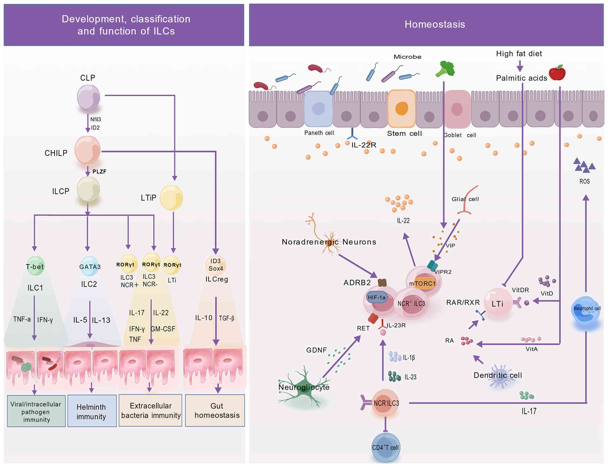 | Figure 1Schematic overview of ILC3
development, differentiation and homeostatic regulation. Left panel
(development, classification and function of ILCs): ILCs originate
from bone marrow-resident CLPs, which differentiate into CHILPs
under the control of NFIL3 and ID2. CHILPs further give rise to
ILC1, ILC2, ILC3 and ILCreg subsets through either the LTiP pathway
or the ILCP route. ILC3s are further subdivided into
NCR+ and NCR− subsets, with RORγt serving as
the master lineage-defining transcription factor. Right panel
(homeostatic regulation): Functions of NCR+ ILC3s and
LTi cells are coordinately regulated by metabolic, neuro-immune and
immune cytokine signals. At the metabolic level, RA acts through
RAR/RXR and VitD through VitDR, to regulate LTi cells, while
palmitic acid derived from a high-fat diet participates in ILC3
homeostasis. At the neuro-immune level, three pathways, GDNF-RET,
VIP-VIPR2 and sympathetic ADRB2, cooperatively regulate IL-22
secretion by ILC3s. In addition, dendritic cell-derived IL-23 and
IL-1β activate ILC3s through IL-23R, thereby maintaining intestinal
epithelial homeostasis. ILC, innate lymphoid cell; CLP, common
lymphoid progenitor; CHILP, common helper-like ILC progenitor;
ILCreg, regulatory ILC; LTi, lymphoid tissue inducer; LTiP, LTi
progenitor; ILCP, ILC precursor; ROS, reactive oxygen species; RA,
retinoic acid; RAR, RA receptor; RXR, retinoid X receptor; VitA,
vitamin A; VitD, vitamin D; VIPR2, VIP receptor 2; VitDR, VitD
receptor; NFIL3, nuclear factor, IL-3 regulated; ID, inhibitor of
DNA binding; PLZF, promyelocytic leukemia zinc finger; T-bet, T-box
expressed in T cells; GATA3, GATA binding protein 3; NCR, natural
cytotoxicity receptor; RORγt, retinoid-related orphan receptor γ-t;
GM-CSF, granulocyte-macrophage colony-stimulating factor; GDNF,
glial cell-derived neurotrophic factor; VIP, vasoactive intestinal
peptide; HIF-1α, hypoxia-inducible factor 1-α; RET, receptor
tyrosine kinase; ADRB2, β2-adrenergic receptor. |
Overall, as important regulators of mucosal
immunity, ILC3s have attracted increasing attention due to their
diverse subsets (3,11), regulatory networks (11,15,16) and interactions with the
microbiota, influencing both tissue homeostasis and disease
development (3,11,15,16). However, the mechanisms by which
these regulatory signals integrate to determine disease-specific
ILC3 responses remain incompletely understood. Therefore, the
present review summarizes the developmental characteristics,
regulatory mechanisms, microbiota interactions and
disease-associated functions of ILC3s, with a particular emphasis
on the molecular mechanisms underlying their functional outcomes
across tissues and disease stages.
Biological characteristics and functional
subsets of ILC3s
ILC3s are characterized by hierarchical lineage
differentiation and functional heterogeneity among subsets. Their
development is regulated by transcription factors, cytokines and
tissue-derived signals. Through functional specialization, distinct
ILC3 subsets contribute to mucosal immune homeostasis and host
defense (Fig. 1).
Development and differentiation pathways
and key regulatory nodes of ILC3
ILC3 development is an ordered process governed by
hierarchical transcriptional regulation, cytokine signaling and the
tissue microenvironment. Compared with other ILC subsets, ILC3s
exhibit distinct lineage bifurcation during development. From a
hematopoietic perspective, CLPs, under the regulation of NFIL3 and
inhibitor of DNA binding 2 (ID2), first diverge from the adaptive
lymphocyte lineage and differentiate into CHILPs. CHILPs express
high levels of ID2 and IL-7R and are restricted to differentiation
into ILC1, ILC2 and ILC3 subsets, having lost the potential to
generate T cells, B cells or NK cells (17). Following the CHILP stage, further
differentiation generates ILCPs, which express the transcription
factor promyelocytic leukemia zinc finger (PLZF) and the surface
molecule programmed cell death protein 1. ILCPs give rise to ILC1,
ILC2 and NCR+ ILC3 subsets, but cannot generate
lymphoid-tissue-inducer (LTi)-like ILC3s (17,18). LTi-like ILC3s are derived from a
distinct LTi progenitor (LTiP). LTiP and ILCP represent two
separate branches that diverge from a common early progenitor
before PLZF expression is initiated (17,18). This progressive commitment to the
ILC lineage is also regulated by a number of transcription factors,
including GATA3, thymocyte selection-associated high mobility group
box (TOX), T cell factor-1 and NFIL3, although their contributions
vary among developmental stages (19-23). Subsequent studies have
demonstrated that LTi-like ILC3s originate from a distinct
progenitor population downstream of CHILPs with the phenotype
ID2+α4β7+-C-X-C chemokine receptor type
(CXCR)-5+. This progenitor population is separated from
the ILCP lineage before PLZF acquisition, thereby establishing a
developmental pathway distinct from that of other ILC subsets
(18,24-27). Accordingly, LTi-like ILC3s
develop through a pathway different from other ILC family members,
including NCR+ ILC3s. This developmental divergence
likely contributes to the transcriptional and functional
differences among ILC3 subsets reported in previous studies
(28-30), including distinct
cytokine-production profiles, divergent surface-marker signatures
and variable ILC3-to-ILC1 plasticity. Accumulating evidence has
further indicated that RORγt (encoded by RORC) directs ILC3
differentiation from ILCPs (31).
Subclassification and functional
heterogeneity of ILC3s
Differentiation of NCR+
ILC3s and NCR− ILC3s
In both mice and humans, ILC3s can be broadly
divided into two major subsets, NCR+ and
NCR−, based on the expression of NCRs, including NKp46
and NKp44 (32). Human
NCR+ ILC3s are characterized by high NKp44 expression
and low NKp46 expression, account for ~70% of intestinal ILCs and
predominantly secrete IL-22. By contrast, NCR− ILC3s
comprise ~15% of intestinal ILCs and primarily produce IL-17
(33,34). In mice, it has been shown that
NCR+ ILC3s are defined by NKp46 expression, whereas
NCR− ILC3s lack CD117 expression. This represents a
species-specific difference given that human NCR− ILC3s
express CD117 (35).
NCR− ILC3s can be generated in vitro following
IL-1β and IL-23 stimulation through Notch2-dependent signaling and
T-bet upregulation (7,36-40). IL-17, produced by NCR−
ILC3s, stimulates epithelial and endothelial cells to release
chemokines such as CXCL8 (41),
thereby promoting neutrophil recruitment and supporting epithelial
barrier function through production of reactive oxygen species
(ROS) and α-defensin (3,42). Following acute intestinal injury,
IL-17 can regulate tight junction proteins in an IL-23-independent
manner and cooperate with early protective IL-17 derived from γδ T
cells to maintain intestinal mucosal integrity. However, in chronic
inflammatory settings, such as recurrent DSS-induced colitis or
established IBD, ILC3-derived IL-17 primarily contributes to
disease progression (42,43).
Functional characteristics of
LTi/LTi-like cells
LTi cells were the first identified subset within
the ILC3 family and are characterized by their distinct
developmental origin and functional properties (25,44). During embryogenesis, LTi cells
arise from the fetal liver and promote the formation of secondary
lymphoid tissues, including lymph nodes and Peyer's patches,
through responses to TNF-α and lymphotoxin-β signaling. This
process establishes the structural foundation for subsequent
adaptive immune responses, including T-cell activation, B-cell
homing to follicles and antibody production (44-48). After birth, LTi cells continue to
develop from bone marrow progenitor cells. Despite their capacity
to induce lymphoid tissue formation in adulthood remaining
incompletely defined, they contribute to gastrointestinal pathogen
defense through IL-17A and IL-22 production and participate in the
formation of cryptopatches and isolated lymphoid follicles
(15,45,46,49).
With regard to surface markers, murine LTi cells
express CD117 (c-kit), CD45, CCR6, CD4 and CD127, whereas human LTi
cells share lineage homology with murine LTi cells but lack CD4
expression. LTi cells can be distinguished from other ILC3 subsets
by the expression of the chemokine receptor CCR6 (15,49,50). CCR6+ LTi cells
represent the predominant ILC3 population in lymphoid organs and
can internalize antigens and present them to CD4+ T
cells, thereby inducing T cell-dependent antibody production
(51). In response to IL-1β, LTi
and LTi-like cells in lymphoid organs express CD80 and CD86 and
produce IL-2, TNF-α and IFN-γ, leading to T-cell activation
(52). Functionally, ILC3s
respond to IL-1β and IL-23. Specifically, IL-23 stimulation induces
IL-17 production mainly in LTi cells and NCR− ILC3s,
whereas both NCR+ and NCR− ILC3s retain the
ability to produce IFN-γ (7,49).
Multidimensional regulatory mechanisms of
ILC3s
Functional activities of ILC3s are regulated by an
interconnected network involving transcriptional, metabolic,
neuroimmune and circadian clock mechanisms. These regulatory
pathways collectively maintain ILC3 homeostasis and effector
functions, whereas disruption of this network contributes to
mucosal immune imbalance and disease development (Fig. 1).
Transcriptional regulatory networks
ROR
ROR, as a transcription factor family primarily
composed of RORγt and RORα, regulates ILC3 lineage development,
functional maintenance and phenotypic stability. Their coordinated
actions shape the biological characteristics of ILC3s. RORγt,
encoded by the RORC gene, is a 495-amino-acid transcription factor
key in the specification of ILC3s and LTi cells. Deletion of RORγt
in mice results has been shown to result in complete failure of
ILC3 development, demonstrating its important role in establishing
the ILC3 lineage (3,53,54). In mature ILC3s, however, RORγt is
not necessarily required for cell survival. Instead, it primarily
regulates cytokine production, including the expression of effector
molecules such as IL-17A and IL-22. RORγt is also expressed by LTi
cells and can be further induced by IL-23, thereby enhancing ILC3
activity (54-56). In mature ILC3s, survival does not
strictly depend on continuous expression of RORγt or RORα (57). However, simultaneous deletion of
RORγt and RORα markedly reduces the expression of metabolic
regulators such as arginase-1. Consequently, ILC3 identity is
disrupted, leading to the transdifferentiation of
RORγt−RORα−T-bet+NCR+
ILC3s into ILC1s (57).
In an IL-23-induced innate colitis model,
RORγt-expressing ILCs have been shown to accumulate in the inflamed
intestine. Conversely, RORγt-deficient mice exhibited markedly
reduced intestinal inflammation, as evidenced by lower histological
scores, diminished inflammatory cell infiltration, and impaired
IL-17 and IL-22 production. These findings establish the pathogenic
contribution of RORγt to IL-23-driven innate intestinal
inflammation (16). Given its
involvement in both ILC3s and Th17 cells, RORγt has emerged as a
potential therapeutic target for intestinal inflammation. Studies
have demonstrated that transient pharmacological RORγt inhibition
in pathogen-infection or neonatal inflammatory mouse models can
selectively dampen pathogenic Th17-cell-derived responses while
preserving the innate protective functions of ILC3s, representing a
promising targeted therapeutic concept (58,59).
T-bet
T-bet suppresses transcription of the RORC gene,
thereby reducing RORγt expression (60). Previous studies have shown that
ex-ILC3s, a transitional population representing ILC3-to-ILC1
conversion, exhibit increased T-bet expression (17,36,57). This finding indicates that the
balance between RORγt and T-bet is key in maintaining an
equilibrium between ILC3 and ILC1 subsets. Fiancette et al
(57) reported that ILC3s with
dual deletion of T-bet and RORγt
(T-bet−/−RORγt−/−) failed to undergo
ILC3-to-ILC1 conversion and instead generated a distinct ILC
population with undefined functions. In addition, these cells were
unable to produce IL-22 following IL-23 stimulation. Collectively,
these findings indicate that T-bet, in addition to RORγt, is
required for ILC3-to-ILC1 conversion. Similarly, Stehle et
al (61) demonstrated in a
mouse model that T-bet deficiency restored lymphoid tissue
organogenesis defects caused by RORγt deficiency. Intestinal
barrier repair in this context was mediated not by ILC3s but by
ILCPs, which lack both T-bet and RORγt and promote repair through
IL-22 secretion (61).
Therefore, low-level T-bet expression in ILC3s may contribute to
maintaining ILC subset balance and immune homeostasis. Recent
studies have further identified an upstream mechanism regulating
T-bet expression. Huang et al (62) demonstrated that the intestinal
lipid metabolite 12R-hydroxyeicosatetraenoic acid functions as an
endogenous ligand for the nuclear receptor Nur77. Nur77 signaling
subsequently increases T-bet expression, promoting the
differentiation of NKp46− ILC3s into NKp46+
ILC3s and enhancing IFN-γ secretion. Nur77-deficient mice exhibit
reduced ILC3 numbers, impaired differentiation of the
NKp46+ subset and increased susceptibility to
Salmonella typhimurium infection. Mechanistically, this
process depends on inosine monophosphate dehydrogenase 1, a
downstream target gene regulated by Nur77 (62). These findings indicate an
association between metabolic signals and T-bet-mediated ILC3
subset fate determination, revealing the role of the
'metabolite-nuclear receptor-T-bet' axis in intestinal immune
development.
GATA3
GATA3 has long been recognized as a key
transcriptional regulator of Th2 cell and ILC2 development and
function (23). However,
subsequent studies have demonstrated that GATA3 is also required
for ILC3 development (20,63). Precursor cells lacking GATA3 fail
to differentiate into ILC3s, whereas wild-type cells undergo normal
development, indicating that GATA3 acts as a cell-intrinsic
regulator of ILC3 lineage establishment (19). Within ILC3s, GATA3 promotes cell
survival and expansion through binding to the RORC and IL-17R gene
loci, thereby enhancing the expression of RORγt and CD127 (54). In addition, GATA3 regulates IL-22
expression while having limited effects on IL-17A production,
suggesting selective regulation of ILC3 effector functions
(54). In infection models,
GATA3-deficient chimeric mice exhibit reduced intestinal IL-22
expression, decreased production of the antimicrobial peptide
regenerating islet-derived protein 3-γ (RegIIIγ) and systemic
pathogen dissemination, demonstrating how GATA3 contributes towards
ILC3-mediated innate immune defense (19).
mTOR
mTOR is a serine/threonine kinase that integrates
intracellular and extracellular signals through two distinct
complexes, mTORC1 and mTORC2, thereby regulating cell
proliferation, metabolism and immune response (64). Recent studies have begun to
reveal the regulatory functions of the mTOR pathway in ILC3
biology.
At the functional level, mTORC1 is required for
ILC3-mediated anti-infective immunity. ILC3s rely on
mTORC1-dependent activation of hypoxia-inducible factor 1-α
(HIF-1α), which drives metabolic reprogramming, enhances
glycolysis, maintains RORγt expression and promotes cell
proliferation and production of the type 3 cytokines IL-22 and
IL-17A (65). Under homeostatic
conditions, both mTORC1 and mTORC2 contribute to maintaining ILC3
stability in the small intestine. Deletion of regulatory protein
associated with mTOR (Rptor), a key component of mTORC1, markedly
reduces the numbers of both NCR+ and NCR−
ILC3 subsets in the small intestine, consistent with observations
reported by Di Luccia et al (65). Similarly, deletion of
rapamycin-insensitive companion of mTOR, a component of mTORC2,
decreases intestinal ILC3 levels, although this effect is less
pronounced compared with that caused by mTORC1 deficiency (64). However, additional studies have
indicated that mTORC2 is not indispensable for basal ILC3
homeostasis (64,66). Selective mTORC2 deletion does not
notably affect ILC3 numbers, subset distribution or cytokine
production, whereas enhanced mTORC2 activity can compensate for
impaired mTORC1 function and provide protective effects (66). mTORC1 and mTORC2 also exhibit
reciprocal regulation. In IL-23-stimulated ILC3s, both complexes
become activated. Conversely, inhibition of mTORC1, such as through
Rptor deletion or rapamycin treatment, further increases mTORC2
activity (66).
Metabolic signaling regulatory
networks
Regulation of ILC3s by vitamin
metabolites
Retinoic acid (RA), a metabolite of vitamin A, is
required for fetal LTi cell development and lymphoid tissue
formation (67). During
embryogenesis, maternally derived RA binds directly to the RORC
gene locus through RA receptor (RAR) and retinoid X receptor (RXR),
thereby increasing RORγt expression. This process promotes the
differentiation of ID2+RORγt+CD4−
LTi cells into ID2+RORγt+CD4+ LTi
cells and facilitates the formation of secondary lymphoid organs
(SLOs) (26,67). Blocking RA signaling has been
shown to reduce the number of
ID2+RORγt+CD4− LTi cells and
decrease SLO density, whereas RA stimulation has been shown to
expand the ID2+RORγt+CD4+ LTi
population (26). Administration
of the RA signaling inhibitor BMS493 to pregnant mice has been
shown to reduce fetal LTi cell numbers and impair SLO development
in the small intestine (67). RA
also contributes to maintaining ILC3 populations in adulthood.
Vitamin A deficiency or RAR inhibition reduces intestinal ILC3
numbers, IL-22 secretion and the frequency of lymphoid follicle
formation in the intestine (68,69). In addition to supporting ILC3
maintenance, RA regulates the balance among ILC subsets. In vitamin
A-deficient mice, ILC3 numbers and their signature cytokines IL-22
and IL-17 are reduced, whereas ILC2 numbers and associated
cytokines, including IL-4, IL-5 and IL-13, are increased. These
findings suggest that vitamin A contributes to maintaining the
balance between ILC2 and ILC3 subsets (68).
In addition to vitamin A, vitamin D also regulates
ILC3 function. Intestinal ILC3s exhibit high expression of the
vitamin D receptor (VDR). ILC3-specific deletion of VDR primarily
affects the expansion of the LTi-like subset, resulting in reduced
proliferation and impaired IL-22 production, ultimately decreasing
host resistance to infection (70). Furthermore, a vitamin D-deficient
diet markedly reduces the proportion of ILC3s in the small
intestine and colon of mice, thereby increasing susceptibility to
infection (71).
Regulation of ILC3s by lipid
metabolites
Short-term consumption of a high-fat diet (HFD)
enriched in saturated long-chain fatty acids markedly suppresses
IL-22 production by murine ILC3s within 48 h. This impairment
compromises intestinal barrier integrity, as indicated by reduced
expression of antimicrobial peptides (including RegIII), mucin
(including Muc2) and tight junction proteins (such as occludin),
together with increased epithelial permeability. Consequently, the
intestine becomes more susceptible to inflammation and injury
(72). Intermittent fasting
induces IL-22 release by ILC3s and enhances their interactions with
dendritic cells and macrophages. These effects promote the browning
of subcutaneous white adipose tissue in obese mice and reveal a
potential mechanism underlying metabolic protection (73).
The molecular mechanisms underlying fatty
acid-mediated regulation of ILC3 function involve two complementary
pathways. First, saturated fatty acids such as palmitic acid can
induce expression of diacylglycerol O-acyltransferase 1, an enzyme
involved in triglyceride synthesis, in ILC3s. However, this
response paradoxically leads to defective lipid droplet formation,
disrupting the balance between lipid storage capacity and metabolic
homeostasis. Second, saturated fatty acids induce mitochondrial
hyperpolarization, ROS accumulation and impaired oxidative
phosphorylation. Enhanced fatty acid oxidation further promotes
metabolic stress in ILC3s, leading to reduced IL-22 expression and
decreased RORγt stability. By contrast, unsaturated fatty acids
such as oleic acid promote the formation of structurally stable
lipid droplets and preserve mitochondrial homeostasis, thereby
supporting sustained IL-22 secretion (72). Intestinal ILC3s, particularly the
CD4+ LTi subset, sense oxysterol signals produced by
intestinal stromal cells through expression of G-protein-coupled
receptor (GPR)-183 (74,75). This chemotactic signal directs
ILC3 localization within cryptopatches and isolated lymphoid
follicles, contributing to the formation of local lymphoid tissues
(74). Experimental studies have
shown that GPR183 knockout disrupts this process, reducing ILC3
accumulation in cryptopatches and isolated lymphoid follicles while
increasing their numbers in mesenteric lymph nodes. These findings
further support the role of the GPR183 signaling axis in
tissue-specific ILC3 localization (74,75).
In addition to these metabolites, prostaglandin E2,
a lipid-derived mediator, regulates ILC3 functions through its
receptors EP4 and EP2. Activation of these pathways promotes IL-22
and HB-EGF secretion, respectively, thereby contributing to mucosal
repair and inflammatory regulation (76,77).
Neuro-immune regulation
Regulation of ILC3s by glial
cells
In response to signals from the intestinal
microenvironment, glial cells release glial cell-derived
neurotrophic factor (GDNF) through a myeloid differentiation
primary response 88 (MyD88)-dependent mechanism. GDNF binds to
receptor tyrosine kinase (RET) expressed on ILC3s and activates the
STAT3 and p38 MAPK/ERK-AKT signaling pathways, resulting in IL-22
production. This neuroimmune signaling axis contributes to
intestinal protection and maintenance of mucosal homeostasis
(78). Systemic RET deletion in
mice impairs Peyer's patch development and reduces intestinal
IL-22+ ILC3 numbers. Conversely, ILC3-specific
conditional deletion of RET aggravates dextran sulfate sodium
(DSS)-induced colitis and Citrobacter rodentium infection in
rodents, whereas gain-of-function RET mutations protect against
DSS-induced intestinal injury (78,79).
Regulation of ILC3s by neural
signals
During intestinal injury repair, a number of neural
signals contribute to an immune regulatory network through their
effects on ILC3s. The sympathetic nervous system regulates ILC3
function through norepinephrine signaling. Norepinephrine directly
promotes IL-22 production by ILC3s through the
β2-adrenergic receptor (ADRB2) expressed on their
surface, thereby enhancing intestinal epithelial regeneration
(80). In this experimental
model, chemical sympathectomy or ADRB2 blockade reduces intestinal
crypt proliferation and decreases expression of the stem cell
marker leucine-rich repeat-containing G protein-coupled receptor 5
(Lgr5). In addition, single-cell RNA sequencing has revealed the
reduced expression of IL-22 downstream target genes, including
C-type regenerating islet derived-3 (Reg3)-b and Reg3g, in
intestinal epithelial cells following denervation. Neutralization
of IL-22 blocks the regenerative effects induced by noradrenergic
nerves, whereas exogenous IL-22-Fc restores repair defects caused
by denervation. These findings indicate that IL-22 functions as the
major effector molecule within the sympathetic neuroimmune pathway
(80).
The parasympathetic nervous system, particularly
the vagus nerve, also regulates ILC3 activity. In addition to RET
expression, ILC3s express a number of cholinergic receptors,
including Chrm1, Chrm2, Chrm4 and Chrm5. Vagus nerve-derived
acetylcholine (ACh) activates the protectin conjugates in tissue
regeneration 1 (PCTR1) biosynthetic pathway in ILC3s (81). Vagotomy reduces peritoneal ILC3
numbers and impairs clearance of Escherichia coli infection.
Conversely, administration of ACh-preactivated ILC3s restores
antimicrobial defense and improves tissue-repair defects caused by
vagotomy (81). These findings
identify vagal cholinergic signaling as another pathway involved in
ILC3 regulation. Collectively, these studies reveal numerous
neuroimmune regulatory mechanisms involving ILC3s, including the
'sympathetic-ILC3-L-22' and 'vagus-ILC3-PCTR1' pathways and suggest
potential therapeutic targets for intestinal injury repair and
inflammatory bowel disease (IBD). Recent studies have further
identified a third neural regulatory mechanism mediated by enteric
GABAergic neurons. Liu et al (82) demonstrated that GABA released
from enteric GABAergic neurons suppresses ILC3 proliferation and
IL-17A production through γ-amino-butyric acid type B receptor 1/2
(GABBR1/2) receptors expressed on ILC3s. Mechanistically, GABA
signaling reduces expression of the liver-enriched inhibitory
protein isoform of CCAAT/enhancer-binding protein β in ILC3s,
thereby relieving transcriptional repression of the insulin like
growth factor binding protein 7 (IGFBP7) gene. IGFBP7 subsequently
regulates ILC3 activation through an autocrine negative feedback
loop mediated by insulin-like growth factor 1 (IGF)-1R (82). Conditional deletion of GABBR1 or
depletion of GABAergic neurons enhances IL-17A-mediated intestinal
inflammation. In addition, inhibition of this pathway is positively
associated with disease severity in patients with IBD (82). The present study establishes the
enteric GABAergic neuron-ILC3 axis as an additional neuroimmune
regulatory pathway, expanding the understanding of how neural
signals contribute to intestinal immune homeostasis.
Circadian clock systems integrate
neuro-environmental signals to regulate rhythmic ILC3 function
Circadian clock systems in ILC3s integrate
environmental cues to coordinate rhythmic cellular functions and
maintain intestinal immune homeostasis. Current evidence has
identified three major regulatory pathways. First, light signals,
acting as primary zeitgebers, are transmitted through the central
clock located in the hypothalamic suprachiasmatic nucleus and
synchronize clock gene expression in ILC3s. Experimental studies
have shown that changes in photoperiod can reverse the rhythmic
expression pattern of the period circadian regulator 1 gene in
ILC3s (83,84). Second, feeding signals regulate
ILC3 rhythmic activity through vasoactive intestinal peptide
(VIP)-ergic neurons. These neurons release VIP, which modulates
rhythmic IL-22 secretion through the VIPR2 receptor expressed on
ILC3s. Exogenous VIP enhances IL-22 production by ILC3s, whereas
VIPR2−/− mice exhibit reduced ILC3-derived IL-22
secretion and aggravated inflammatory responses during DSS-induced
colitis (84,85). Third, the gut microbiota
regulates rhythmic ILC3 function through microbial metabolites.
Antibiotic treatment has been shown to markedly reduce the
rhythmicity of IL-22 production, indicating that the microbiota is
required for maintaining normal circadian ILC3 activity (86).
Collectively, light, feeding and microbial signals
coordinate ILC3 circadian regulation through distinct pathways,
allowing immune functions to adapt to environmental changes and
supporting intestinal mucosal homeostasis. In a complementary
study, Bhattarai et al (87) further elucidated the mechanisms
associating circadian regulation with ILC3 lineage plasticity. This
study demonstrated that circadian clock proteins reverse
(REV)-ERBα/β maintain an accessible chromatin state at the RORC
locus through epigenetic regulation, thereby preserving canonical
ILC3 identity (87).
Simultaneous deletion of REV-ERBα and REV-ERBβ reverses the
repression of the transcription factor NFIL3. NFIL3 subsequently
binds a cis-regulatory element ~2 kb upstream of the RORC gene,
suppresses RORγt transcription and activates a T-bet-dominant
differentiation program. This transcriptional switch promotes
transdifferentiation of NKp46+ ILC3s into ILC1s,
accompanied by reduced IL-22 production and increased IFN-γ
secretion, ultimately impairing host defense against Citrobacter
rodentium infection (87).
Furthermore, loss of REV-ERBα/β reduces surface major
histocompatibility complex (MHC)-II expression on CCR6+
ILC3s, suggesting that circadian regulation influences not only
cytokine production but also immunomodulatory functions of ILC3s,
including antigen presentation (87).
Interactions between ILC3s and the
microbiota
Gut microbiota and ILC3s establish a dynamic
bidirectional association that contributes to mucosal homeostasis.
The microbiota regulates ILC3 development and function through
direct and indirect mechanisms, whereas ILC3s influence microbial
composition and colonization by regulating epithelial barrier
integrity and IgA production. Disruption of this interaction is
associated with the development and progression of diseases
including IBD and tumors (Fig.
2).
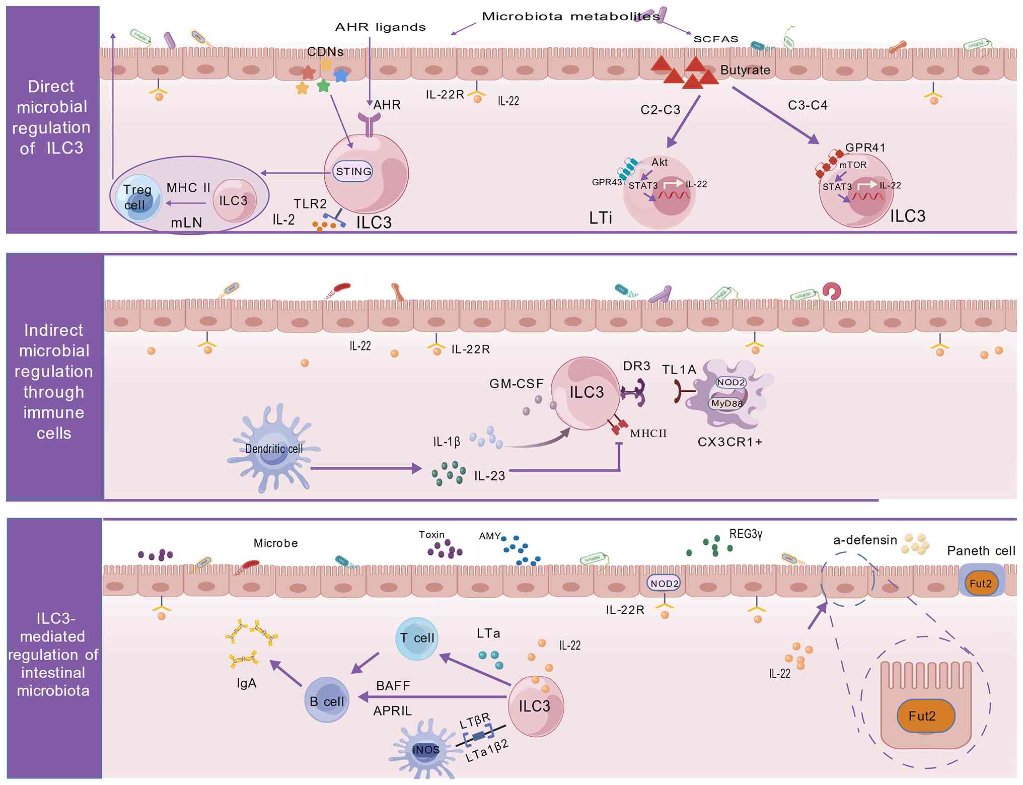 | Figure 2Schematic representation of the
bidirectional regulatory network between ILC3s and the gut
microbiota. Upper panel (direct microbial regulation of ILC3):
Microbiota-derived tryptophan metabolites activate the AHR pathway
in ILC3s to promote IL-22 production. Gut microbial SCFAs act
through distinct receptors: Acetate and propionate signal through
GPR43 (FFAR2) on LTi cells, while propionate and butyrate bind to
GPR41 (FFAR3) on ILC3s; collectively, these signals activate the
STAT3 pathway to drive IL-22 secretion. In addition, microbial
cyclic dinucleotides activate STING signaling in ILC3s, promoting
their migration to mesenteric lymph nodes, where they cooperate
with IL-2 and MHC-II to induce Treg cell differentiation. Middle
panel (indirect microbial regulation through immune cells):
CX3CR1+ dendritic cells recognize intestinal microbial
signals and secrete IL-1β and IL-23, which act on ILC3s. ILC3s
express DR3 and the NOD2/MyD88 complex on their surface, enabling
them to respond to TL1A and intracellular microbial signals.
Furthermore, dendritic cell-derived IL-23 dynamically modulates MHC
class II expression and the antigen-presenting capacity of ILC3s.
Lower panel (ILC3-mediated regulation of intestinal microbiota):
ILC3-derived IL-22 acts on intestinal epithelial cells to induce
the production of antimicrobial peptides (RegIIIβ/γ),
Fut2-dependent epithelial fucosylation and abundant mucin
secretion, thereby establishing physical and chemical barriers that
restrict bacterial invasion. ILC3s also secrete LTα and
membrane-bound LTβ to facilitate T cell-dependent IgA responses,
while simultaneously producing BAFF and APRIL to directly promote T
cell-independent IgA production by B cells. Through mucosal IgA,
ILC3s reshape the colonization structure of the gut microbiota.
ILC, innate lymphoid cell; AHR, aryl hydrocarbon receptor; SCFA,
short-chain fatty acid; GPR, G-protein-coupled receptor; FFAR, free
fatty acid receptor; LTi, lymphoid tissue inducer; STING,
stimulator of interferon genes; MHC, major histocompatibility
complex; Treg, T regulatory cell; CX3CR, CX3C motif chemokine
receptor 1; DR3, death receptor 3; NOD2, nucleotide-binding
oligomerization domain-containing protein 2; MyD88, myeloid
differentiation primary response 88; TL1A, TNF-like ligand 1A;
Fut2, fucosyltransferase 2; BAFF, B cell-activating factor; APRIL,
A proliferation-inducing ligand; TLR, toll-like receptor; REG3γ,
C-type regenerating islet derived-3-γ; RegIII, regenerating
islet-derived protein 3; mLN, mesenteric lymph nodes; GM-CSF,
granulocyte-macrophage colony-stimulating factor; LT, lymphotoxin.
CDNs, cyclic dinucleotides. |
Direct regulation of ILC3s by the
microbiota
Microbial metabolites regulate ILC3 function
through numerous signaling pathways. Microbiota-derived tryptophan
metabolites act as ligands for the AHR. By directly activating AHR
in ILC3s, these metabolites promote IL-22 transcription and
secretion, thereby enhancing epithelial repair and antimicrobial
defense (39,88-90). Studies using human ILC3s have
shown that co-stimulation with AHR agonists and IL-1β induces IL-22
production, whereas AHR antagonists promote the generation of
IFN-γ-expressing ex-ILC3s. These findings support a conserved role
for AHR in maintaining ILC3 function (91). Beyond the AHR pathway,
microbiota-derived short-chain fatty acids (SCFAs) regulate ILC3
activity through specific G protein-coupled receptors. Acetate (C2)
primarily signals through free fatty acid receptor 2 (FFAR2;
GPR43), which regulates the CCR6+ ILC3 subset localized
in colonic cryptopatches and isolated lymphoid follicles (92). Mechanistically, acetate promotes
CCR6+ ILC3 proliferation through FFAR2-mediated
activation of the Akt, STAT3 and mTOR pathways (92). In addition, acetate increases
IL-1β receptor expression on ILC3s, thereby enhancing their
capacity for IL-22 production (92). Consistent with these findings,
conditional deletion of FFAR2 impairs CCR6+ ILC3
proliferation and IL-22 production, resulting in reduced
antimicrobial defense and tissue repair capacity (92). By contrast, propionate (C3)
preferentially enhances IL-22 production by ILC3s through FFAR3
(GPR41) signaling (92).
Butyrate (C4) promotes IL-22 production by RORγt+ ILCs
through the GPR41 pathway (93).
However, butyrate can also act through GPR109a expressed by
dendritic cells to indirectly suppress IL-17 and IL-22 production
by ILC3s under IL-25 stimulation (94,95).
In addition, gut microbiota-derived cyclic
dinucleotides can be directly sensed by ILC3s through the
stimulator of interferon genes (STING) pathway (96). Under homeostatic conditions,
STING activation induces CCR7 expression in ILC3s and promotes
their migration to lymph nodes. This process facilitates
microbiota-specific Treg differentiation, which in turn establishes
immune tolerance by suppressing pathogenic effector T cell
responses against commensal bacteria (96). Microbial signals also regulate
ILC3 function through additional receptor pathways. Upon sensing
microbiota-derived signals, toll-like receptor 2 induces IL-2
production by ILC3s. IL-2 is mainly consumed by local Tregs
expressing high levels of CD25 and promotes IL-22 expression
through an autocrine mechanism, thereby selectively maintaining
Treg homeostasis in the small intestine (97,98). Collectively, the gut microbiota
regulates ILC3 function through diverse molecular mechanisms,
including AHR-mediated sensing of tryptophan metabolites,
GPCR-mediated recognition of short-chain fatty acids,
STING-mediated detection of cyclic dinucleotides and TLR2-mediated
responses to microbial components. However, further studies are
required to define the specific microbial signals that activate,
suppress or fine-tune ILC3 activity.
Indirect regulation of ILC3s by the
microbiota through immune cell mediation
Mononuclear phagocyte systems, particularly CX3C
motif chemokine receptor 1 (CX3CR1)+ macrophages and
CD14− dendritic cells, function as a central hubs for
sensing and transmitting microbiota-derived signals, thereby
regulating ILC3 activation. Upon sensing microbiota-derived cues
through the MyD88 and nucleotide-binding oligomerization
domain-containing protein 2 (NOD2) pathways, CX3CR1+
macrophages specifically produce IL-1β, which directly activates
ILC3s (98,99). Activated ILC3s subsequently
secrete granulocyte-macrophage colony-stimulating factor (GM-CSF,
also known as colony-stimulating factor 2 or CSF2). GM-CSF, in
turn, induces IL-10 secretion by dendritic cells and promotes Treg
differentiation, thereby indicating an association between early
ILC3-mediated innate immune responses and subsequent adaptive
immune tolerance mechanisms (99,100). In addition, microbial
stimulation induces CX3CR1+ macrophages to release
TNF-like ligand 1A (TL1A). Upon binding to death receptor 3, which
is expressed on the surface of human ILC3s, TL1A not only promotes
ILC3 proliferation but also enhances IL-22 production in the
presence of synergistic cytokine stimulation (101,102). Collectively, microbiota-induced
production of signaling molecules, including IL-1β and TL1A, by
mononuclear phagocytes activates ILC3s and regulates their
secretion of cytokines such as GM-CSF and IL-22, thereby
establishing a regulatory axis that coordinates immune regulation
throughout the gastrointestinal tract (98,99).
Under conditions of intestinal homeostasis, the
microbiota also influences the antigen-presenting function of ILC3s
by modulating mononuclear phagocyte responses. Specifically,
microbiota stimulation induces mononuclear phagocytes, including
dendritic cells and macrophages, to secrete IL-23. As a regulatory
signal, IL-23 reversibly suppresses the expression of MHC class II
molecules on the surface of ILC3s, thereby reducing their capacity
to present antigens to CD4+ T cells within the
intestinal mucosa (103). This
reversibility indicates that ILC3 antigen-presenting function is
dynamically regulated: Enhanced IL-23 signaling suppresses MHC II
expression, whereas reduced IL-23 signaling or alterations in the
intestinal microenvironment restore antigen-presenting capacity
(103). This regulatory
mechanism highlights the dynamic role of ILC3s in maintaining
intestinal immune homeostasis, allowing tolerance toward commensal
microbes while preserving the capacity to mount immune responses
against pathogenic infections (103).
Regulatory role of ILC3s in the
microbiota
ILC3s exert multifaceted regulatory effects in
maintaining gut microbiota homeostasis. These functions are
primarily mediated through IL-22 secretion, which reinforces
epithelial barrier integrity, shapes the microbial colonization
environment and regulates the production of antimicrobial effector
molecules. IL-22 initiates signaling by binding to the
heterodimeric IL-22R1/IL-10R2 receptor complex expressed on
intestinal epithelial cells. Under homeostatic conditions, IL-22
signaling induces the production of antimicrobial peptides,
including RegIIIβ, RegIIIγ and S100A/B, thereby limiting excessive
epithelial invasion by commensal bacteria. In addition, IL-22
promotes epithelial expression and fucosylation of
fucosyltransferase 2 (Fut2), thereby enhancing intestinal
resistance and tolerance to pathogenic bacteria. Fut2-positive
Paneth cells further strengthen antimicrobial defense through
secretion of α-defensins (104-108). IL-22 also promotes NOD2
expression, with NOD2 signaling stimulates mucin and antimicrobial
peptide secretion while promoting expansion of commensal bacteria
through N-glycosylation. These commensal bacteria compete with
Clostridium difficile for dietary niches, thereby
contributing to gut microbiota stability (109,110). IL-22 contributes to prevention
of bacterial infection, attenuation of intestinal inflammation and
tissue repair during conditions such as hepatitis and colitis
(111). IL-22 deficiency
compromises barrier integrity, resulting in bacterial translocation
and subsequent systemic inflammation (112). Loss of ILC3 function is
frequently accompanied by alterations in microbial composition,
including expansion of segmented filamentous bacteria and
Clostridiales, together with reduced abundance of beneficial
bacteria such as Lactobacillus spp. These findings suggest
that ILC3s restrict the expansion of potentially pathogenic
bacteria and contribute to maintenance of microbial homeostasis
(113-115). In addition to IL-22-mediated
regulation, ILC3s indirectly shape the microbial ecosystem through
modulation of mucosal immune responses. ILC3s regulate IgA
production by influencing germinal center B cell responses and
antigen presentation to follicular helper T cells. Mechanistically,
ILC3-derived LTα promotes T-cell homing to the intestine, thereby
supporting T cell-dependent IgA induction. In addition, ILC3s
upregulate inducible nitric oxide synthase expression through
membrane-bound lymphotoxin-β (LTβ) interactions with dendritic
cells, promoting IgA generation (115,116). Furthermore, ILC3s directly
support B cell survival and IgA secretion through production of B
cell-activating factor (BAFF) and A proliferation-inducing ligand
(APRIL) (117). As a key
effector molecule of mucosal immunity, IgA influences microbiota
composition through immune exclusion and regulation of bacterial
colonization (118). Therefore,
by regulating IgA responses, ILC3s indirectly contribute to the
maintenance of intestinal microecological homeostasis.
Role of ILC3s in diseases
Role of ILC3 in intestinal diseases
Role in IBD
IBD comprises a group of disorders characterized by
chronic intestinal inflammation, primarily including ulcerative
colitis (UC) and Crohn's disease (CD). Increasing evidence has
indicated that ILC3 dysfunction represents an important mechanism
contributing to the pathogenesis of these conditions (119,120) (Fig. 3). In inflamed intestinal tissues
from patients with IBD, the frequency of ILC3s is markedly reduced,
whereas IFN-γ-secreting ILC1s are notably increased. These changes
suggest that conversion of ILC3s into ILC1s may contribute to
disease progression (120,121). This conversion process is
tightly regulated by the local cytokine environment, consisting
primarily of IL-12, IL-23, IL-1β and IL-18, which are elevated in
the inflamed intestinal mucosa. In vitro experiments and
analyses of clinical tissue specimens have demonstrated that IL-12
produced by CD14+ dendritic cells induces
IL-22-secreting RORγt+NKp44+ ILC3s to convert
into IFN-γ+ ILC1s. This phenotypic shift increases the
proportion of ILC1s while reducing ILC3 numbers in the intestines
of patients with CD, thereby promoting chronic intestinal
inflammation (15,35). Conversely, in the presence of
IL-23, IL-1β and RA produced by CD14− dendritic cells,
CD127+ ILC1s can revert to IL-22+ ILC3s. This
was demonstrated by single-cell cloning experiments showing that
individual CD127+ ILC1 gave rise to progeny containing
both ILC1 and ILC3 phenotypes, and by adoptive-transfer experiments
in which ex-RORγt+ ILC3 (cells that had lost RORγt and
become ILC1-like) re-expressed RORγt after being transferred into
Rag2−/−Il2rg−/− mice (34). Clinical specimen analyses have
further revealed increased expression of ILC3 activation markers,
including NKp44 and CD56, in peripheral blood from patients with CD
and UC. At the same time, human leukocyte antigen-DR isotype
expression on ILC1s and ILC2s is also elevated, supporting the
contribution of ILC lineage imbalance to IBD pathogenesis (122). The GPR183 pathway is closely
associated with IBD. Animal studies have shown that colonic
inflammation increases levels of its ligand
7α,25-hydroxycholesterol, thereby activating GPR183 signaling. This
activation promotes abnormal migration of ILC3s toward inflammatory
sites, disrupting their local distribution and resulting in
excessive IL-22 production (74,123). In addition, the IL-23/IL-17
axis serges an important role in IBD pathogenesis. Animal studies
have further demonstrated that IL-23 activates intestinal
NCR− ILC3s and LTi cells, inducing robust IL-17
secretion and promoting intestinal inflammation (16,64). Consistently, IL-23 levels are
elevated in the intestines of patients with IBD, indicating the
relevance of this pathway in human disease (122). IL-17 promotes tissue injury by
inducing epithelial and endothelial cells to produce chemokines,
which recruit neutrophils into the intestinal mucosa. These
recruited neutrophils subsequently release ROS and proteases,
further contributing to inflammation (42). In addition, IL-17 reduces tight
junction protein expression, thereby impairing epithelial barrier
integrity (43). One mechanism
underlying the therapeutic efficacy of clinically approved IL-23
monoclonal antibodies, such as ustekinumab, is the inhibition of
IL-17 production by ILC3s and Th17 cells (124). Similarly, animal studies have
further demonstrated that IL-23 blockade reduces IL-17 secretion by
ILC3s and attenuates intestinal inflammation (16). In children with IBD, MHC-II
expression on ILC3s has been reported to be notably reduced. Given
that MHC-II+ ILC3s contribute to CD4+ T-cell
apoptosis and maintenance of immune tolerance, restoration or
modulation of this pathway may represent a potential therapeutic
strategy (125).
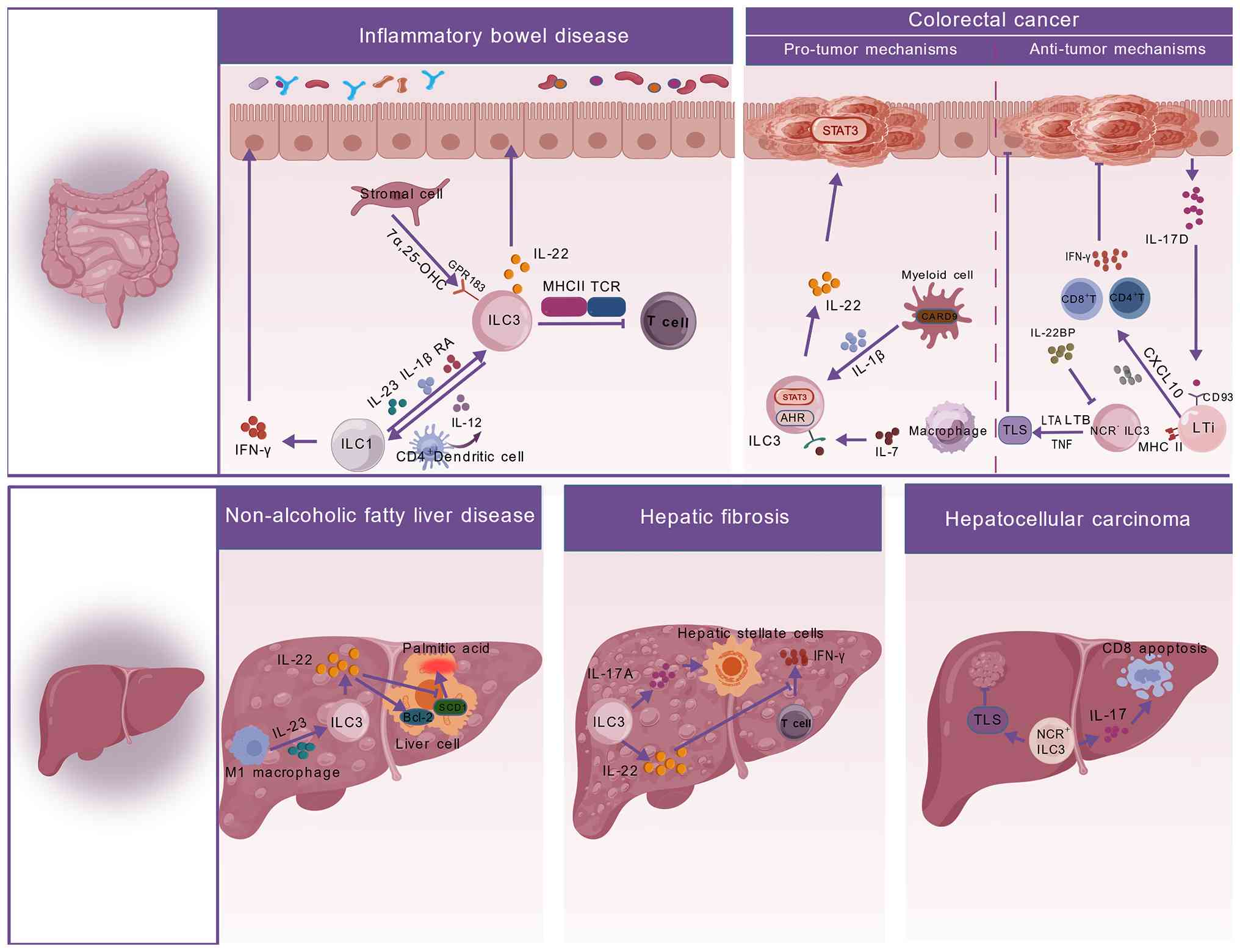 | Figure 3Mechanistic roles of ILC3s in
intestinal and liver diseases. Inflammatory bowel disease:
7α,25-OHC binds to GPR183 on ILC3s, mediating aberrant migration of
ILC3s to inflammatory intestinal lesions. Dendritic cell-derived
IL-12 induces ILC3-to-ILC1 phenotype conversion and the resulting
ILC1s produce large amounts of IFN-γ, which exacerbates epithelial
damage and chronic intestinal inflammation. Colorectal cancer:
Pro-tumor mechanisms encompass macrophage-derived IL-7 acting on
ILC3s, activating intracellular STAT3 and AhR signaling, which
promotes large IL-22 secretion by ILC3s and mediates
pro-tumorigenic effects. Antitumor mechanisms encompass: i) LTi
cells expressing MHC class II molecules and directly presenting
antigens to activate CD4+ and CD8+ antitumor
T cells; ii) NCR+ ILC3s secreting LTA, LTB and TNF to
induce the formation of TLS within tumor lesions and; iii) ILC3s
secreting IL-22BP to antagonize pro-tumorigenic IL-22, while
CD4+/CD8+ T cells secrete IFN-γ,
synergistically exerting antitumor immunity. Non-alcoholic fatty
liver disease: A high-fat diet induces hepatic M1 macrophage
activation and IL-23 secretion whereby IL-23 activates hepatic
ILC3s to secrete IL-22. IL-22 upregulates the anti-apoptotic
protein Bcl-2 in hepatocytes and suppresses SCD1 expression,
thereby alleviating hepatic lipid injury and exerting
hepatoprotective effects. Hepatic fibrosis: ILC3-derived IL-17A and
IL-22 directly activate hepatic stellate cells to promote collagen
deposition. IL-22 also suppresses IFN-γ production by T cells,
indirectly exacerbating fibrosis. Hepatocellular carcinoma:
NCR+ ILC3s are present in hepatic tumor lesions and
participate in the formation of intratumoral TLS. The tumor
microenvironment can induce CD8+ T cell apoptosis,
thereby weakening antitumor immune responses. ILC, innate lymphoid
cell; AHR, aryl hydrocarbon receptor; GPR, G-protein-coupled
receptor; MHC, major histocompatibility complex; TLS, tertiary
lymphoid structures; 7α,25-OHC, 7α,25-hydroxycholesterol; NCR,
natural cytotoxicity receptor; LT, lymphotoxin; SCD1, stearoyl-CoA
desaturase; IL-22BP, IL-22 binding protein; RA, retinoic acid;
CXCL, chemokine (C-X-C motif) ligand; CARD9, caspase recruitment
domain-containing protein 9; TCR, T cell receptor. |
Role in colorectal cancer (CRC)
In CRC, ILC3s exhibit both pro-tumorigenic and
anti-tumorigenic activities, which are influenced by subset
composition, local cytokine networks and the gut microbiota
(Fig. 3). Regarding their
pro-tumorigenic functions, studies using mouse models of
colitis-associated cancer have shown that ILC3s promote epithelial
cell proliferation through IL-22-mediated activation of the STAT3
signaling pathway, thereby contributing to CRC initiation and
maintenance (126,127). Further studies using
genetically deficient mouse models demonstrated that this process
is regulated by a myeloid cell-derived caspase recruitment domain
family member 9-IL-1β signaling axis (128). In addition, mouse studies have
shown that IL-17 produced by the NCR− ILC3 subset
following IL-23 stimulation promotes the development of duodenal
adenomas (126,129). An additional study combining
mouse fecal microbiota transplantation experiments with in
vitro coculture systems demonstrated that macrophage-derived
IL-7 enhances STAT3 and AHR expression in ILC3s, thereby increasing
IL-22 production and promoting intestinal tumorigenesis (130). In addition, analyses of human
CRC tissue specimens have revealed reduced expression of IL-22
binding protein (IL-22BP), an endogenous inhibitor of IL-22, in
tumor tissues. Loss of IL-22BP results in dysregulated IL-22
signaling and accelerates inflammation-associated tumor
progression, an effect further validated in mouse models (127,131).
Conversely, ILC3s also exert protective antitumor
functions. Mouse models of colitis-associated cancer have
demonstrated that epithelial cell-derived IL-17D binds to CD93
expressed on the surface of ILC3s and regulates IL-22 secretion.
This pathway subsequently affects antimicrobial peptide expression
and gut microbiota composition, thereby suppressing tumorigenesis
(132). Studies using
genetically deficient mouse models and human CRC tissue specimens
have further shown that CCR6+ ILC3s expressing MHC-II
molecules and displaying an LTi-like phenotype promote type 1
immune responses, enhance antitumor immunity and improve responses
to immunotherapy through interactions with T cells (133,134). In murine tumor models,
cisplatin induces tumor cells to produce C-C motif chemokine ligand
(CCL)-20, while also elevating intratumoral IL-1β levels derived
from myeloid and tumor compartments (135,136). These signals activate ILC3s to
release chemokine (C-X-C motif) ligand (CXCL)-10, which recruits
CD4+ and CD8+ T cells. Consequently,
immunologically 'cold' tumors, which are characterized by sparse T
cell infiltration, low mutational burden and resistance to immune
checkpoint blockade, are converted into 'hot' tumors that exhibit
abundant T cell infiltration, a proinflammatory microenvironment
and enhanced sensitivity to immunotherapy. This conversion toward
an inflamed tumor microenvironment substantially improves
therapeutic responses to immune checkpoint inhibitors (135).
Tertiary lymphoid structures (TLS) exhibit
functional heterogeneity in inflammatory and tumor contexts. In
DSS-induced colitis models, TLS formation is associated with
inflammatory progression. In tissues from patients with UC, TLS are
increased in frequency and are often associated with
extra-intestinal inflammation (137). However, cohort studies in
patients with CRC have demonstrated that TLS serve as independent
prognostic markers associated with favorable outcomes. Mechanistic
studies have shown that mature NKp44+ ILC3s express
TLS-associated genes, including lymphotoxin (LT)-A, LTB and TNF,
thereby contributing to the formation of protective TLS.
Conversely, expression of these genes is markedly reduced in
advanced CRC tissues, accompanied by a decline in ILC3 populations
(138,139).
Role of ILC3s in liver diseases
Role in non-alcoholic fatty liver
disease (NAFLD)
In HFD-induced mouse models of NAFLD/non-alcoholic
steatohepatitis (NASH), ILC3s exert hepatoprotective effects
(Fig. 3). Studies have
demonstrated that HFD promotes the accumulation of hepatic M1
macrophages, which subsequently produce IL-23 and stimulate ILC3
expansion and activation (140). Activated ILC3s secrete IL-22,
which inhibits PA-induced apoptosis in primary hepatocytes and
upregulates anti-apoptotic genes, including Bcl-2 (140). IL-22 treatment reduces
intracellular palmitate levels in hepatocytes and suppresses
expression of stearoyl-CoA desaturase-1 indicating its involvement
in regulating lipid synthesis and accumulation (140). In NAFLD, M1 macrophage-derived
IL-23 induces ILC3s to produce IL-22. This pathway exerts
anti-inflammatory and anti-apoptotic effects, regulates lipid
metabolism, reduces HFD-induced hepatotoxicity and delays
progression toward NASH and liver fibrosis (140).
Role in liver fibrosis
Increasing evidence has indicated that ILC3s also
participate in the progression of numerous liver diseases,
particularly liver fibrosis (Fig.
3). Clinical studies have shown that, in patients with chronic
hepatitis B virus-associated liver fibrosis, IL-17A- and
IL-22-producing ILC3 subsets are highly active during fibrotic
progression. These findings suggest that both cytokines may
contribute to profibrotic responses and are positively associated
with disease severity (141).
Mechanistic studies have revealed that ILC3s regulate liver
fibrosis through a number of pathways. In vitro coculture
experiments have demonstrated that IL-17A and IL-22 secreted by
ILC3s directly act on hepatic stellate cells (HSCs), promoting
their activation, proliferation and collagen production. In a
coculture system involving ILC3s and the HSC cell line LX-2, ILC3s
were shown to notably increase the expression of
fibrosis-associated genes, including collagen type I α 2 chain,
α-SMA and TGF-β1, in LX-2 cells. These effects were specifically
inhibited by neutralizing antibodies against IL-17A or IL-22
(141). In addition, cellular
studies have shown that ILC3-derived IL-22 can suppress production
of the antifibrotic cytokine IFN-γ by immune cells, including T
cells, thereby indirectly promoting fibrotic progression (141). This regulatory pattern,
involving both direct enhancement of HSC activation and indirect
inhibition of antifibrotic immune responses, highlights the complex
role of ILC3s within the hepatic immune microenvironment.
Mouse models have further supported the profibrotic
functions of ILC3s. In liver fibrosis models, ILC3 numbers and
IL-17A+IL-22+ ILC3 subsets are markedly
increased in both the liver and spleen. In recombination-activating
gene-1−/− mice lacking T and B cells, depletion of ILCs
reduces fibrosis severity, whereas adoptive transfer of ILC3s
accelerates fibrotic progression (141). The functions of ILC3s in the
liver vary according to disease stage. In acute liver injury
models, ILC3-mediated hepatoprotection is primarily associated with
IL-22 secretion (142). By
contrast, chronic viral hepatitis and liver fibrosis models are
characterized by predominantly pro-inflammatory and profibrotic
effects of ILC3s (143). This
functional duality is likely associated with ILC3 subset
heterogeneity, local cytokine environments and interactions with
other immune cells.
Role in hepatocellular carcinoma
(HCC)
During the initiation and progression of HCC, the
IL-23/IL-17 axis represents a major inflammatory pathway involved
in tumor development (Fig. 3).
Studies using mouse models of HCC have shown that elevated IL-23
levels within the tumor microenvironment expand the NCR−
ILC3 subset and promote its differentiation into IL-17-high cells,
thereby accelerating HCC progression (144). IL-17 recruits myeloid-derived
suppressor cells and enhances their immunosuppressive activity,
resulting in impaired CD8+ T cell-mediated antitumor
immunity. This mechanism has been validated in a number of HCC
models, including subcutaneous, orthotopic and chemically induced
models (145).
In addition, in vitro coculture experiments
have demonstrated that NCR− ILC3s can directly induce
CD8+ T-cell apoptosis and inhibit their proliferation
through cell-contact-dependent mechanisms. This effect further
contributes to the establishment of an immunosuppressive tumor
microenvironment (144).
However, the effects of ILC3s on tumor immunity are not uniform
across different contexts. Analyses of clinical specimens from
patients with non-small cell lung cancer have shown that the
NCR+ ILC3 subset is associated with TLS formation and
early-stage tumors, suggesting its potential contribution to
antitumor immunity (146).
These findings indicate that ILC3 functions in HCC and other tumor
settings are influenced by cytokine environments, tumor stage,
tissue origin and subset composition.
Role of ILC3s in pulmonary diseases
In the lung, ILC3s contribute to both host defense
and inflammatory pathology across numerous infectious and chronic
diseases (16,39) (Fig. 4). Despite ILC2s being numerically
predominant in lung tissue, ILC3s serve important roles in early
anti-infective immunity, tissue repair and inflammatory regulation
through rapid secretion of effector cytokines, including IL-17,
IL-22 and GM-CSF, in response to inflammatory signals such as IL-1β
and IL-23 (147,148). This function is particularly
relevant during the neonatal period, when IGF1-mediated expansion
and maturation of ILC3 precursors are required for establishing
early pulmonary defense capacity (149).
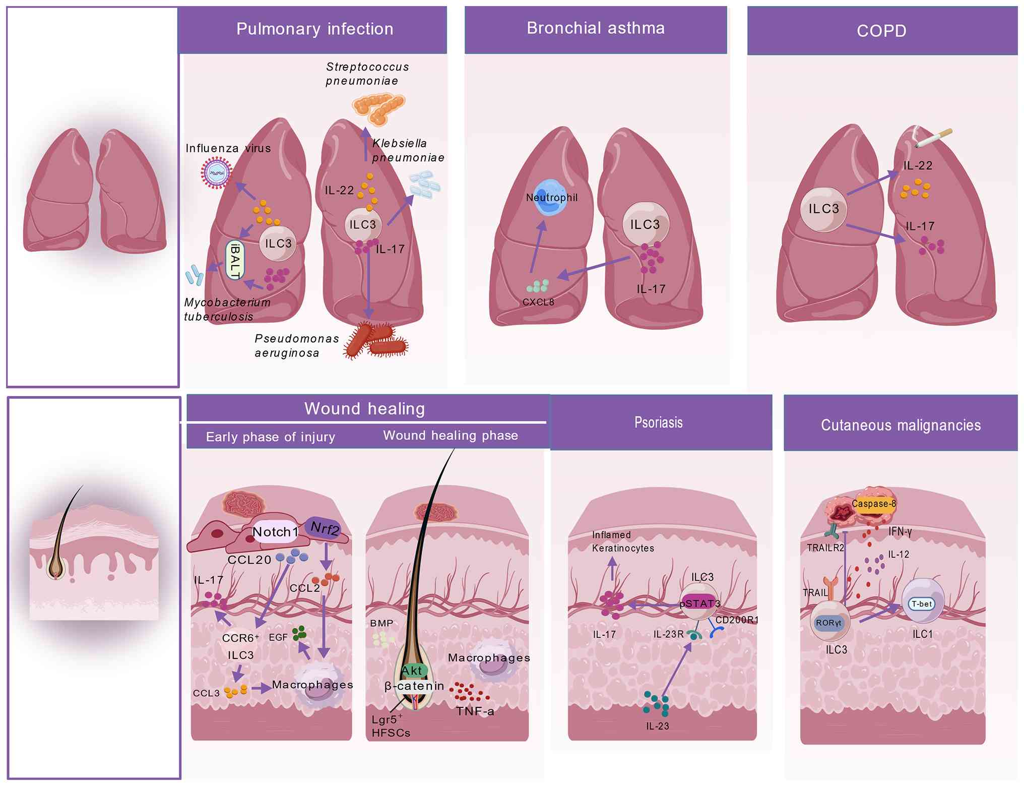 | Figure 4Mechanistic roles of ILC3s in
pulmonary and skin diseases. Pulmonary infection: Infection with
Streptococcus pneumoniae, Klebsiella pneumoniae or
Pseudomonas aeruginosa stimulates pulmonary ILC3s to secrete
IL-17, which mediates neutrophil recruitment to clear pathogens.
During Mycobacterium tuberculosis infection, ILC3s
participate in local immune responses by secreting IL-23, IL-17 and
IL-22 and contribute to the formation of iBALT in the airways.
Influenza virus stimulates ILC3s to produce IL-22, which maintains
airway epithelial integrity. Bronchial asthma: Airway ILC3s secrete
IL-17, which induces CXCL8 release, recruits neutrophils into the
airways and mediates airway inflammation. COPD: ILC3s secrete
IL-17, which sustains chronic pulmonary inflammation and mediates
alveolar tissue damage. Wound healing: Early phase of injury:
Notch1 signaling in skin cells upregulates CCL20, recruiting
CCR6+ ILC3s to the wound site. These CCR6+
ILC3s secrete CCL3 and IL-17F to promote macrophage infiltration
into the wound. Wound healing phase: Macrophages secrete TNF-α,
which, together with BMP and Akt/β-catenin signaling, activates
Lgr5+ hair follicle stem cells, thereby participating in
skin wound healing. Psoriasis: Within the lesional
microenvironment, IL-23 binds to IL-23R on ILC3s, activating the
CD200R1-pSTAT3 signaling pathway, which enhances ILC3 function and
induces robust IL-17 secretion, continuously amplifying the
inflammatory response of skin keratinocytes. Cutaneous
malignancies: IL-12 in the tumor microenvironment drives the
plastic conversion of RORγt+ ILC3s into
T-bet+ ILC1s. Upon activation by inflammatory cytokines,
RORγt+ ILC3s persistently express TRAIL, which binds to
TRAILR2 on tumor cells to initiate caspase-8-dependent apoptosis.
Meanwhile, non-converted activated ILC3s directly secrete IFN-γ and
the differentiated T-bet+ ILC1s also produce large
amounts of IFN-γ, jointly mediating antitumor immune responses.
TRAIL, TNF-related apoptosis-inducing ligand; TRAILR2, TRAIL
receptor 2; Lgr5, leucine-rich repeat-containing G protein-coupled
receptor 5; BMP, bone morphogenetic protein; T-bet, T-box expressed
in T cells; RORγt+, retinoid-related orphan receptor;
pSTAT3, phosphorylated STAT3; CCR, C-C motif chemokine receptor;
CCL, C-C motif chemokine ligand; ILC, innate lymphoid cell; iBALT,
inducible bronchus-associated lymphoid tissue; CXCL, chemokine
(C-X-C motif) ligand; COPD, chronic obstructive pulmonary
disease. |
The contribution of ILC3s is especially evident
during bacterial pneumonia. In a mouse model of Klebsiella
pneumoniae infection, ILC3s were identified as the key source
of early IL-17 and depletion of ILC3s resulted in impaired
bacterial clearance (150).
Similarly, in a mouse model of Streptococcus pneumoniae
infection, IL-22 production was shown to be dependent on ILC3s and
exogenous flagellin treatment enhanced ILC3 function and improved
host survival (147). In
Pseudomonas aeruginosa pulmonary infection models, ~90% of
early IL-17 production was derived from ILC3s, further supporting
their role in bacterial control (151).
ILC3s also contribute to protective immunity during
tuberculosis infection. Mouse studies have demonstrated that ILC3s
participate in granuloma formation and maintenance and promote the
development of inducible bronchus-associated lymphoid tissue
through the IL-23-IL-17/IL-22 axis, thereby enhancing local immune
responses (152,153). Human genetic studies further
support this function: Individuals carrying RORC mutations that
impair IL-17 signaling are more susceptible to severe mycobacterial
infections (154). During
influenza virus infection, ILC3s exhibit context-dependent
functions. Studies in murine models have demonstrated that
excessive or premature IL-17 production drives robust neutrophil
recruitment. Recruited neutrophils exhibit elevated myeloperoxidase
activity, triggering oxidative-mediated lipid peroxidation and
consequent lung tissue injury (155,156). Conversely, IL-22 produced by
ILC3s preserves epithelial integrity and promotes tissue repair,
providing protective effects during infection (157). ILC3s also participate in
chronic inflammatory airway diseases, including asthma. Studies
using obese mouse models and preliminary clinical evidence have
shown that patients with asthma, particularly those with severe or
obesity-associated asthma, exhibit increased numbers of
IL-17+ ILC3s, which is associated with airway
hyperresponsiveness (158,159).
In vitro experiments using human primary
bronchial epithelial cells, together with clinical association
studies, suggest that this phenotype may be mediated by
IL-17-induced production of neutrophil-attracting chemokines,
including CXCL8 (41). However,
a recent study has reported that NCR+ ILC3 frequencies
are notably reduced in the lungs of patients with severe asthma
compared with healthy controls (160). These findings suggest
functional differences among ILC3 subsets during asthma
pathogenesis. In chronic obstructive pulmonary disease, the
smoking-induced inflammatory microenvironment is associated with
increased levels of IL-17 and associated cytokines, suggesting that
ILC3s may also contribute to disease development (161).
Role of ILC3s in skin diseases
Skin cancer
ILC3s are implicated in melanoma immunosurveillance
(Fig. 4). In vivo studies
have indicated that, in the presence of IL-12, NKp46+
ILC3s exert antitumor activity against B16 melanoma cells by
initiating inflammatory cascades and increasing adhesion molecule
expression. These effects remodel the tumor microvasculature,
suppress tumor growth and are dependent on IL-12R and RORγt
signaling (162). In
vitro studies have further demonstrated that human ILC3s
isolated and purified from peripheral blood of healthy donors or
noncancerous liver tissue can directly destroy the melanoma cell
line SK-Mel-37. This cytotoxic activity involves TNF-related
apoptosis-inducing ligand (TRAIL) binding to TRAIL receptor 2,
which activates caspase-8 in target cells and promotes IFN-γ
secretion, ultimately inducing tumor apoptosis (163). However, in murine models, TRAIL
expression has been primarily associated with NK cells and ILC1s
and its surface expression depends on NKp46 (NCR1) (164,165). Notably, NKp46+ ILC3s
can undergo phenotypic conversion into ILC1s, including pre-ILC3
cells, accompanied by reduced RORγt expression and increased T-bet
and Notch signaling (36,166).
Therefore, further studies are required to determine whether human
ILC3s maintain lineage stability during in vitro culture or
acquire TRAIL expression through differentiation toward ILC1-like
states. Clarifying this issue will help define the precise
mechanisms by which human ILC3s contribute to antitumor
immunity.
Psoriasis
Psoriasis is a chronic inflammatory skin disease
predominantly associated with Th17 responses (167) (Fig. 4). In psoriasis, both ILC1 and
ILC3 populations are increased, whereas expansion of ILC2s is
relatively limited (168).
Notably, IL-17- and IL-22-producing NCR+ ILC3s are
markedly increased in both peripheral blood and lesional skin from
affected patients (169). These
NCR+ ILC3s express cutaneous lymphocyte-associated
antigen and their abundance in non-lesional skin is already higher
compared with that in healthy controls, suggesting that they may
contribute to disease initiation and early pathogenesis (170). Psoriatic inflammation driven by
Th17 and ILC3 responses can be attenuated by biologic therapies
targeting IL-23 (124).
Mechanistic studies have shown that ILC3s within psoriatic lesions
express CD200R1. In mouse models, CD200R1 signaling enhances
IL-23-induced IL-17 production by promoting STAT3 phosphorylation,
while IL-23 itself induces CD200R1 expression on ILC3s,
establishing a positive regulatory loop (171). Clinical specimen analyses have
further revealed increased CXCL16 expression in monocytes and skin
cells from patients with psoriasis, together with elevated
expression of CXCR6, the receptor for CXCL16, on ILC3s. These
findings suggest that the CXCL16-CXCR6 axis may promote recruitment
of proinflammatory ILC3s into skin tissues, thereby exacerbating
psoriatic inflammation (172).
Role of ILC3s in wound healing and
hair follicle neogenesis
Following skin injury, epidermal keratinocytes
activate numerous signaling pathways that coordinate immune cell
recruitment and tissue regeneration (Fig. 4). Animal studies have shown that,
during the early phase of injury, Notch1 activation induces the
expression of chemokines such as CCL20, thereby recruiting
CCR6+ ILC3s to the dermis surrounding the wound
(173). These recruited ILC3s
promote early macrophage infiltration through CCL3 secretion and
participate in local inflammatory regulation through IL-17F
production (173). Meanwhile,
mouse models have demonstrated that injury-induced nuclear factor
erythroid 2-related factor 2 (Nrf2) activation directly promotes
CCL2 transcription, thereby recruiting F4/80+
macrophages to the wound site (174). Following CCL2 stimulation,
these macrophages produce EGF, which promotes keratinocyte
proliferation through a feedback loop and accelerates
re-epithelialization (174).
During the remodeling phase of wound repair, ~7-12 days after
injury, microenvironmental changes such as reduced bone
morphogenetic protein signaling induce phenotypic transition of
macrophages. Infiltrating CX3CR1lo/med CCR2+
cells gradually differentiate into
CX3CR1hiCCR2+ macrophages, which subsequently
accumulate around hair follicles. Using a dual-reporter
lineage-tracing system, Rahmani et al (175) demonstrated that this macrophage
subset expresses high levels of TNF-α and TGF-β1. Consistent with
this, the functional requirement of macrophage-derived TNF-α in
wound-induced hair follicle regeneration has been further supported
by an independent study (176).
Macrophage-derived TNF-α activates Akt/β-catenin signaling, which
directly stimulates Lgr5+ hair follicle stem cells and
initiates wound-induced hair follicle neogenesis, thereby promoting
hair follicle cycling and post-injury regeneration (176).
By contrast, TGF-β1 maintains local macrophage
survival and chemotaxis through CX3CR1 signaling, indirectly
supporting tissue regeneration (175). Consistent with the critical
role of CX3CR1 in skin wound healing (177,178), Rahmani et al (175) demonstrated that CX3CR1
deficiency markedly reduces the CX3CR1hi macrophage
population and decreases local TGF-β1 expression, resulting in
complete loss of hair follicle regeneration. Collectively, skin
wound repair is a continuous process initiated by epidermal
signals, including Notch1 and Nrf2 activation and sequentially
mediated by innate immune cells such as ILC3s and macrophages. The
coordinated actions of ILC3-derived CCL3 and keratinocyte-derived
CCL2 facilitate timely macrophage recruitment, whereas phenotypic
transition of CX3CR1+ macrophages and their secretion of
TNF-α and TGF-β1 associate inflammatory resolution with tissue
regeneration.
Conclusions and future prospects
ILC3s have emerged as an important research focus
in innate immunity, particularly due to their subset diversity and
interactions with the microenvironment. The present review
summarizes the developmental and differentiation pathways of ILC3s,
their regulatory networks and the mechanisms underlying their
bidirectional interactions with the gut microbiota. In addition,
the present review highlights the functional heterogeneity of ILC3s
in diseases affecting the gut, liver, lung and skin, emphasizing
their roles in both immune homeostasis and disease progression.
These findings provide a theoretical basis and translational
perspective for precise immune intervention in mucosa-associated
diseases (Tables I and II).
 | Table IFunctional heterogeneity of ILC3s in
intestinal and liver diseases: Mechanistic evidence and
translational potential. |
Table I
Functional heterogeneity of ILC3s in
intestinal and liver diseases: Mechanistic evidence and
translational potential.
| Disease type | Key ILC3
subsets | Protective
mechanisms |
Pathogenic/pro-inflammatory (or
pro-tumorigenic) mechanisms | Key evidence
(models/clinical) and translational risks |
|---|
| IBD | NCR+
ILC3, NCR− ILC3/LTi and MHC-II+ ILC3 | MHC-II+
ILC3 maintain intestinal immune tolerance by inducing
CD4+ T-cell apoptosis; their numbers are markedly
reduced in children with IBD | ii) IL-12 drives
NCR+ ILC3 conversion into ILC1s, leading to increased
IFN-γ production and disruption of the epithelial barrier;
NCR− ILC3/LTis secrete IL-17, which recruits neutrophils
and downregulates tight junction proteins; and iii) GPR183 senses
7α,25-OHC, guiding aberrant ILC3 accumulation at inflammatory
sites | Models: ILC3
deficiency exacerbates pathology in DSS colitis; adoptive transfer
alleviates it; GPR183-deficient mice show reduced colonic
inflammation. Clinical: In patients with IBD, intestinal
NCR+ ILC3 are decreased, while ILC1 and IL-23 levels are
elevated; peripheral ILC3 activation markers (NKp44/CD56) are also
increased. Translational strategies: Anti-IL-23 monoclonal
antibodies; RORγt inhibitors; GPR183 antagonists. Risks: Long-term
immunosuppression may increase infection risk; acute-phase IL-17
blockade may compromise barrier protection. |
| CRC | NCR−
ILC3 (pro-tumor), MHC-II+ CCR6+ ILC3
(anti-tumor) and NKp44+ ILC3 (anti-tumor) | i) Epithelial
IL-17D binds CD93 on ILC3s, regulating IL-22 secretion to inhibit
tumorigenesis; ii) MHC-II+ ILC3 activate T cells,
promoting type 1 anti-tumor immunity and improving immunotherapy
responses and; iii) NKp44+ ILC3s promote TLS formation,
which is positively associated with favorable prognosis | i) ILC3-derived
IL-22 activates STAT3, promoting epithelial cell proliferation; ii)
IL-22BP expression is reduced in tumor tissues, leading to
dysregulated IL-22 signaling; iii) NCR− ILC3 secrete
IL-17, promoting duodenal adenoma development; iv)
macrophage-derived IL-7 enhances STAT3 and AhR expression in ILC3s,
increasing IL-22 production; and v) in advanced CRC, the number of
NKp44+ ILC3s is decreased and expression of
TLS-associated genes (LTA, LTB and TNF) is downregulated, resulting
in impaired TLS formation and loss of anti-tumor immune
surveillance | Models: Anti-IL-22
antibody inhibits tumors in the AOM/DSS model; IL-22BP-deficient
mice show increased tumor burden; MHC-II knockout impairs
anti-tumor immunity. Clinical: Low IL-22BP levels in CRC tissues
correlate with poor prognosis; high TLS density is associated with
good prognosis; NKp44+ILC3 are reduced in advanced CRC.
Translational strategies: IL-22-Fc combined with immune checkpoint
inhibitors; induction of TLS formation (LTβR agonists/chemokine
inducers); IL-17D-CD93 agonists Risks: IL-22-Fc. requires strict
control of timing and administration site to avoid pro-fibrotic and
pro-tumor effects; TLS induction may trigger autoimmunity; the
IL-17D-CD93 axis is mainly validated in animal models, with limited
human evidence and unclear off-target effects. |
| NAFLD | Gut-derived
ILC3 | IL-22 upregulates
the anti-apoptotic gene Bcl-2 in hepatocytes, inhibiting
palmitate-induced apoptosis; it also suppresses SCD1 expression,
thereby regulating lipid metabolism | Predominantly
protective in this disease; pathogenic role is limited | Models:
ILC3-deficient mice show exacerbated liver injury in the high-fat
diet induced NASH model. Clinical evidence: Direct human evidence
is currently lacking. Translational strategies: Target the
IL-23/IL-22 axis to enhance ILC3-derived IL-22 secretion. Risks:
Chronic IL-22 activation may promote fibrosis, requiring
differentiation between acute and chronic phases. |
| Liver fibrosis | IL-17A+
IL-22+ ILC3 | Limited
evidence | i) ILC3-derived
IL-17A and IL-22 directly activate hepatic stellate cells,
promoting collagen deposition and; ii) IL-22 suppresses IFN-γ
production by T cells, indirectly exacerbating fibrosis | Models: In the
CCl4-induced fibrosis model, hepatic ILC3 numbers
associate positively with fibrosis severity; adoptive transfer of
ILC3 exacerbates fibrosis in RAG-deficient mice; neutralizing
antibodies against IL-17A or IL-22 reduce collagen deposition.
Clinical: Peripheral blood ILC3 levels are positively associated
with fibrosis severity in patients with chronic hepatitis B.
Translational strategies: Anti-IL-17A/IL-22 neutralizing
antibodies. Risks: Complete blockade of IL-22 may abrogate its
hepatoprotective functions. |
| HCC | NCR−
ILC3 (pro-tumor) and NCR+ ILC3 (potential anti-tumor
subset) | Limited
evidence | i) IL-23 expands
the NCR− ILC3 subset and induces high IL-17 expression;
ii) IL-17 recruits myeloid-derived suppressor cells and enhances
their immunosuppressive activity, thereby weakening CD8+
CD T-cell-mediated anti-tumor immunity and; iii)
NCR−ILC3 directly inhibit CD8+ T-cell
proliferation and induce their apoptosis through
cell-contact-dependent mechanisms | Models: IL-23
treatment expands NCR− ILC3 and promotes tumor growth in
orthotopic xenograft models; IL-17-deficient mice show reduced
tumor growth and MDSC infiltration; co-culture experiments
demonstrate that NCR− ILC3 directly suppress
8+ T cells. Clinical: NCR+ ILC3 are
associated with TLS in non-small cell lung cancer; HCC-associated
data are still pending. Translational strategies:
Anti-IL-23/anti-IL-17 antibodies; induction of
NCR−ILC3-to-ILC1 conversion. Risks: IL-17 blockade may
compromise ant-iinfective immunity. |
| Table IIFunctional heterogeneity of ILC3s in
pulmonary and skin diseases: Mechanistic evidence and translational
potential. |
Table II
Functional heterogeneity of ILC3s in
pulmonary and skin diseases: Mechanistic evidence and translational
potential.
| Disease type | Key ILC3
subsets | Protective
mechanisms |
Pathogenic/pro-inflammatory
mechanisms | Key evidence
(models/clinical) and translational risks |
|---|
| Pulmonary
infection | NCR+
ILC3 | i) IL-22 maintains
airway epithelial integrity and promotes tissue repair; ii) IL-17
recruits neutrophils to clear extracellular bacteria (for example,
Klebsiella pneumoniae and Pseudomonas aeruginosa)
and; iii) the IL-23-IL-17/IL-22 axis promotes iBALT formation,
enhancing local anti-tuberculosis immunity. | During influenza
virus infection, excessive or premature IL-17 expression can lead
to neutrophil over-infiltration and immunopathological lung tissue
damage | Models: ILC3s are
the predominant early source of IL-17 in the K. pneumoniae
model; their depletion impairs bacterial clearance; in
Streptococcus pneumoniae models, pulmonary ILC3s mediate
protection through IL-22 and flagellin enhances ILC3 function;
RORC-deficient mice exhibit impaired iBALT formation and increased
susceptibility to tuberculosis. Clinical: Patients with RORC
mutations have defective IL-17 signaling and are more susceptible
to mycobacterial infections. Translational strategies:
Flagellin-mediated ILC3 activation; IL-22-Fc to enhance
antimicrobial defense; targeting IL-23 to promote iBALT formation.
Risks: Upregulation of IL-17 may cause immunopathological tissue
injury. |
| Asthma | IL-17+
ILC3 (pro-inflammatory) and NCR+ ILC3 (protective) | NCR+
ILC3 serve a potential protective role in maintaining airway
homeostasis; their frequency is reduced in the lungs of patients
with severe asthma. | IL-17+
ILC3 secrete IL-17, which induces epithelial production of the
neutrophil chemoattractant CXCL8, recruiting neutrophils and
exacerbating airway hyperresponsiveness and inflammation | Models:
IL-17+ ILC3s are increased in obese asthmatic mice and
IL-17 neutralization alleviates airway hyperresponsiveness; IL-23
treatment expands IL17+ ILC3 and aggravates
inflammation. Clinical: NCR+ ILC3s are decreased,
whereas IL-17+ILC3s are increased in patients with
severe asthma. Translational strategies: Anti-IL-23 to suppress
IL-17+ ILC3s; enhancement of NCR+ILC3
function. Risks: Subset-specific targeting techniques are not yet
mature. |
| Skin cancer
(melanoma) | NKp46+
ILC3 (pre-ILC3) | i) In the presence
of IL-12, NKp46+ ILC3 initiate an inflammatory cascade,
upregulate adhesion molecules, remodel tumor microvasculature and
suppress tumor growth in an IL-12R- and RORγt-dependent manner; ii)
NKp46+ ILC3 can convert into ILC1s (with downregulation
of RORγt and upregulation of T-bet/Notch), potentially acquiring
TRAIL expression and; iii) human ILC3s induce melanoma cell
apoptosis through TRAIL-TRAILR2 binding, activating caspase-8 and
promoting IFN-γ secretion | Limited
evidence | Models: Adoptive
transfer of NKp46+ LTi cells in the presence of IL-12
inhibits tumor growth in B16 melanoma grafts. Clinical: Human ILC3s
derived from peripheral blood of healthy donors or non-cancerous
liver tissue directly kill melanoma cell lines in vitro.
Translational strategies: Combine with IL-12 to activate
NKp46+ ILC3; induce TRAIL expression by modulating
T-bet/Notch signaling; expand and maintain human ILC3 lineage
stability in vitro. Risks: Human ILC3s may convert to ILC1s
during in vitro culture, altering their function; IL-12
combination therapy may cause systemic inflammation. |
| Psoriasis | NCR+
ILC3 |
NCR+ILC3s predominantly serve a
pathogenic role in this disease | i) IL-23 stimulates
NCR+ ILC3s to secrete IL-17/IL-22, amplifying skin
inflammation; ii) CD200R1 signaling enhances pSTAT3, promoting
IL-23-driven IL-17 production and; iii) the CXCL16-CXCR6 axis
mediates migration of pro-inflammatory ILCs into the skin | Models:
CD200R1-knockout mice exhibit reduced IL-17 production by ILC3s and
attenuated skin lesions; CD200R1 agonists exacerbate lesions.
Clinical: NCR+ ILC3 are notably increased in peripheral
blood and lesional skin and produce IL-17/IL-22. Translational
strategies: Anti-IL-23 monoclonal antibodies; anti-CD200R1 blocking
antibodies; CXCL16-CXCR6 axis inhibitors. Risks: Long-term blockade
of the IL-23/IL-17 pathway may increase susceptibility to
infections. |
| Skin wound
healing | CCR6+
ILC3 | i) Early after
injury, Notch1 activation upregulates CCL20, recruiting
CCR6+ ILC3 to the dermis surrounding the wound; ii)
ILC3s secrete CCL3 to promote early macrophage infiltration and
IL-17F participates in local inflammatory regulation and; iii)
under CCL2 stimulation, macrophages produce EGF, promoting
keratinocyte proliferation and re-epithelialization | Uncontrolled
inflammatory responses during this process may lead to pathological
scar formation | Models: Notch1
activation upregulates CCL20, driving CCR6+ILC3
recruitment; Nrf2 activation upregulates CCL2, driving macrophage
recruitment. Clinical: Clinical sample data are limited, with the
majority of evidence derived from animal models. Translational
strategies: Enhance ILC3 recruitment (CCL20/Notch1 agonists);
combine with macrophage modulation (for example, Ccl2/Nrf2
signaling modulation); promote phenotypic transition of
CX3CR1+ macrophages. Risks: Excessive inflammation may
lead to scar formation. |
Based on an integrated synthesis of the regulatory
mechanisms governing ILC3 functions across different disease
contexts, the present review proposes, to the best of our
knowledge, for the first time, a systematic classification of three
universal molecular switches that determine the functional outcomes
of ILC3s. Collectively, current evidence supports the existence of
three interconnected regulatory nodes. The first node is the
T-bet/RORγt transcriptional antagonism axis. High RORγt expression
maintains ILC3 lineage identity and promotes IL-17/IL-22
production, whereas increased T-bet expression directly suppresses
RORC promoter activity and drives the conversion of
CD127+ ILC3s toward an ILC1-like phenotype. Through
reciprocal inhibition, these two transcription factors establish a
molecular switch controlling ILC3 fate determination. In IBD,
persistent IL-12 stimulation increases T-bet expression, promoting
phenotypic conversion of NCR+ ILC3s and thereby
exacerbating intestinal inflammation (15,34). The second node is the IL-12/IL-23
signaling balance axis. IL-12 preferentially activates STAT4
phosphorylation, leading to T-bet induction and promoting ILC1-like
proinflammatory or antitumor functions. By contrast, IL-23 favors
STAT3 activation, stabilizes RORγt expression and maintains ILC3
lineage characteristics associated with inflammatory or
tumor-promoting effects (16).
This balance axis is supported by evidence across a number of
diseases (144,145,162,165).
The third node is the IL-22/IL-22BP axis, in which
the relative balance between IL-22 and IL-22BP determines the net
biological effects of IL-22 signaling. In CRC, lymphotoxin LTα1β2
suppresses IL-22BP expression in dendritic cells through NF-κB
signaling, allowing for sustained IL-22 activity. Persistent IL-22
signaling subsequently activates epithelial STAT3, promoting
abnormal proliferation and tumorigenesis. In CRC, IL-22BP
expression is reduced in dendritic cells, partly due to diminished
LTα1β2 signaling, thereby unleashing sustained IL-22 activity.
Persistent IL-22 signaling subsequently activates epithelial STAT3,
driving uncontrolled hyperproliferation of pre-neoplastic and
neoplastic intestinal epithelial cells and promoting tumorigenesis
(127,131). These three molecular switches
function within a hierarchical and interconnected regulatory
network. The IL-12/IL-23 balance regulates the strength of
T-bet/RORγt antagonism through differential STAT activation. This
transcriptional equilibrium subsequently influences IL-22
production and alters the IL-22/IL-22BP balance. Meanwhile,
upstream environmental signals, including gut microbial
metabolites, RA, neural signals and circadian rhythms, can reset
these regulatory states by reshaping cellular metabolism or
modulating cytokine production, ultimately determining ILC3
functional outcomes during infection, chronic inflammation and
tumor development.
Based on the aforementioned three-tier molecular
regulatory network, the present review proposes three
experimentally testable hypotheses. First, with regard to the
T-bet/RORγt transcriptional antagonism axis, it may be hypothesized
that persistent IL-12 stimulation within the IBD microenvironment
drives NCR+ ILC3-to-ILC1 conversion through the
STAT4/T-bet pathway. The present review further predicts that
ILC3-specific T-bet deletion will prevent this conversion and
attenuate DSS-induced intestinal inflammation. This hypothesis can
be evaluated using ILC3-specific gene-targeted mice combined with
flow cytometric analysis of intestinal lymphocytes and
histopathological assessment. Second, regarding the IL-12/IL-23
balance axis, it may be hypothesized that an IL-23-dominant
microenvironment in HCC promotes expansion of pro-tumorigenic
NCR− ILC3s. The present review predicts that low-dose
recombinant IL-12 treatment will restore this imbalance by reducing
the intratumoral NCR−/NCR+ ILC3 ratio,
enhancing CD8+ T-cell infiltration and suppressing tumor
growth. This hypothesis can be tested using orthotopic HCC mouse
models combined with immune phenotyping by flow cytometry. Third,
with regard to the IL-22/IL-22BP balance axis, it may be
hypothesized that reduced IL-22BP expression in the CRC
microenvironment increases free IL-22 availability, thereby
promoting STAT3-dependent epithelial proliferation. The present
study predicts that restoration of IL-22BP through adeno-associated
virus-mediated delivery to colonic tissues will reduce tumor burden
while preserving antimicrobial defense. This hypothesis can be
evaluated using the azoxymethane (AOM)/DSS CRC model together with
phosphorylated STAT3 detection and tumor burden assessment
(127,131).
The biological functions of ILC3s are largely
determined by the dynamic balance between two key cytokine
pathways, namely IL-17 and IL-22. Therefore, ILC3-mediated disease
outcomes are not determined solely by ILC3 abundance, but rather by
the balance between IL-17 and IL-22 production, the nature of
upstream stimuli and the local immune microenvironment. Based on
the integrated analysis of pathological features and multilayer
regulatory networks across diseases, the present review proposes a
'spatiotemporal-quantitative' three-dimensional framework to
interpret the context-dependent functional outcomes of ILC3s. This
framework describes the dynamic transition between protective and
pathogenic states through three dimensions. The first dimension is
the disease phase. During acute injury, ILC3s primarily contribute
to barrier defense through IL-22 production. However, prolonged
stimulation reshapes ILC3 plasticity and promotes conversion toward
ILC1-like or ILCreg states, with persistent IL-17/IL-22 signaling
contributing to pathological progression. The second dimension is
the tissue microenvironment. Organ-specific signals, including gut
microbial metabolites, hepatic lipid signals, cutaneous
neuropeptides, local chemokines and circadian rhythms,
differentially regulate ILC3 recruitment, activation and functional
polarization. The third dimension is signal dosage. Effector
molecules such as IL-17 and IL-22 exhibit concentration-dependent
effects: Physiological levels support antimicrobial defense and
epithelial repair, whereas sustained excessive activation promotes
inflammation, excessive epithelial proliferation and tumor
progression. Collectively, these findings indicate that ILC3s do
not possess an intrinsically fixed pro-inflammatory or
anti-inflammatory identity. Instead, their functional state may
represent an integrated outcome determined by multidimensional
environmental signals. This three-dimensional framework integrates
previously fragmented findings from IBD, solid tumors, metabolic
liver diseases and infectious immunity into a unified conceptual
model. It provides one perspective for understanding the
context-dependent functions of ILC3s and establishes a theoretical
foundation for developing subset-specific therapeutic
strategies.
Based on the three-dimensional analytical
framework, the present study further proposes an integrative
quantitative hypothesis whereby the IL-17/IL-22 secretion ratio of
ILC3s may serve as a quantitative indicator of their functional
polarity. Specifically, it may be hypothesized that during acute
infection or tissue injury, the IL-22/IL-17 ratio increases and is
positively associated with epithelial repair and pathogen
clearance, whereas IL-22 neutralization delays tissue repair.
Conversely, during chronic inflammation and tumor progression, the
IL-17/IL-22 ratio gradually increases and is associated with IBD
histopathological severity and CRC tumor burden. IL-23 blockade is
predicted to reduce this ratio and alleviate tissue damage. This
hypothesis can be evaluated through time-course analyses using
DSS-induced colitis, AOM/DSS CRC and Streptococcus
pneumoniae infection models. ILC3s isolated from tissues at
acute and chronic stages can be analyzed by intracellular cytokine
staining and flow cytometry. Intervention groups involving ILC3
depletion, IL-22 neutralization and IL-23 blockade should be
included to establish causal associations between cytokine ratios
and pathological outcomes.
The regulation of ILC3 plasticity is not mediated
by isolated signaling pathways, but rather by the integration of
multilayered signals through three key molecular hubs: mTOR, STAT3
and AHR. The mTOR complex functions as a metabolic-transcriptional
regulatory hub. It senses extracellular metabolic signals,
including microbiota-derived SCFA signals through FFAR2 (92), as well as intracellular changes
in amino acid availability and energy status. Through the
mTORC1/HIF-1α pathway, mTOR signaling stabilizes RORγt expression,
promotes glycolysis and regulates mitochondrial ROS production,
thereby supporting ILC3 lineage stability and effector functions
(65). When mTORC1 activity is
impaired, mTORC2 can undergo compensatory activation, partially
restoring ILC3 numbers and preserving intestinal antimicrobial
defense (66). STAT3 represents
a common downstream signaling component of neural, microbial and
inflammatory pathways. Enteric glial cells release GDNF, which
activates RET and induces STAT3 phosphorylation through the p38
MAPK/ERK-Akt pathway (78). In
parallel, sympathetic nerve-derived norepinephrine activates ADRB2
and enhances STAT3 activity, with both neural pathways promoting
IL-22 transcription (81). In
addition, IL-23-mediated inflammatory stimulation can directly
activate STAT3 and further enhance IL-22 responses (16). AHR functions as a direct sensor
of microbiota-derived tryptophan metabolites. Binding of indole
derivatives to AHR initiates IL-22 transcriptional programs
(88,89), while AHR activity can also be
modulated by intracellular metabolic and inflammatory signals such
as IL-1β (91). Collectively,
mTOR, STAT3 and AHR form an interconnected regulatory network that
integrates microbial, neural, metabolic and inflammatory signals to
determine ILC3 lineage identity and effector output.
Despite mouse models having provided important
insights into ILC3 biology, notable phenotypic and functional
differences exist between human and murine ILC3s. Human
NCR+ ILC3s are defined primarily by NKp44 expression,
whereas murine NCR+ ILC3s are characterized by NKp46
expression (33-35) In addition, human NCR−
ILC3s express CD117, whereas murine NCR− ILC3s lack this
marker (35). Furthermore, TRAIL
is predominantly expressed by NK cells and ILC1s in mice, whereas
human ILC3s can directly express TRAIL and mediate antitumor
effects (163-165). These interspecies differences
represent a key challenge in translating basic ILC3 research into
clinical applications. Therefore, clinical development of
ILC3-targeting strategies requires precise characterization of
human ILC3 subsets using integrated single-cell multi-omics
approaches and primary patient-derived samples. In addition,
humanized mouse models and organoid-ILC3 coculture systems should
be prioritized to improve recapitulation of human physiology and
facilitate clinically relevant translational studies.
In addition to classical proinflammatory ILC3s, the
discovery of ILCregs has expanded the understanding of mucosal
immune regulation. These two populations form a complementary
regulatory system in the gut: ILCregs suppress excessive activation
of ILC1s and ILC3s through IL-10 while preserving protective IL-22
secretion by ILC3s, thereby contributing to the resolution of
intestinal inflammation (12).
This regulatory pathway functions independently of Treg cells and
represents a specialized tolerance mechanism within innate
immunity. However, in the tumor microenvironment, TGF-β can alter
this balance. Current evidence indicates that ILC3s can
transdifferentiate into ILCregs, which subsequently produce high
levels of IL-10, establish an immunosuppressive microenvironment
and promote tumor immune evasion (14). These findings suggest that ILCreg
functions are strongly influenced by the surrounding
microenvironment, with protective roles during inflammatory
responses but tumor-promoting effects in malignant settings.
Therefore, classification of ILC3s and ILCregs as simply
proinflammatory or anti-inflammatory populations does not fully
reflect their biological plasticity. Further investigation is
required to identify the upstream signals controlling the balance
between these populations and to develop strategies for targeted
intervention. However, the majority of current evidence is derived
from mouse models and the lineage characteristics, differentiation
mechanisms and functional roles of human ILCregs in inflammation
and cancer remain to be systematically defined. Future studies
should aim to integrate humanized mouse models, organoid systems
and clinical samples to investigate cross-species conservation and
microenvironment-driven transdifferentiation mechanisms.
From the perspective of current translational
strategies, three key therapeutic approaches can be considered. The
first involves upstream signaling inhibitors that target key
regulators of ILC3 activation and lineage plasticity, including
IL-23 and RORγt. Anti-IL-23 monoclonal antibodies inhibit
inflammatory pathways associated with excessive ILC3 activation and
have demonstrated improved efficacy compared with conventional
anti-TNF monoclonal antibodies in IBDs such as CD (179). In the phase III SEQUENCE
head-to-head trial in patients with moderate-to-severe Crohn's
disease refractory to anti-TNF therapy, the p19-specific anti-IL-23
monoclonal antibody risankizumab demonstrated superiority in
endoscopic remission compared with ustekinumab (which targets the
shared p40 subunit of IL-12/IL-23), with rates of 31.8 vs. 16.2%
(P<0.001). Safety profiles were acceptable, with comparable
serious infection rates (3.1 vs. 4.2%) and lower serious adverse
events with risankizumab (10.3 vs. 17.4%) (179). These clinical findings support
the therapeutic potential of selective IL-23-p19 inhibition, a
pathway central to ILC3-driven intestinal immunopathology (179). A Bayesian network meta-analysis
of 22 randomized controlled trials further demonstrated that
risankizumab exhibited the lowest overall risks of adverse events,
serious adverse events and serious infections during the induction
phase among evaluated biologic agents for IBD (180). Notably, this safety advantage
was observed only during induction therapy and not reproduced in
the maintenance-phase comparisons. This meta-analytic evidence is
mechanistically relevant to the present review, as risankizumab
selectively targets the IL-23 p19 subunit, a key upstream cytokine
that drives ILC3-mediated intestinal immunopathology. Thus, these
findings complement the head-to-head SEQUENCE trial by providing
aggregated population-level evidence from multiple randomized
controlled trials (180). By
contrast, the oral RORγt inhibitor BI 730357 exhibited only modest
efficacy in a phase II plaque-psoriasis trial: At the highest
tested dose (200 mg once daily), only 30.0% of patients achieved
PASI75 at week 12 vs. 0% with placebo, and the exposure-response
relationship plateaued at doses ≥200 mg, suggesting that robust
clinical responses are unlikely to be attained even at further
elevated drug exposures. Although the safety profile was acceptable
during the 12-week induction phase (drug-related adverse events
≤15.8%), long-term development was discontinued as a non-clinical
toxicology study identified potential human carcinogenic risks that
could not be ruled out (181).
As RORγt acts as a shared lineage-defining transcription factor for
both Th17 cells and ILC3s, pharmacological inhibition of this
protein constitutes an alternative therapeutic approach targeting
the IL-23/IL-17/IL-22 axis upstream of direct cytokine
neutralization. The modest clinical activity and exposure-response
plateau observed for BI 730357 therefore indicate that complete
RORγt suppression may be insufficient to fully abrogate
IL-17-driven inflammation, while potentially disrupting
ILC3-dependent epithelial-barrier protection. These observations
underscore the inherent complexity of therapeutically targeting
this transcription factor in ILC3-associated intestinal
immunopathology (181).
Despite these two drug classes differing in
molecular type, both intervene at upstream nodes of the IL-23/IL-17
axis. The differential efficacy observed between them highlights
notable differences between cytokine-targeting strategies
(anti-IL-23) and transcription factor-targeting strategies (RORγt
inhibitors) in terms of signaling blockade completeness and
functional selectivity. A novel small-molecule GPR183 antagonist,
which inhibits aberrant ILC3 recruitment mediated by chemotactic
signals, has demonstrated dose-dependent intestinal
anti-inflammatory activity in a DSS-induced mouse colitis model,
supporting its potential for further preclinical development
(182). The second category
comprises effector molecule-targeted approaches, which regulate
downstream inflammatory and tissue repair processes through
supplementation or neutralization of specific effector molecules.
IL-22-Fc fusion proteins provide exogenous IL-22 and have
demonstrated mucosal repair effects in animal models of colitis.
However, a phase II clinical trial in UC failed to meet the
prespecified primary endpoints. Although target engagement was
confirmed by dose-dependent elevations of circulating REG3A
biomarker, therapeutic supplementation with IL-22-Fc fusion protein
(efmarodocokin alfa) alone proved insufficient to overcome the
complex inflammatory mucosal microenvironment (183). This trial enrolled 195 patients
with moderate-to-severe ulcerative colitis. At week 8,
clinical-remission rates for efmarodocokin alfa were 12, 9 and 12%
in the 30, 60 and 90 µg/kg dose cohorts respectively, vs. 9%
in the placebo arm. None of these differences met the
study-pre-specified statistical threshold for significance of
P<0.2. Rates of endoscopic healing were likewise comparable to
placebo, and the trial was terminated early following pre-defined
futility analysis (183). These
observations indicate that isolated IL-22 agonism is not sufficient
to drive meaningful clinical improvement in moderate-to-severe
human ulcerative colitis, despite mechanistic expectation from
pre-clinical models. The third category involves
microenvironment-remodeling strategies, which indirectly regulate
ILC3 differentiation and function through metabolic and neural
signals, including SCFAs, vitamin D supplementation and modulation
of the GDNF neuroimmune pathway. However, these
microenvironment-remodeling strategies remain largely at the
preclinical stage, with most evidence derived from murine models.
The complexity of in vivo regulatory networks, coupled with
insufficient tissue-targeting capacity and a lack of validated
biomarkers, has limited their translation into human trials.
Consequently, human clinical data are currently scarce, and
substantial further investigation is required prior to clinical
translation.
Collectively, clinical evidence from these three
therapeutic categories indicates that current ILC3-targeted
translation faces two key challenges: Variable therapeutic efficacy
and long-term safety concerns. Anti-IL-23 monoclonal antibodies
demonstrate the most favorable safety profile but are mainly
applicable to inflammatory conditions dominated by IL-23 signaling.
RORγt inhibitors face limitations associated with efficacy ceilings
and potential carcinogenic risks. IL-22 supplementation alone
appears insufficient to overcome the immunosuppressive
microenvironment associated with tumors and chronic inflammation.
Collectively, these findings suggest that future ILC3-targeted
therapies should move beyond broad-spectrum single-pathway
inhibition toward stratified precision interventions guided by the
three molecular switches proposed in the present review. Therefore,
future clinical trial designs should also incorporate systematic
evaluation of long-term safety outcomes, including infection and
malignancy risks (Table
III).
| Table IIISummary of completed and ongoing
clinical trials targeting ILC3-associated signaling pathways. |
Table III
Summary of completed and ongoing
clinical trials targeting ILC3-associated signaling pathways.
| Target/drug
Class | Drug name | Mechanism of
action | Indication | Clinical trial
phase | Key endpoints and
findings | Development
status |
|---|
| Anti-IL-23 | Risankizumab | Anti-IL-23 p19
monoclonal antibody; selectively blocks IL-23 pathway | Moderate-to-severe
Crohn's disease | Phase III
(SEQUENCE) | Incidence of SAE
was 10.3 vs. 17.4% for ustekinumab; serious infection rates were
3.1 vs. 4.2%, respectively | Completed |
| Anti-IL-23 | Ustekinumab | Anti-IL-12/IL-23
p40 monoclonal antibody; dual blockade of IL-12/IL-23 pathways | Moderate-to-severe
Crohn's disease | Phase III
(SEQUENCE) | SAE rate 17.4%;
serious infection rate 4.2%; served as active comparator against
risankizumab | Completed |
| RORγt
inhibitor | BI 730357 | Oral small-molecule
RORγt transcriptional inhibitor; blocks IL-17-driven
pro-inflammatory pathway | Moderate-to-severe
plaque psoriasis with inadequate response to TNF inhibitors | Phase II
dose-ranging trial | PASI75 response
rate at week 12 was only 30% at the 200 mg dose, with no additional
efficacy at higher doses (plateau effect); overall drug-associated
AEs ≤15.8%; nonclinical toxicology identified potential human
carcinogenicity risk | Phase II main trial
completed; long-term extension and full development
discontinued |
| GPR183
antagonist | Compound 33 | Selective
small-molecule GPR183 antagonist; blocks ILC3 intestinal
chemotactic infiltration | DSS-induced
experimental colitis in mice | Preclinical
only | Dose-dependent
inhibition of aberrant ILC3 intestinal recruitment and colonic
inflammation; weak hERG inhibition with high target selectivity and
low potential cardiotoxicity | Preclinical
candidate; no human trials initiated |
| IL-22-Fc fusion
protein | Efmarodocokin
alfa | IL-22 fusion
protein; exogenously supplies protective IL-22 and upregulates
mucosal repair markers | Moderate-to-severe
ulcerative colitis with prior standard-of-care failure | Phase II
(YELLOWSTONE) | Clinical remission
rates at week 8 were 12, 9 and 12% for the 30, 60 and 90
µg/kg dose groups, respectively, vs. 9% for placebo; no
significant difference in endoscopic healing; trial terminated
early for futility | Phase II
terminated; no further clinical development |
Availability of data and materials
Not applicable.
Authors' contributions
JZ and PZ prepared the figures and drafted the
manuscript. XL, JL, LQ, JC, XH, MZ, JL, XZ, JD, GT, YPT and YF
conceived the present study and performed the literature analysis.
YT, LB and PZ edited and revised the manuscript. All authors listed
have made notable, direct and intellectual contributions to the
present work and approved it for publication. All authors read and
approved the final version of the manuscript. Data authentication
is not applicable.
Ethics approval and consent to
participate
Not applicable.
Patient consent for publication
Not applicable.
Competing interests
The authors declare that they have no competing
interests.
Acknowledgments
Not applicable.
Funding
The present study was supported by the National Natural Science
Foundation of China (grant nos. 82360329 and 82560126), the Inner
Mongolia Key R&D and Technology Transfer Program (grant no.
2026SYFSH0185) and the Inner Mongolia Natural Science Foundation
(grant nos. 2025QN08050 and 2026QA030). Additional support was
provided by the Science and Technology Plan of Inner Mongolia
Autonomous Region (grant no. 2026YFSH0109), the Innovation Team
Development Program for Higher Education Institutions in Inner
Mongolia (grant no. NMGIRT2511), the Talent Project of Inner
Mongolia (grant no. 060196), the Yingcaipeiyu Project of Inner
Mongolia Medical University (grant nos. YCPY2026010, YCPY2026020
and YCPY2026021) and the 2026 University Student Innovation and
Entrepreneurship Project of Inner Mongolia Medical University
(grant nos. S202610132003 and 202610132002).
References
|
1
|
Morita H, Moro K and Koyasu S: Innate
lymphoid cells in allergic and nonallergic inflammation. J Allergy
Clin Immunol. 138:1253–1264. 2016. View Article : Google Scholar : PubMed/NCBI
|
|
2
|
Ebihara T, Song C, Ryu SH,
Plougastel-Douglas B, Yang L, Levanon D, Groner Y, Bern MD,
Stappenbeck TS, Colonna M, et al: Runx3 specifies lineage
commitment of innate lymphoid cells. Nat Immunol. 16:1124–1133.
2015. View Article : Google Scholar : PubMed/NCBI
|
|
3
|
Zeng B, Shi S, Ashworth G, Dong C, Liu J
and Xing F: ILC3 function as a double-edged sword in inflammatory
bowel diseases. Cell Death Dis. 10:3152019. View Article : Google Scholar : PubMed/NCBI
|
|
4
|
Brestoff JR, Kim BS, Saenz SA, Stine RR,
Monticelli LA, Sonnenberg GF, Thome JJ, Farber DL, Lutfy K, Seale P
and Artis D: Group 2 innate lymphoid cells promote beiging of white
adipose tissue and limit obesity. Nature. 519:242–246. 2015.
View Article : Google Scholar
|
|
5
|
Bar-Ephraim TE and Mebius RE: Innate
lymphoid cells in secondary lymphoid organs. Immunol Rev.
271:185–199. 2016. View Article : Google Scholar : PubMed/NCBI
|
|
6
|
Kiniwa T and Moro K: Localization and
site-specific cell-cell interactions of group 2 innate lymphoid
cells. Int Immunol. 33:251–259. 2021. View Article : Google Scholar : PubMed/NCBI
|
|
7
|
Klose CS, Kiss EA, Schwierzeck V, Ebert K,
Hoyler T, d'Hargues Y, Göppert N, Croxford AL, Waisman A, Tanriver
Y and Diefenbach A: A T-bet gradient controls the fate and function
of CCR6-RORүt+ innate lymphoid cells. Nature. 494:261–265. 2013.
View Article : Google Scholar : PubMed/NCBI
|
|
8
|
Jarade A, Di Santo JP and Serafini N:
Group 3 innate lymphoid cells mediate host defense against
attaching and effacing pathogens. Curr Opin Microbiol. 63:83–91.
2021. View Article : Google Scholar : PubMed/NCBI
|
|
9
|
Sanati G, Aryan Z, Barbadi M and Rezaei N:
Innate lymphoid cells are pivotal actors in allergic, inflammatory
and autoimmune diseases. Expert Rev Clin Immunol. 11:885–895. 2015.
View Article : Google Scholar : PubMed/NCBI
|
|
10
|
Zhou W and Sonnenberg GF: Activation and
suppression of group 3 innate lymphoid cells in the gut. Trends
Immunol. 41:721–733. 2020. View Article : Google Scholar : PubMed/NCBI
|
|
11
|
Meininger I, Carrasco A, Rao A, Soini T,
Kokkinou E and Mjosberg J: Tissue-specific features of innate
lymphoid cells. Trends Immunol. 41:902–917. 2020. View Article : Google Scholar : PubMed/NCBI
|
|
12
|
Wang S, Xia P, Chen Y, Qu Y, Xiong Z, Ye
B, Du Y, Tian Y, Yin Z, Xu Z and Fan Z: Regulatory innate lymphoid
cells control innate intestinal inflammation. Cell.
171:201–216.e18. 2017. View Article : Google Scholar : PubMed/NCBI
|
|
13
|
Zeng B, Shi S, Liu J and Xing F:
Commentary: Regulatory innate lymphoid cells control innate
intestinal inflammation. Front Immunol. 9:15222018. View Article : Google Scholar : PubMed/NCBI
|
|
14
|
Wang S, Qu Y, Xia P, Chen Y, Zhu X, Zhang
J, Wang G, Tian Y, Ying J and Fan Z: Transdifferentiation of tumor
infiltrating innate lymphoid cells during progression of colorectal
cancer. Cell Res. 30:610–622. 2020. View Article : Google Scholar : PubMed/NCBI
|
|
15
|
Artis D and Spits H: The biology of innate
lymphoid cells. Nature. 517:293–301. 2015. View Article : Google Scholar : PubMed/NCBI
|
|
16
|
Buonocore S, Ahern PP, Uhlig HH, Ivanov
II, Littman DR, Maloy KJ and Powrie F: Innate lymphoid cells drive
interleukin-23-dependent innate intestinal pathology. Nature.
464:1371–1375. 2010. View Article : Google Scholar : PubMed/NCBI
|
|
17
|
Klose CSN, Flach M, Möhle L, Rogell L,
Hoyler T, Ebert K, Fabiunke C, Pfeifer D, Sexl V, Fonseca-Pereira
D, et al: Differentiation of type 1 ILCs from a common progenitor
to all helper-like innate lymphoid cell lineages. Cell.
157:340–356. 2014. View Article : Google Scholar : PubMed/NCBI
|
|
18
|
Constantinides MG, McDonald BD, Verhoef PA
and Bendelac A: A committed precursor to innate lymphoid cells.
Nature. 508:397–401. 2014. View Article : Google Scholar : PubMed/NCBI
|
|
19
|
Serafini N, Klein Wolterink RG,
Satoh-Takayama N, Xu W, Vosshenrich CA, Hendriks RW and Di Santo
JP: Gata3 drives development of RORgammat+ group 3 innate lymphoid
cells. J Exp Med. 211:199–208. 2014. View Article : Google Scholar : PubMed/NCBI
|
|
20
|
Yagi R, Zhong C, Northrup DL, Yu F,
Bouladoux N, Spencer S, Hu G, Barron L, Sharma S, Nakayama T, et
al: The transcription factor GATA3 is critical for the development
of all IL-7Rα-expressing innate lymphoid cells. Immunity.
40:378–388. 2014. View Article : Google Scholar : PubMed/NCBI
|
|
21
|
Mielke LA, Groom JR, Rankin LC, Seillet C,
Masson F, Putoczki T and Belz GT: TCF-1 controls ILC2 and
NKp46+RORүt+ innate lymphocyte differentiation and protection in
intestinal inflammation. J Immunol. 191:4383–4391. 2013. View Article : Google Scholar : PubMed/NCBI
|
|
22
|
Seillet C, Rankin LC, Groom JR, Mielke LA,
Tellier J, Chopin M, Huntington ND, Belz GT and Carotta S: Nfil3 is
required for the development of all innate lymphoid cell subsets. J
Exp Med. 211:1733–1740. 2014. View Article : Google Scholar : PubMed/NCBI
|
|
23
|
Hoyler T, Klose CS, Souabni A,
Turqueti-Neves A, Pfeifer D, Rawlins EL, Voehringer D, Busslinger M
and Diefenbach A: The transcription factor GATA-3 controls cell
fate and maintenance of type 2 innate lymphoid cells. Immunity.
37:634–648. 2012. View Article : Google Scholar : PubMed/NCBI
|
|
24
|
Yu Y, Tsang JC, Wang C, Clare S, Wang J,
Chen X, Brandt C, Kane L, Campos LS, Lu L, et al: Single-cell
RNA-seq identifies a PD-1(hi) ILC progenitor and defines its
development pathway. Nature. 539:102–106. 2016. View Article : Google Scholar : PubMed/NCBI
|
|
25
|
Eberl G, Marmon S, Sunshine MJ, Rennert
PD, Choi Y and Littman DR: An essential function for the nuclear
receptor RORgamma(t) in the generation of fetal lymphoid tissue
inducer cells. Nat Immunol. 5:64–73. 2004. View Article : Google Scholar
|
|
26
|
Cherrier M, Sawa S and Eberl G: Notch,
Id2, and RORүt sequentially orchestrate the fetal development of
lymphoid tissue inducer cells. J Exp Med. 209:729–740. 2012.
View Article : Google Scholar : PubMed/NCBI
|
|
27
|
Ishizuka IE, Chea S, Gudjonson H,
Constantinides MG, Dinner AR, Bendelac A and Golub R: Single-cell
analysis defines the divergence between the innate lymphoid cell
lineage and lymphoid tissue-inducer cell lineage. Nat Immunol.
17:269–276. 2016. View Article : Google Scholar : PubMed/NCBI
|
|
28
|
Robinette ML, Fuchs A, Cortez VS, Lee JS,
Wang Y, Durum SK, Gilfillan S and Colonna M; Immunological Genome
Consortium: Transcriptional programs define molecular
characteristics of innate lymphoid cell classes and subsets. Nat
Immunol. 16:306–317. 2015. View Article : Google Scholar : PubMed/NCBI
|
|
29
|
Bjorklund AK, Forkel M, Picelli S, Konya
V, Theorell J, Friberg D, Sandberg R and Mjösberg J: The
heterogeneity of human CD127(+) innate lymphoid cells revealed by
single-cell RNA sequencing. Nat Immunol. 17:451–460. 2016.
View Article : Google Scholar : PubMed/NCBI
|
|
30
|
Gury-BenAri M, Thaiss CA, Serafini N,
Winter DR, Giladi A, Lara-Astiaso D, Levy M, Salame TM, Weiner A,
David E, et al: The spectrum and regulatory landscape of intestinal
innate lymphoid cells are shaped by the microbiome. Cell.
166:1231–1246 e13. 2016. View Article : Google Scholar : PubMed/NCBI
|
|
31
|
van de Pavert SA and Vivier E:
Differentiation and function of group 3 innate lymphoid cells, from
embryo to adult. Int Immunol. 28:35–42. 2016. View Article : Google Scholar
|
|
32
|
Jacquelot N, Seillet C, Vivier E and Belz
GT: Innate lymphoid cells and cancer. Nat Immunol. 23:371–379.
2022. View Article : Google Scholar : PubMed/NCBI
|
|
33
|
Monticelli LA, Sonnenberg GF, Abt MC,
Alenghat T, Ziegler CG, Doering TA, Angelosanto JM, Laidlaw BJ,
Yang CY, Sathaliyawala T, et al: Innate lymphoid cells promote
lung-tissue homeostasis after infection with influenza virus. Nat
Immunol. 12:1045–1054. 2011. View Article : Google Scholar : PubMed/NCBI
|
|
34
|
Bernink JH, Krabbendam L, Germar K, de
Jong E, Gronke K, Kofoed-Nielsen M, Munneke JM, Hazenberg MD,
Villaudy J, Buskens CJ, et al: Interleukin-12 and -23 control
plasticity of CD127(+) group 1 and group 3 innate lymphoid cells in
the intestinal lamina propria. Immunity. 43:146–160. 2015.
View Article : Google Scholar : PubMed/NCBI
|
|
35
|
Hazenberg MD and Spits H: Human innate
lymphoid cells. Blood. 124:700–709. 2014. View Article : Google Scholar : PubMed/NCBI
|
|
36
|
Rankin LC, Groom JR, Chopin M, Herold MJ,
Walker JA, Mielke LA, McKenzie AN, Carotta S, Nutt SL and Belz GT:
The transcription factor T-bet is essential for the development of
NKp46+ innate lymphocytes via the Notch pathway. Nat Immunol.
14:389–395. 2013. View Article : Google Scholar : PubMed/NCBI
|
|
37
|
McKenzie ANJ, Spits H and Eberl G: Innate
lymphoid cells in inflammation and immunity. Immunity. 41:366–374.
2014. View Article : Google Scholar : PubMed/NCBI
|
|
38
|
Bernink JH, Peters CP, Munneke M, te Velde
AA, Meijer SL, Weijer K, Hreggvidsdottir HS, Heinsbroek SE, Legrand
N, Buskens CJ, et al: Human type 1 innate lymphoid cells accumulate
in inflamed mucosal tissues. Nat Immunol. 14:221–229. 2013.
View Article : Google Scholar : PubMed/NCBI
|
|
39
|
Cella M, Fuchs A, Vermi W, Facchetti F,
Otero K, Lennerz JK, Doherty JM, Mills JC and Colonna M: A human
natural killer cell subset provides an innate source of IL-22 for
mucosal immunity. Nature. 457:722–725. 2009. View Article : Google Scholar
|
|
40
|
Teunissen MBM, Munneke JM, Bernink JH,
Spuls PI, Res PCM, Te Velde A, Cheuk S, Brouwer MWD, Menting SP,
Eidsmo L, et al: Composition of innate lymphoid cell subsets in the
human skin: enrichment of NCR(+) ILC3 in lesional skin and blood of
psoriasis patients. J Invest Dermatol. 134:2351–2360. 2014.
View Article : Google Scholar : PubMed/NCBI
|
|
41
|
Chesne J, Braza F, Mahay G, Brouard S,
Aronica M and Magnan A: IL-17 in severe asthma. Where do we stand?
Am J Respir Crit Care Med. 190:1094–1101. 2014. View Article : Google Scholar : PubMed/NCBI
|
|
42
|
Muir R, Osbourn M, Dubois AV, Doran E,
Small DM, Monahan A, O'Kane CM, McAllister K, Fitzgerald DC,
Kissenpfennig A, et al: Innate lymphoid cells are the predominant
source of IL-17A during the early pathogenesis of acute respiratory
distress Syndrome. Am J Respir Crit Care Med. 193:407–416. 2016.
View Article : Google Scholar
|
|
43
|
Lee JS, Tato CM, Joyce-Shaikh B, Gulen MF,
Cayatte C, Chen Y, Blumenschein WM, Judo M, Ayanoglu G, McClanahan
TK, et al: Interleukin-23-Independent IL-17 production regulates
intestinal epithelial permeability. Immunity. 43:727–738. 2015.
View Article : Google Scholar : PubMed/NCBI
|
|
44
|
Mebius RE, Rennert P and Weissman IL:
Developing lymph nodes collect CD4+CD3-LTbeta+ cells that can
differentiate to APC, NK cells, and follicular cells but not T or B
cells. Immunity. 7:493–504. 1997. View Article : Google Scholar : PubMed/NCBI
|
|
45
|
Buettner M and Lochner M: Development and
function of secondary and tertiary lymphoid organs in the small
intestine and the colon. Front Immunol. 7:3422016. View Article : Google Scholar : PubMed/NCBI
|
|
46
|
Cording S, Medvedovic J, Lecuyer E, Aychek
T, Dejardin F and Eberl G: Mouse models for the study of fate and
function of innate lymphoid cells. Eur J Immunol. 48:1271–1280.
2018. View Article : Google Scholar : PubMed/NCBI
|
|
47
|
van de Pavert SA and Mebius RE: New
insights into the development of lymphoid tissues. Nat Rev Immunol.
10:664–674. 2010. View Article : Google Scholar : PubMed/NCBI
|
|
48
|
Bouskra D, Brézillon C, Bérard M, Werts C,
Varona R, Boneca IG and Eberl G: Lymphoid tissue genesis induced by
commensals through NOD1 regulates intestinal homeostasis. Nature.
456:507–510. 2008. View Article : Google Scholar : PubMed/NCBI
|
|
49
|
Spits H, Artis D, Colonna M, Diefenbach A,
Di Santo JP, Eberl G, Koyasu S, Locksley RM, McKenzie AN, Mebius
RE, et al: Innate lymphoid cells-a proposal for uniform
nomenclature. Nat Rev Immunol. 13:145–149. 2013. View Article : Google Scholar : PubMed/NCBI
|
|
50
|
Shiu J, Piazuelo MB, Ding H, Czinn SJ,
Drakes ML, Banerjee A, Basappa N, Kobayashi KS, Fricke WF and
Blanchard TG: Gastric LTi cells promote lymphoid follicle formation
but are limited by IRAK-M and do not alter microbial growth.
Mucosal Immunol. 8:1047–1059. 2015. View Article : Google Scholar : PubMed/NCBI
|
|
51
|
Zhong C, Zheng M and Zhu J: Lymphoid
tissue inducer-A divergent member of the ILC family. Cytokine
Growth Factor Rev. 42:5–12. 2018. View Article : Google Scholar : PubMed/NCBI
|
|
52
|
von Burg N, Chappaz S, Baerenwaldt A,
Horvath E, Bose Dasgupta S, Ashok D, Pieters J, Tacchini-Cottier F,
Rolink A, Acha-Orbea H and Finke D: Activated group 3 innate
lymphoid cells promote T-cell-mediated immune responses. Proc Natl
Acad Sci USA. 111:12835–12840. 2014. View Article : Google Scholar : PubMed/NCBI
|
|
53
|
Vallentin B, Barlogis V, Piperoglou C,
Cypowyj S, Zucchini N, Chéné M, Navarro F, Farnarier C, Vivier E
and Vély F: Innate lymphoid cells in cancer. Cancer Immunol Res.
3:1109–1114. 2015. View Article : Google Scholar : PubMed/NCBI
|
|
54
|
Schroeder JH, Howard JK and Lord GM:
Transcription factor-driven regulation of ILC1 and ILC3. Trends
Immunol. 43:564–579. 2022. View Article : Google Scholar : PubMed/NCBI
|
|
55
|
Takatori H, Kanno Y, Watford WT, Tato CM,
Weiss G, Ivanov II, Littman DR and O'Shea JJ: Lymphoid tissue
inducer-like cells are an innate source of IL-17 and IL-22. J Exp
Med. 206:35–41. 2009. View Article : Google Scholar :
|
|
56
|
Lee JS, Cella M, McDonald KG, Garlanda C,
Kennedy GD, Nukaya M, Mantovani A, Kopan R, Bradfield CA, Newberry
RD and Colonna M: AHR drives the development of gut ILC22 cells and
postnatal lymphoid tissues via pathways dependent on and
independent of Notch. Nat Immunol. 13:144–151. 2011. View Article : Google Scholar : PubMed/NCBI
|
|
57
|
Fiancette R, Finlay CM, Willis C,
Bevington SL, Soley J, Ng STH, Baker SM, Andrews S, Hepworth MR and
Withers DR: Reciprocal transcription factor networks govern
tissue-resident ILC3 subset function and identity. Nat Immunol.
22:1245–1255. 2021. View Article : Google Scholar : PubMed/NCBI
|
|
58
|
Withers DR, Hepworth MR, Wang X, Mackley
EC, Halford EE, Dutton EE, Marriott CL, Brucklacher-Waldert V,
Veldhoen M, Kelsen J, et al: Transient inhibition of ROR-үt
therapeutically limits intestinal inflammation by reducing TH17
cells and preserving group 3 innate lymphoid cells. Nat Med.
22:319–323. 2016. View Article : Google Scholar : PubMed/NCBI
|
|
59
|
Zhao X, Liang W, Wang Y, Yi R, Luo L, Wang
W, Sun N, Yu M, Xu W, Sheng Q, et al: Ontogeny of RORγt(+) cells in
the intestine of newborns and its role in the development of
experimental necrotizing enterocolitis. Cell Biosci. 12:32022.
View Article : Google Scholar
|
|
60
|
Schroeder JH, Meissl K, Hromadová D, Lo
JW, Neves JF, Howard JK, Helmby H, Powell N, Strobl B and Lord GM:
T-Bet controls cellularity of intestinal group 3 innate lymphoid
cells. Front Immunol. 11:6233242021. View Article : Google Scholar : PubMed/NCBI
|
|
61
|
Stehle C, Rückert T, Fiancette R, Gajdasik
DW, Willis C, Ulbricht C, Durek P, Mashreghi MF, Finke D, Hauser
AE, et al: T-bet and RORalpha control lymph node formation by
regulating embryonic innate lymphoid cell differentiation. Nat
Immunol. 22:1231–1244. 2021. View Article : Google Scholar : PubMed/NCBI
|
|
62
|
Huang N, Yang Z, You Y, Ye L, Li H, Peng J
and Wei H: 12R-HETE acts as an endogenous ligand for Nur77 in the
intestines and regulates NKp46(+) ILC3 development. Sci Adv.
12:eadz84052026. View Article : Google Scholar : PubMed/NCBI
|
|
63
|
Zhong C, Cui K, Wilhelm C, Hu G, Mao K,
Belkaid Y, Zhao K and Zhu J: Group 3 innate lymphoid cells
continuously require the transcription factor GATA-3 after
commitment. Nat Immunol. 17:169–178. 2016. View Article : Google Scholar :
|
|
64
|
Teufel C, Horvath E, Peter A, Ercan C,
Piscuoglio S, Hall MN, Finke D and Lehmann FM: mTOR signaling
mediates ILC3-driven immunopathology. Mucosal Immunol.
14:1323–1334. 2021. View Article : Google Scholar : PubMed/NCBI
|
|
65
|
Di Luccia B, Gilfillan S, Cella M, Colonna
M and Huang SC: ILC3s integrate glycolysis and mitochondrial
production of reactive oxygen species to fulfill activation
demands. J Exp Med. 216:2231–2241. 2019. View Article : Google Scholar : PubMed/NCBI
|
|
66
|
Deng YF, Wu ST, Peng HY, Tian L, Li YN,
Yang Y, Meng M, Huang LL, Xiong PW, Li SY, et al: mTORC2 acts as a
gatekeeper for mTORC1 deficiency-mediated impairments in ILC3
development. Acta Pharmacol Sin. 44:2243–2252. 2023. View Article : Google Scholar : PubMed/NCBI
|
|
67
|
van de Pavert SA, Ferreira M, Domingues
RG, Ribeiro H, Molenaar R, Moreira-Santos L, Almeida FF, Ibiza S,
Barbosa I, Goverse G, et al: Maternal retinoids control type 3
innate lymphoid cells and set the offspring immunity. Nature.
508:123–127. 2014. View Article : Google Scholar : PubMed/NCBI
|
|
68
|
Spencer SP, Wilhelm C, Yang Q, Hall JA,
Bouladoux N, Boyd A, Nutman TB, Urban JF Jr, Wang J, Ramalingam TR,
et al: Adaptation of innate lymphoid cells to a micronutrient
deficiency promotes type 2 barrier immunity. Science. 343:432–437.
2014. View Article : Google Scholar : PubMed/NCBI
|
|
69
|
Goverse G, Labao-Almeida C, Ferreira M,
Molenaar R, Wahlen S, Konijn T, Koning J, Veiga-Fernandes H and
Mebius RE: Vitamin A controls the presence of RORү+ innate lymphoid
cells and lymphoid tissue in the small intestine. J Immunol.
196:5148–5155. 2016. View Article : Google Scholar : PubMed/NCBI
|
|
70
|
He L, Zhou M and Li YC: Vitamin D/Vitamin
D receptor signaling is required for normal development and
function of group 3 innate lymphoid cells in the gut. iScience.
17:119–131. 2019. View Article : Google Scholar : PubMed/NCBI
|
|
71
|
Lin YD, Arora J, Diehl K, Bora SA and
Cantorna MT: Vitamin D is required for ILC3 Derived IL-22 and
protection from citrobacter rodentium infection. Front Immunol.
10:12019. View Article : Google Scholar : PubMed/NCBI
|
|
72
|
Zhao Q and Jiang C: Dietary fuel or fire:
Fatty acids rewire gut ILC3s. Immunity. 58:1175–1177. 2025.
View Article : Google Scholar : PubMed/NCBI
|
|
73
|
Chen H, Sun L, Feng L, Han X, Zhang Y,
Zhai W, Zhang Z, Mulholland M, Zhang W and Yin Y: Intermittent
fasting promotes type 3 innate lymphoid cells secreting IL-22
contributing to the beigeing of white adipose tissue. Elife.
12:RP910602024. View Article : Google Scholar : PubMed/NCBI
|
|
74
|
Emgård J, Kammoun H, García-Cassani B,
Chesné J, Parigi SM, Jacob JM, Cheng HW, Evren E, Das S, Czarnewski
P, et al: Oxysterol sensing through the receptor GPR183 promotes
the lymphoid-tissue-inducing function of innate lymphoid cells and
colonic inflammation. Immunity. 48:120–132.e8. 2018. View Article : Google Scholar : PubMed/NCBI
|
|
75
|
Chu C, Moriyama S, Li Z, Zhou L, Flamar
AL, Klose CSN, Moeller JB, Putzel GG, Withers DR, Sonnenberg GF and
Artis D: Anti-microbial functions of group 3 innate lymphoid cells
in gut-associated lymphoid tissues are regulated by
G-protein-coupled receptor 183. Cell Rep. 23:3750–3758. 2018.
View Article : Google Scholar : PubMed/NCBI
|
|
76
|
Duffin R, O'Connor RA, Crittenden S,
Forster T, Yu C, Zheng X, Smyth D, Robb CT, Rossi F, Skouras C, et
al: Prostaglandin E(2) constrains systemic inflammation through an
innate lymphoid cell-IL-22 axis. Science. 351:1333–1338. 2016.
View Article : Google Scholar : PubMed/NCBI
|
|
77
|
Zhou L, Zhou W, Joseph AM, Chu C, Putzel
GG, Fang B, Teng F, Lyu M, Yano H, Andreasson KI, et al: Group 3
innate lymphoid cells produce the growth factor HB-EGF to protect
the intestine from TNF-mediated inflammation. Nat Immunol.
23:251–261. 2022. View Article : Google Scholar : PubMed/NCBI
|
|
78
|
Ibiza S, García-Cassani B, Ribeiro H,
Carvalho T, Almeida L, Marques R, Misic AM, Bartow-McKenney C,
Larson DM, Pavan WJ, et al: Glial-cell-derived neuroregulators
control type 3 innate lymphoid cells and gut defence. Nature.
535:440–443. 2016. View Article : Google Scholar : PubMed/NCBI
|
|
79
|
Veiga-Fernandes H, Coles MC, Foster KE,
Patel A, Williams A, Natarajan D, Barlow A, Pachnis V and Kioussis
D: Tyrosine kinase receptor RET is a key regulator of Peyer's patch
organogenesis. Nature. 446:547–551. 2007. View Article : Google Scholar : PubMed/NCBI
|
|
80
|
Wang P, Kljavin N, Nguyen TTT, Storm EE,
Marsh B, Jiang J, Lin W, Menon H, Piskol R and de Sauvage FJ:
Adrenergic nerves regulate intestinal regeneration through IL-22
signaling from type 3 innate lymphoid cells. Cell Stem Cell.
30:1166–1178 e8. 2023. View Article : Google Scholar : PubMed/NCBI
|
|
81
|
Dalli J, Colas RA, Arnardottir H and
Serhan CN: Vagal regulation of group 3 innate lymphoid cells and
the immunoresolvent PCTR1 controls infection resolution. Immunity.
46:92–105. 2017. View Article : Google Scholar : PubMed/NCBI
|
|
82
|
Liu N, He J, Yang Y, Wang Y, Zhang L, Xiao
Z, Xiong Z, Zhong S, Xu Y, Gu Y, et al: Enteric GABAergic
neuron-derived γ-aminobutyric acid initiates expression of Igfbp7
to sustain ILC3 homeostasis. Nat Immunol. 26:404–415. 2025.
View Article : Google Scholar : PubMed/NCBI
|
|
83
|
Godinho-Silva C, Domingues RG, Rendas M,
Raposo B, Ribeiro H, da Silva JA, Vieira A, Costa RM,
Barbosa-Morais NL, Carvalho T and Veiga-Fernandes H:
Light-entrained and brain-tuned circadian circuits regulate ILC3s
and gut homeostasis. Nature. 574:254–258. 2019. View Article : Google Scholar : PubMed/NCBI
|
|
84
|
Seillet C, Luong K, Tellier J, Jacquelot
N, Shen RD, Hickey P, Wimmer VC, Whitehead L, Rogers K, Smyth GK,
et al: The neuropeptide VIP confers anticipatory mucosal immunity
by regulating ILC3 activity. Nat Immunol. 21:168–177. 2020.
View Article : Google Scholar
|
|
85
|
Talbot J, Hahn P, Kroehling L, Nguyen H,
Li D and Littman DR: Feeding-dependent VIP neuron-ILC3 circuit
regulates the intestinal barrier. Nature. 579:575–580. 2020.
View Article : Google Scholar : PubMed/NCBI
|
|
86
|
Wang Q and Colonna M: Keeping time in
group 3 innate lymphoid cells. Nat Rev Immunol. 20:720–726. 2020.
View Article : Google Scholar : PubMed/NCBI
|
|
87
|
Bhattarai B, Antonova AU, Fachi JL,
Hopkins LS, McCullen MVD, Saini A, Oliveira S, Beatty WL, Musiek
ES, Kuchroo VK, et al: Circadian circuits control plasticity of
group 3 innate lymphoid cells by sustaining epigenetic
configuration of RORgammat. Nat Immunol. 26:1527–1539. 2025.
View Article : Google Scholar : PubMed/NCBI
|
|
88
|
Levy M, Thaiss CA and Elinav E:
Metabolites: Messengers between the microbiota and the immune
system. Genes Dev. 30:1589–1597. 2016. View Article : Google Scholar : PubMed/NCBI
|
|
89
|
Qiu J, Heller JJ, Guo X, Chen ZM, Fish K,
Fu YX and Zhou L: The aryl hydrocarbon receptor regulates gut
immunity through modulation of innate lymphoid cells. Immunity.
36:92–104. 2012. View Article : Google Scholar
|
|
90
|
Gronke K, Hernández PP, Zimmermann J,
Klose CSN, Kofoed-Branzk M, Guendel F, Witkowski M, Tizian C, Amann
L, Schumacher F, et al: Interleukin-22 protects intestinal stem
cells against genotoxic stress. Nature. 566:249–253. 2019.
View Article : Google Scholar : PubMed/NCBI
|
|
91
|
Hughes T, Briercheck EL, Freud AG, Trotta
R, McClory S, Scoville SD, Keller K, Deng Y, Cole J, Harrison N, et
al: The transcription Factor AHR prevents the differentiation of a
stage 3 innate lymphoid cell subset to natural killer cells. Cell
Rep. 8:150–162. 2014. View Article : Google Scholar : PubMed/NCBI
|
|
92
|
Chun E, Lavoie S, Fonseca-Pereira D, Bae
S, Michaud M, Hoveyda HR, Fraser GL, Gallini Comeau CA, Glickman
JN, Fuller MH, et al: Metabolite-sensing receptor ffar2 regulates
colonic group 3 innate lymphoid cells and gut immunity. Immunity.
51:871–884 e6. 2019. View Article : Google Scholar : PubMed/NCBI
|
|
93
|
Yang W, Yu T, Huang X, Bilotta AJ, Xu L,
Lu Y, Sun J, Pan F, Zhou J, Zhang W, et al: Intestinal
microbiota-derived short-chain fatty acids regulation of immune
cell IL-22 production and gut immunity. Nat Commun. 11:44572020.
View Article : Google Scholar : PubMed/NCBI
|
|
94
|
Bhatt B, Zeng P, Zhu H, Sivaprakasam S, Li
S, Xiao H, Dong L, Shiao P, Kolhe R, Patel N, et al: Gpr109a limits
microbiota-induced IL-23 production to constrain ILC3-mediated
colonic inflammation. J Immunol. 200:2905–2914. 2018. View Article : Google Scholar : PubMed/NCBI
|
|
95
|
Sawa S, Lochner M, Satoh-Takayama N,
Dulauroy S, Bérard M, Kleinschek M, Cua D, Di Santo JP and Eberl G:
RORүt+ innate lymphoid cells regulate intestinal homeostasis by
integrating negative signals from the symbiotic microbiota. Nat
Immunol. 12:320–326. 2011. View Article : Google Scholar : PubMed/NCBI
|
|
96
|
Zhou W, Zhou JZ, Ahmed A, Kim MJ; JRI Live
Cell Bank; Guo CJ and Sonnenberg GF: ILC3s sense gut microbiota
through STING to initiate immune tolerance. Immunity. 58:1762–1777
e7. 2025. View Article : Google Scholar : PubMed/NCBI
|
|
97
|
Crellin NK, Trifari S, Kaplan CD,
Satoh-Takayama N, Di Santo JP and Spits H: Regulation of cytokine
secretion in human CD127(+) LTi-like innate lymphoid cells by
Toll-like receptor 2. Immunity. 33:752–764. 2010. View Article : Google Scholar : PubMed/NCBI
|
|
98
|
Zhou L, Chu C, Teng F, Bessman NJ, Goc J,
Santosa EK, Putzel GG, Kabata H, Kelsen JR, Baldassano RN, et al:
Innate lymphoid cells support regulatory T cells in the intestine
through interleukin-2. Nature. 568:405–409. 2019. View Article : Google Scholar : PubMed/NCBI
|
|
99
|
Mortha A, Chudnovskiy A, Hashimoto D,
Bogunovic M, Spencer SP, Belkaid Y and Merad M:
Microbiota-dependent crosstalk between macrophages and ILC3
promotes intestinal homeostasis. Science. 343:12492882014.
View Article : Google Scholar : PubMed/NCBI
|
|
100
|
Shaw MH, Kamada N, Kim YG and Nunez G:
Microbiota-induced IL-1β, but not IL-6, is critical for the
development of steady-state TH17 cells in the intestine. J Exp Med.
209:251–258. 2012. View Article : Google Scholar : PubMed/NCBI
|
|
101
|
Ahn YO, Weeres MA, Neulen ML, Choi J, Kang
SH, Heo DS, Bergerson R, Blazar BR, Miller JS and Verneris MR:
Human group3 innate lymphoid cells express DR3 and respond to TL1A
with enhanced IL-22 production and IL-2-dependent proliferation.
Eur J Immunol. 45:2335–2342. 2015. View Article : Google Scholar : PubMed/NCBI
|
|
102
|
Castellanos JG, Woo V, Viladomiu M, Putzel
G, Lima S, Diehl GE, Marderstein AR, Gandara J, Perez AR, Withers
DR, et al: Microbiota-Induced TNF-like Ligand 1A drives group 3
innate lymphoid cell-mediated barrier protection and intestinal T
cell activation during colitis. Immunity. 49:1077–1089 e5. 2018.
View Article : Google Scholar : PubMed/NCBI
|
|
103
|
Lehmann FM, von Burg N, Ivanek R, Teufel
C, Horvath E, Peter A, Turchinovich G, Staehli D, Eichlisberger T,
Gomez de Agüero M, et al: Microbiota-induced tissue signals
regulate ILC3-mediated antigen presentation. Nat Commun.
11:17942020. View Article : Google Scholar : PubMed/NCBI
|
|
104
|
Sonnenberg GF, Monticelli LA, Alenghat T,
Fung TC, Hutnick NA, Kunisawa J, Shibata N, Grunberg S, Sinha R,
Zahm AM, et al: Innate lymphoid cells promote anatomical
containment of lymphoid-resident commensal bacteria. Science.
336:1321–1325. 2012. View Article : Google Scholar : PubMed/NCBI
|
|
105
|
Guo X, Qiu J, Tu T, Yang X, Deng L, Anders
RA, Zhou L and Fu YX: Induction of innate lymphoid cell-derived
interleukin-22 by the transcription factor STAT3 mediates
protection against intestinal infection. Immunity. 40:25–39. 2014.
View Article : Google Scholar : PubMed/NCBI
|
|
106
|
Goto Y, Obata T, Kunisawa J, Sato S,
Ivanov II, Lamichhane A, Takeyama N, Kamioka M, Sakamoto M, Matsuki
T, et al: Innate lymphoid cells regulate intestinal epithelial cell
glycosylation. Science. 345:12540092014. View Article : Google Scholar : PubMed/NCBI
|
|
107
|
Pickard JM, Maurice CF, Kinnebrew MA, Abt
MC, Schenten D, Golovkina TV, Bogatyrev SR, Ismagilov RF, Pamer EG,
Turnbaugh PJ and Chervonsky AV: Rapid fucosylation of intestinal
epithelium sustains host-commensal symbiosis in sickness. Nature.
514:638–641. 2014. View Article : Google Scholar : PubMed/NCBI
|
|
108
|
Pham TA, Clare S, Goulding D, Arasteh JM,
Stares MD, Browne HP, Keane JA, Page AJ, Kumasaka N, Kane L, et al:
Epithelial IL-22RA1-mediated fucosylation promotes intestinal
colonization resistance to an opportunistic pathogen. Cell Host
Microbe. 16:504–516. 2014. View Article : Google Scholar : PubMed/NCBI
|
|
109
|
Philpott DJ, Sorbara MT, Robertson SJ,
Croitoru K and Girardin SE: NOD proteins: Regulators of
inflammation in health and disease. Nat Rev Immunol. 14:9–23. 2014.
View Article : Google Scholar
|
|
110
|
Nagao-Kitamoto H, Leslie JL, Kitamoto S,
Jin C, Thomsson KA, Gillilland MG III, Kuffa P, Goto Y, Jenq RR,
Ishii C, et al: Interleukin-22-mediated host glycosylation prevents
Clostridioides difficile infection by modulating the metabolic
activity of the gut microbiota. Nat Med. 26:608–617. 2020.
View Article : Google Scholar : PubMed/NCBI
|
|
111
|
Martin JC, Bériou G, Heslan M, Bossard C,
Jarry A, Abidi A, Hulin P, Ménoret S, Thinard R, Anegon I, et al:
IL-22BP is produced by eosinophils in human gut and blocks IL-22
protective actions during colitis. Mucosal Immunol. 9:539–549.
2016. View Article : Google Scholar
|
|
112
|
Drommi F, Calabrò A, Vento G, Pezzino G,
Cavaliere R, Omero F, Muscolino P, Granata B, D'Anna F, Silvestris
N, et al: Crosstalk between ILC3s and Microbiota: Implications for
colon cancer development and treatment with immune check point
inhibitors. Cancers (Basel). 15:28932023. View Article : Google Scholar : PubMed/NCBI
|
|
113
|
Qiu J, Guo X, Chen ZM, He L, Sonnenberg
GF, Artis D, Fu YX and Zhou L: Group 3 innate lymphoid cells
inhibit T-cell-mediated intestinal inflammation through aryl
hydrocarbon receptor signaling and regulation of microflora.
Immunity. 39:386–399. 2013. View Article : Google Scholar : PubMed/NCBI
|
|
114
|
Zenewicz LA, Yin X, Wang G, Elinav E, Hao
L, Zhao L and Flavell RA: IL-22 deficiency alters colonic
microbiota to be transmissible and colitogenic. J Immunol.
190:5306–5312. 2013. View Article : Google Scholar : PubMed/NCBI
|
|
115
|
Kruglov AA, Grivennikov SI, Kuprash DV,
Winsauer C, Prepens S, Seleznik GM, Eberl G, Littman DR,
Heikenwalder M, Tumanov AV and Nedospasov SA: Nonredundant function
of soluble LTα3 produced by innate lymphoid cells in intestinal
homeostasis. Science. 342:1243–1246. 2013. View Article : Google Scholar : PubMed/NCBI
|
|
116
|
Melo-Gonzalez F, Kammoun H, Evren E,
Dutton EE, Papadopoulou M, Bradford BM, Tanes C, Fardus-Reid F,
Swann JR, Bittinger K, et al: Antigen-presenting ILC3 regulate T
cell-dependent IgA responses to colonic mucosal bacteria. J Exp
Med. 216:728–742. 2019. View Article : Google Scholar : PubMed/NCBI
|
|
117
|
Magri G, Miyajima M, Bascones S, Mortha A,
Puga I, Cassis L, Barra CM, Comerma L, Chudnovskiy A, Gentile M, et
al: Innate lymphoid cells integrate stromal and immunological
signals to enhance antibody production by splenic marginal zone B
cells. Nat Immunol. 15:354–364. 2014. View Article : Google Scholar : PubMed/NCBI
|
|
118
|
Macpherson AJ, Koller Y and McCoy KD: The
bilateral responsiveness between intestinal microbes and IgA.
Trends Immunol. 36:460–470. 2015. View Article : Google Scholar : PubMed/NCBI
|
|
119
|
Xavier RJ and Podolsky DK: Unravelling the
pathogenesis of inflammatory bowel disease. Nature. 448:427–434.
2007. View Article : Google Scholar : PubMed/NCBI
|
|
120
|
Forkel M, van Tol S, Hoog C, Michaelsson
J, Almer S and Mjosberg J: Distinct alterations in the composition
of mucosal innate lymphoid cells in newly diagnosed and established
Crohn's disease and ulcerative colitis. J Crohns Colitis. 13:67–78.
2019. View Article : Google Scholar
|
|
121
|
Peters CP, Mjosberg JM, Bernink JH and
Spits H: Innate lymphoid cells in inflammatory bowel diseases.
Immunol Lett. 172:124–131. 2016. View Article : Google Scholar
|
|
122
|
Bauche D, Joyce-Shaikh B, Jain R, Grein J,
Ku KS, Blumenschein WM, Ganal-Vonarburg SC, Wilson DC, McClanahan
TK, Malefyt RW, et al: LAG3(+) regulatory T cells restrain
interleukin-23-producing CX3CR1(+) gut-resident macrophages during
group 3 innate lymphoid cell-driven colitis. Immunity. 49:342–352
e5. 2018. View Article : Google Scholar : PubMed/NCBI
|
|
123
|
Jostins L, Ripke S, Weersma RK, Duerr RH,
McGovern DP, Hui KY, Lee JC, Schumm LP, Sharma Y, Anderson CA, et
al: Host-microbe interactions have shaped the genetic architecture
of inflammatory bowel disease. Nature. 491:119–124. 2012.
View Article : Google Scholar : PubMed/NCBI
|
|
124
|
Gao Y, Bian Z, Xue W, Li Q, Zeng Y, Wang
Y, Tang L, Tang T, Gao X and Guo W: Human IL-23R cytokine-binding
homology Region-Fc fusion protein ameliorates psoriasis via the
decrease of systemic Th17 and ILC3 cell responses. Int J Mol Sci.
20:41702019. View Article : Google Scholar : PubMed/NCBI
|
|
125
|
Hepworth MR, Fung TC, Masur SH, Kelsen JR,
McConnell FM, Dubrot J, Withers DR, Hugues S, Farrar MA, Reith W,
et al: Immune tolerance. Group 3 innate lymphoid cells mediate
intestinal selection of commensal bacteria-specific CD4(+) T cells.
Science. 348:1031–1035. 2015. View Article : Google Scholar : PubMed/NCBI
|
|
126
|
Kirchberger S, Royston DJ, Boulard O,
Thornton E, Franchini F, Szabady RL, Harrison O and Powrie F:
Innate lymphoid cells sustain colon cancer through production of
interleukin-22 in a mouse model. J Exp Med. 210:917–931. 2013.
View Article : Google Scholar : PubMed/NCBI
|
|
127
|
Huber S, Gagliani N, Zenewicz LA, Huber
FJ, Bosurgi L, Hu B, Hedl M, Zhang W, O'Connor W Jr, Murphy AJ, et
al: IL-22BP is regulated by the inflammasome and modulates
tumorigenesis in the intestine. Nature. 491:259–263. 2012.
View Article : Google Scholar : PubMed/NCBI
|
|
128
|
Bergmann H, Roth S, Pechloff K, Kiss EA,
Kuhn S, Heikenwälder M, Diefenbach A, Greten FR and Ruland J:
Card9-dependent IL-1beta regulates IL-22 production from group 3
innate lymphoid cells and promotes colitis-associated cancer. Eur J
Immunol. 47:1342–1353. 2017. View Article : Google Scholar : PubMed/NCBI
|
|
129
|
Chan IH, Jain R, Tessmer MS, Gorman D,
Mangadu R, Sathe M, Vives F, Moon C, Penaflor E, Turner S, et al:
Interleukin-23 is sufficient to induce rapid de novo gut
tumorigenesis, independent of carcinogens, through activation of
innate lymphoid cells. Mucosal Immunol. 7:842–856. 2014. View Article : Google Scholar
|
|
130
|
Zhu Y, Shi T, Lu X, Xu Z, Qu J, Zhang Z,
Shi G, Shen S, Hou Y, Chen Y and Wang T: Fungal-induced glycolysis
in macrophages promotes colon cancer by enhancing innate lymphoid
cell secretion of IL-22. EMBO J. 40:e1053202021. View Article : Google Scholar : PubMed/NCBI
|
|
131
|
Kempski J, Giannou AD, Riecken K, Zhao L,
Steglich B, Lücke J, Garcia-Perez L, Karstens KF, Wöstemeier A,
Nawrocki M, et al: IL22BP mediates the antitumor effects of
lymphotoxin against colorectal tumors in mice and humans.
Gastroenterology. 159:1417–1430.e3. 2020. View Article : Google Scholar : PubMed/NCBI
|
|
132
|
Huang J, Lee HY, Zhao X, Han J, Su Y, Sun
Q, Shao J, Ge J, Zhao Y, Bai X, et al: Interleukin-17D regulates
group 3 innate lymphoid cell function through its receptor CD93.
Immunity. 54:673–686 e4. 2021. View Article : Google Scholar : PubMed/NCBI
|
|
133
|
Goc J, Lv M, Bessman NJ, Flamar AL, Sahota
S, Suzuki H, Teng F, Putzel GG; JRI Live Cell Bank; Eberl G, et al:
Dysregulation of ILC3s unleashes progression and immunotherapy
resistance in colon cancer. Cell. 184:5015–5030 e16. 2021.
View Article : Google Scholar : PubMed/NCBI
|
|
134
|
Calabro A, Drommi F, De Pasquale C,
Navarra G, Carrega P, Bonaccorsi I, Ferlazzo G and Campana S:
Microbiota and plasticity of antigen-presenting ILC3s: impact on
antitumor immune response. Gut Microbes. 16:23901352024. View Article : Google Scholar : PubMed/NCBI
|
|
135
|
Bruchard M, Geindreau M, Perrichet A,
Truntzer C, Ballot E, Boidot R, Racoeur C, Barsac E, Chalmin F,
Hibos C, et al: Recruitment and activation of type 3 innate
lymphoid cells promote antitumor immune responses. Nat Immunol.
23:262–274. 2022. View Article : Google Scholar : PubMed/NCBI
|
|
136
|
Li S, Ji J, Zhang Z, Peng Q, Hao L, Guo Y,
Zhou W, Cui Q and Shi X: Cisplatin promotes the expression level of
PD-L1 in the microenvironment of hepatocellular carcinoma through
YAP1. Mol Cell Biochem. 475:79–91. 2020. View Article : Google Scholar : PubMed/NCBI
|
|
137
|
McNamee EN and Rivera-Nieves J: Ectopic
tertiary lymphoid tissue in inflammatory bowel disease: protective
or provocateur? Front Immunol. 7:3082016. View Article : Google Scholar : PubMed/NCBI
|
|
138
|
Di Caro G, Bergomas F, Grizzi F, Doni A,
Bianchi P, Malesci A, Laghi L, Allavena P, Mantovani A and Marchesi
F: Occurrence of tertiary lymphoid tissue is associated with T-cell
infiltration and predicts better prognosis in early-stage
colorectal cancers. Clin Cancer Res. 20:2147–2158. 2014. View Article : Google Scholar : PubMed/NCBI
|
|
139
|
Ikeda A, Ogino T, Kayama H, Okuzaki D,
Nishimura J, Fujino S, Miyoshi N, Takahashi H, Uemura M, Matsuda C,
et al: Human NKp44(+) group 3 innate lymphoid cells associate with
tumor-associated tertiary lymphoid structures in colorectal cancer.
Cancer Immunol Res. 8:724–731. 2020. View Article : Google Scholar : PubMed/NCBI
|
|
140
|
Hamaguchi M, Okamura T, Fukuda T, Nishida
K, Yoshimura Y, Hashimoto Y, Ushigome E, Nakanishi N, Majima S,
Asano M, et al: Group 3 innate lymphoid cells protect
steatohepatitis from high-fat diet induced toxicity. Front Immunol.
12:6487542021. View Article : Google Scholar : PubMed/NCBI
|
|
141
|
Wang S, Li J, Wu S, Cheng L, Shen Y, Ma W,
She W, Yang C, Wang J and Jiang W: Type 3 innate lymphoid cell: A
new player in liver fibrosis progression. Clin Sci (Lond).
132:2565–2582. 2018. View Article : Google Scholar : PubMed/NCBI
|
|
142
|
Matsumoto A, Kanai T, Mikami Y, Chu PS,
Nakamoto N, Ebinuma H, Saito H, Sato T, Yagita H and Hibi T:
IL-22-producing RORүt-dependent innate lymphoid cells play a novel
protective role in murine acute hepatitis. PLoS One. 8:e628532013.
View Article : Google Scholar
|
|
143
|
Jie Z, Liang Y, Hou L, Dong C, Iwakura Y,
Soong L, Cong Y and Sun J: Intrahepatic innate lymphoid cells
secrete IL-17A and IL-17F that are crucial for T cell priming in
viral infection. J Immunol. 192:3289–3300. 2014. View Article : Google Scholar : PubMed/NCBI
|
|
144
|
Liu Y, Song Y, Lin D, Lei L, Mei Y, Jin Z,
Gong H, Zhu Y, Hu B, Zhang Y, et al: NCR(-) group 3 innate lymphoid
cells orchestrate IL-23/IL-17 axis to promote hepatocellular
carcinoma development. EBioMedicine. 41:333–344. 2019. View Article : Google Scholar : PubMed/NCBI
|
|
145
|
Ma S, Cheng Q, Cai Y, Gong H, Wu Y, Yu X,
Shi L, Wu D, Dong C and Liu H: IL-17A produced by γδ T cells
promotes tumor growth in hepatocellular carcinoma. Cancer Res.
74:1969–1982. 2014. View Article : Google Scholar : PubMed/NCBI
|
|
146
|
Carrega P, Loiacono F, Di Carlo E,
Scaramuccia A, Mora M, Conte R, Benelli R, Spaggiari GM, Cantoni C,
Campana S, et al: NCR(+)ILC3 concentrate in human lung cancer and
associate with intratumoral lymphoid structures. Nat Commun.
6:82802015. View Article : Google Scholar : PubMed/NCBI
|
|
147
|
Van Maele L, Carnoy C, Cayet D, Ivanov S,
Porte R, Deruy E, Chabalgoity JA, Renauld JC, Eberl G, Benecke AG,
et al: Activation of Type 3 innate lymphoid cells and interleukin
22 secretion in the lungs during Streptococcus pneumoniae
infection. J Infect Dis. 210:493–503. 2014. View Article : Google Scholar : PubMed/NCBI
|
|
148
|
Aujla SJ, Chan YR, Zheng M, Fei M, Askew
DJ, Pociask DA, Reinhart TA, McAllister F, Edeal J, Gaus K, et al:
IL-22 mediates mucosal host defense against Gram-negative bacterial
pneumonia. Nat Med. 14:275–281. 2008. View
Article : Google Scholar : PubMed/NCBI
|
|
149
|
Oherle K, Acker E, Bonfield M, Wang T,
Gray J, Lang I, Bridges J, Lewkowich I, Xu Y, Ahlfeld S, et al:
Insulin-like growth factor 1 supports a pulmonary niche that
promotes type 3 innate lymphoid cell development in newborn lungs.
Immunity. 52:275–294 e9. 2020. View Article : Google Scholar : PubMed/NCBI
|
|
150
|
Xiong H, Keith JW, Samilo DW, Carter RA,
Leiner IM and Pamer EG: Innate lymphocyte/Ly6C(hi) monocyte
crosstalk promotes klebsiella pneumoniae clearance. Cell.
165:679–689. 2016. View Article : Google Scholar : PubMed/NCBI
|
|
151
|
Bayes HK, Ritchie ND and Evans TJ:
Interleukin-17 is required for control of chronic lung infection
caused by pseudomonas aeruginosa. Infect Immun. 84:3507–3516. 2016.
View Article : Google Scholar : PubMed/NCBI
|
|
152
|
Gopal R, Monin L, Slight S, Uche U,
Blanchard E, Fallert Junecko BA, Ramos-Payan R, Stallings CL,
Reinhart TA, Kolls JK, et al: Unexpected role for IL-17 in
protective immunity against hypervirulent Mycobacterium
tuberculosis HN878 infection. PLoS Pathog. 10:e10040992014.
View Article : Google Scholar : PubMed/NCBI
|
|
153
|
Khader SA, Guglani L, Rangel-Moreno J,
Gopal R, Junecko BA, Fountain JJ, Martino C, Pearl JE, Tighe M, Lin
YY, et al: IL-23 is required for long-term control of Mycobacterium
tuberculosis and B cell follicle formation in the infected lung. J
Immunol. 187:5402–5407. 2011. View Article : Google Scholar : PubMed/NCBI
|
|
154
|
Okada S, Markle JG, Deenick EK, Mele F,
Averbuch D, Lagos M, Alzahrani M, Al-Muhsen S, Halwani R, Ma CS, et
al: IMMUNODEFICIENCIES. Impairment of immunity to Candida and
Mycobacterium in humans with bi-allelic RORC mutations. Science.
349:606–613. 2015. View Article : Google Scholar : PubMed/NCBI
|
|
155
|
Crowe CR, Chen K, Pociask DA, Alcorn JF,
Krivich C, Enelow RI, Ross TM, Witztum JL and Kolls JK: Critical
role of IL-17RA in immunopathology of influenza infection. J
Immunol. 183:5301–5310. 2009. View Article : Google Scholar : PubMed/NCBI
|
|
156
|
Kobasa D, Jones SM, Shinya K, Kash JC,
Copps J, Ebihara H, Hatta Y, Kim JH, Halfmann P, Hatta M, et al:
Aberrant innate immune response in lethal infection of macaques
with the 1918 influenza virus. Nature. 445:319–323. 2007.
View Article : Google Scholar : PubMed/NCBI
|
|
157
|
Ivanov S, Renneson J, Fontaine J,
Barthelemy A, Paget C, Fernandez EM, Blanc F, De Trez C, Van Maele
L, Dumoutier L, et al: Interleukin-22 reduces lung inflammation
during influenza A virus infection and protects against secondary
bacterial infection. J Virol. 87:6911–6924. 2013. View Article : Google Scholar : PubMed/NCBI
|
|
158
|
Kim HY, Lee HJ, Chang YJ, Pichavant M,
Shore SA, Fitzgerald KA, Iwakura Y, Israel E, Bolger K, Faul J, et
al: Interleukin-17-producing innate lymphoid cells and the NLRP3
inflammasome facilitate obesity-associated airway hyperreactivity.
Nat Med. 20:54–61. 2014. View Article : Google Scholar :
|
|
159
|
Kudo M, Melton AC, Chen C, Engler MB,
Huang KE, Ren X, Wang Y, Bernstein X, Li JT, Atabai K, et al:
IL-17A produced by αβ T cells drives airway hyper-responsiveness in
mice and enhances mouse and human airway smooth muscle contraction.
Nat Med. 18:547–554. 2012. View Article : Google Scholar : PubMed/NCBI
|
|
160
|
Ham J, Kim J, Choi S, Park J, Baek MG, Kim
YC, Sohn KH, Cho SH, Yang S, Bae YS, et al: Interactions between
NCR(+) ILC3s and the microbiome in the airways shape asthma
severity. Immune Netw. 21:e252021. View Article : Google Scholar
|
|
161
|
De Grove KC, Provoost S, Verhamme FM,
Bracke KR, Joos GF, Maes T and Brusselle GG: Characterization and
quantification of innate lymphoid cell subsets in human lung. PLoS
One. 11:e01459612016. View Article : Google Scholar : PubMed/NCBI
|
|
162
|
Nussbaum K, Burkhard SH, Ohs I, Mair F,
Klose CSN, Arnold SJ, Diefenbach A, Tugues S and Becher B: Tissue
microenvironment dictates the fate and tumor-suppressive function
of type 3 ILCs. J Exp Med. 214:2331–2347. 2017. View Article : Google Scholar : PubMed/NCBI
|
|
163
|
Siegler JJ, Correia MP, Hofman T, Prager
I, Birgin E, Rahbari NN, Watzl C, Stojanovic A and Cerwenka A:
Human ILC3 Exert TRAIL-mediated cytotoxicity towards cancer cells.
Front Immunol. 13:7425712022. View Article : Google Scholar : PubMed/NCBI
|
|
164
|
Sheppard S, Schuster IS, Andoniou CE,
Cocita C, Adejumo T, Kung SKP, Sun JC, Degli-Esposti MA and Guerra
N: The murine natural cytotoxic receptor NKp46/NCR1 Controls TRAIL
protein expression in NK cells and ILC1s. Cell Rep. 22:3385–3392.
2018. View Article : Google Scholar : PubMed/NCBI
|
|
165
|
Turchinovich G, Ganter S, Bärenwaldt A and
Finke D: NKp46 calibrates tumoricidal potential of type 1 innate
lymphocytes by regulating TRAIL expression. J Immunol.
200:3762–3768. 2018. View Article : Google Scholar : PubMed/NCBI
|
|
166
|
Vonarbourg C, Mortha A, Bui VL, Hernandez
PP, Kiss EA, Hoyler T, Flach M, Bengsch B, Thimme R, Hölscher C, et
al: Regulated expression of nuclear receptor RORүt confers distinct
functional fates to NK cell receptor-expressing RORүt(+) innate
lymphocytes. Immunity. 33:736–751. 2010. View Article : Google Scholar : PubMed/NCBI
|
|
167
|
Rendon A and Schakel K: Psoriasis
pathogenesis and treatment. Int J Mol Sci. 20:14752019. View Article : Google Scholar : PubMed/NCBI
|
|
168
|
Bruggen MC, Bauer WM, Reininger B, Clim E,
Captarencu C, Steiner GE, Brunner PM, Meier B, French LE and Stingl
G: In Situ mapping of innate lymphoid cells in human skin: Evidence
for remarkable differences between normal and inflamed skin. J
Invest Dermatol. 136:2396–2405. 2016. View Article : Google Scholar : PubMed/NCBI
|
|
169
|
Villanova F, Flutter B, Tosi I, Grys K,
Sreeneebus H, Perera GK, Chapman A, Smith CH, Di Meglio P and
Nestle FO: Characterization of innate lymphoid cells in human skin
and blood demonstrates increase of NKp44+ ILC3 in psoriasis. J
Invest Dermatol. 134:984–991. 2014. View Article : Google Scholar :
|
|
170
|
Agak GW, Qin M, Nobe J, Kim MH, Krutzik
SR, Tristan GR, Elashoff D, Garbán HJ and Kim J: Propionibacterium
acnes induces an IL-17 response in acne vulgaris that is regulated
by vitamin A and vitamin D. J Invest Dermatol. 134:366–373. 2014.
View Article : Google Scholar :
|
|
171
|
Linley H, Ogden A, Jaigirdar S, Buckingham
L, Cox J, Priestley M and Saunders A: CD200R1 promotes
interleukin-17 production by group 3 innate lymphoid cells by
enhancing signal transducer and activator of transcription 3
activation. Mucosal Immunol. 16:167–179. 2023. View Article : Google Scholar : PubMed/NCBI
|
|
172
|
Schielke L, Zimmermann N, Hobelsberger S,
Steininger J, Strunk A, Blau K, Hernandez J, Künzel S, Ziegenbalg
R, Rösing S, et al: Metabolic syndrome in psoriasis is associated
with upregulation of CXCL16 on monocytes and a dysbalance in innate
lymphoid cells. Front Immunol. 13:9167012022. View Article : Google Scholar : PubMed/NCBI
|
|
173
|
Li Z, Hodgkinson T, Gothard EJ, Boroumand
S, Lamb R, Cummins I, Narang P, Sawtell A, Coles J, Leonov G, et
al: Epidermal Notch1 recruits RORү(+) group 3 innate lymphoid cells
to orchestrate normal skin repair. Nat Commun. 7:113942016.
View Article : Google Scholar
|
|
174
|
Villarreal-Ponce A, Tiruneh MW, Lee J,
Guerrero-Juarez CF, Kuhn J, David JA, Dammeyer K, Mc Kell R, Kwong
J, Rabbani PS, et al: Keratinocyte-macrophage crosstalk by the
Nrf2/Ccl2/EGF signaling axis orchestrates tissue repair. Cell Rep.
33:1084172020. View Article : Google Scholar : PubMed/NCBI
|
|
175
|
Rahmani W, Liu Y, Rosin NL, Kline A,
Raharjo E, Yoon J, Stratton JA, Sinha S and Biernaskie J:
Macrophages promote wound-induced hair follicle regeneration in a
CX(3)CR1- and TGF-β1-dependent manner. J Invest Dermatol.
138:2111–2122. 2018. View Article : Google Scholar : PubMed/NCBI
|
|
176
|
Wang X, Chen H, Tian R, Zhang Y, Drutskaya
MS, Wang C, Ge J, Fan Z, Kong D, Wang X, et al: Macrophages induce
AKT/β-catenin-dependent Lgr5(+) stem cell activation and hair
follicle regeneration through TNF. Nat Commun. 8:140912017.
View Article : Google Scholar
|
|
177
|
Ishida Y, Gao JL and Murphy PM: Chemokine
receptor CX3CR1 mediates skin wound healing by promoting macrophage
and fibroblast accumulation and function. J Immunol. 180:569–579.
2008. View Article : Google Scholar
|
|
178
|
Clover AJ, Kumar AH and Caplice NM:
Deficiency of CX3CR1 delays burn wound healing and is associated
with reduced myeloid cell recruitment and decreased sub-dermal
angiogenesis. Burns. 37:1386–1393. 2011. View Article : Google Scholar : PubMed/NCBI
|
|
179
|
Peyrin-Biroulet L, Chapman JC, Colombel
JF, Caprioli F, D'Haens G, Ferrante M, Schreiber S, Atreya R,
Danese S, Lindsay JO, et al: Risankizumab versus ustekinumab for
moderate-to-severe Crohn's disease. N Engl J Med. 391:213–223.
2024. View Article : Google Scholar : PubMed/NCBI
|
|
180
|
Su H, Xiao S, Liang Z, Xun T, Zhang J and
Yang X: Systematic review and bayesian network meta-analysis:
Comparative efficacy and safety of six commonly used biologic
therapies for moderate-to-severe Crohn's disease. Front Pharmacol.
15:14752222024. View Article : Google Scholar
|
|
181
|
Gooderham MJ, Mrowietz U, Kadus W, Drda K,
Gu H, Vangerow H, Flack M, Korell J, Sofen H and Papp KA: Phase II
randomized trial of BI 730357, an Oral RORγt inhibitor, for
moderate-to-severe plaque psoriasis. J Invest Dermatol.
145:1969–1978.e14. 2025. View Article : Google Scholar
|
|
182
|
Zeng R, Fang M, Shen A, Chai X, Zhao Y,
Liu M, Zhu L, Rui W, Feng B, Hong L, et al: Discovery of a highly
potent oxysterol receptor GPR183 antagonist bearing the
Benzo[d]thiazole structural motif for the treatment of inflammatory
bowel disease (IBD). J Med Chem. 67:3520–3541. 2024. View Article : Google Scholar : PubMed/NCBI
|
|
183
|
Danese S, Rothenberg ME, Lim JJ, Ding HT,
McBride JM, Chen Y, Dash A, Mar JS, Keir M, Peyrin-Biroulet L, et
al: A Randomized phase II study of efmarodocokin alfa, an
interleukin-22 agonist, versus vedolizumab in patients with
ulcerative colitis. Clin Gastroenterol Hepatol. 23:1387–1397. 2025.
View Article : Google Scholar
|

















