Introduction
Glaucoma, a leading cause of irreversible blindness
worldwide, is characterized by progressive apoptosis and
degeneration of retinal ganglion cells (RGCs) (1). With a prevalence of 3.54% among the
global population aged 40-80 years, the number of affected
individuals is projected to reach 111.8 million by 2040 (2). Lowering intraocular pressure (IOP)
is a primary therapy for delaying the progression of visual field
loss (3). However, progressive
visual field deterioration often continues despite therapeutic IOP
control, suggesting pressure-independent mechanisms in glaucomatous
neurodegeneration (4).
Accumulating clinical evidence has indicated that the immune
response serves a notable role in the pathogenesis of glaucomatous
optic neuropathy (5-7), however, studies in this area remain
limited.
Owing to the immune-privileged nature of the retina,
retinal glial cells serve as immunocompetent cells within the
retina (5). Among these, Müller
cells are the predominant subtype, constituting ~90% of the total
glial population (8). Under
physiological conditions, Müller cells are integral to retinal
homeostasis, providing vital structural and metabolic support to
neurons (9). During the early
stages of glaucoma, Müller cells are activated by pathological
stimulation and exert neuroprotective effects by releasing
neurotrophic factors and antioxidants (10). However, sustained Müller cells
activation drives the release of pro-inflammatory factors,
including TNF-α, ILs and nitric oxide, which in turn initiate
excitotoxic insult and ultimately lead to RGC loss (11,12). Existing research has suggested
that suppressing Müller cell activation can effectively mitigate
neuroinflammation and attenuate RGC loss in glaucoma (13). Despite this, the endogenous
regulators capable of modulating Müller cell activation and
phenotype switching remain largely unexplored.
Identifying endogenous molecules that can modulate
Müller cell activation represents a promising strategy for glaucoma
neuroprotection. A ketogenic diet (KD) is a dietary regimen
characterised by a minimal carbohydrate, high-fat and moderate
protein intake (14). Under this
dietary condition, the body uses ketone bodies, including
β-hydroxybutyrate (BHB), acetoacetate (AcAc) and acetone, as its
primary fuel (15). Among these
ketone bodies, BHB is one of the key circulating ketone body
metabolites in vivo after KD intervention (16,17). Originally a key therapy in
epilepsy management, KD has exhibited therapeutic potential in
neurodegenerative diseases, including Parkinson's disease and
Alzheimer's disease (18).
Emerging evidence has indicated that BHB functions as an important
signalling molecule and epigenetic modulator, acting as an
inhibitor of histone deacetylases to regulate post-translational
protein modifications and as a ligand for G protein-coupled
receptor 109A to modulate metabolism and inflammatory diseases
(19,20). In neurodegenerative diseases, BHB
reduces the pathological changes of Alzheimer's disease by
inhibiting NLR family pyrin domain containing 3 inflammasome
activation (21) and effectively
inhibits myelin loss in multiple sclerosis (22). A recent study indicated that BHB
mitigates neuroinflammation by reducing microglial activation and
pro-inflammatory cytokine (IL-6, IL-1β and TNF-α) levels (23). Despite these promising findings
in other neurodegenerative conditions, research regarding BHB in
ophthalmology, particularly its effects on Müller cells in
glaucoma, remains limited.
The present study aimed to investigate the effects
of KD on retinal neuroinflammation and RGC injury in an
experimental autoimmune glaucoma (EAG) mice model and identify the
underlying molecular mechanisms, with a specific focus on
acetylation-mediated FOXO signalling. Through KD intervention in
EAG mice, BHB treatment in primary Müller cells and 4D
data-independent acquisition (4D-DIA) proteomic analysis, the
present study characterized the association between KD/BHB, FOXO3A
acetylation and metallothionein 2A (MT2A) upregulation.
Furthermore, the potential association between ketone body
metabolism and immune regulation in glaucoma was explored, thus
providing a theoretical basis for metabolism-based neuroprotective
strategies.
Materials and methods
Participants
Guiding principles of the Declaration of Helsinki
were strictly followed in all present procedures. All human
participants were recruited from the Department of Ophthalmology,
The First Affiliated Hospital of Chongqing Medical University
(Chongqing, China). Serum samples were collected between March 2024
and October 2025. The entire study protocol was approved by the
Ethics Committee of the First Affiliated Hospital of Chongqing
Medical University (Chongqing, China; approval no. 2024-087-01).
Written informed consent was obtained from all participants
involved in the research.
To investigate the differences in ketone body levels
between open-angle and angle-closure glaucoma, 15 patients with
primary open-angle glaucoma (POAG), 15 patients with primary
angle-closure glaucoma (PACG) and 17 age- and sex-matched healthy
controls were initially enrolled and analyzed. To further evaluate
the effect of IOP on ketone body levels specifically within POAG,
an additional 15 POAG patients were enrolled, expanding the total
POAG cohort to 30 individuals. These 30 patients were subsequently
stratified by baseline IOP into a high-IOP POAG subgroup (POAG-H;
IOP >21 mmHg; n=15) and a normal-IOP POAG subgroup (POAG-N; IOP
≤21 mmHg; n=15) (24). For the
subgroup comparison, 15 of the 17 original controls were selected
through 1:1 matching based on age and sex to serve as the matched
reference group. The two unmatched controls were excluded from the
subgroup analyses but remained in the initial three-group
comparison. Visual field defects were assessed using a Humphrey
Field Analyzer (Carl Zeiss AG) with the 24-2 Swedish Interactive
Thresholding Algorithm standard strategy (25). Peripheral blood samples were
collected from each participant and processed to isolate serum,
which was stored at -80°C. Patients with a clinical diagnosis of
glaucoma (including both POAG and PACG), confirmed by at least two
experienced ophthalmologists, aged ≥18 years and capable of
providing written informed consent, were enrolled in this study.
Patients with glaucoma of any disease duration, regardless of prior
treatment status, were included. Individuals with other ocular
diseases (such as diabetic retinopathy and age-related macular
degeneration), hypertension, diabetes mellitus, systemic infections
or any condition that could affect systemic metabolism were
excluded. The present study enrolled 62 participants (32 men and 30
women), with a median age of 65 (range: 50-81 years). The details
for each group, including the numbers sex distribution, median age
and age range, are summarized in Table SI. Detailed clinical
characteristics and visual field parameters for the POAG-H and
POAG-N subgroups are presented in Table SII.
Mice and immunization
All animal procedures were approved by the
Institutional Animal Care and Use Committee of Chongqing Medical
University (Chongqing, China; approval no. IACUC-CQMU-2025-0349)
and performed in compliance with the Association for Research in
Vision and Ophthalmology guidelines (26). A total of 72 adult male C57BL/6J
mice (6-8 weeks old) were used in the present study, with 15 being
used for the pre-experiment. Mice were divided into a control group
(CON; n=3) and an EAG group (12 mice, subdivided into four time
points at weeks 2, 4, 6 and 8; n=3 each). Due to the limited amount
of retinal tissue obtained from each mouse, to ensure a sufficient
sample size for multiple assays (n=3-6 per group), 57 mice were
used for the formal experiments, including the CON group (6 mice),
the EAG group (6 mice), the CON + control diet group (15 mice), the
EAG + control diet group (15 mice) and the EAG + KD group (15
mice). The exact n values for each specific assay are indicated in
the corresponding figure legends.
Mice were obtained from the Experimental Animal
Center of Chongqing Medical University (Chongqing, China) and
housed in a pathogen-free environment. EAG was induced as
previously described (27,28). Briefly, fresh bovine eyes were
obtained from a local slaughterhouse (Chongqing, China) for optic
nerve antigen (ONA) extraction, ground at -30°C and then suspended
in PBS (cat. no. BL601A; Biosharp Life Sciences) to obtain a
suspension with a concentration of 1 mg/ml. For immunization, the
mice received an intraperitoneal injection of 50 µl ONA
emulsified with the same volume of incomplete Freund's adjuvant
(FA) (cat. no. P2031; Beyotime Biotechnology), supplemented with 1
µg pertussis toxin (PTX; cat. no. P7208; Sigma-Aldrich;
Merck KGaA). A total of 2 days later, these mice received another
injection of PTX (1 µg) through the same route. PTX was
administered to ensure the permeability of the blood-retinal
barrier as previously described (27). The control mice received
intraperitoneal injection of 100 µl PBS, FA and PTX.
For the pre-experiment, mice were euthanized at
weeks 2, 4, 6 and 8 post-immunization to monitor disease
progression and determine the optimal endpoint for therapeutic
evaluation. For formal experiments, all mice were euthanized at
week 4 post-immunization. At each designated time point, serum
samples (~100 µl) and retinal tissues were harvested for
subsequent analysis. For blood collection, mice were deeply
anesthetized with tribromoethanol (250 mg/kg; cat. no. 2092A;
Nanjing Aibei Biological Technology Co., Ltd.) administered through
intraperitoneal injection. Following the induction of deep
anesthesia (determined by the loss of pedal withdrawal reflex),
blood was collected by enucleation of the eyeball. Immediately
following blood collection, mice were subsequently euthanized
through cervical dislocation. Mortality was determined by the
complete cessation of heartbeat and respiration, as well as the
absence of all reflexes. Retinal tissues were then harvested
immediately after euthanasia. Serum was separated from whole blood
samples by centrifugation (100 × g for 20 min at room temperature)
and stored at -80°C until further analysis. Blood samples and
retinal tissues were collected from separate cohorts of mice at
each time point and all mice were euthanized immediately after each
sample collection.
Mice diets
Semi-purified diets, including a KD (cat. no.
D10070801) and a control diet (CD; cat. no. D10070802), were
purchased from Ready Dietech (Shenzhen) Co., Ltd. Their detailed
compositions are provided in Table
SIII. Immunized mice were randomly assigned to two groups and
fed ad libitum for 4 weeks with either an irradiated KD
(EAG-KD) or an irradiated CD (EAG-CD). A separate cohort of
non-immunized mice was fed the CD (CON-CD) to serve as a negative
control. Upon termination of the present study, retinal tissues and
serum samples were collected from all mice for further experimental
procedures.
Cell culture and cell treatments
All procedures were supported by the Institutional
Animal Care and Use of Chongqing Medical University (Chongqing,
China; approval no. IACUC-CQMU-2025-0349) and in compliance with
Association for Research in Vision and Ophthalmology guidelines.
The culture procedures for primary Müller cells followed a
previously published report with minor modifications (29). Briefly, Müller cells were
isolated from retinas of 5-day-old C57BL/6J mice (mixed-sex
neonatal mice). Based on the present preliminary optimization
experiments, retinas from 8 neonatal mice were pooled and seeded
into a single T75 culture flask, which consistently yielded
sufficient cell numbers with high purity after passaging. To ensure
experimental reproducibility, four independent isolations were
performed for the present study, using a total of 32 neonatal
mice.
Neonatal mice were euthanized by cervical
dislocation before tissue collection. Mortality was determined by
complete cessation of heartbeat and respiration, as well as absence
of reflexes. Following dissection, retinal pieces were digested in
0.25% trypsin (cat. no. 25200072; Gibco; Thermo Fisher Scientific,
Inc.) for 3-5 min at 37°C to obtain separate cells. Dissociated
cells were seeded into poly-d-lysine (cat. no. P2100; Beijing
Solarbio Science & Technology Co., Ltd.) pre-coated 75
cm2 flasks, and then maintained in DMEM (cat. no.
11965092; Gibco; Thermo Fisher Scientific, Inc.) containing 20% FBS
(cat. no. NEW500; Serana Biologicals) and 1%
penicillin-streptomycin (cat. no. C0222; Beyotime Biotechnology)
under standard conditions (37°C and 5% CO2). Medium
changes were performed at intervals of 3-4 days. The culture medium
was replaced twice a week. Subsequently, ~10 days later, the cells
were passaged at a 1:1 ratio. After 2-3 passages at the same split
ratio (1:1), the Müller cells became relatively pure and were
identified by cell morphology and immunofluorescence with glutamine
synthetase (GS). Müller cells in passages 3-5 were used for the
present experiments.
For lipopolysaccharide (LPS; cat. no. L2880;
Sigma-Aldrich; Merck KGaA) treatment, Müller cells, seeded in DMEM
with 5% FBS, were then subjected to LPS (0, 0.1, 0.5, 1, 2, 5 or 10
µg/ml) for 72 h at 37°C. For BHB (MedChemExpress; cat. no.
HY-113378) treatment, Müller cells maintained in DMEM with 5% FBS
were exposed to a range of BHB concentrations (0, 0.5, 1, 2, 5, 10,
20 or 50 mM) for 72 h at 37°C, with or without co-treatment of 1
µg/ml LPS.
Measurement of ketone bodies
Serum was separated from the peripheral blood of
human participants and mice, followed by the measurement of serum
concentrations of BHB and acetoacetic acid using a microplate assay
kit (cat. no. BC5065; Beijing Solarbio Science & Technology
Co., Ltd.) according to the manufacturer's instructions. Briefly,
the working solutions of BHB and AcAc were separately mixed with
the serum and placed in a 96-well microplate for reaction. The
optical density (OD) at 340 nm was measured using a Thermo
Scientific Varioskan™ LUX microplate reader (Thermo Fisher
Scientific, Inc.) at 37°C and then statistical calculations were
conducted.
Measurement of glucose
Serum was separated from mouse peripheral blood,
followed by the measurement of serum glucose concentrations using a
colorimetric assay kit (cat. no. G1212W; Suzhou Grace Biotechnology
Co., Ltd.) according to the manufacturer's instructions. Briefly,
the working solution was mixed with the serum and placed in a
96-well plate at 37°C in the dark for 30 min. The OD at 510 nm was
measured at 37°C using a Thermo Scientific Varioskan LUX microplate
reader (Thermo Fisher Scientific, Inc.) and then statistical
calculations were conducted.
IOP measurements
IOP was measured weekly from 9:00-11:00 AM using a
TonoLab rebound tonometer (iCare Finland Oy) until the experimental
endpoint. Each recorded value constitutes the mean of six
independent IOP measurements, as described in a previous study by
the present authors (30).
Optical coherence tomography (OCT)
For OCT imaging, general anaesthesia was induced to
mice through intraperitoneal injection of tribromoethanol (250
mg/kg). Following anaesthesia, pupils were dilated using compound
tropicamide eye drops to enable clear fundus visualization. Mice
were subjected to OCT using an Ultramicro Ophthalmol Imaging System
(ISOCT; Optoprobe Science Ltd.) for assessment of the ganglion cell
layer (GCL) thickness. All OCT scans, performed using the
Optoprobe-OCT system, were centred on the posterior pole of the
retina. To ensure comparability, scans were consistently acquired
from the same retinal location. The system simultaneously captured
fundus images during scanning and generated detailed fundus maps.
Subsequently, the acquired OCT images were analyzed using the OCT
Image Analysis Software (version 2.0; Optoprobe Science Ltd.) to
obtain the average GCL thickness. After OCT acquisition, mice were
allowed to recover from anaesthesia.
H&E staining
Mouse eyeballs were harvested and immersion-fixed in
undiluted FAS Eyeball Fixative Solution (cat. no. G1109; Wuhan
Servicebio Technology Co., Ltd.) at room temperature for at least
24 h before paraffin embedding. Consecutive 4-µm sections
along the corneal-optic nerve axis were generated. H&E staining
was performed at room temperature using the H&E Staining Kit
(cat. no. G1076; Wuhan Servicebio Technology Co., Ltd.) according
to the manufacturer's standard operating procedures. Briefly,
sections were dewaxed in dewaxing solution (cat. no. G1128; Wuhan
Servicebio Technology Co., Ltd.) for 15 min twice, rehydrated in
graded ethanol (100% ethanol twice, 75% ethanol once, 5 min each;
cat. no. 100092683; Sinopharm Chemical Reagent Co., Ltd.), and
washed with tap water. Sections were then pretreated with
High-Definition Constant Temperature Pretreatment Solution
(Servicebio) for 1 min, followed by staining with hematoxylin for 3
min. After washing, sections were differentiated in differentiation
solution (Wuhan Servicebio Technology Co., Ltd.) for 5 sec, blued
in bluing solution (Wuhan Servicebio Technology Co., Ltd.) for 5
sec and rinsed with tap water. Subsequently, sections were
dehydrated in 95% ethanol for 1 min and stained with eosin for 15
sec. Finally, sections were dehydrated through graded ethanol (100%
ethanol three times, 1 min each) and n-butanol (cat. no. 100052190;
Sinopharm Chemical Reagent Co., Ltd.) twice for 1 min each, cleared
in xylene (cat. no. X100301-S5L; Shanghai Lingfeng Chemical Reagent
Co., Ltd.) twice for 1 min each, and mounted with neutral balsam
(cat. no. 10004160; Sinopharm Chemical Reagent Co., Ltd.) Stained
sections were imaged using a conventional light microscope (Leica
Microsystems GmbH).
Quantification of RGCs in retinal
flat-mounts
Following enucleation, eyeballs were fixed by
immersion in FAS Eyeball Fixative Solution for 2 h at room
temperature. The retinas were dissected and flat-mounted and then
permeabilized with 0.5% Triton X-100 (cat. no. P0096; Beyotime
Biotechnology) for 30 min and blocked in a solution containing 5%
normal goat serum (cat. no. AR0009; Boster Biological Technology,
Co., Ltd.) at room temperature for 1 h. Subsequently, retinas were
incubated overnight at 4°C with the RGC specific marker
brain-specific homeobox/POU domain protein 3A (Brn3a; 1:100; cat.
no. ab81213; Abcam) and then incubated with a 594-conjugated goat
anti-rabbit IgG (H+L) secondary antibody (1:200; cat. no. AS039;
ABclonal Biotech Co., Ltd.) at room temperature for 1 h and
counterstained with DAPI at room temperature for 5 min (cat. no.
P0131; Beyotime Biotechnology) to visualize nuclei. Images were
captured on an immunofluorescence microscope (Leica Microsystems,
Inc.).
Immunofluorescence staining
Deparaffinized sections (cat. no. G1128; Servicebio)
underwent heat-induced epitope retrieval in Tris-EDTA buffer (pH
9.0; 95°C; 30 min). After blocking with 5% normal goat serum at
room temperature for 1 h, sections were probed with primary
antibodies against glial fibrillary acidic protein (GFAP; 1:300;
cat. no. 60190-1-Ig), monocarboxylate transporter (MCT)-1 (1:200;
cat. no. 20139-1-AP), MCT2 (1:300; cat. no. 20355-1-AP) and
3-oxoacid CoA-transferase 1 (OXCT1; 1:200; cat. no. 12175-1-AP;
all, Proteintech Group, Inc.) overnight at 4°C. Detection was
achieved using Alexa Fluor 488-goat anti-rabbit (cat. no. A0423)
and Alexa Fluor 555-donkey anti-mouse (cat. no. A0460) secondary
antibodies (all, 1:500; all, Beyotime Biotechnology) for 1 h at
room temperature, followed by DAPI counterstaining at room
temperature for 5 min. Images were captured on an
immunofluorescence microscope (Leica Microsystems, Inc.).
For the immunofluorescence staining of Müller cells,
briefly, cells from passages 2-3 were seeded on glass slides in a
24-well plate for 24 h. After fixation with 4% paraformaldehyde
(cat. no. P0099; Beyotime Biotechnology) at 37°C for 10 min and
permeabilization with 0.5% Triton X-100 (cat. no. P0096; Beyotime
Biotechnology) at 37°C for 30 min, samples were blocked with 5% BSA
(cat. no. FA016; Genview) at 37°C for 30 min and incubated
overnight at 4°C with primary antibody against GS (1:200; cat. no.
GTX109121; GeneTex, Inc.). Subsequently, cells were incubated with
Alexa Fluor 488-conjugated goat anti-rabbit IgG (H+L) (as
aforementioned) for 1 h at room temperature. Slides were mounted in
anti-fade medium containing DAPI and visualized using an
immunofluorescence microscope (Leica Microsystems, Inc.).
Cell viability assay
A Cell Counting Kit-8 (CCK-8; cat. no. HY-K0301;
MedChemExpress) was employed to determine cell viability. Following
the manufacturer's instructions, 100 µl cells
(1×10⁶ cells/ml) were inoculated into a 96-well plate.
The next day, cells were cultured with numerous concentrations
(0.1, 0.5, 1, 2, 5 or 10 µg/ml) of LPS at 37°C for 72 h or
treated with various concentrations (0, 0.5, 1, 2, 5, 10, 20 or 50
mM) of BHB with or without 1 µg/ml LPS at 37°C for 72 h.
Following a 2-h incubation with 10 µl CCK-8 solution at 37°C
in the dark, the OD at 450 nm of Müller cell cultures was
quantified using a Varioskan LUX microplate reader (Thermo Fisher
Scientific, Inc.). All assays were performed at least three
times.
4D-DIA proteomics analysis
Proteomics analysis was performed by Shanghai
Applied Protein Technology Co., Ltd. A total of three independent
cultures of Müller cells (2×10⁶ cells per 10 cm dish)
were prepared for each group (Control, LPS and LPS + BHB). After 72
h of treatment at 37°C, the culture medium was removed and cells
were washed, scraped and collected into 1.5 ml tubes. After sample
collection, samples were lysed in SDC buffer (5% sodium
deoxycholate; 100 mM Tris-HCl; pH 8.5), followed by protein
reduction and alkylation using 1 mM Tris (2-carboxyethyl) phosphine
and 1 mM chloroacetamide, respectively. After spiking with indexed
retention time peptides, the samples were analysed using an Evosep
One-coupled timsTOF Pro2 mass spectrometer (Bruker Daltonics;
Bruker Corporation) in DIA mode. The resulting 4D-DIA data were
processed using Spectronaut™ (version 19.7) software (31,32). Proteins exhibiting decreased
expression in the LPS group relative to control and increased
expression in the LPS + BHB group relative to LPS were first
identified. From these, proteins meeting the criteria of fold
change >1.2 and P<0.05 in pairwise comparisons were selected
for further analysis. The present mass spectrometry proteomics data
were deposited in the iProX database (https://www.iprox.cn) under accession no.
IPX0018867000.
Reverse transcription quantitative PCR
(RT-qPCR)
Total RNA was extracted from mouse retinal tissues
and cultured Müller cells using Total RNA Isolation Reagent (cat.
no. AG21017; Accurate Biotechnology (Hunan) Co., Ltd.). For cDNA
synthesis, 500 ng of RNA was first treated with 5X gDNA digester
Mix (cat. no. HY-K0511A; MedChemExpress) at 42°C for 2 min in a
15-µl reaction to remove genomic DNA. Subsequently, 4X Super
RT Mix (from the same kit) was added to a final volume of 20
µl, and reverse transcription was performed at 25°C for 5
min, 55°C for 15 min and 85°C for 2 min. The resulting cDNA was
stored at 4°C until use for qPCR. Gene expression was quantified by
qPCR on an Applied Biosystems 7500 system using SYBR Green qPCR
Master Mix (cat. no. HY-K0522; MedChemExpress). The qPCR cycling
conditions were as follows: Initial denaturation at 94°C for 10
sec; 4 cycles of 50°C for 30 sec and 72°C for 1 min; followed by
95°C for 30 sec; and 42 cycles of 95°C for 5 sec, 60°C for 30 sec
and 72°C for 30 sec. Transcript levels for GFAP, IL-1β, IL-6,
TNF-α, MCT1, MCT2 and OXCT1 were calculated using the
2−ΔΔCq method relative to β-actin (33). Primer sequences are listed in
Table SIV.
Western blotting
Retina and Müller cell proteins were extracted using
a lysis buffer composed of RIPA Lysis Buffer (cat. no. P0013B;
Beyotime Biotechnology) and PMSF solution (cat. no. ST506; Beyotime
Biotechnology) at a ratio of 99:1. Protein concentrations were
quantified using a BCA method (cat. no. P0009; Beyotime
Biotechnology). Equal amounts of protein (20 µg per lane)
were resolved on 4-20% gels using SDS-PAGE (cat. no. F15412MGel;
ACE Biotechnology) and transferred to PVDF membranes (cat. no.
IPVH00010; MilliporeSigma; Merck KGaA). Blocking was performed
using QuickBlock™ Buffer (cat. no. P30500; NCM Biotech) for 20 min
at room temperature. Membranes were probed overnight at 4°C with
specific primary antibodies, followed by incubation with secondary
antibodies HRP-conjugated Goat Anti-Rabbit IgG(H+L) (1:10,000; cat.
no. SA00001-2; Proteintech Group, Inc.) and Dylight 800 Goat
Anti-Mouse IgG (1:20,000; cat. no. RS23910; ImmunoWay Biotechnology
Company) for 1 h at room temperature. Protein bands were visualized
using a Western Bright™ ECL kit (cat. no. HY-K2005;
MedChemExpress). The relative protein expression levels were
quantified by grayscale analysis using the ImageJ software (version
1.54p; National Institutes of Health). Specifically, the intensity
of the acetylated FOXO3A band was normalized to the intensity of
the corresponding total FOXO3A band and then further normalized
with β-tubulin as the loading control. All steps were performed
according to the manufacturers' protocols.
Primary antibodies used included: GFAP (1:3,000;
cat. no. 60190-1-Ig; Proteintech Group, Inc.), MCT1 (1:1,000; cat.
no. 20139-1-AP; Proteintech Group, Inc.), MCT2 (1:1,000; cat. no.
20355-1-AP; Proteintech Group, Inc.), OXCT1 (1:1,000; cat. no.
12175-1-AP; Proteintech Group, Inc.), IL-1β (1:1,000; cat. no.
WL02257; Wanleibio Co., Ltd.), IL-6 (1:1,000; cat. no. WL02841;
Wanleibio Co., Ltd.), TNF-α (1:1,000; cat. no. WL01581; Wanleibio
Co., Ltd.), acetylated-lysine (1:2,000; cat. no. HA723073; HUABIO),
FOXO3A (1:2,000; cat. no. ET1604-11; HUABIO), acetyl-FOXO3A
(Lys271; 1:2,000; cat. no. AF3771; Affinity Biosciences),
acetyl-FOXO3A (Lys290; 1:1,000; cat. no. AF3770; Affinity
Biosciences), MT2A (1:2,000; cat. no. DF6755; Affinity
Biosciences), β-actin (1:20,000; cat. no. HA722023; HUABIO) and
β-tubulin (1:20,000; cat. no. ET1602-4; HUABIO).
Statistical analysis
All data are presented as the mean ± SD. Statistical
analyses were performed using SPSS (version 26.0; IBM, Corp.).
GraphPad (version 10.0; Dotmatics) was used to visualize all
experimental data. Normality was assessed using the Shapiro-Wilk
test. Depending on normality, two-group comparisons were made using
unpaired t-tests or Mann-Whitney U tests and multi-group
comparisons used one-way ANOVA with Tukey's multiple comparisons
test or the Kruskal-Wallis tests with Dunn's post-hoc test. The n
values represent the number of samples. A minimum of three
independent replicates were included for all experiments.
Statistical significance is denoted as follows:
*P<0.05, **P<0.01,
***P<0.001 and ****P<0.0001.
Results
Serum ketone body levels decrease in
patients with POAG
Serum levels of BHB and AcAc were first measured in
patients with POAG, patients with PACG and healthy controls to
identify differences among the three groups (Fig. 1A). The POAG group exhibited
significantly lower serum ketone body levels compared with the PACG
and healthy control groups (Fig. 1B
and C). To determine whether decreased serum ketone body levels
were associated with IOP in patients with POAG, patients were
divided into a POAG-H group (30.87±9.70 mmHg) and a POAG-N group
(17.67±1.95 mmHg), based on whether their first IOP measurement on
admission was >21 mmHg (Fig.
1D). Levels of BHB and AcAc were significantly reduced in both
the POAG-H and POAG-N groups relative to controls (Fig. 1E and F). Baseline characteristics
of the POAG-H and POAG-N groups, including disease duration,
medication use and visual field defects, are summarised in Table SII. No significant differences
in these parameters were observed between the two subgroups,
suggesting that the reduction in ketone body levels was independent
of IOP and other clinical confounders.
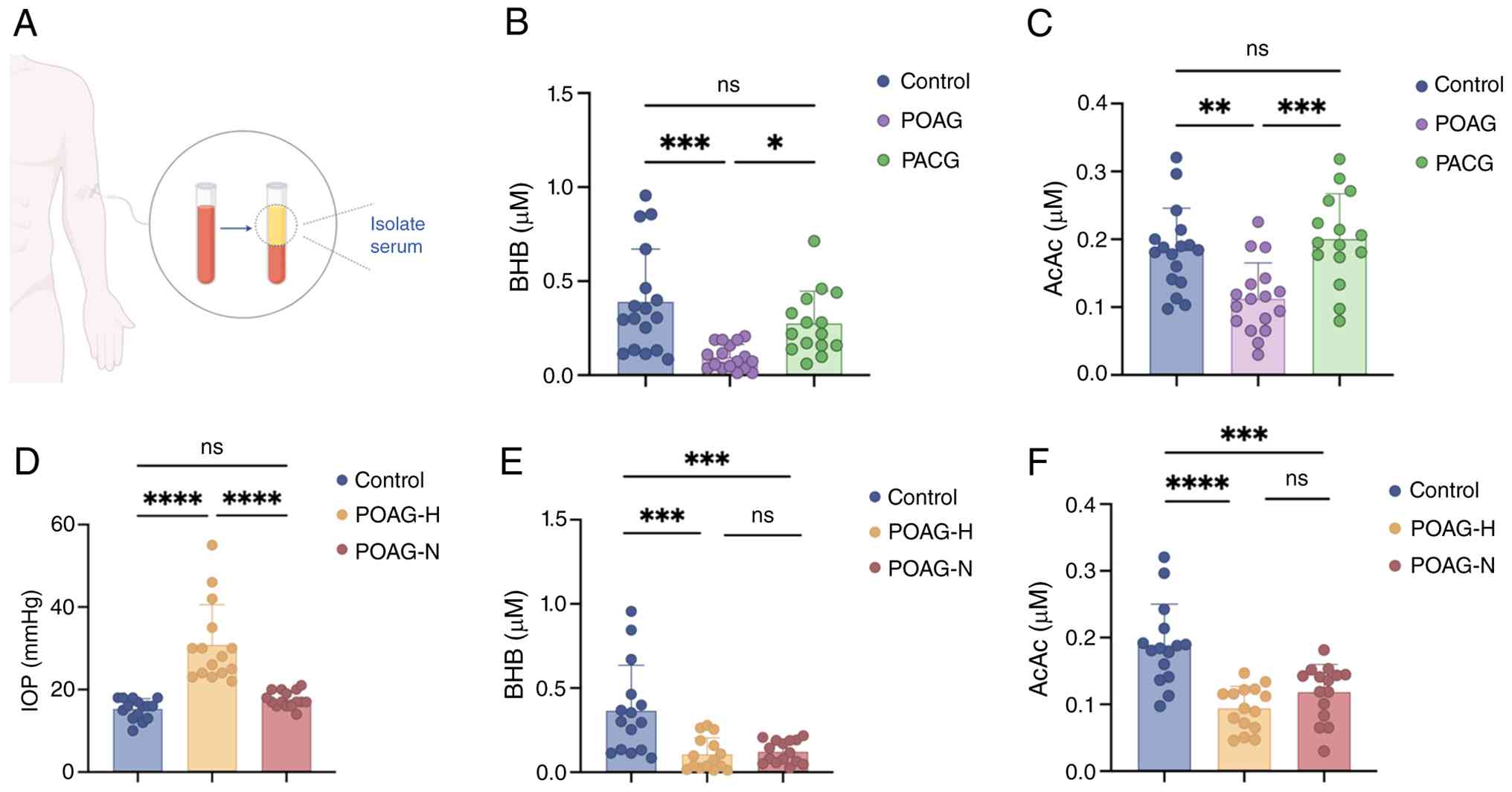 | Figure 1Serum ketone body (BHB and AcAc)
levels were reduced in patients with POAG. (A) Schematic diagram of
serum sample collection. (B) Serum BHB levels in healthy controls
(n=17), POAG patients (n=15) and PACG patients (n=15). (C) Serum
AcAc levels in healthy controls (n=17), POAG patients (n=15) and
PACG patients (n=15). (D) Comparisons of IOP in healthy controls,
POAG-H patients and POAG-N patients (n=15 for three groups). (E)
Serum BHB levels in POAG-H patients, POAG-N patients and healthy
controls (n=15 for three groups). (F) Serum AcAc levels in POAG-H
patients, POAG-N patients and healthy controls (n=15 for three
groups). 'n' represents the number of samples. Data are presented
as the mean ± SD. One-way ANOVA was used for B-F.
*P<0.05, **P<0.01,
***P<0.001 and ****P<0.0001. ns, not
significant; BHB, β-hydroxybutyrate; AcAc, acetoacetic acid; POAG,
primary open-angle glaucoma; PACG, primary angle-closure glaucoma;
IOP, intraocular pressure; POAG-H, high-IOP group; POAG-N,
normal-IOP group. |
EAG mice exhibit glaucomatous injury
accompanied by decreased serum ketone bodies
Subsequently, an ONA-induced EAG model was employed
(Fig. 2A). Quantification of
RGCs by immunofluorescence at 2, 4, 6 and 8 weeks after
immunization revealed statistically significant RGC loss beginning
at week 4 (Fig. S1A). RT-qPCR
analysis of IL-1β, IL-6, TNF-α and GFAP at 0, 2 and 4 weeks
exhibited progressive upregulation of these markers, with a
significant increase at week 4 (Fig. S1B). Collectively, these results
demonstrated successful establishment of the EAG model by week 4
and supported its use in subsequent experiments. During the first 4
weeks after immunization, the IOP remained normal in the EAG and
the control groups, with no significant differences observed
(Fig. 2B). H&E staining and
OCT images exhibited significant thinning of the GCL and loss of
RGCs in the EAG group compared with controls (Fig. 2C and D). RGC loss was further
determined by Brn3a immunostaining, which showed a significant
reduction in RGC numbers in the EAG group compared with the control
group (Fig. 2E and F). The
association between ketone body levels and glaucomatous damage was
then examined, revealing significantly lower serum ketone body
levels in the EAG group compared with the control group, consistent
with observations in POAG (Fig.
2G). These results suggest a possible association between optic
nerve injury and reduced serum ketone body levels in EAG mice.
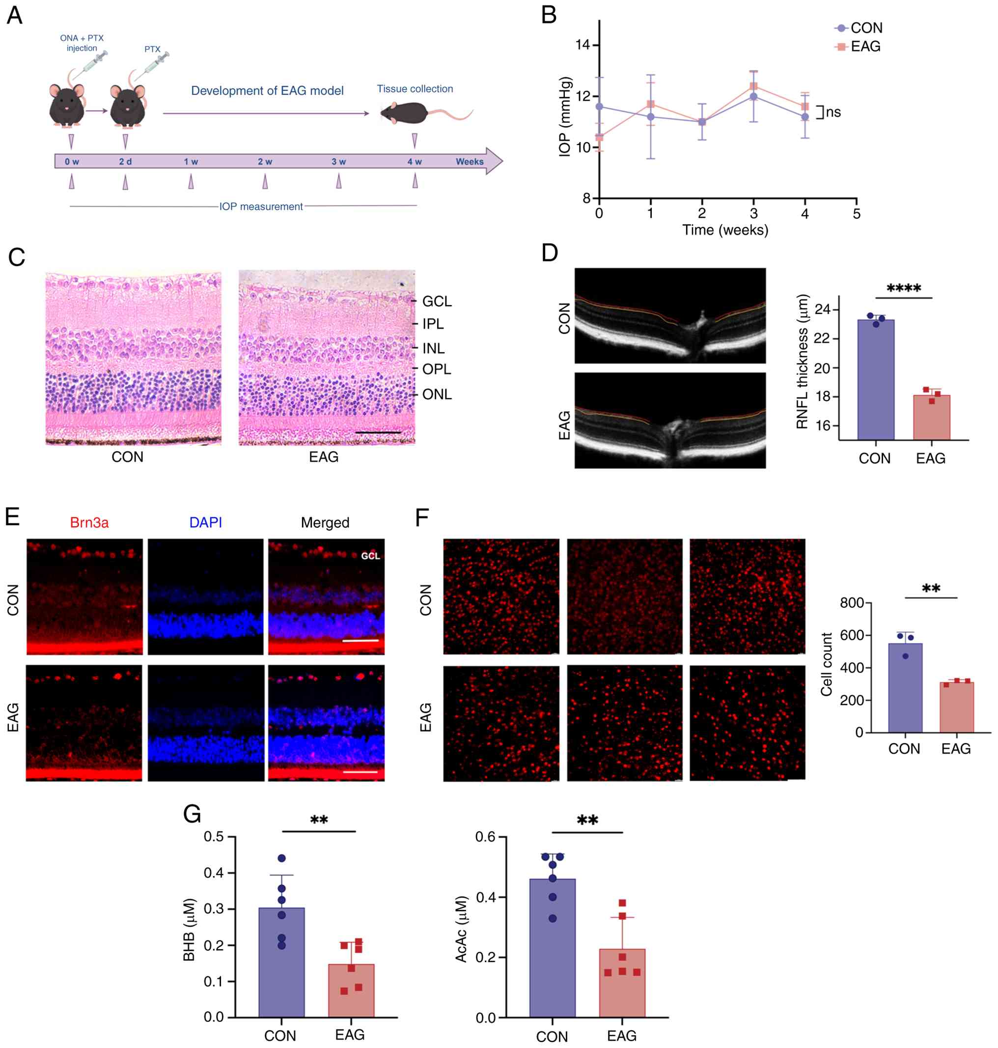 | Figure 2EAG mice exhibit glaucomatous
neurodegeneration and decreased serum ketone bodies. (A) Schematic
of the EAG mouse model. (B) IOP measurements in CON and EAG groups
(n=6 mice for each group). (C) H&E staining of retinal sections
in CON and EAG groups. (n=3 mice for each group). (D) Optical
coherence tomography analysis and quantification data of RNFL
thickness in CON and EAG groups (n=3 mice for each group). (E)
Immunofluorescent staining of Brn3a+ cells in the
retinal sections of CON and EAG groups. (F) Immunofluorescent
staining and quantification data of Brn3a+ cells in the
retinal flat mounts of CON and EAG groups (n=3 mice for each
group). (G) Serum BHB and AcAc levels in CON and EAG groups (n=6
mice for each group; scale bar, 50 µm). Data are presented
as the mean ± SD. Unpaired t-tests were used for (D-G).
**P<0.01 and ****P<0.0001. ns, not
significant; BHB, β-hydroxybutyrate; AcAc, acetoacetic acid; IOP,
intraocular pressure; GCL, ganglion cell layer; IPL, inner
plexiform layer; INL, inner nuclear layer; OPL, outer plexiform
layer; ONL, outer nuclear layer; RNFL, retinal nerve fibre layer;
CON, control; PTX, pertussis toxin; Brn3a, brain-specific
homeobox/POU domain protein 3A; EAG, experimental autoimmune
glaucoma; ONA, optic nerve antigen. |
KD enhances the transport and utilization
of ketone bodies in the retina of EAG mice
KD feeding was used to increase ketone body levels
in vivo. EAG mice were fed a CD (80% CHO, 10% protein and
10% fat) or a KD (0% CHO, 10% protein and 90% fat) for 4 weeks
(Fig. 3A). Serum ketone body
levels were found to be significantly elevated in KD-fed EAG mice
compared with those in the CD-fed group (Fig. 3B). ONA immunization did not
affect blood glucose levels but resulted in body weight loss in
mice. By contrast, KD had no significant effect on either blood
glucose levels or body weight in EAG mice (Fig. 3C and D).
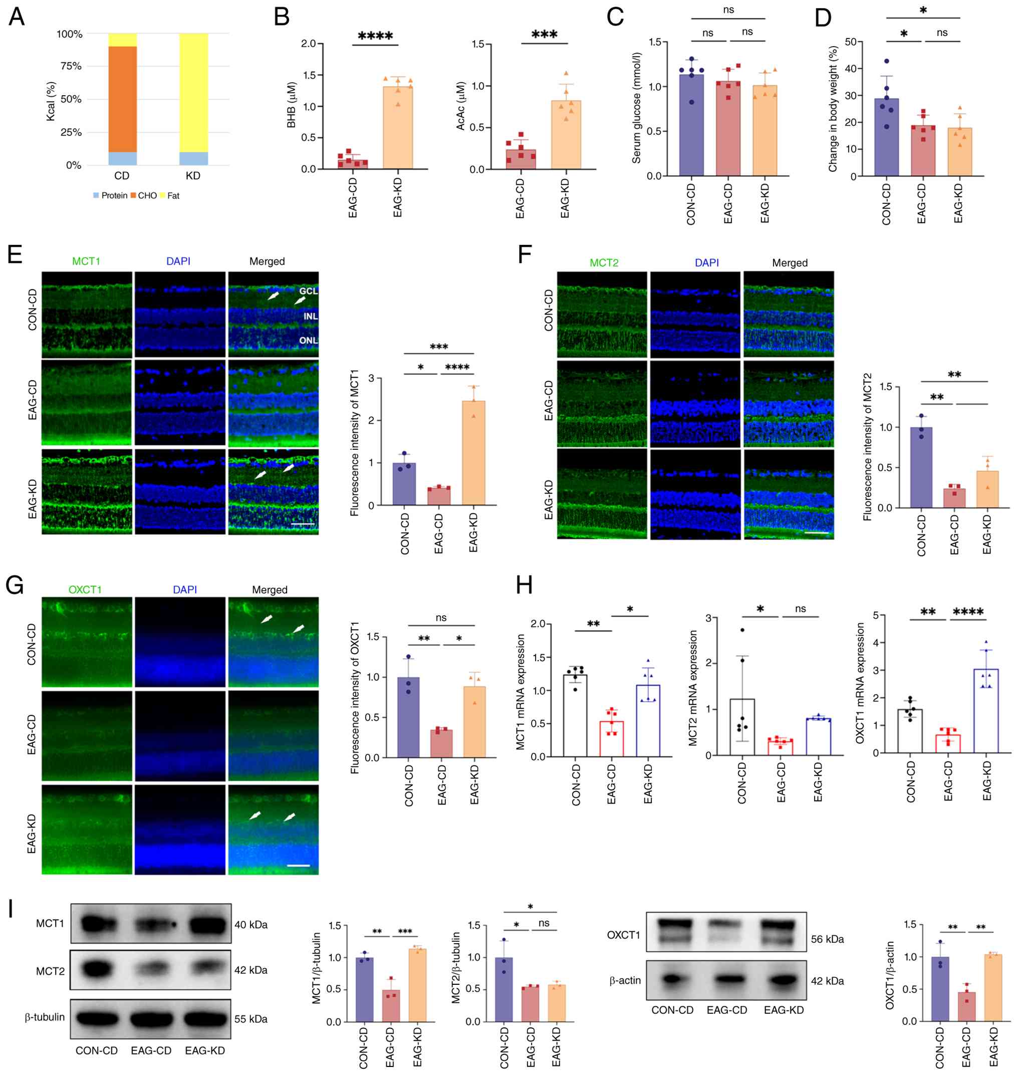 | Figure 3KD feeding enhances ketone body
transportation and utilization in the retina of EAG mice. (A)
Composition formulation of the CD and KD. (B) Serum BHB and AcAc
levels in EAG-CD and EAG-KD groups (n=6 mice for each group). (C)
Serum glucose levels in EAG-CD and EAG-KD groups (n=6 mice for each
group). (D) Body weight changes during the experimental period in
CON-CD, EAG-CD and EAG-KD groups (n=6 mice for each group). (E)
Immunofluorescent staining and quantification data of MCT1 in
retinal sections of CON-CD, EAG-CD and EAG-KD groups (n=3 mice for
each group; white arrows indicate MCT1+ staining). (F)
Immunofluorescent staining and quantification data of MCT2 in
retinal sections of CON-CD, EAG-CD and EAG-KD groups (n=3 mice for
each group). (G) Immunofluorescent staining and quantification of
OXCT1 in retinal sections of CON-CD, EAG-CD and EAG-KD groups (n=3
mice for each group; white arrows indicate OXCT1+
staining). (H) Relative mRNA expression of MCT1, MCT2 and OXCT1 in
the retinas of CON-CD, EAG-CD and EAG-KD groups (n=6 mice for each
group). (I) Western blotting analysis and quantification of MCT1,
MCT2 and OXCT1 protein expressions in the retinas of CON-CD, EAG-CD
and EAG-KD groups (n=3 mice for each group; scale bar, 50
µm). Data are presented as the mean ± SD. Unpaired t-tests
were used for (B). One-way ANOVA and Kruskal-Wallis were used for
(C-I). *P<0.05, **P<0.01,
***P<0.001 and ****P<0.0001. ns, not
significant; KD, ketogenic diet; EAG, experimental autoimmune
glaucoma; CD, control diet; BHB, β-hydroxybutyrate; EAG-KD,
irradiated KD; EAG-CD, irradiated CD; MCT, monocarboxylate
transporter; OXCT1, 3-oxoacid CoA-transferase 1; CHO,
carbohydrate. |
To evaluate changes in ketone body transport and
utilization in the retina, key enzymes and molecules involved in
these processes were examined. The expression of monocarboxylate
transporters (MCT1 and MCT2) and OXCT1 was found to be
significantly decreased in the retinas of EAG mice compared with
healthy controls at both the protein and RNA levels. Following KD
intervention, MCT1 and OXCT1 expression increased, whereas MCT2
expression exhibited no significant change (Fig. 3E-I). These results demonstrated
that KD increased serum ketone body levels and concurrently
upregulated retinal MCT1 and OXCT1 expression in EAG mice.
KD suppresses Müller cell activation and
inflammatory factors release and promotes RGC survival
To evaluate the effects of elevated serum ketone
bodies on EAG mice, retinal layer thickness was assessed by H&E
staining, with OCT and RGCs being quantified by immunofluorescence.
KD-fed EAG mice exhibited increased GCL thickness and significantly
elevated RGC numbers compared with the CD-fed EAG group (Fig. 4A-D). A hallmark of Müller cell
activation is the upregulation of GFAP (10). Immunofluorescence, western
blotting and RT-qPCR analyses revealed significantly increased GFAP
expression in the retina of EAG mice compared with the control,
which was further significantly reversed by KD feeding (Fig. 4E-H). Similarly, western blotting
and RT-qPCR analyses showed significantly elevated expression of
IL-1β, IL-6 and TNF-α in the retina of EAG mice, with KD feeding
further significantly reducing their levels (Fig. 4I and J). These findings indicate
that KD was associated with suppressed Müller cell activation,
reduced inflammatory factor expression and improved RGC survival in
the EAG retina.
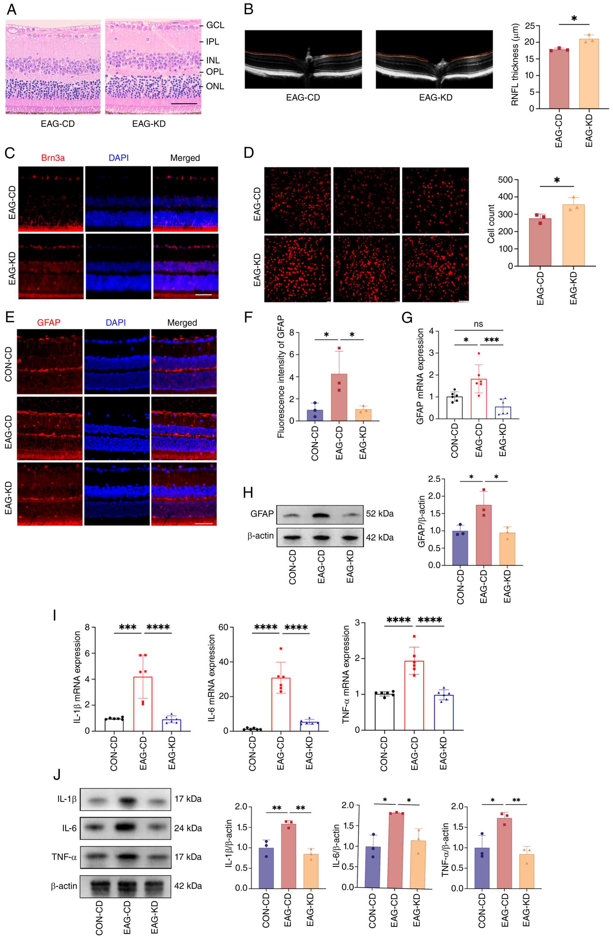 | Figure 4KD suppresses Müller cell activation
and retinal inflammation in EAG mice. (A) H&E staining of
retinal sections in EAG-CD and EAG-KD groups (n=3 mice for each
group). (B) Optical coherence tomography analysis and
quantification of RNFL thickness in EAG-CD and EAG-KD groups (n=3
mice for each group). (C) Immunofluorescent staining of
Brn3a+ cells in the retinal sections of EAG-CD and
EAG-KD groups (n=3 mice for each group). (D) Immunofluorescent
staining quantification data of Brn3a+ cells in the
retinal flat mounts of EAG-CD and EAG-KD groups (n=3 mice for each
group). (E) Immunofluorescent staining and (F) quantification data
of GFAP in the retinal sections of CON-CD, EAG-CD and EAG-KD groups
(n=3 mice for each group). (G) Relative mRNA expression levels of
GFAP in the retinas of CON-CD, EAG-CD and EAG-KD groups (n=6 mice
for each group). (H) Western blotting analysis quantification of
GFAP expression in the retinas of CON-CD, EAG-CD and EAG-KD groups
(n=3 mice for each group). (I) Relative mRNA expression of IL-1β,
IL-6 and TNF-α in the retinas of CON-CD, EAG-CD and EAG-KD groups
(n=6 mice for each group). (J) Western blotting analysis
quantification of IL-1β, IL-6 and TNF-α protein expressions in the
retinas of CON-CD, EAG-CD and EAG-KD groups (n=3 mice for each
group; scale bar, 50 µm). Data are presented as the mean ±
SD. Unpaired t-tests were used for (B and D). One-way ANOVA was
used for (F-J). *P<0.05, **P<0.01,
***P<0.001 and ****P<0.0001. ns, not
significant; EAG, experimental autoimmune glaucoma; KD, ketogenic
diet; CD, control diet; GFAP, glial fibrillary acidic protein; GCL,
ganglion cell layer; IPL, inner plexiform layer; INL, inner nuclear
layer; OPL, outer plexiform layer; ONL, outer nuclear layer; RNFL,
retinal nerve fibre layer; Brn3a, brain-specific homeobox/POU
domain protein 3A; EAG-KD, irradiated KD; EAG-CD, irradiated
CD. |
BHB suppresses Müller cell activation and
inflammatory factors in vitro
To explore the role of BHB in vitro, primary
Müller cells were isolated from neonatal mouse retinas and
cultured. After 2-3 passages, the cells achieved high purity, as
determined by their distinct morphology and positive
immunofluorescence for GS (Fig.
5A; Fig. S2). Müller cell
viability after 72 h of LPS stimulation was measured using a CCK-8
assay. Cell viability decreased significantly at all tested LPS
concentrations >1 µg/ml (2, 5 and 10 µg/ml). Based
on this, 1 µg/ml was chosen as the maximum concentration
that did not compromise cell viability and was used for subsequent
experiments (Fig. 5B). The
effects of BHB on Müller cells were then evaluated with or without
1 µg/ml LPS. In the absence of LPS, BHB concentration up to
be 20 mM did not significantly affect cell viability (Fig. 5C). Under LPS stimulation, no
significant reduction in cell viability was observed at BHB
concentrations up to 5 mM, whereas a significant decline occurred
at 10 mM (Fig. 5D). Accordingly,
5 mM BHB was selected as the maximum non-toxic concentration for
further experiments. After 72 h of LPS stimulation, MCT1 and OXCT1
expression was found to be significantly reduced in Müller cells
compared with controls. In the LPS-stimulated cells treated with
BHB, MCT1 and OXCT1 expression was significantly upregulated
(Fig. 5E and F). Concurrently,
LPS stimulation significantly increased GFAP expression in Müller
cells and further significantly reduced it upon BHB treatment
(Fig. 5G and H). Western
blotting and RT-qPCR analyses showed that LPS-induced increases in
IL-1β, IL-6 and TNF-α were significantly suppressed by BHB
treatment (Fig. 5I and J).
Collectively, these in vitro findings indicate that BHB is
associated with reduced Müller cell activation and inflammatory
factor secretion, consistent with the in vivo
observations.
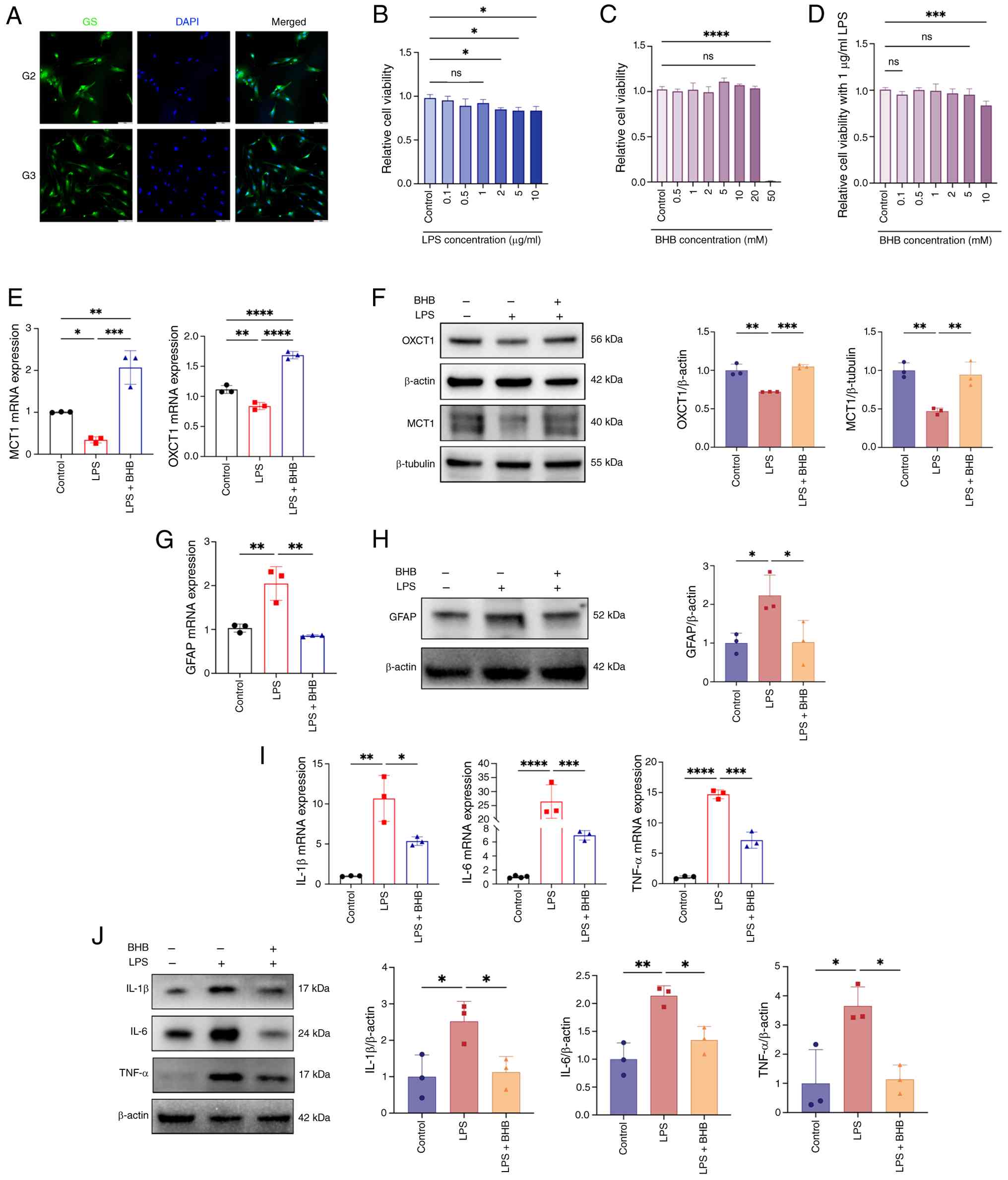 | Figure 5BHB enhances ketone body metabolism
and suppresses expression of inflammatory factors in Müller cells.
(A) Immunofluorescent staining of GS for identification in the
primary Müller cells. (B) CCK-8 assay of cell viability in Müller
cells treated with varying concentrations of LPS (n=3 cultures for
each group). (C) CCK-8 assay of cell viability in Müller cells
treated with varying concentrations of BHB alone (n=3 cultures for
each group). (D) CCK-8 assay for cell viability in LPS-stimulated
Müller cells treated with varying concentrations of BHB (n=3
cultures for each group). (E) Relative mRNA expression levels of
MCT1 and OXCT1 in the control, LPS and LPS + BHB groups (n=3
cultures for each group). (F) Western blotting analysis and
quantification of MCT1 and OXCT1 protein expressions in the
control, LPS and LPS + BHB groups (n=3 cultures for each group).
(G) Relative mRNA expression of GFAP in the control, LPS and LPS +
BHB groups (n=3 cultures for each group). (H) Western blotting
analysis and quantification of GFAP protein expression in the
control, LPS and LPS + BHB groups (n=3 cultures for each group).
(I) Relative mRNA expression of IL-1β, IL-6 and TNF-α in the
control, LPS and LPS + BHB groups (n=3 cultures for each group).
(J) Western blotting analysis and quantification of IL-1β, IL-6 and
TNF-α protein expressions in the control, LPS and LPS + BHB groups
(n=3 cultures for each group; scale bar, 50 µm). Data are
presented as the mean ± SD. One-way ANOVA was used for all data
analysis. *P<0.05, **P<0.01,
***P<0.001 and ****P<0.0001. ns, not
significant; GS, glutamine synthetase; MCT, monocarboxylate
transporter; OXCT1, 3-oxoacid CoA-transferase 1; CCK-8, Cell
Counting Kit-8; GFAP, glial fibrillary acidic protein; BHB,
β-hydroxybutyrate; LPS, lipopolysaccharide. |
KD is associated with increased FOXO3A
acetylation and MT2A expression in vivo and in vitro
KD exerts a similar inhibitory effect to a naturally
occurring and specific inhibitor of class I histone deacetylases.
Pan-acetylation was assessed by detecting overall acetylated lysine
levels. KD feeding and BHB treatment restored the diminished global
acetylation levels in the retinas of EAG mice and in LPS-stimulated
Müller cells, respectively (Fig.
6A). Given the observed global increase in protein acetylation,
4D-DIA proteomic analysis was performed to pinpoint the specific
proteins and pathways affected. Comparative analysis revealed
distinct protein expression profiles among the control, LPS and
LPS+BHB groups (Fig. 6B and C;
Fig. S3A-D). A set of 76
proteins that were decreased in the LPS group relative to controls
and increased in the LPS + BHB group was identified and visualized
using a clustering heatmap (Fig.
6D; Table SV). Among these,
proteins that met the criterion of fold change >1.2 and had a
P<0.05 in paired comparisons were selected. FOXO3A and MT2A were
found to be significantly upregulated and their expression
differences are presented as a volcano plot and a box plot,
respectively (Fig. 6E-G). GO and
KEGG enrichment analyses revealed significant upregulation of the
'FoxO signalling pathway' following BHB treatment in LPS-stimulated
cells. (Figs. 6H and S3E). Reactome analysis further
supported the involvement of FOXO, with the most prominent
enrichment observed in 'Regulation of FOXO transcriptional activity
by acetylation' (Fig. 6I).
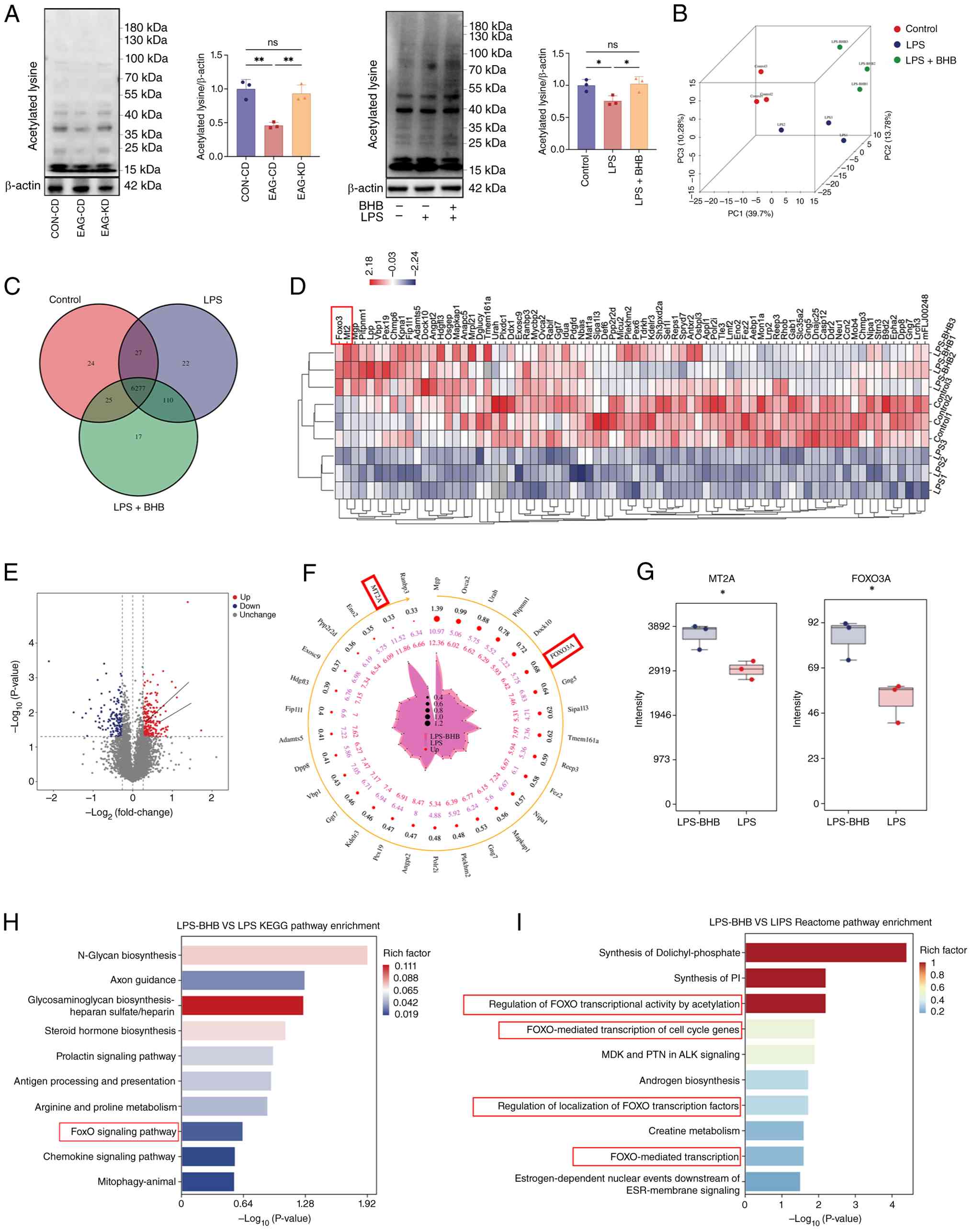 | Figure 6Proteomic analysis via 4D
data-independent acquisition of Müller cells following BHB
treatment under LPS stimulation. (A) Western blotting analysis and
quantification of acetylation levels in the retinas of CON-CD,
EAG-CD and EAG-KD groups of mice (n=3 mice for each group); and in
the control, LPS and LPS + BHB groups of Müller cells (n=3 cultures
for each group). (B) Plot indicating 3D principal component
analysis of distribution of proteomic profiles among the control,
LPS and LPS + BHB groups (n=3 cultures for each group). (C) Venn
diagram showing the overlap of identified peaks among the three
groups. (D) Clustering heatmap of the 76 proteins identified by a
directional trend of decreased expression in the LPS group relative
to control and increased expression in the LPS + BHB group relative
to LPS (P<0.05). (E) Volcano plot and (F) radar plot displaying
differentially expressed proteins, with FOXO3A and MT2A
highlighted. Proteins shown met the criteria of fold change >1.2
and P<0.05 in pairwise comparisons. (G) Box plots showing the
relative expression levels of FOXO3A and MT2A across the three
groups (n=3 cultures for each group). (H) KEGG enrichment analysis
of differential metabolic pathways between the LPS and LPS + BHB
groups. (I) Reactome enrichment analysis of signal transduction
mechanisms between the LPS and LPS + BHB groups. Data are presented
as the mean ± SD. One-way ANOVA was used for (A and B). Unpaired
t-tests were used for (G). *P<0.05 and
**P<0.01. ns, not significant; KEGG, Kyoto
Encyclopaedia of Genes and Genomes; BHB, β-hydroxybutyrate; LPS,
lipopolysaccharide; CD, control diet; KD, ketogenic diet; MCT,
monocarboxylate transporter; EAG-KD, irradiated KD; EAG-CD,
irradiated CD; Up, upregulated; Down, downregulated. |
Western blotting analysis demonstrated that LPS
significantly suppressed FOXO3A and MT2A expression, as well as
FOXO3A acetylation at Lys271, in Müller cells and these effects
were then significantly reversed by BHB treatment (Fig. 7A). Consistent with these in
vitro findings, parallel results were observed in the retina of
EAG mice following KD feeding (Fig.
7B). Acetylation at Lys290, another known acetylation site of
FOXO3A, was also investigated. In retinal tissue, KD feeding
significantly increased FOXO3A acetylation at Lys290, a change
similar to the trend observed at Lys271. However, in cultured
Müller cells, Lys290 acetylation was barely detectable and did not
significantly change upon BHB treatment (Fig. S4). These results indicate that
the protective effect of BHB on Müller cells was accompanied by
increased FOXO3A acetylation at Lys271 and upregulation of
MT2A.
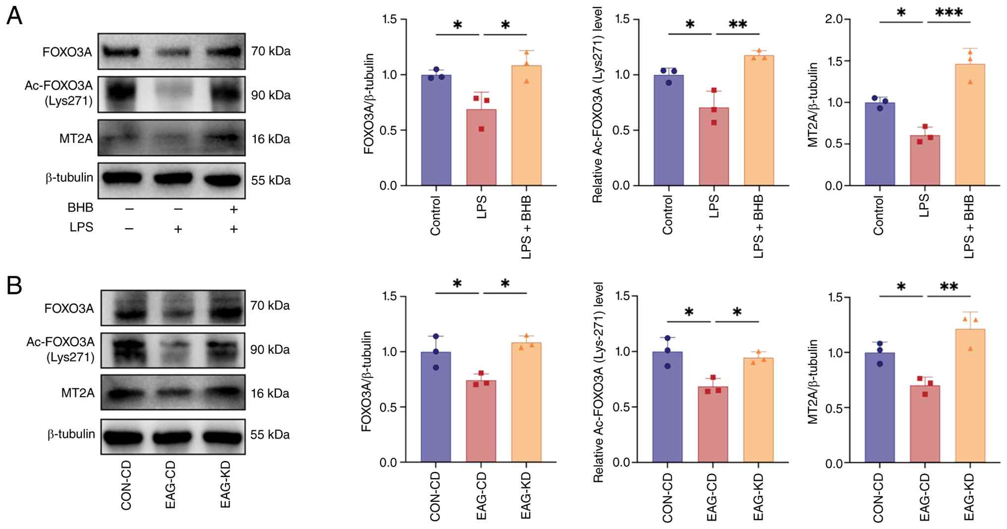 | Figure 7Expression of the FOXO3A, Ac-FOXO3A
and MT2A proteins in EAG mice and primary Müller cells. (A) Western
blotting analysis and quantification of FOXO3A, MT2A and FOXO3A
acetylation at Lys271 in Müller cells from control, LPS and LPS +
BHB groups (n=3 cultures for each group). (B) Western blotting
analysis and quantification of FOXO3A, MT2A and Ac-FOXO3A at Lys271
in the retinas of CON-CD, EAG-CD and EAG-KD groups (n=3 mice for
each group). Data are presented as the mean ± SD. One-way ANOVA was
used for (A and B). *P<0.05, **P<0.01
and ***P<0.001. ns, not significant; Ac-,
acetylation; MT2A, metallothionein 2A; CD, control diet; KD,
ketogenic diet; EAG-KD, irradiated KD; EAG-CD, irradiated CD; BHB,
β-hydroxybutyrate; LPS, lipopolysaccharide; EAG, experimental
autoimmune glaucoma. |
Discussion
To the best of our knowledge, the present study was
the first to show that serum ketone body levels were significantly
lower in patients with POAG compared with healthy controls and
patients with PACG. A similarly significant reduction was
determined in the EAG model. The data further show that KD feeding
elevates circulating BHB levels and increase the expression of
ketone body transporters and metabolic enzymes, while suppressing
Müller cell activation, attenuating neuroinflammation and
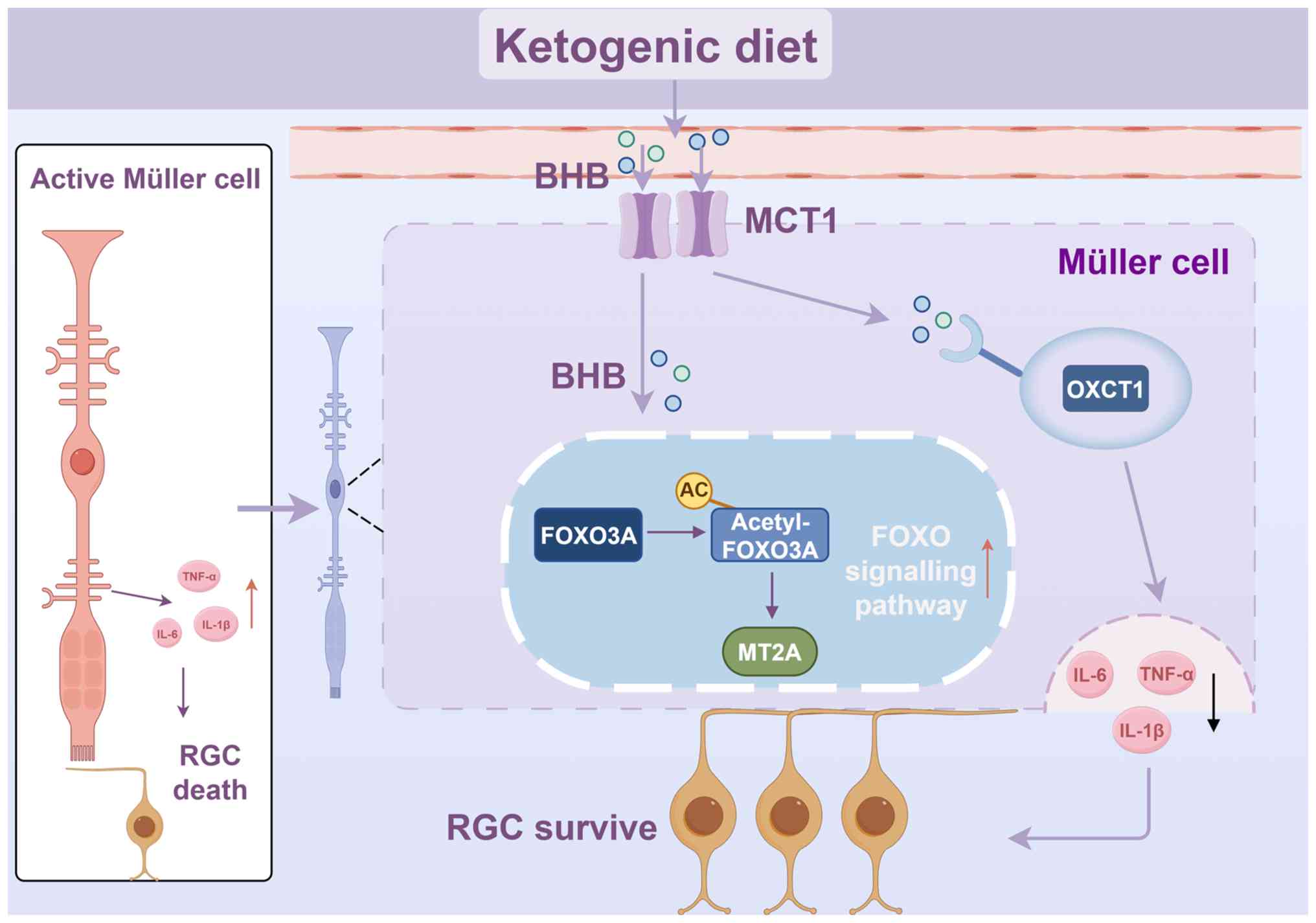
preserving RGCs. These protective effects were accompanied by
upregulation of the FOXO signalling pathway, suggesting FOXO3A
acetylation is a potential mediator of the neuroprotective effects
of KD in glaucoma (as summarised in Fig. 8).
Ketone bodies serve as a key alternative metabolic
fuel across all domains of life. Accumulating evidence has
emphasized their important roles in mammalian cell metabolism,
homeostasis and signalling across diverse physiological and
pathological contexts (14,34-36). The present findings demonstrated
that, unlike in PACG, the specific decreases in BHB and AcAc levels
in POAG suggest that its pathogenesis is modulated by ketone bodies
through an IOP-independent mechanism. A previous gene set analysis
indicated that mitochondrial ketone body synthesis and degradation
were notably associated with POAG (37), but direct detection data to
determine this association are lacking. The present study aimed to
address this gap. Conversely, acute exposure to hypoxia may
increase plasma ketone body metabolism (38). This difference may be
attributable to the hypoxic metabolic disturbances in this
acute-phase study (38)
occurring during the acute phase of the disease, whereas the
decreases in BHB and AcAc observed in the present study were
associated with chronic pathological changes. Subsequently, an
immune glaucoma model was established to investigate the role of
ketone bodies in vivo. Given that oestrogen protects RGCs,
male mice were used for the EAG model to eliminate this variable
(39). EAG mice developed
notable glaucomatous injury, as evidenced by RNFL thinning, RGC
loss, glial activation and increased inflammatory cytokine release.
Notably, decreased BHB and AcAc levels were observed, consistent
with the trend observed in patients with POAG. Notably, decreased
BHB and AcAc levels were also observed, consistent with the trend
seen in patients with POAG. Thus, the EAG model is suitable for
exploring the association between glaucomatous optic nerve injury
and ketone bodies. These findings indicate that serum ketone body
levels may serve as biomarkers for the diagnosis and assessment of
glaucoma.
To investigate whether elevating ketone body levels
could alleviate glaucomatous damage, a reliable and well-tolerated
method of raising circulating ketone bodies is needed. KD drives
sustained endogenous ketogenesis, producing a steady elevation of
BHB along with AcAc and acetone, whereas exogenous BHB salts or
esters tend to produce transient peaks and may cause
gastrointestinal discomfort at higher doses (15,40). KD has also been used safely for
decades in epilepsy and has been increasingly studied in other
neurodegenerative conditions, including Alzheimer's disease,
Parkinson's disease and multiple sclerosis (18), so its long-term feasibility is
well established. Therefore, KD rather than direct BHB
supplementation was chosen to raise circulating ketone body levels.
A total of 4 weeks of KD feeding reliably raised serum ketone body
levels in EAG mice, demonstrating that the dietary intervention was
effective in the present study, with the most notable increase
observed in BHB. In the present study, KD not only elevated serum
ketone body levels but also enhanced ketone body transport,
utilization, as well as overall ketone metabolism in the retina.
Increased expression of MCT1 and OXCT1 was also detected,
accompanied by minimal effects on MCT2 expression. The underlying
reason for this difference may be that MCT1 exhibits a stronger
affinity for ketone bodies than MCT2 (41). This observation aligns with a
previous report that the hippocampus exhibited a robust response to
nutritional ketosis in MCT1 and OXCT1 levels (42). Neuron-specific MCT1 upregulation
alone can markedly promote axonal regeneration and improve recovery
after spinal cord injury (43).
Previous studies have found that MCT1 is more
abundantly expressed in the lactate shuttle between Müller cells
and photoreceptors, whereas MCT2 is expressed in neuronal cells in
the GCL and astrocytes (44,45). Müller cell activation, with the
consequent release of inflammatory mediators, exacerbates RGC
apoptosis in glaucoma (46). The
present evidence suggested that KD inhibits GFAP and inflammatory
mediator expression in the retinas of EAG mice, suggesting
suppression of both Müller cell activation and retinal
inflammation. Müller cells are key effector cells in retinal energy
metabolism. MCT1 and OXCT1 were found to be localized predominantly
in the retinal layers where Müller cells reside and their
expression was regulated by KD, suggesting that Müller cells may
serve a central role in ketone body transport and utilization
within the retina. Based on the present results, it was
hypothesized that the KD-induced increase in retinal ketone body
metabolism is predominantly mediated by Müller cells. Thus, primary
Müller cells were cultured and used for in vitro
experiments, determining that BHB promotes ketone body metabolism
while inhibiting Müller cell activation and release of inflammatory
cytokines (IL-1β, IL-6 and TNF-α). These results support that the
neuroprotection afforded by KD arises from the inhibition of Müller
cell inflammation in the immune pathogenesis of glaucoma,
consistent with previous findings (47,48).
Furthermore, previous studies have shown that KD
increases histone acetylation and global protein acetylation levels
(49,50). In addition, endogenous BHB, which
increases after KD, has been reported to exert a similar inhibitory
effect to hydroxycarboxylic acid 2 to suppress neuroinflammation
and exerts neuroprotective effects (51). In the present study, broad
elevation of protein acetylation was observed in the retina of EAG
mice following KD feeding and in LPS-induced Müller cells treated
with BHB. This shift in acetylation prompted investigation of
specific downstream targets that might mediate the observed
anti-inflammatory effects. Subsequently, 4D-DIA proteomic analysis
identified significant enrichment of a number of FOXO-associated
biological processes, including 'Regulation of FOXO transcriptional
activity by acetylation', which aligned with the present focus on
acetylation regulation. KEGG enrichment analysis further exhibited
significant enrichment of the 'FOXO signalling pathway'. Within
this pathway, FOXO3A and MT2A were among the proteins significantly
upregulated by BHB treatment in Müller cells.
MT2A is a known downstream target gene of FOXO3A
(52) and both molecules have
established roles in anti-inflammatory responses and
cytoprotection. MT2A contributes to these processes through
multiple mechanisms, including inhibition of NF-κB activation
(53), regulation of
endothelial-overexpressed LPS-associated factor-1 (54), and suppression of
pro-inflammatory cytokines such as IL-6, IL-12 and TNF-α (55). FOXO3A exerts anti-inflammatory
effects by upregulating IκB expression and directly binding to
NF-κB p65 to inhibit its transcriptional activity, thereby
suppressing pro-inflammatory cytokines such as TNF-α, IL-1β and
IL-6 (56). The FOXO family of
transcription factors is predominantly regulated by acetylation or
phosphorylation (57) and FOXO3A
is known to be acetylated at numerous lysine residues, among which
Lys271 and Lys290 have been the most extensively studied (58,59). In the present experiments, KD
induced significant alterations in acetylated FOXO3A. Specifically,
although changes were observed at both Lys271 and Lys290 in the
retina, only the modification at Lys271 was detected in cultured
Müller cells. The Lys290 signal was barely detectable above
background levels, making reliable assessment difficult. This may
imply that Lys290 acetylation is either less prominent in Müller
cells or regulated independently of BHB and that Lys271 is the
primary site through which BHB modulates FOXO3A activity in this
cell type. This would be consistent with the concept that different
FOXO acetylation sites can be differentially regulated depending on
cell type and cellular context (60). The subsequent upregulation of
MT2A, a known downstream target of FOXO3A, further supports the
involvement of this pathway. Collectively, these findings suggest
that KD elevates BHB levels and that the accompanying FOXO3A
acetylation at Lys271 and MT2A upregulation are associated with
activation of the FOXO signalling pathway. These changes may
contribute to the attenuation of retinal neuroinflammation in
glaucoma.
The present study exhibits a number of limitations
that should be acknowledged. First, the present findings suggest
that BHB could be a potential diagnostic biomarker for glaucoma.
Despite this, methods for its quantification and detection in
clinical diagnosis and therapy require further investigation.
Second, direct in vivo administration of BHB was not
performed in the present study. Therefore, whether BHB alone can
fully recapitulate the neuroprotective effects of KD in the EAG
model remains to be investigated. Third, mutating the acetylation
sites of FOXO3A in Müller cells will help to clarify the functional
contributions of each acetylation site. Therefore, future research
should aim to focus on addressing these limitations and exploring
other mechanisms of KD in the treatment of glaucoma, as well as
promoting clinical applications and drug development.
In conclusion, the present study revealed an
association between dysregulated ketone body metabolism and
neuroinflammation in glaucoma. To the best of our knowledge, the
present data provide the first evidence in EAG model that KD
alleviates retinal neuroinflammation and attenuates Müller cell
activation through acetylation-mediated FOXO signalling. These
findings offer new insights into the role of metabolic regulation
in neurodegenerative diseases, provide evidence for Müller cell
phenotypic modulation and suggest potential avenues for
metabolism-based neuroprotective strategies. KD and its key
metabolite BHB warrant further investigation to explore their
potential clinical applications in glaucoma treatment.
Supplementary Data
Availability of data and materials
The data generated in the present study may be found
in the iProX database under accession number IPX0018867000 or at
the following URL: https://www.iprox.cn/page/project.html?id=IPX0018867000.
Authors' contributions
SST and XCW contributed equally to the present
study. SST and XCW conceptualized the present study and analyzed
the data, performed the experiments, interpreted the results of the
experiments and prepared the figures, wrote the original draft and
reviewed and edited the manuscript. YFZ, ZNM, LHZ and JCY performed
the experiments and analyzed the data. XCW, XF, MW, YJH and RTOY
performed the investigation, visualization and conducted data
validation. YYP, JQL, XG, DDW and YP recruited the samples. HL
conceptualized the present study, handled supervision and acquired
the funding. SST, XCW and HL confirm the authenticity of all the
raw data. All authors read and approved the final version of the
manuscript and are accountable for all aspects of the work.
Ethics approval and consent to
participate
The guiding principles of the Declaration of
Helsinki were strictly followed in all procedures. The clinical
research process was approved by the Ethical Committee of the First
Affiliated Hospital at Chongqing Medical University (Chongqing,
China; approval no. 2024-087-01). Written informed consent was
obtained from all participants involved in the research. All animal
research was supported by the Institutional Animal Care and Use of
Chongqing Medical University (Chongqing, China; approval no.
IACUC-CQMU-2025-0349) and in compliance with Association for
Research in Vision and Ophthalmology guidelines.
Patient consent for publication
Not applicable.
Competing interests
The authors declare that they have no competing
interests.
Abbreviations:
|
RGCs
|
retinal ganglion cells
|
|
BHB
|
β-hydroxybutyrate
|
|
AcAc
|
acetoacetic acid
|
|
PAOG
|
primary open-angle glaucoma
|
|
PACG
|
primary angle-closure glaucoma
|
|
EAG
|
experimental autoimmune glaucoma
|
|
ONA
|
optic nerve antigen
|
|
KD
|
ketogenic diet
|
|
CD
|
control diet
|
|
4D-DIA
|
4D data-independent acquisition
|
|
OCT
|
optical coherence tomography
|
|
GS
|
glutamine synthetase
|
|
GCL
|
ganglion cell layer
|
|
MCTs
|
monocarboxylate transporters
|
|
OXCT1
|
3-oxoacid CoA-transferase 1
|
|
GFAP
|
glial fibrillary acidic protein
|
|
MT2A
|
metallothionein 2A
|
Acknowledgments
Not applicable.
Funding
The present study was supported by the Natural Science
Foundation of Chongqing Municipality (grant no.
CSTB2024NSCQ-KJZMSX0073), the National Natural Science Foundation
of China (grant no. 82471064) and the Science and Technology
Innovation Key R&D Program of Chongqing (grant no.
CSTB2025TIAD-STX0008).
References
|
1
|
Cao Y, Yin X, Wu L, Huang D, Wang Z, Wu F,
Jiang J, Chen G and Wang Q: High-efficiency ocular delivery of
brain-derived neurotrophic factor and oligomycin for
neuroprotection in glaucoma. Adv Mater. 37:e25006232025. View Article : Google Scholar : PubMed/NCBI
|
|
2
|
Tham YC, Li X, Wong TY, Quigley HA, Aung T
and Cheng CY: Global prevalence of glaucoma and projections of
glaucoma burden through 2040: A systematic review and
meta-analysis. Ophthalmology. 121:2081–2090. 2014. View Article : Google Scholar : PubMed/NCBI
|
|
3
|
Chang YF, Cheng YH, Ko YC, Chiou SH and
Liu CJ: Anti-apoptotic and autophagic effect: Using conditioned
medium from human bone marrow mesenchymal stem cells to treat human
trabecular meshwork cells. Regen Ther. 22:50–58. 2022. View Article : Google Scholar
|
|
4
|
Bartollino S, Chiosi F, di Staso S, Uva M,
Pascotto A, Rinaldi M, Hesselink JMK and Costagliola C: The
retinoprotective role of phenytoin. Drug Des Devel Ther.
12:3485–3489. 2018. View Article : Google Scholar : PubMed/NCBI
|
|
5
|
Baudouin C, Kolko M, Melik-Parsadaniantz S
and Messmer EM: Inflammation in glaucoma: From the back to the
front of the eye, and beyond. Prog Retin Eye Res. 83:1009162021.
View Article : Google Scholar
|
|
6
|
Tian H, Chen Y, Zhao T, Ye L, Li H, Li Z,
Qiu W, Wang W, Li R, Liu F, et al: Single-cell RNA-seq reveals cell
type-specific molecular and genetic associations with primary
open-angle glaucoma. Signal Transduct Target Ther. 10:3382025.
View Article : Google Scholar : PubMed/NCBI
|
|
7
|
Geyer O and Levo Y: Glaucoma is an
autoimmune disease. Autoimmun Rev. 19:1025352020. View Article : Google Scholar : PubMed/NCBI
|
|
8
|
Vecino E, Rodriguez FD, Ruzafa N, Pereiro
X and Sharma SC: Glia-neuron interactions in the mammalian retina.
Prog Retin Eye Res. 51:1–40. 2016. View Article : Google Scholar
|
|
9
|
Reichenbach A and Bringmann A: New
functions of Müller cells. Glia. 61:651–678. 2013. View Article : Google Scholar : PubMed/NCBI
|
|
10
|
Xue LP, Lu J, Cao Q, Hu S, Ding P and Ling
EA: Müller glial cells express nestin coupled with glial fibrillary
acidic protein in experimentally induced glaucoma in the rat
retina. Neuroscience. 139:723–732. 2006. View Article : Google Scholar : PubMed/NCBI
|
|
11
|
Goldman D: Müller glial cell reprogramming
and retina regeneration. Nat Rev Neurosci. 15:431–442. 2014.
View Article : Google Scholar : PubMed/NCBI
|
|
12
|
Penn JS, Madan A, Caldwell RB, Bartoli M,
Caldwell RW and Hartnett ME: Vascular endothelial growth factor in
eye disease. Prog Retin Eye Res. 27:331–371. 2008. View Article : Google Scholar : PubMed/NCBI
|
|
13
|
Miao Y, Zhao GL, Cheng S, Wang Z and Yang
XL: Activation of retinal glial cells contributes to the
degeneration of ganglion cells in experimental glaucoma. Prog Retin
Eye Res. 93:1011692023. View Article : Google Scholar : PubMed/NCBI
|
|
14
|
Williams MS and Turos E: The chemistry of
the ketogenic diet: Updates and opportunities in organic synthesis.
Int J Mol Sci. 22:52302021. View Article : Google Scholar : PubMed/NCBI
|
|
15
|
Newman JC and Verdin E: β-Hydroxybutyrate:
A signaling metabolite. Annu Rev Nutr. 37:51–76. 2017. View Article : Google Scholar : PubMed/NCBI
|
|
16
|
Lopa C, Pietrangelo D, Santulli G,
Gambardella J, Rubattu S, Stefan-Lifshitz M, Garcia CN, Jankauskas
SS and Lombardi A: Pancreas meets brain: β-hydroxybutyrate as a
novel 'β-cellular' metabolism therapy. Metabolism. 174:1564192025.
View Article : Google Scholar
|
|
17
|
McNally MA and Hartman AL: Ketone bodies
in epilepsy. J Neurochem. 121:28–35. 2012. View Article : Google Scholar : PubMed/NCBI
|
|
18
|
Dynka D, Kowalcze K and Paziewska A: The
role of ketogenic diet in the treatment of neurological diseases.
Nutrients. 14:50032022. View Article : Google Scholar : PubMed/NCBI
|
|
19
|
Tsusaka T, Najar MA, Schwarz B, Bohrnsen
E, Oses-Prieto JA, Neudorf H, Lee C, Little JP, Burlingame AL,
Bosio CM, et al: Reversible histone deacetylase activity catalyzes
lysine acylation. Nat Chem Biol. 21:1387–1396. 2025. View Article : Google Scholar : PubMed/NCBI
|
|
20
|
Offermanns S: Hydroxy-carboxylic acid
receptor actions in metabolism. Trends Endocrinol Metab.
28:227–236. 2017. View Article : Google Scholar : PubMed/NCBI
|
|
21
|
Shippy DC, Wilhelm C, Viharkumar PA, Raife
TJ and Ulland TK: β-Hydroxybutyrate inhibits inflammasome
activation to attenuate Alzheimer's disease pathology. J
Neuroinflammation. 17:2802020. View Article : Google Scholar
|
|
22
|
Sun W, Wen M, Liu M, Wang Q, Liu Q, Li L,
Siebert HC, Loers G, Zhang R and Zhang N: Effect of
β-hydroxybutyrate on behavioral alterations, molecular and
morphological changes in CNS of multiple sclerosis mouse model.
Front Aging Neurosci. 14:10751612022. View Article : Google Scholar
|
|
23
|
Deng M, Fang Y, Chu Y, Ding R, Fan X, Wei
H, Song H, Jiang G, Zhang H, Han C and Yuan H: SIRT3 is required
for the protective function of ketogenic diet on neural
inflammation and neuropathic pain. Int J Biol Sci. 21:3011–3029.
2025. View Article : Google Scholar : PubMed/NCBI
|
|
24
|
Stein JD, Khawaja AP and Weizer JS:
Glaucoma in adults-screening, diagnosis, and management: A review.
JAMA. 325:164–174. 2021. View Article : Google Scholar : PubMed/NCBI
|
|
25
|
Wang M, Shen LQ, Pasquale LR, Petrakos P,
Formica S, Boland MV, Wellik SR, De Moraes CG, Myers JS, Saeedi O,
et al: An artificial intelligence approach to detect visual field
progression in glaucoma based on spatial pattern analysis. Invest
Ophthalmol Vis Sci. 60:365–375. 2019. View Article : Google Scholar : PubMed/NCBI
|
|
26
|
Association for Research in Vision and
Ophthalmology (ARVO): ARVO statement for the use of animals in
ophthalmic and vision research. ARVO; Rockville, MD: 2024
|
|
27
|
Wiemann S, Reinhard J, Reinehr S, Cibir Z,
Joachim SC and Faissner A: Loss of the extracellular matrix
molecule tenascin-C leads to absence of reactive gliosis and
promotes anti-inflammatory cytokine expression in an autoimmune
glaucoma mouse model. Front Immunol. 11:5662792020. View Article : Google Scholar : PubMed/NCBI
|
|
28
|
Laspas P, Gramlich OW, Müller HD, Cuny CS,
Gottschling PF, Pfeiffer N, Dick HB, Joachim SC and Grus FH:
Autoreactive antibodies and loss of retinal ganglion cells in rats
induced by immunization with ocular antigens. Invest Ophthalmol Vis
Sci. 52:8835–8848. 2011. View Article : Google Scholar : PubMed/NCBI
|
|
29
|
Liu X, Tang L and Liu Y: Mouse Müller cell
isolation and culture. Bio Protoc. 7:e24292017. View Article : Google Scholar
|
|
30
|
Zheng Y, Mou Z, Tan S, Wang X, Yuan J and
Li H: IL-17A enhances the inflammatory response of glaucoma through
Act1/TRAF6/NF-κB pathway. Neurochem Int. 178:1057872024. View Article : Google Scholar
|
|
31
|
Wiśniewski JR, Zougman A, Nagaraj N and
Mann M: Universal sample preparation method for proteome analysis.
Nat Methods. 6:359–362. 2009. View Article : Google Scholar
|
|
32
|
Yang L, Xu Z, Wang Z, Ding F, Wu Z, Shi X,
Wang J, Ma Y and Jin J: Increased pro-SFTPB in HDL promotes the
pro-inflammatory transition of HDL and represents a sign of poor
prognosis in ARDS patients. J Transl Med. 23:752025. View Article : Google Scholar : PubMed/NCBI
|
|
33
|
Livak KJ and Schmittgen TD: Analysis of
relative gene expression data using real-time quantitative PCR and
the 2(-Delta Delta C(T)) method. Methods. 25:402–408. 2001.
View Article : Google Scholar
|
|
34
|
Tsuruta H, Yamahara K, Yasuda-Yamahara M
and Kume S: Emerging pathophysiological roles of ketone bodies.
Physiology (Bethesda). 39:02024.PubMed/NCBI
|
|
35
|
Yurista SR, Nguyen CT, Rosenzweig A, de
Boer RA and Westenbrink BD: Ketone bodies for the failing heart:
Fuels that can fix the engine? Trends Endocrinol Metab. 32:814–826.
2021. View Article : Google Scholar : PubMed/NCBI
|
|
36
|
Cai W, Chong K, Huang Y, Huang C and Yin
L: Empagliflozin improves mitochondrial dysfunction in diabetic
cardiomyopathy by modulating ketone body metabolism and oxidative
stress. Redox Biol. 69:1030102024. View Article : Google Scholar : PubMed/NCBI
|
|
37
|
Khawaja AP, Cooke Bailey JN, Kang JH,
Allingham RR, Hauser MA, Brilliant M, Budenz DL, Christen WG,
Fingert J, Gaasterland D, et al: Assessing the association of
mitochondrial genetic variation with primary open-angle glaucoma
using gene-set analyses. Invest Ophthalmol Vis Sci. 57:5046–5052.
2016. View Article : Google Scholar : PubMed/NCBI
|
|
38
|
Langbøl M, Rovelt J, Saruhanian A,
Saruhanian S, Tiedemann D, Baskaran T, Bocca C, Vohra R, Cvenkel B,
Lenaers G and Kolko M: Distinct metabolic profiles of ocular
hypertensives in response to hypoxia. Int J Mol Sci. 25:1952023.
View Article : Google Scholar
|
|
39
|
Reichstein D, Ren L, Filippopoulos T,
Mittag T and Danias J: Apoptotic retinal ganglion cell death in the
DBA/2 mouse model of glaucoma. Exp Eye Res. 84:13–21. 2007.
View Article : Google Scholar
|
|
40
|
Stubbs BJ, Cox PJ, Evans RD, Santer P,
Miller JJ, Faull OK, Magor-Elliott S, Hiyama S, Stirling M and
Clarke K: On the metabolism of exogenous ketones in humans. Front
Physiol. 8:8482017. View Article : Google Scholar : PubMed/NCBI
|
|
41
|
Begley DJ: 9-Efflux mechanisms in the
central nervous system: A powerful influence on drug distribution
within the brain. Sharma HS and Westman J: Blood-Spinal Cord and
Brain Barriers in Health and Disease. Academic Press; San Diego,
CA, USA: pp. 83–97. 2004, View Article : Google Scholar
|
|
42
|
Liśkiewicz D, Liśkiewicz A,
Nowacka-Chmielewska MM, Grabowski M, Pondel N, Grabowska K, Student
S, Barski JJ and Małecki A: Differential response of hippocampal
and cerebrocortical autophagy and ketone body metabolism to the
ketogenic diet. Front Cell Neurosci. 15:7336072021. View Article : Google Scholar
|
|
43
|
Shi C, Xu J, Ding Y, Chen X, Yuan F, Zhu
F, Duan C, Hu J, Lu H, Wu T and Jiang L: MCT1-mediated endothelial
cell lactate shuttle as a target for promoting axon regeneration
after spinal cord injury. Theranostics. 14:5662–5681. 2024.
View Article : Google Scholar : PubMed/NCBI
|
|
44
|
Bergersen L, Jóhannsson E, Veruki ML,
Nagelhus EA, Halestrap A, Sejersted OM and Ottersen OP: Cellular
and subcellular expression of monocarboxylate transporters in the
pigment epithelium and retina of the rat. Neuroscience. 90:319–331.
1999. View Article : Google Scholar : PubMed/NCBI
|
|
45
|
Yamagishi H, Kirai N, Morita A, Kashihara
T and Nakahara T: Role of monocarboxylate transporters in
AMPK-mediated protection against excitotoxic injury in the rat
retina. Eur J Pharmacol. 970:1765102024. View Article : Google Scholar : PubMed/NCBI
|
|
46
|
Li Q, Cheng Y, Zhang S, Sun X and Wu J:
TRPV4-induced Müller cell gliosis and TNF-α elevation-mediated
retinal ganglion cell apoptosis in glaucomatous rats via
JAK2/STAT3/NF-κB pathway. J Neuroinflammation. 18:2712021.
View Article : Google Scholar
|
|
47
|
Gough SM, Casella A, Ortega KJ and Hackam
AS: Neuroprotection by the ketogenic diet: Evidence and
controversies. Front Nutr. 8:7826572021. View Article : Google Scholar : PubMed/NCBI
|
|
48
|
Yang H, Shan W, Zhu F, Wu J and Wang Q:
Ketone bodies in neurological diseases: Focus on neuroprotection
and underlying mechanisms. Front Neurol. 10:5852019. View Article : Google Scholar : PubMed/NCBI
|
|
49
|
Whatley EG, Harvey AJ and Gardner DK: A
maternal ketogenic diet alters oviduct fluid nutrients and embryo
histone acetylation in mice. Reproduction. 167:e2400262024.
View Article : Google Scholar : PubMed/NCBI
|
|
50
|
Roberts MN, Wallace MA, Tomilov AA, Zhou
Z, Marcotte GR, Tran D, Perez G, Gutierrez-Casado E, Koike S,
Knotts TA, et al: A ketogenic diet extends longevity and healthspan
in adult mice. Cell Metab. 27:11562018. View Article : Google Scholar : PubMed/NCBI
|
|
51
|
Rahman M, Muhammad S, Khan MA, Chen H,
Ridder DA, Müller-Fielitz H, Pokorná B, Vollbrandt T, Stölting I,
Nadrowitz R, et al: The β-hydroxybutyrate receptor HCA2 activates a
neuroprotective subset of macrophages. Nat Commun. 5:39442014.
View Article : Google Scholar
|
|
52
|
Kops GJPL, Dansen TB, Polderman PE,
Saarloos I, Wirtz KWA, Coffer PJ, Huang TT, Bos JL, Medema RH and
Burgering BM: Forkhead transcription factor FOXO3a protects
quiescent cells from oxidative stress. Nature. 419:316–321. 2002.
View Article : Google Scholar : PubMed/NCBI
|
|
53
|
Takano H, Inoue K, Yanagisawa R, Sato M,
Shimada A, Morita T, Sawada M, Nakamura K, Sanbongi C and Yoshikawa
T: Protective role of metallothionein in acute lung injury induced
by bacterial endotoxin. Thorax. 59:1057–1062. 2004. View Article : Google Scholar : PubMed/NCBI
|
|
54
|
Leng W, Lei X, Meng H, Ouyang X and Liang
Z: EOLA1 inhibits lipopolysaccharide-induced vascular cell adhesion
molecule-1 expression by association with MT2A in ECV304 cells. Int
J Inflamm. 2015:3015622015.
|
|
55
|
Kim B, Kim HY, Yoon BR, Yeo J, In Jung J,
Yu KS, Kim HC, Yoo SJ, Park JK, Kang SW and Lee WW: Cytoplasmic
zinc promotes IL-1β production by monocytes and macrophages through
mTORC1-induced glycolysis in rheumatoid arthritis. Sci Signal.
15:eabi74002022. View Article : Google Scholar
|
|
56
|
Cao G, Lin M, Gu W, Su Z, Duan Y, Song W,
Liu H and Zhang F: The rules and regulatory mechanisms of FOXO3 on
inflammation, metabolism, cell death and aging in hosts. Life Sci.
328:1218772023. View Article : Google Scholar : PubMed/NCBI
|
|
57
|
Liu W, Meng P, Li Z, Shen Y, Meng X, Jin S
and Hu M: The multifaceted impact of physical exercise on FoxO
signaling pathways. Front Cell Dev Biol. 13:16147322025. View Article : Google Scholar : PubMed/NCBI
|
|
58
|
Leem YH, Park JS, Park JE, Kim DY and Kim
HS: Creatine supplementation with exercise reduces α-synuclein
oligomerization and necroptosis in Parkinson's disease mouse model.
J Nutr Biochem. 126:1095862024. View Article : Google Scholar
|
|
59
|
Lu W, Deng Y, Liu M, Hu Y, Yang K, Wang B,
Li Y, Wang G, Ma L, Liu J, et al: Regulation of apoptosis,
ferroptosis, and pyroptosis mediated by acetylation. Cell Death
Discov. 12:152026. View Article : Google Scholar : PubMed/NCBI
|
|
60
|
Calnan DR and Brunet A: The FoxO code.
Oncogene. 27:2276–2288. 2008. View Article : Google Scholar : PubMed/NCBI
|