Introduction
Calcific aortic valve disease (CAVD) is the most
common heart valve disease affecting adults. It is characterized by
fibrosis, inflammation and calcific deposition in the aortic valve,
resulting in aortic valve sclerosis, stenosis, heart failure and,
ultimately, in premature mortality (1). The epidemiological characteristics
of CAVD profoundly reflect the global trends of population aging
and the increasing prevalence of metabolic disorders. According to
estimates from the Global Burden of Disease study, ~12.6 million
individuals were affected by CAVD worldwide in 2017, representing
an increase of ~124% compared with 1990, and the disease accounted
for ~102,700 deaths globally (2). Of particular concern, the
progression of CAVD is irreversible. Although CAVD progresses at a
slow rate, once angina, syncope or heart failure occur, the 2-year
mortality rate escalates to 50%, and the 5-year survival rate
declines to <20% (3). Despite
advancements in medical research, existing treatment strategies for
CAVD have critical limitations, particularly in early-stage
intervention and the regulation of pathological mechanisms.
Pharmacological therapy has proven entirely ineffective, leaving
surgical aortic valve replacement as the only viable therapeutic
option (1). However, the
incomplete elucidation of the pathogenesis of CAVD has markedly
hindered the development of novel therapeutic strategies.
Aortic valve interstitial cells (AVICs), the
predominant cellular component of the aortic valve, primarily
exhibit a fibroblast-like phenotype and are crucial for matrix
remodeling, injury repair and the pathogenesis of CAVD (2). During the progression of CAVD,
AVICs undergo phenotypic transformation in response to oxidative
stress and mechanical stimuli. These AVICs differentiate into
osteoblast-like cells and secrete bone matrix proteins, promoting
valvular calcification (4). In
recent years, ferroptosis, a novel form of programmed cell death,
has garnered increasing attention for its role in the pathogenesis
of CAVD. Research has demonstrated that an imbalance in iron
metabolism and lipid peroxidation stress are significantly elevated
in tissues affected by CAVD (5),
suggesting that ferroptosis plays a crucial role in the occurrence
and progression of CAVD. Notably, ferroptosis is an iron-dependent
form of cell death characterized by the accumulation of lipid
peroxides, with its primary mechanism involving elevated
intracellular free iron levels. Nesfatin-1 can attenuate the
calcification of AVICs by inhibiting ferroptosis (6). However, the specific mechanisms by
which CAVD regulates ferroptosis in AVICs remain unclear. Further
investigations into the molecular mechanisms of AVIC ferroptosis
may provide novel insight into the pathogenesis of CAVD.
In studies on various cardiovascular diseases,
Sirtuin 1 (Sirt1) has been identified as a crucial protective
molecule with multiple biological functions, including
anti-inflammatory, antioxidant and anti-apoptotic properties
(7). As a member of the
NAD+-dependent deacetylase family, Sirt1 exerts broad
cellular regulatory functions by modulating multiple target
proteins, such as peroxisome proliferator-activated receptor gamma
coactivator 1-α (PGC1α). The involvement of Sirt1 in the regulation
of ferroptosis has garnered increasing attention in recent years.
It has been demonstrated that Sirt1 can reduce ferroptosis by
enhancing intracellular antioxidant stress responses (8). Furthermore, Sirt1 plays a role in
maintaining cellular iron homeostasis and preventing the Fenton
reaction, thereby mitigating ferroptosis (9). Research has revealed that the
expression of Sirt1 is markedly downregulated in the valvular
tissue of patients with CAVD (10). The exogenous activation of Sirt1
has been demonstrated to inhibit cellular osteogenic
differentiation and vascular calcification (11,12). These findings suggest that Sirt1
may serve as a key regulator of ferroptosis in AVICs and may play a
crucial role in the onset and progression of CAVD. However, the
precise mechanisms by which Sirt1 regulates ferroptosis and
mitochondrial function in AVICs remain largely unknown. Moreover,
the potential crosstalk between Sirt1 and the nuclear factor
erythroid 2-related factor 2 (Nrf2)/heme oxygenase-1 (HO-1)
signaling pathway in the context of CAVD has not yet been clearly
elucidated, at least to the best of our knowledge. Addressing this
gap is essential, as uncovering upstream modulators of ferroptosis
may provide novel therapeutic strategies beyond symptomatic
surgical interventions.
Therefore, the present study aimed to systematically
investigate whether Sirt1 inhibits the osteogenic differentiation
of and ferroptosis in AVICs via the activation of the Nrf2/HO-1
pathway, and to validate its protective role both in vitro
and in a mouse model of CAVD. The findings presented herein may
provide further insight into the mechanisms of the pathogenesis of
CAVD and identify Sirt1 as a promising molecular target for early
pharmacological intervention.
Materials and methods
Collection of clinical samples
The present retrospective observational study was
approved by the Medical Ethics Committee of the First Affiliated
Hospital of Hainan Medical University on January 24, 2025 (Approval
no. 2025-KYL-016). Sample collection and processing were conducted
in strict accordance with the Declaration of Helsinki and the
Regulations on the Management of Human Genetic Resources. Written
informed consent was obtained from all participants prior to tissue
collection.
Calcified aortic valves were collected from patients
who underwent aortic valve replacement at the First Affiliated
Hospital of Hainan Medical University between January and December,
2024 (CAV group, n=30), and non-calcified aortic valves were
obtained from patients with aortic valve regurgitation due to
aortic annular dilatation (normal group, n=30). The sex and age
characteristics of the patients were as follows: CAV group: 18
males and 12 females; median age, 68 years (range, 56-68 years);
Normal group: 17 males and 13 females; median age, 64 years (range,
48-76 years). Following ethics approval on January 24, 2025,
archived specimens and corresponding clinical data were retrieved
from February 1, 2025, and laboratory analyses commenced on
February 17, 2025.
As regards patients in the CAV group, the diagnosis
of severe aortic valve calcification (aortic valve area <1.0
cm2, transvalvular flow velocity, >4 msec) was
confirmed using echocardiography (transthoracic echocardiogram or
transesophageal echocardiogram) and cardiac computed tomography,
with the exclusion of secondary calcifications, such as those
caused by rheumatic heart disease, infective endocarditis, or
metastatic calcification associated with malignancies. For the
patients in the normal group, the criteria for inclusion were as
follows: Echocardiography confirming an aortic valve annulus
diameter >27 mm (in males) or >25 mm (in females), absence of
calcific lesions on imaging examinations, and preserved leaflet
structure and flexibility, as documented in surgical records.
Exclusion criteria included bicuspid aortic valve, congenital valve
abnormalities, prior valve interventions and advanced chronic
kidney disease.
Isolation and culture of primary human
AVICs
Primary human AVICs (hAVICs) were isolated according
to methods described in previous studies (13,14). In brief, the non-calcified aortic
valve was collected and washed in sterile phosphate-buffered saline
(PBS, Gibco; Thermo Fisher Scientific, Inc.). Subsequently, excess
connective tissue and blood vessels were carefully removed using
ophthalmic scissors and forceps, leaving only the valve tissue. The
processed valve tissue was then cut into small sections of ~1
mm3 and transferred to a new sterile centrifuge tube.
Subsequently, 0.25% trypsin was added, and the mixture was
incubated at 37°C in a water bath or on a shaker for 15 min. The
sample was then centrifuged at 300 × g for 5 min at room
temperature (25°C), and the supernatant was discarded.
Subsequently, 2 mg/ml collagenase type I solution was added, and
the mixture was incubated at 37°C on a shaking incubator for 4 h.
Following incubation, an equal volume of DMEM/F-12 medium
containing 10% fetal bovine serum (FBS, Gibco; Thermo Fisher
Scientific, Inc.) was added. The cell suspension was filtered
through a 70-µm cell strainer and centrifuged at 300 × g for
5 min at room temperature (25°C). The supernatant was discarded,
and the cells were resuspended in DMEM/F-12 medium (containing 10%
FBS). The cell suspension was then seeded into culture dishes and
incubated at 37°C with 5% CO2. When the cells reached
70% confluency, subculture was performed. Cells from passages 3-5
were used in subsequent experiments.
Subsequently, hAVICs at 60-70% confluency were
assigned to the following six groups: Control, OM, OM + vector, OM
+ oe-Sirt1, OM + oe-Sirt1 + ML385, and OM + oe-Sirt1 + Erastin.
Specifically, cells in the control group were cultured in standard
complete medium containing 10% FBS without osteogenic induction or
other interventions. Cells in the OM group were cultured in
osteogenic induction medium containing 10 mM β-glycerophosphate, 50
µg/ml ascorbic acid, 100 nM dexamethasone (MilliporeSigma),
and 10% FBS until osteogenic differentiation was achieved. Cells in
the OM + vector and OM + oe-Sirt1 groups were transfected with the
negative control plasmid, pcDNA3.1, and the pcDNA3.1-Sirt1 plasmid
(Shanghai GenePharma Co., Ltd.), respectively, using Lipofectamine
3000 (Invitrogen; Thermo Fisher Scientific, Inc.). Following
incubation at 37°C for 6-8 h, the medium was replaced with fresh
osteogenic induction medium, and the cells were cultured until
osteogenic differentiation was complete. Sirt1 overexpression
efficiency was verified by RT-qPCR, and the corresponding
validation results are provided in Fig. S1A. As for the cells in the OM +
oe-Sirt1 + ML385 and OM + oe-Sirt1 + erastin groups, they were
transfected with the pcDNA3.1-Sirt1 plasmid as described above;
following 6-8 h of incubation at 37°C, the culture medium was
replaced with osteogenic induction medium, supplemented with either
1 µM of the Nrf2 inhibitor, ML385 (MedChemExpress; OM +
oe-Sirt1 + ML385 group), or 10 µM of the ferroptosis inducer
erastin (MedChemExpress, OM + oe-Sirt1 + erastin group); the cells
were then cultured until osteogenic differentiation was
achieved.
Reverse transcription-quantitative
polymerase chain reaction (RT-qPCR)
Aortic valve tissues (50 mg), either fresh or stored
in liquid nitrogen, were added to 1 ml TRIzol reagent (Invitrogen;
Thermo Fisher Scientific, Inc.) and thoroughly homogenized. Total
RNA was extracted using the chloroform-isopropanol method as
described in the instructions provided with the TRIzol reagent kit,
and the extracted RNA was purified with anhydrous ethanol
(MilliporeSigma). As for the cell samples, they were washed with
PBS and then lysed directly in 1 ml TRIzol reagent. Total RNA was
subsequently extracted. The RNA concentration and purity were
determined using a NanoDrop 2000 spectrophotometer (Thermo Fisher
Scientific, Inc.). Subsequently, 1,000 ng total RNA were reverse
transcribed into cDNA using the PrimeScript RT Reagent kit (RR047Q,
Takara Bio, Inc.). Equal amounts of cDNA were quantified using TB
Green® Premix Ex Taq™ II FAST quantitative polymerase
chain reaction (CN830S, Takara Bio, Inc.). The PCR thermocycling
conditions were initial denaturation at 95°C for 30 sec, followed
by 40 cycles of denaturation at 95°C for 5 sec and
annealing/extension at 60°C for 30 sec. GAPDH was used as the
internal reference gene. Data were analyzed using the
2−ΔΔCq method (15).
The primer sequences used are listed in Table I.
 | Table IPrimer sequences used for RT-qPCR
analysis. |
Table I
Primer sequences used for RT-qPCR
analysis.
| Gene | Species | Primer sequences
(5′-3′) |
|---|
| Sirt1 | Human | F:
CTGGAACAGTGAGAAAATGCTG, |
| R:
TTCATCCTCCATGGGTTCTTC |
| RUNX2 | Human | F:
CCATAACCGTCTTCACAAATCC, |
| R:
GGCGGGGTGTAAGTAAAGG |
| OPN | Human | F:
TCTCACCAGTCTGATGAGTC, |
| R:
CTTGTATGCACCATTCAACTCC |
| Osterix | Human | F:
AGTTCACTATGGCTCCAGTC, |
| R:
TGGGAAAAGGGAGGGTAATC |
| ALP | Human | F:
AGTCCTTCAAAGCTGGAGTC, |
| R:
CATTGGCCATAAGTCACTGG |
| HO-1 | Human | F:
ATGCTGAGTTCATGAGGAACTTT, |
| R:
GGTGCAGCTCTTCTGGGAA |
| FTH1 | Human | F:
GTGGCTTTGAAGAACTTTGCC, |
| R:
GGGGTCATTTTTGTCAGTGG |
| GAPDH | Human | F:
AAGATCATCAGCAATGCCTCC, |
| R:
AGGTTTTTCTAGACGGCAGG |
| RUNX2 | Mouse | F:
CATCCCAGTATGAGAGTAGG, |
| R:
CTCTTCTTACTGAGAGAGGAAGG |
| OPN | Mouse | F:
GACTGAGGTCAAAGTCTAGG, |
| R:
AACTTGTGGCTCTGATGTTCC |
| Osterix | Mouse | F:
TTCTCAAGCACCAATGGACTC, |
| R:
AGCCATAGGGATGAGTCATATC |
| ALP | Mouse | F:
CGAAGAACAGAACTGATGTGG, |
| R:
TTGAGGTTTTTGGTCAGGATCC |
| GAPDH | Mouse | F:
AGGTCGGTGTGAACGGATTTG, |
| R:
TGTAGACCATGTAGTTGAGGTCA |
Enzyme-linked immunosorbent assay
(ELISA)
Following treatment, the culture supernatants of
hAVICs were collected and analyzed for PGC1α (cat. no. KTE61195,
Abbkine Scientific Co. Ltd.), mitofusin 2 (MFN2; at. no. KTE61636,
Abbkine Scientific Co. Ltd.), optic atrophy 1 (OPA1; at. no.
USEE291, Wuhan USCN Business Co., Ltd.), fission 1 (FIS1; at. no.
USEJ105, Wuhan USCN Business Co., Ltd.), dynamin-related protein 1
(DRP1; at. no. ab315308, Abcam) levels using commercially available
human ELISA kits. All procedures were performed in accordance with
the manufacturer's instructions. Briefly, standards and samples
were added to pre-coated 96-well plates, followed by incubation at
37°C for 60 min, washing, enzyme conjugation and substrate
reaction. The absorbance was measured at 450 nm using a using a
Synergy H1 microplate reader (BioTek Instruments, Inc.), and
protein concentrations were calculated based on the standard curve.
All samples were measured in duplicate, and the average value was
used for statistical analysis.
Western blot analysis
Aortic valve tissues (50 mg) that were previously
ground in liquid nitrogen, or treated hAVICs, were lysed with 500
µl radioimmunoprecipitation assay buffer containing 1 mM
phenylmethylsulfonyl fluoride (Beyotime Biotechnology).
Additionally, cytoplasmic and nuclear proteins were isolated using
a nuclear protein extraction kit (cat. no. P0027, Beyotime
Biotechnology). The samples were sonicated on ice (10 sec x 3
cycles, with 30-sec intervals). The lysates were centrifuged at
12,000 × g for 15 min at 4°C, and the supernatant was collected.
Protein concentrations were measured using the bicinchoninic acid
protein assay kit (cat. no. P0012, Beyotime Biotechnolog) according
to the standard curve. The protein concentration of all samples was
adjusted to 1-2 µg/µl, after which 5X sodium dodecyl
sulfate-polyacrylamide gel electropheresis sample buffer (Beyotime
Biotechnology) was added, and the samples were denatured in a 95°C
metal bath for 5 min. The proteins were separated by 10% sodium
dodecyl sulfate-polyacrylamide gel electropheresis (stacking gel:
80 V for 30 min; separating gel: 120 V for 90 min). The proteins
were then transferred onto methanol-activated polyvinylidene
fluoride (PVDF) membranes by wet transfer at 300 mA for 90 min. The
membranes were blocked with 5% skim milk for 1 h at room
temperature and subsequently incubated with the corresponding
primary antibodies overnight at 4°C. The primary antibodies used
were as follows: Sirt1 (1:1,000, cat. no. 9475, Cell Signaling
Technology, Inc.), glutathione peroxidase 4 (GPX4; 1:1,000, cat.
no. ab125066, Abcam), acyl-CoA synthetase long-chain family member
4 (ACSL4; 1:1,000, cat. no. 9189, Cell Signaling Technology, Inc.),
Runt-related transcription factor 2 (RUNX2; 1:1,000, cat. no.
ab192256, Abcam), solute carrier family 7 member 11 (SLC7A11;
1:1,000, cat. no. 26864-1-AP, Proteintech Group, Inc.), ferritin
heavy chain 1 (FTH1; 1:1,000, cat. no. ab75973, Abcam), Kelch-like
ECH-associated protein 1 (KEAP1; 1:1,000, cat. no. 10503-2-AP,
Proteintech Group, Inc.), HO-1 (1:1,000, cat. no. 5853, Cell
Signaling Technology, Inc.), NAD(P)H quinone dehydrogenase 1 (NQO1;
1:1,000, cat. no. ab80588, Abcam), Nrf2 (1:3,000, cat. no.
PA5-27882, Thermo Fisher Scientific, Inc.), β-actin (1:5,000, cat.
no. A5441, MilliporeSigma) and histone H3 (1:1,000, cat. no. 4499,
Cell Signaling Technology, Inc.). After washing, the membrane was
incubated with HRP-conjugated secondary antibodies [anti-rabbit IgG
(1:5,000, cat. no. 7074, Cell Signaling Technology, Inc.) and
anti-mouse IgG (1:5,000, cat. no. 7076, Cell Signaling Technology,
Inc.)] at room temperature for 1 h. Subsequently, enhanced
chemiluminescence substrate (cat. no. P0018S, Beyotime
Biotechnology) was added to the membrane for 30 sec of incubation.
The band grayscale values were quantified using ImageJ software
(version 1.53, National Institutes of Health), and the relative
expression of the target protein was calculated using β-actin and
histone H3 as internal controls.
ALP staining and Alizarin Red S
staining
A total of 1×10⁴ hAVICs were seeded in
each well of a 24-well plate. Upon different treatments and
osteogenic induction, the cells were washed twice with PBS and then
fixed in 4% paraformaldehyde (Gibco; Thermo Fisher Scientific,
Inc.) at room temperature for 15 min. Following three additional
PBS washes, the cells were subjected to ALP staining and Alizarin
Red S staining.
The ALP staining working solution was prepared
according to the instructions provided with the ALP Staining kit
(C3206, Beyotime Biotechnology). BCIP (5-bromo-4-chloro-3-indolyl
phosphate) and NBT (nitro blue tetrazolium), supplied with the kit,
were mixed at a 1:1 ratio. Subsequently, 500 µl ALP working
solution were added to each well to cover the cells. The cells were
then incubated at 37°C for 30 min in the dark until the blue-violet
precipitate appeared. Subsequently, the staining solution was
discarded, and the cells were washed three times with PBS. Finally,
the blue-violet ALP-positive cells were observed and photographed
under an optical microscope (Olympus Corporation), and the staining
intensity and area were quantified using ImageJ software (version
1.53, National Institutes of Health).
Alizarin Red S staining solution was prepared
according to the instructions provided with the Alizarin Red S
Staining kit (C0148S, Beyotime Biotechnology). Cells in each well
was supplemented with 500 µl Alizarin Red S staining
solution and incubated at room temperature for 30 min. The staining
solution was then removed, and the cells were washed three times
with PBS. The stained cells were then observed and photographed
under an optical microscope (Olympus Corporation). Immediately
following image acquisition, a 10% citric acid-ammonia buffer (pH
4.1, supplied with the kit) was added to the wells, followed by a
30-min incubation period at room temperature to completely dissolve
the staining substance. Lastly, the absorbance at 415 nm was
measured using a microplate reader (FLx800, BioTek; Agilent
Technologies, Inc.) to quantify the mineralization level.
Measurement of cell-related
parameters
The cells were seeded in 96- or 6-well plates and
treated when they reached a confluency of 60-80%. Following the
indicated osteogenic induction and pharmacological/genetic
treatments described above, the culture medium was removed, and the
cells were gently washed twice with PBS. Subsequently, the total
reactive oxygen species (ROS) levels, mitochondrial ROS levels,
mitochondrial membrane potential (JC-1), mitochondrial DNA (mtDNA),
adenosine triphosphate (ATP) levels and cellular malondialdehyde
(MDA), glutathione (GSH), ferrous ion (Fe2+) and total
iron levels were determined as described below.
Measurement of intracellular and
mitochondrial ROS levels
Total ROS levels in the cells were measured using
2′,7′-dichlorodihydrofluorescein diacetate (DCFH-DA, S0034S,
Beyotime Biotechnology) and mitochondrial ROS levels were assessed
using MitoSOX™ Red (S0061S, Beyotime Biotechnology). The cells were
incubated with 10 µM DCFH-DA or 5 µM MitoSOX™ Red at
37°C for 30 min, followed by three washes with PBS. Fluorescence
was then detected using a microplate reader (FLx800, BioTek;
Agilent Technologies, Inc.) with DCFH-DA [excitation/emission
(Ex/Em)=488/525 nm] or MitoSOX™ Red (Ex/Em=510/580 nm).
Quantification of mtDNA
Relative changes in mtDNA were evaluated by qPCR.
Mitochondrial DNA was first extracted using a mitochondrial DNA
extraction kit. Mitochondrial COX1 was selected as the target gene,
and quantification was performed using SYBR-Green Polymerase Chain
Reaction Master Mix (cat. no. A25742; Applied Biosystems; Thermo
Fisher Scientific, Inc.). The thermocycling conditions were as
follows: Initial denaturation at 95°C for 10 min, followed by 40
cycles of denaturation at 95°C for 15 sec and annealing/extension
at 60°C for 1 min. GAPDH was used as the internal control, and data
were analyzed using the 2−ΔΔCq method (15). The primer sequences used were as
follows: COX1 forward, 5′-GAA TGC CAC CTT CAT CCG AGA AG-3′ and
reverse, 5′-GCT CAC ATT GGA GAA GGA CTC C-3′; and GAPDH forward,
5′-AAG ATC ATC AGC AAT GCC TCC-3′ and reverse, 5′-AGG TTT TTC TAG
ACG GCA GG-3′.
Measurement of ATP content
The ATP content in the cells was measured using the
ATP assay kit (cat. no. S0026, Beyotime Biotechnology).
Specifically, ATP lysis buffer was added to a 6-well plate and the
cells were lysed on ice for 5 min. The lysate was then centrifuged
at 10,000 g for 10 min at 4°C. Immediately following
centrifugation, 50 µl of the supernatant were mixed with 50
µl of the fluorescent substrate and incubated at room
temperature for 5 min. The ATP levels were then measured using a
microplate reader (FLx800, BioTek; Agilent Technologies, Inc.) at
Ex/Em=485/520 nm, and the ATP concentration was calculated based on
the standard curve.
Measurement of lipid peroxidation and
iron metabolism parameters
The levels of MDA, GSH, Fe2+ and total
iron were measured according to the instructions provided with the
MDA assay kit (cat. no. S0131M, Beyotime Biotechnology), GSH assay
kit (cat. no. S0053, Beyotime Biotechnology), Fe2+ assay
kit (cat. no. BC5415, Beijing Solarbio Science & Technology
Co., Ltd.) and total iron assay kit (cat. no. BC5315, Beijing
Solarbio Science & Technology Co., Ltd.). Briefly, cells from a
6-well plate were harvested and lysed, after which reaction
solutions were added. The absorbance was then measured using a
microplate reader (FLx800, BioTek; Agilent Technologies, Inc.), and
the MDA, GSH, Fe2+ and total iron levels were calculated
based on the standard curve.
Measurement of mitochondrial membrane
potential
Mitochondrial membrane potential was measured using
the JC-1 mitochondrial membrane potential assay kit (cat. no.
C2003S, Beyotime Biotechnology). The cells were incubated with JC-1
working solution at 37°C for 20-30 min, followed by three washes
with PBS. The ratio of red fluorescence (Ex/Em=550/600 nm) to green
fluorescence (Ex/Em=488/525 nm) was detected using a microplate
reader (FLx800, BioTek; Agilent Technologies, Inc.).
Establishment and maintenance of mouse
models
All animal experiments in the present study were
conducted in accordance with the guidelines outlined in the Guide
for the Care and Use of Laboratory Animals (National Institutes of
Health Publication No. 85-23, 2011 revision) (16) and approved by the Ethics
Committee of Hainan Medical University (Approval no.
HYALL-2025-028). Male apolipoprotein E-deficient
(Apoe−/−) mice (8 weeks old, weighing 20-25 g; n=30)
were purchased from the Guangdong Provincial Laboratory Animal
Center. Mice were housed under specific pathogen-free conditions at
22±2°C and 50-60% relative humidity, with a 12/12-h light/dark
cycle and ad libitum access to food and water.
Following acclimatization, mice were randomly
assigned to the following three groups (n=10 per group): Normal,
calcified and calcified + SRT2104. The mice in the normal group
received standard laboratory chow (Beijing Keao Xieli Feed Co.,
Ltd. China), consisting primarily of corn, soybean meal, fish meal,
wheat flour, yeast powder, vegetable oil, vitamins and minerals,
with ≥18% crude protein and ≥4% crude fat. The mice in the
calcified group received a Western diet (Beijing Keao Xieli Feed
Co., Ltd.) containing 21% fat and 0.15% cholesterol. The mice in
the calcified + SRT2104 group received the same Western diet
supplemented with SRT2104, a Sirt1 agonist (MedChemExpress), at
1.33 g/kg diet, corresponding to ~100 mg/kg/day based on the
average daily food intake per mouse under standard housing
conditions.
For mechanistic validation, recombinant AAV9 vectors
carrying shRNAs targeting mouse HO-1 or FTH1, together with the
corresponding negative-control vector (AAV-NC), were commercially
constructed, packaged, purified and titrated by GeneChem Co., Ltd.
The shRNA sequences used for HO-1 and FTH1 silencing are provided
in Table SI. The purified viral
preparations were supplied at a titer of ~1×10¹2 vg/ml.
Each mouse received 1×10¹¹ vg in a final volume of 100 µl
via tail-vein injection. Successful knockdown was subsequently
verified in mouse aortic valve tissues by measuring HO-1 and FTH1
mRNA expression (Fig. S1).
All mice were housed in a Specific Pathogen-Free
(SPF) facility under a 12/12-h light/dark cycle. The experimental
period lasted for 24 weeks. For terminal tissue collection,
anesthesia was induced with 3-4% isoflurane and maintained with
1.5-2.0% isoflurane throughout the procedure. At the experimental
endpoint, mice were euthanized by inhalation of 5% isoflurane until
complete loss of the pedal withdrawal reflex and spontaneous
respiration. Death was confirmed by the absence of both heartbeat
and respiration before tissue collection, in accordance with
institutional animal care and ethical guidelines.
Histopathological staining of mouse
aortic valve tissues
After completing the experimental protocol, mice
were anesthetized with 2.5% isoflurane, followed by left
ventricular perfusion with PBS and 4% paraformaldehyde for tissue
fixation. The heart and aorta were then excised and further fixed
in 4% paraformaldehyde at 4°C for 24 h. Following washing with PBS,
the tissues were immersed in 15% sucrose at 4°C for 12 h, and then
transferred to 30% sucrose at 4°C for a further 12 h for gradient
dehydration. Subsequently, the tissues were embedded in optimal
cutting temperature compounds, frozen at -20°C, and sectioned at a
thickness of 5 µm. For H&E staining, the frozen sections
were brought to room temperature, washed with PBS, and stained with
hematoxylin and eosin staining solutions (Wuhan Servicebio
Technology Co., Ltd.). Sections were stained with hematoxylin for 5
min at room temperature, rinsed with running water, differentiated
and blued, and subsequently counterstained with eosin for 2 min at
room temperature. After washing, the sections were dehydrated,
cleared and mounted. Histopathological changes in the aortic valve
leaflets were examined and photographed using a light microscope
(Olympus Corporation).
Statistical analysis
Statistical analyses were performed using SPSS
version 26.0 (IBM Corp.) and graphs were generated with GraphPad
Prism 9.2.0 (GraphPad Software). Data distribution was assessed
using the Shapiro-Wilk test for normality and Levene's test for
homogeneity of variance. All data are presented as the mean ±
standard deviation (SD). For comparisons between two groups, an
unpaired two-tailed Student's t-test was applied. For comparisons
among multiple groups, one-way analysis of variance (ANOVA)
followed by Tukey's post hoc test was used. A value of P<0.05
was considered to indicate a statistically significant
difference.
Results
Sirt1 expression is downregulated in
calcified aortic valve tissues and osteogenically-induced
hAVICs
To explore the role of Sirt1 in CAVD, the present
study first examined its expression in human aortic valve tissues
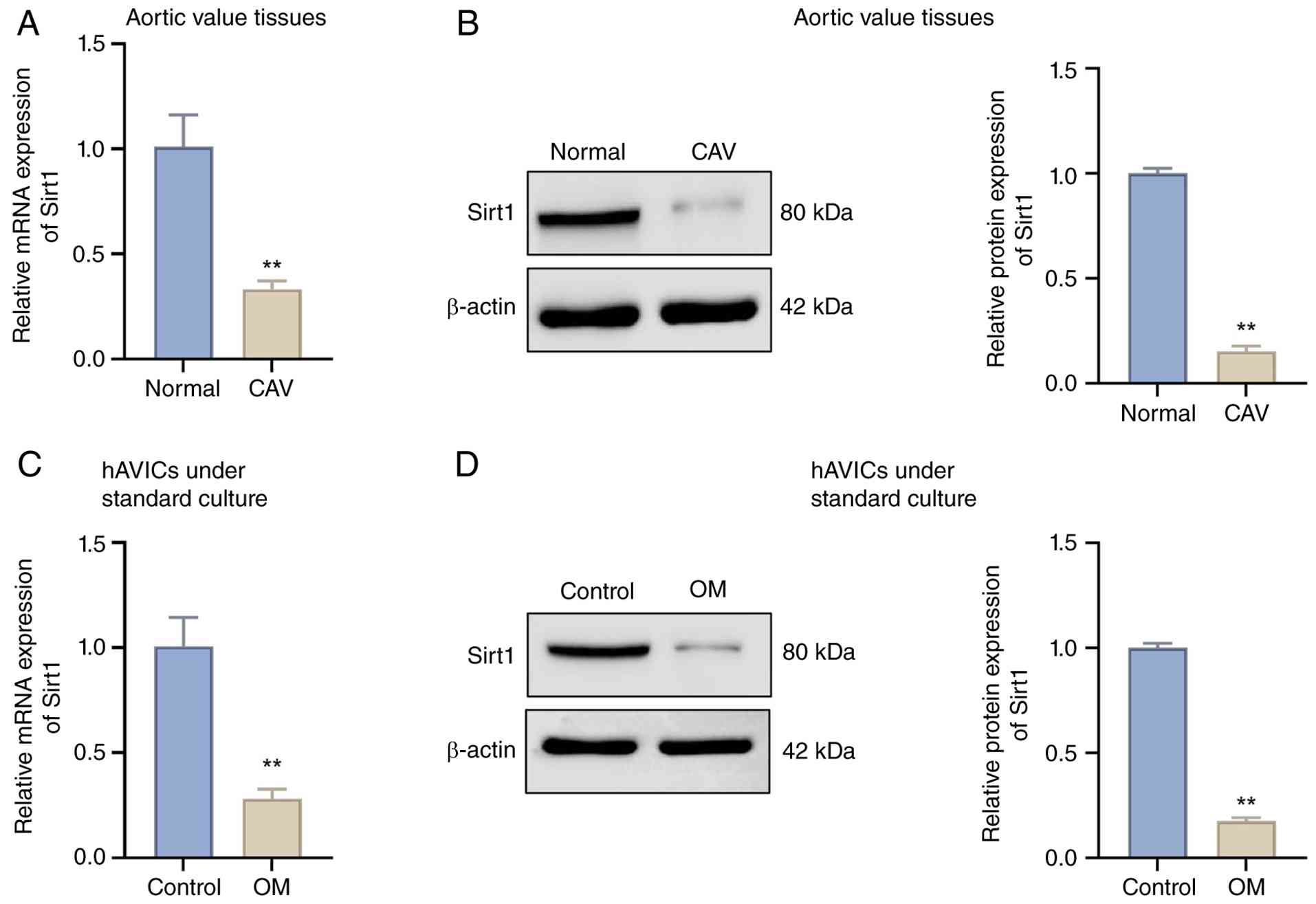
and in vitro osteogenic models. The results of RT-qPCR and
western blot analysis disclosed that, compared with the normal
group, Sirt1 expression was significantly lower in the aortic valve
tissue of the CAV group (Fig. 1A and
B). Similarly, osteogenic stimulation significantly reduced the
expression of Sirt1 in cultured hAVICs relative to the control
group (Fig. 1C and D). These
data suggested the consistent downregulation of Sirt1 in both
clinical and experimental calcification models.
Overexpression of Sirt1 attenuates the
osteogenic differentiation of hAVICs
Given the reduced expression of Sirt1 in calcified
settings, the present study then investigated whether restoring its
expression could influence osteogenic transition. To further
investigate the association between Sirt1 and CAVD, Sirt1
expression was increased in hAVICs in vitro using an
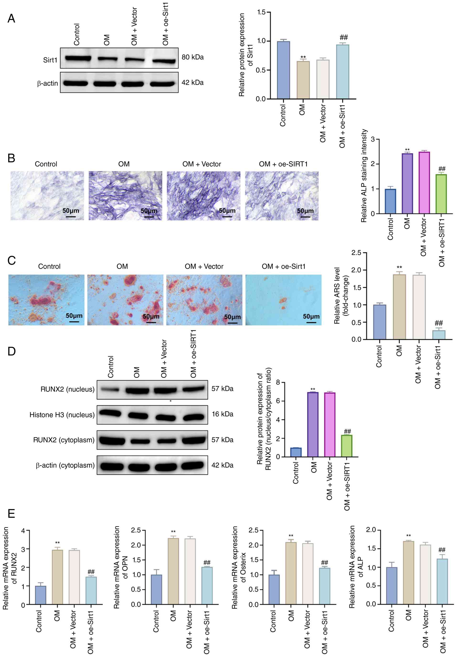
overexpression vector (pcDNA3.1-Sirt1) (Fig. 2A). The cells were subjected to
osteogenic induction, and ALP and Alizarin Red S staining were
carried out when the cells reached full maturation. The
experimental results revealed that in comparison with the control
group, the cells in the OM group exhibited a significantly higher
ALP activity, enhanced mineralization and the increased formation
of calcified nodules. By contrast, the OM + oe-Sirt1 group
displayed a significantly reduced ALP activity, lower
mineralization and decreased calcified nodule formation compared
with the OM + vector group (Fig. 2B
and C). Consistently, the calcifying conditions markedly
increased nuclear RUNX2 accumulation, whereas Sirt1 overexpression
significantly reduced nuclear RUNX2 levels, indicating the
inhibition of RUNX2 nuclear translocation (Fig. 2D). RT-qPCR revealed elevated mRNA
levels of the osteogenic markers, RUNX2, OPN, osterix and ALP in
the OM group, which were all significantly downregulated in the OM
+ oe-Sirt1 group (Fig. 2E).
These data demonstrated that the overexpression of Sirt1 inhibited
the calcification of osteogenically-induced hAVICs.
Overexpression of Sirt1 enhances
mitochondrial function in osteogenically-induced hAVICs
Recent research has proposed that alterations in
mitochondrial metabolism are a key determinant in the development
of CAVD (17). Based on this,
herein, it was hypothesized that Sirt1 may serve as a critical link
between CAVD and mitochondrial function. As shown in Fig. 3A and B, the overexpression of
Sirt1 significantly decreased both total and mitochondrial ROS
levels in hAVICs compared to the OM + vector group.
Correspondingly, JC-1 assay revealed that mitochondrial membrane
potential, which was reduced in the OM-treated cells, was markedly
restored by the overexpression of Sirt1 (Fig. 3C). In line with this, the mtDNA
content and intracellular ATP levels were also significantly
elevated in the OM + oe-Sirt1 group (Fig. 3D and E). ELISA revealed that the
overexpression of Sirt1 reversed the OM-induced downregulation of
PGC1α, MFN2 and OPA1, and suppressed the upregulation of FIS1 and
DRP1, indicating improved mitochondrial dynamics (Fig. 3F-J). These results suggested that
the overexpression of Sirt1 improved mitochondrial function under
conditions of osteogenic stress.
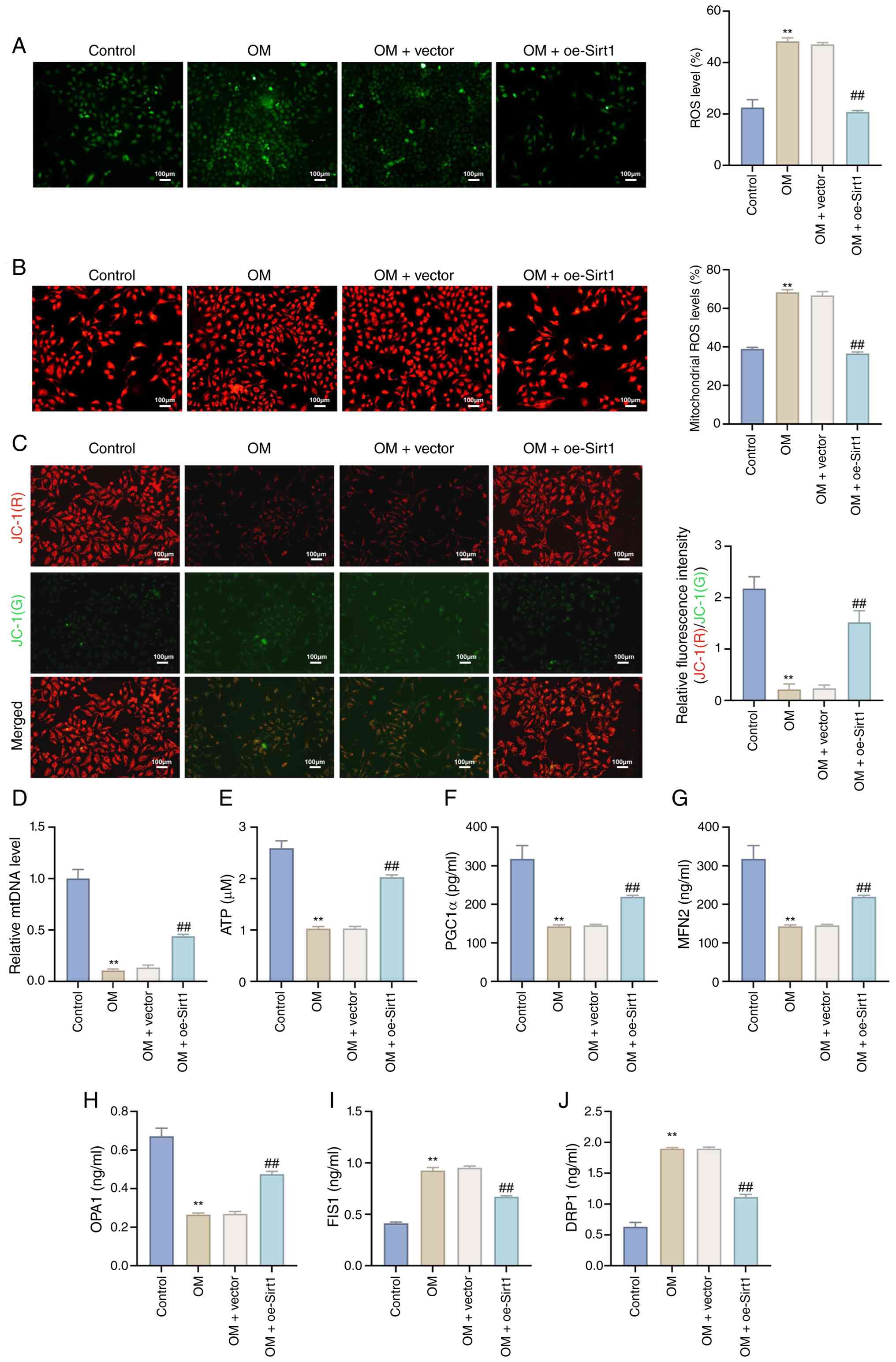 | Figure 3Overexpression of Sirt1 enhances
mitochondrial function in osteogenically-induced aortic valve
interstitial cells. (A and B) Fluorescent probes were used to
detect (A) intracellular ROS and (B) mitochondrial ROS levels. (C)
JC-1 staining was applied to assess mitochondrial membrane
potential (JC-1). (D) Reverse transcription-quantitative polymerase
chain reaction was used to measure the mtDNA content. (E)
Intracellular ATP levels were detected using a luminescence-based
ATP assay; (F-J) ELISA was performed to evaluate the expression of
mitochondrial function-related proteins (PGC1α, MFN2, OPA1, FIS1
and DRP1). Data represent the mean ± SD from three independent
experiments. Comparisons were performed using one-way ANOVA.
**P<0.01 vs. control; ##P<0.01 vs. OM +
vector. ROS, reactive oxygen species; mtDNA, mitochondrial DNA;
ATP, adenosine triphosphate; PGC1α, peroxisome
proliferator-activated receptor gamma coactivator 1-alpha; MFN2,
mitofusin 2; OPA1, optic atrophy 1; FIS1, fission 1; DRP1,
dynamin-related protein 1; Sirt1, sirtuin 1; OM, osteogenic medium;
oe, overexpression. |
Overexpression of Sirt1 inhibits
ferroptosis in osteogenically-induced hAVICs
Additionally, it has previously been demonstrated
that ferroptosis plays a critical role in CADV (5). Notably, Sirt1, mitochondria and
ferroptosis are often interrelated (18). Therefore, in the present study,
it was hypothesized that Sirt1 may induce ferroptosis in cells. To
validate this hypothesis, the levels of MDA, GSH, Fe2+
and total iron in hAVICs were measured following the overexpression
of Sirt1. The results revealed that the overexpression of Sirt1
significantly reduced the MDA, Fe2+ and total iron
levels, while it significantly restored the GSH levels in the
OM-treated hAVICs (Fig. 4A-D).
In parallel, the results of western blot analysis revealed that the
overexpression of Sirt1 increased the expression of the
anti-ferroptotic proteins, GPX4, SLC7A11 and FTH1, and suppressed
the pro-ferroptotic marker, ACSL4, compared to the OM + vector
group (Fig. 4E and F). These
findings suggest that Sirt1 inhibits ferroptosis in hAVICs under
conditions of osteogenic stress.
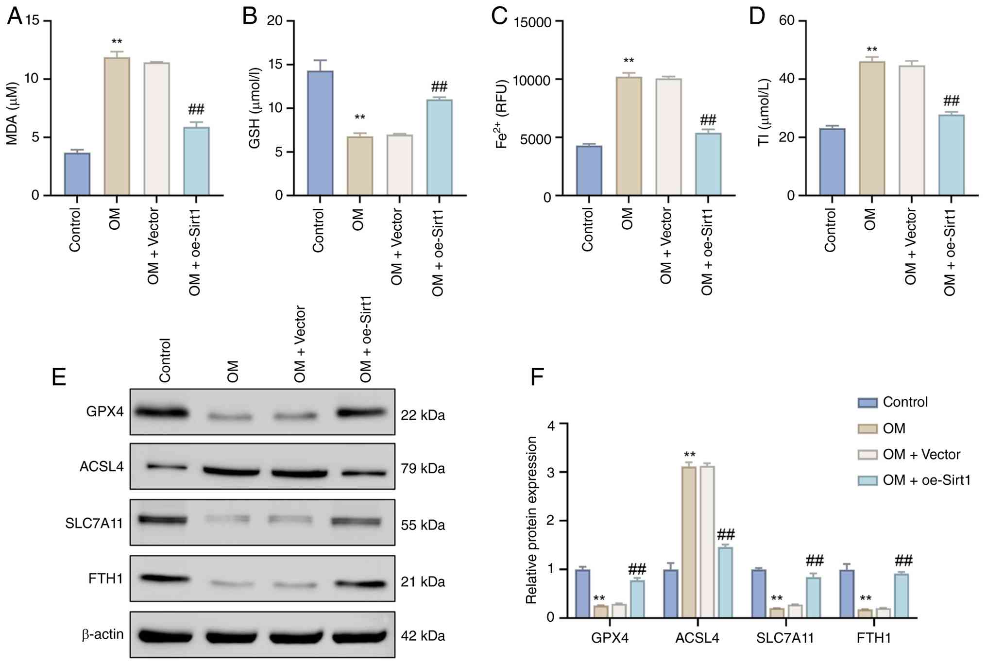 | Figure 4Overexpression of Sirt1 inhibits
ferroptosis in osteogenically-induced aortic valve interstitial
cells. (A-D) Biochemical assays were used to quantify intracellular
(A) MDA, (B), (C) Fe2+, and (D) total iron levels. (E
and F) Western blot analysis was performed to evaluate the
expression of ferroptosis-related proteins, including GPX4, ACSL4,
SLC7A11 and FTH1. Data represent the mean ± SD from three
independent experiments. Comparisons were performed using one-way
ANOVA. **P<0.01 vs. control; ##P<0.01
vs. OM + vector. MDA, malondialdehyde; GSH, glutathione;
Fe2+, ferrous ion; GPX4, glutathione peroxidase 4;
ACSL4, acyl-CoA synthetase long-chain family member 4; SLC7A11,
solute carrier family 7 member 11; FTH1, ferritin heavy chain 1;
Sirt1, sirtuin 1; OM, osteogenic medium; oe, overexpression. |
Overexpression of Sirt1 activates the
Nrf2/HO-1 pathway in osteogenically-induced hAVICs
Previous studies have demonstrated that Sirt1
activates the Nrf2/HO-1 pathway through its deacetylase activity,
thereby inhibiting ferroptosis (19,20). Therefore, it was hypothesized
that the Nrf2/HO-1 pathway may be the key signaling pathway
responsible for the induction of ferroptosis following the reduced
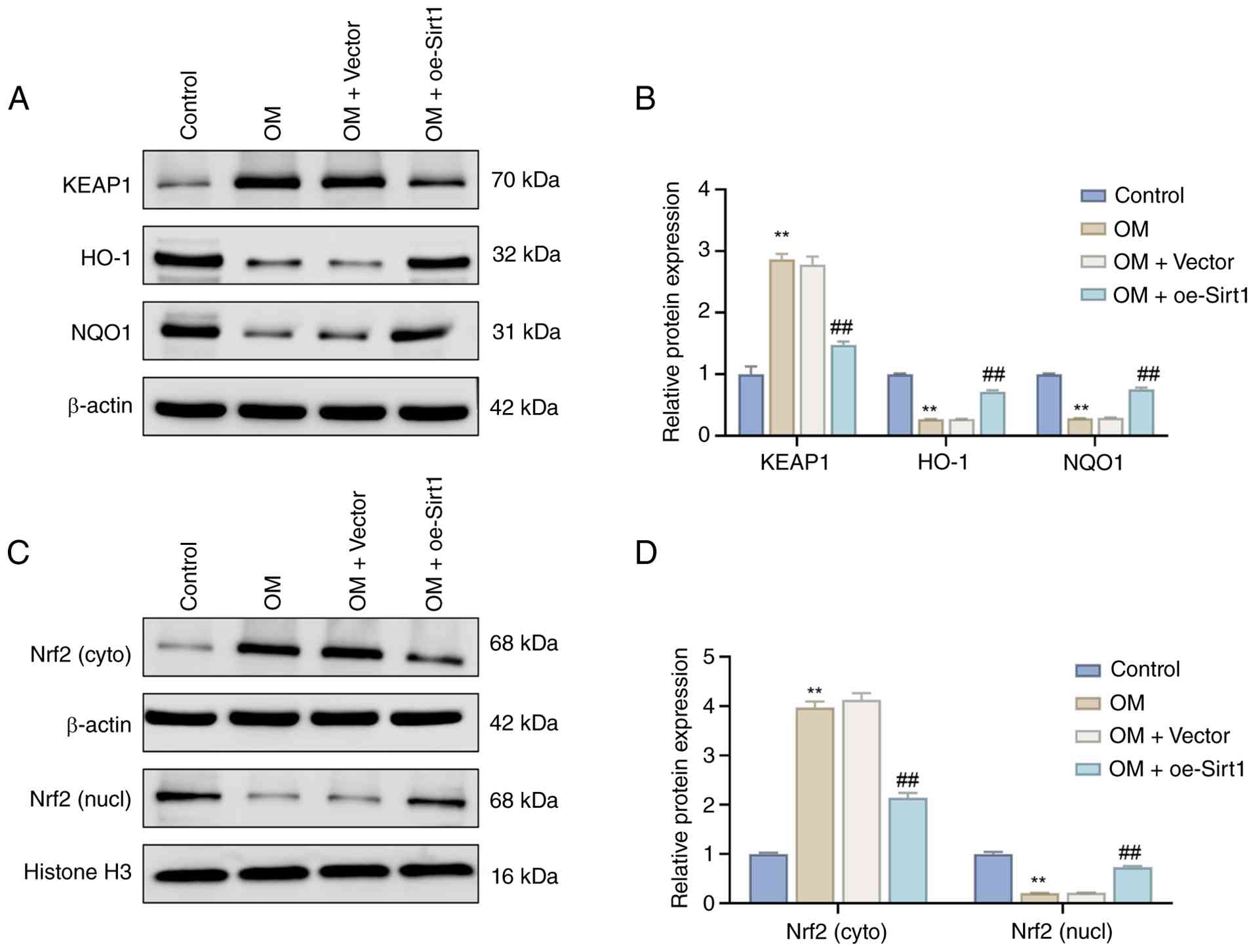
expression of Sirt1 in CAVD. Western blot analysis revealed that,
relative to the control group, the OM group exhibited a significant
upregulation in KEAP1 protein expression, a marked downregulation
in the levels of HO-1 and NQO1 proteins, and a significant increase
in the Nrf2 protein levels in the cytoplasm, while the nuclear Nrf2
protein levels were significantly decreased. Conversely, compared
to the OM + vector group, the OM + oe-Sirt1 group had significantly
lower KEAP1 levels, higher HO-1 and NQO1 levels, as well as reduced
Nrf2 levels in the cytoplasm, and elevated Nrf2 levels in the
nucleus (Fig. 5). These findings
suggest that the overexpression of Sirt1 activates the Nrf2/HO-1
pathway in osteogenically-induced hAVICs.
Inhibition of Nrf2/HO-1 or the induction
of ferroptosis reverse the protective effects of Sirt1
overexpression in hAVICs
To verify whether the anti-calcification effects of
Sirt1 are dependent on Nrf2/HO-1 activation and the suppression of
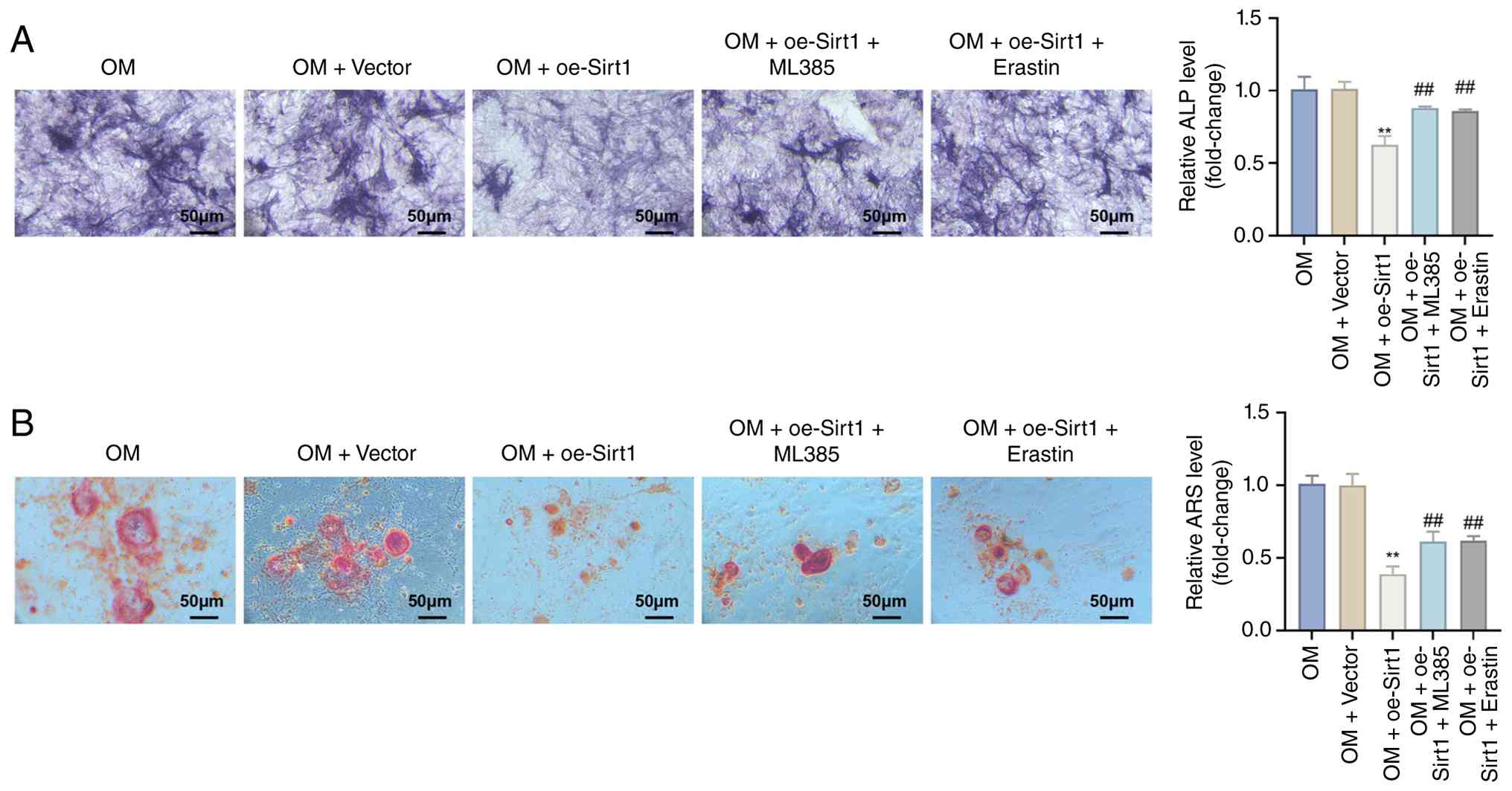
ferroptosis, hAVICs overexpressing Sirt1 were treated with Nrf2
inhibitor (ML385) or ferroptosis inducer (erastin). Initially, ALP
staining and Alizarin Red S staining revealed that, compared to the
OM + oe-Sirt1 group, the OM + oe-Sirt1 + ML385 and OM + oe-Sirt1 +
erastin groups exhibited a significantly increased ALP activity,
mineralization and calcified nodule formation (Fig. 6A and B). Consistently, the
expression levels of the osteogenic markers, RUNX2, OPN, osterix
and ALP, were upregulated (Fig.
7A). Moreover, the levels of the mitochondrial function
markers, PGC1α, MFN2 and OPA1, were downregulated, whereas the
levels of FIS1 and DRP1 were upregulated (Fig. 7B). Additionally, in comparison
with the OM + oe-Sirt1 group, the OM + oe-Sirt1 + ML385 and OM +
oe-Sirt1 + erastin groups exhibited significantly reduced levels of
GPX4, SLC7A11 and FTH1, and a significant increase in ACSL4 protein
expression (Fig. 7C). These data
indicated that the inhibition of the Nrf2/HO-1 pathway or the
promotion of ferroptosis enhanced the calcification of
osteogenically-induced hAVICs.
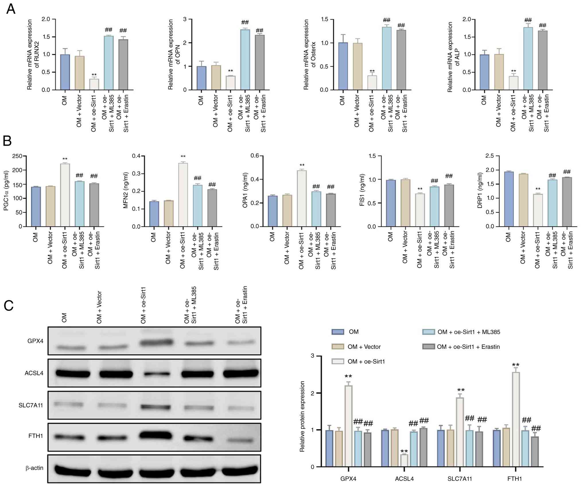 | Figure 7Inhibition of Nrf2 signaling or
induction of ferroptosis reverses the molecular protective effects
of Sirt1 overexpression in hAVICs. (A) Reverse
transcription-quantitative polymerase chain reaction was used to
evaluate the expression of the osteogenic markers, RUNX2, OPN,
osterix and ALP; (B) Mitochondrial function-related proteins PGC1α,
MFN2, OPA1, FIS1 and DRP1 were measured using ELISA; (C) Western
blot analysis of ferroptosis-related proteins (GPX4, ACSL4, SLC7A11
and FTH1). Data represent the mean ± SD from three independent
experiments. Comparisons were performed using one-way ANOVA.
**P<0.01 vs. OM + vector; ##P<0.01 vs.
OM + oe-Sirt1. ALP, alkaline phosphatase; RUNX2, RUNX family
transcription factor 2; OPN, osteopontin; PGC1α, peroxisome
proliferator-activated receptor gamma coactivator 1-alpha; MFN2,
mitofusin 2; OPA1, optic atrophy 1; FIS1, fission 1; DRP1,
dynamin-related protein 1; GPX4, glutathione peroxidase 4; ACSL4,
acyl-CoA synthetase long-chain family member 4; SLC7A11, solute
carrier family 7 member 11; FTH1, ferritin heavy chain 1. ML385,
Nrf2 inhibitor; erastin, ferroptosis inducer; Sirt1, sirtuin 1; OM,
osteogenic medium; oe, overexpression. |
Sirt1-mediated anti-calcification effects
involve both HO-1 and FTH1 in hAVICs
To further investigate the roles of HO-1 and FTH1 in
the Sirt1-mediated anti-calcification effects, hAVICs
overexpressing Sirt1 were subjected to HO-1 or FTH1 knockdown under
osteogenic conditions.
Alizarin Red S and ALP staining demonstrated that
the overexpression of Sirt1 markedly reduced mineralization and
osteogenic activity, whereas silencing either HO-1 or FTH1
partially reversed these inhibitory effects (Fig. 8A and B). Both interventions
increased calcified nodule formation and ALP activity compared with
the Sirt1 overexpression group.
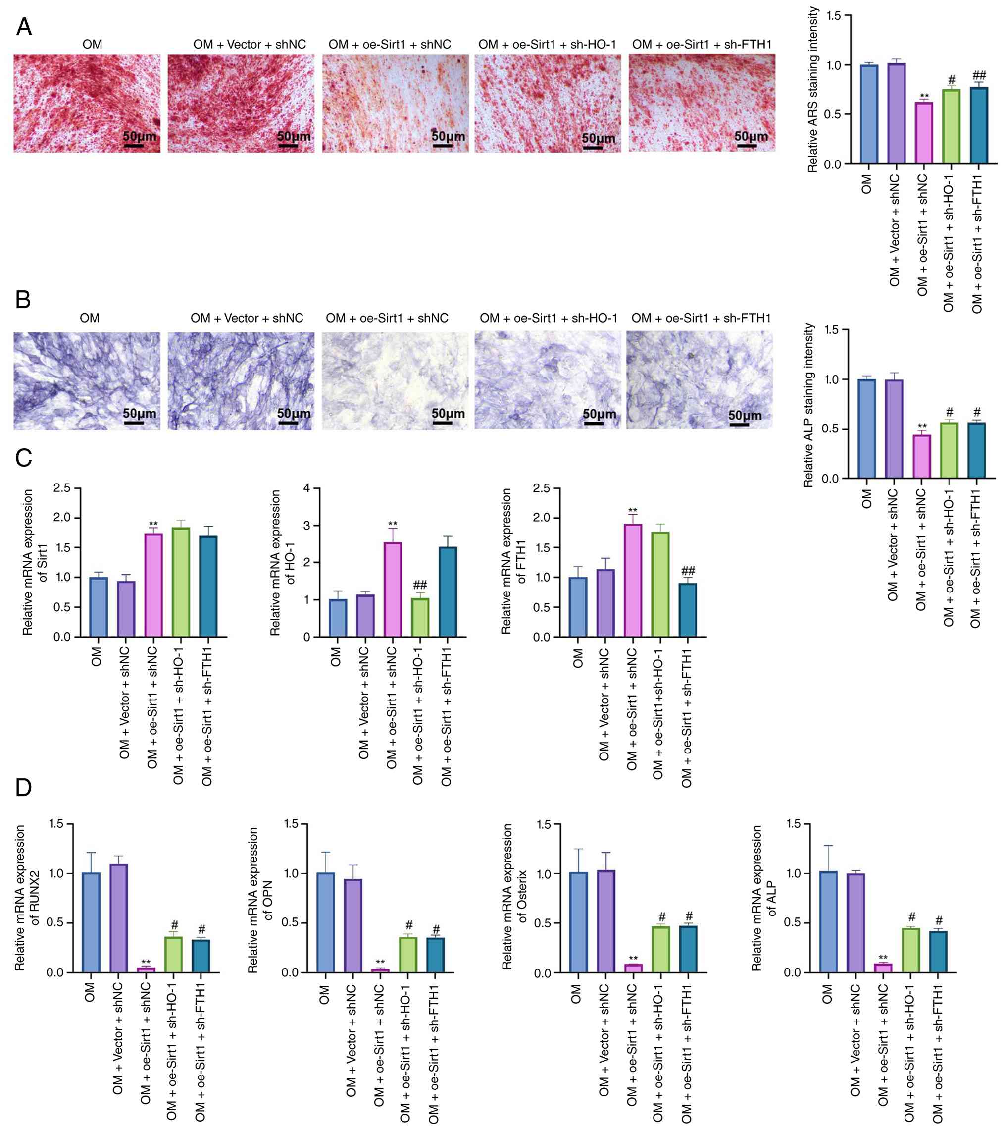 | Figure 8HO-1 and FTH1 contribute to the
Sirt1-mediated inhibition of osteogenic differentiation and
calcification in hAVICs. (A) Alizarin Red S staining and (B) ALP
staining were performed to assess mineralization and osteogenic
activity in each group; (C) RT-qPCR analysis of Sirt1, HO-1 and
FTH1 mRNA expression levels in the different groups; (D) RT-qPCR
analysis of osteogenic markers RUNX2, OPN, osterix and ALP
expression levels. Data represent the mean ± SD from three
independent experiments. Comparisons were performed using one-way
ANOVA. **P<0.01 vs. OM + vector + shNC;
#P<0.05 and ##P<0.01 vs. OM + oe-Sirt1+
shNC. RT-qPCR, reverse transcription-quantitative PCR; OM,
osteogenic medium; Sirt1, sirtuin 1; HO-1, heme oxygenase 1; FTH1,
ferritin heavy chain 1; RUNX2, RUNX family transcription factor 2;
OPN, osteopontin; ALP, alkaline phosphatase. |
At the transcriptional level, the overexpression of
Sirt1 significantly increased Sirt1 and HO-1 mRNA expression,
whereas HO-1 knockdown effectively reduced the HO-1 levels. By
contrast, FTH1 expression was decreased following FTH1 silencing,
confirming the efficiency of gene-specific knockdown (Fig. 8C). Consistently, the expression
of osteogenic markers, including RUNX2, OPN, osterix and ALP, was
significantly suppressed by Sirt1 overexpression, but was restored
upon the knockdown of either HO-1 or FTH1 (Fig. 8D).
These findings were further supported by western
blot analysis, which revealed an increased nuclear RUNX2
accumulation following HO-1 or FTH1 silencing compared with the
Sirt1 overexpression group (Fig.
S2A). To further investigate the underlying signaling pathway,
key components of the Nrf2/HO-1 axis were analyzed. Compared with
the OM and OM + vector + shNC groups, Sirt1 overexpression markedly
reduced KEAP1 protein expression and increased the nuclear Nrf2
levels, accompanied by a decrease in cytoplasmic Nrf2, and the
upregulation of HO-1 and NQO1 expression. Upon HO-1 knockdown, HO-1
protein expression was significantly reduced, and NQO1 levels were
correspondingly decreased. In addition, KEAP1 expression and both
nuclear and cytoplasmic Nrf2 levels exhibited no obvious changes
compared with the Sirt1 overexpression group. By contrast, FTH1
silencing did not significantly affect the expression of HO-1,
NQO1, KEAP1, or the nuclear and cytoplasmic distribution of Nrf2
compared with the Sirt1 overexpression group (Fig. S2B).
In addition, biochemical analysis revealed that
Sirt1 overexpression reduced MDA levels and increased the GSH
content, while these effects were reversed by the knockdown of
either HO-1 or FTH1 (Fig. S2C),
indicating a restoration of oxidative stress and
ferroptosis-related imbalance. Collectively, these results indicate
that both HO-1 and FTH1 are involved in mediating the inhibitory
effects of Sirt1 on osteogenic differentiation and ferroptosis in
hAVICs.
Activation of Sirt1 attenuates aortic
valve calcification in Apoe−/− mice
To validate the in vitro findings in
vivo, a mouse model of CAVD was established using
Apoe−/− mice fed a Western diet, with or without the
administration of SRT2104, a pharmacological Sirt1 activator.
Following 24 weeks of feeding, the tissue sample was collected, and
H&E staining was performed for histological evaluation.
The histological evaluation of aortic valve tissues
via H&E staining revealed that the calcified tissues exhibited
marked valve thickening, structural disorganization and
inflammatory cell infiltration. By contrast, the SRT2104-treated
group demonstrated preserved trilaminar architecture, reduced
leaflet thickness and diminished inflammatory features, suggesting
alleviated pathological remodeling (Fig. 9A). These findings indicate that
SRT2104 ameliorates calcification-associated structural changes
in vivo.
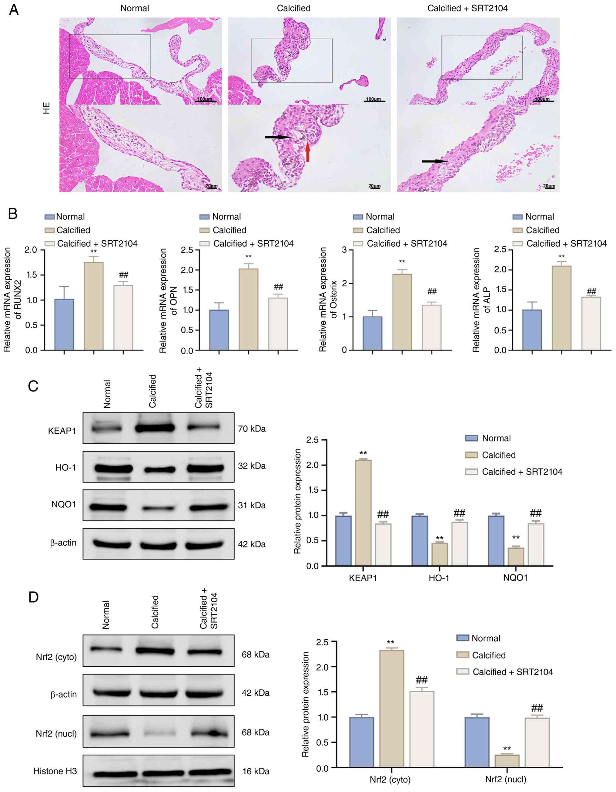 | Figure 9Activation of Sirt1 in vivo
attenuates calcification-associated pathological remodeling in
Apoe−/− mice. (A) H&E staining for examining the
pathological damage of aortic valve tissues from all groups; (B)
reverse transcription-quantitative PCR analysis of osteogenic
marker (RUNX2, OPN, osterix and ALP) expression levels in aortic
valve tissues; (C) western blot analysis of KEAP1, HO-1 and NQO1
protein levels in aortic valve tissues; (D) western blot analysis
of Nrf2 protein levels in cytoplasmic and nuclear fractions. For
animal experiments, each individual mouse was considered as one
biological replicate (n=10 per group). Comparisons were performed
using one-way ANOVA. **P<0.01 vs. normal;
##P<0.01 vs. calcified + SRT2104. H&E,
hematoxylin and eosin; RUNX2, RUNX family transcription factor 2;
OPN, osteopontin; ALP, alkaline phosphatase; KEAP1, Kelch-like
ECH-associated protein 1; HO-1, heme oxygenase 1; NQO1, NAD (P) H
quinone dehydrogenase 1. SRT2104, a pharmacological sirtuin 1
activator. |
At the molecular level, RT-qPCR analysis revealed
that the levels of osteogenic markers (RUNX2, OPN, osterix and ALP)
were significantly upregulated in the calcified group and were
markedly reduced following SRT2104 treatment (Fig. 9B), indicating the suppression of
osteogenic transformation.
To determine whether the protective effects were
mediated via the Nrf2/HO-1 pathway, the levels of related proteins
were assessed. Compared to the calcified group, the SRT2104-treated
mice exhibited reduced KEAP1 levels, an elevated expression of HO-1
and NQO1, and an enhanced nuclear accumulation of Nrf2, along with
decreased cytoplasmic retention (Fig. 9C and D). Collectively, these
findings suggest that the pharmacological activation of Sirt1
attenuates calcification-associated pathological remodeling in
vivo, at least in part, through the reactivation of Nrf2/HO-1
signaling and the suppression of osteogenic transformation.
FTH1 mediates the protective effects of
Sirt1 against aortic valve calcification in vivo
To validate the relative contributions of HO-1 and
FTH1 in vivo, Apoe−/− mice treated with SRT2104
were further subjected to AAV-mediated silencing of HO-1 or FTH1.
Histological analyses, including H&E and Alizarin Red S
staining, revealed that SRT2104 markedly attenuated aortic valve
pathological remodeling and calcium deposition compared with the
calcified group. The silencing of either HO-1 or FTH1 partially
reversed these protective effects, as evidenced by increased
leaflet thickening, structural disorganization, inflammatory
infiltration and Alizarin Red-positive staining. The AAV-shHO-1 and
AAV-shFTH1 groups exhibited comparable pathological changes and
calcium deposition (Fig. 10A and
B).
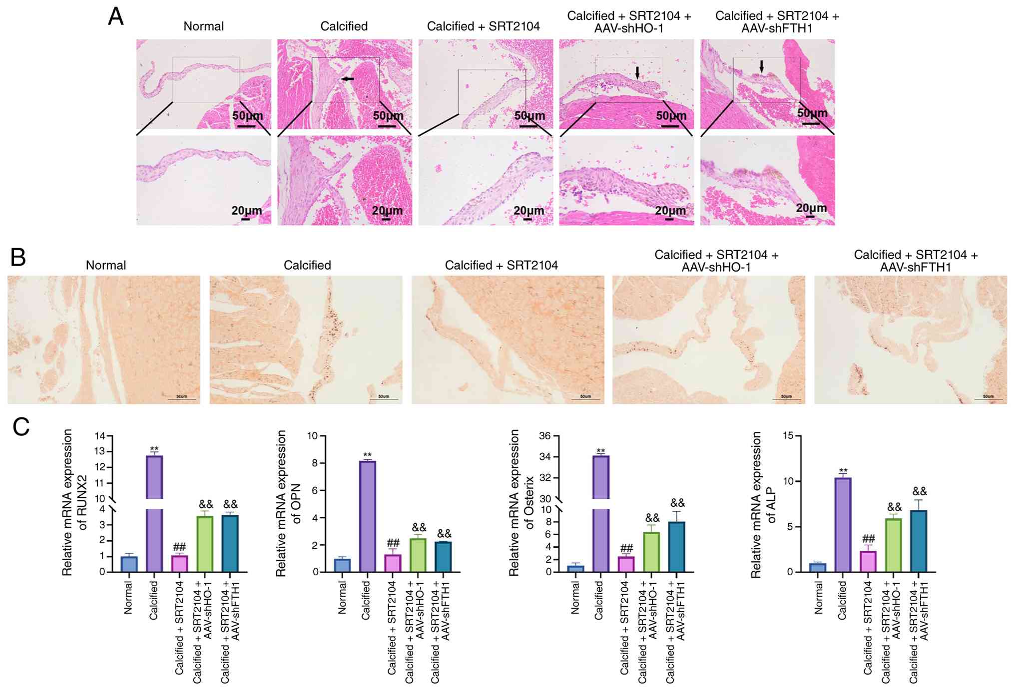 | Figure 10HO-1 and FTH1 contribute to the
protective effects of Sirt1 activation against aortic valve
calcification in vivo. Representative (A) H&E staining
and (B) Alizarin Red S staining of mouse aortic valve sections
illustrating leaflet morphology and pathological remodeling in
different experimental groups. (C) Reverse
transcription-quantitative PCR analysis of the expression levels of
the osteogenic markers, RUNX2, OPN, osterix and ALP. Data represent
the mean ± SD. For animal experiments, each individual mouse was
considered as one biological replicate (n=10 per group).
Comparisons were performed using one-way ANOVA.
**P<0.01 vs. normal; ##P<0.01 vs.
calcified; &&P<0.01 vs. calcified + SRT2104.
Normal, Apoe−/− mice mice on a standard diet; calcified,
Apoe−/− mice fed a Western diet; Calcified + SRT2104,
Apoe−/− mice treated with SRT2104; AAV-shHO-1,
adeno-associated virus-mediated HO-1 knockdown; AAV-shFTH1,
adeno-associated virus-mediated FTH1 knockdown; RUNX2, RUNX family
transcription factor 2; OPN, osteopontin; ALP, alkaline
phosphatase; KEAP1, Kelch-like ECH-associated protein 1; Nrf2,
nuclear factor erythroid 2-related factor 2; HO-1, heme oxygenase
1; NQO1, NAD(P)H quinone dehydrogenase 1; FTH1, ferritin heavy
chain 1. |
At the molecular level, RT-qPCR demonstrated that
SRT2104 significantly reduced the expression of osteogenic markers
(RUNX2, OPN, osterix and ALP), whereas the knockdown of HO-1 or
FTH1 partially restored their expression levels (Fig. 10C). Consistently, western blot
analysis revealed that SRT2104 decreased nuclear RUNX2 protein
expression, while this effect was reversed following the silencing
of HO-1 or FTH1 (Fig. S3A).
Further analysis of the Nrf2/HO-1 signaling pathway
revealed that SRT2104 treatment decreased KEAP1 expression,
increased nuclear Nrf2 levels, reduced cytoplasmic Nrf2 levels, and
upregulated the expression of HO-1, NQO1 and FTH1. Notably, FTH1
expression was reduced in calcified mice compared with the normal
controls and was restored following SRT2104 treatment. Upon HO-1
knockdown, HO-1 protein expression was markedly reduced,
accompanied by decreases in the NQO1 and FTH1 levels, whereas KEAP1
expression and Nrf2 nuclear and cytoplasmic distribution exhibited
no obvious changes compared with the SRT2104-treated group. By
contrast, FTH1 silencing markedly reduced FTH1 protein expression,
while the KEAP1, Nrf2 (nuclear and cytoplasmic), HO-1 and NQO1
levels remained largely unaltered compared with the SRT2104-treated
group (Fig. S3B). Collectively,
these results indicate that both HO-1 and FTH1 participate in the
protective effects of Sirt1 activation against aortic valve
calcification in vivo.
Discussion
CAVD is a progressive degenerative cardiovascular
disorder characterized by a markedly increased prevalence with
advancing age; in severe cases, it may progress to calcific aortic
stenosis, ultimately leading to heart failure (1). Given the absence of effective drug
therapies capable of preventing or reversing CAVD progression,
there is an urgent need to unravel the underlying molecular
mechanisms of CAVD pathogenesis and to identify potential
therapeutic targets. It is important to acknowledge that the
pathogenesis of CAVD is multifactorial, involving a complex
interplay of chronic inflammation, lipid dysregulation, oxidative
stress, aberrant cell differentiation, and extracellular matrix
remodeling. The present study focused on investigating a specific
regulatory axis within this network. The present provides evidence
that Sirt1 may play a pivotal role in the progression of CAVD by
regulating ferroptosis, providing important insights into potential
strategies for delaying disease progression.
As a deacetylase, Sirt1 regulates the activity of
multiple signaling pathways and has long been demonstrated to be
closely associated with vascular calcification. For instance,
Bartoli-Leonard et al (21) reported that the loss of Sirt1
expression in patients with diabetes accelerated DNA damage and
promoted vascular calcification. Liu et al (11) demonstrated that the upregulation
of Sirt1 expression by spermidine effectively inhibited mineral
deposition in rat and human vascular smooth muscle cells. Moreover,
Yu et al (22) revealed
that luteolin could directly bind to and activates Sirt1, thereby
inhibiting oxidative stress and enhancing autophagy to protect
against vascular calcification. These findings suggest that Sirt1
is a key participant in the process of vascular calcification.
Consistently, the present study revealed that Sirt1 expression was
markedly downregulated in aortic valve tissues from both patients
with CAVD and cellular models. Notably, Sirt1 overexpression
effectively attenuated AVIC calcification, consistent with the
previously reported effects of Sirt1 on calcification in other
cardiovascular diseases (23,24). These observations indicate that
Sirt1 plays a decisive role in the progression of CAVD.
In recent years, mitochondrial dysfunction has been
recognized as a key driving factor in the onset and progression of
CAVD (25). In contrast to
nuclear DNA, mtDNA is not bound to histones, rendering mtDNA repair
mechanisms relatively inefficient. As a result, mtDNA is inherently
unstable and susceptible to mutations and deletions (26). Rothwell et al (27) observed that DNA polymerase
γ-deficient mice displayed substantial mtDNA damage and aggravated
vascular calcification. Additionally, extensive mitochondrial
fragmentation has been observed in calcified cardiovascular
diseases, and this fragmentation has been shown to induce oxidative
stress and promote mitochondrial depolarization by regulating the
expression of mitochondrial fission-fusion proteins (28). Notably, research has revealed
that DRP1 is enriched in calcified areas of the human carotid
artery, and inhibiting DRP1 can alleviate oxidative stress, thereby
mitigating cardiovascular calcification (29). In the present study, the
expression of mitochondrial fusion proteins (MFN2 and OPA1) was
decreased, while the expression of mitochondrial fission proteins
(DRP1 and FIS1) was increased in CAVD cells, suggesting a shift in
mitochondrial dynamics towards fission (30). Furthermore, mitochondrial
membrane potential was significantly reduced, indicating
mitochondrial dysfunction (31).
Additionally, mtDNA replication capacity decreased, ATP production
was reduced, and both ROS and mitochondrial-specific ROS levels
were significantly elevated, all indicating mitochondrial damage in
CAVD cells. However, it was also observed that the overexpression
of Sirt1 effectively alleviated mitochondrial damage in CAVD
cells.
Similarly, ferroptosis has been linked to various
cardiovascular calcification-related diseases (32). The core mechanism of ferroptosis
is the accumulation of lipid peroxides, primarily mediated by
iron-dependent Fenton reactions, resulting in elevated ROS levels
and damage to the cell membrane (33). In the process of cardiovascular
calcification, vascular endothelial cells and smooth muscle cells
are exposed to high phosphate environments, inflammatory cytokines
and oxidative stress, resulting in mitochondrial dysfunction. This,
in turn, promotes the accumulation of iron ions and lipid
peroxidation, ultimately inducing ferroptosis (34). It has been demonstrated that
ferroptosis not only exacerbates AVICs, but also induces the
mineralization of the extracellular matrix, accelerating calcific
deposition in the valve (6). The
present study revealed that under osteogenic medium induction, MDA,
Fe2+ and total iron levels accumulated in AVICs, while
GSH was depleted. Additionally, a significant decrease in the
expression of key negative regulators of ferroptosis, including
GPX4, SLC7A11 and FTH1 was observed, while the expression of the
ferroptosis-promoting protein, ACSL4, was upregulated, all of which
are consistent with the results of previous research (35,36). These findings indicate an
elevated level of ferroptosis in CAVD cells.
More notably, the present study revealed that the
overexpression of Sirt1 effectively inhibited mitochondrial
dysfunction and ferroptosis in cells. As a deacetylase, Sirt1
regulates the activity of multiple signaling pathways. Among these,
the activation of the Nrf2/HO-1 pathway is considered to reduce
cardiovascular calcification (37). Notably, the protective role of
the Nrf2/HO-1 axis against calcification may extend beyond HO-1
itself, involving its downstream effector, H-ferritin (FTH1). A
well-established regulatory circuit exists where HO-1 catalyzes
heme degradation, releasing iron which in turn transcriptionally
upregulates ferritin, particularly the heavy chain (FTH1), via
iron-responsive elements (38).
Herein, the authors did consider this important scenario. The data
presented herein demonstrated that Sirt1 activation concurrently
upregulated both HO-1 and FTH1 in cells, and reduced labile
Fe2+ levels. This pattern is consistent with the
described feedback mechanism where HO-1 induction leads to
increased cellular iron, which post-transcriptionally induces FTH1
synthesis to manage the iron load and exert its ferroxidase
activity. The observed reduction in labile Fe2+ upon
Sirt1 overexpression is plausible given the concurrent upregulation
of FTH1. The ferroxidase activity of H-ferritin is crucial for
sequestering labile iron and mitigating iron-catalyzed oxidative
damage (39), thereby exerting
anti-calcification effects in vascular smooth muscle cells and
valvular interstitial cells (40-42). While HO-1 and ferritin expression
levels are often connected in various pathologies (43), their functions can be distinct,
and the benefit against mineralization has been specifically
attributed to the ferroxidase activity of H-ferritin rather than to
HO-1-derived products such as carbon monoxide or bilirubin
(42). Lu et al (44) discovered that the activation of
the Nrf2/HO-1 pathway activity reduced vascular calcification in
mice with chronic kidney disease; it also inhibits osteoblast
maturation and mineralization (45). Sirt1-mediated deacetylation can
suppress Keap1-mediated ubiquitination degradation, activating Nrf2
and facilitating its nuclear translocation to induce the expression
of HO-1 and other antioxidant and iron homeostasis genes (46). Of note, in the present study,
loss-of-function experiments further refined the hierarchical
association between HO-1 and FTH1 within this axis. In
vitro, the silencing of HO-1 led to a reduction in NQO1
expression without significantly affecting KEAP1 or Nrf2
distribution, whereas FTH1 knockdown had a minimal impact on
upstream Nrf2/HO-1 signaling components. Similarly, in vivo
experiments demonstrated that HO-1 silencing reduced both HO-1 and
NQO1 expression, while FTH1 knockdown selectively decreased FTH1
levels without altering KEAP1 or Nrf2 localization. These findings
suggest that HO-1 primarily functions as an upstream regulator
within the Nrf2 signaling pathway, whereas FTH1 functions as a
downstream effector involved in iron sequestration and ferroptosis
modulation. Therefore, the anti-calcification effects of Sirt1 are
likely mediated through coordinated regulation of both signaling
activation (via HO-1) and iron homeostasis control (via FTH1). HO-1
contributes to cellular iron metabolism by catalyzing heme
degradation, whereas the sequestration of labile iron is primarily
mediated by ferritin, particularly FTH1. The reductive capacity of
HO-1 can effectively reduce mitochondrial ROS levels, thereby
protecting mitochondrial function (47). The findings of the present study
confirm that Sirt1 effectively activates the Nrf2/HO-1 pathway, and
in vivo experiments in mice provided evidence that Sirt1
activation may alleviate aortic calcification via the Nrf2/HO-1
pathway.
However, the present study has several limitations
which should be acknowledged. First, the temporal changes in Sirt1
expression during different stages of CAVD progression were not
explored, which may provide deeper insight into its role in disease
development. Second, although the involvement of the Nrf2/HO-1
pathway was supported by pharmacological inhibition, the absence of
genetic validation (e.g., siRNA or knockout models) limits the
conclusiveness of pathway specificity. Third, the present study
utilized a mixed osteogenic cocktail (containing
β-glycerophosphate) for induction. Future studies are required to
examine whether Sirt1 exerts similar protective effects against
calcification induced specifically by elevated inorganic phosphate,
a key pathological driver of calcification in chronic kidney
disease-related CAVD. Finally, in vivo experiments relied on
the pharmacological activation of Sirt1, and direct histological
assessment of calcium deposition (e.g., Alizarin Red or OsteoSense
staining) was not performed; therefore, stronger evidence could be
obtained by incorporating genetic models (e.g., Sirt1 knockout or
conditional knockout) and more specific mineralization assays in
future studies. Further studies are thus warranted to enhance
mechanistic clarity and provide translational relevance.
In conclusion, the present study demonstrates that
Sirt1 protects against CAVD by activating the Nrf2/HO-1 pathway,
leading to FTH1-dependent maintenance of iron homeostasis, the
suppression of ferroptosis, the preservation of mitochondrial
function and the inhibition of osteogenic differentiation in AVICs,
thereby attenuating calcification-associated pathological
remodeling. These findings identify the Sirt1/Nrf2/HO-1/FTH1
signaling axis as a key regulatory pathway linking iron
homeostasis, ferroptosis, mitochondrial dysfunction and CAVD
progression, and highlight Sirt1 as a therapeutic target for
CAVD.
Supplementary Data
Availability of data and materials
The data generated in the present study may be
requested from the corresponding author.
Authors' contributions
FY was involved in the conceptualization of the
study, in data curation, formal analysis, data investigation, study
supervision, data validation and visualization, as well as in the
writing of the original draft, and in the writing, reviewing and
editing of the manuscript. LS was involved in data curation, formal
analysis, investigation, in the study methodology, and in the
writing, reviewing and editing of the manuscript. YWa was involved
in data curation, data investigation, in the study methodology, and
writing, reviewing and editing of the manuscript. YZ was involved
in data curation and in the formal analysis, as well as in the
writing, reviewing and editing of the manuscript. YWu was involved
in the formal analysis, data investigation, in the study
methodology, and in the writing, reviewing and editing of the
manuscript. DX, YL, NW, HC and MZ were involved in data curation
and in the formal analysis, as well as in the writing, reviewing
and editing of the manuscript. FY and MZ confirm the authenticity
of all the raw data. All authors have read and approved the final
manuscript.
Ethics approval and consent to
participate
Prior to initiation, the present study was approved
by the Medical Ethics Committee of the First Affiliated Hospital of
Hainan Medical University (Approval no. 2025-KYL-016). Sample
collection and processing were conducted in strict accordance with
the Declaration of Helsinki and the Regulations on the Management
of Human Genetic Resources. Written informed consent was obtained
from all participants prior to tissue collection. All animal
experiments were approved by Ethics Committee of Hainan Medical
University (Approval no. HYALL-2025-028) and were performed in
accordance with the Guide for the Care and Use of Laboratory
Animals.
Patient consent for publication
Not applicable.
Competing interests
The authors declare that they have no competing
interests.
Acknowledgments
Not applicable.
Funding
The present study was supported by the National Natural Science
Foundation of China (grant no. 82460074) and the Hainan Province
Science and Technology Special Fund (ZDKJ2021038).
References
|
1
|
Kraler S, Blaser MC, Aikawa E, Camici GG
and Lüscher TF: Calcific aortic valve disease: From molecular and
cellular mechanisms to medical therapy. Eur Heart J. 43:683–697.
2022. View Article : Google Scholar :
|
|
2
|
Yu J, Wang Z, Bao Q, Lei S, You Y, Yin Z
and Xie X: Global burden of calcific aortic valve disease and
attributable risk factors from 1990 to 2019. Front Cardiovasc Med.
9:10032332022. View Article : Google Scholar : PubMed/NCBI
|
|
3
|
Tanase DM, Valasciuc E, Gosav EM, Floria
M, Costea CF, Dima N, Tudorancea I, Maranduca MA and Serban IL:
Contribution of oxidative stress (OS) in calcific aortic valve
disease (CAVD): From pathophysiology to therapeutic targets. Cells.
11:26632022. View Article : Google Scholar : PubMed/NCBI
|
|
4
|
Liu Z, Dong N, Hui H, Wang Y, Liu F, Xu L,
Liu M, Rao Z, Yuan Z, Shang Y, et al: Endothelial cell-derived
tetrahydrobiopterin prevents aortic valve calcification. Eur Heart
J. 43:1652–1664. 2022. View Article : Google Scholar : PubMed/NCBI
|
|
5
|
Li XZ, Xiong ZC, Zhang SL, Hao QY, Gao M,
Wang JF, Gao JW and Liu PM: Potential ferroptosis key genes in
calcific aortic valve disease. Front Cardiovasc Med. 9:9168412022.
View Article : Google Scholar : PubMed/NCBI
|
|
6
|
Wang S, Gu J, Bian J, He Y, Xu X, Wang C,
Li G, Zhang H, Ni B, Chen S, et al: Nesfatin-1 mitigates calcific
aortic valve disease via suppressing ferroptosis mediated by
GSH/GPX4 and ZIP8/SOD2 axes. Free Radic Biol Med. 222:149–164.
2024. View Article : Google Scholar : PubMed/NCBI
|
|
7
|
Budbazar E, Rodriguez F, Sanchez JM and
Seta F: The role of sirtuin-1 in the vasculature: Focus on aortic
aneurysm. Front Physiol. 11:10472020. View Article : Google Scholar : PubMed/NCBI
|
|
8
|
Chen H, Lin X, Yi X, Liu X, Yu R, Fan W,
Ling Y, Liu Y and Xie W: SIRT1-mediated p53 deacetylation inhibits
ferroptosis and alleviates heat stress-induced lung epithelial
cells injury. Int J Hyperthermia. 39:977–986. 2022. View Article : Google Scholar : PubMed/NCBI
|
|
9
|
Chen Z, Li J, Peng H, Zhang M, Wu X, Gui
F, Li W, Ai F, Yu B and Liu Y: Meteorin-like/Meteorin-β protects
LPS-induced acute lung injury by activating SIRT1-P53-SLC7A11
mediated ferroptosis pathway. Mol Med. 29:1442023. View Article : Google Scholar
|
|
10
|
Li SJ, Cheng TY, Kao YH, Chen WY, Chung
CC, Trang NN, Chen PH and Chen YJ: Uremic toxin p-cresyl sulfate
enhances the calcification of aortic valvular interstitial cells
via klotho/sirtuin-1 signaling. Mol Med Rep. 33:1622026. View Article : Google Scholar
|
|
11
|
Liu X, Chen A, Liang Q, Yang X, Dong Q, Fu
M, Wang S, Li Y, Ye Y, Lan Z, et al: Spermidine inhibits vascular
calcification in chronic kidney disease through modulation of SIRT1
signaling pathway. Aging Cell. 20:e133772021. View Article : Google Scholar : PubMed/NCBI
|
|
12
|
Ouyang X, Ding Y, Yu L, Xin F and Yang X:
LncRNA TUG regulates osteogenic differentiation of bone marrow
mesenchymal stem cells via miRNA-204/SIRT 1. J Musculoskelet
Neuronal Interact. 22:401–410. 2022.PubMed/NCBI
|
|
13
|
Li Y, Zeng X, Lu D, Yin M, Shan M and Gao
Y: Erastin induces ferroptosis via ferroportin-mediated iron
accumulation in endometriosis. Hum Reprod. 36:951–964. 2021.
View Article : Google Scholar
|
|
14
|
Song J, Wang H, Sheng J, Zhang W, Lei J,
Gan W, Cai F and Yang Y: Vitexin attenuates chronic kidney disease
by inhibiting renal tubular epithelial cell ferroptosis via NRF2
activation. Mol Med. 29:1472023. View Article : Google Scholar : PubMed/NCBI
|
|
15
|
Livak KJ and Schmittgen TD: Analysis of
relative gene expression data using real-time quantitative PCR and
the 2(-Delta Delta C(T)) method. Methods. 25:402–408. 2001.
View Article : Google Scholar
|
|
16
|
National Research Council (US) Committee
for the Update of the Guide for the Care and Use of Laboratory
Animals: Guide for the Care and Use of Laboratory Animals. 8th
edition. National Academies Press (US); Washington, DC: 2011
|
|
17
|
Combi Z, Potor L, Nagy P, Sikura KÉ,
Ditrói T, Jurányi EP, Galambos K, Szerafin T, Gergely P, Whiteman
M, et al: Hydrogen sulfide as an anti-calcification stratagem in
human aortic valve: Altered biogenesis and mitochondrial metabolism
of H2S lead to H2S deficiency in calcific
aortic valve disease. Redox Biol. 60:1026292023. View Article : Google Scholar
|
|
18
|
Zeng Y, He Y and Wang L, Xu H, Zhang Q,
Wang Y, Zhang J and Wang L: Dihydroquercetin improves experimental
acute liver failure by targeting ferroptosis and
mitochondria-mediated apoptosis through the SIRT1/p53 axis.
Phytomedicine. 128:1555332024. View Article : Google Scholar : PubMed/NCBI
|
|
19
|
Dang R, Wang M, Li X, Wang H, Liu L, Wu Q,
Zhao J, Ji P, Zhong L, Licinio J and Xie P: Edaravone ameliorates
depressive and anxiety-like behaviors via Sirt1/Nrf2/HO-1/Gpx4
pathway. J Neuroinflammation. 19:412022. View Article : Google Scholar : PubMed/NCBI
|
|
20
|
Lv QK, Tao KX, Yao XY, Pang MZ, Cao BE,
Liu CF and Wang F: Melatonin MT1 receptors regulate the
Sirt1/Nrf2/Ho-1/Gpx4 pathway to prevent α-synuclein-induced
ferroptosis in Parkinson's disease. J Pineal Res. 76:e129482024.
View Article : Google Scholar
|
|
21
|
Bartoli-Leonard F, Wilkinson FL, Schiro A,
Serracino Inglott F, Alexander MY and Weston R: Loss of SIRT1 in
diabetes accelerates DNA damage-induced vascular calcification.
Cardiovasc Res. 117:836–849. 2021. View Article : Google Scholar :
|
|
22
|
Yu X, Xu L, Su C, Wang C, Wang Z, Wang Y,
Lu X and Sun H: Luteolin protects against vascular calcification by
modulating SIRT1/CXCR4 signaling pathway and promoting autophagy.
AAPS J. 26:1112024. View Article : Google Scholar : PubMed/NCBI
|
|
23
|
Lu CL, Liao MT, Hou YC, Fang YW, Zheng CM,
Liu WC, Chao CT, Lu KC and Ng YY: Sirtuin-1 and Its relevance in
vascular calcification. Int J Mol Sci. 21:15932020. View Article : Google Scholar : PubMed/NCBI
|
|
24
|
Ren Z, Song B, Peng Y, Bian C, Shen Q,
Zhou M, Li B and Jin X: Mechanism and treatment of Sirtuin family
in vascular calcification. Front Cardiovasc Med. 12:17322812025.
View Article : Google Scholar
|
|
25
|
Li M, Zhu Y, Jaiswal SK and Liu NF:
Mitochondria homeostasis and vascular medial calcification. Calcif
Tissue Int. 109:113–120. 2021. View Article : Google Scholar : PubMed/NCBI
|
|
26
|
de Souza-Pinto NC, Mason PA, Hashiguchi K,
Weissman L, Tian J, Guay D, Lebel M, Stevnsner TV, Rasmussen LJ and
Bohr VA: Novel DNA mismatch-repair activity involving YB-1 in human
mitochondria. DNA Repair (Amst). 8:704–719. 2009. View Article : Google Scholar : PubMed/NCBI
|
|
27
|
Rothwell S, Ng I, Shalchy-Tabrizi S,
Kalinowski P, Taha OM, Paris I, Baniqued A, Lin L, Mezei MM, Lehman
A, et al: Loss-of-function mitochondrial DNA polymerase gamma
variants cause vascular smooth muscle cells to secrete a diffusible
mitogenic factor. Front Physiol. 15:14882482025. View Article : Google Scholar : PubMed/NCBI
|
|
28
|
Wu S, Zhou F, Zhang Z and Xing D:
Mitochondrial oxidative stress causes mitochondrial fragmentation
via differential modulation of mitochondrial fission-fusion
proteins. FEBS J. 278:941–954. 2011. View Article : Google Scholar : PubMed/NCBI
|
|
29
|
Rogers MA, Maldonado N, Hutcheson JD,
Goettsch C, Goto S, Yamada I, Faits T, Sesaki H, Aikawa M and
Aikawa E: Dynamin-related protein 1 inhibition attenuates
cardiovascular calcification in the presence of oxidative stress.
Circ Res. 121:220–233. 2017. View Article : Google Scholar : PubMed/NCBI
|
|
30
|
Liu YZ, Li ZX, Zhang LL, Wang D and Liu
YP: Phenotypic plasticity of vascular smooth muscle cells in
vascular calcification: Role of mitochondria. Front Cardiovasc Med.
9:9728362022. View Article : Google Scholar : PubMed/NCBI
|
|
31
|
Wang PW, Pang Q, Zhou T, Song XY, Pan YJ,
Jia LP and Zhang AH: Irisin alleviates vascular calcification by
inhibiting VSMC osteoblastic transformation and mitochondria
dysfunction via AMPK/Drp1 signaling pathway in chronic kidney
disease. Atherosclerosis. 346:36–45. 2022. View Article : Google Scholar : PubMed/NCBI
|
|
32
|
Chen Z, Sun X, Li X and Liu N:
Oleoylethanolamide alleviates hyperlipidaemia-mediated vascular
calcification via attenuating mitochondrial DNA stress triggered
autophagy-dependent ferroptosis by activating PPARα. Biochem
Pharmacol. 208:1153792023. View Article : Google Scholar
|
|
33
|
Ma WQ, Sun XJ, Zhu Y and Liu NF: Metformin
attenuates hyperlipidaemia-associated vascular calcification
through anti-ferroptotic effects. Free Radic Biol Med. 165:229–242.
2021. View Article : Google Scholar : PubMed/NCBI
|
|
34
|
Wang Y, Zhang Y, Gao M, Chen Z, Lu J, Li
Y, Di Y, Zhao Y, Liu B and Tang R: Lipocalin-2 promotes CKD
vascular calcification by aggravating VSMCs ferroptosis through
NCOA4/FTH1-mediated ferritinophagy. Cell Death Dis. 15:8652024.
View Article : Google Scholar : PubMed/NCBI
|
|
35
|
Ye Y, Chen A, Li L, Liang Q, Wang S, Dong
Q, Fu M, Lan Z, Li Y, Liu X, et al: Repression of the antiporter
SLC7A11/glutathione/glutathione peroxidase 4 axis drives
ferroptosis of vascular smooth muscle cells to facilitate vascular
calcification. Kidney Int. 102:1259–1275. 2022. View Article : Google Scholar : PubMed/NCBI
|
|
36
|
Li M, Wang ZW, Fang LJ, Cheng SQ, Wang X
and Liu NF: Programmed cell death in atherosclerosis and vascular
calcification. Cell Death Dis. 13:4672022. View Article : Google Scholar : PubMed/NCBI
|
|
37
|
Chowdhury A, Balogh E, Ababneh H, Tóth A
and Jeney V: Activation of Nrf2/HO-1 antioxidant pathway by heme
attenuates calcification of human lens epithelial cells.
Pharmaceuticals (Basel). 15:4932022. View Article : Google Scholar : PubMed/NCBI
|
|
38
|
Balla J, Jacob HS, Balla G, Nath K, Eaton
JW and Vercellotti GM: Endothelial-cell heme uptake from heme
proteins: Induction of sensitization and desensitization to oxidant
damage. Proc Natl Acad Sci USA. 90:9285–9289. 1993. View Article : Google Scholar : PubMed/NCBI
|
|
39
|
Balla G, Jacob HS, Balla J, Rosenberg M,
Nath K, Apple F, Eaton JW and Vercellotti GM: Ferritin: A
cytoprotective antioxidant strategem of endothelium. J Biol Chem.
267:18148–18153. 1992. View Article : Google Scholar : PubMed/NCBI
|
|
40
|
Becs G, Zarjou A, Agarwal A, Kovács KÉ,
Becs Á, Nyitrai M, Balogh E, Bányai E, Eaton JW, Arosio P, et al:
Pharmacological induction of ferritin prevents osteoblastic
transformation of smooth muscle cells. J Cell Mol Med. 20:217–230.
2016. View Article : Google Scholar :
|
|
41
|
Sikura KÉ, Potor L, Szerafin T, Zarjou A,
Agarwal A, Arosio P, Poli M, Hendrik Z, Méhes G, Oros M, et al:
Potential role of H-ferritin in mitigating valvular mineralization.
Arterioscler Thromb Vasc Biol. 39:413–431. 2019. View Article : Google Scholar : PubMed/NCBI
|
|
42
|
Zarjou A, Jeney V, Arosio P, Poli M,
Antal-Szalmás P, Agarwal A, Balla G and Balla J: Ferritin prevents
calcification and osteoblastic differentiation of vascular smooth
muscle cells. J Am Soc Nephrol. 20:1254–1263. 2009. View Article : Google Scholar : PubMed/NCBI
|
|
43
|
Balla J, Vercellotti GM, Jeney V, Yachie
A, Varga Z, Jacob HS, Eaton JW and Balla G: Heme, heme oxygenase,
and ferritin: How the vascular endothelium survives (and dies) in
an iron-rich environment. Antioxid Redox Signal. 9:2119–2137. 2007.
View Article : Google Scholar : PubMed/NCBI
|
|
44
|
Lu CW, Lee CJ, Hsieh YJ and Hsu BG:
Empagliflozin attenuates vascular calcification in mice with
chronic kidney disease by regulating the NFR2/HO-1
anti-inflammatory pathway through AMPK activation. Int J Mol Sci.
24:100162023. View Article : Google Scholar : PubMed/NCBI
|
|
45
|
Kook SH, Kim KA, Ji H, Lee D and Lee JC:
Irradiation inhibits the maturation and mineralization of
osteoblasts via the activation of Nrf2/HO-1 pathway. Mol Cell
Biochem. 410:255–266. 2015. View Article : Google Scholar : PubMed/NCBI
|
|
46
|
Huang WC, Liou CJ, Shen SC, Hu S, Chao JC,
Huang CH and Wu SJ: Punicalagin from pomegranate ameliorates
TNF-α/IFN-γ-induced inflammatory responses in HaCaT cells via
regulation of SIRT1/STAT3 axis and Nrf2/HO-1 signaling pathway. Int
Immunopharmacol. 130:1116652024. View Article : Google Scholar
|
|
47
|
Jin W, Zhu X, Yao F, Xu X, Chen X, Luo Z,
Zhao D, Li X, Leng X and Sun L: Corrigendum to 'Cytoprotective
effect of Fufang Lurong Jiangu capsule against hydrogen
peroxide-induced oxidative stress in bone marrow stromal
cell-derived osteoblasts through the Nrf2/HO-1 signaling pathway'
[Biomed. Pharmacother. 121 (2020) 109676]. Biomed Pharmacother.
122:1098082020. View Article : Google Scholar
|