Introduction
Cardiovascular diseases remain the leading cause of
global mortality, with acute myocardial infarction (MI)
contributing markedly to these deaths (1). Timely restoration of coronary blood
flow through primary percutaneous coronary intervention or
thrombolysis constitutes the cornerstone of modern MI therapy;
however, reperfusion paradoxically triggers myocardial
ischemia-reperfusion injury (MIRI), markedly diminishing the
benefits of revascularization (2). MIRI is characterized by a
self-amplifying cascade involving a reactive oxygen species (ROS)
burst, intracellular Ca2+ overload, mitochondrial
energetic failure and sterile inflammation, ultimately leading to
cardiomyocyte death and impaired cardiac function. Despite decades
of intensive research, no targeted intervention for MIRI has been
successfully translated into routine clinical practice,
highlighting the urgent need for novel cardioprotective
strategies.
Ferroptosis, a recently identified form of
iron-dependent regulated necrosis driven by lethal lipid
peroxidation, has emerged as a critical mediator of
reperfusion-induced tissue damage across multiple organ systems
(3-5). This unique cell death pathway is
tightly regulated by the cystine-glutamate antiporter SLC7A11
(system Xc-), which maintains intracellular glutathione (GSH)
levels by mediating cystine uptake. GSH serves as a cofactor for
glutathione peroxidase 4 (GPX4), an enzyme that neutralizes lipid
hydroperoxides and prevents ferroptotic cell death. Disruption of
the SLC7A11-GSH-GPX4 axis, through cystine deprivation, GSH
depletion, or direct GPX4 inactivation, leads to the accumulation
of labile iron and lipid peroxides, culminating in plasma membrane
rupture and cell death (5,6).
Evidence implicates ferroptosis in the pathogenesis of acute kidney
injury, neurodegenerative disorders and ischemic stroke (7-10), with pharmacological inhibition
consistently attenuating experimental ischemia/reperfusion (I/R)
injury (11). However, the
precise role and therapeutic potential of targeting ferroptosis in
MIRI remain incompletely defined, representing a critical knowledge
gap in the field.
Remimazolam (RMZ), an ultra-short-acting
benzodiazepine, functions as a high-affinity, selective agonist at
γ-aminobutyric acid type A (GABA_A) receptors and is widely used
for procedural sedation, general anesthesia induction/maintenance
and intensive care unit sedation (12-16). Beyond established
sedative-hypnotic properties (17,18), emerging preclinical evidence
suggests that RMZ may exert pleiotropic organ-protective effects.
Animal studies have shown that RMZ mitigates cerebral and hepatic
I/R injury by suppressing oxidative stress and apoptosis (19-23). However, whether RMZ confers
cardioprotection against MIRI and if this effect involves
modulation of ferroptosis, remains unknown. The present study
systematically evaluated the cardioprotective efficacy of RMZ using
a murine model of coronary artery ligation/reperfusion and a
cardiomyocyte OGD/R model, while exploring its underlying
mechanisms.
Materials and methods
Animals
Specific pathogen-free C57BL/6J wild-type male mice
(8-10 weeks old, 22-25 g) were obtained from Beijing Vital River
Laboratory Animal Technology Co., Ltd. A total of 50 mice were
housed in groups of five per cage in a specific pathogen-free
environment, maintained under controlled temperature of 22-24°C and
relative humidity of 50-60%, with a 12-h light/dark cycle, with
access to standard chow and sterile water. The present study was
approved by the Institutional Animal Ethics Committee at The
Affiliated Cancer Hospital of Xiangya School of Medicine and
adhered to the National Institutes of Health Guide for the Care and
Use of Laboratory Animals (8th edition, 2011; https://www.ncbi.nlm.nih.gov/books/NBK54050/)
Randomization was conducted using a computer-generated random
number table, ensuring group allocation was concealed in sealed
opaque envelopes opened immediately prior to surgery. All outcome
assessments were performed by investigators blinded to group
allocation. Mice were randomly assigned to five groups: Sham, RMZ,
I/R, I/R + RMZ and I/R + Fer-1. All mice were anesthetized with
2.5% isoflurane for induction and 1.5-2% isoflurane for maintenance
(RWD Life Science Co., Ltd.) for the same duration and underwent
identical surgical procedures, excluding LAD ligation. To minimize
potential confounding due to the anesthetic agent, all groups
received an identical isoflurane regimen and comparable surgical
exposure time, ensuring that intergroup differences were
attributable to the interventions and not variations in anesthesia.
The Sham and RMZ groups underwent thoracotomy without ligation of
the left anterior descending coronary artery (LAD), while I/R group
mice underwent LAD ligation, inducing 30 min of ischemia followed
by 24 h of reperfusion to establish a mouse model of left
ventricular anterior wall MIRI. Successful ischemia was confirmed
by visible blanching of the left ventricular anterior wall upon LAD
ligation and successful reperfusion was indicated by a rapid return
of color (flush) upon release of the ligature. Mice in the RMZ and
I/R + RMZ groups received an intraperitoneal injection of 15 mg/kg
RMZ (Jiangsu Hengrui Pharmaceutical Co., Ltd.) before reperfusion,
while mice in the I/R + Fer-1 group were injected with 1 mg/kg
Ferrostatin-1 (Fer-1; cat. no. HY-151563; MedchemExpress) prior to
reperfusion; 0.9% normal saline served as the control in the Sham
and I/R groups. Dosages of RMZ and Fer-1 were based on previous
literature (24,25). Mice were euthanized immediately
after 24 h of reperfusion by intraperitoneal injection of an
overdose of sodium pentobarbital (150 mg/kg; cat. no. P3761;
MilliporeSigma). Death was confirmed by i) absence of spontaneous
respiration and heartbeat for at least 2 min, ii) bilateral pupil
dilation and iii) absence of response to toe pinch.
The sample size was determined based on preliminary
experiments and reference to published data from comparable models,
ensuring adequate statistical power. Animals were excluded if they
i) died during surgery or reperfusion, or ii) failed LAD ligation.
During model construction, one mouse in the I/R group and one in
the I/R + RMZ group died unexpectedly due to pneumothorax, while no
mortality occurred in the Sham or RMZ groups. Animals meeting
humane endpoints (persistent bradycardia, gasping, or loss of
righting reflex) were immediately sacrificed and excluded from
final analysis, with no animals meeting humane endpoints prior to
the scheduled endpoint. The final sample size of n=6 per group for
outcome analyses reflects animals that successfully completed the
protocol without exclusion.
After reperfusion, hearts were rapidly excised and
rinsed in ice-cold PBS. The left ventricle was carefully dissected,
with the ischemic/infarcted zone defined as the myocardium distal
to the LAD ligation suture, appearing pale and dyskinetic with
clear demarcation from well-perfused adjacent tissue. This area was
meticulously separated from the non-ischemic portion. The same
cohort of surviving animals was used for all outcome measurements,
with tissue samples allocated according to the requirements of each
assay.
Echocardiography
Cardiac function was meticulously recorded and
analyzed using a VisualSonics® Vevo3100®
ultra-high resolution small animal ultrasound system (VisualSonics,
Inc.). Initially, the B-mode ultrasound image of the long-axis
section (LAX) of the left ventricle was obtained. The probe was
then rotated 90° to acquire a short-axis section (SAX) view of the
left ventricular papillary muscles, from which an M-mode ultrasound
image was derived. Parameters including left ventricular fractional
shortening (FS), ejection fraction (EF), left ventricular
end-diastolic dimension (LVEDD) and left ventricular end-systolic
dimension (LVESD) were calculated. The heart rate of control mice
was recorded at 450±20 beats per min.
Hematoxylin and eosin staining
Cardiac tissues were fixed in 4% paraformaldehyde at
4°C for 24 h, then dehydrated through a graded alcohol series (70,
80, 95 and 100% ethanol; 1 h each), cleared in xylene (two changes,
1 h each), embedded in paraffin wax, and sectioned at 5 μm.
Sections were stained with hematoxylin for 10 min at room
temperature, rinsed in running water for 2 min and differentiated
in 1% hydrochloric acid alcohol for 30 sec. After a water wash,
counterstaining was performed with eosin for 5 min. Dehydration of
the sections took place through a graded alcohol series, followed
by clearing in xylene and mounting. Histological assessment focused
on qualitative evaluation of myofibrillar architecture,
interstitial edema and cytoplasmic vacuolation.
Immunofluorescence
Tissue fixation, embedding, and sectioning were
performed as described above for hematoxylin and eosin staining.
Sections were subjected to antigen retrieval using an appropriate
buffer to expose epitopes for antibody binding at 95°C for 15 min.
Blocking was performed with 10% normal goat serum (1:20; cat. no.
SL038; Beijing Solarbio Science & Technology Co., Ltd.) diluted
in phosphate-buffered saline (PBS) for 1 h at room temperature to
minimize non-specific binding. Primary antibodies were applied
overnight at 4°C to enhance binding affinity. Following incubation,
sections were washed three times with PBS for 10 min each to remove
unbound antibodies. Secondary antibodies were then applied for 1 h
at room temperature. The antibodies utilized included
4-Hydroxynonenal (4-HNE; mouse; 1:100; cat. no. 68538-1-Ig;
Proteintech Group, Inc.) and wheat germ agglutinin (WGA; cat. no.
FL-1021; Vector Labs), as well as goat anti-mouse Alexa Fluor 594
(1:200, cat. no. A-11005; Invitrogen; Thermo Fisher Scientific,
Inc.). Nuclei were counterstained with DAPI (1 μg/ml; cat.
no.C0065; Beijing Solarbio Science & Technology Co., Ltd.) for
5 min at room temperature. For histological and immunofluorescence
analyses, three non-consecutive sections per heart were evaluated
and five randomly selected fields per section were analyzed at x20
magnification. All quantifications were performed using ImageJ
software (version 1.53K; National Institutes of Health) by
investigators blinded to group allocation.
TUNEL staining protocol
Following deparaffinization in xylene and
rehydration through a graded ethanol series, tissue sections were
treated with 20 μg/ml DNase-free Proteinase K at 37°C for 30
min. After three washes with 1X PBS for 5 min each, the TUNEL
reaction mixture (cat. no. C1089; Beyotime Biotechnology) was
applied. Sections were incubated with the mixture at 37°C in the
dark for 60 min, followed by three washes with 1X PBS.
Counterstaining with DAPI for 5 min at room temperature was
performed and the sections were mounted with an anti-fade mounting
medium.
Western blotting
Proteins were extracted from tissues and cells
utilizing RIPA lysis buffer supplemented with protease inhibitors.
Protein concentration was determined using the BCA assay (cat. no.
23225; Thermo Fisher Scientific, Inc.). Equal amounts of protein
(20 μg per lane) were then separated by 15% SDS-PAGE and
transferred to nitrocellulose membranes. Following a blocking step
with 5% skimmed milk for 2 h at room temperature, membranes were
incubated overnight at 4°C with primary antibodies specific to
β-actin (cat. no. 66009-1-Ig; 1:20,000; Proteintech Group, Inc.),
β-Tubulin (cat. no. 10094-1-AP; 1:20,000; Proteintech Group, Inc.),
p-Akt (Ser473) (cat. no. 9271; 1:1,000, CST, Danvers, MA, USA), Akt
(cat. no. 9272; 1:1,000; Cell Signaling Technology, Inc.), SLC7A11
(cat. no. A03036-2; 1:1,000; Wuhan Boster Biological Technology,
Ltd.), GPX4 (cat. no. DF6701; 1:1,000, Affinity) and proliferating
cell nuclear antigen (PCNA, cat. no. 10205-2-AP; 1:5,000;
Proteintech Group, Inc.). Following primary antibody incubation,
membranes were treated with HRP-conjugated secondary antibodies
(HRP-conjugated Goat Anti-Mouse IgG, cat. no. SA00001-1.
HRP-conjugated Goat Anti-Rabbit IgG, cat. no. SA00001-2; 1:5,000;
Proteintech Group, Inc.) for 2 h at room temperature. Protein bands
were visualized using an ECL reagent (cat. no. SQ201; Epizyme
Biomedical Technology Co., Ltd., Shanghai, China) and imaging was
performed with a ChemiDoc XRS Plus system (Bio-Rad Laboratories,
Inc.). Densitometric analysis was performed using ImageJ software
(version 1.53k; National Institutes of Health).
Transcriptomic data source
The present study obtained two MIRI-related
datasets, GSE141512 (human) (26) and GSE160516 (mouse, http://www.ncbi.nlm.nih.gov/geo), from the Gene
Expression Omnibus (GEO) database (http://www.ncbi.nlm.nih.gov/geo). GSE141512 includes
peripheral blood mononuclear cells transcriptomic profiles from six
control subjects and six patients with acute MI, profiled on the
GPL10558 platform (Illumina HumanHT-12 V4.0 expression beadchip;
Illumina, Inc.). GSE160516 consists of left ventricular tissue
transcriptomic profiles from four sham-operated mice and four mice
subjected to MIRI, profiled on the GPL24247 platform (Illumina
NovaSeq 6000, Mus musculus; Illumina, Inc.). No batch
correction was necessary as each dataset was independently
analyzed. For both datasets, probe IDs were converted to gene
symbols and expression data were log2-transformed and
quantile-normalized. Differential expression analysis was performed
on the normalized gene expression profiles using R software
(https://www.r-project.org/; limma
package, version 3.26.9). Genes with |log2FoldChange
(FC)|≥0.5 and FDR <0.05 were classified as differentially
expressed genes (DEGs). Gene expression heatmaps and volcano plots
were generated using the Sangerbox online platform (http://vip.sangerbox.com/).
Identification of DEGs and
ferroptosis-related genes (FRGs)
To compile a comprehensive list of FRGs, gene sets
were integrated from three sources: the FerrDb database (version
V2, 2022; http://www.zhounan.org/ferrdb/), the GOBP_FERROPTOSIS
(M46862) gene set and the WP_FERROPTOSIS (M39768) gene set from the
Molecular Signatures Database (https://www.gsea-msigdb.org; version 2026.1). After
removing duplicates, a unified list of 731 FRGs was compiled for
subsequent analyses. The VennDiagram package in R software
(https://cran.r-project.org/package=VennDiagram;
version 1.8.2) was utilized to identify the intersection between
DEGs and FRGs, with overlapping genes defined as differentially
expressed FRGs (DEFRGs).
Functional enrichment analysis
Gene Ontology (GO) and Kyoto Encyclopedia of Genes
and Genomes (KEGG) enrichment analyses of the identified DEFRGs
were conducted using the DAVID online tool (http://david.ncifcrf.gov/). Statistical significance
was determined by adjusted P-values (q-values) calculated using the
Benjamini-Hochberg method (27)
to correct for multiple hypothesis testing. GO terms and KEGG
pathways with q<0.05 were considered markedly enriched.
Gene set enrichment analysis (GSEA)
The association between cardiac transcriptomes and
the FRG set was explored using the GSEA tool available on the
Sangerbox website (http://vip.sangerbox.com/). Gene expression values in
the dataset were ranked and scored to calculate the FDR and
normalized enrichment score (NES). An FDR <0.05 and |NES|>1
were considered statistically significant.
Transmission electron microscopy
(TEM)
Mouse left ventricular myocardium or H9C2 cells were
fixed in 2.5% glutaraldehyde (0.1 M phosphate buffer, pH 7.4)
overnight at 4°C. Cells were additionally scraped and pelleted.
After washing, samples were post-fixed in 1% OsO4 (2 h
for tissues, 1 h for cells), dehydrated in ethanol, embedded in
Epon 812 and polymerized at 70°C for 48 h. Ultrathin sections (70
nm) were stained with 2% uranyl acetate in 50% ethanol for 15 min
at room temperature in the dark, followed by lead citrate for 5 min
at room temperature, and examined under a transmission electron
microscope (HT7800; Hitachi, Ltd.). TEM images were analyzed using
ImageJ software (version 1.53k; National Institutes of Health).
Cell culture
H9C2 cells (cat. no. ZQ0102; Zhongqiaoxinzhou
Biotechnology Co., Ltd.) were cultured in 6-well plates at a
density of 1x10⁵ cells/well in DMEM (Gibco; Thermo
Fisher Scientific, Inc.) supplemented with 10% FBS (Gibco; Thermo
Fisher Scientific, Inc.) and 1% Penicillin-Streptomycin Liquid
(Beijing Solarbio Science & Technology Co., Ltd.). The cells
were incubated in a CO2 incubator with 95% air and 5%
CO2 at 37°C.
To model myocardial OGD/R injury, H9C2 cells were
cultured in a hypoxic incubator (37°C, 94% N2, 5%
CO2 and 1% O2) with glucose-free and
serum-free medium for 6 h. Following this, cells were cultured with
fresh medium and underwent reoxygenation for 12 h in a mixed gas
environment (95% air and 5% CO2 at 37°C). For groups
treated with RMZ, reoxygenation was conducted using DMEM containing
100 μg/ml RMZ, a dosage determined based on prior literature
(24). MK2206 (HY-108232, MCE)
was dissolved in DMSO (10 mmol/l). For the OGD/R + RMZ + DMSO
group, pretreatment was performed with DMEM containing DMSO (0.25
ml/l) and for the OGD/R + RMZ + MK2206 group, pretreatment was
performed with DMEM containing 2.5 μmol/l MK2206.
Measurement of creatine kinase MB
(CK-MB), lactate dehydrogenase (LDH), iron, GSH and oxidized
glutathione (GSSG) contents
After treatment, tissues and cells were homogenized
with PBS (G4202; Wuhan Servicebio Technology Co., Ltd.). The
supernatant was collected for measuring CK-MB (cat. no. JM-03084M1,
Jiangsu Jingmei Biotechnology Co., Ltd.), LDH (cat. no. C0016;
Beyotime Biotechnology), iron (cat. no. E-BC-K773-M; Elabscience
Bionovation Inc.), GSH (cat. no. E-BC-K030-M; Elabscience
Bionovation Inc.) and GSSG (cat. no. E-BC-K097-M; Elabscience
Bionovation Inc.) contents, following the instruction of the
respective kits.
Dihydroethidine (DHE) staining
H9C2 cells were stained with the
superoxide-sensitive dye DHE (cat. no. 104821-25-2, Beijing
Solarbio Science & Technology Co., Ltd.) and incubated for 30
min at 37°C in a humidified dark chamber. Afterwards, cells were
washed three times with 1X PBS, counterstained with DAPI for 5 min
at room temperature and mounted with an anti-fade mounting
medium.
Hoechst 33342/PI double staining
For Hoechst 33342/PI staining, H9C2 cells grown on
coverslips were treated with 5 μg/ml Hoechst 33342 (15 min)
and 10 μg/ml propidium iodide (PI, 5 min) at 37°C in
accordance with the Hoechst 33342/PI Double Stain Kit (cat. no.
CA1120, Beijing Solarbio Science & Technology Co., Ltd.).
Statistical analysis
Data are presented as mean ± SEM. Normality was
assessed using the Shapiro-Wilk test and homogeneity of variances
was evaluated with Levene's test. Multiple group comparisons were
analyzed using one-way ANOVA followed by Tukey's post-hoc test.
When assumptions were violated, non-parametric alternatives
(Kruskal-Wallis test followed by Dunn's post-hoc test) were
applied. All statistical analyses were conducted using GraphPad
Prism version 8.0.2 (Dotmatics). P<0.05 was considered to
indicate a statistically significant difference.
Results
RMZ alleviates I/R-induced cardiac
dysfunction
To evaluate the myocardial protective effect of RMZ,
serum levels of CK-MB and LDH, along with echocardiographic
parameters, were measured. Myocardial ischemia-reperfusion surgery
was conducted to assess the therapeutic potential of RMZ (Fig. 1A). Successful induction of I/R
injury was confirmed by significant elevations in serum CK-MB and
LDH levels in the I/R group compared to sham controls (Fig. 1B). Cardiac function was further
assessed using M-mode echocardiography (Fig. 1C). No significant differences in
EF, FS, LVESD and LVEDD were observed between the Sham and RMZ
groups under baseline conditions (Fig. 1D). The I/R group demonstrated
markedly impaired left ventricular systolic function, indicated by
reduced EF and FS, along with enlarged ventricular dimensions,
evidenced by increased LVESD and LVEDD (Fig. 1C and D). By contrast, RMZ
treatment markedly attenuated these I/R-induced alterations.
Compared to the I/R group, mice in the I/R + RMZ group exhibited
higher EF and FS values, along with lower LVESD and LVEDD (Fig. 1C and D). Consistent with these
findings, serum levels of CK-MB and LDH were notably decreased in
RMZ-treated mice following I/R injury (Fig. 1B). These biochemical results
closely correlate with the echocardiographic data, indicating that
RMZ confers substantial cardioprotective effects in the setting of
MIRI.
 | Figure 1RMZ alleviates I/R-induced cardiac
dysfunction. (A) Schematic of the experimental design. The I/R
model was established by ligation of the LAD for 30 min followed by
reperfusion for 24 h. RMZ (15 mg/kg) or NS was administered before
reperfusion. (B) Serum CK-MB and LDH levels in each group. (C)
Representative echocardiographic images of hearts from each group.
(D) Quantitative analysis of left ventricular EF, left ventricular
FS, LVEDD and LVESD determined by echocardiography. Data are
presented as mean ± SEM, n=6 mice; *P<0.05,
**P<0.01, and ****P<0.0001. RMZ,
Remimazolam; I/R, ischemia and reperfusion; LAD, left anterior
descending coronary artery; NS, normal saline; CK-MB, MB form of
creatine kinase; LDH, lactate dehydrogenase; EF, ejection fraction
FS, fractional shortening; LVEDD, left ventricular end-diastolic
dimension; LVESD, left ventricular end-systolic dimension. |
RMZ attenuates I/R-induced CMs
histological damage and cell death
Representative myocardial tissue sections from each
group are shown in Fig. 2A.
Hematoxylin and eosin staining revealed extensive disorganization
of left ventricular CMs in the I/R group, characterized by evident
fragmentation of myocardial fibers. This structural disruption was
markedly attenuated by RMZ treatment (Fig. 2A). Additionally, WGA staining of
the CM membrane demonstrated structural damage, blurred cellular
boundaries and reduced cell size in the I/R group (Fig. 2A). RMZ administration markedly
ameliorated these MIRI-induced abnormalities and increased CM size
(Fig. 2B). Furthermore, the I/R
+ RMZ group exhibited a reduction in the number of TUNEL-positive
nuclei compared to the I/R group (Fig. 2C and D).
Transcriptomic analysis highlights the
negative role of ferroptosis in MIRI
To obtain initial exploratory evidence regarding the
potential involvement of ferroptosis in MIRI, the publicly
available dataset GSE141512 was analyzed, which comprises
peripheral blood mononuclear cells transcriptomic profiles from six
control subjects and six patients with acute MI. This dataset
served as an initial screening tool to identify ferroptosis-related
transcriptional signatures associated with MI. Applying screening
thresholds of |log2FC|≥0.5 and FDR <0.05, a total of
870 upregulated and 1,440 downregulated genes were identified in
the peripheral blood mononuclear cell samples from patients with
MI. A |log2FC| threshold of 0.5 was selected to capture
biologically meaningful variations while maintaining sensitivity to
subtle transcriptional alterations. All DEGs are represented in the
volcano plot (Fig. 3A). An
intersection analysis between the identified DEGs and FRGs revealed
80 DEFRGs.
 | Figure 3Transcriptomic analysis reveals the
negative role of ferroptosis in MIRI. (A) Volcano plot of
differentially expressed genes in peripheral blood mononuclear
cells from patients with MI vs. controls (GSE141512). (B) GO
enrichment analysis of DEFRGs in GSE141512. (C) KEGG enrichment
analysis of DEFRGs in GSE141512. (D) GSEA confirmed positive
enrichment of DEFRGs in the ferroptosis pathway in GSE141512
(NES=1.68, FDR <0.05). (E) Volcano plot of mouse GSE160516. (F)
GO enrichment analysis of DEFRGs in GSE160516. (G) KEGG enrichment
analysis of DEFRGs in GSE160516. (H) GSEA confirmed positive
enrichment of DEFRGs in the ferroptosis pathway in GSE160516
(NES=1.1, FDR <0.05). MIRI, myocardial ischemia reperfusion
injury; MI, myocardial infarction; DEFRGs, differentially expressed
ferroptosis-related genes; NES, normalized enrichment score; FDR,
false discovery rate; GO, Gene Ontology; KEGG, Kyoto Encyclopedia
of Genes and Genomes; GSEA, Gene Set Enrichment Analysis; I/R,
ischemia and reperfusion. |
To explore the biological features of these DEFRGs,
GO and KEGG pathway enrichment analyses were conducted (Fig. 3B and C). Within the GO category,
DEFRGs exhibited significant enrichment in long-chain fatty acid
import across the plasma membrane, deoxyribonuclease activity,
lipoxygenase activity and 1-phosphatidylinositol-3-kinase (PI3K)
activity (Fig. 3B). KEGG pathway
analysis identified significant enrichments in pathways including
ferroptosis, apoptosis and necroptosis (Fig. 3C). Furthermore, GSEA revealed a
significant positive enrichment of DEFRGs in the ferroptosis
pathway (NES=1.68; FDR <0.05; Fig. 3D).
Subsequently, the dataset GSE160516 was analyzed to
obtain transcriptomic data from myocardial tissue, complementing
the aforementioned human blood-based analysis. This dataset,
obtained from the GEO database, comprises transcriptomic profiles
from heart tissues of four sham mice and four MIRI mice. A total of
2,799 upregulated and 2,566 downregulated genes were identified in
MIRI tissues (Fig. 3E),
including 198 DEFRGs. Within the GO category, DEFRGs exhibited
significant enrichment in the inflammatory response and long-chain
fatty acid import across the plasma membrane (Fig. 3F). KEGG pathway analysis revealed
significant enrichments in pathways including ferroptosis, fatty
acid biosynthesis and metabolism (Fig. 3G). Similarly, GSEA indicated
significant positive enrichment of DEFRGs in the ferroptosis
pathway (NES=1.1; FDR <0.05; Fig.
3H). These transcriptomic findings provide preliminary evidence
supporting an association between ferroptosis and MIRI.
RMZ protects against I/R-induced cardiac
injury by inhibiting ferroptosis in mice
To further investigate the impact of RMZ on
ferroptosis during MIRI, the ferroptosis inhibitor Fer-1 was
employed as a positive control (Fig.
4A). The cardioprotective effects of RMZ against I/R-induced
myocardial injury were first evaluated. I/R-induced cardiac injury
resulted in significant myocardial damage, evidenced by markedly
elevated serum CK-MB and LDH levels (Fig. 4B and C). Hematoxylin and eosin
staining revealed that the Sham group exhibited regularly arranged
cardiomyocytes with intact myofibrillar architecture and no obvious
pathological changes (Fig. 4D and
E). In the I/R group, the ischemic zone displayed severe
myofibrillar disarray, extensive disruption, dissolution and patchy
pallor, with a notably increased infarct area, indicating profound
myocardial structural damage induced by MIRI (Fig. 4D and E). Fer-1 treatment markedly
attenuated these ischemic alterations, leading to reduced
myofibrillar disruption, improved alignment and a marked decrease
in infarct area (Fig. 4D and E).
Similarly, RMZ administration alleviated I/R-induced myocardial
injury, with reduced pallor area, diminished myofibrillar
disruption and dissolution, improved myofibrillar alignment and a
markedly reduced infarct area compared to the I/R group (Fig. 4D and E).
 | Figure 4RMZ inhibits I/R-induced ferroptosis
in vivo. (A) Schematic of the experimental design. The I/R
model was established by ligation of the LAD for 30 min followed by
reperfusion for 24 h. Fer-1 (1 mg/kg), RMZ (15 mg/kg), or NS was
administered before reperfusion. (B) Serum CK-MB and (C) LDH levels
in each group. (D) Representative HE staining of heart tissues from
each group (scale bar, 1 mm). The infarcted area is marked by the
solid line. (E) Quantitative analysis of infarct area. (F)
Transmission electron microscopy of myocardial tissues from mice in
different groups. Red arrows indicate swollen mitochondria with
disrupted cristae and outer membrane dissolution. Blue arrows
indicate relatively preserved mitochondria with mild cristae
disruption (scale bar, 2 μm). (G) GSH, GSH/GSSG and
Fe2+ levels in heart tissues. (H) Western blotting
images of GPX4 and β-Tubulin. (I) Quantitative analysis of GPX4
protein expression. (J) 4-HNE staining of heart tissues. (K)
Quantitative analysis of 4-HNE fluorescence intensity. Data are
presented as mean ± SEM, n=6 mice. **P<0.01,
***P<0.001 and ****P<0.0001. RMZ,
Remimazolam; I/R, ischemia and reperfusion; LAD, left anterior
descending coronary artery; Fer-1, Ferrostatin-1; NS, normal
saline; CK-MB, MB form of creatine kinase; LDH, lactate
dehydrogenase; HE, hematoxylin and eosin; GSH, glutathione; GSSG,
oxidized GSH; GPX4; glutathione peroxidase 4; TEM, transmission
electron microscopy. |
TEM further confirmed the protective effects of RMZ
on myocardial ultrastructure. In the Sham group, mitochondria were
regularly arranged, with intact membranes and well-defined cristae
(Fig. 4F). By contrast, the I/R
group exhibited disorganized mitochondrial arrangement, with some
mitochondria showing swelling, degeneration, disrupted cristae and
partial dissolution of the outer membrane, while others appeared
shrunken and electron-dense (Fig.
4F). Fer-1 treatment markedly mitigated mitochondrial damage,
with relatively intact membranes and improved cristae structure
compared to the I/R group (Fig.
4F). RMZ administration also markedly protected mitochondrial
ultrastructure (Fig. 4F). These
findings indicate that RMZ, akin to the ferroptosis inhibitor
Fer-1, exerts potent cardioprotective effects against MIRI.
Next, the association between the cardioprotective
effect of RMZ and ferroptosis inhibition was examined. I/R injury
triggered ferroptosis, as evidenced by a marked reduction in GSH
content, a decreased GSH/GSSG ratio and significant Fe2+
accumulation (Fig. 4G).
Furthermore, I/R injury led to increased lipid peroxidation,
indicated by downregulated GPX4 protein expression (Fig. 4H and I) and elevated 4-HNE
fluorescence intensity (Fig. 4J and
K). Both Fer-1 and RMZ interventions effectively reversed these
ferroptotic hallmarks, demonstrating that RMZ alleviates MIRI by
inhibiting ferroptosis, restoring antioxidant capacity and
preserving GPX4-mediated lipid peroxide clearance.
The protective effect of RMZ against
OGD/R-induced injury in H9C2 cells is mediated by p-AKT
AKT plays a critical role in regulating ferroptosis,
as its activation inhibits proteins and pathways associated with
ferroptosis, promoting cell survival (28). AKT inhibitors mitigate the
cardioprotective and anti-ferroptotic effects against
cardiotoxicity marked by ferroptosis (29). Building on transcriptomic
evidence indicating altered PI3K activity (Fig. 3B), corresponding protein changes
were assessed. In the present study, western blotting results
revealed a significant reduction in p-AKT (Ser473) expression
following I/R intervention, while RMZ administration led to an
upregulation of p-AKT expression (Fig. 5A-E). Thus, p-AKT activation may
mediate the anti-ferroptosis effect of RMZ in the context of
I/R.
 | Figure 5RMZ attenuates OGD/R-induced injury
in H9C2 cells. (A) Western blotting images of p-AKT, AKT, SLC7A11,
GPX4 and β-actin in mouse heart tissues. Quantitative analysis of
(B) p-AKT/AKT, (C) AKT, (D) SLC7A11 and (E) GPX4 protein
expression. (F) Experimental protocol using cultured H9C2 cells.
(G) Quantitative analysis of PI-positive cells. (H) Effects of RMZ
on cell viability were evaluated using PI staining. Data are
presented as mean ± SEM, n=3 independent experiments per condition.
Statistical significance was determined as follows:
*P<0.05, **P<0.01,
***P<0.001 and ****P<0.0001. RMZ,
Remimazolam; OGD/R, oxygen-glucose deprivation/reoxygenation; p-,
phosphorylated; SLC7A11, solute carrier family 7 member 11; GPX4;
glutathione peroxidase 4; PI, propidium iodide. |
To investigate the underlying mechanism further, an
in vitro OGD/R model was established using H9C2 cells
(Fig. 5F). In alignment with the
in vivo findings, OGD/R conditions resulted in a significant
increase in the PI-positive rate, indicating enhanced cell death
(Fig. 5G and H). RMZ treatment
notably reversed this effect, suggesting protective actions of RMZ
against OGD/R-induced injury (Fig.
5G and H). To determine whether p-AKT mediates the anti-injury
effects of RMZ, MK2206, a well-established AKT inhibitor, was used
to suppress p-AKT expression. The inhibition of AKT phosphorylation
negated the protective effects of RMZ, evidenced by the reversal in
the PI-positive rate (Fig. 5G and
H). These results implied that p-AKT participates functionally
in the cytoprotective pathway activated by RMZ. The effect of AKT
activation on SLC7A11 and GPX4 is context-dependent; in sham
hearts, RMZ did not further elevate SLC7A11 or GPX4 expression.
Under I/R injury, however, oxidative stress depletes these proteins
and RMZ-mediated AKT activation counteracts this depletion, thereby
restoring their expression (Fig.
5A-E). Consequently, p-AKT activation may mediate RMZ's
anti-ferroptosis effect during I/R.
RMZ alleviates OGD/R-induced ferroptosis
in H9C2 cells
To assess RMZ's effect on OGD/R-induced ferroptosis
in H9C2 cells, ROS levels were measured using DHE staining, while
intracellular Fe2+, GSH and GSSG levels were quantified
through colorimetric assays (Fig.
6). ROS accumulation in H9C2 cells was quantified based on DHE
fluorescence intensity (Fig.
6A). Results demonstrated that RMZ markedly attenuated
OGD/R-induced ROS accumulation, indicating suppression of
ferroptosis (Fig. 6A and B).
However, this protective effect was largely abolished by inhibiting
AKT phosphorylation, which markedly increased ROS fluorescence
intensity compared to the OGD/R + RMZ group (Fig. 6A and B).
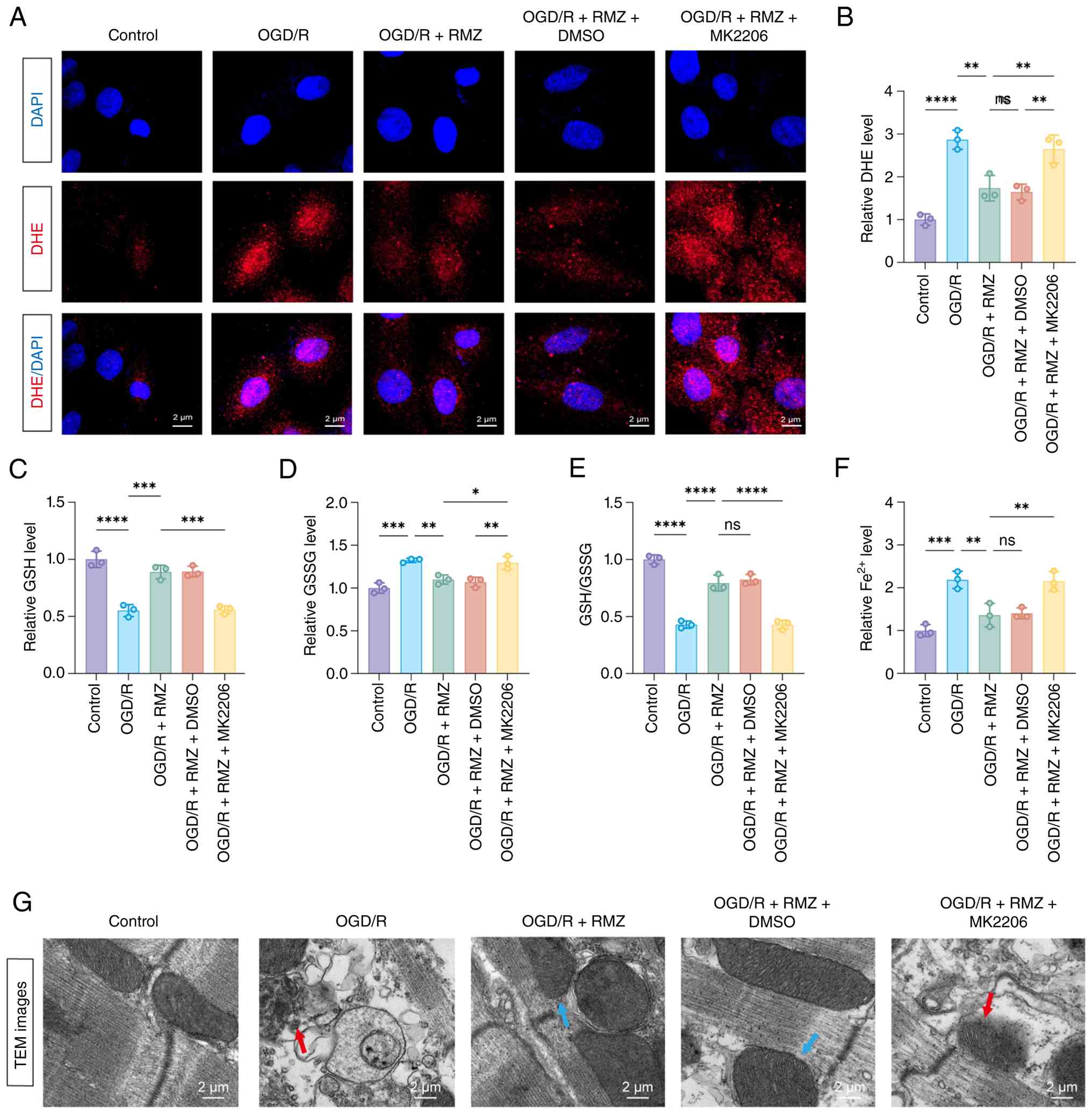 | Figure 6The protective effect of RMZ against
OGD/R-induced ferroptosis is mediated by p-AKT. (A) ROS
accumulation in H9C2 cells was quantified based on DHE staining.
(B) Quantitative analysis of DHE fluorescence intensity. Relative
levels of cellular (C) GSH, (D) GSSG, (E) GSH/GSSG and (F)
Fe2+ in cells. (G) Transmission electron microscopy of
cells from different groups. Red arrows indicate swollen
mitochondria with disrupted cristae and outer membrane dissolution.
Blue arrows indicate relatively preserved mitochondria with mild
cristae disruption. Data are presented as mean ± SEM, n=3
independent experiments per condition. Statistical significance was
determined as follows: *P<0.05,
**P<0.01, ***P<0.001 and
****P<0.0001. RMZ, Remimazolam; OGD/R, oxygen-glucose
deprivation/reoxygenation; p-, phosphorylated; ROS, reactive oxygen
species; DHE, dihydroethidium; GSH, glutathione; GSSG, oxidized
GSH; Fe2+, ferrous ion; GPX4; glutathione peroxidase 4;
TEM, transmission electron microscopy; SLC7A11, solute carrier
family 7 member 11. |
Consistent with the ROS data, examination of the
myocardial GSH antioxidant system revealed that OGD/R injury
decreased GSH levels, elevated GSSG levels and reduced the GSH/GSSG
ratio, alongside increased intracellular Fe2+ levels,
indicating enhanced oxidative stress and diminished antioxidant
capacity with disrupted iron homeostasis (Fig. 6C-G). RMZ treatment effectively
reversed these alterations, elevating GSH, reducing GSSG, restoring
the GSH/GSSG ratio and decreasing Fe2+ accumulation,
thus supporting antioxidant capacity and mitigating ferroptosis
(Fig. 6C-G). Notably, inhibiting
AKT phosphorylation counteracted these beneficial effects,
resulting in a lower GSH/GSSG ratio and elevated Fe2+
levels compared to the RMZ group (Fig. 6C-G). These findings suggested
that p-AKT is essential for RMZ-mediated protection against
OGD/R-induced ferroptosis in H9C2 cells.
The protective effect of RMZ against
OGD/R-induced ferroptosis involves the AKT/SLC7A11/GPX4 axis
To further elucidate the underlying mechanism, it is
proposed that RMZ confers cardioprotection by attenuating
ferroptosis, specifically through the upregulation of the
AKT/SLC7A11/GPX4 axis. To test this hypothesis, the expression of
key regulators involved in ferroptosis, including p-AKT, AKT,
SLC7A11, GPX4 and PCNA, was examined (Fig. 7). Results indicated that OGD/R
treatment markedly reduced the protein levels of p-AKT, SLC7A11,
GPX4 and PCNA, all of which were markedly restored by RMZ treatment
(Fig. 7A-H). Furthermore, MK2206
administration markedly counteracted the RMZ-mediated
anti-ferroptotic effect, inhibiting the elevation of p-AKT along
with the upregulation of SLC7A11 and GPX4 expression (Fig. 7A-H). These findings suggested
that SLC7A11 and GPX4 function downstream of p-AKT in the
anti-ferroptotic action of RMZ. Given that ferroptosis impairs DNA
repair capacity, PCNA expression, assessed as an indicator of
cellular resilience, was restored by RMZ in an AKT-dependent manner
in OGD/R-treated H9C2 cells. These findings suggested that RMZ
exerts anti-ferroptosis effects associated with the
AKT/SLC7A11/GPX4 axis (Fig.
8).
 | Figure 7RMZ alleviates OGD/R-induced
ferroptosis in H9C2 cells involves the AKT/SLC7A11/GPX4 axis. (A)
Western blotting image of the AKT/SLC7A11/GPX4 axis. (B) Flowchart
of the underlying pathway mechanism. Quantitative analysis of (C)
p-AKT, (D) AKT, (E) p-AKT/AKT, (F) SLC7A11, (G) GPX4 and (H) PCNA
protein expression levels in H9C2 cells. Data are presented as mean
± SEM, n=3 independent experiments per condition.
*P<0.05, **P<0.01,
***P<0.001 and ****P<0.0001. RMZ,
Remimazolam; OGD/R, oxygen-glucose deprivation/reoxygenation; AKT,
Protein Kinase B; SLC7A11, solute carrier family 7 member 11; GPX4;
glutathione peroxidase 4; p-, phosphorylated; PCNA, proliferating
cell nuclear antigen. |
Discussion
The present study provided compelling evidence that
RMZ confers cardioprotective effects against MIRI by targeting
ferroptosis, a regulated cell death pathway characterized by
iron-dependent lipid peroxidation. In vivo experiments
demonstrated that RMZ administration markedly improved I/R-induced
contractile dysfunction, as evidenced by enhanced left ventricular
EF and FS, while histological analysis confirmed reduced
fragmentation of myocardial fibers and infarct size. These findings
were corroborated by in vitro studies using H9C2 cells
subjected to OGD/R, where RMZ inhibited ferroptotic hallmarks,
including lipid peroxidation accumulation, Fe2+ overload
and ROS bursts. These results indicated RMZ as a novel ferroptosis
regulator with potential therapeutic utility in MIRI.
Mechanistically, the AKT/SLC7A11/GPX4 axis serves as
a mediator of RMZ's anti-ferroptotic effects. This evidence
originated from transcriptomic analysis, revealing significant
enrichment in PI3K-AKT signaling and ferroptosis-related pathways.
Subsequent validation of this pathway at the protein level
demonstrated that RMZ treatment reversing I/R-induced suppression
of this axis and RMZ upregulated p-AKT in both in vivo and
in vitro models, concomitant with increased expression of
SLC7A11 (a cystine-glutamate antiporter) and GPX4 (a selenoprotein
peroxidase that neutralizes lipid peroxides). Notably,
pharmacological inhibition of AKT phosphorylation abolished
RMZ-induced upregulation of SLC7A11 and GPX4, reduced GSH levels
and reversed Fe2+ accumulation. These findings indicated
that SLC7A11 and GPX4 operate downstream of p-AKT, linking survival
signaling to ferroptosis resistance.
Notably, the regulation of SLC7A11 and GPX4 by RMZ
exhibited a clear context dependence. Under sham conditions, RMZ
did not further elevate SLC7A11 or GPX4 protein levels beyond
baseline, despite increasing p-AKT. By contrast, during I/R injury
RMZ restored their expression toward sham levels in an
AKT-dependent manner. This context-dependent pattern aligns with
the established concept that AKT functions as a signaling hub,
where downstream outputs are shaped by cellular stress status
(30). Prior studies similarly
report that AKT activation can exert divergent effects on
ferroptosis-related targets depending on the presence or absence of
pathological stress (31,32).
While the present data did not directly define the molecular
mechanisms underlying this restoration, in other systems AKT has
been shown to promote ferroptosis resistance via downstream
pathways such as NRF2 activation or modulation of protein turnover
(31,32). However, whether these mechanisms
actually contribute to the restoration of SLC7A11 and GPX4 in the
I/R + RMZ group remains to be determined and direct experimental
testing is warranted in future studies. In addition to these
anti-ferroptotic effects, RMZ also restored PCNA expression in an
AKT-dependent manner in OGD/R-treated H9C2 cells. In post-mitotic
cardiomyocytes, PCNA upregulation does not signify proliferation
but rather reflects enhanced DNA repair capacity and cellular
resilience in response to stress. During I/R injury, oxidative DNA
damage accumulates and impaired DNA repair exacerbates
cardiomyocyte dysfunction and death (33). Thus, RMZ-induced restoration of
PCNA expression likely contributes to promoting recovery of injured
cardiomyocytes, which may synergize with the anti-ferroptotic
pathway to promote cardiomyocyte survival and functional recovery
following I/R injury.
The findings of the present study align with and
extend previous reports. Prior studies linked RMZ to AKT activation
in cerebral I/R (21) and acute
lung injury (34), suggesting
that AKT modulation may represent a common mechanism underlying
RMZ's protective effects across multiple organ systems. However,
the present study demonstrated that RMZ-induced AKT activation is
associated with the SLC7A11/GPX4 signaling to inhibit ferroptosis.
This ferroptosis mechanism adds a new dimension to the
understanding of RMZ's pharmacological actions.
The anti-ferroptotic properties of RMZ expand its
established pharmacological profile beyond sedative-hypnotic
effects. As an ultra-short-acting benzodiazepine, RMZ is widely
used in clinical practice for procedural sedation and general
anesthesia due to its rapid onset, esterase-dependent metabolism
and minimal hemodynamic impact (35-38). Emerging evidence suggests
pleiotropic organ-protective effects: in cardiac surgery, RMZ
combined with esketamine reduces perioperative myocardial injury
biomarkers (39,40), while in neurocritical care, it
improves neurological outcomes in patients with ischemic stroke
(22,41). Preclinical studies have linked
RMZ to the inhibition of oxidative stress (via AKT/GSK-3β/NRF2 in
cerebral I/R) (21) and
apoptosis (via FOXO1/3 in hepatic I/R) (20). The findings presented in the
present study add a new dimension to this protective repertoire,
demonstrating that RMZ modulates ferroptosis, a pathway not
previously associated with benzodiazepine pharmacology.
The role of ferroptosis in MIRI pathogenesis is
increasingly recognized, rendering the targeting of this pathway a
promising therapeutic strategy. Therapeutic options involving
anti-ferroptotic compounds for cardiovascular diseases include
deferoxamine, Fer-1 and liproxstatin-1 (Lip-1). Lip-1 and Fer-1 can
both reduce infarct size by preserving GPX4 activity and inhibiting
lipid peroxidation (42,43). Similarly, iron chelators such as
deferoxamine mitigate MIRI by limiting labile iron availability
(44,45). However, the main disadvantage of
Lip-1 and Fer-1 is their poor solubility in water, complicating
intravenous administration in acute pathologies such as AMI,
takotsubo syndrome and sepsis. Water-soluble ferroptosis inhibitors
similar to Lip-1 and Fer-1 are thus needed.
The present study presented a clinically promising
option to address these shortcomings, demonstrating that RMZ
reduces Fe2+ accumulation and lipid ROS production in
MIRI models. Intraperitoneal administration immediately before
reperfusion was selected to mimic the clinically relevant timing of
RMZ administration in patients undergoing primary PCI, cardiac
surgery, or procedural sedation in catheterization laboratories.
Its rapid onset (1-3 min) and favorable hemodynamic profile further
reinforce the clinical relevance of this administration regimen.
Notably, Li et al (46)
recently demonstrated that RMZ improves neurological function in a
murine model of intracerebral hemorrhage by suppressing
ferroptosis, suggesting that anti-ferroptotic effects may represent
a conserved mechanism of RMZ's organ protection across different
tissues. This consistency across systems enhances the translational
potential of these findings. For patients experiencing MI,
maintaining stable blood pressure is crucial. The primary advantage
of RMZ in patients with cardiovascular disease lies in its
excellent hemodynamic stability (47). Compared with propofol, a commonly
used clinical sedative, RMZ is associated with a markedly lower
risk of hypotension. Additionally, the incidence and severity of
respiratory depression caused by RMZ are considered lower than
those associated with propofol (47). While these characteristics
suggest potential roles for RMZ during the clinical timeline of MI
(pre-hospital or catheterization use), direct extrapolation is
speculative due to differences in administration route, timing and
patient populations between the experimental model and clinical
practice. Thus, this proposal serves as a working hypothesis that
requires validation in large-animal models and clinical trials,
with long-term outcomes needing further investigation.
Despite these advances, several limitations warrant
consideration. First, the upstream molecular target through which
RMZ activates AKT remains unidentified. While RMZ, as a
benzodiazepine, typically acts via GABA_A receptors, it remains
unclear whether this interaction plays a role in AKT
phosphorylation and requires further investigation. Unbiased
methodologies, such as chemical proteomics or affinity
purification, could help identify direct RMZ-binding proteins.
Second, the study focused on acute MIRI endpoints at 24 h
post-reperfusion. Future research should evaluate long-term
outcomes, including cardiac remodeling and survival, using
large-animal models (such as swine with coronary artery occlusion)
to more closely mimic clinical conditions. Third, the potential
involvement of additional pathways, such as Nrf2-mediated
antioxidant responses or AMPK-dependent mitochondrial protection,
cannot be excluded, as they may synergize with the AKT/SLC7A11/GPX4
axis to enhance RMZ's cardioprotective effects. Multi-omics
integration and CRISPR screening could aid in mapping the
comprehensive spectrum of RMZ's molecular targets in MIRI. Fourth,
although the RMZ concentrations and doses used in the present study
were based on existing literature, conducting a systematic
dose-response experiment would further substantiate its
concentration-dependent cardioprotective effects. Finally, the use
of H9C2 cells instead of primary cardiomyocytes limits
translational implications; validation in primary or hiPSC-derived
cells would enhance the relevance of the findings.
In conclusion, the present study is the first, to
the best of the authors' knowledge, to demonstrate that RMZ
attenuates MIRI by inhibiting ferroptosis, primarily involving the
activation of the AKT/SLC7A11/GPX4 axis. This discovery not only
deepens the understanding of the regulatory network governing
ferroptosis but also offers a novel clinical therapeutic option for
the combination therapy of MIRI.
Availability of data and materials
The data generated in the present study may be
requested from the corresponding author.
Authors' contributions
XY-C and XT-H carried out most of the in
vitro and in vivo experiments. YQ-Z, JW and CJ
contributed to experimental designs and revised the manuscript. JX,
YY, JH-Z and XY-C conceived the study, supervised the work and
revised the manuscript. All authors discussed the data and
commented on the manuscript. XY-C, JX and YY confirm the
authenticity of all the raw data. All authors read and approved the
final manuscript.
Ethics approval and consent to
participate
The present study was authorized by the Animal
Studies Ethics Committee of Hunan cancer hospital (approval no.
2025-410) and was implemented in accordance with the Guide for the
Care and Use of Laboratory Animals.
Patient consent for publication
Not applicable.
Competing interests
The authors declare that they have no competing
interests.
Acknowledgements
Not applicable.
Funding
The present study was supported by grants from the Hunan
Provincial Natural Science Foundation of China (grant nos.
2024JJ5243, 2025JJ81005 and 2026JJ82381), the Hunan Cancer Hospital
Climb Plan (grant no. 2023NSFC-A005) and the Wu Jieping Medical
Foundation (grant no. 320.6750.2025-5-54).
References
|
1
|
GBD 2016 Causes of Death Collaborators:
Global, regional, and national age-sex specific mortality for 264
causes of death, 1980-2016: A systematic analysis for the global
burden of disease study 2016. Lancet. 390:1151–1210. 2017.
View Article : Google Scholar : PubMed/NCBI
|
|
2
|
Heusch G: Myocardial ischaemia-reperfusion
injury and cardioprotection in perspective. Nat Rev Cardiol.
17:773–789. 2020. View Article : Google Scholar : PubMed/NCBI
|
|
3
|
Du B, Fu Q, Yang Q, Yang Y, Li R, Yang X,
Yang Q, Li S, Tian J and Liu H: Different types of cell death and
their interactions in myocardial ischemia-reperfusion injury. Cell
Death Discov. 11:872025. View Article : Google Scholar : PubMed/NCBI
|
|
4
|
Xiang Q, Yi X, Zhu XH, Wei X and Jiang DS:
Regulated cell death in myocardial ischemia-reperfusion injury.
Trends Endocrinol Metab. 35:219–234. 2024. View Article : Google Scholar
|
|
5
|
Dixon SJ, Lemberg KM, Lamprecht MR, Skouta
R, Zaitsev EM, Gleason CE, Patel DN, Bauer AJ, Cantley AM, Yang WS,
et al: Ferroptosis: An iron-dependent form of nonapoptotic cell
death. Cell. 149:1060–1072. 2012. View Article : Google Scholar : PubMed/NCBI
|
|
6
|
Tang D, Chen X, Kang R and Kroemer G:
Ferroptosis: Molecular mechanisms and health implications. Cell
Res. 31:107–125. 2021. View Article : Google Scholar :
|
|
7
|
Masaldan S, Bush AI, Devos D, Rolland AS
and Moreau C: Striking while the iron is hot: Iron metabolism and
ferroptosis in neurodegeneration. Free Radic Biol Med. 133:221–233.
2019. View Article : Google Scholar
|
|
8
|
Zhou Y, Lin W, Rao T, Zheng J, Zhang T,
Zhang M and Lin ZL: Ferroptosis and its potential role in the
nervous system diseases. J Inflamm Res. 15:1555–1574. 2022.
View Article : Google Scholar : PubMed/NCBI
|
|
9
|
Chen F, Kang R, Tang D and Liu J:
Ferroptosis: Principles and significance in health and disease. J
Hematol Oncol. 17:412024. View Article : Google Scholar : PubMed/NCBI
|
|
10
|
Wang Y, Wu S, Li Q, Sun H and Wang H:
Pharmacological inhibition of ferroptosis as a therapeutic target
for neurodegenerative diseases and strokes. Adv Sci (Weinh).
10:e23003252023. View Article : Google Scholar : PubMed/NCBI
|
|
11
|
Gao X, Hu W, Qian D, Bai X, He H, Li L and
Sun S: The mechanisms of ferroptosis under hypoxia. Cell Mol
Neurobiol. 43:3329–3341. 2023. View Article : Google Scholar : PubMed/NCBI
|
|
12
|
Kilpatrick GJ, Mcintyre MS, Cox RF,
Stafford JA, Pacofsky GJ, Lovell GG, Wiard RP, Feldman PL, Collins
H, Waszczak BL and Tilbrook GS: CNS 7056: A novel
ultra-short-acting benzodiazepine. Anesthesiology. 107:60–66. 2007.
View Article : Google Scholar : PubMed/NCBI
|
|
13
|
Rogers WK and McDowell TS: Remimazolam, a
short-acting GABA(A) receptor agonist for intravenous sedation
and/or anesthesia in day-case surgical and non-surgical procedures.
IDrugs. 13:929–937. 2010.PubMed/NCBI
|
|
14
|
Song X, Wang F, Dong R, Zhu K and Wang C:
Efficacy and safety of remimazolam tosilate combined with
esketamine for analgesic sedation in mechanically ventilated ICU
patients: A single-arm clinical study protocol. Front Med
(Lausanne). 9:8321052022. View Article : Google Scholar : PubMed/NCBI
|
|
15
|
Tanious MK, Beutler SS, Kaye AD and Urman
RD: New hypnotic drug development and pharmacologic considerations
for clinical anesthesia. Anesthesiol Clin. 35:e95–e113. 2017.
View Article : Google Scholar : PubMed/NCBI
|
|
16
|
Goudra B, Gouda G and Mohinder P: Recent
developments in drugs for GI endoscopy sedation. Dig Dis Sci.
65:2781–2788. 2020. View Article : Google Scholar : PubMed/NCBI
|
|
17
|
Sneyd JR: Remimazolam-current status,
opportunities and challenges. Anesthesiol Perioper Sci. 1:252023.
View Article : Google Scholar
|
|
18
|
Hansen TG and Engelhardt T: Remimazolam in
children: A comprehensive narrative review. Anesthesiol Perioper
Sci. 3:72025. View Article : Google Scholar
|
|
19
|
Liu T, Qin L, Su M, Wei Y, Yu H, Wei H,
Xie Y and Chen J: Remimazolam and propofol combination in ischemic
cerebrovascular disease endovascular treatment: A randomized study.
Sci Rep. 15:210832025. View Article : Google Scholar : PubMed/NCBI
|
|
20
|
Zhou B, Liu J, Jin L and Huang X:
Remimazolam alleviates hepatic ischemia-reperfusion injury by
activating FOXO1/3 signaling: Remimazolam alleviates hepatic
ischemia reperfusion injury. BMC Gastroenterol. 25:2832025.
View Article : Google Scholar
|
|
21
|
Duan M, Yu N, Liu J, Zhao Y, Zhang J, Song
S and Wang S: Remimazolam suppresses oxidative stress and apoptosis
in cerebral ischemiareperfusion injury by regulating
AKT/GSK-3β/NRF2 pathway. Drug Des Devel Ther. 19:111–128. 2025.
View Article : Google Scholar
|
|
22
|
Kim J, Lee S, Park B, Sim WS, Ahn HJ, Park
MH and Jeong JS: Effect of remimazolam versus propofol anesthesia
on postoperative delirium in neurovascular surgery: Study protocol
for a randomized controlled, non-inferiority trial. Perioper Med
(Lond). 13:562024. View Article : Google Scholar : PubMed/NCBI
|
|
23
|
Shi M, Chen J, Liu T, Dai W, Zhou Z, Chen
L and Xie Y: Protective effects of remimazolam on cerebral
ischemia/reperfusion injury in rats by inhibiting of NLRP3
inflammasome-dependent pyroptosis. Drug Des Devel Ther. 16:413–423.
2022. View Article : Google Scholar : PubMed/NCBI
|
|
24
|
Liu X, Shui G, Wang Y, Chen T, Zhang P,
Liu L, Li C, Li T and Wang X: Remimazolam alleviates myocardial
ischemia/reperfusion injury and inflammation via inhibition of the
NLRP3/IL-1β pathway in mice. Int J Mol Med. 55:572025. View Article : Google Scholar
|
|
25
|
Dong Y, Wu F, Liu K, Yue Y, Shen X, Qu Z,
Yu S and Du W: Kindlin-2/Otub1/Slc7a11 axis improved cardiac
ischemia reperfusion injury by inhibiting cardiomyocyte
ferroptosis. Antioxid Redox Signal. 43:727–744. 2025. View Article : Google Scholar : PubMed/NCBI
|
|
26
|
Osmak G, Baulina N, Koshkin P and Favorova
O: Collapsing the list of myocardial infarction-related
differentially expressed genes into a diagnostic signature. J
Transl Med. 18:2312020. View Article : Google Scholar : PubMed/NCBI
|
|
27
|
Sherman BT, Hao M, Qiu J, Jiao X, Baseler
MW, Lane HC, Imamichi T and Chang W: DAVID: A web server for
functional enrichment analysis and functional annotation of gene
lists (2021 update). Nucleic Acids Res. 50(W1): W216–W221. 2022.
View Article : Google Scholar : PubMed/NCBI
|
|
28
|
Zhang GY, Gao Y, Guo XY, Wang GH and Guo
CX: MiR-199a-5p promotes ferroptosis-induced cardiomyocyte death
responding to oxygen-glucose deprivation/reperfusion injury via
inhibiting Akt/eNOS signaling pathway. Kaohsiung J Med Sci.
38:1093–1102. 2022. View Article : Google Scholar : PubMed/NCBI
|
|
29
|
Liu X, Li D, Pi W, Wang B, Xu S, Yu L, Yao
L, Sun Z, Jiang J and Mi Y: LCZ696 protects against
doxorubicin-induced cardiotoxicity by inhibiting ferroptosis via
AKT/SIRT3/SOD2 signaling pathway activation. Int Immunopharmacol.
113:1093792022. View Article : Google Scholar : PubMed/NCBI
|
|
30
|
Toker A and Marmiroli S: Signaling
specificity in the Akt pathway in biology and disease. Adv Biol
Regul. 55:28–38. 2014. View Article : Google Scholar : PubMed/NCBI
|
|
31
|
Fu C, Wu Y, Liu S, Luo C, Lu Y, Liu M,
Wang L, Zhang Y and Liu X: Rehmannioside A improves cognitive
impairment and alleviates ferroptosis via activating PI3K/AKT/Nrf2
and SLC7A11/GPX4 signaling pathway after ischemia. J
Ethnopharmacol. 289:1150212022. View Article : Google Scholar : PubMed/NCBI
|
|
32
|
Gao N, Huang Z, Xie J, Gao S, Wang B, Feng
H, Bao C, Tian H and Liu X: Cryptotanshinone alleviates cerebral
ischemia reperfusion injury by regulating ferroptosis through the
PI3K/AKT/Nrf2 and SLC7A11/GPX4 signaling pathway. J Ethnopharmacol.
348:1198002025. View Article : Google Scholar : PubMed/NCBI
|
|
33
|
Strzalka W and Ziemienowicz A:
Proliferating cell nuclear antigen (PCNA): A key factor in DNA
replication and cell cycle regulation. Ann Bot. 107:1127–1140.
2011. View Article : Google Scholar
|
|
34
|
Li R, Zhang C, Ren J, Deng G, Gao Y, Gao
X, Li J, Zhang J, Xu X, Jin X, et al: Remimazolam inhibits
apoptosis of endothelial and epithelial cells by activating the
PI3K/AKT pathway in acute lung injury. Int Immunopharmacol.
147:1139492025. View Article : Google Scholar : PubMed/NCBI
|
|
35
|
Hu Q, Liu X, Wen C, Li D and Lei X:
Remimazolam: An updated review of a new sedative and anaesthetic.
Drug Des Devel Ther. 16:3957–3974. 2022. View Article : Google Scholar : PubMed/NCBI
|
|
36
|
Kim KM: Remimazolam: Pharmacological
characteristics and clinical applications in anesthesiology. Anesth
Pain Med (Seoul). 17:1–11. 2022. View Article : Google Scholar : PubMed/NCBI
|
|
37
|
Lee A and Shirley M: Remimazolam: A review
in procedural sedation. Drugs. 81:1193–1201. 2021. View Article : Google Scholar : PubMed/NCBI
|
|
38
|
Zhang H, Li H, Zhao S and Bao F:
Remimazolam in general anesthesia: A comprehensive review of its
applications and clinical efficacy. Drug Des Devel Ther.
18:3487–3498. 2024. View Article : Google Scholar : PubMed/NCBI
|
|
39
|
Xi L, Liu H, Tang X and Jiang Z: Effects
of esketamine combined with remimazolam tosylate on hemodynamics
during cardiovascular anesthesia. Clin Transl Sci. 18:e702322025.
View Article : Google Scholar : PubMed/NCBI
|
|
40
|
Lee JY, Kim HT, Kim YJ, Lee JS, Park JW
and Shin YD: Anesthetic management using remimazolam in a patient
with atrial flutter: A case report. Am J Transl Res. 14:5754–5759.
2022.PubMed/NCBI
|
|
41
|
Koo CH, Lee SU, Kim HG, Lee S, Bae YK, Oh
AY, Jeon YT and Ryu JH: Effect of remimazolam on intraoperative
hemodynamic stability in patients undergoing cerebrovascular bypass
surgery: A prospective randomized controlled trial. Korean J
Anesthesiol. 78:148–158. 2025. View Article : Google Scholar : PubMed/NCBI
|
|
42
|
Feng Y, Madungwe NB, Imam Aliagan AD,
Tombo N and Bopassa JC: Liproxstatin-1 protects the mouse
myocardium against ischemia/reperfusion injury by decreasing VDAC1
levels and restoring GPX4 levels. Biochem Biophys Res Commun.
520:606–611. 2019. View Article : Google Scholar : PubMed/NCBI
|
|
43
|
Ru Q, Li Y, Chen L, Wu Y, Min J and Wang
F: Iron homeostasis and ferroptosis in human diseases: Mechanisms
and therapeutic prospects. Signal Transduct Target Ther. 9:2712024.
View Article : Google Scholar : PubMed/NCBI
|
|
44
|
Yan HF, Zou T, Tuo QZ, Xu S, Li H, Belaidi
AA and Lei P: Ferroptosis: Mechanisms and links with diseases.
Signal Transduct Target Ther. 6:492021. View Article : Google Scholar : PubMed/NCBI
|
|
45
|
Zheng Y, Yan F, He S and Luo L: Targeting
ferroptosis in autoimmune diseases: Mechanisms and therapeutic
prospects. Autoimmun Rev. 23:1036402024. View Article : Google Scholar : PubMed/NCBI
|
|
46
|
Li X, Xing C, Tian M, Liu B, Tong J, Yu H,
Huang C, Su L, You X and Wu J: Remimazolam alleviates intracerebral
hemorrhage outcomes by suppressing neuronal ferroptosis. Eur J
Pharmacol. 1006:1782032025. View Article : Google Scholar : PubMed/NCBI
|
|
47
|
Lee JH: Remimazolam-expectations and
concerns. Korean J Anesthesiol. 78:89–90. 2025. View Article : Google Scholar : PubMed/NCBI
|

















