Gastric cancer (GC) remains a major global health
burden and one of the most lethal malignancies of the digestive
system (1). Recent
epidemiological data report nearly 1 million new cases and ~650,000
deaths annually, ranking GC among the leading causes of cancer
incidence and mortality, with a particularly high burden in East
Asia (2). Although screening and
early detection have reduced incidence rates in some regions, the
absolute number of cases continues to rise due to population aging
and growth, and projections estimate that global incidence will
increase by >60% by 2040, reaching ~1.77 million new cases,
highlighting a sustained and escalating public health challenge
(3). Etiologically,
Helicobacter pylori (H. pylori) infection is the most
well-established and modifiable risk factor, accounting for 85-90%
of non-cardia GC cases, while dietary patterns, smoking, metabolic
disturbances and genetic susceptibility further contribute to tumor
initiation and progression (4,5).
At the molecular level, GC represents a heterogeneous disease
characterized by diverse genomic alterations, tumor
microenvironmental features and evolutionary trajectories, which
drive variability in responses to chemotherapy, targeted therapy
and immunotherapy, as well as clinical outcomes (6). Clinically, the 5-year survival rate
for advanced or metastatic GC remains <10%, emphasizing the
urgent need to define its pathogenic networks and develop more
precise and effective therapeutic strategies (7).
This molecular heterogeneity is further reflected by
established genomic classifications of GC, including Epstein-Barr
virus (EBV)-positive, microsatellite instability (MSI), chromosomal
instability (CIN) and genomically stable (GS) subtypes, which
exhibit distinct biological characteristics and therapeutic
vulnerabilities (8-10). EBV-positive tumors are
characterized by immune-rich microenvironments and frequent
alterations in immune regulatory pathways, potentially influencing
inflammatory cell death responses and immunotherapy sensitivity.
MSI tumors harbor high mutational burdens and enhanced neoantigen
generation, which may reshape interactions between regulated cell
death (RCD) pathways and antitumor immunity. By contrast, CIN and
GS tumors are associated with genomic instability, altered
signaling networks and metabolic adaptations, which may contribute
to differential dependencies on apoptotic regulation, ferroptosis
susceptibility and stress adaptation mechanisms. Therefore,
understanding how molecular subtype-specific features influence
cell death regulation may provide a more precise framework for
interpreting therapeutic responses and identifying vulnerabilities
in GC.
Dysregulated cell death is a core mechanism that
drives tumor initiation, progression and variability in therapeutic
responses (11,12). Apoptosis has long served as the
principal effector pathway in anticancer therapy, and standard
chemotherapeutic agents for GC, including fluorouracil and platinum
compounds, act largely by inducing apoptotic cell death (13,14). However, growing insights into RCD
show that GC cells do not depend on suppression of a single pathway
for survival. Instead, they reprogram multiple death programs to
adapt to sustained proliferation, metabolic stress, redox imbalance
and chronic inflammation (15,16). In this context, 'cell death
reprogramming' refers to the dynamic remodeling of cellular death
susceptibility and execution patterns, rather than the simple
activation or inhibition of an individual death pathway. This
process involves alterations in death thresholds, transitions or
compensation between distinct cell death modalities, and adaptive
rewiring of death signaling networks in response to therapeutic
pressure and changes within the tumor microenvironment (TME).
Through this plasticity, cancer cells can evade lethal stress,
maintain survival advantages and reshape their responses to
anticancer interventions. This reprogramming alters both the
activity and threshold of canonical apoptotic signaling and
differentially regulates inflammatory cell death,
autophagy-dependent cell death, ferroptosis and cuproptosis across
distinct molecular contexts and disease stages (17). These death modalities interact
through shared signaling axes and organelle stress responses,
forming interconnected networks that enable functional compensation
and cross-regulation. As a result, they shape tumor growth,
invasive capacity, and sensitivity to chemotherapy, targeted
therapy and immunotherapy (18,19). In GC, dissecting these pathways
clarifies the molecular basis of resistance and recurrence and
supports patient stratification, response prediction and
therapeutic development (20,21). Accordingly, the present review
systematically reviews the molecular mechanisms and interaction
networks of cell death modalities in GC, integrating intracellular
signaling, metabolic reprogramming and TME regulation to refine the
framework of cell death reprogramming and guide multi-target
therapeutic strategies.
GC cell survival and death are not controlled by a
single pathway but are coordinated by multiple forms of RCD with
distinct molecular mechanisms. Across different genetic backgrounds
and disease stages, these RCD modalities display differential
activation patterns and marked heterogeneity in magnitude,
signaling intensity and biological effects. Together, they form an
integrated regulatory network that determines tumor cell fate.
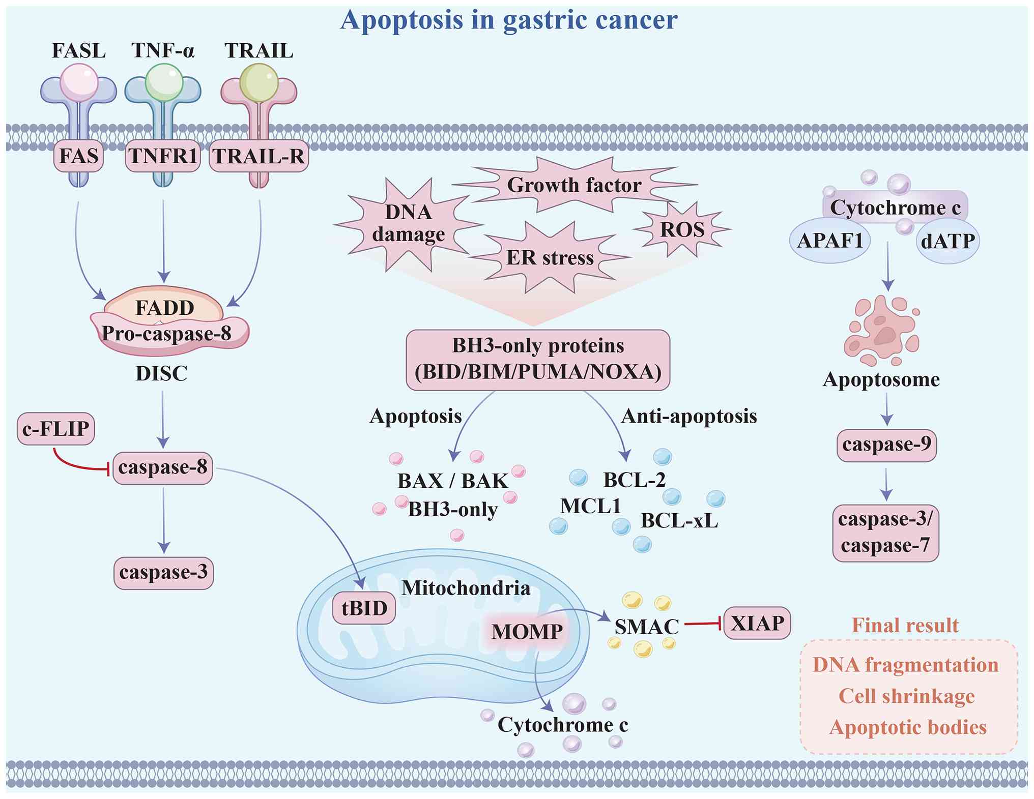
Apoptosis is a well-characterized form of RCD that
integrates extrinsic death receptor and intrinsic mitochondrial
pathways to convert stress signals into caspase activation and
irreversible execution (Fig. 1)
(22). In cancer, tumor cells
rarely lose apoptotic capacity; instead, they attenuate
therapy-induced cell death by blocking signaling at multiple
regulatory levels or by increasing the apoptotic threshold
(23). Therefore, defining the
functional status and plasticity of key apoptotic nodes is
essential for understanding tumor biology and developing precise
therapeutic strategies.
Apoptotic signaling integrates extrinsic death
receptor and intrinsic mitochondrial pathways, with caspase
cascades executing cell death and mitochondrial outer membrane
permeabilization (MOMP) serving as the commitment event that
determines cell fate (13). The
intrinsic pathway responds to diverse stresses, including DNA
damage, oxidative stress, endoplasmic reticulum stress, growth
factor deprivation and metabolic imbalance. These signals induce or
activate BH3-only proteins through transcriptional and
post-translational mechanisms and converge on the BCL-2 family
regulatory network (11).
Activator BH3 proteins, such as BH3-Interacting Domain Death
Agonist (BID), Bcl-2 interacting mediator of cell death (BIM), p53
upregulated modulator of apoptosis (PUMA) and NOXA, promote
conformational activation and oligomerization of BAX and BAK at the
mitochondrial outer membrane (24,25). Sensitizer BH3 proteins displace
anti-apoptotic family members and lower the apoptotic threshold. By
contrast, anti-apoptotic proteins, including BCL-2, BCL-xL and
myeloid cell leukemia 1 (MCL1), sequester BH3 proteins and BAX/BAK
to maintain a dynamic threshold for apoptosis (26,27). Once this control is overcome,
BAX/BAK form pores that release intermembrane proteins such as
cytochrome c and second mitochondria-derived activator of caspases
(SMAC). Cytochrome c assembles with apoptotic protease-activating
factor 1 (APAF1) and deoxyATP (ATP) into the apoptosome, which
activates caspase-9 and subsequently caspase-3 and caspase-7
(28). In parallel, SMAC
antagonizes inhibitor of apoptosis (IAP) family proteins such as
X-linked inhibitor of apoptosis protein (XIAP) and enhances caspase
activity and death signaling flux (29,30). The extrinsic pathway is initiated
by death receptors, including FAS, tumor necrosis factor receptor 1
(TNFR1) and TNF-related apoptosis-inducing ligand (TRAIL)
receptors. Ligand binding induces receptor trimerization and
recruitment of Fas-associated via death domain (FADD) and
pro-caspase-8 to form the death-inducing signaling complex, where
caspase-8 undergoes activation. Activated caspase-8 directly
cleaves executioner caspases or processes BID into tBID to engage
the mitochondrial pathway and amplify the signal (31-34). TNFR1 signaling exhibits
ubiquitin-dependent branching. Ubiquitinated receptor-interacting
serine/threonine-protein kinase 1 (RIPK1) primarily promotes NF-κB-
and MAPK-mediated survival signaling, whereas deubiquitinated RIPK1
associates with FADD and caspase-8 to form pro-apoptotic complexes
(35). Cellular FLICE-like
inhibitory protein (c-FLIP) isoforms further fine-tune caspase-8
activity through competitive regulation and thereby control the
strength of apoptotic execution (36).
As a context-dependent extension of apoptosis,
anoikis is triggered by loss of adhesion to the extracellular
matrix or neighboring cells and retains substantial overlap with
the canonical mitochondrial pathway (37). Loss of adhesion disrupts
integrin-mediated survival signaling and induces intracellular
stress reprogramming, thereby lowering the threshold for BCL-2
family-regulated MOMP and initiating caspase activation and
programmed cell clearance (38,39). Under physiological conditions,
this process removes displaced cells to maintain tissue
architecture and homeostasis, whereas during tumor progression it
acts as a critical barrier to metastasis. Tumor cells overcome this
barrier by attenuating apoptotic signaling, sustaining pro-survival
pathways or reprogramming metabolism and phenotype, thereby
acquiring anoikis resistance that enables anchorage-independent
survival and supports distant colonization (40). Overall, apoptosis functions as an
RCD program centered on mitochondrial commitment, integrates death
receptor-mediated inputs, and executes cell dismantling through
caspase cascades, with its activity controlled by coordinated,
multilayered networks including the BCL-2 family, IAP family and
epigenetic regulators; this plastic threshold-control system
provides a molecular basis for tumor cell survival advantage and
for the development of targeted therapeutic strategies.
In GC, apoptotic pathways are preserved but
functionally reprogrammed through threshold remodeling. Compared
with normal epithelial cells, GC cells show reduced apoptotic
sensitivity and limited access to the commitment point, especially
at the level of mitochondrial decision-making, resulting in
decreased apoptotic priming (41). At the input level, peripheral
myelin protein 22 suppresses p53 transcriptional activity and
downregulates pro-apoptotic genes such as BAX and
PUMA, thereby weakening mitochondrial signaling strength
(42). TRIM family E3 ubiquitin
ligase 17 further promotes BAX ubiquitination and degradation,
which reduces the probability of MOMP and attenuates
chemotherapy-induced apoptotic flux (43). Metabolic and redox buffering also
elevate the apoptotic threshold. Nicotinamide nucleotide
transhydrogenase sustains NADPH production and glutathione (GSH)
reduction capacity, limits reactive oxygen species (ROS)
accumulation and suppresses mitochondrial stress amplification,
thereby enhancing resistance to apoptosis and anoikis under
nutrient deprivation or detachment conditions (44). On the contrary, loss of tumor
suppressors further increases the activation threshold.
Downregulation of gastrokine-2 impairs NF-κB/JNK-mediated stress
signal routing and prevents efficient transmission of oxidative
stress into caspase-9/3 activation (45). Promoter methylation-mediated
silencing of heart- and neural crest derivatives-expressed protein
1 (HAND1) disrupts coupling of endoplasmic reticulum stress to the
CHOP-BAK axis, whereas restoration of HAND1 enhances
cisplatin-induced apoptosis (46). Metabolic regulation also
modulates apoptosis in GC by reshaping cellular energy metabolism,
redox homeostasis and mitochondrial function. UMP suppresses NR4A1
and limits its mitochondrial translocation, thereby reducing
cytochrome c release, whereas relieving this constraint or
combining with NR4A1 agonists enhances apoptosis in xenograft and
patient-derived xenograft (PDX) models (47). Beyond mitochondrial control,
extrinsic signaling is also tightly regulated. O-GlcNAc
modification of DR4 is required for TRAIL-DISC assembly and
caspase-8 recruitment, and its disruption leads to receptor-level
signaling defects and TRAIL resistance (48). 3-hydroxybutyrate dehydrogenase
type 2 (BDH2) downregulation sustains Nrf2-dependent antioxidant
transcription and Akt/mTOR survival signaling, which limits ROS
accumulation and attenuates caspase-3 activation (49). By contrast, stromal interaction
molecule 1-mediated store-operated calcium entry increases
cytosolic Ca2+ levels and activates endoplasmic
reticulum stress pathways, thereby upregulating BAX and cleaved
caspase-3 to enhance apoptotic execution (50). At the execution stage, GC cells
further dampen apoptosis by reducing caspase activation amplitude
and substrate cleavage efficiency, leading to suboptimal or
reversible activation states. In a gp130F/F intestinal-type GC
model, the ASC-IL18 axis suppresses caspase-8-dependent apoptosis
within tumor epithelium, whereas loss of ASC or IL18 enhances TUNEL
positivity and cleaved caspase-8 while reducing tumor burden
(51). During apoptosome
assembly, long non-coding RNA (lncRNA) ABL competitively binds
APAF1 and blocks its interaction with cytochrome c, thereby
inhibiting caspase-9/3 activation and promoting chemoresistance
(52). At the
post-transcriptional level, N6-methyladenosine (m6A)-modified
proteasome 20S subunit α 3 antisense RNA 1 and mir22HG suppress
caspase-related outputs through competing endogenous RNA (ceRNA)
and protein interaction networks while maintaining stem-like
phenotypes (53).
Caspase-3-mediated cleavage of CAD at Asp1371 is required for
chemotherapy-induced apoptosis, and disruption of this process or
CAD overexpression reduces cell death and promotes resistance
(54,55). In CIN and GS GCs, where genomic
instability and survival pathway activation are prominent features,
alterations in apoptotic thresholds may represent important
mechanisms underlying therapeutic resistance (56). Overall, GC attenuates signal
strength and transmission efficiency across key nodes, including
upstream inputs, mitochondrial commitment and execution cascades,
thereby preventing apoptotic stimuli from exceeding the clearance
threshold and establishing a low-sensitivity state associated with
therapeutic resistance.
Apoptosis is a central execution endpoint for tumor
clearance in GC during chemotherapy and targeted therapy. The
efficacy of platinum agents, including cisplatin and oxaliplatin,
depends on activation of the mitochondrial apoptotic pathway, and
modulation of metabolic or redox states can further amplify
apoptotic flux without increasing toxicity. Metformin enhances
oxaliplatin-induced tumor cell clearance by regulating the
BCL-2/BAX balance and activating caspase-3 (57). In neoadjuvant chemotherapy
cohorts, cellular retinoic acid-binding protein 2 mediates
oxaliplatin resistance by promoting BAX ubiquitination and
degradation, thereby suppressing mitochondrial apoptosis; its
inhibition restores apoptosis and reverses resistance in cell
line-derived xenograft and PDX models (58). Natural compounds also enhance
apoptotic responses. WZ35 inhibits thioredoxin reductase 1, induces
ROS accumulation and activates p38/JNK signaling to potentiate
cisplatin-induced apoptosis (59). Isorhamnetin suppresses NF-κB
signaling and downregulates anti-apoptotic proteins such as
survivin to amplify capecitabine-induced apoptosis (60). Components of Tripterygium
wilfordii, a traditional Chinese medicinal herb, induce
mitochondrial apoptosis by inhibiting peroxiredoxin 2 (PRDX2), and
high PRDX2 expression is associated with poor prognosis; related
clinical studies are ongoing (61-63). Organoid-PDX models show that
Ailanthone disrupts the p23/HSP90 axis, destabilizes x-ray repair
cross-complementing protein 1 and impairs base excision repair,
allowing DNA damage to exceed the apoptotic threshold (64). Direct targeting of anti-apoptotic
pathways has entered clinical evaluation. The pan-BCL-2 inhibitor
AT-101 combined with chemoradiotherapy shows favorable response
rates (65). In tumors with high
EGFR copy number, pyrotinib activates endoplasmic reticulum
stress-related apoptosis through the EGFR-GRP78 axis and suppresses
DNA repair, thereby enhancing chemosensitivity and enabling
stratified therapy (66).
Anti-programmed cell death protein 1 (PD-1) immunotherapy also
provides survival benefit in a subset of patients, partly through T
cell-mediated induction of tumor cell apoptosis via death receptor
and granzyme pathways (67).
Increasing tumor susceptibility to apoptosis while limiting
apoptotic stress in effector T cells may therefore improve the
durability of therapeutic responses (68).
Beyond therapy, apoptosis-related molecules also
contribute to diagnosis and prognostic assessment. Gastrokine-1
expression decreases during gastric carcinogenesis, and its
restoration enhances 5-FU-induced apoptosis (69). Apogossypolone enables real-time
in vivo monitoring of apoptosis through bioluminescence
imaging, supporting its use as a quantifiable pharmacodynamic
readout (70). Dual-modal
imaging combined with ultrasound-guided delivery further enhances
caspase- and PARP-associated apoptosis while enabling integrated
imaging and histological validation (71,72). Several apoptosis regulators are
associated with patient outcomes. Caspase-3 is an independent
prognostic factor (73);
hypermethylation of calcium-activated potassium channel, subfamily
M subunit α-1 predicts poor survival (74); and high insulin receptor tyrosine
kinase substrate expression associates with unfavorable prognosis
(75). Additional regulators,
including serine protease 23, gastrokine-2, estrogen-related
receptor α, and non-coding RNAs (ncRNAs), such as miR-216 and
circ-DONSON, modulate the survival-death balance and are associated
with clinical features such as TNM stage (45,76-78). Taken together, apoptosis-related
molecules link mechanistic insights with clinical application and
support the development of precision and personalized therapies in
GC.
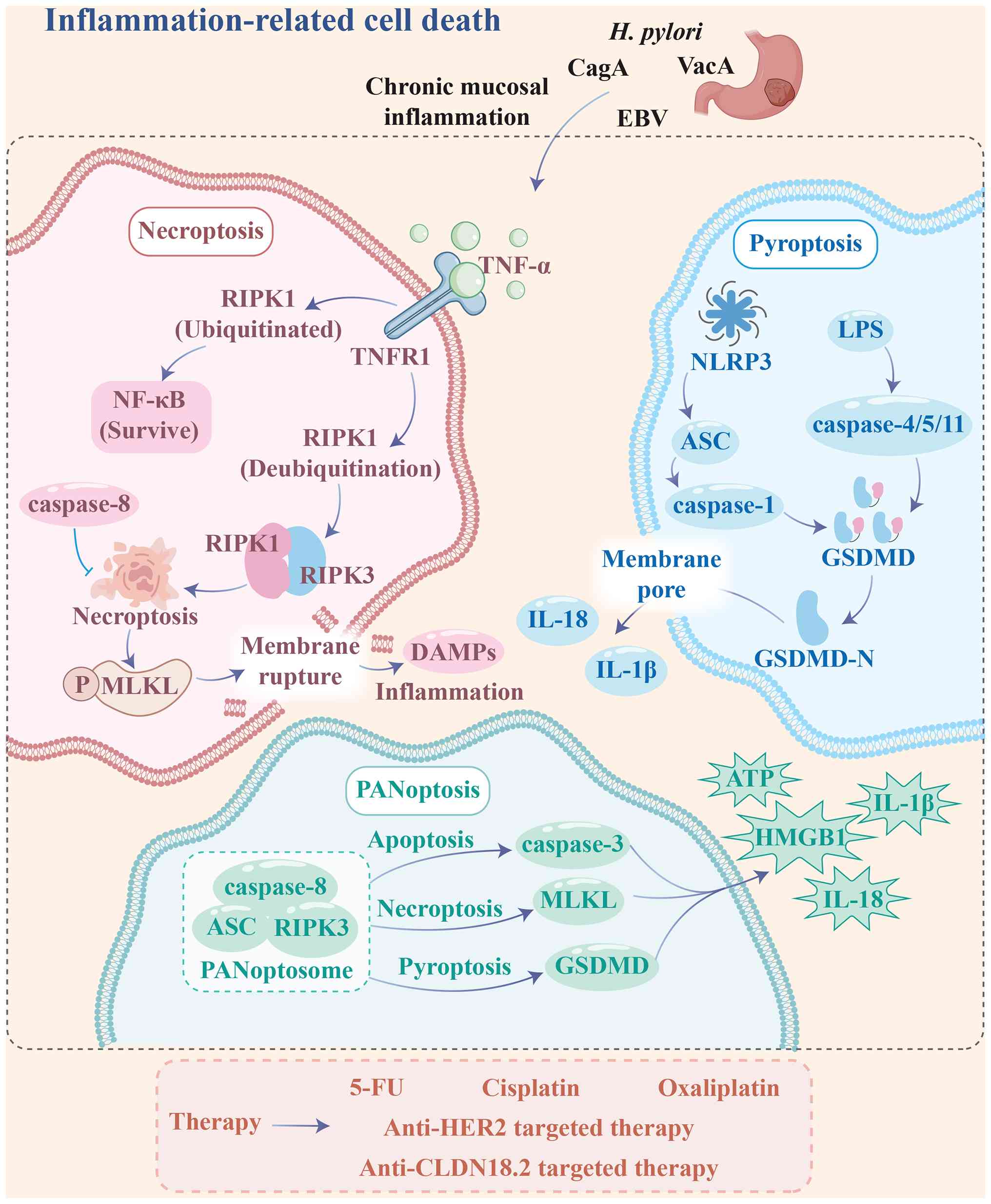
Unlike canonical apoptosis, inflammation-related
cell death features loss of plasma membrane integrity, release of
inflammatory mediators, and sustained activation of innate immune
signaling. These processes reshape the TME and influence immune
surveillance, therapeutic response and disease progression
(79). In GC, these cell death
modalities show marked context dependence and bidirectional
regulation. They can promote tumor progression through chronic
inflammation or enhance antitumor immunity by amplifying immune
activation. This dual role underscores their mechanistic importance
and translational potential (Fig.
2).
Necroptosis is a regulated form of necrotic cell
death characterized by mixed lineage kinase domain-like protein
(MLKL)-mediated membrane permeabilization and typically occurs when
death receptor or innate immune signaling is activated but
caspase-8-dependent apoptosis is limited. In the TNFα-TNFR1
pathway, the ubiquitination status of RIPK1 determines signaling
output. Ubiquitinated RIPK1 promotes NF-κB- and MAPK-driven
survival signaling. By contrast, deubiquitinated RIPK1 with kinase
activity engages RIPK3 through RIP homotypic interaction motif
(RHIM) domains to form the necrosome, which activates RIPK3 and
drives MLKL phosphorylation, oligomerization and membrane
translocation, leading to membrane rupture and damage-associated
molecular patterns (DAMPs) release that amplify inflammatory and
immune responses (80,81). RHIM-mediated amyloid-like
assembly stabilizes this platform and enhances signal propagation
(82). Moreover, caspase-8 and
c-FLIP function as a checkpoint that regulates the balance between
apoptosis and necroptosis. During execution, ion flux and cellular
content release further activate inflammasomes and PANoptosis
networks, linking membrane disruption to amplified inflammatory
cascades (83). Consequently,
necroptosis exerts context-dependent effects in tumors. It can
enhance antitumor immunity through DAMP-driven antigen presentation
and CD8+ T cell activation. It can also promote tumor
progression by sustaining inflammatory signaling, recruiting
immunosuppressive myeloid cells and supporting metastatic niche
formation (84-86). In GC, necroptosis is regulated by
threshold mechanisms and can be reactivated under therapeutic or
oxidative stress. HUHS1015 induces RIPK1-dependent
membrane-disruptive cell death, whereas chelerythrine promotes ROS
accumulation and shifts cell fate toward necroptosis (87,88). Endogenous regulators further
modulate this pathway. miR-204-3p suppresses RIPK1 and MLKL
activation while enhancing mitochondrial apoptosis, whereas
Clofoctol induces TNF-dependent necroptosis in stem-like cells,
indicating a targetable vulnerability within this axis (89,90). Spatial and single-cell
transcriptomic analyses indicate that necroptosis-associated DAMP
signaling contributes to metastatic niche remodeling and
immunosuppressive microenvironment formation, suggesting that
necroptosis functions as a dynamic regulator of tumor-immune
interactions rather than a simple cell death program (91). Overall, necroptosis in GC is a
RIPK1-RIPK3-MLKL-driven membrane-disruptive process regulated by
caspase-8 checkpoints and redox and proteostasis stress, providing
both an alternative route to overcome apoptosis resistance and a
modulatable inflammatory output that shapes immune responses and
therapeutic sensitivity (81,92).
The clinical relevance of necroptosis in cancer
stems from its pathway activity and inflammatory output, which
reshape the TME and influence tumor classification, prognosis and
therapeutic response. In hepatocellular carcinoma models,
activation of the RIPK3-MLKL axis associates with specific cytokine
profiles and drives transition toward a cholangiocarcinoma-like
phenotype. By contrast, inhibition of RIPK1 or MLKL alters tumor
progression and histological features, indicating that
necroptosis-related signaling can serve as a molecular readout for
tumor stratification and risk assessment (93). In immunotherapy settings,
induction of RIPK3-dependent necroptosis promotes mitochondrial DNA
release and activates the cGAS-STING pathway, which enhances type I
interferon production and dendritic cell cross-presentation. This
response amplifies the efficacy of radiotherapy or cisplatin
combined with anti-PD-1 therapy and associates with favorable
immune infiltration and treatment outcomes (94). Consistently, pharmacological
activation of RIPK1 and MLKL enhances intratumoral CD8+
T cell function and increases sensitivity to immune checkpoint
blockade (95). However,
necroptosis-associated inflammation can also drive tumor
progression in certain contexts. In pancreatic cancer,
MLKL-associated programs associate with increased liver metastasis
and promote pro-metastatic niche formation through myeloid cell
recruitment and immunosuppressive signaling (96). In GC, necroptosis-related gene
signatures are incorporated into prognostic and therapeutic
stratification models. Analyses of The Cancer Genome Atlas and Gene
Expression Omnibus datasets show that high-risk groups have poorer
overall survival and retain independent predictive value in
multivariate models. These groups also display distinct patterns of
immune infiltration, immune checkpoint expression, tumor mutational
burden (TMB) and TIDE scores, linking necroptosis-associated
transcriptional programs to immune exclusion or dysfunction and
potential immunotherapy response (97,98). A validated six-gene model further
shows that high-risk tumors associate with advanced stage, higher
grade, and immunosuppressive features, whereas low-risk tumors
exhibit increased CD8+ T cell and Th1 activity and
greater drug sensitivity. Single-cell communication analyses
indicate that necroptosis-related signaling is activated through
TNF pathways in myeloid cells and couples with glycolytic
reprogramming, linking inflammation, metabolism, and prognosis
(99). Incorporation of
necroptosis into the broader PANoptosis framework further connects
prognosis, immune contexture and chemotherapy sensitivity,
supporting therapy selection based on cell death network states
(100). In conclusion,
necroptosis-related pathways act as indicators of
inflammation-immunity-metabolism coupling and as targets for
immunotherapy sensitization and combination strategies, with
clinical utility dependent on tumor subtype and microenvironmental
context in GC.
Pyroptosis is a lytic form of RCD driven by
Gasdermin (GSDM)-mediated pore formation in the plasma membrane. In
the canonical pathway, pattern recognition receptors such as NLRP3
and AIM2 sense pathogen-associated molecular patterns or DAMPs and
assemble inflammasomes that activate caspase-1 and cleave GSDMD,
leading to cell swelling, membrane rupture, and release of
inflammatory mediators such as IL-1β and IL-18, whereas in the
non-canonical pathway, cytosolic lipopolysaccharide (LPS) directly
activates caspase-4, caspase-5 or caspase-11 and similarly
converges on GSDMD cleavage (101,102). In tumors, pyroptosis arises
through additional mechanisms, as caspase-3 or caspase-8 cleave
GSDMC, and cytotoxic lymphocytes release granzymes that target
GSDMB or GSDME, thereby linking immune cytotoxicity to pyroptotic
execution (103-106). Pyroptotic activity is further
regulated by GSDM stability and cellular stress states, with USP48
stabilizing GSDME through deubiquitination to increase pyroptotic
sensitivity, whereas the IRE1α-RIDD axis limits
chemotherapy-induced NLRP3-GSDMD pyroptosis by degrading
double-stranded RNA (107,108). In GC, pyroptosis shows marked
context dependence, acting as an antitumor effector under
therapeutic stress, where agents such as 5-FU, oxaliplatin and
cisplatin activate caspase-3 and cleave GSDME, shifting cell death
from apoptosis to a more immunogenic pyroptotic form accompanied by
membrane blebbing and lactate dehydrogenase release (109-111), while ROS accumulation and NF-κB
activation further promote NLRP3 inflammasome assembly and
caspase-1 activation to enhance GSDMD- or GSDME-mediated pyroptosis
(112,113). Conversely, GC cells suppress
pyroptosis through multilayered regulation, including GSDME
promoter hypermethylation, ALKBH4-mediated reduction of H3K4me3
enrichment and EBV-associated epigenetic silencing that limit GSDME
expression, as well as METTL3-PFKFB3-driven metabolic reprogramming
and HIC1 downregulation that inhibit the NLRP3-GSDMD pathway or
reduce GSDMD transcription (114-117). Notably, under chronic
inflammatory conditions, persistent H. pylori infection
activates inflammasome and GSDMD signaling through virulence
factors such as VacA and CagA, contributing to sustained mucosal
inflammation and tumor initiation (118,119). Collectively, GC cells
dynamically regulate pyroptotic output by modulating GSDM
expression, inflammasome activation thresholds and metabolic
states, enabling pyroptosis to function either as an effector of
therapy-induced tumor killing or as a driver of tumor-promoting
inflammation in chronic inflammatory settings.
The therapeutic value of pyroptosis in cancer lies
in its dual role as a quantifiable stratification marker and a
reactivatable effector pathway. Pan-cancer studies show that
GSDM-dependent pyroptosis influences both chemotherapy sensitivity
and the conversion of treatment-induced inflammatory signals into
effective immune recruitment and tumor clearance. In several tumor
models, low GSDME expression or suppression of the
NLRP3-caspase-1-GSDMD axis associates with poor therapeutic
response, whereas restoration of pyroptotic execution enhances
cytotoxicity and antitumor immunity, supporting its potential as
both a companion biomarker and a combinatorial therapeutic target
(120,121). In GC, pyroptosis contributes to
clinical management in three main ways. First, pyroptosis-related
signatures derived from genes, ncRNAs, or PANoptosis features
associate with overall survival, immune infiltration, TMB, MSI and
immune checkpoint expression, supporting their potential utility
for risk stratification and prediction of immunotherapy response,
including in neoadjuvant settings (122-124). Second, pyroptosis provides an
entry point for chemosensitization and reversal of drug resistance,
as agents such as BIX-01294 or lens protein with glutamine
synthetase domain inhibition can activate GSDME- or
NLRP3-GSDMD-dependent pathways to enhance responses to cisplatin,
5-FU and oxaliplatin, suggesting that redirecting resistant or
stem-like tumor cells toward the pyroptotic threshold may reduce
relapse (125,126). Third, pyroptosis-related
molecules themselves show therapeutic relevance, with elevated
caspase-1, IL-1β and IL-18 reflecting activation of the pyroptotic
inflammatory axis and GSDMD linking tumor growth, immune contexture
and treatment sensitivity (127,128). However, pyroptosis is not
uniformly beneficial, as its inflammatory output may also sustain
chronic inflammation, promote stromal remodeling, and drive
immunosuppressive microenvironments (129). In summary, pyroptosis in GC is
transitioning from a mechanistic concept to a clinically relevant
framework for stratification, response prediction and combination
therapy design, although further prospective validation and
standardized assessment remain necessary.
PANoptosis is an integrated cell death program
coordinated by shared signaling nodes and defined by the
coordinated activation of pyroptosis, apoptosis and necroptosis
within common sensing and execution platforms, accompanied by
robust inflammatory and immunogenic outputs (130-132). Pan-cancer studies support this
framework, as sterile α motif and HD domain-containing protein 1
deficiency in diffuse large B-cell lymphoma induces cytosolic
double stranded (ds)DNA accumulation and activates STING, promoting
formation of CASP8-, RIPK3- and ASC-containing complexes and
triggering GSDME cleavage, caspase-3 activation and MLKL
phosphorylation (133), while
deoxyribonuclease 1L3 in hepatocellular carcinoma enhances dsDNA
accumulation and AIM2-dependent PANoptosome assembly, shifting
sorafenib-induced cell death toward stronger PANoptotic output with
enhanced antitumor immunity (134). These findings position
PANoptosis as a convergence point linking multiple death pathways
with therapeutic sensitization and immune remodeling. Direct
evidence in GC remains limited but is supported by functional
studies, as Jolkinolide B induces PANoptotic death through
caspase-8 activation with concurrent increases in cleaved
caspase-3/7, GSDMD-N, and phosphorylated RIPK1 and MLKL, and this
effect is partially reversed by caspase-8 inhibition, indicating a
central integrative role (135); similarly, a nanoparticle-based
photodynamic system (CJP-TiN) amplifies PANoptosis via ROS
accumulation and caspase-8 activation, enhances cleaved
caspase-3/7/8, GSDME-N and p-MLKL signaling, and promotes release
of ATP, HMGB1, IL-1β and IL-18, demonstrating coordinated
activation of multiple death pathways and immunostimulatory outputs
(136). By contrast, GC cells
suppress PANoptosis to sustain survival and drug resistance, as
Y-box binding protein 1 (YBX1) is upregulated in
oxaliplatin-resistant cells and restricts activation of apoptosis,
necroptosis, and pyroptosis through a PPM1B-YBX1-USP10 regulatory
axis that modulates platinum sensitivity (137), while high heat shock protein
β-1 expression suppresses RIPK3, cleaved caspase-1, NLRP3 and
cleaved caspase-3, and its silencing reduces proliferation,
invasion and tumorigenicity, supporting a tumor-suppressive role
for PANoptosis (138).
Clinically, PANoptosis in GC is mainly characterized through
transcriptomic stratification and risk modeling, where high
PANoptosis activity associates with stronger immune signaling,
higher tumor mutational burden or MSI-H status, increased effector
immune cell infiltration, and improved survival, whereas low
activity is associated with epithelial-mesenchymal transition,
extracellular matrix remodeling, TGF-β signaling, enrichment of
cancer-associated fibroblasts (CAFs) and M2 macrophages, and immune
evasion phenotypes (100,139), and PANoptosis-based risk scores
further show prognostic value and may predict responses to immune
checkpoint inhibitors, chemotherapy, and targeted therapies
(140-143). In conclusion, PANoptosis in GC
functions as an integrative framework linking cell death programs,
immune heterogeneity, and therapeutic response, although its
regulatory mechanisms and translational potential require further
systematic investigation.
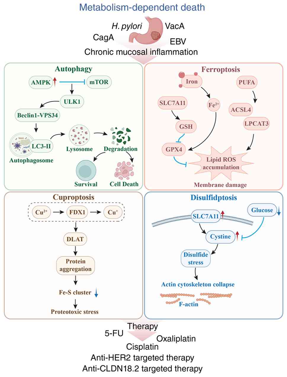
Tumor cells continuously reprogram metabolic
networks to sustain rapid proliferation and adapt to a complex
microenvironment. This reprogramming, centered on lipid, amino acid
and energy metabolism, supports growth and sets the threshold for
tolerating metabolic stress. Cell death driven by metabolic
imbalance has therefore emerged as a key process. It is triggered
by disruptions in redox homeostasis, abnormal metal ion
accumulation or loss of proteostasis. GC cells can delay or evade
this process through adaptive metabolic rewiring. When these
compensatory mechanisms fail, metabolic stress converts into lethal
signaling. Metabolism-dependent cell death thus reflects tumor
metabolic plasticity and provides an opportunity to overcome
therapeutic resistance and improve treatment efficacy (Fig. 3).
Autophagy is a multi-step degradative process
triggered by nutrient deprivation, oxidative stress, endoplasmic
reticulum stress, infection or energy imbalance and regulated by
AMPK-unc-51-like autophagy-activating kinase (ULK)1 and
PI3K-Akt-mTOR signaling. It proceeds through phagophore nucleation
via the Beclin1-VPS34 complex, autophagosome formation mediated by
the ATG5-ATG12-ATG16L system and LC3 lipidation and substrate
degradation after fusion with lysosomes (144,145). In GC, autophagy primarily
functions as a context-dependent adaptive process rather than an
autonomous cell death program, exhibiting either cytoprotective or
tumor-suppressive effects depending on disease stage and cellular
context. During early H. pylori-associated lesions, it acts
as a protective mechanism that limits oncoprotein accumulation,
buffers oxidative stress and maintains genomic stability. For
example, VacA activates ROS-dependent Akt-MDM2-p53 signaling to
induce autophagy and promote CagA degradation in normal gastric
epithelial cells, whereas elevated xCT-mediated GSH in
CD44v9-positive stem-like cells suppresses autophagy and permits
CagA accumulation (146).
Moreover, H. pylori induced nuclear translocation of
low-density lipoprotein receptor-related protein 1-isocitrate
dehydrogenase (IDH) promotes lysosomal-associated membrane protein
1 transcription and autophagolysosome formation, while capping
actin protein of muscle z-line subunit α 1 upregulation disrupts
this process and facilitates CagA persistence (147). Sustained infection further
impairs autophagy, leading to p62 accumulation and Rad51
degradation, which compromises DNA repair and promotes
tumorigenesis (148). In
established GC, autophagy primarily supports adaptive survival. E2
transcription factor 4 activates ATG2A and ULK2 to promote growth
and invasion, SEC23A is induced by STAT3 under endoplasmic
reticulum stress and enhances autophagy via annexin A2 membrane
translocation to buffer stress and reduce 5-FU-induced apoptosis,
c-Jun upregulates protein phosphatase 1 regulatory subunit 15A
under glucose deprivation to sustain autophagosome formation, and
latent membrane protein 2A activates the
PI3K/Akt-NRF1-CXCR4-ZEB1-ATG7 axis to maintain autophagy and viral
persistence (149-152). However, under specific
experimental conditions, sustained or excessive autophagy has been
reported to suppress tumor progression and may contribute to cell
death-associated phenotypes, although direct evidence for
autophagy-dependent cell death remains limited in GC.
Death-associated protein kinase 3 phosphorylates ULK1 at Ser556 to
promote autophagy initiation and suppress proliferation and
metastasis, BDH2 induces ROS accumulation and inhibits Akt/mTOR
signaling to trigger autophagy with enhanced apoptosis, and glucose
deprivation-induced autophagy promotes sequestosome-1/LC3-dependent
degradation of N-acetyltransferase 10 to suppress hexokinase
2-driven glycolysis and tumor growth (49,153,154). Dysregulation in GC often occurs
at the level of autophagic flux rather than initiation. VacA
impairs lysosomal function by inhibiting transient receptor
potential mucolipin 1 and blocks autophagosome maturation and
substrate clearance, whereas polypyrimidine tract binding protein 1
deficiency increases thioredoxin-interacting protein and ROS
levels, disrupts lysosomal integrity and prevents
autophagosome-lysosome fusion, autophagosome accumulation,
lysosomal dysfunction and subsequent cell death, although the death
phenotype may not be exclusively mediated by autophagy itself
(155,156). Overall, autophagy in GC is
therefore a dynamic and context-dependent process, with biological
effects determined by disease stage and cellular state. According
to current consensus, autophagy should not be considered synonymous
with autophagy-dependent cell death, unless cell death is
demonstrated to rely directly on the autophagic machinery.
The clinical relevance of autophagy in GC depends on
its function across disease stages and molecular contexts. During
persistent H. pylori infection and premalignant lesions,
autophagy acts as a protective mechanism that limits oncoprotein
accumulation, maintains genomic stability and modulates
inflammation, thereby supporting risk stratification and early
intervention. IFN-γ induces Beclin-1-dependent autophagy to
suppress inflammation-driven tumorigenesis and reduce abnormal
epithelial proliferation and progenitor expansion (157,158). In established GC, autophagy
primarily supports adaptation to metabolic and therapeutic stress
and influences responses to chemotherapy and targeted therapy.
Numerous agents induce protective autophagy that attenuates cell
death signaling, whereas inhibition of autophagy enhances treatment
efficacy. For example, quercetin induces HIF-1α-associated
autophagy, and chloroquine treatment or knockdown of ATG5 or
Beclin-1 increases apoptosis (159). In HER2-positive GC, GSDMB
promotes Rab7-dependent autophagosome maturation and contributes to
anti-HER2 resistance, indicating that autophagy-related markers may
identify patients who benefit from combination therapy (160). Under specific experimental
conditions, excessive or sustained autophagy has been associated
with tumor cell death or enhanced therapeutic efficacy. However,
because definitive evidence for autophagy-dependent cell death
remains limited, these observations should be interpreted
cautiously as autophagy-associated cell death processes rather than
autonomous execution by autophagy. The compound w09 enhances
autophagic flux and triggers caspase-dependent apoptosis, and
8-paradol activates PINK1/Parkin-dependent mitophagy to induce
mitochondrial damage and apoptosis, supporting modulation of
mitophagy as a potential therapeutic strategy (161,162). Clinically, autophagy-related
markers and signatures are used for prognostic stratification.
Immunophenotypes defined by LC3A/B, Beclin-1 and AMBRA-1 associate
with tumor grade and survival, and gene-based risk models
consistently stratify patients by prognosis and immune features,
including immune infiltration, checkpoint expression and treatment
response (163-167). Autophagy also regulates
immunotherapy response. Its inhibition increases programmed
death-ligand 1 (PD-L1) expression through the p62/NF-κB axis and
alters sensitivity to immune checkpoint blockade (168). Autophagy-targeted delivery and
combination strategies, including nanomedicine approaches, are
under development to enhance chemosensitivity and drug
accumulation, although most remain preclinical (169-171). Taken together, autophagy
primarily functions as a context-dependent regulator of tumor
adaptation and therapeutic response in GC, while its direct role as
an execution mechanism of cell death requires further experimental
validation.
Ferroptosis in GC is a regulated oxidative
stress-driven process determined by the balance between lipid
peroxidation and antioxidant capacity and occurs when lipid
peroxide accumulation exceeds cellular detoxification thresholds
and causes irreversible membrane damage (172,173). At the execution level, Acyl-CoA
synthetase long-chain family member 4 (ACSL4) and
lysophosphatidylcholine acyltransferase 3 promote incorporation of
polyunsaturated fatty acids (PUFAs) into membrane phospholipids and
expand the pool of oxidizable substrates. Transferrin
receptor-mediated iron uptake and nuclear receptor coactivator
4-dependent ferritinophagy increase labile iron and amplify Fenton
reactions, which accelerate lipid peroxidation (172,174). Antioxidant systems counteract
this process through System Xc−-mediated cystine import
and the GSH-GSH peroxidase 4 (GPX4) axis, together with ferroptosis
suppressor protein 1 (FSP1)-CoQ, DHODH-CoQ and GCH1-BH4 pathways
that detoxify lipid peroxides and determine ferroptosis sensitivity
(174-176). In GC, this network is not lost
but reprogrammed to raise the threshold for lethal lipid
peroxidation (174,177). Tumor-intrinsic signaling
pathways regulate this balance. In cisplatin-resistant cells,
activation of the NRF2/xCT axis increases GSH levels, whereas loss
of activating transcription factor 3 (ATF3) stabilizes antioxidant
defenses; restoration of ATF3 suppresses NRF2 and SLC7A11 and
restores ferroptosis sensitivity (178). Wnt/β-catenin signaling
similarly upregulates GPX4 through transcription factor 4, reduces
lipid ROS and attenuates ferroptosis, thereby promoting drug
resistance (20). The TME
further increases ferroptosis tolerance. Under hypoxia,
HIF-1α-induced long non-coding RNA (lncRNA) PMAN stabilizes SLC7A11
mRNA through ELAV-like protein 1 translocation to maintain GSH and
support peritoneal dissemination, whereas lncRNA CBSLR reduces
cystathionine β-synthase expression via YTHDF2 and destabilizes
ACSL4, limiting PUFA-PE substrate availability and promoting
ferroptosis resistance and chemoresistance (179,180). Stromal interactions also
suppress lipid peroxidation. CAF-derived exosomal miR-522
downregulates arachidonate 15-lipoxygenase, and chemotherapy
activates the ubiquitin-specific peptidase 7-hnRNPA1 axis in
fibroblasts to sustain miR-522 secretion and maintain low lipid
peroxide levels in tumor cells (181). Ferroptosis also interacts with
immune regulation. CAFs alter iron export and induce natural killer
(NK) cell ferroptosis through follistatin-like protein 1 (FSTL1)
signaling, while tumor-derived L-kynurenine triggers ferroptosis in
NK cells and reduces their infiltration, indicating that
ferroptosis can both eliminate tumor cells and impair antitumor
immunity (182,183). Notably, ferroptosis is not
uniformly suppressed in GC. H. pylori CagA enhances ether
lipid synthesis and expands PUFA-containing phospholipid pools,
which increases sensitivity to xCT or GPX4 inhibition (184). Metabolic adaptations observed
in CIN and GS subtypes may create distinct vulnerabilities to
ferroptotic induction. The therapeutic potential of ferroptosis
therefore depends on biological context and treatment timing, and
effective strategies should maximize tumor cell killing while
limiting immunosuppression and off-target toxicity (185).
The clinical relevance of ferroptosis in GC centers
on biomarker development, risk stratification and therapeutic
sensitization. Ferroptosis networks contribute to tumor biology and
provide measurable features for patient classification and
treatment optimization. Multi-omics models show that
ferroptosis-related gene signatures stratify patients by survival
and associate with immune contexture and treatment response,
supporting their use in postoperative management and therapy
selection (186). These
associations extend across disease stages. In premalignant lesions,
ferroptosis-related genes support diagnostic and risk assessment
models. During tumor progression, adaptations in the lymph node
lipid microenvironment and PPARγ-FABP1-related metabolism promote
ferroptosis tolerance. In peritoneal metastasis, stromal-targeted
ferroptosis induction combined with imaging shows therapeutic
potential, indicating that ferroptosis signaling spans tumor
evolution (187-189). Targeting ferroptosis resistance
has emerged as a sensitization strategy. ATF2 stabilizes SLC7A11
and reduces sorafenib-induced ferroptosis, whereas its inhibition
restores drug sensitivity (190). STAT3 suppresses ferroptosis and
promotes chemoresistance, and its inhibition induces
ferroptosis-dependent antitumor effects in organoid and PDX models
and improves chemosensitivity (191). Small molecules such as the
Jiyuan oridonin A derivative a2 induce ferroptosis and suppress
tumor growth, with GPX4 serving as a potential sensitivity marker
(192). Ferroptosis also
interacts with immune regulation. Cadherin 19 functions as both a
stratification marker and a predictor of immunotherapy response
(193). Inhibition of
ferroptosis in tumor-associated neutrophils reduces
immunosuppressive lipid peroxidation and enhances responses to
anti-PD-1 therapy, highlighting the need to balance tumor cell
killing with preservation of antitumor immunity (194). Overall, ferroptosis thus
provides a framework for understanding tumor heterogeneity and
offers opportunities for clinical translation in GC.
Cuproptosis in GC is an RCD process driven by
mitochondrial metabolism and protein lipoylation, rather than by
copper accumulation alone. It depends on ferredoxin 1
(FDX1)-mediated reduction of Cu2+, maintenance of
lipoylated proteins by lipoyl synthase (LIAS) and lipoyltransferase
1, and subsequent aggregation of lipoylated tricarboxylic acid
cycle proteins with disruption of Fe-S cluster homeostasis
(195-197). This mechanism is conserved
across cancers. Excess copper preferentially targets lipoylated
proteins such as dihydrolipoamide S-acetyltransferase (DLAT) and
dihydrolipoamide S-succinyltransferase (DLST), induces their
oligomerization and triggers proteotoxic stress, whereas enhanced
glycolysis, hypoxia adaptation, increased copper efflux or
preserved Fe-S assembly reduce susceptibility (198,199). In GC, methyltransferase-like
protein 16 (METTL16) undergoes K229 lactylation under high copper
conditions, increases m6A methyltransferase activity, stabilizes
FDX1 mRNA and amplifies cuproptosis (200). Tumor cells attenuate this
pathway through multiple mechanisms. Kynureninase downregulates
LIAS through a non-canonical mechanism and reduces protein
lipoylation, which increases resistance (201). In peritoneal metastases,
polypyrimidine tract-binding protein 3 alters COX11 splicing,
reduces mitochondrial copper accumulation, and limits
FDX1-dependent DLAT aggregation (202). In diffuse-type GC, high
integrin β-1 expression associates with reduced expression of FDX1,
DLAT, DLST and pyruvate dehydrogenase E1 component subunit α-1 and
with lower cuproptosis scores, indicating that integrin-driven
metabolic reprogramming maintains a low-sensitivity state (203). Similar regulatory patterns
occur in other cancers. In esophageal squamous cell carcinoma,
lactate-induced nudix hydrolase 21 lactylation suppresses FDX1
expression and promotes resistance, whereas ATP7A-mediated copper
efflux reduces cuproptosis susceptibility in KRAS-mutant lung
adenocarcinoma and glioblastoma stem cells (204-206). Cuproptosis sensitivity can also
be restored. In GC, mitochondrial uncoupling increases FDX1 and
DLAT expression and enhances elesclomol-copper-induced DLAT
oligomerization and Fe-S protein loss and punicalagin further
promotes DLAT aggregation by targeting FDX1, thereby increasing
cuproptotic responses (207,208). Cross-talk with other
metal-dependent cell death pathways further shapes this process.
Metal-regulatory transcription factor 1 coordinates iron
homeostasis and Fe-S cluster assembly and limits synergy between
ferroptosis and cuproptosis (209). Cuproptosis therefore represents
a targetable metabolic vulnerability in GC driven by the coupling
of copper homeostasis and mitochondrial metabolism.
Research on cuproptosis in GC is transitioning from
mechanistic studies to translational evaluation and currently
focuses on prognostic stratification, immune characterization and
prediction of therapeutic response. Multi-cohort analyses show that
risk models based on copper-binding proteins or cuproptosis-related
genes have independent prognostic value, with high-risk groups
showing poorer survival and increased disease progression (210,211). These molecular features reflect
both metabolic states and immune contexture. Low-risk patients show
higher CD8+ T cell infiltration, increased tumor
mutational burden or MSI, and more active antitumor immunity,
whereas high-risk groups show enrichment of immunosuppressive cells
and stromal remodeling (212,213). Accordingly, cuproptosis-based
metrics can support immunotherapy stratification. Low-risk patients
are more likely to benefit from PD-1 blockade, whereas high-risk
patients display immune-excluded or dysfunctional phenotypes.
Cuproptosis also influences chemotherapy response. FDX1 expression
correlates with sensitivity to cisplatin and 5-FU, indicating that
mitochondrial metabolism affects platinum efficacy (214). Differences in drug sensitivity
across risk groups further support its role in treatment selection
(215). Cuproptosis-related
gene signatures also distinguish tumor from normal tissue and
support diagnostic model development (216). Meanwhile, therapeutic
strategies targeting this pathway are emerging. Nanoparticle-based
systems that increase intratumoral copper can induce cuproptosis
and enhance cisplatin efficacy in preclinical models with
acceptable safety (217).
Evidence from other cancers shows that cuproptosis-related features
contribute to tumor stratification, radiosensitization, and immune
microenvironment remodeling, supporting their relevance in GC
(218-220). Overall, cuproptosis represents
a clinically relevant pathway with growing translational potential
in GC.
Disulfidptosis is an RCD process linked to cystine
metabolism and cytoskeletal integrity. It arises in cells with high
SLC7A11 expression under glucose deprivation or limited reducing
capacity, where cystine reduction to cysteine fails and
intracellular disulfides accumulate. These changes induce aberrant
disulfide bonding in actin cytoskeletal proteins and drive F-actin
contraction. The process depends on RAC1-WRC-mediated branched
actin networks and reflects cytoskeletal instability under defined
metabolic conditions rather than nonspecific oxidative damage
(221-223). In GC, evidence supports the
presence of disulfidptosis-associated states, although canonical
execution pathways remain incompletely defined. Cohort studies show
dysregulation of SLC7A11, SLC3A2, ribophorin 1 and NCK-associated
protein 1 (NCKAP1), which associate with actin dynamics, GTPase
signaling, invasion, migration and immune infiltration. These
changes sustain tumor survival through enhanced cystine uptake and
antioxidant capacity but create dependence on glucose and NADPH,
which increases vulnerability to disulfide stress when redox
balance fails (224,225). Mechanistically, SLC7A11
controls cystine influx and reductive burden, whereas NCKAP1 and
related proteins link disulfide stress to cytoskeletal instability,
indicating coordinated regulation by metabolic supply and
structural control. This state also relates to metabolic
reprogramming and microenvironmental adaptation. NRP1 may buffer
disulfide stress through glutamine metabolism, while subtype
analyses show that high-risk tumors associate with hypoxia,
angiogenesis and stromal remodeling, whereas low-risk tumors
display oxidative phosphorylation and improved treatment response
(226-228). lncRNA studies further connect
MYH10, tight junctions, and actin regulation, reinforcing the role
of cytoskeletal dynamics (229). Disulfidptosis sensitivity is
also shaped by the thioredoxin system, endoplasmic reticulum
stress, and DNA repair. Endoplasmic reticulum stress partially
buffers high-SLC7A11 cells, whereas cell cycle arrest and DNA
repair defects increase susceptibility (230,231). Clinically, gene- and
lncRNA-based models stratify patients and associate with MSI, TMB,
TIDE and immune infiltration, supporting their use in predicting
immunotherapy response and chemotherapy sensitivity (232-234). However, disulfidptosis is not
restricted to tumor cells. Exhausted CD8+ T cells may
also undergo this process, which can impair antitumor immunity
(235,236). In conclusion, disulfidptosis
represents an emerging metabolism-dependent vulnerability with
potential for therapeutic targeting in GC.
NETosis in GC represents a pathological program
linked to metastatic dissemination, immune remodeling and systemic
inflammation rather than a simple consequence of neutrophil
infiltration. Circulating neutrophil extracellular trap (NET)
levels increase with tumor stage and associate with lymph node and
distant metastasis. Primary tumors induce systemic NET formation in
the absence of infection or surgical stress, and NET levels decline
after tumor resection, indicating that tumor burden drives NETosis
(236). Mechanistically, NETs
promote adhesion and retention of circulating tumor cells in
distant microvascular beds, particularly in liver sinusoids,
thereby facilitating metastasis. Pan-cancer studies further show
that tumor-derived chemokines, complement activation, exosomes and
therapy-related stress induce NETosis, which proceeds through ROS
accumulation, nuclear translocation of neutrophil elastase and
myeloperoxidase, chromatin decondensation and release of
DNA-protein networks (237,238). These structures capture
circulating tumor cells, promote extravasation and colonization and
restrict interactions with cytotoxic T lymphocytes and NK cells
through spatial sequestration. They also drive
epithelial-mesenchymal transition, reactivation of dormant cells,
and formation of pre-metastatic niches (239,240). In GC, NETosis-related
signatures associate with hypoxia, stromal remodeling, and
increased immune checkpoint expression and associate with poor
prognosis. This inflammatory state does not improve immunotherapy
response. Instead, it coincides with neutrophil enrichment,
increased M2 macrophages and reduced effector lymphocyte activity
(241,242). Additionally, in highly
aggressive disease with bone marrow metastasis and disseminated
intravascular coagulation, NETosis presents as a systemic
neutrophil program, where immature yet activated neutrophils expand
and amplify complement-coagulation cascades and immunosuppression
(243).
Parthanatos is a caspase-independent cell death
program driven by PARP1 hyperactivation and characterized by
collapse of cellular energy metabolism. Excessive DNA damage and
ROS trigger rapid poly (ADP-ribose) synthesis, deplete
NAD+ and ATP, and induce apoptosis-inducing factor (AIF)
translocation to the nucleus, leading to large-scale DNA
fragmentation and a death outcome distinct from apoptosis (244). In GC, glucose deprivation
elevates ROS, disrupts NADPH/NADP+ balance, and
activates PARP1, resulting in PAR accumulation and AIF nuclear
translocation; olaparib reverses these effects, confirming PARP1
dependence. Lactate metabolism counteracts this process through the
lactate dehydrogenase B-IDH1 axis, which enhances NADPH production,
buffers oxidative stress, and limits PARP1 overactivation, thereby
suppressing Parthanatos under metabolic stress (245). Beyond this core pathway,
Parthanatos shows mechanistic plasticity. It engages not only the
canonical PAR-AIF-MIF axis but also ROS amplification,
Ca2+ imbalance, JNK signaling, and RIP1-associated
pathways, and integrates with glycolytic inhibition and energy
crisis to determine cell fate (246-248). At the tissue level, multi-omics
analyses reveal marked single-cell and spatial heterogeneity of
Parthanatos-related gene networks in GC, involving tumor
epithelial, myeloid, and proliferative cell populations. Across
molecular subtypes, these networks consistently associate with
extracellular matrix remodeling, TGF-β and WNT signaling, CAF
enrichment, reduced tumor purity and increased immune checkpoint
expression (249,250).
In addition to NETosis and Parthanatos, several
non-canonical cell death-related processes contribute to GC
biology. Entosis is a non-apoptotic process in which a viable cell
actively invades a neighboring homotypic cell to form a
cell-in-cell structure. This process relies on cadherin-mediated
adhesion and RhoA-ROCK-actomyosin contractility. The internalized
cell may undergo lysosomal degradation, escape, survive, or divide
within the host cell (251-253). In GC, cell-in-cell structures
are enriched in high-grade dysplasia and early-stage tumors and
associate with CDC20 overexpression and cell competition,
supporting a role in clonal selection and tumor evolution during
malignant transformation. Similar morphologies do not always
indicate entosis. Tumor cells can engulf apoptotic neutrophils, a
process consistent with cannibalism rather than active invasion
(254). Moreover, GC cells also
undergo caspase-independent death. Phenoxazine derivatives induce
TUNEL-positive, z-VAD-fmk-insensitive cell death, indicating
alternative lethal pathways (255). Excessive macropinocytosis
provides another route to cell death. Jaspine B induces large
single-membrane vacuoles and disrupts cytoplasmic architecture,
producing a methuosis-like phenotype distinct from apoptosis or
autophagy (256).
Collectively, these processes indicate that cell
death in GC operates as a dynamic network rather than a single
pathway. This network integrates metabolic reprogramming,
inflammatory signaling, immune regulation and therapeutic stress,
thereby shaping tumor progression and informing patient
stratification and treatment strategies. Given the heterogeneous
nature of the available evidence, mechanistically validated
findings are distinguished from bioinformatic associations
throughout the following sections, with representative studies and
their evidence levels summarized in Table I. Notably, for the drug delivery
systems, natural products and multi-component interventions
discussed in the present review, the currently available evidence
remains entirely preclinical, with no established clinical evidence
supporting these cell death-targeting strategies in GC to date.
In GC, cell death is not governed solely by tumor
cell-intrinsic signaling but is shaped by multi-level regulatory
inputs. These include stromal and immune components of the TME,
intercellular signaling networks and systemic metabolic and
inflammatory states. Together, these inputs determine the
activation threshold, execution efficiency, and functional outcomes
of RCDs. Accordingly, cell death pathways do not act independently;
they are co-regulated under stress and interact through signaling
crosstalk that produces compensatory or amplifying effects. This
coordinated regulation defines tumor cell survival and influences
therapeutic response.
The TME in GC functions as an active regulatory
system rather than a passive backdrop. It comprises immune cells,
fibroblasts, endothelial cells, extracellular matrix components and
conditions such as hypoxia, inflammation and metabolic stress,
which collectively shape tumor cell survival, death thresholds and
therapeutic sensitivity (257,258). Accordingly, targeting the TME
has become an important therapeutic strategy, as modulation of
immune pressure, inflammatory signaling and metabolic states can
lower death thresholds and restore impaired cell death programs,
thereby enhancing treatment efficacy (259). Mechanistically, this regulation
can be understood through four interconnected processes: Immune
suppression, stromal interaction, metabolic adaptation and spatial
niche remodeling, which together determine the activation,
execution, and outcomes of RCD. Immunosuppressive conditions impair
antitumor effector function, with regulatory T cell expansion and
IL-10 signaling reducing CD8+ T cell cytotoxicity, while
tumor-derived kynurenine, CAF-mediated iron export and FSTL1
signaling induce ferroptosis in NK cells and oxidized lipids
released from ferroptotic neutrophils further suppress T cell
activity (183,194,260). In parallel, CAFs act as active
regulators rather than structural support; they induce apoptosis
through DR4-caspase-8 signaling, yet vesicles released from
apoptotic cells promote CAF migration and facilitate invasion and
lymphatic dissemination (261),
and CAF- and neutrophil-derived exosomes further enhance tumor cell
resistance to ferroptosis, thereby weakening chemotherapy-induced
clearance (262,263). The metabolic microenvironment
also constrains cell death by buffering lethal stress, as redox
balance and lipid homeostasis limit effective execution of
stress-induced death programs; although glucose deprivation can
trigger parthanatos through ROS accumulation and PARP1 activation,
lactate metabolism counteracts this process via the LDHB-IDH1-NADPH
axis (245), and in lipid-rich
niches, GC cells evade ferroptosis by restricting polyunsaturated
fatty acid availability or altering cholesterol metabolism, thereby
promoting metastasis and drug resistance (188). In addition, inflammatory
signals and spatial constraints can redirect cell death toward
pro-metastatic outcomes, with primary tumors inducing systemic
NETosis to enhance circulating tumor cell adhesion and metastatic
seeding (264), while entosis
under detachment and nutrient stress contributes to clonal
selection and tumor evolution. By contrast, when immunogenic cell
death effectively activates the microenvironment, it promotes
antitumor immunity; for example, pyroptosis mediated by the
HIC1-GSDMD axis enhances CD8+ T cell infiltration, and
activation of the cGAS-STING-IFN axis drives macrophage
polarization and tumor clearance (265). Overall, the TME regulates not
only whether cell death occurs but also which cells undergo death,
how it is executed, and what signals are released, thereby shaping
immune responses, therapeutic outcomes, and the evolution of
resistance and metastasis.
Notably, epigenetic regulation serves as a key
interface linking external stress signals, gene expression programs
and the execution threshold of cell death in GC. It operates across
multiple layers, including DNA and histone modifications,
epitranscriptomic regulation, ncRNA networks and post-translational
modifications, which together determine whether tumor cells under
therapeutic pressure, metabolic imbalance and oxidative stress
survive, develop resistance or undergo cell death. At the
epitranscriptomic and chromatin levels, METTL3/IGF2BP2-dependent
m6A modification stabilizes SUV39H2 transcripts, thereby promoting
H3K9me3-mediated silencing of dual specificity phosphatase 6,
enhancing DNA damage repair, and suppressing cisplatin-induced
apoptosis, which establishes a coordinated regulatory axis between
RNA modification and histone methylation (266,267). Under elevated copper and
lactate conditions, METTL16 undergoes lactylation, which increases
its m6A methyltransferase activity, upregulates FDX1 and promotes
cuproptosis (200). In
addition, ncRNAs broadly RCD programs; microRNAs (miRNAs), lncRNAs
and circular RNAs modulate apoptosis, autophagy and therapy-related
death thresholds through ceRNA networks, mRNA stability and
translational control. For example, circCUL2 suppresses protective
autophagy and enhances cisplatin-induced apoptosis via the
miR-142-3p/ROCK2 axis, indicating that post-transcriptional
regulation directly shapes cell death outcomes under therapeutic
stress (268). Moreover,
post-translational modifications provide rapid and direct control
over death thresholds; TRIM17-mediated ubiquitination of BAX
reduces mitochondrial apoptosis, whereas USP29 and SMURF1 regulate
ferroptosis suppressor protein 1 stability to determine ferroptosis
sensitivity and chemoresistance (43,269). Together, epigenetic and
post-transcriptional mechanisms coordinate how GC cells integrate
stress signals and execute cell death programs, thereby influencing
tumor progression and therapeutic response.
Unlike the TME, which reflects local cellular
interactions and stromal organization, the tumor macroenvironment
captures the sustained influence of systemic host states on tumor
biology. This influence arises from integrated signals that shape
inflammatory tone, immune clearance capacity, redox balance,
nutrient and lipid availability and organelle homeostasis, thereby
defining cell death thresholds and outcomes in both tumor and
non-tumor cells (270-272). In GC, this regulation is most
evident in infection-associated contexts, particularly chronic
inflammation driven by persistent H. pylori colonization.
Chronic infection maintains a high-turnover state marked by
epithelial loss, compensatory proliferation and accumulated DNA
damage. Reactive oxygen and nitrogen species, together with
Th1-skewed cytokines, promote epithelial apoptosis, whereas
concurrent activation of NF-κB, EGFR and AKT pathways supports
survival and antioxidant responses, allowing damaged cells to evade
clearance and facilitating precancerous progression (273-275). Notably, these effects extend
beyond apoptosis. CagA-positive H. pylori increases
PUFA-ether phospholipid content and sensitizes cells to ferroptosis
induced by RSL3 or erastin (184); under different metabolic and
redox conditions, it suppresses ferroptosis through the
NOX4/NRF2/GPX4 axis (276).
These context-dependent effects indicate that the macroenvironment
does not simply enhance or inhibit a specific pathway but biases
cell fate toward distinct death outcomes. Viral factors exert
similar control. EBV-encoded miRNAs suppress BAD expression,
inhibit mitochondrial apoptosis and confer resistance to 5-FU and
docetaxel (277), while
EBV-mediated promoter hypermethylation of GSDME reduces baseline
pyroptosis but permits a shift from apoptosis to pyroptosis when
GSDME expression is restored under therapeutic stress (115). Systemic metabolic and
nutritional states represent another major regulatory axis. Glucose
deprivation induces apoptosis and cell cycle arrest; however, GC
cells activate JUN-PPP1R15A-dependent protective autophagy to
maintain energy homeostasis and partially offset lethal stress
(151). Under severe nutrient
deprivation, ULK1 directly activates GSDMA to trigger pyroptosis,
indicating that metabolic stress can initiate lytic cell death
(278). Lipid availability
further modulates death susceptibility. Mesenchymal-type GC, with
elevated elongation of very long chain fatty acids protein 5 and
fatty acid desaturase 1 expression, maintains abundant arachidonic
acid (AA) and adrenic acid lipid pools and shows increased
ferroptosis sensitivity, whereas intestinal-type tumors exhibit
ferroptosis resistance due to epigenetic silencing of these
enzymes, which can be partially reversed by exogenous AA
supplementation (279). Obesity
and aging extend this regulation at the systemic level.
Palmitate-induced endothelial senescence releases SLC1A5-enriched
exosomes that suppress ferroptosis through the EGFR/SRC/YAP1/GPX4
axis and promote invasion and metastasis (280). Neuroendocrine signaling also
contributes to cell death of GC. Serotonin acting through
5-hydroxytryptamine receptor 2B remodels lipid metabolism via the
Fyn/PI3K/Akt/mTOR/HIF1α/ABCD1 pathway, reduces ROS and lipid
peroxidation, and enhances tumor cell survival by suppressing
ferroptosis (281). Moreover,
the macroenvironment affects host immune cells as well as tumor
cells. Untreated patients with GC exhibit systemic oxidative
stress, mitochondrial dysfunction and increased apoptosis in
peripheral blood mononuclear cells across multiple immune subsets,
indicating impaired immune clearance capacity (282). Environmental exposures such as
microplastics and nanoplastics further contribute by inducing ROS
accumulation, activating NLRP3 and MAPK signaling and disrupting
mitochondrial and barrier function, thereby establishing a chronic
high-damage, low-clearance state that may impair cell death
efficiency (283).
Collectively, the tumor macroenvironment integrates infection,
metabolism, immunity and stress signals to regulate the balance
between cell survival and death, thereby influencing GC initiation,
progression and therapeutic response.
Accumulating evidence indicates that tumor cell
death is not governed by a single program but by multiple
modalities operating under shared stress and metabolic constraints.
Rather than functioning as isolated pathways, these death programs
form an interconnected network through convergence on shared stress
signals, bifurcation or switching at common regulatory nodes and
compensatory regulation of death thresholds. These pathways
converge on common upstream processes, including oxidative stress,
mitochondrial dysfunction, lipid remodeling and inflammatory
signaling, which provide a common stress landscape from which
different death programs can be activated in parallel or
sequentially (284). In GC,
several consistent patterns of crosstalk have emerged, most notably
between apoptosis and ferroptosis. Diverse therapeutic stimuli can
simultaneously induce mitochondrial apoptosis and lipid
peroxidation-driven ferroptosis via ROS accumulation, disruption of
lipid homeostasis or mTOR inhibition, and blocking either pathway
only partially restores cell viability, indicating parallel
contributions to cell clearance (285,286). This interaction arises from a
shared stress framework in which lipid peroxidation and antioxidant
collapse amplify mitochondrial damage and apoptotic signaling,
whereas mitochondrial dysfunction and ROS accumulation further
enhance ferroptotic susceptibility, forming a self-reinforcing
loop. Ferroptosis and cuproptosis represent another example of
stress convergence, as both can be co-amplified through redox
imbalance, mitochondrial dysfunction and disruption of
metal-dependent metabolic homeostasis. Thus, oxidative,
mitochondrial and metabolic stress can function as upstream hubs
that converge on multiple death pathways rather than exclusively
committing cells to a single mode of death.
In contrast to pathway convergence, other forms of
crosstalk are determined by fate-decision nodes that redirect
signaling from one execution program to another. Apoptosis and
necroptosis are primarily linked through conditional switching at
bifurcation nodes, with RIPK1 acting as a central checkpoint that
directs signaling toward apoptosis when caspase-8 is active and
toward RIPK3-MLKL-dependent necroptosis when it is inhibited.
Accordingly, miR-148a/152-mediated RIPK1 downregulation suppresses
both pathways and promotes chemoresistance, whereas severe stress
can co-activate them to enhance cytotoxicity (287). Apoptosis and pyroptosis are
connected more directly, as caspase-3 not only executes apoptosis
but also cleaves GSDME to trigger membrane-lytic pyroptosis,
thereby altering inflammatory output (288). This caspase-3-GSDME axis
enables an initially apoptotic signal to switch toward a lytic and
inflammatory mode of cell death. PANoptosis further extends this
concept by coordinately engaging apoptotic, pyroptotic and
necroptotic machinery through shared regulatory nodes rather than
simply activating three independent pathways. By contrast,
autophagy exerts context-dependent effects, as it can limit
mitochondrial damage and ROS accumulation to suppress apoptosis and
reduce treatment efficacy (289), or accompany non-apoptotic death
without serving as a primary driver. Autophagy therefore acts
mainly as a threshold-modulating process within the death network:
its stress-buffering capacity can delay the activation of other
death programs, whereas failure or overwhelming of this capacity
may facilitate their execution.
Beyond these relatively well-defined interactions,
additional connections among metabolic and inflammatory death
pathways are beginning to emerge. Ferroptosis can synergize with
pyroptosis to enhance antitumor immunity and improve immunotherapy
responses, linking lipid peroxidation-driven stress with
inflammatory cell death and immune activation (209). Although evidence for
disulfidptosis and other metabolic death programs in GC remains
limited, their reliance on the SLC7A11-GSH-GPX4 axis, mitochondrial
metabolism and redox homeostasis suggests potential integration
into this framework. However, these emerging relationships should
be interpreted cautiously, because some are supported by direct
functional experiments, whereas others are inferred from shared
therapeutic responses or transcriptomic associations rather than
demonstrated pathway switching. At the systems level, integrated
analyses show that combined signatures of multiple death pathways
outperform single-pathway markers in predicting prognosis, immune
infiltration and therapeutic response (290,291). Such transcriptomic
co-occurrence should not itself be considered evidence of
mechanistic crosstalk. Collectively, cell death crosstalk in GC can
therefore be understood as a dynamic network governed by shared
stress hubs, fate-decision nodes, and compensatory mechanisms that
collectively determine whether death pathways converge, diverge, or
switch under different cellular contexts. This framework also
suggests that targeting upstream stress regulators or shared
fate-decision nodes may provide greater therapeutic leverage than
targeting a single terminal execution pathway.
Cell death in GC constitutes the final effector
through which most anticancer therapies achieve tumor clearance.
The efficacy of chemotherapy, targeted therapy and immunotherapy
depends on their ability to induce and complete specific cell death
programs in tumor cells. Therapeutic resistance arises when these
programs are attenuated or delayed at multiple regulatory levels.
Distinct modes of cell death differ in signal input, execution
mechanisms and inflammatory output, which results in
context-dependent activation under therapeutic pressure. Reframing
treatment response and resistance through the lens of cell death
and identifying actionable regulatory nodes with translational
relevance therefore represent a key direction in GC research.
Conventional treatment for GC is based on surgery,
chemotherapy and radiotherapy, with strategies determined by
disease stage. Early-stage disease is managed by endoscopic
resection or radical gastrectomy, whereas locally advanced tumors
are treated with perioperative therapy plus surgery, with the FLOT
regimen (5-fluorouracil, leucovorin, oxaliplatin, and docetaxel) as
a standard approach. For recurrent, unresectable or metastatic
disease, fluoropyrimidine- and platinum-based regimens form the
backbone of systemic therapy, and radiotherapy is used in selected
cases to improve local control (1). Despite this framework, clinical
responses remain heterogeneous, with frequent progression from
initial sensitivity to acquired resistance, limited durability and
reduced efficacy upon retreatment. These limitations reflect not
only pharmacokinetic variability or single resistance mechanisms
but also a broader shift in cell fate determination under
therapeutic stress. In DNA damage-based treatments, including
radiotherapy and platinum compounds, cytotoxicity requires
effective activation of apoptotic execution; impairment of p53
signaling, checkpoint adaptation or disruption of mitochondrial
apoptosis allows survival despite damage and reduces treatment
sensitivity (292). In
parallel, some tumor cells rely on adaptive programs such as
autophagy, metabolic buffering and redox homeostasis to withstand
therapeutic stress, making autophagy an active contributor to
cisplatin and oxaliplatin resistance rather than a passive response
(293). Reduced susceptibility
to ferroptosis further weakens treatment efficacy, as tumor cells
and their microenvironment remodel lipid peroxidation, cystine
metabolism, GSH buffering and mitochondrial function to limit
oxidative damage under 5-FU or platinum-based therapy (294,295). Similarly, impaired pyroptosis
alters both the magnitude and form of therapy-induced cell death,
further shaping treatment response (296). Resistance is also enriched in
stem-like populations, metabolically adaptable subclones and
microenvironment-protected cell states, which sustain survival and
drive relapse under prolonged treatment pressure (297). Thus, the primary challenge in
conventional therapy lies not in insufficient drug potency but in
the progressive establishment of adaptive survival programs,
including redistribution among cell death pathways, metabolic and
redox reprogramming, microenvironmental protection and persistence
of resistant cell subsets (191,298). Reframing treatment resistance
through cell death regulation provides a basis for therapeutic
optimization by prioritizing suppressed death pathways and
targeting the mechanisms that sustain tumor cell survival.
Beyond conventional treatments, emerging strategies
are expanding therapeutic options in GC and shifting the focus from
increasing cytotoxic intensity to mechanism-driven interventions
guided by molecular subtypes, tumor ecology and cell death
vulnerabilities. Notably, the level of evidence supporting these
approaches varies considerably. Immune checkpoint inhibitors and
selected targeted therapies have entered routine clinical practice
or late-stage clinical evaluation, whereas most nanomedicine-based
platforms, natural products and organelle-targeted strategies
remain supported predominantly by preclinical evidence.
Immunotherapy represents a key component of this transition.
PD-1/PD-L1 blockade is now widely used in advanced disease, but its
efficacy varies and depends on both the immunogenic state of the
TME and the capacity of tumor cells to execute cell death programs.
Tumors with higher pyroptotic activity show increased effector T
cell infiltration, enhanced immune activation and improved
responses to neoadjuvant immunotherapy, whereas IL-1β-driven
inflammatory conditions reinforce metabolic buffering and iron
homeostasis, suppress ferroptosis sensitivity and reduce
responsiveness to PD-1 blockade (299,300). These observations suggest that
therapeutic outcomes depend not only on inflammatory status but
also on whether tumor cells engage immunostimulatory forms of cell
death. In parallel, targeted therapy is moving from single-receptor
inhibition toward refined patient stratification and combination
strategies. HER2- and CLDN18.2-directed therapies exemplify this
shift, while antibody-drug conjugates, bispecific antibodies and
CAR-T platforms further extend precision treatment (2,301). These approaches not only
introduce new targets but also help overcome apoptosis resistance,
pro-survival signaling and microenvironmental protection, thereby
restoring susceptibility to tumor cell clearance. Drug delivery
systems have emerged as promising platforms for modulating cell
death pathways by enhancing intratumoral drug exposure and
optimizing drug release. However, current evidence is derived
predominantly from cell lines and animal models. Clinical
translation remains limited by challenges including pharmacokinetic
variability, systemic toxicity, insufficient in vivo
delivery efficiency, manufacturing complexity and the lack of
prospective clinical validation. Current strategies can be grouped
into three categories. Biomarker-guided nanodelivery systems
concentrate agents in defined contexts such as HER2-positive tumors
and induce apoptosis or autophagy-related death (302). Integrated platforms combine
nucleic acid therapy, photothermal treatment and immune activation
to amplify local cytotoxic and immune effects (303,304). Sustained or rhythm-controlled
systems, including injectable hydrogels and biomimetic
nanoparticles, enhance ROS accumulation, mitochondrial damage,
ferroptosis or thermal sensitivity while limiting systemic toxicity
(305). Rather than replacing
existing drugs, these systems optimize the spatial and temporal
delivery of lethal signals and support combination therapy. Natural
products and multi-component interventions have shown promising
activity in modulating cell death pathways and enhancing
therapeutic sensitivity in experimental GC models. However, their
current translational value should be interpreted cautiously, as
most evidence is limited to preclinical studies and challenges
related to bioavailability, pharmacokinetic properties, target
specificity and clinical validation remain to be resolved. For
example, resveratrol restores apoptotic execution and suppresses
epithelial-mesenchymal transition-associated phenotypes in
doxorubicin-resistant GC, whereas baicalin, Yiqi Jiedu formulations
and alkannin enhance the efficacy of 5-FU and platinum drugs by
weakening antioxidant defenses and lipid metabolic adaptation
(285,306). Emerging physical and
organelle-targeted approaches further broaden this landscape.
Rotating magnetic field-driven nanoparticles induce mitochondrial
apoptosis and ferroptosis in GC stem-like cells, and mitochondrial
transfer reprograms metabolic state and stress adaptation to
enhance 5-FU sensitivity (307,308). Taken together, these strategies
emphasize patient stratification, optimized delivery and
diversified death induction, which together restore suppressed
clearance pathways and provide a rational basis for combination
therapy.
In addition to drug development and combination
strategies, imaging has emerged as a distinct translational
direction in cell death research. Its value lies not in improved
visualization of tumor lesions but in converting treatment-induced
cell death into functional signals that can be detected earlier,
monitored dynamically and used to guide clinical decisions. Because
most anticancer therapies ultimately rely on tumor cell clearance,
while conventional CT and MRI capture delayed anatomical changes,
cell death-oriented imaging can shift response evaluation from
structural endpoints to mechanistic readouts. Current strategies
fall into three main categories. One approach targets molecular
events associated with cell death; probes recognizing
phosphatidylserine externalization enable early detection of
apoptosis in GC models and, when combined with photoacoustic
imaging, support lesion localization, localized intervention and
response monitoring, indicating that cell death signals can precede
tumor shrinkage (71). A second
approach integrates imaging modalities, including MRI,
near-infrared fluorescence, ultrasound and CT, with photothermal
therapy, photodynamic therapy, chemotherapy or gene delivery to
establish theranostic platforms. These systems assess intratumoral
accumulation and distribution, guide intervention timing, and
achieve tumor clearance through apoptosis, ferroptosis or related
programs. Although validation still depends largely on histological
and molecular endpoints, this strategy shows that imaging can
inform treatment timing, local dose modulation and dynamic response
assessment rather than serving as a static comparator (309-311). The development of
ferroptosis-oriented platforms further extends imaging beyond
apoptosis by enabling functional interrogation of lipid
peroxidation, ROS amplification and metabolic vulnerabilities
(310). A third approach
focuses on indirect imaging of microenvironmental and molecular
states that determine death thresholds. Fibroblast activation
protein inhibitor-based imaging improves detection of CAF-rich
primary tumors and peritoneal metastases and complements
conventional metabolic imaging in stromal-dominant GC. Although it
does not directly visualize cell death execution, it captures
conditions associated with apoptosis resistance, ferroptosis
tolerance and therapeutic failure, thereby maintaining relevance to
cell death-oriented translation (312). Additional advances include
MRI-responsive platforms that simultaneously target tumor cells and
CAFs to induce dual ferroptosis, linking microenvironmental
remodeling, therapeutic delivery and real-time monitoring within a
unified framework (189).
Preclinical studies using 3D spheroids and live-cell imaging
further show that apoptosis can be continuously and quantitatively
tracked, supporting translation from in vitro systems to
in vivo and clinical functional imaging (313). Collectively, cell
death-centered imaging represents a promising strategy for
functional assessment of therapeutic response. Nevertheless, most
imaging platforms remain at the preclinical or early translational
stage, and further prospective clinical studies are required before
routine clinical implementation.
Overall, the translational maturity of cell
death-targeted strategies varies considerably. Conventional
chemotherapy, immune checkpoint blockade and selected targeted
therapies are supported by robust clinical evidence, whereas
nanomedicine, natural products, organelle-targeted interventions
and most imaging-based approaches remain largely investigational
and are supported predominantly by preclinical studies. Despite
encouraging progress, their broader clinical application continues
to face important challenges, including suboptimal pharmacokinetic
properties, systemic toxicity, limited tumor-specific delivery
efficiency, manufacturing complexity, tumor heterogeneity and the
lack of prospective randomized clinical trials. Future advances
will depend on molecular stratification, biomarker-guided patient
selection, optimization of drug delivery systems, and well-designed
clinical studies to translate these promising strategies into
routine clinical practice.
Rather than viewing cell death in GC as a set of
independent molecular pathways, it is better understood as a
dynamically regulated process of cell fate determination reshaped
by sustained stress and adaptation. GC cells do not lose the
capacity for cell death; instead, they adjust the transmission and
execution of death signals at multiple levels, which favors
survival under damage, metabolic stress and immune attack.
Meanwhile, the local microenvironment and systemic host state
continuously redefine the conditions under which these processes
occur, producing distinct outcomes even under similar stimuli. This
perspective explains the variability in therapeutic responses and
indicates that cell death is not a single downstream event but a
constraint operating throughout treatment. This variability extends
beyond the occurrence of cell death to include its magnitude,
kinetics and completeness of clearance. When these processes are
delayed or interrupted, cells persist in a damaged yet uncleared
state, which supports resistance and relapse. Despite these
insights, several limitations remain. Most evidence derives from
single models or isolated pathway analyses and does not capture the
coexistence and dynamic switching of multiple death programs in
tumors. In addition, numerous emerging forms of cell death lack
in vivo validation, and their functional roles in GC remain
unclear. The consequences of cell death are also context-dependent
and bidirectional; they can promote tumor clearance but may also
exacerbate inflammation, immune suppression or metastatic
progression, which limits direct therapeutic translation. Current
assessments rely on endpoint measurements or static molecular
markers and do not capture dynamic changes, which restricts
clinical application. Future research should adopt an integrative
approach that evaluates different death programs in physiologically
relevant tumor contexts and links them to molecular subtypes,
metabolic states and immune landscapes. It is also necessary to
develop dynamic indicators of cell death to guide treatment
selection and response assessment.
In conclusion, alterations in cell death are not
secondary events in GC progression but key determinants of tumor
behavior and therapeutic outcomes. A systematic understanding of
these processes may improve treatment response and delay resistance
within current therapeutic frameworks. More importantly,
translating fragmented molecular insights into actionable
principles can inform clinical decision-making and promote more
consistent and predictable management of GC.
Not applicable.
ML wrote the original draft, conducted the
literature search and curated the relevant literature and data. ZW
curated the literature and contributed to the organization and
synthesis of the evidence and visualization. QC conducted the
literature search, verified the extracted information and
contributed to the review framework. WN provided resources and
contributed to literature verification and critical evaluation of
the evidence. YC contributed to evidence synthesis and
visualization and curated the relevant literature and data. YZ
conducted the literature search, verified the extracted information
and provided resources. YC contributed to the development of the
review framework, literature organization and evidence synthesis.
HA and YL supervised the study and reviewed and edited the
manuscript. HA acquired the funding. All authors read and approved
the final version of the manuscript. Data authentication not
applicable.
Not applicable.
Not applicable.
The authors declare that they have no competing
interests.
Not applicable.
The present study was supported by the Ningbo Health and
Wellness Science and Technology Plan Project (grant no. 2025Y34)
and the fifth round of key medical discipline construction projects
in Ningbo City (grant no. 2026-ZF01).
|
1
|
Patel AK, Sethi NS and Park H: Gastric
cancer: A review. JAMA. 335:439–450. 2026. View Article : Google Scholar : PubMed/NCBI
|
|
2
|
Sundar R, Nakayama I, Markar SR, Shitara
K, van Laarhoven HWM, Janjigian YY and Smyth EC: Gastric cancer.
Lancet. 405:2087–2102. 2025. View Article : Google Scholar : PubMed/NCBI
|
|
3
|
Thrift AP, Wenker TN and El-Serag HB:
Global burden of gastric cancer: Epidemiological trends, risk
factors, screening and prevention. Nat Rev Clin Oncol. 20:338–349.
2023. View Article : Google Scholar : PubMed/NCBI
|
|
4
|
Duan Y, Xu Y, Dou Y and Xu D: Helicobacter
pylori and gastric cancer: Mechanisms and new perspectives. J
Hematol Oncol. 18:102025. View Article : Google Scholar : PubMed/NCBI
|
|
5
|
Jayakrishnan T and Ng K: Early-onset
gastrointestinal cancers: A review. JAMA. 334:1373–1385. 2025.
View Article : Google Scholar : PubMed/NCBI
|
|
6
|
Alsina M, Arrazubi V, Diez M and Tabernero
J: Current developments in gastric cancer: From molecular profiling
to treatment strategy. Nat Rev Gastroenterol Hepatol. 20:155–170.
2023. View Article : Google Scholar
|
|
7
|
Siegel RL, Kratzer TB, Wagle NS, Sung H
and Jemal A: Cancer statistics, 2026. CA Cancer J Clin.
76:e700432026.PubMed/NCBI
|
|
8
|
Liu Y, Sethi NS, Hinoue T, Schneider BG,
Cherniack AD, Sanchez-Vega F, Seoane JA, Farshidfar F, Bowlby R,
Islam M, et al: Comparative molecular analysis of gastrointestinal
adenocarcinomas. Cancer Cell. 33:721–735.e8. 2018. View Article : Google Scholar : PubMed/NCBI
|
|
9
|
Joshi SS and Badgwell BD: Current
treatment and recent progress in gastric cancer. CA Cancer J Clin.
71:264–279. 2021.PubMed/NCBI
|
|
10
|
Bos J, Groen-van Schooten TS, Brugman CP,
Jamaludin FS, van Laarhoven HWM and Derks S: The tumor immune
composition of mismatch repair deficient and Epstein-Barr
virus-positive gastric cancer: A systematic review. Cancer Treat
Rev. 127:1027372024. View Article : Google Scholar : PubMed/NCBI
|
|
11
|
Galluzzi L, Kepp O, Zitvogel L, Tang D and
Kroemer G: Cancer cell death: Cell-autonomous and immunogenic
dimensions. Cancer Cell. 44:281–305. 2026. View Article : Google Scholar : PubMed/NCBI
|
|
12
|
Meng Y, Chen Q, Zhou Z and Li M: Regulated
cell death in cancer: Mechanisms, crosstalk, and opportunities for
therapy. Cancer Lett. 635:2180772025. View Article : Google Scholar : PubMed/NCBI
|
|
13
|
Moyer A, Tanaka K and Cheng EH: Apoptosis
in cancer biology and therapy. Annu Rev Pathol. 20:303–328. 2025.
View Article : Google Scholar : PubMed/NCBI
|
|
14
|
Schirizzi A, Renna N, De Leonardis G,
Montanaro R, Mastropasqua F, Graziano G, Riganti C, Pisano I,
Laghezza A, Abate C, et al: CC48 a new CB2R agonist/FAAH inhibitor
dual drug blocks gastric cancer progression and overcomes
paclitaxel resistance. J Exp Clin Cancer Res. 44:2092025.
View Article : Google Scholar : PubMed/NCBI
|
|
15
|
Vitale I, Pietrocola F, Guilbaud E,
Aaronson SA, Abrams JM, Adam D, Agostini M, Agostinis P, Alnemri
ES, Altucci L, et al: Apoptotic cell death in disease-current
understanding of the NCCD 2023. Cell Death Differ. 30:1097–1154.
2023. View Article : Google Scholar : PubMed/NCBI
|
|
16
|
Galluzzi L: Metabolic switches in cell
death regulation. Cell Metab. 37:1252–1254. 2025. View Article : Google Scholar : PubMed/NCBI
|
|
17
|
He R, Liu Y, Fu W, He X, Liu S, Xiao D and
Tao Y: Mechanisms and cross-talk of regulated cell death and their
epigenetic modifications in tumor progression. Mol Cancer.
23:2672024. View Article : Google Scholar : PubMed/NCBI
|
|
18
|
Peng F, Liao M, Qin R, Zhu S, Peng C, Fu
L, Chen Y and Han B: Regulated cell death (RCD) in cancer: Key
pathways and targeted therapies. Signal Transduct Target Ther.
7:2862022. View Article : Google Scholar : PubMed/NCBI
|
|
19
|
Mao C, Wang M, Zhuang L and Gan B:
Metabolic cell death in cancer: Ferroptosis, cuproptosis,
disulfidptosis, and beyond. Protein Cell. 15:642–660. 2024.
View Article : Google Scholar : PubMed/NCBI
|
|
20
|
Wang Y, Zheng L, Shang W, Yang Z, Li T,
Liu F, Shao W, Lv L, Chai L, Qu L, et al: Wnt/beta-catenin
signaling confers ferroptosis resistance by targeting GPX4 in
gastric cancer. Cell Death Differ. 29:2190–2202. 2022. View Article : Google Scholar : PubMed/NCBI
|
|
21
|
Wang Q, Xue F, Assaraf YG and Lin Y:
Harnessing natural products to surmount drug resistance in gastric
cancer: Mechanisms and therapeutic perspectives. Int J Biol Sci.
21:4604–4628. 2025. View Article : Google Scholar : PubMed/NCBI
|
|
22
|
Letai AG: Diagnosing and exploiting
cancer's addiction to blocks in apoptosis. Nat Rev Cancer.
8:121–132. 2008. View Article : Google Scholar : PubMed/NCBI
|
|
23
|
Tian X, Srinivasan PR, Tajiknia V, Sanchez
Sevilla Uruchurtu AF, Seyhan AA, Carneiro BA, De La Cruz A,
Pinho-Schwermann M, George A, Zhao S, et al: Targeting apoptotic
pathways for cancer therapy. J Clin Invest. 134:e1795702024.
View Article : Google Scholar : PubMed/NCBI
|
|
24
|
Diepstraten ST, Anderson MA, Czabotar PE,
Lessene G, Strasser A and Kelly GL: The manipulation of apoptosis
for cancer therapy using BH3-mimetic drugs. Nat Rev Cancer.
22:45–64. 2022. View Article : Google Scholar
|
|
25
|
Glytsou C, Chen X, Zacharioudakis E,
Al-Santli W, Zhou H, Nadorp B, Lee S, Lasry A, Sun Z, Papaioannou
D, et al: Mitophagy promotes resistance to BH3 mimetics in acute
myeloid leukemia. Cancer Discov. 13:1656–1677. 2023. View Article : Google Scholar : PubMed/NCBI
|
|
26
|
Vogler M, Braun Y, Smith VM, Westhoff MA,
Pereira RS, Pieper NM, Anders M, Callens M, Vervliet T, Abbas M, et
al: The BCL2 family: From apoptosis mechanisms to new advances in
targeted therapy. Signal Transduct Target Ther. 10:912025.
View Article : Google Scholar : PubMed/NCBI
|
|
27
|
Willis SN, Fletcher JI, Kaufmann T, van
Delft MF, Chen L, Czabotar PE, Ierino H, Lee EF, Fairlie WD,
Bouillet P, et al: Apoptosis initiated when BH3 ligands engage
multiple Bcl-2 homologs, not Bax or Bak. Science. 315:856–859.
2007. View Article : Google Scholar : PubMed/NCBI
|
|
28
|
Zhou Z, Arroum T, Luo X, Kang R, Lee YJ,
Tang D, Hüttemann M and Song X: Diverse functions of cytochrome c
in cell death and disease. Cell Death Differ. 31:387–404. 2024.
View Article : Google Scholar : PubMed/NCBI
|
|
29
|
Ehrmann JF, Grabarczyk DB, Heinke M,
Deszcz L, Kurzbauer R, Hudecz O, Shulkina A, Gogova R, Meinhart A,
Versteeg GA and Clausen T: Structural basis for regulation of
apoptosis and autophagy by the BIRC6/SMAC complex. Science.
379:1117–1123. 2023. View Article : Google Scholar : PubMed/NCBI
|
|
30
|
Dietz L, Ellison CJ, Riechmann C, Cassidy
CK, Felfoldi FD, Pinto-Fernández A, Kessler BM and Elliott PR:
Structural basis for SMAC-mediated antagonism of caspase inhibition
by the giant ubiquitin ligase BIRC6. Science. 379:1112–1117. 2023.
View Article : Google Scholar : PubMed/NCBI
|
|
31
|
Coppola V, Marino I, Warnken U, Falchi M,
Pasquini L, Biffoni M, De Maria R and Haas TL: The autophagic
protein FYCO1 controls TNFRSF10/TRAIL receptor induced apoptosis
and is inactivated by CASP8 (caspase 8). Autophagy. 19:2733–2751.
2023. View Article : Google Scholar : PubMed/NCBI
|
|
32
|
Ma H, Suleman M, Zhang F, Cao T, Wen S,
Sun D, Chen L, Jiang B, Wang Y, Lin F, et al: Pirin inhibits
FAS-mediated apoptosis to support colorectal cancer survival. Adv
Sci (Weinh). 11:e23014762024. View Article : Google Scholar :
|
|
33
|
Lamarque M, Gautier EF, Rodrigues F,
Guillem F, Bayard E, Broussard C, Maciel Trovati T, Arlet JB,
Mayeux P, Hermine O and Courtois G: Role of caspase-10-P13tBID axis
in erythropoiesis regulation. Cell Death Differ. 30:208–220. 2023.
View Article : Google Scholar :
|
|
34
|
Hung CL, Chang HH, Lee SW and Chiang YW:
Stepwise activation of the pro-apoptotic protein Bid at
mitochondrial membranes. Cell Death Differ. 28:1910–1925. 2021.
View Article : Google Scholar : PubMed/NCBI
|
|
35
|
Clucas J and Meier P: Roles of RIPK1 as a
stress sentinel coordinating cell survival and immunogenic cell
death. Nat Rev Mol Cell Biol. 24:835–852. 2023. View Article : Google Scholar : PubMed/NCBI
|
|
36
|
Ivanisenko NV, Seyrek K, Hillert-Richter
LK, König C, Espe J, Bose K and Lavrik IN: Regulation of extrinsic
apoptotic signaling by c-FLIP: Towards targeting cancer networks.
Trends Cancer. 8:190–209. 2022. View Article : Google Scholar : PubMed/NCBI
|
|
37
|
Shaw P, Dey Bhowmik A, Gopinatha Pillai
MS, Robbins N, Dwivedi SKD and Rao G: Anoikis resistance in cancer:
Mechanisms, therapeutic strategies, potential targets, and models
for enhanced understanding. Cancer Lett. 624:2177502025. View Article : Google Scholar : PubMed/NCBI
|
|
38
|
Cardone MH, Salvesen GS, Widmann C,
Johnson G and Frisch SM: The regulation of anoikis: MEKK-1
activation requires cleavage by caspases. Cell. 90:315–323. 1997.
View Article : Google Scholar : PubMed/NCBI
|
|
39
|
Mason JA, Hagel KR, Hawk MA and Schafer
ZT: Metabolism during ECM detachment: Achilles heel of cancer
cells? Trends Cancer. 3:475–481. 2017. View Article : Google Scholar : PubMed/NCBI
|
|
40
|
Wang Y, Cheng S, Fleishman JS, Chen J,
Tang H, Chen ZS, Chen W and Ding M: Targeting anoikis resistance as
a strategy for cancer therapy. Drug Resist Updat. 75:1010992024.
View Article : Google Scholar : PubMed/NCBI
|
|
41
|
Yao M, Miao L, Wang X and Han Y: Targeting
programmed cell death pathways in gastric cancer: A focus on
pyroptosis, apoptosis, necroptosis and PANoptosis. Gene.
960:1495462025. View Article : Google Scholar : PubMed/NCBI
|
|
42
|
Hou J, Wang L, Zhao J, Zhuo H, Cheng J,
Chen X, Zheng W, Hong Z and Cai J: Inhibition of protein PMP22
enhances etoposide-induced cell apoptosis by p53 signaling pathway
in gastric cancer. Int J Biol Sci. 17:3145–3157. 2021. View Article : Google Scholar : PubMed/NCBI
|
|
43
|
Shen J, Yang H, Qiao X, Chen Y, Zheng L,
Lin J, Lang J, Yu Q and Wang Z: The E3 ubiquitin ligase TRIM17
promotes gastric cancer survival and progression via controlling
BAX stability and antagonizing apoptosis. Cell Death Differ.
30:2322–2335. 2023. View Article : Google Scholar : PubMed/NCBI
|
|
44
|
Li S, Zhuang Z, Wu T, Lin JC, Liu ZX, Zhou
LF, Dai T, Lu L and Ju HQ: Nicotinamide nucleotide
transhydrogenase-mediated redox homeostasis promotes tumor growth
and metastasis in gastric cancer. Redox Biol. 18:246–255. 2018.
View Article : Google Scholar : PubMed/NCBI
|
|
45
|
Zhang Z, Xue H, Dong Y, Zhang J, Pan Y,
Shi L, Xiong P, Zhu J, Li W, Zheng W, et al: GKN2 promotes
oxidative stress-induced gastric cancer cell apoptosis via the
Hsc70 pathway. J Exp Clin Cancer Res. 38:3382019. View Article : Google Scholar : PubMed/NCBI
|
|
46
|
Kuang Y, He Z, Li L, Wang C, Cheng X, Shi
Q, Fu G, Ying J, Tao Q and Hu X: The developmental regulator HAND1
inhibits gastric carcinogenesis through enhancing ER stress
apoptosis via targeting CHOP and BAK which is augmented by
cisplatin. Int J Biol Sci. 19:120–136. 2023. View Article : Google Scholar : PubMed/NCBI
|
|
47
|
Cai G, Zhang Z, Zhong L, Wang H, Miao M,
Su J, An Y, Zhang C, Luo X, Ju HQ, et al: UMP functions as an
endogenous regulator of NR4A1 to control gastric cancer
progression. Mol Cell. 85:4347–4364.e12. 2025. View Article : Google Scholar : PubMed/NCBI
|
|
48
|
Lee H, Oh Y, Jeon YJ, Lee SY, Kim H, Lee
HJ and Jung YK: DR4-Ser424 O-GlcNAcylation promotes sensitization
of TRAIL-tolerant persisters and TRAIL-resistant cancer cells to
death. Cancer Res. 79:2839–2852. 2019. View Article : Google Scholar : PubMed/NCBI
|
|
49
|
Liu JZ, Hu YL, Feng Y, Jiang Y, Guo YB,
Liu YF, Chen X, Yang JL, Chen YY, Mao QS and Xue WJ: BDH2 triggers
ROS-induced cell death and autophagy by promoting Nrf2
ubiquitination in gastric cancer. J Exp Clin Cancer Res.
39:1232020. View Article : Google Scholar : PubMed/NCBI
|
|
50
|
Ye Y, Li X, Wang Z, Ye F, Xu W, Lu R, Shen
H and Miao S: 3,3'-Diindolylmethane induces gastric cancer cells
death via STIM1 mediated store-operated calcium entry. Int J Biol
Sci. 17:1217–1233. 2021. View Article : Google Scholar : PubMed/NCBI
|
|
51
|
Deswaerte V, Nguyen P, West A, Browning
AF, Yu L, Ruwanpura SM, Balic J, Livis T, Girard C, Preaudet A, et
al: Inflammasome adaptor ASC suppresses apoptosis of gastric cancer
cells by an IL18-mediated inflammation-independent mechanism.
Cancer Res. 78:1293–1307. 2018. View Article : Google Scholar
|
|
52
|
Wang Q, Chen C, Xu X, Shu C, Cao C, Wang
Z, Fu Y, Xu L, Xu K, Xu J, et al: APAF1-binding long noncoding RNA
promotes tumor growth and multidrug resistance in gastric cancer by
blocking apoptosome assembly. Adv Sci (Weinh). 9:e22018892022.
View Article : Google Scholar : PubMed/NCBI
|
|
53
|
Ci Y, Zhang Y and Zhang X: Methylated
lncRNAs suppress apoptosis of gastric cancer stem cells via the
lncRNA-miRNA/protein axis. Cell Mol Biol Lett. 29:512024.
View Article : Google Scholar : PubMed/NCBI
|
|
54
|
Ma J, Zhao J, Zhang C, Tan J, Cheng A, Niu
Z, Lin Z, Pan G, Chen C, Ding Y, et al: Cleavage of CAD by
caspase-3 determines the cancer cell fate during chemotherapy. Nat
Commun. 16:50062025. View Article : Google Scholar : PubMed/NCBI
|
|
55
|
Wang S, Xing H, Long M, Zhou M, Wang Z,
Hou B, Mo S, Na D and Bu S: Overexpression of CAD in stomach
adenocarcinoma tissues and its clinical significance. Semin Oncol.
52:1523962025. View Article : Google Scholar : PubMed/NCBI
|
|
56
|
Nanki K, Toshimitsu K, Takano A, Fujii M,
Shimokawa M, Ohta Y, Matano M, Seino T, Nishikori S, Ishikawa K, et
al: Divergent routes toward Wnt and R-spondin niche independency
during human gastric carcinogenesis. Cell. 174:856–869.e17. 2018.
View Article : Google Scholar : PubMed/NCBI
|
|
57
|
Zhu M, Wang J and Zhou R: Combination of
metformin and oxaliplatin inhibits gastric cancer cell
proliferation and induces apoptosis. Acta Biochim Pol. 69:321–326.
2022.PubMed/NCBI
|
|
58
|
Tang X, Liang Y, Sun G, He Q, Hou Z, Jiang
X, Gao P and Qu H: Upregulation of CRABP2 by TET1-mediated DNA
hydroxymethylation attenuates mitochondrial apoptosis and promotes
oxaliplatin resistance in gastric cancer. Cell Death Dis.
13:8482022. View Article : Google Scholar : PubMed/NCBI
|
|
59
|
He W, Xia Y, Cao P, Hong L, Zhang T, Shen
X, Zheng P, Shen H, Liang G and Zou P: Curcuminoid WZ35 synergize
with cisplatin by inducing ROS production and inhibiting TrxR1
activity in gastric cancer cells. J Exp Clin Cancer Res.
38:2072019. View Article : Google Scholar : PubMed/NCBI
|
|
60
|
Manu KA, Shanmugam MK, Ramachandran L, Li
F, Siveen KS, Chinnathambi A, Zayed ME, Alharbi SA, Arfuso F, Kumar
AP, et al: Isorhamnetin augments the anti-tumor effect of
capecitabine through the negative regulation of NF-κB signaling
cascade in gastric cancer. Cancer Lett. 363:28–36. 2015. View Article : Google Scholar : PubMed/NCBI
|
|
61
|
Chen X, Zhao Y, Luo W, Chen S, Lin F,
Zhang X, Fan S, Shen X, Wang Y and Liang G: Celastrol induces
ROS-mediated apoptosis via directly targeting peroxiredoxin-2 in
gastric cancer cells. Theranostics. 10:10290–10308. 2020.
View Article : Google Scholar : PubMed/NCBI
|
|
62
|
Chen P, Zhong X, Song Y, Zhong W, Wang S,
Wang J, Huang P, Niu Y, Yang W, Ding Z, et al: Triptolide induces
apoptosis and cytoprotective autophagy by ROS accumulation via
directly targeting peroxiredoxin 2 in gastric cancer cells. Cancer
Lett. 587:2166222024. View Article : Google Scholar : PubMed/NCBI
|
|
63
|
Lim SH, Saluja A, Vickers S, Hong JY, Kim
ST, Lavania S, Pandey S, Gupta VK, Velagapudi MR and Lee J: The
safety and efficacy outcomes of Minnelide given alone or in
combination with paclitaxel in advanced gastric cancer: A phase I
trial. Cancer Lett. 597:2170412024. View Article : Google Scholar : PubMed/NCBI
|
|
64
|
Wang CM, Li HF, Wang XK, Li WG, Su Q, Xiao
X, Hao TF, Chen W, Zhang YW, Zhang HY, et al: Ailanthus
altissima-derived ailanthone enhances gastric cancer cell apoptosis
by inducing the repression of base excision repair by
downregulating p23 expression. Int J Biol Sci. 17:2811–2825. 2021.
View Article : Google Scholar : PubMed/NCBI
|
|
65
|
Song S, Chen Q, Li Y, Lei G, Scott A, Huo
L, Li CY, Estrella JS, Correa A, Pizzi MP, et al: Targeting cancer
stem cells with a pan-BCL-2 inhibitor in preclinical and clinical
settings in patients with gastroesophageal carcinoma. Gut.
70:2238–2248. 2021. View Article : Google Scholar : PubMed/NCBI
|
|
66
|
Bao L, Wang X, Liao X, Li D, Li C, Dai N,
Dai X, Yang J, Hu N, Tong X, et al: Pyrotinib targeted EGFR/GRP78
mediated cell apoptosis in high EGFR gene copy number gastric
cancer. J Exp Clin Cancer Res. 44:2452025. View Article : Google Scholar : PubMed/NCBI
|
|
67
|
Wang Z, Zhu M, Dong R, Cao D, Li Y, Chen
Z, Cai J and Zuo X: TH-302-loaded nanodrug reshapes the hypoxic
tumour microenvironment and enhances PD-1 blockade efficacy in
gastric cancer. J Nanobiotechnology. 21:4402023. View Article : Google Scholar : PubMed/NCBI
|
|
68
|
Lee YG, Yang N, Chun I, Porazzi P,
Carturan A, Paruzzo L, Sauter CT, Guruprasad P, Pajarillo R and
Ruella M: Apoptosis: A Janus bifrons in T-cell immunotherapy. J
Immunother Cancer. 11:e0059672023. View Article : Google Scholar : PubMed/NCBI
|
|
69
|
Mao W, Chen J, Peng TL, Yin XF, Chen LZ
and Chen MH: Downregulation of gastrokine-1 in gastric cancer
tissues and restoration of its expression induced gastric cancer
cells to apoptosis. J Exp Clin Cancer Res. 31:492012. View Article : Google Scholar : PubMed/NCBI
|
|
70
|
Xin J, Zhan Y, Liu M, Hu H, Xia L, Nie Y,
Wu K, Liang J and Tian J: ApoG2 induces ER stress-dependent
apoptosis in gastric cancer cells in vitro and its real-time
evaluation by bioluminescence imaging in vivo. Cancer Lett.
336:260–269. 2013. View Article : Google Scholar : PubMed/NCBI
|
|
71
|
Zhang W, Ding X, Cheng H, Yin C, Yan J,
Mou Z, Wang W, Cui D, Fan C and Sun D: Dual-targeted gold nanoprism
for recognition of early apoptosis, dual-model imaging and precise
cancer photothermal therapy. Theranostics. 9:5610–5625. 2019.
View Article : Google Scholar : PubMed/NCBI
|
|
72
|
Chen KW, Hsu PH, Huang HL, Liu HL, Lin YT,
Hsu CY, Lin JH and Lin YH: Targeting nanoparticle-conjugated
microbubbles combined with ultrasound-mediated microbubble
destruction for enhanced tumor therapy. Pharmacol Res.
186:1065322022. View Article : Google Scholar : PubMed/NCBI
|
|
73
|
Huang KH, Fang WL, Li AF, Liang PH, Wu CW,
Shyr YM and Yang MH: Caspase-3, a key apoptotic protein, as a
prognostic marker in gastric cancer after curative surgery. Int J
Surg. 52:258–263. 2018. View Article : Google Scholar : PubMed/NCBI
|
|
74
|
Ma G, Liu H, Hua Q, Wang M, Du M, Lin Y,
Ge Y, Gong W, Zhao Q, Qiang F, et al: KCNMA1 cooperating with PTK2
is a novel tumor suppressor in gastric cancer and is associated
with disease outcome. Mol Cancer. 16:462017. View Article : Google Scholar : PubMed/NCBI
|
|
75
|
Huang LY, Wang X, Cui XF, Li H, Zhao J, Wu
CC, Min L, Zhou Z, Wan L, Wang YP, et al: IRTKS is correlated with
progression and survival time of patients with gastric cancer. Gut.
67:1400–1409. 2018. View Article : Google Scholar
|
|
76
|
Han B, Yang Y, Chen J, He X, Lv N and Yan
R: PRSS23 knockdown inhibits gastric tumorigenesis through EIF2
signaling. Pharmacol Res. 142:50–57. 2019. View Article : Google Scholar : PubMed/NCBI
|
|
77
|
Li FN, Zhang QY, Li O, Liu SL, Yang ZY,
Pan LJ, Zhao C, Gong W, Shu YJ and Dong P: ESRRA promotes gastric
cancer development by regulating the CDC25C/CDK1/CyclinB1 pathway
via DSN1. Int J Biol Sci. 17:1909–1924. 2021. View Article : Google Scholar : PubMed/NCBI
|
|
78
|
Ding L, Zhao Y, Dang S, Wang Y, Li X, Yu
X, Li Z, Wei J, Liu M and Li G: Circular RNA circ-DONSON
facilitates gastric cancer growth and invasion via NURF complex
dependent activation of transcription factor SOX4. Mol Cancer.
18:452019. View Article : Google Scholar : PubMed/NCBI
|
|
79
|
Hsu SK, Li CY, Lin IL, Syue WJ, Chen YF,
Cheng KC, Teng YN, Lin YH, Yen CH and Chiu CC: Inflammation-related
pyroptosis, a novel programmed cell death pathway, and its
crosstalk with immune therapy in cancer treatment. Theranostics.
11:8813–8835. 2021. View Article : Google Scholar : PubMed/NCBI
|
|
80
|
Dannappel M, Vlantis K, Kumari S,
Polykratis A, Kim C, Wachsmuth L, Eftychi C, Lin J, Corona T,
Hermance N, et al: RIPK1 maintains epithelial homeostasis by
inhibiting apoptosis and necroptosis. Nature. 513:90–94. 2014.
View Article : Google Scholar : PubMed/NCBI
|
|
81
|
Gong Y, Fan Z, Luo G, Yang C, Huang Q, Fan
K, Cheng H, Jin K, Ni Q, Yu X and Liu C: The role of necroptosis in
cancer biology and therapy. Mol Cancer. 18:1002019. View Article : Google Scholar : PubMed/NCBI
|
|
82
|
Mompeán M, Li W, Li J, Laage S, Siemer AB,
Bozkurt G, Wu H and McDermott AE: The structure of the necrosome
RIPK1-RIPK3 core, a human hetero-amyloid signaling complex. Cell.
173:1244–1253.e10. 2018. View Article : Google Scholar : PubMed/NCBI
|
|
83
|
Zheng M, Karki R, Vogel P and Kanneganti
TD: Caspase-6 is a key regulator of innate immunity, inflammasome
activation, and host defense. Cell. 181:674–687.e13. 2020.
View Article : Google Scholar : PubMed/NCBI
|
|
84
|
Seifert L, Werba G, Tiwari S, Giao Ly NN,
Alothman S, Alqunaibit D, Avanzi A, Barilla R, Daley D, Greco SH,
et al: The necrosome promotes pancreatic oncogenesis via CXCL1 and
Mincle-induced immune suppression. Nature. 532:245–249. 2016.
View Article : Google Scholar : PubMed/NCBI
|
|
85
|
Rosenbaum SR, Wilski NA and Aplin AE:
Fueling the fire: Inflammatory forms of cell death and implications
for cancer immunotherapy. Cancer Discov. 11:266–281. 2021.
View Article : Google Scholar : PubMed/NCBI
|
|
86
|
Hänggi K, Li J, Gangadharan A, Liu X,
Celias DP, Osunmakinde O, Keske A, Davis J, Ahmad F, Giron A, et
al: Interleukin-1α release during necrotic-like cell death
generates myeloid-driven immunosuppression that restricts
anti-tumor immunity. Cancer Cell. 42:2015–2031.e11. 2024.
View Article : Google Scholar
|
|
87
|
Kaku Y, Tsuchiya A, Kanno T and Nishizaki
T: HUHS1015 induces necroptosis and caspase-independent apoptosis
of MKN28 human gastric cancer cells in association with AMID
accumulation in the nucleus. Anticancer Agents Med Chem.
15:242–247. 2015. View Article : Google Scholar
|
|
88
|
Liu M, Sun S, Meng Y, Wang L, Liu H, Shi
W, Zhang Q, Xu W, Sun B and Xu J: Benzophenanthridine alkaloid
chelerythrine elicits necroptosis of gastric cancer cells via
selective conjugation at the redox hyperreactive C-terminal
Sec498 residue of cytosolic selenoprotein thioredoxin
reductase. Molecules. 28:68422023. View Article : Google Scholar
|
|
89
|
Li X, Tibenda JJ, Nan Y, Huang SC, Ning N,
Chen GQ, Du YH, Yang YT, Meng FD and Yuan L: MiR-204-3p
overexpression inhibits gastric carcinoma cell proliferation by
inhibiting the MAPK pathway and RIP1/MLK1 necroptosis pathway to
promote apoptosis. World J Gastroenterol. 29:4542–4556. 2023.
View Article : Google Scholar : PubMed/NCBI
|
|
90
|
Liu Y, Ma Y, Zhou B, Bian B, Zhou Y, Chen
S, Zhang P, Shen L and Chen H: Clofoctol impairs the stemness of
gastric cancer and induces TNF-mediated necroptosis by directly
binding to RanBP2. Cell Mol Life Sci. 82:1942025. View Article : Google Scholar : PubMed/NCBI
|
|
91
|
Hu Y, Shen F, Zhang H, Long Z, Zhao X, Wen
J, Sheng Y, Huang J, Chen Y and Guo Q: Single-cell and spatial
dissection of necroptosis spatiotemporal evolution driving lymph
node metastasis in gastric cancer. Cell Death Discov. 11:5352025.
View Article : Google Scholar : PubMed/NCBI
|
|
92
|
Qi L, Lan B, Zhao Z, Ma Y, Song J, Jia Q,
Zhao P and Du X: Research advances of PANoptosis in
gastrointestinal tumors. Int Immunopharmacol. 159:1149312025.
View Article : Google Scholar : PubMed/NCBI
|
|
93
|
Seehawer M, Heinzmann F, D'Artista L,
Harbig J, Roux PF, Hoenicke L, Dang H, Klotz S, Robinson L, Doré G,
et al: Necroptosis microenvironment directs lineage commitment in
liver cancer. Nature. 562:69–75. 2018. View Article : Google Scholar : PubMed/NCBI
|
|
94
|
Luo R, Onyshchenko K, Wang L, Gaedicke S,
Grosu AL, Firat E and Niedermann G: Necroptosis-dependent
immunogenicity of cisplatin: Implications for enhancing the
radiation-induced abscopal effect. Clin Cancer Res. 29:667–683.
2023. View Article : Google Scholar
|
|
95
|
Zhang X, Zhang D, Zhou Z, Liu W, Zhu S,
Xiong S, Chen X and Peng C: Targeted induction of cancer cell
necroptosis potentiates anti-PD-1 immunotherapy via CD80
activation. Int J Biol Sci. 22:2085–2100. 2026. View Article : Google Scholar : PubMed/NCBI
|
|
96
|
Liao CY, Li G, Kang FP, Lin CF, Xie CK, Wu
YD, Hu JF, Lin HY, Zhu SC, Huang XX, et al: Necroptosis enhances
'don't eat me' signal and induces macrophage extracellular traps to
promote pancreatic cancer liver metastasis. Nat Commun.
15:60432024. View Article : Google Scholar : PubMed/NCBI
|
|
97
|
Li Z, Yang W, Liu D, Ye W, Du G and Li X:
Construction of a novel signature and prediction of the immune
landscape in gastric cancer based on necroptosis-related genes. Sci
Rep. 12:132902022. View Article : Google Scholar : PubMed/NCBI
|
|
98
|
Xia Y, Zhang R, Wang M, Li J, Dong J, He
K, Guo T, Ju X, Ru J, Zhang S and Sun Y: Development and validation
of a necroptosis-related gene prognostic score to predict prognosis
and efficiency of immunotherapy in gastric cancer. Front Immunol.
13:9773382022. View Article : Google Scholar : PubMed/NCBI
|
|
99
|
Wu N, Liu F, Huang Y, Su X, Zhang Y, Yu L
and Liu B: Necroptosis related genes predict prognosis and
therapeutic potential in gastric cancer. Biomolecules. 13:1012023.
View Article : Google Scholar : PubMed/NCBI
|
|
100
|
Liu Z, Sun L, Peng X, Zhu J, Wu C, Zhu W,
Huang C and Zhu Z: PANoptosis subtypes predict prognosis and immune
efficacy in gastric cancer. Apoptosis. 29:799–815. 2024. View Article : Google Scholar : PubMed/NCBI
|
|
101
|
Zhou CB and Fang JY: The role of
pyroptosis in gastrointestinal cancer and immune responses to
intestinal microbial infection. Biochim Biophys Acta Rev Cancer.
1872:1–10. 2019. View Article : Google Scholar : PubMed/NCBI
|
|
102
|
Liu Y, Pan R, Ouyang Y, Gu W, Xiao T, Yang
H, Tang L, Wang H, Xiang B and Chen P: Pyroptosis in health and
disease: Mechanisms, regulation and clinical perspective. Signal
Transduct Target Ther. 9:2452024. View Article : Google Scholar : PubMed/NCBI
|
|
103
|
Tan Y, Chen Q, Li X, Zeng Z, Xiong W, Li
G, Li X, Yang J, Xiang B and Yi M: Pyroptosis: A new paradigm of
cell death for fighting against cancer. J Exp Clin Cancer Res.
40:1532021. View Article : Google Scholar : PubMed/NCBI
|
|
104
|
Zhou Z, He H, Wang K, Shi X, Wang Y, Su Y,
Wang Y, Li D, Liu W, Zhang Y, et al: Granzyme A from cytotoxic
lymphocytes cleaves GSDMB to trigger pyroptosis in target cells.
Science. 368:eaaz75482020. View Article : Google Scholar : PubMed/NCBI
|
|
105
|
Zhang JY, Zhou B, Sun RY, Ai YL, Cheng K,
Li FN, Wang BR, Liu FJ, Jiang ZH, Wang WJ, et al: The metabolite
α-KG induces GSDMC-dependent pyroptosis through death receptor
6-activated caspase-8. Cell Res. 31:980–997. 2021. View Article : Google Scholar : PubMed/NCBI
|
|
106
|
Li Z, Ma R, Tang H, Guo J, Shah Z, Zhang
J, Liu N, Cao S, Marcucci G, Artis D, et al: Therapeutic
application of human type 2 innate lymphoid cells via induction of
granzyme B-mediated tumor cell death. Cell. 187:624–641.e23. 2024.
View Article : Google Scholar : PubMed/NCBI
|
|
107
|
Ren Y, Feng M, Hao X, Liu X, Li J, Li P,
Gao J, Qi Q, Du L, Wang C, et al: USP48 stabilizes gasdermin e to
promote pyroptosis in cancer. Cancer Res. 83:1074–1093. 2023.
View Article : Google Scholar : PubMed/NCBI
|
|
108
|
Xu L, Peng F, Luo Q, Ding Y, Yuan F, Zheng
L, He W, Zhang SS, Fu X, Liu J, et al: IRE1α silences dsRNA to
prevent taxane-induced pyroptosis in triple-negative breast cancer.
Cell. 187:7248–7266.e34. 2024. View Article : Google Scholar
|
|
109
|
Wang Y, Yin B, Li D, Wang G, Han X and Sun
X: GSDME mediates caspase-3-dependent pyroptosis in gastric cancer.
Biochem Biophys Res Commun. 495:1418–1425. 2018. View Article : Google Scholar
|
|
110
|
Wang G, Li Y, Guo Z, He Q, Liu Z and Deng
B: Tanshinone I stimulates pyroptosis of cisplatin-resistant
gastric cancer cells by activating the NF-κB/Caspase-3(8)/GSDME
signaling pathway. DNA Cell Biol. 43:185–196. 2024. View Article : Google Scholar : PubMed/NCBI
|
|
111
|
Jiang W, Zhang M, Hu N, Mao C, Han Y, Zhao
L, Liang M and Wu F: Knockdown of NFS1 and oxaliplatin combination
induces pyroptosis via the caspase-3/GSDME pathway in gastric
cancer cells. Comb Chem High Throughput Screen. Aug 6–2025.Epub
ahead of print. View Article : Google Scholar
|
|
112
|
Liu J, Qi X, Gu P, Wang L, Song S and Shu
P: Baicalin induces gastric cancer cell pyroptosis through the
NF-κB-NLRP3 signaling axis. J Cancer. 15:494–507. 2024. View Article : Google Scholar
|
|
113
|
Xu Q, Jin X, Song S, Zhang H, Ding H, Liu
C, Li L, Peng S, Cheng Y, Zhong M, et al: pH-responsive
ZIF-8@quercetin nanoparticles induce pyroptosis for targeted
gastric cancer therapy. Mater Today Bio. 37:1028062026. View Article : Google Scholar : PubMed/NCBI
|
|
114
|
Xia Y, Jin Y, Cui D, Wu X, Song C, Jin W
and Huang H: Antitumor effect of simvastatin in combination with
DNA methyltransferase inhibitor on gastric cancer via
GSDME-mediated pyroptosis. Front Pharmacol. 13:8605462022.
View Article : Google Scholar : PubMed/NCBI
|
|
115
|
Lee MH, Lee JY, Kim JY, An YR and Lee SK:
Epstein-Barr virus silences GSDME and pyroptosis in gastric cancer.
Microorganisms. 13:27042025. View Article : Google Scholar : PubMed/NCBI
|
|
116
|
Chen W, Ye X, Chen Y, Zhao T and Zhou H:
M6A methylation of FKFB3 reduced pyroptosis of gastric cancer by
NLRP3. Anticancer Drugs. 35:344–357. 2024. View Article : Google Scholar : PubMed/NCBI
|
|
117
|
Kang M, Du W, Ding L, Wu M and Pei D: HIC1
suppresses tumor progression and enhances CD8+ T cells
infiltration through promoting GSDMD-induced pyroptosis in gastric
cancer. Adv Sci (Weinh). 12:e24120832025. View Article : Google Scholar
|
|
118
|
Gu Y, Xu Y, Wang P, Zhao Y and Wan C:
Research progress on molecular mechanism of pyroptosis caused by
Helicobacter pylori in gastric cancer. Ann Med Surg (Lond).
86:2016–2022. 2024. View Article : Google Scholar : PubMed/NCBI
|
|
119
|
Xiao S, Shen Y, Zhang M, Liu X, Cai T and
Wang F: VacA promotes pyroptosis via TNFAIP3/TRAF1 signaling to
induce onset of atrophic gastritis. Microbiol Res. 296:1281422025.
View Article : Google Scholar : PubMed/NCBI
|
|
120
|
Peng Z, Wang P, Song W, Yao Q, Li Y, Liu
L, Li Y and Zhou S: GSDME enhances Cisplatin sensitivity to regress
non-small cell lung carcinoma by mediating pyroptosis to trigger
antitumor immunocyte infiltration. Signal Transduct Target Ther.
5:1592020. View Article : Google Scholar : PubMed/NCBI
|
|
121
|
Su L, Chen Y, Huang C, Wu S, Wang X, Zhao
X, Xu Q, Sun R, Kong X, Jiang X, et al: Targeting Src reactivates
pyroptosis to reverse chemoresistance in lung and pancreatic cancer
models. Sci Transl Med. 15:eabl78952023. View Article : Google Scholar : PubMed/NCBI
|
|
122
|
Liang C, Fan J, Liang C and Guo J:
Identification and validation of a pyroptosis-related prognostic
model for gastric cancer. Front Genet. 12:6995032022. View Article : Google Scholar : PubMed/NCBI
|
|
123
|
Yang W, Niu L, Zhao X, Duan L, Wang X, Li
Y, Chen J, Zhou W, Zhang Y, Fan D and Hong L: Pyroptosis impacts
the prognosis and treatment response in gastric cancer via immune
system modulation. Am J Cancer Res. 12:1511–1534. 2022.PubMed/NCBI
|
|
124
|
Jiang M, Fang C and Ma Y: Prognosis risk
model based on pyroptosis-related lncRNAs for gastric cancer.
Biomolecules. 13:4692023. View Article : Google Scholar : PubMed/NCBI
|
|
125
|
Deng BB, Jiao BP, Liu YJ, Li YR and Wang
GJ: BIX-01294 enhanced chemotherapy effect in gastric cancer by
inducing GSDME-mediated pyroptosis. Cell Biol Int. 44:1890–1899.
2020. View Article : Google Scholar : PubMed/NCBI
|
|
126
|
Li YT, Tan XY, Ma LX, Li HH, Zhang SH,
Zeng CM, Huang LN, Xiong JX and Fu L: Targeting LGSN restores
sensitivity to chemotherapy in gastric cancer stem cells by
triggering pyroptosis. Cell Death Dis. 14:5452023. View Article : Google Scholar : PubMed/NCBI
|
|
127
|
Li G, Zhu L, Cao Z, Wang J, Zhou F, Wang
X, Li X and Nie G: A new participant in the pathogenesis of
alcoholic gastritis: Pyroptosis. Cell Physiol Biochem. 49:406–418.
2018. View Article : Google Scholar : PubMed/NCBI
|
|
128
|
Hu J, Song Y, Cai X, Halina H, Qiao K, Lu
J, Yin C and Gao F: A novel pyroptosis gene expression-based risk
score for survival in gastric cancer. Front Endocrinol (Lausanne).
14:11202162023. View Article : Google Scholar : PubMed/NCBI
|
|
129
|
Li Z, Wang Y, Mo F, Wolter T, Hong R,
Barrett A, Richmond N, Liu F, Chen Y, Yang X, et al: Engineering
pyroptotic vesicles as personalized cancer vaccines. Nat
Nanotechnol. 20:1108–1118. 2025. View Article : Google Scholar : PubMed/NCBI
|
|
130
|
Pandeya A and Kanneganti TD: Therapeutic
potential of PANoptosis: Innate sensors, inflammasomes, and RIPKs
in PANoptosomes. Trends Mol Med. 30:74–88. 2024. View Article : Google Scholar
|
|
131
|
Ocansey DKW, Qian F, Cai P, Ocansey S,
Amoah S, Qian Y and Mao F: Current evidence and therapeutic
implication of PANoptosis in cancer. Theranostics. 14:640–661.
2024. View Article : Google Scholar : PubMed/NCBI
|
|
132
|
Lin JF, Wang TT, Huang RZ, Tan YT, Chen DL
and Ju HQ: PANoptosis in cancer: Bridging molecular mechanisms to
therapeutic innovations. Cell Mol Immunol. 22:996–1011. 2025.
View Article : Google Scholar : PubMed/NCBI
|
|
133
|
Cai Y, Chen X, Lu T, Fang X, Ding M, Yu Z,
Hu S, Liu J, Zhou X and Wang X: Activation of STING by SAMHD1
deficiency promotes PANoptosis and enhances efficacy of PD-L1
blockade in diffuse large B-cell lymphoma. Int J Biol Sci.
19:4627–4643. 2023. View Article : Google Scholar : PubMed/NCBI
|
|
134
|
Wang J, Chen Y, Xu Y, Zhang J, Yang S,
Zhou Y, Lei J, Ren R, Chen Y, Zhao H, et al: DNASE1L3-mediated
PANoptosis enhances the efficacy of combination therapy for
advanced hepatocellular carcinoma. Theranostics. 14:6798–6817.
2024. View Article : Google Scholar : PubMed/NCBI
|
|
135
|
Ma C, Gao L, Song K, Gu B, Wang B, Pu W
and Chen H: Jolkinolide B inhibits gastric cancer growth by
targeting the PANoptosis molecular switch caspase-8. J Cancer.
15:6038–6051. 2024. View Article : Google Scholar : PubMed/NCBI
|
|
136
|
Ma C, Gao L, Song K, Gu B, Wang B, Yu Y,
Wang X, Li X, Hu J, Pu W, et al: Targeted dual-responsive liposomes
co-deliver jolkinolide B and Ce6 to synergistically enhance the
photodynamic/immunotherapy efficacy in gastric cancer through the
PANoptosis pathway. Adv Sci (Weinh). 12:e022892025. View Article : Google Scholar : PubMed/NCBI
|
|
137
|
Lin C, Lin P, Yao H, Liu S, Lin X, He R,
Teng Z, Zuo X, Li Y, Ye J and Zhu G: Modulation of YBX1-mediated
PANoptosis inhibition by PPM1B and USP10 confers chemoresistance to
oxaliplatin in gastric cancer. Cancer Lett. 587:2167122024.
View Article : Google Scholar : PubMed/NCBI
|
|
138
|
Yang L, Zeng M, Wang B, Yang Z, Li B and
Zhu X: HSPB1 promotes gastric cancer progression by suppressing
PANoptosis. Acta Histochem. 127:1522772025. View Article : Google Scholar : PubMed/NCBI
|
|
139
|
Pan H, Pan J, Li P and Gao J:
Characterization of PANoptosis patterns predicts survival and
immunotherapy response in gastric cancer. Clin Immunol.
238:1090192022. View Article : Google Scholar : PubMed/NCBI
|
|
140
|
Qing X, Jiang J, Yuan C, Xie K and Wang K:
Expression patterns and immunological characterization of
PANoptosis-related genes in gastric cancer. Front Endocrinol
(Lausanne). 14:12220722023. View Article : Google Scholar
|
|
141
|
Hong Y, Luo C, Liu Y, Wang Z, Shen H, Niu
W, Ge J, Xuan J, Hu G, Li B, et al: Development of a
PANoptosis-related LncRNAs for prognosis predicting and immune
infiltration characterization of gastric cancer. Sci Rep.
15:76992025. View Article : Google Scholar : PubMed/NCBI
|
|
142
|
Qu C and Yang H: Prognostic significance
and immune environment analysis using PANoptosis molecular
clustering in gastric cancer. Med Sci Monit. 31:e9477102025.
View Article : Google Scholar : PubMed/NCBI
|
|
143
|
Wu X, Sun L, Liu P, Wang Q, Wang Y and
Nair S: Development of novel PANoptosis-related gene signatures to
predict the prognosis of patients with stomach adenocarcinoma. Hum
Mutat. 2025:53194442025. View Article : Google Scholar : PubMed/NCBI
|
|
144
|
Xia H, Green DR and Zou W: Autophagy in
tumour immunity and therapy. Nat Rev Cancer. 21:281–297. 2021.
View Article : Google Scholar : PubMed/NCBI
|
|
145
|
Debnath J, Gammoh N and Ryan KM: Autophagy
and autophagy-related pathways in cancer. Nat Rev Mol Cell Biol.
24:560–575. 2023. View Article : Google Scholar : PubMed/NCBI
|
|
146
|
Tsugawa H, Suzuki H, Saya H, Hatakeyama M,
Hirayama T, Hirata K, Nagano O, Matsuzaki J and Hibi T: Reactive
oxygen species-induced autophagic degradation of Helicobacter
pylori CagA is specifically suppressed in cancer stem-like cells.
Cell Host Microbe. 12:764–777. 2012. View Article : Google Scholar : PubMed/NCBI
|
|
147
|
Tsugawa H, Mori H, Matsuzaki J, Sato A,
Saito Y, Imoto M, Suematsu M and Suzuki H: CAPZA1 determines the
risk of gastric carcinogenesis by inhibiting Helicobacter pylori
CagA-degraded autophagy. Autophagy. 15:242–258. 2019. View Article : Google Scholar :
|
|
148
|
Xie C, Li N, Wang H, He C, Hu Y, Peng C,
Ouyang Y, Wang D, Xie Y, Chen J, et al: Inhibition of autophagy
aggravates DNA damage response and gastric tumorigenesis via Rad51
ubiquitination in response to H. pylori infection. Gut Microbes.
11:1567–1589. 2020. View Article : Google Scholar : PubMed/NCBI
|
|
149
|
Xiao W, Wang J, Wang X, Cai S, Guo Y, Ye
L, Li D, Hu A, Jin S, Yuan B, et al: Therapeutic targeting of the
USP2-E2F4 axis inhibits autophagic machinery essential for zinc
homeostasis in cancer progression. Autophagy. 18:2615–2635. 2022.
View Article : Google Scholar : PubMed/NCBI
|
|
150
|
Cheng Q, Liu K, Xiao J, Shen K, Wang Y,
Zhou X, Wang J, Xu Z and Yang L: SEC23A confers ER stress
resistance in gastric cancer by forming the ER
stress-SEC23A-autophagy negative feedback loop. J Exp Clin Cancer
Res. 42:2322023. View Article : Google Scholar : PubMed/NCBI
|
|
151
|
Cui Y, Cao X, Zhang Y, Fu C, Li D, Sun Y,
Zhang Y, Xu T, Tsukamoto T, Cao D and Jiang J: Protein phosphatase
1 regulatory subunit 15 A (PPP1R15A) promoted the progression of
gastric cancer by activating cell autophagy under energy stress. J
Exp Clin Cancer Res. 44:522025. View Article : Google Scholar
|
|
152
|
Wang W, Zhang Y, Liu W, Zhang X, Xiao H,
Zhao M and Luo B: CXCR4 induces cell autophagy and maintains EBV
latent infection in EBVaGC. Theranostics. 10:11549–11561. 2020.
View Article : Google Scholar : PubMed/NCBI
|
|
153
|
Li GM, Li L, Li MQ, Chen X, Su Q, Deng ZJ,
Liu HB, Li B, Zhang WH, Jia YX, et al: DAPK3 inhibits gastric
cancer progression via activation of ULK1-dependent autophagy. Cell
Death Differ. 28:952–967. 2021. View Article : Google Scholar
|
|
154
|
Wang Q, Li M, Chen C, Xu L, Fu Y, Xu J,
Shu C, Wang B, Wang Z, Chen C, et al: Glucose homeostasis controls
N-acetyltransferase 10-mediated ac4C modification of HK2 to drive
gastric tumorigenesis. Theranostics. 15:2428–2450. 2025. View Article : Google Scholar : PubMed/NCBI
|
|
155
|
Capurro MI, Greenfield LK, Prashar A, Xia
S, Abdullah M, Wong H, Zhong XZ, Bertaux-Skeirik N, Chakrabarti J,
Siddiqui I, et al: VacA generates a protective intracellular
reservoir for Helicobacter pylori that is eliminated by activation
of the lysosomal calcium channel TRPML1. Nat Microbiol.
4:1411–1423. 2019. View Article : Google Scholar : PubMed/NCBI
|
|
156
|
Wang S, Wang X, Qin C, Liang C, Li W, Ran
A, Ma Q, Pan X, Yang F, Ren J, et al: PTBP1 knockdown impairs
autophagy flux and inhibits gastric cancer progression through
TXNIP-mediated oxidative stress. Cell Mol Biol Lett. 29:1102024.
View Article : Google Scholar : PubMed/NCBI
|
|
157
|
Tu SP, Quante M, Bhagat G, Takaishi S, Cui
G, Yang XD, Muthuplani S, Shibata W, Fox JG, Pritchard DM and Wang
TC: IFN-γ inhibits gastric carcinogenesis by inducing epithelial
cell autophagy and T-cell apoptosis. Cancer Res. 71:4247–4259.
2011. View Article : Google Scholar : PubMed/NCBI
|
|
158
|
Nabavi-Rad A, Yadegar A, Sadeghi A,
Aghdaei HA, Zali MR, Klionsky DJ and Yamaoka Y: The interaction
between autophagy, Helicobacter pylori, and gut microbiota in
gastric carcinogenesis. Trends Microbiol. 31:1024–1043. 2023.
View Article : Google Scholar : PubMed/NCBI
|
|
159
|
Wang K, Liu R, Li J, Mao J, Lei Y, Wu J,
Zeng J, Zhang T, Wu H, Chen L, et al: Quercetin induces protective
autophagy in gastric cancer cells: involvement of Akt-mTOR- and
hypoxia-induced factor 1α-mediated signaling. Autophagy. 7:966–978.
2011. View Article : Google Scholar : PubMed/NCBI
|
|
160
|
Gámez-Chiachio M, Molina-Crespo Á,
Ramos-Nebot C, Martinez-Val J, Martinez L, Gassner K, Llobet FJ,
Soriano M, Hernandez A, Cordani M, et al: Gasdermin B
over-expression modulates HER2-targeted therapy resistance by
inducing protective autophagy through Rab7 activation. J Exp Clin
Cancer Res. 41:2852022. View Article : Google Scholar : PubMed/NCBI
|
|
161
|
Zhang P, Zheng Z, Ling L, Yang X, Zhang N,
Wang X, Hu M, Xia Y, Ma Y, Yang H, et al: w09, a novel autophagy
enhancer, induces autophagy-dependent cell apoptosis via activation
of the EGFR-mediated RAS-RAF1-MAP2K-MAPK1/3 pathway. Autophagy.
13:1093–1112. 2017. View Article : Google Scholar : PubMed/NCBI
|
|
162
|
Wang R, Lee YG, Dhandapani S, Baek NI, Kim
KP, Cho YE, Xu X and Kim YJ: 8-Paradol from ginger exacerbates
PINK1/Parkin mediated mitophagy to induce apoptosis in human
gastric adenocarcinoma. Pharmacol Res. 187:1066102023. View Article : Google Scholar
|
|
163
|
Ieni A, Cardia R, Giuffrè G, Rigoli L,
Caruso RA and Tuccari G: Immunohistochemical expression of
autophagy-related proteins in advanced tubular gastric
adenocarcinomas and its implications. Cancers (Basel). 11:3892019.
View Article : Google Scholar : PubMed/NCBI
|
|
164
|
Qiu J, Sun M, Wang Y and Chen B:
Identification and validation of an individualized
autophagy-clinical prognostic index in gastric cancer patients.
Cancer Cell Int. 20:1782020. View Article : Google Scholar : PubMed/NCBI
|
|
165
|
Tong T, Zhang J, Zhu X, Hui P, Wang Z, Wu
Q, Tang J, Chen H and Tian X: Prognostic autophagy-related model
revealed by integrating single-cell RNA sequencing data and bulk
gene profiles in gastric cancer. Front Cell Dev Biol. 9:7294852022.
View Article : Google Scholar : PubMed/NCBI
|
|
166
|
Wang Z, Chen C, Ai J, Shu J, Ding Y, Wang
W, Gao Y, Jia Y and Qin Y: Identifying mitophagy-related genes as
prognostic biomarkers and therapeutic targets of gastric carcinoma
by integrated analysis of single-cell and bulk-RNA sequencing data.
Comput Biol Med. 163:1072272023. View Article : Google Scholar : PubMed/NCBI
|
|
167
|
Chen S, Han X, Lu Y, Wang S, Fang Y, Leng
C, Sun X, Li X, Qiu W and Qi W: A prognostic model based on
autophagy-and senescence-related genes for gastric cancer:
Implications for immunotherapy and personalized treatment. Front
Oncol. 15:15097712025. View Article : Google Scholar : PubMed/NCBI
|
|
168
|
Wang X, Wu WKK, Gao J, Li Z, Dong B, Lin
X, Li Y, Li Y, Gong J, Qi C, et al: Autophagy inhibition enhances
PD-L1 expression in gastric cancer. J Exp Clin Cancer Res.
38:1402019. View Article : Google Scholar : PubMed/NCBI
|
|
169
|
Wang R, Ha KY, Dhandapani S and Kim YJ:
Biologically synthesized black ginger-selenium nanoparticle induces
apoptosis and autophagy of AGS gastric cancer cells by suppressing
the PI3K/Akt/mTOR signaling pathway. J Nanobiotechnology.
20:4412022. View Article : Google Scholar : PubMed/NCBI
|
|
170
|
Song D, Liu Q, Yan Z, Wang Q, Su M, Zhang
H, Shi L, Fan Y, Zhang Q, Yang H and Zhang D: Inhibition of gastric
adenocarcinoma proliferation by WSGC@MS: Role of KEAP1/NRF2
signaling pathway and autophagy regulation. Mater Today Bio.
33:1019952025. View Article : Google Scholar : PubMed/NCBI
|
|
171
|
Song D, Liu Q, Yan Z, Wang Q, Su M, Zhang
H, Shi L, Fan Y, Zhang Q, Yang H and Zhang D:
WSGC@FA@PEG/PEI-SPIONs mitigate chemoresistance in gastric
adenocarcinoma by modulating the notch signaling pathway and
mitophagy. Adv Sci (Weinh). 12:e158402025. View Article : Google Scholar : PubMed/NCBI
|
|
172
|
Zhou Q, Meng Y, Li D, Yao L, Le J, Liu Y,
Sun Y, Zeng F, Chen X and Deng G: Ferroptosis in cancer: From
molecular mechanisms to therapeutic strategies. Signal Transduct
Target Ther. 9:552024. View Article : Google Scholar : PubMed/NCBI
|
|
173
|
Lei G, Zhuang L and Gan B: Targeting
ferroptosis as a vulnerability in cancer. Nat Rev Cancer.
22:381–396. 2022. View Article : Google Scholar : PubMed/NCBI
|
|
174
|
Le J, Pan G, Zhang C, Chen Y, Tiwari AK
and Qin JJ: Targeting ferroptosis in gastric cancer: Strategies and
opportunities. Immunol Rev. 321:228–245. 2024. View Article : Google Scholar
|
|
175
|
Lei G, Zhuang L and Gan B: The roles of
ferroptosis in cancer: Tumor suppression, tumor microenvironment,
and therapeutic interventions. Cancer Cell. 42:513–534. 2024.
View Article : Google Scholar : PubMed/NCBI
|
|
176
|
Wahida A and Conrad M: Decoding
ferroptosis for cancer therapy. Nat Rev Cancer. 25:910–924. 2025.
View Article : Google Scholar : PubMed/NCBI
|
|
177
|
Zhao H, Ao L, Sorina, Wei Y, Yin HZ, Zhang
N, Lee XQ, Du FL and Zhou GL: Ferroptosis and gastric cancer: From
molecular mechanisms to clinical implications. Front Immunol.
16:15819282025. View Article : Google Scholar : PubMed/NCBI
|
|
178
|
Fu D, Wang C, Yu L and Yu R: Induction of
ferroptosis by ATF3 elevation alleviates cisplatin resistance in
gastric cancer by restraining Nrf2/Keap1/xCT signaling. Cell Mol
Biol Lett. 26:262021. View Article : Google Scholar : PubMed/NCBI
|
|
179
|
Lin Z, Song J, Gao Y, Huang S, Dou R,
Zhong P, Huang G, Han L, Zheng J, Zhang X, et al: Hypoxia-induced
HIF-1α/lncRNA-PMAN inhibits ferroptosis by promoting the
cytoplasmic translocation of ELAVL1 in peritoneal dissemination
from gastric cancer. Redox Biol. 52:1023122022. View Article : Google Scholar
|
|
180
|
Yang H, Hu Y, Weng M, Liu X, Wan P, Hu Y,
Ma M, Zhang Y, Xia H and Lv K: Hypoxia inducible lncRNA-CBSLR
modulates ferroptosis through m6A-YTHDF2-dependent modulation of
CBS in gastric cancer. J Adv Res. 37:91–106. 2021. View Article : Google Scholar
|
|
181
|
Zhang H, Deng T, Liu R, Ning T, Yang H,
Liu D, Zhang Q, Lin D, Ge S, Bai M, et al: CAF secreted miR-522
suppresses ferroptosis and promotes acquired chemo-resistance in
gastric cancer. Mol Cancer. 19:432020. View Article : Google Scholar : PubMed/NCBI
|
|
182
|
Yao L, Hou J, Wu X, Lu Y, Jin Z, Yu Z, Yu
B, Li J, Yang Z, Li C, et al: Cancer-associated fibroblasts impair
the cytotoxic function of NK cells in gastric cancer by inducing
ferroptosis via iron regulation. Redox Biol. 67:1029232023.
View Article : Google Scholar : PubMed/NCBI
|
|
183
|
Cui JX, Xu XH, He T, Liu JJ, Xie TY, Tian
W and Liu JY: L-kynurenine induces NK cell loss in gastric cancer
microenvironment via promoting ferroptosis. J Exp Clin Cancer Res.
42:522023. View Article : Google Scholar : PubMed/NCBI
|
|
184
|
Peng Y, Lei X, Yang Q, Zhang G, He S, Wang
M, Ling R, Zheng B, He J, Chen X, et al: Helicobacter pylori
CagA-mediated ether lipid biosynthesis promotes ferroptosis
susceptibility in gastric cancer. Exp Mol Med. 56:441–452. 2024.
View Article : Google Scholar : PubMed/NCBI
|
|
185
|
Xu JX, Guo YM, Wang B, Xie YY, Hao MY, Li
HJ, Han HS, Li YG and Wu DD: Role of ferroptosis in gastric cancer.
Cell Signal. 139:1122932026. View Article : Google Scholar
|
|
186
|
Song S and Shu P: Expression of
ferroptosis-related gene correlates with immune microenvironment
and predicts prognosis in gastric cancer. Sci Rep. 12:87852022.
View Article : Google Scholar : PubMed/NCBI
|
|
187
|
Kuang Y, Yang K, Meng L, Mao Y, Xu F and
Liu H: Identification and validation of ferroptosis-related
biomarkers and the related pathogenesis in precancerous lesions of
gastric cancer. Sci Rep. 13:160742023. View Article : Google Scholar : PubMed/NCBI
|
|
188
|
Liu Y, Tang L, Peng B, Zhao S, Shao Z, Sun
K, Ye J, Chen W and Xu J: Gastric cancer adapts high lipid
microenvironment via suppressing PPARG-FABP1 axis after arriving in
the lymph node. Redox Biol. 85:1037592025. View Article : Google Scholar : PubMed/NCBI
|
|
189
|
Zhao C, Ren Y, Yang J, Jiang J, Lu Y, Yu
X, Feng J, Shen Z and Hu Y: FAPI-engineered nanoprobe induces
dual-cell ferroptosis therapy and enables light-up magnetic
resonance imaging for gastric cancer peritoneal metastasis. Mater
Today Bio. 34:1022612025. View Article : Google Scholar : PubMed/NCBI
|
|
190
|
Xu X and Li Y, Wu Y, Wang M, Lu Y, Fang Z,
Wang H and Li Y: Increased ATF2 expression predicts poor prognosis
and inhibits sorafenib-induced ferroptosis in gastric cancer. Redox
Biol. 59:1025642023. View Article : Google Scholar
|
|
191
|
Ouyang S, Li H, Lou L, Huang Q, Zhang Z,
Mo J, Li M, Lu J, Zhu K, Chu Y, et al: Inhibition of
STAT3-ferroptosis negative regulatory axis suppresses tumor growth
and alleviates chemoresistance in gastric cancer. Redox Biol.
52:1023172022. View Article : Google Scholar : PubMed/NCBI
|
|
192
|
Liu Y, Song Z, Liu Y, Ma X, Wang W, Ke Y,
Xu Y, Yu D and Liu H: Identification of ferroptosis as a novel
mechanism for antitumor activity of natural product derivative a2
in gastric cancer. Acta Pharm Sin B. 11:1513–1525. 2021. View Article : Google Scholar : PubMed/NCBI
|
|
193
|
Wang S, Zhang S, Li X, Leng C, Li X, Lv J,
Zhao S, Qiu W and Guo J: Development of oxidative stress- and
ferroptosis-related prognostic signature in gastric cancer and
identification of CDH19 as a novel biomarker. Hum Genomics.
18:1212024. View Article : Google Scholar : PubMed/NCBI
|
|
194
|
Zhu X, Zheng W, Wang X, Li Z, Shen X, Chen
Q, Lu Y, Chen K, Ai S, Zhu Y, et al: Enhanced photodynamic therapy
synergizing with inhibition of tumor neutrophil ferroptosis boosts
Anti-PD-1 therapy of gastric cancer. Adv Sci (Weinh).
11:e23078702024. View Article : Google Scholar : PubMed/NCBI
|
|
195
|
Xie J, Yang Y, Gao Y and He J:
Cuproptosis: Mechanisms and links with cancers. Mol Cancer.
22:462023. View Article : Google Scholar : PubMed/NCBI
|
|
196
|
Tang D, Kroemer G and Kang R: Targeting
cuproplasia and cuproptosis in cancer. Nat Rev Clin Oncol.
21:370–388. 2024. View Article : Google Scholar : PubMed/NCBI
|
|
197
|
Zhang C, Huang T and Li L: Targeting
cuproptosis for cancer therapy: Mechanistic insights and clinical
perspectives. J Hematol Oncol. 17:682024. View Article : Google Scholar : PubMed/NCBI
|
|
198
|
Guo Z, Chen D, Yao L, Sun Y, Li D, Le J,
Dian Y, Zeng F, Chen X and Deng G: The molecular mechanism and
therapeutic landscape of copper and cuproptosis in cancer. Signal
Transduct Target Ther. 10:1492025. View Article : Google Scholar : PubMed/NCBI
|
|
199
|
Hu J and Jiang X: When essential metal
elements become culprits-cuproptosis in focus. Cancer Cell.
43:1181–1185. 2025. View Article : Google Scholar : PubMed/NCBI
|
|
200
|
Sun L, Zhang Y, Yang B, Sun S, Zhang P,
Luo Z, Feng T, Cui Z, Zhu T, Li Y, et al: Lactylation of METTL16
promotes cuproptosis via m6A-modification on FDX1 mRNA
in gastric cancer. Nat Commun. 14:65232023. View Article : Google Scholar
|
|
201
|
Liu Y, Li C, Cui X, Liu C, Xiao P and Yang
W: Kynureninase induce cuproptosis resistance in gastric cancer
progression through downregulating lipotic acid synthetase mediated
non-canonical mechanism. Cell Signal. 127:1115652025. View Article : Google Scholar
|
|
202
|
Zhou Y, Dong C, Shen X, Wang P, Chen T, Li
W, Sun X, Li P, Xu C, Duan K, et al: Targeting PTBP3-mediated
alternative splicing of COX11 induces cuproptosis for inhibiting
gastric cancer peritoneal metastasis. Adv Sci (Weinh).
12:e24159832025. View Article : Google Scholar : PubMed/NCBI
|
|
203
|
Zhu X, Chen H, Li H, Ren H, Ye C, Xu K,
Liu J, Du F, Zhang Z, Liu Y, et al: ITGB1-mediated molecular
landscape and cuproptosis phenotype induced the worse prognosis in
diffuse gastric cancer. Front Oncol. 13:11155102023. View Article : Google Scholar : PubMed/NCBI
|
|
204
|
Lin J, Yin Y, Cao J, Zhang Y, Chen J, Chen
R, Zou B, Huang C, Lv Y, Xu S, et al: NUDT21 lactylation reprograms
alternative polyadenylation to promote cuproptosis resistance. Cell
Discov. 11:522025. View Article : Google Scholar : PubMed/NCBI
|
|
205
|
Zhao J, Zhang W, Zeng Y, Lu D, Ma C, Zhang
M, Cai X, Huang Z, Yang H, Li J, et al: Targeting ATP7A-dependent
copper metabolic homeostasis induces cuproptosis and suppresses the
progression of mutant KRAS-driven lung cancer. Cancer Res.
85:3999–4017. 2025. View Article : Google Scholar : PubMed/NCBI
|
|
206
|
Yuan F, Wu X, Yuan H, Wang D, Huang T,
Zhang P, Mi H, Wu W, Taori S, Chan P, et al: Glioblastoma stem
cells resist cuproptosis with circadian variation of copper levels.
J Clin Invest. 136:e1925992026. View Article : Google Scholar : PubMed/NCBI
|
|
207
|
Lu Y, Li Y, Sun X, Li X, Zhao S, Fang Y,
Qiu W, Luo C and Qi W: Mitochondrial uncoupling sensitizes gastric
cancer cells to elesclomol-induced cuproptosis via FDX1/DLAT
upregulation. Free Radic Biol Med. 244:284–295. 2026. View Article : Google Scholar
|
|
208
|
Fang YY, Zhang SY, Leng CY, Jing-Lv, Zhao
SF, Sun XY, Xin-Li, Lu YY, Bing-Wang and Qi WW: Punicalagin targets
FDX1 to induce cuproptosis for the treatment of gastric cancer.
IUBMB Life. 78:e700882026. View Article : Google Scholar : PubMed/NCBI
|
|
209
|
Luan M, Zhu W, Feng Z, Jing F, Xing Y, Ma
X, Wang Y, Ning B and Jia Y: MTF1 attenuates ferroptosis and
cuproptosis synergistic potentiation in gastric cancer. Cell Death
Differ. 33:1103–1119. 2026. View Article : Google Scholar
|
|
210
|
Tang X, Guo T, Wu X, Gan X, Wang Y, Jia F,
Zhang Y, Xing X, Gao X and Li Z: Clinical significance and immune
infiltration analyses of the cuproptosis-related human copper
proteome in gastric cancer. Biomolecules. 12:14592022. View Article : Google Scholar : PubMed/NCBI
|
|
211
|
Liu X, Tao P, Su H and Li Y: Machine
learning-random forest model was used to construct gene signature
associated with cuproptosis to predict the prognosis of gastric
cancer. Sci Rep. 15:41702025. View Article : Google Scholar : PubMed/NCBI
|
|
212
|
Chong W, Ren H, Chen H, Xu K, Zhu X, Liu
Y, Sang Y, Li H, Liu J, Ye C, et al: Clinical features and
molecular landscape of cuproptosis signature-related molecular
subtype in gastric cancer. Imeta. 3:e1902024. View Article : Google Scholar : PubMed/NCBI
|
|
213
|
Yan JN, Guo LH, Zhu DP, Ye GL, Shao YF and
Zhou HX: Clinical significance and potential application of
cuproptosis-related genes in gastric cancer. World J Gastrointest
Oncol. 15:1200–1214. 2023. View Article : Google Scholar : PubMed/NCBI
|
|
214
|
Zuo X, Lei Y, Ou S, Yuan X, Shi P, Li Q
and Xu Y: Integration of cuproptosis-related gene signatures in
stomach adenocarcinoma: Implications for prognostic prediction and
therapeutic strategies in cancer drug resistance. Discov Oncol.
16:8852025. View Article : Google Scholar : PubMed/NCBI
|
|
215
|
Huang H, Zhang F, Chen Z, Wang J, Zou Y,
Wang W and Xu Z: Development and validation of a
cuproptosis-related gene signature for predicting prognosis and
drug sensitivity in gastric cancer. Clin Transl Oncol.
28:1761–1778. 2026. View Article : Google Scholar
|
|
216
|
Jiang L, Liao J and Han Y: Study on the
role and pharmacology of cuproptosis in gastric cancer. Front
Oncol. 13:11454462023. View Article : Google Scholar : PubMed/NCBI
|
|
217
|
Zhang H and Liu G: Targeted ZnO@CuEA
nanoplatform for cuproptosis-based synergistic cancer therapy. J
Cell Mol Med. 29:e706362025. View Article : Google Scholar : PubMed/NCBI
|
|
218
|
Bu F, Li X, Zhao Y, Bai L, Zhang S and Min
L: Pan-cancer patterns of cuproptosis markers reveal biologically
and clinically relevant cancer subtypes. Biomark Res. 11:132023.
View Article : Google Scholar : PubMed/NCBI
|
|
219
|
Guo B, Yang F, Zhang L, Zhao Q, Wang W,
Yin L, Chen D, Wang M, Han S, Xiao H and Xing N: Cuproptosis
induced by ros responsive nanoparticles with elesclomol and copper
combined with αPD-L1 for enhanced cancer immunotherapy. Adv Mater.
35:e22122672023. View Article : Google Scholar
|
|
220
|
Lei G, Sun M, Cheng J, Ye R, Lu Z, Horbath
A, Huo D, Wu S, Alapati A, Aggarwal S, et al: Radiotherapy promotes
cuproptosis and synergizes with cuproptosis inducers to overcome
tumor radioresistance. Cancer Cell. 43:1076–1092.e5. 2025.
View Article : Google Scholar : PubMed/NCBI
|
|
221
|
Machesky LM: Deadly actin collapse by
disulfidptosis. Nat Cell Biol. 25:375–376. 2023. View Article : Google Scholar : PubMed/NCBI
|
|
222
|
Liu X, Zhuang L and Gan B: Disulfidptosis:
Disulfide stress-induced cell death. Trends Cell Biol. 34:327–337.
2024. View Article : Google Scholar
|
|
223
|
Mi T, Kong X, Chen M, Guo P and He D:
Inducing disulfidptosis in tumors:potential pathways and
significance. MedComm (2020). 5:e7912024. View Article : Google Scholar : PubMed/NCBI
|
|
224
|
Yan J, Fang Z, Shi M, Tu C, Zhang S, Jiang
C, Li Q and Shao Y: Clinical Significance of disulfidptosis-related
genes and functional analysis in gastric cancer. J Cancer.
15:1053–1066. 2024. View Article : Google Scholar : PubMed/NCBI
|
|
225
|
Chen Y, Zhang D, Yang H, Wu J and He W:
Advances in the study of disulfidptosis in digestive tract tumors.
Discov Oncol. 16:1862025. View Article : Google Scholar : PubMed/NCBI
|
|
226
|
Li Q, Shi G, Li Y, Lu R and Liu Z:
Integrated analysis of disulfidoptosis-related genes identifies
NRP1 as a novel biomarker promoting proliferation of gastric cancer
via glutamine mediated energy metabolism. Discov Oncol. 15:3372024.
View Article : Google Scholar : PubMed/NCBI
|
|
227
|
Wu W, Tao G, Chen Z and Zhou Q:
Identification of disulfidptosis-related subtypes in gastric cancer
and GAMT is a key gene during disulfidptosis. Sci Rep. 15:1112025.
View Article : Google Scholar : PubMed/NCBI
|
|
228
|
Zhang H, Hu J, Li Y, Liu Y, Shen H, Wang Z
and Li Q: Comprehensive analysis and experimental validation of
disulfidptosis-associated prognostic signature and immune
microenvironment characterization of gastric cancer. Cancer Immunol
Immunother. 74:1162025. View Article : Google Scholar : PubMed/NCBI
|
|
229
|
Yu J, Yin L, Li M, Li Q, Qin X, Qin L and
Tian Y: Disulfidptosis-related LncRNA signatures in gastric cancer:
Regulation of MYH10-driven cytoskeletal remodeling and therapeutic
implications. Discov Oncol. 16:13852025. View Article : Google Scholar : PubMed/NCBI
|
|
230
|
Wang J, Chen J, Fan K, Wang M, Gao M, Ren
Y, Wu S, He Q, Tu K, Xu Q and Zhang Y: Inhibition of endoplasmic
reticulum stress cooperates with SLC7A11 to promote disulfidptosis
and suppress tumor growth upon glucose limitation. Adv Sci (Weinh).
12:e24087892025. View Article : Google Scholar : PubMed/NCBI
|
|
231
|
Lin J, Yang X, Jiang C, Liu X and Shi J:
Enhancing cancer susceptibility to disulfidptosis by inducing cell
cycle arrest and impairing DNA repair. Theranostics. 16:637–650.
2026. View Article : Google Scholar :
|
|
232
|
Schaechter M: The birth of the copenhagen
school: Personal recollections at the EMBO workshop on bacterial
growth physiology, 2022. Life (Basel). 13:22352023.PubMed/NCBI
|
|
233
|
Zhang P and Chen Z, Lin X, Yu S, Yu X and
Chen Z: Unravelling diagnostic clusters and immune landscapes of
disulfidptosis patterns in gastric cancer through bioinformatic
assay. Aging (Albany NY). 15:15434–15450. 2023. View Article : Google Scholar : PubMed/NCBI
|
|
234
|
Wang Y, Guo F, Song W, Liu Y and Xiang Q:
Integrated genomic analysis identifies clinically relevant
molecular subtypes and disulfidptosis-related prognostic signature
of gastric cancer. J Immunother. 48:343–357. 2025. View Article : Google Scholar : PubMed/NCBI
|
|
235
|
Du F, Wang G, Dai Q, Huang J, Li J, Liu C,
Du K, Tian H, Deng Q, Xie L, et al: Targeting novel regulated cell
death: Disulfidptosis in cancer immunotherapy with immune
checkpoint inhibitors. Biomark Res. 13:352025. View Article : Google Scholar : PubMed/NCBI
|
|
236
|
Wan J, Shi JH, Shi M, Huang H, Zhang Z, Li
W, Guo C, Bao R, Yu X, Han Q, et al: Lactate dehydrogenase B
facilitates disulfidptosis and exhaustion of tumour-infiltrating
CD8+ T cells. Nat Cell Biol. 27:972–982. 2025.
View Article : Google Scholar : PubMed/NCBI
|
|
237
|
Ma Y, Wei J, He W and Ren J: Neutrophil
extracellular traps in cancer. MedComm (2020). 5:e6472024.
View Article : Google Scholar : PubMed/NCBI
|
|
238
|
Shahzad MH, Rayes RF, Cools-Lartigue J and
Spicer JD: Neutrophil extracellular traps in cancer. Nat Rev
Cancer. 26:104–117. 2026. View Article : Google Scholar
|
|
239
|
Yazdani HO, Roy E, Comerci AJ, van der
Windt DJ, Zhang H, Huang H, Loughran P, Shiva S, Geller DA,
Bartlett DL, et al: Neutrophil extracellular traps drive
mitochondrial homeostasis in tumors to augment growth. Cancer Res.
79:5626–5639. 2019. View Article : Google Scholar : PubMed/NCBI
|
|
240
|
Teijeira Á, Garasa S, Gato M, Alfaro C,
Migueliz I, Cirella A, de Andrea C, Ochoa MC, Otano I, Etxeberria
I, et al: CXCR1 and CXCR2 chemokine receptor agonists produced by
tumors induce neutrophil extracellular traps that interfere with
immune cytotoxicity. Immunity. 52:856–871.e8. 2020. View Article : Google Scholar : PubMed/NCBI
|
|
241
|
Xiang T, Wei Z, Ye C and Liu G: Prognostic
impact and immunotherapeutic implications of NETosis-related gene
signature in gastric cancer patients. J Cell Mol Med.
28:e180872024. View Article : Google Scholar :
|
|
242
|
Chen R, Yao Z and Jiang L: Risk signature
of NETosis-related subtype predicts prognosis and evaluates
immunotherapy effectiveness in gastric cancer. Transl Cancer Res.
13:5165–5177. 2024. View Article : Google Scholar : PubMed/NCBI
|
|
243
|
Ma R, Zhou X, Zhai X, Wang C, Hu R, Chen
Y, Shi L, Fang X, Liao Y, Ma L, et al: Single-cell RNA sequencing
reveals immune cell dysfunction in the peripheral blood of patients
with highly aggressive gastric cancer. Cell Prolif. 57:e135912024.
View Article : Google Scholar : PubMed/NCBI
|
|
244
|
Zhou Y, Liu L, Tao S, Yao Y, Wang Y, Wei
Q, Shao A and Deng Y: Parthanatos and its associated components:
Promising therapeutic targets for cancer. Pharmacol Res.
163:1052992021. View Article : Google Scholar
|
|
245
|
Niu D, Xia Y, Qin X, Zhang M, Pan S, Yuan
X, Tang G, Ai W, Liang Y, Li Z and Xie Z: Lactate prevents glucose
deprivation-induced parthanatos in gastric cancer cells through
alleviating intracellular reactive oxygen species. Cell Biol Int.
49:1441–1453. 2025. View Article : Google Scholar : PubMed/NCBI
|
|
246
|
Chiu LY, Ho FM, Shiah SG, Chang Y and Lin
WW: Oxidative stress initiates DNA damager MNNG-induced
poly(ADP-ribose) polymerase-1-dependent parthanatos cell death.
Biochem Pharmacol. 81:459–470. 2011. View Article : Google Scholar
|
|
247
|
Sala R, Esquer H, Kellett T, Kearns JT,
Awolade P, Zhou Q and LaBarbera DV: CHD1L regulates cell survival
in breast cancer and its inhibition by OTI-611 impedes the DNA
damage response and induces PARthanatos. Int J Mol Sci.
25:85902024. View Article : Google Scholar : PubMed/NCBI
|
|
248
|
Li RZ, Pan GQ, Xiong C, Ding ZN, Zhang TS,
Yan LJ, Wang D, Zhang X, Dong XF, Yan YC, et al: Hypoxia-induced
creatine uptake reprograms metabolism to antagonize PARP1-mediated
cell death and facilitate tumor progression in hepatocellular
carcinoma. Cancer Res. 85:3671–3688. 2025. View Article : Google Scholar : PubMed/NCBI
|
|
249
|
Qiao X, Sun J, Ren P, Guo H, Xu H, Bao C
and Jiang C: Integrated single-cell sequencing, spatial
transcriptome sequencing and bulk RNA sequencing highlights the
molecular characteristics of parthanatos in gastric cancer. Aging
(Albany NY). 16:5471–5500. 2024. View Article : Google Scholar : PubMed/NCBI
|
|
250
|
Wu X, Cai L, Liu Y, Wang B, Xia T and Liu
Z: Multi-omics analysis of parthanatos related molecular subgroup
and prognostic model development in stomach adenocarcinoma. PLoS
One. 20:e03329882025. View Article : Google Scholar : PubMed/NCBI
|
|
251
|
Overholtzer M, Mailleux AA, Mouneimne G,
Normand G, Schnitt SJ, King RW, Cibas ES and Brugge JS: A
nonapoptotic cell death process, entosis, that occurs by
cell-in-cell invasion. Cell. 131:966–979. 2007. View Article : Google Scholar : PubMed/NCBI
|
|
252
|
Mlynarczuk-Bialy I, Dziuba I, Sarnecka A,
Platos E, Kowalczyk M, Pels KK, Wilczynski GM, Wojcik C and Bialy
LP: Entosis: From cell biology to clinical cancer pathology.
Cancers (Basel). 12:24812020. View Article : Google Scholar : PubMed/NCBI
|
|
253
|
Gaptulbarova KА, Tsydenova IA, Dolgasheva
DS, Kravtsova EA, Ibragimova MK, Vtorushin SV and Litviakov NV:
Mechanisms and significance of entosis for tumour growth and
progression. Cell Death Discov. 10:1092024. View Article : Google Scholar : PubMed/NCBI
|
|
254
|
Caruso RA, Fedele F, Finocchiaro G, Arena
G and Venuti A: Neutrophil-tumor cell phagocytosis (cannibalism) in
human tumors: An update and literature review. Exp Oncol.
34:306–311. 2012.PubMed/NCBI
|
|
255
|
Kasuga T, Tabuchi T, Shirato K, Imaizumi K
and Tomoda A: Caspase-independent cell death revealed in human
gastric cancer cell lines, MKN45 and KATO III treated with
phenoxazine derivatives. Oncol Rep. 17:409–415. 2007.PubMed/NCBI
|
|
256
|
Cingolani F, Simbari F, Abad JL,
Casasampere M, Fabrias G, Futerman AH and Casas J: Jaspine B
induces nonapoptotic cell death in gastric cancer cells
independently of its inhibition of ceramide synthase. J Lipid Res.
58:1500–1513. 2017. View Article : Google Scholar : PubMed/NCBI
|
|
257
|
Yaacoub K, Pedeux R, Tarte K and
Guillaudeux T: Role of the tumor microenvironment in regulating
apoptosis and cancer progression. Cancer Lett. 378:150–159. 2016.
View Article : Google Scholar : PubMed/NCBI
|
|
258
|
Zhang H, Li S, Liu S, Liao Y, Liu H, Yang
M and Chen P: Therapeutic targeting of cell death-immune crosstalk
in cancer to rewire the tumor immune microenvironment. Mol Cancer.
24:2772025. View Article : Google Scholar : PubMed/NCBI
|
|
259
|
Zhang X, Tang B, Luo J, Yang Y, Weng Q,
Fang S, Zhao Z, Tu J, Chen M and Ji J: Cuproptosis, ferroptosis and
PANoptosis in tumor immune microenvironment remodeling and
immunotherapy: Culprits or new hope. Mol Cancer. 23:2552024.
View Article : Google Scholar : PubMed/NCBI
|
|
260
|
Kindlund B, Sjöling Å, Yakkala C, Adamsson
J, Janzon A, Hansson LE, Hermansson M, Janson P, Winqvist O and
Lundin SB: CD4+ regulatory T cells in gastric cancer
mucosa are proliferating and express high levels of IL-10 but
little TGF-β. Gastric Cancer. 20:116–125. 2017. View Article : Google Scholar
|
|
261
|
Itoh G, Chida S, Yanagihara K, Yashiro M,
Aiba N and Tanaka M: Cancer-associated fibroblasts induce cancer
cell apoptosis that regulates invasion mode of tumours. Oncogene.
36:4434–4444. 2017. View Article : Google Scholar : PubMed/NCBI
|
|
262
|
Qu X, Liu B, Wang L, Liu L, Zhao W, Liu C,
Ding J, Zhao S, Xu B, Yu H, et al: Loss of cancer-associated
fibroblast-derived exosomal DACT3-AS1 promotes malignant
transformation and ferroptosis-mediated oxaliplatin resistance in
gastric cancer. Drug Resist Updat. 68:1009362023. View Article : Google Scholar : PubMed/NCBI
|
|
263
|
Dong X, Xu J, Zhang J, Qian Y, Wang Y, Fan
B, Shi J, Wang J, Fu M, Zhang X, et al: Exosomes from
tumor-associated neutrophils suppress ferroptosis and confer
chemoresistance in gastric cancer via miR-9-3p/ACSL4 axis. Free
Radic Biol Med. 239:49–62. 2025. View Article : Google Scholar : PubMed/NCBI
|
|
264
|
Rayes RF, Mouhanna JG, Nicolau I, Bourdeau
F, Giannias B, Rousseau S, Quail D, Walsh L, Sangwan V, Bertos N,
et al: Primary tumors induce neutrophil extracellular traps with
targetable metastasis promoting effects. JCI Insight.
5:e1280082019. View Article : Google Scholar : PubMed/NCBI
|
|
265
|
Wang H, Nie H, Zhao X, Chen K, Peng C,
Zhang X, Guo H, Chen Y, Yang X, Wang D, et al: Tumor-derived PRMT1
suppresses macrophage antitumor activity by inhibiting cGAS/STING
signaling in gastric cancer cells. Cell Death Dis. 16:6492025.
View Article : Google Scholar : PubMed/NCBI
|
|
266
|
Yang J, Xu P, Chen Z, Zhang X, Xia Y, Fang
L, Xie L, Li B and Xu Z: N6-methyadenosine modified SUV39H2
regulates homologous recombination through epigenetic repression of
DUSP6 in gastric cancer. Cancer Lett. 558:2160922023. View Article : Google Scholar : PubMed/NCBI
|
|
267
|
Pradhan SP, Gadnayak A, Pradhan SK and
Epari V: Epidemiology and prevention of gastric cancer: A
comprehensive review. Semin Oncol. 52:1523412025. View Article : Google Scholar : PubMed/NCBI
|
|
268
|
Peng L, Sang H, Wei S, Li Y, Jin D, Zhu X,
Li X, Dang Y and Zhang G: circCUL2 regulates gastric cancer
malignant transformation and cisplatin resistance by modulating
autophagy activation via miR-142-3p/ROCK2. Mol Cancer. 19:1562020.
View Article : Google Scholar : PubMed/NCBI
|
|
269
|
Wu Z, Tu X, Zhu S, Jiang Q, Wang L, Tao K,
Lou Z, Deng M and Zeng X: USP29 and SMURF1 orchestrate
FSP1-mediated ferroptosis suppression to facilitate chemoresistance
in gastric cancer. Nat Commun. 16:114392025. View Article : Google Scholar : PubMed/NCBI
|
|
270
|
Swanton C, Bernard E, Abbosh C, André F,
Auwerx J, Balmain A, Bar-Sagi D, Bernards R, Bullman S, DeGregori
J, et al: Embracing cancer complexity: Hallmarks of systemic
disease. Cell. 187:1589–1616. 2024. View Article : Google Scholar : PubMed/NCBI
|
|
271
|
Chen Q, Jiang K, Bronze MS, Li M, Houchen
CW and Zhang Y: Tumor microenvironment and macroenvironment: A new
perspective on holistic oncology. Cancer Lett. 634:2180762025.
View Article : Google Scholar : PubMed/NCBI
|
|
272
|
Zhou H, Zhong L, Jin Z, Hou X, Tang H,
Yang S, He Y, Song W, Zhang C and Wei Z: Psoas muscle depletion
correlates with poor prognosis and compromised immunity in
resectable gastric cancer: A multicenter study. Semin Oncol.
52:1524002025. View Article : Google Scholar : PubMed/NCBI
|
|
273
|
Ernst P: Review article: The role of
inflammation in the pathogenesis of gastric cancer. Aliment
Pharmacol Ther. 13(Suppl 1): S13–S18. 1999. View Article : Google Scholar
|
|
274
|
Hardbower DM, de Sablet T, Chaturvedi R
and Wilson KT: Chronic inflammation and oxidative stress: The
smoking gun for Helicobacter pylori-induced gastric cancer? Gut
Microbes. 4:475–481. 2013. View Article : Google Scholar : PubMed/NCBI
|
|
275
|
Wang S, Chen Z, Zhu S, Lu H, Peng D,
Soutto M, Naz H, Peek R Jr, Xu H, Zaika A, et al: PRDX2 protects
against oxidative stress induced by H. pylori and promotes
resistance to cisplatin in gastric cancer. Redox Biol.
28:1013192020. View Article : Google Scholar
|
|
276
|
Lu Y, Sun J, Yang M, Xing Y, Zhu W, Zhu J,
Ma X, Wang Y, Wang L and Jia Y: Myricetin induces ferroptosis and
inhibits gastric cancer progression by targeting NOX4. J Agric Food
Chem. 72:6178–6188. 2024. View Article : Google Scholar : PubMed/NCBI
|
|
277
|
Kim H, Choi H and Lee SK: Epstein-Barr
virus miR-BART20-5p regulates cell proliferation and apoptosis by
targeting BAD. Cancer Lett. 356:733–742. 2015. View Article : Google Scholar
|
|
278
|
Li X, Li X, Xiang C, Cao J, Guo J, Zhu S,
Tan J, Wang L, Gao C, Liu S, et al: Starvation-induced
phosphorylation activates gasdermin A to initiate pyroptosis. Cell
Rep. 43:1147282024. View Article : Google Scholar : PubMed/NCBI
|
|
279
|
Lee JY, Nam M, Son HY, Hyun K, Jang SY,
Kim JW, Kim MW, Jung Y, Jang E, Yoon SJ, et al: Polyunsaturated
fatty acid biosynthesis pathway determines ferroptosis sensitivity
in gastric cancer. Proc Natl Acad Sci USA. 117:32433–32442. 2020.
View Article : Google Scholar : PubMed/NCBI
|
|
280
|
Zhang Y, Ren Y, Wang Z, Zhang X, Li X, Yu
Y, Qian L and Xiong Y: Exosomal SLC1A5 from senescent endothelial
cells promotes gastric cancer progression by dampening ferroptosis
via the EGFR/SRC/YAP1/GPX4 signaling. Free Radic Biol Med.
235:25–42. 2025. View Article : Google Scholar : PubMed/NCBI
|
|
281
|
Tu RH, Wu SZ, Huang ZN, Zhong Q, Ye YH,
Zheng CH, Xie JW, Wang JB, Lin JX, Chen QY, et al: Neurotransmitter
receptor HTR2B regulates lipid metabolism to inhibit ferroptosis in
gastric cancer. Cancer Res. 83:3868–3885. 2023. View Article : Google Scholar : PubMed/NCBI
|
|
282
|
Liu W, Gao Y, Li H, Wang X, Jin M, Shen Z,
Yang D, Zhang X, Wei Z, Chen Z and Li J: Association between
oxidative stress, mitochondrial function of peripheral blood
mononuclear cells and gastrointestinal cancers. J Transl Med.
21:1072023. View Article : Google Scholar : PubMed/NCBI
|
|
283
|
Zhao H, Lin G, Yin Y, Wu Q, Wang Y, Tang N
and Qi X: Impact of micro- and nanoplastics on gastrointestinal
diseases: Recent advances. Eur J Intern Med. 139:1064192025.
View Article : Google Scholar : PubMed/NCBI
|
|
284
|
Cao K, Zhu J, Lu M, Zhang J, Yang Y, Ling
X, Zhang L, Qi C, Wei S, Zhang Y and Ma J: Analysis of multiple
programmed cell death-related prognostic genes and functional
validations of necroptosis-associated genes in oesophageal squamous
cell carcinoma. EBioMedicine. 99:1049202024. View Article : Google Scholar
|
|
285
|
Yu H, Kou Q, Yuan H, Qi Y, Li Q, Li L,
Zhao G, Wang G, Li S, Qu J, et al: Alkannin triggered apoptosis and
ferroptosis in gastric cancer by suppressing lipid metabolism
mediated by the c-Fos/SREBF1 axis. Phytomedicine. 140:1566042025.
View Article : Google Scholar : PubMed/NCBI
|
|
286
|
Chen C, He J, Cai J, Zheng H, Wang Y,
Zhang B, He X and Xu J: Wedelolide A induces ferroptosis and
apoptosis in gastric cancer via keap1/Nrf2 modulation and ROS
generation. Biochem Pharmacol. 243:1175192026. View Article : Google Scholar
|
|
287
|
Li J, Li Q, Ji Y, Zhao C, Du Y, He S and
Yu X: The miR-148/152 family suppresses apoptosis and necroptosis
for cancer evasion through direct RIPK1 repression. Cell Biol Int.
50:e701312026. View Article : Google Scholar : PubMed/NCBI
|
|
288
|
Song Y, Long C, Chen W, Li H, Zhao H and
Liu L: Cratoxylum formosum ssp. pruniflorum induces gastric cancer
cell apoptosis and pyroptosis through the elevation of ROS and cell
cycle arrest. Cell Biochem Biophys. 82:2937–2955. 2024. View Article : Google Scholar : PubMed/NCBI
|
|
289
|
Xu J, Zhang G, Tong Y, Yuan J, Li Y and
Song G: Corilagin induces apoptosis, autophagy and ROS generation
in gastric cancer cells in vitro. Int J Mol Med. 43:967–979.
2019.
|
|
290
|
Huo J, Xie W, Fan X and Sun P: Pyroptosis,
apoptosis, and necroptosis molecular subtype derived prognostic
signature universal applicable for gastric cancer-A large sample
and multicenter retrospective analysis. Comput Biol Med.
149:1060372022. View Article : Google Scholar : PubMed/NCBI
|
|
291
|
Qu H, Zhou P, Yang Z, Wang H, Deng K, Wang
N, Li Y, Zhao Y, Chen Y, Yang Q and Xia J: Comprehensive analysis
of prognosis and drug sensitivity of programmed cell death
pattern-related genes in gastric cancer patients. Sci Rep.
15:225352025. View Article : Google Scholar : PubMed/NCBI
|
|
292
|
Chen Y, Mi Y, Tan S, Chen Y, Liu S, Lin S,
Yang C, Hong W and Li W: CEA-induced PI3K/AKT pathway activation
through the binding of CEA to KRT1 contributes to oxaliplatin
resistance in gastric cancer. Drug Resist Updat. 78:1011792025.
View Article : Google Scholar
|
|
293
|
Ren J, Hu Z, Niu G, Xia J, Wang X, Hong R,
Gu J, Wang D and Ke C: Annexin A1 induces oxaliplatin resistance of
gastric cancer through autophagy by targeting PI3K/AKT/mTOR. FASEB
J. 37:e227902023. View Article : Google Scholar : PubMed/NCBI
|
|
294
|
Wu C, Wang S, Huang T, Xi X, Xu L, Wang J,
Hou Y, Xia Y, Xu L, Wang L and Huang X: NPR1 promotes cisplatin
resistance by inhibiting PARL-mediated mitophagy-dependent
ferroptosis in gastric cancer. Cell Biol Toxicol. 40:932024.
View Article : Google Scholar : PubMed/NCBI
|
|
295
|
Zeng M, Li B, Guan Q and Cai H: CBX2 and
EZH2 cooperatively contribute to 5-Fu resistance in gastric cancer
by suppressing ferroptosis via trimethylation of H3k27. Cell
Signal. 136:1120782025. View Article : Google Scholar : PubMed/NCBI
|
|
296
|
Jiang X, Zhu Z, Ding L, Du W and Pei D:
ALKBH4 impedes 5-FU Sensitivity through suppressing GSDME induced
pyroptosis in gastric cancer. Cell Death Dis. 15:4352024.
View Article : Google Scholar : PubMed/NCBI
|
|
297
|
Yu M, Fei B and Chu S: Targeting HNRNPA2B1
to overcome chemotherapy resistance in gastric cancer stem cells:
Mechanisms and therapeutic potential. J Biol Chem. 301:1082342025.
View Article : Google Scholar : PubMed/NCBI
|
|
298
|
Lu P, Xue J and Ji X: Radiosensitizing
effects of Withaferin A in gastric cancer cells via autophagy
inhibition and mitochondrial disruption. Sci Rep. 15:196952025.
View Article : Google Scholar : PubMed/NCBI
|
|
299
|
Wang JB, Gao YX, Ye YH, Zheng QL, Luo HY,
Wang SH, Zhang T, Jin QW, Zheng CH, Li P, et al: Comprehensive
multi-omics analysis of pyroptosis for optimizing neoadjuvant
immunotherapy in patients with gastric cancer. Theranostics.
14:2915–2933. 2024. View Article : Google Scholar : PubMed/NCBI
|
|
300
|
Han Y, Zhang YY, Pan YQ, Zheng XJ, Liao K,
Mo HY, Sheng H, Wu QN, Liu ZX, Zeng ZL, et al: IL-1β-associated NNT
acetylation orchestrates iron-sulfur cluster maintenance and cancer
immunotherapy resistance. Mol Cell. 83:1887–1902.e8. 2023.
View Article : Google Scholar
|
|
301
|
Calice G, Calabrese C and Notarangelo T:
Innovative CAR-T approaches targeting Claudin 18.2 to counteract
drug resistance in gastric cancer. Biomed Pharmacother.
193:1188632025. View Article : Google Scholar : PubMed/NCBI
|
|
302
|
Kubota T, Kuroda S, Kanaya N, Morihiro T,
Aoyama K, Kakiuchi Y, Kikuchi S, Nishizaki M, Kagawa S, Tazawa H
and Fujiwara T: HER2-targeted gold nanoparticles potentially
overcome resistance to trastuzumab in gastric cancer. Nanomedicine.
14:1919–1929. 2018. View Article : Google Scholar : PubMed/NCBI
|
|
303
|
Zhang J, Zhao T, Han F, Hu Y and Li Y:
Photothermal and gene therapy combined with immunotherapy to
gastric cancer by the gold nanoshell-based system. J
Nanobiotechnology. 17:802019. View Article : Google Scholar : PubMed/NCBI
|
|
304
|
Niu C, Zhang J, Okolo PI III and Daglilar
E: Plant polyphenols in gastric cancer: Nature's healing touch.
Semin Oncol. 52:1523332025. View Article : Google Scholar : PubMed/NCBI
|
|
305
|
Zhou J, Zhang Z, Zhang Z, Wu T, Li H, Hao
X, Liu X, Gong T, Liu D and Wei S: A facile dual-drug delivery
system using cellulose-based microgel/hydrogel for enhanced gastric
cancer therapy. Colloids Surf B Biointerfaces. 254:1148132025.
View Article : Google Scholar : PubMed/NCBI
|
|
306
|
Huang W, Wen F, Yang P, Li Y, Li Q and Shu
P: Yi-qi-hua-yu-jie-du decoction induces ferroptosis in
cisplatin-resistant gastric cancer via the AKT/GSK3β/NRF2/GPX4
axis. Phytomedicine. 123:1552202024. View Article : Google Scholar
|
|
307
|
Wang J, Hou Q, Qu J, Huo X, Li H, Feng Y,
Wang Q, Chang L and Xu C: Polyhedral magnetic nanoparticles induce
apoptosis in gastric cancer stem cells and suppressing tumor growth
through magnetic force generation. J Control Release. 373:370–384.
2024. View Article : Google Scholar : PubMed/NCBI
|
|
308
|
Tsai HY, Tsai KJ, Wu DC, Huang YB and Lin
MW: Transplantation of gastric epithelial mitochondria into human
gastric cancer cells inhibits tumor growth and enhances
chemosensitivity by reducing cancer stemness and modulating gastric
cancer metabolism. Stem Cell Res Ther. 16:872025. View Article : Google Scholar : PubMed/NCBI
|
|
309
|
Guo W, Chen Z, Chen J, Feng X, Yang Y,
Huang H, Liang Y, Shen G, Liang Y, Peng C, et al: Biodegradable
hollow mesoporous organosilica nanotheranostics (HMON) for
multi-mode imaging and mild photo-therapeutic-induced mitochondrial
damage on gastric cancer. J Nanobiotechnology. 18:992020.
View Article : Google Scholar : PubMed/NCBI
|
|
310
|
Chen Z, Li Z, Li C, Huang H, Ren Y, Li Z,
Hu Y and Guo W: Manganese-containing polydopamine nanoparticles as
theranostic agents for magnetic resonance imaging and
photothermal/chemodynamic combined ferroptosis therapy treating
gastric cancer. Drug Deliv. 29:1201–1211. 2022. View Article : Google Scholar : PubMed/NCBI
|
|
311
|
Liu H, Yang Y, Zhang N, Hou Y, Zhang Z, Yu
X, Huang Y, Yan B, Zhou H, Mu M, et al: Overcoming photothermal
resistance of gastric cancer by bionic 2D iron-based nanoplatforms
with precise CRISPR/Cas9 delivery. ACS Nano. 19:18188–18202. 2025.
View Article : Google Scholar : PubMed/NCBI
|
|
312
|
Miao Y, Feng R, Guo R, Huang X, Hai W, Li
J, Yu T, Qu Q, Zhang M, Shangguan C, et al: Utility of
[68Ga]FAPI-04 and [18F]FDG dual-tracer PET/CT
in the initial evaluation of gastric cancer. Eur Radiol.
33:4355–4366. 2023. View Article : Google Scholar :
|
|
313
|
Alzeeb G, Arzur D, Trichet V, Talagas M,
Corcos L and Le Jossic-Corcos C: Gastric cancer cell death analyzed
by live cell imaging of spheroids. Sci Rep. 12:14882022. View Article : Google Scholar : PubMed/NCBI
|