Introduction
Myocardial ischemia/reperfusion (I/R) injury
represents a notable clinical challenge in cardiovascular disease,
commonly observed in conditions such as acute myocardial infarction
and cardiac surgery (1,2). Following the restoration of blood
flow to ischemic myocardial tissue, accumulated metabolites,
oxygen-derived free radicals and other harmful substances generated
during the ischemic phase trigger the production of damaging
agents, such as reactive oxygen species (ROS), reactive nitrogen
species and lipid peroxidation products (3,4).
This initiates a cascade of complex pathophysiological responses,
including excessive oxidative stress, mitochondrial dysfunction,
inflammatory responses and cardiomyocyte death, which severely
compromise cardiac function and may even prove fatal (5). Consequently, investigating the
underlying mechanisms of myocardial I/R injury and developing
effective preventive and therapeutic strategies are major research
priorities in cardiovascular science (6).
In previous years, ferroptosis, an iron-dependent
form of regulated cell death driven by excessive lipid peroxide
accumulation, has gained increasing attention for its role in
myocardial I/R injury (7,8).
Under physiological conditions, cells maintain iron homeostasis
through sophisticated regulatory mechanisms, ensuring iron
participation in essential biological processes including oxygen
transport, electron transfer and DNA synthesis (9,10). However, during myocardial I/R
injury, intracellular iron homeostasis becomes disrupted, leading
to notable iron accumulation (11). The excess iron ions (primarily
Fe2+) catalyze generation of ROS, particularly the
oxidative hydroxyl radical, through the Fenton reaction (12). These ROS molecules subsequently
attack poly-unsaturated fatty acids in cell membranes, initiating
severe lipid peroxidation. When lipid peroxide production exceeds
cellular clearance capacity, such as when the function of key
antioxidant defense systems including glutathione peroxidase 4
(GPX4) is compromised, membrane integrity becomes destroyed,
leading to irreversible cell death (13,14). A study has demonstrated that the
ferroptosis-specific inhibitor ferrostatin-1 (Fer-1) effectively
mitigates myocardial I/R injury, providing interventional evidence
for the role of ferroptosis in this pathological process (15).
Oxidative stress constitutes a fundamental
pathological event in myocardial I/R injury (16). When the myocardium experiences an
I/R insult, the balance between oxidative and antioxidant systems
becomes disrupted, resulting in excessive ROS generation and/or
impaired antioxidant capacity. During myocardial I/R, the increase
in ROS primarily originates from mitochondrial dysfunction and
NADPH oxidase activation (17).
In the early reperfusion phase, the mitochondrial electron
transport chain serves as a major ROS source (18). Concurrently, various I/R-related
stimuli, including calcium overload, inflammatory cytokines and
angiotensin II, activate NADPH oxidases located on cell membranes
and mitochondria, directly catalyzing ROS production (19). Excessive ROS not only directly
damage cellular components, inducing lipid peroxidation, protein
inactivation and DNA damage, but also activate intricate
intracellular signaling networks that amplify cellular injury
(20). For instance, ROS can
activate the inflammasome, promoting pro-inflammatory cytokine
release and establishing a vicious cycle of damage (21).
A tight interplay exists between oxidative stress
and ferroptosis (22). Oxidative
stress disrupts intracellular iron metabolism, promoting free
ferrous ion accumulation, while ROS-driven lipid peroxide
accumulation represents a hallmark of ferroptosis. Thus, in
myocardial I/R injury, oxidative stress and ferroptosis form a
positive feedback loop through multiple molecular mechanisms,
mutually promoting cardiomyocyte dysfunction and death (11).
Plants of the Rhodiola genus have a long
history of use in traditional medicine. Salidroside (Sal), the
primary active component of Rhodiola, has attracted
considerable interest due to its diverse pharmacological
properties, including antioxidant, anti-hypoxic, anti-fatigue and
anti-inflammatory effects (23).
In the cardiovascular system, Sal has been shown to ameliorate
myocardial I/R injury, reduce infarct size and improve cardiac
function (24); its antioxidant
activity, potentially mediated through direct ROS scavenging and
modulation of antioxidant enzymes such as superoxide dismutase
(SOD), is considered a key mechanism underlying these protective
effects (25). However, the
impact of Sal on myocardial I/R-induced ferroptosis and its precise
molecular mechanisms require further investigation.
Ubiquitin-specific protease 11 (USP11), a
deubiquitinating enzyme family member, regulates substrate
stability, activity and localization by removing ubiquitin
molecules from target proteins (26). USP11 participates in various
cellular processes including DNA damage repair and cell cycle
progression (27). Emerging
evidence suggests that USP11 may influence cellular oxidative
stress responses by deubiquitinating and stabilizing sirtuin 3,
thereby regulating oxidative stress-induced ferroptosis (28). However, its specific role in
myocardial I/R injury remains unclear.
Peroxiredoxin 2 (PRDX2), a ubiquitously expressed
antioxidant enzyme, serves roles in maintaining cellular redox
homeostasis by efficiently scavenging ROS such as hydrogen peroxide
and peroxides (29). Under
oxidative stress conditions, PRDX2 function can be altered by
post-translational modifications such as S-nitrosylation, which may
affect its antioxidant activity and mitochondrial homeostasis,
thereby influencing cell survival and function (30). The stability and activity of
PRDX2 are modulated by various factors, with S-nitrosylation
serving as a prominent regulatory mechanism (31). Nonetheless, the potential impact
of ubiquitination on PRDX2 degradation and function necessitates
further exploration.
The present study aimed to investigate the effects
of Sal on ferroptosis and oxidative stress markers, along with its
regulatory role on USP11 and PRDX2 expression and ubiquitination,
using an in vitro H9c2 cell model of A/R injury and an in
vivo Sprague-Dawley (SD) rat model of myocardial I/R injury.
The findings from the present study will help elucidate the
molecular mechanisms through which Sal alleviates myocardial I/R
injury.
Materials and methods
Reagents and antibodies
Sal (purity ≥98%) was purchased from Beijing
Solarbio Science & Technology Co., Ltd. Fer-1 (cat. no.
HY-100579) and erastin (cat. no. HY-15763) were obtained from
MedChemExpress. Sal and Fer-1 were dissolved in DMSO. The final
concentration of DMSO in the cellular culture medium was <0.1%
(v/v) for all experiments to preclude any vehicle-induced
cytotoxicity. Primary antibodies against USP11 (cat. no. R22861;
ZenBio; Chengdu Zhengneng Biotechnology Co., Ltd.), ubiquitin (cat.
no. R382766; ZenBio; Chengdu Zhengneng Biotechnology Co., Ltd.),
PRDX2 (cat. no. R27157; ZenBio; Chengdu Zhengneng Biotechnology
Co., Ltd.), GPX4 (cat. no. 30388-1-AP; Proteintech Group, Inc.),
prostaglandin-endoperoxide synthase 2 (PTGS2; cat. no. AF7003;
Affinity Biosciences) and β-actin (cat. no. TA-09; Beijing
Zhongshan Jinqiao Biotechnology Co., Ltd.) were used. Horseradish
peroxidase (HRP)-conjugated goat anti-rabbit (cat. no. 511203;
ZenBio; Chengdu Zhengneng Biotechnology Co., Ltd.) and goat
anti-mouse (cat. no. 511103; ZenBio; Chengdu Zhengneng
Biotechnology Co., Ltd.) antibodies served as secondary
antibodies.
Animals
A total of 18 male SD rats (8 weeks old; weighing
250±20 g) were obtained from Hubei Tianqin Biotechnology Group Co.,
Ltd. Rats were housed under standard laboratory conditions at a
temperature of 22-24°C and relative humidity of 40-60%, with a 12-h
light/dark cycle. All rats had free access to food and water
throughout the experimental period and were randomized into
experimental groups using a random number table. Outcome
assessments were conducted in a blinded manner. All animal
experiments were approved by the Ethics Committee of the First
Affiliated Hospital of Nanchang University (Nanchang, China;
approval no. CDYFY-IACUC-2025092003) and conducted in accordance
with the National Institutes of Health guidelines.
In vivo experiments
Animal experiments
The myocardial I/R model was established as
previously described (32). A
total of 18 rats were used in the study, with 6 rats in each group.
Rats were randomly assigned to three groups: The sham group
underwent thoracotomy without left anterior descending (LAD)
ligation; the I/R group received 4 weeks of daily normal saline
gavage before I/R surgery; and the I/R + Sal group received 4 weeks
of daily Sal (50 mg/kg/day) gavage before I/R surgery. This dosage
was selected based on a previous study demonstrating its robust
cardioprotective efficacy against myocardial I/R injury without
inducing systemic toxicity (33). Rats were monitored daily for
general health and behavior throughout the experimental period,
including activity, food and water intake, body weight, posture and
signs of pain or distress. Animals were euthanized if they
exhibited severe or persistent signs of distress, including marked
reductions in activity or food/water intake, severe respiratory
distress, inability to ambulate or maintain normal posture or other
signs of severe pain that could not be alleviated. No animals met
the predefined humane endpoint criteria during the present study.
The total experimental period was 4 weeks of daily gavage followed
by myocardial I/R surgery and 2 h of reperfusion. Briefly, rats
were anesthetized with isoflurane (induction at 3%, maintenance at
1.5%) and mechanically ventilated, followed by hair removal and a
left thoracotomy at the fourth intercostal space to expose the
heart. The LAD coronary artery was ligated with a 7-0 silk suture
for 30 min of ischemia, after which the ligature was released to
allow 2 h of reperfusion. The chest was closed in layers with a 4-0
silk suture after evacuating residual air from the pleural cavity.
At the end of the experiment, echocardiographic assessment was
performed, followed by blood collection from all rats while they
remained under anesthesia. According to the American Veterinary
Medical Association Guidelines for the Euthanasia of Animals
(34), rats were euthanized by a
deep anesthetic overdose using 5% isoflurane, and death was
confirmed by cessation of respiration and heartbeat. No rats died
during surgery or the experimental period; no animals were found
dead and no animals were excluded from the final analysis. All 18
rats were euthanized at the end of the experiment.
Echocardiographic measurements
Under isoflurane anesthesia, left ventricular
function was assessed using M-mode echocardiography [V6 lead, 23
MHz linear transducer; VINNO Technology (Suzhou) Co., Ltd.] to
measure left ventricular fractional shortening (LVFS) and left
ventricular ejection fraction (LVEF).
Measurement of lactate dehydrogenase
(LDH) and creatine kinase isoenzyme (CK-MB) levels
After echocardiographic measurement and while under
isoflurane anesthesia, ~2 ml blood was collected from the abdominal
aorta. The rats were then immediately euthanized. Blood samples
were stored at 4°C overnight and then centrifuged at 12,000 × g for
15 min at 4°C to obtain serum. LDH and CK-MB levels were determined
using corresponding assay kits (LDH, cat. no. A020-2-2; CK-MB, cat.
no. H197-1-1; Nanjing Jiancheng Bioengineering Institute) as per
the manufacturer's protocol.
Triphenyl tetrazolium chloride (TTC)
staining
Following euthanasia, the hearts were immediately
excised, washed with PBS and frozen at -20°C for 30 min. Each heart
was sliced into 2 mm sections, followed by incubation in 2% TTC
solution (cat. no. C0652; Beyotime Biotechnology) at 37°C in the
dark for 30 min. Digital images of the heart slices were obtained
and analyzed using ImageJ software (version 1.8.0; National
Institutes of Health). To quantify myocardial infarct size, the
infarcted area and the total ventricular slice area were measured
manually in a blinded manner. The infarct size was calculated as
the percentage of the infarcted area relative to the total
ventricular slice area (used as the denominator).
H&E staining
Myocardial tissue samples used for H&E staining
were collected separately from those used for TTC staining and were
not subjected to the freezing procedure. Following the respective
treatments, the tissues were fixed in 4% paraformaldehyde at room
temperature for 24 h. The fixed tissues were then dehydrated,
embedded in paraffin and sectioned at a thickness of 5 μm.
According to the manufacturer's instructions (cat. no. G1076; Wuhan
Servicebio Technology Co., Ltd.), sections were stained with
hematoxylin at room temperature for 10 min, followed by eosin
staining at room temperature for 2 min. Stained sections were
observed and imaged at ×400 magnification using an optical
microscope (LX73; Olympus Corporation).
In vitro experiments
Cell culture
H9c2 cells were purchased from the Cell Bank/Stem
Cell Bank of the Chinese Academy of Sciences. The cells were
cultured in Dulbecco's Modified Eagle Medium (DMEM; Beijing
Solarbio Science & Technology Co., Ltd.; cat. no. 12100)
supplemented with 10% fetal bovine serum (FBS; cat. no. ZQ0500;
Shanghai Zhongqiao Xinzhou Biotechnology Co., Ltd.) and 1%
penicillin-streptomycin and maintained at 37°C in a humidified
atmosphere containing 5% CO2.
Anoxia/reoxygenation (A/R) injury
model
In accordance with a previous study, an H9c2 cell
A/R injury model was established to simulate in vivo I/R
injury (35). Briefly, cells
were incubated in an anoxic buffer containing 1.0 mM
CaCl2, 20.0 mM HEPES, 10.0 mM KCl, 1.2 mM
MgSO4, 98.5 mM NaCl, 0.9 mM
NaH2PO4, 6.0 mM NaHCO3 and 40.0 mM
sodium lactate (pH 6.8) for 4 h at 37°C under anoxia conditions
(95% N2 and 5% CO2). Subsequently, the medium
was replaced with a reoxygenation solution composed of 1.0 mM
CaCl2, 5.5 mM glucose, 20.0 mM HEPES, 5.0 mM KCl, 1.2 mM
MgSO4, 129.5 mM NaCl, 0.9 mM
NaH2PO4 and 20.0 mM NaHCO3 (pH
7.4), and cells were further incubated for 4 h at 37°C in a sealed
chamber equilibrated with 95% O2 and 5%
CO2.
Cell transfection
To investigate the role of USP11 in myocardial I/R
injury, genetic manipulations were performed in H9c2 cells to knock
down or overexpress USP11. For USP11 knockdown, small interfering
(si) RNA (si-USP11) and its negative control (si-NC) (Shanghai
Focus Bioscience Co., Ltd.; Table
SI) were transfected into H9c2 cells at a final concentration
of 50 nM using jetOPTIMUS transfection reagent (cat. no. 101000006;
Polyplus-transfection SA) according to the manufacturer's
instructions. The cells were incubated at 37°C in a humidified
atmosphere containing 5% CO2, and subsequent experiments
were performed after 48 h of transfection. For USP11
overexpression, the USP11 empty vector (USP11-EV; pCMV-3xFLAG-Neo)
and USP11 overexpression plasmid [USP11-OE;
pCMV-Usp11(rat)-3xFLAG-Neo] were constructed using the same
pCMV-3xFLAG-Neo backbone and obtained from Wuhan MiaoLing Biotech
Science Co., Ltd. The plasmids were transfected into H9c2 cells
using 4-6 μg of plasmid DNA per transfection with HighGene
plus transfection reagent (cat. no. RM09014P; ABclonal Biotech Co.,
Ltd.) following the manufacturer's protocol. The cells were
incubated at 37°C in a humidified atmosphere containing 5%
CO2 for 6 h during transfection, after which the culture
medium was replaced. Subsequent experiments were performed 48 h
after transfection.
H9c2 cells treatment
All treatments were performed at 37°C. The
experimental design consisted of the following groups: i) Control
group of H9c2 cells maintained under standard culture conditions;
ii) A/R group of H9c2 cells subjected to A/R injury; iii) Sal
concentration-dependent group of H9c2 cells pretreated with varying
concentrations of Sal (1.25, 2.5, 5, 10, 20 and 40 μM) for
48 h; iv) Sal concentration-dependent + A/R group of H9c2 cells
pretreated with Sal at concentrations of 1.25, 2.5, 5, 10, 20 and
40 μM for 48 h, followed by A/R injury; v) 10 μM Sal
+ A/R group of H9c2 cells pretreated with 10 μM Sal for 48 h
prior to A/R injury; vi) Fer-1 + A/R group of H9c2 cells pretreated
with 5 μM Fer-1 for 2 h prior to A/R injury; vii) erastin
group of H9c2 cells pretreated with 10 μM erastin for 24 h
prior to protein extraction (this group served strictly as a
positive methodological control to validate the efficacy of the
ferroptosis detection assays, rather than to model A/R injury);
viii) 10 μM Sal + si-USP11 + A/R group of H9c2 cells
transfected with si-USP11 and pretreated with 10 μM Sal for
48 h before A/R injury; and ix) USP11-OE + A/R group of H9c2 cells
transfected with USP11-OE for 48 h prior to A/R injury.
Cell viability assay
Cell viability was assessed using the Cell Counting
Kit-8 (CCK-8; cat. no. GK10001; GLPBIO Technology LLC) according to
the manufacturer's instructions. H9c2 cells were seeded into
96-well plates and subjected to the indicated treatments.
Subsequently, serum-free DMEM containing 10% CCK-8 solution was
added. The cells were then incubated at 37°C for 30 min and the
optical density was measured at a wavelength of 450 nm.
Lipid peroxidation assay
Lipid peroxidation levels in H9c2 cells were
evaluated by measuring 4-hydroxynonenal (4-HNE) and malondialdehyde
(MDA) contents. MDA levels were quantified using an MDA assay kit
(cat. no. S0131; Beyotime Biotechnology) and 4-HNE levels were
determined using a 4-HNE assay kit (cat. no. JL46304; Jianglai
Biotechnology Co., Ltd.), according to the manufacturers'
instructions.
To further assess lipid peroxidation, a
boron-dipyrromethene (BODIPY) 581/591 C11 lipid peroxidation assay
kit (cat. no. S0043; Beyotime Biotechnology) was used according to
the manufacturer's instructions. The BODIPY 581/591 C11 probe was
diluted 1:1,000 to a final concentration of 2 μM and
incubated with H9c2 cells at 37°C in the dark for 20 min. After
three washes with PBS, fluorescence images were captured using an
optical microscope (LX73; Olympus Corporation) at ×200
magnification. Red fluorescence indicates the non-oxidized form of
the BODIPY 581/591 C11 probe, whereas green fluorescence indicates
the oxidized form, reflecting increased lipid peroxidation.
Measurement of SOD, glutathione
disulfide (GSSG) and glutathione (GSH) levels
After the designated treatments, cell lysates were
collected from each group. SOD activity (cat. no. S0101S; Beyotime
Biotechnology) and the levels of reduced GSH and oxidized GSSG
(cat. no. S0053; Beyotime Biotechnology) were measured according to
the manufacturer's instructions. The GSH/GSSG ratio was
subsequently calculated.
Measurement of intracellular ROS
Intracellular ROS levels were measured using a ROS
assay kit (cat. no. S0033S; Beyotime Biotechnology). The
2',7'-dichlorodihydrofluorescein diacetate probe was diluted to a
final concentration of 10 μM and incubated with H9c2 cells
at 37°C in the dark for 20 min. Cells were then washed three times
with PBS and fluorescence images were captured using fluorescence
microscopy (LX73; Olympus Corporation) at ×100 magnification.
Ferrous ion detection
Ferrous ion levels in H9c2 cells were assessed using
a ferrous ion detection kit (cat. no. F374; Dojindo Molecular
Technologies, Inc.). According to the manufacturer's instructions,
the FerroOrange working solution was prepared and diluted with
Hank's balanced salt solution to a final concentration of 1
μM. Cells were incubated with the 1 μM working
solution at 37°C in the dark for 30 min. Fluorescence images were
subsequently acquired using an optical microscope (magnification,
×200; LX73; Olympus Corporation).
Co-immunoprecipitation (Co-IP)
Cells were lysed in RIPA buffer (cat. no. R0010;
Beijing Solarbio Science & Technology Co., Ltd.) supplemented
with protease and phosphatase inhibitors. Lysates were centrifuged
at 12,000 × g for 15 min at 4°C and total protein concentrations
were determined using a BCA protein assay kit. To ensure equal
total protein input across all experimental groups, equal amounts
of total protein from each sample were adjusted to the same final
volume and used for each IP reaction. Each reaction contained 600
μl of lysate and was incubated with 3 μl of
anti-PRDX2 antibody at 4°C with gentle rotation overnight. Protein
A+G agarose beads (50 μl suspension; cat. no. P2055;
Beyotime Biotechnology) were then added and the mixture was
incubated for an additional 8 h at 4°C with gentle rotation. The
agarose beads were collected by centrifugation at 3,000 × g for 30
sec at room temperature, washed three times with PBST (0.1%
Tween-20) and boiled directly in SDS-PAGE loading buffer. The
immunoprecipitants were subsequently separated by 10% SDS-PAGE and
analyzed using western blotting.
Western blotting
Following the respective treatments, cells were
lysed in RIPA buffer containing 1% PMSF (cat. no. R0010; Beijing
Solarbio Science & Technology Co., Ltd.). The lysates were
collected and protein concentrations were determined using a BCA
Protein Assay Kit (cat. no. GK10009; GLPBIO Technology LLC). Equal
amounts of protein (30 μg) were separated by 10% SDS-PAGE
and transferred onto PVDF membranes. The membranes were blocked
with 5% non-fat milk for 2 h at room temperature, followed by
incubation with primary antibodies at 4°C overnight. The primary
antibodies used were PTGS2, GPX4, USP11, PRDX2, ubiquitin and
β-actin (all at 1:1,000). After incubation with HRP-conjugated
secondary antibodies (1:5,000) for 2 h at room temperature, protein
bands were visualized using an Ultra-High Sensitivity ECL Kit (cat.
no. GK10008; GLPBIO Technology LLC) and imaged with a FluorChem FC3
system (ProteinSimple). All immunoblot experiments were performed
in at least three independent biological replicates. Target protein
expression was normalized to the corresponding β-actin internal
loading control. Band intensities were semi-quantified using ImageJ
software (version 1.8.0; National Institutes of Health).
Cellular thermal shift assay
(CETSA)
Cell lysates were prepared as aforementioned and the
supernatants were collected. Each lysate was divided into two equal
portions and incubated with Sal (10 μM) or DMSO (0.1%) at
room temperature for 30 min. The samples were then aliquoted into
six equal fractions and heated at 50, 54, 58, 62, 66 or 70°C for 3
min. Following heat treatment, samples were centrifuged at 12,000 ×
g for 15 min at 4°C, and the resulting supernatants were collected,
mixed with loading buffer, boiled and subsequently subjected to
western blot analysis. As a control experiment to assess the
thermal stability of USP11 at 50°C, cell lysates were divided into
equal portions and treated with Sal (10 μM) or DMSO (0.1%)
at room temperature for 30 min, followed by heating at 34, 38, 42,
46 or 50°C for 3 min. Samples were then processed for western blot
analysis, and USP11 band intensities were quantified. Notably, a
conventional loading control (such as β-actin) was not employed in
this assay, as the majority of cellular proteins undergo
unpredictable thermal denaturation across the applied temperature
gradient. Instead, the reliability of the assay was ensured by
strictly equalizing the starting protein concentration and dividing
the lysates into equal aliquots prior to heat treatment, in
accordance with established pioneering CETSA protocols (36-38). USP11 does not undergo significant
thermal denaturation at 50°C; therefore, for quantitative analysis
the band intensity at 50°C was defined as the intact baseline
reference (100%) and the subsequent temperature points were
normalized to this value to determine the thermal denaturation
curves.
Cycloheximide (CHX) chase assay
H9c2 cells were transfected with si-USP11 and/or
treated with Sal (10 μM), as indicated. Subsequently, cells
were treated with CHX (10 μg/ml; cat. no. HY-12320;
MedChemExpress) to inhibit de novo protein synthesis. Cells
were collected at 0, 6, 12 and 24 h after CHX treatment, and total
protein was extracted and analyzed by western blotting. The protein
levels of USP11 and PRDX2 were determined, with β-actin used as the
loading control. PRDX2 stability was assessed by comparing its
relative protein abundance at each time point with that at 0 h.
Ubiquitination analysis
H9c2 cells were treated with Sal (10 μM)
and/or transfected with si-USP11 according to the experimental
design. MG132 (10 μM; cat. no. HY-13259; MedChemExpress) was
added 4 h before protein collection to inhibit proteasomal
degradation. Cell lysates were subjected to immunoprecipitation
with an anti-PRDX2 antibody following the procedure described for
the Co-IP assay. The resulting immunoprecipitants were subsequently
analyzed by western blotting with an anti-ubiquitin antibody to
assess PRDX2 ubiquitination.
Molecular docking
Molecular docking was performed using CB-Dock2
(http://clab.labshare.cn/cb-dock2/)
(39,40). The three-dimensional structure of
USP11 (PDB ID, 4MEM) was obtained from the Protein Data Bank
(http://www.rcsb.org/) (41) and that of Sal (PubChem CID,
159278) was retrieved from the PubChem database (https://pubchem.ncbi.nlm.nih.gov/).
Statistical analysis
Statistical analyses were performed using GraphPad
Prism (version 8.0.2; Dotmatics). The exact number of independent
biological replicates for each individual experiment is specified
in the corresponding figure legends. Data distribution was
evaluated for normality using the Shapiro-Wilk test. Data are
presented as the mean ± standard deviation. Differences between two
groups were assessed using an unpaired Student's t-test.
Comparisons among multiple groups were conducted using one-way
ANOVA followed by Tukey's multiple comparison post hoc test. Exact
P-values are provided in the figure legends and results text where
possible. P<0.05 was considered to indicate a statistically
significant difference.
Results
Sal pretreatment alleviates A/R injury in
H9c2 cells
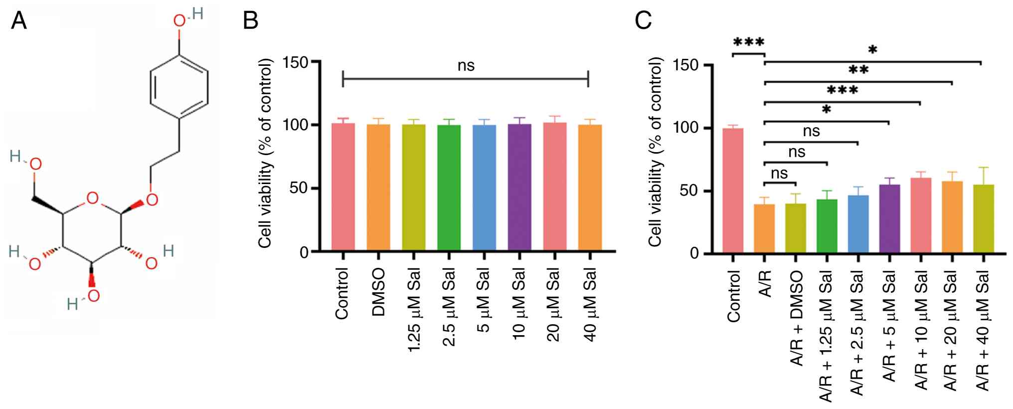
Fig. 1A shows the
chemical structure of Sal. To evaluate the biosafety and optimal
protective concentration of Sal, separate evaluations were
performed under normoxic and A/R conditions. First, to assess
potential baseline cytotoxicity, normal H9c2 cells were treated
with increasing concentrations of Sal (0 to 40 μM) for 48 h
under normoxic conditions. As demonstrated in Fig. 1B, cell viability remained
unaffected across this entire concentration range, confirming that
Sal exhibits no intrinsic cytotoxicity. Second, to determine
cardioprotective efficacy, H9c2 cells were pretreated with the same
concentration gradient of Sal for 48 h, after which the
Sal-containing medium was replaced with specific anoxic and
reoxygenation buffers to induce A/R injury. As demonstrated in
Fig. 1C, A/R injury markedly
reduced cell viability; however, this detrimental effect was
attenuated by Sal pretreatment in a bell-shaped
concentration-dependent manner, with the optimal protective effect
observed at 10 μM. Therefore, a 48-h pretreatment with 10
μM Sal was selected for all subsequent mechanistic
experiments.
Sal attenuates A/R-induced oxidative
stress and ferroptosis in H9c2 cells
To determine whether Sal exerts cardioprotective
effects by suppressing oxidative stress and ferroptosis, a
systematic evaluation of key associated indicators was conducted.
Considering that erastin can simultaneously induce oxidative stress
and ferroptosis (42),
erastin-treated cells were utilized as a positive control to
validate the ferroptosis detection system. The results demonstrated
that A/R injury produced ferroptotic marker alterations comparable
to those induced by erastin; however, while this phenotypic
similarity is supportive, the mechanistic involvement of
ferroptosis in A/R injury was further corroborated by the
protective effects of the specific inhibitor Fer-1. Western blot
analysis demonstrated that A/R injury significantly upregulated the
downstream ferroptosis-associated marker PTGS2 while downregulating
the ferroptosis-related protein GPX4 and the oxidative
stress-related protein PRDX2. Pretreatment with Sal or the
ferroptosis inhibitor Fer-1 (5 μM) attenuated these
alterations in protein expression (Fig. 2A and B). Further analyses
revealed that A/R injury markedly increased intracellular ROS,
ferrous ion levels and lipid peroxidation, whereas pretreatment
with Sal or Fer-1 significantly attenuated these elevations
(Fig. 2C-E). Moreover,
assessment of oxidative and antioxidative parameters demonstrated
that MDA and 4-HNE levels were markedly elevated in the A/R group,
accompanied by pronounced reductions in SOD activity and the
GSH/GSSG ratio. Notably, treatment with Sal and Fer-1 effectively
reversed these alterations, as evidenced by decreased levels of MDA
and 4-HNE, along with increased SOD activity and GSH/GSSG ratio
(Fig. 2F-I). Given the
previously reported involvement of USP11 in oxidative
stress-induced ferroptosis (28), USP11 expression was also examined
to explore its potential involvement in A/R injury. Notably,
compared with the control group, A/R injury resulted in a
significant downregulation of USP11 expression in H9c2 cells,
whereas pretreatment with Sal or Fer-1 partially restored USP11
expression (Fig. 2A and B).
Collectively, these findings indicated that Sal may attenuate
A/R-induced oxidative stress and protect cells against ferroptotic
injury, although whether these effects are mediated via USP11
requires further investigation.
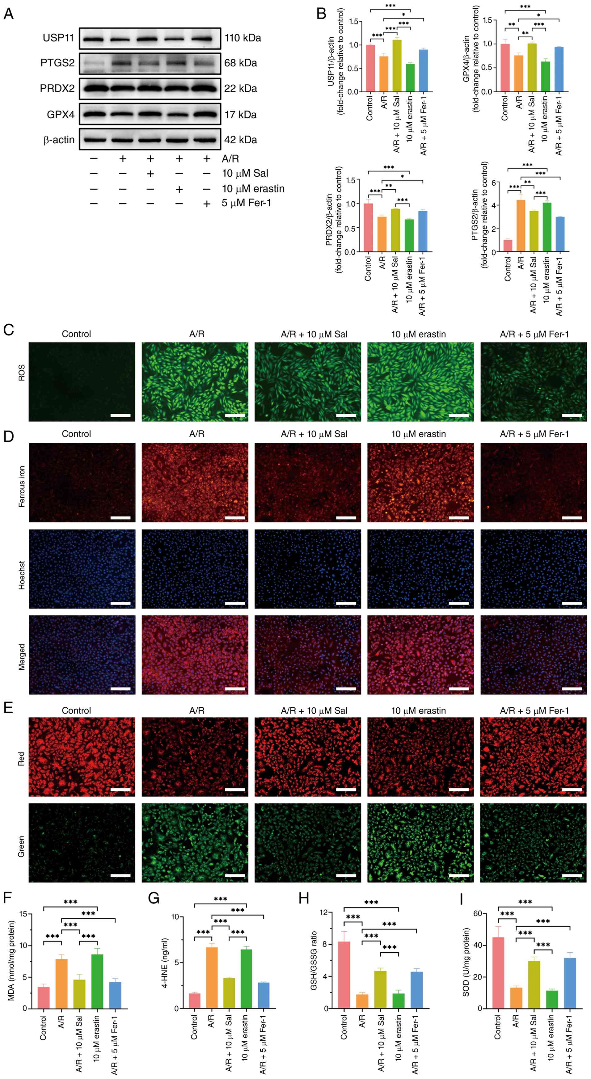 | Figure 2Sal attenuates A/R-induced oxidative
stress and ferroptosis in H9c2 cells. (A) Western blot analysis of
PTGS2, GPX4, PRDX2 and USP11 proteins in different groups of H9c2
cells (n=3). (B) Relative expression analysis of PTGS2, GPX4, PRDX2
and USP11 (n=3). (C) Intracellular ROS levels detected using
2',7'-dichlorodihydrofluorescein diacetate fluorescence staining
(magnification, ×100; scale bar, 800 μm; n=3). (D)
Intracellular ferrous iron levels measured using FerroOrange probe
(magnification, ×200; scale bar, 400 μm; n=3). (E) Lipid
peroxidation assessed using boron-dipyrromethene 581/591 C11
fluorescence staining (magnification, ×200; scale bar, 400
μm; n=3). Biochemical quantification of (F) MDA, (G) 4-HNE,
(H) GSH/GSSG ratio and (I) SOD activity (n=6). Data are presented
as mean ± SD, with *P<0.05, **P<0.01
and ***P<0.001 indicating statistical significance.
Sal, salidroside; Fer-1, ferrostatin-1; A/R, anoxia/reoxygenation;
PTGS2, prostaglandin-endoperoxide synthase 2; GPX4, glutathione
peroxidase 4; PRDX2, peroxiredoxin 2; USP11, ubiquitin-specific
protease 11; ROS, reactive oxygen species; MDA, malondialdehyde;
4-HNE, 4-hydroxynonenal; GSSG, glutathione disulfide; GSH,
glutathione; SOD, superoxide dismutase. |
Sal mitigates A/R-induced oxidative
stress and ferroptosis in H9c2 cells by enhancing USP11-mediated
signaling
To determine whether Sal confers protection against
A/R-induced oxidative stress and ferroptosis through USP11, a
USP11-OE plasmid and a specific siRNA (si-USP11) were constructed,
and their transfection efficiencies verified using western blotting
(Fig. 3A and B). Subsequent
experiments demonstrated that Sal pretreatment significantly
increased the expression levels of USP11, GPX4 and PRDX2, while
suppressing PTGS2 expression. However, silencing USP11 abolished
these effects, as evidenced by increased PTGS2 levels and reduced
USP11, GPX4 and PRDX2 expression (Fig. 3C and D). These findings indicated
that Sal may alleviate A/R-induced oxidative stress and ferroptosis
through the upregulation of USP11. Oxidative stress and
ferroptosis-related cellular parameters were further evaluated. Sal
markedly reduced A/R-induced ROS accumulation, ferrous iron
overload and lipid peroxidation. USP11 knockdown notably weakened
the inhibitory effects of Sal on ROS accumulation and lipid
peroxidation, while partially attenuating its effect on ferrous
iron accumulation (Fig. 3E-G).
Biochemical analyses, including MDA, 4-HNE, SOD activity and the
GSH/GSSG ratio, consistently supported the potential role of USP11
in mediating the antioxidative and anti-ferroptotic actions of Sal
(Fig. 3H-K). Consistently, USP11
overexpression was associated with a coordinated attenuation of the
A/R-induced oxidative stress and ferroptotic phenotype, including
reduced PTGS2 expression, ROS accumulation, ferrous iron overload
and lipid peroxidation, MDA and 4-HNE levels, accompanied by
increased GPX4 and PRDX2 expression, SOD activity and GSH/GSSG
ratio (Fig. 3C-K). Taken
together, these results demonstrated that Sal may suppress
A/R-induced oxidative stress and ferroptosis in H9c2 cells through
the upregulation of USP11.
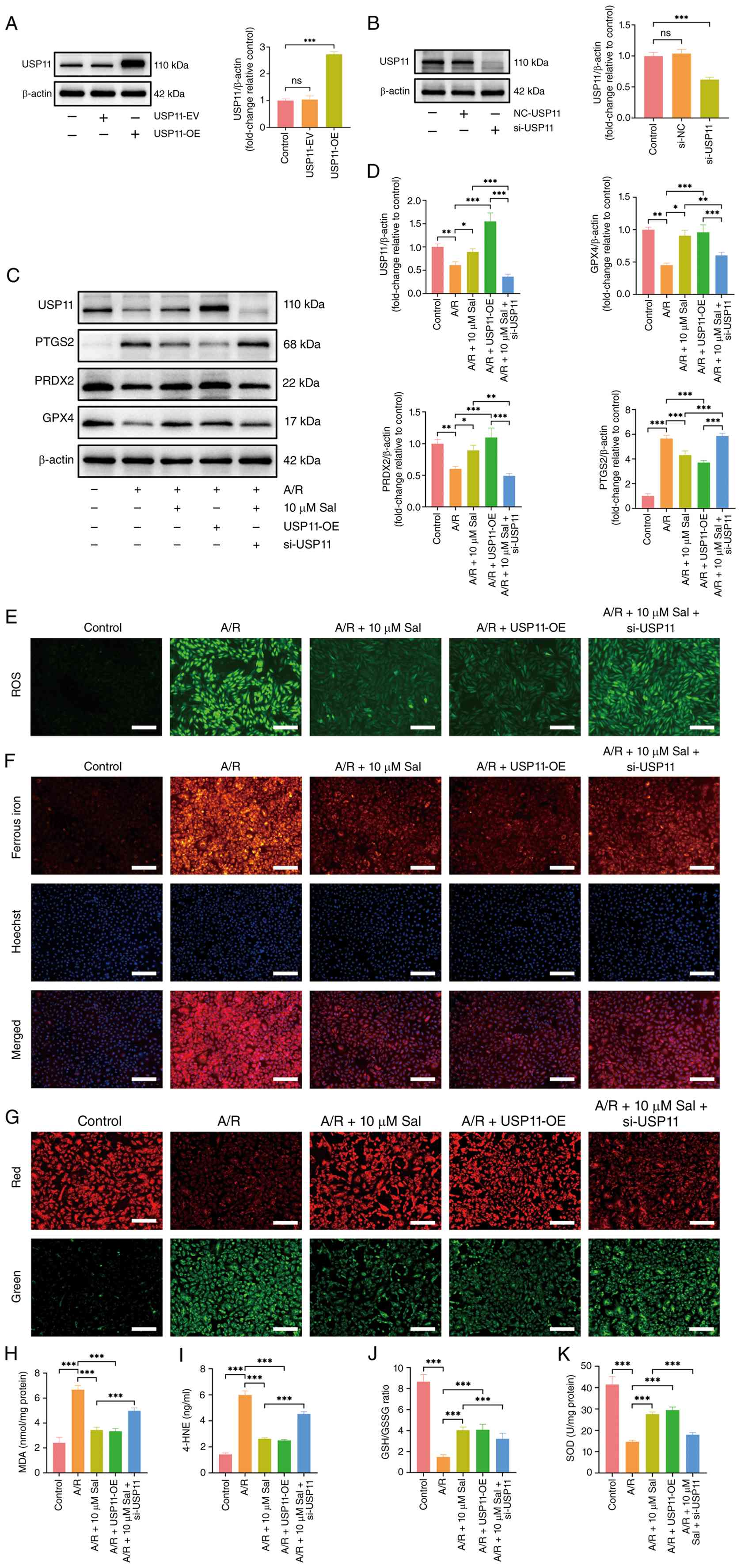 | Figure 3Sal mitigates A/R-induced oxidative
stress and ferroptosis in H9c2 cells by enhancing USP11-mediated
signaling. Validation of USP11 (A) silencing (si-USP11) and (B)
overexpression (USP11-OE) efficiency in H9c2 cells via western
blotting (n=3). (C) Western blot analysis of PTGS2, GPX4, PRDX2 and
USP11 proteins in different groups of H9c2 cells (n=3). (D)
Relative expression analysis of PTGS2, GPX4, PRDX2 and USP11 (n=3).
(E) Intracellular ROS levels detected using
2',7'-dichlorodihydrofluorescein diacetate fluorescence staining
(magnification, ×100; scale bar, 800 μm; n=3). (F)
Intracellular ferrous iron levels measured using FerroOrange probe
(magnification, ×200; scale bar, 400 μm; n=3). (G) Lipid
peroxidation assessed using boron-dipyrromethene 581/591 C11
fluorescence staining (magnification, ×200; scale bar, 400
μm; n=3). Biochemical quantification of (H) MDA, (I) 4-HNE,
(J) GSH/GSSG ratio and (K) SOD activity (n=6). Data are presented
as mean ± SD, with *P<0.05, **P<0.01
and ***P<0.001 indicating statistical significance.
ns, not significant; Sal, salidroside; A/R, anoxia/reoxygenation;
PTGS2, prostaglandin-endoperoxide synthase 2; GPX4, glutathione
peroxidase 4; PRDX2, peroxiredoxin 2; USP11, ubiquitin-specific
protease 11; ROS, reactive oxygen species; MDA, malondialdehyde;
4-HNE, 4-hydroxynonenal; GSSG, glutathione disulfide; GSH,
glutathione; SOD, superoxide dismutase; si, small interfering; OE,
overexpression; EV, empty vector. |
Sal binds to USP11 and enhances the
USP11-PRDX2 interaction in H9c2 cells
Based on the aforementioned findings, PRDX2 was
selected as a potential downstream target of USP11 since its
expression changed in parallel with USP11 and was responsive to
USP11 knockdown and overexpression. In addition, PRDX2 is an
antioxidant protein whose stability may be influenced by
ubiquitination, providing a mechanistic basis for investigating
whether it is regulated by the deubiquitinase, USP11. Therefore, it
was hypothesized that Sal may directly interact with USP11 and
thereby modulate its association with PRDX2. To test this,
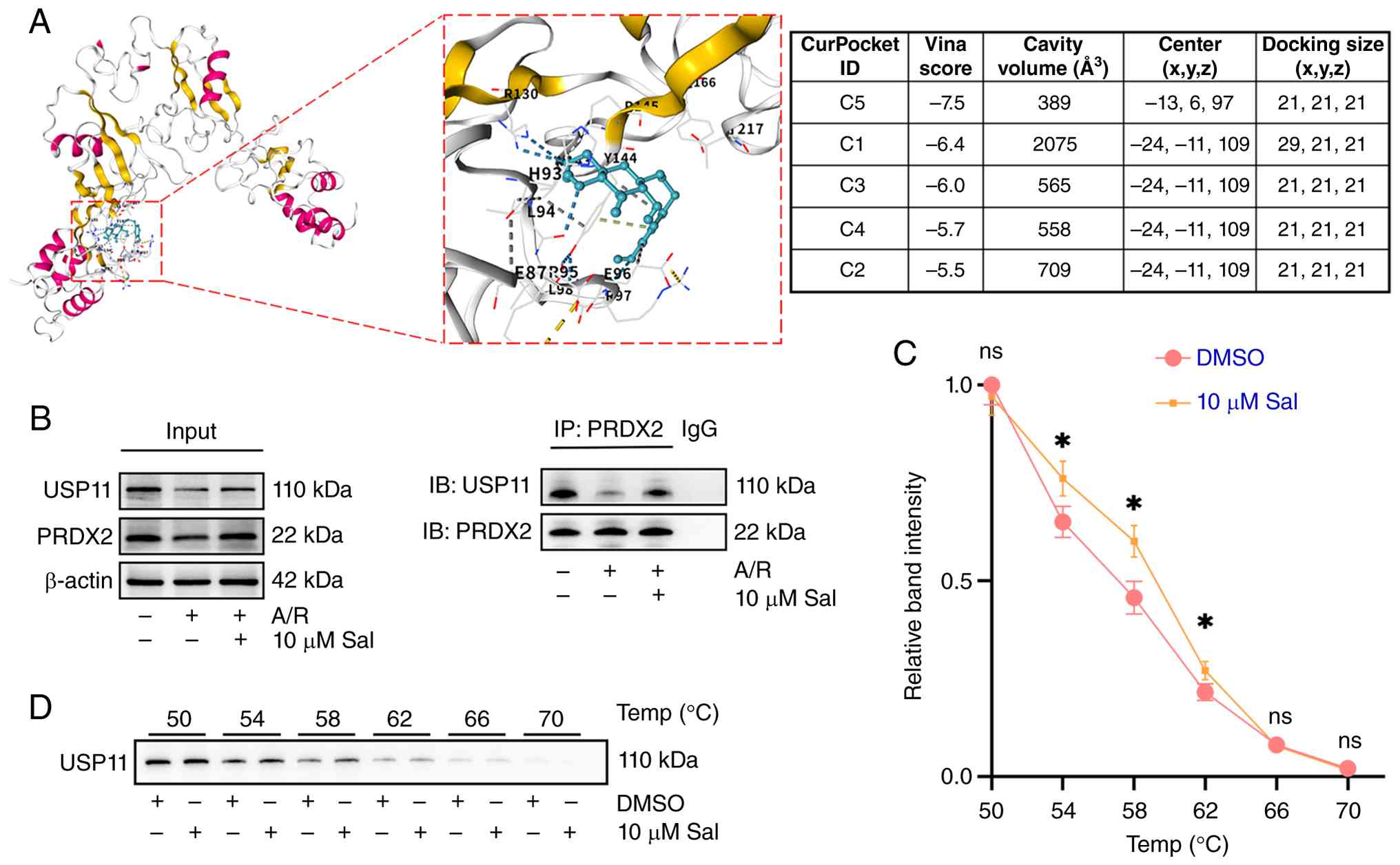
molecular docking, Co-IP and CETSA assays were performed. Molecular
docking using CB-Dock2 revealed that Sal fits into several
potential binding cavities of USP11, with the C5 pocket showing the
highest affinity (Vina score: -7.5; Fig. 4A). Consistently, Co-IP analysis
demonstrated that A/R injury markedly attenuated the USP11-PRDX2
interaction, whereas Sal pretreatment effectively restored their
binding (Fig. 4B). Furthermore,
CETSA demonstrated that Sal increased the thermal stability of
USP11, supporting an interaction between Sal and USP11 (Fig. 4C and D). Preliminary thermal
evaluations confirmed that USP11 does not undergo significant
thermal denaturation or precipitation up to 50°C and Sal treatment
alone did not alter baseline USP11 solubility within this lower
temperature range (Fig. S1).
These results suggest that Sal may exert cardioprotective effects
through modulation of USP11-associated signaling and strengthening
its interaction with PRDX2.
Sal enhances USP11-mediated
deubiquitination and stabilization of PRDX2
The aforementioned findings demonstrated an
interaction between USP11 and PRDX2 and showed that PRDX2
expression was responsive to changes in USP11 levels. These
observations prompted further investigation into whether USP11
regulates PRDX2 stability through deubiquitination and whether Sal
modulates this process. To determine whether Sal stabilizes PRDX2
in a USP11-dependent manner, CHX chase assays were performed.
Silencing USP11 markedly shortened the half-life of PRDX2, whereas
Sal treatment effectively attenuated the accelerated degradation
caused by USP11 depletion (Fig. 5A
and B). Consistently, ubiquitination analyses revealed that Sal
reduced PRDX2 ubiquitination, while USP11 knockdown exerted the
opposite effect and significantly increased PRDX2 ubiquitination
(Fig. 5C and D). Together, these
results demonstrate that Sal may stabilize PRDX2 by enhancing
USP11-mediated deubiquitination, thereby contributing to its
protective effects against A/R injury.
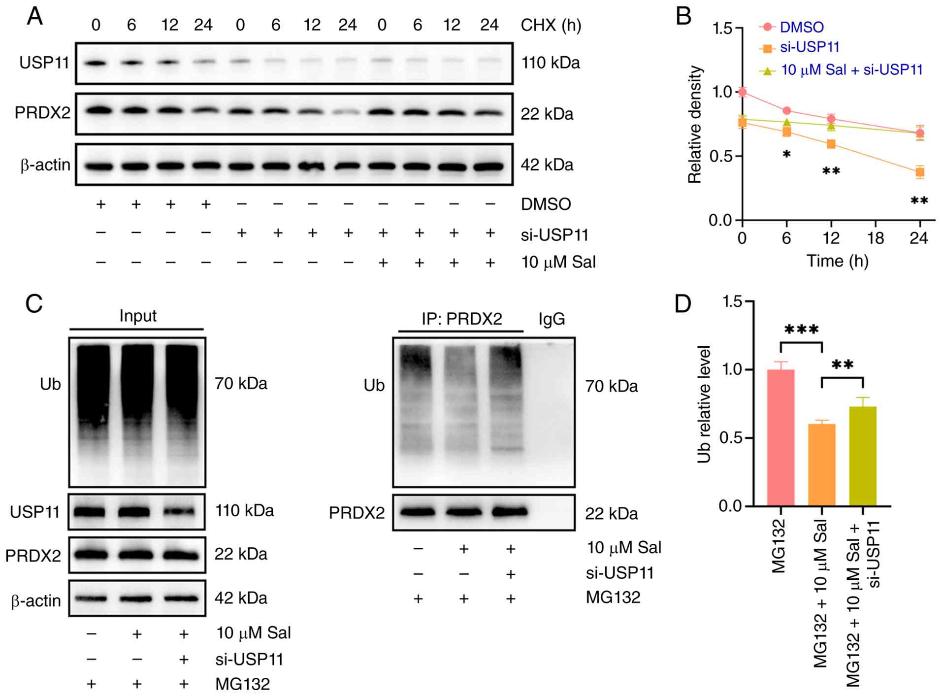 | Figure 5Sal enhances USP11-mediated
deubiquitination and stabilization of PRDX2. (A) Representative
western blot analysis of USP11 and PRDX2 in H9c2 cells treated with
CHX (10 μg/ml) for 0, 6, 12 and 24 h in the presence or
absence of Sal and si-USP11 (n=3). (B) Semi-quantification of PRDX2
protein levels during the CHX chase assay (n=3; si-USP11 vs. 10
μM Sal + si-USP11). (C) Representative western blot analysis
of PRDX2 ubiquitination in H9c2 cells treated with Sal and/or
si-USP11 in the presence of MG132. The ubiquitination smear
represents the extent of PRDX2 polyubiquitination (n=3). (D)
Semi-quantitative analysis of the ubiquitination levels of PRDX2
(n=3). Data are presented as mean ± SD, with *P<0.05,
**P<0.01 and ***P<0.001 indicating
statistical significance. Sal, salidroside; USP11,
ubiquitin-specific protease 11; PRDX2, peroxiredoxin 2; CHX,
cycloheximide; si, small interfering; Ub, ubiquitin. |
Sal pretreatment protects the myocardium
against I/R injury
To evaluate the protective effect of Sal against
myocardial I/R injury in vivo, a rat model of LAD coronary
artery ligation was established. Compared with the sham-operated
group, the serum levels of the myocardial injury markers CK-MB and
LDH were markedly elevated following I/R injury (Fig. 6A and B), and the infarct area
increased markedly (Figs. 6F and
S2). Echocardiography further
demonstrated severe cardiac dysfunction in the I/R group, evidenced
by pronounced reductions in LVEF and LVFS (Fig. 6C-E). Sal pretreatment effectively
attenuated these alterations, thereby mitigating myocardial damage
and functional decline. Morphological assessment revealed that I/R
injury induced myofibrillar disarray, cardiomyocyte swelling and
interstitial edema with inflammatory infiltration, as shown by
H&E staining, whereas these pathological abnormalities were
markedly ameliorated by Sal pretreatment (Fig. 6G). Analysis of oxidative stress-
and ferroptosis-related proteins demonstrated increased PTGS2
expression and decreased USP11, GPX4 and PRDX2 expression after I/R
injury; Sal pretreatment attenuated these molecular changes
(Fig. 6H-L). Collectively, these
findings indicate that Sal pretreatment may confer cardioprotection
by modulating the expression levels of USP11 and other related
proteins, thereby mitigating I/R-induced oxidative injury and
ferroptosis.
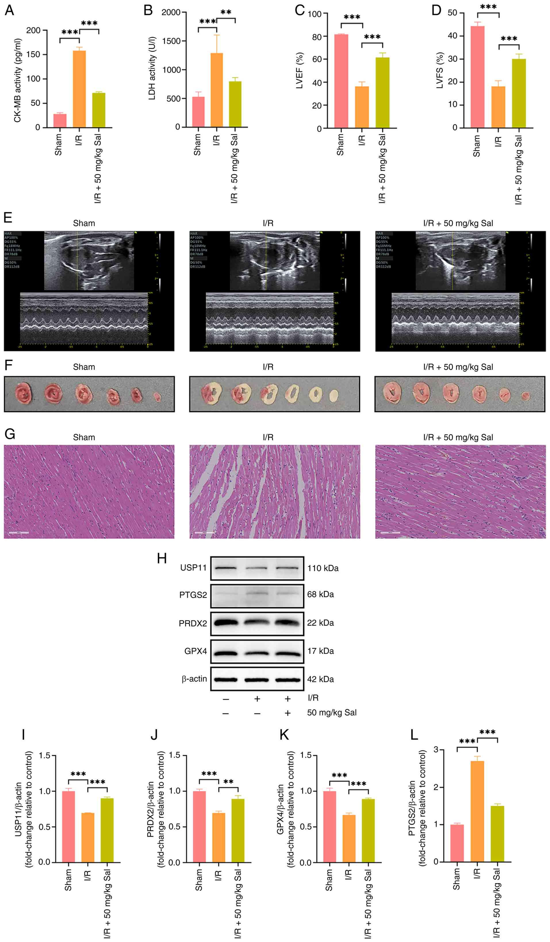 | Figure 6Sal pretreatment protects the
myocardium against I/R injury. Serum levels of (A) CK-MB and (B)
LDH were measured 2 h after reperfusion as markers of acute
myocardial damage (n=6). (C) LVEF and (D) LVFS were quantified by
echocardiography to evaluate cardiac contractile function (n=6).
(E) Representative M-mode echocardiographic tracings (n=6). (F)
Representative images of heart sections stained with 2% triphenyl
tetrazolium chloride; white areas represent infarcted tissue, while
red areas represent viable myocardium (n=3). (G) Representative
H&E staining images (magnification, ×400; scale bar, 400
μm) showing myocardial histopathological changes (n=3). (H)
Representative immunoblot of USP11, PRDX2, GPX4 and PTGS2.
Semi-quantitative analysis of (I) USP11, (J) PRDX2, (K) GPX4 and
(L) PTGS2 in rat myocardial tissues (n=3). Data are presented as
mean ± SD, with **P<0.01 and ***P<0.001
indicating statistical significance. Sal, salidroside; I/R,
ischemia/reperfusion; CK-MB, creatine kinase isoenzyme; LDH,
lactate dehydrogenase; LVEF, left ventricular ejection fraction;
LVFS, left ventricular fractional shortening; USP11,
ubiquitin-specific protease 11; PRDX2, peroxiredoxin 2; GPX4,
glutathione peroxidase 4; PTGS2, prostaglandin-endoperoxide
synthase 2. |
Discussion
In the present study, an in vitro A/R model
using H9c2 cells was combined with an in vivo rat model of
myocardial I/R injury to systematically investigate the
cardioprotective effects of Sal and its underlying molecular
mechanisms. The present results demonstrated that Sal pretreatment
significantly improved left ventricular systolic function, as
indicated by increased LVEF and LVFS, and reduced infarct size in
I/R-injured rats, while effectively attenuating A/R-induced loss of
H9c2 cell viability. The findings from the present study are
consistent with previous reports demonstrating the cardioprotective
and antioxidant properties of Sal in experimental models of
myocardial injury (23,33,43,44). The present data further indicated
that inhibition of oxidative stress-driven ferroptosis represents a
critical mechanism by which Sal confers cardioprotection.
Mechanistically, Sal interacted with the deubiquitinating enzyme
USP11, increasing its thermal stability and its affinity for the
antioxidant protein PRDX2, thereby promoting USP11-mediated
deubiquitination and stabilization of PRDX2. Activation of this
Sal-USP11-PRDX2 axis preserved intracellular redox homeostasis and
prevented lethal lipid peroxidation, ultimately mitigating
myocardial I/R injury (Fig.
7).
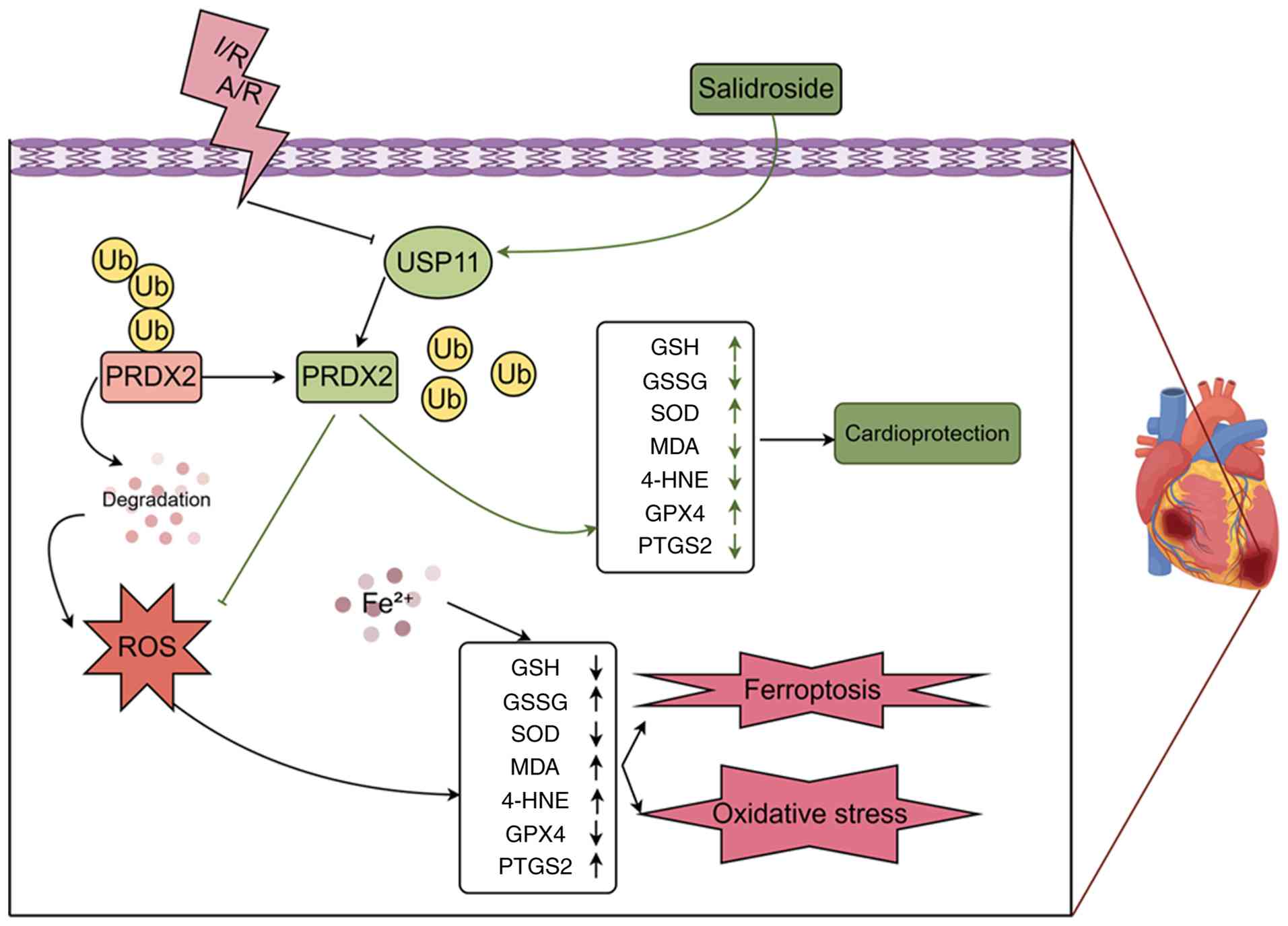 | Figure 7Schematic representation of the
mechanism by which salidroside may attenuate myocardial I/R injury.
I/R, ischemia/reperfusion; A/R, anoxia/reoxygenation; USP11,
ubiquitin-specific protease 11; PRDX2, peroxiredoxin 2; ROS,
reactive oxygen species; SOD, superoxide dismutase; MDA,
malondialdehyde; GSH, glutathione; GSSG, glutathione disulfide;
4-HNE, 4-hydroxynonenal; GPX4, glutathione peroxidase 4; PTGS2,
prostaglandin-endoperoxide synthase 2. |
Accumulating evidence has established oxidative
stress and ferroptosis as central pathological processes in
myocardial I/R injury, particularly during the reperfusion phase
(6,45,46). Ferroptosis is characterized by
iron overload, GSH depletion, lipid peroxidation, downregulation of
GPX4 and upregulation of PTGS2 (47), all of which were observed in the
A/R and I/R models in the present study. In the present study, GPX4
and PTGS2 were assessed as downstream phenotypic indicators of
ferroptosis and were not investigated as candidate direct
substrates of USP11. PRDX2 functions as an important antioxidant
protein by scavenging ROS and peroxides (29), and recent evidence has shown that
USP11 can stabilize PRDX2 by reducing its ubiquitination (48). Loss of antioxidant capacity can
promote ROS accumulation and lipid peroxidation, thereby favoring
ferroptotic responses, in which GPX4 constitutes a major defense
against lipid hydroperoxides and PTGS2 upregulation serves as a
downstream phenotypic marker (49). In the present study, Sal
treatment markedly reversed these ferroptotic features, exhibiting
a protective profile comparable to that of the ferroptosis-specific
inhibitor Fer-1. These findings strongly suggest that suppression
of ferroptosis constitutes an essential component of Sal-mediated
cardioprotection. Further mechanistic analyses revealed that USP11
expression was significantly reduced following A/R injury, whereas
Sal restored USP11 levels. Molecular docking and CETSA assays
supported an interaction between Sal and USP11, resulting in
enhanced thermal stability of USP11. This interaction may have
facilitated the association between USP11 and PRDX2 and promoted
PRDX2 deubiquitination, thereby prolonging its protein
half-life.
Due to the critical role of PRDX2 in scavenging
excessive ROS and maintaining redox balance in cardiomyocytes
(29,50), stabilization of PRDX2 effectively
curtails lipid peroxidation and reduces susceptibility to
ferroptotic cell death (51). A
notable observation in the present study was that Sal treatment
partially attenuated the accelerated degradation of PRDX2 even when
USP11 was knocked down. This phenomenon is likely attributed to the
inherent nature of transient siRNA transfection, which typically
results in incomplete silencing of the target protein. Residual
USP11 expression after incomplete siRNA-mediated knockdown may
contribute to the partial restoration of PRDX2 stability by Sal. In
addition, USP11-independent antioxidant mechanisms of Sal may also
partially contribute to PRDX2 preservation. Compared with previous
studies, the present work extends current understanding of the
cardiovascular pharmacology of Sal by uncovering a novel
post-translational regulatory mechanism. Earlier investigations
have largely attributed the cardioprotective effects of Sal to
activation of classical signaling pathways, such as PI3K/AKT,
AMP-activated protein kinase or the nuclear factor erythroid
2-related factor 2/heme oxygenase-1 axis (52-54). While the present findings are
generally consistent with the established antioxidant role of Sal,
they further demonstrate that Sal exerts sustained cytoprotective
effects by modulating protein stability through the
ubiquitin-proteasome system.
In addition, the present study assigns a previously
unrecognized role to USP11 in the context of cardiovascular
disease. Although USP11 has been extensively studied in cancer
biology and DNA damage repair (28,55,56), reports on its involvement in
myocardial I/R injury and ferroptosis remain limited (57). Recent studies have implicated
other deubiquitinating enzymes in ferroptosis regulation, including
USP7 in BRAFV600E-mutant thyroid cancer, where
pharmacological inhibition of USP7 promotes ferroptosis, and
BRCA1-associated protein 1 in inflammatory bowel disease, where it
promotes ferroptosis through SLC7A11 suppression (58,59). The present data demonstrate that
loss of USP11 accelerates PRDX2 degradation and increases
ferroptotic vulnerability, identifying PRDX2 as a functional
substrate of USP11 in H9c2 cells.
The present study proposes a
'small-molecule-deubiquitinase-antioxidant protein' regulatory
paradigm, providing insight into how natural compounds exert
cardioprotective effects by fine-tuning protein stability. Despite
these encouraging findings, several limitations of the present
study should be acknowledged. First, while direct C11-BODIPY lipid
peroxidation staining and classical biochemical markers were
examined to validate ferroptosis, future studies incorporating
transmission electron microscopy to assess mitochondrial
ultrastructure and testing a broader panel of targets (such as
acyl-CoA synthetase long-chain family member 4 and solute carrier
family 7 member 11) will provide a more exhaustive validation.
Second, although the use of Fer-1 confirmed that ferroptosis is a
critical component of myocardial A/R injury, it does not completely
rule out the synergistic involvement of other cell death
modalities, such as apoptosis or necrosis. Whether ferroptosis acts
as the dominant form of cell death requires further broad-spectrum
cross-validation using novel ferroptosis modulators such as
Liproxstatin-1 or RAS-selective lethal small molecule 3. Third,
while the CETSA and functional assays demonstrated that Sal may
physically engage USP11 and promote USP11-dependent PRDX2
stabilization, the detailed upstream mechanisms, specifically
whether Sal directly modulates USP11 transcription, baseline
protein half-life or intrinsic catalytic activity, warrant further
dedicated biochemical investigation. Additionally, while present
data suggests target engagement, definitive validation of direct
binding kinetics and the precise binding pocket will require future
orthogonal biophysical assays (such as surface plasmon resonance)
and targeted mutagenesis. Moreover, identifying the specific
ubiquitin linkage type (such as K48 or K63) and employing
catalytically inactive USP11 mutants or cell-free in vitro
deubiquitination systems will be essential to fully elucidate the
biochemical nuances of this interaction. Fourth, a pretreatment
strategy was utilized to provide proof-of-concept for the
prophylactic potential of Sal in anticipated I/R scenarios (such as
scheduled coronary artery bypass grafting). Post-ischemic
therapeutic administration remains to be evaluated for
unpredictable acute events such as myocardial infarction. Fifth,
while in vitro genetic silencing supported the functional
involvement of USP11 in Sal-mediated protection, the in vivo
rat model primarily demonstrated a robust phenotypic and molecular
association. Definitive proof of in vivo causality will
require future investigations employing adeno-associated
virus-mediated cardiac-specific USP11 knockdown or knockout animal
models. Sixth, the in vivo model relied on a single dose of
Sal and lacked a Fer-1 positive control, limiting the
characterization of dose-dependent pharmacological effects.
Seventh, the in vitro mechanistic investigations were
performed in the H9c2 cardiomyoblast cell line. While these
findings were phenotypically supported by the in vivo adult
rat model, future investigations employing primary rat
cardiomyocytes will be essential to definitively validate this
biochemical axis in mature cellular physiology.
In conclusion, the present study demonstrated that
Sal attenuated myocardial I/R injury by promoting USP11-mediated
deubiquitination and stabilization of PRDX2, thereby suppressing
oxidative stress-induced ferroptosis.
Supplementary Data
Availability of data and materials
The data generated in the present study may be
requested from the corresponding author.
Authors' contributions
ZCQ, JNL and YZL performed experiments, analyzed
data and wrote the manuscript. ZMW and XDL performed experiments
and analyzed data. RYZ and SQL analyzed and interpreted data. SQL,
STZ and LW designed the experiments and provided financial support.
SQL, STZ and LW confirm the authenticity of all the raw data. All
authors have read and approved the final version of the
manuscript.
Ethics approval and consent to
participate
The present research protocol was reviewed and
approved by the Ethics Committee of the First Affiliated Hospital
of Nanchang University (approval no. CDYFY-IACUC-2025092003).
Patient consent for publication
Not applicable.
Competing interests
The authors declare that they have no competing
interests.
Abbreviations:
|
I/R
|
ischemia/reperfusion
|
|
A/R
|
anoxia/reoxygenation
|
|
Sal
|
salidroside
|
|
USP11
|
ubiquitin-specific protease 11
|
|
PRDX2
|
peroxiredoxin 2
|
|
ROS
|
reactive oxygen species
|
|
GPX4
|
glutathione peroxidase 4
|
|
PTGS2
|
prostaglandin-endoperoxide synthase
2
|
|
LAD
|
left anterior descending
|
|
Fer-1
|
ferrostatin-1
|
|
CETSA
|
cellular thermal shift assay
|
|
Co-IP
|
co-immunoprecipitation
|
Acknowledgements
Not applicable.
Funding
The present study was supported by the National Natural Science
Foundation of China (grant nos. 82460057 and 82560074) and The
First Affiliated Hospital of Nanchang University Young Talent
Research Cultivation Fund (grant no. YFYPY202538).
References
|
1
|
Ferenczyová K, Kindernay L, Vlkovičová J,
Kaločayová B, Rajtík T and Barteková M: Pharmacology of catechins
in Ischemia-reperfusion injury of the heart. Antioxidants (Basel).
10:13902021. View Article : Google Scholar : PubMed/NCBI
|
|
2
|
Ghanta SN, Kattamuri LPV, Odueke A and
Mehta JL: Molecular insights into Ischemia-reperfusion injury in
coronary artery disease: Mechanisms and therapeutic implications: A
comprehensive review. Antioxidants (Basel). 14:2132025. View Article : Google Scholar : PubMed/NCBI
|
|
3
|
Francisco J and Del Re DP: Inflammation in
myocardial Ischemia/reperfusion injury: Underlying mechanisms and
therapeutic potential. Antioxidants (Basel). 12:19442023.
View Article : Google Scholar : PubMed/NCBI
|
|
4
|
Zhang J, Zhao Y and Gong N: Endoplasmic
reticulum stress signaling modulates ischemia/reperfusion injury in
the aged heart by regulating mitochondrial maintenance. Mol Med.
30:1072024. View Article : Google Scholar : PubMed/NCBI
|
|
5
|
Davidson SM, Ferdinandy P, Andreadou I,
Bøtker HE, Heusch G, Ibáñez B, Ovize M, Schulz R, Yellon DM,
Hausenloy DJ, et al: Multitarget strategies to reduce myocardial
Ischemia/Reperfusion injury: JACC review topic of the week. J Am
Coll Cardiol. 73:89–99. 2019. View Article : Google Scholar : PubMed/NCBI
|
|
6
|
Zhang T, Deng W, Deng Y, Liu Y, Xiao S,
Luo Y, Xiang W and He Q: Mechanisms of ferroptosis regulating
oxidative stress and energy metabolism in myocardial
ischemia-reperfusion injury and a novel perspective of natural
plant active ingredients for its treatment. Biomed Pharmacother.
165:1147062023. View Article : Google Scholar : PubMed/NCBI
|
|
7
|
Fang X, Ardehali H, Min J and Wang F: The
molecular and metabolic landscape of iron and ferroptosis in
cardiovascular disease. Nat Rev Cardio. 20:7–23. 2023. View Article : Google Scholar
|
|
8
|
Chen X, Kang R, Kroemer G and Tang D:
Broadening horizons: The role of ferroptosis in cancer. Nat Rev
Clin Oncol. 18:280–296. 2021. View Article : Google Scholar : PubMed/NCBI
|
|
9
|
Yan HF, Zou T, Tuo QZ, Xu S, Li H, Belaidi
AA and Lei P: Ferroptosis: Mechanisms and links with diseases.
Signal Transduct Target Ther. 6:492021. View Article : Google Scholar : PubMed/NCBI
|
|
10
|
Galaris D, Barbouti A and Pantopoulos K:
Iron homeostasis and oxidative stress: An intimate relationship.
Biochim Biophys Acta Mol Cell Res. 1866:1185352019. View Article : Google Scholar : PubMed/NCBI
|
|
11
|
Yu Y, Yan Y, Niu F, Wang Y, Chen X, Su G,
Liu Y, Zhao X, Qian L, Liu P and Xiong Y: Ferroptosis: A cell death
connecting oxidative stress, inflammation and cardiovascular
diseases. Cell Death Discov. 7:1932021. View Article : Google Scholar : PubMed/NCBI
|
|
12
|
Tang D, Chen X, Kang R and Kroemer G:
Ferroptosis: Molecular mechanisms and health implications. Cell
Res. 31:107–125. 2021. View Article : Google Scholar :
|
|
13
|
Stockwell BR: Ferroptosis turns 10:
Emerging mechanisms, physiological functions, and therapeutic
applications. Cell. 185:2401–2421. 2022. View Article : Google Scholar : PubMed/NCBI
|
|
14
|
Liu J, Kuang F, Kroemer G, Klionsky DJ,
Kang R and Tang D: Autophagy-dependent ferroptosis: Machinery and
regulation. Cell Chem Biol. 27:420–435. 2020. View Article : Google Scholar : PubMed/NCBI
|
|
15
|
Sun S, Wu Y, Maimaitijiang A, Huang Q and
Chen Q: Ferroptotic cardiomyocyte-derived exosomes promote cardiac
macrophage M1 polarization during myocardial infarction. PeerJ.
10:e137172022. View Article : Google Scholar : PubMed/NCBI
|
|
16
|
Xu C, Liu Y, Yang J, Zhai M, Fan Z, Qiao
R, Jin P and Yang L: Effects of berbamine against myocardial
ischemia/reperfusion injury: Activation of the 5'adenosine
monophosphate-activated protein kinase/nuclear factor erythroid
2-related factor pathway and changes in the mitochondrial state.
Biofactors. 48:651–664. 2022. View Article : Google Scholar : PubMed/NCBI
|
|
17
|
Peoples JN, Saraf A, Ghazal N, Pham TT and
Kwong JQ: Mitochondrial dysfunction and oxidative stress in heart
disease. Exp Mol Med. 51:1–13. 2019. View Article : Google Scholar : PubMed/NCBI
|
|
18
|
Remex NS, Abdullah CS, Aishwarya R, Sinha
MH, Dhar P, Dhaou CB, Orr AW and Bhuiyan MS: Deletion of Sigmar1
leads to increased arterial stiffness and altered mitochondrial
respiration resulting in vascular dysfunction. Front Cardiovasc
Med. 15:13862962024.
|
|
19
|
Matsushima S and Sadoshima J: Yin and yang
of NADPH oxidases in myocardial Ischemia-reperfusion. Antioxidants
(Basel). 11:10692022. View Article : Google Scholar : PubMed/NCBI
|
|
20
|
Li HX, Wang TH, Wu LX, Xue FS, Zhang GH
and Yan T: Role of Keap1-Nrf2/ARE signal transduction pathway in
protection of dexmedetomidine preconditioning against myocardial
ischemia/reperfusion injury. Biosci Rep. 42:BSR202213062022.
View Article : Google Scholar : PubMed/NCBI
|
|
21
|
Zheng Y, Mao L, Wang Q, Hu H, Xarpidin B,
Luo Z and Wu YL: Mitochondria-targeted ROS scavenging natural
enzyme cascade nanogels for periodontitis treatment via hypoxia
alleviation and immunomodulation. Adv Sci (Weinh). 12:e074812025.
View Article : Google Scholar : PubMed/NCBI
|
|
22
|
Dodson M, Castro-Portuguez R and Zhang DD:
NRF2 plays a critical role in mitigating lipid peroxidation and
ferroptosis. Redox Biol. 23:1011072019. View Article : Google Scholar : PubMed/NCBI
|
|
23
|
Zhang X, Xie L, Long J, Xie Q, Zheng Y,
Liu K and Li X: Salidroside: A review of its recent advances in
synthetic pathways and pharmacological properties. Chem Biol
Interact. 339:1092682021. View Article : Google Scholar : PubMed/NCBI
|
|
24
|
Chang X, Zhang K, Zhou R, Luo F, Zhu L,
Gao J, He H, Wei T, Yan T and Ma C: Cardioprotective effects of
salidroside on myocardial ischemia-reperfusion injury in coronary
artery occlusion-induced rats and Langendorff-perfused rat hearts.
Int J Cardiol. 215:532–544. 2016. View Article : Google Scholar : PubMed/NCBI
|
|
25
|
Sun YM, Wang XY, Zhou XR, Zhang C, Liu KJ,
Zhang FY and Xiang B: Salidroside Ameliorates radiation damage by
reducing mitochondrial oxidative stress in the submandibular gland.
Antioxidants (Basel). 11:14142022. View Article : Google Scholar : PubMed/NCBI
|
|
26
|
Zhang Y, Hailati J, Ma X, Midilibieke H
and Liu Z: Ubiquitin-specific protease 11 Aggravates
Ischemia-reperfusion-induced Cardiomyocyte Pyroptosis and injury by
promoting TRAF3 Deubiquitination. Balkan Med J. 40:205–214. 2023.
View Article : Google Scholar : PubMed/NCBI
|
|
27
|
Rajkumar S, Jung JH, Kim JY, Karapurkar
JK, Birappa G, Gowda DAA, Ajaykumar CB, Perumalsamy H, Suresh B,
Kim KS, et al: Loss of Ubiquitin-specific protease 11 mitigates
pulmonary fibrosis in human pluripotent stem Cell-derived alveolar
organoids. Int J Stem Cells. 18:205–213. 2025. View Article : Google Scholar : PubMed/NCBI
|
|
28
|
Zhu J, Sun R, Sun K, Yan C, Jiang J, Kong
F and Shi J: The deubiquitinase USP11 ameliorates intervertebral
disc degeneration by regulating oxidative stress-induced
ferroptosis via deubiquitinating and stabilizing Sirt3. Redox Biol.
62:1027072023. View Article : Google Scholar : PubMed/NCBI
|
|
29
|
Balasubramanian P, Vijayarangam V,
Deviparasakthi MKG, Palaniyandi T, Ravi M, Natarajan S, Viswanathan
S, Baskar G, Wahab MRA and Surendran H: Implications and
progression of peroxiredoxin 2 (PRDX2) in various human diseases.
Pathol Res. 254:1550802024. View Article : Google Scholar
|
|
30
|
Han F, Dong Y, Liu Q, Song L, Guo H, Zhu
L, Sun B, Zhao W and Chen L: S-nitrosylation of peroxiredoxin 2
exacerbates hyperuricemia-induced renal injury through regulation
of mitochondrial homeostasis. Free Radic Biol Med. 230:66–78. 2025.
View Article : Google Scholar : PubMed/NCBI
|
|
31
|
Zhang Y, Sun C, Xiao G, Shan H, Tang L, Yi
Y, Yu W and Gu Y: S-nitrosylation of the Peroxiredoxin-2 promotes
S-nitrosoglutathione-mediated lung cancer cells apoptosis via
AMPK-SIRT1 pathway. Cell Death Dis. 10:3292019. View Article : Google Scholar : PubMed/NCBI
|
|
32
|
Zhao ST, Qiu ZC, Xu ZQ, Tao ED, Qiu RB,
Peng HZ, Zhou LF, Zeng RY, Lai SQ and Wan L: Curcumin attenuates
myocardial ischemia-reperfusion-induced autophagy-dependent
ferroptosis via Sirt1/AKT/FoxO3a signaling. Int J Mol Med.
55:512025. View Article : Google Scholar
|
|
33
|
Yang B, Xu Y, Yu J, Wang Q, Fan Q, Zhao X,
Qiao Y, Zhang Z, Zhou Q, Yin D, et al: Salidroside pretreatment
alleviates ferroptosis induced by myocardial ischemia/reperfusion
through mitochondrial superoxide-dependent AMPKα2 activation.
Phytomedicine. 128:1553652024. View Article : Google Scholar
|
|
34
|
American Veterinary Medical Association:
AVMA guidelines for the euthanasia of animals. 2020 Edition.
American Veterinary Medical Association; Schaumburg, IL: 2020
|
|
35
|
Zhao ST, Qiu ZC, Zeng RY, Zou HX, Qiu RB,
Peng HZ, Zhou LF, Xu ZQ, Lai SQ and Wan L: Exploring the molecular
biology of ischemic cardiomyopathy based on ferroptosis-related
genes. Exp Ther Med. 27:2212024. View Article : Google Scholar
|
|
36
|
Martinez Molina D, Jafari R,
Ignatushchenko M, Seki T, Larsson EA, Dan C, Sreekumar L, Cao Y and
Nordlund P: Monitoring drug target engagement in cells and tissues
using the cellular thermal shift assay. Science. 341:84–87. 2013.
View Article : Google Scholar : PubMed/NCBI
|
|
37
|
Jafari R, Almqvist H, Axelsson H,
Ignatushchenko M, Lundbäck T, Nordlund P and Martinez Molina D: The
cellular thermal shift assay for evaluating drug target
interactions in cells. Nat Protoc. 9:2100–2122. 2014. View Article : Google Scholar : PubMed/NCBI
|
|
38
|
Savitski MM, Reinhard FB, Franken H,
Werner T, Savitski MF, Eberhard D, Martinez Molina D, Jafari R,
Dovega RB, Klaeger S, et al: Tracking cancer drugs in living cells
by thermal profiling of the proteome. Science. 346:12557842014.
View Article : Google Scholar : PubMed/NCBI
|
|
39
|
Huang J, Yang Q, Zeng L and Deng K:
Multi-omics network toxicology reveals the role of benzo[a]pyrene
in ovarian cancer: Integrating gut microbiota dynamics and
Mendelian randomization. Ecotoxicol Environ Saf. 302:1186012025.
View Article : Google Scholar : PubMed/NCBI
|
|
40
|
Liu Y, Yang X, Gan J, Chen S, Xiao ZX and
Cao Y: CB-Dock2: Improved protein-ligand blind docking by
integrating cavity detection, docking and homologous template
fitting. Nucleic Acids Res. 50:W159–W164. 2022. View Article : Google Scholar : PubMed/NCBI
|
|
41
|
Harper S, Gratton HE, Cornaciu I, Oberer
M, Scott DJ, Emsley J and Dreveny I: Structure and catalytic
regulatory function of ubiquitin specific protease 11 N-terminal
and ubiquitin-like domains. Biochemistry. 53:2966–2978. 2014.
View Article : Google Scholar : PubMed/NCBI
|
|
42
|
Wang X, Wang Z, Cao J, Dong Y and Chen Y:
Melatonin alleviates acute sleep Deprivation-Induced memory loss in
mice by suppressing hippocampal ferroptosis. Front Pharmacol.
12:7086452021. View Article : Google Scholar : PubMed/NCBI
|
|
43
|
Feng T, Shi J, Zhao J, Zhao Q, Wang T, Wan
S, Fan C, Wang S, Lai C and Yao Y: Salidroside can protect against
ferroptosis in cardiomyocytes and may be related to the regulation
of GGT1. Front Pharmacol. 16:15805062025. View Article : Google Scholar : PubMed/NCBI
|
|
44
|
Tian X, Huang Y, Zhang X, Fang R, Feng Y,
Zhang W, Li L and Li T: Salidroside attenuates myocardial
ischemia/reperfusion injury via AMPK-induced suppression of
endoplasmic reticulum stress and mitochondrial fission. Toxicol
Appl Pharmacol. 448:1160932022. View Article : Google Scholar : PubMed/NCBI
|
|
45
|
He J, Liu D, Zhao L, Zhou D, Rong J, Zhang
L and Xia Z: Myocardial ischemia/reperfusion injury: Mechanisms of
injury and implications for management (Review). Exp Ther Med.
23:4302022. View Article : Google Scholar : PubMed/NCBI
|
|
46
|
Li JY, Liu SQ, Yao RQ, Tian YP and Yao YM:
A novel insight into the fate of cardiomyocytes in
Ischemia-reperfusion injury: From iron metabolism to ferroptosis.
Front Cell Dev Biol. 9:7994992021. View Article : Google Scholar : PubMed/NCBI
|
|
47
|
Du YW, Li XK, Wang TT, Zhou L, Li HR, Feng
L, Ma H and Liu HB: Cyanidin-3-glucoside inhibits ferroptosis in
renal tubular cells after ischemia/reperfusion injury via the AMPK
pathway. Mol Med. 29:422023. View Article : Google Scholar : PubMed/NCBI
|
|
48
|
Kong F, Luan J, Pan Q, Qiao Y and Wang W:
Ubiquitination-mediated PRDX2 alleviates intervertebral disc
degeneration via restraining TBHP-induced nucleus pulposus cell
apoptosis, ferroptosis and ECM degradation. J Inflamm (Lond).
23:152026. View Article : Google Scholar : PubMed/NCBI
|
|
49
|
Yang WS, SriRamaratnam R, Welsch ME,
Shimada K, Skouta R, Viswanathan VS, Cheah JH, Clemons PA, Shamji
AF, Clish CB, et al: Regulation of ferroptotic cancer cell death by
GPX4. Cell. 156:317–331. 2014. View Article : Google Scholar : PubMed/NCBI
|
|
50
|
Zhang H, Xu T, Mei X, Zhao Q, Yang Q, Zeng
X, Ma Z, Zhou H, Zeng Q, Xu D and Ren H: PINK1 modulates Prdx2 to
reduce lipotoxicity-induced apoptosis and attenuate cardiac
dysfunction in heart failure mice with a preserved ejection
fraction. Clin Transl Med. 15:e701662025. View Article : Google Scholar : PubMed/NCBI
|
|
51
|
Li H, Yang H, Wang D, Zhang L and Ma T:
Peroxiredoxin2 (Prdx2) reduces oxidative stress and apoptosis of
myocardial cells induced by acute myocardial infarction by
inhibiting the TLR4/Nuclear factor kappa B (NF-κB) signaling
pathway. Med Sci Monit. 26:e9262812020. View Article : Google Scholar
|
|
52
|
Liu Q, Luo Q, Zhong B, Tang K, Chen X,
Yang S and Li X: Salidroside attenuates myocardial remodeling in
DOCA-salt-induced mice by inhibiting the endothelin 1 and
PI3K/AKT/NFκB signaling pathways. Eur J Pharmacol. 962:1762362024.
View Article : Google Scholar
|
|
53
|
Zhang L, Liu Y, Hua Z, Qin Y, Wang T, Li N
and Song L: Salidroside alleviates cardiomyocyte senescence by
activating the AMPK-mediated signaling pathway. J Mol Histol.
56:2522025. View Article : Google Scholar : PubMed/NCBI
|
|
54
|
Wang N, Zuo Z, Meng T, Liu Y, Zheng X and
Ma Y: Salidroside alleviates simulated microgravity-induced bone
loss by activating the Nrf2/HO-1 pathway. J Orthop Surg Res.
19:5312024. View Article : Google Scholar : PubMed/NCBI
|
|
55
|
Ting X, Xia L, Yang J, He L, Si W, Shang Y
and Sun L: USP11 acts as a histone deubiquitinase functioning in
chromatin reorganization during DNA repair. Nucleic Acids Res.
47:9721–9740. 2019. View Article : Google Scholar : PubMed/NCBI
|
|
56
|
Zhu X, Zhang Y, Luo Q, Wu X, Huang F, Shu
T, Wan Y, Chen H and Liu Z: The deubiquitinase USP11 promotes
ovarian cancer chemoresistance by stabilizing BIP. Signal Transduct
Target Ther. 6:2642021. View Article : Google Scholar : PubMed/NCBI
|
|
57
|
Liao Y, Zhou D, Wang P, Yang M and Jiang
N: Ubiquitin specific peptidase 11 as a novel therapeutic target
for cancer management. Cell Death Discov. 8:2922022. View Article : Google Scholar : PubMed/NCBI
|
|
58
|
Hu C, Tian W, Tang Y, Wang P, Kuan T, Wei
W, Chen D, Li P and Su X: USP7 inhibitor P5091 enhances the
antitumor efficacy of vemurafenib in BRAFV600E-mutant thyroid
cancer via ferroptosis. Biochem Pharmacol. 243:1175222026.
View Article : Google Scholar
|
|
59
|
Chen X, Yang Y, Han B, Zhou Z, Xu C, Gu M,
Huang Z, Liu H, Ren K and Luan Y: BAP1 exacerbates inflammatory
bowel disease by promoting ferroptosis via SLC7A11 suppression. Int
Immunopharmacol. 160:1149572025. View Article : Google Scholar : PubMed/NCBI
|