Introduction
Diabetic foot ulcer (DFU) is a prevalent chronic
complication in patients with diabetes, characterized by difficulty
in its treatment, a poor prognosis and a high recurrence rate.
Individuals with diabetes with DFU are 2.5-fold more likely to
succumb to the disease within 5 years compared to those without
foot ulcers (1). Therefore, DFU
should be considered as a critical warning sign of an increased
risk of mortality in patients with diabetes. The pathogenesis of
DFU is complex and remains incompletely understood. Current
evidence suggests that peripheral neuropathy and peripheral
vascular disease are the primary pathophysiological mechanisms
driving the onset and progression of DFU (2). Wound healing generally occurs in
three overlapping stages: The inflammatory phase, the
neovascularization phase and the matrix remodeling phase.
Disruptions at any stage can lead to delayed wound healing. This
complex multistep mechanism entails the coordinated interplay among
multiple cell types, notably comprising endothelial cells,
keratinocytes, fibroblasts, along with diverse immune and
inflammatory cell populations. Factors, such as hyperglycemia,
persistent oxidative stress, inadequate angiogenesis and the
dysfunction of immune-inflammatory cells at the wound site
contribute to the difficulty in healing DFU wounds (3). Notably, reduced neovascularization,
which leads to insufficient blood supply and impaired metabolism in
the wound, is a key contributor to delayed skin wound healing in
patients with diabetes (4).
MicroRNAs (miRNAs/miRs), functioning as endogenous
non-coding regulatory molecules, mediate post-transcriptional gene
silencing through sequence-specific interactions with complementary
mRNA targets at 3'-untranslated regions, ultimately suppressing
protein synthesis via translational repression or transcript
destabilization mechanisms (5).
miRNAs serve as critical regulators of cellular homeostasis,
orchestrating fundamental biological programs through precise
modulation of cell cycle progression, differentiation, apoptosis
and immunological response. The aberrant expression of miRNAs can
alter gene profiles, affecting a range of physiological processes
and contributing to disease development (6). Increasing evidence indicates that
the dysregulated expression of miRNAs is closely linked to the
occurrence of DFU and impaired foot ulcer wound healing (7,8).
Previous studies by the authors have identified that the altered
expression of miR-204-3p, miR-34c and miR-155 plays a crucial role
in the chronic wound healing of DFUs (9-11). Furthermore, a previous clinical
study revealed that elevated levels of miR-222-3p in both
peripheral blood and wound marginal tissues were closely associated
with the onset of DFU and significantly affected its prognosis
(12). Moreover, the expression
of miR-222-3p in the peripheral circulating blood and peri-ulcer
dermal specimens of patients with DFU demonstrated significant
inverse associations with transcutaneous oxygen pressure, vascular
endothelial growth factor (VEGF) and CD31 levels. These findings
suggest that miR-222-3p may influence DFU wound healing through the
regulation of angiogenesis, warranting further investigation.
Methylglyoxal (MGO) is an intermediate metabolite
during glycolysis, typically detoxified via the glyoxalase system.
However, in patients with diabetes, elevated glucose levels inhibit
glyoxalase activity, leading to the accumulation of MGO in the body
(13). Functioning as a
principal pathogenic precursor to diabetic pathophysiology, MGO
drives advanced glycation end-product formation, while perpetuating
oxidative stress cascades, ultimately inducing the irreversible
impairment of structural and functional integrity in critical
macromolecules, including proteins, lipids and nucleic acids
(14). It has been validated as
a central mediator in the onset and progression of diabetic
vascular complications (15-17). Berlanga et al (18) reported degenerative changes in
the skin microvessels of rats chronically exposed to high MGO,
including endothelial cell loss, thickening of the basement
membrane, lumen occlusion and impaired acute granulation formation.
These changes all led to vascular damage resembling that observed
in diabetes (18).
Apelin (APLN) is a protein-coding gene predominantly
expressed in endothelial cells of vascular tissues. The expression
of APLN is associated with various physiological processes, such as
heart development, fluid balance, angiogenesis and energy
metabolism (19-22), and plays a role in the
pathogenesis of conditions, such as heart failure, obesity,
diabetes and cancer (23).
Apelin, the peptide encoded by APLN, serves as a critical
angiogenic factor that regulates endothelial cell migration,
proliferation and apoptosis (24). Research has demonstrated that the
apelin-APJ signaling pathway is essential for cardiovascular
development, with the disruption of APLN expression in
Xenopus embryos causing defects in vascular formation
(25). In glioblastoma models,
APLN knockdown has been shown to lead to reduced tumor vessel
branching, decreased vessel density and impaired angiogenic budding
(26). However, the role of APLN
in neovascularization during DFU wound healing remains unexplored.
In the ENCORI/starBase database (http://starbase.sysu.edu.cn/), potential binding sites
between miR-222-3p and APLN were identified. Thus, the present
study aimed to investigate whether miR-222-3p can modulate
microvascular endothelial cell function by targeting APLN, thereby
inhibiting wound healing in DFU.
Materials and methods
Cells and cell culture
Human dermal microvascular endothelial cells
(HMEC-1) were obtained from the American Type Culture Collection
(ATCC, CRL-3243). The cells were cultured in MCDB 131 complete
medium (Gibco; Thermo Fisher Scientific, Inc.) supplemented with
10% fetal bovine serum (FBS; Nanjing Wisent Biotechnology Co.,
Ltd.), 10 mM L-glutamine (MedChemExpress), 10 ng/ml recombinant
human epidermal growth factor (EGF; MilliporeSigma; Merck KGaA), 1
μg/ml hydrocortisone (MilliporeSigma; Merck KGaA) and 1%
penicillin-streptomycin (Beyotime Biotechnology). The cells were
maintained at 37°C in a humidified incubator with 5%
CO2. The medium was changed every 2-3 days, and cells
were passaged upon reaching 80-90% confluency.
Cell grouping and transfection
For transfection, cells in optimal growth conditions
were used. miR-222-3p mimic (Guangzhou RiboBio Co., Ltd.) and
si-APLN (Shanghai Sangon Biotech Co., Ltd.) were transfected using
an in vitro miRNA/siRNA transfection reagent (Yisheng
Biotechnology Co., Ltd.). Transfection of the APLN overexpression
plasmid (Shanghai Sangon Biotech Co., Ltd.) was performed using
Lipofectamine™ 2000 transfection reagent (Thermo Fisher Scientific
Inc.). Following the manufacturer's instructions, transient
transfection was performed when the cell confluency reached 50%.
For each transfection, 2.5 μg nucleic acid was used. The
nucleic acid was incubated with the transfection reagent for 10 min
at room temperature, and the transfection mixture was then added to
the cells. Subsequent experiments were performed 48 h following
transfection. HMEC-1 cells were randomly divided into the following
groups: Normal control (NC), MGO intervention (MGO, MilliporeSigma;
diluted with PBS to concentrations of 50, 100, 200, 400, 600, 800
and 1,000 μmol/l), MGO group (400 μmol/l), MGO +
miR-con (MGO-treated HMEC-1 cells transfected with negative control
miR-con), MGO + miR-222-3p (MGO-treated HMEC-1 cells transfected
with miR-222-3p mimic), MGO + miR-222-3p + oe-APLN group
(miR-222-3p mimic and APLN overexpression plasmid were
co-transfected into MGO-treated HMEC-1 cells), MGO + miR-222-3p +
Apelin-13 group [miR-222-3p mimic was transferred into MGO-treated
HMEC-1 cells and 50 ng/ml Apelin-13 (MedChemExpress) was
supplemented], MGO + si-APLN (MGO-treated HMEC-1 cells transfected
with si-APLN), and MGO + si-NC (MGO-treated HMEC-1 cells
transfected with negative control si-NC). The specific sequences of
the miR-222-3p mimic and its negative control (miR-con) are
proprietary to the supplier and are not disclosed. The siRNA
sequences are listed in Table
SI.
Reverse transcription-quantitative
polymerase chain reaction (RT-qPCR)
RT-qPCR was performed to measure the expression
levels of miR-222-3p and APLN. Total RNA was isolated from cells,
animal tissues and human tissues using TRIzol reagent (Thermo
Fisher Scientific, Inc.). RNA concentration was quantified using a
spectrophotometer (NanoDrop; Thermo Fisher Scientific, Inc.).
According to the manufacturer's protocol, reverse transcription and
quantitative PCR for miR-222-3p were performed using the miRNA
reverse transcription and fluorescence quantitative detection kit
(cat. no. E22003, Shanghai GenePharma Co., Ltd.). The PCR
thermocycling conditions for miR-222-3p were as follows: An initial
denaturation at 95°C for 3 min, followed by 40 cycles of 95°C for
12 sec and 62°C for 40 sec, with fluorescence signal acquisition at
the 62°C step. Similarly, mRNA reverse transcription and
amplification were conducted using the mRNA reverse transcription
and fluorescence quantitative detection kit (cat. nos. 22107 and
22204, Shanghai Tolo Gang Biotechnology Co., Ltd.). The PCR
thermocycling conditions for mRNA were as follows: An initial
denaturation at 95°C for 30 sec, followed by 40 cycles of 95°C for
10 sec and 60°C for 30 sec, with fluorescence signal acquisition at
the 60°C step. Relative expression levels of miR-222-3p and APLN
were calculated using the comparative threshold cycle method
(2−ΔΔCq) (27),
implementing U6 snRNA and GAPDH as endogenous reference genes in a
triplicate experimental design, and average values were used for
analysis. The primer sequences are listed in Table SII.
Western blot analysis
Total protein was extracted from cells, mouse wound
tissues, and human tissue specimens using RIPA lysis buffer
(Beyotime Biotechnology), with the subsequent quantification of
protein concentrations conducted through bicinchoninic acid assay.
Following denaturation by boiling, 15 μg of protein samples
were loaded into 12% SDS-PAGE gel wells for electrophoresis to
separate proteins based on their molecular weight. The proteins
were then transferred onto polyvinylidene fluoride (PVDF)
microporous membranes. Following membrane transfer, the membranes
underwent blocking in 5% (w/v) non-fat dairy protein solution
(25°C, 2 h). The membranes were then incubated overnight (16-18 h)
cold incubation (4°C) with primary antibodies diluted at
pre-determined working concentrations, as follows: Rabbit
anti-β-actin (1:10,000; cat. no. R380624, Chengdu Zen-Bioscience
Co., Ltd.), rabbit anti-APLN (1:2,000; cat. no. BD-PT5163,
Biodragon; https://www.biodragons.com/cn/goods/goodsView?GoodsId=23366&Catalog=),
rabbit anti-IL-1β (1:1,000; cat. no. 16806-1-AP, Proteintech Group,
Inc.), anti-IL-6 (1:1,000; cat. no. 21865-1-AP, Proteintech Group,
Inc.) and anti-IL-8 (1:1,000; cat. no. 27095-1-AP, Proteintech
Group, Inc.). Following overnight incubation (4°C), the membranes
underwent sequential Tris-buffered saline-Tween 20 (TBST, Shanghai
Sangon Biotech Co., Ltd., cat. no. B548105) rinses (3×8 min) prior
to 2 h of exposure (25°C) to horseradish peroxidase-conjugated
species-matched secondary antibodies (1:5,000; cat. no. 511203,
Zen-Bioscience). The membranes were then rinsed with TBST three
times again. Chemiluminescent signal acquisition was performed
using a Tanon 5200 chemiluminescence imaging system (Tanon Science
& Technology Co., Ltd.), followed by densitometric
quantification via Image J software (version 1.8.0; National
Institutes of Health) with β-actin normalization for relative
protein expression determination.
Cell viability assay
Cell viability was assessed using the Cell Counting
kit-8 (CCK-8; Labgic Technology Co., Ltd.). Following cell
digestion, cells from each group were resuspended, and the density
was adjusted. Cells were plated at 5×10³/well density in
96-well microplates, with at least six replicates per group.
Following the designated intervention, the CCK-8 reagent was
diluted with serum-free medium at a 10:1 ratio to prepare the CCK-8
working solution, and 100 μl of this solution were added to
each well. Following 1 h of incubation under standard culture
conditions (37°C, 5% CO2), optical density measurements
were conducted at 450 nm using a multimode plate reader (Tecan
Group Ltd.).
Cell apoptosis assay
Cellular apoptosis was evaluated via
dual-fluorochrome labeling using the Annexin V-FITC/PI Apoptosis
Detection kit (cat. no. K2003, APExBIO Technology LLC) according to
the manufacturer's protocols. Following designated intervention,
the old culture medium was collected to terminate digestion.
Adherent cells underwent enzymatic dissociation using EDTA-free
trypsinization. The cell suspension was subjected to sequential
centrifugation (300 × g, 5 min, 4°C) and dual PBS rinses at 4°C,
followed by resuspension in 500 μl binding buffer (cat. no.
K2003, APExBIO Technology LLC). Fluorescent staining was achieved
by the addition of 5 μl Annexin V-FITC and 5 μl
propidium iodide (PI) working solutions, with subsequent
light-protected incubation (25°C, 10 min). Immediate flow
cytometric analysis was performed using the Beckman Coulter
CytoFLEX LX platform.
Cell scratch assay
The principle of the cell scratch assay is to
evaluate coordinated cell migration driven by intercellular
adhesion and matrix contact through the measurement of wound
closure rates. It is primarily suitable for studies related to skin
wound healing and epithelial repair (28-30). In the present study, when the
cell density reached ~80-90%, two parallel lines were drawn on the
bottom of the culture plate, followed by the application of two
additional lines on the cell surface using a 200-μl pipette
tip, perpendicular to the parallel lines at the plate bottom.
Post-PBS rinsing (2X), serum-depleted growth medium was introduced
for controlled migration studies under standard incubation. The
scratch wound was photographed at 0 and 24 h using the MShot
digital imaging system (Guangzhou Mingmei Photoelectric Technology
Co., Ltd.), with cell motility quantified as follows: Relative
migration rate (%)=[(0 h scratch distance -24 h scratch distance)/0
h scratch distance] ×100%.
Tube formation assay
In the tube formation assay protocol, 50 μl
Matrigel matrix was uniformly dispensed into 96-well culture plates
and allowed to solidify at 37°C with 5% CO2 for 60 min.
Following density calibration to 3×10⁵ cells/ml, 100
μl of the standardized cell suspension were dispensed into
each well using calibrated micropipettes. Following 4 h of
incubation (37°C), tube formation was observed under the microscope
and photographed using the MShot digital imaging system (Guangzhou
Mingmei Photoelectric Technology Co., Ltd.). The total number of
branch points and total tube length were quantified using ImageJ
software (version 1.8.0; National Institutes of Health).
Dual-luciferase reporter gene assay
The wild-type plasmid vector (APLN 3'-UTR-WT) and
the mutant plasmid vector (APLN 3'-UTR-Mut) were constructed
(General Bio Co., Ltd.). The two plasmids were co-transfected with
either miR-222-3p mimic or miR-con using Lipo8000™ Transfection
Reagent (cat. no. C0533, Beyotime Biotechnology) according to the
manufacturer's instructions. For each transfection, 2.5 μg
nucleic acid was used. The transfection mixture was incubated at
room temperature for 10 min and then added to the cells. The groups
were classified as follows: miR-222-3p + APLN-WT, miR-con +
APLN-WT, miR-222-3p + APLN-Mut, and miR-con + APLN-Mut. Following
48 h of transfection, 100 μl Firefly luciferase detection
reagent were added to each well according to the dual luciferase
reporter gene assay kit protocol (cat. no. RG027, Beyotime
Biotechnology). Firefly luciferase activity was measured using a
multimode plate reader (Tecan Group Ltd.) after mixing.
Subsequently, 100 μl Renilla luciferase detection
reagent (cat. no. RG027, Beyotime Biotechnology) were added, and
Renilla luciferase activity was detected after mixing.
Reactive oxygen species (ROS)
detection
Intracellular ROS levels were measured using a ROS
Assay kit (cat. no. S0033S Beyotime Biotechnology). Following
washing with PBS, the cells were incubated with DCFH-DA working
solution (included with the kit) diluted 1:1,000 in serum-free
medium at 37°C for 30 min in the dark, with gentle mixing every
5-10 min. The cells were then washed three times with serum-free
medium to remove extracellular probe. For nuclear staining, cells
were incubated with Hoechst 33342 (cat. no. C1029, Beyotime
Biotechnology; diluted 1:100 in serum-free medium) at 37°C for 15
min in the dark. Following incubation, the cells were washed three
times with PBS. Fluorescence images were captured using a
fluorescence microscope (Olympus Corporation; three random fields
per group), and green fluorescence intensity indicated cellular ROS
levels.
Immunohistochemistry
Paraffin-embedded tissue sections from mouse
peri-wound tissues (5-μm thick) were routinely
deparaffinized in xylene and rehydrated through graded ethanol.
Heat-induced antigen retrieval was performed in citrate buffer (pH
6.0) under high pressure for 2 min. Endogenous peroxidase activity
was blocked with 3% hydrogen peroxide at room temperature for 20
min. After washing with PBS, the sections were incubated with
rabbit anti-CD31 antibody (cat. no. ab182981, 1:1,000, Abcam) and
rabbit anti-VEGFA antibody (1:400; cat. no. GB15165, Wuhan
Servicebio Technology Co., Ltd.) at 37°C for 60 min. Following
washes with PBS, the sections were incubated with an HRP-conjugated
goat anti-rabbit IgG secondary antibody (1:2,000; cat. no.
ab205718, Abcam) at 37°C for 20 min. Following further PBS washes,
immunoreactivity was visualized using 3,3'-diaminobenzidine (DAB;
cat. no. ZLI-9018, Zsbio) substrate, and the sections were
counterstained with hematoxylin (cat. no. BA-4041, Baso Diagnostics
Inc.) at room temperature for 1 min, dehydrated through graded
ethanol (cat. no. A500737, Shanghai Sangon Biotech Co., Ltd.),
cleared in xylene (cat. no. A530011, Shanghai Sangon Biotech Co.,
Ltd.) and mounted with neutral resin (cat. no. C0173, Beyotime
Biotechnology). Images were acquired using a light microscope
(Nikon Corporation). Brown-yellow granular staining was considered
positive.
Clinical specimens
DFU tissues were obtained from patients with type 2
diabetes undergoing debridement or amputation (DFU group, n=12; 8
males and 4 females; age range, 50-78 years; median age, 61 years).
Non-diabetic chronic lower limb ulcer tissues were collected from
non-diabetic patients with chronic wounds as the controls (skin
ulcer control, SUC group, n=12; 7 males and 5 females; age range,
45-72 years; median age, 58 years). All tissue specimens were
collected at the First Affiliated Hospital of Anhui Medical
University (Hefei, China) between June, 2023 and December, 2023.
Written informed consent was obtained from all participants, and
the study was approved by the Medical Ethics Committee of the First
Affiliated Hospital of Anhui Medical University (Ethics batch
number P20210039). Following surgical excision, tissue samples were
immediately rinsed with sterile saline to remove blood, snap-frozen
in liquid nitrogen, and stored at −80°C for RNA and protein
extraction.
Animal experiments
The study design involving animal models received
ethical validation (approval no. LLSC20201040) from the Animal Care
and Use Committee of Anhui Medical University (Hefei, China), with
operational protocols strictly conforming to their guidelines. Male
C57BL/6 mice (6-8 weeks old; median age, 7 weeks; n=70 males, 0
females; GemPharmatech Co., Ltd.) were intraperitoneally injected
with streptozotocin (STZ, 50 mg/kg; cat. no. B2001, Beijing
Boaigang Biotechnology Co., Ltd.) for 5 consecutive days to induce
diabetes. Fasting blood glucose (FBG) levels were measured 2 weeks
thereafter, and mice with FBG levels ≥16.7 mmol/l on two
consecutive measurements were diagnosed as diabetic. Thereafter,
FBG was monitored twice weekly to confirm persistent hyperglycemia.
All diabetic mice were maintained in the hyperglycemic state for 4
weeks prior to use in subsequent experiments. Prior to wound
creation, FBG and body weight were measured in all mice, and both
parameters were monitored and recorded throughout the wound healing
experiment (post-operative day 0 to day 8). The exclusion criteria
were defined as follows: (i) FBG <16.7 mmol/l at any monitoring
point during the experiment; (ii) >20% loss of initial body
weight after wounding; (iii) severe health issues caused by
hyperglycemia or infection, such as persistent lethargy,
piloerection, or markedly reduced activity. Mice meeting any of
these criteria were excluded from the final data analysis. No
animals met these exclusion criteria during the experiment. For the
APLN overexpression groups, AAV-oe-APLN vectors
(1.0×10¹¹ vector genomes/mouse) were administered by
multi-point injection into the dorsal skin 3 weeks prior to wound
creation. Both normal and diabetic mice were anesthetized with 1%
sodium pentobarbital (50 mg/kg), and a full-thickness excisional
wound (6 mm in diameter) was created on the dorsal midline.
According to the experimental design, the following reagents were
injected around the wound at a dose of 2.5 nmol per wound, every
other day, for a total of four injections: miR-222-3p agomiR,
agomiR negative control (agomiR-NC), miR-222-3p antagomiR and
antagomiR negative control (antagomiR-NC) (Guangzhou RiboBio Co.,
Ltd.). The specific sequences of these products are proprietary to
the supplier and are not disclosed. The groups were as follows:
Ctrl + agomiR-NC, Ctrl + agomiR-222-3p, DM + agomiR-NC, DM +
agomiR-222-3p, DM + agomiR-222-3p + oe-APLN, DM + antagomiR NC, and
DM + antagomiR-222-3p (n=10 per group). On post-operative days 4
and 8, the mice were first deeply anesthetized with 1% sodium
pentobarbital (50 mg/kg) administered intraperitoneally, and were
then immediately euthanized by cervical dislocation. Death was
confirmed by the cessation of respiration and bilateral pupillary
dilation. Peri-wound tissues within ~2 mm of the wound margin were
then collected. Of note, one portion of the tissue was snap-frozen
in liquid nitrogen and stored at −80°C for RNA and protein
analyses, and the remainder was fixed in 4% paraformaldehyde (cat.
no. BL539A, Biosharp Life Sciences; at room temperature for 48 h),
embedded in paraffin and processed for histological observation.
The wound area was quantified using ImageJ software (version 1.8.0;
National Institutes of Health), and the relative healing rate was
calculated using the following formula: Relative healing rate
(%)=[(day 0 wound area - day 8 wound area)/day 0 wound area]
×100%.
Statistical analysis
All data were analyzed using GraphPad Prism 9.0
software. The normality of data distribution was assessed using the
Shapiro-Wilk test. Normally distributed data are presented as the
mean ± standard deviation (X̅ ± S). Comparisons between two groups
were performed using an independent samples t-test, and comparisons
among multiple groups were performed using one-way analysis of
variance (ANOVA) followed by Tukey's post hoc test for pairwise
comparisons. Non-normally distributed data are expressed as median
(interquartile range) [M (P25, P75)] and were compared using the
Mann-Whitney U test for two groups or the Kruskal-Wallis test with
Dunn's multiple comparison test for multiple groups. All
experiments were independently repeated at least three times. All
statistical tests were two-sided, and P<0.05 was considered to
indicate a statistically significant difference.
Results
MGO upregulates miR-222-3p expression in
HMEC-1 cells
The glycolytic intermediate, MGO, is recognized as a
contributor to vascular complications in diabetes. In a previous
study, 400 μmol/l MGO was employed to establish an in
vitro diabetic wound healing model in human skin keratinocytes
(31). In the present study,
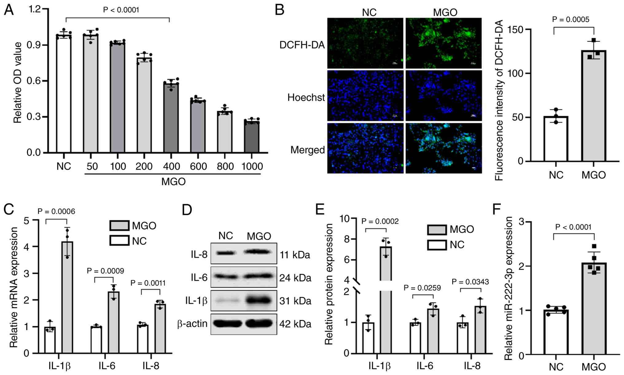
HMEC-1 cells were exposed to increasing concentrations of MGO (50,
100, 200, 400, 600, 800 and 1,000 μmol/l). CCK-8 assay
revealed that MGO inhibited cell proliferation in a
concentration-dependent manner (Fig.
1A). At 400 μmol/l, MGO induced a marked reduction in
proliferation, while maintaining sufficient cell viability for
subsequent functional assays. Thus, 400 μmol/l MGO was used
in subsequent experiments. To further validate the
pathophysiological relevance of the selected MGO concentration, ROS
production and pro-inflammatory cytokine expression in HMEC-1 cells
treated with 400 μmol/l MGO were assessed. The results
revealed that intracellular ROS levels were significantly elevated
in the MGO-treated group compared with the control group (Fig. 1B). Moreover, MGO treatment
markedly upregulated the expression levels of IL-1β, IL-6 and IL-8
(Fig. 1C-E). These results
demonstrated that 400 μmol/l MGO effectively induced
oxidative stress and inflammatory responses in HMEC-1 cells,
supporting its suitability for use in subsequent experiments as a
relevant pathophysiological stimulus. The present study then
investigated whether MGO treatment affects miR-222-3p expression.
RT-qPCR revealed that the miR-222-3p levels were significantly
elevated in the MGO-treated cells compared with the untreated
controls (Fig. 1F). This finding
indicates that MGO-induced cellular stress is accompanied by the
upregulation of miR-222-3p expression.
miR-222-3p overexpression aggravates
MGO-induced endothelial dysfunction
To further explore the role of miR-222-3p in
MGO-treated HMEC-1 cells, we transfected a miR-222-3p mimic to
overexpress miR-222-3p. Transfection efficiency was confirmed by
RT-qPCR (Fig. 2A). miR-222-3p
expression was significantly higher in the MGO + miR-222-3p group
than in the MGO + miR-con group, confirming successful
transfection. Subsequently, cell function was then evaluated by
CCK-8 assay for cell proliferation, flow cytometry for apoptosis,
scratch assay for migration ability, and tube formation assay for
angiogenic capacity (Fig. 2B-E).
Compared with the NC group, the MGO group exhibited a significantly
reduced proliferation, increased apoptosis, impaired migration and
diminished tube formation, as reflected by fewer total branch
points and a shorter total tube length. Furthermore, compared with
the MGO + miR-con group, the MGO + miR-222-3p group exhibited
further reductions in proliferation, migration and tube formation,
along with a further increase in apoptosis. These results indicate
that the overexpression of miR-222-3p exacerbates MGO-induced
injury in microvascular endothelial cells.
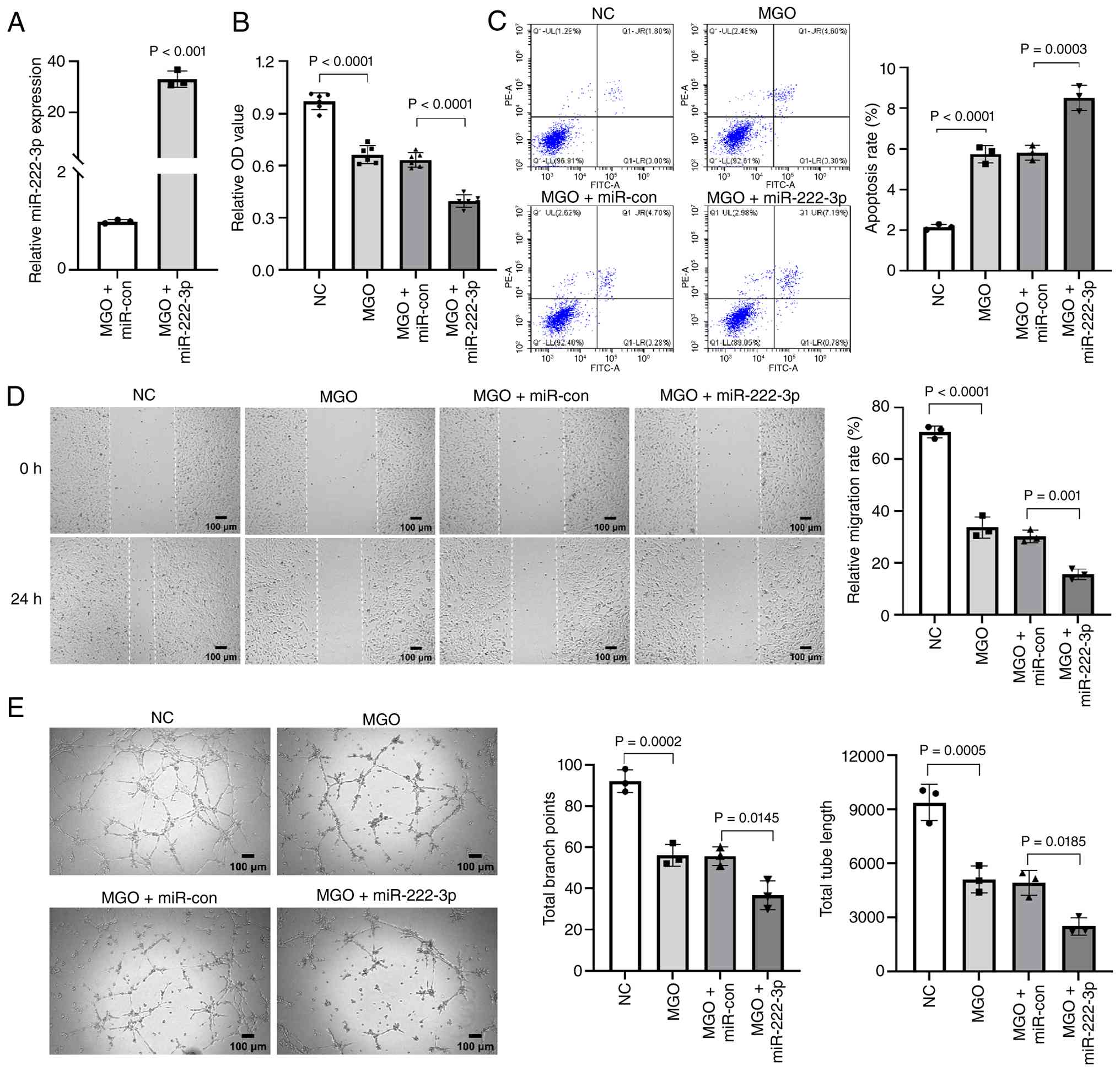 | Figure 2miR-222-3p overexpression aggravates
MGO-induced endothelial dysfunction. (A) miR-222-3p expression
levels in the MGO + miR-con and MGO + miR-222-3p groups were
detected using reverse transcription-quantitative PCR. (B) Cell
proliferation in the NC, MGO, MGO + miR-con and MGO + miR-222-3p
groups was assessed using CCK-8 assay. (C) Apoptosis in the NC,
MGO, MGO + miR-con, and MGO + miR-222-3p groups was detected using
flow cytometry. (D) Cell migration in the NC, MGO, MGO + miR-con,
and MGO + miR-222-3p groups was evaluated using scratch assay.
Scale bars, 100 μm. (E) Tube formation in the NC, MGO, MGO +
miR-con, and MGO + miR-222-3p groups was assessed using tube
formation assay. Scale bars, 100 μm. All data are presented
as the mean ± SD. Statistical analysis was carried out using an
independent samples t-test or one-way ANOVA with Tukey's multiple
comparisons test (n=3 or 6 biologically independent samples). NC,
negative control; MGO, methylglyoxal. |
miR-222-3p negatively regulates APLN
Through the analysis of the ENCORI/starBase database
(http://starbase.sysu.edu.cn/), a
nucleotide sequence within the 3'UTR of APLN mRNA with the
potential to bind miR-222-3p was identified, suggesting APLN as a
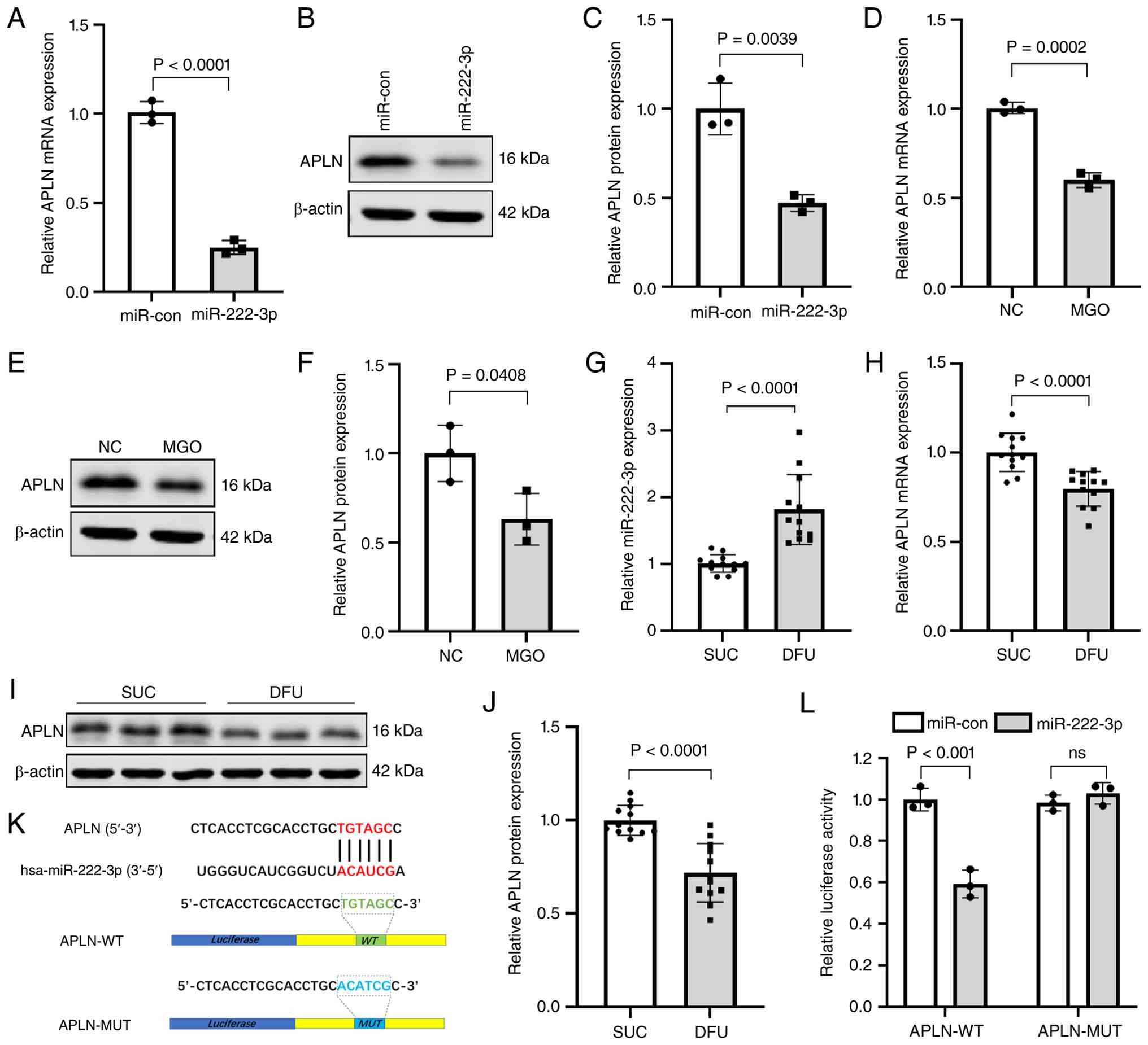
downstream target of miR-222-3p. To examine this, miR-222-3p was
first overexpressed in HMEC-1 cells by transfection with a
miR-222-3p mimic. The results of RT-qPCR and western blot analysis
revealed that the overexpression of miR-222-3p significantly
reduced both the APLN mRNA and protein levels compared with the
miR-con group (Fig. 3A-C). Given
that MGO treatment upregulated miR-222-3p expression, the present
study then determined whether MGO can similarly suppress APLN
expression. Indeed, in the MGO-treated HMEC-1 cells, both the APLN
mRNA and protein levels were markedly decreased relative to the NC
group (Fig. 3D-F), consistent
with endogenous miR-222-3p upregulation under diabetic-like stress
conditions. This inverse association was further validated in
clinical specimens. The expression levels of miR-222-3p and APLN
were simultaneously examined in tissues from patients with DFU and
tissues from individuals with non-diabetic chronic lower limb ulcer
(SUC). Compared with the SUC tissues, DFU tissues exhibited a
significantly elevated expression of miR-222-3p and markedly
reduced mRNA and protein levels of APLN (Fig. 3G-J), demonstrating a negative
association between the two molecules in human diseased tissues. A
dual-luciferase reporter assay was then performed to confirm the
direct interaction. Co-transfection of the miR-222-3p mimic with
the wild-type APLN 3'UTR reporter plasmid resulted in a marked
decrease in luciferase activity compared with the miR-con group
(Fig. 3K and L), whereas no
significant difference in luciferase activity was observed between
the two groups when the mutant plasmid was co-transfected.
Collectively, these findings confirm that APLN is a direct
downstream target of miR-222-3p, and this regulatory axis is
conserved from in vitro MGO-induced stress to human DFU
pathology.
Silencing of APLN mimics
miR-222-3p-induced endothelial dysfunction
Having confirmed that APLN is directly targeted and
downregulated by miR-222-3p, the present study then wished to
examine whether the loss of APLN itself is sufficient to drive the
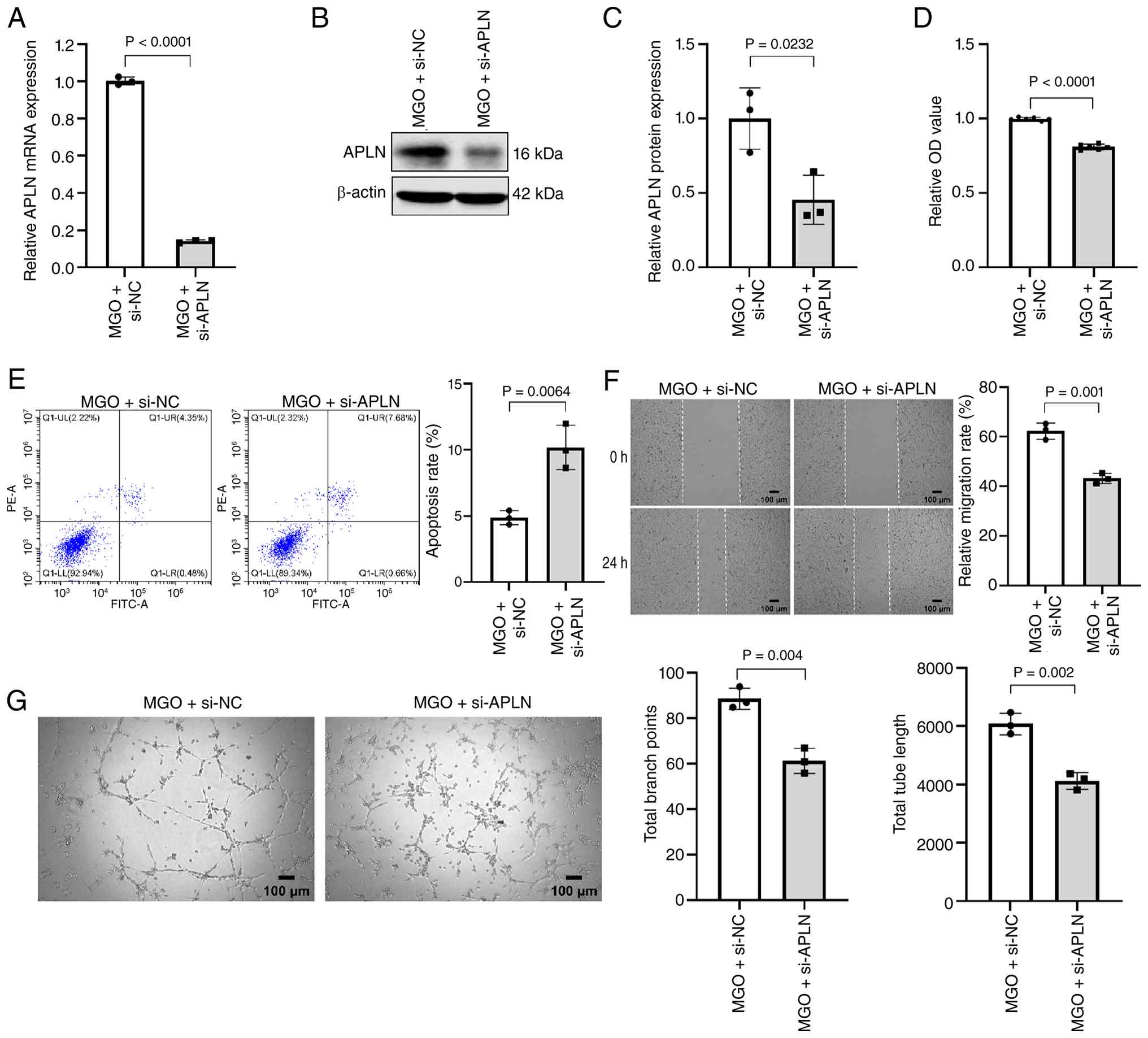
endothelial dysfunction caused by miR-222-3p. To this end, APLN was
silenced in MGO-treated HMEC-1 cells using siRNA. RT-qPCR and
western blot analysis were performed to assess the APLN mRNA and
protein levels (Fig. 4A-C).
Compared with the MGO + si-NC group, APLN mRNA and protein
expression was significantly reduced in the MGO + si-APLN group,
confirming the successful knockdown of APLN. Functional assays,
including CCK-8 assay, flow cytometry, scratch assay and tube
formation assay, were used to evaluate endothelial cell
proliferation, apoptosis, migration and tube formation (Fig. 4D-G). In comparison with the MGO +
si-NC group, the MGO + si-APLN group demonstrated a decline in cell
proliferation activity, an elevation in the number of apoptotic
cells, a reduction in cell migration capacity, and a decrease in
both the total number of branch points and the total tube length of
tubule formation. These data demonstrate that the silencing of APLN
mimics the detrimental effects of miR-222-3p, reinforcing APLN as a
critical functional mediator downstream of miR-222-3p.
Restoration of APLN attenuates
miR-222-3p-induced endothelial dysfunction
To determine whether APLN functionally mediates
miR-222-3p-induced endothelial injury, rescue experiments were
performed. Apelin-13, the most active subtype produced by the
enzymatic cleavage of the APLN-encoded precursor protein, was used
for exogenous supplementation. Previous studies have demonstrated
that Apelin-13 activates the human APJ receptor with a half-maximal
effective concentration (EC50) of ~0.3 nM and exerts
concentration-dependent pro-proliferative, anti-apoptotic and
endothelial protective effects within the 1-1,000-nM range
(32-35). Accordingly, in the present study,
a concentration of 50 ng/ml (~32 nM) Apelin-13 was selected, which
far exceeds the receptor activation threshold, is sufficient to
saturate the APJ receptor, and falls well within the established
bioactive window. In parallel, APLN expression was restored at the
genetic level by transfecting an APLN overexpression plasmid
(oe-APLN). The successful overexpression was confirmed by RT-qPCR
and western blot analysis, which revealed markedly elevated APLN
mRNA and protein levels in the oe-APLN group relative to the
control (Fig. 5A-C).
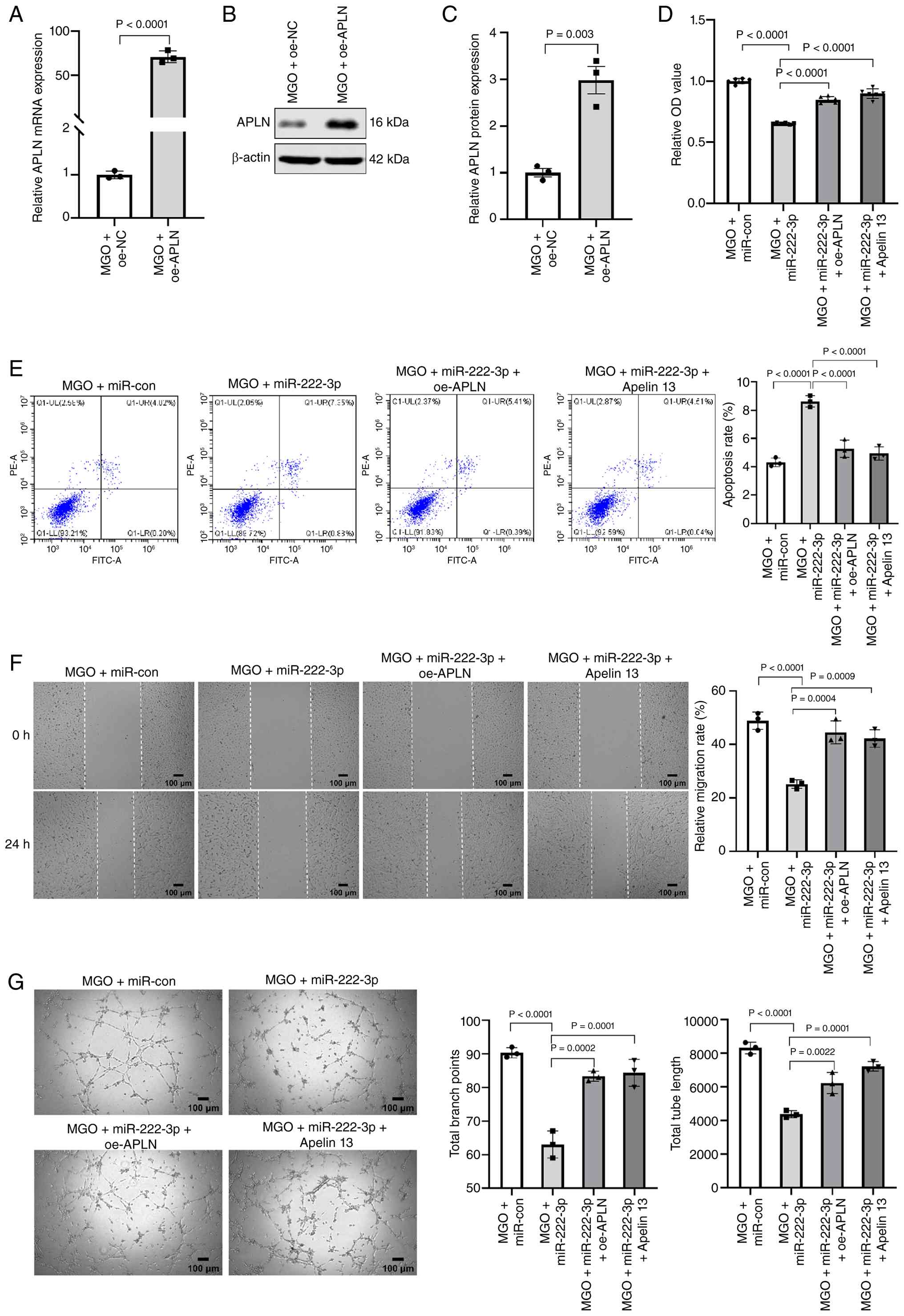 | Figure 5Restoration of APLN expression
attenuates miR-222-3p-induced endothelial dysfunction. (A) The mRNA
expression of APLN in the MGO + oe-NC and MGO + oe-APLN groups was
assessed using reverse transcription-quantitative PCR. (B and C)
APLN protein expression in the MGO + oe-NC and MGO + oe-APLN groups
was detected using western blot analysis. (D) The cell
proliferative activity in the MGO + miR-con, MGO + miR-222-3p, MGO
+ miR-222-3p + oe-APLN and MGO + miR-222-3p + Apelin-13 groups was
measured using CCK-8 assay. (E) Apoptosis in the MGO + miR-con, MGO
+ miR-222-3p, MGO + miR-222-3p + oe-APLN and MGO + miR-222-3p +
Apelin-13 groups was detected using flow cytometry. (F) Migration
ability in the MGO + miR-con, MGO + miR-222-3p, MGO + miR-222-3p +
oe-APLN and MGO + miR-222-3p + Apelin-13 groups was evaluated using
the scratch assay. Scale bars, 100 μm. (G) Tube formation
ability of cells in the MGO + miR-con, MGO + miR-222-3p, MGO +
miR-222-3p + oe-APLN and MGO + miR-222-3p + Apelin-13 groups was
assessed using tube formation assay. Scale bars, 100 μm. All
data are presented as the mean ± SD. Statistical analysis was
carried out using an independent samples t-test or one-way ANOVA
with Tukey's multiple comparisons test (n=3 or 6 biologically
independent samples). APLN, apelin; NC, negative control; MGO,
methylglyoxal. |
In the MGO-treated HMEC-1 cells, the overexpression
of miR-222-3p significantly reduced cell proliferation, migration
and tube formation, and markedly increased apoptosis compared with
the control. Notably, both exogenous Apelin-13 supplementation and
APLN overexpression significantly reversed these endothelial
dysfunctions, as evidenced by restored proliferation, migration and
tube formation, as well as reduced apoptosis (Fig. 5D-G). The highly consistent
effects of the two rescue strategies collectively indicate that
APLN is a critical downstream target required for
miR-222-3p-mediated impairment of microvascular endothelial
function.
miR-222-3p impairs skin wound healing in
mice
To further evaluate the impact of miR-222-3p on skin
wound healing in vivo, both gain- and loss-of-function
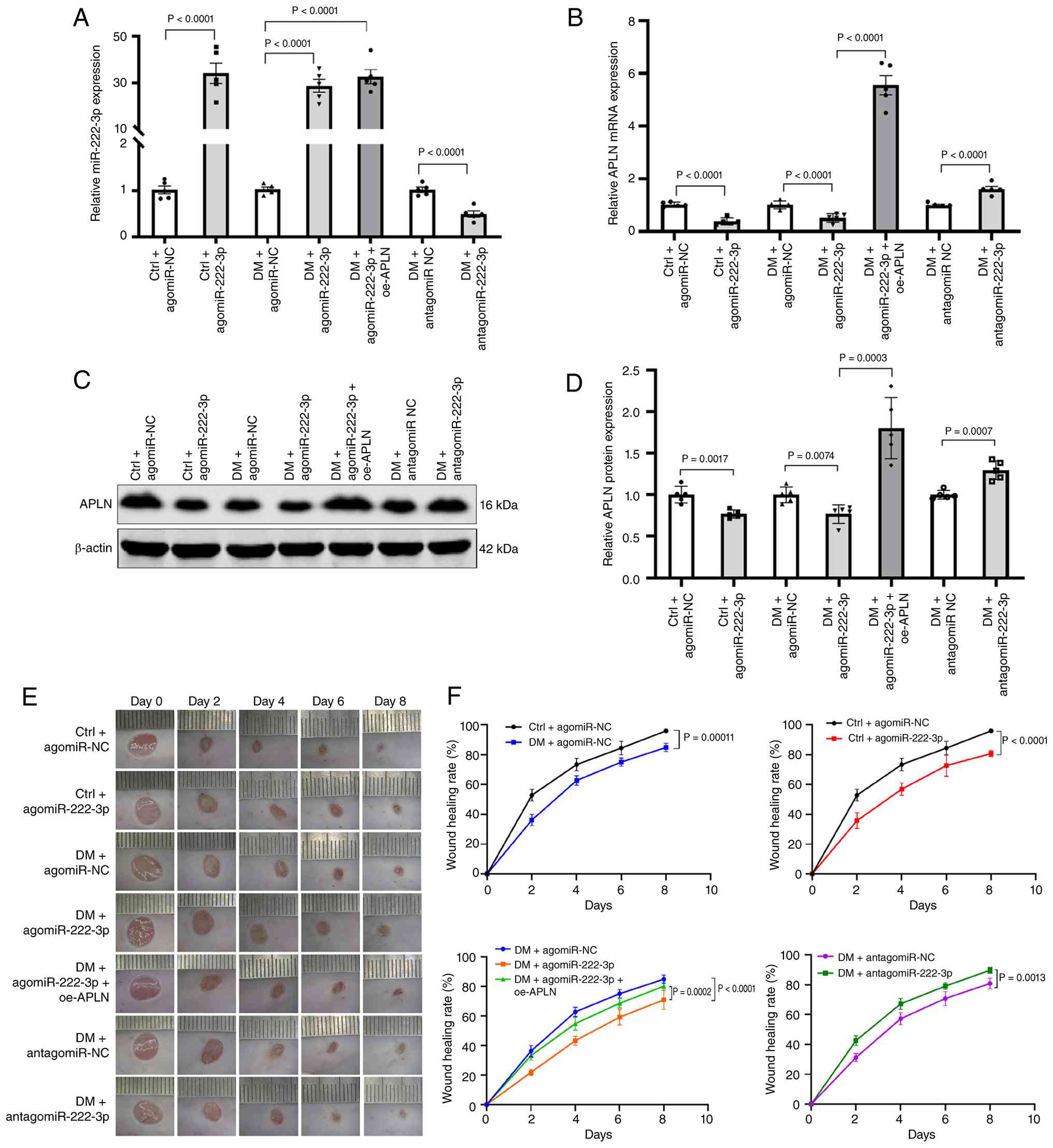
experiments were performed in normal and diabetic mice. miR-222-3p
agomiR and its negative control (agomiR-NC) were injected locally
into the peri-wound area of the dorsal skin in both diabetic and
normal mice. The results revealed that, compared with the
respective agomiR-NC groups, the miR-222-3p levels in the
peri-wound skin were significantly elevated in the agomiR-222-3p
groups of both diabetic and normal mice, while APLN expression was
markedly reduced (Fig. 6A-D).
Wound healing analysis demonstrated that, relative to their
agomiR-NC controls, the agomiR-222-3p groups exhibited
significantly delayed wound closure in both the diabetic and normal
mice, indicating that miR-222-3p overexpression alone is sufficient
to inhibit skin wound healing. To further determine whether this
inhibitory effect is mediated by APLN downregulation, a rescue
group in diabetic mice was established, in which APLN was
overexpressed in the peri-wound skin via AAV vector prior to
agomiR-222-3p treatment. The results revealed that APLN
overexpression effectively reversed the delayed wound healing
caused by agomiR-222-3p (Fig. 6E and
F).
To assess whether silencing endogenous miR-222-3p
promotes diabetic wound healing, DM + antagomiR-NC and DM +
antagomiR-222-3p groups were established, in which miR-222-3p
antagomiR was injected locally into the peri-wound area of diabetic
mice. Compared with the antagomiR-NC group, the antagomiR-222-3p
group exhibited significantly decreased peri-wound miR-222-3p
levels, an elevated expression of APLN, and markedly accelerated
wound closure, confirming the negative regulatory role of
miR-222-3p in wound repair from the loss-of-function perspective
(Fig. 6A-F).
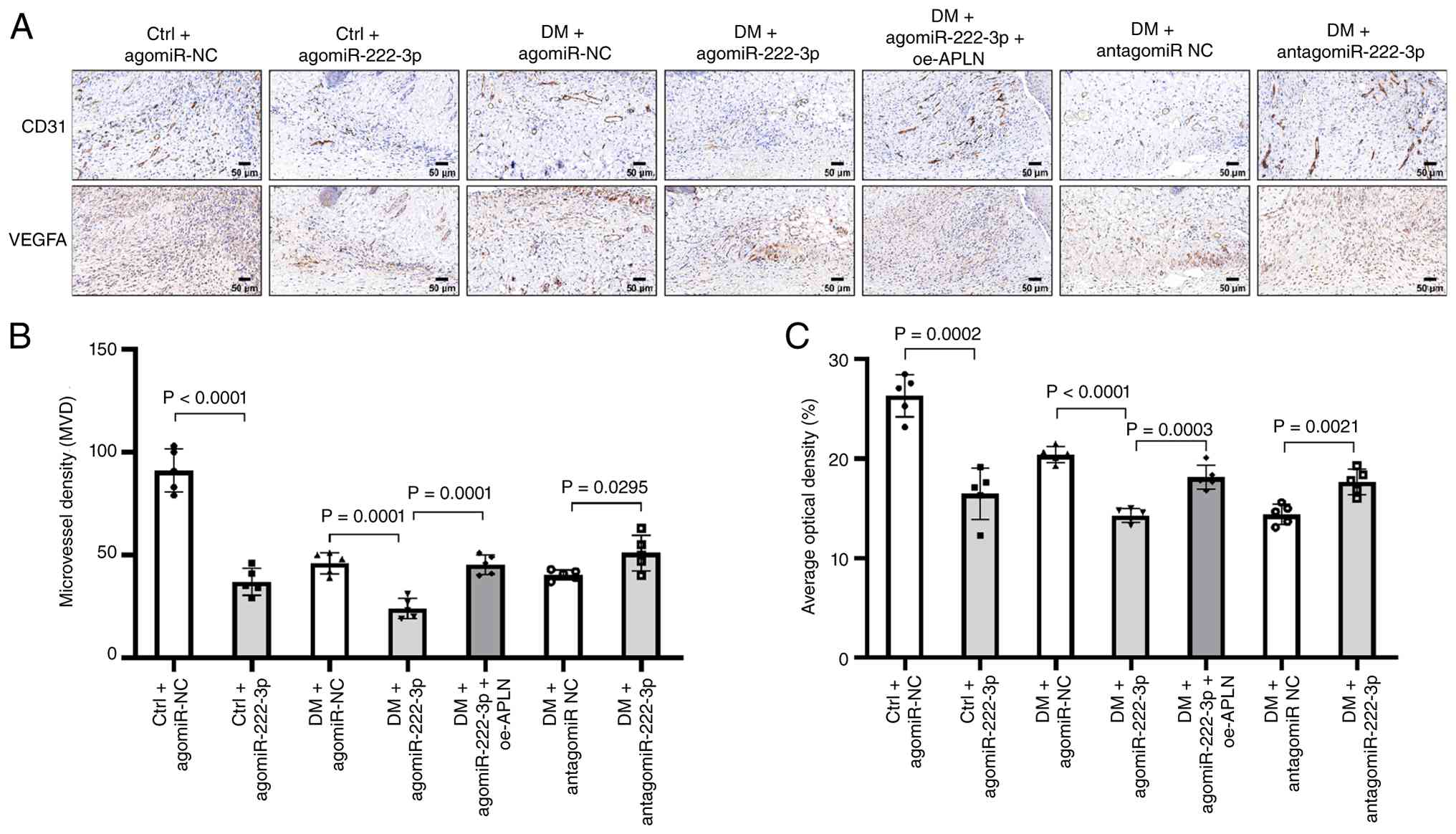
To provide histopathological evidence for the
underlying mechanisms, immunohistochemical staining for VEGFA and
CD31 was performed on peri-wound tissues from all groups. In
diabetic mice, the agomiR-222-3p group exhibited a reduced VEGFA
and CD31 expression compared with the control group, whereas the
antagomiR-222-3p group exhibited the opposite pattern. In normal
mice, agomiR-222-3p similarly led to downregulation of VEGFA and
CD31. Notably, in the rescue experiment, APLN overexpression
significantly restored the VEGFA and CD31 levels, providing
histopathological evidence that APLN is a key downstream effector
through which miR-222-3p impairs wound angiogenesis (Fig. 7A-C). Taken together, these
gain-and loss-of-function results demonstrate that miR-222-3p
impairs skin wound healing in vivo by inhibiting
angiogenesis.
Discussion
MGO serves as a primary contributor to glycation and
oxidative stress. Extensive research has demonstrated that the
substantial accumulation of MGO plays a critical role in hindering
wound healing and tissue repair in individuals with diabetes
(36-39). Pang et al (40) demonstrated that the
glyoxalase-mediated removal of MGO promoted the wound healing of
DFUs by modulating the diabetic microenvironment and restoring the
function of diabetic stem cells. MGO markedly decreases the protein
expression of factors associated with angiogenesis, resulting in
hindered neovascular formation. Conversely, the application of
methylglyoxal scavengers improves MGO-induced endothelial cell
dysfunction through the reduction of ROS generation, the
suppression of cell apoptosis and the enhancement of angiogenic
factor levels (41). Bone
marrow-derived progenitor cells, crucial for vascular repair and
angiogenesis, exhibit impaired tube formation, migration,
proliferation and increased apoptosis when exposed to MGO (42). The present study confirmed that
treatment of the HMEC-1 cells with MGO significantly elevated
intracellular ROS levels and upregulated the expression of
pro-inflammatory cytokines (IL-1β, IL-6 and IL-8), demonstrating
the induction of glycative stress and inflammatory responses.
Consistent with these findings, functional assays revealed that MGO
exposure decreased cell proliferation, increased apoptosis,
impaired migration and reduced tube formation, underscoring the
detrimental role of MGO in endothelial injury and dysfunction.
Notably, these phenotypic changes were accompanied by a marked
upregulation of miR-222-3p, suggesting a potential involvement of
miR-222-3p in mediating MGO-induced endothelial damage. However,
the specific mechanism by which MGO regulates miR-222-3p expression
warrants further investigation.
Insufficient angiogenesis and impaired blood supply
due to endothelial cell dysfunction are major contributors to
delayed wound healing. miR-222 is abundantly expressed in
endothelial cells and plays a critical role in their function and
regulation (43). Endothelial
progenitor cells (EPCs) are crucial precursor cells that, under
normal conditions, are rapidly mobilized from the bone marrow upon
vascular injury and differentiate into mature vascular endothelial
cells under the stimulation of various secreted cytokines, thereby
playing a vital role in vascular repair and neovascularization.
However, EPC function is often impaired in a high-glucose
environment (44). A previous
study indicated that the downregulation of miR-222-3p enhanced EPC
function by activating the AMPK signaling pathway through targeting
ADIPOR1, thereby promoting EPC migration, invasion and recruitment
(45). miR-222-3p is highly
expressed in endothelial cells of atherosclerotic vascular intima.
Its upregulation inhibits PGC-1α protein production and reduces
mitochondrial quantity, thereby inducing excessive ROS generation
and apoptosis in human aortic endothelial cells, leading to
endothelial cell injury and dysfunction (46). In the present study, HMEC-1 cells
treated with MGO and subjected to miR-222-3p overexpression
exhibited a further exacerbation in microvascular endothelial cell
injury and dysfunction, as evidenced by further reductions in
proliferation, migration and tube formation, and a further increase
in apoptosis. Consistent with these in vitro findings, the
local application of miR-222-3p agomiR significantly delayed wound
healing in both normal and diabetic mice, whereas the silencing of
endogenous miR-222-3p with antagomiR in diabetic wounds markedly
accelerated wound closure. Histopathological analysis further
revealed that miR-222-3p agomiR suppressed VEGFA and CD31
expression in peri-wound tissues, while antagomir treatment
reversed these changes. Collectively, these results demonstrate
that miR-222-3p functions as an anti-angiogenic miRNA that directly
impairs microvascular endothelial cell function and inhibits skin
wound healing.
miRNAs typically function by binding to the mRNA of
downstream targets, inhibiting translation or promoting mRNA
degradation (47). In the
present study, miR-222-3p overexpression in HMEC-1 cells
significantly suppressed APLN expression at both the mRNA and
protein levels. Notably, MGO treatment similarly reduced APLN mRNA
and protein expression, accompanied by a concomitant increase in
endogenous miR-222-3p expression, indicating that this regulatory
association is recapitulated under diabetic-like stress.
Dual-luciferase reporter assays confirmed that APLN is a direct
downstream target negatively regulated by miR-222-3p through
sequence-specific binding to its 3'-UTR. In vivo, the
elevated expression of miR-222-3p in the peri-wound tissue of
diabetic mice was accompanied by the decreased expression of APLN.
This inverse association was also observed in clinical specimens,
where DFU tissues exhibited significantly elevated miR-222-3p
levels and markedly reduced APLN expression levels compared with
non-diabetic chronic ulcer tissues, providing translational
relevance to the in vitro findings. Numerous studies have
highlighted the importance of APLN in angiogenesis. Apelin
signaling activates endothelial cells, driving them toward a highly
migratory and proliferative state, thereby facilitating
angiogenesis. Zebrafish lacking apelin signaling exhibit defects in
endothelial tip cell morphology and sprouting (48). Moreover, apelin inhibits the
NF-κB pathway and promotes endothelial cell proliferation upon
binding to its receptor, alleviating hyperglycemia-induced
microvascular dysfunction and inflammation. In vivo research
has demonstrated that apelin enhances VEGFR2 expression, which
promotes angiogenesis and increases microvessel density in the
cardiac endothelial cells of diabetic mice (49). In the present study, the
silencing of APLN expression in HMEC-1 cells through siRNA resulted
in decreased proliferation and migration, increased apoptosis and
reduced tube formation, indicating that the downregulation of APLN
impairs endothelial cell function. Conversely, both exogenous
Apelin-13 supplementation and APLN overexpression significantly
rescued miR-222-3p-induced endothelial dysfunction in vitro.
Notably, in vivo rescue experiments further demonstrated
that APLN overexpression in the peri-wound skin effectively
reversed agomiR-222-3p-induced delay in wound healing and restored
angiogenesis markers in diabetic mice. Taken together, these
findings establish a mechanistic cascade in which miR-222-3p
directly targets APLN, suppresses its expression and thereby
impairs microvascular endothelial function, ultimately contributing
to delayed wound healing in diabetes.
The present study has several limitations which
should be mentioned. First, although the Apelin-13 concentration
used in the present study was selected based on published
pharmacological evidence and falls within the established bioactive
window, future dose-response studies will help to determine the
optimal concentration for therapeutic application. Second, all
in vitro experiments in the present study were performed
using the human dermal microvascular endothelial cell line
(HMEC-1). HMEC-1 is the first immortalized endothelial cell line
successfully established from human dermal microvasculature, and it
retains the morphological, phenotypic and functional
characteristics of primary endothelial cells, including cobblestone
morphology, the secretion of von Willebrand factor, the uptake of
acetylated low-density lipoprotein, tube formation on Matrigel, and
the expression of key markers, such as CD31, intercellular adhesion
molecule-1 and vascular cell adhesion molecule-1 (50). This cell line has been
extensively used and validated in studies on diabetic skin wound
healing, angiogenesis, and diabetic complications (51-54). However, as an immortalized cell
line, HMEC-1 may not fully recapitulate all physiological
properties of primary human dermal microvascular endothelial cells.
Therefore, while HMEC-1 provides a stable and reliable tool for
mechanistic exploration in the present study, future investigations
using primary cells are warranted to validate the key findings and
further confirm the translational relevance of our conclusions.
Collectively, the in vitro and in vivo
findings of the present study demonstrate that miR-222-3p impairs
microvascular endothelial function and delays diabetic wound
healing by directly targeting APLN. These results reveal a novel
pathogenic mechanism in DFU and highlight the miR-222-3p/APLN axis
as a potential therapeutic target for angiogenic therapies.
Supplementary Data
Availability of data and materials
The data generated in the present study may be
requested from the corresponding author.
Authors' contributions
RJ, YT and TW performed the experiments, acquired
the data, and participated in data analysis and interpretation. CZ
and MS assisted with the animal experiments and data acquisition.
XZ and MC conceived and designed the study, supervised the
research, and participated in data interpretation. XZ and MC
confirm the authenticity of all the raw data. All authors
contributed to the drafting and revision of the manuscript, read
and approved the final version, agreed on the journal for
submission, and are accountable for all aspects of the work.
Ethics approval and consent to
participate
All tissue specimens were collected at the First
Affiliated Hospital of Anhui Medical University (Hefei, China).
Written informed consent was obtained from all participants, and
the study was approved by the Medical Ethics Committee of the First
Affiliated Hospital of Anhui Medical University (Ethics batch no.
P20210039). Animal experiments were conducted in accordance with
the 'Methods for Quality Control of Laboratory Animals' and the
'Regulations of the People's Republic of China on the
Administration of Laboratory Animals', and were approved by the
Experimental Animal Ethics Committee of Anhui Medical University
(approval no. LLSC20201040).
Patient consent for publication
Not applicable.
Competing interests
The authors declare that they have no competing
interests.
Acknowledgements
The authors would like to express their gratitude to
Professor Liu Lei of the Department of Endocrinology, First
Affiliated Hospital of Anhui Medical University (Hefei, China), for
providing technical guidance. The authors would also like to thank
the Central Laboratory of the First Affiliated Hospital of Anhui
Medical University for providing the experimental equipment.
Funding
The present study was supported by the Natural Science
Foundation of Anhui Province, China (grant no. 2108085MH269), the
Natural Science Research Project of Colleges and Universities in
Anhui Province (grant no. KJ2021A0274) and the Clinical Medical
Research Translation Special Project of Anhui Province in China
(grant no. 202427b10020133).
References
|
1
|
Walsh JW, Hoffstad OJ, Sullivan MO and
Margolis DJ: Association of diabetic foot ulcer and death in a
population-based cohort from the United Kingdom. Diabet Med.
33:1493–1498. 2016. View Article : Google Scholar : PubMed/NCBI
|
|
2
|
Armstrong DG, Tan TW, Boulton A and Bus
SA: Diabetic foot ulcers: A review. JAMA. 330:62–75. 2023.
View Article : Google Scholar : PubMed/NCBI
|
|
3
|
Wang N and Ju S: Research advances on the
mechanism of refractory healing of diabetic foot ulcer. Zhonghua
Shao Shang Yu Chuang Mian Xiu Fu Za Zhi. 38:1085–1089. 2022.In
Chinese. PubMed/NCBI
|
|
4
|
Rai V, Moellmer R and Agrawal DK: Stem
cells and angiogenesis: Implications and limitations in enhancing
chronic diabetic foot ulcer healing. Cells. 11:22872022. View Article : Google Scholar : PubMed/NCBI
|
|
5
|
Urbich C, Kuehbacher A and Dimmeler S:
Role of microRNAs in vascular diseases, inflammation, and
angiogenesis. Cardiovasc Res. 79:581–588. 2008. View Article : Google Scholar : PubMed/NCBI
|
|
6
|
Saliminejad K, Khorram Khorshid HR,
Soleymani Fard S and Ghaffari SH: An overview of microRNAs:
Biology, functions, therapeutics, and analysis methods. J Cell
Physiol. 234:5451–5465. 2019. View Article : Google Scholar
|
|
7
|
Xu YX, Pu SD, Li X, Yu ZW, Zhang YT, Tong
XW, Shan YY and Gao XY: Exosomal ncRNAs: Novel therapeutic target
and biomarker for diabetic complications. Pharmacol Res.
178:1061352022. View Article : Google Scholar : PubMed/NCBI
|
|
8
|
Goodarzi G, Maniati M and Qujeq D: The
role of microRNAs in the healing of diabetic ulcers. Int Wound J.
16:621–633. 2019. View Article : Google Scholar : PubMed/NCBI
|
|
9
|
Wu T, Xie D, Zhao X, Xu M, Luo L, Deng D
and Chen M: Enhanced expression of mir-34c in peripheral plasma
associated with diabetic foot ulcer in type 2 diabetes patients.
Diabetes Metab Syndr Obes. 14:4263–4273. 2021. View Article : Google Scholar : PubMed/NCBI
|
|
10
|
Zhao X, Xu M, Tang Y, Xie D, Deng L, Chen
M and Wang Y: Decreased expression of miR-204-3p in peripheral
blood and wound margin tissue associated with the onset and poor
wound healing of diabetic foot ulcers. Int Wound J. 20:413–429.
2023. View Article : Google Scholar :
|
|
11
|
Xu M, Li Y, Tang Y, Zhao X, Xie D and Chen
M: Increased expression of miR-155 in peripheral blood and wound
margin tissue of type 2 diabetes mellitus patients associated with
diabetic foot ulcer. Diabetes Metab Syndr Obes. 15:3415–3428. 2022.
View Article : Google Scholar : PubMed/NCBI
|
|
12
|
Jie R, Qian J, Tang Y, Li Y, Xu M, Zhao X
and Chen M: Role of increased miR-222-3p expression in peripheral
blood and wound marginal tissues of type 2 diabetes mellitus
patients with diabetic foot ulcer. Diabetes Metab Syndr Obes.
16:2419–2432. 2023. View Article : Google Scholar : PubMed/NCBI
|
|
13
|
Rabbani N: Methylglyoxal and glyoxalase
1-a metabolic stress pathway-linking hyperglycemia to the unfolded
protein response and vascular complications of diabetes. Clin Sci
(Lond). 136:819–824. 2022. View Article : Google Scholar : PubMed/NCBI
|
|
14
|
Bellier J, Nokin MJ, Lardé E, Karoyan P,
Peulen O, Castronovo V and Bellahcène A: Methylglyoxal, a potent
inducer of AGEs, connects between diabetes and cancer. Diabetes Res
Clin Pract. 148:200–211. 2018. View Article : Google Scholar
|
|
15
|
Schalkwijk CG and Stehouwer CDA:
Methylglyoxal, a highly reactive dicarbonyl compound, in diabetes,
its vascular complications, and other age-related diseases. Physiol
Rev. 100:407–461. 2020. View Article : Google Scholar
|
|
16
|
Stratmann B: Dicarbonyl stress in diabetic
vascular disease. Int J Mol Sci. 23:61862022. View Article : Google Scholar : PubMed/NCBI
|
|
17
|
Schalkwijk CG, Micali LR and Wouters K:
Advanced glycation endproducts in diabetes-related macrovascular
complications: focus on methylglyoxal. Trends Endocrinol Metab.
34:49–60. 2023. View Article : Google Scholar
|
|
18
|
Berlanga J, Cibrian D, Guillén I, Freyre
F, Alba JS, Lopez-Saura P, Merino N, Aldama A, Quintela AM, Triana
ME, et al: Methylglyoxal administration induces diabetes-like
microvascular changes and perturbs the healing process of cutaneous
wounds. Clin Sci (Lond). 109:83–95. 2005. View Article : Google Scholar : PubMed/NCBI
|
|
19
|
D'aniello C, Lonardo E, Iaconis S,
Guardiola O, Liguoro AM, Liguori GL, Autiero M, Carmeliet P and
Minchiotti G: G protein-coupled receptor APJ and its ligand apelin
act downstream of Cripto to specify embryonic stem cells toward the
cardiac lineage through extracellular signal-regulated kinase/p70S6
kinase signaling pathway. Circ Res. 105:231–238. 2009. View Article : Google Scholar : PubMed/NCBI
|
|
20
|
Flahault A, Girault-Sotias PE, Keck M,
Alvear-Perez R, De Mota N, Estéoulle L, Ramanoudjame SM, Iturrioz
X, Bonnet D and Llorens-Cortes C: A metabolically stable apelin-17
analog decreases AVP-induced antidiuresis and improves
hyponatremia. Nat Commun. 12:3052021. View Article : Google Scholar : PubMed/NCBI
|
|
21
|
Malchow J, Eberlein J, Li W, Hogan BM,
Okuda KS and Helker CSM: Neural progenitor-derived Apelin controls
tip cell behavior and vascular patterning. Sci Adv.
10:eadk11742024. View Article : Google Scholar : PubMed/NCBI
|
|
22
|
Son JS, Zhao L, Chen Y, Chen K, Chae SA,
de Avila JM, Wang H, Zhu MJ, Jiang Z and Du M: Maternal exercise
via exerkine apelin enhances brown adipogenesis and prevents
metabolic dysfunction in offspring mice. Sci Adv. 6:eaaz03592020.
View Article : Google Scholar : PubMed/NCBI
|
|
23
|
Pitkin SL, Maguire JJ, Bonner TI and
Davenport AP: International union of basic and clinical
pharmacology. LXXIV. Apelin receptor nomenclature, distribution,
pharmacology, and function. Pharmacol Rev. 62:331–342. 2010.
View Article : Google Scholar : PubMed/NCBI
|
|
24
|
Cox CM, D'agostino SL, Miller MK, Heimark
RL and Krieg PA: Apelin, the ligand for the endothelial
G-protein-coupled receptor, APJ, is a potent angiogenic factor
required for normal vascular development of the frog embryo. Dev
Biol. 296:177–189. 2006. View Article : Google Scholar : PubMed/NCBI
|
|
25
|
Inui M, Fukui A, Ito Y and Asashima M:
Xapelin and Xmsr are required for cardiovascular development in
Xenopus laevis. Dev Biol. 298:188–200. 2006. View Article : Google Scholar : PubMed/NCBI
|
|
26
|
Frisch A, Kälin S, Monk R, Radke J,
Heppner FL and Kälin RE: Apelin controls angiogenesis-dependent
glioblastoma growth. Int J Mol Sci. 21:41792020. View Article : Google Scholar : PubMed/NCBI
|
|
27
|
Livak KJ and Schmittgen TD: Analysis of
relative gene expression data using real-time quantitative PCR and
the 2(-Delta Delta C(T)) method. Methods. 25:402–408. 2001.
View Article : Google Scholar
|
|
28
|
Chen X, Tong G, Fan J, Shen Y, Wang N,
Gong W, Hu Z, Zhu K, Li X, Jin L, et al: FGF21 promotes migration
and differentiation of epidermal cells during wound healing via
SIRT1-dependent autophagy. Br J Pharmacol. 179:1102–1121. 2022.
View Article : Google Scholar
|
|
29
|
Xiang H, Ding P, Qian J, Lu E, Sun Y, Lee
S and Zhao Z, Sun Z and Zhao Z: Exosomes derived from minor
salivary gland mesenchymal stem cells: A promising novel exosome
exhibiting pro-angiogenic and wound healing effects similar to
those of adipose-derived stem cell exosomes. Stem Cell Res Ther.
15:4622024. View Article : Google Scholar : PubMed/NCBI
|
|
30
|
Weiss E, Schrüfer A, Tocantins C, Diniz
MS, Novakovic B, van Bergen AS, Kulovic-Sissawo A, Saffery R, Boon
RA and Hiden U: Higher gestational weight gain delays wound healing
and reduces expression of long non-coding RNA KLRK1-AS1 in neonatal
endothelial progenitor cells. J Physiol. 601:3961–3974. 2023.
View Article : Google Scholar : PubMed/NCBI
|
|
31
|
Yang C, Zhao Y, Xian M, Li JH, Dong Q, Bai
HB, Xu JD and Zhang MF: A novel controllable hydrogen
sulfide-releasing molecule protects human skin keratinocytes
against methylglyoxal-induced injury and dysfunction. Cell Physiol
Biochem. 34:1304–1317. 2014. View Article : Google Scholar : PubMed/NCBI
|
|
32
|
Tatemoto K, Hosoya M, Habata Y, Fujii R,
Kakegawa T, Zou MX, Kawamata Y, Fukusumi S, Hinuma S, Kitada C, et
al: Isolation and characterization of a novel endogenous peptide
ligand for the human APJ receptor. Biochem Biophys Res Commun.
251:471–476. 1998. View Article : Google Scholar : PubMed/NCBI
|
|
33
|
Zhang BH, Wang W, Wang H, Yin J and Zeng
XJ: Promoting effects of the adipokine, apelin, on diabetic
nephropathy. PLoS One. 8:e604572013. View Article : Google Scholar : PubMed/NCBI
|
|
34
|
Yang X, Zhu W, Zhang P, Chen K, Zhao L, Li
J, Wei M and Liu M: Apelin-13 stimulates angiogenesis by promoting
cross-talk between AMP-activated protein kinase and Akt signaling
in myocardial microvascular endothelial cells. Mol Med Rep.
9:1590–1596. 2014. View Article : Google Scholar : PubMed/NCBI
|
|
35
|
Kim S, Kim S, Hwang AR, Choi HC, Lee JY
and Woo CH: Apelin-13 inhibits methylglyoxal-induced unfolded
protein responses and endothelial dysfunction via regulating AMPK
pathway. Int J Mol Sci. 21:40692020. View Article : Google Scholar : PubMed/NCBI
|
|
36
|
Wang W, Ye J, Guo Z, Ma Y, Yang Q, Zhong
W, Du S and Bai J: A novel glycoprotein from earthworm extract
PvE-3: Insights of their characteristics for promoting diabetic
wound healing and attenuating methylglyoxal-induced cell damage.
Int J Biol Macromol. 239:1242672023. View Article : Google Scholar : PubMed/NCBI
|
|
37
|
Panasenko OM, Ivanov VA, Mikhalchik EV,
Gorudko IV, Grigorieva DV, Basyreva LY, Shmeleva EV, Gusev SA,
Kostevich VA, Gorbunov NP and Sokolov AV: Methylglyoxal-modified
human serum albumin binds to leukocyte myeloperoxidase and inhibits
its enzymatic activity. Antioxidants (Basel). 11:22632022.
View Article : Google Scholar : PubMed/NCBI
|
|
38
|
Nickel K, Wensorra U, Wenck H, Peters N
and Genth H: Evaluation of immunomodulatory responses and changed
wound healing in type 2 diabetes-A study exploiting dermal
fibroblasts from diabetic and non-diabetic human donors. Cells.
10:29312021. View Article : Google Scholar : PubMed/NCBI
|
|
39
|
Alqahtani AS, Li KM, Razmovski-Naumovski
V, Kam A, Alam P and Li GQ: Attenuation of methylglyoxal-induced
glycation and cellular dysfunction in wound healing by Centella
cordifolia. Saudi J Biol Sci. 28:813–824. 2021. View Article : Google Scholar : PubMed/NCBI
|
|
40
|
Pang N, Laiva AL, Sulaiman NZ, Das P,
O'Brien FJ and Keogh MB: Dual Glyoxalase-1 and β-Klotho
gene-activated scaffold reduces methylglyoxal and reprograms
diabetic adipose-derived stem cells: Prospects in improved wound
healing. Pharmaceutics. 16:2652024. View Article : Google Scholar
|
|
41
|
Chen W, Huang W, Yang Y and Li K:
Methylglyoxal scavengers attenuate angiogenesis dysfunction induced
by methylglyoxal and oxygen-glucose deprivation. Oxid Med Cell
Longev. 2022:88544572022. View Article : Google Scholar : PubMed/NCBI
|
|
42
|
Li H, O'meara M, Zhang X, Zhang K, Seyoum
B, Yi Z, Kaufman RJ, Monks TJ and Wang JM: Ameliorating
methylglyoxal-induced progenitor cell dysfunction for tissue repair
in diabetes. Diabetes. 68:1287–1302. 2019. View Article : Google Scholar : PubMed/NCBI
|
|
43
|
Kuehbacher A, Urbich C, Zeiher AM and
Dimmeler S: Role of Dicer and Drosha for endothelial microRNA
expression and angiogenesis. Circ Res. 101:59–68. 2007. View Article : Google Scholar : PubMed/NCBI
|
|
44
|
Kang H, Ma X, Liu J, Fan Y and Deng X:
High glucose-induced endothelial progenitor cell dysfunction. Diab
Vasc Dis Res. 14:381–394. 2017. View Article : Google Scholar : PubMed/NCBI
|
|
45
|
Gao L, Chen M and Li F: MiR-222-3p
downregulation prompted the migration, invasion and recruitment of
endothelial progenitor cells via ADIPOR1 expression
increase-induced AMKP activation. Microvasc Res. 135:1041342021.
View Article : Google Scholar : PubMed/NCBI
|
|
46
|
Xue Y, Wei Z, Ding H, Wang Q, Zhou Z,
Zheng S, Zhang Y, Hou D, Liu Y, Zen K, et al: MicroRNA-19b/221/222
induces endothelial cell dysfunction via suppression of PGC-1α in
the progression of atherosclerosis. Atherosclerosis. 241:671–681.
2015. View Article : Google Scholar : PubMed/NCBI
|
|
47
|
Bartel DP: MicroRNAs: Genomics,
biogenesis, mechanism, and function. Cell. 116:281–297. 2004.
View Article : Google Scholar : PubMed/NCBI
|
|
48
|
Helker CS, Eberlein J, Wilhelm K, Sugino
T, Malchow J, Schuermann A, Baumeister S, Kwon HB, Maischein HM,
Potente M, et al: Apelin signaling drives vascular endothelial
cells toward a pro-angiogenic state. Elife. 9:e555892020.
View Article : Google Scholar : PubMed/NCBI
|
|
49
|
Li B, Yin J, Chang J, Zhang J, Wang Y,
Huang H, Wang W and Zeng X: Apelin/APJ relieve diabetic
cardiomyopathy by reducing microvascular dysfunction. J Endocrinol.
249:1–18. 2021. View Article : Google Scholar : PubMed/NCBI
|
|
50
|
Ades EW, Candal FJ, Swerlick RA, George
VG, Summers S, Bosse DC and Lawley TJ: HMEC-1: Establishment of an
immortalized human microvascular endothelial cell line. J Invest
Dermatol. 99:683–690. 1992. View Article : Google Scholar : PubMed/NCBI
|
|
51
|
Liu Y, Sun J, Ma X, Li S, Ai M, Xu F and
Qiu L: Vaccarin regulates diabetic chronic wound healing through
FOXP2/AGGF1 Pathways. Int J Mol Sci. 21:19662020. View Article : Google Scholar : PubMed/NCBI
|
|
52
|
Zhang C, Xiang J, Wang G, Di T, Chen L,
Zhao W, Wei L, Zhou S, Wu X, Zhang Y, et al: Salvianolic acid B
improves diabetic skin wound repair through Pink1/Parkin-mediated
mitophagy. Arch Physiol Biochem. 131:40–51. 2025. View Article : Google Scholar
|
|
53
|
Li M, Ke Q, Tao SC, Guo SC, Rui BY and Guo
YP: Fabrication of hydroxyapatite/chitosan composite hydrogels
loaded with exosomes derived from miR-126-3p overexpressed synovial
mesenchymal stem cells for diabetic chronic wound healing. J Mater
Chem B. 4:6830–6841. 2016. View Article : Google Scholar : PubMed/NCBI
|
|
54
|
Zhang Z, Zhao Z, Huang X, Zhou L, Jiang X,
Wu H, Liu C, Huang K, Wen J, Liu Y, et al: Galectin-3-integrin α5β1
phase separation disrupted by advanced glycation end-products
impairs diabetic wound healing in rodents. Nat Commun. 16:72872025.
View Article : Google Scholar
|