Introduction
Nephrolithiasis affects ~12% of the global
population over a lifetime (1).
Population-based studies demonstrate substantial kidney-stone
incidence across different economic settings: In Olmsted County,
Minnesota, USA, the incidence of confirmed symptomatic kidney
stones increased between 1984 and 2012 from 145 to 299 per 100,000
person-years in men and from 51 to 217 per 100,000 person-years in
women, whereas prospective urban cohorts in Shanghai, China,
reported incidence rates of 3.80 and 2.10 per 1,000 person-years in
men and women, respectively (2,3).
Calcium oxalate (CaOx) accounts for 70-80% of all cases with
urinary stones, and recurrence rates reach up to 50% within 5 years
of the index episode despite conventional interventions (1,4).
This high recurrence rate indicates gaps in the current
understanding of how stones develop, which have marked clinical
costs.
Randall's plaque (RP), comprising subepithelial
hydroxyapatite (HAP) deposits within renal papillae, has been
recognized as a key attachment substrate for a subset of idiopathic
CaOx stone formation, originally identified in the autopsy study by
Alexander Randall in 1937 on 429 renal pairs (5). Endoscopic mapping and direct
stone-attachment studies indicate that exposed interstitial RP can
serve as an attachment and heterogeneous nucleation substrate for
CaOx stone overgrowth (6-12).
Multimodal imaging further demonstrates spatial and mineral
continuity from interstitial HAP-rich plaque, through the exposed
papillary surface, to HAP-containing regions at the base of
attached CaOx stones (13-16). Quantitative papillary mapping has
exhibited higher plaque coverage in individuals with idiopathic
CaOx stones (CaOx stone formers) compared with that in non-stone
controls and has associated plaque burden with higher urinary
calcium and lower urine volume (17). By contrast, the identification of
idiopathic CaOx stone formers with little plaque indicates that
plaque overgrowth is not the only route to CaOx stone formation
(18). Therefore, these
observations support RP as a notable origin for a subset of
idiopathic CaOx stones, particularly in cases where stones form on
exposed interstitial plaque.
Recent ultrastructural and molecular observations
have suggested that, in addition to physicochemical
supersaturation, local microenvironmental remodeling contributes to
RP formation (19-22). The renal papillary interstitium,
comprising immune cells, fibroblasts and extracellular matrix
(ECM), is a dynamic microenvironment where homeostasis is disrupted
by metabolic, inflammatory and osteogenic signals. Therefore, RP
may be further understood as a local consequence of widespread
interstitial dysfunction, in contrast to solely an anchoring
surface for crystal growth.
Previous reviews have addressed specific facets of
this process: Khan et al (23) identified the roles of immunity
and inflammation in RP pathogenesis, while Sivaguru et al
(21) proposed a GeoBioMed
framework in understanding renal biomineralization. However, an
attempt to link these factors, from physicochemical priming and
cell injury to immune shifts and metabolic signals, within one
integrated model remains to be explored. The present review details
the clinical foundations (section 2), dissects the molecular
architecture of the pro-calcific microenvironment (section 3),
examines systemic drivers and microbiome contributions (sections 4
and 5), appraises animal models (section 6) and assesses
translational opportunities (section 7).
As research focused specifically on RP remains
limited (19-22,24), the hypotheses proposed in the
present review focus on RP pathological morphology, RP-associated
tissue sequencing, cell culture and animal experiments and
CaOx-associated epidemiological studies. To clarify which evidence
is considered strong or tenuous, the present review classifies the
evidence as follows: i) Level A, pathological and
mineral-morphological evidence obtained from RP; ii) level B,
sequencing-based or other molecular observations from RP-associated
tissue; iii) level C, in vitro cell experiments and animal
models; and iv) level D, epidemiological association analyses
associated with CaOx stone disease. These labels are provided
throughout the discussion of the renal interstitial
microenvironment in RP formation for reference. Furthermore, the
labels indicate the source of evidence and do not establish
causality.
Historical and clinical foundations
Randall hypothesis: Original observations
and the HAP-CaOx compositional duality
The concept of RP was derived from the systematic
examination of 429 paired kidneys at autopsy by Alexander Randall,
published in 1937 (5). Randall
documented subepithelial calcified lesions on the renal papillae in
~17% of specimens, of which 28 specimens harbored CaOx stones
directly adherent to the plaque surface. A key mineralogical
observation was that the plaque core consisted predominantly of
HAP, largely carbonate-substituted apatite (carbapatite), whereas
the overlying stones were CaOx (5). Modern mineralogical and imaging
studies have since confirmed this two-phase mineral pattern, with
carbapatite accounting for ~97.6% of the plaque mineral (15,16,25,26). Randall's original hypothesis
posited a sequential process in which progressive HAP accumulation
within the papillary interstitium breaches the overlying renal
papillary epithelium, exposing a mineralized HAP surface to urinary
constituents and thereby providing a heterogeneous nucleation site
for CaOx crystal attachment and growth (5).
Endoscopic and imaging validation
The advent of digital ureteroscopy and
intraoperative papillary mapping transformed RP from a postmortem
observation into a clinically assessable lesion. In an early
endoscopic mapping study, Low and Stoller (6) identified papillary RP in 42 of 57
patients (74%) undergoing stone removal and in 3 of 7 patients
(43%) undergoing endoscopic procedures unrelated to stone disease.
Due to the small non-stone comparison group, these statistics
should be interpreted as descriptive endoscopic observations rather
than a definitive population-prevalence estimate. Direct endoscopic
evidence of calculus attachment to RP was subsequently provided in
a study by Matlaga et al (7), which examined 24 kidneys and 172
renal papillae in 23 idiopathic CaOx stone formers; all examined
kidneys contained papillary plaque and 11 patients (48%) had
attached calculi. Quantitative mapping provided more informative
evidence of plaque burden. Kuo et al (17) mapped the papillae of 14
idiopathic CaOx stone formers and four non-stone controls and
identified significantly higher mean polar fractional plaque
coverage in the stone formers (7.4 vs. 0.5%; P=0.012). However,
within idiopathic CaOx stone formers, plaque burden was
heterogeneous. Wang et al (18) divided 42 patients into a
low-plaque group with <5% plaque coverage per papilla (mean,
1.5%; n=32; 76.2%) and a high-plaque group with ≥5% coverage (mean,
10.5%; n=10; 23.8%). Compared with high-plaque stone formers,
low-plaque stone formers were more frequently obese (50 vs. 10%;
P=0.03) and had a more frequent history of urinary tract infection
(34 vs. 0%; P=0.04), but were less likely to have experienced at
least four previous stone events (22 vs. 80%; P=0.002) and had
lower mean 24-h urinary calcium excretion (187±86 vs. 291±99
mg/day; P<0.01). The stones in the low-plaque stone formers also
lacked a calcium phosphate (CaP) core on micro-CT, and their
papillary biopsies demonstrated less interstitial and
basement-membrane punctate crystallization. Therefore, these
observations supported a major role for plaque-associated stone
attachment in a subset of idiopathic CaOx stone formers, while also
demonstrating clinical and morphological heterogeneity consistent
with alternative or plaque-limited pathways.
Among idiopathic CaOx stone formers with measurable
plaque, plaque surface coverage associates positively with stone
burden: The RP-to-papilla area ratio scales with stone number after
adjusting for disease duration (27) and a 'papillary calcification
index' demonstrates a linear relationship with cumulative stone
mass (28). This association is
stone-type specific; struvite stone formers exhibit RP prevalences
statistically indistinguishable from those of non-stone controls
(29), supporting a closer
plaque-stone relationship in idiopathic CaOx disease.
Micro-CT and multimodal imaging studies have
extended these observations to the microstructural level by
enabling non-destructive three-dimensional visualization of mineral
continuity within intact papillae and stones (11-16). Williams et al (12) identified annular HAP-rich
deposits at the apex of small CaOx calculi; Miller et al
(11) demonstrated that
free-floating, unattached CaOx stones also harbor internal HAP-rich
regions whose mineral signature and spatial orientation are
consistent with remnant RP. Multimodal imaging combining micro-CT,
confocal autofluorescence microscopy and electron energy-loss
spectroscopy has delineated a multi-stage progression model:
Nascent HAP crystallites arise within the interstitial matrix,
coalesce into plaque, extend through the renal papillary epithelium
and are then colonized by CaOx from pelvic urine (13,14,16,30). This continuum of mineralization
from the inner medullary interstitium to the stone surface provides
structural evidence (level A) that a subset of idiopathic CaOx
stones forms on exposed RP.
Anatomical origin and the physicochemical
basis of plaque initiation
A notable finding by Evan et al (19) localized the earliest RP mineral
deposits to the basement membranes of the thin limbs of the loop of
Henle in the inner medulla. This anatomical specificity carries
physicochemical significance: The inner medullary interstitium is a
uniquely pro-calcific compartment. The countercurrent
multiplication system generates interstitial calcium concentrations
several-fold higher compared with that in plasma, while acid
loading, through bicarbonate reabsorption and proton secretion,
paradoxically alkalinizes the interstitial fluid compared with that
in the tubular lumen (19,31). Thus, the combination of elevated
calcium activity and high local pH drives CaP supersaturation to
levels that favor spontaneous HAP nucleation (32,33). Low medullary blood flow and high
tissue osmolality further concentrate solutes, compounding
supersaturation. Therefore, these conditions make the inner
medullary interstitium the sole site in the kidney where
spontaneous apatite nucleation is thermodynamically favorable. The
inner medullary interstitium is also where RP consistently
originates.
A key observation that any microenvironment-centric
model of RP should address is the apparent histological normalcy of
plaque-bearing papillary tissue. In a 2003 biopsy study, Evan et
al (19) noted no overt
evidence of cell injury, inflammation or fibrosis in the papillary
interstitium of idiopathic CaOx stone formers at the
light-microscopic level. This observation may contrast the
molecular injury signatures (for example, oxidative stress, immune
activation and ECM remodeling) described below.
Canela et al (22) provide a notable molecular atlas
of the stone-forming papilla. However, these data should not be
interpreted as a plaque-exclusive transcriptome. As noted in the
original study, an intratubular plug present in one analyzed
specimen was not separately segmented from the plaque-focused
region (22). The single-nucleus
and spatial signatures may therefore include cells adjacent to
ductal plugging as well as cells associated with interstitial
plaque. In the present review, these findings are classified as
level B evidence for molecular remodeling in the stone-forming
papilla, rather than as direct proof that each identified pathway
is intrinsic to interstitial plaque. This limitation reinforces the
need to distinguish interstitial from intratubular pathology in
future spatial studies, as discussed later in section 7.
This apparent discordance may reflect molecular
remodeling that precedes changes detectable by routine light
microscopy; however, the cross-sectional biopsy data cannot
distinguish an early transient response from a stable subclinical
state. Longitudinal papillary biopsies with matched multi-omics
profiling (technically challenging; however, increasingly feasible)
will be warranted to distinguish these possibilities in the
future.
Molecular architecture of the pro-calcific
microenvironment
The following discussion is organized as a
five-phase sequence: Initiation, amplification, transformation,
immune regulation and renal papillary epithelial breach (Fig. 1). The present review used this
order as it tracks a direct mechanistic logic in the available
data; however, it is a conceptual framework, not a validated
timeline. Available evidence largely derives from cross-sectional
human studies, endpoint histology and experimental cell/animal
models (19,22,24,34,35); no longitudinal dataset provides
the order in which these processes begin in a patient. These five
processes probably run concurrently within the same tissue
compartment and may be associated by candidate mediators, including
reactive oxygen species (ROS) and the NOD-like receptor family
pyrin domain-containing 3 inflammasome (NLRP3), rather than by
temporal hand-offs. The processes have been numbered for
simplicity, and not to suggest that one process finishes before the
next begins. In any patient, one or more processes may predominate
depending on the metabolic background; the relative contributions
of the processes are discussed in section 7.
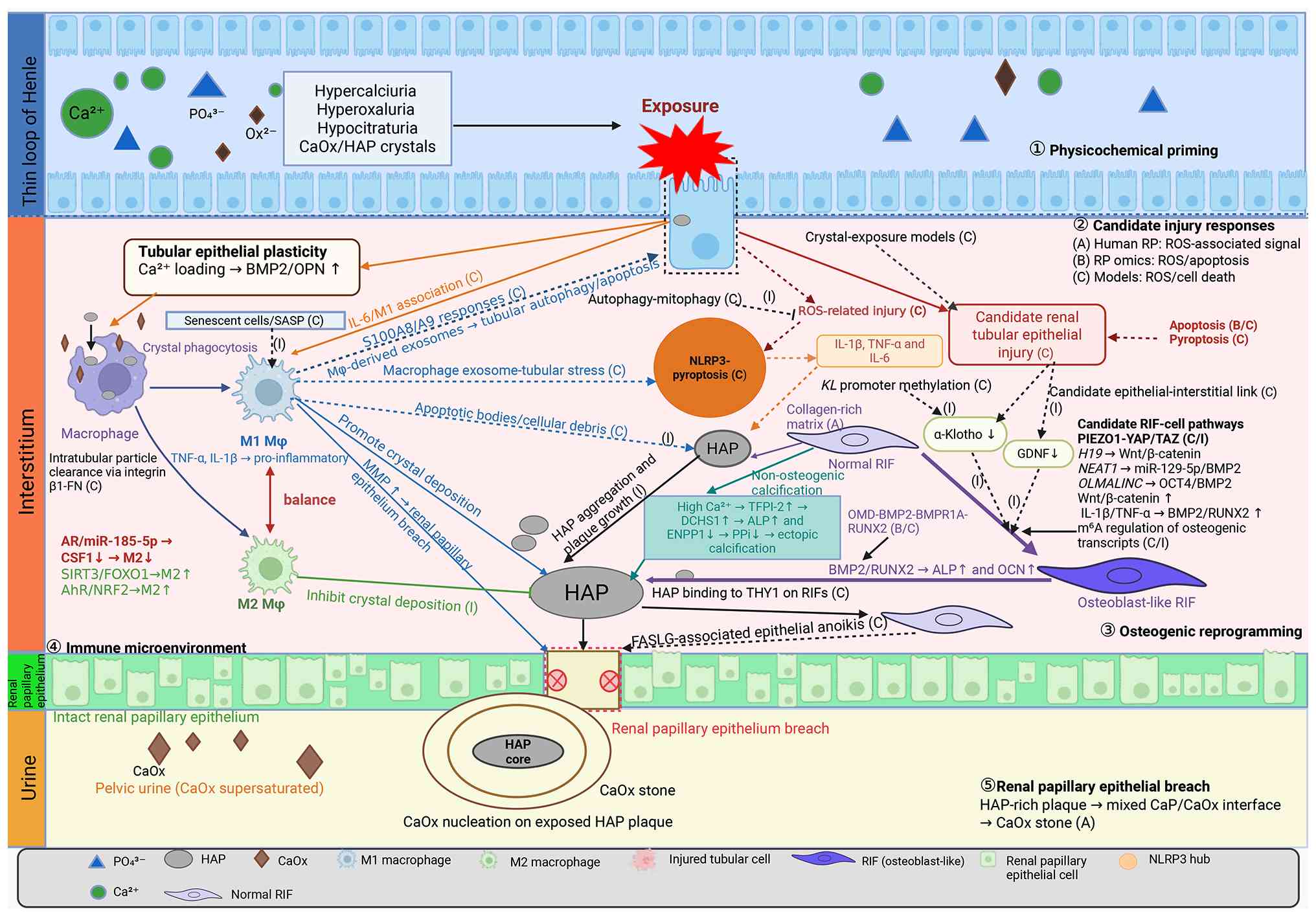 | Figure 1Proposed renal interstitial
microenvironment framework for RP formation. The schematic
illustrates five interconnected processes involved in RP
development and CaOx stone overgrowth: ① Physicochemical priming; ②
candidate injury responses; ③ calcification routes of RIFs; ④
immune microenvironment remodeling; and ⑤ renal papillary
epithelial breach. Evidence levels indicate the source of
supporting evidence: A, morphologically verified human RP pathology
or ultrastructural evidence; B, RP-associated human tissue omics or
molecular profiling; C, cell or animal model evidence; and I,
inferential or hypothesis-generating relationships. Arrow colors
represent inflammatory/crystal-handling, injury, anti-calcific,
mineralization and osteogenic pathways. Upward (↑), downward (↓)
and rightward (→) arrows indicate increased expression, decreased
expression and directional transition, respectively. Dashed arrows
indicate proposed relationships without direct validation in human
RP, whereas solid arrows indicate experimentally supported
associations. RP, Randall's plaque; HAP, hydroxyapatite; CaOx,
calcium oxalate; CaP, calcium phosphate; RIF, renal interstitial
fibroblast; SASP, senescence-associated secretory phenotype; ROS,
reactive oxygen species; miR, microRNA. |
Of note, the present review examines the
interstitial-plaque pathway, in which CaOx stones can grow on a
subepithelial-exposed interstitial HAP plaque (5,19). However, this is not the sole
route to CaOx stone formation. Intratubular CaP plugging within the
inner medullary collecting ducts and other plaque-independent
processes may represent distinct pathways that occur in different
anatomical and metabolic settings (26,36,37). In particular, collecting duct
plugging has been documented in a subset of idiopathic CaOx stone
formers, whereas other idiopathic CaOx stone formers have low
plaque burden (18,37). Therefore, the framework below
does not treat all CaOx stone studies as evidence for interstitial
plaque formation. Evidence from non-RP models is labeled according
to the level A-D framework and is used to generate verifiable
hypotheses rather than to establish plaque-specific mechanisms.
Supersaturation microenvironment
Prior to discussing specific molecular events, the
present review briefly revisits the physicochemical conditions of
the inner medullary interstitium, which provide the thermodynamic
foundation for RP formation. As described in section 2, the
countercurrent multiplication system elevates interstitial calcium
concentrations to levels several-fold higher compared with that in
plasma, while bicarbonate reabsorption and proton secretion
paradoxically alkalinize the interstitial fluid compared with that
in the tubular lumen (19,31). The combination of high calcium
activity and elevated local pH induces CaP supersaturation beyond
the threshold for spontaneous HAP nucleation (32,33). Low medullary blood flow and high
tissue osmolality compound the problem by further concentrating
solutes. These conditions are sufficient to promote spontaneous HAP
nucleation in the absence of any biological catalyst. Idiopathic
hypercalciuria, which increases calcium delivery to the medulla,
amplifies this baseline supersaturation and is the metabolic
phenotype most closely associated with interstitial plaque
formation (19,26). These are the baseline conditions
within which the molecular events described below unfold.
Candidate injury responses: Oxidative
stress and cell death Oxidative stress: Evidence from RP tissue and
experimental models
Evidence directly obtained from RP tissue for
oxidative stress remains limited. In human RP tissue,
dihydroethidium staining combined with E-cadherin and CD34
immunofluorescence demonstrated increased oxidative stress signal
in renal tubules adjacent to calcium deposits (level A) (38). In parallel, microdissected RP
transcriptomic data and subsequent analyses of RP datasets have
identified oxidative stress-associated expression signatures (level
B) (24,39). These observations support an
association between RP-related tissue and oxidative stress
responses; however, they do not identify the responsible nephron
segment, establish temporal order or demonstrate that oxidative
stress initiates interstitial HAP deposition.
By contrast, CaOx- or oxalate-exposed proximal
tubular human kidney-2 cells, porcine proximal tubular Lilly
Laboratories culture-porcine kidney 1 cells and Madin-Darby canine
kidney cells, as well as high-calcium normal rat kidney-52E cells
and vitamin D-associated rodent models, have reported
ROS-associated epithelial injury responses (level C) (40-42). These systems do not reproduce the
thin loop basement membrane and papillary interstitial environment
in which human RP initially develops (19). Although CaOx crystals may contact
tubular luminal surfaces in certain stone formers, these exposure
paradigms do not model the initial interstitial HAP mineralization
of RP. Thus, these studies have been retained only to generate the
verifiable hypothesis that epithelial cells anatomically adjacent
to plaque may exhibit associated injury responses; these studies
are not interpreted as direct evidence of ROS-mediated plaque
initiation.
Whether epithelial or interstitial cells adjacent to
early HAP deposits in the human papilla display oxidative stress
responses in vivo remains to be elucidated. Addressing this
question warrants spatially resolved analyses of human papillary
tissue and experimental systems that reproduce the local
interstitial mineral environment in the future.
Apoptosis and NLRP3-associated
pyroptotic injury
Apoptosis-related transcriptional programs were
enriched in microdissected human RP tissue compared with adjacent
non-calcified papillary tissue (level B) (24). In a Brd4+/M149T
mouse model of papillary interstitial calcification, terminal
deoxynucleotidyl transferase-mediated dUTP nick-end
labeling-positive nuclei were observed in interstitial papillary
areas corresponding to calcified lesions (level C) (34). These observations identify
apoptosis as a candidate injury response in plaque-associated
microenvironments; however, these findings do not establish that
apoptosis occurs in thin loop cells adjacent to early human RP or
that it precedes plaque mineralization.
NLRP3-associated pyroptotic injury has been
demonstrated mainly in hyperoxaluria and CaOx crystal-nephropathy
models (level C) (43-45). In these systems, CaOx exposure is
associated with NLRP3 activation, inflammatory injury and crystal
deposition, and interruption of pyroptotic signaling reduces
experimental injury readouts. Related nephrocalcinosis models have
also implicated NLRP3-associated macrophage responses in
mineral-related renal injury (46). However, no study has directly
demonstrated NLRP3-mediated pyroptosis in cells adjacent to
morphologically verified human interstitial RP, to the best of our
knowledge. The relevance of NLRP3-mediated pyroptosis to plaque
formation therefore remains a hypothesis warranting plaque-specific
validation in future research.
Autophagy and mitophagy as
counter-regulatory responses
Autophagy is a lysosome-dependent quality control
process that can counterbalance regulated cell death by removing
damaged proteins and organelles before they become persistent
sources of cellular stress (47). In high-glucose-treated HK-2
cells, a mitochondrial fission phenotype was associated with
reduced autophagic activity and increased apoptosis, whereas
pharmacological inhibition of autophagy further decreased cell
viability, supporting a protective role for basal autophagy in this
tubular-cell setting (level C) (48).
Mitophagy provides a more selective form of this
response by removing damaged mitochondria that would otherwise
sustain mitochondrial ROS (mtROS) production. In iohexol-exposed
HK-2 cells and mouse models of contrast-induced acute kidney
injury, PTEN-induced kinase 1 (PINK1)/Parkin (PARK)-dependent
mitophagy limited mitochondrial damage, mtROS accumulation, NLRP3
inflammasome activation and tubular-cell apoptosis. Silencing or
genetic deficiency of PINK1 or PARK2 impaired
mitophagy and aggravated each of these injury responses (level C)
(49). Consistently, in
high-glucose-treated murine renal tubular epithelial cells,
optineurin-enhanced mitophagy reduced mtROS and suppressed NLRP3
expression, caspase-1 cleavage, and interleukin (IL)-1β and IL-18
release (level C) (50).
However, the effects of autophagy appear to be
context-dependent rather than uniformly protective. In renal tissue
from patients with CaOx nephrolithiasis, LC3-II and Beclin 1
(BECN1) expression and the number of autophagic vacuoles were
increased. Mechanistic experiments in CaOx-exposed HK-2 cells
further demonstrated that ROS-associated autophagy was enhanced,
whereas pharmacological inhibition of autophagy or BECN1
knockdown attenuated tubular-cell injury; rapamycin produced the
opposite effect (level C) (51).
This model reflects tubular CaOx crystal injury rather than the
initiation of interstitial HAP plaque. Thus, autophagy and
mitophagy provide plausible associations among mitochondrial
quality control, ROS, NLRP3 activation and regulated cell death;
however, no study has directly demonstrated altered autophagic flux
or mitophagy in cells adjacent to morphologically verified human
RP, to the best of our knowledge. The contributions of autophagy
and mitophagy to plaque formation remain hypothetical and warrant
plaque-specific spatial validation in future research.
Therefore, the available data do not establish a
unified ROS-cell-death-autophagy network in human RP. Oxidative
stress-associated signals in RP tissue, apoptosis-related
expression changes, and autophagic, mitophagic and pyroptotic
responses observed in experimental models identify candidate
processes for future investigations. ROS-dependent regulation of
multiple cell-death programs provides broader biological context
for these experimental observations but is not specific to human RP
(52). Their spatial
relationship to thin loop basement membrane mineralization,
temporal order and contribution to plaque growth remain to be
elucidated.
Transformation: Osteogenic reprogramming
of the interstitium
Cell death is a key process; however, surviving
cells undergo changes as well, and what these cells develop into
may markedly impact long-term plaque growth. Injury-associated
inflammatory signals may favor osteoblast-like reprogramming of
resident fibroblasts and tubular cells in experimental systems;
whether this transition initiates or drives human RP remains to be
investigated in the future.
Histological and molecular evidence of
an osteogenic phenotype
The present section integrates level A
mineral-morphological observations from human RP, level B molecular
observations from stone-bearing papillae and level C evidence from
renal cell systems. Previous human RP studies reported calcifying
vesicles and HAP-rich plaque architecture, including concentric
mineral layers with needle-like morphology, combined with
osteopontin (OPN) localization around mineral deposits (16,53,54). Classic biomineralization studies
also support the capacity of calcium-phospholipid-phosphate
complexes to participate in HAP formation (55). Altered matrix γ-carboxyglutamic
acid protein (MGP) and bone morphogenetic protein 2 (BMP2)
expression has also been reported in papillary tissue from CaOx
stone formers (56). These
observations support a mineralization-competent papillary
microenvironment, but do not establish that a complete osteogenic
transcriptional program is active within morphologically verified
human RP.
Ultrastructural analyses such as Evan et al
(53,54) used immune-transmission electron
microscopy to investigate the calcified core of early RP,
identifying calcifying vesicles and HAP crystals intercalated with
organic matter and circumscribed by OPN. This molecular
architecture defines the nucleation microenvironment at the leading
edge of the plaque. Khan et al (57), using scanning and transmission
electron microscopy, demonstrated that HAP crystals deposit
preferentially within collagen-rich interstitial zones, confirming
that the ECM serves as both a structural scaffold and a template
for oriented mineral growth. In medullary sponge kidney (MSK)
primary renal-cell cultures, spontaneous calcification has been
accompanied by osteocalcin (OCN) and osteonectin expression,
supporting osteogenic activity in this non-RP system (58).
Renal interstitial fibroblasts (RIFs):
Principal effectors
Among the cell types in the papilla, RIFs are
candidate cellular effectors of mineralization-associated
remodeling. However, their contribution to the initiation or
progression of morphologically verified human RP remains to be
elucidated. Traditionally regarded as matrix-producing support
cells, RIFs possess an intrinsic osteogenic differentiation
capacity that exceeds that of renal tubular epithelial cells when
assessed under identical induction conditions (35). Exposure of human RIFs (hRIFs) to
osteogenic medium or elevated extracellular calcium induces their
trans-differentiation into osteoblast-like cells, accompanied by
robust ECM calcification (35).
In primary renal cells derived from papillary tissue of a patient
with MSK, spontaneous Ca2PO4 deposition was
associated with reduced glial cell line-derived neurotrophic factor
(GDNF) expression and an osteoblast-like phenotype (level C)
(58). These MSK-derived primary
cell observations and RIF culture studies support the ability of
renal interstitial cells to adopt pro-mineralizing phenotypes in
non-RP systems; however, these studies do not demonstrate that
runt-related transcription factor 2 (RUNX2)- or OCN-dependent
reprogramming is established within morphologically verified human
RP.
The BMP2/RUNX2 axis is a candidate regulatory
circuit implicated in osteogenic differentiation. BMP2 activates
SMAD-dependent transcription that converges on RUNX2, which in turn
activates mineralization effectors including OCN, alkaline
phosphatase (ALP) and OPN. Several long non-coding RNAs, including
H19 (59), NEAT1
(60) and OLMALINC (61), have been reported to modulate
BMP2/RUNX2-associated osteogenic responses in RIF systems; however,
their relevance to morphologically verified human RP remains to be
established in future research. Furthermore, an osteomodulin
(OMD)-mediated osteogenic pathway has been proposed in a previous
study investigating renal papillary specimens (62). OMD was increased in
vimentin-positive interstitial cells and colocalized with CaP
deposits and calcifying vesicles; tissue OMD abundance was also
positively associated with BMP2, RUNX2 and OCN expression (level B)
(62). In cultured hRIFs, OMD
promoted osteogenic-like differentiation through an OMD/BMP2/BMP
receptor 1A/RUNX2 positive-feedback loop. Of note, mineral deposits
were observed in the renal papillary tissues, including those
around the renal tubules, within the interstitium, around the
peritubular vessels and in the subepithelial layer of the renal
papilla. This distribution pattern was consistent with findings
from several studies on calcified renal papillae (19,63,64). However, considering that the
aforementioned observations did not reproduce the earliest
canonical pattern of thin loop basement membrane deposition
described in RP (65), and that
the eroded calcific deposits in the subepithelial region could not
be definitively excluded as Randall's plugs, the features of this
study as a characteristic of RP should be interpreted with
caution.
Counterbalancing this pro-osteogenic machinery,
α-Klotho secreted by renal tubular epithelial cells suppresses the
Wnt/β-catenin pathway in neighboring fibroblasts, thereby blocking
osteoblast-like transformation (35). These findings suggest that
reduced tubular α-Klotho signaling may weaken an anti-osteogenic
paracrine brake on neighboring RIFs in experimental systems;
whether this mechanism contributes to human RP formation remains
unknown (35). Whether α-Klotho
reduction precedes plaque initiation in human RP remains to be
elucidated.
Liu et al (66) recently demonstrated that high
extracellular calcium activates the tissue factor pathway inhibitor
2 (TFPI-2)/dachsous cadherin-related 1 (DCHS1) axis in these cells,
upregulating ALP while suppressing ectonucleotide
pyrophosphatase/phosphodiesterase 1 (ENPP1), which depletes the
endogenous mineralization inhibitor pyrophosphate (PPi). OPN, RUNX2
and BMP2 expression were unchanged during this process, indicating
that the TFPI-2/DCHS1 pathway operates independently of the
aforementioned osteogenic program. In RIF culture systems, these
findings suggested that mineralization can proceed through at least
two experimentally distinguishable routes: BMP2/RUNX2-associated
and TFPI-2/DCHS1-associated pathways. However, which pathway, if
any, predominates during human plaque maturation remains to be
elucidated.
RIFs also modulate the mineralization
microenvironment through collagen secretion. Alterations in
collagen composition have been associated with ectopic
calcification (67,68); however, whether
collagen-type-specific mineralization occurs in RP remains to be
investigated in the future.
Emerging mechanosensitive, epigenetic
and epitranscriptomic modifiers
Mechanosensitive PIEZO1-Yes-associated protein (YAP)
signaling, along with the context-dependent effects of
YAP/transcriptional coactivator with PDZ-binding motif (TAZ) on
osteogenic plasticity, represents a candidate regulatory mechanism
relevant to osteogenic reprogramming. The renal inner medulla is
continuously exposed to steep osmotic gradients, providing a
biologically plausible setting for mechanosensitive signaling. In
mouse inner medullary collecting duct cell models, hyperosmotic
NaCl/urea exposure activated the Ca2+-permeable channel
PIEZO1, increased Ca2+ influx and mtROS, and reduced
cell viability (level C) (69).
This finding established the responsiveness of renal medullary
collecting-duct epithelial cells to PIEZO1-dependent osmotic
signaling; however, the model does not reproduce the thin loop
basement membrane or papillary interstitial compartment in which
early human RP develops. Furthermore, the study did not examine
YAP/TAZ activation or osteogenic differentiation.
Additional evidence directly associating PIEZO1 with
osteogenic transcription has been reported in calcific aortic valve
disease. In human aortic valve interstitial cells and two mouse
models of valve calcification, oscillatory mechanical stress or
pharmacological PIEZO1 activation promoted
Ca2+-dependent nuclear activation of YAP. YAP
subsequently regulated glutaminase 1 (GLS1)-mediated
glutaminolysis, which enhanced histone acetylation at RUNX2
promoter regions and promoted osteogenic differentiation and
mineralization. Inhibition or knockdown of PIEZO1, YAP or GLS1
attenuated these responses (level C) (70). Notably, this study established a
PIEZO1-YAP pathway but did not independently demonstrate
involvement of TAZ.
The effects of YAP/TAZ are also cell-context
dependent. In vascular smooth muscle cells, loss of YAP/TAZ
promoted dishevelled segment polarity protein 3 nuclear
translocation, upregulated a Wnt-responsive osteogenic program
independently of canonical Wnt/β-catenin activation and resulted in
vascular calcification (level C) (71). Thus, osmotic or mechanical cues
may theoretically influence RIF phenotypes through
PIEZO1-YAP/TAZ-related mechanotransduction; however, PIEZO1
activity, YAP/TAZ localization and downstream osteogenic signaling
have not been demonstrated in RP-derived RIFs or morphologically
verified human RP. This proposed association remains inferential
and warrants plaque-specific validation in the future.
Epigenetic silencing of the KL (KLOTHO) gene
represents another candidate modifier of the anti-calcific
microenvironment. In cell-based and mouse experiments,
cytosine-phosphate-guanine (CpG) methylation of the KL
promoter restricted its transcriptional activity, whereas
DNA-demethylating treatment restored promoter activity and
increased KL expression (level C) (72). Cross-disease human evidence
further demonstrated that renal KL promoter methylation was
increased in patients with chronic kidney disease (CKD) and was
inversely associated with renal α-Klotho protein immunostaining and
estimated glomerular filtration rate, while associating positively
with tubulointerstitial fibrosis (73). Regarding pathological
mineralization, indoxyl sulfate increased DNA methyltransferase
(DNMT)1 and DNMT3A expression, promoted KL CpG
hypermethylation, reduced Klotho expression and enhanced
calcification in human aortic smooth muscle cells and
5/6-nephrectomized rats; these effects were attenuated by
5-aza-2'-deoxycytidine (level C) (74). Tubular cell-derived α-Klotho has
been reported to suppress Wnt/β-catenin-dependent osteogenic
differentiation of neighboring RIFs in an experimental renal cell
system (35), therefore
KL promoter hypermethylation could theoretically weaken this
paracrine anti-osteogenic brake. However, the available methylation
evidence derives from CKD and vascular calcification rather than
idiopathic CaOx stone papillae (73,74), and neither KL promoter
methylation nor DNMT activation has been demonstrated in
morphologically verified human RP, to the best of our knowledge.
The proposed association between KL methylation and loss of
the α-Klotho-RIF paracrine brake therefore represents a cross-model
inference rather than an established mechanism of human RP.
At the epitranscriptomic level,
N6-methyladenosine (m6A) has emerged as a
regulator of vascular aging and related disease processes (75) and may also influence osteogenic
plasticity. In human calcified arteries, methyltransferase-like 14
(METTL14) expression and total RNA m6A levels were
increased. In indoxyl sulfate-treated primary human artery smooth
muscle cells and rat models of uremic vascular calcification,
experimental reduction of METTL14 attenuated m6A
accumulation and calcification and improved vascular
repair-associated responses (level C) (76). METTL14-dependent m6A
modification also altered the stability and protein output of
vascular-protective transcripts, indicating that RNA methylation
can influence the balance between anti- and pro-calcific cellular
programs (76). Complementary
evidence demonstrated that the m6A reader YTHDF2, an
m6A-binding protein that recognizes
m6A-modified transcripts and can regulate their
stability and translation, was downregulated in in vitro and
in vivo vascular calcification models. YTHDF2 overexpression
recognized an m6A-modified site in the transcript
encoding RUNX2, shortened its half-life and suppressed
vascular smooth muscle cell osteogenic transition and calcium
deposition (level C) (77).
These findings indicated that the effects of m6A are
regulator- and transcript-dependent rather than uniformly
pro-calcific. As RUNX2-related programs contribute to osteogenic
differentiation of RIFs in experimental systems, altered
m6A writing or reading could theoretically modify
osteogenic transcript stability in the papillary interstitium.
However, global m6A abundance, METTL14 or YTHDF2
expression and transcript-specific m6A marks have not
been examined in RP-derived RIFs or morphologically verified human
RP. Therefore, this proposed association remains a cross-disease,
hypothesis-generating inference.
Tubular epithelial plasticity and
paracrine contribution
Renal tubular epithelial cells are anatomically
adjacent to papillary calcification and, in experimental renal-cell
systems, these cells can exhibit injury-associated or
osteogenic-like responses under high-calcium or osteogenic-culture
conditions (78-80).
Primary tubular cells isolated from patients with
idiopathic CaOx stone exhibit dose-dependent upregulation of BMP2
and OPN upon calcium loading (Ca2+, 0.5-2.5 mM),
consistent with calcium-associated phenotypic remodeling through
BMP-related signaling in this primary-cell system (level C)
(78). In osteogenic HK-2
cultures, apoptosis-related changes were detected prior to overt
CaP deposition (level C) (79).
In primary renal cells derived from papillary tissue of a patient
with MSK, reduced GDNF expression was associated with
Ca2PO4 deposition and an osteoblast-like
phenotype (level C) (58). A
subsequent study using this model reported caspase-independent
programmed cell death during the calcification process (level C)
(80).
These findings are derived from non-RP MSK- or
HK-2-based systems and should be interpreted as level C
hypothesis-generating evidence rather than proof of a mechanism in
human RP. Further research using spatially resolved multi-omics
analyses of human papillary biopsies is warranted to investigate
how tubular cells and fibroblasts coordinate osteogenic commitment
in vivo.
Regulation: Immune microenvironment
The renal papillary interstitium harbors
tissue-resident macrophages that actively patrol the medullary
space (81,82). Macrophages are present in the
papillary interstitium and may participate in local inflammatory
responses. Proposed interactions between macrophages,
oxidative-stress signals and fibroblast remodeling are derived
mainly from crystal injury models and warrant plaque-specific
validation in future research. Whether these interactions favor
mineralization or resolution may depend partly on the balance
between classically activated, pro-inflammatory M1 macrophages and
alternatively activated, repair-associated M2 macrophages.
Immune mediators in crystal
handling
A network of soluble immune mediators participates
in crystal handling and interstitial calcification, with individual
factors exerting context-dependent pro- or anti-calcific effects
(Table SI) (23,83). Three mechanistic themes are
highlighted in the present review. First, fetuin-A, a
hepatocyte-derived glycoprotein, inhibits calcification
systemically by forming colloidal calciprotein particles that
sequester calcium and phosphate (84); its local depletion in the
interstitium may lower the threshold for spontaneous CaP
nucleation, although this process remains to be investigated in
morphologically verified human RP (23). Second, the S100 calcium-binding
proteins A8/A9 (S100A8/A9), which are upregulated in M1 macrophages
upon crystal exposure, directly promote CaOx crystal aggregation
and amplify oxidative injury through a self-amplifying
IL-6-dependent circuit (85).
Third, fibronectin serves a dual function: Fibronectin mediates
crystal adhesion to damaged tubular surfaces but also participates
in macrophage-mediated crystal clearance via integrin β1 engagement
(81). The net calcific outcome
of these factors depends on their local concentration,
post-translational modification state and the local balance of pro-
and anti-inflammatory mediators and immune-cell phenotypes.
OPN warrants separate discussion because its effects
on inflammation and mineralization are highly context-dependent and
can be either pro- or anti-calcific depending on its
post-translational state. As an immune mediator, OPN interacts with
αvβ3 and CD44 receptors to regulate macrophage migration, adhesion
and survival, and is also a T helper 1-polarizing cytokine
(86). OPN has a paradoxical
role as a mineralization modulator. Genetic evidence demonstrated
that OPN has a protective function: Opn−/− mice
develop exacerbated ectopic calcification when crossed with
MGP-deficient backgrounds (87),
and OPN deficiency increases CaOx crystal retention in renal
tubules (88). However, in bone
biology, OPN promotes osteogenesis by binding CaP microspheres and
modulating osteoclast activity (89).
The functional effects of OPN depend partly on its
post-translational state (88-91). Phosphorylation of OPN by casein
kinases generates a polyanion that adsorbs to crystal surfaces and
sterically inhibits further growth (90). By contrast, proteolytic cleavage
by thrombin and matrix metalloproteinases generates N- and
C-terminal fragments with altered integrin-binding specificity: The
thrombin-cleaved N-terminal fragment exposes a cryptic SVVYGLR
motif that potently recruits macrophages and promotes M1
polarization (91). Temporal
dynamics provide an additional mechanism: Acute OPN elevation at
the crystal-tissue interface may predominantly exert inhibitory
effects through intact phosphorylated forms, whereas chronic
exposure in an inflammatory milieu favors proteolytic processing
and the generation of pro-inflammatory, pro-calcific fragments
(89).
Ultrastructural evidence from human RP tissue
further clarifies the role of OPN. Immunoelectron microscopy
demonstrated that OPN protein tightly circumscribes calcium salt
deposits within the plaque interior (53). However, the phosphorylated vs.
cleaved fraction of OPN at each stage of plaque maturation remains
to be elucidated; spatially resolved phospho-proteomics applied to
RP tissue sections could provide further understanding. In
persistent inflammatory settings, proteolytic processing of OPN
could alter macrophage recruitment and mineral interactions
(91). Whether a self-sustaining
feedback loop involving OPN, M1-polarized macrophages and
osteogenic remodeling operates in morphologically verified human RP
remains unknown. Therefore, the net effect of sustained OPN
expression in established plaque should not be defined as uniformly
protective or pro-calcific.
Macrophage polarization as a candidate
modifier
Macrophage polarization may influence whether the
local microenvironment tends toward mineralization or resolution
(Fig. 1). M1/M2-associated
expression changes in RP-related tissue are classified as level B
observations, whereas mechanistic macrophage-polarization studies
are predominantly classified as level C.
CaOx crystals drive the balance toward M1
macrophage polarization: Human monocytes exposed to CaOx in
vitro differentiate into an inflammatory phenotype secreting
tumor necrosis factor-α (TNF-α), IL-1β and IL-8 (92). By contrast, He et al
(81) reported that
tissue-resident renal macrophages remove intratubular particles
through integrin β1-fibronectin-dependent trans-epithelial
migration. The study did not assign this clearance program to a
canonical M1 or M2 phenotype. In crystal-handling models,
macrophage polarization influences the magnitude and character of
local inflammatory signaling and the persistence of crystal
retention. In microdissected RP-related tissue, M1-associated
transcripts were increased whereas M2-associated signatures were
reduced (24). Whether
macrophage polarization serves a similar regulatory role within
human interstitial RP remains to be elucidated.
Colony stimulating factor 1
(Csf1)−/− mice, which lack M2 macrophages,
develop markedly increased renal CaOx crystal deposition under
hyperoxaluric conditions; reconstitution with recombinant CSF1 or
adoptive transfer of M2 macrophages rescues the phenotype (93). Taguchi et al (94) demonstrated that systemic M1
induction (lipopolysaccharide + IFN-γ) promotes crystal formation,
whereas M2 induction (IL-4) reduces CaOx crystal deposition.
Several signaling axes feed into this balance, including
SIRT3/FOXO1-mediated M2 promotion (95), AR-mediated suppression of
CSF1-dependent macrophage recruitment and M2-like polarization
through microRNA (miR)-185-5p (96) and aryl hydrocarbon receptor- and
nuclear factor erythroid 2-related factor 2-dependent M2 commitment
(97,98). Bioinformatic analyses suggested
that immune polarization and ECM remodeling are transcriptionally
linked in RP tissue (39,99).
Cellular senescence and the
senescence-associated secretory phenotype (SASP)
Cellular senescence is a durable state of
cell-cycle arrest in which metabolically active cells can acquire
an SASP. The composition of the SASP varies according to cell type
and stimulus, but commonly includes inflammatory cytokines,
chemokines, growth factors and matrix-remodeling mediators that can
modify neighboring cells (100). In human nephropathy samples and
experimental kidney-injury models, Wnt9a expression was
associated with tubular p16INK4A expression and renal
fibrosis. Mechanistic experiments further demonstrated that
Wnt9a-β-catenin signaling accelerated tubular epithelial-cell
senescence, while TGF-β1 released by senescent tubular cells
promoted renal fibroblast proliferation and activation (level C)
(101,102).
A related study identified Brahma-related gene 1
(BRG1) as an upstream regulator of tubular senescence. In mouse and
primary renal tubular-cell systems, BRG1 activated Wnt/β-catenin
signaling, impaired autophagic activity and increased
senescence-associated markers. The altered secretome of these
senescent tubular cells promoted fibroblast proliferation and
activation, whereas BRG1 suppression, Wnt/β-catenin inhibition or
restoration of autophagy attenuated these effects (level C)
(102).
In one vascular-calcification model, the SASP was
associated with pathological mineralization. In senescent human
aortic smooth muscle cells, miR-34a increased IL-6 secretion and
induced a secretome enriched in inflammatory cytokines, chemokines,
pro-senescent growth factors and matrix-degrading molecules.
Conditioned medium from miR-34a-overexpressing cells accelerated
senescence and calcification in neighboring cells, while
Mir34a deficiency reduced vitamin D-induced medial
calcification and IL-6 expression in mice (level C) (103).
Therefore, these cross-disease findings suggested
that senescent papillary epithelial or interstitial cells could
sustain an inflammatory and matrix-remodeling milieu that
influences macrophage activity, fibroblast activation and
osteogenic plasticity. However, these mechanisms are derived from
renal fibrosis and vascular calcification systems. Senescent-cell
burden, SASP composition and their spatial association with early
interstitial HAP deposits have not been characterized in
morphologically verified human RP. Cellular senescence and the SASP
should therefore be regarded as hypothesis-generating modifiers
rather than established drivers of plaque formation.
Exposure of RP to urine: Renal papillary
epithelial breach and the interstitium-to-lumen transition
Interstitial calcification does not solely produce
a kidney stone. For stones forming via the interstitial-plaque
route, focal disruption of the overlying renal papillary epithelium
exposes the interstitial HAP deposit to pelvic urine and permits
heterogeneous CaOx crystal nucleation and overgrowth (5,19,20). The interstitium-to-lumen
transition phase in the RP-to-stone sequence remains to be
elucidated. The following section examines the available
evidence.
Renal papillary epithelium and its
vulnerability
The epithelial surface of the human renal papilla
is a specialized papillary epithelium that is distinct from the
transitional urothelium of the renal pelvis (104). To avoid terminological
ambiguity, the present review uses 'renal papillary epithelium'
when referring to the epithelial surface overlying renal papillae
and reserves 'transitional urothelium' for the renal pelvis. In
intraoperative biopsy and related structural studies, Evan et
al (19,20) and Coe et al (36) reported that the renal papillary
epithelium is intact over papillary areas where RP has not reached
the surface, whereas at plaque-associated stone-attachment sites,
it is focally lost, exposing the interstitial HAP deposit to pelvic
urine. Plaque exposure is therefore a spatially restricted event
associated with focal loss of the renal papillary epithelium.
The process by which an enlarging interstitial HAP
deposit becomes exposed through the renal papillary epithelium
remains to be investigated. Mechanical displacement by an expanding
mineral deposit has been proposed as a possible structural
explanation; however, human structural studies do not identify the
initiating cellular event at plaque-exposure sites (5,12-14,16,19,30). A recent experimental study
suggested that a HAP-rich microenvironment may alter RIF signaling
and promote epithelial anoikis (level C) (105). Whether this candidate
HAP-fibroblast-epithelial pathway operates at human plaque-exposure
sites remains to be determined in future research.
Events at the exposed plaque
surface
Once the renal papillary epithelium is breached,
the exposed mineral surface encounters a different chemical
environment. Pelvic urine contains CaOx at or beyond
supersaturation, along with macromolecular modulators such as OPN
and Tamm-Horsfall protein (THP; uromodulin). Detailed
ultrastructural analyses by Coe et al (36) and Evan et al (53,54) of the stone-tissue interface in
idiopathic CaOx stone formers revealed a characteristic layered
architecture at the attachment site: The interstitial HAP plaque is
coated by a ribbon-like structure composed of alternating mineral
and organic-matrix layers (termed the 'ribbon'). OPN localizes
along the crystal-matrix interface, whereas inter-α-trypsin
inhibitor heavy chain 3 is present within the plaque matrix
(53,106). Microstructural analysis of
RP-associated calcium oxalate monohydrate (COM) stones by Sethmann
et al (107) provided
complementary mineralogical evidence at the plaque-stone interface.
The earliest COM crystals on exposed plaque exhibited morphologies
consistent with different local levels of COM supersaturation and
were frequently coated with CaP. The study proposed that
CaP-supersaturated interstitial fluid may diffuse through porous
plaque into pelvic urine, locally increasing COM supersaturation
and thereby promoting initial COM nucleation. Replenishment of pore
fluid within early COM layers may subsequently permit additional
CaP precipitation within the interface. Thus, this study associated
exposed interstitial HAP plaque, CaOx overgrowth and the mixed
CaP/COM interface with local precipitation conditions; however, the
proposed ion-diffusion mechanism remains a structural
interpretation rather than a directly observed dynamic process
(107).
Structural and molecular observations from
available studies are consistent with a proposed sequence at this
interface. The exposed HAP plaque surface can interact with urinary
macromolecules. OPN localizes at the plaque crystal-matrix boundary
in human papillae (53), whereas
THP is a major urinary crystal-modifying protein; its direct
localization or passivating function at the exposed human plaque
surface has not been established (108). Urinary prothrombin fragment 1
has also been reported to modulate CaOx crystallization, with its
sialylation state potentially influencing CaOx stone formation
(109,110); however, its role at the exposed
human RP surface has not been established. OPN in its
phosphorylated form is a well-established inhibitor of CaOx crystal
growth and retention in renal tubules (88), and its immunolocalization to the
crystal-matrix boundary in the ribbon zone is consistent with this
inhibitory role (53). However,
under conditions of sustained urinary CaOx supersaturation may
eventually overcome protein-mediated inhibition: CaP and CaOx
co-precipitate in the transition zone, and the stone composition
progressively shifts to predominantly CaOx (36). This compositional gradient, from
an HAP-rich plaque interior through a mixed CaP/CaOx transition
zone to predominantly CaOx in the bulk stone, has been confirmed by
micro-Fourier-transform infrared spectroscopy across the
stone-plaque interface (25,26,36) and provides notable chemical and
spatial evidence consistent with direct CaOx overgrowth on exposed
RP. However, these observations are cross-sectional rather than
dynamic.
Endoscopic observations provide clinical support to
the breach model. Borofsky et al (10) reported that papillary surface
pits (focal depressions in the renal papillary epithelium) are
markedly associated with RP stone anchors (RPAs) and proposed that
these pits mark the sites where stones were once attached to the
papillary plaque before dislodging from the papillary surface.
Micro-CT of surgically extracted calculi confirmed that RPAs are
discrete HAP-rich regions at the stone base whose mineral signature
matches interstitial plaque (10,12); spontaneously passed CaOx stones
also contain internal HAP-rich remnants consistent with prior
papillary attachment (11).
These residual papillary defects may represent potential sites for
recurrent crystal nucleation; however, their direct contribution to
clinical recurrence risk has not been established.
Systemic drivers of the pro-calcific
niche
Conceptual framework: How systemic
signals converge on the interstitium
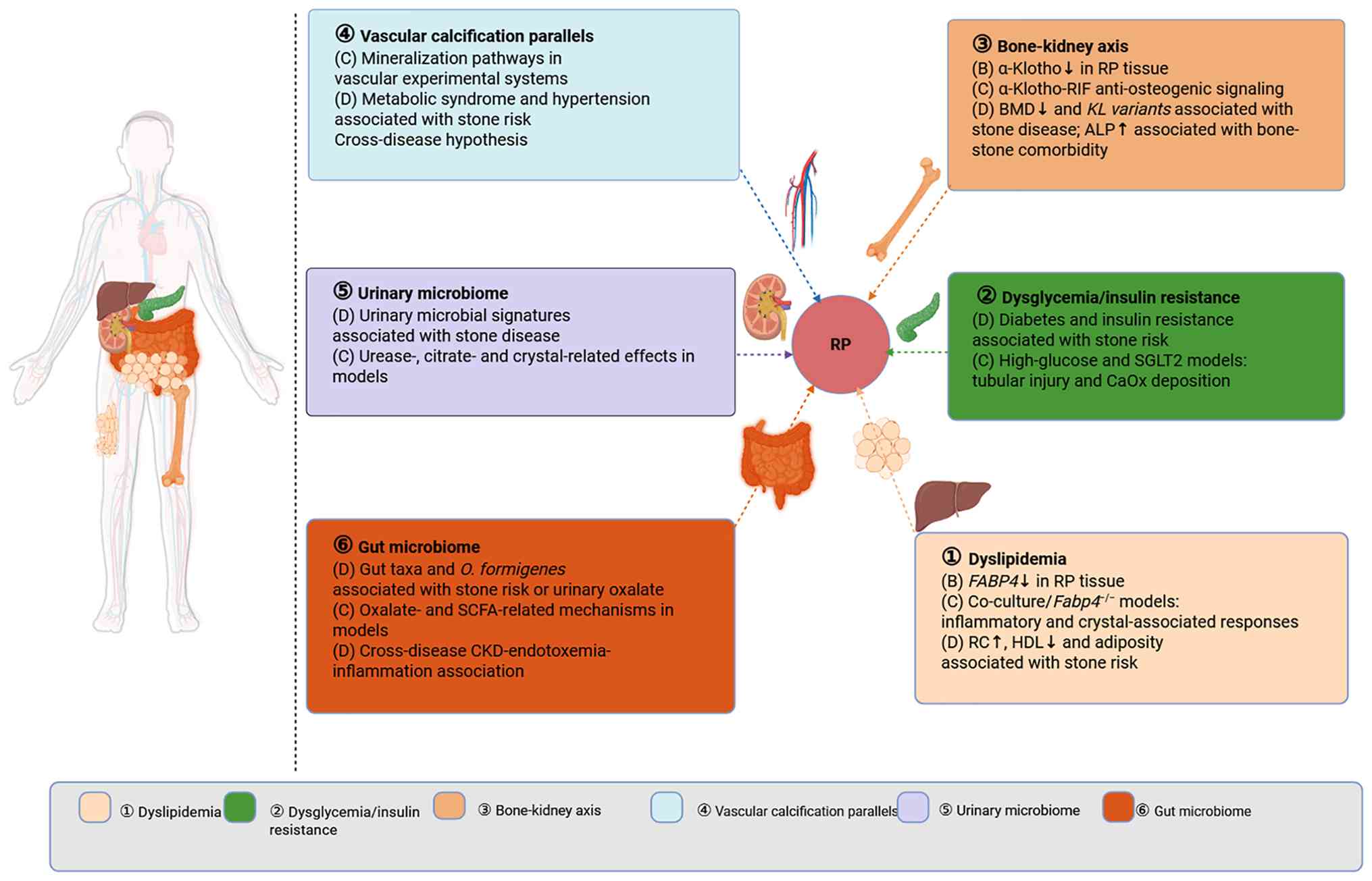
The systemic and microbiome factors discussed in
sections 4 and 5 are hypothesized modifiers of the papillary
microenvironment rather than established determinants of RP
presence or burden. No systemic or microbiome factor has been
reported to alter endoscopic or histological RP burden in humans to
date. Current tissue-level evidence directly derived from
RP-related human papillary specimens is limited to level B
molecular observations, including altered fatty acid-binding
protein 4 (FABP4) and α-Klotho expression (35,111); other proposed associations are
derived from level C cell or animal studies and level D stone
epidemiology or clinical associations. Fig. 2 distinguishes these evidence
sources and presents only proposed routes of influence.
Dyslipidemia: Remnant cholesterol (RC),
FABP4 and inflammatory reprogramming
Beyond the epidemiological association between
dyslipidemia and nephrolithiasis, an important question is how
abnormal lipid metabolism may modify the papillary interstitial
milieu. Epidemiological data consistently associate dyslipidemia
with nephrolithiasis risk. Cross-sectional analyses identify
abdominal adiposity (112),
elevated RC (113) and reduced
high-density lipoprotein (114)
as independent risk factors, with RC exhibiting a dose-response
relationship when low-density lipoprotein cholesterol is controlled
(113). These associations hold
across Japanese (115) and
American populations (112,114), identifying dyslipidemia as a
robust systemic risk factor for nephrolithiasis.
In vitro co-culture studies have begun to
explore how a metabolic-syndrome-like environment may enhance
tubular inflammatory responses and CaOx crystal adhesion. Ichikawa
et al (116)
demonstrated that adipocyte-tubular cell co-culture enhances CaOx
crystal adhesion and upregulates IL-6 expression in tubular cells.
Zuo et al (117)
extended this system to include macrophages, demonstrating that the
tri-cellular metabolic milieu markedly upregulates monocyte
chemoattractant protein-1, OPN and TNF-α, consistent with an
M1-promoting inflammatory milieu associated with crystal retention.
These findings position adipose tissue-derived paracrine signals as
potential contributors to the interstitial inflammatory cascade,
although the specific lipid mediators responsible have not been
fully identified.
Notably, Taguchi et al (111) performed transcriptomic
profiling of microdissected human RP tissue and identified marked
enrichment of differentially expressed genes in lipid metabolism
pathways. Among these, FABP4 was markedly downregulated in
plaque tissue. Functional validation in Fabp4−/−
mice subjected to glyoxylate-induced hyperoxaluria demonstrated
that FABP4 deficiency markedly exacerbated both interstitial and
intratubular CaOx crystal deposition compared with wild-type
controls (111). This level C
glyoxylate-model phenotype suggests a candidate protective role for
FABP4 in experimental renal calcification; its contribution to
human RP burden remains to be elucidated.
FABP4 exhibits a notable tissue-specific paradox:
Although decreased renal expression promotes local calcification,
elevated serum FABP4 levels are associated with the severity of
vascular calcification (118),
and FABP4 inhibition has been explored as a therapeutic strategy
for cardiovascular disease (119). The inverse relationship
suggests that circulating FABP4 reflects systemic metabolic
disturbance (acting as a biomarker), while interstitial FABP4
functions as a local calcification suppressor (acting as a
protective effector). The mechanistic basis of this dichotomy
(intracellular lipid-chaperoning vs. extracellular signaling) has
not been investigated, to the best of our knowledge.
Dysglycemia and insulin resistance:
SGLT2-associated pathways and other metabolic mechanisms
A meta-analysis of cohort studies reported a 16%
higher kidney-stone risk among participants with diabetes mellitus
than among those without diabetes (RR=1.16); the pooled analysis
did not consistently stratify diabetes by type (120). Gestational diabetes mellitus
(GDM), a distinct form of hyperglycemia first recognized during
pregnancy, has also been associated with future kidney-stone risk
(121), while Mendelian
randomization supports a causal association between type 2 diabetes
mellitus and kidney stones (122). The risk of kidney-stone
formation is not confined to overt diabetes mellitus: Multiple
insulin resistance indices, including the metabolic score for
insulin resistance (METs-IR) (123), homeostatic model assessment of
insulin resistance (HOMA-IR) (123) and triglyceride-glucose-body
mass index (TyG-BMI) (123),
were positively associated with both kidney-stone occurrence and
recurrence in individuals without diagnosed diabetes, with each
1-unit increase in METs-IR corresponding to odds ratios of 1.020
for kidney-stone occurrence and 1.033 for recurrence, relative to a
METs-IR value one unit lower (123). These data support insulin
resistance as a metabolic correlate that may precede overt
diabetes
A key route associating dysglycemia with renal
crystal injury involves tubular epithelial stress in experimental
systems (level C) (48,50,124). High-glucose exposure has been
associated with increased apoptosis and reduced autophagic activity
in HK-2 cells (level C) (48).
In parallel, optineurin-enhanced mitophagy reduced mtROS
accumulation and NLRP3 inflammasome signaling in
high-glucose-treated murine renal tubular epithelial cells (level
C) (50). These findings support
a candidate tubular stress pathway; however, these findings do not
establish high-glucose-induced pyroptosis or ferroptosis in the
models cited in the present review, nor do these findings
demonstrate relevance to human RP. Anan et al (124) reported that SGLT2 silencing in
HK-2 cells attenuated high-glucose-induced OPN and CD44
upregulation and reduced CaOx crystal adhesion. In vivo,
phlorizin attenuated ethylene glycol-induced renal stone formation
in rats and downregulated kidney injury molecule-1 and OPN without
altering water intake or urine volume, whereas
Sglt2−/− mice were resistant to glyoxylic
acid-induced CaOx deposition (124). Therefore, level D stone
associations and level C SGLT2-related experimental findings
identify a candidate tubular injury pathway; however, whether SGLT2
inhibition alters human RP burden remains to be elucidated. In a
distinct vascular model, high-glucose exposure promoted vascular
smooth muscle cell calcification and senescence through
BMF-AS1/BMF-associated signaling, providing cross-disease evidence
that hyperglycemic stress can promote ectopic mineralization
outside the kidney (125).
However, these vascular findings do not establish a corresponding
mechanism in human RP.
Bone-kidney axis: Mineral homeostasis,
ALP and α-Klotho
Kidney stone formers frequently exhibit lower bone
mineral density (BMD) compared with non-stone controls, and
clinical studies and reviews support an association between calcium
stone disease and bone demineralization (126-128). Stone formers may also exhibit
increased urinary calcium and altered bone-remodeling markers
(129-132). In a cross-sectional study, Zhu
et al (133) reported an
inverse association between BMD and kidney stones and identified
that elevated ALP was independently associated with the coexistence
of osteoporosis/osteopenia and kidney stones.
ALP hydrolyzes PPi in mineralizing systems, thereby
reducing an endogenous inhibitor of HAP nucleation. ALP expression
is also upregulated in osteogenically induced RIF cultures
(66). Aged bone-matrix-derived
extracellular vesicles have been reported to promote osteogenic
phenotypic switching and vascular calcification in experimental
systems (134). Whether
bone-derived extracellular vesicles reach or influence the renal
papillary interstitium remains to be elucidated.
α-Klotho serves a key role in the bone-kidney axis.
Reduced circulating soluble α-Klotho has been associated with
CKD-related mineral and vascular abnormalities; however,
methodological and clinical interpretation limitations remain
(135). In a cross-sectional
National Health and Nutrition Examination Survey (NHANES) analysis
of middle-aged and older adults with diabetes mellitus (diabetes
type was not stratified in the cited analysis), serum α-Klotho
levels were inversely associated with prevalent kidney stones
(136). KLOTHO G395A
polymorphism has been associated with kidney-stone susceptibility
in a previous case-control study (137). In one study of RP-related human
papillary tissue, α-Klotho expression was reduced, and
complementary cell experiments showed that tubular epithelial
cell-derived α-Klotho inhibited Wnt/β-catenin signaling and
osteogenic differentiation in neighboring RIFs (35). Reduced α-Klotho expression in
RP-related tissue (level B) and α-Klotho-RIF interactions in
experimental systems (level C) support a potential association
between anti-calcific signaling and the papillary interstitium.
Whether systemic mineral homeostasis directly alters human RP
burden remains to be investigated in future research.
The fibroblast growth factor 23 (FGF23)-parathyroid
hormone (PTH)-vitamin D axis regulates CaP homeostasis through
coordinated renal and intestinal effects. FGF23 and PTH decrease
proximal tubular phosphate reabsorption and thereby promote
phosphaturia; FGF23 suppresses renal 1α-hydroxylase and
1,25-dihydroxyvitamin D production, whereas PTH stimulates vitamin
D activation. Active 1,25-dihydroxyvitamin D, in turn, increases
intestinal calcium and phosphate absorption (138). Dysregulation of the
FGF23-Klotho-mineral axis is also implicated in CKD-associated
mineral abnormalities and ectopic vascular calcification (139); however, it remains to be
elucidated whether variation within this axis modifies kidney-stone
risk or human RP burden independently of established metabolic
abnormalities.
Cross-disease parallels: Vascular
calcification as a mirror of RP
Metabolic syndrome and hypertension are associated
with kidney-stone risk (140,141). Vascular calcification is
considered primarily as a cross-disease mechanistic comparison
rather than an established epidemiological predictor of stone
disease in the present review. The molecular parallels between
vascular calcification and RP are extensive enough to suggest
overlapping disease mechanisms beyond what epidemiological
co-occurrence alone would imply (detailed in section 3).
Vascular calcification and RP-related experimental
systems share several candidate features, including osteogenic
signaling, inflammatory pathways and altered calcification
inhibitors (35,43,44,56,62,72-74,84,142,143); however, the evidence is
heterogeneous and does not establish a shared causal mechanism in
human RP. These cross-disease parallels nominate, rather than
validate, potential therapeutic targets for future RP research.
Furthermore, regarding causal directionality, it remains to be
elucidated whether systemic vascular calcification drives renal
interstitial calcification through circulating mediators or if both
processes independently result from a shared metabolic insult.
Sex hormone status and kidney stone
risk
A recent global meta-analysis estimated
urolithiasis prevalence at 12.93% in men and 8.91% in women;
however, the magnitude of the sex difference varied substantially
according to population, diagnostic method and geographical region
(144). In a population-based
study from Rochester, the men-to-women incidence ratio for
symptomatic nephrolithiasis decreased from 3.1 in 1970 to 1.3 in
2000 (level D) (145). In three
large prospective cohorts, stone incidence remained higher in men
than in women, with rates of 271 and 159 per 100,000 person-years,
respectively; differences in lifestyle factors and urine chemistry
provided partial explanations of the excess male risk (level D)
(146). These epidemiological
patterns alone do not establish a direct causal role for sex
steroids. Clinical findings regarding estrogen status have also
been reported to be inconsistent. Natural menopause was not
independently associated with incident kidney stones in the Nurses'
Health Study (147), whereas
randomized Women's Health Initiative trials reported that estrogen
therapy increased nephrolithiasis risk (148). Therefore, estrogen should not
be described as uniformly protective in clinical stone disease.
Furthermore, the mechanistic studies discussed below primarily
focus on urinary solute metabolism, tubular CaOx crystal adhesion
or injury and experimental crystal deposition; the findings do not
establish that androgen or estrogen signaling alters the burden,
mineral composition or molecular phenotype of morphologically
verified human interstitial RP.
Circulating testosterone levels should not be
treated as a direct surrogate for intrarenal androgen receptor (AR)
activity. In a cross-sectional NHANES analysis, using the highest
serum testosterone quartile as the reference, men in the lower
three quartiles had higher adjusted odds of kidney stones (ORs,
1.375, 1.348 and 1.472 for the first, second and third quartiles,
respectively), with the inverse association being most evident in
men aged >40 years (level D) (149). A prospective controlled study
of male patients with CaOx stones identified higher blood AR
mRNA and plasma miR-185-5p levels, along with lower blood
CSF1 mRNA expression, compared with that in healthy
controls; circulating androgen levels did not differ markedly
between the groups (150).
These non-RP human observations are associative and do not localize
the pathway to renal papillary tissue.
Mechanistic evidence has been derived from
COM-exposed renal tubular epithelial cells and experimental CaOx
deposition models. In HK-2 and HKC-8 cells, AR bound an
androgen-response element within the miR-185-5p promoter and
increased miR-185-5p transcription; miR-185-5p in turn directly
targeted the 3'-untranslated region of CSF1 mRNA, reducing
CSF1 expression and secretion. AR depletion increased
CSF1-dependent macrophage recruitment, CD163/CD206-associated
M2-like polarization and COM-crystal phagocytosis, whereas CSF1
suppression or miR-185-5p restoration partially reversed these
effects (level C) (96).
Consistently, renal tubule-specific loss or pharmacological
degradation of AR increased renal CSF1 and M2-like macrophage
accumulation and reduced CaOx deposition in glyoxylate-treated mice
and hydroxy-L-proline-treated rats (level C) (96). This pathway aligns with
independent evidence that CSF1 deficiency reduces renal M2-like
macrophages and increases experimental CaOx deposition, whereas
recombinant CSF1 or M2-macrophage transfer promotes crystal
clearance (level C) (93).
However, these systems model hyperoxaluria-associated intrarenal
CaOx deposition rather than the initiation of chronic interstitial
HAP plaque, and the AR-miR-185-5p-CSF1 axis has not been
demonstrated in morphologically verified human RP.
A mechanistic study reported two anatomically
distinct ERβ-dependent effects in experimental CaOx deposition. In
HepG2 hepatocytes, ERβ bound estrogen-response elements within the
promoter of alanine-glyoxylate aminotransferase 1 (AGT1), increased
AGT1 expression and reduced oxalate production. In oxalate-exposed
HK-2 and HKC-8 renal tubular epithelial cells, ERβ suppressed NADPH
oxidase 2 (NOX2) transcription, ROS and H2O2
production and cell injury; NOX2 knockdown partially reversed the
oxidative stress and injury responses caused by ERβ depletion.
In vivo, genetic loss or pharmacological inhibition of ERβ
increased urinary oxalate excretion, renal oxidative stress and
CaOx deposition in glyoxylate-treated mice and
hydroxy-L-proline-treated rats, whereas pharmacological NOX
inhibition attenuated experimental crystal deposition (level C)
(151). These findings support
a dual hepatic AGT1 and renal tubular NOX2 mechanism in
experimental CaOx disease. However, the renal systems model
oxalate-exposed tubular injury and intrarenal CaOx deposition
rather than early thin loop basement membrane HAP nucleation, and
neither ERβ activity nor AGT1/NOX2 signaling has been demonstrated
in morphologically verified human interstitial RP. Independently,
17β-estradiol exposure in MDCK renal tubular cells reduced CaOx
crystal-binding capacity and promoted cell migration and
tissue-healing phenotypes, providing additional experimental
evidence that estrogen signaling can modify tubular crystal
interactions (152).
Another candidate mechanism involves cross-talk
between ER signaling, Wnt/β-catenin and fibroblast plasticity,
although the available evidence is indirect and context-dependent.
In established osteoblast-precursor cell lines and primary
osteoblast progenitors, selective activation of kinase-mediated ER
signaling stimulated Wnt and BMP2 pathways and promoted osteogenic
differentiation, whereas conventional 17β-estradiol suppressed
BMP2-induced osteoblast commitment (level C) (153). These opposing effects indicated
that ER-Wnt interactions depend on the ligand, signaling mode and
cellular context rather than exhibiting a uniformly pro- or
anti-osteogenic effect of estrogen. In renal injury models,
tubule-specific blockade of Wnt secretion reduced β-catenin
activation, renal fibroblast proliferation and myofibroblast
expansion, while tubule-derived Wnts promoted renal fibroblast
activation in vitro (level C) (154). Furthermore, in the context of
RP, HK-2-derived α-Klotho inhibited osteogenic differentiation of
hRIFs by suppressing Wnt/β-catenin signaling (level C) (35), while activation of this pathway
and reduced expression of its antagonists were observed in
RP-related tissue (level B) (35). Therefore, these findings
suggested that estrogen or ER signaling could modify
epithelial-fibroblast Wnt communication or the osteogenic
plasticity of RIFs. However, no study has directly investigated
estrogen or ER pathway modulation in RP-derived RIFs, and
ER-Wnt/β-catenin cross-talk has not been spatially demonstrated in
morphologically verified human RP. This proposed association
therefore remains a cross-model, hypothesis-generating
inference.
Microbiome-metabolism axis
The microbiome may influence stone-associated
biology through two proposed routes: Urinary microbial communities
may modify local urinary chemistry and crystal behavior (155-165), whereas gut microbial
communities may influence systemic oxalate handling and
inflammatory signaling (166-172). Direct effects on human RP
remain unproven. Notably, unlike the structural evidence for
plaque-stone continuity and selected local osteogenic or immune
observations described in section 3, the microbiome-interstitium
link currently rests predominantly on associative data and
inference rather than direct evidence in human RP. While a resident
kidney microbiota that responds to antibiotics in a previous study
(155) and the identification
of multi-species bacterial networks that collectively regulate
oxalate homeostasis (166) have
provided a basis for mechanistic evidence, the key association with
interstitial remodeling has not been investigated to date.
Urinary microbiome: Local modulator of
the pro-calcific niche
The traditional assumption that urine from the
healthy urinary tract is sterile has been challenged by expanded
quantitative culture and sequencing studies demonstrating resident
urinary microbial communities in individuals without clinical
urinary tract infection (156-164). The concept of a urinary
microbiome has recently been extended to the kidney parenchyma
itself. Agudelo et al (155) reported that murine kidneys
harbor a low-biomass, metabolically active microbiota in
microniches between glomeruli and tubules. Cefazolin, a
perioperative antibiotic extensively used in stone surgery, shifted
the population away from uroprotective Lactobacillus species
and toward pro-lithogenic Enterobacteriaceae; the effect was
duration-dependent and reversible after cessation. In vitro,
Lactobacillus crispatus inhibited CaOx crystallization,
whereas stone-associated Escherichia coli promoted it. RNA
sequencing and RNA fluorescence in situ hybridization
imaging of human kidney tissue confirmed microbial signatures in
both glomerular and tubular compartments (155). These findings raise the
possibility that perioperative antibiotics may alter the renal
microbiome in ways relevant to stone biology. Expanded quantitative
urine culture and 16S ribosomal RNA sequencing studies have
countered the dogma of sterile urine and identified resident
urinary microbial communities in adults (156-164). In urinary stone disease,
Zampini et al (165)
reported urinary and gut dysbiosis in stone formers compared with
controls, including altered Lactobacillus- and
Enterobacteriaceae-associated microbial community patterns;
however, a direct effect on papillary interstitial RP remains to be
investigated in the future.
Urease-producing bacteria can alkalinize urine and
favor phosphate precipitation (173). In vitro, selected
Enterobacteriaceae can modify CaOx crystal interactions.
E. coli-associated citrate depletion has also been proposed
as a lithogenic mechanism (174). However, the direct influence of
these microbial effects on papillary interstitial RP remains to be
elucidated.
Gut microbiome: Systemic regulator of
mineral and oxalate homeostasis
If the urinary microbiome acts locally, the gut
microbiome influences stone risk distally, primarily through its
effects on oxalate handling and systemic inflammation. Oxalate
homeostasis is regulated by intestinal oxalotrophic bacteria,
particularly Oxalobacter formigenes, which degrades dietary
oxalate via formyl-CoA transferase and oxalyl-CoA decarboxylase
(167-170). Reduced O. formigenes
colonization has been associated with altered intestinal oxalate
regulation and higher urinary oxalate in certain studies (167-170). However, these observations are
associative and do not establish that antibiotic exposure or
dietary change uniformly increases urinary oxalate through loss of
O. formigenes.
Comparative gut microbiome profiling revealed
extensive dysbiosis in stone formers. Bacteroides is
~3.4-fold more abundant in patients with kidney stones, while
Prevotella is 2.8-fold more prevalent in controls (171). In a small pilot cohort study,
Eubacterium abundance demonstrated an inverse trend with
urinary oxalate, whereas Escherichia abundance exhibited an
inverse trend with urinary citrate (171). These exploratory associations
warrant validation in larger cohorts in the future.
Although much research has focused on O.
formigenes, the broader gut microbial community may also
contribute to intestinal oxalate metabolism and kidney-stone
susceptibility (166,172). Miller et al (166) used metagenomic analysis to
describe a network of bacterial taxa that co-occur with O.
formigenes in non-stone-forming controls and are
underrepresented in stone formers, suggesting that oxalate
homeostasis depends on a consortium rather than a single species. A
two-sample Mendelian randomization study reported no causal
association between genus Oxalobacter and kidney stones, but
identified protective effects for Actinomycetales, Clostridiaceae
and Hungatella, and risk associations with Haemophilus and
Subdoligranulum (172).
Miller et al (166)
suggested that community-level oxalate-degrading capacity,
including genes such as Oxc and Frc, may be more informative than
the presence of any single taxon.
Beyond oxalate degradation, the gut microbiome may
influence the renal interstitium through systemic inflammatory
routes, although this hypothesis has not been validated in an RP
model. In a cross-disease context, CKD-mineral and bone disorder
includes vascular calcification as a recognized complication
(175), whereas circulating
endotoxemia in CKD is associated with systemic inflammation and
cardiovascular disease (176).
The contribution of gut-derived inflammatory signals to papillary
interstitial mineralization in stone formers remains inferential.
Short-chain fatty acids (SCFAs), whose production depends on gut
microbial composition, regulate macrophage polarization in multiple
organs, and fecal SCFA levels differ between stone formers and
controls (177). Whether any of
these gut-derived signals reach the renal papillary interstitium
and modulate NLRP3 activation or M1/M2 macrophage balance remains
to be elucidated. Future research is warranted to compare
macrophage polarization and osteogenic markers in renal papillae of
germ-free vs. conventionally housed mice under hyperoxaluric
challenge.
Animal models: Strengths, limitations and
emerging platforms
To date, no animal model fully recapitulates human
RP. The persistent, progressive interstitial HAP deposition that
characterizes RP in the multilobar human kidney has proved
difficult to reproduce in rodent kidneys, whose unilobar anatomy
and papillary organization differ markedly from those of humans
(178). Nevertheless, the
aforementioned models in the present review provide key insights
into human RP, including current limitations and scope for future
research.
Chemically induced models: Hyperoxaluria
and combined-hit models, in which two or more lithogenic or
metabolic perturbations are applied together
Chemically induced hyperoxaluria remains one of the
most widely used strategies for experimentally inducing renal CaOx
crystal deposition. Inducing agents, such as ethylene glycol and
glyoxylic acid, are administered to experimental animals via
dietary supplementation or injection (178-181). These protocols produce
predictable hyperoxaluria and renal crystal deposition; however,
the mineral phase and anatomical distribution of deposits depend on
the inducing regimen (179-181). Single-agent hyperoxaluria
models are limited by crystal transience: Deposits resolve upon
cessation of the inducing stimulus, contrasting with the more
persistent and progressive nature of human RP (178-181). This divergence restricts the
utility of these models in studying chronic plaque maturation and
limits their predictive validity for therapeutic durability.
Combined metabolic disruptions can increase renal
crystal deposition in experimental models; however, the mineral
phase, compartment and chronicity vary across protocols. Such
models may approximate selected metabolic combinations observed in
kidney stone disease; however, these models do not reproduce the
full progressive interstitial plaque sequence of human RP (182,183). Therapeutically, induced models
have identified druggable targets. The peroxisome
proliferator-activated receptor-γ (PPARγ) agonist pioglitazone
reduces glyoxylic acid-induced CaOx deposition via suppression of
M1 macrophage polarization (179), while phlorizin attenuates
ethylene glycol-induced renal stone formation in rats and
Sglt2 deficiency reduces glyoxylic acid-induced CaOx
deposition in mice (124).
These findings provide experimental proof-of-concept in targeting
tubular injury and inflammatory responses; however, neither model
reproduces human interstitial RP.
Genetic models: Comparative analysis by
phenotype
The present review classifies the genetic and
hereditary models according to the location and inducibility of
their mineral phenotype (Fig.
3); Table SII summarizes
the corresponding mechanisms and limitations. This grouping is
intended to facilitate comparison of model phenotypes and should
not be interpreted as an absolute biological classification.
Certain models, particularly Thp−/−, are present
near the boundaries between groups; therefore, the categories
should not be viewed as absolute.
Group A: Selected spontaneous renal
CaP phenotypes
These models develop renal CaP deposition without
exogenous induction. The anatomical correspondence of these models
to human RP remains incomplete and may differ across models.
Claudin-2 (Cldn2)−/− mice lack the tight-junction
protein CLDN2, impairing paracellular calcium reabsorption and
causing hypercalciuria with papillary nephrocalcinosis by 6 months
of age. The precise mineral compartment and similarity to
progressive human interstitial plaque remain incompletely
elucidated (184). ATP-binding
cassette subfamily C member 6 (Abcc6)−/− mice lack
ABCC6; ABCC6 deficiency reduces circulating PPi and is associated
with spontaneous interstitial CaP deposition and BMP-related
signaling (185,186). This finding supports a role for
the PPi/BMP axis in this model, which is informative for
PPi-dependent mineralization and BMP-related signaling (level C);
however, it does not investigate an ROS-dependent mechanism of
human RP. Enpp1−/− mice have reduced PPi and can
develop ectopic mineralization involving the kidney (level C).
However, the presence, distribution and severity of renal
mineralization vary with the model background and experimental
conditions, and the deposits reported in these studies do not
establish the canonical thin loop basement membrane or progressive
interstitial pattern of human RP (187-189).
Group B: CaP deposition under
disturbed phosphate or mineral homeostasis
Sodium-dependent phosphate transport protein 2A
(Npt2a)−/− mice (sodium-phosphate cotransporter
deficiency) develop persistent corticomedullary CaP deposits
postnatally, accompanied by hyperphosphaturia, hypophosphatemia and
reduced urinary OPN (190-192). The burden and distribution of
mineralization in this model can also be modified by dietary
calcium and phosphorus (192).
Na+/H+ exchanger regulatory factor 1
(Nherf1)−/− mice phenocopy the
Npt2a−/− model through scaffold protein-mediated
downregulation of NPT2a, producing a hypercalciuric,
hyperphosphaturic state with renal interstitial CaP deposition
(193). Both models highlighted
the role of phosphate transport dysfunction in interstitial
calcification but required disruption of mineral homeostasis to
induce the phenotype.
Group C: Primarily tubular deposition
or crystal retention modulators
Thp−/− mice develop CaP crystal
deposition in renal papillae in 14-17% of animals, with Mendelian
randomization confirming that reduced urinary uromodulin increases
human stone risk (108,194,195). Opn−/− mice exhibit
altered induced CaOx deposition; the direction and distribution of
the phenotype depend on the injury model and OPN domain context
(88,196). These models primarily reveal
the role of urinary protein defenses, not interstitial
reprogramming. Solute carrier family 26 member 1
(Slc26a1)−/− and solute carrier family 26 member 6
(Slc26a6)−/− mice develop hyperoxaluria through sulfate
transporter dysfunction, producing bladder CaOx stones in 26 and
88% of animals, respectively (197,198); the pathology of these models is
predominantly intratubular and extra-renal, limiting RP relevance.
Fabp4−/− mice require glyoxylate induction to develop
corticomedullary CaOx deposits (111); the principal value of this
model lies in establishing the lipid metabolism-calcification
association (section 4), not in recapitulating spontaneous RP.
Emerging platforms: Organoids, biomimetic
models and patient-derived systems
The limitations of rodent models have promoted
research using human-derived and biomimetic in vitro
platforms. Patient-derived primary cultures of RIFs and tubular
epithelial cells have contributed to the aforementioned
osteogenic-differentiation studies (35,58,78), while biomimetic RP models using
acidic biopolymer matrices have been developed to examine the
effects of organic matrix components on crystal nucleation
(199). Renal organoids and
kidney-on-chip systems may provide additional human-background
platforms; however, no available system recapitulates the complete
adult papillary interstitial niche, including sustained medullary
gradients and coordinated epithelial, interstitial, immune,
vascular and ECM components, to date. These platforms should
therefore be viewed as complementary tools rather than replacements
for animal models in the future.
Unresolved questions and future
directions
Spatiotemporal initiation: Nucleation of
the first crystal
Available translational opportunities, stratified
by biomarker readiness and therapeutic evidence level, are
summarized in Fig. 4. Evan et
al (19) localized the
earliest RP mineral to the basement membranes of the thin loops of
Henle; however, the temporal coordination of immune cell
recruitment, fibroblast activation and matrix remodeling at this
site remains to be elucidated. In a previous study by Canela et
al (22), the integration of
single-nucleus RNA sequencing with spatial transcriptomics revealed
that the injury signature in stone-bearing papillae, including
immune activation, oxidative stress and ECM remodeling, extends
beyond focal mineral deposits. This pattern is compatible with
molecular remodeling beyond visible plaque but does not establish
that such remodeling temporally precedes plaque formation.
Furthermore, as noted in section 2, the dataset did not separately
resolve interstitial from intratubular mineralization. Therefore,
future studies should adopt a morphology-first, lesion-staged
design rather than including further candidate pathways to an
undifferentiated list. Based on established RP morphology and
mineral-mapping studies (19,20,36,53,54), it may be proposed to classify
serial papillary sections as non-mineralized tissue, early
thin-loop basement-membrane HAP, expanding interstitial plaque,
exposed plaque or intratubular plug before spatial profiling.
Spatial transcriptomics combined with multiplexed protein imaging,
such as multiplexed ion beam imaging by time of flight or
co-detection by indexing, may then define cell identities,
subcellular protein localization, cellular neighborhoods and
distance-dependent molecular gradients around each lesion class
(200,201). Comparisons may be proposed
among plaque-adjacent cells, distance-matched cells from the same
papilla, non-stone controls and plug-dominant papillae, informed by
prior papillary spatial profiling and emerging spatial multi-omics
approaches (22,202,203), so that plaque-associated
remodeling can be distinguished from generalized stone-related or
intratubular injury (Fig.
4).
Candidate mechanisms should be evaluated as
prespecified biological modules associated with the aforementioned
framework described in section 3. A mechanotransduction-osteogenic
module should assess PIEZO1 abundance, YAP/TAZ subcellular
localization, local matrix properties and RUNX2-related output. A
stress-resolution module should examine oxidative-stress signals
along with autophagy- and mitophagy-associated proteins. However,
fixed-tissue abundance of LC3-β, sequestosome 1/p62, PINK1 or
Parkin can identify spatially associated machinery but these
proteins are unable to establish altered autophagic or mitophagic
flux, which requires dynamic assays in viable ex vivo or
experimental systems (204). A
senescence-immune module should combine cell-cycle-arrest markers,
loss of nuclear-envelope integrity, SASP mediators and macrophage
phenotypes rather than defining senescence from a single p16 or p21
signal (100). Lastly,
morphology-guided laser-capture microdissection could isolate
plaque-adjacent RIFs and epithelial cells for targeted analysis of
KL promoter methylation, DNMT activity, m6A
abundance and transcript-specific m6A regulation.
Emerging spatial methylome-transcriptome and spatial m6A
technologies provide a potential route to retain the tissue
microenvironment; however, neither platform has yet been validated
in human papillary RP (202,203).
It may be proposed that a candidate mechanism
should be considered plaque-associated when it is reproducibly
enriched in defined cells adjacent to early interstitial HAP,
varies with lesion stage or distance from the mineral and is
unevenly present in non-mineralized or plug-dominant tissue. Such
spatial associations could then be tested functionally in papillary
cell co-cultures, organotypic tissue systems or plaque-relevant
mineral-exposure models using pathway-specific disruption. Temporal
inference could combine standardized endoscopic plaque mapping,
clinically obtained papillary specimens, longitudinal biospecimens
and subsequent plaque-growth or recurrence data, rather than
depending on routine repeated papillary biopsies (19,22,202,203). This staged framework could
distinguish a fixed sequence of events from a coupled network
present at plaque initiation and from patient-specific entry into
different nodes of the network.
Pathway hierarchy: The dominant driver in
a patient
Oxidative stress, immune remodeling and osteogenic
changes have been observed in RP-related tissue or experimental
models (24,35,38,39,56,62), whereas dyslipidemia, dysglycemia
and microbiome alterations are supported mainly by stone-related
experimental or epidemiological evidence (111-124,155-174); however, the hierarchical
relationship between these processes remains to be elucidated. To
address whether lipid-induced inflammation and hyperglycemia-driven
oxidative stress contribute independently or converge on pathways
such as NLRP3, future studies could combine multicellular
co-culture systems previously used to model metabolic inflammatory
interactions (116,117) with controlled metabolic
perturbation. Human cohorts could additionally be stratified by
metabolic phenotype and studied using matched endoscopic plaque
quantification together with papillary multi-omics or spatial
profiling (22,202,203) to identify candidate subgroups
for targeted investigation. Another key question is whether these
drivers can be ranked by importance, and if the associations
established to date reflect causal relationships. Both of these
issues warrant similar methodological advances in future
studies.
Model similarity
The metabolic syndrome-stone association rests
predominantly on cross-sectional and retrospective data. Mendelian
randomization has strengthened the causal association for type 2
diabetes mellitus (122) but
has not been systematically applied to individual
metabolic-syndrome components (dyslipidemia and insulin resistance)
or specific microbial taxa, to the best of our knowledge.
Prospective cohort studies using serial endoscopic papillary
assessment combined with longitudinal multi-omics profiling of
urine, blood and stool are warranted to establish temporal
precedence. Interventional studies (randomized trials of SGLT2
inhibitors, M2-promoting agents or microbiome-targeted therapies)
with plaque burden as the primary endpoint would provide notable
causal evidence.
The transient crystal deposition of chemically
induced models and the single-gene focus of KO models capture only
a fragment of multifactorial human RP. Next-generation models
should incorporate multiple simultaneous targets. For example,
crossing Cldn2−/− mice onto a high-fat diet
background would combine hypercalciuria with metabolic syndrome and
allow analysis of pathway interactions in vivo. Longitudinal
intravital two-photon imaging of the rodent papilla could resolve
the temporal sequence of cell death, immune infiltration and
mineral nucleation that may not be captured by endpoint histology.
For human-relevant platforms, papilla-on-chip devices incorporating
primary patient-derived cells under medullary concentration
gradients, combined with immune cell co-culture, represent the most
promising path toward in vitro models that recapitulate the
complexity of the human mineralization-permissive milieu. As a
proposed next-generation human-relevant platform, a multicellular
perfused 3D papillary co-culture incorporating papillary
epithelial/tubular cells, RIFs and immune cells under
medullary-like chemical gradients could enable controlled
investigation of mineralization-associated cell-cell
interactions.
Conclusion
RP formation likely involves a convergence of
physicochemical priming, epithelial injury responses, regulated
cell death, osteogenic reprogramming and immune remodeling, all
unfolding within a microenvironment where homeostasis is disrupted
by local and systemic signals. However, the supporting evidence for
this mechanism remains inconsistent. Observations of mineral
continuity from interstitial HAP through papillary epithelial
breach to the overlying CaOx stone provide the strongest structural
foundation for the RP-to-CaOx stone pathway described in this
review. Transcriptomic and molecular data from RP-bearing tissue
have revealed various signatures; however, these remain associative
and, in certain datasets, confounded by coexistent ductal plugs.
Cell and animal models have generated numerous candidate
mechanisms; however, few models reproduce either the site-specific
origin of calcification or the papillary interstitial
microenvironment of human RP. Epidemiological findings associate
systemic disorders with nephrolithiasis; however, direct effects on
human RP burden remain to be elucidated.
Therefore, the integrated framework proposed in the
present review should be interpreted as a set of verifiable
hypotheses rather than a validated causal sequence. The potential
of this framework lies in linking structural observations,
tissue-level molecular findings, experimental models and clinical
associations to specific questions that can be investigated in
longitudinal human papillary studies and RP-relevant experimental
platforms in the future. By considering RP as a renal interstitial
microenvironmental disorder, it may lead to the potential
development of biomarkers for early papillary remodeling and,
ultimately, preventive strategies to treat CaOx stones that
originate from RP in the future.
Supplementary Data
Availability of data and materials
Not applicable.
Authors' contributions
JW conceived the structure of the present review,
conducted the literature search and drafted the manuscript. ZL
participated in the literature search and contributed in drafting
sections 3 and 4 of the manuscript. ML contributed in drafting
sections 5 and 6 of the manuscript and assisted with reference
management. YW contributed in drafting section 4 of the manuscript
and assisted with figure preparation. YC contributed to the
critical revision of the manuscript for important intellectual
content. HC contributed to the critical revision of the manuscript
and provided supervision. FZ and ZZ conceived and designed the
present review, supervised the work, critically revised the
manuscript and were responsible for the overall content. All
authors read and approved the final manuscript. Data authentication
is not applicable.
Ethics approval and consent to
participate
Not applicable.
Patient consent for publication
Not applicable.
Competing interests
The authors declare that they have no competing
interests.
Acknowledgements
Not applicable.
Funding
The present review was supported by the National Natural Science
Foundation of China [grant nos. 82500932 (to ZZ), 82170781 (to HC)
and 82370771 (to FZ)], the Young Investigator Cultivation
Initiative of the National Major Science and Technology Program for
Chronic Disease Prevention and Control Research [grant no.
2024ZD0537600 (to ZZ)], the Youth Science Foundation of Xiangya
Hospital [grant no. 2024Q20 (to ZZ)], the Youth Science Foundation
of Hunan Province [grant no. 2025JJ60680 (to ZZ)], the China
National Postdoctoral Program for Innovative Talents [grant no.
BX20250266 (to ZZ)] and the China Postdoctoral Science Foundation
[grant no. BHMS20252439 (to ZZ)].
References
|
1
|
Khan SR, Pearle MS, Robertson WG, Gambaro
G, Canales BK, Doizi S, Traxer O and Tiselius HG: Kidney stones.
Nat Rev Dis Primers. 2:160082016. View Article : Google Scholar : PubMed/NCBI
|
|
2
|
Kittanamongkolchai W, Vaughan LE, Enders
FT, Dhondup T, Mehta RA, Krambeck AE, McCollough CH, Vrtiska TJ,
Lieske JC and Rule AD: The changing incidence and presentation of
urinary stones over 3 decades. Mayo Clin Proc. 93:291–299. 2018.
View Article : Google Scholar : PubMed/NCBI
|
|
3
|
Shu X, Cai H, Xiang YB, Li H, Lipworth L,
Miller NL, Zheng W, Shu XO and His RS: Nephrolithiasis among middle
aged and elderly Urban Chinese: A report from prospective cohort
studies in Shanghai. J Endourol. 31:1327–1334. 2017. View Article : Google Scholar : PubMed/NCBI
|
|
4
|
Rule AD, Lieske JC, Li X, Melton LJ III,
Krambeck AE and Bergstralh EJ: The ROKS nomogram for predicting a
second symptomatic stone episode. J Am Soc Nephrol. 25:2878–2886.
2014. View Article : Google Scholar : PubMed/NCBI
|
|
5
|
Randall A: The origin and growth of renal
calculi. Ann Surg. 105:1009–1027. 1937. View Article : Google Scholar : PubMed/NCBI
|
|
6
|
Low RK and Stoller ML: Endoscopic mapping
of renal papillae for Randall's plaques in patients with urinary
stone disease. J Urol. 158:2062–2064. 1997. View Article : Google Scholar : PubMed/NCBI
|
|
7
|
Matlaga BR, Williams JC Jr, Kim SC, Kuo
RL, Evan AP, Bledsoe SB, Coe FL, Worcester EM, Munch LC and
Lingeman JE: Endoscopic evidence of calculus attachment to
Randall's plaque. J Urol. 175:1720–1724; discussion 1724. 2006.
View Article : Google Scholar : PubMed/NCBI
|
|
8
|
Miller NL, Gillen DL, Williams JC Jr, Evan
AP, Bledsoe SB, Coe FL, Worcester EM, Matlaga BR, Munch LC and
Lingeman JE: A formal test of the hypothesis that idiopathic
calcium oxalate stones grow on Randall's plaque. BJU Int.
103:966–971. 2009. View Article : Google Scholar
|
|
9
|
Pless MS, Williams JC Jr, Andreassen KH,
Jung HU, Osther SS, Christensen DR and Osther PJS: Endoscopic
observations as a tool to define underlying pathology in kidney
stone formers. World J Urol. 37:2207–2215. 2019. View Article : Google Scholar : PubMed/NCBI
|
|
10
|
Borofsky MS, Williams JC Jr, Dauw CA,
Cohen A, Evan AC, Coe FL, Worcester E and Lingeman JE: Association
between Randall's plaque stone anchors and renal papillary pits. J
Endourol. 33:337–342. 2019. View Article : Google Scholar : PubMed/NCBI
|
|
11
|
Miller NL, Williams JC Jr, Evan AP,
Bledsoe SB, Coe FL, Worcester EM, Munch LC, Handa SE and Lingeman
JE: In idiopathic calcium oxalate stone-formers, unattached stones
show evidence of having originated as attached stones on Randall's
plaque. BJU Int. 105:242–245. 2010. View Article : Google Scholar
|
|
12
|
Williams JC Jr, Matlaga BR, Kim SC,
Jackson ME, Sommer AJ, McAteer JA, Lingeman JE and Evan AP: Calcium
oxalate calculi found attached to the renal papilla: Preliminary
evidence for early mechanisms in stone formation. J Endourol.
20:885–890. 2006. View Article : Google Scholar : PubMed/NCBI
|
|
13
|
Winfree S, Weiler C, Bledsoe SB, Gardner
T, Sommer AJ, Evan AP, Lingeman JE, Krambeck AE, Worcester EM,
El-Achkar TM and Williams JC Jr: Multimodal imaging reveals a
unique autofluorescence signature of Randall's plaque.
Urolithiasis. 49:123–135. 2021. View Article : Google Scholar
|
|
14
|
Sherer BA, Chen L, Kang M, Shimotake AR,
Wiener SV, Chi T, Stoller ML and Ho SP: A continuum of
mineralization from human renal pyramid to stones on stems. Acta
Biomater. 71:72–85. 2018. View Article : Google Scholar : PubMed/NCBI
|
|
15
|
Williams JC Jr, Lingeman JE, Coe FL,
Worcester EM and Evan AP: Micro-CT imaging of Randall's plaques.
Urolithiasis. 43(Suppl 1): S13–S17. 2015. View Article : Google Scholar
|
|
16
|
Gay C, Letavernier E, Verpont MC, Walls M,
Bazin D, Daudon M, Nassif N, Stéphan O and de Frutos M: Nanoscale
analysis of Randall's plaques by electron energy loss
spectromicroscopy: Insight in early biomineral formation in human
kidney. ACS Nano. 14:1823–1836. 2020. View Article : Google Scholar : PubMed/NCBI
|
|
17
|
Kuo RL, Lingeman JE, Evan AP, Paterson RF,
Parks JH, Bledsoe SB, Munch LC and Coe FL: Urine calcium and volume
predict coverage of renal papilla by Randall's plaque. Kidney Int.
64:2150–2154. 2003. View Article : Google Scholar : PubMed/NCBI
|
|
18
|
Wang X, Krambeck AE, Williams JC Jr, Tang
X, Rule AD, Zhao F, Bergstralh E, Haskic Z, Edeh S, Holmes DR III,
et al: Distinguishing characteristics of idiopathic calcium oxalate
kidney stone formers with low amounts of Randall's plaque. Clin J
Am Soc Nephrol. 9:1757–1763. 2014. View Article : Google Scholar : PubMed/NCBI
|
|
19
|
Evan AP, Lingeman JE, Coe FL, Parks JH,
Bledsoe SB, Shao Y, Sommer AJ, Paterson RF, Kuo RL and Grynpas M:
Randall's plaque of patients with nephrolithiasis begins in
basement membranes of thin loops of Henle. J Clin Invest.
111:607–616. 2003. View Article : Google Scholar : PubMed/NCBI
|
|
20
|
Evan A, Lingeman J, Coe FL and Worcester
E: Randall's plaque: Pathogenesis and role in calcium oxalate
nephrolithiasis. Kidney Int. 69:1313–1318. 2006. View Article : Google Scholar : PubMed/NCBI
|
|
21
|
Sivaguru M, Saw JJ, Wilson EM, Lieske JC,
Krambeck AE, Williams JC, Romero MF, Fouke KW, Curtis MW,
Kear-Scott JL, et al: Human kidney stones: A natural record of
universal biomineralization. Nat Rev Urol. 18:404–432. 2021.
View Article : Google Scholar : PubMed/NCBI
|
|
22
|
Canela VH, Bowen WS, Ferreira RM, Syed F,
Lingeman JE, Sabo AR, Barwinska D, Winfree S, Lake BB, Cheng YH, et
al: A spatially anchored transcriptomic atlas of the human kidney
papilla identifies significant immune injury in patients with stone
disease. Nat Commun. 14:41402023. View Article : Google Scholar : PubMed/NCBI
|
|
23
|
Khan SR, Canales BK and
Dominguez-Gutierrez PR: Randall's plaque and calcium oxalate stone
formation: role for immunity and inflammation. Nat Rev Nephrol.
17:417–433. 2021. View Article : Google Scholar : PubMed/NCBI
|
|
24
|
Taguchi K, Hamamoto S, Okada A, Unno R,
Kamisawa H, Naiki T, Ando R, Mizuno K, Kawai N, Tozawa K, et al:
Genome-wide gene expression profiling of Randall's plaques in
calcium oxalate stone formers. J Am Soc Nephrol. 28:333–347. 2017.
View Article : Google Scholar
|
|
25
|
Cifuentes Delatte L, Miñón-Cifuentes J and
Medina JA: New studies on papillary calculi. J Urol. 137:1024–1029.
1987. View Article : Google Scholar : PubMed/NCBI
|
|
26
|
Daudon M, Bazin D and Letavernier E:
Randall's plaque as the origin of calcium oxalate kidney stones.
Urolithiasis. 43(Suppl 1): S5–S11. 2015. View Article : Google Scholar
|
|
27
|
Kim SC, Coe FL, Tinmouth WW, Kuo RL,
Paterson RF, Parks JH, Munch LC, Evan AP and Lingeman JE: Stone
formation is proportional to papillary surface coverage by
Randall's plaque. J Urol. 173:117–119; discussion 119. 2005.
View Article : Google Scholar
|
|
28
|
Strohmaier WL, Hörmann M and Schubert G:
Papillary calcifications: A new prognostic factor in idiopathic
calcium oxalate urolithiasis. Urolithiasis. 41:475–479. 2013.
View Article : Google Scholar : PubMed/NCBI
|
|
29
|
Jaeger CD, Rule AD, Mehta RA, Vaughan LE,
Vrtiska TJ, Holmes DR III, McCollough CM, Ziegelmann MJ, Herrera
Hernandez LP, Lieske JC and Krambeck AE: Endoscopic and pathologic
characterization of papillary architecture in struvite stone
formers. Urology. 90:39–44. 2016. View Article : Google Scholar : PubMed/NCBI
|
|
30
|
Wiener SV, Ho SP and Stoller ML:
Beginnings of nephrolithiasis: Insights into the past, present and
future of Randall's plaque formation research. Curr Opin Nephrol
Hypertens. 27:236–242. 2018. View Article : Google Scholar : PubMed/NCBI
|
|
31
|
Bushinsky DA: Nephrolithiasis: Site of the
initial solid phase. J Clin Invest. 111:602–605. 2003. View Article : Google Scholar : PubMed/NCBI
|
|
32
|
Asplin JR, Mandel NS and Coe FL: Evidence
of calcium phosphate supersaturation in the loop of Henle. Am J
Physiol. 270(4 Pt 2): F604–F613. 1996.PubMed/NCBI
|
|
33
|
Tiselius HG: The role of calcium phosphate
in the development of Randall's plaques. Urolithiasis. 41:369–377.
2013. View Article : Google Scholar : PubMed/NCBI
|
|
34
|
Gorvin CM, Loh NY, Stechman MJ, Falcone S,
Hannan FM, Ahmad BN, Piret SE, Reed AA, Jeyabalan J, Leo P, et al:
Mice with a Brd4 mutation represent a new model of
nephrocalcinosis. J Bone Miner Res. 34:1324–1335. 2019. View Article : Google Scholar : PubMed/NCBI
|
|
35
|
Zhu Z, Ruan S, Jiang Y, Huang F, Xia W,
Chen J, Cui Y, He C, Zeng F, Li Y, et al: α-Klotho released from
HK-2 cells inhibits osteogenic differentiation of renal
interstitial fibroblasts by inactivating the Wnt-β-catenin pathway.
Cell Mol Life Sci. 78:7831–7849. 2021. View Article : Google Scholar : PubMed/NCBI
|
|
36
|
Coe FL, Evan AP, Worcester EM and Lingeman
JE: Three pathways for human kidney stone formation. Urol Res.
38:147–160. 2010. View Article : Google Scholar : PubMed/NCBI
|
|
37
|
Rivera M, Cockerill PA, Enders F, Mehta
RA, Vaughan L, Vrtiska TJ, Herrera Hernandez LP, Holmes DR III,
Rule AD, Lieske JC and Krambeck AE: Characterization of inner
medullary collecting duct plug formation among idiopathic calcium
oxalate stone formers. Urology. 94:47–52. 2016. View Article : Google Scholar : PubMed/NCBI
|
|
38
|
Wu J, Liu M, Gao M, Li Y, Zhang Y, Tang L,
Yu H, Liao Z, Cui Y, Zeng F, et al: Epigallocatechin gallate
attenuates CaOx crystal-induced renal tubular injury to inhibit
CaOx nephrolithiasis via GRP94/PI3K/AKT signaling. Biomater Res.
29:02712025. View Article : Google Scholar : PubMed/NCBI
|
|
39
|
Li F, Shi K, Li S, Wei Y and Jia Z:
Bioinformatics identifies key genes and potential therapeutic
targets in the pathological mechanism of oxidative stress in
Randall's plaque. Sci Rep. 14:313642024. View Article : Google Scholar : PubMed/NCBI
|
|
40
|
Liu Q, Liu Y, Guan X, Wu J, He Z, Kang J,
Tao Z and Deng Y: Effect of M2 macrophages on injury and apoptosis
of renal tubular epithelial cells induced by calcium oxalate
crystals. Kidney Blood Press Res. 44:777–791. 2019. View Article : Google Scholar : PubMed/NCBI
|
|
41
|
Xun Y, Zhou P, Yang Y, Li C, Zhang J, Hu
H, Qin B, Zhang Z, Wang Q, Lu Y and Wang S: Role of Nox4 in high
calcium-induced renal oxidative stress damage and crystal
deposition. Antioxid Redox Signal. 36:15–38. 2022. View Article : Google Scholar
|
|
42
|
Khand FD, Gordge MP, Robertson WG,
Noronha-Dutra AA and Hothersall JS: Mitochondrial superoxide
production during oxalate-mediated oxidative stress in renal
epithelial cells. Free Radic Biol Med. 32:1339–1350. 2002.
View Article : Google Scholar : PubMed/NCBI
|
|
43
|
Joshi S, Wang W, Peck AB and Khan SR:
Activation of the NLRP3 inflammasome in association with calcium
oxalate crystal induced reactive oxygen species in kidneys. J Urol.
193:1684–1691. 2015. View Article : Google Scholar
|
|
44
|
Chen Y, Yang S, Kong H, Wang Q, Chen S,
Wang X, Chen L and Qi S: Oxalate-induced renal pyroptotic injury
and crystal formation mediated by NLRP3-GSDMD signaling in vitro
and in vivo. Mol Med Rep. 28:2092023. View Article : Google Scholar
|
|
45
|
Khan SR and Alli AA: Apoptosis,
ferroptosis, necrosis, necroptosis and pyroptosis in the formation
of calcium oxalate kidney stones. Urolithiasis. 53:1532025.
View Article : Google Scholar : PubMed/NCBI
|
|
46
|
Anders HJ, Suarez-Alvarez B, Grigorescu M,
Foresto-Neto O, Steiger S, Desai J, Marschner JA, Honarpisheh M,
Shi C, Jordan J, et al: The macrophage phenotype and inflammasome
component NLRP3 contributes to nephrocalcinosis-related chronic
kidney disease independent from IL-1-mediated tissue injury. Kidney
Int. 93:656–669. 2018. View Article : Google Scholar
|
|
47
|
Liang S, Wu YS, Li DY, Tang JX and Liu HF:
Autophagy and renal fibrosis. Aging Dis. 13:712–731. 2022.
View Article : Google Scholar : PubMed/NCBI
|
|
48
|
Lee WC, Chiu CH, Chen JB, Chen CH and
Chang HW: Mitochondrial fission increases apoptosis and decreases
autophagy in renal proximal tubular epithelial cells treated with
high glucose. DNA Cell Biol. 35:657–665. 2016. View Article : Google Scholar : PubMed/NCBI
|
|
49
|
Lin Q, Li S, Jiang N, Shao X, Zhang M, Jin
H, Zhang Z, Shen J, Zhou Y, Zhou W, et al: PINK1-parkin pathway of
mitophagy protects against contrast-induced acute kidney injury via
decreasing mitochondrial ROS and NLRP3 inflammasome activation.
Redox Biol. 26:1012542019. View Article : Google Scholar : PubMed/NCBI
|
|
50
|
Chen K, Feng L, Hu W, Chen J, Wang X, Wang
L and He Y: Optineurin inhibits NLRP3 inflammasome activation by
enhancing mitophagy of renal tubular cells in diabetic nephropathy.
FASEB J. 33:4571–4585. 2019. View Article : Google Scholar
|
|
51
|
Liu Y, Li D, He Z, Liu Q, Wu J, Guan X,
Tao Z and Deng Y: Inhibition of autophagy-attenuated calcium
oxalate crystal-induced renal tubular epithelial cell injury in
vivo and in vitro. Oncotarget. 9:4571–4582. 2017. View Article : Google Scholar
|
|
52
|
Villalpando-Rodriguez GE and Gibson SB:
Reactive oxygen species (ROS) regulates different types of cell
death by acting as a rheostat. Oxid Med Cell Longev.
2021:99124362021. View Article : Google Scholar : PubMed/NCBI
|
|
53
|
Evan AP, Coe FL, Rittling SR, Bledsoe SM,
Shao Y, Lingeman JE and Worcester EM: Apatite plaque particles in
inner medulla of kidneys of calcium oxalate stone formers:
Osteopontin localization. Kidney Int. 68:145–154. 2005. View Article : Google Scholar : PubMed/NCBI
|
|
54
|
Evan AP, Coe FL, Lingeman JE and Worcester
E: Insights on the pathology of kidney stone formation. Urol Res.
33:383–389. 2005. View Article : Google Scholar : PubMed/NCBI
|
|
55
|
Boskey AL and Posner AS: The role of
synthetic and bone extracted Ca-phospholipid-PO4 complexes in
hydroxyapatite formation. Calcif Tissue Res. 23:251–258. 1977.
View Article : Google Scholar : PubMed/NCBI
|
|
56
|
Zhu M, Zeng F, Cui Y, Liu X and Chen H:
Expression of matrix Gla protein and bone morphogenetic protein 2
in renal papillary tissues in patients with calcium oxalate kidney
stones. Zhong Nan Da Xue Xue Bao Yi Xue Ban. 42:277–283. 2017.In
Chinese. PubMed/NCBI
|
|
57
|
Khan SR, Rodriguez DE, Gower LB and Monga
M: Association of Randall plaque with collagen fibers and membrane
vesicles. J Urol. 187:1094–1100. 2012. View Article : Google Scholar : PubMed/NCBI
|
|
58
|
Mezzabotta F, Cristofaro R, Ceol M, Del
Prete D, Priante G, Familiari A, Fabris A, D'Angelo A, Gambaro G
and Anglani F: Spontaneous calcification process in primary renal
cells from a medullary sponge kidney patient harbouring a GDNF
mutation. J Cell Mol Med. 19:889–902. 2015. View Article : Google Scholar : PubMed/NCBI
|
|
59
|
Zhu Z, Cui Y, Huang F, Zeng H, Xia W, Zeng
F, He C, Chen J, Chen Z, Chen H and Li Y: Long non-coding RNA H19
promotes osteogenic differentiation of renal interstitial
fibroblasts through Wnt-β-catenin pathway. Mol Cell Biochem.
470:145–155. 2020. View Article : Google Scholar : PubMed/NCBI
|
|
60
|
Zhu Z, Zhang X, Jiang Y, Ruan S, Huang F,
Zeng H, Liu M, Xia W, Zeng F, Chen J, et al: NEAT1 functions as a
key mediator of BMP2 to promote osteogenic differentiation of renal
interstitial fibroblasts. Epigenomics. 13:1171–1186. 2021.
View Article : Google Scholar : PubMed/NCBI
|
|
61
|
Zhu Z, Huang F, Jiang Y, Ruan S, Liu M,
Zhang Y, Li Y, Chen J, Cui Y, Chen Z, et al: OLMALINC/OCT4/BMP2
axis enhances osteogenic-like phenotype of renal interstitial
fibroblasts to participate in Randall's plaque formation. Mol Med.
28:1622022. View Article : Google Scholar : PubMed/NCBI
|
|
62
|
Zhu Z, Huang F, Gao M, Liu M, Zhang Y,
Tang L, Wu J, Yu H, He C, Chen J, et al: Osteogenic-Like
microenvironment of renal interstitium induced by osteomodulin
contributes to Randall's plaque formation. Adv Sci (Weinh).
11:e24058752024. View Article : Google Scholar : PubMed/NCBI
|
|
63
|
Haggitt RC and Pitcock JA: Renal medullary
calcifications: A light and electron microscopic study. J Urol.
106:342–347. 1971. View Article : Google Scholar : PubMed/NCBI
|
|
64
|
Weller RO, Nester B and Cooke SA:
Calcification in the human renal papilla: An electron-microscope
study. J Pathol. 107:211–216. 1972. View Article : Google Scholar : PubMed/NCBI
|
|
65
|
Evan AP, Coe FL, Lingeman J, Bledsoe S and
Worcester EM: Randall's plaque in stone formers originates in
ascending thin limbs. Am J Physiol Renal Physiol. 315:F1236–F1242.
2018. View Article : Google Scholar : PubMed/NCBI
|
|
66
|
Liu M, Liu Z, Huang F, Chen H, Yang Z and
Zhu Z: A high-calcium environment induced ectopic calcification of
renal interstitial fibroblasts via TFPI-2-DCHS1-ALP/ENPP1 axis to
participate in Randall's plaque formation. Urolithiasis.
52:1222024. View Article : Google Scholar
|
|
67
|
Kuzan A, Wisniewski J, Maksymowicz K,
Kobielarz M, Gamian A and Chwilkowska A: Relationship between
calcification, atherosclerosis and matrix proteins in the human
aorta. Folia Histochem Cytobiol. 59:8–21. 2021. View Article : Google Scholar : PubMed/NCBI
|
|
68
|
Zhao Y, Sun Z, Li L, Yuan W and Wang Z:
Role of collagen in vascular calcification. J Cardiovasc Pharmacol.
80:769–778. 2022. View Article : Google Scholar : PubMed/NCBI
|
|
69
|
Probst S, Romanova N, Herbrechter R, Kern
T, Bergmeier M, Lee WK and Thévenod F: Hyperosmolarity-induced
activation of PIEZO1 engages detrimental calcium/oxidative stress
signaling and adaptive catalase response in renal inner medullary
collecting duct (mIMCD(3)) cells. Biochim Biophys Acta Mol Cell
Res. 1872:1200412025. View Article : Google Scholar : PubMed/NCBI
|
|
70
|
Zhong G, Su S, Li J, Zhao H, Hu D, Chen J,
Li S, Lin Y, Wen L, Lin X, et al: Activation of Piezo1 promotes
osteogenic differentiation of aortic valve interstitial cell
through YAP-dependent glutaminolysis. Sci Adv. 9:eadg04782023.
View Article : Google Scholar : PubMed/NCBI
|
|
71
|
Wang L, Chennupati R, Jin YJ, Li R, Wang
S, Günther S and Offermanns S: YAP/TAZ are required to suppress
osteogenic differentiation of vascular smooth muscle cells.
iScience. 23:1018602020. View Article : Google Scholar : PubMed/NCBI
|
|
72
|
Azuma M, Koyama D, Kikuchi J, Yoshizawa H,
Thasinas D, Shiizaki K, Kuro-o M, Furukawa Y and Kusano E: Promoter
methylation confers kidney-specific expression of the Klotho gene.
FASEB J. 26:4264–4274. 2012. View Article : Google Scholar : PubMed/NCBI
|
|
73
|
Chen J, Zhang X, Zhang H, Lin J, Zhang C,
Wu Q and Ding X: Elevated Klotho promoter methylation is associated
with severity of chronic kidney disease. PLoS One. 8:e798562013.
View Article : Google Scholar : PubMed/NCBI
|
|
74
|
Chen J, Zhang X, Zhang H, Liu T, Zhang H,
Teng J, Ji J and Ding X: Indoxyl sulfate enhance the
hypermethylation of klotho and promote the process of vascular
calcification in chronic kidney disease. Int J Biol Sci.
12:1236–1246. 2016. View Article : Google Scholar : PubMed/NCBI
|
|
75
|
Li C, Liu L, Li S and Liu YS:
N6-Methyladenosine in vascular aging and related diseases: Clinical
perspectives. Aging Dis. 15:1447–1473. 2024.
|
|
76
|
Chen J, Ning Y, Zhang H, Song N, Gu Y, Shi
Y, Cai J, Ding X and Zhang X: METTL14-dependent m6A regulates
vascular calcification induced by indoxyl sulfate. Life Sci.
239:1170342019. View Article : Google Scholar : PubMed/NCBI
|
|
77
|
Li L, Cheng M, Jin J, Zhao Y, Bai W, Zhang
D, Zhang S, Bai Y and Xu J: The m6A reader YTHDF2 protects vascular
smooth muscle cells against the osteogenic differentiation through
targeting Runx2. Ren Fail. 47:24888762025. View Article : Google Scholar : PubMed/NCBI
|
|
78
|
He D, Wang S, Jia Z, Cui L, Lu Y, Hu H and
Qin B: Calcium ions promote primary renal epithelial cell
differentiation into cells with bone-associated phenotypes via
transforming growth factor-β1-induced epithelial-mesenchymal
transition in idiopathic hypercalciuria patients. Mol Med Rep.
11:2199–2206. 2015. View Article : Google Scholar
|
|
79
|
Priante G, Ceol M, Gianesello L, Furlan C,
Del Prete D and Anglani F: Human proximal tubular cells can form
calcium phosphate deposits in osteogenic culture: Role of cell
death and osteoblast-like transdifferentiation. Cell Death Discov.
5:572019. View Article : Google Scholar : PubMed/NCBI
|
|
80
|
Priante G, Quaggio F, Gianesello L, Ceol
M, Cristofaro R, Terrin L, Furlan C, Del Prete D and Anglani F:
Caspase-independent programmed cell death triggers Ca(2) PO(4)
deposition in an in vitro model of nephrocalcinosis. Biosci Rep.
38:BSR201712282018. View Article : Google Scholar
|
|
81
|
He J, Cao Y, Zhu Q, Wang X, Cheng G, Wang
Q, He R, Lu H, Weng Y, Mao G, et al: Renal macrophages monitor and
remove particles from urine to prevent tubule obstruction.
Immunity. 57:106–123.e7. 2024. View Article : Google Scholar : PubMed/NCBI
|
|
82
|
Lazarov T, Juarez-Carreño S, Cox N and
Geissmann F: Physiology and diseases of tissue-resident
macrophages. Nature. 618:698–707. 2023. View Article : Google Scholar : PubMed/NCBI
|
|
83
|
Dominguez-Gutierrez PR, Kwenda EP, Khan SR
and Canales BK: Immunotherapy for stone disease. Curr Opin Urol.
30:183–189. 2020. View Article : Google Scholar : PubMed/NCBI
|
|
84
|
Heiss A, Eckert T, Aretz A, Richtering W,
van Dorp W, Schäfer C and Jahnen-Dechent W: Hierarchical role of
fetuin-A and acidic serum proteins in the formation and
stabilization of calcium phosphate particles. J Biol Chem.
283:14815–14825. 2008. View Article : Google Scholar : PubMed/NCBI
|
|
85
|
Wang Q, Zhang J, Chen X, Sun F and Jiang
K: Renal tubular epithelial cells treated with calcium oxalate
up-regulate S100A8 and S100A9 expression in M1-polarized
macrophages via interleukin 6. Iran J Basic Med Sci. 26:603–608.
2023.PubMed/NCBI
|
|
86
|
Lund SA, Giachelli CM and Scatena M: The
role of osteopontin in inflammatory processes. J Cell Commun
Signal. 3:311–322. 2009. View Article : Google Scholar : PubMed/NCBI
|
|
87
|
Speer MY, McKee MD, Guldberg RE, Liaw L,
Yang HY, Tung E, Karsenty G and Giachelli CM: Inactivation of the
osteopontin gene enhances vascular calcification of matrix Gla
protein-deficient mice: Evidence for osteopontin as an inducible
inhibitor of vascular calcification in vivo. J Exp Med.
196:1047–1055. 2002. View Article : Google Scholar : PubMed/NCBI
|
|
88
|
Wesson JA, Johnson RJ, Mazzali M,
Beshensky AM, Stietz S, Giachelli C, Liaw L, Alpers CE, Couser WG,
Kleinman JG and Hughes J: Osteopontin is a critical inhibitor of
calcium oxalate crystal formation and retention in renal tubules. J
Am Soc Nephrol. 14:139–147. 2003. View Article : Google Scholar
|
|
89
|
Shirakawa K and Sano M: Osteopontin in
cardiovascular diseases. Biomolecules. 11:10472021. View Article : Google Scholar : PubMed/NCBI
|
|
90
|
Narisawa S, Yadav MC and Millán JL: In
vivo overexpression of tissue-nonspecific alkaline phosphatase
increases skeletal mineralization and affects the phosphorylation
status of osteopontin. J Bone Miner Res. 28:1587–1598. 2013.
View Article : Google Scholar : PubMed/NCBI
|
|
91
|
Kaleta B: The role of osteopontin in
kidney diseases. Inflamm Res. 68:93–102. 2019. View Article : Google Scholar
|
|
92
|
Dominguez-Gutierrez PR, Kusmartsev S,
Canales BK and Khan SR: Calcium oxalate differentiates human
monocytes into inflammatory M1 macrophages. Front Immunol.
9:18632018. View Article : Google Scholar : PubMed/NCBI
|
|
93
|
Taguchi K, Okada A, Kitamura H, Yasui T,
Naiki T, Hamamoto S, Ando R, Mizuno K, Kawai N, Tozawa K, et al:
Colony-stimulating factor-1 signaling suppresses renal crystal
formation. J Am Soc Nephrol. 25:1680–1697. 2014. View Article : Google Scholar : PubMed/NCBI
|
|
94
|
Taguchi K, Okada A, Hamamoto S, Unno R,
Moritoki Y, Ando R, Mizuno K, Tozawa K, Kohri K and Yasui T:
M1/M2-macrophage phenotypes regulate renal calcium oxalate crystal
development. Sci Rep. 6:351672016. View Article : Google Scholar : PubMed/NCBI
|
|
95
|
Xi J, Chen Y, Jing J, Zhang Y, Liang C,
Hao Z and Zhang L: Sirtuin 3 suppresses the formation of renal
calcium oxalate crystals through promoting M2 polarization of
macrophages. J Cell Physiol. 234:11463–11473. 2019. View Article : Google Scholar
|
|
96
|
Zhu W, Zhao Z, Chou F, Zuo L, Liu T, Yeh
S, Bushinsky D, Zeng G and Chang C: Loss of the androgen receptor
suppresses intrarenal calcium oxalate crystals deposition via
altering macrophage recruitment/M2 polarization with change of the
miR-185-5p/CSF-1 signals. Cell Death Dis. 10:2752019. View Article : Google Scholar : PubMed/NCBI
|
|
97
|
Yang X, Liu H, Ye T, Duan C, Lv P, Wu X,
Liu J, Jiang K, Lu H, Yang H, et al: AhR activation attenuates
calcium oxalate nephrocalcinosis by diminishing M1 macrophage
polarization and promoting M2 macrophage polarization.
Theranostics. 10:12011–12025. 2020. View Article : Google Scholar : PubMed/NCBI
|
|
98
|
Liu H, Yang X, Tang K, Ye T, Duan C, Lv P,
Yan L, Wu X, Chen Z, Liu J, et al: Sulforaphane elicts dual
therapeutic effects on Renal Inflammatory Injury and crystal
deposition in calcium oxalate nephrocalcinosis. Theranostics.
10:7319–7134. 2020. View Article : Google Scholar : PubMed/NCBI
|
|
99
|
Yu L, Li G, Jin S, Su J and Li S:
Identification of the core genes in Randall's plaque of kidney
stone and immune infiltration with WGCNA network. Front Genet.
14:10489192023. View Article : Google Scholar : PubMed/NCBI
|
|
100
|
Gorgoulis V, Adams PD, Alimonti A, Bennett
DC, Bischof O, Bishop C, Campisi J, Collado M, Evangelou K,
Ferbeyre G, et al: Cellular senescence: Defining a path forward.
Cell. 179:813–827. 2019. View Article : Google Scholar : PubMed/NCBI
|
|
101
|
Luo C, Zhou S, Zhou Z, Liu Y, Yang L, Liu
J, Zhang Y, Li H, Liu Y, Hou FF and Zhou L: Wnt9a promotes renal
fibrosis by accelerating cellular senescence in tubular epithelial
cells. J Am Soc Nephrol. 29:1238–1256. 2018. View Article : Google Scholar : PubMed/NCBI
|
|
102
|
Gong W, Luo C, Peng F, Xiao J, Zeng Y, Yin
B, Chen X, Li S, He X, Liu Y, et al: Brahma-related gene-1 promotes
tubular senescence and renal fibrosis through
Wnt/β-catenin/autophagy axis. Clin Sci (Lond). 135:1873–1895. 2021.
View Article : Google Scholar : PubMed/NCBI
|
|
103
|
Zuccolo E, Badi I, Scavello F, Gambuzza I,
Mancinelli L, Macrì F, Tedesco CC, Veglia F, Bonfigli AR, Olivieri
F and Raucci A: The microRNA-34a-induced senescence-associated
secretory phenotype (SASP) favors vascular smooth muscle cells
calcification. Int J Mol Sci. 21:44542020. View Article : Google Scholar : PubMed/NCBI
|
|
104
|
Yanagizawa R: A scanning and transmission
electron microscopic study on the pelvic and papillary epithelia of
the human and rat kidney. Nihon Hinyokika Gakkai Zasshi.
78:1792–1802. 1987.In Japanese. PubMed/NCBI
|
|
105
|
Liu M, Wu M, Gao M, Li Y, Yuan M, Liao Z,
Liu Z, Wang Y, Cui Y, Chen J, et al: FASLG derived from fibroblasts
in hydroxyapatite-rich microenvironment induces urothelial anoikis
to trigger randall's plaque exposure. Adv Sci (Weinh).
13:e216052026. View Article : Google Scholar : PubMed/NCBI
|
|
106
|
Evan AP, Bledsoe S, Worcester EM, Coe FL,
Lingeman JE and Bergsland KJ: Renal inter-alpha-trypsin inhibitor
heavy chain 3 increases in calcium oxalate stone-forming patients.
Kidney Int. 72:1503–1511. 2007. View Article : Google Scholar : PubMed/NCBI
|
|
107
|
Sethmann I, Wendt-Nordahl G, Knoll T,
Enzmann F, Simon L and Kleebe HJ: Microstructures of Randall's
plaques and their interfaces with calcium oxalate monohydrate
kidney stones reflect underlying mineral precipitation mechanisms.
Urolithiasis. 45:235–248. 2017. View Article : Google Scholar
|
|
108
|
Mo L, Huang HY, Zhu XH, Shapiro E, Hasty
DL and Wu XR: Tamm-Horsfall protein is a critical renal defense
factor protecting against calcium oxalate crystal formation. Kidney
Int. 66:1159–1166. 2004. View Article : Google Scholar : PubMed/NCBI
|
|
109
|
Liu J, Chen J, Wang T, Wang S and Ye Z:
Effects of urinary prothrombin fragment 1 in the formation of
calcium oxalate calculus. J Urol. 173:113–116. 2005. View Article : Google Scholar
|
|
110
|
Webber D, Radcliffe CM, Royle L, Tobiasen
G, Merry AH, Rodgers AL, Sturrock ED, Wormald MR, Harvey DJ, Dwek
RA and Rudd PM: Sialylation of urinary prothrombin fragment 1 is
implicated as a contributory factor in the risk of calcium oxalate
kidney stone formation. FEBS J. 273:3024–3037. 2006. View Article : Google Scholar : PubMed/NCBI
|
|
111
|
Taguchi K, Chen L, Usawachintachit M,
Hamamoto S, Kang M, Sugino T, Unno R, Tzou DT, Sherer BA, Okada A,
et al: Fatty acid-binding protein 4 downregulation drives
calcification in the development of kidney stone disease. Kidney
Int. 97:1042–1056. 2020. View Article : Google Scholar : PubMed/NCBI
|
|
112
|
Li G, Liang H, Hao Y, Huang Q, Shen X,
Chen Y, Chen M, Xi J and Hao Z: Association between body fat
distribution and kidney stones: Evidence from a US population.
Front Endocrinol (Lausanne). 13:10323232022. View Article : Google Scholar : PubMed/NCBI
|
|
113
|
Yao L and Yang P: Relationship between
remnant cholesterol and risk of kidney stones in U.S. Adults: A
2007-2016 NHANES analysis. Ann Med. 56:23197492024. View Article : Google Scholar : PubMed/NCBI
|
|
114
|
Gao M, Liu M, Zhu Z and Chen H: The
association of dyslipidemia with kidney stone: Result from the
NHANES 2007-2020. Int Urol Nephrol. 56:35–44. 2024. View Article : Google Scholar
|
|
115
|
Kohjimoto Y, Sasaki Y, Iguchi M, Matsumura
N, Inagaki T and Hara I: Association of metabolic syndrome traits
and severity of kidney stones: results from a nationwide survey on
urolithiasis in Japan. Am J Kidney Dis. 61:923–939. 2013.
View Article : Google Scholar : PubMed/NCBI
|
|
116
|
Ichikawa J, Okada A, Taguchi K, Fujii Y,
Zuo L, Niimi K, Hamamoto S, Kubota Y, Umemoto Y, Itoh Y, et al:
Increased crystal-cell interaction in vitro under co-culture of
renal tubular cells and adipocytes by in vitro co-culture paracrine
systems simulating metabolic syndrome. Urolithiasis. 42:17–28.
2014. View Article : Google Scholar
|
|
117
|
Zuo L, Tozawa K, Okada A, Yasui T, Taguchi
K, Ito Y, Hirose Y, Fujii Y, Niimi K, Hamamoto S, et al: A
paracrine mechanism involving renal tubular cells, adipocytes and
macrophages promotes kidney stone formation in a simulated
metabolic syndrome environment. J Urol. 191:1906–1912. 2014.
View Article : Google Scholar : PubMed/NCBI
|
|
118
|
Zhou S, Wang X, Shi J, Han Q, He L, Tang W
and Zhang A: Serum fatty acid binding protein 4 levels are
associated with abdominal aortic calcification in peritoneal
dialysis patients. Ren Fail. 43:1539–1548. 2021. View Article : Google Scholar : PubMed/NCBI
|
|
119
|
Garaikoetxea M, Martín-Núñez E, Navarro A,
Matilla L, Fernández-Celis A, Arrieta V, García-Peña A, Gainza A,
Álvarez V, Sádaba R, et al: Targeting fatty acid-binding protein 4
improves pathologic features of aortic stenosis. Int J Mol Sci.
23:84392022. View Article : Google Scholar : PubMed/NCBI
|
|
120
|
Aune D, Mahamat-Saleh Y, Norat T and
Riboli E: Body fatness, diabetes, physical activity and risk of
kidney stones: A systematic review and meta-analysis of cohort
studies. Eur J Epidemiol. 33:1033–1047. 2018. View Article : Google Scholar : PubMed/NCBI
|
|
121
|
Mao Y, Hu W, Liu L and Liu Q: Association
between gestational diabetes mellitus and future risk of kidney
stones. Front Public Health. 10:8433832022. View Article : Google Scholar : PubMed/NCBI
|
|
122
|
Yuan S and Larsson SC: Assessing causal
associations of obesity and diabetes with kidney stones using
Mendelian randomization analysis. Mol Genet Metab. 134:212–215.
2021. View Article : Google Scholar : PubMed/NCBI
|
|
123
|
Yang YX, Xiang JC, Ye GC, Luo KD, Wang SG
and Xia QD: Association of insulin resistance indices with kidney
stones and their recurrence in a non-diabetic population: an
analysis based on NHANES data from 2007-2018. Ren Fail.
47:24902032025. View Article : Google Scholar : PubMed/NCBI
|
|
124
|
Anan G, Hirose T, Kikuchi D, Takahashi C,
Endo A, Ito H, Sato S, Nakayama S, Hashimoto H, Ishiyama K, et al:
Inhibition of sodium-glucose cotransporter 2 suppresses renal stone
formation. Pharmacol Res. 186:1065242022. View Article : Google Scholar : PubMed/NCBI
|
|
125
|
Lin X, Xiang QY, Li S, Song WL, Wang YJ,
Ni YQ, Zhao Y, Li C, Wang Y, Li HH, et al: BMF-AS1/BMF Promotes
Diabetic Vascular Calcification and Aging both in vitro and in
vivo. Aging Dis. 14:170–183. 2023. View Article : Google Scholar : PubMed/NCBI
|
|
126
|
Shavit L, Girfoglio D, Vijay V, Goldsmith
D, Ferraro PM, Moochhala SH and Unwin R: Vascular calcification and
bone mineral density in recurrent kidney stone formers. Clin J Am
Soc Nephrol. 10:278–285. 2015. View Article : Google Scholar : PubMed/NCBI
|
|
127
|
Krieger NS and Bushinsky DA: The relation
between bone and stone formation. Calcif Tissue Int. 93:374–381.
2013. View Article : Google Scholar
|
|
128
|
Arrabal-Polo MA, Cano-García Mdel C,
Canales BK and Arrabal-Martín M: Calcium nephrolithiasis and bone
demineralization: Pathophysiology, diagnosis, and medical
management. Curr Opin Urol. 24:633–638. 2014. View Article : Google Scholar : PubMed/NCBI
|
|
129
|
Arrabal-Polo MA, Arrabal-Martin M, de
Haro-Munoz T, Lopez-Leon VM, Merino-Salas S, Ochoa-Hortal MA,
Garrido-Gomez J, Lahoz-Garcia C and Zuluaga-Gomez A: Mineral
density and bone remodelling markers in patients with calcium
lithiasis. BJU Int. 108:1903–1908; discussion 1908. 2011.
View Article : Google Scholar : PubMed/NCBI
|
|
130
|
Caudarella R, Vescini F, Buffa A,
Sinicropi G, Rizzoli E, La Manna G and Stefoni S: Bone mass loss in
calcium stone disease: Focus on hypercalciuria and metabolic
factors. J Nephrol. 16:260–266. 2003. View Article : Google Scholar : PubMed/NCBI
|
|
131
|
Arrabal-Polo MA, Arias-Santiago S, de
Haro-Muñoz T, Lopez-Ruiz A, Orgaz-Molina J, Gonzalez-Torres S,
Zuluaga-Gomez A and Arrabal-Martin M: Effects of
aminobisphosphonates and thiazides in patients with
osteopenia/osteoporosis, hypercalciuria, and recurring renal
calcium lithiasis. Urology. 81:731–737. 2013. View Article : Google Scholar : PubMed/NCBI
|
|
132
|
Alshara L, Batagello CA, Armanyous S, Gao
T, Patel N, Remer EM and Monga M: The impact of thiazides and
potassium citrate on bone mineral density evaluated by CT scan in
stone formers. J Endourol. 32:559–564. 2018. View Article : Google Scholar : PubMed/NCBI
|
|
133
|
Zhu Z, Liu M, Zhang Y, Wu J, Gao M, Lei T,
Huang F, Chen H and Wu M: Risk factors for the comorbidity of
osteoporosis/osteopenia and kidney stones: A cross-sectional study.
Arch Osteoporos. 18:1282023. View Article : Google Scholar : PubMed/NCBI
|
|
134
|
Wang ZX, Luo ZW, Li FX, Cao J, Rao SS, Liu
YW, Wang YY, Zhu GQ, Gong JS, Zou JT, et al: Aged bone
matrix-derived extracellular vesicles as a messenger for
calcification paradox. Nat Commun. 13:14532022. View Article : Google Scholar : PubMed/NCBI
|
|
135
|
Martín-Vírgala J, Martín-Carro B,
Fernández-Villabrille S, Ruiz-Torres MP, Gómez-Alonso C,
Rodríguez-García M, Fernández-Martín JL, Alonso-Montes C, Panizo S,
Cannata-Andía JB, et al: Soluble klotho, a potential biomarker of
chronic kidney disease-mineral bone disorders involved in healthy
ageing: Lights and shadows. Int J Mol Sci. 25:18432024. View Article : Google Scholar : PubMed/NCBI
|
|
136
|
Xiao Y and Xiao Z: Association between
serum klotho and kidney stones in US middle-aged and older
individuals with diabetes mellitus: Results from 2007 to 2016
National health and nutrition survey. Am J Nephrol. 54:224–233.
2023. View Article : Google Scholar : PubMed/NCBI
|
|
137
|
Telci D, Dogan AU, Ozbek E, Polat EC,
Simsek A, Cakir SS, Yeloglu HO and Sahin F: KLOTHO gene
polymorphism of G395A is associated with kidney stones. Am J
Nephrol. 33:337–343. 2011. View Article : Google Scholar : PubMed/NCBI
|
|
138
|
Ho BB and Bergwitz C: FGF23 signalling and
physiology. J Mol Endocrinol. 66:R23–R32. 2021. View Article : Google Scholar
|
|
139
|
Ren SC, Mao N, Yi S, Ma X, Zou JQ, Tang X
and Fan JM: Vascular calcification in chronic kidney disease: An
update and perspective. Aging Dis. 13:673–697. 2022. View Article : Google Scholar : PubMed/NCBI
|
|
140
|
Wong YV, Cook P and Somani BK: The
association of metabolic syndrome and urolithiasis. Int J
Endocrinol. 2015:5706742015. View Article : Google Scholar : PubMed/NCBI
|
|
141
|
Domingos F and Serra A: Metabolic
syndrome: A multifaceted risk factor for kidney stones. Scand J
Urol. 48:414–419. 2014. View Article : Google Scholar : PubMed/NCBI
|
|
142
|
Zebboudj AF, Imura M and Boström K: Matrix
GLA protein, a regulatory protein for bone morphogenetic protein-2.
J Biol Chem. 277:4388–4394. 2002. View Article : Google Scholar
|
|
143
|
Lu J, Xie S, Deng Y, Xie X and Liu Y:
Blocking the NLRP3 inflammasome reduces osteogenic calcification
and M1 macrophage polarization in a mouse model of calcified aortic
valve stenosis. Atherosclerosis. 347:28–38. 2022. View Article : Google Scholar : PubMed/NCBI
|
|
144
|
Vera-Ponce VJ, Sanchez-Tamay NM,
Ballena-Caicedo J, Zuzunaga-Montoya FE, De Carrillo CIG and Poemape
Mestanza RL: Global prevalence of urolithiasis: A meta-analysis
accounting for methodological heterogeneity. Front Urol.
5:17059532025. View Article : Google Scholar : PubMed/NCBI
|
|
145
|
Lieske JC, Peña de la Vega LS, Slezak JM,
Bergstralh EJ, Leibson CL, Ho KL and Gettman MT: Renal stone
epidemiology in Rochester, Minnesota: An update. Kidney Int.
69:760–764. 2006. View Article : Google Scholar : PubMed/NCBI
|
|
146
|
Ferraro PM, Taylor EN and Curhan GC:
Factors associated with sex differences in the risk of kidney
stones. Nephrol Dial Transplant. 38:177–183. 2023. View Article : Google Scholar :
|
|
147
|
Mattix Kramer HJ, Grodstein F, Stampfer MJ
and Curhan GC: Menopause and postmenopausal hormone use and risk of
incident kidney stones. J Am Soc Nephrol. 14:1272–1277. 2003.
View Article : Google Scholar : PubMed/NCBI
|
|
148
|
Maalouf NM, Sato AH, Welch BJ, Howard BV,
Cochrane BB, Sakhaee K and Robbins JA: Postmenopausal hormone use
and the risk of nephrolithiasis: Results from the Women's Health
Initiative hormone therapy trials. Arch Intern Med. 170:1678–1685.
2010. View Article : Google Scholar : PubMed/NCBI
|
|
149
|
Huang F, Li Y, Cui Y, Zhu Z, Chen J, Zeng
F, Li Y, Chen Z and Chen H: Relationship between serum testosterone
levels and kidney stones prevalence in men. Front Endocrinol
(Lausanne). 13:8636752022. View Article : Google Scholar : PubMed/NCBI
|
|
150
|
Elshal AM, Shamshoun H, Awadalla A, Elbaz
R, Ahmed AE, El-Khawaga OY and Shokeir AA: Hormonal and molecular
characterization of calcium oxalate stone formers predicting
occurrence and recurrence. Urolithiasis. 51:762023. View Article : Google Scholar : PubMed/NCBI
|
|
151
|
Zhu W, Zhao Z, Chou FJ, Zuo L, Liu T,
Bushinsky D, Chang C, Zeng G and Yeh S: The protective roles of
estrogen receptor β in renal calcium oxalate crystal formation via
reducing the liver oxalate biosynthesis and renal oxidative
stress-mediated cell injury. Oxid Med Cell Longev.
2019:53050142019. View Article : Google Scholar
|
|
152
|
Peerapen P and Thongboonkerd V: protective
cellular mechanism of estrogen against kidney stone formation: A
proteomics approach and functional validation. Proteomics.
19:e19000952019. View Article : Google Scholar : PubMed/NCBI
|
|
153
|
Kousteni S, Almeida M, Han L, Bellido T,
Jilka RL and Manolagas SC: Induction of osteoblast differentiation
by selective activation of kinase-mediated actions of the estrogen
receptor. Mol Cell Biol. 27:1516–1530. 2007. View Article : Google Scholar :
|
|
154
|
Zhou D, Fu H, Zhang L, Zhang K, Min Y,
Xiao L, Lin L, Bastacky SI and Liu Y: Tubule-Derived Wnts Are
required for fibroblast activation and kidney fibrosis. J Am Soc
Nephrol. 28:2322–2336. 2017. View Article : Google Scholar : PubMed/NCBI
|
|
155
|
Agudelo J, Chen X, Mukherjee SD, Nguyen
JK, Bruggeman LA and Miller AW: Cefazolin shifts the kidney
microbiota to promote a lithogenic environment. Nat Commun.
15:105092024. View Article : Google Scholar : PubMed/NCBI
|
|
156
|
Wolfe AJ, Toh E, Shibata N, Rong R, Kenton
K, Fitzgerald M, Mueller ER, Schreckenberger P, Dong Q, Nelson DE
and Brubaker L: Evidence of uncultivated bacteria in the adult
female bladder. J Clin Microbiol. 50:1376–1383. 2012. View Article : Google Scholar : PubMed/NCBI
|
|
157
|
Siddiqui H, Nederbragt AJ, Lagesen K,
Jeansson SL and Jakobsen KS: Assessing diversity of the female
urine microbiota by high throughput sequencing of 16S rDNA
amplicons. BMC Microbiol. 11:2442011. View Article : Google Scholar : PubMed/NCBI
|
|
158
|
Pearce MM, Hilt EE, Rosenfeld AB, Zilliox
MJ, Thomas-White K, Fok C, Kliethermes S, Schreckenberger PC,
Brubaker L, Gai X and Wolfe AJ: The female urinary microbiome: A
comparison of women with and without urgency urinary incontinence.
mBio. 5:e01283–14. 2014. View Article : Google Scholar : PubMed/NCBI
|
|
159
|
Nienhouse V, Gao X, Dong Q, Nelson DE, Toh
E, McKinley K, Schreckenberger P, Shibata N, Fok CS, Mueller ER, et
al: Interplay between bladder microbiota and urinary antimicrobial
peptides: Mechanisms for human urinary tract infection risk and
symptom severity. PLoS One. 9:e1141852014. View Article : Google Scholar : PubMed/NCBI
|
|
160
|
Lewis DA, Brown R, Williams J, White P,
Jacobson SK, Marchesi JR and Drake MJ: The human urinary
microbiome; bacterial DNA in voided urine of asymptomatic adults.
Front Cell Infect Microbiol. 3:412013. View Article : Google Scholar : PubMed/NCBI
|
|
161
|
Khasriya R, Sathiananthamoorthy S, Ismail
S, Kelsey M, Wilson M, Rohn JL and Malone-Lee J: Spectrum of
bacterial colonization associated with urothelial cells from
patients with chronic lower urinary tract symptoms. J Clin
Microbiol. 51:2054–2062. 2013. View Article : Google Scholar : PubMed/NCBI
|
|
162
|
Hilt EE, McKinley K, Pearce MM, Rosenfeld
AB, Zilliox MJ, Mueller ER, Brubaker L, Gai X, Wolfe AJ and
Schreckenberger PC: Urine is not sterile: use of enhanced urine
culture techniques to detect resident bacterial flora in the adult
female bladder. J Clin Microbiol. 52:871–876. 2014. View Article : Google Scholar :
|
|
163
|
Fouts DE, Pieper R, Szpakowski S, Pohl H,
Knoblach S, Suh MJ, Huang ST, Ljungberg I, Sprague BM, Lucas SK, et
al: Integrated next-generation sequencing of 16S rDNA and
metaproteomics differentiate the healthy urine microbiome from
asymptomatic bacteriuria in neuropathic bladder associated with
spinal cord injury. J Transl Med. 10:1742012. View Article : Google Scholar : PubMed/NCBI
|
|
164
|
Brubaker L, Nager CW, Richter HE, Visco A,
Nygaard I, Barber MD, Schaffer J, Meikle S, Wallace D, Shibata N
and Wolfe AJ: Urinary bacteria in adult women with urgency urinary
incontinence. Int Urogynecol J. 25:1179–1184. 2014. View Article : Google Scholar : PubMed/NCBI
|
|
165
|
Zampini A, Nguyen AH, Rose E, Monga M and
Miller AW: Defining dysbiosis in patients with urolithiasis. Sci
Rep. 9:54252019. View Article : Google Scholar : PubMed/NCBI
|
|
166
|
Miller AW, Choy D, Penniston KL and Lange
D: Inhibition of urinary stone disease by a multi-species bacterial
network ensures healthy oxalate homeostasis. Kidney Int.
96:180–188. 2019. View Article : Google Scholar : PubMed/NCBI
|
|
167
|
Mehta M, Goldfarb DS and Nazzal L: The
role of the microbiome in kidney stone formation. Int J Surg. 36(Pt
D): 607–612. 2016. View Article : Google Scholar : PubMed/NCBI
|
|
168
|
Sadaf H, Raza SI and Hassan SW: Role of
gut microbiota against calcium oxalate. Microb Pathog. 109:287–291.
2017. View Article : Google Scholar : PubMed/NCBI
|
|
169
|
Ticinesi A, Nouvenne A, Chiussi G,
Castaldo G, Guerra A and Meschi T: Calcium oxalate nephrolithiasis
and gut microbiota: Not just a gut-kidney axis. A nutritional
perspective. Nutrients. 12:5482020. View Article : Google Scholar : PubMed/NCBI
|
|
170
|
Ticinesi A, Milani C, Guerra A, Allegri F,
Lauretani F, Nouvenne A, Mancabelli L, Lugli GA, Turroni F, Duranti
S, et al: Understanding the gut-kidney axis in nephrolithiasis: An
analysis of the gut microbiota composition and functionality of
stone formers. Gut. 67:2097–2106. 2018. View Article : Google Scholar : PubMed/NCBI
|
|
171
|
Stern JM, Moazami S, Qiu Y, Kurland I,
Chen Z, Agalliu I, Burk R and Davies KP: Evidence for a distinct
gut microbiome in kidney stone formers compared to non-stone
formers. Urolithiasis. 44:399–407. 2016. View Article : Google Scholar : PubMed/NCBI
|
|
172
|
Liu M, Zhang Y, Wu J, Gao M, Zhu Z and
Chen H: Causal relationship between kidney stones and gut
microbiota contributes to the gut-kidney axis: A two-sample
Mendelian randomization study. Front Microbiol. 14:12043112023.
View Article : Google Scholar : PubMed/NCBI
|
|
173
|
Wang Z, Zhang Y, Zhang J, Deng Q and Liang
H: Recent advances on the mechanisms of kidney stone formation
(Review). Int J Mol Med. 48:1492021. View Article : Google Scholar : PubMed/NCBI
|
|
174
|
Barr-Beare E, Saxena V, Hilt EE,
Thomas-White K, Schober M, Li B, Becknell B, Hains DS, Wolfe AJ and
Schwaderer AL: The interaction between enterobacteriaceae and
calcium oxalate deposits. PLoS One. 10:e01395752015. View Article : Google Scholar : PubMed/NCBI
|
|
175
|
Lalayiannis AD, Soeiro EMD, Moysés RMA and
Shroff R: Chronic kidney disease mineral bone disorder in childhood
and young adulthood: A 'growing' understanding. Pediatr Nephrol.
39:723–739. 2024. View Article : Google Scholar :
|
|
176
|
McIntyre CW, Harrison LE, Eldehni MT,
Jefferies HJ, Szeto CC, John SG, Sigrist MK, Burton JO, Hothi D,
Korsheed S, et al: Circulating endotoxemia: A novel factor in
systemic inflammation and cardiovascular disease in chronic kidney
disease. Clin J Am Soc Nephrol. 6:133–141. 2011. View Article : Google Scholar
|
|
177
|
Liu Y, Jin X, Hong HG, Xiang L, Jiang Q,
Ma Y, Chen Z, Cheng L, Jian Z, Wei Z, et al: The relationship
between gut microbiota and short chain fatty acids in the renal
calcium oxalate stones disease. FASEB J. 34:11200–11214. 2020.
View Article : Google Scholar : PubMed/NCBI
|
|
178
|
Khan SR: Animal models of kidney stone
formation: An analysis. World J Urol. 15:236–243. 1997. View Article : Google Scholar : PubMed/NCBI
|
|
179
|
Chen Z, Yuan P, Sun X, Tang K, Liu H, Han
S, Ye T, Liu X, Yang X, Zeng J, et al: Pioglitazone decreased renal
calcium oxalate crystal formation by suppressing M1 macrophage
polarization via the PPAR-γ-miR-23 axis. Am J Physiol Renal
Physiol. 317:F137–F151. 2019. View Article : Google Scholar
|
|
180
|
Khan SR, Shevock PN and Hackett RL:
Urinary enzymes and calcium oxalate urolithiasis. J Urol.
142:846–849. 1989. View Article : Google Scholar : PubMed/NCBI
|
|
181
|
Khan SR: Crystal-induced inflammation of
the kidneys: results from human studies, animal models, and
tissue-culture studies. Clin Exp Nephrol. 8:75–88. 2004. View Article : Google Scholar : PubMed/NCBI
|
|
182
|
Khan SR and Glenton PA: Calcium oxalate
crystal deposition in kidneys of hypercalciuric mice with disrupted
type IIa sodium-phosphate cotransporter. Am J Physiol Renal
Physiol. 294:F1109–F1115. 2008. View Article : Google Scholar : PubMed/NCBI
|
|
183
|
Okamoto N, Aruga S, Tomita K, Takeuchi T
and Kitamura T: Chronic acid ingestion promotes renal stone
formation in rats treated with vitamin D3. Int J Urol. 14:60–66.
2007. View Article : Google Scholar : PubMed/NCBI
|
|
184
|
Curry JN, Saurette M, Askari M, Pei L,
Filla MB, Beggs MR, Rowe PS, Fields T, Sommer AJ, Tanikawa C, et
al: Claudin-2 deficiency associates with hypercalciuria in mice and
human kidney stone disease. J Clin Invest. 130:1948–1960. 2020.
View Article : Google Scholar : PubMed/NCBI
|
|
185
|
Letavernier E, Kauffenstein G, Huguet L,
Navasiolava N, Bouderlique E, Tang E, Delaitre L, Bazin D, de
Frutos M, Gay C, et al: ABCC6 deficiency promotes development of
Randall plaque. J Am Soc Nephrol. 29:2337–2347. 2018. View Article : Google Scholar : PubMed/NCBI
|
|
186
|
Blazquez-Medela AM, Guihard PJ, Yao J,
Jumabay M, Lusis AJ, Boström KI and Yao Y: ABCC6 deficiency is
associated with activation of BMP signaling in liver and kidney.
FEBS Open Bio. 5:257–263. 2015. View Article : Google Scholar : PubMed/NCBI
|
|
187
|
Mackenzie NC, Zhu D, Milne EM, van 't Hof
R, Martin A, Darryl Quarles L, Millán JL, Farquharson C and MacRae
VE: Altered bone development and an increase in FGF-23 expression
in Enpp1(-/-) mice. PLoS One. 7:e321772012. View Article : Google Scholar : PubMed/NCBI
|
|
188
|
Rutsch F, Ruf N, Vaingankar S, Toliat MR,
Suk A, Höhne W, Schauer G, Lehmann M, Roscioli T, Schnabel D, et
al: Mutations in ENPP1 are associated with 'idiopathic' infantile
arterial calcification. Nat Genet. 34:379–381. 2003. View Article : Google Scholar : PubMed/NCBI
|
|
189
|
Albright RA, Stabach P, Cao W, Kavanagh D,
Mullen I, Braddock AA, Covo MS, Tehan M, Yang G, Cheng Z, et al:
ENPP1-Fc prevents mortality and vascular calcifications in rodent
model of generalized arterial calcification of infancy. Nat Commun.
6:100062015. View Article : Google Scholar : PubMed/NCBI
|
|
190
|
Chau H, El-Maadawy S, McKee MD and
Tenenhouse HS: Renal calcification in mice homozygous for the
disrupted type IIa Na/Pi cotransporter gene Npt2. J Bone Miner Res.
18:644–657. 2003. View Article : Google Scholar : PubMed/NCBI
|
|
191
|
Caballero D, Li Y, Ponsetto J, Zhu C and
Bergwitz C: Impaired urinary osteopontin excretion in
Npt2a−/− mice. Am J Physiol Renal Physiol. 312:F77–F83.
2017. View Article : Google Scholar
|
|
192
|
Li Y, Caballero D, Ponsetto J, Chen A, Zhu
C, Guo J, Demay M, Jüppner H and Bergwitz C: Response of Npt2a
knockout mice to dietary calcium and phosphorus. PLoS One.
12:e01762322017. View Article : Google Scholar : PubMed/NCBI
|
|
193
|
Evan AP, Weinman EJ, Wu XR, Lingeman JE,
Worcester EM and Coe FL: Comparison of the pathology of
interstitial plaque in human ICSF stone patients to NHERF-1 and
THP-null mice. Urol Res. 38:439–452. 2010. View Article : Google Scholar : PubMed/NCBI
|
|
194
|
Serafini-Cessi F, Malagolini N and
Cavallone D: Tamm-Horsfall glycoprotein: Biology and clinical
relevance. Am J Kidney Dis. 42:658–676. 2003. View Article : Google Scholar : PubMed/NCBI
|
|
195
|
Jian Z, Yuan C, Xiong Z, Li H, Jin X and
Wang K: Kidney function may partially mediated the protective
effect of urinary uromodulin on kidney stone. Urolithiasis.
51:652023. View Article : Google Scholar : PubMed/NCBI
|
|
196
|
Hamamoto S, Nomura S, Yasui T, Okada A,
Hirose M, Shimizu H, Itoh Y, Tozawa K and Kohri K: Effects of
impaired functional domains of osteopontin on renal crystal
formation: Analyses of OPN transgenic and OPN knockout mice. J Bone
Miner Res. 25:2712–2723. 2010. View Article : Google Scholar
|
|
197
|
Jiang Z, Asplin JR, Evan AP, Rajendran VM,
Velazquez H, Nottoli TP, Binder HJ and Aronson PS: Calcium oxalate
urolithiasis in mice lacking anion transporter Slc26a6. Nat Genet.
38:474–478. 2006. View
Article : Google Scholar : PubMed/NCBI
|
|
198
|
Dawson PA, Russell CS, Lee S, McLeay SC,
van Dongen JM, Cowley DM, Clarke LA and Markovich D: Urolithiasis
and hepatotoxicity are linked to the anion transporter Sat1 in
mice. J Clin Invest. 120:706–712. 2010. View Article : Google Scholar : PubMed/NCBI
|
|
199
|
Chidambaram A, Rodriguez D, Khan S and
Gower L: Biomimetic Randall's plaque as an in vitro model system
for studying the role of acidic biopolymers in idiopathic stone
formation. Urolithiasis. 43(Suppl 1): S77–S92. 2015. View Article : Google Scholar
|
|
200
|
Keren L, Bosse M, Thompson S, Risom T,
Vijayaragavan K, McCaffrey E, Marquez D, Angoshtari R, Greenwald
NF, Fienberg H, et al: MIBI-TOF: A multiplexed imaging platform
relates cellular phenotypes and tissue structure. Sci Adv.
5:eaax58512019. View Article : Google Scholar : PubMed/NCBI
|
|
201
|
Goltsev Y, Samusik N, Kennedy-Darling J,
Bhate S, Hale M, Vazquez G, Black S and Nolan GP: Deep profiling of
mouse splenic architecture with CODEX multiplexed imaging. Cell.
174:968–981.e15. 2018. View Article : Google Scholar : PubMed/NCBI
|
|
202
|
Lee CN, Fu H, Cardilla A, Zhou W and Deng
Y: Spatial joint profiling of DNA methylome and transcriptome in
tissues. Nature. 646:1261–1271. 2025. View Article : Google Scholar : PubMed/NCBI
|
|
203
|
Xiao Y, Bai Z, Zou Z, Ye C, Tao B, Zheng
Z, Chen YM, Zou Z, Jiang L, Zhao L, et al: Spatially resolved m(6)A
profiling using m(6)A-ARTR-DBiT. Nat Methods. 23:1318–1326. 2026.
View Article : Google Scholar : PubMed/NCBI
|
|
204
|
Klionsky DJ, Abdel-Aziz AK, Abdelfatah S,
Abdellatif M, Abdoli A, Abel S, Abeliovich H, Abildgaard MH, Abudu
YP, Acevedo-Arozena A, et al: Guidelines for the use and
interpretation of assays for monitoring autophagy (4th edition)(1).
Autophagy. 17:1–382. 2021. View Article : Google Scholar : PubMed/NCBI
|