Introduction
Osteoarthritis (OA) is a highly prevalent degenerative joint disease characterized by progressive cartilage destruction and chronic synovial inflammation (1-5). The socioeconomic burden of hip-knee OA in East Asia has been underscored by recent epidemiological modeling (6). Despite the increasing incidence of OA, no effective disease modifying drugs are currently available, primarily due to the cellular and molecular mechanisms. Such mechanisms include the dysregulation of apoptotic pathways that drive persistent synovitis which remains incompletely understood (7-9). While it is well-established that synovial macrophages play pivotal roles in modulating this inflammatory response, the profound heterogeneity and specific mechanisms by which distinct subsets evade apoptosis or modulate cell death pathways in the synovial microenvironment remain poorly characterized (10-12). Consequently, elucidating the specific macrophage subsets responsible for chronic inflammation is of considerable clinical importance for identifying actionable therapeutic targets.
Recent advances in single-cell RNA sequencing (scRNA-seq) and spatial transcriptomics have provided powerful tools for dissecting the complex cellular architecture of OA synovial tissue at high resolution (13-15). Unlike conventional techniques, spatial transcriptomics retains the original localization of cells, revealing spatial distribution patterns and proximal communications between different cell subsets (16,17). These findings offer new opportunities to elucidate the mechanisms by which spatially organized inflammatory microenvironments arise and persist in OA (18).
In the present study, by integrating spatial transcriptomics, scRNA-seq and functional validation experiments, an SPP1+ macrophage subset was identified in OA synovium and it was demonstrated that its differentiation is driven by TGF-β derived from senescent synovial fibroblasts (19-22). Mechanistically, a novel anti-apoptotic paradigm was revealed whereby these SPP1+ macrophages sustain chronic inflammation not merely by secreting cytokines, but by directly suppressing apoptosis of inflammatory macrophages through the SPP1-CD44 axis, while simultaneously increasing inflammatory cell accumulation via CCL3-mediated chemotaxis (23,24). This discovery establishes a mechanistic link between macrophage survival pathways and persistent synovitis. Therapeutically, targeted intervention against SPP1 or CCL3 effectively alleviated synovitis and cartilage degeneration in a mouse OA model (25). To bridge these molecular discoveries with clinical application, an in silico virtual knockout model was used to map the SPP1 mediated pathogenic network and computational drug repositioning was performed.
Materials and methods
Ethical approval and patient consent
Human OA synovial tissue samples were obtained from patients undergoing total knee arthroplasty. Normal synovial tissues were collected from donors with lower limb amputation due to traffic accidents and no history of arthritis. The collection and processing of all samples were approved by The Ethics Committee of Northern Jiangsu People's Hospital Affiliated to Yangzhou University (Yangzhou, China; approval no. 2023ky132) and conducted in accordance with the principles of the Declaration of Helsinki. All participants provided written informed consent prior to sample collection.
Human synovial sample collection and processing
A total of six synovial tissue samples were collected, including three from patients with OA and three from healthy controls. The OA group comprised 3 males (median age 71 years; range 67-81 years) and the control group comprised 3 males (median age 39 years; range 32-45 years). One OA and one control sample were used for spatial transcriptome sequencing (10x Genomics Visium platform). The remaining two OA and two control specimens, which were not used for Visium analysis, were used for three-marker multiplex immunofluorescence (SPP1, CD68 and COL1A1). Sample sizes for the other histological and immunostaining analyses are specified in the corresponding figure legends. Clinical characteristics of the patients are described in Table SI.
Spatial transcriptome sequencing and analysis
The present study utilized formalin-fixed paraffin-embedded (FFPE) synovial tissue samples. Sections of 10 μm-thickness were prepared and mounted onto the capture areas of 10x Genomics Visium chips. After pre-processing including deparaffinization, rehydration and antigen retrieval, sections were stained with H&E and images were captured. Following permeabilization to release intracellular mRNA, the released mRNA was captured by oligonucleotide probes with spatial barcodes on the chip. Subsequent cDNA synthesis and library construction were strictly performed according to the 10X Genomics Visium Spatial Gene Expression Reagent Kit (FFPE compatible workflow). Sequencing was performed on the Illumina NovaSeq platform (Illumina, Inc.) with a target depth of at least 50,000 reads/spot.
Raw sequencing data were aligned and gene count matrices generated using Space Ranger (v2.0; 10x Genomics, Inc.). Further data integration, normalization and unsupervised clustering were performed using Seurat (v4.3; Satija Lab, New York Genome Center) and Giotto (v1.4; Dries Lab). Cell type annotation was based on the CellMarker 2.0 database (http://117.50.127.228/CellMarker/) and the Human Cell Atlas (https://data.humancellatlas.org/). Spatial deconvolution was performed using CARD (v1.0) with minCountGene=100 and minCountSpot=5 and remaining parameters were kept at package defaults.
Single-cell RNA data analysis
The scRNA-seq dataset GSE216651, containing synovial samples from healthy individuals (n=4) and patients with OA (n=5), was downloaded from Gene Expression Omnibus (GEO; https://www.ncbi.nlm.nih.gov/geo/). Cells with >20% mitochondrial reads or <200 genes were excluded, with 105,786 cells retained. Harmony (v0.1.0; Broad Institute of MIT and Harvard) was applied with 20 principal components for batch correction. Quality control (QC) metrics, including the number of detected genes, total UMI counts and the percentage of mitochondrial counts, are shown in Fig. S1A.
Standard Seurat workflows were applied (SCTransform normalization, 3,000 variable genes, clustering resolution 0.8), and cell annotation followed published marker sets. Pseudotime trajectory analysis used Monocle3 (v1.3; Cole-Trapnell Lab, University of Washington) and Slingshot (v2.2; Bioconductor). Differentially expressed genes were identified using FindMarkers (Wilcoxon rank-sum test) with Bonferroni correction (adjusted P<0.05 was considered to indicate a statistically significant difference).
Pseudotime trajectory and pathway enrichment analysis
Slingshot (v2.2) was used for pseudotime trajectory analysis of monocytes and macrophages. Hallmark and KEGG pathway enrichment analyses were performed (FDR<0.05).
Cell-cell communication analysis
The CellChat (v1.6.1; Jin Lab) was applied separately to the HD and OA myeloid cell subsets using the complete CellChatDB.human database (https://github.com/sqjin/CellChat). Communication probabilities were calculated using the truncated-mean method (trim=0.1); interactions involving fewer than 10 cells were filtered. Remaining parameters were kept at package defaults.
Virtual knockout simulation and drug repositioning analysis
The scTenifoldKnk (v1.0.3; https://github.com/cailab-tamu/scTenifoldKnk) was used to perform perturbation analysis on the myeloid cell subpopulation at the single-cell level (26). A single-cell gene co-expression network was constructed with 10 sub-networks (nc_nNet=10), 500 cells per subsample (nc_nCells=500) and tensor decomposition at K=3 (td_K=3). The SPP1 gene was deleted in silico, and the manifold distance of all genes was calculated to evaluate structural perturbations of the network. Genes with an adjusted P<0.05 (Benjamini-Hochberg) were ranked by descending manifold distance and defined as significantly differentially regulated genes. The top 100 perturbed genes were subsequently queried against the DSigDB database via the Enrichr platform (https://maayanlab.cloud/Enrichr/) for drug repositioning analysis. Candidates were ranked by Combined Score; those with an adjusted P<0.05 were retained.
Cell lines and culture
The RAW 264.7 macrophage cell line (cat. no. CL-0190; Procell Life Science & Technology Co., Ltd.) and mouse synovial fibroblasts (cat1. no. CP-M323; Procell Life Science & Technology Co., Ltd.) were authenticated using STR and certified as mycoplasma negative by the supplier. Cells were cultured in high-glucose DMEM supplemented with 10% heat-inactivated fetal bovine serum (FBS; Gibco; Thermo Fisher Scientific, Inc.) and 100 U/ml penicillin/streptomycin, at 37°C in a 5% CO2 incubator.
Bone marrow-derived macrophages (BMDMs) were flushed from the femurs and tibiae of three eight-week-old C57BL/6 mice and cultured in DMEM with 10% FBS, 100 U/ml penicillin/streptomycin and 20 ng/ml M-CSF (MedChemExpress) for six days. Differentiated BMDMs were treated with lipopolysaccharide (LPS; 100 ng/ml) with or without osteopontin (OPN; 200 ng/ml) for 12 h. After which, the cells were harvested for western blotting or Annexin V-FITC/PI flow cytometry (Beyotime Institute of Biotechnology).
Cell interventions
To perform SPP1+ macrophage induction, RAW 264.7 cells were seeded at 1×10⁵ cells/well in 6-well plates. After adherence, the medium was replaced with medium containing recombinant human TGF-β1 (5 ng/ml; MedChemExpress) for 12 h to induce differentiation towards an SPP1+ phenotype. To perform inflammatory macrophage induction, RAW 264.7 cells were stimulated with LPS (100 ng/ml; MedChemExpress) for 12 h to establish an inflammatory macrophage model. To investigate the function of OPN, recombinant mouse OPN protein (200 ng/ml; MedChemExpress) was added to the medium 1 h prior to LPS stimulation. Finally, to investigate the specific receptor for SPP1 action, the CD44 inhibitor (IM7; 10 μg/ml; MedChemExpress) or integrin inhibitor (MK0429; 3 nM; MedChemExpress) were added 1 h before the interventions. Concentrations were selected based on supplier data and prior literature (27,28).
The small interfering RNA (siRNA) for CD44 (sense, 5'-CGAUGGACCGGUUACCAUATT-3'; antisense, 5'-UAUGGUAACCGGUCCAUCGTT-3') and the siRNA for NC (sense, 5'-UUCUCCGAACGUGUCACGUTT-3'; antisense, 5'-ACGUGACACGUUCGGAGAATT-3') were synthesized by WZ Biosciences Inc. RAW 264.7 cells plated in six-well plates were transfected with siRNA (40 nM) using Lipofectamine RNAiMAX Transfection Reagent (Thermo Fisher Scientific, Inc.) at 37°C for 6 h, after which the medium was replaced. Subsequent treatments were initiated 48 h after the start of transfection.
The short hairpin RNA (shRNA) constructs targeting SPP1 (sense, 5'-GTTTCACAGCCACAAGGACAA-3'; antisense, 5'-TTGTCCTTGTGGCTGTGAAAC-3') and the negative control (sh-NC; sense, 5'-GATTGACGCAACCGAGAACA-3'; antisense, 5'-TGTTCTCGGTTGCGTCAATC-3') were synthesized by WZ Biosciences Inc. RAW 264.7 cells plated in six-well plates were transfected with the shRNA constructs (2 μg/well) using Lipofectamine 3000 Transfection Reagent (Thermo Fisher Scientific, Inc.) at 37°C for 6 h, after which the medium was replaced. Subsequent treatments were initiated 48 h after the start of transfection. The SPP1-targeting sequence was identical to that used in the AAV construct in vivo. Knockdown efficiency was assessed by western blotting.
Flow cytometric detection of BMDM apoptosis
BMDMs were subjected to the indicated treatments with LPS and OPN. After treatment, the culture supernatants containing detached cells were collected, and the remaining adherent cells were gently dissociated. The adherent and floating cells from each well were pooled, washed twice with ice-cold PBS and resuspended in 1X Annexin V binding buffer.
A total of ~1×10⁵ cells were incubated with Annexin V-FITC and PI using an Annexin V-FITC/PI apoptosis detection kit (Beyotime Institute of Biotechnology) for 15 min at room temperature in the dark. After staining, the samples were immediately analyzed using a CytoFLEX flow cytometer (Beckman Coulter, Inc.). Debris and doublets were excluded before quadrant analysis. At least 10,000 singlet events were collected/sample, and the data were analyzed using CytExpert software (version 2.7; Beckman Coulter, Inc.).
Early apoptotic cells were defined as Annexin V-FITC+/PI− cells, whereas Annexin V-FITC+/PI+ cells were considered late apoptotic or membrane-compromised cells. The total apoptotic rate was calculated as the sum of the early and late apoptotic populations, with three independent biological experiments performed.
Cell co-culture model
A non-contact co-culture system (Transwell; Corning, Inc.; pore size 0.4 μm) was established to study paracrine interactions between SPP1+ macrophages and inflammatory macrophages. The upper chamber contained LPS-induced inflammatory macrophages or non-induced macrophages, whilst the lower chamber contained TGF-β1 pre-treated SPP1+ macrophages. After 12 h of co-culture, cells from the upper chamber were collected for western blot analysis.
Senescence and hypoxia model establishment
To establish the senescence model, mouse synovial fibroblasts at passage three were treated continuously with D-galactose (MedChemExpress) for 72 h to induce senescence. Successful establishment of the senescence model was verified using a SA-β-galactosidase staining kit (Beyotime Institute of Biotechnology). To establish the hypoxia model, senescent cells were placed in a tri-gas incubator (Thermo Fisher Scientific, Inc.) under conditions of 1% O2, 5% CO2 and 94% N2 for 48 h to simulate the in vivo hypoxic microenvironment. After that, cells were collected for western blot analysis.
Western blotting
After cell lysis using RIPA lysis buffer (medium; cat. no. P0013C; Beyotime Institute of Biotechnology), protein concentration was determined using the BCA method and densitometric analysis of western blot bands was performed using ImageJ2 (version 2.1.0; National Institutes of Health). Protein samples (15 μg/lane) were separated by SDS-PAGE using 4-20% Bis-Tris precast gels (Yeasen Biotechnology Shanghai Co., Ltd.), transferred to PVDF membranes, blocked with 1X protein-free rapid blocking buffer (cat. no. PS108P; Epizyme Biotech) at room temperature for 15 min and incubated overnight at 4°C with the following primary antibodies: OPN (1:1,000; cat. no. 30200-1-AP), phosphorylated NF-κB p65 (p-p65; 1:1,000; cat. no. 80379-2-RR), NF-κB p65 (1:1,000; cat. no. 80979-1-RR), caspase-3 (1:1,000; cat. no. 19677-1-AP), cleaved caspase-3 (1:1,000; cat. no. 68773-1-Ig), BCL-2 (1:1,000; cat. no. 68103-1-Ig), Bax (1:1,000; cat. no. 60267-1-Ig), CD44 (1:1,000; cat. no. 30854-1-AP) and TGF-β (1:1,000; cat. no. 26155-1-AP) (all from Proteintech Group, Inc.). After incubation with the primary antibodies, membranes were incubated with HRP-conjugated goat anti-mouse IgG (1:10,000; cat. no. SA00001-1) or goat anti-rabbit IgG (1:10,000; cat. no. SA00001-2) secondary antibodies (both from Proteintech Group, Inc.) at room temperature for 1 h. Protein bands were detected using an ECL detection system.
Immunohistochemistry (IHC) and immunofluorescence (IF)
Tissue sections (5-μm thick) underwent deparaffinization using BioDewax and Clear Solution (cat. no. G1128-500ML; Wuhan Servicebio Technology Co., Ltd.), rehydration through two changes of absolute ethanol followed by 95, 80 and 70% ethanol and distilled water, and antigen retrieval, followed by incubation overnight at 4°C with the following primary antibodies: OPN (1:100; cat. no. 30200-1-AP), CD68 (1:100; cat. no. 25747-1-AP), COL1A1 (1:100; cat. no. 66761-1-Ig), TGF-β (1:100; cat. no. 69512-1-Ig), phosphorylated NF-κB p65 (p-p65; 1:100; cat. no. 80379-2-RR), cleaved caspase-3 (1:100; cat. no. 68773-1-Ig), CD44 (1:100; cat. no. 30854-1-AP), TNF-α (1:100; cat. no. 17590-1-AP), COL2A1 (1:100; cat. no. 86139-2-RR), MMP13 (1:100; cat. no. 18165-1-AP) and CCL3 (1:100; cat. no. 32190-1-AP) (all from Proteintech Group, Inc.). For IHC, sections were incubated with ready-to-use HRP-conjugated goat anti-mouse/rabbit IgG polymer (cat. no. PV-9000; Beijing Zhongshan Golden Bridge Biotechnology Co., Ltd.), used undiluted, at room temperature for 20 min. For IF, sections were incubated at room temperature for 1 h with CoraLite488-conjugated goat anti-mouse IgG (cat. no. SA00013-1) or goat anti-rabbit IgG (cat. no. SA00013-2), or rhodamine-conjugated goat anti-mouse IgG (cat. no. SA00007-1) or goat anti-rabbit IgG (cat. no. SA00007-2) secondary antibodies (all 1:100; Proteintech Group, Inc.). Nuclei were counterstained with DAPI (0.002 mg/ml; cat. no. G1012; Wuhan Servicebio Technology Co., Ltd.). IHC images were captured using a light microscope (Carl Zeiss AG), and IF images were captured using a fluorescence microscope (Carl Zeiss AG).
Animal experiments
All animal experiments were approved by The Yangzhou University Animal Ethics Committee (Yangzhou, China; approval no. 202303842) and were conducted in accordance with the guidelines of the China Council on Animal Care and Use. A total of 39 male C57BL/6 mice (eight-weeks-old; initial body weight, 22-26 g) were used to maintain consistency with the established DMM protocol (29) and because OA severity in this model is sex-dependent, with males developing more consistent cartilage damage (30). Mice were housed under specific pathogen-free conditions with a 12/12-h light/dark cycle and free access to food and water. A total of 24 mice were randomly allocated to the following four groups (n=6/group) using a random number table: i) Sham; ii) DMM + normal saline; iii) DMM + adeno-associated virus (AAV)-NC; and iv) DMM + AAV-shSpp1. A formal a priori power calculation was not performed, given the exploratory mechanistic nature of the study. Group sizes were determined with reference to comparable published DMM intervention studies employing intra-articular AAV-shRNA delivery with equivalent group sizes (31-33), while balancing biological replication against the Reduction principle. OA was induced by destabilization of the medial meniscus (DMM) surgery in the DMM groups. Briefly, under anesthesia induced by intraperitoneal injection of pentobarbital sodium (50 mg/kg), the joint cavity was exposed via a medial parapatellar incision, the medial meniscotibial ligament was transected and the incision was sutured. In the Sham group, the joint cavity was opened without meniscal injury. Throughout the present study, animals were monitored daily for general health, body weight and signs of pain or distress. And exclusion criteria included perioperative mortality, postoperative joint infection, severe wound complications, inability to access food and water, and failure of surgery or tissue processing that precluded reliable histological assessment. No animals met these criteria; no animals or data points were excluded from the final analyses. No obvious signs of infection, abnormal behavior or unexpected mortality were observed in any animal. At the end of the experimental period, mice were deeply anaesthetized with pentobarbital sodium (50 mg/kg; intraperitoneal injection) and euthanized by cervical dislocation. After that, knee joints were harvested for histopathological processing. All histological outcome assessments, including the Osteoarthritis Research Society International (OARSI) cartilage degradation score and the synovitis inflammation score, were performed by two independent observers who were blinded to group allocation. For each animal, scores from multiple non-adjacent sections were averaged to obtain a single representative value, with the individual mouse considered as the experimental unit for all statistical analyses. The present study was conducted in compliance with the ARRIVE guidelines (https://arriveguidelines.org).
For AAV-shSPP1 intervention, the CD68 promoter was used to drive AAV2/5 carrying either the control interference sequence (AAV2/5-NC) or the Spp1 interference sequence (AAV2/5-sh-Spp1). The targeting portions of the shRNA sequences were as follows: AAV-shSpp1 (5'-GTTTCACAGCCACAAGGACAA-3'); AAV-sh-NC (5'-GATTGACGCAACCGAGAACA-3'). The Spp1-targeting sequence was obtained from Ahmed et al (34) and corresponds to nucleotides 871-891 of the mouse SPP1 transcript NM_009263.3, with complete sequence identity (21/21 nucleotides). In vivo knockdown was assessed by quantitative CD68/SPP1 dual immunofluorescence to evaluate SPP1 expression within CD68-positive synovial macrophages. A total of 1 week after surgery, each mouse received an intra-articular injection of 10 μl (1×10¹¹ GC) of either AAV2/5-sh-Spp1 (targeting macrophages) or AAV2/5-NC control virus. AAV2/5-sh-Spp1 and AAV2/5-NC (contract no. HYKY-240228009-DAAV) were purchased from OBiO Technology (Shanghai) Corp., Ltd. Viral injections were repeated every two weeks for a total of two injections.
For CCL3 neutralizing antibody intervention (15 mice; n=5/group), 1-week post-surgery, the joint cavity was injected with anti-CCL3 neutralizing antibody (200 ng each time; 10 μl; R&D Systems, Inc.) or isotype IgG control. Injections were repeated every two weeks, for a total of two injections.
Mice were sacrificed at six weeks post-surgery, and knee joints were collected for micro-CT scanning. Briefly, knee joints fixed in 4% paraformaldehyde (PFA) at 4°C for 24 h were imaged using a micro-CT system (NMC-200; PINGSENG Healthcare Inc.; https://www.pingseng.com/) at a resolution of 15 μm, operating at 80 kV and 0.06 mA. Consistent thresholds were applied to evaluate each sample. Osteophyte volume, bone volume fraction (BV/TV) and trabecular microstructure parameters were analyzed.
Histology and IHC
Knee joints were fixed in 4% PFA at 4°C for 24 h, decalcified with EDTA, paraffin-embedded and sectioned for H&E and Safranin O-Fast Green staining. Cartilage damage was assessed using the OARSI scoring system. IHC and IF were performed to detect the expression of COL2A1, MMP13, SPP1, CD68, Cleaved Caspase-3 and TNF-α. Synovitis was assessed using a synovitis scoring system.
Statistical analysis
For cell-based assays, n denotes the number of independent biological replicates; for human tissue immunostaining, n denotes the number of biologically independent synovial samples; and for in vivo studies, n denotes the number of mice/group, with each data point representing one mouse and the mouse considered as the experimental unit. The exact n for each panel is indicated in the corresponding figure legend. The RAW 264.7 RNA-seq experiment used three biological replicates/condition. Data are presented as the mean ± SD. A two-tailed unpaired Student's t-tests was performed for two-group comparisons, whilst comparisons among ≥3 groups were performed with one-way or two-way ANOVA followed by Tukey's post hoc test. P-values for differential gene expression were adjusted for multiple comparisons using the Bonferroni method (adjusted P<0.05), and pathway enrichment analyses were considered significant at FDR<0.05. P<0.05 was considered to indicate a statistically significant difference. Analyses were performed in GraphPad Prism software (version 10.2.0; Dotmatics).
Results
SPP1+ macrophages are enriched in the inflamed synovial tissue of patients with OA
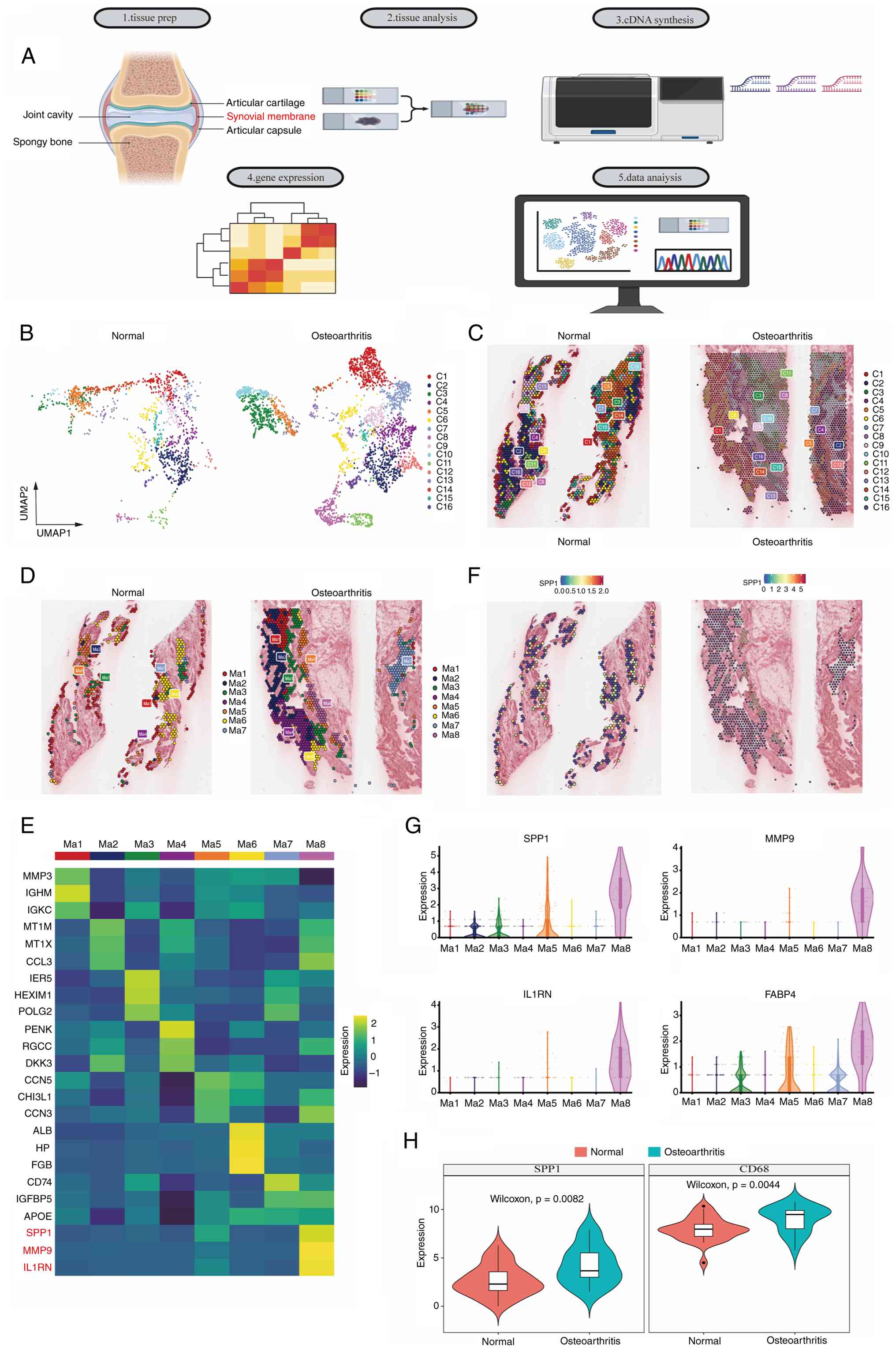
A previous study has reported prominent macrophage infiltration in the synovial tissue of patients with OA (35). In the present study, knee synovial tissue samples were clinically collected from six patients (3 patients with OA and 3 normal controls). OA synovial tissue was obtained from patients with K-L stage IV OA, whereas normal synovial tissue was obtained from donors who underwent amputation due to traffic accidents and had no history of arthritis. Exploratory spatial transcriptome analysis was performed on synovial tissue from one patient with OA and one normal control (Fig. 1A).
Using the 10x Genomics Visium platform to measure total mRNA in intact tissue sections, after standard quality control and integration using the Seurat package, a total of 16 distinct cellular subclusters (c1-c16) mapped onto the corresponding H&E images were obtained (Fig. 1B and C). Subsequent annotation of the 16 subclusters based on CellMarker 2.0, followed by extraction and reclustering of the macrophage-containing subcluster, yielded eight macrophage subclusters (Ma1-Ma8; Fig. 1D). On the basis of the top three marker genes for each of the eight macrophage subclusters (Fig. 1E), it was found that the Ma8 macrophage subcluster was increased in the OA synovium (Fig. 1F). Violin plots revealed the top four marker genes of the Ma8 subcluster, which primarily expressed SPP1, MMP9, IL1RN and FABP4 (Fig. 1G).
To further validate these findings in an independent cohort, the bulk RNA-seq dataset GSE89408 (normal, n=28; OA, n=22) was analyzed, which was downloaded from the GEO database. The expression of SPP1 and the macrophage marker CD68 was significantly elevated in OA synovial tissue (P<0.05), which is consistent with the spatial transcriptomic observations (Fig. 1H).
These results indicate increased expression of SPP1+ macrophages during OA progression, suggesting their potential involvement in regulating cell survival pathways within the OA synovium. Due to the limited number of Visium cases, spatial localization of SPP1+ macrophages was initially focused on, with subsequent multilevel validation performed through public scRNA-seq datasets and functional experiments examining macrophage survival.
Senescent synovial fibroblast-derived TGF-β promotes the acquisition of an SPP1+ macrophage phenotype
To further investigate changes in SPP1+ macrophages during OA progression, the scRNA-seq dataset GSE216651 was downloaded from the GEO, which contains synovial samples from healthy individuals (n=4) and patients with OA (n=5). A standard Seurat workflow and annotation based on previously published literature (22) revealed fibroblasts, endothelial cells, myeloid cells, lymphocytes, pericytes and adipose stem cells, along with their marker genes (Fig. S1A and B). Myeloid cells were extracted, re-clustered and annotated, yielding eight cell subclusters. UMAP plots and dot plots revealed the following eight subclusters: i) Tissue-resident macrophages; ii) monocytes; iii) inflammatory macrophages; iv) dendritic cells 1; v) SPP1+ macrophages; vi) dendritic cells 2; vii) mast cells; and viii) granulocytes (Fig. 2A and B). Pseudotime trajectory analysis using Slingshot revealed a bifurcating differentiation path originating from CD16+ monocytes, leading to the generation of inflammatory macrophages (Inflamm.Mac) or SPP1+ macrophages (SPP1.Mac; Fig. 2C and D). Along the trajectory towards SPP1.Mac, the expression of SPP1, FABP4, MMP9 and MMP19 gradually increased, suggesting the progressive acquisition of the SPP1+ phenotype (Fig. 2E). Hallmark and KEGG pathway analyses revealed enrichment of several pathways in SPP1.Mac (Fig. 2F), including inflammation-related pathways (such as MAPK, NF-κB and TNF) and cell survival pathways (such as PI3K-Akt and mTOR). Notably, anti-apoptotic signatures appeared to be enriched in SPP1. Mac, suggesting a possible role in promoting macrophage persistence in the inflammatory environment. Concurrently, the canonical TGF-β pathway was observed to be upregulated in monocytes, potentially pointing to a role for TGF-β in the differentiation of monocytes into SPP1+ macrophages (Fig. 2F).
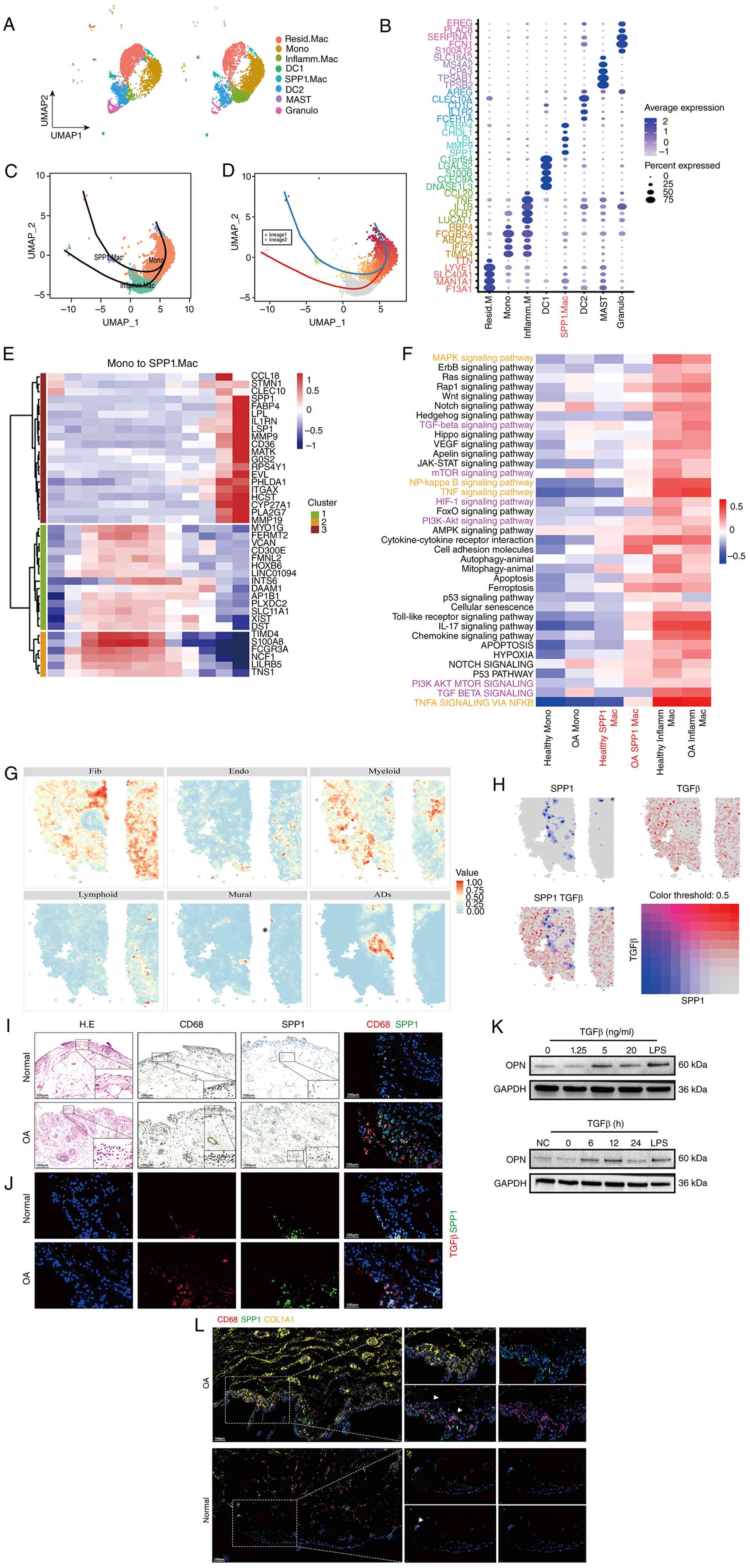 |
Figure 2
Senescent synovial fibroblast-derived TGF-β promotes the acquisition of an SPP1+ macrophage phenotype. (A) UMAP plot of myeloid cells from healthy donors (n=4) and patients with OA (n=5). (B) Dot plot of marker gene expression in each cell cluster. Color indicates scaled average expression, and dot size indicates the percentage of cells expressing each gene. (C) Pseudotime trajectories of monocyte and macrophage populations generated using Slingshot. (D and E) Gene expression along the pseudotime trajectory from Mono to SPP1. Mac; the color scale indicates relative gene expression. (F) Hallmark and KEGG pathway analyses of each cell cluster in HD and OA samples. The color scale indicates scaled gene set enrichment scores; inflammation-related pathways are highlighted in yellow and TGF-β-related pathways in purple. (G) Cell type annotation of OA synovial tissue using CARD deconvolution. (H) Spatial co-localization of SPP1 and TGFB1 expression in OA synovium. (I) H&E, IHC and IF staining of synovium from normal controls (n=3) and patients with OA (n=3). Scale bar, 100 μm. (J) IF staining for TGF-β and SPP1 in synovium from normal controls (n=3) and patients with OA (n=3). Scale bar, 100 μm. (K) Western blot analysis of OPN protein expression in TGF-β-treated RAW 264.7 cells (n=3). (L) Representative three-marker multiplex IF images of human synovium from normal controls (n=2) and patients with OA (n=2), showing SPP1+CD68+ cells within COL1A1+ fibroblast-associated regions. Scale bar, 100 μm. SPP1, secreted phosphoprotein 1; TGF-β, transforming growth factor-β; OA, osteoarthritis; UMAP, uniform manifold approximation and projection; KEGG, Kyoto Encyclopedia of Genes and Genomes; CARD, conditional autoregressive-based deconvolution; H&E, hematoxylin and eosin; IHC, immunohistochemistry; IF, immunofluorescence; OPN, osteopontin.
|
To integrate spatial context, CARD deconvolution of OA synovial tissue was performed, and the following six major cell types were annotated: i) Fibroblasts; ii) endothelial cells; iii) myeloid cells; iv) lymphocytes; v) pericytes; and vi) adipose stem cells. High spatial colocalization of fibroblasts and myeloid cells was detected within the synovium (Fig. 2G). Given that a single spot on the 10x Visium platform can contain up to ten cells, this suggests spatial proximity and niche colocalization. Colocalization analysis of the TGFB1 and SPP1 genes in the OA synovium revealed their presence in the same regions (Fig. 2H). IHC and IF colocalization confirmed the expression of SPP1+ in macrophages in the OA synovium (Fig. 2I). In parallel, multiplex IF of human synovial tissue also showed that SPP1+CD68+ cells were concentrated in COL1A1+ fibroblast-associated areas, providing qualitative protein level evidence consistent with potential spatial proximity (Fig. 2L).
Hypoxia scoring and visualization of senescence-associated genes (P16 and P21) indicated that fibroblasts adjacent to SPP1+ macrophage clusters exhibited hypoxic and senescent phenotypes (Fig. S2A), conditions that promote TGF-β secretion and modulate apoptotic sensitivity in the synovial microenvironment. In vitro, senescence and hypoxia models were established in mouse synovial fibroblasts, and western blotting/ELISA experiments revealed increased TGF-β secretion under these conditions (Fig. S2B-D). IF staining validated the spatial proximity of SPP1 and TGF-β (Fig. 2J). Finally, stimulation of RAW 264.7 macrophages with TGF-β strongly increased OPN protein expression, supporting the direct regulatory effect of TGF-β on SPP1+ macrophage differentiation (Fig. 2K).
CellChat analysis predicts enhanced crosstalk between SPP1+ macrophages and inflammatory macrophages in the OA synovium
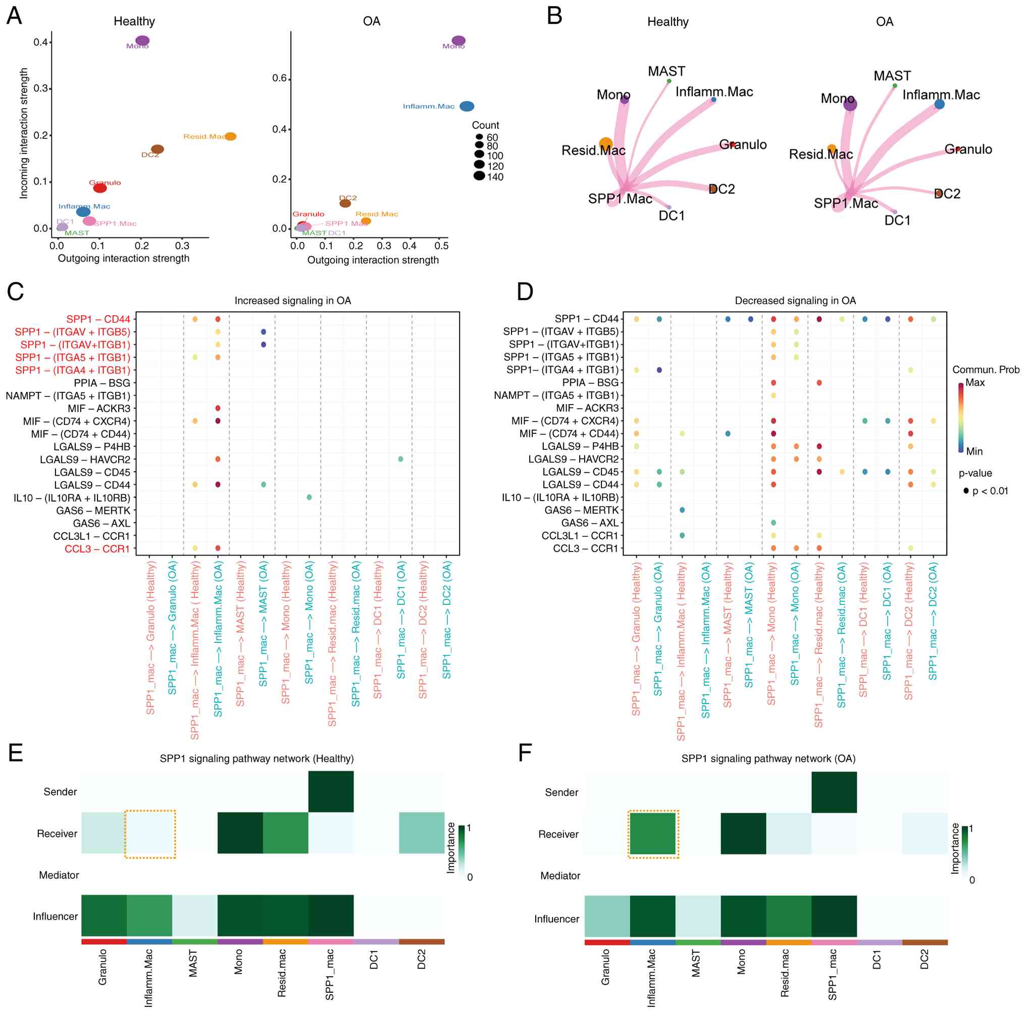
To elucidate the mechanism by which SPP1+ macrophages affect the behavior of inflammatory macrophages, CellChat was used to analyze intercellular communication among myeloid subsets in synovial tissue.
Computational analysis revealed significantly increased outgoing and incoming signal strength for the inflammatory macrophage cluster and decreased strength for the tissue-resident macrophage cluster in the OA synovium (Fig. 3A). Moreover, a chord diagram displays the interactions between SPP1 macrophages and different myeloid cell subpopulations (Fig. 3B). Analysis of ligand-receptor pairs revealed that in OA, SPP1+ macrophages primarily exhibited increased interactions with inflammatory macrophages via SPP1-CD44, SPP1-integrin and CCL3-CCR1 interactions (Fig. 3C and D). Centrality scores were calculated to assess the primary roles of different myeloid subsets related to SPP1 signaling. These analyses revealed increased SPP1 signal receiver function in inflammatory macrophages and increased SPP1 signal sender function in SPP1+ macrophages in OA tissue (Fig. 3E and F).
SPP1+ macrophages suppress apoptosis of inflammatory macrophages through the SPP1-CD44 signaling axis
Building on the aforementioned findings and CellChat-inferred communication patterns, the direct effects of OPN on macrophages were examined in vitro. RAW 264.7 macrophages were treated for 12 h under three conditions (control, OPN and OPN + LPS). Western blot analysis revealed that OPN reduced NF-κB pathway activation in macrophages by suppressing p65 phosphorylation under basal conditions (Fig. 4A). However, when macrophages were polarized into an inflammatory phenotype by LPS, OPN no longer modulated NF-κB activation, but instead exerted anti-apoptotic effects through alternative mechanisms (Fig. 4A).
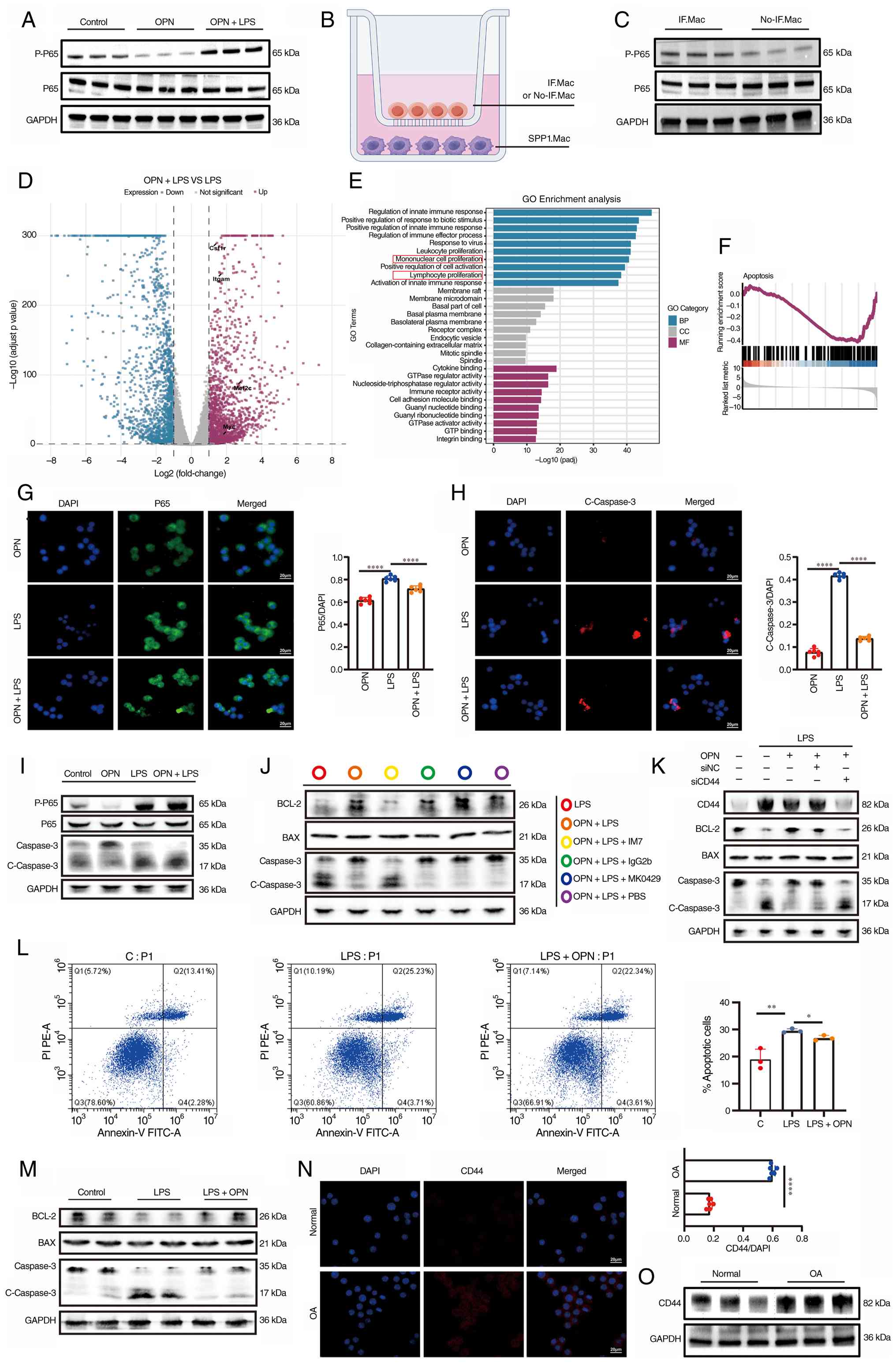 |
Figure 4
SPP1+ macrophages suppress apoptosis of inflammatory macrophages through the SPP1-CD44 signaling axis. (A) Western blot analysis of p-p65 and total p65 in RAW 264.7 cells under the indicated OPN and LPS treatment conditions (n=3). (B) Schematic of the co-culture of SPP1.Mac with IF.Mac or No-IF.Mac. (C) Western blot analysis of p-p65 and p65 in RAW 264.7 cells under the indicated co-culture conditions (n=3). (D) Volcano plot of differentially expressed genes in OPN-treated RAW 264.7 cells. (E) GO analysis of the differentially expressed genes following OPN treatment. (F) GSEA of the apoptosis pathway following OPN treatment. (G and H) IF staining for p65 and cleaved caspase-3, respectively, in OPN-treated RAW 264.7 cells (n=6). Scale bar, 20 μm. (I) Western blot analysis of p-p65, total p65, total caspase-3 and cleaved caspase-3 following OPN treatment (n=3). (J) Western blot analysis of total caspase-3, cleaved caspase-3, BCL-2 and Bax following OPN treatment under the indicated inhibitory conditions (n=3). (K) Western blot analysis of CD44, BCL-2, Bax, total caspase-3 and cleaved caspase-3 in RAW 264.7 macrophages following Cd44 siRNA knockdown (siCd44) or control siRNA (siNC) under LPS + OPN treatment (n=3). (L) Flow cytometric analysis of apoptosis using Annexin V-FITC/PI staining in BMDMs treated with LPS with or without OPN (n=3 independent experiments). (M) Western blot analysis of total caspase-3, cleaved caspase-3, BCL-2 and Bax in primary mouse BMDMs treated with LPS with or without OPN (n=3). (N) IF staining for CD44 in RAW 264.7 cells under normal and OA-model conditions (n=6). Scale bar, 20 μm. (O) Western blot analysis of CD44 in RAW 264.7 cells under normal and OA-model conditions (n=3). Data are presented as the mean ± SD. *P<0.05, **P<0.01 and ****P<0.0001. SPP1, secreted phosphoprotein 1; OPN, osteopontin; LPS, lipopolysaccharide; IF.Mac, inflammatory macrophage; No-IF.Mac, non-inflammatory macrophage; GO, Gene Ontology; GSEA, gene set enrichment analysis; IF, immunofluorescence; BMDM, bone marrow-derived macrophage; FITC, fluorescein isothiocyanate; PI, propidium iodide; HD, healthy donor; OA, osteoarthritis; SD, standard deviation; p-, phosphorylated.
|
To investigate further, a Transwell co-culture model was established in which inflammatory or noninflammatory macrophages were co-cultured with TGF-β-induced SPP1+ macrophages (Fig. 4B). Western blot analysis confirmed that SPP1+ macrophages did not directly activate the NF-κB pathway via p65 phosphorylation in co-cultured macrophages (Fig. 4C).
To comprehensively evaluate transcriptional responses, RAW 264.7 macrophages were divided into control, LPS and OPN + LPS groups and subjected to RNA-seq. Among these genes, genes related to monocyte proliferation, such as Myc, Mef2c, Itgam and Csf1r, were significantly upregulated in the OPN + LPS group (Fig. 4D). Differential GO analysis revealed enrichment in terms such as monocyte proliferation and leukocyte proliferation (Fig. 4E). GSEA revealed significant suppression of pro-apoptotic gene signatures in the OPN + LPS group compared with LPS treatment alone (Fig. 4F), indicating that OPN counteracts inflammation-induced apoptotic programming. IF staining revealed that OPN inhibited p65 phosphorylation and suppressed apoptosis under normal conditions. However, in inflammatory macrophages, OPN appeared to modestly reduce p65 nuclear localization while still inhibiting apoptosis (Fig. 4G and H). Western blotting confirmed that OPN treatment in LPS-stimulated inflammatory macrophages did not alter p65 phosphorylation but significantly reduced cleaved Caspase-3 expression and modulated BCL-2 family protein balance, confirming its specific anti-apoptotic function independent of NF-κB signaling (Fig. 4I). CellChat analysis predicted that SPP1+ macrophages interact with inflammatory macrophages via SPP1-CD44 and SPP1-integrin ligand-receptor pairs. To determine which receptor mediated the anti-apoptotic effect in the OA synovium, inflammatory macrophages were treated with the CD44 receptor inhibitor IM7, the integrin inhibitor MK0429 or an IgG2b control antibody. Western blot analysis revealed that CD44 blockade, but not integrin inhibition, abrogated the anti-apoptotic effect of OPN in inflammatory macrophages, demonstrating that SPP1+ macrophages may suppress apoptosis of inflammatory macrophages through the SPP1-CD44 signaling axis (Fig. 4J). To address potential off-target concerns associated with the pharmacological CD44 inhibitor, siRNA-mediated knockdown of CD44 in RAW 264.7 macrophages was further performed. Western blot confirmed reduced CD44 protein expression following siCd44 transfection (Fig. S2E). Under LPS + OPN conditions, CD44 silencing effectively reversed the OPN-induced anti-apoptotic protein profile, as evidenced by decreased BCL-2 expression and increased cleaved Caspase-3 levels compared with control siRNA-treated cells (Fig. 4K). To confirm these findings, complementary experiments were performed in primary mouse BMDMs. Flow cytometry with Annexin V-FITC/PI staining (Fig. 4L) showed that the LPS induced increase in total apoptotic cells was partially reversed by OPN treatment. Concurrently, western blot analysis (Fig. 4M) revealed that OPN added to LPS-challenged BMDMs reduced cleaved Caspase-3 and skewed the BCL-2/Bax equilibrium toward an anti-apoptotic state. Dot plots from the scRNA-seq data revealed upregulated CD44 receptor expression on inflammatory macrophages in OA compared with normal conditions (Fig. S2F). In vitro interventions followed by IF and western blotting confirmed the increase in CD44 receptor expression (Fig. 4N and O).
Taken together, these in vitro studies reveal a mechanistic basis whereby SPP1+ macrophages prevent the apoptosis of inflammatory macrophages through the SPP1-CD44 signaling axis. To assess the in vivo significance of this mechanism, next its functional relevance in a mouse model of OA was examined.
Macrophage-targeted SPP1 knockdown restores apoptotic sensitivity and mitigates synovial inflammation in OA
To investigate the effect of SPP1+ macrophages on OA progression, an OA model was established in eight-week-old C57BL/6 mice via DMM surgery. Post-surgery, the mice received intra-articular injections of AAV-shRNA targeting SPP1 under the CD68 promoter (specific for macrophages) or saline control (Fig. 5A). Knee joint samples were collected at six weeks post-surgery for micro-CT and histopathological analysis (Fig. 5B-D). H&E, Safranin O-Fast Green, COL2A1 IHC and MMP13 IF staining were performed on knee joint sections (Fig. 5E and F). Western blot analysis confirmed reduced SPP1 protein expression in RAW 264.7 macrophages expressing shSpp1 compared with sh-NC, supporting the knockdown efficacy of the targeting sequence used in the AAV construct (Fig. S2G). In addition, IF co-staining for TNF-α, SPP1, Cleaved Caspase-3 and CD68 was performed on synovial tissue (Fig. 5G). The saline group showed significant subchondral bone changes, particularly osteophyte formation, and a significant decrease in the BV/TV. Notably, sh-SPP1 treatment effectively ameliorated these pathological changes, inhibiting alterations in trabecular bone and bone density (Fig. 5H-J). Furthermore, IHC staining of articular cartilage revealed upregulated COL2A1 expression after sh-SPP1 treatment, and IF staining revealed decreased MMP13 production (Fig. 5E, F, K and L). sh-SPP1 significantly reduced synovial SPP1+ macrophages and restored apoptotic markers. In inflammatory macrophages, AAV-shSpp1 treatment increased cleaved Caspase-3+ macrophages cells, while decreasing TNF-α expression (Fig. 5G and M-O), confirming that SPP1-driven anti-apoptotic signaling sustains persistent inflammation.
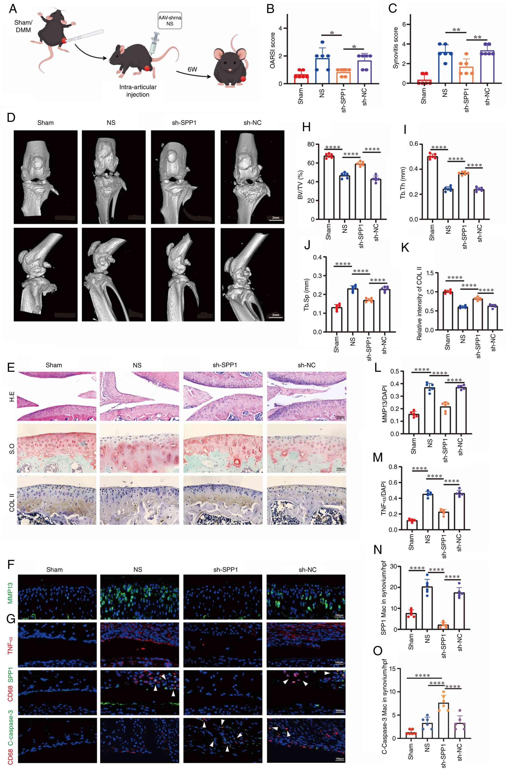 |
Figure 5
Macrophage-targeted SPP1 knockdown restores apoptotic sensitivity and mitigates synovial inflammation in OA. (A) Schematic of the DMM-induced mouse OA model and treatment protocol. (B) OARSI scores (n=6 mice/group). (C) Synovitis pathology scores (n=6 mice/group). (D) Representative micro-CT images of mouse knee joints. Scale bar, 2 mm. (E and F) H&E and Safranin O staining, COL2A1 IHC and MMP13 IF staining of mouse knee joints. Scale bars, 200 μm in (E) and 100 μm in (F). (G) IF co-staining for TNF-α, CD68, cleaved caspase-3 and SPP1 in mouse synovium. Scale bar, 100 μm. (H-J) Quantification of BV/TV and trabecular parameters (n=6 mice/group). (K and L) Quantification of COL2A1 and MMP13 staining (n=6 mice/group). (M-O) Quantification of TNF-α, CD68/SPP1 co-localization and cleaved caspase-3+ inflammatory macrophages; panel N shows SPP1 expression within CD68+ macrophages, confirming SPP1 knockdown efficiency (n=6 mice/group). Data are presented as the mean ± SD. *P<0.05, **P<0.01 and ****P<0.0001. SPP1, secreted phosphoprotein 1; DMM, destabilization of the medial meniscus; OA, osteoarthritis; OARSI, Osteoarthritis Research Society International; micro-CT, micro-computed tomography; H&E, hematoxylin and eosin; COL2A1, type II collagen; IHC, immunohistochemistry; MMP13, matrix metallopeptidase 13; IF, immunofluorescence; BV/TV, bone volume fraction; SD, standard deviation.
|
In summary, a specific reduction in SPP1+ macrophages in the synovium effectively alleviated TNF-α secretion and synovitis, thereby slowing OA progression in mice. The observed histological changes in the mice are consistent with previous in vitro analyses.
CCL3 neutralization reduces myeloid cell accumulation and promotes resolution of synovitis in OA
Previous studies reported that SPP1+ macrophages can chemoattract monocytes (36,37). Consistently, the present scRNA-seq CellChat analysis revealed CCL3-CCR1 interactions between SPP1+ and inflammatory macrophages. Colocalization analysis of the CCL3 and SPP1 genes in OA synovial spatial transcriptomes (Fig. 6A) and IF staining of human synovial tissue (Fig. 6B) revealed that CCL3 is predominantly expressed in the surrounding SPP1+ macrophages. During monocyte differentiation into SPP1+ and inflammatory macrophages, CCL3 expression initially increased but subsequently decreased (Fig. 6C).
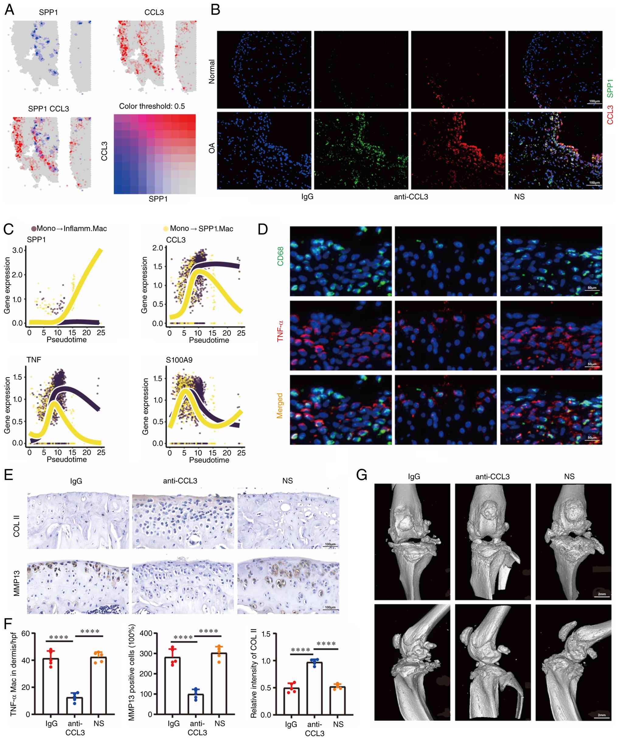 |
Figure 6
CCL3 neutralization reduces myeloid cell accumulation and promotes resolution of synovitis in OA. (A) Spatial co-localization of SPP1 and CCL3 expression in OA synovium. (B) IF staining for SPP1 and CCL3 in synovium from normal controls (n=3) and patients with OA (n=3). Scale bar, 100 μm. (C) Expression of the indicated genes along pseudotime trajectories from Mono to SPP1.Mac (yellow) and from Mono to Inflamm.Mac (purple). (D) IF co-staining for TNF-α and CD68 in mouse synovium (n=5 mice/group). Scale bar, 50 μm. (E) IHC staining for COL2A1 and MMP13 in mouse knee joints (n=5 mice/group). Scale bar, 100 μm. (F) Quantification of TNF-α/CD68 IF and COL2A1/MMP13 IHC staining (n=5 mice/group). (G) Micro-CT images of mouse knee joints (n=5 mice/group). Scale bar, 2 mm. Data are presented as the mean ± SD. ****P<0.0001. CCL3, C-C motif chemokine ligand 3; SPP1, secreted phosphoprotein 1; OA, osteoarthritis; IF, immunofluorescence; IHC, immunohistochemistry; COL2A1, type II collagen; MMP13, matrix metallopeptidase 13; micro-CT, micro-computed tomography; SD, standard deviation.
|
In the mouse DMM OA model, intra-articular administration of a CCL3 neutralizing antibody was performed. IF and IHC analyses of knee joint samples are shown in Fig. 6D and E. The CCL3 neutralizing antibody group showed a significant reduction in TNF-α+ inflammatory macrophages in the synovium, decreased MMP13 in cartilage and increased COL2A1 levels (Fig. 6F). Micro-CT analysis revealed that CCL3 neutralization reduced osteophyte formation in OA mouse knees (Fig. 6G).
These results suggest that SPP1+ macrophages exacerbate OA progression through dual mechanisms, including direct suppression of the apoptosis of inflammatory macrophages via SPP1-CD44 signaling and indirect promotion of myeloid cell accumulation via CCL3-mediated chemotaxis.
In silico SPP1 perturbation nominates apoptosis-related networks and candidate compounds. To evaluate the regulatory role of SPP1 on the synovial microenvironment at a systems level, an in silico virtual knockout simulation targeting myeloid SPP1 was performed. Manifold distance analysis identified the top 20 significantly perturbed genes, including key regulatory factors such as TTN, F13A1 and LYVE1 (Fig. 7A and B).
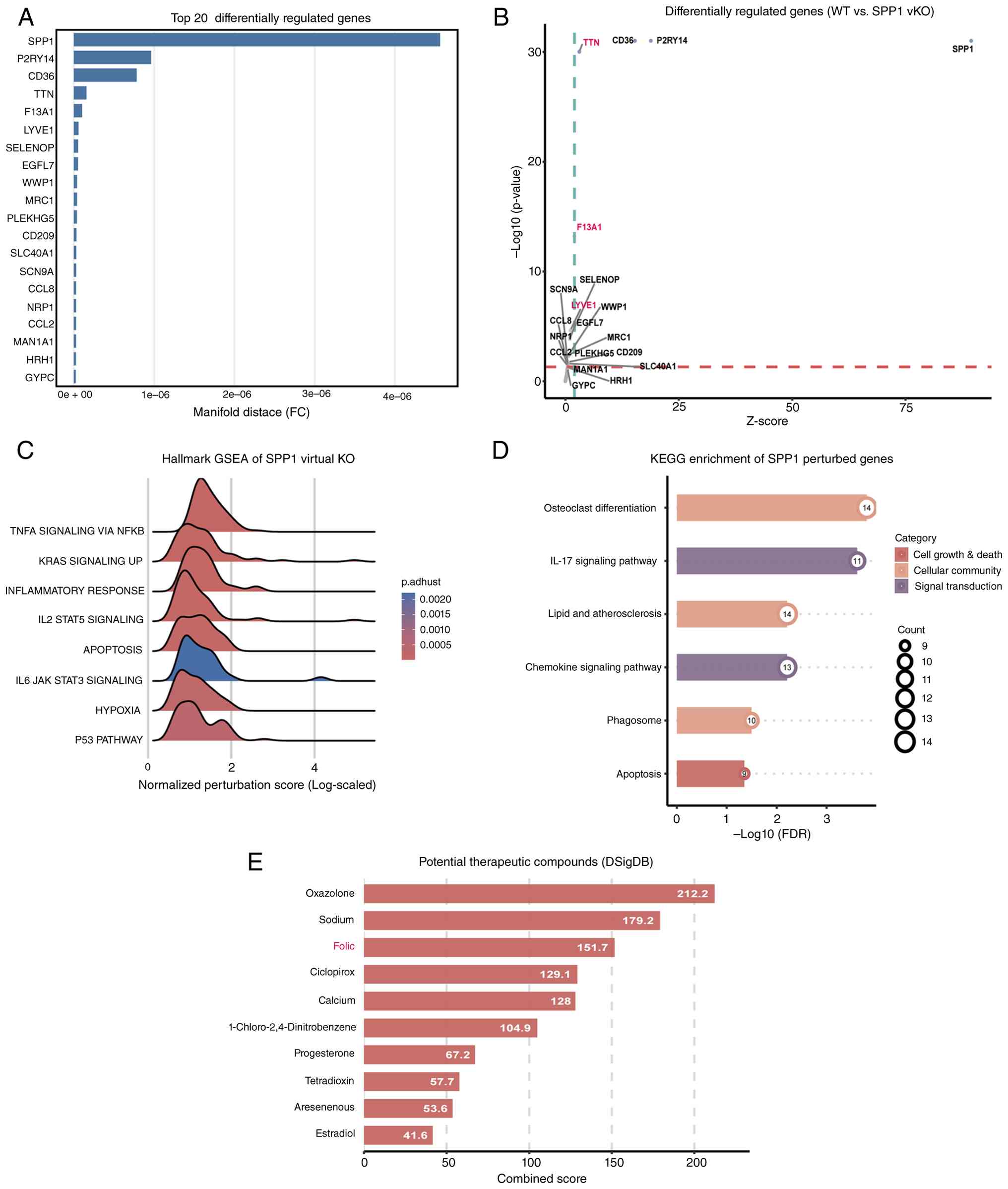 |
Figure 7
In silico SPP1 perturbation nominates apoptosis-related networks and candidate compounds. (A) Bar chart of the top 20 significantly differentially regulated genes following SPP1 virtual knockout, ranked by manifold distance. (B) Volcano plot showing the distribution of differentially regulated genes; the x-axis indicates the Z-score. (C) Hallmark GSEA ridge plot of the perturbed network genes, showing enrichment of apoptosis and IL-6-JAK-STAT3 signaling. (D) KEGG pathway enrichment analysis highlighting perturbed genes associated with cell proliferation and death and chemokine signaling. (E) Candidate compounds identified by screening the DSigDB database; oxazolone, sodium and folic acid were the three highest-ranked candidates. SPP1, secreted phosphoprotein 1; GSEA, gene set enrichment analysis; IL-6, interleukin-6; JAK, Janus kinase; STAT3, signal transducer and activator of transcription 3; KEGG, Kyoto Encyclopedia of Genes and Genomes; DSigDB, Drug Signatures Database.
|
Functional enrichment analysis revealed that the gene network regulated by SPP1 was significantly enriched in apoptosis regulation, particularly the p53 signaling pathway and BCL-2 family-mediated cell death pathways, alongside NF-κB inflammatory responses (Fig. 7C and D). This computational finding is highly consistent with the present in vitro results that demonstrated that SPP1 suppresses the apoptosis of inflammatory macrophages. DSigDB screening ranked oxazolone first, sodium second and folic acid third (Fig. 7E), but these computational findings require further experimental verification.
Discussion
Despite advances in understanding OA, the disease still lacks effective disease-modifying drugs, largely because the cellular mechanisms of synovial inflammation remain elusive. The present study identified SPP1+ macrophages as critical regulators of apoptosis resistance in the OA synovium. In addition, spatial transcriptomics, single-cell transcriptomics and mechanistic experiments were integrated to define a previously uncharacterized SPP1+ macrophage subset that expands in the human OA synovium and contributes to persistent synovitis. Senescent/hypoxic synovial fibroblasts secrete TGF-β (38,39), which promotes the differentiation of a CD16+ monocyte population into SPP1+ macrophages via noncanonical signaling pathways. These SPP1+ macrophages sustain synovial inflammation through two complementary mechanisms: i) Preventing the apoptosis of inflammatory macrophages through SPP1-CD44 signaling; and ii) recruiting additional myeloid cells via CCL3 secretion. In the present study, the single-cell model was further leveraged to perform in silico drug repositioning, establishing the SPP1-mediated pathogenic network as a biological target for pharmacological intervention.
Initially, spatial transcriptomic profiling of synovial tissue from one healthy donor and one patient with OA was performed to delineate the cellular landscape and spatial organization of the OA synovium. Spatial colocalization of fibroblasts and myeloid cells were observed and indicated spatial proximity and potential niche colocalization. These findings align with the emerging view that synovial fibroblasts are key architects of the immune microenvironmental 'niche' (40-47). Although the Visium dataset is exploratory (n=1/group) and its spot-level resolution aggregates multiple cells, the inferred spatial proximity between fibroblasts and myeloid cells was independently validated by multiplex IF on human synovial tissues. Thus, despite these technical limitations, this orthogonal protein-level evidence supports the existence of a spatially organized fibroblast-myeloid niche, providing a novel foundation for understanding synovitis in OA (48-52).
Among the various synovial cell types, macrophages have garnered attention due to their high plasticity and central role in inflammation regulation (10,53-56). The present study confirms prior observations of increased macrophage infiltration in the OA synovium. More importantly, a distinct SPP1+ macrophage subset (Ma8) that expands in OA and expresses a unique molecular signature (SPP1, MMP9 and FABP4) was identified. Next, pseudotime trajectory analysis revealed that SPP1+ macrophages and inflammatory macrophages share a common origin from CD16+ monocytes but diverge into distinct differentiation paths. This finding establishes SPP1+ macrophages as an independent functional subset rather than a mere transitional state of inflammatory macrophages.
Next, the upstream regulatory mechanisms governing the emergence of SPP1+ macrophages were explored. The findings indicate that senescent or hypoxia-stressed synovial fibroblasts act as key drivers in this process (57-62). Spatial transcriptomics revealed enrichment of senescence-associated genes (such as P16 and P21) in fibroblast regions adjacent to SPP1+ macrophages, strongly suggesting that fibroblast senescence and the associated senescence-associated secretory phenotype may be crucial in shaping the local inflammatory microenvironment. In vitro assays further demonstrated that senescent or hypoxic fibroblasts exhibit significantly enhanced TGF-β secretion. The role of TGF-β in OA is complex and context dependent and is often described as a 'double-edged sword' (63,64). Although its role in subchondral bone sclerosis has been well studied, its mechanisms in synovitis remain unclear. Through spatial and single-cell transcriptomics coupled with in vitro validation, the present study demonstrated that TGF-β secreted by senescent/stressed synovial fibroblasts may promote the acquisition of an SPP1+ macrophage phenotype, potentially involving PI3K-Akt and mTOR signaling).
Previous studies have reported conflicting roles for SPP1 (OPN), with evidence supporting both proinflammatory and anti-inflammatory functions (65-70). The present findings clarify a previously unappreciated anti-apoptotic role for SPP1+ macrophages, whereby they directly inhibit apoptosis of inflammatory macrophages through SPP1-CD44 signaling in a manner independent of canonical NF-κB pathway activation (71,72). This discovery provides a novel mechanistic explanation for the persistence of synovial inflammation in OA. Specifically, SPP1+ macrophages perpetuate inflammation by sustaining the population of inflammatory macrophages through suppression of their apoptotic clearance. This pro-survival effect operates independently of canonical NF-κB activation (73), providing a novel mechanistic explanation which suggest that rather than directly driving NF-κB-mediated inflammation, SPP1+ macrophages perpetuate it by sustaining the inflammatory cell population.
This discovery integrates the key elements of TGF-β, SPP1+ macrophages and inflammatory macrophages into a coherent cellular cascade. Moreover, cell-cell communication analysis revealed a self-amplifying inflammatory positive feedback loop in the OA synovium. SPP1+ macrophages not only maintain inflammatory macrophage survival via the SPP1-CD44 axis but also, potentially through CCL3 secretion, promote further recruitment and activation of monocytes or macrophages, which is consistent with the reported chemotactic functions of SPP1 (74,75). Consequently, both AAV-shRNA-mediated specific knockdown of SPP1 in macrophages and CCL3 neutralizing antibody treatment significantly alleviated synovitis, cartilage destruction and osteophyte formation in the mouse model. Notably, while gene therapy and neutralizing antibodies validated the biological targets, the present study sought to translate these findings into accessible clinical interventions. By employing a virtual knockout model on the scRNA-seq datasets, the SPP1-mediated network perturbation was mapped and drug repositioning strategies were used to find promising therapeutic candidates.
Interpretation of the present findings is subject to several caveats. First, the Visium spatial data were derived from a limited number of samples (n=1/group), and the age disparity between OA and control synovial tissues may confound senescence-related interpretations. However, the major spatial observations are corroborated by independent bulk and single-cell RNA-seq cohorts as well as multiplex IF. In addition, although the anti-apoptotic effect of OPN was independently confirmed in primary murine BMDMs, CD44 dependence was demonstrated only in RAW264.7 macrophages by pharmacological blockade and siRNA-mediated knockdown. Direct CD44 perturbation in primary macrophages and dose-response and time-course analyses were not performed. The in vivo AAV-shSpp1 experiment supports the functional relevance of macrophage-associated SPP1 reduction but does not constitute a CD44-mediated rescue experiment. Direct confirmation in human synovial macrophages also remains to be established. Only male mice were used in the present study to maintain consistency with the established DMM protocol. Given the emerging evidence of sex-dependent differences in OA pathogenesis, whether the SPP1-CD44 axis operates similarly in female mice remains to be determined. The computational drug repositioning analysis is hypothesis-generating and awaits rigorous experimental validation. These limitations do not undermine the principal conclusions but rather delineate priorities for future investigation.
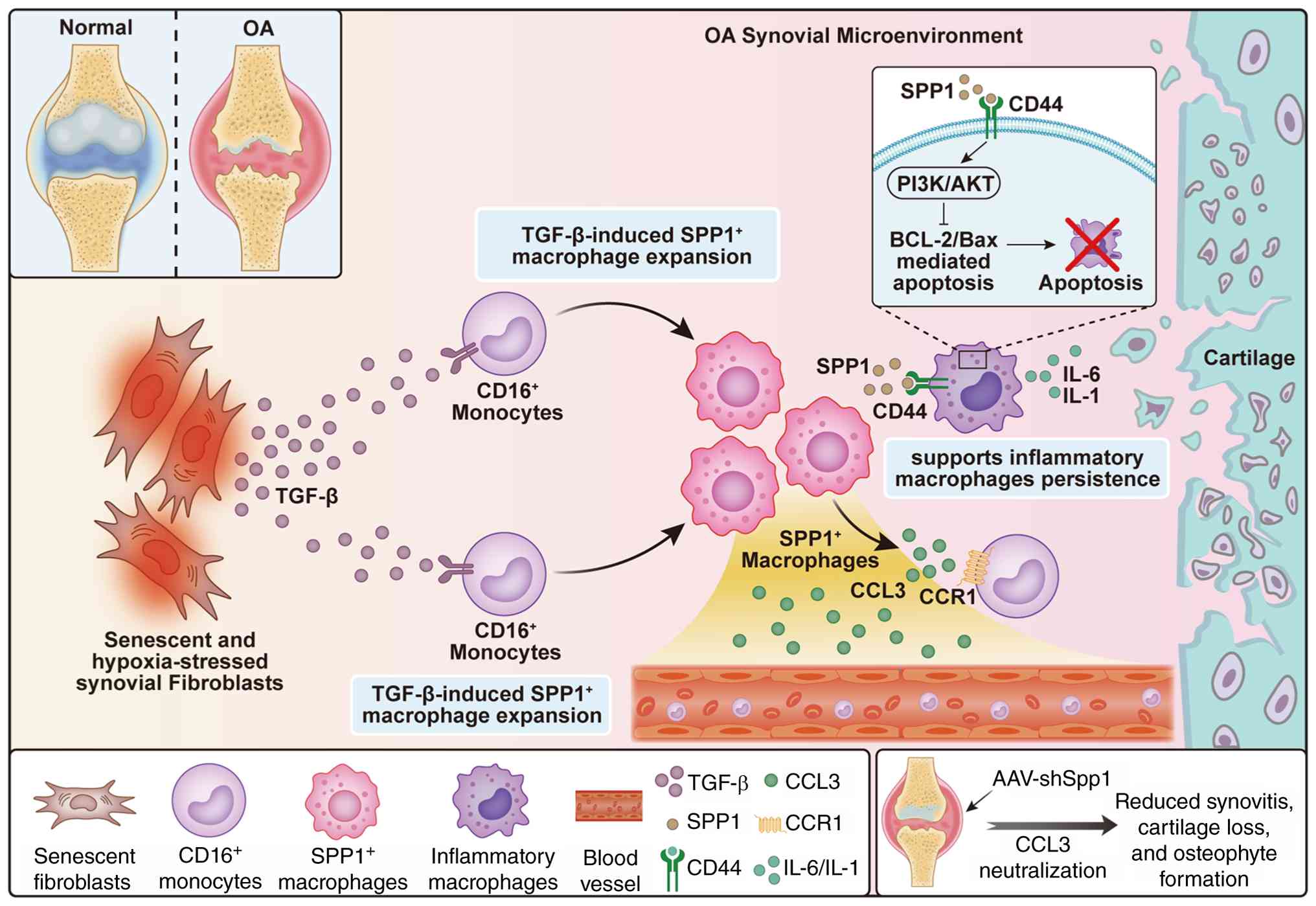
In summary, a TGF-β driven SPP1+ macrophage subset was identified that establishes an anti-apoptotic niche in the OA synovium, directly suppressing inflammatory macrophage apoptosis via SPP1-CD44 signaling, while concurrently promoting myeloid cell recruitment through CCL3 (Fig. 8). This spatially defined circuit provides a mechanistic framework for understanding persistent synovitis not merely as unremitting inflammation, but as a failure of apoptotic clearance sustained by a specific cellular microenvironment. In vivo disruption of SPP1 or CCL3 effectively restores macrophage apoptosis and attenuates disease progression, validating this pathway as a therapeutic target.
Supplementary Data
Availability of data and materials
The data generated in the present study may be found in the Gene Expression Omnibus under accession numbers GSE216651, GSE89408 and GSE345213 or at the following URL: https://www.ncbi.nlm.nih.gov/geo/query/acc.cgi?acc=GSE216651; https://www.ncbi.nlm.nih.gov/geo/query/acc.cgi?acc=GSE89408; https://www.ncbi.nlm.nih.gov/geo/query/acc.cgi?acc=GSE345213. The data generated in the present study may be found in the CNGB Nucleotide Sequence Archive (CNSA) of CNGBdb under accession number CNP0009177 or at the following URL: https://doi.org/10.26036/CNP0009177.
Authors' contributions
HG, HS, FZ, YS and LW conceived and designed the research project. HG and HS performed the cell studies. HG and HS performed the animal studies. HG and HS performed the bioinformatics analysis. HG, HS, FZ and WW analyzed and interpreted the data and prepared the figures. SZ contributed to the interpretation of data and provided critical scientific input during the development of the study. HG and HS drafted the manuscript. HG, HS, YS, SZ and LW reviewed and revised the manuscript. All authors read and approved the final version of the manuscript. HG and HS confirm the authenticity of all the raw data.
Ethics approval and consent to participate
The collection and processing of all human samples were approved by The Ethics Committee of Northern Jiangsu People's Hospital Affiliated to Yangzhou University (Yangzhou, China; approval no. 2023ky132) and conducted in accordance with the principles of the Declaration of Helsinki. All participants provided written informed consent prior to sample collection. All animal experiments were approved by The Yangzhou University Animal Ethics Committee (Yangzhou, China; approval no. 202303842).
Patient consent for publication
Not applicable.
Competing interests
The authors declare that they have no competing interests.
Acknowledgements
Not applicable.
Funding
The present study was supported by The Key Project of Jiangsu Commission of Health (grant no. K2023047), the Natural Science Foundation of Jiangsu Province (grant no. BK20201221), the Key Project of Jiangsu Commission of Health (grant no. K2025011), the Key Project Supported by Medical Science and Technology Development Foundation, the Nanjing Department of Health (grant no. JQX24004) and the Yangzhou Municipal 'Lvyang Jinfeng Plan' (grant no. LYJF00099).
References
|
1
|
Cho Y, Jeong S, Kim H, Kang D, Lee J, Kang SB and Kim JH: Disease-modifying therapeutic strategies in osteoarthritis: Current status and future directions. Exp Mol Med. 53:1689–1696. 2021. View Article : Google Scholar : PubMed/NCBI
|
|
2
|
Yao Q, Wu X, Tao C, Gong W, Chen M, Qu M, Zhong Y, He T, Chen S and Xiao G: Osteoarthritis: Pathogenic signaling pathways and therapeutic targets. Signal Transduct Target Ther. 8:562023. View Article : Google Scholar : PubMed/NCBI
|
|
3
|
Hu Y, Chen X, Wang S, Jing Y and Su J: Subchondral bone microenvironment in osteoarthritis and pain. Bone Res. 9:202021. View Article : Google Scholar : PubMed/NCBI
|
|
4
|
Sanchez-Lopez E, Coras R, Torres A, Lane NE and Guma M: Synovial inflammation in osteoarthritis progression. Nat Rev Rheumatol. 18:258–275. 2022. View Article : Google Scholar : PubMed/NCBI
|
|
5
|
Wang R, Mehrjou B, Dehghan-Banian D, Wang BYH, Li Q, Deng S, Liu C, Zhang Z, Zhu Y, Wang H, et al: Targeting long noncoding RNA H19 in subchondral bone osteocytes and the alleviation of cartilage degradation in osteoarthritis. Arthritis Rheumatol. 77:283–297. 2025. View Article : Google Scholar :
|
|
6
|
Chen ZY, Guo Q and Huang CS: Spatiotemporal trends and socioeconomic drivers of hip-knee osteoarthritis burden in East Asia: A GBD-based modeling study (1990-2023). Clin Rheumatol. 45:2855–2876. 2026. View Article : Google Scholar : PubMed/NCBI
|
|
7
|
Courties A, Kouki I, Soliman N, Mathieu S and Sellam J: Osteoarthritis year in review 2024: Epidemiology and therapy. Osteoarthritis Cartilage. 32:1397–1404. 2024. View Article : Google Scholar : PubMed/NCBI
|
|
8
|
Gelber AC: Knee osteoarthritis. Ann Intern Med. 177:ITC129–ITC144. 2024. View Article : Google Scholar : PubMed/NCBI
|
|
9
|
Tong L, Yu H, Huang X, Shen J, Xiao G, Chen L, Wang H, Xing L and Chen D: Current understanding of osteoarthritis pathogenesis and relevant new approaches. Bone Res. 10:602022. View Article : Google Scholar : PubMed/NCBI
|
|
10
|
Motta F, Barone E, Sica A and Selmi C: Inflammaging and osteoarthritis. Clin Rev Allergy Immunol. 64:222–238. 2022. View Article : Google Scholar : PubMed/NCBI
|
|
11
|
Liu B, Xian Y, Chen X, Shi Y, Dong J, Yang L, An X, Shen T, Wu W, Ma Y, et al: Inflammatory Fibroblast-like Synoviocyte-derived exosomes aggravate osteoarthritis via enhancing macrophage glycolysis. Adv Sci (Weinh). 11:23073382024. View Article : Google Scholar : PubMed/NCBI
|
|
12
|
Chi Y, Jiang H, Yin Y, Zhou X, Shao Y, Li Y and Rao J: Macrophage signaling pathways in health and disease: From bench to bedside applications. MedComm (2020). 6:e702562025. View Article : Google Scholar : PubMed/NCBI
|
|
13
|
Philpott HT, Birmingham TB, Blackler G, Klapak JD, Knights AJ, Farrell EC, Fiset B, Walsh LA, Giffin JR, Vasarhelyi EM, et al: Association of synovial innate immune exhaustion with worse pain in knee osteoarthritis. Arthritis Rheumatol. 77:664–676. 2025. View Article : Google Scholar :
|
|
14
|
Hu Y, Jia H, Cui H and Song J: Application of spatial omics in the cardiovascular system. Research (Wash D C). 8:06282025.PubMed/NCBI
|
|
15
|
Jin X, Jian Z, Ma Y, Wen J, Chao N, Chen X, Xiang L, Yuan Y, Liu L, Li Y, et al: Single-cell RNA sequencing analysis reveals the role of Macrophage-Mediated CD44-AKT-CCL2 pathways in renal tubule injury during calcium oxalate crystal formation. Research (Wash D C). 8:06902025.PubMed/NCBI
|
|
16
|
Shao X, Li C, Yang H, Lu X, Liao J, Qian J, Wang K, Cheng J, Yang P, Chen H, et al: Knowledge-graph-based cell-cell communication inference for spatially resolved transcriptomic data with SpaTalk. Nat Commun. 13:44292022. View Article : Google Scholar : PubMed/NCBI
|
|
17
|
Ma Y and Zhou X: Spatially informed cell-type deconvolution for spatial transcriptomics. Nat Biotechnol. 40:1349–1359. 2022. View Article : Google Scholar : PubMed/NCBI
|
|
18
|
Fan X, Sun AR, Young RSE, Afara IO, Hamilton BR, Ong LJY, Crawford R and Prasadam I: Spatial analysis of the osteoarthritis microenvironment: Techniques, insights, and applications. Bone Res. 12:72024. View Article : Google Scholar : PubMed/NCBI
|
|
19
|
Bill R, Wirapati P, Messemaker M, Roh W, Zitti B, Duval F, Kiss M, Park JC, Saal TM, Hoelzl J, et al: CXCL9:SPP1 macrophage polarity identifies a network of cellular programs that control human cancers. Science. 381:515–524. 2023. View Article : Google Scholar : PubMed/NCBI
|
|
20
|
Xia X, He C, Xue Z, Wang Y, Qin Y, Ren Z, Huang Y, Luo H, Chen HN, Zhang WH, et al: Single cell immunoprofile of synovial fluid in rheumatoid arthritis with TNF/JAK inhibitor treatment. Nat Commun. 16:21522025. View Article : Google Scholar : PubMed/NCBI
|
|
21
|
Qi J, Sun H, Zhang Y, Wang Z, Xun Z, Li Z, Ding X, Bao R, Hong L, Jia W, et al: Single-cell and spatial analysis reveal interaction of FAP+ fibroblasts and SPP1+ macrophages in colorectal cancer. Nat Commun. 13:17422022. View Article : Google Scholar
|
|
22
|
Tang SA, Yao L, Ruan J, Kang J, Cao Y, Nie X, Lan W, Zhu Z, Han W, Liu Y, et al: Single-cell atlas of human infrapatellar fat pad and synovium implicates APOE signaling in osteoarthritis pathology. Sci Transl Med. 16:eadf45902024. View Article : Google Scholar : PubMed/NCBI
|
|
23
|
Chen H, Jin Z, Peng Y, Li Y, Li Z, Zhang X, Xie Y, Dong J, Ma L and Ji Z: Malevolent alliance of MYBL2hi cancer stem cell and SPP1+ macrophage confers resistance to neoadjuvant immunotherapy in bladder cancer. J Immunother Cancer. 13:e0113192025. View Article : Google Scholar
|
|
24
|
Pang X, Gong K, Zhang X, Wu S, Cui Y and Qian BZ: Osteopontin as a multifaceted driver of bone metastasis and drug resistance. Pharmacol Res. 144:235–244. 2019. View Article : Google Scholar : PubMed/NCBI
|
|
25
|
Wang Y, Zhang W, Yang Y, Qin J, Wang R, Wang S, Fu W, Niu Q, Wang Y, Li C, et al: Osteopontin deficiency promotes cartilaginous endplate degeneration by enhancing the NF-κB signaling to recruit macrophages and activate the NLRP3 inflammasome. Bone Res. 12:532024. View Article : Google Scholar
|
|
26
|
Osorio D, Zhong Y, Li G, Xu Q, Yang Y, Tian Y, Chapkin RS, Huang JZ and Cai JJ: scTenifoldKnk: An efficient virtual knockout tool for gene function predictions via single-cell gene regulatory network perturbation. Patterns (NY). 3:1004342022. View Article : Google Scholar
|
|
27
|
Yang J, Yin W, Ge J, Gao G, Yao X and Huang Y: Thyroid cancer-derived exosomal SPP1 promotes tumor progression by driving macrophage M2 polarization through the CD44/JAK2/STAT3 signaling pathway. Organogenesis. 22:26701522026. View Article : Google Scholar : PubMed/NCBI
|
|
28
|
Zhou X, Zhang J, Haimbach R, Zhu W, Mayer-Ezell R, Garcia-Calvo M, Smith E, Price O, Kan Y, Zycband E, et al: An integrin antagonist (MK-0429) decreases proteinuria and renal fibrosis in the ZSF1 rat diabetic nephropathy model. Pharmacol Res Perspect. 5:e003542017. View Article : Google Scholar : PubMed/NCBI
|
|
29
|
Glasson SS, Blanchet TJ and Morris EA: The surgical destabilization of the medial meniscus (DMM) model of osteoarthritis in the 129/SvEv mouse. Osteoarthritis Cartilage. 15:1061–1069. 2007. View Article : Google Scholar : PubMed/NCBI
|
|
30
|
Ma HL, Blanchet TJ, Peluso D, Hopkins B, Morris EA and Bhatt PR: Osteoarthritis severity is sex dependent in a surgical mouse model. Osteoarthritis Cartilage. 15:1175–1181. 2007. View Article : Google Scholar
|
|
31
|
Yao Z, Yuan Z, Li Y, Li X, Peng C, Jin J, Zhang H, Bai X, Pan J and Cai D: Upregulation of ACSL1 in synovial macrophages promotes lipid peroxidation via the IκB/NF-κB pathway to accelerate osteoarthritis. J Orthop Translat. 55:360–375. 2025. View Article : Google Scholar
|
|
32
|
Guo H, Sun M, Tao M, Zhu J, Song Y, Chen S, Guo Z, Zhang X, Hou F, Sun C, et al: MAVS signaling exacerbates chondrocytes extracellular matrix degradation in osteoarthritis. Cell Rep. 45:1167782026. View Article : Google Scholar : PubMed/NCBI
|
|
33
|
Liu Y, Hao R, Lv J, Yuan J, Wang X, Xu C, Ma D, Duan Z, Zhang B, Dai L, et al: Targeted knockdown of PGAM5 in synovial macrophages efficiently alleviates osteoarthritis. Bone Res. 12:172024. View Article : Google Scholar
|
|
34
|
Ahmed M, Sottnik JL, Dancik GM, Sahu D, Hansel DE, Theodorescu D and Schwartz MA: An Osteopontin/CD44 axis in RhoGDI2-Mediated metastasis suppression. Cancer Cell. 30:432–443. 2016. View Article : Google Scholar : PubMed/NCBI
|
|
35
|
Thomson A and Hilkens CMU: Synovial macrophages in osteoarthritis: The key to understanding pathogenesis? Front Immunol. 12:6787572021. View Article : Google Scholar : PubMed/NCBI
|
|
36
|
Ye W, Sun J, Shen E, Yang B, Li L, Long Y, Shen J, Liu W, Xiao M, Wu H, et al: Macrophage-Specific SPP1 contributes to pressure overload-induced cardiac dysfunction and maladaptive remodeling. JACC Basic Transl Sci. 11:1016232026. View Article : Google Scholar : PubMed/NCBI
|
|
37
|
Duan X, Zhang L, Liu K, Guo K, You Y, Jia H, Zhou S and Han B: Macrophage-derived SPP1 exacerbate myocardial injury by interacting with fibroblasts in viral myocarditis. Biol Direct. 20:302025. View Article : Google Scholar : PubMed/NCBI
|
|
38
|
Zhang F, Guo J, Yu S, Zheng Y, Duan M, Zhao L, Wang Y, Yang Z and Jiang X: Cellular senescence and metabolic reprogramming: Unraveling the intricate crosstalk in the immunosuppressive tumor microenvironment. Cancer Commun (Lond). 44:929–966. 2024. View Article : Google Scholar : PubMed/NCBI
|
|
39
|
Wu Y, Wang Z, Ge Y, Zhu Y, Tian T, Wei J, Jin Y, Zhao Y, Jia Q, Wu J and Ge L: Microenvironment responsive hydrogel exerting inhibition of cascade immune activation and elimination of synovial fibroblasts for rheumatoid arthritis therapy. J Control Release. 370:747–762. 2024. View Article : Google Scholar : PubMed/NCBI
|
|
40
|
Vasilopoulos Y, Gkretsi V, Armaka M, Aidinis V and Kollias G: Actin cytoskeleton dynamics linked to synovial fibroblast activation as a novel pathogenic principle in TNF-driven arthritis. Ann Rheum Dis. 66:iii23–iii28. 2007. View Article : Google Scholar : PubMed/NCBI
|
|
41
|
Zheng L, Gu M, Li X, Hu X, Chen C, Kang Y, Pan B, Chen W, Xian G, Wu X, et al: ITGA5+ synovial fibroblasts orchestrate proinflammatory niche formation by remodelling the local immune microenvironment in rheumatoid arthritis. Ann Rheum Dis. 84:232–252. 2025. View Article : Google Scholar : PubMed/NCBI
|
|
42
|
Huynh NCN, Ling R, Komagamine M, Shi T, Tsukasaki M, Matsuda K, Okamoto K, Asano T, Muro R, Pluemsakunthai W, et al: Oncostatin M-driven macrophage-fibroblast circuits as a drug target in autoimmune arthritis. Inflamm Regen. 44:362024. View Article : Google Scholar : PubMed/NCBI
|
|
43
|
Taylor CT and Colgan SP: Regulation of immunity and inflammation by hypoxia in immunological niches. Nat Rev Immunol. 17:774–785. 2017. View Article : Google Scholar : PubMed/NCBI
|
|
44
|
Bolton C, Mahony CB, Clay E, Nisa PR, Lomholt S, Hackland A, Chin PS, Smith CG, Alexiou V, Nguyen HD, et al: Synovial tissue atlas in juvenile idiopathic arthritis reveals pathogenic niches associated with disease severity. Sci Transl Med. 17:eadt60502025. View Article : Google Scholar : PubMed/NCBI
|
|
45
|
Collins FL, Roelofs AJ, Symons RA, Kania K, Campbell E, Collie-duguid ESR, Riemen AHK, Clark SM and De Bari C: Taxonomy of fibroblasts and progenitors in the synovial joint at single-cell resolution. Ann Rheum Dis. 82:428–437. 2023. View Article : Google Scholar :
|
|
46
|
Rai MF, Collins KH, Lang A, Maerz T, Geurts J, Ruiz-Romero C, June RK, Ramos Y, Rice SJ, Ali SA, et al: Three decades of advancements in osteoarthritis research: Insights from transcriptomic, proteomic, and metabolomic studies. Osteoarthritis Cartilage. 32:385–397. 2024. View Article : Google Scholar
|
|
47
|
Onder L, Cheng HW and Ludewig B: Visualization and functional characterization of lymphoid organ fibroblasts. Immunol Rev. 306:108–122. 2022. View Article : Google Scholar :
|
|
48
|
Zhang L, Chen D, Song D, Liu X, Zhang Y, Xu X and Wang X: Clinical and translational values of spatial transcriptomics. Signal Transduct Target Ther. 7:1112022. View Article : Google Scholar : PubMed/NCBI
|
|
49
|
Foster DS, Januszyk M, Delitto D, Yost KE, Griffin M, Guo J, Guardino N, Delitto AE, Chinta M, Burcham AR, et al: Multiomic analysis reveals conservation of cancer-associated fibroblast phenotypes across species and tissue of origin. Cancer Cell. 40:1392–1406.e7. 2022. View Article : Google Scholar : PubMed/NCBI
|
|
50
|
Fan G, Xie T, Li L, Tang L, Han X and Shi Y: Single-cell and spatial analyses revealed the co-location of cancer stem cells and SPP1+ macrophage in hypoxic region that determines the poor prognosis in hepatocellular carcinoma. NPJ Precis Oncol. 8:752024. View Article : Google Scholar : PubMed/NCBI
|
|
51
|
Peng Z, Ye M, Ding H, Feng Z and Hu K: Spatial transcriptomics atlas reveals the crosstalk between cancer-associated fibroblasts and tumor microenvironment components in colorectal cancer. J Transl Med. 20:3022022. View Article : Google Scholar : PubMed/NCBI
|
|
52
|
Li Y, Li H, Peng C, Meng G, Lu Y, Liu H, Cui L, Zhou H, Xu Z, Sun L, et al: Unraveling the spatial organization and development of human thymocytes through integration of spatial transcriptomics and single-cell multi-omics profiling. Nat Commun. 15:77842024. View Article : Google Scholar : PubMed/NCBI
|
|
53
|
Wang Y, He Y, Zang Y, Zhang Z, Li G, Fu W and Ma G: Applications of osteoimmunomodulation models in evaluating osteogenic biomaterials. J Funct Biomater. 16:2172025. View Article : Google Scholar : PubMed/NCBI
|
|
54
|
Yin J, Zeng H, Fan K, Xie H, Shao Y, Lu Y, Zhu J, Yao Z, Liu L, Zhang H, et al: Pentraxin 3 regulated by miR-224-5p modulates macrophage reprogramming and exacerbates osteoarthritis associated synovitis by targeting CD32. Cell Death Dis. 13:5672022. View Article : Google Scholar : PubMed/NCBI
|
|
55
|
Yin H, Li M, Tian G, Ma Y, Ning C, Yan Z, Wu J, Ge Q, Sui X, Liu S, et al: The role of extracellular vesicles in osteoarthritis treatment via microenvironment regulation. Biomater Res. 26:522022. View Article : Google Scholar : PubMed/NCBI
|
|
56
|
Bondeson J, Wainwright SD, Lauder S, Amos N and Hughes CE: The role of synovial macrophages and macrophage-produced cytokines in driving aggrecanases, matrix metalloproteinases, and other destructive and inflammatory responses in osteoarthritis. Arthritis Res Ther. 8:R1872006. View Article : Google Scholar : PubMed/NCBI
|
|
57
|
Wang H, Liang Y, Liu Z, Zhang R, Chao J, Wang M, Liu M, Qiao L, Xuan Z, Zhao H, et al: POSTN+ cancer-associated fibroblasts determine the efficacy of immunotherapy in hepatocellular carcinoma. J Immunother Cancer. 12:e0087212024. View Article : Google Scholar
|
|
58
|
Domanskyi A, Geiβler C, Vinnikov IA, Alter H, Schober A, Vogt MA, Gass P, Parlato R and Schütz G: Pten ablation in adult dopaminergic neurons is neuroprotective in Parkinson's disease models. FASEB J. 25:2898–2910. 2011. View Article : Google Scholar : PubMed/NCBI
|
|
59
|
Liu ZL, Meng XY, Bao RJ, Shen MY, Sun JJ, Chen WD, Liu F and He Y: Single cell deciphering of progression trajectories of the tumor ecosystem in head and neck cancer. Nat Commun. 15:25952024. View Article : Google Scholar : PubMed/NCBI
|
|
60
|
Xie Z, Zheng G, Niu L, Du K, Li R, Dan H, Duan L, Wu H, Ren G, Dou X, et al: SPP1 macrophages in colorectal cancer: Markers of malignancy and promising therapeutic targets. Genes Dis. 12:1013402025. View Article : Google Scholar
|
|
61
|
Tang Z, Xia Z, Wang X and Liu Y: The critical role of osteopontin (OPN) in fibrotic diseases. Cytokine Growth Factor Rev. 74:86–99. 2023. View Article : Google Scholar : PubMed/NCBI
|
|
62
|
Palma A: The landscape of SPP1+ macrophages across tissues and diseases: A comprehensive review. Immunology. 176:179–196. 2025. View Article : Google Scholar : PubMed/NCBI
|
|
63
|
Zhen G, Guo Q, Li Y, Wu C, Zhu S, Wang R, Guo XE, Kim BC, Huang J, Hu Y, et al: Mechanical stress determines the configuration of TGFβ activation in articular cartilage. Nat Commun. 12:17062021. View Article : Google Scholar
|
|
64
|
Zhen G and Cao X: Targeting TGFβ signaling in subchondral bone and articular cartilage homeostasis. Trends Pharmacol Sci. 35:227–236. 2014. View Article : Google Scholar : PubMed/NCBI
|
|
65
|
Yshii L, Pasciuto E, Bielefeld P, Mascali L, Lemaitre P, Marino M, Dooley J, Kouser L, Verschoren S, Lagou V, et al: Astrocyte-targeted gene delivery of interleukin 2 specifically increases brain-resident regulatory T cell numbers and protects against pathological neuroinflammation. Nat Immunol. 23:878–891. 2022. View Article : Google Scholar : PubMed/NCBI
|
|
66
|
Murthy S, Karkossa I, Schmidt C, Hoffmann A, Hagemann T, Rothe K, Seifert O, Anderegg U, von Bergen M, Schubert K, et al: Danger signal extracellular calcium initiates differentiation of monocytes into SPP1/osteopontin-producing macrophages. Cell Death Dis. 13:532022. View Article : Google Scholar : PubMed/NCBI
|
|
67
|
Pinkney HR, Ross CR, Hodgson TO, Pattison ST and Diermeier SD: Discovery of prognostic lncRNAs in colorectal cancer using spatial transcriptomics. NPJ Precis Oncol. 8:2302024. View Article : Google Scholar : PubMed/NCBI
|
|
68
|
Van Roy Z, Kak G, Fallet RW and Kielian T: Interferon-gamma receptor signaling regulates innate immunity during Staphylococcus aureus craniotomy infection. J Neuroinflammation. 22:462025. View Article : Google Scholar : PubMed/NCBI
|
|
69
|
Horna P, King RL, Jevremovic D, Fajgenbaum DC and Dispenzieri A: The lymph node transcriptome of unicentric and idiopathic multicentric Castleman disease. Haematologica. 108:207–218. 2022. View Article : Google Scholar : PubMed/NCBI
|
|
70
|
Silver SV and Popovics P: The multifaceted role of osteopontin in prostate pathologies. Biomedicines. 11:28952023. View Article : Google Scholar : PubMed/NCBI
|
|
71
|
Zheng Y, Xiong Z, Zhao L, Huang C, Yong Q, Fang D, Liu F and Li Y: The role of SPP1 in MASLD pathogenesis: Therapeutic insights into ursolic acid's mechanisms of action: Correspondence to editorial on 'Ursolic acid targets secreted phosphoprotein 1 to regulate Th17 cells against metabolic dysfunction-associated steatotic liver disease'. Clin Mol Hepatol. 30:1019–1022. 2024. View Article : Google Scholar : PubMed/NCBI
|
|
72
|
Kim SJ and Hyun J: Ursolic acid: A promising therapeutic agent for metabolic dysfunction-associated steatotic liver disease via inhibition of SPP1-induced Th17 cell differentiation: Editorial on 'Ursolic acid targets secreted phosphoprotein 1 to regulate Th17 cells against metabolic dysfunction-associated steatotic liver disease'. Clin Mol Hepatol. 30:709–713. 2024. View Article : Google Scholar : PubMed/NCBI
|
|
73
|
Shi M, Sun Y, Ding L, Li X, Xu Q, Wei F, Gao T and Deng DYB: Integrative analysis and experimental validation identify the role of CD44 and Nucleolin in regulating gliogenesis following spinal cord injury. Cell Regen. 14:352025. View Article : Google Scholar : PubMed/NCBI
|
|
74
|
Méndez-Ferrer S, Bonnet D, Steensma DP, Hasserjian RP, Ghobrial IM, Gribben JG, Andreeff M and Krause DS: Bone marrow niches in haematological malignancies. Nat Rev Cancer. 20:285–298. 2020. View Article : Google Scholar : PubMed/NCBI
|
|
75
|
Ohtani N and Kawada N: Role of the Gut-Liver axis in liver inflammation, fibrosis, and cancer: A special focus on the gut microbiota relationship. Hepatol Commun. 3:456–470. 2019. View Article : Google Scholar : PubMed/NCBI
|