Introduction
Inflammation is a central pathological process in
numerous diseases and plays a critical role in the pathogenesis of
conditions such as atherosclerosis (AS), arthritis, and liver
injury. Inflammation encompasses both acute and chronic responses
and represents a defensive reaction to diverse injurious stimuli
(1). Resolution of inflammation
involves a complex network of signaling molecules, including
interleukin-6 (IL-6) and interleukin-8 (IL-8), that regulate
cellular activities involved in the removal of inflammatory cells
from affected tissues and the restoration of tissue homeostasis
(2). Dysregulation of these
processes can contribute to persistent inflammation and disease
progression. The development of safe and effective agents capable
of regulating inflammatory responses and their underlying signaling
networks remains clinically important.
Paeonia lactiflora Pall. is a traditional
Chinese medicine with ~1,200 years of documented use (3). Its medicinal use has been recorded
in Chinese herbal classics across successive dynasties, including
the Essential Prescriptions of the Golden Chamber (late
Eastern Han Dynasty), the Illustrated Materia Medica (Song
Dynasty), and the Compendium of Materia Medica (Ming
Dynasty). These texts describe classical herbal formulas containing
peony root, including Jin Gui Shen Qi Pill, Da Huang Mu Dan
Decoction, and Wen Jing Decoction. According to traditional Chinese
medicine theory, peony root has the properties of clearing heat and
cooling blood, activating blood circulation, and resolving blood
stasis. It has traditionally been used to treat gynecological
disorders, such as dysmenorrhea and amenorrhea, cardiovascular
conditions associated with blood stasis syndrome, and disorders
involving pain and inflammation, such as abdominal pain (4). Research on classical herbal
formulations has provided further evidence for the pharmacological
effects associated with peony root. For example, Shao Yao Gan Cao
Decoction, derived from Zhang Zhongjing's Treatise on Cold
Damage and Miscellaneous Diseases (Han Dynasty), was
historically used to alleviate spasmodic pain (5). Contemporary studies have shown that
this formula suppresses the progression of inflammatory diseases
through inhibition of the TLR4/NF-κB and JAK2/STAT6 pathways, which
are established regulators of inflammatory responses (6). These findings provide a basis for
examining the active constituents of Paeonia lactiflora,
particularly paeoniflorin (PF), as potential mediators of its
anti-inflammatory effects.
PF, isolated from the roots of Paeonia
lactiflora Pall. or Paeonia suffruticosa, is regarded as
a major bioactive substance underlying the pharmacological effects
of peony. PF was first isolated in 1963 and was approved for drug
production by the China National Medical Products Administration in
1998 (3). PF has also been
investigated clinically for rheumatoid arthritis (RA) (7), AS (8), vasculitis (9), pancreatic cancer (10) and other diseases. In AS, for
example, increasing PF doses have been reported to enhance serum
superoxide dismutase (SOD) activity and reduce serum total
phospholipid (TP) and free cholesterol (FC) levels. PF also exerts
protective effects on key cells involved in AS, including
endothelial cells and smooth muscle cells, and ameliorates
pathological changes associated with their dysfunction, including
endothelial injury, chronic inflammation and abnormal platelet
activation (8). These findings
suggest that the biological effects of PF extend beyond the
modulation of individual inflammatory mediators and involve
multiple cellular processes relevant to disease progression.
Further investigation of the molecular structure and
physicochemical properties of PF may support its application in
drug design and development. PF has been investigated in several
preparation forms, which introduces an important consideration when
interpreting evidence across studies. To maintain consistency in
evidence evaluation, the present review categorizes the included
studies into six groups according to the intervention used: i)
Purified PF, which is a high-purity PF monomer, identified by
reported purity, explicit designation as a monomer compound, or
procurement from recognized high-purity suppliers such as
Sigma-Aldrich, MedChemExpress, or the National Institutes for Food
and Drug Control; ii) total glucosides of peony (TGP), which are
extracts containing PF as the major component together with other
analogues; iii) PF-containing herbal extracts, which are aqueous or
ethanolic extracts prepared from Paeonia raw materials; iv) PF
derivatives, which are chemically modified PF products; v)
PF-containing compound formulas, formulations in which PF is
combined with other drugs; and vi) PF nano-formulations, which are
PF incorporated into liposomes, nanoparticles, or other delivery
systems. Purified PF studies are considered the primary evidence
for mechanistic interpretation, whereas other intervention
categories are identified when they first appear. This distinction
is important because effects observed with TGP, herbal extracts,
compound formulas, or delivery systems cannot necessarily be
attributed to PF alone. Available evidence indicates that PF may
suppress inflammatory responses through several interconnected
mechanisms:
Amelioration of mitochondrial dysfunction: PF
increases mitochondrial membrane potential and improves the
functional activity of electron transport chain complexes, thereby
alleviating mitochondrial dysfunction (11). Maintenance of mitochondrial
function may contribute to the anti-inflammatory effects of PF by
limiting cellular disturbances associated with inflammatory
activation.
Promotion of anti-inflammatory factors: PF
significantly promotes the secretion of the anti-inflammatory
factors interleukin-10 (IL-10), arginase-1 (Arg-1) and transforming
growth factor-β (TGF-β). PF also restores olanzapine-suppressed
components of the TGF-β1 pathway, including transforming growth
factor-β receptor II (TβRII), TβRI, and phosphorylated SMAD family
member 3 (p-SMAD3), in tissues and cells, with histological
findings confirming reduced inflammation (12,13).
Suppression of proinflammatory mediators: PF
suppresses the secretion of inflammatory effectors, particularly
tumor necrosis factor-alpha (TNF-α) and interleukin-1β (IL-1β),
through disruption of signal transduction in the toll-like receptor
4/nuclear factor kappa-B (TLR4/NF-κB) pathway (14,15). PF also reduces proinflammatory
cytokine levels by directly decreasing IL-6 and inhibiting
interleukin-22 (IL-22) expression through suppression of the p38
MAPK pathway, contributing to its anti-inflammatory effects
(1,16).
Attenuation of oxidative stress: PF promotes the
transcriptional activation of Nrf2 and its downstream target HO-1,
thereby reducing oxidative stress (17).
Regulation of immune responses: PF also exerts
immunomodulatory effects, including regulation of dendritic cell
activity (14).
The close relationship between inflammatory and
immune responses provides a mechanistic basis for considering PF as
a regulator of interconnected inflammatory processes rather than as
an agent acting on a single inflammatory mediator. Despite the
availability of diverse therapeutic strategies, long-term
management of inflammatory diseases remains challenging because of
adverse effects, inadequate efficacy in some patients, and limited
durability of therapeutic responses. These limitations support
continued investigation of agents capable of regulating multiple
inflammatory and immune pathways.
Network pharmacology (NP) is an emerging discipline
that investigates drug-biological network interactions by
integrating multiple disciplines and aims to elucidate the
multi-target and multi-signaling mechanisms of therapeutic agents.
The multi-component and multi-target characteristics of traditional
Chinese medicine (TCM) make NP particularly applicable to the study
of TCM formulations. TCM formulations are considered to exert
therapeutic effects through coordinated regulation of multiple
biological targets and pathways, with the overall aim of restoring
physiological balance, strengthening healthy qi, and
eliminating pathogenic factors (18). Advances in artificial
intelligence and large-scale data analysis have further expanded
the application of NP to TCM research, providing approaches for
investigating treatment mechanisms at the systems level (19). NP applications in TCM include
efficacy evaluation, mechanism elucidation, target prediction,
safety assessment, drug repurposing and drug design (20). The integration of NP with TCM
research has shifted the focus from the conventional 'single
target, single drug' paradigm toward a 'network target,
multi-component' framework. This approach enables the
identification of potentially synergistic components in herbal
formulas, analysis of the compatibility principles of Chinese
herbal medicines, characterization of network-level regulatory
mechanisms, and investigation of the multi-target basis of TCM
therapeutic effects (21).
As a major active compound of Paeonia
lactiflora, PF exhibits broad anti-inflammatory and
immunomodulatory activities. By applying NP, the present review
comprehensively examines the multi-target and multi-pathway
mechanisms through which PF may regulate inflammatory responses.
The analysis provides a scientific basis for understanding the
potential application of PF in inflammation-related disorders,
including arthritis and colitis. By focusing on the interactions
between PF, inflammatory targets, and signaling pathways, the
present review also provides a framework for further investigation
of the anti-inflammatory mechanisms and therapeutic potential of
PF.
The present review was based on a search of the
PubMed database using 'paeoniflorin' as the search term. As of
December 2025, the initial search identified 1,990 records. After
exclusion of articles considered irrelevant to the
anti-inflammatory mechanisms of PF, non-research articles, and
articles published before 2019, 964 articles remained. A further
833 articles were excluded because of insufficient essential
information, including unclear targets, incomplete data, or
undefined pathways. This screening process reportedly yielded 131
articles for analysis. Additionally, articles unrelated to PF or
those involving NP only were also excluded. Finally, 123 articles
were included in this review (Fig.
S1).
Previous reviews have summarized the pharmacological
effects of PF in individual disease models. The present review
focuses on the broader relationship between PF and inflammatory
regulation and provides three specific contributions: i)
Construction of a multi-target/multi-pathway inflammatory
regulatory network of PF by integrating NP and molecular docking;
ii) a pan-disease comparison to identify conserved core modules
while distinguishing general inflammatory hubs from
disease-specific effectors; and iii) comprehensive synthesis of
emerging evidence on PF derivatives and nano-formulations as
potential strategies for therapeutic development, which have
received less comprehensive coverage in previous reviews.
Pharmacokinetics
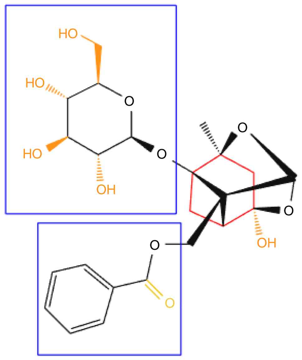
PF, C23H28O11, is a
monoterpene glycoside from Paeonia species. Its pinane-type
skeleton carries a β-D-glucosyl moiety at C-1 and a benzoyl group
esterified at C-9, along with multiple hydroxyl groups. These
features give PF high water solubility, beneficial for formulation,
but restrict membrane permeability and limit oral bioavailability.
In animal studies, oral bioavailability of PF is ~3-4% (22). A human pharmacokinetic study
reported that after an oral dose of an extract containing 108.18 mg
of PF, plasma Cmax reached only 6.97 ng/ml (23). Although the physicochemical and
pharmacokinetic properties of PF have been described, how its
solubility relates to absorption, distribution and pharmacological
efficacy is not fully understood (24,25).
In the molecular structure diagram, the
red-highlighted region corresponds to the C-ring of the core
scaffold, the central pharmacophore required for biological
activity. The blue box covers the benzoyl and glucosyl moieties,
both critical to PF's anti-inflammatory and analgesic effects.
Specifically, the carbonyl oxygen in the benzoyl group (yellow)
forms hydrogen bonds with enzymes or receptors in vivo,
improving target-binding affinity and serving as the primary site
for anti-inflammatory action. The orange regions mark functional
groups that increase hydrophilicity and support dissolution,
absorption, and systemic transport. These include the hydroxymethyl
group (-CH2OH at C-6 of the glucose unit), which
increases molecular hydrophilicity and promotes dissolution and
transmembrane transport, and the hydroxyl groups (-OH) on the
glucopyranose ring, which enhance water solubility through higher
polarity and hydrogen-bonding capacity, improving diffusion in
biological fluids and drug action (Fig. 1). That said, direct experimental
data are still insufficient to clarify the hydrolysis mechanism,
the exact metabolite profile, and their quantitative impact on the
duration of pharmacological effect and oral bioavailability
(26). In addition, the hydroxyl
groups of PF can form non-covalent interactions with biological
targets (enzymes or receptors), including hydrogen bonds and van
der Waals forces, as supported by molecular docking and
pharmacological studies (27).
Pharmacokinetic data indicate that although PF
concentrations after oral administration are markedly lower than
those after intravenous injection, rapid absorption occurs, with
peak plasma levels reached within 10 min (28). Orally administered PF is poorly
absorbed in the gastrointestinal tract; most is metabolized by gut
microbiota in the distal small intestine, cecum, and colon, sites
where hydrolysis and biotransformation are accelerated, producing a
range of metabolites (29).
Given intravenously, PF crosses the blood-brain barrier (BBB) and
distributes to the hippocampus and cerebral cortex (28). Both the parent compound and its
intestinal metabolites then undergo hepatic biotransformation,
including oxidation, reduction, hydroxylation, methylation and
conjugation reactions (for example, glucuronidation and sulfation).
Metabolism is mediated mainly by cytochrome P450 enzymes via
hydroxylation and demethylation. Unchanged PF accounts for only a
minor fraction in vivo; most is detected in plasma, urine,
and feces as metabolites (30,31). Benzoic acid, a major metabolite,
crosses the BBB and produces antidepressant effects, whereas the
distribution and functional roles of other metabolites remain to be
determined (29).
Taken together, PF is extensively transformed in
vivo, hydrolyzed by gut microbiota and then processed by
hepatic oxidative and conjugative metabolism, generating
metabolites with distinct biological activities. This low systemic
exposure after oral administration contrasts with the
concentrations used in in vitro studies, pointing to a gap
between experimental findings and clinical conditions. These
metabolites not only mediate PF's anti-inflammatory efficacy but
also provide the molecular basis for its multi-target and
multi-cascade pharmacological actions.
Molecular mechanisms of PF for modulating
inflammation
Inhibitory effect of PF on the NF-κB
signaling pathway
NF-κB is a family of transcription factors that
plays crucial roles in various physiological and pathological
processes. NF-κB signaling is primarily mediated through two
distinct pathways, the canonical (classical) and non-canonical
(alternative) pathways, which differ in their activation mechanisms
and biological functions. The canonical NF-κB pathway is a central
regulator of inflammatory responses and is therefore an important
target of PF in the modulation of inflammation (32).
Inhibition of nuclear transcription
factor activity
In the hippocampus of mice with depression induced
by the chronic unpredictable mild stress (CUMS) model, western
blotting and immunofluorescence analyses showed that high-dose PF
significantly inhibited the stress-induced increase in NF-κB p65
protein expression and nuclear fluorescence intensity (33). As a pivotal transcription factor,
NF-κB regulates the production and release of diverse inflammatory
cytokines. PF effectively inhibited NF-κB activation in this model,
providing experimental evidence for its ability to regulate
inflammation-related transcriptional activity (33).
Inhibition of IKK activity
IKK (IκB kinase), the key kinase responsible for the
phosphorylation of NF-κB inhibitor alpha (IκBα), plays a central
role in NF-κB activation. PF inhibits IKK activity, thereby
reducing IκBα phosphorylation and subsequent proteasome-mediated
degradation. This preserves cytoplasmic retention of the NF-κB
p65/p50 dimer and limits its nuclear translocation and
transcriptional activity, contributing to the anti-inflammatory
effects of PF (34).
Inhibiting the degradation of IκBα
IκBα is a pivotal negative regulator of the NF-κB
pathway. By binding NF-κB subunits such as p65 in the cytoplasm,
IκBα prevents their nuclear translocation and limits the
transcription of proinflammatory genes. PF inhibits IκBα
phosphorylation, thereby suppressing its
ubiquitin-proteasome-mediated degradation, increasing IκBα
stability, and restricting NF-κB nuclear translocation. Experiments
in activated T lymphocytes and a mouse model of acute contact
dermatitis induced by 1-chloro-2,4-dinitrobenzene (DNCB)
demonstrated that PF treatment dose-dependently inhibited IκBα
phosphorylation and NF-κB p65 activation while reducing
interferon-γ secretion. These findings support a mechanism in which
PF suppresses inflammatory responses by stabilizing IκBα and
inhibiting NF-κB signaling (35).
Inhibition of nuclear translocation of
the p65/p50 complex
The p65/p50 heterodimer is the primary functional
form of NF-κB involved in canonical pathway signaling. PF reduces
NF-κB nuclear translocation by suppressing its activation. In
activated T lymphocytes, PF (10 μM) significantly reduced
NF-κB p65 expression and dose-dependently inhibited IκBα
phosphorylation. In a mouse model of acute contact dermatitis
induced by DNCB, oral administration of PF (75 or 150 mg/kg/day)
dose-dependently reduced NF-κB p65 expression and IκBα
phosphorylation in T lymphocytes. These findings indicate that PF
suppresses NF-κB activation and nuclear translocation by reducing
p65 expression and maintaining IκBα-mediated cytoplasmic retention
of the NF-κB complex, thereby limiting the transcription of
downstream proinflammatory genes (34).
Inhibitory effects of PF on the MAPK
signaling pathway
The MAPK pathway constitutes a three-tiered kinase
cascade in which MAPK kinase kinases (MAPKKKs) respond to diverse
extracellular and intracellular signals and activate MAPK kinases
(MAPKKs) through phosphorylation. MAPKKs then phosphorylate MAPKs
at specific sites, leading to their activation. Activated MAPKs
regulate various downstream target proteins and, in response to
extracellular stimuli, control cell fate and cellular responses
(36). The MAPK pathway is also
involved in the production of inflammatory mediators, activation of
immune cells, and regulation of tissue inflammatory responses.
Aberrant MAPK activation is associated with several inflammatory
diseases, including RA, neuroinflammation and cardiovascular
inflammation.
Inhibition of MAPKK (MKK4)
Western blot analysis demonstrated that tributyltin
chloride (TBTC) treatment significantly increased the
phosphorylation of MAPKK 4 (MKK4) in a TBTC-induced neurotoxicity
model, indicating activation of the MKK4 pathway. Pretreatment with
PF at 50 and 100 μM dose-dependently suppressed this
increase, with p-MKK4 expression in the 100 μM PF group
significantly lower than that in the TBTC model group. These
findings indicate that PF suppresses upstream stress signaling by
inhibiting MKK4 phosphorylation and activation, which may
contribute to its neuroprotective effects (37).
Inhibition of MAPKs [Jun N-terminal
kinase (JNK), p38, ERK1/2, ERK5]
PF inhibits JNK phosphorylation
PF's regulation of JNK is context-dependent rather
than uniformly inhibitory. JNK can act as an upstream regulator of
Nrf2 under oxidative stress conditions. Compared with the
H2O2-only group, PF enhanced JNK activation
in an oxidative stress model (38). In chronic inflammatory models, PF
inhibits JNK phosphorylation, which may reduce AP-1-mediated
inflammatory signaling. By contrast, under acute oxidative stress,
PF can enhance JNK activation and promote Nrf2 nuclear
translocation and HO-1 expression, supporting an antioxidant
response. In both the spontaneously hypertensive rat (SHR) cardiac
model and adjuvant-induced arthritis (AA) rat fibroblast-like
synoviocyte (FLS) experiments, PF significantly reduced p-JNK
levels and was associated with reduced inflammatory responses
together with inhibition of p38/ERK signaling. In an
H2O2-induced melanocyte oxidative stress
model, PF enhanced JNK activation, promoted Nrf2 nuclear
translocation and HO-1 upregulation, and exerted a protective
effect that was reversed by the JNK inhibitor SP600125. These
findings support a context-dependent role of JNK in PF-mediated
cellular protection and inflammatory regulation. PF may suppress
sustained JNK-mediated inflammatory signaling under chronic
inflammatory conditions while promoting JNK-dependent antioxidant
responses under oxidative stress (39).
PF inhibits p38 MAPK
phosphorylation
PF exerts anti-inflammatory effects in part through
inhibition of p38 MAPK phosphorylation, thereby reducing the
production of p38-dependent proinflammatory mediators, including
TNF-α, IL-6 and IL-1β. In models including bupivacaine-induced
neurotoxicity in human neuroblastoma SH-SY5Y cells, the SHR cardiac
remodeling model, and the AA rat model, PF markedly suppressed p38
activation and alleviated neuroinflammation, myocardial fibrosis
and other pathological changes. Its inhibitory effect was
comparable to that of the specific p38 inhibitor SB203580. Given
the central role of p38 in inflammatory signaling, suppression of
p38 activation represents an important component of the
anti-inflammatory effects of PF (39).
PF significantly inhibits ERK
phosphorylation
In FLS derived from AA rats, western blotting
confirmed that PF significantly inhibited ERK phosphorylation
(p-ERK). The proposed mechanism involves inhibition of the aberrant
translocation of G protein-coupled receptor kinase 2 (GRK2) to the
cell membrane, thereby restoring the normal interaction between
GRK2 and phosphorylated ERK (p-ERK) and reducing pathological
accumulation of p-ERK in the nucleus. This effect was particularly
evident under inflammatory stimulation, such as prostaglandin E2
(PGE2) exposure, during which PF reversed both increased
GRK2 membrane localization and p-ERK nuclear translocation. PF may
thus regulate inflammatory signaling through the GRK2/p-ERK axis
(40). ERK5 is a distinct MAPK
family member activated through phosphorylation by MAPKK 5 (MEK5).
Activated ERK5 regulates transcription factors, including myocyte
enhancer factor, cellular-myelocytomatosis oncogene homolog, and
cellular-FBJ murine osteosarcoma viral oncogene homolog, which are
involved in the regulation of cell-cycle progression and apoptosis.
PF activates endoplasmic reticulum stress and induces calpain-1
activation, which promotes the proteolytic cleavage and degradation
of ERK5 and thereby suppresses ERK5-mediated signaling (41).
PF applies anti-inflammatory impacts
via broad inhibition of the MAPK pathway
MAPKs are central components of inflammatory signal
transduction. The three major MAPK subtypes, p38, JNK and ERK, are
frequently activated under inflammatory conditions and can
phosphorylate downstream transcription factors that promote
inflammatory mediator production and tissue injury. p38 is strongly
associated with the production of inflammatory mediators, JNK
contributes to inflammatory signaling and apoptosis, and ERK
participates in the progression of several inflammatory processes.
Inhibition of MAPK phosphorylation can therefore attenuate
inflammatory responses. For example, PF reduced p-p38, p-JNK and
p-ERK levels in SHR models and alleviated inflammatory responses.
In RA models, the PF derivative PF-6'-O-benzenesulfonate (CP-25)
inhibited activation of all three major MAPK subtypes in synovial
cells through partially distinct mechanisms. CP-25 inhibits GRK2
membrane translocation, thereby reducing ERK nuclear translocation
and downstream inflammatory mediator production. It also suppresses
p38 and JNK through pathways that are independent of GRK2 (41,42).
Inhibitory effect of PF on the JAK-STAT
signaling pathway
The Janus kinase-signal transducers and activators
of transcription (JAK-STAT) pathway is a central regulatory system
involved in inflammatory homeostasis. Activation of this pathway by
cytokines regulates the expression of factors such as suppressor of
cytokine signaling 3 and B-cell lymphoma 3-encoded protein, which
provide negative feedback to inflammatory signaling and can limit
NF-κB activity and the production of proinflammatory mediators such
as TNF-α and IL-6 (43). PF and
its structurally modified derivatives have been reported to
regulate this pathway at several levels, providing another
mechanism through which PF may modulate inflammatory responses.
Inhibition of JAK phosphorylation and
activation
The PF derivative CP-25 selectively inhibited the
phosphorylation of JAK1, JAK2 and JAK3 (p-JAKs) in human umbilical
vein endothelial cells (HUVECs) exposed to interleukin-17A (IL-17A;
50 ng/ml), without altering total JAK protein levels. This
inhibition was accompanied by significant reductions in p-STAT3 and
secretion of the inflammatory adhesion molecule intercellular
adhesion molecule-1 (ICAM-1) (9). Time-course experiments showed that
suppression of JAK phosphorylation preceded the reduction in STAT3
phosphorylation, supporting JAK inhibition as an upstream event in
CP-25-mediated regulation of STAT3. These findings indicate that
CP-25 can modulate JAK activation without reducing total JAK
protein abundance. The same study reported concentration-dependent
inhibition of JAK1, JAK2 and JAK3 phosphorylation by CP-25 in
IL-17A-stimulated HUVECs, together with reductions in p-STAT3 and
ICAM-1, providing consistent evidence for regulation of the
JAK-STAT axis (9). Evidence from
PF-containing formulations also supports the involvement of this
pathway. In Buyang Huanwu Decoction, a formula containing PF and
other glycosides, the glycoside fraction attenuated AS in
ApoE−/− mice and reduced inflammatory
responses, accompanied by decreased phosphorylation of JAK2, STAT1
and STAT3. Molecular docking further suggested interactions between
PF and proteins associated with the JAK-STAT pathway (44). Because these findings were
obtained with a PF derivative or a multi-component formulation
rather than purified PF alone, they provide indirect evidence for
PF-associated JAK-STAT regulation. Direct experiments using
purified PF are required to establish whether inhibition of JAK
phosphorylation is an intrinsic property of PF.
Inhibition of STAT protein
phosphorylation and nuclear translocation
STAT proteins are key transcriptional effectors
downstream of JAKs. Following phosphorylation, STAT proteins
dimerize and translocate to the nucleus, where they regulate the
transcription of target genes involved in cytokine signaling and
inflammatory responses (43).
Evidence indicates that PF can regulate STAT signaling at the
levels of gene expression, phosphorylation and downstream cellular
function. At the level of STAT expression, droplet digital PCR and
immunoblotting showed that PF significantly reduced both
Stat1 and Stat3 mRNA and protein expression while
also inhibiting JAK2 phosphorylation (9). These findings suggest that PF can
influence both the expression and activation of components of the
JAK-STAT pathway. In an LPS-induced macrophage polarization model,
PF and its nano-formulation also modulated STAT phosphorylation by
suppressing p-STAT1, a mediator associated with M1 macrophage
polarization, while promoting recovery of p-STAT6, which is
associated with M2 polarization. The targeted nano-formulation
Pae-PPNPs-DS produced stronger changes in p-STAT1 and p-STAT6 than
the parent compound and other tested carriers, suggesting that the
delivery system can enhance the effects of PF on STAT signaling
(12).
Overall, these findings indicate that PF and
PF-derived interventions can regulate the JAK-STAT pathway at
multiple levels, including JAK phosphorylation, STAT expression,
STAT phosphorylation and immune-cell function. The available
evidence also suggests that these effects may contribute to
macrophage polarization and suppression of inflammatory responses.
Since some of the reported effects involve CP-25 or PF
nano-formulations rather than purified PF, the extent to which the
complete regulatory pattern represents an intrinsic property of PF
requires further investigation.
Inhibitory effect of PF on NOD-like
receptor family pyrin domain-containing 3 (NLRP3) inflammasome
activation
The NLRP3 inflammasome is a multiprotein signaling
complex composed primarily of NLRP3 as the sensor,
apoptosis-associated speck-like protein containing a caspase
recruitment domain (ASC) as the adaptor, and caspase-1 as the
downstream effector protease. Upon activation, NLRP3 promotes ASC
oligomerization and caspase-1 activation, leading to maturation and
release of IL-1β and IL-18 and, in numerous settings, gasdermin D
(GSDMD)-mediated pyroptosis (45). PF has been reported to inhibit
NLRP3 inflammasome activation through regulation of both upstream
inflammatory signaling and inflammasome-associated components.
Inhibition of NLRP3 inflammasome
assembly and activation
In both in vivo CUMS-induced depressive mice
and in vitro LPS/ATP-stimulated BV2 microglia models, PF
treatment significantly reduced the protein levels of NLRP3, ASC
and caspase-1 in hippocampal tissue and cells. These changes were
accompanied by reduced IL-1β secretion and GSDMD-mediated
pyroptosis. Mechanistically, PF (100 μM) increased Sirtuin 1
(SIRT1) expression and inhibited NF-κB p65 nuclear translocation,
whereas inhibition of SIRT1 with EX-527 abolished the protective
effects of PF. These findings support involvement of the
SIRT1/NF-κB/NLRP3 axis in PF-mediated suppression of inflammasome
activation (33). A separate
study identified a metabolic signaling pathway linking succinate
accumulation to NLRP3 activation. In a Complete Freund's
Adjuvant-induced model, succinate accumulation increased succinate
receptor 1 (SUCNR1) and hypoxia-inducible factor 1-alpha (HIF-1α)
expression and was associated with NLRP3 inflammasome activation.
Immunofluorescence and qPCR showed increased NLRP3 and caspase-1
protein expression and increased Nlrp3 and Caspase-1
mRNA levels in dorsal root ganglion neurons, accompanied by
increased IL-1β release and neuroinflammation. PF suppressed SUCNR1
and HIF-1α expression, reduced inflammasome activity, and decreased
IL-1β production. These findings indicate that PF may alleviate
neuropathic pain through modulation of the
succinate-SUCNR1-HIF-1α-NLRP3 axis (46).
Suppression of caspase-1 activation
and mature IL-1β secretion
PF also regulates downstream components of NLRP3
inflammasome signaling. In CUMS-induced mice, hippocampal caspase-1
protein levels were markedly higher than those in control mice,
whereas PF treatment reduced caspase-1 expression in a
dose-dependent manner, with the strongest effect observed at the
highest dose tested. The associated reduction in IL-1β production
and pyroptosis suggests that PF can attenuate caspase-1-dependent
inflammatory signaling (33).
Inhibition of the TLR4/MyD88 signaling
pathway by PF
TLR4 is a transmembrane pattern-recognition receptor
that detects bacterial lipopolysaccharide (LPS) and initiates
signaling cascades involved in innate immune and inflammatory
responses (47,48). PF has been reported to attenuate
inflammatory responses partly through suppression of TLR4/MyD88
signaling.
Inhibition of TLR4 activation
In AA rats, TLR4 surface expression on peritoneal
macrophages was increased together with MyD88, NLRP3, ASC,
caspase-1 and GSDMD-N, indicating activation of TLR4-associated
inflammatory and pyroptotic signaling. PF treatment reduced the
expression of these markers. Flow cytometry further showed
increased TLR4 fluorescence in macrophages from AA rats, which was
reduced by PF at 1 and 10 μM. In vitro, LPS/ATP
stimulation increased TLR4 expression, whereas PF reduced the
corresponding fluorescence intensity. Western blotting produced
consistent findings, supporting the inhibitory effect of PF on TLR4
expression and downstream signaling (49).
Suppression of MyD88-dependent
signaling
In a model of cholestatic liver injury induced by
17α-ethinyl-estradiol, PF and its micellar formulation (PF-PLC)
suppressed the TLR4/MyD88/NF-κB axis. PF-PLC markedly reduced MyD88
protein expression and was associated with decreased NF-κB nuclear
translocation and inflammatory cytokine production. These effects
were supported by western blotting and a reduction in
NF-κB-positive cells detected by immunofluorescence. The findings
indicate that modulation of MyD88-dependent signaling may
contribute to the hepatoprotective effects of PF-PLC (50).
Multi-pathway crosstalk in the
anti-inflammatory network of PF
The aforementioned signaling pathways are
interconnected rather than functioning as independent pathways. PF
has been reported to inhibit both NF-κB nuclear translocation and
MAPK phosphorylation, including JNK, p38 and ERK, suggesting that
upstream signaling nodes may contribute to the coordinated
regulation of these pathways. TAK1 is one such convergence point
because TLR4/MyD88 signaling can activate TAK1, which in turn
contributes to activation of IKK and downstream NF-κB signaling as
well as MKK4/7 and JNK signaling. The observed effects of PF on
NF-κB and MAPK pathways are therefore consistent with regulation at
shared upstream signaling nodes, although direct evidence
demonstrating TAK1 as a molecular target of PF remains necessary.
PF also affects the SIRT1/NF-κB/NLRP3 axis. Increased SIRT1
activity can reduce NF-κB p65 transcriptional activity, thereby
limiting the priming of NLRP3 inflammasome components. PF-mediated
suppression of the succinate-SUCNR1-HIF-1α-NLRP3 axis provides
another connection between metabolic stress and inflammasome
activation. HIF-1α can regulate inflammatory gene expression,
including NLRP3-associated responses, allowing metabolic and
inflammatory signals to converge at the inflammasome (51). These observations suggest that PF
may influence inflammatory signaling through several interconnected
upstream regulators rather than through a single molecular target.
The JAK-STAT pathway provides another point of interaction within
this network. CP-25 inhibits JAK1/2/3 phosphorylation and reduces
STAT3 activation, whereas PF has been shown to suppress NF-κB
signaling. JAK-STAT and NF-κB pathways can interact at the level of
shared inflammatory mediators and cellular responses, including
IL-6-associated signaling and endothelial inflammatory
activation.
PF may also influence inflammatory responses through
interactions between different cell types. PF has been reported to
regulate microglia-astrocyte crosstalk through the HSP90AA1/HMGB1
pathway in neuroinflammation and macrophage-intestinal stem cell
(ISC) crosstalk through C1q in chronic colitis. These findings
extend the network concept beyond intracellular signaling to
communication between immune and tissue-resident cells. Ferroptosis
may represent another process linked to inflammatory regulation
because lipid peroxidation and ferroptotic cell death can interact
with inflammatory and oxidative stress pathways. Studies of related
herbal formulas have shown that CXCR4 upregulation can suppress
GPX4-mediated ferroptosis (52).
Such findings provide a possible connection between antioxidant
activity and inflammatory regulation, but evidence directly linking
PF to this mechanism remains limited. Overall, the available
evidence indicates that PF can influence interconnected
intracellular and intercellular processes, although the
contribution of individual nodes and their causal relationships
require further validation.
PF exhibits anti-inflammatory activity through
coordinated regulation of several signaling pathways. It suppresses
TLR4/MyD88 signaling, inhibits NF-κB activation and nuclear
translocation, attenuates phosphorylation of MAPK components
including JNK, p38, ERK1/2 and ERK5, modulates JAK-STAT signaling,
and suppresses NLRP3 inflammasome activation (Fig. 2A-E). These pathways are
interconnected through shared upstream regulators and inflammatory
mediators, providing a mechanistic basis for the multi-target
effects of PF. The available evidence supports a network-level mode
of action involving TLR4/MyD88, NF-κB, MAPK, JAK-STAT and NLRP3
signaling rather than a single dominant target. Further studies
using purified PF and direct target-validation approaches are
needed to define the molecular nodes responsible for these
coordinated effects.
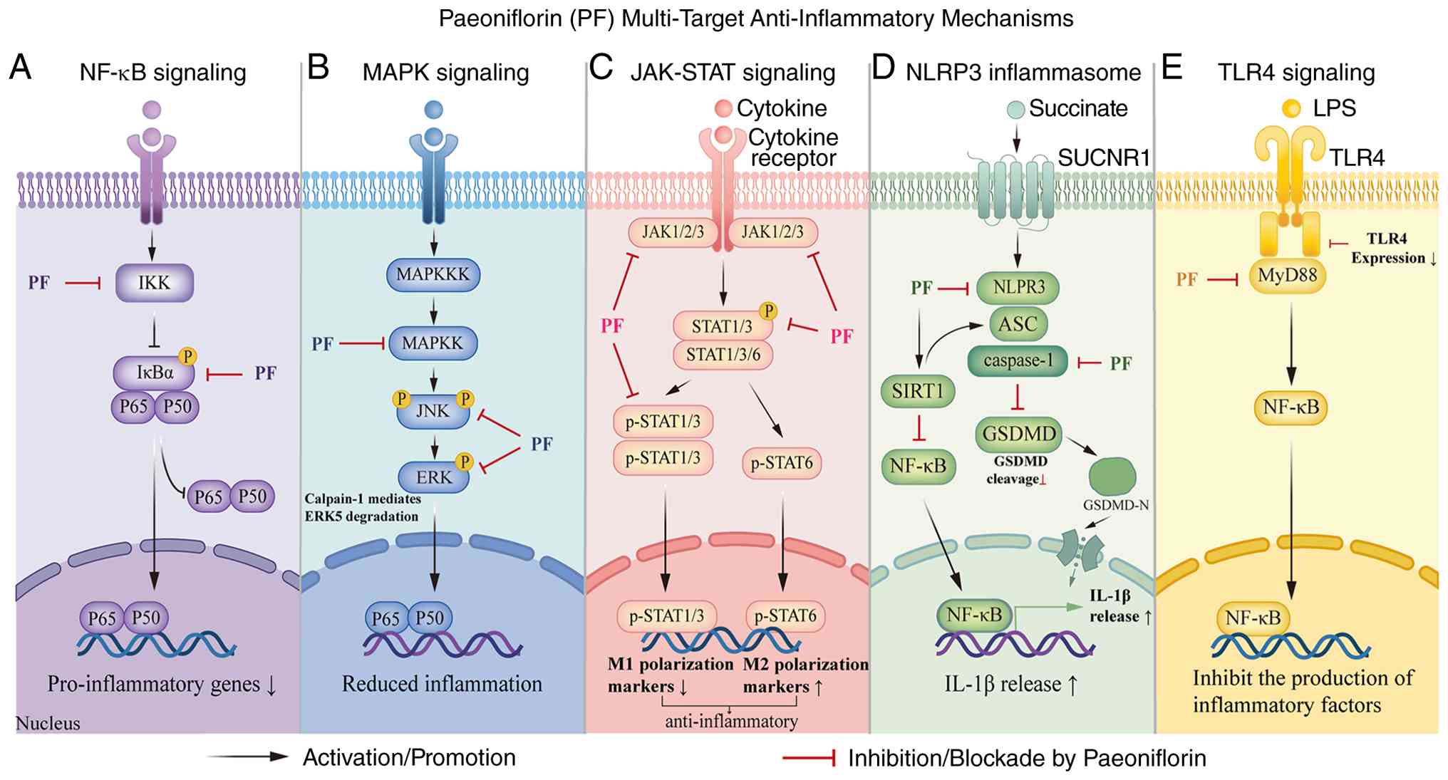 | Figure 2Anti-inflammatory mechanisms of PF.
PF inhibits (−) five signaling cascades: (A) TLR4/MyD88, (B) NF-κB
activation, (C) MAPK phosphorylation (JNK/p38/ERK1/2/ERK5), (D)
JAK-STAT signaling and (E) NLRP3 inflammasome assembly. Arrows (→)
indicate activation; T-bars (−) indicate inhibition by PF. PF,
paeoniflorin; ASC, apoptosis-associated Speck-like Protein
containing a CARD; CAPN1, Calpain-1; caspase-1, Caspase-1; ERK,
Extracellular signal-regulated kinase; ERK5, Extracellular
signal-regulated kinase 5; GSDMD, Gasdermin D; IKK, IκB Kinase;
JAK, Janus kinase; JNK, Jun N-terminal kinase; MAPK,
Mitogen-activated protein kinase; MyD88, Myeloid differentiation
factor 88; NLRP3, NOD-, LRR- and Pyrin Domain-containing Protein 3;
p38, p38 Mitogen-activated protein kinase; STAT, Signal transducers
and activators of transcription; STAT, signal transducers and
activators of transcription; TLR4, Toll-like receptor 4. |
Mechanisms of PF for modulating inflammatory
diseases
Molecular targets prediction of PF's
anti-inflammatory effects
NP analysis
NP analysis was performed using standard workflows.
PF compounds were screened via TCMSP (v2.3, OB ≥30%, DL ≥0.18);
disease targets were retrieved from GeneCards (v5.18; https://www.genecards.org/), OMIM (v2024-08;
https://omim.org/), TTD (v2024-10; https://db.idrblab.net/ttd/), Malacards (v6.1,
https://www.malacards.org/), HPO
(v2025-01; https://hpo.jax.org/), PharmGKB
(accessed May 2025; https://www.pharmgkb.org/), and Open Targets (v25.03;
https://platform.opentargets.org/), all
accessed in May 2025, with GeneCards filtered by Relevance score ≥
median and other databases by default thresholds. Targets were
standardized via UniProt (https://www.uniprot.org/). PPI networks were
constructed using STRING (v12.0; https://string-db.org/; species: Homo sapiens,
confidence ≥0.400, max ≤20 interactors), and hub genes were
predicted via Cytoscape (v3.10.2; http://www.cytoscape.org/) with the CytoHubba-MCC
algorithm (top 10 by MCC score). Hub molecules, including TNF,
IL1B, nuclear factor of kappa light polypeptide gene enhancer in
B-cells inhibitor-alpha (NFKBIA), and AKT serine/threonine kinase 1
(AKT1), emerged as core nodes within PF's action network. These
targets, particularly TNF and IL1B, were consistently identified
across models of rheumatic and inflammatory diseases, including RA,
chronic kidney injury and colitis.
Molecular docking was performed using AutoDock Vina
(version 1.2.7; https://github.com/ccsb-scripps/AutoDock-Vina).
Three-dimensional structures of target proteins (TNF, IL1B, NFKBIA,
and AKT1) were retrieved from the RCSB Protein Data Bank (PDB,
https://www.rcsb.org/, accessed May 2025), and
input files were prepared using AutoDockTools (version 1.5.7). The
three-dimensional structure of PF was generated using Open Babel
and prepared with PyMOL (including water molecule removal, hydrogen
addition and adjustment of symmetric bi-molecules). For each
target, the grid box was set individually according to the position
and size of its predicted active pocket. Exhaustiveness was set to
the default value of 8, and the number of binding modes (num_modes)
was set to 9. A binding energy ≤-7.0 kcal/mol was considered the
threshold for strong binding potential. The docking results
predicted highly negative binding energy values for these targets,
suggesting stable occupancy of protein active sites and potential
interference with inflammatory signaling. In disease models
including RA, diabetic complications, and liver injury, PF showed
comparable or more favorable binding energies with TNF and AKT1
than several reference anti-inflammatory compounds (53,54). Of note, molecular docking is a
computational prediction tool with inherent limitations, including
rigid protein models, simplified solvent treatments, and empirical
scoring functions. Accordingly, these docking results serve as
preliminary conformational support for NP predictions, rather than
experimental evidence of binding or causality.
Collectively, NP and molecular docking predicted
TNF, IL1B, NFKBIA, AKT1 and related proteins as key molecular
targets of PF. The predicted binding affinity of PF for these
targets provides a preliminary molecular basis for its multi-level
regulation of inflammatory responses. This multi-target,
multi-pathway profile offers a theoretical basis for further
exploring PF's therapeutic potential.
Protein-protein interaction (PPI)
network of the molecular targets
PPI networks of these predicted targets were
constructed to reveal the molecular relationship. Across disease
models, including depression, diabetes, RA, chronic hepatitis and
autoimmune disorders, core PF targets encompassed AKT1, SRC,
HSP90AA1, TNF, CASP3, MAPK1, VEGFA and EGFR, all critically
involved in inflammatory signaling, cell survival and apoptosis
(55-58).
Within disease-specific PPI networks, PF-targeted
proteins often occupied hub positions, serving not only within
individual pathways but also as convergence points for multiple
inflammatory and apoptotic cascades. The high connectivity of AKT1,
SRC and MAPK1 suggests that PF's modulation of these hubs
concurrently affects diverse downstream processes, enabling
multi-level control of inflammation (59,60). Disease-specific PPI analyses were
further conducted to delineate the core regulatory nodes and
hierarchical mechanisms underlying PF's anti-inflammatory
actions.
PF and its predicted targets in
inflammatory diseases
AS
AS involves plaque formation within the arterial
intima, driven by various cells, lipids and cellular debris. It is
recognized as a chronic vascular inflammatory disease mediated by
traditional and non-traditional risk factors, accounting for most
myocardial infarctions, numerous strokes, and disability in
peripheral arterial occlusive disease. Historically considered as a
disorder of cholesterol accumulation triggered by retention of
low-density lipoprotein (LDL) in the arterial intima, scavenger
receptor-mediated uptake of LDL promotes sustained immune cell
infiltration into atherosclerotic plaques (61-64). Current therapeutic strategies
targeting inflammatory modulation include suppression of
proinflammatory cytokines, disruption of critical inflammatory
signaling cascades, and facilitation of inflammation resolution.
Animal experiments demonstrated that in a high-fat diet-induced AS
rat model, escalating doses of PF-containing plant extract
significantly increased SOD activity while decreasing serum levels
of TP and FC. PF also exerted protective effects on key target
cells, including vascular endothelial cells and smooth muscle
cells. It alleviated core pathological processes such as
endothelial injury, chronic inflammatory responses, and abnormal
platelet activation resulting from cellular dysfunction (8).
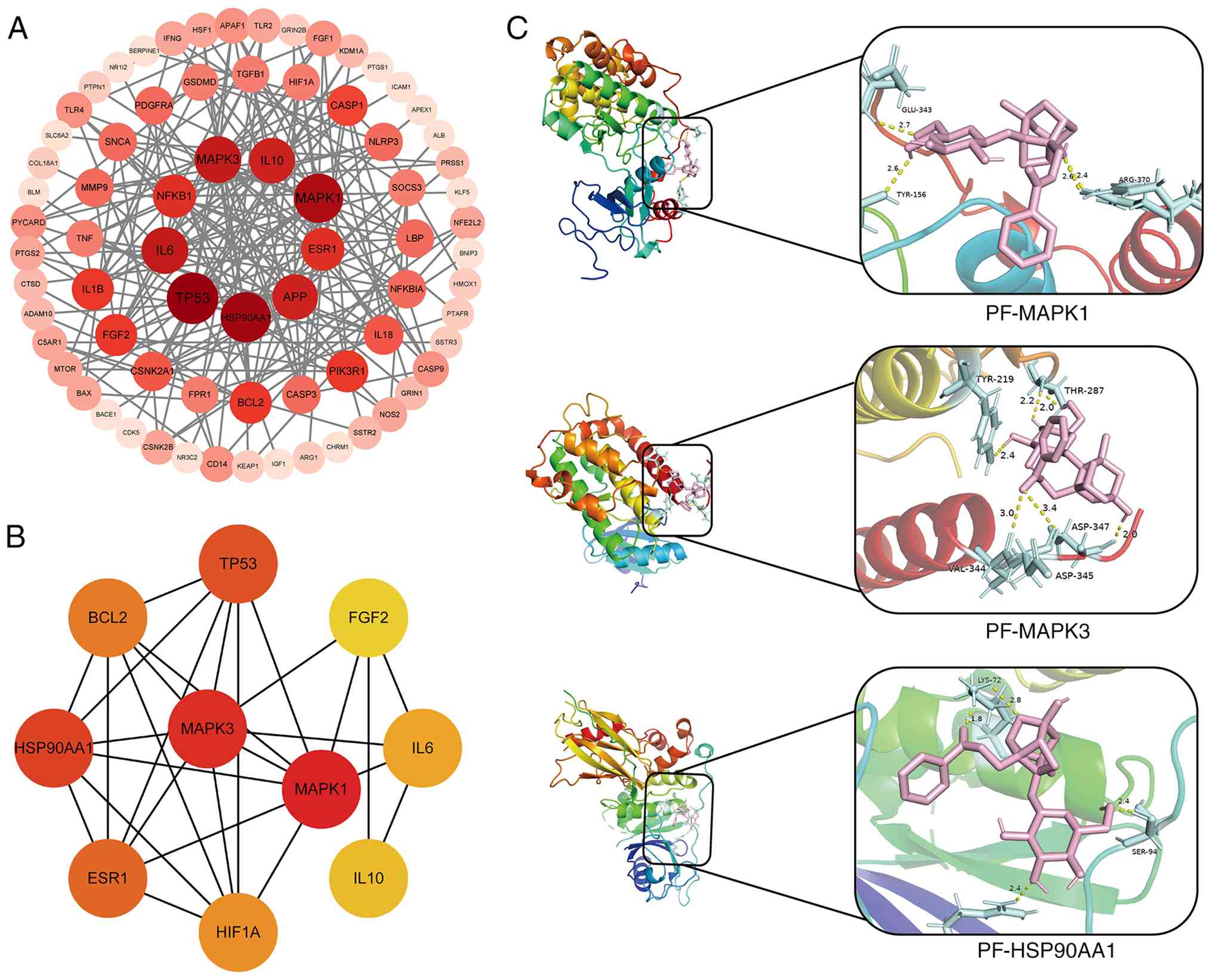
For NP-targets prediction in AS, a total of 4,815
AS-related targets were retrieved. After cross-matching with PF
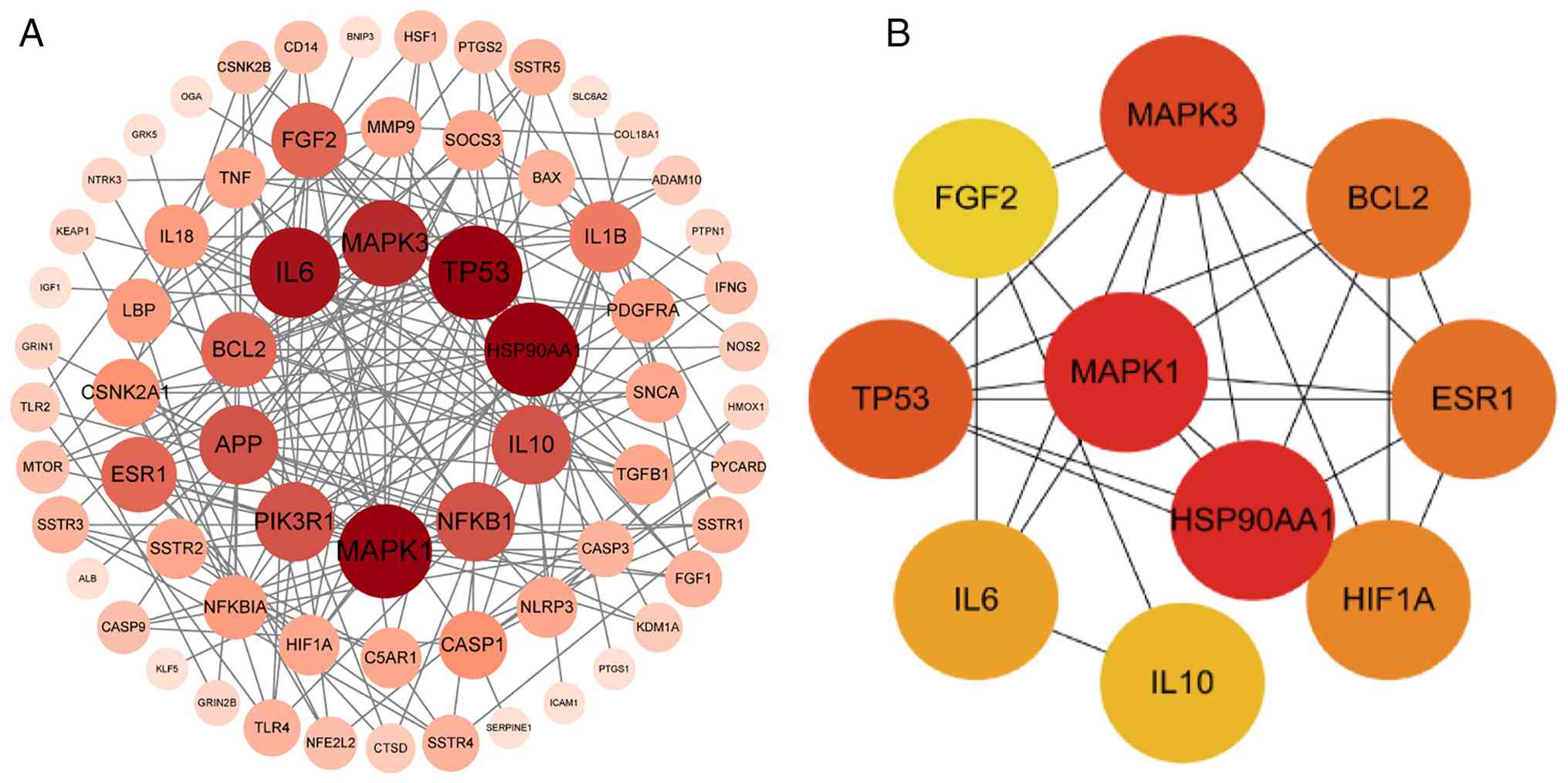
targets, a PPI network was constructed (Fig. 3A), and 10 hub genes were
prioritized: MAPK1, MAPK3, HSP90AA1, TP53, ESR1, BCL2, HIF1A, IL6,
IL10 and FGF2 (Fig. 3B).
Molecular docking of the top three hub targets (MAPK1, MAPK3,
HSP90AA1) revealed that PF formed conventional hydrogen bonds with
Glu343, Tyr156, Tyr219, Thr287, Ser94, Arg370 and Asp345; a
carbon-hydrogen bond with Val344; and salt bridges with Lys72 and
Asp347 (Fig. 3C). Molecular
docking across all hub genes (Fig.
S2A-J) further suggested high binding affinity between PF and
its targets.
Respiratory diseases
Pneumonia is the most common cause of acute lung
injury (ALI) and acute respiratory distress syndrome (ARDS)
(65). 'Phlegm and blood stasis'
form the pathological basis of ALI. With few effective treatments
available, ALI and ARDS remain severe, life-threatening conditions
(66). A recent study
highlighted macrophages as key contributors to the dysregulated
cytokine response in virally triggered ALI (67). As central components of pulmonary
innate immunity, macrophages support pathogen clearance through
phagocytosis or secretion of pro-inflammatory cytokines, mechanisms
essential for limiting initial viral spread. However, prolonged
viral exposure may trigger a self-amplifying inflammatory cascade
that disrupts immunological balance and leads to aberrant immune
activation. This pathological shift manifests as hyperactivated
macrophages secreting an excess of inflammatory mediators,
including IL-6, TNF-α, IL-1β, IFN-γ, IFN-α, CCL2, IL-8 and IP-10.
The resulting systemic cytokine surge compromises the structural
and functional integrity of the alveolar-capillary barrier,
increases vascular hyperpermeability, triggers pulmonary edema,
impairs alveolar architecture, and drives pathological progression
toward ALI/ARDS (68). NP
screening of the Chuanxiong-Danshen herb pair identified PF as a
potential active component against LPS-induced ALI. In
LPS-stimulated RAW264.7 macrophages and BALB/c mice, the
PF-containing fraction from this herb pair suppressed inflammatory
responses and oxidative stress, reducing ROS, NO, TNF-α, IL-6 and
IL-1β levels, with mechanistic studies pointing to involvement of
the NF-κB and MAPK pathways (69,70). The antiviral effect of PF in ALI
is also noteworthy. In a mouse model infected with influenza virus,
oral treatment with PF (50 and 100 mg/kg) increased the survival
rate of infected mice (by 40% in the 50 mg/kg group and 50% in the
100 mg/kg group) and alleviated early-stage pulmonary edema and
pathological changes caused by direct viral damage (71). Its antiviral mechanism may
involve a unique pathway: PF inhibits expression of αvβ3 integrin
in lung tissue. αvβ3 serves as a potential co-receptor for viral
entry and intracellular signal transduction. Inhibiting it may
interfere with viral infection and subsequent pathological signal
amplification at the source, thereby mitigating initial damage.
Subsequently, by inhibiting pathways such as NF-κB/p38 MAPK, PF
curbs the resulting excessive inflammation and fibrotic cascade,
achieving overall protection against viral ALI. In this context, PF
demonstrates a synergistic
'antiviral-anti-inflammatory-anti-damage' effect, making it a
promising multifunctional drug candidate (70).
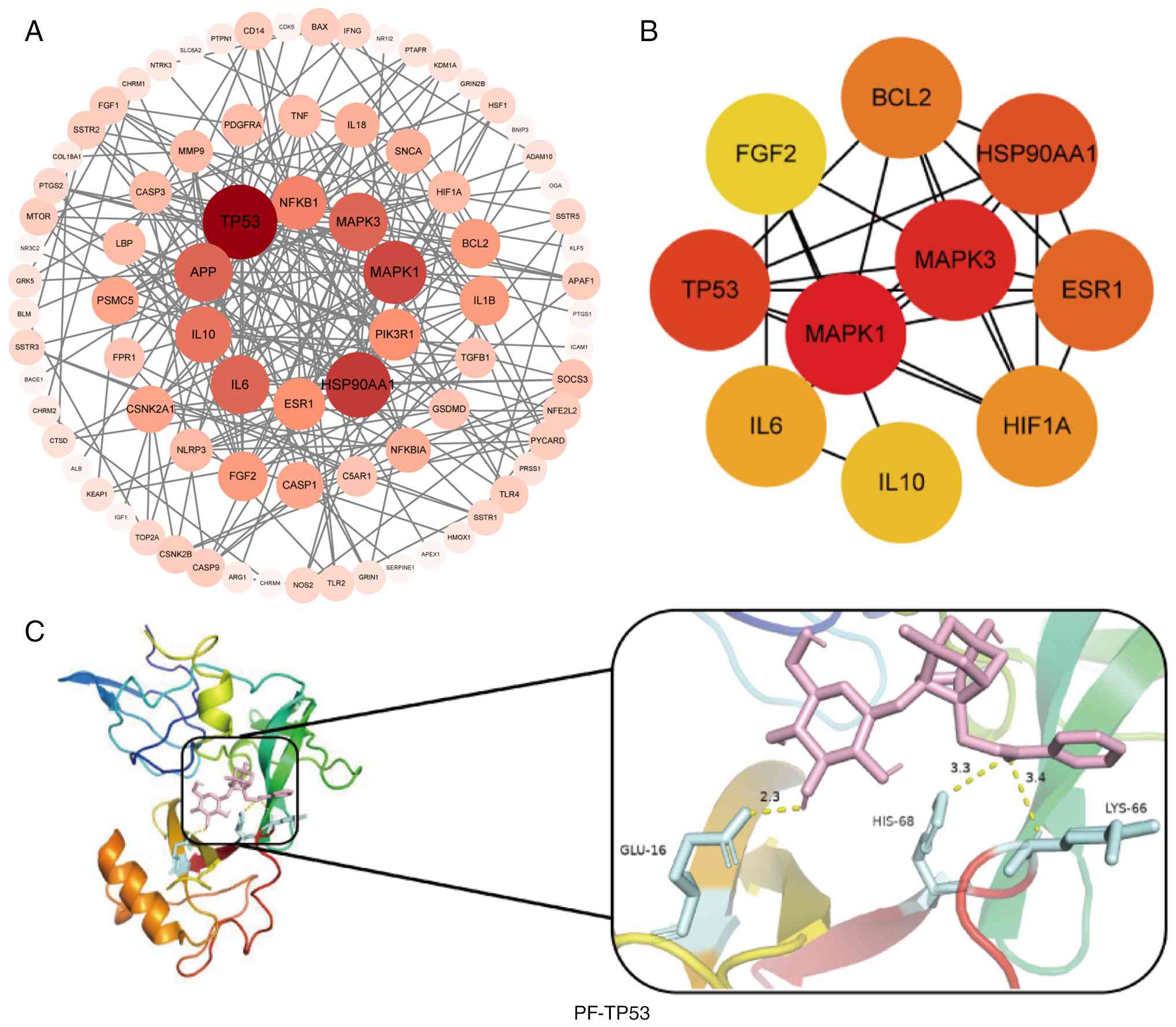
A total of 10,376 ALI-related targets were compiled.
After intersecting these with PF targets, a PPI network analysis
was performed using the STRING database (Fig. 4A), and ten core genes were
predicted: MAPK1, MAPK3, TP53, HSP90AA1, ESR1, BCL2, HIF1A, IL6,
IL10 and FGF2 (Fig. 4B).
Molecular docking results for the top three hub genes (MAPK1, MAPK3
and TP53) suggested that PF may form conventional hydrogen bonds
with glutamate at position 343, tyrosine at positions 219 and 156,
arginine at 370, and aspartate at 345 (MAPK1); a carbon-hydrogen
bond with valine at 344 (MAPK3); and a salt bridge with aspartate
at 347 (TP53) (Figs. 3C and
4C). Analysis of the remaining
seven hub genes via molecular docking also indicated strong binding
potential (Fig. S2).
Liver injury
Hepatic ischemia-reperfusion injury (HIRI) refers to
the tissue damage that follows reestablishment of perfusion after
transient liver ischemia and is an inevitable complication during
liver trauma, resection, and transplantation (72,73). The ischemic phase causes hypoxia
and direct cellular injury due to inadequate blood supply.
Subsequent reperfusion initiates a complement cascade, leading to
secondary damage characterized by excessive ROS generation and
secretion of pro-inflammatory cytokines from hepatocytes. As a
result, immune cells, particularly neutrophils, are recruited from
the circulation, causing further direct damage to hepatic tissue
(72,74). Novel approaches, including
RNA-based therapies, gene modification and progenitor cell
techniques, offer potential avenues for advancing HIRI management
(75). PF exerts its effects by
specifically targeting the Tmem176b+ macrophage subset
in HIRI. It suppresses excessive inflammation through activation of
the THBS1-CD47 immunoregulatory axis while concurrently blocking
the pro-inflammatory SPP1-CD44 signaling pathway. This two-pronged
mechanism drives macrophages from an inflammation-promoting M1
state to an inflammation-resolving M2 state, remodeling the liver's
immunological milieu and mitigating parenchymal damage (76). To elucidate its multi-target
profile, 1,493 HIRI-related targets were retrieved, and PPI
topology was assembled using STRING and analyzed by Cytoscape
(Fig. S3A), with core targets
MAPK1, MAPK3, HSP90AA1, TP53, ESR1, BCL2, HIF1A, IL6, IL10 and FGF2
(Fig. 3B). Molecular docking
analysis further indicated that PF's mode of action on these
IRI-related targets, including specific binding sites and
interaction forces, closely aligns with findings from AS studies,
offering preliminary structural insights into its broad
pharmacological activity (Figs.
3C and S2).
Cholestatic liver diseases (CHOL) encompass a
spectrum of hepatobiliary disorders characterized by impaired bile
flow, including primary biliary cholangitis (PBC), primary
sclerosing cholangitis (PSC), intrahepatic cholestasis of
pregnancy, IgG4-related sclerosing cholangitis, and progressive
familial intrahepatic cholestasis (PFIC). The pathological process
is marked by dysregulated cholangiocyte proliferation, periductal
fibrotic remodeling, myofibroblast activation, accumulation of
cytotoxic bile acids, and an exaggerated ductular reaction.
Inflammatory cell infiltration exacerbates cholangiocyte apoptosis,
leading to biliary obstruction and disrupted bile acid excretion,
particularly in PBC and PSC. The accumulated bile acids then
perpetuate a vicious cycle of fibrosis and inflammation (77,78). In models of cholestatic liver
injury, PF significantly improves both functional and
histopathological parameters. The core mechanism involves its
direct interaction with the GSDMD protein, inhibiting
GSDMD-mediated pyroptosis and NLRP3 inflammasome activation to
block the downstream inflammatory cascade (79). In addition, PF modulates key
metabolic pathways and associated signaling networks involving
targets such as CDK1, CYP2C9, MAOB, mTOR and ABCB1. These
modulations elevate levels of the hepatoprotective bile acid
tauro-ursodeoxycholic acid and readjust linoleic acid and
phosphatidylinositol metabolism. By reducing inflammatory stimuli
from toxic bile acid accumulation, PF indirectly dampens
overactivation of inflammatory signaling, leading to improvement in
serum biochemical markers and liver histology (80). A target screening approach
predicted 1,235 disease-associated targets from these databases.
Intersection with PF targets and subsequent PPI network analysis
(Fig. S3B) yielded ten core
targets: MAPK1, MAPK3, TP53, ESR1, BCL2, HIF1A, IL6, IL1B, TNF and
IL18 (MCC algorithm, Fig. S4A).
Docking simulations of the top three molecules showed that their
binding to PF matched the results obtained in ALI, suggesting a
conserved interaction pattern as indicated by the same binding
pocket and comparable free energy levels (Figs. 3C and 4C). Docking analysis of the hub genes
also suggested strong binding potential (Fig. S4B-K).
Colitis
Inflammatory bowel disease (IBD) encompasses
chronic, systemic inflammatory conditions affecting the
gastrointestinal tract and multiple extra-intestinal sites, mainly
Crohn's disease and ulcerative colitis (UC) (81,82). Despite advances in therapeutic
options, IBD remains prone to relapse, profoundly impairing patient
quality of life and imposing a substantial socioeconomic burden
(83-85). Notably, PF has demonstrated
distinctive dual functionality, simultaneously suppressing
inflammation and promoting mucosal repair, in IBD therapy. In
chronic colitis models, PF directly engages C1qa to orchestrate
dual regulatory effects: it upregulates membrane-bound C1q to
enhance macrophage-mediated bacterial clearance and accelerate
inflammation resolution, while concurrently inhibiting pathological
C1q secretion to suppress aberrant Wnt/β-catenin pathway
activation. This dual modulation restores ISC homeostasis,
effectively addressing the intertwined challenges of inflammatory
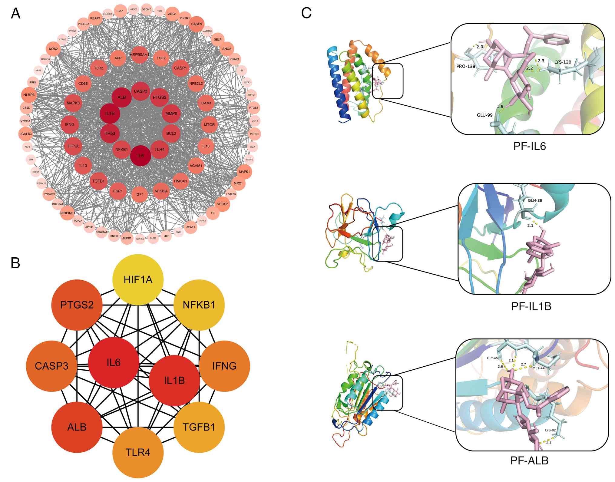
control and epithelial regeneration (86). A total of 4,384
colitis-associated targets were retrieved by NP. After
cross-matching with PF targets, a PPI network was constructed
(Fig. 5A), and 10 hub genes were
identified: IL6, IL1B, ALB, PTGS2, CASP3, IFNG, TLR4, TGFB1, NFKB1
and HIF1A (Fig. 5B). Molecular
docking suggested high-affinity binding between PF and these
targets. Critical interactions involve hydrogen bonds with Pro139,
Lys120/82, Gln39, Gly45, and Met44, and a salt bridge with Glu99
(Figs. 5C and S5A-J).
UC presents with recurrent abdominal pain,
hematochezia and diarrhea; prolonged disease duration may induce
mucosal architectural distortion and elevate colorectal neoplasia
risk (87). Compelling evidence
indicates that dysregulated death of intestinal epithelial cells
(IECs) compromises barrier integrity, triggering inflammatory
cascades central to UC progression. Current first-line
pharmacotherapy employs mesalazine (5-aminosalicylic acid, 5-ASA)
to suppress mucosal inflammation and induce remission (88,89); however, a significant proportion
of patients show inadequate response or treatment refractoriness
due to disease complexity (90).
PF has been shown to drive intestinal epithelial regeneration:
in vivo, PF markedly ameliorates dextran sulfate
sodium-induced colitis and restores mucosal architecture; in
vitro, PF activates the PI3K/Akt/mTOR axis to counteract
TNF-α-mediated growth suppression, accelerates repair of
LPS-injured IEC-6 cells, and upregulates regeneration-associated
molecular markers, providing multi-level mechanistic validation of
its reparative efficacy (91). A
total of 10,781 UC-related targets underwent identical NP
processing; a PPI network was constructed (Fig. S3C), which predicted a distinct
core target set: MAPK1, MAPK3, HSP90AA1, TP53, ESR1, BCL2, HIF1A,
IL6, IL10 and FGF2 (Fig. 3B).
Molecular docking analyses further suggested that PF's binding
modes with these targets, including binding pockets and computed
free energies, were generally comparable to those observed in the
ALI model (Figs. 3C, 4C and S2).
Acute kidney injury (AKI)
AKI is a heterogeneous, multifactorial syndrome
driven by renal tubular injury and inflammation (92). Pathogenesis of AKI involves
extensive activation of innate and adaptive immunity; specifically,
inflammation- and oxidative stress-induced endothelial dysfunction,
coupled with coagulopathy, precipitates microcirculatory failure
and a marked reduction in glomerular filtration rate (92,93). Although renal replacement therapy
remains standard care, the challenge of early AKI detection
underscores the need for preventive and therapeutic innovations
(94,95). In a murine bilateral renal
ischemia-reperfusion (IR) AKI model, PF-enriched extract
dose-dependently attenuated tissue injury, inflammation and
ferroptosis, findings corroborated in a hypoxia/reoxygenation model
using human renal tubular epithelial cells (HK2). Mechanistically,
PF upregulated solute carrier family 7 member 11 (Slc7a11 in mice;
SLC7A11 in humans), enhancing glutathione biosynthesis and
conferring resistance to ferroptosis, thereby enabling coordinated
suppression of ferroptosis and inflammation (96). A total of 10,131 AKI-associated
genes were overlapped with predicted PF targets (Fig. S3D); a PPI network was
constructed using STRING, and 10 hub targets, MAPK1, MAPK3, TP53,
HSP90AA1, ESR1, BCL2, HIF1A, IL6, IL10 and FGF2 (Fig. 4B), were predicted via MCC
algorithm in Cytoscape. Ligand-receptor interaction modeling
demonstrated favorable binding energies and three-dimensional poses
between PF and these targets, with binding free energies and
active-site interactions highly consistent with prior ALI studies
(Figs. 3C and 4C).
Diseases of the musculoskeletal
system
RA
RA is a systemic autoimmune disorder characterized
by synovial hyperplasia, autoantibody production, progressive bone
erosion and articular symptoms (for example, joint swelling,
tenderness). A core therapeutic challenge lies in rebalancing
inflammatory and reparative processes (97). Pathogenically, synovial
macrophages secrete proinflammatory cytokines (TNF-α, IL-1), which,
alongside FLS, activate osteoclasts and drive bone destruction
(98). Although
disease-modifying antirheumatic drugs remain foundational, novel
therapeutics are urgently needed (99). In a collagen-induced arthritis
rat model, PF and its active constituents ameliorated disease by:
i) Suppressing inflammation (reduced TNF-α, IL-1β, IL-6; elevated
IL-10); ii) mitigating oxidative stress (decreased malondialdehyde
and nitric oxide; increased SOD); and iii) promoting synovial cell
apoptosis via Bcl-2 downregulation and AMPK-mediated mitochondrial
fission (100). Bioinformatics
integration of 5,369 RA-related targets with PF targets (Fig. S3E), followed by PPI network
analysis (STRING) and MCC-based hub identification in Cytoscape,
predicted 10 core targets: MAPK1, MAPK3, HSP90AA1, TP53, ESR1,
BCL2, HIF1A, IL6, IL10 and FGF2 (Fig. 3B). Functional enrichment analysis
implied profiles consistent with prior AS findings (Figs. 3C and S2), reinforcing PF's multi-disease
modulatory potential.
Osteoarthritis (OA)
OA, the most prevalent chronic joint disorder, is a
leading cause of disability (101,102). A pivotal mechanism in OA
pathogenesis involves disruption of cytokine homeostasis:
Pro-inflammatory cytokines such as IL-1β, TNF-α and IL-6 initiate a
self-perpetuating cycle that ultimately damages articular cartilage
and other intra-articular structures through activation of
catabolic enzymes, notably matrix metalloproteinases (MMPs)
(103,104). PF demonstrates
chondroprotective effects on OA chondrocytes even at low
concentrations. Flow cytometric analysis showed that PF reduced
apoptosis to varying degrees by suppressing inflammatory cytokine
production and modulating ferroptosis, elevating the proportion of
normal cells to 82.7% (105). A
total of 4,200 OA-associated targets were retrieved. Following
intersection with PF targets, a PPI network of the shared targets
was assembled via the STRING platform (Fig. 6A), yielding core targets: MAPK1,
HSP90AA1, MAPK3, TP53, ESR1, BCL2, HIF1A, IL6, IL10 and FGF2
(Fig. 6B). Molecular docking
analysis further suggested stable binding between PF and all 10 hub
gene-encoded proteins (Fig.
S2).
Muscle atrophy
Muscle breakdown, characterized by loss of skeletal
muscle mass and locomotive ability, is categorized as primary or
secondary and arises from an imbalance between protein anabolism
and catabolism. The effective treatments remain scarce due to
incomplete understanding of the underlying mechanisms (106,107), underscoring the need for deeper
mechanistic insight and novel therapeutic targets. Cisplatin, a
highly effective broad-spectrum chemotherapeutic agent, frequently
induces skeletal muscle atrophy as a side effect, highlighting the
need for protective strategies (108). Oral administration of PF
markedly attenuated cisplatin-induced skeletal muscle loss and
functional impairment. Mechanistically, PF suppressed activation of
the NF-κB signaling pathway in skeletal muscle tissue and reduced
the expression of associated inflammatory cytokines. Concurrently,
PF downregulated muscle-specific ubiquitin E3 ligases and restored
expression of related proteins, indicating that PF reverses
cisplatin-induced atrophy by inhibiting NF-κB-mediated inflammation
and subsequently attenuating key atrophy-associated ubiquitin
ligases (109). A total of
8,660 candidate genes were intersected with PF targets (Fig. S3F), yielding core targets:
MAPK1, MAPK3, TP53, HSP90AA1, ESR1, BCL2, HIF1A, IL1B, IL6 and IL18
(Fig. S6A). Molecular docking
analysis further confirmed consistent binding characteristics
(interaction sites, binding free energy) between PF and the top
three targets, with interaction patterns comparable to those
observed in prior disease models and the ALI model (Figs. 3C, 4C and S6B-K).
Cross-disease mechanisms of PF
Across the diseases examined, PF consistently
suppresses pro-inflammatory mediators (TNF-α, IL-6 and IL-1β) and
engages NF-κB signaling in multiple models (ALI, cholestatic liver
injury, muscle wasting). However, disease-specific regulatory
modules are equally prominent: in AS, PF modulates
IκB-α/NF-κB/p38/JNK to counter vascular inflammation; in HIRI, it
uniquely drives macrophage polarization via the THBS1-CD47 and
SPP1-CD44 axes; in cholestatic liver injury, PF activates Nrf2
while inhibiting the NLRP3 inflammasome; in colitis, it regulates
Wnt/β-catenin; in UC, it engages PI3K-AKT-mTOR for epithelial
repair; in AKI, it targets Slc7a11/GPX4 to counteract ferroptosis;
and in RA, it modulates AMPK-Drp1. This spectrum of pathway
engagement illustrates that PF's therapeutic actions comprise a
conserved anti-inflammatory core complemented by tissue-specific
regulatory adaptations dictated by the dominant pathology of each
disease.
NP analysis integrating PF across representative
inflammatory diseases, including AS, points to its broad-spectrum,
multi-target therapeutic potential (Table I). Comprehensive evaluation of
molecular docking results for the 20 non-redundant core targets
(Table SI) further suggests
high-affinity binding of PF to critical pathogenic proteins across
diverse inflammatory conditions. To assess the stability of the hub
genes identified by the MCC algorithm, Degree and Betweenness
centrality for each disease were additionally calculated. The
rankings were largely consistent across the three methods (Table SII), confirming the reliability
of the hub gene selection and providing a preliminary mechanistic
framework for its clinical application in managing inflammatory
diseases.
 | Table IThe models and mechanisms of
inflammatory diseases treated by PF. |
Table I
The models and mechanisms of
inflammatory diseases treated by PF.
| Authors, year | Body system | Disease | Cell/animal
model | Inflammatory
mediators | Signaling
pathways | (Refs.) |
|---|
| Yu et al,
2022 | Cardiovascular
system |
Atherosclerosis |
Apoe−/− mice fed
high-fat diet | TNF-α ↓, IL-6 ↓,
IL-1β ↓, CRP ↓ |
IκB-α/NF-κB/p38/JNK | (8) |
| Liu et al,
2024 | Respiratory
system | Acute lung
injury | LPS-induced BALB/c
mice | TNF-α ↓, IL-6 ↓,
IL-1β ↓ | NF-κB, MAPK | (69) |
| Tang et al,
2025 | Hepatobiliary
system | Hepatic
ischemia-reperfusion injury | C57BL/6 mice with
hepatic I/R surgery | - | THBS1-CD47,
SPP1-CD44 | (76) |
| Chen et al,
2021 | Hepatobiliary
system | ANIT-induced
cholestatic liver injury | ANIT-induced ICR or
C57BL/6 mice | TNF-α ↓, IL-1β ↓,
IL-6 ↓ | Nrf2, NF-κB,
NLRP3 | (80) |
| Wang et al,
2022 | Digestive
system | Chronic
colitis |
IL-10−/− mice | TNF-α ↓, IL-17A ↓,
IFN-γ ↓, IL-23↓, IL-1β ↓, IL-6 ↓ | Wnt/β-catenin | (86) |
| Ma et al,
2023 | Digestive
system | Ulcerative
colitis | DSS-induced C57BL/6
mice | TNF-α ↓, IL-1β
↓ | PI3K-AKT-mTOR | (91) |
| Ma et al,
2023 | Urinary system |
Ischemia/reperfusion-induced acute kidney
injury | C57BL/6 mice with
bilateral renal ischemia-reperfusion | TNF-α ↓, IL-1β ↓,
IL-6 ↓ | Slc7a11/GPX4 | (96) |
| Huang et al,
2021 |
Musculoskeletalsystem | Rheumatoid
arthritis | Collagen-induced
arthritis rats | TNF-α ↓, IL-1β
↓,IL-6 ↓ | AMPK-Drp1 | (100) |
| He et al,
2019 |
Musculoskeletalsystem | Osteoarthritis | IL-1β-induced
rabbitosteoarthritis chondrocytes | - | - | (105) |
| Jang et al,
2021 |
Musculoskeletalsystem | Cisplatin-induced
muscle wasting | Cisplatin-treated
C57BL/6 mice | TNF-α ↓, IL-6 ↓,
IL-1β ↓ | NF-κB,
MuRF1/MAFbx | (109) |
Multi-target mechanisms of PF in
inflammatory regulation
Poly-pharmacological network of PF in
inflammatory cascade regulation
PF, a naturally derived active compound with low
toxicity and multi-target capacity, exerts its pharmacological
actions through diverse mechanisms. Central among these is its
modulation of key inflammatory signaling cascades, particularly the
PI3K/AKT and NF-κB axes, both of which play a pivotal role in the
initiation and perpetuation of inflammatory responses (110). By engaging these pathways, PF
fine-tunes immune activity (for example, impeding B lymphocyte
migration), stimulates mitophagy, and reduces the secretion of
pro-inflammatory cytokines such as IL-6 and IL-22, thereby
containing the onset and propagation of inflammation. This broad
regulatory profile positions PF as a promising candidate for
treating inflammatory conditions. Current studies draw on NP,
integrated with PPI analysis and molecular docking, to achieve
coordinated modulation of inflammatory cascades. Through
high-throughput data integration, including the screening and
molecular docking of thousands of candidate targets, these studies
demonstrate that PF delivers comprehensive, system-level
anti-inflammatory effects via simultaneous engagement of multiple
pathways.
Among the 10 disease models examined, 10 targets,
MAPK1, MAPK3, HSP90AA1, TP53, ESR1, BCL2, HIF1A, IL6, IL10 and
FGF2, emerged as recurrent core hubs. Their consistent
co-occurrence across diverse pathological settings supports
designation as a 'Highly Conserved Core Target Module' within PF's
anti-inflammatory network. The repeated identification of this
module across distinct disease contexts can be traced to three
interrelated factors: i) These targets predominantly converge
within central inflammatory signaling cascades, including the MAPK,
PI3K-AKT and p53 pathways, which function as universal regulatory
hubs for inflammation irrespective of the triggering etiology; ii)
PF interacts with these shared signaling nodes to elicit
broad-spectrum anti-inflammatory outcomes that extend beyond
individual disease boundaries; and iii) the module reflects a
hierarchical regulatory logic in which PF operates through a
'multi-target → common pathway → disease-specific downstream
effector' framework. This pattern likely originates from PF's
recognition of a shared conformational state among these targets,
thereby activating common anti-inflammatory mechanisms across
diverse pathologies, including ankylosing spondylitis (AS), RA and
IR injury (8,100). Cross-model comparison of degree
centrality across the 10 inflammatory disease models further
stratified the recurrent hubs into three functional categories.
MAPK1, MAPK3, IL6, IL10 and BCL2 ranked among the top ten in ≥9
models with little inter-disease fluctuation, qualifying them as
generic inflammatory relays, indispensable nodes essential for PF's
broad-spectrum anti-inflammatory activity.
Conversely, HSP90AA1 and ESR1 exhibited marked
disease-selective enrichment. HSP90AA1 ranked 10th in AS, 4th in
ALI, and 3rd in HIRI, yet it emerged as the top-ranked hub in RA
(degree=1) and remained highly ranked in OA (3rd) and MA (2nd),
consistent with its recognized function as a stress-responsive
chaperone in synovial tissues. ESR1, while broadly detectable,
showed its highest betweenness centrality specifically in RA and
CHOL, where it co-occurred with disease-selective partners such as
LGALS3 and SSTR2, pointing to context-dependent roles. These two
targets are therefore classified as disease-enriched effectors.
TP53 presented an intermediate pattern, highly
conserved across models (top three in 8 of 10 models) yet
displaying context-dependent partner switching (for example,
switching from NLRP3/APP in CHOL to CASP3/PTGS2 in colitis).
Importantly, although the core target module remains highly
conserved, distinct ancillary targets and downstream effector
molecules operate in different diseases, a feature that explains
why PF is effective across a wide range of inflammatory conditions
while exhibiting differential efficacy spectra across specific
disorders.
Within each disease context, PF markedly lowers the
levels of pro-inflammatory cytokines, thereby attenuating tissue
injury and dampening inflammatory cell infiltration. These actions
involve the modulation of canonical inflammatory axes, NF-κB, MAPK,
JAK/STAT and TLR4/NLRP3, underscoring the broad regulatory reach of
PF. In parallel, PF simultaneously engages multiple protein targets
(for example, IL-1β, NLRP3, STAT1, STAT3 and TLR4), shaping
downstream signals to enable multi-tiered inflammatory control
(46,48,111). Going forward, research efforts
should center on these shared targets to accelerate the development
of PF-based broad-spectrum anti-inflammatory therapeutics for
related inflammatory disorders.
At the mechanistic level, PF achieves precise
regulation of inflammation through interconnected synergistic
routes, including the modulation of macrophage polarization,
blockade of NLRP3 inflammasome assembly, interference with the
TLR4/NF-κB signaling cascade, and upregulation of anti-inflammatory
effectors such as IL-10 and Arg-1 (112-114). In addition, PF indirectly
restrains the perpetuation and advancement of inflammation by
reprogramming energy metabolism, counteracting oxidative stress,
and promoting tissue repair. NP combined with molecular docking
further confirms PF's high binding affinity toward multiple key
inflammatory targets, providing a molecular rationale for its
multi-target synergistic anti-inflammatory actions. Collectively,
evidence from both cellular models and animal studies shows that
the anti-inflammatory effects of PF do not hinge on a single target
but emerge from network-based regulation spanning multiple targets
and pathways, an attribute that confers distinct therapeutic
advantages in managing complex inflammatory diseases.
PF combinatorial therapy and structural
modification
Harnessing drug synergy offers a forward-looking
route to broaden therapeutic impact. Pairing agents that act
through unrelated mechanisms frequently sharpens both efficacy and
the overall safety margin (115). When PF is paired with
mainstream anti-inflammatory drugs, whether non-steroidal
anti-inflammatory drugs, immunosuppressants, or biologics, the two
can reinforce one another, lifting treatment response while
softening the liability of adverse events through cooperative
interaction. As one illustration, giving PF alongside other
botanicals (for example, Polygonatum odoratum, Cornus
officinalis) or their isolates outperforms standalone therapy
in correcting the inflammatory-immune disequilibrium and metabolic
derangement seen in RA models (100,116). PF additionally tempers the
hepatotoxic burden imposed by standard RA regimens such as
methotrexate and leflunomide (117).
Plant metabolites have long furnished chemists with
structurally diverse lead scaffolds. Judicious chemical remodeling
then opens a practical route to build new entities with stronger
pharmacological behavior (118). Derivatizing PF, through
esterification or glycosylation, for example, or wrapping it in
nano-formulations can raise bioavailability, sharpen target
engagement, stabilize the molecule, and ease its passage across
membranes. Close scrutiny of highly active analogues such as CP-25,
with attention to anti-inflammatory potency, clinical tolerability,
and pharmacokinetic behavior, stands to surface preferable drug
candidates (119). Such
analogues further bring distinct strengths to bear on
inflammatory-cell fate, blunting T helper 17 (Th17)
differentiation, for instance, and on reshaping the immune milieu,
thereby opening fresh angles for combined immunomodulatory and
anti-inflammatory intervention. Along similar lines, the PF
metabolite albiflorin shows tighter docking to sites such as MMP-9
and stronger cellular anti-inflammatory activity than the parent
compound, making clear how minor structural tweaks can sharply
raise potency (120).
Cooperative effects between PF and other bio-actives (flavonoids
among them) also deepen the dampening of inflammatory cascades,
chiefly NF-κB and MAPK, through structurally complementary action
(69).
Because HSP90AA1 and ESR1 stand out as
disease-enriched effectors in RA and OA, future analogue design
that centers on these nodes should give rise to agents that are
both more selective and better matched to their disease context.
Taken together, edits to the sugar moiety, swaps in aglycone
substituents, and deliberate compound pairing all bear directly on
how well PF binds its targets and what it does biologically.
Progress in docking and structural engineering now places PF and
its derivatives at the center of molecular templates for building
anti-inflammatory drugs that hit multiple targets, work robustly,
and stay safe.
Advancing NP for PF mechanisms and
translational prospects
NP has operated as a central framework for
dissecting the anti-inflammatory machinery of PF. By systematically
broadening target discovery, this discipline enables integrated
analysis of the intricate interaction networks underpinning PF's
activity. Such work lays the conceptual groundwork for PF-centered
drug development and clinical advancement, while affirming the
field's value in modernizing traditional Chinese medicine and
expediting novel therapeutic discovery. Prospective convergence of
multi-omics, artificial intelligence and refined computational
modeling should sharpen dynamic, high-resolution insight into PF's
multi-target actions. The specificity-index stratification proposed
here can function as a quantitative compass for biomarker
exploration, guiding, for example, the prioritization of HSP90AA1
in RA/OA and ESR1 in RA/CHOL as disease-tailored pharmacodynamic
readouts for future clinical profiling. This merged strategy should
accelerate the deployment of PF across a wider range of
inflammation-linked disorders.
Although these network-pharmacology interrogations
yield informative system-level views of PF's candidate multi-target
mechanisms, their computational basis calls for candid treatment of
methodological limitations. The interactome datasets drawn upon
originate from public repositories (ChEMBL, GeneCards and STRING,
among others) that capture static knowledge states and therefore
miss the temporal evolution of drug-target engagement. These same
resources carry selection bias: Heavily studied canonical nodes
dominate, whereas poorly characterized or recently identified
targets risk omission. Computational inference also invites
false-positive links spawned by indirect connectivity and
incomplete or flawed annotation. Off-target engagement and
poly-pharmacology likewise escape full resolution through
topological measures alone, given the absence of thermodynamic and
kinetic binding terms. Accordingly, the candidate targets surfaced
here, MAPK1, MAPK3 and HSP90AA1 included, merit interpretation as
computational hypotheses rather than validated pharmacological
entities, and demand orthogonal experimental corroboration. Useful
corroboration routes include surface plasmon resonance for affinity
determination, cellular thermal shift assay and drug-affinity
responsive target stability for engagement verification, and RNA
interference-based knockdown for functional readout. Independent of
these in silico predictions, the clinical advancement of PF
still hinges on more rigorous pharmacokinetic characterization and
controlled clinical assessment.
Challenges in the clinical translation of
PF
PF is a natural bioactive compound that has
attracted considerable attention for its anti-inflammatory effects.
Current evidence indicates that PF can regulate multiple
inflammatory signaling pathways, including the NF-κB, MAPK,
JAK/STAT and PI3K/AKT pathways, leading to changes in the
production and release of proinflammatory mediators and attenuation
of inflammatory responses. This multi-target activity may
contribute to the broad effects of PF across different inflammatory
conditions, while also creating challenges for predicting its
effects across disease contexts. Unlike agents developed to act
primarily on a defined molecular target, PF may influence multiple
signaling cascades simultaneously. Such poly-pharmacology may be
therapeutically advantageous in complex inflammatory diseases but
can also result in context-dependent effects, dose-dependent
responses, and difficulties in identifying the contribution of
individual targets to clinical outcomes. Despite its therapeutic
potential, several challenges must be addressed before PF can be
translated into a well-defined clinical intervention:
Uncertainty of effects: PF's poly-pharmacological
activity can produce different effects across disease models,
depending on the biological context and the pathways involved. For
example, activation of the PI3K pathway may promote mucosal repair
in colitis (91), whereas
activation of the same pathway may contribute to disease
progression in acne (121).
Dose-dependent adverse effects: Dose dependency
also warrants careful consideration. Although higher PF doses have
shown greater efficacy in some models of polycystic ovary syndrome,
they may also be associated with adverse events such as Grade 3
diarrhea (91,122). Extensive clinical evaluation is
therefore required to define the therapeutic window and establish
the dose-response relationship of PF. PF can also exert protective
effects at relatively low concentrations. In OA chondrocytes, for
example, low concentrations of PF inhibited inflammatory factor
production and modulated ferroptosis (105). These findings suggest that
improving the delivery and tissue distribution of PF may allow
therapeutic effects to be achieved at lower systemic doses. Recent
studies of PF nano-formulations provide a potential strategy for
addressing this issue. Glycyrrhiza protein-based PF nanoparticles,
~150 nm in size, significantly increased oral bioavailability, with
a 2.6-fold increase in Cmax and a reported 0.95-fold change in AUC,
and enhanced analgesic efficacy through caveolin-mediated
endocytosis (123). Targeted
delivery systems may thus help increase PF exposure at the intended
site of action while limiting unnecessary systemic exposure. The
contrasting effects observed at different doses and in different
disease models highlight the need to define an appropriate
therapeutic window rather than assuming that increased PF exposure
will uniformly improve efficacy.
Clinical evidence and translational challenges of
TGP: Total glucosides of peony (TGP), a standardized preparation
containing PF as a major component together with other
constituents, has been widely investigated and applied in
autoimmune and inflammatory diseases (124) evaluate the clinical evidence
for TGP and its relevance to the translation of PF,
English-language clinical studies published after 2020 were
examined. In RA, a systematic review and meta-analysis of 63
randomized controlled trials involving 5,293 participants reported
that TGP may improve disease activity, as measured by DAS28, and
reduce ESR, CRP, TNF-α and IL-6 levels in patients with
inflammatory arthritis, including RA, AS, OA, JIA and PsA (125). In psoriasis, a meta-analysis of
36 RCTs involving 3,140 participants showed that TGP combined with
conventional therapy was superior to conventional therapy alone in
achieving PASI 60 (RR=1.32; 95% CI: 1.25-1.39) and PASI 50
(RR=1.44; 95% CI: 1.13-1.84) (126). In autoimmune hepatitis, TGP
treatment was associated with improved liver function, reflected by
reductions in serum ALT, AST, GGT and total bilirubin levels in
patients with autoimmune liver disease (127). However, before PF can be
advanced as a single-agent drug, the translational obstacles
inherent to TGP-derived products must be critically examined. These
clinical findings suggest that TGPMAPK has beneficial effects in
inflammatory diseases, providing a proof-of-concept validation for
the anti-inflammatory potential of PF, as the latter has been shown
to modulate multiple core signaling pathways including NF-κB and
JAK/STAT to balance immune cell function. However, TGP is a
multi-component extract rather than purified PF, and the specific
contribution of PF to TGP's overall efficacy cannot be fully
determined from TGP clinical data alone. Thus, while TGP's clinical
efficacy supports the rationale for investigating PF as an active
anti-inflammatory agent, dedicated studies on purified PF, such as
the present review, remain essential to establish its direct
pharmacological effects and to bridge the gap between TGP-based
clinical evidence and PF-based drug development.
For safety profile, recent reviews have
consistently demonstrated that PF exhibits favorable safety
characteristics, including low acute toxicity, minimal subacute and
chronic toxicity, and no genotoxic or mutational toxic effects
(128). TGP has been clinically
applied in multiple autoimmune diseases with satisfactory
therapeutic outcomes and minor side effects (124). However, it should be noted that
the clinical safety data derive primarily from TGP rather than
purified PF as a single agent; systematic human safety evaluations
of purified PF remain to be conducted.
Notably, the core anti-inflammatory target network
of PF established in the present study, comprising highly conserved
hub modules such as MAPK1, MAPK3 and HSP90AA1, can serve as a
computational screening framework for evaluating novel PF
derivatives or compound combinations. Through molecular docking and
NP comparisons, this framework enables the rapid prioritization of
lead candidates by assessing their target coverage and pathway
perturbation profiles prior to experimental validation. First, to
mitigate dose-dependent toxicity, priority should be assigned to
developing PF-derived agents that offer improved efficacy alongside
reduced adverse profiles. In light of PF's protective action on
osteoarthritic chondrocytes at low concentrations contrasted with
grade 3 diarrhea at high doses secondary to systemic exposure,
specific carrier systems is proposed, such as nanocarriers, can be
used to enhance drug delivery efficiency and reduce side effects
while improving efficacy (129). These methods aim to enrich PFs
at the site of disease, allowing for successful treatment at lower
doses while limiting systemic off-target effects. Notably, the
context-dependent regulation of JNK by PF, switching between
inhibition in chronic inflammation and activation in acute
oxidative stress, further underscores the need for
cell-type-specific and dose-response profiling when optimizing
delivery strategies. Second, to address efficacy uncertainty
originating from PF's poly-pharmacological properties, biomarker
discovery initiatives should leverage PF as a molecular probe
alongside multi-omics technologies. There have been successful
studies on developing Corydalis decumbens as molecular
probes for targeting FoSL2 to treat RA and inflammation. For PF,
similar methods can also be adopted (130). Such efforts aim to stratify
patient subpopulations and disease phases according to PF
responsiveness, thereby translating population-level TGP clinical
data into individualized therapeutic strategies. Third, future work
should move beyond single-constituent optimization and adopt the
rational engineering of pharmaceutical systems inspired by
foundational TCM therapeutic concepts. This involves developing
evidence-based, adjustable multi-agent therapeutic modules, for
example, fixed-ratio formulations containing PF and a partner
component (termed 'Component X'), paired with adaptive release
technologies that afford spatiotemporally programmed liberation
matching distinct treatment phases (for example, acute
anti-inflammatory vs. immune regulatory stages). During the
COVID-19 pandemic, a successful graph artificial intelligence
framework has been established for quantifying multi-drug treatment
modules in Chinese herbal formulas, increasing target coverage by
86.7% (131). This indicates
that paeonol's future development will likely achieve significant
success following this approach. In closing, the strategic emphasis
should shift from iterative refinement of individual molecules to
the intelligent engineering of drug systems anchored in core TCM
therapeutic principles.
In summary, PF is a plant-derived natural compound
with broad anti-inflammatory activity and multiple molecular
targets. Its ability to regulate interconnected inflammatory
pathways is consistent with the multi-component and multi-target
characteristics emphasized in TCM. NP combined with molecular and
cellular approaches has expanded understanding of the signaling
pathways potentially involved in PF activity. These approaches also
provide mechanistic hypotheses for further experimental
investigation. Translation into clinical applications and
development of PF-based therapeutics still require systematic
evaluation of efficacy, safety, pharmacokinetics, bioavailability,
tissue distribution, and dose-response relationships. Preclinical
findings should be validated using well-controlled studies,
followed by adequately designed clinical trials that distinguish
the effects of purified PF from those of PF-containing
preparations. Rigorous evaluation of these factors will be
necessary to determine whether the mechanistic activity observed in
experimental models can be translated into clinically meaningful
therapeutic effects. The findings of the present review should
therefore be interpreted as mechanistic evidence and hypotheses
rather than direct predictions of clinical efficacy. Further
pharmacokinetic and clinical investigations are required to
establish the therapeutic potential and safety profile of PF.
Supplementary Data
Availability of data and materials
Not applicable.
Authors' contributions
JZ wrote the original draft. JY and KJ conducted
investigation and wrote, reviewed and edited the manuscript. XS and
XY developed methodology and wrote, reviewed and edited the
manuscript. YZ and NZ acquired funding and wrote, reviewed and
edited the manuscript. All authors read and approved the final
version of the manuscript. Data authentication is not
applicable.
Ethics approval and consent to
participate
Not applicable.
Patient consent for publication
Not applicable.
Competing interests
The authors declare that they have no competing
interests.
Abbreviations:
|
AA
|
adjuvant-induced arthritis
|
|
AKI
|
acute kidney injury
|
|
ALI
|
acute lung injury
|
|
ARDS
|
acute respiratory distress
syndrome
|
|
CHOL
|
cholestatic liver diseases
|
|
CP-25
|
paeoniflorin-6'-O-benzenesulfonate
|
|
CUMS
|
chronic unpredictable mild stress
|
|
FLS
|
fibroblast-like synoviocytes
|
|
HIRI
|
hepatic ischemia-reperfusion
injury
|
|
IBD
|
inflammatory bowel disease
|
|
NP
|
network pharmacology
|
|
OA
|
osteoarthritis
|
|
PF
|
paeoniflorin
|
|
PF-PLC
|
PF-phospholipid complex
|
|
PPI
|
protein-protein interaction
|
|
RA
|
rheumatoid arthritis
|
|
TCM
|
Traditional Chinese Medicine
|
|
TGP
|
total glucosides of peony
|
|
UC
|
ulcerative colitis
|
Acknowledgements
Not applicable.
Funding
The present study was supported by Hubei Provincial Natural
Science Foundation of China (grant no. 2023AFB1120), the Research
Project of Hubei Provincial Department of Education (grant no.
D20242804), the Special Project for Big Health of the Xianning
Natural Science Foundation (grant no. 2024DJK04), the Research
Development Foundation of the Hubei University of Science and
Technology Specialization in Xianning Medical College (grant no.
2023YKY07) the Specialization in School of Stomatology and
Ophthalmology (grant no. 2021WG02) and the Industry-University
Collaboration Project (grant no. 2025HX070/2025HX073).
References
|
1
|
Zhang L, Qin Z, Sun H, Chen X, Dong J,
Shen S, Zheng L, Gu N and Jiang Q: Nanoenzyme engineered
neutrophil-derived exosomes attenuate joint injury in advanced
rheumatoid arthritis via regulating inflammatory environment.
Bioact Mater. 18:1–14. 2022.PubMed/NCBI
|
|
2
|
Han L, Wu X, Wang O, Luan X, Velander WH,
Aynardi M, Halstead ES, Bonavia AS, Jin R, Li G, et al: Mesenchymal
stromal cells and alpha-1 antitrypsin have a strong synergy in
modulating inflammation and its resolution. Theranostics.
13:2843–2862. 2023. View Article : Google Scholar : PubMed/NCBI
|
|
3
|
Zhou YX, Gong XH, Zhang H and Peng C: A
review on the pharmacokinetics of paeoniflorin and its
anti-inflammatory and immunomodulatory effects. Biomed
Pharmacother. 130:1105052020. View Article : Google Scholar : PubMed/NCBI
|
|
4
|
Li P, Shen J, Wang Z, Liu S, Liu Q, Li Y,
He C and Xiao P: Genus Paeonia: A comprehensive review on
traditional uses, phytochemistry, pharmacological activities,
clinical application, and toxicology. J Ethnopharmacol.
269:1137082021. View Article : Google Scholar
|
|
5
|
Hong D and Xiao Y: Shaoyao gancao
decoction: A comprehensive review of modern clinical applications
and underlying pharmacological mechanisms. Front Pharmacol.
16:16562172025. View Article : Google Scholar : PubMed/NCBI
|
|
6
|
Chang ZP, Deng GF, Shao YY, Xu D, Zhao YN,
Sun YF, Zhang SQ, Hou RG and Liu JJ: Shaoyao-Gancao decoction
ameliorates the inflammation state in polycystic ovary syndrome
rats via remodeling gut microbiota and suppressing the TLR4/NF-κB
pathway. Front Pharmacol. 12:6700542021. View Article : Google Scholar
|
|
7
|
Liu X, Wang Z, Qian H, Tao W, Zhang Y, Hu
C, Mao W and Guo Q: Natural medicines of targeted rheumatoid
arthritis and its action mechanism. Front Immunol. 13:9451292022.
View Article : Google Scholar : PubMed/NCBI
|
|
8
|
Yu W, Ilyas I, Hu X, Xu S and Yu H:
Therapeutic potential of paeoniflorin in atherosclerosis: A
cellular action and mechanism-based perspective. Front Immunol.
13:10720072022. View Article : Google Scholar
|
|
9
|
Cai XY, Ge JR, Xu L, Liang FQ, Zhu Y, Tai
Y, Zhang XZ, Shu JL, Mei D, Han L, et al: Paeoniflorin-6'-o-benzene
sulfonate (CP-25) improves vasculitis through inhibiting
IL-17A/JAK/STAT3 signaling pathway in endothelial cells of HFD CIA
rats. Phytother Res. 35:1033–1047. 2021. View Article : Google Scholar
|
|
10
|
Cao C, Zhao W, Chen X, Shen B, Wang T, Wu
C and Rong X: Deciphering the action mechanism of paeoniflorin in
suppressing pancreatic cancer: A network pharmacology study and
experimental validation. Front Pharmacol. 13:10322822022.
View Article : Google Scholar : PubMed/NCBI
|
|
11
|
Hong Z, Zhang X, Zhang T, Hu L, Liu R,
Wang P, Wang H, Yu Q, Mei D, Xue Z, et al: The
ROS/GRK2/HIF-1α/NLRP3 pathway mediates pyroptosis of
fibroblast-like synoviocytes and the regulation of monomer
derivatives of paeoniflorin. Oxid Med Cell Longev.
2022:45668512022. View Article : Google Scholar
|
|
12
|
Zhang J, Yang J, Yu Z, Bai H, Wang Y and
Wang R: DS-modified paeoniflorin pH-responsive lipid-polymer hybrid
nanoparticles for targeted macrophage polarization in a rat model
of rheumatoid arthritis. Int J Nanomedicine. 20:8967–8992. 2025.
View Article : Google Scholar : PubMed/NCBI
|
|
13
|
Huang X, Ren L, Hou L, Fan H, Wang C, Wang
C and Li Y: Paeoniflorin ameliorates antipsychotic-induced
hyperprolactinemia in rats by attenuating impairment of the
dopamine D2 receptor and TGF-β1 signaling pathways in the
hypothalamus and pituitary. J Ethnopharmacol. 257:1128622020.
View Article : Google Scholar
|
|
14
|
Zhang L, Wang Y and Wang D: Paeoniflorin
increases the survival of Pseudomonas aeruginosa infected
Caenorhabditis elegans at the immunosuppression stage by activating
PMK-1, BAR-1, and EGL-1 signals. Arch Pharm Res. 46:616–628. 2023.
View Article : Google Scholar : PubMed/NCBI
|
|
15
|
Zhang S, Jiang M, Yan S, Liang M, Wang W,
Yuan B and Xu Q: Network Pharmacology-based and experimental
identification of the effects of paeoniflorin on major depressive
disorder. Front Pharmacol. 12:7930122021. View Article : Google Scholar
|
|
16
|
Hu Q, Xie J, Jiang T, Gao P, Chen Y, Zhang
W, Yan J, Zeng J, Ma X and Zhao Y: Paeoniflorin alleviates
DSS-induced ulcerative colitis by suppressing inflammation,
oxidative stress, and apoptosis via regulating serum metabolites
and inhibiting CDC42/JNK signaling pathway. Int J Mol Sci.
142:1130392024.
|
|
17
|
Xi H, Wang Z, Li M, Duan X and Li Y:
Paeoniflorin promotes ovarian development in mice by activating
mitophagy and preventing oxidative Stress. Int J Mol Sci.
25:83552024. View Article : Google Scholar : PubMed/NCBI
|
|
18
|
Huang T, Zhao L, Lin CY, Lu L, Ning ZW, Hu
DD, Zhong LLD, Yang ZJ and Bian ZX: Chinese herbal medicine
(MaZiRenWan) improves bowel movement in functional constipation
through Down-regulating oleamide. Front Pharmacol. 10:15702019.
View Article : Google Scholar
|
|
19
|
Zhang P, Zhang D, Zhou W, Wang L, Wang B,
Zhang T and Li S: Network pharmacology: Towards the artificial
intelligence-based precision traditional Chinese medicine. Brief
Bioinform. 25:bbad5182023. View Article : Google Scholar
|
|
20
|
Li X, Liu Z, Liao J, Chen Q, Lu X and Fan
X: Network pharmacology approaches for research of traditional
Chinese medicines. Chin J Nat Med. 21:323–332. 2023.
|
|
21
|
Long S, Ji S, Xue P, Xie H, Ma Y and Zhu
S: Network pharmacology and molecular docking analysis reveal
insights into the molecular mechanism of shiliao decoction in the
treatment of cancer-associated malnutrition. Front Nutr.
9:9859912022. View Article : Google Scholar : PubMed/NCBI
|
|
22
|
Shen C, Shen B, Zhu J, Wang J, Yuan H and
Li X: Glycyrrhizic acid-based self-assembled micelles for improving
oral bioavailability of paeoniflorin. Drug Dev Ind Pharm.
47:207–214. 2021. View Article : Google Scholar
|
|
23
|
Heo SH, Lee DS, Ham SH, Cho JH, Kwon YD,
Lee YB and Cho HY: Pharmacokinetic evaluation of paeoniflorin after
oral administration of Paeoniae Radix extract powder to healthy
Korean subjects using UPLC-MS/MS. J Pharm Investig. 46:273–282.
2016. View Article : Google Scholar
|
|
24
|
Xu SY, Cao HY, Yang RH, Xu RX, Zhu XY, Ma
W, Liu XB, Yan XY and Fu P: Genus Paeonia monoterpene glycosides: A
systematic review on their pharmacological activities and molecular
mechanisms. Phytomedicine. 127:1554832024. View Article : Google Scholar : PubMed/NCBI
|
|
25
|
Hou Y, Li H, Zhu L, Quan T, Feng X, Li Y,
Bian Y and Wei Y: Advances of paeoniflorin in depression: The
molecular mechanism and formula application. Front Pharmacol.
16:16144292025. View Article : Google Scholar :
|
|
26
|
Lu Z, Huang L, Bunsupa S, Hamezah HS, Tong
X, Jin C, Ou J and Han R: Construction of a Multi-Omics database
for Paeonia lactiflora: A resource for comprehensive data
integration and analysis. BMC Plant Biol. 25:10472025. View Article : Google Scholar :
|
|
27
|
Phong NV, Kim HS, Yoon JW, Zhao Y, Yeom E
and Yang SY: Bioactive metabolites from Paeonia lactiflora protect
against heat-induced male infertility in Drosophila melanogaster by
modulating Vasa: Integrating in vivo and computational analyses. J
Comput Aided Mol Des. 39:992025. View Article : Google Scholar : PubMed/NCBI
|
|
28
|
Wang YR, Gao YQ, Liu ZL, Liu W, Cui WQ, Wu
HY and Xu XQ: Paeoniflorin as a potential therapeutic agent for
cerebrovascular diseases: A comprehensive review. Pharmacol Res.
221:1079652025. View Article : Google Scholar : PubMed/NCBI
|
|
29
|
Ye Y, Pei H, Cao X, Liu X, Li Z, Wang B,
Pan Y and Zheng J: The study of a novel Paeoniflorin-converting
enzyme from cunninghamella blakesleeana. Molecules. 28:12892023.
View Article : Google Scholar :
|
|
30
|
Zhang J, Lv Y, Zhang J, Shi WJ, Guo XY, Xu
JJ, Wang PP, Chen XT, Xiang LH, Xu F, et al: Metabolism of paeoniae
radix rubra and its 14 constituents in mice. Front Pharmacol.
13:9956412022. View Article : Google Scholar : PubMed/NCBI
|
|
31
|
Zhou W, Zhang X, Liu M, Wan M, Jiang S,
Shang E and Duan J: Comprehensive analysis of the in vivo and in
vitro metabolism of Guishao Yigong Decoction based on
UPLC-Q-TOF/MS. J Pharm Biomed Anal. 265:1170662025. View Article : Google Scholar : PubMed/NCBI
|
|
32
|
Yu H, Lin L, Zhang Z, Zhang H and Hu H:
Targeting NF-κB pathway for the therapy of diseases: Mechanism and
clinical study. Signal Transduct Target Ther. 5:2092020. View Article : Google Scholar
|
|
33
|
Wang X, Su L, Liu S, He Z, Li J, Zong Y,
Chen W and Du R: Paeoniflorin inhibits the activation of microglia
and alleviates depressive behavior by regulating
SIRT1-NF-kB-NLRP3/Pyroptosis pathway. Int J Mol Sci. 25:125432024.
View Article : Google Scholar : PubMed/NCBI
|
|
34
|
Sun X, Zhu K, Feng C, Zhu J, Chen S, Tang
W and Wang Z, Xiao L, Li H, Geng D and Wang Z: Paeoniflorin
ameliorates Hyperprolactinemia-induced inhibition of
osteoblastogenesis by suppressing the NF-κB signaling pathway. Int
J Endocrinol. 2022:45720332022. View Article : Google Scholar
|
|
35
|
Wu X, Qi X, Wang J, Zhang Y, Xiao Y, Tu C
and Wang A: Paeoniflorin attenuates the allergic contact dermatitis
response via inhibiting the IFN-γ production and the NF-κB/IκBα
signaling pathway in T lymphocytes. Int Immunopharmacol.
96:1076872021. View Article : Google Scholar
|
|
36
|
Lee S, Rauch J and Kolch W: Targeting MAPK
signaling in cancer: Mechanisms of drug resistance and sensitivity.
Int J Mol Sci. 21:11022020. View Article : Google Scholar : PubMed/NCBI
|
|
37
|
Cong C, Kluwe L, Li S, Liu X, Liu Y, Liu
H, Gui W, Liu T and Xu L: Paeoniflorin inhibits tributyltin
chloride-induced apoptosis in hypothalamic neurons via inhibition
of MKK4-JNK signaling pathway. J Ethnopharmacol. 237:1–8. 2019.
View Article : Google Scholar
|
|
38
|
Yuan J, Lu Y, Wang H, Feng Y, Jiang S, Gao
XH, Qi R, Wu Y and Chen HD: Paeoniflorin resists H2O2-Induced
oxidative stress in melanocytes by JNK/Nrf2/HO-1 pathway. Front
Pharmacol. 11:5362020. View Article : Google Scholar
|
|
39
|
Chen L, Li Q, Wang H, Chen Q, Wu Y and
Shang Y: Paeoniflorin attenuated bupivacaine-induced neurotoxicity
in SH-SY5Y cells via suppression of the p38 MAPK pathway. J Cell
Biochem. 120:7015–7023. 2019. View Article : Google Scholar
|
|
40
|
Yang X, Zhao Y, Jia X, Wang C, Wu Y, Zhang
L, Chang Y and Wei W: CP-25 combined with MTX/LEF ameliorates the
progression of adjuvant-induced arthritis by the inhibition on GRK2
translocation. Biomed Pharmacother. 110:834–843. 2019. View Article : Google Scholar
|
|
41
|
Liu SC, Pan HC, Chu PY, Tsai MC, Chen LJ,
Lin YC, Chang CW, Tsai YC, Tsai CY, Sheu ML and Lai DW:
Paeoniflorin promotes therapeutic senescence in melanoma through
endoplasmic reticulum stress and the calpain1/ERK5/p27 axis.
Phytomedicine. 150:1577302026. View Article : Google Scholar
|
|
42
|
Liu X, Chen K, Zhuang Y, Huang Y, Sui Y,
Zhang Y, Lv L and Zhang G: Paeoniflorin improves pressure
overload-induced cardiac remodeling by modulating the MAPK
signaling pathway in spontaneously hypertensive rats. Biomed
Pharmacother. 111:695–704. 2019. View Article : Google Scholar : PubMed/NCBI
|
|
43
|
Xue C, Yao Q, Gu X, Shi Q, Yuan X, Chu Q,
Bao Z, Lu J and Li L: Evolving cognition of the JAK-STAT signaling
pathway: Autoimmune disorders and cancer. Signal Transduct Target
Ther. 8:2042023. View Article : Google Scholar : PubMed/NCBI
|
|
44
|
Fu X, Sun Z, Long Q, Tan W, Ding H, Liu X,
Wu L, Wang Y and Zhang W: Glycosides from Buyang Huanwu Decoction
inhibit atherosclerotic inflammation via JAK/STAT signaling
pathway. Phytomedicine. 105:1543852022. View Article : Google Scholar
|
|
45
|
Deng Y, Xie M, Li Q, Xu X, Ou W, Zhang Y,
Xiao H, Yu H, Zheng Y and Liang Y: Targeting
Mitochondria-Inflammation Circuit by β-Hydroxybutyrate Mitigates
HFpEF. Circ Res. 128:232–245. 2021. View Article : Google Scholar
|
|
46
|
Ruan Y, Ling J, Ye F, Cheng N, Wu F, Tang
Z, Cheng X and Liu H: Paeoniflorin alleviates CFA-induced
inflammatory pain by inhibiting TRPV1 and
succinate/SUCNR1-HIF-1α/NLPR3 pathway. Int Immunopharmacol.
101:1083642021. View Article : Google Scholar
|
|
47
|
Ciesielska A, Matyjek M and Kwiatkowska K:
TLR4 and CD14 trafficking and its influence on LPS-induced
pro-inflammatory signaling. Cell Mol Life Sci. 78:1233–1261. 2021.
View Article : Google Scholar :
|
|
48
|
Kim HJ, Kim H, Lee JH and Hwangbo C:
Toll-like receptor 4 (TLR4): New insight immune and aging. Immun
Ageing. 20:672023. View Article : Google Scholar
|
|
49
|
Xu L, Wang H, Yu QQ, Ge JR, Zhang XZ, Mei
D, Liang FQ, Cai XY, Zhu Y, Shu JL, et al: The monomer derivative
of paeoniflorin inhibits macrophage pyroptosis via regulating
TLR4/NLRP3/GSDMD signaling pathway in adjuvant arthritis rats. Int
Immunopharmacol. 101:1081692021. View Article : Google Scholar
|
|
50
|
Yuan T, Lv S, Zhang W, Tang Y, Chang H, Hu
Z, Fang L, Du J, Wu S, Yang X, et al: PF-PLC micelles ameliorate
cholestatic liver injury via regulating TLR4/MyD88/NF-κB and
PXR/CAR/UGT1A1 signaling pathways in EE-induced rats. Int J Pharm.
615:1214802022. View Article : Google Scholar
|
|
51
|
Wan X, Xu P, Zhou X, Liu J, Yang Y, Liang
C, Wang J, Wang W, Xu F, Wan X, et al: Qi-Gu capsule alleviates
osteoporosis by inhibiting mesenchymal stem cell senescence via the
HIF-1α/AMPK axis. Phytomedicine. 142:1567642025. View Article : Google Scholar
|
|
52
|
Xie Z, Xu P, Xie J, Ma T, Wang W, Yang Y,
Sheng C, Wang J, Wu M, Zhou X, et al: Bu-Yang decoction attenuates
OVX-induced sarcopenia by upregulating CXCR4 to suppress
GPX4-mediated ferroptosis. J Ethnopharmacol. 352:1201662025.
View Article : Google Scholar : PubMed/NCBI
|
|
53
|
Li K, Liu X, Hou R, Zhao H, Zhao P, Tian Y
and Li J: Uncovering mechanisms of Baojin Chenfei formula treatment
for silicosis by inhibiting inflammation and fibrosis based on
serum pharmacochemistry and network analysis. Ecotoxicol Environ
Saf. 260:1150822023. View Article : Google Scholar : PubMed/NCBI
|
|
54
|
Li H, Zhao Y, Wang J, Peng C, Tang K, Sun
M, Yang Y, Liu Q and Liu F: Screening of potential antioxidant
bioactive Q-markers of paeoniae radix rubra based on an integrated
multimodal strategy. Front Pharmacol. 15:14479592024. View Article : Google Scholar : PubMed/NCBI
|
|
55
|
Li Q, Zheng S, Niu K, Qiao Y, Liu Y, Zhang
Y, Li B, Zheng C and Yu B: Paeoniflorin improves ulcerative colitis
via regulation of PI3K-AKT based on network pharmacology analysis.
Exp Ther Med. 27:1252024. View Article : Google Scholar
|
|
56
|
Zhao Y, Li H, Li X, Sun Y, Shao Y, Zhang Y
and Liu Z: Network pharmacology-based analysis and experimental in
vitro validation on the mechanism of Paeonia lactiflora Pall. in
the treatment for type I allergy. BMC Complement Med Ther.
22:1992022. View Article : Google Scholar :
|
|
57
|
Yu C, Yang F, Zou Y, Zhang Y and Pan S:
The therapeutic effects of Paeoniae Radix Rubra on chronic
hepatitis through network pharmacology and molecular docking.
Medicine (Baltim). 103:e407962024. View Article : Google Scholar
|
|
58
|
He ZQ, Huan PF, Wang L and He JC:
Paeoniflorin ameliorates cognitive impairment in Parkinson's
disease via JNK/p53 signaling. Metab Brain Dis. 37:1057–1070. 2022.
View Article : Google Scholar : PubMed/NCBI
|
|
59
|
Zhu Y, Hao S, Wu Y, Lin Y, Liu X, Luo T,
Zhou Y, Yang X and Xu H: New insight into the molecular mechanism
of TCM Bufei Huoxue formula for chronic obstructive pulmonary
disease based on network pharmacology and experimental
verification. J Pharm Pharmacol. 76:1340–1351. 2024. View Article : Google Scholar : PubMed/NCBI
|
|
60
|
Zhu M, Guo H, Dai LP and Wang ZM:
Mechanisms of shaoyao Gancao Decoction in treatment of rheumatoid
arthritis based on UPLC-Orbitrap-MS~2, network pharmacology, and
cellular experiment verification. Zhongguo Zhong Yao Za Zhi.
49:6149–6164. 2024.In Chinese.
|
|
61
|
Libby P, Buring JE, Badimon L, Hansson GK,
Deanfield J, Bittencourt MS, Tokgözoğlu L and Lewis EF:
Atherosclerosis. Nat Rev Dis Primers. 5:562019. View Article : Google Scholar : PubMed/NCBI
|
|
62
|
Kong P, Cui ZY, Huang XF, Zhang DD, Guo RJ
and Han M: Inflammation and atherosclerosis: Signaling pathways and
therapeutic intervention. Signal Transduct Target Ther. 7:1312022.
View Article : Google Scholar : PubMed/NCBI
|
|
63
|
Chong B, Jayabaskaran J, Jauhari SM, Chan
SP, Goh R, Kueh MTW, Li H, Chin YH, Kong G, Anand VV, et al: Global
burden of cardiovascular diseases: Projections from 2025 to 2050.
Eur J Prev Cardiol. 32:1001–1015. 2025. View Article : Google Scholar
|
|
64
|
Sarraju A and Nissen SE: Atherosclerotic
plaque stabilization and regression: A review of clinical evidence.
Nat Rev Cardiol. 21:487–497. 2024. View Article : Google Scholar : PubMed/NCBI
|
|
65
|
Long ME, Mallampalli RK and Horowitz JC:
Pathogenesis of pneumonia and acute lung injury. Clin Sci (Lond).
136:747–769. 2022. View Article : Google Scholar : PubMed/NCBI
|
|
66
|
Wang Y, Yuan Y, Wang W, He Y, Zhong H,
Zhou X, Chen Y, Cai XJ and Liu LQ: Mechanisms underlying the
therapeutic effects of Qingfeiyin in treating acute lung injury
based on GEO datasets, network pharmacology and molecular docking.
Comput Biol Med. 145:1054542022. View Article : Google Scholar
|
|
67
|
Janssen M, Endeman H and Bos LDJ: Targeted
immunomodulation: A primer for intensivists. Intensive Care Med.
49:462–464. 2023. View Article : Google Scholar : PubMed/NCBI
|
|
68
|
Yang L, Zhou X, Liu J, Yang G, Tan W, Ding
H, Fang X, Yu J, Li W, He J, et al: PEBL, a component-based Chinese
medicine, reduces virus-induced acute lung injury by targeting FXR
to decrease ACE2 levels. J Adv Res. 80:1099–1119. 2026. View Article : Google Scholar :
|
|
69
|
Liu Z, Gao J, Ban Y, Wan TT, Song W, Zhao
W and Teng Y: Synergistic effect of paeoniflorin combined with
luteolin in alleviating Lipopolysaccharides-induced acute lung
injury. J Ethnopharmacol. 327:1180222024. View Article : Google Scholar : PubMed/NCBI
|
|
70
|
Yu W, Zeng M, Xu P, Liu J and Wang H:
Effect of paeoniflorin on acute lung injury induced by influenza A
virus in mice. Evidences of its mechanism of action. Phytomedicine.
92:1537242021. View Article : Google Scholar : PubMed/NCBI
|
|
71
|
Cron RQ, Goyal G and Chatham WW: Cytokine
storm syndrome. Annu Rev Med. 74:321–337. 2023. View Article : Google Scholar
|
|
72
|
Liu J, Luo R, Zhang Y and Li X: Current
status and perspective on molecular targets and therapeutic
intervention strategy in hepatic ischemia-reperfusion injury. Clin
Mol Hepatol. 30:585–619. 2024. View Article : Google Scholar :
|
|
73
|
Kong WN, Li W, Bai C, Dong Y, Wu Y and An
W: Augmenter of liver regeneration-mediated mitophagy protects
against hepatic ischemia/reperfusion injury. Am J Transplant.
22:130–143. 2022. View Article : Google Scholar
|
|
74
|
Liu R, Cao H, Zhang S, Cai M, Zou T, Wang
G, Zhang D, Wang X, Xu J, Deng S, et al: ZBP1-mediated apoptosis
and inflammation exacerbate steatotic liver ischemia/reperfusion
injury. J Clin Invest. 134:e1804512024. View Article : Google Scholar : PubMed/NCBI
|
|
75
|
de Oliveira THC and Gonçalves GKN: Liver
ischemia reperfusion injury: Mechanisms, cellular pathways, and
therapeutic approaches. Int Immunopharmacol. 150:1142992025.
View Article : Google Scholar : PubMed/NCBI
|
|
76
|
Tang X, Tan Y, Gao F, Wang Z, Ye X, Qiu X,
Lou Z, Yang X, Chen J, Xu H, et al: Paeoniflorin attenuates hepatic
ischemia-reperfusion injury by modulating Tmem176b+ macrophages
polarization. Int Immunopharmacol. 167:1156572025. View Article : Google Scholar
|
|
77
|
Chen J and Zhang S: The role of
inflammation in cholestatic liver injury. J Inflamm Res.
16:4527–4540. 2023. View Article : Google Scholar : PubMed/NCBI
|
|
78
|
Levy C, Manns M and Hirschfield G: New
treatment paradigms in primary biliary cholangitis. Clin
Gastroenterol Hepatol. 21:2076–2087. 2023. View Article : Google Scholar
|
|
79
|
Ma X, Zhang W, Chen Y, Hu Q, Wang Z, Jiang
T, Zeng Y and Efferth T: Paeoniflorin inhibited GSDMD to alleviate
ANIT-induced cholestasis via pyroptosis signaling pathway.
Phytomedicine. 134:1560212024. View Article : Google Scholar : PubMed/NCBI
|
|
80
|
Chen L, Zhao X, Wei S, Ma X, Liu H, Li J,
Jing M, Wang M and Zhao Y: Mechanism of paeoniflorin on
ANIT-Induced cholestatic liver injury using integrated metabolomics
and network pharmacology. Front Pharmacol. 12:7376302021.
View Article : Google Scholar : PubMed/NCBI
|
|
81
|
Rogler G, Singh A, Kavanaugh A and Rubin
DT: Extraintestinal manifestations of inflammatory bowel disease:
Current concepts, treatment, and implications for disease
management. Gastroenterology. 161:1118–1132. 2021. View Article : Google Scholar : PubMed/NCBI
|
|
82
|
Bruner LP, White AM and Proksell S:
Inflammatory bowel disease. Prim Care. 50:411–427. 2023. View Article : Google Scholar
|
|
83
|
Agrawal M, Allin KH, Petralia F, Colombel
JF and Jess T: Multiomics to elucidate inflammatory bowel disease
risk factors and pathways. Nat Rev Gastroenterol Hepatol.
19:399–409. 2022. View Article : Google Scholar : PubMed/NCBI
|
|
84
|
Li L, Cheng R, Wu Y, Lin H, Gan H and
Zhang H: Diagnosis and management of inflammatory bowel disease. J
Evid Based Med. 17:409–433. 2024. View Article : Google Scholar
|
|
85
|
Moutsoglou D, Ramakrishnan P and Vaughn
BP: Microbiota transplant therapy in inflammatory bowel disease:
Advances and mechanistic insights. Gut Microbes. 17:24772552025.
View Article : Google Scholar : PubMed/NCBI
|
|
86
|
Wang Y, You K, You Y, Li Q, Feng G, Ni J,
Cao X, Zhang X, Wang Y, Bao W and Wang X: Paeoniflorin prevents
aberrant proliferation and differentiation of intestinal stem cells
by controlling C1q release from macrophages in chronic colitis.
Pharmacol Res. 182:1063092022. View Article : Google Scholar
|
|
87
|
Wangchuk P, Yeshi K and Loukas A:
Ulcerative colitis: Clinical biomarkers, therapeutic targets, and
emerging treatments. Trends Pharmacol Sci. 45:892–903. 2024.
View Article : Google Scholar : PubMed/NCBI
|
|
88
|
Chen K, Shang S, Yu S, Cui L, Li S and He
N: Identification and exploration of pharmacological
pyroptosis-related biomarkers of ulcerative colitis. Front Immunol.
13:9984702022. View Article : Google Scholar : PubMed/NCBI
|
|
89
|
Yao D, Dai W, Dong M, Dai C and Wu S: MUC2
and related bacterial factors: Therapeutic targets for ulcerative
colitis. EBioMedicine. 74:1037512021. View Article : Google Scholar :
|
|
90
|
Kucharzik T, Koletzko S, Kannengiesser K
and Dignass A: Ulcerative Colitis-diagnostic and therapeutic
algorithms. Dtsch Arztebl Int. 117:564–574. 2020.PubMed/NCBI
|
|
91
|
Ma Y, Lang X, Yang Q, Han Y, Kang X, Long
R, Du J, Zhao M, Liu L, Li P and Liu J: Paeoniflorin promotes
intestinal stem cell-mediated epithelial regeneration and repair
via PI3K-AKT-mTOR signalling in ulcerative colitis. Int
Immunopharmacol. 119:1102472023. View Article : Google Scholar
|
|
92
|
Ostermann M, Lumlertgul N, Jeong R, See E,
Joannidis M and James M: Acute kidney injury. Lancet. 405:241–256.
2025. View Article : Google Scholar : PubMed/NCBI
|
|
93
|
Pais T, Jorge S and Lopes JA: Acute Kidney
Injury in Sepsis. Int J Mol Sci. 25:59242024. View Article : Google Scholar : PubMed/NCBI
|
|
94
|
Wald R, Beaubien-Souligny W, Chanchlani R,
Clark EG, Neyra JA, Ostermann M, Silver SA, Vaara S, Zarbock A and
Bagshaw SM: Delivering optimal renal replacement therapy to
critically ill patients with acute kidney injury. Intensive Care
Med. 48:1368–1381. 2022. View Article : Google Scholar
|
|
95
|
Gumbert SD, Kork F, Jackson ML, Vanga N,
Ghebremichael SJ, Wang CY and Eltzschig HK: Perioperative acute
kidney injury. Anesthesiology. 132:180–204. 2020. View Article : Google Scholar
|
|
96
|
Ma L, Liu X, Zhang M, Zhou L, Jiang L, Gao
L, Wang X, Huang Y, Zeng H and Wu Y: Paeoniflorin alleviates
ischemia/reperfusion induced acute kidney injury by inhibiting
Slc7a11-mediated ferroptosis. Int Immunopharmacol. 116:1097542023.
View Article : Google Scholar : PubMed/NCBI
|
|
97
|
Di Matteo A, Bathon JM and Emery P:
Rheumatoid arthritis. Lancet. 402:2019–2033. 2023. View Article : Google Scholar
|
|
98
|
Wu D, Luo Y, Li T, Zhao X, Lv T, Fang G,
Ou P, Li H, Luo X, Huang A and Pang Y: Systemic complications of
rheumatoid arthritis: Focus on pathogenesis and treatment. Front
Immunol. 13:10510822022. View Article : Google Scholar
|
|
99
|
Uke P, Maharaj A and Adebajo A: A review
on the epidemiology of rheumatoid arthritis: An update and trends
from current literature. Best Pract Res Clin Rheumatol.
39:1020362025. View Article : Google Scholar
|
|
100
|
Huang L, Hu S, Shao M, Wu X, Zhang J and
Cao G: Combined Cornus officinalis and Paeonia lactiflora pall
therapy alleviates rheumatoid arthritis by regulating synovial
apoptosis via AMPK-Mediated mitochondrial fission. Front Pharmacol.
12:6390092021. View Article : Google Scholar : PubMed/NCBI
|
|
101
|
Geng N, Fan M, Kuang B, Zhang F, Xian M,
Deng L, Chen C, Pan Y, Chen J, Feng N, et al: 10-hydroxy-2-decenoic
acid prevents osteoarthritis by targeting aspartyl β hydroxylase
and inhibiting chondrocyte senescence in male mice preclinically.
Nat Commun. 15:77122024. View Article : Google Scholar
|
|
102
|
Liu S, Pan Y, Li T, Zou M, Liu W, Li Q,
Wan H, Peng J and Hao L: The role of regulated programmed cell
death in osteoarthritis: From pathogenesis to therapy. Int J Mol
Sci. 24:53642023. View Article : Google Scholar :
|
|
103
|
Molnar V, Matišić V, Kodvanj I, Bjelica R,
Jeleč Ž, Hudetz D, Rod E, Čukelj F, Vrdoljak T, Vidović D, et al:
Cytokines and chemokines involved in osteoarthritis pathogenesis.
Int J Mol Sci. 22:92082021. View Article : Google Scholar :
|
|
104
|
Sun K, Luo J, Guo J, Yao X, Jing X and Guo
F: The PI3K/AKT/mTOR signaling pathway in osteoarthritis: A
narrative review. Osteoarthritis Cartilage. 28:400–409. 2020.
View Article : Google Scholar
|
|
105
|
He YK, Cen XT, Liu SS, Lu HD and He CN:
Protective effects of ten oligostilbenes from Paeonia suffruticosa
seeds on interleukin-1β-induced rabbit osteoarthritis chondrocytes.
BMC Chem. 13:722019. View Article : Google Scholar
|
|
106
|
Bilgic SN, Domaniku A, Toledo B, Agca S,
Weber BZC, Arabaci DH, Ozornek Z, Lause P, Thissen JP, Loumaye A
and Kir S: EDA2R-NIK signalling promotes muscle atrophy linked to
cancer cachexia. Nature. 617:827–834. 2023. View Article : Google Scholar : PubMed/NCBI
|
|
107
|
Yin L, Li N, Jia W, Wang N, Liang M, Yang
X and Du G: Skeletal muscle atrophy: From mechanisms to treatments.
Pharmacol Res. 172:1058072021. View Article : Google Scholar : PubMed/NCBI
|
|
108
|
Conte E, Bresciani E, Rizzi L, Cappellari
O, De Luca A, Torsello A and Liantonio A: Cisplatin-induced
skeletal muscle dysfunction: Mechanisms and counteracting
therapeutic strategies. Int J Mol Sci. 21:12422020. View Article : Google Scholar :
|
|
109
|
Jang J, Lee H, Song J, Bae T, Park M, Kwon
YV, Lee D and Yoon Y: Paeonia lactiflora extract suppresses
cisplatin-induced muscle wasting via downregulation of
muscle-specific ubiquitin E3 ligases, NF-κB signaling, and cytokine
levels. J Ethnopharmacol. 266:1134032021. View Article : Google Scholar
|
|
110
|
Shen S, Fu B, Deng L, Zhu G, Shi H, Tian
G, Han C, Yi P and Peng L: Paeoniflorin protects chicken against
APEC-induced acute lung injury by affecting the endocannabinoid
system and inhibiting the PI3K/AKT and NF-κB signaling pathways.
Poult Sci. 103:1038662024. View Article : Google Scholar
|
|
111
|
Sheng L, Hu F, Yu H, Tao X, Jia R, Gu Y,
Chen L, Kong H, Miao C, Fei W, et al: Paeoniflorin inhibits ASK1-TF
axis by Up-regulating SOCS3 to alleviate radiation enteritis. Front
Pharmacol. 13:7437082022. View Article : Google Scholar :
|
|
112
|
Cao Y, Xiong J, Guan X, Yin S, Chen J,
Yuan S, Liu H, Lin S, Zhou Y, Qiu J, et al: Paeoniflorin suppresses
kidney inflammation by regulating macrophage polarization via
KLF4-mediated mitophagy. Phytomedicine. 116:1549012023. View Article : Google Scholar : PubMed/NCBI
|
|
113
|
Wang D, Yang F, Shang W, Zhao Z, Shen J
and Cai H: Paeoniflorin-loaded pH-sensitive liposomes alleviate
synovial inflammation by altering macrophage polarity via STAT
signaling. Int Immunopharmacol. 101:1083102021. View Article : Google Scholar : PubMed/NCBI
|
|
114
|
Yuan S, Zhang Y, Wu Y, Liu B, Nie W, Ye Z,
Yang H and You T: Investigating the therapeutic effects of
paeoniflorin on atopic dermatitis and its potential pathogenesis
through network pharmacology and in vivo and in vitro experiments.
Eur J Pharmacol. 1005:1780572025. View Article : Google Scholar : PubMed/NCBI
|
|
115
|
Calzetta L, Page C, Matera MG, Cazzola M
and Rogliani P: Drug-drug interactions and synergy: From
pharmacological models to clinical application. Pharmacol Rev.
76:1159–1220. 2024. View Article : Google Scholar : PubMed/NCBI
|
|
116
|
Mao X, Li W, Chen W, Li Y, Wang Q, Wang X,
Pi Z, Wang D, Xu H, Guo Q, et al: Exploring and characterizing a
novel combination of paeoniflorin and talatizidine for the
treatment of rheumatoid arthritis. Pharmacol Res. 153:1046582020.
View Article : Google Scholar : PubMed/NCBI
|
|
117
|
Huang Y, Wang H, Chen Z, Wang Y, Qin K,
Huang Y, Ba X, Lin W and Tu S: Efficacy and safety of total
glucosides of paeony combined with methotrexate and leflunomide for
active rheumatoid arthritis: A meta-analysis. Drug Des Devel Ther.
13:1969–1984. 2019. View Article : Google Scholar : PubMed/NCBI
|
|
118
|
Chopra B and Dhingra AK: Natural products:
A lead for drug discovery and development. Phytother Res.
35:4660–4702. 2021. View Article : Google Scholar : PubMed/NCBI
|
|
119
|
Tao J, Jiang C, Guo P, Chen H, Zhu Z, Su
T, Zhou W, Tai Y, Han C, Ma Y, et al: A novel GRK2 inhibitor
alleviates experimental arthritis through restraining Th17 cell
differentiation. Biomed Pharmacother. 157:1139972023. View Article : Google Scholar
|
|
120
|
Cao M, Xu LS, Huang P, Fan BB and Zhang
YH: Network pharmacology analysis and molecular mechanism of
paeoniflorin and its metabolite in prolactinoma cells. Mol Divers.
29:1415–1425. 2025. View Article : Google Scholar
|
|
121
|
Cai C, Liu S, Liu Y, Huang S, Lu S, Liu F,
Luo X, Zouboulis CC and Shi G: Paeoniflorin mitigates insulin-like
growth factor 1-induced lipogenesis and inflammation in human
sebocytes by inhibiting the PI3K/Akt/FoxO1 and JAK2/STAT3 signaling
pathways. Nat Prod Bioprospect. 14:562024. View Article : Google Scholar :
|
|
122
|
Strobel O, Lorenz P, Hinz U, Gaida M,
König AK, Hank T, Niesen W, Kaiser JÖR, Al-Saeedi M, Bergmann F, et
al: Actual Five-year survival after upfront resection for
pancreatic ductal adenocarcinoma: Who beats the odds? Ann Surg.
275:962–971. 2022. View Article : Google Scholar
|
|
123
|
Yang X, Guo Q, Dong Y, Wang H, Dang M, Sun
Y, Gao Y and Bai D: Glycyrrhiza Protein-based nanoparticles enhance
the oral bioavailability and analgesic efficacy of paeoniflorin.
Int J Nanomedicine. 20:13781–13795. 2025. View Article : Google Scholar : PubMed/NCBI
|
|
124
|
Zhang L and Wei W: Anti-inflammatory and
immunoregulatory effects of paeoniflorin and total glucosides of
paeony. Pharmacol Ther. 207:1074522020. View Article : Google Scholar
|
|
125
|
Yang K, Zeng L, Long Z, He Q, Xiang W, Ge
A, Zhen H, Xiao W and Ge J: Efficacy and safety of total glucosides
of paeony in the treatment of 5 types of inflammatory arthritis: A
systematic review and meta-analysis. Pharmacol Res. 195:1068422023.
View Article : Google Scholar : PubMed/NCBI
|
|
126
|
Li Z, Lu J, Guan K, Liang H, Lu C and Yu
J: Add-on effects of total glucosides of paeony on conventional
therapies for psoriasis: A systematic review and meta-analysis of
randomized controlled trials. Front Pharmacol. 16:15272882025.
View Article : Google Scholar : PubMed/NCBI
|
|
127
|
Shen M, Men R, Fan X, Wang T, Huang C,
Wang H, Ye T, Luo X and Yang L: Total glucosides of paeony
decreases apoptosis of hepatocytes and inhibits maturation of
dendritic cells in autoimmune hepatitis. Biomed Pharmacother.
124:1099112020. View Article : Google Scholar : PubMed/NCBI
|
|
128
|
Ou X, Yu Z, Pan C and Zheng X, Li D, Qiao
Z and Zheng X: Paeoniflorin: A review of its pharmacology,
pharmacokinetics and toxicity in diabetes. Front Pharmacol.
16:15513682025. View Article : Google Scholar : PubMed/NCBI
|
|
129
|
Wang Y, Huang R, Feng S and Mo R: Advances
in nanocarriers for targeted drug delivery and controlled drug
release. Chin J Nat Med. 23:513–528. 2025.PubMed/NCBI
|
|
130
|
Gao P, Yuan S, Wang Y, Wang Y, Li X, Liu
T, Zheng Y, Wang J, Liu D, Xu L, et al: Corydalis decumbens and
tetrahydropalmatrubin inhibit macrophages inflammation to relieve
rheumatoid arthritis by targeting Fosl2. J Ethnopharmacol.
341:1193482025. View Article : Google Scholar : PubMed/NCBI
|
|
131
|
Zeng J and Jia X: Quantifying
compatibility mechanisms in traditional Chinese medicine with
interpretable graph neural networks. J Pharm Anal. 15:1013422025.
View Article : Google Scholar : PubMed/NCBI
|