Introduction
Complex chronic diseases such as cancer, metabolic
disorders, cardiovascular diseases (CVDs) and neurodegenerative
conditions continue to pose formidable global health challenges.
These multifactorial illnesses are typically driven by a
combination of genetic predisposition, environmental factors and
lifestyle, and are notable contributors to worldwide morbidity and
mortality (1). Noncommunicable
diseases caused at least 43 million deaths in 2021, accounting for
75% of non-pandemic-related deaths worldwide (2). Despite advances in modern medicine,
effective therapeutic strategies, particularly those that are both
efficacious and safe for long-term use, remain an unmet clinical
need for a number of these conditions. The limitations of
single-target drugs, coupled with drug resistance and adverse
effects, have spurred the search for novel multi-target agents
derived from nature.
Natural products are a cornerstone in drug
discovery; >60% of approved small-molecule drugs are inspired by
or derived from natural sources (3,4).
Although the overall number of novel molecular entities approved
annually fluctuates, natural product-based drugs have maintained a
steady trajectory, highlighting their enduring relevance and
chemical diversity (3).
Traditional Chinese medicine (TCM) uses multicomponent herbal
preparations, whose constituents may act on multiple biological
pathways relevant to complex diseases (3).
Schisandra chinensis (Turcz.) Baill. is used
in TCM. Its dried ripe fruits are constituents of herbal
preparations investigated for liver disorder and neurobehavioral
effects (5,6). These traditional applications
motivate investigation of the biological activities of its
individual constituents.
Gomisin A (GA), also known as schisandrol B or
schizandrol B, is a dibenzocyclooctadiene lignan isolated from
S. chinensis. The identification of GA as a notable
bioactive constituent has bridged the gap between traditional
medicinal use of S. chinensis and modern pharmacological
research. Preliminary and growing evidence indicates that GA
exhibits bioactivity, including hepatoprotective, anticancer,
anti-inflammatory, antioxidant, neuroprotective and
cardiovascular-modulating effects (7-10). However, the current research
landscape remains fragmented. Although individual studies have
reported diverse biological effects of GA, these findings have not
yet been evaluated within a unified framework that considers
pharmacological evidence, pharmacokinetic (PK) behavior, and
computational analyses. Furthermore, a coherent framework linking
the diverse activities of GA with specific molecular targets and
pathways is needed to guide future research and development.
Therefore, the present review aims to provide a
holistic assessment of GA, its pharmacological profile, PK behavior
and associated challenges, and to integrate novel network
pharmacology and molecular docking analyses to predict and
elucidate its polypharmacological mechanisms. The present review
also aims to assess the potential of GA as a promising multi-target
lead compound, identify existing research gaps and provide
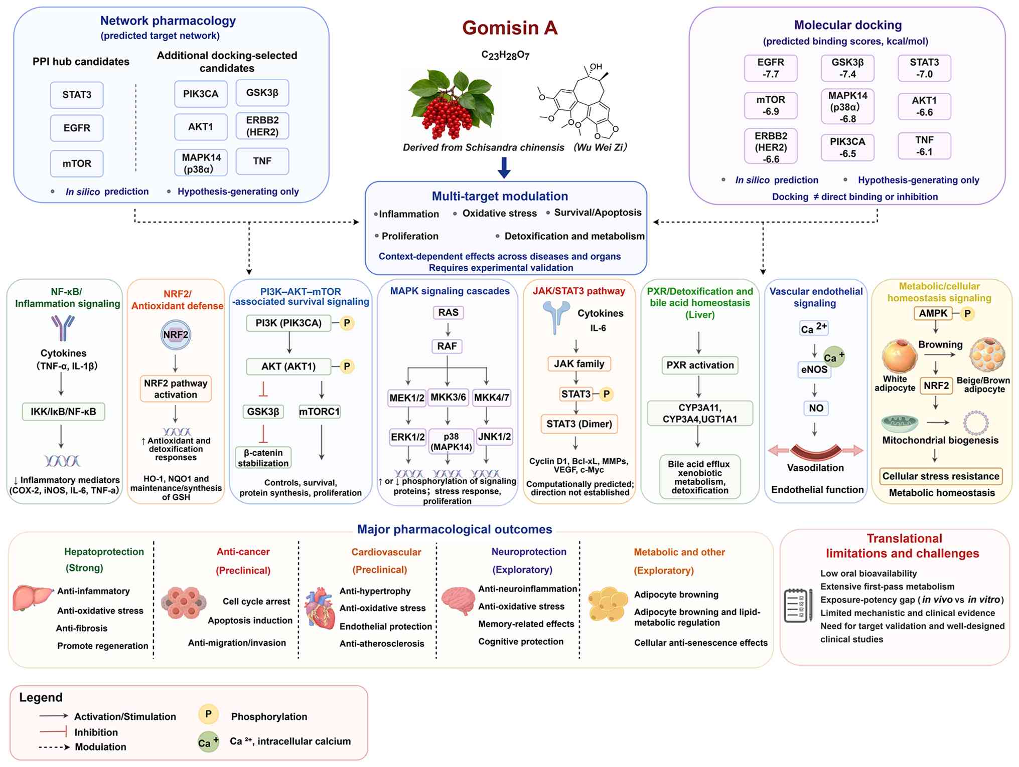
priorities for future translational studies. Fig. 1 summarizes the reported
hepatoprotective (7,11,12), anticancer (8,13), cardiovascular (9,14), neurological (10,15) and anti-inflammatory/antioxidant
effects (16,17) of GA, together with PK and safety
constraints (18,19), as well as candidate target
network and docking results generated in the present study; these
computational interactions remain hypothesis-generating.
Review approach
Literature search strategy
The present review used PubMed (https://pubmed.ncbi.nlm.nih.gov/) and the China
National Knowledge Infrastructure database (cnki.net/).
The search terms included GA, Schisandrol B, and combinations with
hepatoprotective, anticancer, cardiovascular, neuroprotective,
anti-inflammatory, antioxidant, PK and network pharmacology key
words. Studies using purified GA monomers were prioritized for
mechanistic analysis. Studies of well-characterized
Schisandra-derived extracts or multicomponent preparations
were retained as supportive evidence where relevant, but their
effects were not attributed specifically to GA unless its
independent contribution could be established.
Hierarchy of mechanistic evidence
To avoid equally weighting conclusions of varying
reliability, each mechanistic claim was assigned to one of four
evidence tiers (Table I): Tier
A, direct target engagement shown by both a biophysical or cellular
binding assay [surface plasmon resonance (SPR), cellular thermal
shift assay (CETSA), drug affinity responsive target stability
assay (DARTS) or co-crystallography] and a functional dependency
experiment (knockdown, knockout or rescue), representing the
strongest level of mechanistic support; tier B, target or pathway
dependency is supported by loss- or gain-of-function studies (small
interfering and short hairpin RNA knockdown, clustered regularly
interspaced short palindromic repeats knockout, selective
pharmacological inhibition or overexpression rescue) but lacking
experimentally demonstrated direct target binding or cellular
target engagement. Computational docking predictions, when
available, were not considered direct binding evidence; tier C,
pathway association includes findings inferred from reporter
activity, changes in gene or protein expression, phosphorylation
status, nuclear translocation, subcellular localization, or
downstream biomarkers without formal perturbation demonstrating
that the pathway is required for the phenotype; and tier D,
computational prediction encompassing network pharmacology targets,
molecular docking poses and molecular dynamics (MD) simulations,
none of which have been experimentally validated.
 | Table IHierarchy of mechanistic evidence
used for critical appraisal. |
Table I
Hierarchy of mechanistic evidence
used for critical appraisal.
| Tier | Definition | Criteria | Representative
examples |
|---|
| A (direct target
engagement) | Direct physical
binding confirmed + functional dependency verified | Biophysical or
cellular binding assay (SPR, CETSA, DARTS or co-crystallography)
and functional rescue/perturbation confirming target
dependency | None identified
among the studies included in the present review |
| B (target or
pathway dependency) | Functional
dependency confirmed, lacking experimentally demonstrated direct
target binding or cell target engagement | Loss- or
gain-of-function studies (siRNA/shRNA knockdown, CRISPR knockout,
selective inhibitor blockade or overexpression rescue); no direct
binding data | PXR-dependent
anticholestatic protection: Loss of the major protective effects in
PXR-null mice, supported by reporter transactivation and PXR
target-gene induction |
| C (pathway
association) | Pathway involvement
inferred from biomarker changes, no dependency test | Evidence from
expression levels, phosphorylation status, subcellular
localization, reporter activity or downstream biomarker shifts; no
formal perturbation to confirm causality | NF-κB suppression;
NRF2 nuclear translocation and ARE reporter activation; p38/ERK/JNK
and PI3K/AKT phosphorylation changes; BCL-2/BAX and
caspase-associated changes; downstream target gene expression
changes in the absence of dependency testing |
| D (computational
prediction) | In silico
prediction only, no experimental validation | Targets predicted
by network pharmacology, binding poses predicted by molecular
docking and complex behavior assessed by molecular dynamics
simulations | EGFR, STAT3 and
mTOR docking binding modes |
Additional dimensions of evidence
maturity
Beyond the mechanistic hierarchy, several contextual
factors shape overall confidence in a pharmacological claim. These
include the pathophysiological relevance of the disease model to
human disease, independent replication across laboratories, whether
the study used purified monomers or crude extract, whether
effective concentrations or doses fall within a physiologically
achievable range, whether dosing was prophylactic or therapeutic,
the adequacy of selectivity and vehicle controls, the availability
of paired PK data for PK/pharmacodynamic (PD) alignment, and the
existence of any human or clinical data. These dimensions yield a
more balanced assessment of translational potential than a single
positive experiment alone.
Application of the evidence
framework
Applying this appraisal across therapeutic areas
places the activities of GA into three broad categories of
translational maturity. Well-established preclinical activity is
represented by hepatoprotection, the most thoroughly studied area,
with replicated findings across acute injury, cholestasis, fibrosis
and regeneration models (7,11,12, 20-22). Promising but preclinical activity
includes anticancer and cardiovascular effects; both have
substantial in vitro data but more limited in vivo
validation, with most mechanisms remaining at the associative level
(tier C) and little PK/PD alignment to support translatability
(8,9,13,14,23-25). Exploratory observations include
neuroprotection, anti-obesity, renoprotection, erectile function,
antimicrobial and immunomodulatory effects (largely single-center,
single-model reports with small sample sizes and thin mechanistic
grounding, regarded as preliminary) (10,15,26-29).
Within this framework, examples of tier C evidence
include NF-κB suppression (7,16), nuclear factor erythroid 2-related
factor 2 (NRF2) nuclear translocation and antioxidant response
element reporter activation (17), and MAPK- and apoptosis-associated
protein changes (8,26), where pathway-associated changes
were reported without experimental demonstration of pathway
necessity.
Network pharmacology analysis
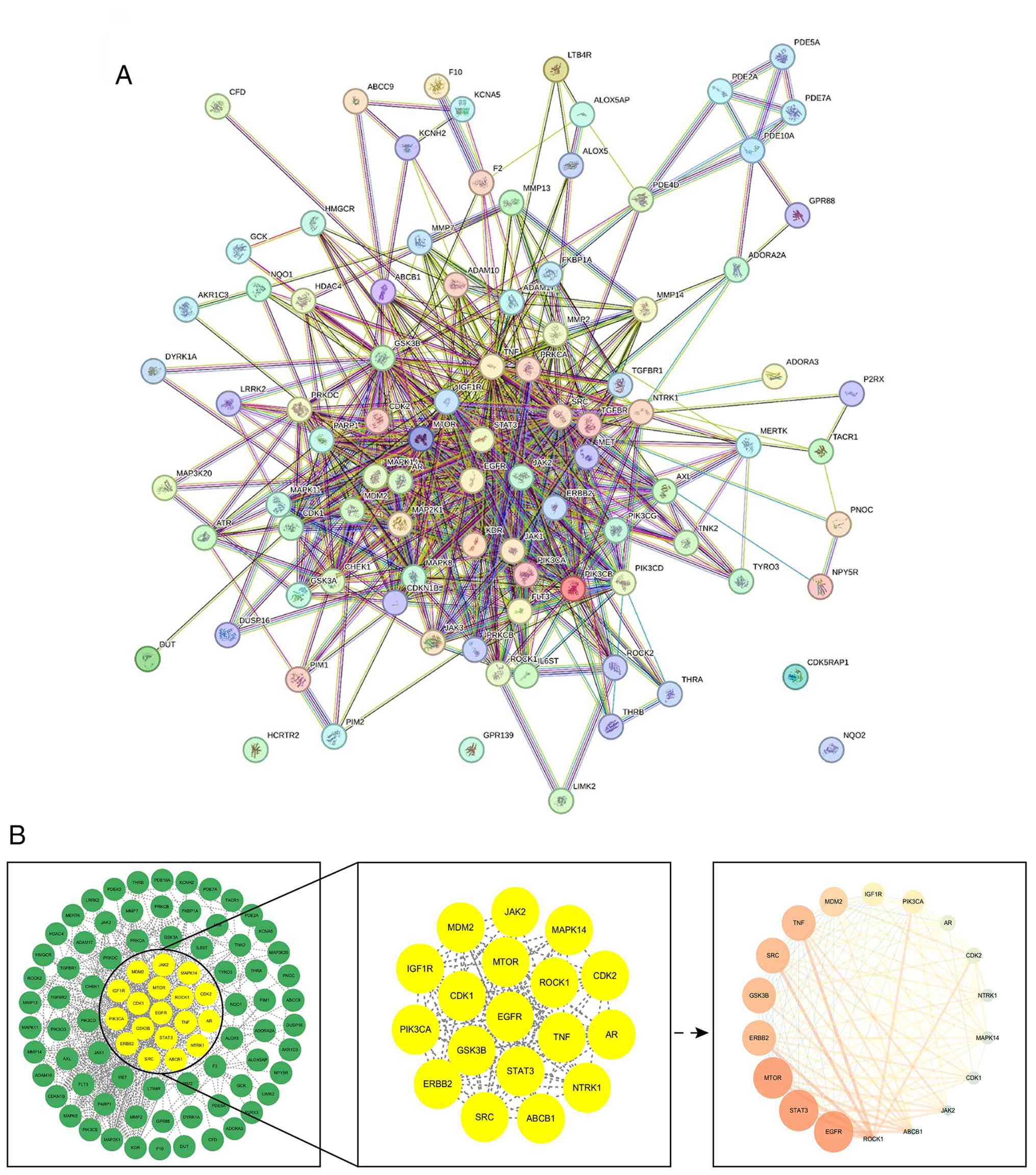
To investigate the potential molecular mechanisms
underlying the effects of GA, the present study used the Swiss
Target Prediction platform (https://www.swisstargetprediction.ch/) to identify its
candidate target genes in Homo sapiens. Only targets with a
non-zero prediction probability were retained for subsequent
analysis, yielding 100 predicted candidate targets. This
computational workflow was designed to prioritize candidate targets
for experimental validation, rather than to confirm functional
target engagement. The present study constructed a protein-protein
interaction (PPI) network for these target genes using the STRING
database (version 12.0; string-db. org/). Subsequently, to identify
the core components of the PPI network, Cytoscape (version 3.10.4;
https://cytoscape.org/) was used for network
screening.
To explore potential associations between predicted
GA targets and disease-associated genes while reducing non-specific
background noise, disease-associated genes were retrieved
separately for each disease entity from the GeneCards database
(https://www.genecards.org/). A GeneCards
Relevance Score ≥10 was applied as a predefined retrieval cutoff
and the resulting gene sets were used for descriptive overlap
analysis. The disease entities included ALI, hepatic fibrosis,
cholestasis, liver regeneration, HC, CRC, OC, prostate cancer,
breast cancer, retinoblastoma and metastatic melanoma. For each
disease, the number of retained GeneCards entries and genes
overlapping with the 100 predicted GA targets were summarized
descriptively (Table SI).
Because the GeneCards-derived disease sets were not restricted to
one predefined set of human genes eligible to be counted in every
disease and target analysis, no formal enrichment significance was
inferred from these overlaps. The overlap analysis was used to
provide descriptive disease-context information for the predicted
GA targets and should not be interpreted as evidence of target
specificity, causal disease mechanisms or direct GA target
engagement. The disease overlap analysis was restricted to
well-defined disease entities (liver disease and individual cancer
types) with notable pharmacological evidence. Cardiovascular and
neurological effects were not subjected to analogous GeneCards
overlap analysis because these encompass heterogeneous phenotypes
and exploratory indications, and their mechanistic interpretation
is based primarily on experimental literature and pathway-level
evidence.
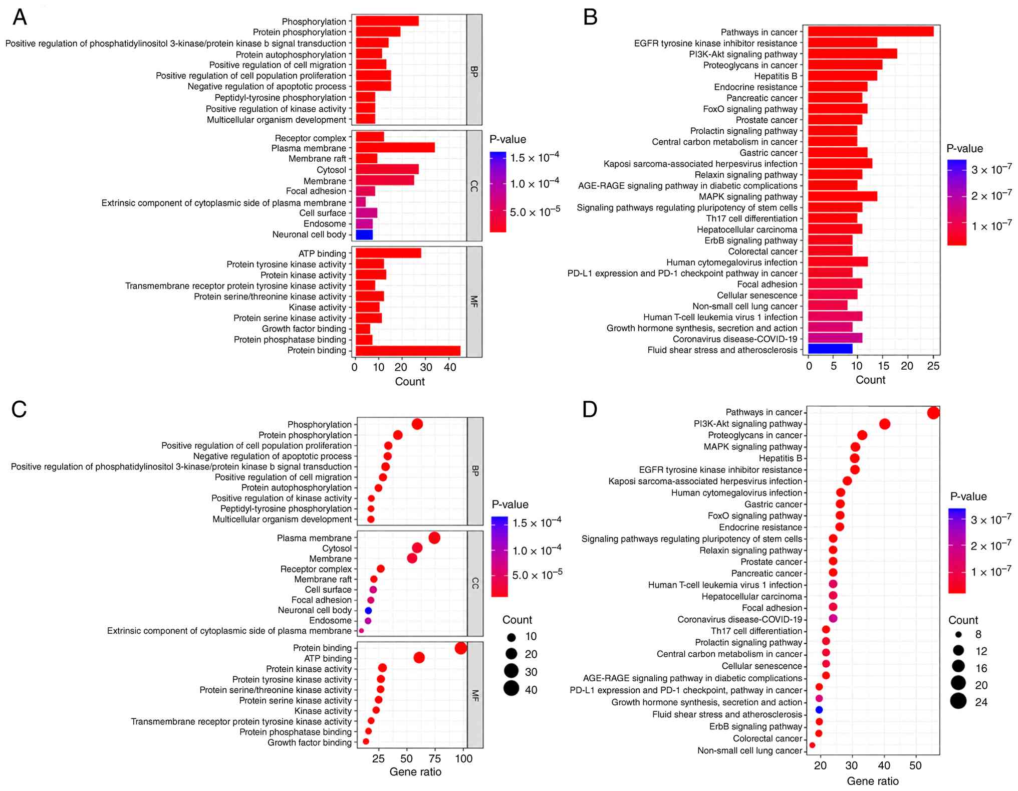
Gene Ontology (GO; geneontology.org/) and Kyoto Encyclopedia of Genes and
Genomes (KEGG; kegg.jp/) enrichment analyses were performed on the
100 candidate target genes using the STRING platform, with the
human whole genome as the background set and false discovery rate
(FDR) correction applied for multiple testing. The 100 predicted GA
targets and the full KEGG enrichment results are provided in
Tables SII and SIII, respectively. Only terms with an
FDR <0.05 were considered.
Target prioritization
Key targets for experimental validation were
identified as follows: i) Core hub proteins derived from the PPI
network prioritized via topological indices (STAT3, EGFR and mTOR);
ii) central nodes within significantly enriched KEGG pathways,
including PIK3CA and AKT1 in the PI3K-Akt signaling pathway and
MAPK14 in the MAPK signaling pathway; iii) molecules concurrently
identified in network pharmacology and pathway enrichment analyses
(ERBB2); and iv) proteins with experimental biological relevance
associated with GA responses, including TNF (10,16), or pathway-associated relevance,
including GSK3β (30-33), while recognizing that these
studies do not establish direct GA binding.
Molecular docking methods. Protein information for
the target structures was obtained from UniProt (https://www.uniprot.org/). Molecular docking of GA
(PubChem Compound ID: 634470) with these proteins was performed
using AutoDock Vina 1.1.2 software (34). These proteins were preprocessed
in PyMOL 2.4 (pymol.org/; removal of water molecules
and unnecessary ligands, and addition of hydrogen atoms). PDBQT
files for docking simulations were generated using AutoDock Tools
1.5.6 (ccsb.scripps.edu/mgltools/). The docking results were
set to output the top nine optimal docking poses. For each target,
the top-ranked pose with the lowest Vina score was selected for
interaction analysis. Finally, Protein-Ligand Interaction Profiler
(version 3.0.0; plip-tool. biotec.tu-dresden.de/plip-web/plip)
(35) and PyMOL 2.4 software
were used to visualize the docking results to display predicted
ligand-receptor contacts. According to the predicted docking score
classification criteria from previous studies (36,37), a predicted docking score <-7.0
kcal/mol was assigned to the more favorable-score category; a value
≥-7.0 and <-5.5 kcal/mol was assigned to the intermediate-score
category; and a value ≥-5.5 and ≤-4.25 kcal/mol was assigned to the
less favorable-score category. These categories do not establish
binding affinity or specificity. These criteria were used only for
descriptive comparison of the predicted docking scores of GA across
the target proteins.
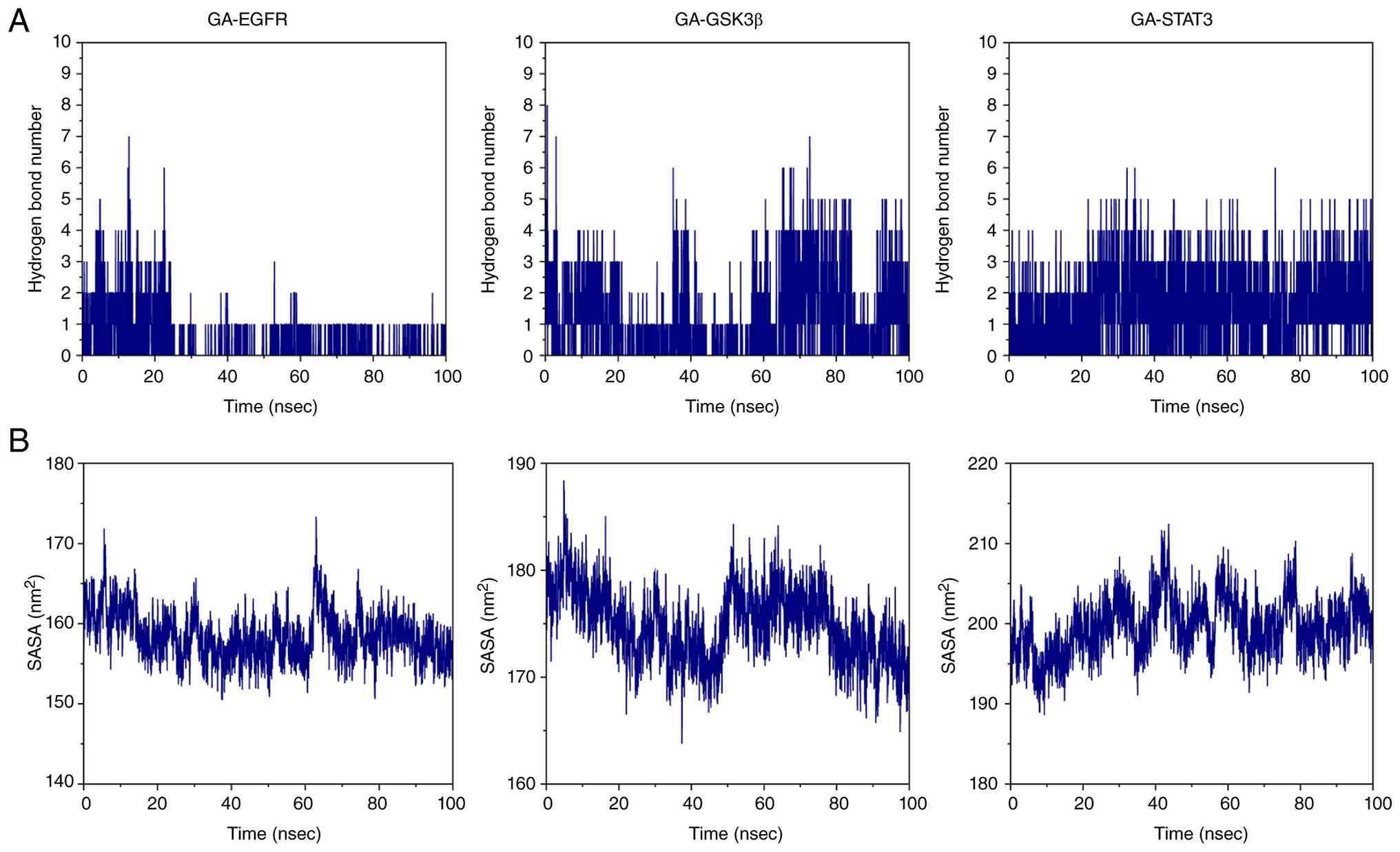
All simulations were performed in GROMACS 2020.6
(38). Protein topologies were
generated using the AMBER99SB-ILDN force field, the ILDN
modification of ff99SB described by Lindorff-Larsen et al
(39); (https://doi.org/10.1002/prot.22711) and GA ligand
parameters were derived from the General Amber Force Field using
Sobtop 1.0 (sobereva.com/soft/Sobtop/). Each protein-ligand
complex was solvated in a truncated octahedral transferable
intermolecular potential with 3 points water box with a 1.0-nm
minimum distance between the solute and the box boundary, and
Na+/Cl− counterions were added to neutralize
the net system charge (40).
Energy minimization was conducted via 2,500 steps of steepest
descent followed by 2,500 steps of conjugate gradient. Systems were
equilibrated for 100 psec under the constant
number-volume-temperature ensemble at 298.15 K and 100 psec under
the constant number-pressure-temperature (NPT) ensemble at 298.15 K
and 101.325 kPa. Production simulations were run for 100-nsec under
NPT conditions with periodic boundary conditions and a 2-fsec
integration time step (41).
Long-range electrostatic interactions were treated with the
particle mesh Ewald method and short-range electrostatic and van
der Waals cutoffs were set to 1.0 nm. Trajectory frames were saved
every 10 psec for analyses of the RMSD, root-mean-square
fluctuation (RMSF), radius of gyration (Rg), intermolecular
hydrogen bonds and solvent-accessible surface area (SASA) (42-44).
PAINS structural alert screening. Structural alert
screening was performed using the RDKit FilterCatalog module (RDKit
version 2025.09.4; https://www.rdkit.org/). The stereochemically defined
structure of GA was evaluated against the combined RDKit PAINS
catalog, comprising the PAINS_A, PAINS_B and PAINS_C rule sets.
Chemical identity and physicochemical
properties of GA
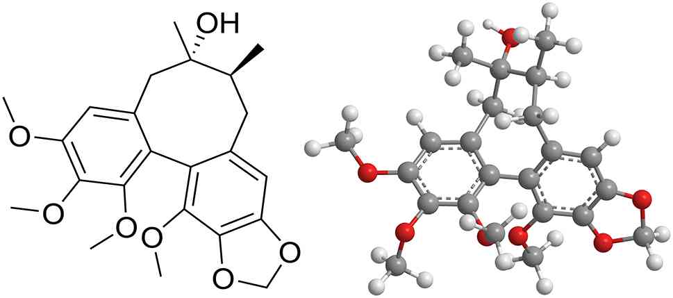
GA is a stereochemically defined
dibenzocyclooctadiene lignan with the molecular formula
C23H28O7 and a molecular weight of
416.47 g/mol (45). The scaffold
of GA contains two aromatic rings connected through an
eight-membered carbocyclic framework, one methylenedioxy group,
four methoxy substituents, two methyl groups and one non-phenolic
hydroxyl group (Fig. 2)
(46). The primary
physicochemical properties of GA are summarized in Table II.
| Table IIPhysicochemical properties and
structural characteristics of gomisin A. |
Table II
Physicochemical properties and
structural characteristics of gomisin A.
| Property | Description |
|---|
| Chemical class |
Dibenzocyclooctadiene lignan |
| Molecular
formula |
C23H28O7 |
| Molecular
weight | 416.47 g/mol |
| Calculated
cLogP | 3.60 |
| Topological polar
surface area | 75.61
Å2 |
| Hydrogen bond
donors/acceptors | 1/7 |
| Rotatable
bonds | 4 |
| PAINS/structural
alerts | 0a |
Using the stereochemically defined structure and
descriptors calculated with RDKit, an open-source cheminformatics
toolkit (rdkit.org/), GA has a calculated
molecular weight of 416.47 g/mol, calculated octanol-water
partition coefficient (cLogP) of 3.60, topological polar surface
area of 75.61 Å2, one hydrogen bond donor, seven
hydrogen bond acceptors and four rotatable bonds. These properties
are consistent with a moderately lipophilic small molecule that may
cross biological membranes, but they do not predict oral
bioavailability because first-pass metabolism and transporter
interactions are substantial.
A structural-alert screen using the RDKit
FilterCatalog found no matches to pan-assay interference compounds
(PAINS) A, B, or C filters. This negative result decreases concern
about classic PAINS substructures but does not establish assay
specificity. High-micromolar lipophilic compounds cause colloidal
aggregation, membrane perturbation, fluorescence or absorbance
interference, redox effects and non-specific protein binding. PAINS
filters are best used as flags for experimental follow-up rather
than as proof of validity or invalidity (47,48).
Pharmacological effects of GA
As a constituent of S. chinensis, GA has
preclinical hepatoprotective (7,11,12), anticancer and chemosensitizing
(8,13,49), cardiovascular (9,14,24,25), neuroprotective (10,15), and anti-inflammatory and
antioxidant effects (16,17)
(Fig. 3).
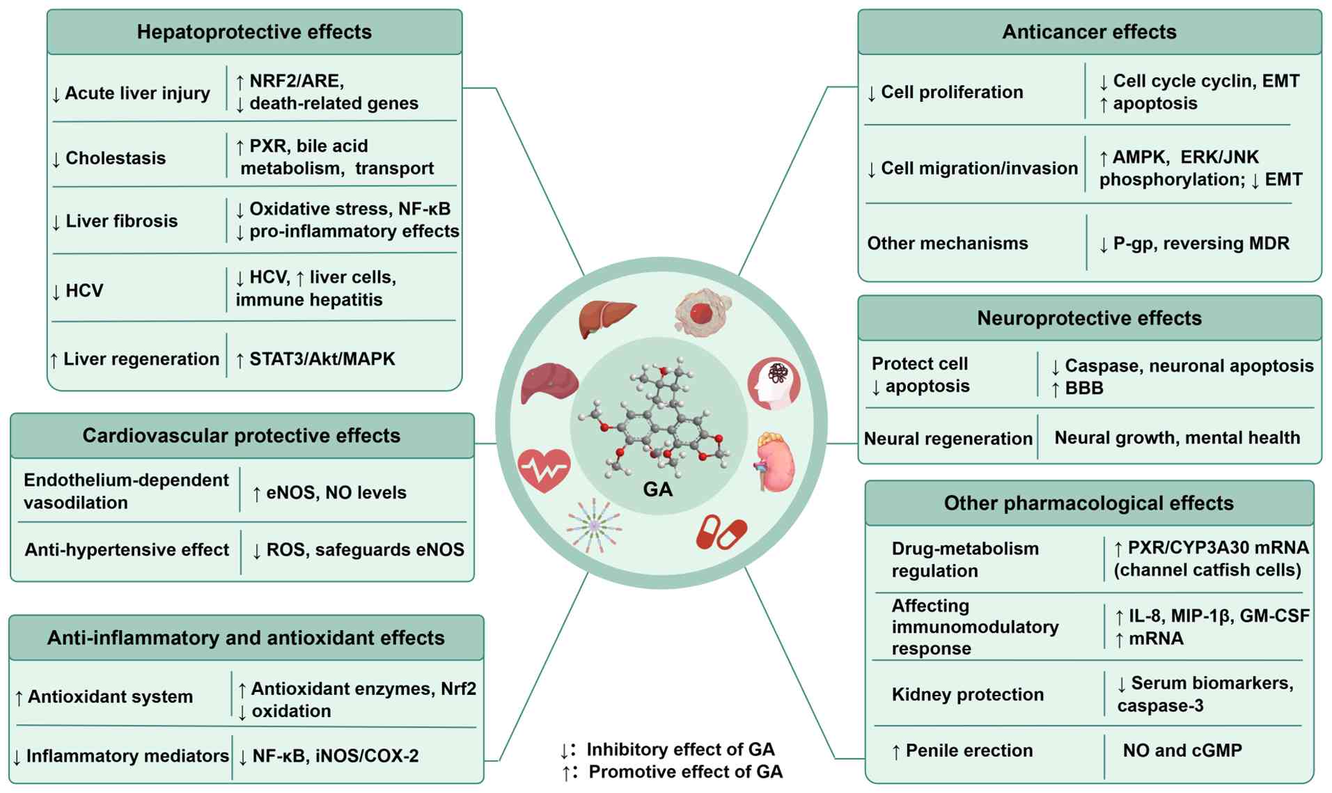 | Figure 3Overview of the reported
pharmacological activities of GA. GA, gomisin A; PXR, pregnane X
receptor; HCV, hepatitis C virus; iNOS, inducible nitric oxide
synthase; P-gp, P-glycoprotein; MDR, multidrug resistance; EMT,
epithelial-mesenchymal transition; BBB, blood-brain barrier; ROS,
reactive oxygen species; eNOS, endothelial nitric oxide synthase;
MIP, macrophage inflammatory protein; GM-CSF,
granulocyte-macrophage colony-stimulating factor; ARE, antioxidant
response element; COX-2, cyclooxygenase-2. |
To determine the molecular basis underlying the
reported hepatoprotective, anticancer, cardiovascular,
neurological, anti-inflammatory and antioxidant effects, the
present review identified nine core targets of GA [EGFR, erb-b2
receptor tyrosine kinase 2 (ERBB2),
phosphatidylinositol-4,5-bisphosphate 3-kinase catalytic subunit α
(PIK3CA), AKT1, mTOR, MAPK14, GSK3β, STAT3 and TNF] via network
pharmacology screening, prioritized by molecular docking. These
computational predictions were integrated into a unified
mechanistic interpretation of the activity of GA in the
pathological context. The nine targets were prioritized by their
acknowledged key position in the pathophysiology of the disease in
question. Second, consideration was given to whether the direction
of their pathway regulation can directly account for the
experimental phenotypes reported in the literature. Third,
selection was informed by their tissue and cell-type expression
specificity. A target not elaborated upon does not mean it is
uninvolved in that process but that it serves a secondary or
auxiliary role and is therefore not treated as a main line of
mechanistic explanation. This variable weighting of targets across
disease contexts reflects the tissue specificity and pathway
preference inherent to the multi-target pharmacological profile of
GA.
Hepatoprotection: Best-supported
preclinical domain
The liver is a key metabolic organ that serves a
pivotal role in numerous physiological processes, including
nutrient metabolism, lipid and cholesterol homeostasis, and the
degradation of xenobiotic compounds (50). In recent years, the number of
individuals with liver disease in the world has increased annually
(51). Therefore, the search for
drugs to treat liver disease is a topic of concern. Numerous
studies indicate that GA may exert a protective effect on hepatic
disorder (7,11,12,20).
Alleviation of acute LI (ALI)
The occurrence of ALI is attributable to various
etiologies, encompassing viral infection, substance abuse (alcohol
or drugs) and exposure to toxic agents (52). LI can be divided into four
categories: Chemically, immune-, drug- and alcohol-induced LI.
Experimental ALI is induced with hepatotoxic agents (53), including carbon tetrachloride
(CCl4), acetaminophen [N-acetyl-para-aminophenol
(APAP)], D-galactosamine (D-GalN) and lipopolysaccharide (LPS)
(Fig. 4). These models encompass
the major experimental settings in which GA-mediated
hepatoprotection has been mechanistically investigated (7,54,55).
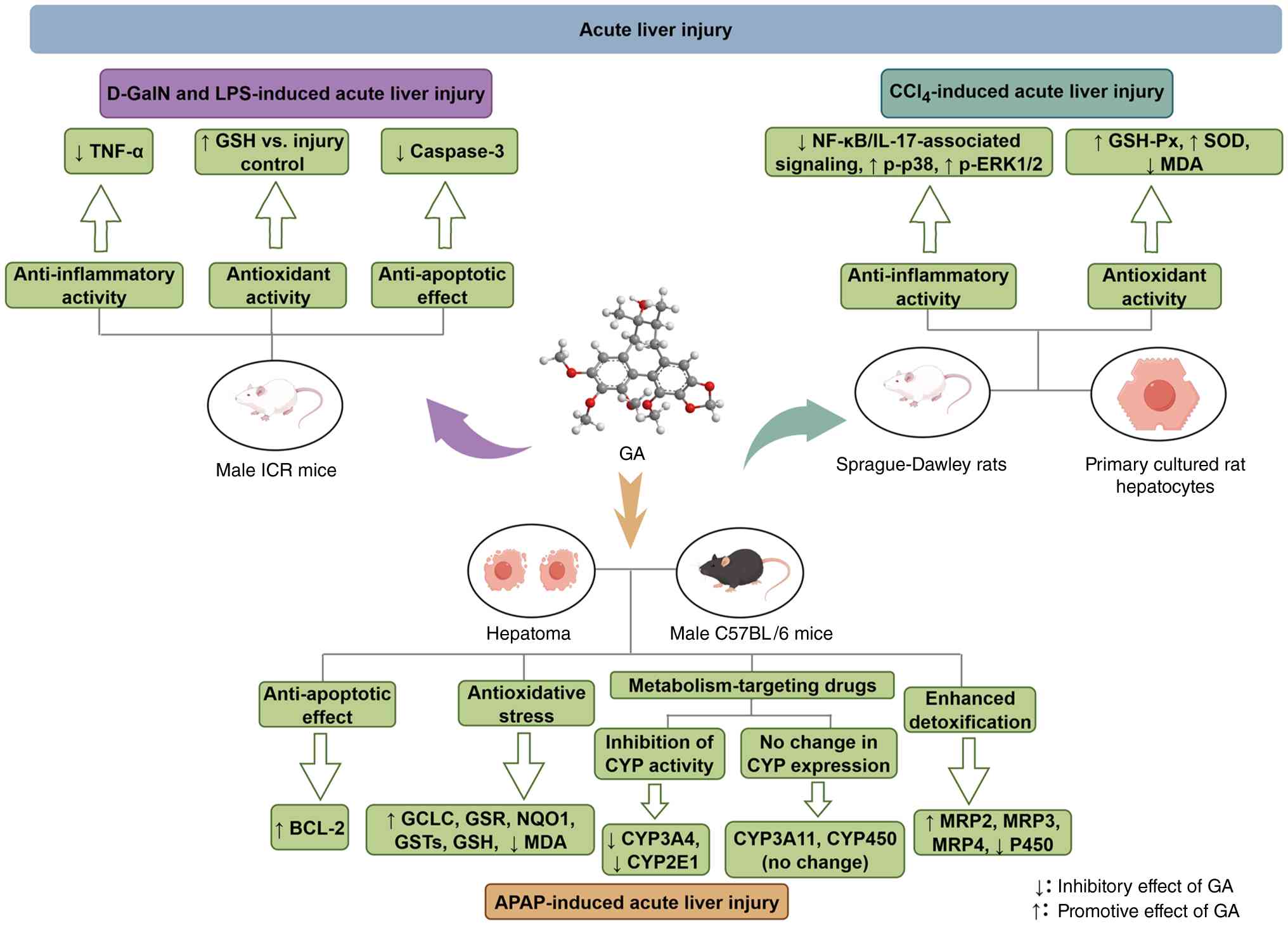 | Figure 4Effects of GA in experimental models
of acute liver injury. D-GalN, D-galactosamine; LPS,
lipopolysaccharide; GSH-Px, glutathione peroxidase; ICR, Institute
of Cancer Research mouse stock; SOD, superoxide dismutase; MDA,
malondialdehyde; GCLC, glutamate-cysteine ligase catalytic subunit;
GSR, glutathione reductase; NQO1, NAD(P)H quinone dehydrogenase 1;
GST, glutathione S-transferase; GA, gomisin A; CYP, cytochrome
P450; MRP, multidrug resistance-associated protein; APAP,
acetaminophen. |
CCl4
CCl4 is one of the most widely used
hepatotoxic agents and the mouse CCl4-induced ALI model
is a classic paradigm of oxidative liver damage research (7,26,56). GA reduces hepatic malondialdehyde
(MDA) content and restores glutathione (GSH) peroxidase and
superoxide dismutase activities, supporting an antioxidant
contribution to GA-mediated protection. These biochemical changes
are accompanied by marked decreases in serum aspartate
aminotransferase (AST) and alanine aminotransferase (ALT) levels,
indicating overall protection against hepatocellular damage
(7,26). The anti-inflammatory component of
this protection is associated with NF-κB suppression (7,54). GA downregulates NF-κB and IL-17
signaling, with corresponding decreases in inducible nitric oxide
(NO) synthase (iNOS) and cyclooxygenase-2 (COX-2) expression in
injured liver tissue (54).
NF-κB inhibition may contribute to the reduction in early fibrotic
changes, given the well-established link between inflammatory
signaling and hepatic fibrogenic activation (7,57,58).
In the rat liver, GA increased ERK1/2 and p38
phosphorylation relative to CCl4-treated controls; JNK did not show
the same clearly documented increase (26). These changes coincide with
upregulated BAX levels and decreased caspase-3 levels, suggesting
that MAPK signaling shapes the apoptotic response to
CCl4 injury (26).
The net direction of this regulation is not straightforward because
both pro-apoptotic (BAX induction) and anti-apoptotic (caspase-3
suppression) features are observed. Microarray data demonstrate
that GA-induced liver repair is associated with TGF-β-dependent
transcriptional changes, including the upregulation of cell cycle
genes and downregulation of cell death-associated transcripts,
consistent with a pro-regenerative gene expression program
(59).
Taken together, GA counteracts CCl4
hepatotoxicity through overlapping antioxidant, anti-inflammatory,
apoptosis-regulating and pro-repair pathways. These pathway
alterations should therefore be interpreted as associated
biological responses rather than experimentally confirmed direct
molecular targets. The data suggest a combined decrease in
oxidative and inflammatory burden, together with repair-associated
transcriptional changes (7,59). Inconsistencies, such as increased
BAX levels together with reduced active caspase-3 levels in the
CCl4 study (26),
caution against assigning a simple anti-apoptotic mechanism from
single protein measurements. Most CCl4 studies do not
use target-selective antagonists, gene deletion or rescue
experiments; consequently, NF-κB, MAPKs, TGF-β-associated genes and
apoptosis proteins are pathway-associated readouts rather than
established direct GA targets (7,26,54,59).
APAP
APAP is one of the most widely used analgesics and
antipyretics worldwide. Although safe at therapeutic doses,
overdose causes severe hepatotoxicity and remains a notable cause
of acute liver failure globally (60). APAP undergoes bioactivation by
cytochrome P450 (CYP) enzymes, principally CYP2E1, CYP3A4 and
CYP1A2, to form the reactive metabolite N-acetyl-p-benzoquinone
imine (NAPQI), which depletes hepatic GSH levels and triggers
mitochondrial dysfunction and oxidative stress (61). GA intervenes at several points
along this cascade. Upstream, it inhibits CYP3A4 activity and
decreases CYP-mediated APAP bioactivation (62,63). Further downstream, GA activates
the NRF2/antioxidant responsive element (ARE) pathway and
upregulates NRF2-driven detoxification genes, thereby enhancing
cell antioxidant capacity and limiting GSH depletion in
APAP-challenged animals (17).
At the regenerative stage, GA modulates p53, p21, cyclin D1
(CCND1), proliferating cell nuclear antigen and BCL-2 expression to
promote hepatocyte proliferation and tissue repair (11). Functionally, these effects
translate into decreased serum ALT and AST activity, diminished
hepatic MDA formation and preserved mitochondrial GSH pools in a
dose-dependent manner (11).
In summary, GA protects against APAP hepatotoxicity
through decreased metabolic activation, enhanced antioxidant
defense and accelerated liver regeneration. These findings are
compatible with antioxidant, metabolic and regenerative
contributions, but the relative importance of each mechanism
remains uncertain because intrahepatic exposure, NAPQI-protein
adducts and target-dependency interventions have not been
consistently measured across studies (11,17,63). In addition, numerous experiments
use pretreatment schedules, which demonstrate prophylactic activity
rather than therapeutic efficacy against established injury
(11,17,63).
D-GalN and LPS. D-GalN is selectively taken up by
hepatocytes, where it depletes intracellular uridine nucleotide
pools, and impairs RNA and protein synthesis. When co-administered
with low-dose LPS, it triggers fulminant hepatic failure that
recapitulates the metabolic and inflammatory features of acute
liver failure in humans (64).
The resulting lesion is driven by two parallel pathways. On the
inflammatory side, NF-κB nuclear translocation increases, driving
transcriptional upregulation of iNOS, TNF-α, IFN-γ and multiple ILs
(IL-1β, IL-6, IL-12 and IL-18). On the antioxidant side, NRF2 and
its downstream targets NAD(P)H quinone dehydrogenase 1, heme
oxygenase-1 (HO-1) and GSH S-transferase α are concurrently
suppressed, further tilting the balance toward oxidative damage
(65). In this model, GA reduces
serum ALT levels and preserves hepatic GSH content relative to
D-GalN/LPS-treated controls, while also attenuating mitochondrial
edema. These effects are attributable to reduced oxidative stress
and suppression of TNF-α and caspase-3 activity, suggesting
combined anti-inflammatory and anti-apoptotic actions (20). GA can inhibit the expression of
TNF-α itself by downregulating pathways such as the NF-κB pathway
(16), and decrease inflammatory
responses in experimental asthma and activated microglia (10,66). Thus, experimental data support
decreased inflammatory mediator production associated with NF-κB
suppression (10,16); direct GA-TNF binding remains a
computational hypothesis. In summary, GA protects against
D-GalN/LPS-induced LI through concurrent anti-inflammatory,
antioxidant and anti-apoptotic mechanisms.
In conclusion, across CCl4-, APAP- and
D-GalN/LPS- induced injury, the most reproducible phenotype is the
attenuation of hepatocellular damage. GA decreases serum
aminotransferase activity, histological necrosis, lipid
peroxidation and mitochondrial injury, while improving antioxidant
indices (7,11,17,20,26,54,59,63). The convergence of these endpoints
across distinct injury models supports hepatoprotective
pharmacology. It does not, however, establish one universal
molecular target, because the initiating toxic mechanisms differ
between CCl4 and APAP bioactivation and GalN/LPS-driven
inflammatory apoptosis. Taken together, ALI studies provide
moderate-to-strong evidence for a hepatoprotective phenotype and
low-to-moderate evidence for most individual signaling mechanisms.
Future work should use post-injury dosing, exposure-matched
concentrations, APAP-protein adduct measurements and
pathway-specific loss-of-function designs.
Cholestatic LI and PXR dependency
Cholestatic liver disease, characterized by impaired
hepatic bile formation or biliary bile flow (67), is induced by genetic immune
defects, toxins and physiological dysregulation that damage bile
ducts, leading to bile retention, hepatic injury, inflammation,
fibrosis and progression to cirrhosis or liver malignancy (68).
In the lithocholic acid (LCA)-induced cholestatic
injury model, GA decreases mortality and improves both histological
and biochemical markers of damage. Serum lactate dehydrogenase,
ALT, AST, alkaline phosphatase, total bile acids and total
bilirubin levels are all lowered and TUNEL-positive cells and
hepatocyte membrane pore formation are decreased (69,70).
The protective mechanism converges on PXR
activation, which drives two parallel downstream programs. On the
inflammatory and cell death side, PXR signaling suppresses NF-κB
activity, which reduces NLR family pyrin domain containing 3
(NLRP3) inflammasome assembly; PXR also downregulates FoxO1,
decreasing apoptotic protease-activating factor 1
(APAF-1)-dependent atypical inflammasome activation (12,70). On the detoxification side, PXR
upregulates bile acid metabolism and efflux machinery, including
the prototypical target genes CYP3A11 and uridine diphosphate
glucuronosyltransferase 1A1, accelerating metabolic conversion and
fecal clearance of toxic bile acids (12,70). Direct activation of human PXR
with the corresponding upregulation of its target genes has also
been demonstrated (70).
In summary, GA prevents LCA-induced cholestatic LI
primarily through PXR-mediated dual effects: Suppression of
inflammasome-driven injury, and enhancement of bile acid
detoxification and excretion. PXR is the best-supported
functionally required mediator of GA-induced anticholestatic
effects. Reporter transactivation, induction of PXR target genes
and the marked loss of the anticholestatic phenotype in
Pxr-null mice support PXR dependency (12). However, the aforementioned PXR
studies provide functional rather than experimentally demonstrated
direct physical GA-PXR binding evidence (12,69,70). Therefore, the PXR mechanism
should be classified as tier B. The evidence suggests PXR-dependent
protection but does not prove that GA directly binds to PXR or
directly targets each downstream component of the proposed
NF-κB/NLRP3 and FoxO1/APAF-1 pathways. Translational limitations
remain substantial. The protective doses used in rodent studies are
high, in the order of 100 mg/kg twice daily in representative
protocols (12,71). Rodent and human PXR differ in
ligand selectivity, so activity in mouse models does not guarantee
equivalent potency at the human receptor (12). Clinical efficacy cannot currently
be determined because controlled human studies remain unavailable.
Furthermore, PXR activation upregulates the expression of CYP
enzymes and drug transporters (72), which carries clinically
meaningful drug-interaction risk.
Improvement of liver fibrosis
Liver fibrosis develops when chronic or repeated
hepatocellular injury drives excessive accumulation of the
extracellular matrix (ECM), predominantly collagen. Without
intervention, it progresses to cirrhosis and, in certain patients,
hepatocellular carcinoma (57).
Two signaling axes are key to fibrogenesis. The TGF-β1/Smad pathway
is the canonical driver: TGF-β1 activates hepatic stellate cells
(HSCs) via Smad proteins, stimulating ECM secretion and scar
formation (73). The NF-κB
pathway acts in parallel by sustaining the inflammatory milieu that
fuels fibrogenic progression (58).
Surfeit locus protein 4 (Surf4), an endoplasmic
reticulum (ER) cargo receptor involved in ER-to-Golgi trafficking,
has been identified as a potential pro-fibrotic regulator: Surf4
facilitates the secretion of serum amyloid A1 (SAA1) from
hepatocytes. SAA1 promotes HSC activation, at least in part through
toll-like receptor 2 signaling (74). Hepatocyte-specific Surf4 deletion
markedly attenuates CCl4-induced liver fibrosis in mice,
whereas increased Surf4 and SAA1 expression are observed in human
fibrotic liver samples (74).
These findings support the Surf4-SAA1-HSC axis as a potential
pathogenic pathway contributing to liver fibrosis (74).
In CCl4-induced fibrotic mice, combined
treatment with GA and wedelolactone, a plant-derived coumarin,
reverses established fibrosis, potentially by inhibiting TGF-β1 and
NF-κB pathway activity and decreasing cytoglobin expression, a
marker of HSC activation and the fibrogenic response (21).
To the best of our knowledge, evidence supporting
the antifibrotic activity of GA remains limited. In
CCl4-induced fibrotic mice, GA monotherapy shows
antifibrotic activity; however, its combination with wedelolactone
produces greater improvements than either monotherapy (21). However, superiority of the
combination treatment alone does not establish pharmacological
synergy; formal interaction analyses were not reported. Changes in
cytoglobin and fibrosis-associated markers, together with decreased
TGF-β1/Smad and NF-κB signaling, support the antifibrotic activity
of the treatment regimen but do not establish a GA-specific
molecular target (21). Thus,
the combination treatment shows promising antifibrotic activity in
mice but the confirmation of pharmacological synergy and
clarification of GA-specific mechanisms require formal combination
analysis, independent replication and target-dependency
experiments.
Regulation of hepatitis C virus
(HCV)
Chronic HC is a global public health burden, with
~71 million individuals living with HCV worldwide and substantial
risk of progression to cirrhosis and hepatocellular carcinoma
(75). GA exerts dose-dependent
anti-HCV activity in vitro, putatively through the
suppression of host membrane proteins involved in viral binding or
entry (5,76). However, the specific host
proteins involved have not been definitively identified. The
evidence base remains small and largely preclinical (5,76), and unsupported by contemporary
antiviral target validation or PK exposure studies. Neither the
relevant host factor, target dependency, resistance profile nor
comparative activity against modern direct-acting antivirals have
been established. These findings are best framed as preliminary
antiviral pharmacology: Exploratory observations insufficient to
position GA as an antiviral candidate without renewed testing in
state-of-the-art HCV systems.
Promotion of liver regeneration
The liver possesses notable regenerative capacity,
restoring mass and function following injury or partial hepatectomy
through tightly controlled hepatocyte proliferation (77,78). GA accelerates this process
through two converging mechanisms. First, it increases the hepatic
levels of key growth factors and cytokines, such as hepatocyte
growth factor (HGF), EGF and IL-6, thereby activating the STAT3,
AKT and MAPK cascades that drive hepatocyte proliferation. This
pathway is associated with faster functional recovery than vehicle
treatment following partial hepatectomy in mice (22). Second, GA-mediated PXR activation
enhances bile acid detoxification and efflux, decreasing
cholestatic stress and creating a more permissive environment for
regeneration (12). Although
these regeneration signals are biologically coherent, existing data
represent pathway associations rather than established direct
targets; the contribution of STAT3, AKT, and MAPK signaling to
GA-mediated regeneration has not been established because pathway
inhibition or genetic validation studies remain limited.
Furthermore, whether the pathway activations reflect direct kinase
targeting by GA or are downstream consequences of improved tissue
viability and decreased bile acid toxicity remains unclear.
Temporal profiling, pathway blockade experiments and
target-engagement assays are needed to distinguish these
possibilities. Finally, enhanced growth signaling may benefit
post-resection recovery, but may result in undesirable effects in
premalignant or chronically injured livers (77).
CYP3A4-mediated drug interactions and
metabolic dysfunction-associated steatotic liver disease (MASLD)
therapy
CYP3A4 is most abundantly expressed in the liver
and is also present in the human prostate, breast, gastrointestinal
tract, colon and small intestine tissue. In total, ~38 structural
classes of substrates, totaling >150 drugs, are metabolized by
CYP3A4, accounting for ~50% of all medicines metabolized.
Therefore, CYP3A4 is among the most clinically important P450
enzymes in drug metabolism and drug-interaction risk assessment
(79). The interaction between
GA and CYP3A4 is multifaceted. GA has also been developed as an
isoform-selective fluorescent probe substrate for CYP3A4 in
microsomes and living cells (80). This analytical application should
be separated from hepatoprotection. GA inhibits CYP3A activity
(62), whereas PXR activation
may increase the expression of CYP3A and transporters over a longer
time scale (12). Therefore, the
net effect may depend on dose, exposure duration, species and
disease state, creating a risk of altered exposure to
co-administered CYP3A substrates.
Beyond toxin-, cholestasis- and inflammation-driven
models of LI, MASLD represents a major metabolic liver disorder; a
meta-analysis using the earlier NAFLD definition estimated global
prevalence at 32.4% (81). MASLD
is also a relatively underexplored therapeutic context for GA.
Hepatic steatosis, lipotoxicity and oxidative stress are key
features of MASLD pathogenesis, whereas peroxisome
proliferator-activated receptor α (PPARα) is key in regulating
hepatic fatty acid oxidation and lipid homeostasis. In transgenic
hamsters, expression of the Caenorhabditis elegans ω-3
fatty-acid desaturase gene fat-1 promotes the nuclear localization
of PPARα, accompanied by improvements in dyslipidemia, hepatic
steatosis and atherosclerotic lesions. These findings suggest that
PPARα serves as a molecular link between hepatic lipid metabolism
and cardiovascular metabolic homeostasis (82). Although direct evidence regarding
the effects of GA in MASLD remains limited, its proprotein
convertase subtilisin/kexin type 9 (PCSK9)-mediated low-density
lipoprotein (LDL)-lowering effect, PXR-dependent bile acid
regulation, and broad NRF2/NF-κB-modulated antioxidant and
anti-inflammatory activity collectively support mechanistic
plausibility (55,70). The involvement of PPARα or other
lipid-sensing nuclear receptors in GA-associated metabolic
regulation requires further experimental clarification; therefore,
MASLD represents a high-potential, mechanistically coherent
direction for future hepatoprotective investigation.
Critical assessment and translational
considerations
LI models provide a general picture of the dose
range, translational potential and possible mechanisms of GA
(7,11,20). Rodent hepatoprotection has been
reported with oral GA pretreatment in CCl4-, APAP- and
D-GalN/LPS-associated injury, although dose and dosing frequency
differ across models (7,11,20). A rat tissue-distribution study
found preferential GA accumulation in the liver and kidneys after
oral administration (18). This
may make liver disease a more practical therapeutic setting for GA
than indications that depend on sustained exposure in peripheral
tissues, such as cancer or CVD (7,11,12,18,20).
Nonetheless, studies employ pretreatment schedules
(7,11,17), so the data support prophylactic
protection rather than therapeutic reversal of established injury.
In fibrosis, combination of GA and wedelolactone is more effective
than monotherapy (21).
Differences in GA purity, vehicle and dosing frequency across
laboratories further complicate cross-study dose-response
comparisons. On balance, hepatoprotection is the best-supported
pharmacological activity of GA, but meaningful clinical translation
requires post-injury treatment protocols, precise dose-response
characterization and direct measurement of the hepatic tissue
concentration.
Mechanistic insights from network
pharmacology
Network pharmacology and molecular docking data
provide a multi-target framework that may account for the breadth
of this hepatoprotective phenotype, organized into three signaling
tiers. At the most upstream receptor tier, EGFR (predicted affinity
score, −7.7 kcal/mol) is a membrane tyrosine kinase whose
inhibition would simultaneously dampen both PI3K/AKT and MAPK
signaling, potentially decreasing proliferation of activated HSCs,
a prediction consistent with reported anti-fibrotic trends across
liver and vascular injury models (26,83), while modulating hepatocyte
proliferation during regeneration. At the intermediate kinase tier,
four nodes are central: The PI3K/AKT/mTOR pathway (affinity, −6.5,
−6.6 and −6.9 kcal/mol, respectively), where PIK3CA shows moderate
binding consistent with the regulatory (rather than complete
inhibitory) modulation of PI3K/AKT signaling observed in ALI
(54); MAPK14 may constitute a
link between the computational network and the inflammatory and
apoptosis-associated responses observed in LI; in the
CCl4 model, GA suppresses NF-κB/IL-17 signaling and
decreases iNOS and COX-2 expression, whereas separate experiments
have demonstrated increased hepatic p38 and ERK phosphorylation
relative to CCl4-treated controls (26,54); GSK3β exhibits a predicted
affinity of −7.4 kcal/mol and may represent an upstream regulatory
candidate for the antioxidant branch of the hepatoprotective
response. This hypothesis is consistent with activation of NRF2/ARE
signaling, preservation of hepatic GSH pools, decreased MDA
accumulation and improvement of other antioxidant indices in APAP-
and D-GalN/LPS-associated injury models (11,17,20). To the best of our knowledge,
however, whether GSK3β contributes to GA-mediated NRF2 regulation
has not been experimentally established. Experimentally, increased
HGF, EGF and IL-6 levels and the activation of STAT3, AKT and MAPK
signaling have been associated with GA-promoted liver regeneration
following partial hepatectomy or transplantation (22). Consistent with these experimental
findings, STAT3 (predicted docking score, −7.0 kcal/mol)
participates in IL-6-dependent regenerative transcription and is
associated with GA-promoted liver regeneration (22,84). At the downstream cytokine tier,
TNF (-6.1 kcal/mol) is predicted to interact with GA at the trimer
interface, adding a potential protein-level layer of suppression on
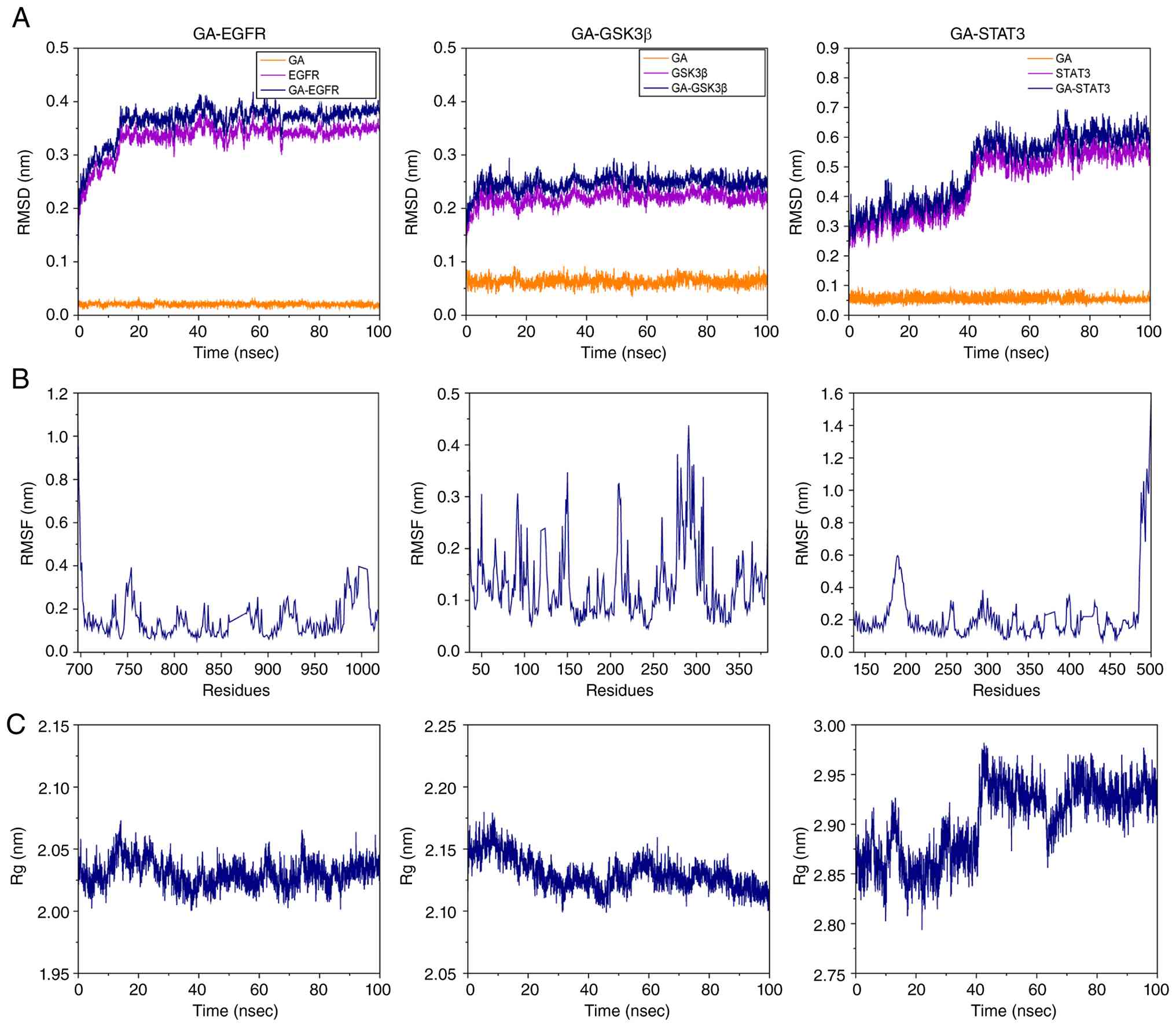
top of the transcriptional inhibition via NF-κB signaling (16). In line with these docking-derived
hypotheses, the 100-nsec trajectories support the dynamic
plausibility of the GA-EGFR and GA-GSK3β complexes, whereas the
GA-STAT3 system undergoes a longer relaxation period and exhibits
greater conformational mobility (Fig. 5A). These findings provide
structural support for candidate interactions but do not establish
direct target engagement in hepatocytes.
These computational data suggest GA targets across
receptor, kinase, transcription factor and cytokine levels,
consistent with the integrated anti-inflammatory, antioxidant,
antifibrotic and pro-regenerative phenotype observed experimentally
(16,17,21,22). GA exhibits preclinical promise
across a spectrum of liver disease models (Table III; Fig. 6) (11,12,17,20,22,85).
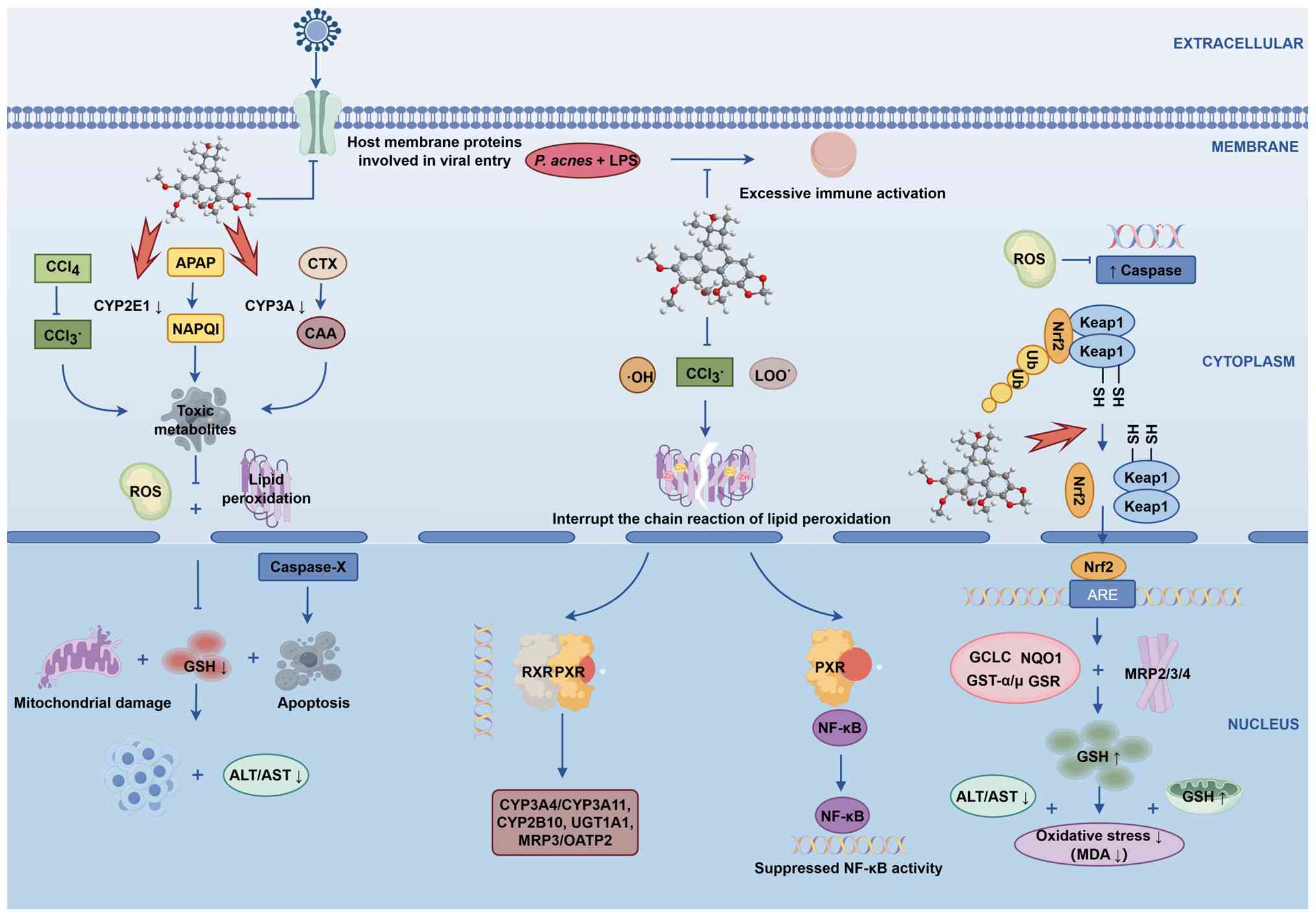 | Figure 6Proposed integrated mechanisms
underlying the hepatoprotective effects of gomisin A. P.
acnes, Propionibacterium acnes; LPS, lipopolysaccharide;
APAP, acetaminophen; CYP, cytochrome P450; CTX, cyclophosphamide;
NAPQI, N-acetyl-p-benzoquinone imine; CAA, chloroacetaldehyde; ROS,
reactive oxygen species; RXR, retinoid X receptor; PXR, pregnane X
receptor; Ub, ubiquitin; GSH, glutathione; ALT, alanine
aminotransferase; AST, aspartate aminotransferase; GCLC,
glutamate-cysteine ligase catalytic subunit; NQO1, NAD(P)H quinone
oxidoreductase 1; GST, glutathione S-transferase; GSR, glutathione
reductase; MRP, multidrug resistance-associated protein; UGT1A1,
uridine diphosphate glucuronosyltransferase 1A1; OATP2, organic
anion-transporting polypeptide 2; Keap1, Kelch-like ECH-associated
protein 1; ARE, antioxidant response element; MDA,
malondialdehyde. |
| Table IIIHepatoprotective effects of gomisin A
in experimental liver disease. |
Table III
Hepatoprotective effects of gomisin A
in experimental liver disease.
| Condition | Model | Biochemical
effects | Antioxidant
effects | Histological
effects | (Refs.) |
|---|
| Acute liver
injury |
CCl4-induced mice | Decreased ALT and
AST | Increased GSH-Px
and SOD; decreased MDA | Reduced hepatocyte
injury | (54) |
| Acute liver
injury |
CCl4-treated SD rats | Decreased ALT, AST
and ALP | Decreased lipid
peroxidation; increased antioxidant defenses | Decreased
hepatocellular necrosis and central-vein dilation | (26) |
| Acute liver
injury |
CCl4-treated SD rats | Decreased serum
liver injury markers | Restoration of
oxidative stress-related pathways and antioxidant responses | Reduced
CCl4-induced liver damage | (59) |
| Acute liver
injury | APAP-treated
mice | Decreased ALT and
AST | Preserved GSH;
decreased MDA | Decreased hepatic
necrosis | (11,17,63) |
| APAP
bioactivation | Mouse liver
microsomes | Reduced
CYP-mediated APAP bioactivation | Not applicable | Not applicable | (17,63) |
| Drug
metabolism | CYP3A4 cells | Inhibition of
CYP3A4 activity | Not applicable | Not applicable | (62) |
| Antioxidant
response | HepG2 cells | NRF2/ARE reporter
activation; induction of protective genes | Activation of
antioxidant defenses | Not applicable | (17) |
| Acute liver
injury | D-GalN/LPS-treated
mice | Decreased ALT and
AST | Preserved GSH | Decreased
hepatocyte necrosis and hemorrhage | (20) |
| Cholestasis | LCA-treated
mice | Decreased liver
injury markers and bile-acid burden | Not specifically
evaluated | Reduced liver
necrosis and inflammatory/neutrophil infiltration | (12,69,70) |
| Cholestatic
mechanism | HepG2 and 293T
cells | PXR transactivation
and downstream gene regulation | Not specifically
evaluated | Not applicable | (12,70) |
| Liver fibrosis | BDL-induced
fibrosis in mice; GA alone and with wedelolactone | Decreased liver
injury markers; combination treatment more effective | Modulation of
oxidative stress-related pathways associated with fibrosis
improvement | Reduced hepatocyte
injury and collagen deposition | (21) |
| Liver fibrosis |
CCl4-induced fibrosis in mice;
GA + wedelolactone | Decreased liver
injury markers | Modulation of
oxidative stress-related pathways associated with fibrosis
improvement | Reduced hepatic
injury and fibrosis | (21) |
| Hepatocyte
injury | APAP-treated L02
cells | Improved cell
viability | Not specifically
evaluated | Not applicable | (21) |
| Hepatitis C | MOLT-4 cells | Inhibition of HCV
infection | Not applicable | Not applicable | (5) |
| Liver
regeneration | Partial hepatectomy
in mice | Decreased TBA | Not specifically
evaluated | Increased
liver-to-body weight ratio | (22) |
Anticancer activity: Promising phenotypes
with a substantial exposure gap
Cancer is a severe disease that endangers human
health. Cancer results from cells in local tissue losing normal
proliferation regulation at the genetic level under the influence
of carcinogenic factors, leading to abnormal clonal proliferation
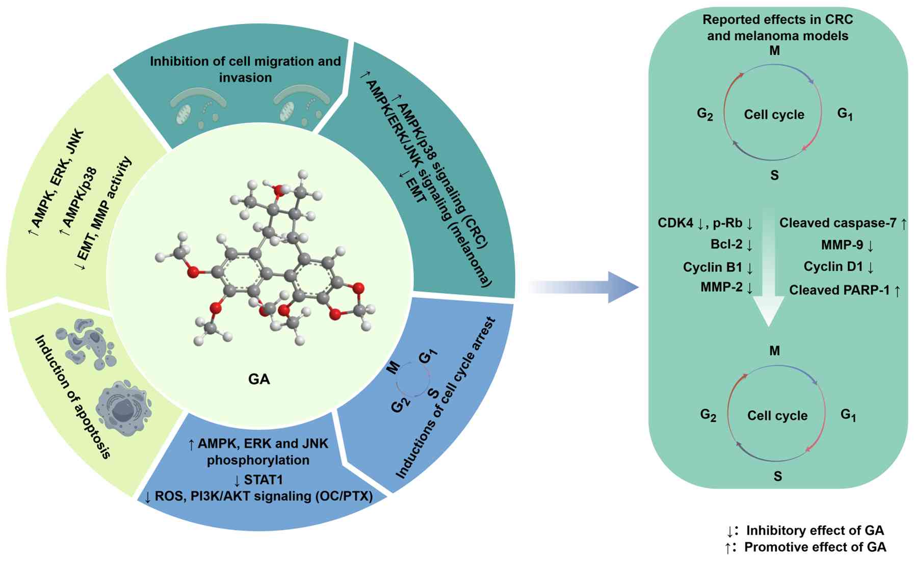
and the formation of neoplastic lesions (86). GA has notable anticancer effects
across a broad spectrum of malignancies, encompassing colorectal,
ovarian, breast and human prostate cancer, as well as metastatic
melanoma (8,13,23,87). Experimental evidence indicates
that the anticancer activity of GA involves multiple complementary
mechanisms, including the inhibition of cell proliferation,
induction of cell cycle arrest and cell death, suppression of cell
migration and invasion, and modulation of chemosensitivity and drug
resistance (Figs. 7 and 8) (23,49).
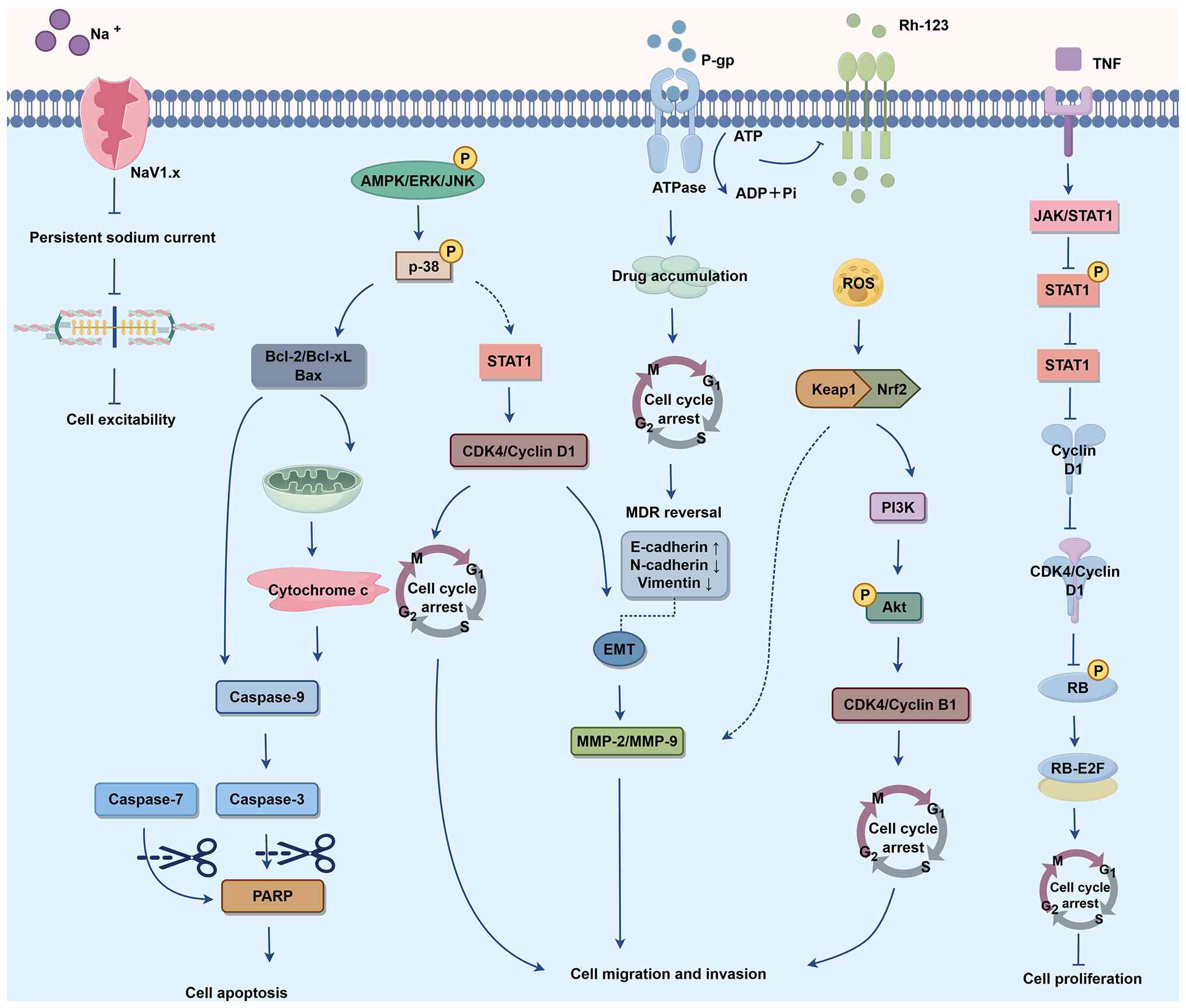 | Figure 8Major cellular pathways implicated in
the anticancer activity of gomisin A. NaV1.x, voltage-gated sodium
channel family 1; P-gp, P-glycoprotein; Rh, rhodamine; Pi,
inorganic phosphate; MDR, multidrug resistance; EMT,
epithelial-mesenchymal transition; ROS, reactive oxygen species;
RB-E2F, retinoblastoma protein-E2F transcription factor pathway;
AMPK, AMP-activated protein kinase; P, phosphorylation; PARP, poly
(ADP-ribose) polymerase; Keap1, Kelch-like ECH-associated protein
1. |
Inhibition of cell proliferation
Cancer cells are not controlled by the normal
regulatory system. Abnormal growth and proliferation are notable
characteristics of cancer (88),
making the inhibition of tumor cell proliferation a potential
strategy for cancer treatment. Induction of cell cycle arrest and
promotion of apoptosis effectively limit the proliferation of
cancer cells (8,23).
Induction of cell cycle arrest
Cell cycle arrest can directly inhibit cellular
proliferation, rendering cell cycle regulation pivotal in
preventing cancerous development (89). The cell cycle, comprising
G0/G1, S, G2 and M phases, is key
for cell viability and is regulated by a series of molecular
mechanisms. These cell cycle stages are controlled by cyclins, CDKs
and CDK inhibitors (90). GA
consistently induces G0/G1 phase arrest in
CRC and melanoma cells. The central effector mechanism is
downregulation of the CCND1-CDK4 complex, which reduces
retinoblastoma protein phosphorylation and blocks progression
through the G1/S restriction point. This phenotype has
been documented in both colorectal cancer (CRC) (8) and melanoma cells (23).
Two upstream signaling pathways converge on this
key cell cycle effect. First, GA suppresses STAT1 expression, which
in turn reduces CCND1 transcription and reinforces G1
arrest (91). Second, it
activates AMP-activated protein kinase (AMPK) and MAPK signaling,
increasing the phosphorylation of AMPK, ERK and JNK in melanoma
cells, leading to dose-dependent decreases in CCND1 and CDK4 levels
at both the mRNA and protein levels (23). In ovarian cancer (OC) cells, GA
serves primarily as a paclitaxel (PTX) sensitizer. By reducing
intracellular reactive oxygen species (ROS) levels, GA enhances the
antiproliferative and cell cycle-arresting effects of PTX,
accompanied by decreased expression of CDK4 and cyclin B1. This
combination improved antitumor efficacy in both cell-based assays
and an OC mouse model (13). The
effect is somewhat counterintuitive as reduced ROS levels would
typically be expected to attenuate chemotherapeutic toxicity, and
likely reflects the selective suppression of pro-survival ROS
signaling rather than global antioxidant activity (89).
In summary, GA drives G0/G1
arrest in CRC and melanoma via STAT1- and AMPK/MAPK-mediated
downregulation of CCND1-CDK4 expression, whereas in OC, it
primarily serves as a PTX sensitizer acting through ROS and
PI3K/AKT signaling.
Induction of cell death
Apoptosis is a regulated form of programmed cell
death. Apoptosis is necessary for successful organogenesis during
embryonic development and for maintaining normal tissue homeostasis
(92). Insufficient apoptosis
may contribute to uncontrolled cell proliferation, including in
cancer (92). Inducing apoptosis
is a major mechanism of numerous anticancer agents such as PTX
(90). GA is reported to promote
apoptosis primarily through mitochondria-dependent mechanisms, with
an additional calcium-dependent cytotoxic mechanism identified in
prostate cancer cells (8,87).
GA shifts the BCL-2 family balance toward
mitochondrial outer membrane permeabilization. GA upregulates
expression of the pro-apoptotic protein BAX, while downregulating
that of anti-apoptotic BCL-2 and BCL-xL, leading to the activation
of caspase-9, caspase-3 and PARP cleavage in CRC cells (8). Cleavage of caspase-7, as an
additional executioner caspase, has also been documented in HCT-116
colon cancer cells (93).
AMPK and MAPK activation are implicated upstream of
both canonical pathways. In CRC cells, GA increases AMPK and p38
phosphorylation, which feeds into the mitochondrial apoptosis
cascade (8). In melanoma cells,
GA activates AMPK, ERK and JNK, all converging on caspase-3 and
PARP cleavage (23). An
additional calcium-dependent cytotoxic mechanism has been reported
in prostate cancer cells. GA induces Ca2+ influx via
protein kinase C-dependent and store-operated pathways, together
with phospholipase C-mediated Ca2+ release from the ER.
This calcium mobilization promotes cell death independently of the
canonical caspase pathways (87).
In conclusion, GA promotes cancer cell death
through at least two experimentally reported mechanisms:
Mitochondria/caspase-associated apoptosis in CRC and melanoma
models and a calcium-dependent cytotoxic mechanism in prostate
cancer cells.
Inhibition of cell migration and
invasion
Cancer cell invasion involves migration through
tissue barriers and extracellular matrix (94). Therefore, inhibiting the
migration and invasion of tumor cells serves a significant role in
suppressing tumor progression. GA suppresses migration and invasion
in both CRC and melanoma cells, primarily through inhibition of
epithelial-mesenchymal transition (EMT). GA reverses the EMT marker
profile by upregulating E-cadherin expression while downregulating
that of N-cadherin, vimentin, snail and twist, and it decreases MMP
activity (8,23). In summary, EMT reversal and MMP
inhibition account for the reduced migratory and invasive capacity
observed across GA-sensitive cancer cell lines.
Chemosensitization and resistance
modulation
Beyond direct cytotoxicity, GA enhances the
activity of conventional chemotherapeutics through several distinct
mechanisms. GA reverses multidrug resistance (MDR) via the
non-competitive inhibition of two major ATP-binding cassette
transporters. GA alters substrate interactions with P-glycoprotein
(P-gp) and induces conformational changes that impair transport
function, restoring chemosensitivity in HepG2-DR cells (49). GA also inhibits breast cancer
resistance protein (BCRP)-mediated transport in a non-competitive
manner, thus broadening its MDR-reversal spectrum (95).
Beyond efflux pumps, GA inhibits drug-metabolizing
enzymes, such as CYP3A4 and CYP2C8, which increases systemic
exposure to co-administered chemotherapeutics. For PTX, the dual
inhibition of intestinal P-gp and CYP3A levels notably improves
oral bioavailability by reducing both efflux and first-pass
metabolism (96). Similarly,
evidence from Schisandra-derived preparations suggests
lignan-mediated modulation of CYP3A4 may increase bosutinib
exposure, although the specific contribution of purified GA has not
been established (97). In
summary, GA enhances chemotherapeutic efficacy through
transporter-mediated MDR reversal and metabolic enzyme inhibition
that boosts drug bioavailability.
Critical assessment and translational
considerations
The aforementioned anticancer phenotypes are well
established at the cellular level, including in CRC, melanoma, OC
and prostate cancer (8,13,23,87). For anticancer activity, the core
bottleneck is that the concentrations needed for efficacy in cell
culture are higher than what oral dosing delivers in circulation.
This concentration dependence follows a consistent pattern. At the
high range tested in vitro (25-100 μM), GA acts as a
direct cytotoxic agent, suppressing viability, driving
G0/G1 arrest and triggering apoptosis. At
lower concentrations (5-20 μM), the effects shift toward
targeting metastatic phenotypes, reversing EMT, dampening MMP
activity, and reducing migration and invasion. Corresponding in
vivo doses range from 2 to 50 mg/kg, with CRC lung metastasis
studies typically using 50 mg/kg orally or intraperitoneally
(8,23). Oral GA exposure varies with
species, dose and preparation; human extract data may not predict
the exposure achieved by purified GA in rodents (18,98). Low rat oral bioavailability has
been reported (~20.8% in one study), but the relative contributions
of intestinal absorption and presystemic metabolism remain
incompletely resolved (99,100). Dose-matched measurements of
unbound plasma and tumor GA are therefore required to quantify the
exposure gap.
Exposure cannot be inferred from dose alone. Human
extract data and certain rodent preparation studies report low
plasma GA concentrations, whereas purified-compound and extract
exposures vary substantially by formulation (18,98). Effective cell concentrations
should therefore be compared with measured unbound plasma and tumor
concentrations in the same dosing regimen, rather than assuming a
universal three-order-of-magnitude gap.
Unless GA concentrates selectively in tumor tissue,
active metabolites accumulate or formulation strategies drive local
high exposure, the G0/G1 arrest and apoptosis
phenotypes observed in vitro are better treated as
mechanistic readouts and screening tools than as effects readily
reproducible in vivo by conventional oral dosing and do not
directly support oral GA as a clinical anticancer agent. The MDR
reversal and chemosensitization activity, by contrast, translate
more plausibly. Intestinal transporter and enzyme inhibition may
alter co-administered drug exposure, but the required local GA
concentrations need to be measured (96,101). Framing GA as a chemosensitizer
or bioavailability enhancer therefore fits its PK profile better
than positioning it as a standalone cytotoxic drug. Current in
vivo cancer studies of GA combinations remain restricted mainly
to CRC/metastasis and OC models (8,13), and additional animal studies are
needed to determine whether these effects extend beyond the
currently studied settings.
Mechanistic insights from network
pharmacology
The present network pharmacology and molecular
docking analyses suggested the anticancer effects of GA involved
multiple receptor-associated, intracellular signaling and
transcriptional targets. The experimental phenotypes used for this
comparison derive from CRC, melanoma and OC studies (8,13,23). These computational findings
should be viewed primarily as a framework for generating testable
hypotheses rather than as evidence of direct target inhibition.
Among the predicted membrane-associated targets,
EGFR and ERBB2 are docking candidates. GA is predicted to adopt
putative ATP-pocket poses in the present docking analysis; these
predictions do not establish ATP-competitive inhibition (102). Potential modulation of ERBB
signaling may influence pathways involved in cell proliferation,
survival and EMT (103).
However, the reported effects of GA on G0/G1 arrest, CCND1-CDK4
downregulation and EMT inhibition have not been experimentally
attributed to EGFR or ERBB2 inhibition (8,23).
PIK3CA, AKT1 and mTOR have been identified as
candidate targets, with docking scores of −6.5, −6.6 and −6.9
kcal/mol, respectively. These predictions suggest GA interacts with
components of the PI3K/AKT/mTOR pathway. In OC cells, GA enhances
PTX sensitivity while decreasing intracellular ROS levels and
decreasing PI3K/AKT-related signaling (13). However, current evidence does not
establish direct binding between GA and AKT1 or other components of
this pathway.
MAPK14/p38α is a docking candidate. Experimental
studies support the involvement of MAPK signaling in GA-associated
cancer-cell apoptosis and growth inhibition, as GA increases AMPK
and p38 phosphorylation in CRC cells, and activates AMPK, ERK and
JNK signaling in melanoma cells (8,23). These findings contradict uniform
MAPK inhibition and suggest the context-dependent modulation of
MAPK signaling.
STAT3 is another candidate target, with a docking
score of −7.0 kcal/mol, and a potential STAT3 interaction region
was suggested by the docking pose. Although modulation of STAT3 may
affect proliferation- and survival-associated transcription, STAT1
expression is decreased during GA- and TNF-α-induced G1
arrest in HeLa cells (91).
Thus, the data support the potential involvement of STAT family
signaling but do not establish STAT3 as a direct molecular target
of GA.
Outside this receptor-kinase-transcription factor
network, inhibition of drug transporters is a more directly
supported and mechanistically independent anticancer mechanism
(49,95). GA non-competitively inhibits P-gp
and BCRP-mediated drug efflux (49,95) and increases PTX exposure through
combined effects on intestinal P-gp and CYP3A (96); evidence from
Schisandra-derived preparations suggests a potential
CYP3A4-mediated interaction with bosutinib, although a GA-specific
contribution has not been established (97). Thus, the anticancer profile of GA
comprises a predicted three-tiered receptor-kinase-transcription
factor cascade (ERBB/PI3K/MAPK/STAT signaling network) and a
separately validated transporter- and metabolism-based
chemosensitization mechanism (49,95,96). The present MD simulations
assessed the structural behavior of selected predicted complexes;
they do not establish modulation of downstream signaling.
For EGFR, STAT3 and mTOR, the present computational
analyses provide tier D evidence; cancer studies provide tier C
support for downstream pathway changes (8,13,23). Network pharmacology and molecular
docking analyses prioritize these as core targets, placing them at
tier D. Downstream experimental observations, including CCND1-CDK4
downregulation, BCL-2 family shifts and caspase activation, are
directionally consistent with receptor-kinase-transcription factor
cascade modulation and provide tier C associative support (8,23). However, EGFR remains a candidate
target rather than an experimentally validated target of GA because
direct kinase inhibition, genetic dependency, and cellular
target-engagement studies have not been demonstrated.
In summary, GA exerts a variety of anticancer
effects and may become a potential antineoplastic drug. Despite
this, the studies summarized here remain preclinical (8,13,23), and clinical trials are needed to
verify its safety and effectiveness.
Effects on CVDs: Early but
mechanistically diverse evidence
CVD caused an estimated 19.8 million deaths
worldwide in 2022, ~32% of all deaths, imposing a substantial
public health and economic burden (104,105). The CVD literature on GA spans
several experimental levels, including cultured vascular cells,
isolated vessel preparations, a mouse hypertension model and
multicomponent herbal mixtures (9,14,24,25,106). These models address different
layers of biological organization and do not provide equivalent
evidence for clinical cardiovascular efficacy.
In angiotensin II-infused mice, continuous GA
infusion at 2 or 10 μg/kg/min attenuates blood pressure
elevation, preserves plasma and vascular NO indices, restores
endothelial NOS (eNOS) phosphorylation, and decreases vascular ROS
generation (9). This in
vivo blood pressure phenotype, paired with concordant vascular
measurements, provides evidence for an NO/oxidative stress
mechanism. A notable limitation is that the aforementioned study
used continuous parenteral infusion rather than oral
administration, bypassing absorption and first-pass metabolism;
oral efficacy at physiologically relevant exposures therefore
remains unconfirmed.
At the cell and isolated vessel level, the most
fully characterized mechanistic effect is vasorelaxation, which
operates through both endothelium-dependent and -independent
pathways. Endothelium-dependent relaxation is driven by
Ca2+-mediated eNOS activation and translocation to the
cytoplasm, leading to increased NO production (14), with partial contribution from
myosin light chain dephosphorylation (24). In endothelium-denuded vessels, GA
acts via the RhoA/Rho-associated coiled-coil-containing protein
kinase pathway to decrease vascular contractility (25).
GA suppresses both mRNA and protein expression of
PCSK9 (55). Because PCSK9
typically reduces LDL receptor density and impairs LDL-cholesterol
(LDL-C) clearance, decreased PCSK9 expression provides a hypothesis
for improving LDL-C clearance, but does not demonstrate LDL-C
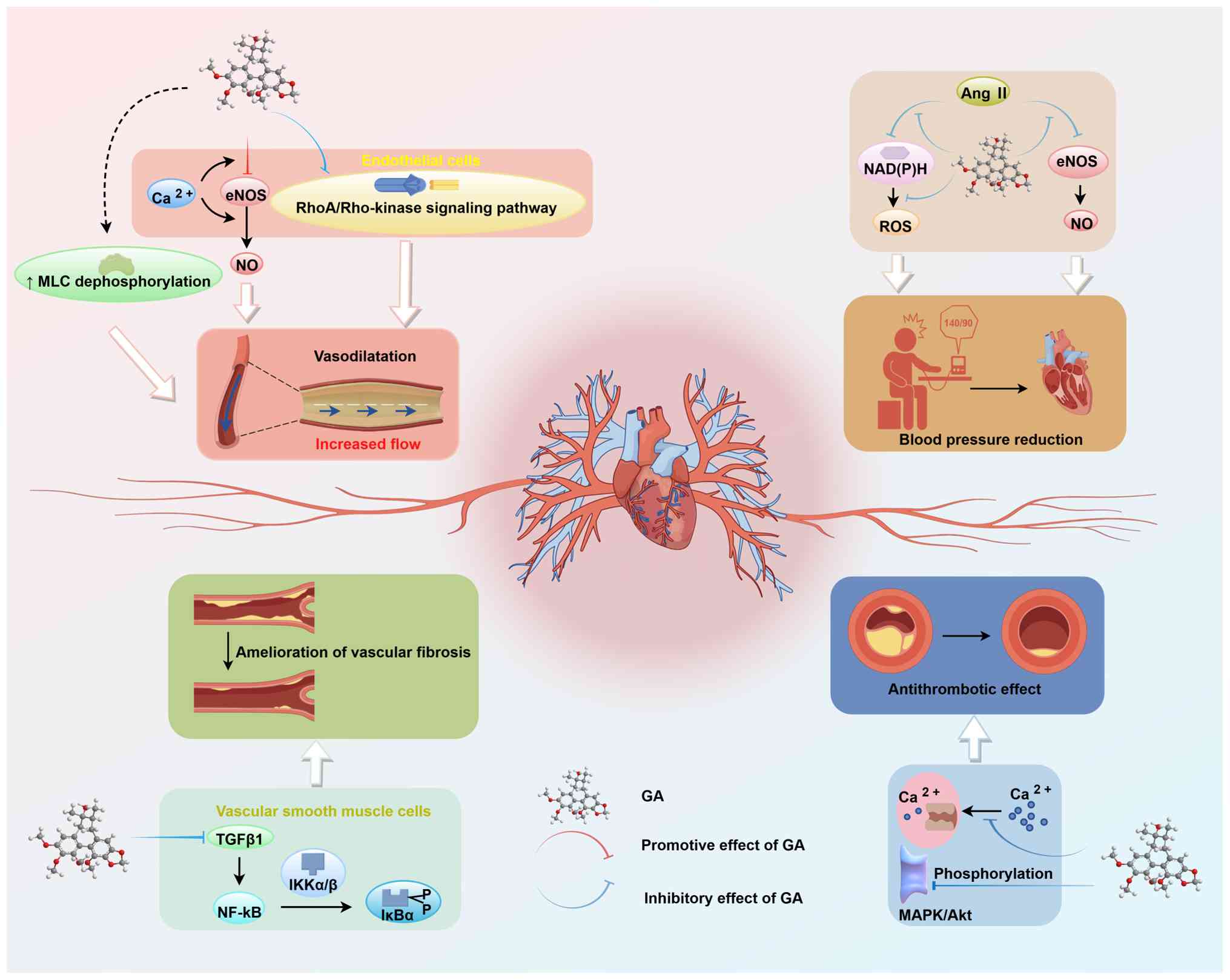
lowering by GA in vivo (55). For vascular fibrosis, GA inhibits
TGF-β1-driven fibrogenic signaling in vascular smooth muscle cells
(VSMCs) through a Smad-independent mechanism. GA suppresses
inhibitor of NF-κB kinase α/β activation and IκBα phosphorylation,
thereby inhibiting the NF-κB component of the TGF-β1 response that
contributes to vascular fibrotic disease (83). Antithrombotic effects have been
reported for a S. chinensis + Morus alba L. mixture
in which GA serves as a marker compound. The mixture decreases
platelet activation and thrombus formation, associated with the
decreased phosphorylation of MAPK (ERK and JNK) and AKT in platelet
signaling pathways (106).
However, whether this activity is attributable to GA alone or
reflects additive or synergistic effects with other components
remains unresolved.
In summary, GA shows promising and mechanistically
diverse cardiovascular activity in rodent and cell-based models
(9,14,24,25); the evidence base remains early,
however, with most data from small animal and in vitro
studies and no large-animal or clinical validation. The
antithrombotic data rely on mixture formulations rather than on
purified GA, which limits mechanistic interpretation (106). The schematic also includes
PCSK9 expression in cultured cells (55) and TGF-β1-associated vascular
fibrotic signaling (83).
Further studies should establish dose-response associations,
confirm target engagement and assess translational potential
(Fig. 9).
Critical assessment and translational
considerations
The cardiovascular evidence includes continuous GA
infusion in mice and experiments in vascular cells and isolated rat
vessels (9,14,24,25). The aforementioned studies did not
measure unbound GA concentrations at the vascular site of action
alongside efficacy; therefore, it remains unclear whether the
effective concentrations can be achieved by oral dosing. The
continuous-infusion hypertension experiment also bypassed oral
absorption and first-pass metabolism (9). Pharmacological inhibitor
experiments support an NO/eNOS contribution to vasorelaxation
(14,24), but direct GA target engagement
and durable oral efficacy remain unresolved. CYP3A and P-gp
inhibition create a potential for interactions with co-administered
drugs that requires exposure-based evaluation (49,62).
Mechanistic insights from network
pharmacology
The present network pharmacology and molecular
docking analyses provided hypotheses for experimental observations
concerning endothelial function (9,14), VSMC fibrotic responses (83), PCSK9 expression (55) and platelet activation by a
multicomponent preparation (106). Among the predicted targets,
PIK3CA, AKT1, GSK3β and MAPK14 have emerged as major candidate
nodes. AKT1 and GSK3β form a functionally connected regulatory
module, with predicted docking scores of −6.6 and −7.4 kcal/mol,
respectively. AKT is an established upstream regulator of GSK3β and
suppresses its kinase activity via phosphorylation at Ser9
(107). However, these docking
results indicate only a potential interaction with GA and do not
establish whether either kinase is activated or inhibited.
AKT1 is also a well-characterized upstream kinase
of eNOS. Activation of the AKT1/eNOS pathway promotes
phosphorylation of the activating Ser1177-equivalent residue and
enhances NO production, providing a plausible mechanistic basis for
the endothelium-dependent vasorelaxation observed in experimental
models (14,24,108). eNOS phosphorylation is not
exclusively dependent on PI3K/AKT signaling, as AMPK-mediated
phosphorylation at Ser1177 has also been reported in response to
thrombin and histamine (109).
GSK3β is implicated in the regulation of VSMC
proliferation, migration and phenotypic remodeling, although the
direction of its effects may be context-dependent. For example,
increased GSK3β expression is associated with decreased angiotensin
II-induced VSMC proliferation and migration; circular UBE4B RNA
sequesters microRNA-199a-5p, thereby increasing GSK3β expression
(110). These findings support
a role for GSK3β-associated signaling in vascular remodeling but do
not confirm that inhibition of GSK3β is responsible for the
anti-fibrotic effects attributed to GA. Similarly, GSK3β-associated
signaling is associated with PCSK9-induced metabolic injury, but
current evidence does not identify GSK3β as an upstream regulator
of PCSK9 expression (111).
Therefore, the contribution of this pathway to decreased PCSK9 and
LDL-C levels remains speculative (111).
MAPK14, which encodes p38α, exhibits a predicted
affinity score of −6.8 kcal/mol and may represent a parallel node
linking vascular inflammation, fibrotic signaling and platelet
activation. This prediction is consistent with the involvement of
p38α in platelet-mediated inflammatory and thrombotic responses and
with reports that decreased MAPK- and AKT-associated
phosphorylation accompanies decreased platelet activation and
thrombus formation (106,112). The aforementioned
antithrombotic evidence was obtained using a multicomponent S.
chinensis-Morus alba preparation rather than purified GA
and (106) did not directly
demonstrate inhibition of MAPK14 by GA.
PIK3CA, with a predicted binding affinity of −6.5
kcal/mol, is an upstream component of the canonical PI3K/AKT/eNOS
pathway. The inclusion of PIK3CA in the predicted target network
supports the potential involvement of this signaling pathway,
although the direction and functional consequences of the
interaction remain to be experimentally determined. Taken together,
the predicted target profile is broadly compatible with the
antihypertensive, lipid-lowering, anti-fibrotic and vasorelaxant
effects reported in preclinical studies (9,14,24,25,55,83). However, these associations should
be regarded as hypothesis-generating; direct target-engagement
assay, kinase activity measurements, pathway-specific intervention
studies and validation using purified GA are required to establish
causal associations.
Overall, the cardiovascular evidence supports
vasorelaxant and antihypertensive pharmacology in preclinical
systems, with moderate phenotypical and low-to-moderate
target-level confidence. However, clinical efficacy has not yet
been evaluated in controlled human studies.
Neurological activity: Exploratory
evidence and unresolved brain exposure
GA exerts a range of neuropharmacological effects
in preclinical models, spanning anti-neuroinflammatory,
cognition-enhancing, anxiolytic, sedative-hypnotic and analgesic
activity (6,10,15,19,113). A notable PK advantage is that
GA penetrates the blood-brain barrier (BBB) (18,114,115), establishing feasibility for
central nervous system (CNS) targeting.
Reported central effects include
anti-neuroinflammatory and neuroprotective activity (10,116). In LPS-activated microglial
cells, GA suppresses NF-κB and MAPK signaling, decreasing
pro-inflammatory output (10).
In a 3-nitropropionic acid model of Huntington's disease, S.
chinensis stem extract protected against striatal toxicity
through combined antioxidant and anti-inflammatory actions
(117). A separate study
examined Schisandra lignans, including GA, in
serum/glucose-deprived SH-SY5Y cells; this is an in vitro
deprivation model, not an in vivo ischemic-stroke experiment
(116).
In terms of cognitive function, GA inhibits
acetylcholinesterase (AChE) activity in a dose-dependent manner
in vitro (45,118) and reverses scopolamine-induced
memory impairment in vivo (15). These findings align with
enhancement of cholinergic neurotransmission, a classical
symptomatic strategy for cognitive impairment (15). Mood- and sleep-associated effects
have also been reported, although largely from multi-component
herbal preparations (6,114,115,119). In the Suanzaoren-Wuweizi
herb-pair, GA has been identified as a putative active constituent
modulating serotonin and dopamine signaling to mediate anxiolytic
effects (6).
Antidepressant-associated observations of S. chinensis
preparations do not establish an antidepressant mechanism of
purified GA (119).
Sedative-hypnotic activity is hypothesized to involve binding to
neurotransmitter receptors in the hypothalamus following BBB
penetration (114,115). To the best of our knowledge,
whether purified GA alone reproduces these mood-associated effects
has not been tested. On the analgesic side, GA inhibits
capsaicin-induced transient receptor potential vanilloid 1 (TRPV1)
channel currents in a concentration-dependent, reversible and
non-competitive manner, independent of voltage. This action may
alleviate pain hypersensitivity and neuropathic pain driven by
excessive TRPV1 activation (113).
Critical assessment and translational
considerations
The key translational bottleneck for
neuroprotective applications lies in uncertainty regarding target
exposure within the brain. Brain homogenate and microdialysis
studies have measured GA-related brain exposure after Schisandra
preparations (18,115). However, unbound brain exposure
matched to the efficacy regimen and target potency remains
insufficiently defined. If brain exposure is in the same order of
magnitude as plasma levels, a notable gap would separate achievable
concentrations from the micromolar levels required to inhibit
kinases such as GSK3β and STAT3, calling into question the
physiological relevance of kinase-mediated mechanisms. A second key
limitation is the mismatch between model duration and clinical
disease course. The majority of studies employ acute injury models
with short-term treatment regimens (15,116,117), which differ substantially from
the progressive, chronic pathology of neurodegenerative conditions
such as Alzheimer's and Huntington's disease. Efficacy in acute
settings does not guarantee that long-term intervention would slow
disease progression.
Among neuropharmacological effects, AChE is
supported by a direct enzyme assay (118). To the best of our knowledge,
its in vitro inhibitory concentration has not been matched
with unbound brain GA exposure, therefore superior translational
feasibility over predicted kinase mechanisms cannot yet be
established. Because unbound brain drug concentrations remain
undefined, findings across Alzheimer's disease, Huntington's
disease, depression, anxiety, insomnia and neuropathic pain should
be framed as exploratory preclinical observations rather than
evidence of broad neuropsychiatric therapeutic potential. Future
studies should establish quantitative brain exposure measurements
via microdialysis or brain homogenate assays to provide a firmer
basis for evaluating the druggability of each proposed
mechanism.
Mechanistic insights from network
pharmacology
Network pharmacology analysis has identified GSK3β
as a prominent candidate hub within the putative
neuropharmacological target network of GA in the present analysis.
GSK3β is biologically relevant to processes implicated in
neurological disease, including tau hyperphosphorylation, amyloid-β
(Aβ) accumulation and microglia-associated neuroinflammation
(30-32). Modulation of the PI3K/AKT/GSK3β
pathway is associated with decreased tau hyperphosphorylation in
experimental models of Alzheimer's disease (30), decreased accumulation of Aβ and
pyroglutamate-modified Aβ (31),
and attenuation of microglia-mediated inflammation and neuronal
apoptosis following ischemic stroke (32). Although these studies did not
examine GA directly, they support the biological relevance of
GSK3β-associated signaling to the memory-enhancing (15) and neuroprotective (116) effects reported for GA in
experimental models (15,116).
The predicted GA-GSK3β complex was evaluated using
a 100-nsec MD simulation. The protein backbone root-mean-square
deviation (RMSD) profile supported the relative conformational
stability of the simulated complex (Fig. 5A). However, neither molecular
docking nor MD analysis establishes direct target engagement or
determines whether GA activates or inhibits GSK3β. Biochemical
kinase assays and cell target engagement studies are required
before GSK3β can be regarded as a functionally validated target of
GA.
PIK3CA and AKT1, which are upstream of GSK3β in the
canonical PI3K/AKT signaling pathway, were identified as docking
candidates. Activated AKT inhibits GSK3β through phosphorylation at
Ser9, thereby promoting neuronal survival under several forms of
cell stress (33). This
signaling association provides a plausible framework for
interpreting the anti-apoptotic effects reported for GA in
serum/glucose-deprived neuronal cells and memory-impairment
(15,116). Nevertheless, the predicted
interactions with PIK3CA, AKT1 and GSK3β do not establish the
direction of pathway regulation by GA, and activation of the
PI3K/AKT/GSK3β pathway remains to be demonstrated
experimentally.
STAT3 and MAPK14, also known as p38α, are
additional inflammation-associated candidate nodes, with predicted
binding affinities of −7.0 and −6.8 kcal/mol, respectively. Both
signaling proteins participate in the regulation of inflammatory
gene expression, cell stress responses and survival in neurons and
glial cells (10,120). Their inclusion in the predicted
network is compatible with the decreased NF-κB signaling and
pro-inflammatory cytokine production observed in LPS-stimulated
microglial models treated with GA (10,117). However, this does not
demonstrate direct inhibition of STAT3 or MAPK14, because decreased
inflammatory signaling may arise through upstream or indirect
mechanisms. TNF may be more appropriately regarded as a downstream
inflammatory mediator within this network, consistent with its
decreased levels in the microglial inflammatory response (10); accordingly, TNF should be
considered a candidate downstream mediator rather than a validated
direct target of GA, as direct binding and functional
neutralization studies are lacking.
Detection of GA in brain samples following
administration provides evidence of CNS exposure but does not
demonstrate that GA binds GSK3β, PIK3CA, AKT1, STAT3 or MAPK14 in
the brain (18,114,115). The strength of this conclusion
depends on whether the detected species is unchanged GA or a
metabolite, the integrity of the BBB in the experimental model and
whether the measured brain concentrations are sufficient to produce
pharmacological effects.
Taken together, the computational findings
identified GSK3β as a potential point of convergence between
PI3K/AKT-dependent survival signaling and pathological processes
involving tau phosphorylation, Aβ accumulation and
neuroinflammation. STAT3 and MAPK14 may constitute parallel
inflammation-associated components of the same predicted network.
These associations provide a mechanistic framework that is broadly
consistent with the reported effects of GA on cognition,
neuroinflammation, ischemic neuronal injury and mutant
huntingtin-associated toxicity (10,15,116,117). They should nevertheless be
considered hypothesis-generating until confirmed by direct binding
assays, measurements of kinase activity and phosphorylation,
pathway-specific pharmacological or genetic intervention and
studies conducted at pharmacologically relevant brain
concentrations.
GA exhibits a broad spectrum of
neuropharmacological activity in preclinical models. The evidence
base remains largely exploratory overall. Anti-neuroinflammatory
effects are supported by microglial experiments (10), while AChE inhibition and TRPV1
channel modulation rest on more direct functional assays (113,118). Anxiolytic and antidepressant
findings rely on mixture studies and require confirmation using the
purified compound (6,119). The neurological evidence
remains largely at the phenotype-association level, as
pathway-specific inhibition, genetic validation, and clinical
efficacy studies are currently lacking. Brain detection supports
CNS accessibility, but efficacy-matched unbound brain exposure
remains a translational gap (18,115). Future studies prioritizing
brain concentration measurement and target validation are essential
to determine the true therapeutic potential of GA in neurological
disorders (Fig. 10) (15,19,116,117).
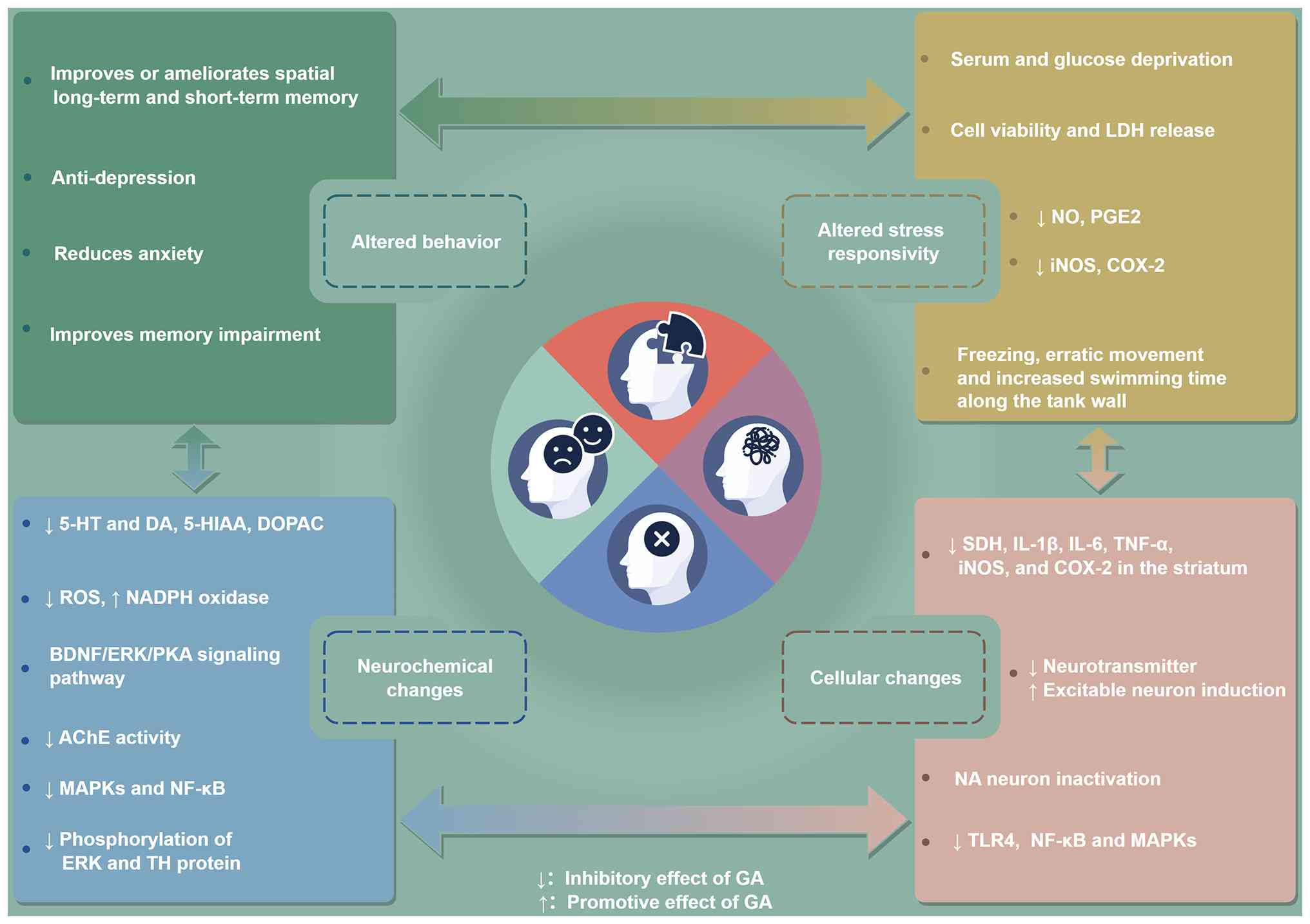 | Figure 10Reported neuropharmacological effects
and associated mechanisms of GA. GA, gomisin A; LDH, lactate
dehydrogenase; PGE, prostaglandin E; iNOS, inducible nitric oxide
synthase; COX, cyclooxygenase; SDH, succinate dehydrogenase; NA,
noradrenaline; TLR, toll-like receptor; 5-HT, 5-hydroxytryptamine;
DA, dopamine; 5-HIAA, 5-hydroxyindoleacetic acid; DOPAC,
3,4-dihydroxyphenylacetic acid; ROS, reactive oxygen species; BDNF,
brain-derived neurotrophic factor; AChE, acetylcholinesterase; PKA,
protein kinase A; TH, tyrosine hydroxylase. |
Anti-inflammatory and antioxidant
effects
GA exhibits both anti-inflammatory and antioxidant
activity in preclinical models, operating via partially overlapping
NF-κB and NRF2 pathways (10,16,17).
The anti-inflammatory effect centers on suppression
of receptor-interacting protein 2 (RIP2) and NF-κB activation,
which decreases the expression of downstream inflammatory
mediators, including COX-2, iNOS, IL-6, TNF-α and NO (16). In an asthma model, GA similarly
acts via NF-κB inhibition to reduce serum IgE levels, lower IL-4
and IL-5 concentrations in bronchoalveolar lavage fluid, and
ameliorate lung tissue pathology (66). In human diploid fibroblasts,
GA-associated changes in MAPK/NF-κB signaling accompanied reduced
cellular-senescence markers and improved mitochondrial biogenesis;
these findings concern cellular senescence, not demonstrated
slowing of organismal aging (121).
In terms of antioxidant activity, GA drives cell
defense primarily through NRF2 nuclear translocation, which
upregulates downstream cytoprotective genes, including HO-1. This
decreases ROS production and senescence-associated β-galactosidase
(SA-β-gal) activity, while enhancing autophagy and mitochondrial
biogenesis. In stress-induced premature senescence, these effects
improve mitochondrial function and modulate aging-related molecular
markers (121). Under
high-glucose conditions, GA upregulates HO-1 and preserves
mitochondrial homeostasis to support osteoblast differentiation,
with potential relevance for diabetic osteoporosis and implant
failure (122). Capillary
electrophoresis profiling has identified GA as one of the primary
antioxidant lignans in Schisandra extracts, consistent with
the established association between total lignan content and
overall antioxidant capacity (123,124).
Anti-inflammatory and antioxidant activities
represent the most common properties of GA across all therapeutic
indications. NF-κB suppression, decreased ROS levels and NRF2
activation have been observed in APAP liver injury, activated
macrophages and microglia (10,16,17). This broad activity profile
supports the pleiotropic character of GA, but also raises questions
about mechanistic specificity. It is unclear whether the
anti-inflammatory effect derives from selective inhibition of
specific kinases or from non-specific membrane effects and radical
scavenging common to lipophilic molecules tested at high
concentrations.
Critical assessment and translational
considerations
If GA operates through a polypharmacological mode
of partial multi-target inhibition rather than complete
single-target blockade, the overall effect threshold may fall below
the concentration required for full inhibition of any individual
kinase, which would leave a window for oral translational
feasibility. The converse is also true, however. A weak
multi-target mode of action would be difficult to verify through
single target knockdown approaches, increasing the technical
challenge of mechanistic studies. Anti-inflammatory research relies
on transcriptional and protein-level endpoint readouts, with little
direct kinase activity data available, making it difficult to
pinpoint the action of GA to specific nodes within signaling
cascades (10,16,66).
NF-κB suppression is supported by evidence at the
boundary between tiers B and C. NF-κB suppression is consistently
replicated across multiple disease models (7,10,16), with decreased p65 nuclear
translocation, attenuated IκBα degradation, and coordinated
decreases in downstream COX-2, iNOS, IL-6 and TNF-α levels. The
coherence across independent laboratories and injury models raises
confidence above simple pathway association. However, to the best
of our knowledge, p65 overexpression or pathway-rescue experiments
to establish strict causal dependency have not been reported.
Mechanistic insights from network
pharmacology
In the present molecular docking analysis, the
anti-inflammatory and antioxidant activity of GA may be interpreted
within two conceptually distinguishable but potentially
interconnected signaling modules: A MAPK14/NF-κB-associated
inflammatory module and a GSK3β/NRF2-associated antioxidant module.
These assignments provide a framework for integrating the
computational predictions with previously reported biological
effects but do not establish direct regulation of the proposed
targets by GA (10,16,17).
MAPK14, which encodes p38α, has emerged as a
prominent inflammation-associated candidate, with a predicted
binding affinity of −6.8 kcal/mol. A potential interaction between
GA and MAPK14 may provide a structural hypothesis for the recurrent
involvement of MAPK and NF-κB signaling in experimental studies
(10,16,66). GA suppresses RIP2/NF-κB
activation, and decreases the production or expression of COX-2,
iNOS, IL-6, TNF-α and NO (16).
In an experimental model of asthma, attenuation of NF-κB signaling
was accompanied by decreased IgE, IL-4 and IL-5 levels, and
improved pulmonary histopathology (66). GA also decreases
NF-κB/MAPK-associated inflammatory responses in activated microglia
(10,117), while modulation of MAPK and
NF-κB signaling is implicated in its anti-senescence activity
(121). Together, these
findings show pathway-level consistency with the MAPK14 prediction.
However, they do not demonstrate direct binding to MAPK14 or
inhibition of its kinase activity by GA.
TNF, with a predicted binding affinity of −6.1
kcal/mol, has also been identified within the
inflammation-associated target network. Although small molecules
can, in principle, modulate TNF activity by interacting with the
trimer interface and altering receptor-competent conformations, the
docking prediction alone is insufficient to establish a mechanism
for GA. TNF should be regarded primarily as a downstream
inflammatory mediator and a tentative direct binding candidate
rather than a validated molecular target. Direct interference with
TNF trimer stability, receptor binding or biological activity
remains to be examined experimentally (16,66).
STAT3 is a docking candidate that may constitute an
additional immunoregulatory component of the network. STAT3
participates in both pro-inflammatory and anti-inflammatory
signaling, depending on the upstream cytokine, cell type and
biological context (120).
Consequently, the functional role and direction of any interaction
between GA and STAT3 cannot be inferred from docking analysis
alone.
The antioxidant module is centered computationally
on GSK3β, which showed a predicted binding affinity of −7.4
kcal/mol. GSK3β is a recognized regulator of NRF2 stability and
nuclear localization, providing a biologically plausible connection
between the computational prediction and the antioxidant responses
reported for GA (125). These
responses include increased NRF2 nuclear accumulation and HO-1
expression, decreased ROS production and SA-β-gal activity and
improvements in autophagic activity and mitochondrial biogenesis
(17,121,122). Activation of the NRF2/ARE
pathway has also been observed in APAP-associated LI (17), whereas HO-1 induction and
preservation of mitochondrial homeostasis have been reported under
high-glucose conditions (122).
Nevertheless, these findings do not establish GSK3β as the direct
mediator of NRF2 activation by GA because the changes may arise
through Kelch-like ECH-associated protein 1-dependent regulation or
other oxidative stress-responsive pathways (126).
Analytical studies have associated GA with the
antioxidant activity of Schisandra-derived preparations
(123,124). However, the extent to which GA
independently accounts for the activity of multicomponent
preparations requires confirmation using purified compounds and
pharmacologically relevant concentrations.
Taken together, the predicted MAPK14/NF-κB- and
GSK3β/NRF2-associated modules provide a working model for
integrating the anti-inflammatory and antioxidant effects of GA.
The two modules are unlikely to operate in isolation given the
extensive crosstalk between inflammatory and redox-sensitive
signaling pathways (127).
Accordingly, the proposed network should be regarded as
hypothesis-generating. Direct kinase assays, biophysical target
engagement measurements, genetic loss-of-function approaches and
pathway rescue experiments will be necessary to distinguish primary
molecular interactions from downstream or compensatory signaling
changes.
Other pharmacological activities
GA exhibits a range of additional pharmacological
activities in preclinical models (26-28), although most remain at an early
exploratory stage. In terms of renal protection, GA modulates MAPK
signaling and apoptosis pathways to reduce pathological kidney
damage (26). GA increases
phosphorylation of p38 and JNK, while decreasing ERK
phosphorylation, and lowers active caspase-3 levels, while shifting
the BCL-2/BAX balance toward cell survival (26). In drug metabolism and PK
interactions, GA acts on two key pathways. GA induces PXR gene
expression, which upregulates renal CYP3A30 and alters the
expression profile of drug-metabolizing enzymes, with implications
for drug-drug interactions and tolerance development (12,128). Separately, GA inhibits
P-gp-mediated efflux, which increases the blood concentration, area
under the curve (AUC) and oral bioavailability of the
immunosuppressant tacrolimus in a dose-dependent manner. This P-gp
inhibition raises the potential of using GA as a bioavailability
enhancer to decrease tacrolimus dosing and associated costs
(101).
Anti-obesity activity has been documented in both
animal and cells (27). GA
decreases weight gain, epididymal white adipose tissue mass and
liver weight, while increasing serum high-density
lipoprotein-cholesterol levels (27). GA inhibits adipogenesis across
multiple cell types and promotes fat browning through AMPK
activation, shifting the balance between adipose tissue browning
and whitening to suppress weight gain (27). An extract of S. chinensis
fruits acts against Vibrio parahaemolyticus by disrupting
cell membrane integrity, suppressing polysaccharide intercellular
adhesin synthesis, decreasing extracellular DNA secretion and
lowering biofilm metabolic activity. The extract inhibits biofilm
formation and disrupts established biofilms but does not establish
GA as the responsible antimicrobial constituent (129). For erectile function, GA
relaxes penile corpus cavernosum smooth muscle through both NO-cGMP
and cAMP signaling pathways. GA also enhances sildenafil-induced
relaxation, which may offer a supplemental option for patients who
respond incompletely to standard erectile dysfunction treatment
(28,130). Immunomodulatory effects have
been observed in THP-1 monocytic leukemia cells, where GA induces
release of IL-8, macrophage inflammatory protein (MIP)-1β and
granulocyte-macrophage colony-stimulating factor (GM-CSF),
suggesting modulation of innate immune responses (29). Finally, an electrophysiological
study shows that GA inhibits sodium currents in pituitary GH3 and
pancreatic INS-1 cells, affecting both peak amplitude and current
sustainability. This provides a preliminary electrophysiological
basis for potential applications in hyperprolactinemia and
prolactinoma (131).
Critical assessment and translational
considerations
Studies across the aforementioned diverse endpoints
typically remain at a single-model, single-laboratory, small-sample
exploratory stage with overall low evidence strength (26-29,131). The most robust finding is
combined P-gp/CYP3A-mediated enhancement of tacrolimus
bioavailability. This effect is supported by direct transporter
functional assays and rat PK data (101), but clinical dosing utility and
safety of purified GA have not been established. Anti-obesity
effects have been observed at both cell and animal levels, but the
causal chain between AMPK activation and fat browning has not been
confirmed through inhibitor or knockdown experiments. Renal
protection data remain at the level of pathway biomarkers and
histopathology, lacking long-term follow-up of gold standard renal
function endpoints such as creatinine clearance and urinary
protein. To the best of our knowledge, erectile function
improvement has been validated in isolated tissue strip
experiments, with no in vivo functional data available.
Antibacterial and immunomodulatory studies are preliminary, with
both reproducibility and specificity yet to be established.
Mechanistic insights from network
pharmacology
Network pharmacology may also place less
extensively studied pharmacological effects of GA within a broader
mechanistic context. In relation to its anti-obesity activity,
GSK3β and AKT1 were identified as potential components of a
metabolism-associated target network. This prediction is broadly
consistent with the report that GA suppresses adipogenesis and
promotes adipose tissue browning in association with activation of
AMPK signaling (27). However,
to the best of our knowledge, the evidence does not establish a
continuous AKT1/GSK3β/AMPK signaling pathway, nor has it been
determined whether pharmacological inhibition or genetic depletion
of GSK3β or AKT1 modifies the metabolic response to GA. The
association between these predicted targets and AMPK-associated
adipose browning therefore remains to be clarified.
The reported renoprotective effects of GA also show
potential pathway-level relevance to the predicted MAPK14 node
(26). Hwang et al
(26) described differential
regulation of MAPK signaling, involving changes in p38, JNK and ERK
activity (26). Because these
kinases exert distinct and context-dependent effects on tubular
cell stress, apoptosis, inflammation and repair, the pattern cannot
be attributed solely to direct regulation of MAPK14 (26). The phosphorylation measurements
reported by Hwang et al (26) do not distinguish direct action on
p38 from an indirect response to altered cell stress. Direct
engagement and functional modulation of MAPK14 therefore remain
unconfirmed. A separate diabetic rat study reported modest renal
improvement with GA alone and greater improvement with GA plus
metformin, associated with AGE/RAGE and NF-κB modulation; this does
not independently establish a GA-MAPK14 mechanism (132).
For erectile function, the predicted PIK3CA and
AKT1 nodes provide a potential upstream link to NO-dependent
signaling and cavernosal smooth muscle relaxation. These findings
are compatible with modulation of endothelial and cyclic
nucleotide-dependent smooth muscle pathways. However, the
contribution of PIK3CA, AKT1 or eNOS signaling to GA-mediated
erectile effects has not been experimentally established, and
additional mechanisms acting directly within cavernosal smooth
muscle cannot be excluded.
The immunomodulatory findings require a different
interpretation. MAPK14 and STAT3 are candidate signaling nodes that
may influence cytokine production and monocyte activation (29). Because these mediators are
upregulated rather than uniformly suppressed, this response should
not be interpreted as evidence of inhibition of MAPK14 or STAT3.
Instead, it may reflect a context-dependent immunostimulatory or
cell activation phenotype. The docking results indicate potential
in monocyte activation and cytokine regulation, but neither the
direction of their regulation nor the broader immunological
consequences have been established at present. Taken together,
these findings extend the predicted target network of GA to
metabolic regulation, renal stress response, cavernosal relaxation
and immune cell signaling. Nevertheless, the mechanistic links
remain largely inferential. Target-specific inhibition or
knockdown, phosphorylation and activity measurements, and
pathway-rescue experiments will be required to determine whether
the predicted nodes are necessary for the corresponding
pharmacological effects.
GA exerts a range of pharmacological activities,
covering renal protection, drug metabolism regulation,
anti-obesity, antibacterial, immunomodulatory and endocrine
modulatory effects, as well as improvements in erectile function
(26-29,101,131). These pharmacological findings
enrich the overall pharmacological profile of GA, yet they are
limited in depth and reliability to support clinical translation.
Studies are confined to exploratory preclinical research (26-29,131), with uneven mechanistic
exploration and inadequate in vivo experimental
verification. The present network analysis prioritized candidate
proteins that may be relevant to these phenotypes; it does not
demonstrate that these proteins mediate the reported effects.
Nevertheless, such associations are primarily hypothetical.
Experimental evidence primarily validates the participation of
signaling pathways, rather than the direct interaction of GA with
key targets.
GA is a dibenzocyclooctadiene lignan from S.
chinensis with a broad spectrum of preclinical pharmacological
activity. Its best-supported indication is hepatoprotection, with
consistent efficacy across chemical, cholestatic and fibrotic LI
models. Anticancer activity is well-documented at the cell level
but faces a significant in vitro-in vivo
concentration gap, positioning GA more plausibly as a
chemosensitizer than a single-agent cytotoxic therapy.
Cardiovascular and neurological effects show promising phenotypic
signals but require target validation and quantitative exposure
data. Anti-inflammatory and antioxidant activity represents a
shared, foundational property across all disease contexts,
potentially operating through a combination of NF-κB suppression
and NRF2 activation. Additional activities, such as renal
protection, anti-obesity, antibiofilm, erectile function,
immunomodulation and endocrine modulation effects, have been
reported but remain exploratory. Across all indications, the
evidence base is predominantly preclinical, with no controlled
clinical efficacy studies of purified GA. Network pharmacology and
molecular docking studies suggest a multi-target profile centered
on EGFR, GSK3β, STAT3, MAPK14 and mTOR, but these computational
predictions require orthogonal validation through binding assays,
target dependency studies and structural biology approaches. The
absence of classical PAINS alerts decreases concern regarding
recognized PAINS chemotypes but does not establish target
specificity or polypharmacology, and non-specific assay
interference remains possible. Quantitative tissue distribution
studies are needed to establish actual target exposure,
particularly in the brain and tumor compartments. Pathway-specific
rescue experiments are essential to distinguish direct target
engagement from downstream correlative changes. Oral formulation
optimization to improve the ~20% oral bioavailability would also
advance translational prospects. Early-phase clinical trials in the
most feasible indications, such as hepatoprotection or
chemosensitization, are needed to move beyond the current
preclinical evidence base.
PK characteristics of GA
PK research provides a scientific basis for drug
development, and new drug safety, pharmacology and toxicology
assessments. PK studies of GA are conducted using
ultra-high-performance liquid chromatography-tandem mass
spectrometry (UHPLC-MS/MS), ultra-fast LC-MS/MS, UHPLC-quadrupole
time-of-flight MS, LC-MS/MS, UHPLC-electrospray ionization MS and
HPLC for the analysis of rat plasma samples (18,98-100,114,133-138). Table IV provides a summary of the PK
characteristics of GA reported in humans and experimental models.
Cross-study comparisons must remain cautious because certain
reports examined purified GA, whereas others measured GA following
administration of Schisandra extracts or multi-component
preparations (18,98,99,133,134). The dose, formulation and
analytical treatment of metabolites are not uniform.
| Table IVPharmacokinetic characteristics of GA
reported in humans and experimental models. |
Table IV
Pharmacokinetic characteristics of GA
reported in humans and experimental models.
| Authors, year | Model/source | Route and dose | Cmax | Tmax | AUC |
Elimination/distribution | Critical
interpretation | (Refs.) |
|---|
| Kim et al,
2017 | Human | Oral Schisandrae
fructus extract powder; 0.36 mg GA-equivalent | 8.98±0.79
ng/ml | 0.30±0.10 h | AUC0-24, 23.5±4.8
ng·h/ml | t½, 3.94±0.65 h;
Vd/F, 94.35±8.35 liter; CL/F, 19.80±2.63 l/h | Limited human
evidence; Tmax is 0.30±0.10 h and must not be confused with
t½. | (98) |
| Kim et al,
2014 | Male SD rats | IV; 30 mg/kg | Not stated | Not applicable | 21.7±8.9 μg·
min/ml | t½, 78±27 min; CL,
37.3±18 ml/min/kg | Provides IV
reference for bioavailability calculation | (99) |
| Kim et al,
2014 | Male SD rats | Oral; 150
mg/kg | Not stated | Not stated | 22.5±9.4 μg·
min/ml | t½, 195±172 min;
absolute F, 20.8% | Low oral
bioavailability despite evidence compatible with gastrointestinal
absorption; substantial first-pass loss is likely | (99) |
| Sun et al,
2013 | Male Wistar
rats | Oral; dose not
stated | Not stated | 2.93±1.36 h | AUCinf, 38.31±17.93
ng· h/ml | t½, 5.19±4.40
h | Low exposure | (134) |
| Wei et al,
2013 | Male Sprague-Dawley
rats; normal vs. insomnic | Oral S.
chinensis extract; 7.5 g/kg | Normal:
141.95±45.06 μg/l; insomnic: 225.2±53.95 μg/l | Normal: 1.75±0.27
h; insomnic: 1.83±0.26 h | AUC0–t: 1305±441.6
vs. 1935±558.2 μg h/l; AUC0-∞: 1347±457.9 vs. 2059±542.0 μg· h/l
(normal vs. insomnic) | t½: 6.40±0.50 vs.
8.07±1.68 h; CLz: 13.52±5.555 vs. 8.349±2.292 l/kg/h (normal vs.
insomnic) | GA exposure was
higher in insomnic rats (Cmax + ~58.6%; AUC0-∞ + ~52.9%), with
prolonged t½ and reduced clearance; Tmax was essentially
unchanged. | (114) |
| Xu et al,
2013 | Male vs. female
Sprague-Dawley rats | Oral S.
chinensis extract, 20 mg/kg (≈1.2 mg/kg GA) | Male: 4.59±1.92
ng/ml; female: 45.22±14.82 ng/ml | Male: 0.3±0.2 h;
female: 0.6±0.3 h | Male: 2.37±0.82 ng·
h/ml; female: 71.62±21.32 ng· h/ml | Male: t½, 0.3±0.1
h; female, 1.4±0.2 h | Marked sex
difference, potentially associated with lower CYP activity and
metabolite excretion in females | (136) |
| Zhang et al,
2021 | Rats | Oral; dose not
stated | 29.76±4.45
ng/ml | 0.28±0.07 h | AUC0-t, 69.67±9.41;
AUC0-∞, 122.61±44.35 ng· h/ml | t½, 17.88±5.19 h;
MRT0-t, 8.47±0.52 h | The reported values
describe exposure; they do not establish poor absorption or lower
exposure without a defined comparator | (137) |
| Yang et al,
2015 | Normal vs. fibrotic
male Wistar rats | Oral; 15 g/kg;
GA-containing preparation | Not applicable | Not applicable | GA/SOB AUC0–t in
fibrotic rats= 295.08% of the normal-rat value (P<0.05) | Decreased clearance
in fibrosis | Pathological state
of liver fibrosis may enhance the absorption of GA and decrease its
CL/F | (133) |
| Zhan et al,
2014 | Male Sprague-Dawley
rats (293±28 g) | IV Shengmai
injection, 10.8 ml/kg (equivalent to 7.10 μg/kg GA) | Not applicable | Not applicable | Not applicable | Rapid distribution
from blood into most tissues; very low unchanged biliary, urinary
and fecal excretion |
Tissue-distribution/excretion study. The
7.10 μg/kg value is the GA-equivalent dose within Shengmai
injection. | (135) |
| Qin et al,
2016 | Male Sprague-Dawley
rats | Oral Wuzhi tablet,
0.25 g/kg (≈0.223 mg/kg GA based on 0.89±0.10 mg/g) | Not applicable | Not applicable | Not applicable | Tissue
concentrations were highest at 0.25 h; GA concentration was higher
in small intestine than liver; undetectable in tissues at 24
h. | The 0.25-h value is
a tissue sampling/distribution observation, not plasma Tmax. Tissue
data should not be reported as plasma AUC/Tmax. | (138) |
| Madgula et
al, 2008 | Caco-2 cell
monolayer | 50, 100 and 200
μM | Not applicable | Not applicable | Not applicable | Efflux ratio,
~1 | Quantitative
analysis of the bidirectional transport characteristics of GA in a
Caco-2 cell monolayer model | (100) |
Three recurring PK considerations emerge across the
aforementioned studies, although their strength has not been
compared statistically. First, oral systemic exposure is
low-to-moderate and highly variable. The reported rat absolute
bioavailability of 20.8% suggests notable pre-systemic loss
(99). A Caco-2 efflux ratio
close to 1, calculated as secretory divided by absorptive apparent
permeability, indicates similar transport in both directions under
those assay conditions (100),
and thus, poor oral exposure should not be attributed to failed
intestinal permeation. Intestinal and hepatic metabolism,
formulation and dose-dependent solubility are plausible
contributors.
Second, sex and disease state influence exposure.
Female rats exhibit an ~10-fold higher maximum plasma concentration
(Cmax) and ~30-fold higher AUC than male rats at the same
GA-equivalent dose of S. chinensis extract (133). Liver fibrosis and the
experimental insomnia model used in rats alter absorption or
clearance (114,133). These findings do not support
one universal dosing scheme and indicate that disease-mediated
changes in CYP activity, hepatic blood flow, transport and protein
binding require direct measurement.
Third, the plasma concentration of unchanged GA is
an incomplete representation of pharmacologically relevant
exposure. GA distributes rapidly into tissue, while unchanged
excretion is low and metabolism is extensive (135). However, unbound tissue
concentrations and the pharmacological activity of metabolites are
largely unknown. A low plasma Cmax does not exclude liver or
intestinal activity, but limits claims for tissues unless tissue
exposure is measured.
Formulation approaches, including natural deep
eutectic solvents and hydrogel carriers, may improve apparent
solubility or delivery (139).
These are proof-of-concept technologies, not established solutions.
Future studies should define the solubility-vs. metabolism-limited
components of oral exposure, then select a formulation that raises
the unbound target site concentration without disproportionately
increasing CYP or transporter interactions.
Safety, toxicity, drug interactions and
assay-interference liability
The safety database for purified GA is less
developed than the efficacy literature. A historical mouse study
reported acute median lethal dose values of 777 mg/kg orally, 390
mg/kg intraperitoneally and 500 mg/kg subcutaneously (19). When the dosage is increased, GA
induces acute toxic symptoms, including somnolence, ataxia,
hypothermia and muscle relaxation, and may cause respiratory
depression and death (19).
These data provide only an acute toxicity estimate (19). They do not substitute for modern
repeat-dose toxicology, toxicokinetics, histopathology,
genotoxicity, reproductive and developmental toxicity,
carcinogenicity, immunotoxicity or cardiovascular safety
assessment. Current safety evidence is summarized in Table V.
| Table VSafety profile and potential
liabilities of GA. |
Table V
Safety profile and potential
liabilities of GA.
| Liability | Available
evidence | Implications | Limitations | (Refs.) |
|---|
| Acute toxicity | Mouse LD50: oral,
777 mg/kg; intraperitoneal, 390 mg/kg; subcutaneous, 500 mg/kg | Route-dependent
acute toxicity; therapeutic margin undefined | Historical non-GLP
study; no repeat-dose data or toxicokinetics | (19) |
| Central nervous
system | Somnolence, ataxia,
hypothermia, muscle relaxation and respiratory depression at toxic
exposure | Sedation and
psychomotor or respiratory depression may be dose-limiting | No
exposure-response margin, combination studies or human data | (19) |
| CYP3A | Competitive, time-
and NADPH-dependent inhibition; later CYP3A upregulation in
rats | Bidirectional,
schedule-dependent interactions with CYP3A substrates | Clinical magnitude
and chronic net effect unknown | (140) |
|
CYP2C8/CYP2C19/UGTs | Time-dependent
CYP2C8, CYP2C19 and CYP3A4 inhibition; limited UGT inhibition | Potential
interactions with P450-cleared drugs | No quantitative
IVIVE or clinical validation | (141) |
| P-gp/BCRP | Transport
inhibition; increased paclitaxel and tacrolimus exposure in
rats | May reverse
resistance but increase substrate toxicity | CYP3A contribution
and repeated-dose effects unresolved | (49,95,96,101) |
| Genotoxicity,
reproductive/developmental toxicity, carcinogenicity | No adequate
purified GA studies | Critical
development gap | Standard regulatory
battery required | |
| Cardiovascular
safety | Vasorelaxation and
Na+/ERG-mediated K+ current inhibition | Hypotension; risk
of impaired cardiac electrical conduction or delayed ventricular
repolarization cannot be excluded | Non-cardiac cell
data; no hERG, telemetry or QT assessment | (9,14,24,25,131) |
| Assay
interference | No PAINS alerts
detected; micromolar to high-micromolar testing is common | Classical PAINS
risk is low; non-specific effects possible | No aggregation,
optical-interference or orthogonal counterscreens | (47,143,144) |
The central-depressant findings are relevant
because sedative and hypnotic effects are also potential
therapeutic activities (19). A
dose-related continuum may exist between sleep promotion and
adverse psychomotor or respiratory depression. Brain exposure,
electroencephalography, motor coordination and interaction with
sedatives should therefore be included in safety studies.
Drug interactions are a translational liability. GA
inhibits CYP3A activity, including time- and NADPH-dependent
inhibition, whereas longer exposure may induce CYP3A expression
through PXR-associated regulation (12,62,140). In the rat cyclophosphamide
study, altered CYP3A-mediated metabolism reduced formation of
chloroacetaldehyde, illustrating the potential for
schedule-dependent drug interactions (140). In total, six
dibenzocyclooctadiene lignans from S. chinensis have been
screened for inhibitory effects on nine CYP450 (CYP) and six
uridine diphosphate glucuronosyltransferase (UGT) enzymes using
human liver microsomes, demonstrating that the time-dependent
inhibition of GA against CYP2C8, CYP2C19 and CYP3A4 is attributed
to reactive carbene metabolites, whereas these lignans exhibit only
mild UGT inhibition, which facilitates the prediction of herb-drug
interactions between Schisandra lignans and P450-cleared
co-administered drugs (141).
These findings collectively underscore the need to consider
CYP-mediated GA-drug interactions when using Schisandra
preparations concomitantly with CYP-metabolized medications.
P-gp and BCRP inhibition broadens the interaction
risk (49,95). Increases in PTX or tacrolimus
exposure (96,101) may increase both pharmacological
effects and concentration-dependent toxicity, and require safety
monitoring rather than being treated as an unqualified dosing
benefit. Tacrolimus has a narrow therapeutic index; uncontrolled
elevation increases nephrotoxicity, neurotoxicity and the infection
risk (142). Similarly, higher
anticancer drug exposure may increase toxicity. The direction and
magnitude of interaction may also change with repeated
administration if PXR-mediated enzyme induction competes with acute
inhibition (72).
For cell assays conducted at micromolar
concentrations, aggregation, non-specific membrane effects and
general cytotoxicity are plausible. Recommended controls include
detergent-sensitive aggregation testing, dynamic light scattering
(DLS) where appropriate, orthogonal readouts, counterscreens
against unrelated proteins, evaluation of fluorescence/absorbance
interference, measurement of the free concentration in culture
medium and demonstration of a pharmacological window relative to
non-malignant or primary cells (47,143,144).
In silico target prioritization and
docking-based hypotheses
The constructed PPI network contains 88 nodes and
654 edges (Fig. 11A). Core hub
targets were screened by integrating three topological parameters:
Degree, betweenness centrality and closeness centrality, with the
top 10% ranked nodes defined as core hubs. STAT3, EGFR and mTOR
ranked highest across all three parameters and were identified as
core candidate targets (Fig.
11B). High network centrality reflects the topological
importance of these proteins in the interaction network, and does
not equate to biological causality or confirm that GA exerts its
pharmacological effects through these targets. These hubs should be
prioritized as candidates for subsequent mechanistic
validation.
GO enrichment analysis indicated that the predicted
target set was enriched for the biological process terms
'phosphorylation' and 'protein autophosphorylation'.
Cellular-component annotations included 'plasma membrane',
'membrane raft' and 'receptor complex', while molecular-function
annotations included 'ATP binding' and 'protein kinase activity'
(Fig. 12A and C). These are
annotations of predicted target proteins, not measurements of GA
localization or evidence that GA activates or inhibits the
annotated functions. KEGG terms included 'pathways in cancer',
'EGFR tyrosine kinase inhibitor resistance', 'PI3K-Akt signaling
pathway', 'MAPK signaling pathway' and 'ErbB signaling pathway'
(Fig. 12B and D; Table SIII). The enrichment profiles
describe the functional context of predicted targets and do not
establish pathway regulation by GA in vivo.
Structure-based assessment of prioritized
GA-target interactions
The selected proteins have pharmacological research
relevance, including receptor-kinase, PI3K/mTOR, STAT3 and p38
signaling studies (103,145-148).
Their biological activities align with the known hepatoprotective,
anticancer, anti-inflammatory and neuroprotective effects of GA
(7,8,10). Such congruence strengthens the
credibility of the predicted multi-target network, although it does
not confirm direct binding between GA and these proteins.
Based on the aforementioned criteria, nine
proteins, including STAT3, EGFR, mTOR, TNF, MAPK14, PIK3CA, AKT1,
ERBB2 and GSK3β, were selected for molecular docking validation.
Further structural simulation aimed to characterize binding modes
between GA and these hub molecules (Fig. 13).
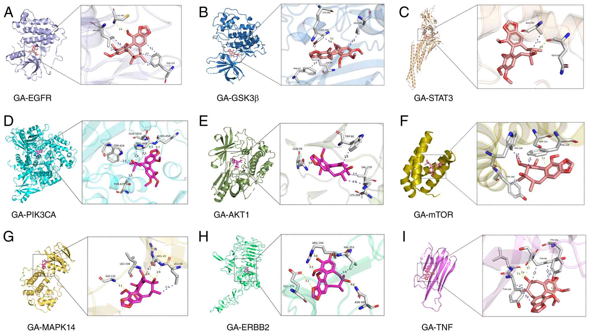 | Figure 13Predicted binding modes of GA with
nine prioritized candidate targets. Representative molecular
docking poses are shown for GA with (A) EGFR, (B) GSK3β, (C) STAT3,
(D) PIK3CA, (E) AKT1, (F) mTOR, (G) MAPK14, (H) ERBB2 and (I) TNF.
ERBB2, erb-b2 receptor tyrosine kinase 2; GA, gomisin A; PIK3CA,
phosphatidylinositol-4,5-bisphosphate 3-kinase catalytic subunit
α. |
GA yielded favorable docking poses across the nine
selected proteins. (−7.7 for EGFR, −7.4 for GSK3β, −7.0 for STAT3,
−6.9 for mTOR, −6.8 for MAPK14, −6.6 for AKT1 and ERBB2, −6.5 for
PIK3CA, and −6.1 kcal/mol for TNF); representative poses are shown
in Fig. 13. These calculations
generate structural hypotheses and prioritize candidates for
biochemical follow-up; they do not constitute evidence of
functional target engagement.
EGFR, GSK3β and STAT3 were selected for MD
simulation using a combined criterion that considered docking
score, network priority, disease relevance and target-class
representation. EGFR and GSK3β showed the most favorable docking
scores, whereas STAT3 ranked among the principal network hubs and
represented a non-kinase regulatory target with broad relevance to
the reported pharmacological effects of GA (22,77,145). The 100-nsec simulations were
used to examine the dynamic behavior of these docking-derived
complexes under explicit solvent conditions.
To elucidate the characteristics of these
interactions, analysis of the specific binding modes and potential
pharmacological implications between GA and each key target was
performed, based on the strength of the predicted docking score and
functional relevance.
Structural interaction characteristics of
major hub targets
EGFR. EGFR is a key transmembrane protein that
serves a central role in maintaining cell homeostasis by regulating
signaling pathways related to cell survival, migration and
proliferation (103). The
abnormal activation and/or overexpression of EGFR are closely
associated with the development and progression of solid tumors,
such as breast and non-small cell lung cancer and CRC (149,150).
Molecular docking generated a putative GA-EGFR pose
(Fig. 13A). The polar and
hydrophobic contacts are docking-derived observations and do not
establish ATP-competitive inhibition or downstream MAPK/PI3K
regulation.
GSK3β. The GSK3β protein is a multifunctional
serine/threonine kinase that serves as a key regulatory node in
signaling pathways, including the Wnt/β-catenin, insulin and
PI3K/AKT pathways (151). In
metabolism, GSK3β is involved in regulating glycogen synthesis,
glucose homeostasis and insulin signaling. Aberrant activity of
GSK3β is associated with metabolic conditions such as diabetes and
obesity (152). Within the
nervous system, excessive activation of GSK3β promotes
hyperphosphorylation of tau protein, accumulation of Aβ and
neuroinflammation, constituting one of the core pathological
mechanisms in neurodegenerative diseases such as Alzheimer's
disease (153). GSK3β is
regarded as a pivotal hub target linking metabolic disorders and
neurodegenerative processes. The present docking analysis generated
a favorable GA-GSK3β pose (Fig.
13B). Binding mode analysis indicated that GA stably occupied
the ATP-binding pocket through its rigid skeleton, suggesting a
putative inhibitory potential against GSK3β kinase activity. This
high-affinity ATP-pocket engagement structurally supports GSK3β as
a core multi-system regulatory target of GA.
STAT3. STAT3 is a key member of the STAT protein
family and serves a central regulatory role in tumorigenesis and
progression (154). By
activating target genes such as CCND1 and BCL-xL, it promotes tumor
cell proliferation, inhibits apoptosis, and drives angiogenesis and
invasion processes (145).
Furthermore, the STAT3 signaling pathway is involved in tumor
immune evasion: It can suppress the presentation of
tumor-associated antigens and promote the activation of
immunosuppressive cells such as regulatory T and myeloid-derived
suppressor cells, thereby weakening antitumor immune responses
(155). Given its key roles in
tumor progression and the regulation of the immune
microenvironment, STAT3 has become a prominent antitumor target
(145,155).
The present molecular docking analysis generated a
predicted GA-STAT3 binding pose. The docking results suggested
potential interactions between GA and the modeled STAT3 region;
however, the exact binding domain and the functional consequences
for STAT3 activity require further structural and experimental
validation.
PI3K/AKT/mTOR. The PI3K/AKT/mTOR pathway is a key
pathway regulating cell proliferation, survival and metabolism, and
can be activated by growth factors through receptor tyrosine
kinases, among other mechanisms (146). Signal transduction in this
pathway begins with the activation of PI3K, which catalyzes the
production of phosphatidylinositol (3,4,5)-trisphosphate (PIP3). PIP3 recruits
and activates AKT (also known as protein kinase B). AKT, serving as
a key hub, phosphorylates various downstream target proteins,
including mTOR (156). mTOR
forms two complexes, mTOR complex 1 (mTORC1) and mTORC2, with
mTORC1 serving a central role in responding to growth signals and
nutrient status by directly regulating protein synthesis by
phosphorylating substrates such as ribosomal protein S6 kinase 1
and eukaryotic translation initiation factor 4E-binding protein 1
(147). Aberrant activation of
this pathway is prevalent in various diseases, including cancer,
making it a notable therapeutic target (146,147). GA yielded similar docking
scores for three nodal proteins along this axis, PIK3CA, AKT1 and
mTOR, with Vina scores of −6.5, −6.6 and −6.9 kcal/mol,
respectively. These results provide structural support for a
potential multi-node interaction pattern within the PI3K/AKT/mTOR
pathway, although the direction and magnitude of any functional
effects cannot be inferred from docking alone. For PIK3CA, the
predicted pose was located within the ATP-binding pocket and
involved a network of hydrogen bond interactions. For AKT1, the
predicted interaction included cation-π and hydrophobic contacts.
Notably, mTOR showed a relatively favorable docking score,
accompanied by a hydrogen bond network and hydrophobic contacts.
The broadly similar docking scores across the three proteins
support the structural plausibility of potential interactions at
multiple nodes of the pathway, with mTOR prioritized because it was
also identified as a core PPI-network hub, rather than because its
small score difference establishes dominant inhibition.
MAPK14. Among the key targets highlighted by KEGG
pathway analysis, prior literature and network pharmacology, the
MAPK signaling pathway is a classic cell signal transduction
pathway. Its key mechanism involves transmitting extracellular
signals to the nucleus through a series of kinase cascades, thereby
regulating gene expression (148). As a key member of the MAPK
family, MAPK14 (p38α) serves a key role in pathophysiological
processes such as inflammation, stress responses and apoptosis
(148).
The predicted GA-MAPK14 pose included a hinge
region hydrogen bond network and extensive hydrophobic interactions
within the ATP-binding pocket. These findings provide structural
support for putative GA-mediated modulation of the MAPK signaling
pathway via interaction with MAPK14.
ERBB2. ERBB2 is a crucial member of the EGFR
family. Its aberrant activation or overexpression is associated
with the progression and poor prognosis of malignant tumors, such
as breast and gastric cancer (157), making it one of the key targets
in tumor-targeted therapy.
Molecular docking generated a putative GA-ERBB2
pose. GA forms a single hydrogen bond between its C-4' hydroxyl
group and Cys-805 in the hinge region. Due to partial occlusion of
the active pocket entrance by the gatekeeper residue Thr798, the
cyclooctadiene skeleton of GA undergoes a conformational adjustment
(twist-boat angle decreased to 32°) to accommodate the
narrower channel, resulting in a smaller hydrophobic contact area
(~240 Å2) and weaker water displacement compared with
EGFR. This compromised binding mode is consistent with its lower
affinity relative to EGFR and suggests GA may co-target ERBB family
receptors.
TNF. TNF is a key cytokine in inflammatory
responses, regulating acute and chronic inflammation, autoimmune
disease, and the tumor microenvironment (158). Its excessive production is
associated with the pathological processes of rheumatoid arthritis,
inflammatory bowel disease and cancer-associated inflammation
(158). Molecular docking
indicated that GA has binding potential to TNF. GA may insert its
dibenzocyclooctadiene skeleton into a continuous hydrophobic groove
formed by Tyr59, Tyr119 and Tyr151 on the TNF surface, establishing
multiple π-π stacking interactions. This structural feature implies
GA may directly interfere with TNF trimerization or receptor
binding, representing an additional protein-level anti-inflammatory
mechanism.
MD assessment of representative GA-target
complexes: Conformational stability and binding interface
analysis
The protein-ligand systems reached conformational
plateaus during the latter portions of the trajectories, supporting
the conformational plausibility of the docking-derived complexes.
The GSK3β complex equilibrated most rapidly, with backbone RMSD
reaching a stable plateau after ~10 nsec. The EGFR system reached
equilibrium at ~20 nsec. The STAT3 complex required the longest
adjustment period, with backbone fluctuations stabilizing only near
80 nsec, indicating greater structural adjustment in the simulated
STAT3 construct. At equilibrium, the backbone RMSD values were 0.25
for GSK3β, 0.38 for EGFR and 0.63 nm for STAT3. The higher STAT3
RMSD cannot by itself identify the responsible domain motion or
establish ligand retention within a specific binding groove. Rg
analysis supported global structural integrity. After initial
relaxation, Rg values remained steady across all three complexes
(2.11 for GSK3β, 2.04 for EGFR and 2.93 nm for STAT3) with no
persistent up- or downward drift, supporting the maintenance of
overall protein compactness, with no sustained global structural
expansion observed (Fig. 5).
Residue-level flexibility and binding interface
behavior were evaluated using RMSF and intermolecular hydrogen bond
analyses. For all three complexes, residue fluctuations were
generally lower around the predicted binding interfaces than in
solvent-exposed loop regions, although ligand-free control
simulations are required to determine whether this pattern was
specifically associated with GA binding. Intermolecular hydrogen
bonds formed and broke dynamically throughout the trajectories,
with no hydrogen bond and intermittent higher-count peaks; the EGFR
complex generally shows fewer hydrogen bonds during the latter part
of the trajectory (Fig. 14A).
Intermolecular hydrogen bonds were intermittently maintained
throughout the trajectory, although their occupancy and
donor-acceptor identities varied over time. These observations
indicated that polar interactions between GA and the predicted
binding sites were dynamically retained during the simulation,
while their specific contribution to complex stability cannot be
established from hydrogen bond counts alone.
Total SASA decreased during the early relaxation
phase of all simulations and stabilized in later trajectory
segments, consistent with decreased overall solvent exposure as the
systems adjusted from their rigid docking starting conformations.
This aligns with the expected pattern of hydrophobic residue burial
at the protein-ligand interface, although interface-specific SASA
calculations are required to formally attribute the changes to
ligand binding rather than global protein compaction (Fig. 14B).
Taken together, the 100-nsec simulation provided
multi-dimensional support for the conformational plausibility of
the top-ranked docking poses. Across all three complexes, GA
maintained its core binding orientation within the cognate pocket
over the production phase, with preserved global protein folds,
stable interface core rigidity, persistent polar interactions and
SASA trends consistent with hydrophobic packing. These findings
extend static docking predictions into a dynamic solvated context
and reinforce the computational plausibility of the proposed
multi-target interaction profile of GA. These trajectory-level
findings provide a dynamic consistency check for the selected
docking poses but do not determine whether GA binds these proteins
selectively in biochemical or cell settings.
Specificity assessment of the predicted
multi-target profile: PAINS structural alerts and remaining
uncertainties
GA was predicted to interact with a broad set of
proteins, including kinases, such as EGFR, GSK3β, mTOR, MAPK14,
AKT1, PIK3CA and ERBB2, the transcription factor STAT3 and the
cytokine TNF, raising an important question regarding target
specificity. Specifically, it remains necessary to distinguish
genuine polypharmacology from frequent hitter behavior or assay
interference. PAINS alerts are substructural patterns originally
associated with compounds that repeatedly produce apparent activity
in high-throughput screening assays. However, a PAINS match is a
warning requiring further investigation rather than definitive
evidence that a compound is artifactual; conversely, the absence of
a PAINS match does not establish assay specificity (47,143).
No PAINS substructure match was returned for GA.
This indicates that GA does not contain any of the classical PAINS
motifs represented in the applied RDKit catalogs.
The negative RDKit result does not demonstrate that
GA is free from assay interference and does not exclude mechanisms
that are not encoded by the applied structural rules. In
particular, colloidal aggregation, non-specific membrane
partitioning, fluorescence or absorbance interference,
redox-associated effects and non-specific protein binding at high
compound concentrations cannot be assessed by PAINS substructure
matching alone (47,143,144). Therefore, the present results
decrease concern regarding classical PAINS chemotypes but do not
establish that GA is assay-specific or non-promiscuous (47,143,144).
Structural features compatible with, but not
diagnostic of, polypharmacology. The dibenzocyclooctadiene scaffold
of GA possesses a moderately lipophilic and conformationally
adaptable framework (46). Its
ether oxygen atoms and non-phenolic hydroxyl group support polar
interactions, whereas its aromatic and carbocyclic framework may
accommodate hydrophobic regions within protein-binding pockets. The
possible contacts described here are interpretations of the
structure and the present docking poses, not evidence of
experimentally verified polypharmacology. Most of the
computationally prioritized proteins are kinases with
evolutionarily associated ATP-binding pockets. Consequently,
similar docking orientations across EGFR, GSK3β, mTOR, MAPK14,
AKT1, PIK3CA and ERBB2 may partly reflect conservation of kinase
pocket architecture and the preferences of the docking scoring
function, rather than experimentally verified engagement of these
proteins (34,103). The Vina docking scores of −6.1
to −7.7 kcal/mol should be used for relative pose and target
prioritization only. They should not be interpreted as
experimentally measured binding free energies or converted directly
into micromolar binding potency.
The MD trajectories provide an additional dynamic
consistency check for the GA-EGFR, GA-GSK3β and GA-STAT3 docking
poses. However, trajectory persistence does not demonstrate
biochemical selectivity, direct target engagement or functional
inhibition. Furthermore, these findings cannot be extrapolated to
mTOR, MAPK14, AKT1, PIK3CA, ERBB2 or TNF, which were not subjected
to MD simulation.
Caveats and recommended orthogonal validation. GA
is moderately lipophilic, with a calculated cLogP of ~3.6, and cell
effects such as CRC apoptosis and melanoma growth suppression occur
at micromolar or high-micromolar concentrations (8,23). Under these conditions,
non-specific membrane partitioning, colloidal aggregation or
general cytotoxicity remains plausible in the absence of a PAINS
alert. A tiered program of orthogonal validation is therefore
recommended, proceeding from biochemical binding through cell
target engagement to functional dependency and structural
confirmation.
Biochemical-level validation should be pursued. For
kinase candidates among the top predicted targets, including EGFR,
GSK3β and mTOR, recombinant kinase assays should measure direct
effects on catalytic activity and derive IC50 values at
physiologically relevant ATP concentrations. Parallel biophysical
binding measurements using SPR, biolayer interferometry, microscale
thermophoresis or isothermal titration calorimetry (ITC) may
confirm direct physical binding and provide equilibrium
dissociation constants together with association and dissociation
kinetics.
CETSA test target stabilization in cells, while
DARTS can test ligand-associated protease protection in an
appropriate protein preparation (159,160). These methods bridge recombinant
biochemistry and the intracellular environment, where off-target
binding, metabolic conversion and restricted compartmental access
may modify the effective interactome (159,160).
Functional dependency validation is critical.
Target knockdown, knockout, point mutation, overexpression or
rescue experiments are required to determine whether the reported
cell phenotypes depend on EGFR, GSK3β, STAT3 or other predicted
proteins. Attenuation or loss of the GA response following target
disruption is the strongest available preclinical evidence that a
candidate target mediates the observed phenotype.
Co-crystallography or cryo-electron microscopy of
GA in complex with its highest-confidence targets would
definitively resolve the binding mode and distinguish specific
ligand-protein interactions from non-specific hydrophobic
associations. This level of validation sets the long-term standard
for target confirmation.
Before target-specific assays are interpreted,
basic counterscreens for artifactual activity are key.
Aggregation-sensitive controls include parallel testing with and
without a low concentration of non-ionic detergent, such as ~0.01%
Triton X-100, as well as BSA or other decoy protein additions. DLS
may be used where technically applicable to determine whether GA
forms colloidal particles under the assay conditions. Additional
controls should include fluorescence and absorbance interference
testing, orthogonal assay formats, counterscreens against unrelated
proteins, concentration-response analysis and parallel assessment
of cytotoxicity in non-malignant or primary cells. Detergent and
BSA concentrations should be optimized for each assay because these
additives may alter target activity or decrease the free
concentration of GA (144).
In summary, the molecular docking and network
pharmacology support target prioritization and hypothesis
generation. They do not constitute target confirmation or evidence
that GA directly engages these proteins in a functional setting.
Taken together, the absence of an RDKit PAINS match argues against
the presence of classical PAINS substructures in GA, but does not
establish genuine polypharmacology. The present network
pharmacology, docking and MD findings support a set of structurally
plausible and biologically relevant candidate interactions. Until
these interactions are confirmed through orthogonal biochemical,
cell target engagement and functional dependency experiments, EGFR,
GSK3β, STAT3, mTOR and the other predicted proteins should be
regarded as computationally prioritized candidate targets rather
than established direct targets of GA.
Discussion
The pathogenesis of complex chronic diseases relies
on dynamic crosstalk across multiple signaling pathways and cell
networks, a number of which remain incompletely characterized
(1). Treatment limitations
differ by liver disease and cancer type; incomplete responses,
resistance and adverse effects are relevant in selected settings
(57,157). By contrast, traditional Chinese
herbal medicines, as natural active drugs, boast advantages such as
abundant resources, high safety, low toxicity, low production cost
and suitability for long-term use (161). Furthermore, the complex
composition of herbal medicines enables them to exert combined
therapeutic activities through binding with multiple targets.
Additionally, due to the unique structures and pharmacological
actions of herbal medicines, individual constituents exhibit
several preclinical activities, as illustrated by GA-associated
apoptosis and transporter inhibition (8,49). Therefore, developing therapeutic
agents from traditional herbs has become a promising direction for
treating complex diseases such as liver disease, cancer, CVD, and
neurodegenerative and inflammatory disorders.
Hepatoprotection is the best-supported preclinical
activity, with consistent efficacy replicated across multiple
independent laboratories and injury models, including
CCl4-, APAP-, D-GalN/LPS- and LCA-induced cholestasis
(7,11,12,20). The incorporated studies on the
Surf4-SAA1 paracrine axis and PPARα nuclear localization extend the
mechanistic discussion of liver fibrosis and MASLD beyond the
canonical TGF-β/Smad and NF-κB pathways to hepatocyte secretome
biology and nuclear receptor subcellular trafficking, providing
biological context rather than evidence that GA directly acts on
Surf4 or PPARα (74,82). Beyond shared antioxidant and
anti-inflammatory mechanisms, PXR-mediated bile acid regulation is
the most strongly supported functionally dependent mechanism in the
GA literature. Genetic evidence from Pxr-null mice supports
tier B classification, but direct physical binding of GA to PXR has
not been established (12).
Anticancer and cardiovascular activity form a
second tier of promising but less mature evidence. Both are
supported by substantial cell and in vivo phenotypic data,
with plausible mechanistic links to cell cycle regulation,
apoptosis and endothelial function (8,9,13,14,24,25). However, a notable gap separates
effective in vitro concentrations from physiologically
achievable concentrations during in vivo exposure, and most
pathway-level observations remain associative rather than formally
confirmed as target-dependent.
Neuroprotection, anti-inflammatory and antioxidant
effects, renal protection, anti-obesity activity, and other
exploratory endpoints constitute a third tier of preliminary
observations. Most derive from single-center, single-model reports
with limited mechanistic depth and should be regarded as
hypothesis-generating leads rather than translation-ready findings
(10,15,26,27,29).
GA faces well-documented PK challenges, including
limited oral bioavailability, rapid systemic clearance, a large
volume of distribution and predominantly non-renal elimination
(18,99,135). Pathological states may alter
its absorption, metabolism and tissue exposure (114,133). While formulation strategies,
such as natural deep eutectic solvents, have shown early potential
to improve absorption and bioavailability (139), these approaches remain at the
proof-of-concept stage (18,99,114,133).
Translational development is constrained by two
poorly addressed risks: Limited safety data and clinically
meaningful drug-interaction liability. Available acute toxicity
data provide only an estimate of tolerability and modern
repeat-dose, genotoxicity, reproductive and carcinogenicity studies
are lacking. The central depressant effects of GA also suggest a
dose continuum between desired sedative-hypnotic activity and
adverse CNS depression, which requires formal exposure-response
characterization (19). GA
exerts bidirectional regulatory effects on CYP3A4, and inhibits
P-gp and BCRP transporters (12,49,62,95,140). When co-administered with narrow
therapeutic index agents such as tacrolimus or PTX, it may increase
systemic exposure and elevate the adverse event risk (96,101,142). This drug-herb interaction
potential must be prioritized in safety assessment and clinical
development planning.
Network pharmacology and molecular docking analyses
provide a computational framework to generate hypotheses about the
molecular basis of GA-mediated pharmacological actions, rather than
definitive mechanistic proof. Network pharmacology screening
identified 100 candidate targets with non-zero prediction
probability and generated a PPI network, with STAT3, EGFR and mTOR
ranking as core topological hubs. These candidate targets align
directionally with the hepatoprotective and anticancer phenotypes
of GA (8,11,12). Several methodological limitations
should be noted. Disease gene sets derived from GeneCards cover a
broad functional scope, which may overestimate the biological
relevance of target-disease overlaps. Network centrality reflects
topological importance rather than causal involvement and docking
affinities in the −6 to −8 kcal/mol range only indicate moderate
binding potential. All computational findings fall under tier D of
the evidence hierarchy and require orthogonal experimental
validation before definitive mechanistic conclusions can be
drawn.
In summary, GA is a prototypical multi-target
natural product whose most prominent pharmacological activity
(hepatoprotection, anticancer, neuroprotection and
anti-inflammation/antioxidant effects) are primarily attributable
to its concerted modulation of key signaling hubs, including
PI3K/AKT/mTOR, MAPK and NF-κB, and targets, such as STAT3, EGFR and
GSK3β. These targets and pathways intersect to regulate cell
survival, death, inflammation and metabolism, forming an integrated
network that underlies GA-mediated polypharmacology and its ability
to address multifactorial diseases. Its historical application in
liver disorders corresponds to robust hepatoprotective and
antifibrotic effects, while its role as an adaptogen is consistent
with observed neuroprotective, anti-inflammatory and antioxidant
properties (5,10,16,17,21). Nonetheless, the evidence base
remains predominantly preclinical and phenotype-driven (7,8,10,17). Incomplete target validation,
suboptimal PK properties, and insufficient safety and
drug-interaction data represent the principal barriers to clinical
translation. Future research should prioritize rigorous target
validation and focused translational development.
Building on validated target mechanisms, future
studies should further prioritize developing advanced delivery
systems such as nanoformulations and prodrugs to improve PK
properties, conducting rigorous proof-of-concept clinical trials in
well-supported indications such as chemotherapy-induced LI and
metabolic dysfunction-associated fatty liver disease, applying
systems pharmacology and multi-omics to delineate its comprehensive
target network and combined modes of action, and systematically
assessing drug-herb interactions to ensure clinical safety. These
efforts may bridge traditional knowledge and modern science,
advancing GA toward a scientifically grounded, multi-target
therapeutic agent.
To conclude, GA, a natural TCM monomer with
extensive application prospects, may propel the modernization of
TCM through thorough research and development. Rigorous scientific
investigation, clinical trials and precise elucidation of
disease-alleviating targets may establish GA as a paradigm for TCM
integration into international markets. This will not only foster
the standardization and global acceptance of TCM but may furnish
robust evidentiary support for the scientific inquiry and clinical
use of traditional remedies.
Supplementary Data
Availability of data and materials
The data generated in the present study may be
requested from the corresponding author.
Authors' contributions
JX conceived the study and wrote the manuscript.
CZ, JZ and SS performed the computational analyses. QS and SZ
analyzed and interpreted data. YZ and PC conceived the study and
revised the manuscript. All authors have read and approved the
final manuscript. JX and PC confirm the authenticity of all the raw
data.
Ethics approval and consent to
participate
Not applicable.
Patient consent for publication
Not applicable.
Competing interests
The authors declare that they have no competing
interests.
Abbreviations:
|
APAP
|
N-acetyl-para-aminophenol
|
|
CYP3A4
|
cytochrome P450 family 3 subfamily A
member 4
|
|
ERBB2
|
erb-b2 receptor tyrosine kinase 2
|
|
GA
|
gomisin A
|
|
MD
|
molecular dynamics
|
|
PAINS
|
pan-assay interference compounds
|
|
P-gp
|
P-glycoprotein
|
|
PIK3CA
|
phosphatidylinositol-4,5-bisphosphate
3-kinase catalytic subunit α
|
|
PK
|
pharmacokinetics
|
|
PXR
|
pregnane X receptor
|
Acknowledgements
Not applicable.
Funding
The present study was supported by the National Natural Science
Foundation of China (grant no. 82304840), the Zhejiang Provincial
Natural Science Foundation of China (grant no. LQ23H280021), and
the Traditional Chinese Medicine Science and Technology Project of
Zhejiang Province (grant no. 2024ZF006).
References
|
1
|
Cvijovic M and Polster A: Network
medicine: Facilitating a new view on complex diseases. Front
Bioinform. 3:11634452023. View Article : Google Scholar : PubMed/NCBI
|
|
2
|
World Health Organization: Noncommunicable
diseases. https://www.who.int/news-room/fact-sheets/detail/noncommunicable-diseases.
Accessed September 11, 2026
|
|
3
|
Zhang L, Song J, Kong L, Yuan T, Li W,
Zhang W, Hou B, Lu Y and Du G: The strategies and techniques of
drug discovery from natural products. Pharmacol Ther.
216:1076862020. View Article : Google Scholar
|
|
4
|
Naeem A, Hu P, Yang M, Zhang J, Liu Y, Zhu
W and Zheng Q: Natural products as anticancer agents: Current
status and future perspectives. Molecules. 27:83672022. View Article : Google Scholar :
|
|
5
|
Cyong JC, Ki SM, Iijima K, Kobayashi T and
Furuya M: Clinical and pharmacological studies on liver diseases
treated with Kampo herbal medicine. Am J Chin Med. 28:351–360.
2000. View Article : Google Scholar
|
|
6
|
Liu J, Shi JL, Guo JY, Chen Y, Ma XJ, Wang
SN, Zheng ZQ, Lin MX and He S: Anxiolytic-like effect of
Suanzaoren-Wuweizi herb-pair and evidence for the involvement of
the monoaminergic system in mice based on network pharmacology. BMC
Complement Med Ther. 23:72023. View Article : Google Scholar : PubMed/NCBI
|
|
7
|
Teraoka R, Shimada T and Aburada M: The
molecular mechanisms of the hepatoprotective effect of gomisin A
against oxidative stress and inflammatory response in rats with
carbon tetrachloride-induced acute liver injury. Biol Pharm Bull.
35:171–177. 2012. View Article : Google Scholar : PubMed/NCBI
|
|
8
|
Kee JY, Han YH, Mun JG, Park SH, Jeon HD
and Hong SH: Gomisin A suppresses colorectal lung metastasis by
inducing AMPK/p38-mediated apoptosis and decreasing metastatic
abilities of colorectal cancer cells. Front Pharmacol. 9:9862018.
View Article : Google Scholar :
|
|
9
|
Young Park J, Wook Yun J, Whan Choi Y, Ung
Bae J, Won Seo K, Jin Lee S, Youn Park S, Whan Hong K and Kim CD:
Antihypertensive effect of gomisin A from Schisandra chinensis on
angiotensin II-induced hypertension via preservation of nitric
oxide bioavailability. Hypertens Res. 35:928–934. 2012. View Article : Google Scholar : PubMed/NCBI
|
|
10
|
Wang X, Hu D, Zhang L, Lian G, Zhao S,
Wang C, Yin J, Wu C and Yang J: Gomisin A inhibits
lipopolysaccharide-induced inflammatory responses in N9 microglia
via blocking the NF-κB/MAPKs pathway. Food Chem Toxicol.
63:119–127. 2014. View Article : Google Scholar
|
|
11
|
Jiang Y, Fan X, Wang Y, Chen P, Zeng H,
Tan H, Gonzalez FJ, Huang M and Bi H: Schisandrol B protects
against acetaminophen-induced hepatotoxicity by inhibition of
CYP-mediated bioactivation and regulation of liver regeneration.
Toxicol Sci. 143:107–115. 2015. View Article : Google Scholar
|
|
12
|
Zeng H, Jiang Y, Chen P, Fan X, Li D, Liu
A, Ma X, Xie W, Liu P, Gonzalez FJ, et al: Schisandrol B protects
against cholestatic liver injury through pregnane X receptors. Br J
Pharmacol. 174:672–688. 2017. View Article : Google Scholar : PubMed/NCBI
|
|
13
|
Wang T, Liu J, Huang X, Zhang C, Shangguan
M, Chen J, Wu S, Chen M, Yang Z and Zhao S: Gomisin A enhances the
antitumor effect of paclitaxel by suppressing oxidative stress in
ovarian cancer. Oncol Rep. 48:2022022. View Article : Google Scholar :
|
|
14
|
Park JY, Shin HK, Choi YW, Lee YJ, Bae SS,
Han J and Kim CD: Gomisin A induces Ca2+-dependent activation of
eNOS in human coronary artery endothelial cells. J Ethnopharmacol.
125:291–296. 2009. View Article : Google Scholar
|
|
15
|
Kim DH, Hung TM, Bae KH, Jung JW, Lee S,
Yoon BH, Cheong JH, Ko KH and Ryu JH: Gomisin A improves
scopolamine-induced memory impairment in mice. Eur J Pharmacol.
542:129–135. 2006. View Article : Google Scholar : PubMed/NCBI
|
|
16
|
Jeong HJ, Han NR, Kim KY, Choi IS and Kim
HM: Gomisin A decreases the LPS-induced expression of iNOS and
COX-2 and activation of RIP2/NF-κB in mouse peritoneal macrophages.
Immunopharmacol Immunotoxicol. 36:195–201. 2014. View Article : Google Scholar : PubMed/NCBI
|
|
17
|
Jiang YM, Wang Y, Tan HS, Yu T, Fan XM,
Chen P, Zeng H, Huang M and Bi HC: Schisandrol B protects against
acetaminophen-induced acute hepatotoxicity in mice via activation
of the NRF2/ARE signaling pathway. Acta Pharmacol Sin. 37:382–389.
2016. View Article : Google Scholar : PubMed/NCBI
|
|
18
|
Liang D, Wu Z, Liu Y, Li C, Li X, Yang B,
Xie H and Sun H: HPLC-MS/MS-mediated analysis of the
pharmacokinetics, bioavailability, and tissue distribution of
schisandrol B in rats. Int J Anal Chem. 2021:88622912021.
View Article : Google Scholar
|
|
19
|
Maeda S, Sudo K, Aburada M, Ikeya Y,
Taguchi H, Yoshioka I and Harada M: Pharmacological studies on
Schizandra fruit. I. General pharmacological effects of gomisin A
and schizandrin (author's transl). Yakugaku Zasshi. 101:1030–1041.
1981.In Japanese. View Article : Google Scholar
|
|
20
|
Kim SH, Kim YS, Kang SS, Bae K, Hung TM
and Lee SM: Anti-apoptotic and hepatoprotective effects of gomisin
A on fulminant hepatic failure induced by D-galactosamine and
lipopolysaccharide in mice. J Pharmacol Sci. 106:225–233. 2008.
View Article : Google Scholar : PubMed/NCBI
|
|
21
|
Ai Y, Shi W, Zuo X, Sun X, Chen Y, Wang Z,
Li R, Song X, Dai W, Mu W, et al: The combination of schisandrol B
and wedelolactone synergistically reverses hepatic fibrosis via
modulating multiple signaling pathways in mice. Front Pharmacol.
12:6555312021. View Article : Google Scholar : PubMed/NCBI
|
|
22
|
Li X, Sun J, Fan X, Guan L, Li D, Zhou Y,
Zeng X, Chen Y, Zhang H, Xu L, et al: Schisandrol B promotes liver
regeneration after partial hepatectomy in mice. Eur J Pharmacol.
818:96–102. 2018. View Article : Google Scholar
|
|
23
|
Han YH, Mun JG, Jeon HD, Park J, Kee JY
and Hong SH: Gomisin A ameliorates metastatic melanoma by
inhibiting AMPK and ERK/JNK-mediated cell survival and metastatic
phenotypes. Phytomedicine. 68:1531472020. View Article : Google Scholar
|
|
24
|
Park JY, Lee SJ, Yun MR, Seo KW, Bae SS,
Park JW, Lee YJ, Shin WJ, Choi YW and Kim CD: Gomisin A from
Schisandra chinensis induces endothelium-dependent and direct
relaxation in rat thoracic aorta. Planta Med. 73:1537–1542. 2007.
View Article : Google Scholar
|
|
25
|
Seok YM, Choi YW, Kim GD, Kim HY, Takuwa Y
and Kim IK: Effects of gomisin A on vascular contraction in rat
aortic rings. Naunyn Schmiedebergs Arch Pharmacol. 383:45–56. 2011.
View Article : Google Scholar
|
|
26
|
Hwang IS, Kim JE, Lee YJ, Kwak MH, Choi
YH, Kang BC, Hong JT and Hwang DY: Protective effects of gomisin A
isolated from Schisandra chinensis against CCl(4)-induced hepatic
and renal injury. Int J Mol Med. 31:888–898. 2013. View Article : Google Scholar
|
|
27
|
Han YH, Kee JY and Hong SH: Gomisin A
alleviates obesity by regulating the phenotypic switch between
white and brown adipocytes. Am J Chin Med. 49:1929–1948. 2021.
View Article : Google Scholar : PubMed/NCBI
|
|
28
|
Choi BR, Kim HK and Park JK: Effects of
Schisandra chinensis fruit extract and gomisin A on the
contractility of penile corpus cavernosum smooth muscle: A
potential mechanism through the nitric oxide-cyclic guanosine
monophosphate pathway. Nutr Res Pract. 12:291–297. 2018. View Article : Google Scholar :
|
|
29
|
Lin RD, Mao YW, Leu SJ, Huang CY and Lee
MH: The immuno-regulatory effects of Schisandra chinensis and its
constituents on human monocytic leukemia cells. Molecules.
16:4836–4849. 2011. View Article : Google Scholar : PubMed/NCBI
|
|
30
|
Liu S, Xu L, Shen Y, Wang L, Lai X and Hu
H: Qingxin Kaiqiao Fang decreases Tau hyperphosphorylation in
Alzheimer's disease via the PI3K/Akt/GSK3β pathway in vitro and in
vivo. J Ethnopharmacol. 318:1170312024. View Article : Google Scholar
|
|
31
|
Lee N, Youn K, Kwon H, Lee J, Lee E, Jung
JW, Moon M, Lee DS, Kim DH, Ho CT and Jun M: Fucoxanthin suppresses
amyloid-β and pyroglutamate-3-Aβ accumulation by modulating the
PI3K/Akt/GSK-3β signaling pathway. Food Funct. 16:9102–9117. 2025.
View Article : Google Scholar : PubMed/NCBI
|
|
32
|
Duan Z, Chen H, Miao W, He J, Xu D, Qi Z,
Yang L, Jia W and Wu C: Scutellarin alleviates microglia-mediated
neuroinflammation and apoptosis after ischemic stroke through the
PI3K/AKT/GSK3β signaling pathway. J Cell Commun Signal.
18:e120232024. View Article : Google Scholar
|
|
33
|
Li M, Jiang Y, Xue Y, Duan Y, Liu Y, Xin
X, Wei X, Hu Z, Yang P, Diao S, et al: Eupalinolide B prevents
cerebral ischemia-reperfusion injury via the PI3K/Akt/GSK3β(Ser9)
signaling pathway. Bioorg Chem. 173:1096352026. View Article : Google Scholar
|
|
34
|
Trott O and Olson AJ: AutoDock Vina:
Improving the speed and accuracy of docking with a new scoring
function, efficient optimization, and multithreading. J Comput
Chem. 31:455–461. 2010. View Article : Google Scholar
|
|
35
|
Adasme MF, Linnemann KL, Bolz SN, Kaiser
F, Salentin S, Haupt VJ and Schroeder M: PLIP 2021: Expanding the
scope of the protein-ligand interaction profiler to DNA and RNA.
Nucleic Acids Res. 49(W1): W530–W534. 2021. View Article : Google Scholar : PubMed/NCBI
|
|
36
|
Hsin KY, Ghosh S and Kitano H: Combining
machine learning systems and multiple docking simulation packages
to improve docking prediction reliability for network pharmacology.
PLoS One. 8:e839222013. View Article : Google Scholar
|
|
37
|
Ni X, Bao H, Guo J, Li D, Wang L, Zhang W
and Sun G: Discussion on the mechanism of Danggui Sini decoction in
treating diabetic foot based on network pharmacology and molecular
docking and verification of the curative effect by meta-analysis.
Front Endocrinol (Lausanne). 15:13470212024. View Article : Google Scholar : PubMed/NCBI
|
|
38
|
Van Der Spoel D, Lindahl E, Hess B,
Groenhof G, Mark AE and Berendsen HJC: GROMACS: Fast, flexible, and
free. J Comput Chem. 26:1701–1718. 2005. View Article : Google Scholar
|
|
39
|
Lindorff-Larsen K, Piana S, Palmo K,
Maragakis P, Klepeis JL, Dror RO and Shaw DE: Improved side-chain
torsion potentials for the Amber ff99SB protein force field.
Proteins. 78:1950–1958. 2010. View Article : Google Scholar : PubMed/NCBI
|
|
40
|
Jorgensen WL, Chandrasekhar J, Madura JD,
Impey RW and Klein ML: Comparison of simple potential functions for
simulating liquid water. J Chem Phys. 79:926–935. 1983. View Article : Google Scholar
|
|
41
|
Lemkul JA: From proteins to perturbed
hamiltonians: A suite of tutorials for the GROMACS-2018 molecular
simulation package [article v1.0]. Living J Comp Mol Sci.
1:50682018. View Article : Google Scholar
|
|
42
|
Valdés-Tresanco MS, Valdés-Tresanco ME,
Valiente PA and Moreno E: gmx_MMPBSA: A new tool to perform
end-state free energy calculations with GROMACS. J Chem Theory
Comput. 17:6281–6291. 2021. View Article : Google Scholar
|
|
43
|
Sahihi M and Ghayeb Y: An investigation of
molecular dynamics simulation and molecular docking: Interaction of
citrus flavonoids and bovine β-lactoglobulin in focus. Comput Biol
Med. 51:44–50. 2014. View Article : Google Scholar : PubMed/NCBI
|
|
44
|
Han P, An N, Yang L, Ren X, Lu S, Ji H,
Wang Q and Dong J: Molecular dynamics simulation of the
interactions between sesamol and myosin combined with spectroscopy
and molecular docking studies. Food Hydrocoll. 131:1078012022.
View Article : Google Scholar
|
|
45
|
Sobstyl E, Szopa A, Ekiert H, Gnat S,
Typek R and Choma IM: Effect directed analysis and TLC screening of
Schisandra chinensis fruits. J Chromatogr A. 1618:4609422020.
View Article : Google Scholar : PubMed/NCBI
|
|
46
|
Ikeya Y, Taguchi H, Yosioka I and
Kobayashi H: The constituents of Schizandra chinensis Baill. I.
Isolation and structure determination of five new lignans, gomisin
A, B, C, F and G, and the absolute structure of schizandrin. Chem
Pharm Bull (Tokyo). 27:1383–1394. 1979. View Article : Google Scholar : PubMed/NCBI
|
|
47
|
Baell JB and Holloway GA: New substructure
filters for removal of pan assay interference compounds (PAINS)
from screening libraries and for their exclusion in bioassays. J
Med Chem. 53:2719–2740. 2010. View Article : Google Scholar
|
|
48
|
Capuzzi SJ, Muratov EN and Tropsha A:
Phantom PAINS: Problems with the utility of alerts for pan-assay
interference compounds. J Chem Inf Model. 57:417–427. 2017.
View Article : Google Scholar
|
|
49
|
Wan CK, Zhu GY, Shen XL, Chattopadhyay A,
Dey S and Fong WF: Gomisin A alters substrate interaction and
reverses P-glycoprotein-mediated multidrug resistance in HepG2-DR
cells. Biochem Pharmacol. 72:824–837. 2006. View Article : Google Scholar : PubMed/NCBI
|
|
50
|
Trefts E, Gannon M and Wasserman DH: The
liver. Curr Biol. 27:R1147–R1151. 2017. View Article : Google Scholar : PubMed/NCBI
|
|
51
|
Jalan-Sakrikar N, Brevini T, Huebert RC
and Sampaziotis F: Organoids and regenerative hepatology.
Hepatology. 77:305–322. 2023. View Article : Google Scholar :
|
|
52
|
Luedde T, Kaplowitz N and Schwabe RF: Cell
death and cell death responses in liver disease: Mechanisms and
clinical relevance. Gastroenterology. 147:765–783.e4. 2014.
View Article : Google Scholar
|
|
53
|
Bai P, Ye H, Xie M, Saxena P, Zulewski H,
Charpin-El Hamri G, Djonov V and Fussenegger M: A synthetic
biology-based device prevents liver injury in mice. J Hepatol.
65:84–94. 2016. View Article : Google Scholar : PubMed/NCBI
|
|
54
|
Xu G, Lv X, Feng Y, Li H, Chen C, Lin H,
Li H, Wang C, Chen J and Sun J: Study on the effect of active
components of Schisandra chinensis on liver injury and its
mechanisms in mice based on network pharmacology. Eur J Pharmacol.
910:1744422021. View Article : Google Scholar : PubMed/NCBI
|
|
55
|
Pel P, Chae HS, Nhoek P, Yeo W, Kim YM and
Chin YW: Lignans from the fruits of Schisandra chinensis (Turcz.)
Baill inhibit proprotein convertase subtilisin/kexin type 9
expression. Phytochemistry. 136:119–124. 2017. View Article : Google Scholar : PubMed/NCBI
|
|
56
|
Ma JQ, Li Z, Xie WR, Liu CM and Liu SS:
Quercetin protects mouse liver against CCl4-induced
inflammation by the TLR2/4 and MAPK/NF-κB pathway. Int
Immunopharmacol. 28:531–539. 2015. View Article : Google Scholar : PubMed/NCBI
|
|
57
|
Kisseleva T and Brenner D: Molecular and
cellular mechanisms of liver fibrosis and its regression. Nat Rev
Gastroenterol Hepatol. 18:151–166. 2021. View Article : Google Scholar
|
|
58
|
Yu Y, Li MP, Xu B, Fan F, Lu SF, Pan M and
Wu HS: A study of regulatory effects of TLR4 and NF-κB on primary
biliary cholangitis. Eur Rev Med Pharmacol Sci. 23:3951–3959.
2019.PubMed/NCBI
|
|
59
|
Choi YM, Choi IS, Lee SM, Hwang DY, Choi
YW and Park YH: Transcriptome analysis of the effects of gomisin a
on the recovery of carbon tetrachloride-induced damage in rat
liver. Lab Anim Res. 27:161–169. 2011. View Article : Google Scholar :
|
|
60
|
Dargan PI and Jones AL: Acetaminophen
poisoning: An update for the intensivist. Crit Care. 6:108–110.
2002. View
Article : Google Scholar
|
|
61
|
Wolf KK, Wood SG, Allard JL, Hunt JA,
Gorman N, Walton-Strong BW, Szakacs JG, Duan SX, Hao Q, Court MH,
et al: Role of CYP3A and CYP2E1 in alcohol-mediated increases in
acetaminophen hepatotoxicity: Comparison of wild-type and
Cyp2e1(-/-) mice. Drug Metab Dispos. 35:1223–1231. 2007. View Article : Google Scholar
|
|
62
|
Wan CK, Tse AK, Yu ZL, Zhu GY, Wang H and
Fong DW: Inhibition of cytochrome P450 3A4 activity by schisandrol
A and gomisin A isolated from Fructus Schisandrae chinensis.
Phytomedicine. 17:702–705. 2010. View Article : Google Scholar : PubMed/NCBI
|
|
63
|
Jiang Y, Fan X, Wang Y, Tan H, Chen P,
Zeng H, Huang M and Bi H: Hepato-protective effects of six
Schisandra lignans on acetaminophen-induced liver injury are
partially associated with the inhibition of CYP-mediated
bioactivation. Chem Biol Interact. 231:83–89. 2015. View Article : Google Scholar : PubMed/NCBI
|
|
64
|
Wu YH, Hu SQ, Liu J, Cao HC, Xu W, Li YJ
and Li LJ: Nature and mechanisms of hepatocyte apoptosis induced by
D-galactosamine/lipopolysaccharide challenge in mice. Int J Mol
Med. 33:1498–1506. 2014. View Article : Google Scholar : PubMed/NCBI
|
|
65
|
Das J, Ghosh J, Roy A and Sil PC:
Mangiferin exerts hepatoprotective activity against D-galactosamine
induced acute toxicity and oxidative/nitrosative stress via
Nrf2-NFκB pathways. Toxicol Appl Pharmacol. 260:35–47. 2012.
View Article : Google Scholar
|
|
66
|
Lv X, Xu Z, Xu G, Li H, Wang C, Chen J and
Sun J: Investigation of the active components and mechanisms of
Schisandra chinensis in the treatment of asthma based on a network
pharmacology approach and experimental validation. Food Funct.
11:3032–3042. 2020. View Article : Google Scholar : PubMed/NCBI
|
|
67
|
Zollner G and Trauner M: Molecular
mechanisms of cholestasis. Wien Med Wochenschr. 156:380–385. 2006.
View Article : Google Scholar : PubMed/NCBI
|
|
68
|
Hirschfield GM, Heathcote EJ and Gershwin
ME: Pathogenesis of cholestatic liver disease and therapeutic
approaches. Gastroenterology. 139:1481–1496. 2010. View Article : Google Scholar : PubMed/NCBI
|
|
69
|
Liang H, Yang X, Li H, Wang X, Su H, Li X,
Tian J, Cai C, Huang M and Bi H: Schisandrol B protects against
cholestatic liver injury by inhibiting pyroptosis through pregnane
X receptor. Biochem Pharmacol. 204:1152222022. View Article : Google Scholar
|
|
70
|
Fan S, Liu C, Jiang Y, Gao Y, Chen Y, Fu
K, Yao X, Huang M and Bi H: Lignans from Schisandra sphenanthera
protect against lithocholic acid-induced cholestasis by pregnane X
receptor activation in mice. J Ethnopharmacol. 245:1121032019.
View Article : Google Scholar : PubMed/NCBI
|
|
71
|
Yang X, Liang H, Li X, Tian J, Fan S,
Huang M, Wan J, Zuo Z, Guo H and Bi H: Schisandrol B protects
against lithocholic acid-induced cholestatic liver injury in mice
through mitochondrial biogenesis. Chin Med. 21:1162026. View Article : Google Scholar : PubMed/NCBI
|
|
72
|
Wang J, Yu Y, Xiong Z, Wang P, Wang Z, Du
A, Han S, Wang X and Zhang L: METTL3 regulates rifampicin-induced
CYP3A4 expression by activating PXR translation and nuclear import
and stabilizing CYP3A4 mRNA. Biochem Pharmacol. 239:1170162025.
View Article : Google Scholar
|
|
73
|
Kamato D, Burch ML, Piva TJ, Rezaei HB,
Rostam MA, Xu S, Zheng W, Little PJ and Osman N: Transforming
growth factor-β signalling: Role and consequences of Smad linker
region phosphorylation. Cell Signal. 25:2017–2024. 2013. View Article : Google Scholar : PubMed/NCBI
|
|
74
|
Wang B, Li H, Gill G, Zhang X, Tao G, Liu
B, Zhai L, Chen W, Wang H, Gu HM, et al: Hepatic Surf4 deficiency
impairs serum amyloid A1 secretion and attenuates liver fibrosis in
mice. Research (Wash D C). 7:04352024.PubMed/NCBI
|
|
75
|
Mysore KR and Leung DH: Hepatitis B and C.
Clin Liver Dis. 22:703–722. 2018. View Article : Google Scholar
|
|
76
|
Azzam HS, Goertz C, Fritts M and Jonas WB:
Natural products and chronic hepatitis C virus. Liver Int.
27:17–25. 2007. View Article : Google Scholar : PubMed/NCBI
|
|
77
|
Michalopoulos GK and Bhushan B: Liver
regeneration: Biological and pathological mechanisms and
implications. Nat Rev Gastroenterol Hepatol. 18:40–55. 2021.
View Article : Google Scholar
|
|
78
|
Du J, Liao W, Wang H, Hou G, Liao M, Xu L,
Huang J, Yuan K, Chen X and Zeng Y: MDIG-mediated H3K9me3
demethylation upregulates Myc by activating OTX2 and facilitates
liver regeneration. Signal Transduct Target Ther. 8:3512023.
View Article : Google Scholar : PubMed/NCBI
|
|
79
|
Westlind A, Löfberg L, Tindberg N,
Andersson TB and Ingelman-Sundberg M: Interindividual differences
in hepatic expression of CYP3A4: Relationship to genetic
polymorphism in the 5'-upstream regulatory region. Biochem Biophys
Res Commun. 259:201–205. 1999. View Article : Google Scholar : PubMed/NCBI
|
|
80
|
Wu JJ, Ge GB, He YQ, Wang P, Dai ZR, Ning
J, Hu LH and Yang L: Gomisin A is a novel isoform-specific probe
for the selective sensing of human cytochrome P450 3A4 in liver
microsomes and living cells. AAPS J. 18:134–145. 2016. View Article : Google Scholar :
|
|
81
|
Riazi K, Azhari H, Charette JH, Underwood
FE, King JA, Afshar EE, Swain MG, Congly SE, Kaplan GG and Shaheen
AA: The prevalence and incidence of NAFLD worldwide: A systematic
review and meta-analysis. Lancet Gastroenterol Hepatol. 7:851–861.
2022. View Article : Google Scholar : PubMed/NCBI
|
|
82
|
Zhang W, Guo J, Miao G, Chen J, Xu Y, Lai
P, Zhang L, Han Y, Lam SM, Shui G, et al: Fat-1 ameliorates
metabolic dysfunction-associated fatty liver disease and
atherosclerosis through promoting the nuclear localization of PPARα
in hamsters. Research (Wash D C). 8:05772025.
|
|
83
|
Chun JN, Park S, Lee S, Kim JK, Park EJ,
Kang M, Kim HK, Park JK, So I and Jeon JH: Schisandrol B and
schisandrin B inhibit TGFβ1-mediated NF-κB activation via a
Smad-independent mechanism. Oncotarget. 9:3121–3130. 2017.
View Article : Google Scholar
|
|
84
|
Cressman DE, Greenbaum LE, DeAngelis RA,
Ciliberto G, Furth EE, Poli V and Taub R: Liver failure and
defective hepatocyte regeneration in interleukin-6-deficient mice.
Science. 274:1379–1383. 1996. View Article : Google Scholar : PubMed/NCBI
|
|
85
|
Mizoguchi Y, Kawada N, Ichikawa Y and
Tsutsui H: Effect of gomisin A in the prevention of acute hepatic
failure induction. Planta Med. 57:320–324. 1991. View Article : Google Scholar : PubMed/NCBI
|
|
86
|
Liang X, Hu C, Han M, Liu C, Sun X, Yu K,
Gu H and Zhang J: Solasonine inhibits pancreatic cancer progression
with involvement of ferroptosis induction. Front Oncol.
12:8347292022. View Article : Google Scholar
|
|
87
|
Hao LJ, Lin RA, Chen LC, Wang JL, Chen IS,
Kuo CC, Chou CT, Chien JM and Jan CR: Action of the natural
compound gomisin a on Ca2+ movement in human prostate
cancer cells. Chin J Physiol. 65:151–157. 2022. View Article : Google Scholar
|
|
88
|
Jarrett AM, Lima EABF, Hormuth DA II,
McKenna MT, Feng X, Ekrut DA, Resende ACM, Brock A and Yankeelov
TE: Mathematical models of tumor cell proliferation: A review of
the literature. Expert Rev Anticancer Ther. 18:1271–1286. 2018.
View Article : Google Scholar : PubMed/NCBI
|
|
89
|
Schwartz GK and Shah MA: Targeting the
cell cycle: A new approach to cancer therapy. J Clin Oncol.
23:9408–9421. 2005. View Article : Google Scholar
|
|
90
|
Alexandre J, Hu Y, Lu W, Pelicano H and
Huang P: Novel action of paclitaxel against cancer cells: Bystander
effect mediated by reactive oxygen species. Cancer Res.
67:3512–3517. 2007. View Article : Google Scholar : PubMed/NCBI
|
|
91
|
Waiwut P, Shin MS, Yokoyama S, Saiki I and
Sakurai H: Gomisin A enhances tumor necrosis factor-α-induced G1
cell cycle arrest via signal transducer and activator of
transcription 1-mediated phosphorylation of retinoblastoma protein.
Biol Pharm Bull. 35:1997–2003. 2012. View Article : Google Scholar
|
|
92
|
Elmore S: Apoptosis: A review of
programmed cell death. Toxicol Pathol. 35:495–516. 2007. View Article : Google Scholar : PubMed/NCBI
|
|
93
|
Hwang D, Shin SY, Lee Y, Hyun J, Yong Y,
Park JC, Lee YH and Lim Y: A compound isolated from Schisandra
chinensis induces apoptosis. Bioorg Med Chem Lett. 21:6054–6057.
2011. View Article : Google Scholar
|
|
94
|
Novikov NM, Zolotaryova SY, Gautreau AM
and Denisov EV: Mutational drivers of cancer cell migration and
invasion. Br J Cancer. 124:102–114. 2021. View Article : Google Scholar :
|
|
95
|
Wu L, Liu J, Hou J, Zhan T, Yuan L, Liu F,
Xiong Y, Hu J and Xia C: Interactions of the major effective
components in Shengmai formula with breast cancer resistance
protein at the cellular and vesicular levels. Biomed Pharmacother.
133:1109392021. View Article : Google Scholar
|
|
96
|
Jin J, Bi H, Hu J, Zhong G, Zhao L, Huang
Z and Huang M: Enhancement of oral bioavailability of paclitaxel
after oral administration of Schisandrol B in rats. Biopharm Drug
Dispos. 31:264–268. 2010. View Article : Google Scholar : PubMed/NCBI
|
|
97
|
Adiwidjaja J, Boddy AV and McLachlan AJ:
Potential for pharmacokinetic interactions between Schisandra
sphenanthera and bosutinib, but not imatinib: In vitro metabolism
study combined with a physiologically-based pharmacokinetic
modelling approach. Br J Clin Pharmacol. 86:2080–2094. 2020.
View Article : Google Scholar : PubMed/NCBI
|
|
98
|
Kim SJ, Shin H, Cheon SM, Ko SM, Ham SH,
Kwon YD, Lee YB and Cho HY: A sensitive UHPLC-MS/MS method for the
simultaneous quantification of three lignans in human plasma and
its application to a pharmacokinetic study. J Sep Sci.
40:3430–3439. 2017. View Article : Google Scholar : PubMed/NCBI
|
|
99
|
Kim YJ, Lee HJ, Kim CY, Han SY, Chin YW
and Choi YH: Simultaneous determination of nine lignans from
Schisandra chinensis extract using ultra-performance liquid
chromatography with tandem mass spectrometry in rat plasma, urine,
and gastrointestinal tract samples: Application to the
pharmacokinetic study of Schisandra chinensis. J Sep Sci.
37:2851–2863. 2014. View Article : Google Scholar : PubMed/NCBI
|
|
100
|
Madgula VLM, Avula B, Choi YW, Pullela SV,
Khan IA, Walker LA and Khan SI: Transport of Schisandra chinensis
extract and its biologically-active constituents across Caco-2 cell
monolayers-an in-vitro model of intestinal transport. J Pharm
Pharmacol. 60:363–370. 2008. View Article : Google Scholar : PubMed/NCBI
|
|
101
|
Qin XL, Chen X, Wang Y, Xue XP, Wang Y, Li
JL, Wang XD, Zhong GP, Wang CX, Yang H, et al: In vivo to in vitro
effects of six bioactive lignans of Wuzhi tablet (Schisandra
sphenanthera extract) on the CYP3A/P-glycoprotein-mediated
absorption and metabolism of tacrolimus. Drug Metab Dispos.
42:193–199. 2014. View Article : Google Scholar
|
|
102
|
Aertgeerts K, Skene R, Yano J, Sang BC,
Zou H, Snell G, Jennings A, Iwamoto K, Habuka N, Hirokawa A, et al:
Structural analysis of the mechanism of inhibition and allosteric
activation of the kinase domain of HER2 protein. J Biol Chem.
286:18756–18765. 2011. View Article : Google Scholar : PubMed/NCBI
|
|
103
|
Wieduwilt MJ and Moasser MM: The epidermal
growth factor receptor family: Biology driving targeted
therapeutics. Cell Mol Life Sci. 65:1566–1584. 2008. View Article : Google Scholar : PubMed/NCBI
|
|
104
|
World Health Organization: Cardiovascular
diseases (CVDs). July 31–2025, https://www.who.int/news-room/fact-sheets/detail/cardiovascular-diseases-(cvds).
Accessed September 11, 2026
|
|
105
|
GBD 2017 Causes of Death Collaborators:
Global, regional, and national age-sex-specific mortality for 282
causes of death in 195 countries and territories, 1980-2017: A
systematic analysis for the global burden of disease study 2017.
Lancet. 392:1736–1788. 2018. View Article : Google Scholar : PubMed/NCBI
|
|
106
|
Kim DS, Irfan M, Sung YY, Kim SH, Park SH,
Choi YH, Rhee MH and Kim HK: Schisandra chinensis and Morus alba
synergistically inhibit in vivo thrombus formation and platelet
aggregation by impairing the glycoprotein VI pathway. Evid Based
Complement Alternat Med. 2017:78396582017. View Article : Google Scholar :
|
|
107
|
Cross DA, Alessi DR, Cohen P, Andjelkovich
M and Hemmings BA: Inhibition of glycogen synthase kinase-3 by
insulin mediated by protein kinase B. Nature. 378:785–789. 1995.
View Article : Google Scholar
|
|
108
|
Schleicher M, Yu J, Murata T, Derakhshan
B, Atochin D, Qian L, Kashiwagi S, Di Lorenzo A, Harrison KD, Huang
PL and Sessa WC: The Akt1-eNOS axis illustrates the specificity of
kinase-substrate relationships in vivo. Sci Signal. 2:ra412009.
View Article : Google Scholar : PubMed/NCBI
|
|
109
|
Thors B, Halldórsson H and Thorgeirsson G:
Thrombin and histamine stimulate endothelial nitric-oxide synthase
phosphorylation at Ser1177 via an AMPK mediated pathway independent
of PI3K-Akt. FEBS Lett. 573:175–180. 2004. View Article : Google Scholar : PubMed/NCBI
|
|
110
|
Wang R, He Y, Ma W, Xu J, Zhong Q, Tan B
and Huang C: CircUBE4B attenuates AngII-induced vascular smooth
muscle cell proliferation and migration through upregulating GSK3B
by sponging miR-199a-5p. Int Immunopharmacol. 168:1159252026.
View Article : Google Scholar
|
|
111
|
Teng W, Zhao D, Luo K, Zhang P, Nie L and
Wang Y: Lipid metabolism abnormalities induced by elevated PCSK9 in
polycystic ovary syndrome mice lead to ovarian granulosa cell
apoptosis via AMPK and GSK-3β. Reproduction. 171:xaag0502026.
View Article : Google Scholar
|
|
112
|
Shi P, Zhang L, Zhang M, Yang W, Wang K,
Zhang J, Otsu K, Huang G, Fan X and Liu J: Platelet-specific p38α
deficiency improved cardiac function after myocardial infarction in
mice. Arterioscler Thromb Vasc Biol. 37:e185–e196. 2017. View Article : Google Scholar
|
|
113
|
Lee SB, Noh S, Yeom HD, Jo H, Eom S, Kim
YS, Nam S, Bae H and Lee JH: A molecular basis for the inhibition
of transient receptor potential vanilloid type 1 by gomisin A. Evid
Based Complement Alternat Med. 2017:64519052017. View Article : Google Scholar : PubMed/NCBI
|
|
114
|
Wei B, Li Q, Su D, Fan R, Zhao L, Geng L,
He B, Chen X, Jia Y and Bi K: Development of a UFLC-MS/MS method
for simultaneous determination of six lignans of Schisandra
chinensis (Turcz.) Baill. In rat plasma and its application to a
comparative pharmacokinetic study in normal and insomnic rats. J
Pharm Biomed Anal. 77:120–127. 2013. View Article : Google Scholar
|
|
115
|
Wei B, Li Q, Fan R, Su D, Ou X, Chen K,
Chen X, Jia Y and Bi K: UFLC-MS/MS method for simultaneous
determination of six lignans of Schisandra chinensis (Turcz.)
Baill. In normal and insomniac rats brain microdialysates and
homogenate samples: Towards an in-depth study for its
sedative-hypnotic activity. J Mass Spectrom. 48:448–458. 2013.
View Article : Google Scholar : PubMed/NCBI
|
|
116
|
E Q, Tang M, Zhang X, Shi Y, Wang D, Gu Y,
Li S, Liang X, Wang Z and Wang C: Protection of seven
dibenzocyclooctadiene lignans from Schisandra chinensis against
serum and glucose deprivation injury in SH-SY5Y cells. Cell Biol
Int. 39:1418–1424. 2015. View Article : Google Scholar : PubMed/NCBI
|
|
117
|
Kim EJ, Jang M, Lee MJ, Choi JH, Lee SJ,
Kim SK, Jang DS and Cho IH: Schisandra chinensis stem ameliorates
3-nitropropionic acid-induced striatal toxicity via activation of
the Nrf2 pathway and inhibition of the MAPKs and NF-κB pathways.
Front Pharmacol. 8:6732017. View Article : Google Scholar
|
|
118
|
Hung TM, Na M, Min BS, Ngoc TM, Lee I,
Zhang X and Bae K: Acetylcholinesterase inhibitory effect of
lignans isolated from Schizandra chinensis. Arch Pharm Res.
30:685–690. 2007. View Article : Google Scholar : PubMed/NCBI
|
|
119
|
Zhang Y, Li S, Yang P, Zhang Q, Xu H, Bi K
and Li Q: A multidimensional strategy for uncovering comprehensive
quality markers of Schisandra chinensis (Turcz.) Baill based on
pharmacodynamics and chemical properties. Phytomedicine.
106:1542732022. View Article : Google Scholar
|
|
120
|
Murray PJ: STAT3-mediated
anti-inflammatory signalling. Biochem Soc Trans. 34:1028–1031.
2006. View Article : Google Scholar
|
|
121
|
Kim JS, Jeong SH, Han SH and Yi HK:
Gomisin A modulates aging progress via mitochondrial biogenesis in
human diploid fibroblast cells. Clin Exp Pharmacol Physiol.
45:547–555. 2018. View Article : Google Scholar
|
|
122
|
Takanche JS, Kim JE, Han SH and Yi HK:
Effect of gomisin A on osteoblast differentiation in high
glucose-mediated oxidative stress. Phytomedicine. 66:1531072020.
View Article : Google Scholar
|
|
123
|
Wang X, Yu J, Li W, Wang C, Li H, Ju W,
Chen J and Sun J: Characteristics and antioxidant activity of
lignans in Schisandra chinensis and Schisandra sphenanthera from
different locations. Chem Biodivers. 15:e18000302018. View Article : Google Scholar : PubMed/NCBI
|
|
124
|
Ma H, Liu T, Li J, Ding M, Gao XM and
Chang YX: The
in-capillary-2,2-azinobis-(3-ethylbenzothiazoline-6-sulfonic
acid)-sweeping micellar electrokinetic chromatography-Diode array
detector method for screening and quantifying trace natural
antioxidants from Schisandra chinensis. J Chromatogr A.
1593:147–155. 2019. View Article : Google Scholar : PubMed/NCBI
|
|
125
|
Rada P, Rojo AI, Chowdhry S, McMahon M,
Hayes JD and Cuadrado A: SCF/{beta}-TrCP promotes glycogen synthase
kinase 3-dependent degradation of the Nrf2 transcription factor in
a Keap1-independent manner. Mol Cell Biol. 31:1121–1133. 2011.
View Article : Google Scholar :
|
|
126
|
Itoh K, Wakabayashi N, Katoh Y, Ishii T,
Igarashi K, Engel JD and Yamamoto M: Keap1 represses nuclear
activation of antioxidant responsive elements by Nrf2 through
binding to the amino-terminal Neh2 domain. Genes Dev. 13:76–86.
1999. View Article : Google Scholar
|
|
127
|
Liu GH, Qu J and Shen X: NF-kappaB/p65
antagonizes Nrf2-ARE pathway by depriving CBP from Nrf2 and
facilitating recruitment of HDAC3 to MafK. Biochim Biophys Acta.
1783:713–727. 2008. View Article : Google Scholar : PubMed/NCBI
|
|
128
|
Wang Z, Liu Y, Ai X, Zhong L, Han G, Song
J, Yang Q and Dong J: Effects of 27 natural products on drug
metabolism genes in channel catfish (Ictalurus punctatus) cell
line. Xenobiotica. 50:1043–1051. 2020. View Article : Google Scholar : PubMed/NCBI
|
|
129
|
Zhang Z, Zhao Y, Cai J, Wang T, Song Y, Lu
J, Du H, Wang W, Zhao Y and Guo L: Optimized extraction,
identification and anti-biofilm action of Wu Wei Zi (Fructus
Schisandrae Chinensis) extracts against Vibrio parahaemolyticus.
Molecules. 28:22682023. View Article : Google Scholar
|
|
130
|
Kim HK, Bak YO, Choi BR, Zhao C, Lee HJ,
Kim CY, Lee SW, Jeon JH and Park JK: The role of the lignan
constituents in the effect of Schisandra chinensis fruit extract on
penile erection. Phytother Res. 25:1776–1782. 2011. View Article : Google Scholar : PubMed/NCBI
|
|
131
|
Chang WT and Wu SN: Inhibitory
Effectiveness of Gomisin A, a Dibenzocyclooctadiene Lignan Isolated
from Schizandra chinensis, on the amplitude and gating of
voltage-gated Na+ current. Int J Mol Sci. 21:88162020.
View Article : Google Scholar
|
|
132
|
Nayak SPRR, Pohokar P, Arasu MV,
Soundharrajan L and Arockiaraj J: Gomisin A and metformin
co-treatment attenuates diabetic nephropathy and cardiovascular
dysfunction by modulating the AGE/RAGE pathway in a diabetic rat
model. Mol Cell Biochem. 481:1789–1805. 2026. View Article : Google Scholar : PubMed/NCBI
|
|
133
|
Yang T, Liu S, Zheng TH, Tao YY and Liu
CH: Comparative pharmacokinetics and tissue distribution profiles
of lignan components in normal and hepatic fibrosis rats after oral
administration of Fuzheng Huayu recipe. J Ethnopharmacol.
166:305–312. 2015. View Article : Google Scholar : PubMed/NCBI
|
|
134
|
Sun H, Wu F, Zhang A, Wei W, Han Y and
Wang X: Pharmacokinetic study of schisandrin, schisandrol B,
schisantherin A, deoxyschisandrin, and schisandrin B in rat plasma
after oral administration of Shengmaisan formula by UPLC-MS. J Sep
Sci. 36:485–491. 2013. View Article : Google Scholar : PubMed/NCBI
|
|
135
|
Zhan SY, Shao Q, Fan XH, Li Z and Cheng
YY: Tissue distribution and excretion of herbal components after
intravenous administration of a Chinese medicine (Shengmai
injection) in rat. Arch Pharm Res. Apr 19–2014.Epub ahead of print.
View Article : Google Scholar : PubMed/NCBI
|
|
136
|
Xu H, Gan J, Liu X, Wu R, Jin Y, Li M and
Yuan B: Gender-dependent pharmacokinetics of lignans in rats after
single and multiple oral administration of Schisandra chinensis
extract. J Ethnopharmacol. 147:224–231. 2013. View Article : Google Scholar
|
|
137
|
Zhang H, Chen R, Xu C, Zhang Y, Tian Q,
Wang B, Zhang G, Guan Y and Yan J: Simultaneous determination of
saponins and lignans in rat plasma by UPLC-MS/MS and its
application to a pharmacokinetic study of Shenqi jiangtang granule.
Curr Drug Metab. 22:224–231. 2021. View Article : Google Scholar
|
|
138
|
Qin XL, Duan WH, Wang HF, Wang Y, Chen X,
Huang M and Bi HC: Determination of hepatic and small intestinal
distribution of lignans of Wuzhi tablet in rats by liquid
chromatography tandem mass spectrometry method. Yao Xue Xue Bao.
51:1891–1896. 2016.In Chinese.
|
|
139
|
Liu Y, Zhang Y, Chen SN, Friesen JB,
Nikolić D, Choules MP, McAlpine JB, Lankin DC, Gemeinhart RA and
Pauli GF: The influence of natural deep eutectic solvents on
bioactive natural products: Studying interactions between a
hydrogel model and Schisandra chinensis metabolites. Fitoterapia.
127:212–219. 2018. View Article : Google Scholar : PubMed/NCBI
|
|
140
|
Zhai J, Zhang F, Gao S, Chen L, Feng G,
Yin J and Chen W: Time- and NADPH-dependent inhibition on CYP3A by
gomisin A and the pharmacokinetic interactions between gomisin A
and cyclophosphamide in rats. Molecules. 22:12982017. View Article : Google Scholar
|
|
141
|
Seo HJ, Ji SB, Kim SE, Lee GM, Park SY, Wu
Z, Jang DS and Liu KH: Inhibitory effects of Schisandra lignans on
cytochrome P450s and uridine 5'-diphospho-glucuronosyl transferases
in human liver microsomes. Pharmaceutics. 13:3712021. View Article : Google Scholar : PubMed/NCBI
|
|
142
|
Bentata Y: Tacrolimus: 20 years of use in
adult kidney transplantation. What we should know about its
nephrotoxicity. Artif Organs. 44:140–152. 2020. View Article : Google Scholar
|
|
143
|
Baell JB and Nissink JWM: Seven year itch:
Pan-assay interference compounds (PAINS) in 2017-utility and
limitations. ACS Chem Biol. 13:36–44. 2018. View Article : Google Scholar
|
|
144
|
McGovern SL, Helfand BT, Feng B and
Shoichet BK: A specific mechanism of nonspecific inhibition. J Med
Chem. 46:4265–4272. 2003. View Article : Google Scholar
|
|
145
|
Chai EZ, Shanmugam MK, Arfuso F,
Dharmarajan A, Wang C, Kumar AP, Samy RP, Lim LH, Wang L, Goh BC,
et al: Targeting transcription factor STAT3 for cancer prevention
and therapy. Pharmacol Ther. 162:86–97. 2016. View Article : Google Scholar
|
|
146
|
Verret B, Cortes J, Bachelot T, Andre F
and Arnedos M: Efficacy of PI3K inhibitors in advanced breast
cancer. Ann Oncol. 30(Suppl 10): x12–x20. 2019. View Article : Google Scholar :
|
|
147
|
Takahara T, Amemiya Y, Sugiyama R, Maki M
and Shibata H: Amino acid-dependent control of mTORC1 signaling: A
variety of regulatory modes. J Biomed Sci. 27:872020. View Article : Google Scholar
|
|
148
|
Cheng F, Twardowski L, Fehr S, Aner C,
Schaeffeler E, Joos T, Knorpp T, Dorweiler B, Laufer S, Schwab M
and Torzewski M: Selective p38α MAP kinase/MAPK14 inhibition in
enzymatically modified LDL-stimulated human monocytes: Implications
for atherosclerosis. FASEB J. 31:674–686. 2017. View Article : Google Scholar
|
|
149
|
Zhu Q, Duan XB, Hu H, You R, Xia TL, Yu T,
Xiang T and Chen MY: EBV-induced upregulation of CD55 reduces the
efficacy of cetuximab treatment in nasopharyngeal carcinoma. J
Transl Med. 22:11112024. View Article : Google Scholar : PubMed/NCBI
|
|
150
|
Patil BR and Patel HM: Catalytic Lysine745
targeting strategy in fourth-generation EGFR tyrosine kinase
inhibitors to address C797S mutation resistance. Eur J Med Chem.
283:1171402025. View Article : Google Scholar
|
|
151
|
Wu H, Lu XX, Wang JR, Yang TY, Li XM, He
XS, Li Y, Ye WL, Wu Y, Gan WJ, et al: TRAF6 inhibits colorectal
cancer metastasis through regulating selective autophagic
CTNNB1/β-catenin degradation and is targeted for
GSK3B/GSK3β-mediated phosphorylation and degradation. Autophagy.
15:1506–1522. 2019. View Article : Google Scholar
|
|
152
|
Martin SA, Souder DC, Miller KN, Clark JP,
Sagar AK, Eliceiri KW, Puglielli L, Beasley TM and Anderson RM:
GSK3β regulates brain energy metabolism. Cell Rep. 23:1922–1931.e4.
2018. View Article : Google Scholar
|
|
153
|
Zhao J, Wei M, Guo M, Wang M, Niu H, Xu T
and Zhou Y: GSK3: A potential target and pending issues for
treatment of Alzheimer's disease. CNS Neurosci Ther. 30:e148182024.
View Article : Google Scholar
|
|
154
|
Ma JH, Qin L and Li X: Role of STAT3
signaling pathway in breast cancer. Cell Commun Signal. 18:332020.
View Article : Google Scholar :
|
|
155
|
Hu Y, Dong Z and Liu K: Unraveling the
complexity of STAT3 in cancer: Molecular understanding and drug
discovery. J Exp Clin Cancer Res. 43:232024. View Article : Google Scholar
|
|
156
|
Cui X, Feng J, Wu J, Zhang X and Ding M:
Propofol postpones colorectal cancer development through
circ_0026344/miR-645/Akt/mTOR signal pathway. Open Med (Wars).
16:570–580. 2021. View Article : Google Scholar : PubMed/NCBI
|
|
157
|
Yoon J and Oh DY: HER2-targeted therapies
beyond breast cancer-an update. Nat Rev Clin Oncol. 21:675–700.
2024. View Article : Google Scholar
|
|
158
|
Chédotal H, Narayanan D, Povlsen K,
Gotfredsen CH, Brambilla R, Gajhede M, Bach A and Clausen MH:
Small-molecule modulators of tumor necrosis factor signaling. Drug
Discov Today. 28:1035752023. View Article : Google Scholar
|
|
159
|
Martinez Molina D, Jafari R,
Ignatushchenko M, Seki T, Larsson EA, Dan C, Sreekumar L, Cao Y and
Nordlund P: Monitoring drug target engagement in cells and tissues
using the cellular thermal shift assay. Science. 341:84–87. 2013.
View Article : Google Scholar
|
|
160
|
Lomenick B, Hao R, Jonai N, Chin RM,
Aghajan M, Warburton S, Wang J, Wu RP, Gomez F, Loo JA, et al:
Target identification using drug affinity responsive target
stability (DARTS). Proc Natl Acad Sci USA. 106:21984–21989. 2009.
View Article : Google Scholar
|
|
161
|
Fu K, Wang C, Ma C, Zhou H and Li Y: The
potential application of Chinese medicine in liver diseases: A new
opportunity. Front Pharmacol. 12:7714592021. View Article : Google Scholar :
|