Introduction
Stroke is one of the leading causes of mortality and
disability worldwide (1). Of
note, 85% of stroke cases are caused by ischemia, and ischemic
stroke is mainly an acute attack of local brain dysfunction caused
by interrupted blood flow, manifested as a focal infarction
(2). The mortality rate for
ischemic stroke is ~25% (3).
Stem cell transplantation is currently recognized as an effective
treatment and is increasingly used in the clinical treatment of a
variety of human diseases and injuries, including stroke (4-7).
Mesenchymal stem cells (MSCs) are stromal precursor
cells of mesoblastic cells, and these cells can be isolated from
almost all connective tissues. Bone marrow is the most commonly
used source for obtaining MSCs. MSCs have the advantages of easy
access, abundant sources, no obvious transplant rejection and easy
introduction of foreign genes, and have immense application
prospects in the treatment of nervous system diseases (8,9).
However, the therapeutic efficacy of transplantation is often
unsatisfactory as the survival and homing capabilities of MSCs are
limited (10). Gene modification
is a promising approach (11).
Sine Oculis Homeobox Homolog 3 (Six3) is a
transcription factor that promotes or inhibits transcription. Six3
plays a crucial role in brain and nervous system development,
promoting nerve/neuroblast proliferation, migration and
differentiation (12-16). It has been reported that Six3
expression is increased in brain tissue following the treatment of
experimental cerebral ischemia with ischemic post-conditioning,
suggesting that Six3 may be involved in the neuroprotective effects
(17). However, the role of Six3
in MSCs remains unclear.
BTG Anti-Proliferation Factor 2 (Btg2) gene encodes
a member of the BTH/Tob family, which has anti-proliferation
functions (18). Btg2 has been
reported to be involved in the regulation of cell proliferation,
differentiation and DNA repair, particularly in the migration and
differentiation of neural precursor cells (19-21). It has been reported that Btg2 is
highly expressed in the peri-infarct and infarcted border zones of
brain tissue following cerebral ischemia and infarction and is
involved in the survival and differentiation of nerve cells, and
plays a neuroprotective role (22,23). It has been demonstrated that Btg2
promotes the proliferation and neural differentiation of bone
marrow-derived mesenchymal stem cell (BMSC) (24). In the present study, the
transcriptome of Six3-overexpressing BMSCs was analyzed, and the
results revealed that the expression of Btg2 was upregulated
(log2FC=1.45, P<0.05). The prediction results from
the JASPAR database (https://jaspar.elixir.no/) indicate that there are
binding sites for Six3 protein and Btg2 promoter regions.
Therefore, it was hypothesized that Six3 promoted the transcription
of Btg2 and regulated the phenotype of BMSCs.
Materials and methods
Isolation of BMSCs
A total of 130 C57BL/6J mice (half male and half
female), aged 6 to 8 weeks, were euthanized by cervical dislocation
following the inhalation of 5% isoflurane. Subsequently, the
muscles were removed to expose and collect the femur and tibia. The
isolation of mouse BMSCs was performed as previously described
(25). In brief, bone marrow was
obtained by flushing the marrow cavity using DMEM (Wuhan Servicebio
Technology Co., Ltd.) containing 15% fetal bovine serum (FBS). The
collected cell suspension was filtered using a 70-mm filter to
remove bone spurs, muscle and cell masses. Red cell lysis buffer
(cat. no. R1010, Beijing Solarbio Science & Technology Co.,
Ltd.) was added to the cells and centrifuged at 500 × g for 5 min
to collect the precipitated cells. The cells were cultured in DMEM
supplemented with 15% FBS and 1% penicillin-streptomycin for 24 h,
after which the medium was replaced with fresh medium. The cells at
passage three were collected for use in subsequent experiments.
Characterization of BMSCs
BMSCs at passage three were used for
characterization. As previously reported (26), BMSCs were cultured for 3 weeks
with conditional media from osteoblasts [complete medium + 100 nM
dexamethasone (Shanghai Aladdin Biochemical Technology Co., Ltd.) +
50 μg/ml L-ascorbic acid (Macklin) + 10 mM β-sodium
glycerophosphate (Macklin)], lipoblasts (complete medium + 100 nM
dexamethasone + 50 μg/ml indomethacin (Macklin) + 50
μg/ml L-ascorbic acid) and chondroblasts [(complete medium
+1% insulin-transferrin-selenium additive (ITS) (iCell)
+10−7 M dexamethasone +100 μM L-ascorbic acid +
10 ng/ml TGF-β1 (Sino Biological)], respectively, and stained with
Alizarin red, Oil Red O and toluidine blue, respectively, and then
observed for osteogenic, lipogenic and chondrogenic
differentiation. The cell suspensions were incubated with CD44
(cat. no. E-AB-F1100D), CD90 (cat. no. E-AB-F1283D), CD31 (cat. no.
E-AB-F1180D) and CD45 (cat. no. E-AB-F1136D) antibodies
(Elabscience Biotechnology, Inc.) conjugated to the fluorescent
protein Phycoerythrin (PE), and the expression of cell surface
markers was analyzed by flow cytometry. The established MSC markers
CD44 and CD90 exhibited strong positivity, whereas the endothelial
cell marker, CD31, and the hematopoietic cell marker, CD45,
exhibited minimal positivity, indicating that the isolation of
BMSCs was successful (27).
Lentivirus infection
The present study employed the 2nd generation
lentiviral systems. The coding sequence (CDS) of Six3 (NM_011381:
407-1408 bp) was cloned into the pCDH-CMV-MCS-EF1-GFP-T2A-puro
plasmid (cat. no. BR318; Hunan Fenghui Biotechnology Co., Ltd.)
(restriction enzyme cutting site, XbaI/NheI) for the construction
of a Six3 overexpression plasmid. The CDS of Btg2 (NM_007570:
103-579 bp) was cloned into the pLVX-IRES-puro plasmid (cat. no.
BR025; Hunan Fenghui Biotechnology Co., Ltd.) (restriction enzyme
cutting site, EcoRI/BamHI), and the short hairpin RNA sequence
targeting Btg2 (5'-ATGGCCGTCTCCAGCTAGATA-3') was cloned into the
pLKO.1-EGFP-puro plasmid (cat. no. BR524, Hunan Fenghui
Biotechnology Co., Ltd.) (restriction enzyme cutting site,
AgeI/EcoRI). The empty plasmids without any foreign fragments were
used as a negative control. The 14 μg recombinant
shuttle-plasmid, together with the 10.5 μg pSPAX2 and 3.5
μg pMD2.G plasmids (cat. no. BR036 and BR037, Hunan Fenghui
Biotechnology Co., Ltd.), were transfected into 293T cells (iCell)
using Lipofectamine 3000 (cat. no. L3000015; Thermo Fisher
Scientific, Inc.) to package the infectious lentiviral particles.
The 293T cells were cultured in DMEM (Wuhan Servicebio Technology
Co., Ltd.) supplemented with 10% FBS at 37°C and 5%CO2,
and the cell supernatant was collected and filtered through a
0.45-μm filter at 48 and 72 h following transfection. BMSCs
were cultured in DMEM supplemented with 15% FBS and 1%
penicillin-streptomycin at 37°C and 5% CO2. When the
confluency of BMSCs reached 60%, they were infected with lentivirus
(MOI=20) for 48 h and then observed under a fluorescence
microscope. Cell activity was measured using MTT assay.
Reverse transcription-quantitative
polymerase chain reaction (RT-qPCR)
The cells or tissues were incubated in TRIpure lysis
buffer (5~10×106 cells/ml; cat. no. RP1001; BioTeke
Corporation), and the mixture was extracted using chloroform. The
water phase was collected, and isopropyl alcohol and ethanol were
added to it to precipitate the total RNA. The RNA concentration was
determined using an ultraviolet spectrophotometer. cDNA was
obtained by reverse transcription using All-in-One First-Strand
SuperMix according to the manufacturer's instructions. RT-qPCR
analysis was performed on a fluorescence quantifier (Bioneer
Corporation) in a system containing cDNA, primers and SYBR-Green
Master Mix (Beijing Solarbio Science & Technology Co., Ltd.).
The thermal cycling conditions are as follows: 95°C, for 5 min
(pre-degeneration); 95°C, for 10 sec; 60°C, for 10 sec; 72°C, for
15 sec. A total of 40 cycles were conducted. Actin beta (Actb) was
regarded as a regulatory gene. The primer sequences were as
follows: Btg2 forward, 5'-GCACTGACCGATCATTACAAA-3' and reverse,
5'-GCAGGTGAGGAGCCCATA-3'; Six3 forward, 5'-GCTGTCGTCGCCTTCCA-3' and
reverse, 5'-CGCACGCGGTACTTGTC-3'; Actb forward,
5'-CATCCGTAAAGACCTCTATGCC-3' and reverse, 5'-ATGGAGCCACCGATCCACA3'.
Relative gene expression was calculated using the 2−ΔΔCq
method (28).
Western blot analysis
The cells or tissues were lysed in a lysate buffer
(cat. no. P0013; Beyotime Institute of Biotechnology) supplemented
with PMSF, and the mixture was centrifuged (10,000 × g) at 4°C for
5 min to separate the supernatant. The protein content in the
supernatant was quantified using a BCA protein concentration assay
kit and diluted to a uniform concentration using a PBS buffer. The
protein solution was mixed with a 5X Loading Buffer and boiled to
denature the protein. The prepared protein samples were loaded onto
polyacrylamide gel (~30 μg protein per lane) and separated
by SDS-PAGE (5% spacer gel and 12% separation gel). The protein
bands were transferred to PVDF membranes and incubated in the
blocking solution (cat. no. P0023; Beyotime Institute of
Biotechnology) for 1 h at room temperature (~26°C). PVDF membranes
were then incubated with primary antibodies for 12 h at 4°C and
secondary antibodies for 1 h at room temperature and then
visualized using ECL chemiluminescence reagents (cat. no. P0018;
Beyotime Institute of Biotechnology). The optical density values of
the blots were analyzed using Gel-Pro Analyzer 4.0 software (Media
Cybernetics, Inc.). Antibody information is provided in Table SI.
Wound healing test assay
The cells were cultured until fused, treated with
mitomycin C (MilliporeSigma) for 1 h under serum-free conditions,
and then scratched using a pipette tip. The cell surface was
cleaned using serum-free medium and then images were captured under
a light microscope. Cell migration was observed following 24 h of
culture.
Immunofluorescence
Tissues or cell proliferating on glass coverslips
were fixed in 4% paraformaldehyde for 24 h or 15 min at room
temperature. The tissues were embedded in OCT embedding agent and
pre-cooled, then cut into 10-μm-thick slices. The prepared
samples were then incubated with primary and fluorescently labeled
secondary antibodies. DAPI (1 mg/ml) was used for nuclear staining.
The staining results were observed under a fluorescence microscope.
Antibody information is provided in Table SI.
Enzyme-linked immunosorbent assay
(ELISA)
The levels of brain-derived neurotrophic factor
(BDNF) and nerve growth factor (NGF) in the cell supernatant were
determined using corresponding ELISA kits (cat. nos. EM0020 and
EM0148; FineBio; http://www.finebio.cn/) according to the
manufacturer's instructions.
Transcriptomic analysis
Total RNA was extracted using the TRNzol universal
reagent (cat. no. DP424, Tiangen Biotech Co., Ltd.), and mRNA was
enriched from total RNA using Oligo (dT) magnetic beads (cat. no.
N403-01; Vazyme Biotech Co., Ltd.), and then fragmented in
fragmentation buffers. cDNA was synthesized from mRNA template and
purified using AMPure XP beads. Following terminal repair, PolyA
tail addition, and sequencing aptamer connection, the purified DNA
fragments were screened using AMPure XP beads. PCR was performed to
construct the DNA library, and the qPCR program was used to examine
the concentration of DNA libraries. The original concentration of
the library was ~4 μM. The integrity of the genomic DNA was
analyzed using an Agilent 5400 fragment analyzer (Agilent
Technologies, Inc.). The qualified DNA libraries were sequenced
(nucleotide length: 150 bp; paired-end) using NovaSeq 6000
high-throughput sequencing platform (Illumina Inc.). The loading
volume was 5 μl and the concentration was ~20 pM. Data
filtering was performed using the fastp software (version 0.23.1;
HaploX), and alignment with the reference genome was conducted
using the HISAT2 software (version 2.2.1, Johns Hopkins University)
(29). The HTSeq software
(version 2.0.2, European Molecular Biology Laboratory) (30) was used for analyzing gene
expression, and the R package DESeq2 (version 1.24.0, European
Molecular Biology Laboratory) (31) was used for analyzing
differentially expressed genes.
Double luciferase report assay
The nucleotide sequence containing the Btg2 promoter
region was cloned into the pGL3-Basic plasmid vector (restriction
enzyme cutting site, NheI/HindIII) and co-transfected into 293T
cells with the Six3 overexpression plasmid using Lipofectamine 3000
(cat. no. L3000015; Thermo Fisher Scientific, Inc.). Following 48
h, the luciferase activity was detected using a dual luciferase
assay kit (cat. no. KGE3302; Nanjing KeyGen Biotech Co., Ltd.). The
ratio of Firefly luciferase activity to sea kidney luciferase
activity was calculated.
Chromatin immunoprecipitation
(ChIP)-PCR
The cells were incubated in formaldehyde at 37°C for
10 min to cross-link proteins and DNA. After washing, the cells
were re-suspended in an SDS Lysis Buffer and incubated on ice for
10 min to be fully lysed. The lysate was ultrasound treated to cut
the DNA. The lysates were incubated with Six3 antibody at 4°C
overnight and then with Protein A+G Agarose/Salmon Sperm DNA at 4°C
for 1 h. The DNA-protein complex was eluted from Protein A+G
Agarose/Salmon Sperm DNA and was used for PCR. The primer sequences
designed for the potential binding sites of Six3 in the Btg2
promoter region were as follows: Forward, 5'-GGTACTCCGAGTTAAAGA-3'
and reverse, 5'-CTGACCAGCAAGACATT-3'. PCR products were examined
using agarose gel electrophoresis.
DNA pull-down assay
DNA pulldown kit (cat. no. Bes5004; Bersinbio) was
used for the DNA pull-down assay according to the method provided
by the manufacturer. The biotinylated DNA probes were added to
streptavidin magnetic beads and incubated in the DNA beads buffer
at 25°C for 30 min to prepare the DNA probe-bead complex. Agarose
beads (40 μl) were added to the protein samples for
pre-cleaning and then incubated with DNA probe-bead complex in 500
μl binding buffer, 5 μl poly (dI-dC), 5 μl
protease inhibitor, 5 μl DTT, 9 μl EDTA and 4.5
μl EGTA at 4°C for 1 h. Magnetic beads were then collected
using a magnetic separation device and washed in 0.8 ml binding
buffer and 4 μl DTT. The DNA-protein complex was eluted
using 60 μl Protein Elution buffer and 0.6 μl DTT at
37°C for 2 h. After removing the magnetic beads, the supernatant
was obtained, which was used for western blot analysis.
MCAO/reperfusion (MCAO/R) modeling and
BMSC transplantation
Male C57BL/6J mice (22-25 g), aged 10 to 12 weeks,
were housed in a 12-h light/dark cycle environment with a
temperature of 22±1°C and a humidity of 45-55%. They were also
allowed free access to food and water. The mice were anesthetized
with 5% isoflurane for induction and 1.0% isoflurane for
maintenance. The mice were subjected to MCAO according to the
previously reported methods (32), maintained for 90 min and followed
by reperfusion for 24 h. At the same time as the initiation of
reperfusion, 0.3 ml PBS containing 1×10⁵ BMSCs was
injected into the tail vein once (33). The mice in the sham-operated
group underwent the same surgical procedure, but without the
insertion of sutures. During the operation, throughout the recovery
period, and within 24 h after reperfusion, a constant-temperature
heating pad was used to maintain the animal's body temperature.
Moist feed was placed at the bottom of the cage to facilitate
feeding. After mice underwent MCAO, continuous observation was
conducted throughout the recovery period until they regained
spontaneous breathing and basic activities. Subsequently, the
animals' spontaneous breathing, righting reflex and bleeding were
checked every 30 min. Following reperfusion, continuous monitoring
continued until the mice regained consciousness, with assessments
every 30 min during the first 0-2 h, every hour from 2 to 6 h, and
every 4 h from 6 to 24 h. The ischemic nerve defect score was
assessed following reperfusion (33). Two weeks after the MCAO
procedure, the mice underwent the cylinder test (34). For mice subjected to long-term
behavioral assessments, monitoring was performed every 12 h
starting 24 h after reperfusion, focusing primarily on body weight,
food and water intake, activity, and reactivity. Subsequently, the
animals were euthanized by cervical dislocation following the
inhalation of 5% isoflurane, and brain tissues were collected for
use in further experiments. The humane termination criteria were
determined earlier, including persistent or recurrent convulsions
that do not subside, coma unresponsive to stimulation, respiratory
distress, and weight loss exceeding 20%. Once any of these criteria
are met, euthanasia should be performed immediately. Death was
confirmed by the absence of a heartbeat (assessed via chest
palpation), cessation of spontaneous respiratory movements for more
than 30 sec, and the absence of both the corneal reflex and
response to a vigorous toe pinch. The animal experiments were
approved by the Animal Ethics Committee of the First Affiliated
Hospital of Zhengzhou University (approval no. 2025-KY-1279;
Zhengzhou, China).
A total of 256 male C57BL/6 mice were used in the
stroke study, of which 244 underwent MCAO surgery, and 12 mice
underwent sham surgery. During the establishment of the MCAO model,
39 mice succumbed within 24 h after reperfusion (found dead), and
the similar mortality rates have been reported before (35). The primary causes of death were
intraoperative bleeding or severe brain oedema following
reperfusion. The remaining 205 mice that underwent MCAO survived
until 24 h after reperfusion. Based on the neurological deficit
scores, 166 models were confirmed as successful (scores ≥1) and
used for subsequent experiments, while 39 models (scores <1)
were classified as unsuccessful and euthanized. Among them, 156
MCAO mice and 12 Sham mice were used to generate valid research
data. Before the formal experiment, 10 spare mice were used to
explore the antibody incubation conditions and to optimize the
cylinder test setup.
Triphenyl-tetrazolium chloride (TTC)
staining
Fresh brain tissue was frozen at −20°C and cut into
six consecutive slices at a thickness of 1-mm, and then incubated
in TTC solution for 15 min. Following staining, the ischemic area
appears grayish white, and the normal tissue appears dark red.
Nissl staining
The ischemic penumbral cortex tissue was isolated,
placed in OCT embedding agent, pre-cooled, and then cut into
10-μm-thick slices. The slices were covered with cresol
purple solution (Sinopharm Chemical Regent Co., Ltd.) and incubated
for 10 min, then placed in ethanol solution containing 0.25% acetic
acid to remove the excess dye. The results of neuron staining were
observed under a light microscope.
Statistical analysis
The data were analyzed using GraphPad Prism software
(version 9.3; Dotmatics). The data is presented as the mean ±
standard deviation. A one-way ANOVA followed by Tukey's multiple
comparisons test was employed to assess differences between groups.
Data that do not conform to the rules of ANOVA were analyzed using
unpaired t-tests. P<0.05 was considered to indicate a
statistically significant difference.
Results
Isolation and identification of mouse
BMSCs
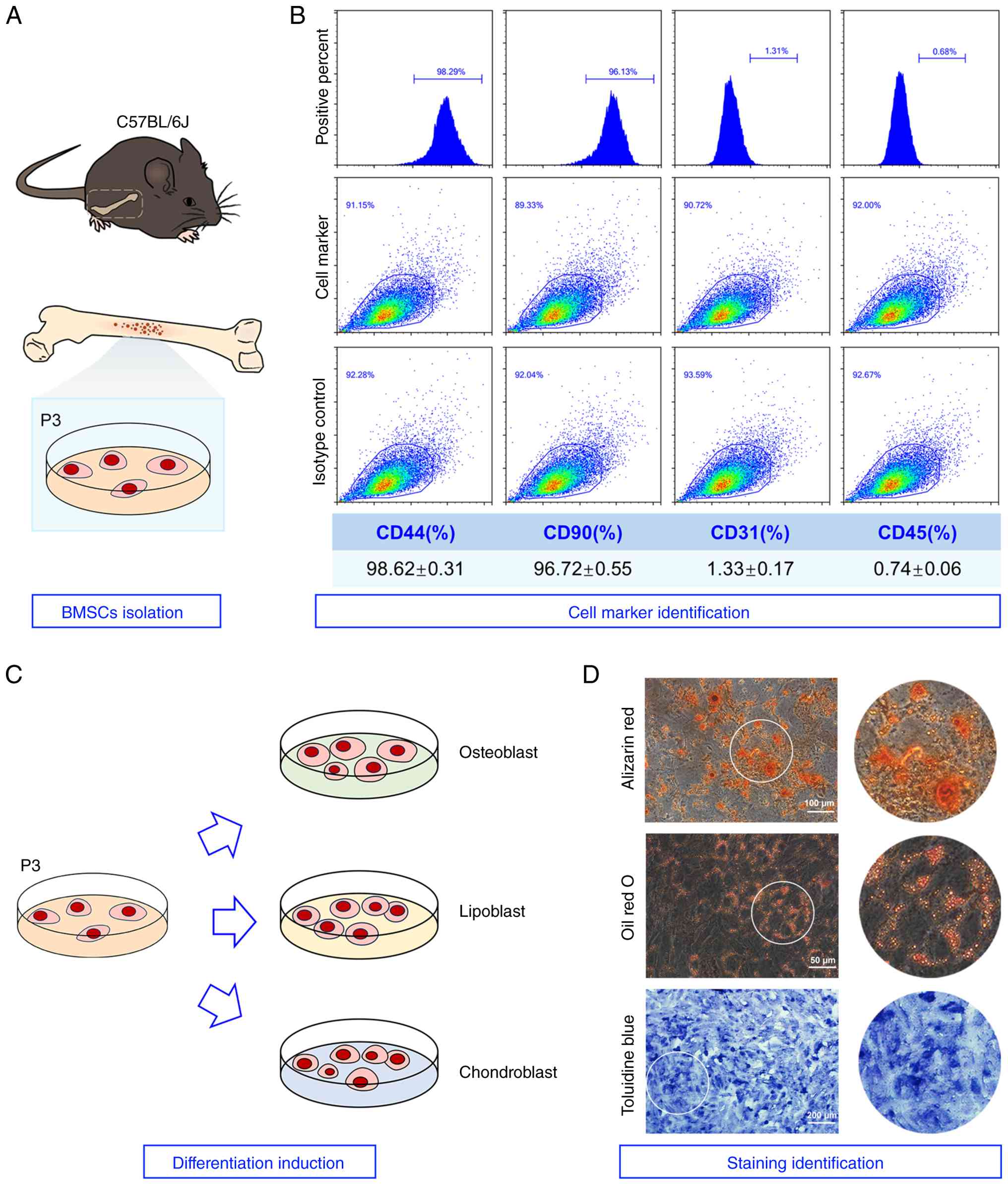
BMSCs were isolated from mouse bone marrow and
cultured to the third passage for use in the following experiments
(Fig. 1A). Flow cytometry was
used to detect the expression of cell surface marker proteins,
including CD44, CD90, CD31 and CD45. The results show revealed that
the expression levels of CD44 and CD90 on the cell surface were
increased, while the expression levels of CD31 and CD45 were
decreased (Fig. 1B). Following
the induced differentiation culture (Fig. 1C), Alizarin red, Oil Red O and
toluidine blue staining yielded strongly positive results,
indicating that the cells had the potential to differentiate into
osteogenesis, adipogenesis and chondrogenesis (Fig. 1D). These findings indicated that
BMSCs were successfully isolated.
Overexpression of Six3 enhances the
proliferation, migration and neural differentiation of BMSCs
As shown in Figs.
S1 and S2, the Gene
Ontology (GO) enrichment analyses of the differentially expressed
genes (DEGs) provided by the GSE8590 and GSE150710 datasets were
analyzed. The results revealed that they were generally involved in
biological processes such as neural development, stem cell
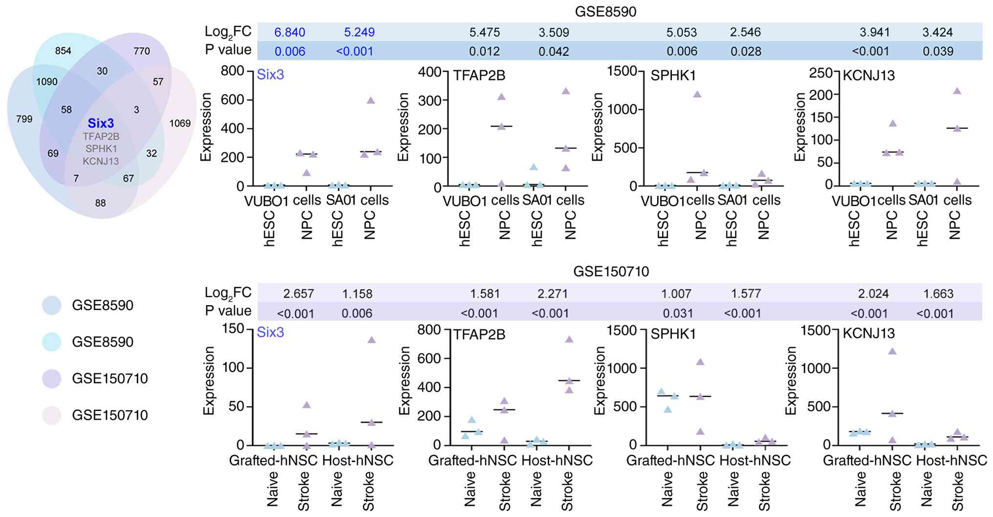
differentiation and neuronal regeneration. Of note, four potential
target genes were screened by analyzing the GSE8590 and GSE150710
data sets (Fig. 2). GSE8590
indicated that the expression of Six3 was elevated in neural
progenitor cells derived from hESCs. Notably, Six3 exhibited the
largest upregulation in expression among the four genes, suggesting
that Six3 may be involved in the neural differentiation of MSCs.
GSE150710 also revealed that the expression of Six3 was enhanced in
the host receiving human neural stem cells (hNSCs), and it was also
increased in the transplanted hNSCs, which further emphasized the
role of Six3 in stem cell transplantation for the treatment of
stroke.
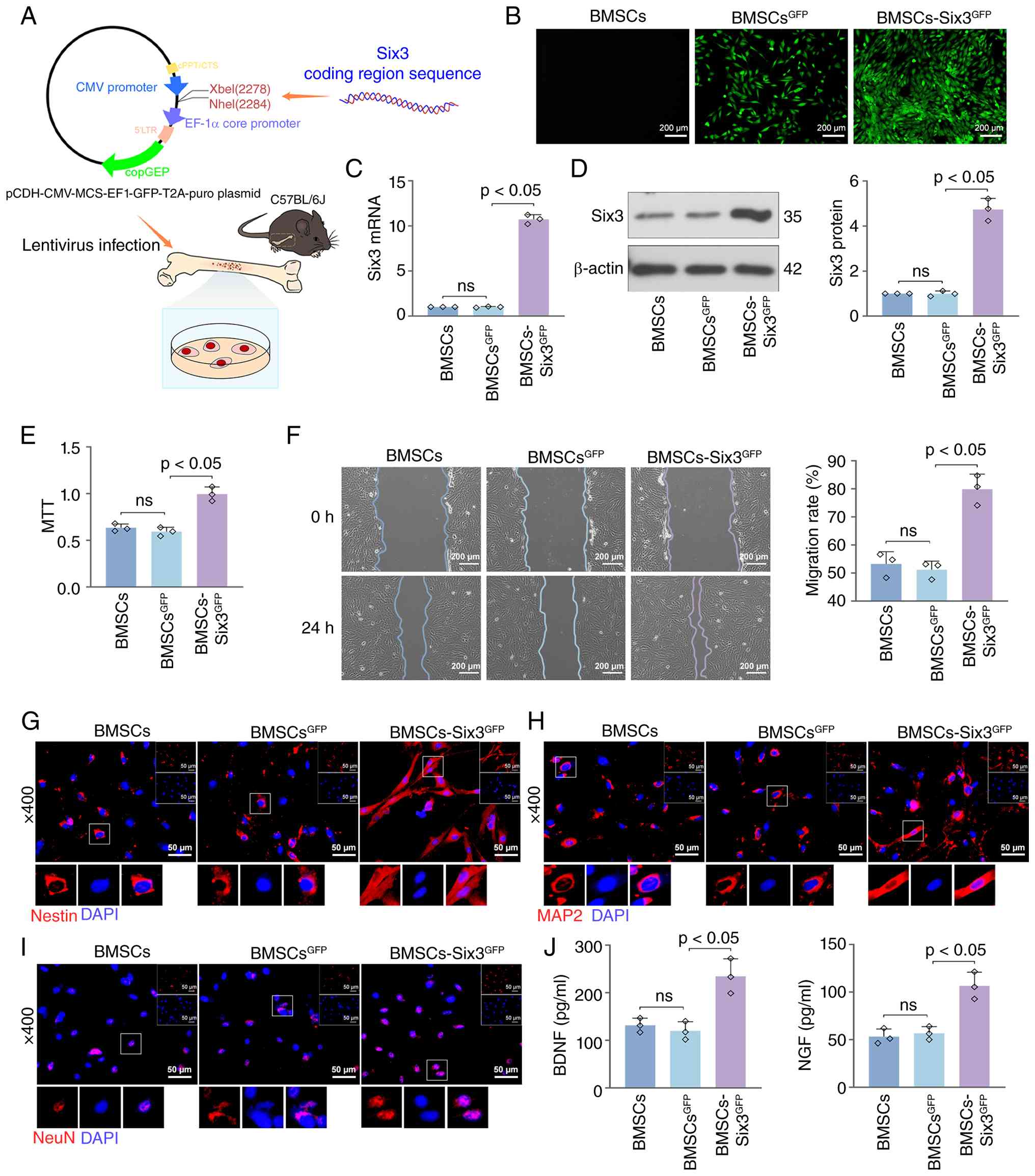
The present study aimed to modify BMSCs through a
lentivirus-mediated Six3 overexpression system and then explore the
role of Six3 in BMSCs (Fig. 3A).
Following 48 h of infection, the successful infection by the
lentivirus was observed under a fluorescence microscope (Fig. 3B). Additionally, it was observed
that the overexpression of Six3 enhanced the proliferation of
BMSCs. RT-qPCR and western blot analysis revealed that Six3 was
successfully overexpressed in BMSCs (Fig. 3C and D). The overexpression of
Six3 enhanced BMSC viability (Fig.
3E) and promoted BMSC migration in vitro (Fig. 3F). According to the
representative immunofluorescence images of neuronal marker
proteins, the overexpression of Six3 increased the expression of
Nestin (Fig. 3G), microtubule
associated protein 2 (MAP2) (Fig.
3H) and neuronal nuclei (NeuN) (Fig. 3I) in BMSCs. The levels of the
neuronal growth factors, BDNF and NGF, in the cell supernatant were
increased by the overexpression of Six3 (Fig. 3J). These results demonstrate the
potential of Six3 to promote the differentiation of BMSCs into
neurons.
Six3 overexpression activates Btg2
transcription and promotes its expression in BMSCs
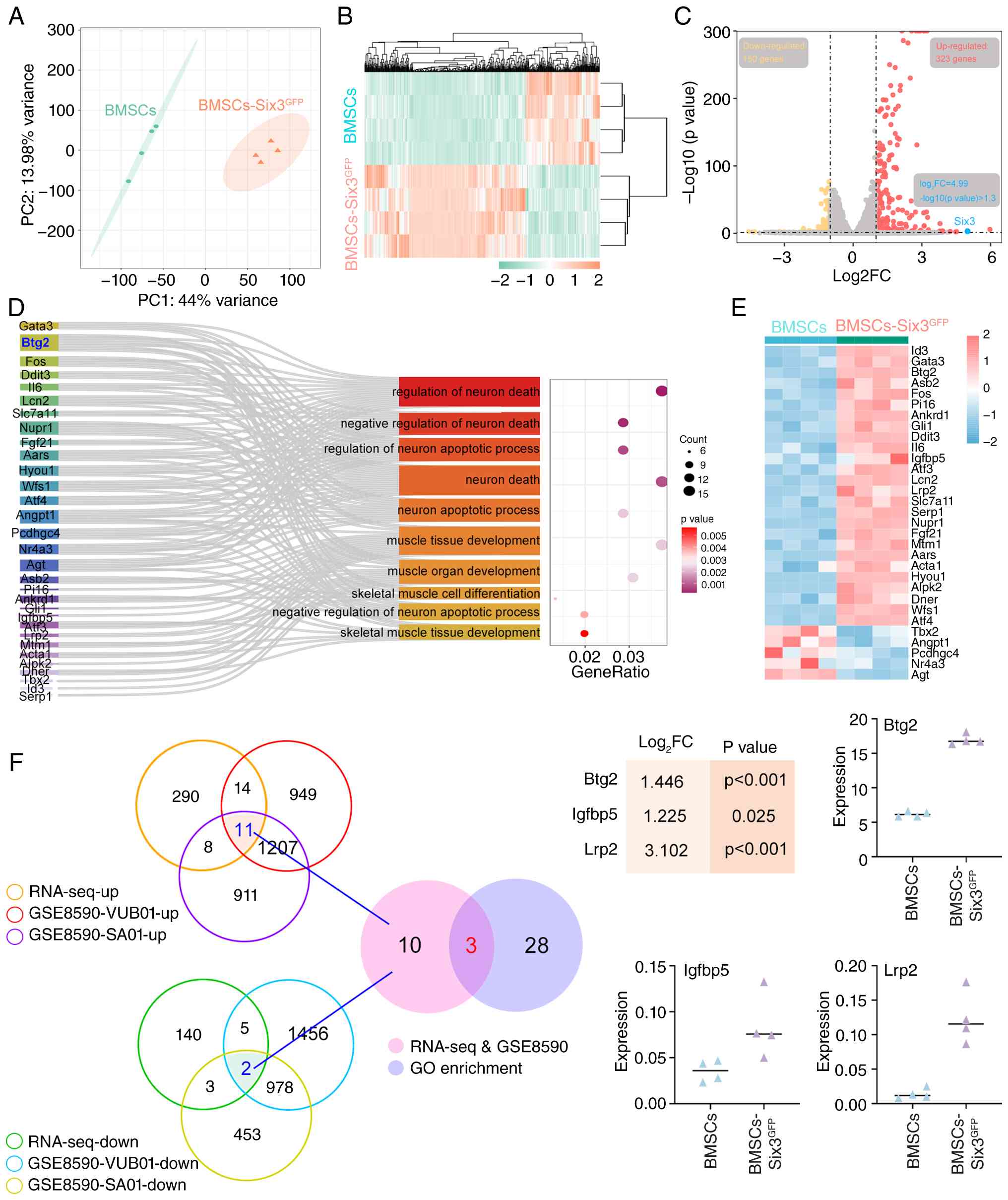
As demonstrated in Fig. 4A, the results revealed a
significant separation of BMSCs and BMSCS-Six3GFP
samples for transcriptomic analysis, and the favorable homogeneity
of samples within the groups. The findings depicted in Fig. 4B also demonstrated that the
overexpression of Six3 significantly altered the transcriptomes of
BMSCs, including 150 downregulated genes and 323 upregulated genes
(|Log2FC|>1, P<0.05); among these, the expression
of Six3 was significantly increased (Log2FC=4.99,
P<0.05), indicating that Six3 was successfully overexpressed
(Fig. 4C). Through GO annotation
and enrichment analysis of the aforementioned DEGs, it was observed
that Six3 overexpression targets the genes related to neuronal
death (Fig. 4D), including five
downregulated genes and 26 upregulated genes (Fig. 4E). In order to further explore
the mechanisms of Six3 overexpression, the upregulated or
downregulated genes in the results of RNA-sequencing and DEGs in
the GSE8590 dataset were comprehensively analyzed (Fig. 4F), and 13 shared genes were
obtained. Among these, three genes were found to be enriched in
terms related to neuronal death, including Btg2, Igfbp5 and Lrp2.
It was observed that Btg2 was significantly increased following the
overexpression of Six3 and had the highest basal expression in
BMSCs. Therefore, Btg2 was selected for further analyses.
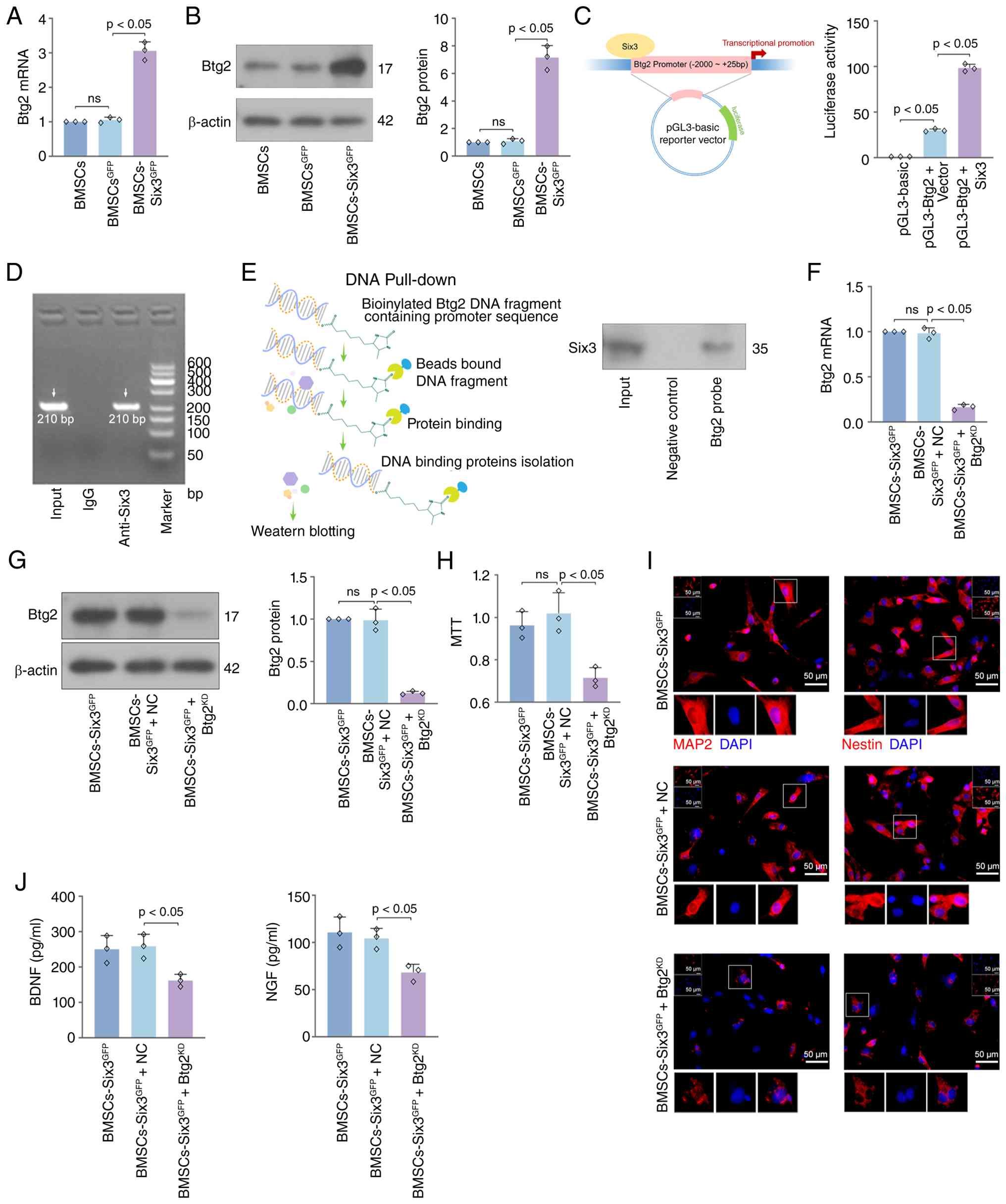
RT-qPCR and western blot analysis further confirmed
that the overexpression of Six3 increased the expression of Btg2
(Fig. 5A and B). Dual luciferase
report assay revealed that Six3 enhanced Btg2 promoter activity
(Fig. 5C), and the ChIP-PCR
results indicated that Six3 bound to the Btg2 promoter region
(Fig. 5D). The Btg2 promoter
binding protein was obtained through DNA pull-down assay, and the
binding of Six3 and Btg2 promoter was further demonstrated by
western blot analysis (Fig.
5E).
Six3 promotes BMSC viability and neural
differentiation by promoting Btg2 expression
RT-qPCR and western blot analysis demonstrated that
Btg2 expression in BMSCs was upregulated by the overexpression of
Six3 and downregulated by the BTG2-targeting lentiviral system
(Fig. 5F and G). Consistent with
the aforementioned findings, the overexpression of Six3 increased
the cell viability of BMSCs (Fig.
5H), the expression of neuronal marker proteins (MAP2 and
Nestin) (Fig. 5I), and the
secretion of BDNF and NGF (Fig.
5J). However, Btg2 knockdown reversed the effects of Six3;
these findings indicated that Btg2 mediated the promoting effects
of Six3 on the neural differentiation of BMSCs.
Six3 overexpression improves the
therapeutic effects of BMSC transplantation on stroke
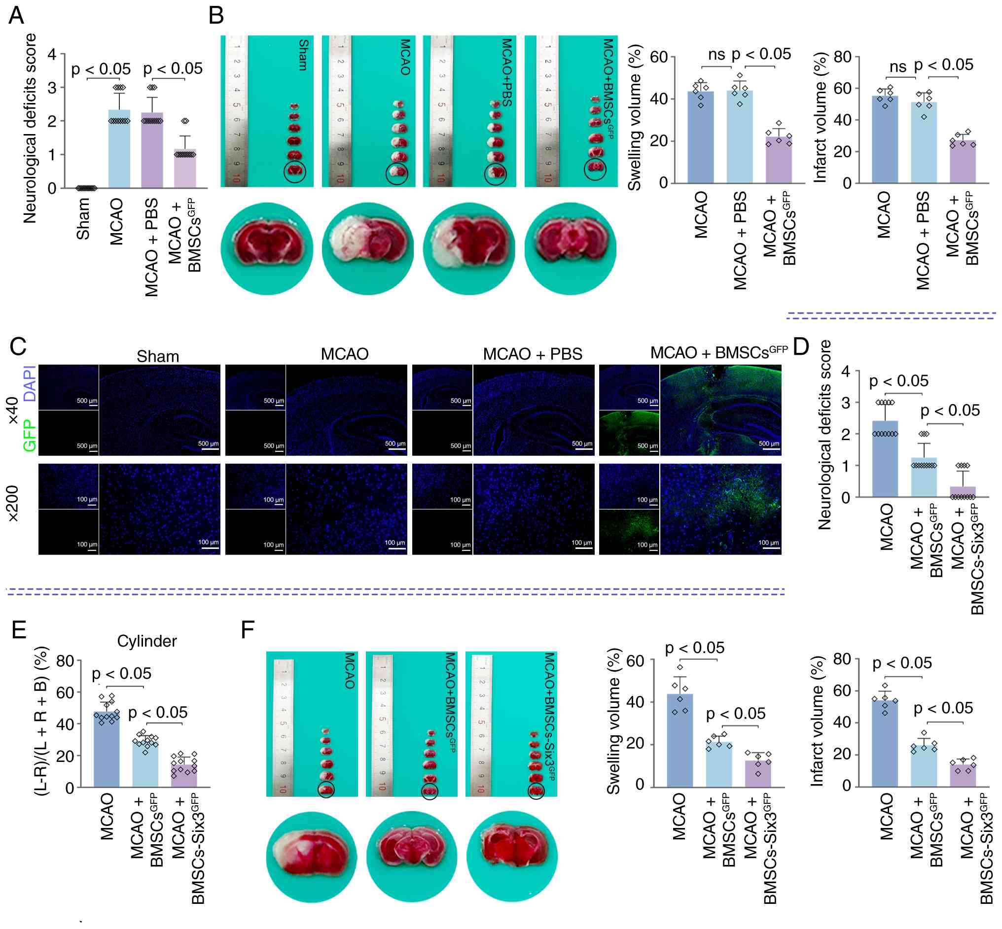
Mice subjected to MCAO exhibited moderate to severe
neurological deficits (Fig. 6A),
such as circling to the contralateral side. Mice transplanted with
BMSCs exhibited mild neurological deficits. Representative TTC
staining images revealed that BMSC transplantation alleviated
cerebral ischemic injury in mice subjected to MCAO (Fig. 6B). Specifically, BMSC
transplantation reduced brain swelling and cerebral infarction
caused by MCAO. By observing the distribution of GFP in brain
tissue, it was also observed that BMSCs were transplanted into the
brain tissue (Fig. 6C). The
200-fold magnification images demonstrated GFP expression in the
ischemic penumbra, demonstrating the migration of BMSCs to the
injured area. As demonstrated in Fig. 6D, the neural deficits were
further improved in the mice subjected to MCAO and transplanted
with Six3-overexpressing BMSCs. The infusion of BMSCs reduced the
limb asymmetry in stroke-stricken mice. Mice infused with Six3
overexpressed BMSCs exhibited more balanced limb use (Fig. 6E). The TTC staining results
revealed that compared with BMSC transplantation, the
transplantation of Six3-overexpressing BMSCs had a more prominent
reducing effect on brain swelling and cerebral infarction (Fig. 6F).
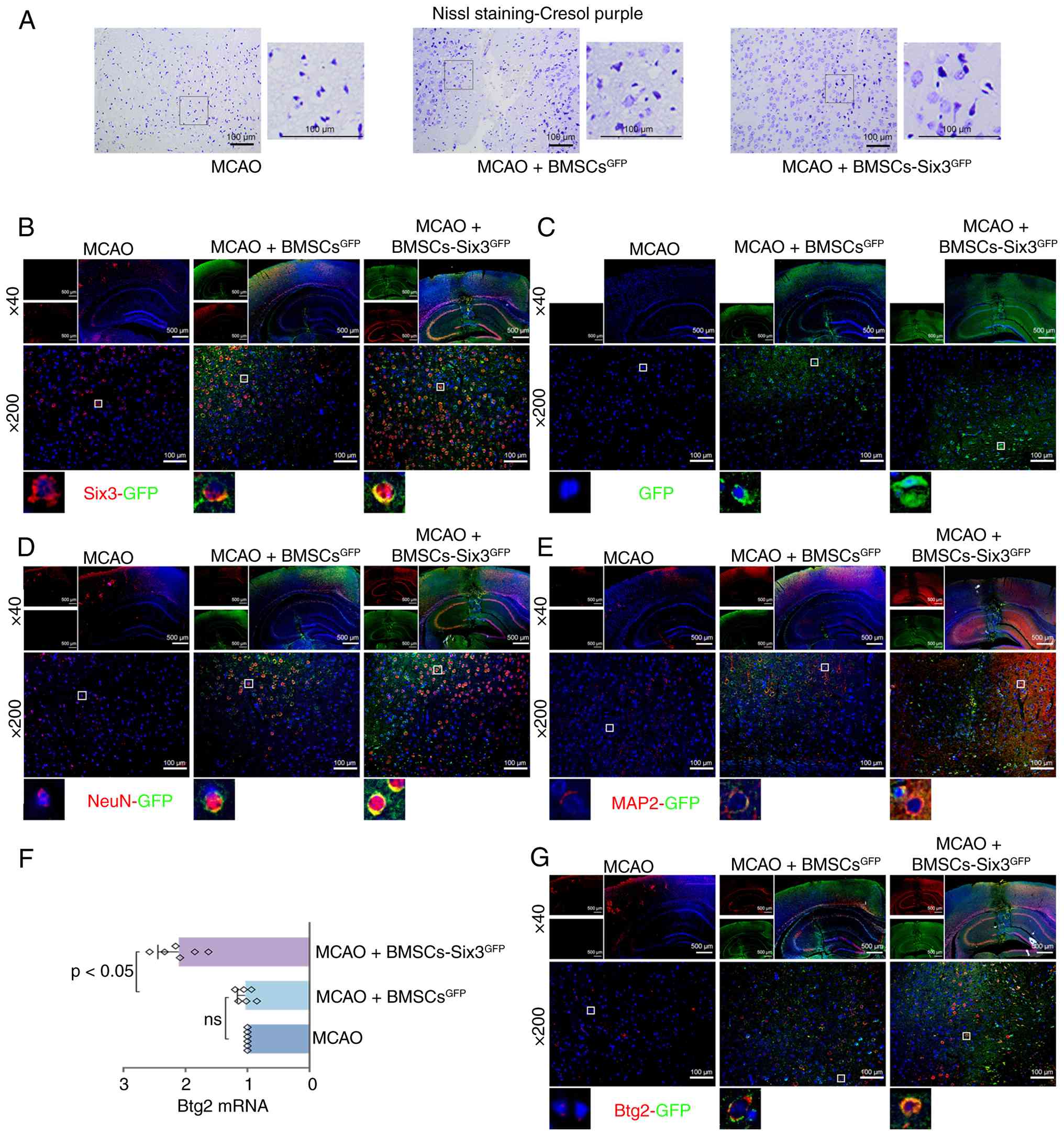
Neuronal atrophy and loss were detected in the brain
tissue of mice subjected to MCAO, which was reduced by BMSC
transplantation (Fig. 7A). The
transplantation of Six3-overexpressing BMSCs further protected the
morphology of neurons in the brain tissue of mice subjected to MCAO
and increased the number of neurons. The expression of Six3 and GFP
in brain tissue is illustrated in Fig. 7B. The colocalization of Six3 and
GFP expression was observed in the ischemic penumbra of mice
transplanted with BMSCs. The high expression of Six3 in BMSCs was
observed in the brain tissues of mice in the MCAO + BMSCs-Six3
group. The expression of GFP is illustrated in Fig. 7C. Compared with the MCAO + BMSCs
group, the overexpression of Six3 promoted the migration of BMSCs
to the infarct area in brain tissue. As demonstrated in Fig. 7D and E, the expression of the
neuronal markers, NeuN and MAP2, was increased in the BMSCs within
the host. Notably, the overexpression of Six3 in BMSCs enhanced
enhances the expression of these neuronal markers in BMSCs within
the host brain tissue. These findings suggest that the
overexpression of Six3 in BMSCs enhances their homing ability and
neural differentiation in the damaged brain tissue of mice
subjected to MCAO. Additionally, it was observed that the
expression of Btg2 in the brain tissues of mice with stroke was
increased following the transplantation of Six3-overexpressing
BMSCs (Fig. 7F). As demonstrated
in Fig. 7G, Btg2 was widely
expressed in the brain tissues of mice subjected to MCAO. The
colocalization of Btg2 and GFP was detected in the brain tissue of
mice transplanted with BMSCs. The increased expression of Btg2 was
observed in the Six3-overexpressing BMSCs within the injured brain
tissue. The aforementioned evidence indicates that the promoting
effect of the Six3-Btg2 axis on the migration and neural
differentiation of BMSCs in vitro is also observed in the
brain tissues of mice subjected to MCAO that received BMSC
transplantation.
Btg2 silencing reverses the effects of
Six3 overexpression on BMSCs in vivo
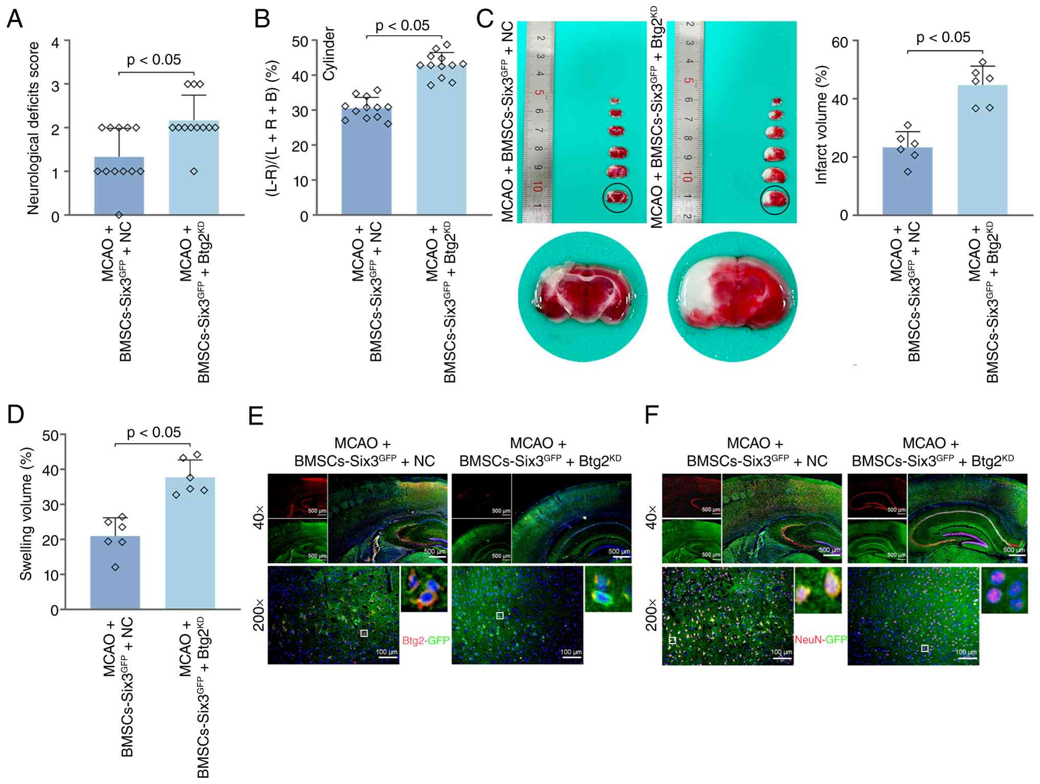
The in vivo rescue experiments demonstrated
that Btg2 mediated the enhancing effects of Six3 on the therapeutic
efficacy of infused BMSCs for the treatment of stroke. The
silencing of Btg2 led to an increase in the neurological deficit
score (Fig. 8A), an increase in
the asymmetry of limb use (Fig.
8B), an increase in cerebral infarction (Fig. 8C) and an increase in brain
swelling (Fig. 8D). The
double-stained immunofluorescence images confirmed the reduction of
Btg2 expression in BMSCs (Fig.
8E). Btg2 knockdown abolished the promoting effects of Six3 on
the neural differentiation of infused BMSCs (Fig. 8F).
Overexpression of Six3 enhances the
neural differentiation of BMSCs by activating the Btg2-mediated Akt
signaling pathway
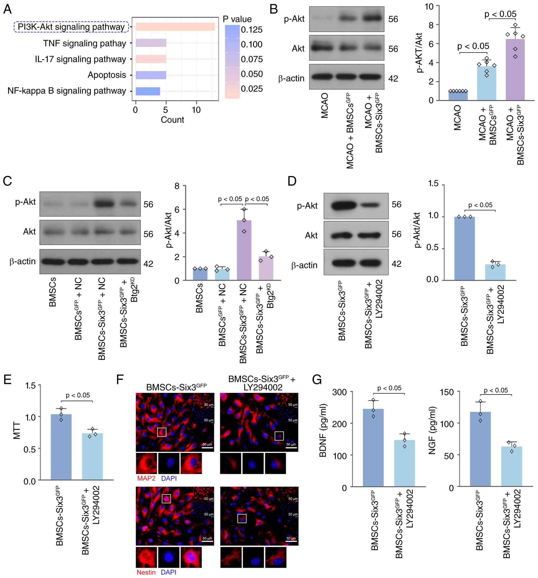
Kyoto Encyclopedia of Genes and Genomes enrichment
analysis of the upregulated DEGs that were identified through RNA
sequencing was performed to investigate the regulatory effects of
Six3 on downstream signaling pathways. The results revealed that
the overexpression of Six3 primarily affected the PI3K-Akt
signaling pathway (Fig. 9A).
Elevated levels of Akt phosphorylation were observed in brain
tissues of mice transplanted with Six3-overexpressing BMSCs
(Fig. 9B). The silencing of Btg2
partially suppressed Akt phosphorylation in BMSCs induced by Six3
overexpression (Fig. 9C). It was
demonstrated that the Akt signaling pathway was directly activated
by the overexpression of Btg2, rather than being secondary to
altered cellular states (Fig.
S3A-C). In rescue experiments, BMSCs were treated with the PI3K
inhibitor, LY294002, to inhibit the activation of the Akt signaling
pathway (Fig. 9D). Treatment
with LY294002 reversed the promoting effects of Six3 overexpression
on BMSC viability (Fig. 9E) and
neural differentiation, as evidenced by the suppression of MAP2 and
Nestin expression (Fig. 9F) and
the reduced secretion of BDNF and NGF (Fig. 9G). To determine whether the Akt
signaling pathway is directly regulated by Btg2, rather than being
secondary to changes in the cell state, loss-of-function and rescue
experiments were conducted. As demonstrated in Fig. S1A and B, Btg2 silencing reversed
the effects of Btg2 overexpression, including reducing the
viability of BMSCs and inactivating the Akt signaling pathway.
Discussion
In the present study, the tail vein infusion of
Six3-overexpressing BMSCs into mice subjected to MCAO resulted in
an increase in the number of neurons, a decreased infarct size and
improved neurological symptoms. The efficacy of BMSCs on cerebral
ischemia was consistent with that demonstrated in previous research
(36), indicating that BMSC
transplantation was effective in alleviating experimental cerebral
ischemia. Notably, Six3-overexpressing BMSCs exerted an improved
therapeutic effect than unmodified BMSCs.
Through the comprehensive analysis of GSE8590 and
GSE150710 databases, the shared genes, Six3, TFAP2B, SPHK1 and
KCNJ13, were obtained. Through a review of the literature, it was
found that the role of Six3 in cerebral ischemia has not been
revealed. According to current reports, Six3 may play a critical
important role in neuronal differentiation. Six3 has been reported
to promote neuronal differentiation by inhibiting Wnt5a expression
in neural stem cells during early differentiation (12). It has been shown that the
conditional deletion of Six3 blocks the differentiation of
precursor cells of medium spiny neurons, and the conditional
overexpression of Six3 promotes the differentiation of precursor
cells in lateral ganglionic eminence (14). Although previous studies
(12,14) have suggested the potential of
Six3 in promoting neuronal differentiation, the role of Six3 on
BMSCs remains unclear. Based on this, the present study used the
Six3-overexpressing lentivirus system to enforce Six3 expression in
mouse BMSCs. The overexpression of Six3 enhanced the viability and
migration of BMSCs, a finding which was consistent with that of a
previous study (13). The
overexpression of Six3 promoted the expression of Nestin, MAP2 and
NeuN in BMSCs, and increased the production of BDNF and NGF. The
increase in these markers indicates that BMSCs differentiate into
nerve cells, which can be found consistently in previously reported
models of MCAO (33,37). This elucidated that the
overexpression of Six3 promoted the differentiation of BMSCs into
neurons.
In the present study, in order to further explore
the downstream molecular mechanisms of Six3 regulating the
therapeutic effects of BMSCs, the target genes of Six3 were
analyzed using transcriptomics. The bioinformatics analysis results
revealed three candidate genes, Btg2, Igfbp5 and Lrp2. Among these,
Btg2 with a high basal expression was significantly upregulated in
the genes of interest and may be involved in the regulation of
neuronal cell death. During the extensive literature review
process, it was found that Btg2 promotes neuronal differentiation
and inhibits the apoptosis of terminally differentiated PC12 nerve
cells (21). Btg2 is highly
expressed in the ischemic penumbra of the brain, participating in
the survival and differentiation of nerve cells and exerting
neuroprotective effects (22).
Btg2 knockout has been shown to hinder the activation of the Akt
signaling pathway in mouse embryonic fibroblasts and bone marrow
progenitor cells (38). In
addition, the activation of the Akt signaling pathway has been
reported to promote the neural differentiation and migration of
BMSCs (39,40). The aforementioned evidence
prompted the authors to pay attention to Btg2 as a target gene for
Six3 in regulating the neural differentiation and migration of
BMSCs. The dual luciferase reporter and ChIP assays demonstrated
that Six3 directly binds to the Btg2 promoter and activates Btg2
transcription. The rescue experiments revealed that Btg2 knockdown
inhibited BMSC viability and neuronal differentiation, which
reversed the effects of Six overexpression. The present study
reported that the knockdown of Btg2 inhibited the Akt signaling
pathway, consistent with its previously reported function.
Therefore, it can be concluded that activation of the Six3/Btg2/Akt
axis is an effective strategy for improving the therapeutic
efficacy of BMSC transplantation in experimental stroke.
However, the significant critical role of the
candidate genes other than Btg2 in the nervous system cannot be
overlooked. IGFBP5 has been reported to be upregulated in the
nerves of patients with diabetic neuropathy and to lead to
neurodegenerative changes (41).
It has been demonstrated that Igfbp5 knockdown reduces
methamphetamine-induced neurobehavioral disorders and dopaminergic
neuronal apoptosis (42). It has
been reported that the absence of Lrp2 in the developing central
nervous system of mice leads to impaired closure of the lateral
cranial nerve canal and a reduction in folic acid uptake (43).
Furthermore, in the present study, the therapeutic
effects of Six3-overexpressing BMSC transplantation on stroke were
examined in mice subjected to MCAO. The intravenous infusion of
Six3-overexpressing BMSCs demonstrated superior efficacy in the
treatment of experimental ischemic injury than BMSCs alone, as
evidenced by neuronal protection, infarction inhibition and a
reduction in nerve defects, which was consistent with the results
of the in vitro experiments and attributed to the
improvement of the cell viability and migratory ability of BMSCs by
Six3. The silencing of Btg2 reversed the effects of Six3 in
vivo. This evidence indicates that the overexpression of Six3
is an effective genetic modification method for optimizing the
therapeutic effects of BMSCs on stroke, by promoting the
transcription of Btg2.
The present study adopted the administration route
of tail vein injection, which is a common administration method due
to its low invasiveness, simple operation and high repeatability
(33,44). It is particularly suitable for
non-invasive administration during the acute phase to avoid damage
caused by surgery. Although tail vein injection requires BMSCs to
pass through systemic veins and then cross the blood-brain barrier,
this limits the number of BMSCs that can reach the ischemic brain
tissues. However, the blood-brain barrier in MCAO mice is severely
damaged, and this pathological condition facilitates the delivery
of BMSCs to the injured area through tail vein injection.
Additionally, the present study aims to promote cell homing through
genetic modification rather than seeking to achieve the maximum
homing efficiency through drug administration. Therefore, a safe
and easy-to-operate intravenous administration method was adopted,
and the Six3 gene was overexpressed in BMSCs to enhance their
homing efficiency. This not only improved the efficacy of BMSCs
delivery in vivo but also ensured the safety and
repeatability of the administration.
The present study serves as a preclinical
experiment, with the core objective being to elucidate the
regulatory role and molecular mechanisms of Six3 overexpression on
the migration ability and neural differentiation potential of
BMSCs. Therefore, the currently most mature and efficient
lentiviral vector system, and the easy-to-operate tail vein
injection were selected for this purpose. However, the authors are
fully aware that the potential off-target effects caused by the
random integration characteristics of lentiviral vectors are the
main bottleneck restricting their future clinical application. In
clinical transformation work, whole-genome sequencing for
high-precision single-copy level analysis of integration sites may
be an effective means to assess off-target effects. Moreover, it is
necessary to consider that different administration routes, such as
intranasal delivery, may also be suitable for BMSC gene
modification and clinical use.
As regards the tumorigenicity and long-term effects
of Six3 overexpression, although no malignant transformation
phenotype was observed in the short-term in vivo experiments
of the present study, due to the current experimental period and
sample size, long-term tumorigenicity tests in immunodeficient mice
or the tracking of the clonal dynamics of transplanted cells in the
host body were not conducted. This constitutes a limitation of the
present study. The current experimental evidence indicates that
MSCs have a low tumorigenicity on their own (45,46). In addition, previous research has
discovered safe gene-modified lentiviral vectors encoding herpes
simplex virus thymidine kinase containing a point mutation that
results in an alanine to histidine substitution at residue 168
[HSV-TK(A168H)], and it has been indicated that this vector would
not cause the uncontrolled proliferation of human MSCs (47). This provides support for the
future clinical transformation of gene modification.
Given these limitations, it is considered that
before moving towards clinical application, further safe and
controllable gene delivery strategies should be developed. Future
research is required to focus on: i) Achieving the transient and
safe expression of exogenous genes using non-integrative vectors
(such as free plasmids, mRNA electroporation); ii) or using the
CRISPR/Cas9-mediated site-specific integration technology to
precisely insert therapeutic genes into the genome, fundamentally
avoiding long-term risks associated with random insertion mutations
and persistent overexpression.
In conclusion, the present study demonstrated that
the overexpression of Six3 promoted the viability, migration and
differentiation of BMSCs by activating Btg2 transcription, thereby
enhancing the therapeutic effects of BMSC transplantation on
stroke. The present study provides preclinical evidence for
lentiviral system-mediated gene modification to improve the
efficacy of BMSC transplantation for stroke.
Supplementary Data
Availability of data and materials
The data generated in the present study may be found
in the Gene Expression Omnibus under accession numbers GSE344610 or
at the following URL: https://www.ncbi.nlm.nih.gov/geo/query/acc.cgi?acc=GSE344610.
Authors' contributions
ZF and ZL acquired funding, conceptualized the
study, developed methodology, provided resources, conducted formal
analysis, curated data and wrote the original draft. LZ and WH
conducted investigation, formal analysis and data curation. HY and
JW performed validation, formal analysis, visualization and data
curation. HY and JW confirm the authenticity of all the raw data.
All authors read and approved the final version of the
manuscript.
Ethics approval and consent to
participate
Animal experiments were approved by the Animal
Ethics Committee of the First Affiliated Hospital of Zhengzhou
University (approval no. 2025-KY-1279; Zhengzhou, China).
Patient consent for publication
Not applicable.
Competing interests
The authors declare that they have no competing
interests.
Acknowledgements
Not applicable.
Funding
The present study was supported by the Key Project of Provincial
and Ministerial Co-construction for Medical Science and Technology
Research in Henan (grant no. SBGJ202102141) and the Key Scientific
Research Projects of Higher Education Institutions in Henan (grant
no. 22A320047).
References
|
1
|
GBD 2016 Stroke Collaborators: Global,
regional, and national burden of stroke, 1990-2016: A systematic
analysis for the global burden of disease study 2016. Lancet
Neurol. 18:439–458. 2019. View Article : Google Scholar : PubMed/NCBI
|
|
2
|
Feigin VL and Krishnamurthi R: Stroke is
largely preventable across the globe: Where to next? Lancet.
388:733–734. 2016. View Article : Google Scholar : PubMed/NCBI
|
|
3
|
Andersen KK, Olsen TS, Dehlendorff C and
Kammersgaard LP: Hemorrhagic and ischemic strokes compared: Stroke
severity, mortality, and risk factors. Stroke. 40:2068–2072. 2009.
View Article : Google Scholar
|
|
4
|
Namiot ED, Niemi JVL, Chubarev VN, Tarasov
VV and Schiöth HB: Stem cells in clinical trials on neurological
disorders: Trends in stem cells origins, indications, and status of
the clinical trials. Int J Mol Sci. 23:114532022. View Article : Google Scholar : PubMed/NCBI
|
|
5
|
Salehi MS, Safari A, Pandamooz S, Jurek B,
Hooshmandi E, Owjfard M, Bayat M, Zafarmand SS, Miyan JA and
Borhani-Haghighi A: The beneficial potential of genetically
modified stem cells in the treatment of stroke: A review. Stem Cell
Rev Rep. 18:412–440. 2022. View Article : Google Scholar
|
|
6
|
Anthony S, Cabantan D, Monsour M and
Borlongan CV: Neuroinflammation, stem cells, and stroke. Stroke.
53:1460–1472. 2022. View Article : Google Scholar : PubMed/NCBI
|
|
7
|
Rawat M, Ng C, Khan E, Shah RA, Ashfaq S,
Bahader GA and Shah ZA: Use of mesenchymal stem cell therapy in
COVID-19 related strokes. Neural Regen Res. 18:1881–1883. 2023.
View Article : Google Scholar :
|
|
8
|
Isaković J, Šerer K, Barišić B and
Mitrečić D: Mesenchymal stem cell therapy for neurological
disorders: The light or the dark side of the force? Front Bioeng
Biotechnol. 11:11393592023. View Article : Google Scholar
|
|
9
|
Asgari Taei A, Khodabakhsh P, Nasoohi S,
Farahmandfar M and Dargahi L: Paracrine effects of mesenchymal stem
cells in ischemic stroke: Opportunities and challenges. Mol
Neurobiol. 59:6281–6306. 2022. View Article : Google Scholar : PubMed/NCBI
|
|
10
|
Li Y, Zhong W and Tang X: Strategies to
improve the efficiency of transplantation with mesenchymal stem
cells for the treatment of ischemic stroke: A review of recent
progress. Stem Cells Int. 2021:99291282021. View Article : Google Scholar : PubMed/NCBI
|
|
11
|
Zhang Q, Zhou M, Wu X, Li Z, Liu B, Gao W,
Yue J and Liu T: Promoting therapeutic angiogenesis of focal
cerebral ischemia using thrombospondin-4 (TSP4) gene-modified bone
marrow stromal cells (BMSCs) in a rat model. J Transl Med.
17:1112019. View Article : Google Scholar : PubMed/NCBI
|
|
12
|
Shrestha P, Jaganathan A, Huilgol D,
Ballon C, Hwangbo Y and Mills AA: Chd5 regulates the transcription
factor Six3 to promote neuronal differentiation. Stem Cells.
41:242–251. 2023. View Article : Google Scholar : PubMed/NCBI
|
|
13
|
Pandolfi EC, Hoffmann HM, Schoeller EL,
Gorman MR and Mellon PL: Haploinsufficiency of SIX3 abolishes male
reproductive behavior through disrupted olfactory development, and
impairs female fertility through disrupted GnRH neuron migration.
Mol Neurobiol. 55:8709–8727. 2018. View Article : Google Scholar
|
|
14
|
Song X, Chen H, Shang Z, Du H, Li Z, Wen
Y, Liu G, Qi D, You Y, Yang Z, et al: Homeobox gene Six3 is
required for the differentiation of D2-type medium spiny neurons.
Neurosci Bull. 37:985–998. 2021. View Article : Google Scholar
|
|
15
|
Su Z, Wang Z, Lindtner S, Yang L, Shang Z,
Tian Y, Guo R, You Y, Zhou W, Rubenstein JL, et al:
Dlx1/2-dependent expression of Meis2 promotes neuronal fate
determination in the mammalian striatum. Development.
149:dev2000352022. View Article : Google Scholar : PubMed/NCBI
|
|
16
|
Xu Z, Liang Q, Song X, Zhang Z, Lindtner
S, Li Z, Wen Y, Liu G, Guo T, Qi D, et al: SP8 and SP9 coordinately
promote D2-type medium spiny neuron production by activating Six3
expression. Development. 145:dev1654562018. View Article : Google Scholar
|
|
17
|
Zhao L, Tan S, Liao Q, Li X, Ke T and Li
S: The neuroprotective effect and RNA-sequence analysis of
postconditioning on the ischemic stroke with diabetes mellitus tree
shrew model. Brain Behav. 11:e23542021. View Article : Google Scholar : PubMed/NCBI
|
|
18
|
Matsuda S, Rouault J, Magaud J and Berthet
C: In search of a function for the TIS21/PC3/BTG1/TOB family. FEBS
Lett. 497:67–72. 2001. View Article : Google Scholar : PubMed/NCBI
|
|
19
|
Tirone F: The gene PC3(TIS21/BTG2),
prototype member of the PC3/BTG/TOB family: Regulator in control of
cell growth, differentiation, and DNA repair? J Cell Physiol.
187:155–165. 2001. View Article : Google Scholar
|
|
20
|
Micheli L, Ceccarelli M, Farioli-Vecchioli
S and Tirone F: Control of the normal and pathological development
of neural stem and progenitor cells by the PC3/Tis21/Btg2 and Btg1
genes. J Cell Physiol. 230:2881–2890. 2015. View Article : Google Scholar : PubMed/NCBI
|
|
21
|
el-Ghissassi F, Valsesia-Wittmann S,
Falette N, Duriez C, Walden PD and Puisieux A: BTG2(TIS21/PC3)
induces neuronal differentiation and prevents apoptosis of
terminally differentiated PC12 cells. Oncogene. 21:6772–6778. 2002.
View Article : Google Scholar : PubMed/NCBI
|
|
22
|
Slevin M, Sanfeliu C, Turu MM,
Grau-Olivares M, Ferrer I, Boluda S, Marti-Fabregas J, Kumar S,
Kumar P and Krupinski J: B-cell translocation gene 2 is
over-expressed in peri-infarct neurons after ischaemic stroke.
Pathobiology. 76:129–135. 2009. View Article : Google Scholar : PubMed/NCBI
|
|
23
|
Lai JS, Cheng CW, Lo A, Sung TY and Hsu
WL: Lipid exposure prediction enhances the inference of rotational
angles of transmembrane helices. BMC Bioinformatics. 14:3042013.
View Article : Google Scholar :
|
|
24
|
Li M, Zhao W, Gao Y, Hao P, Shang J, Duan
H, Yang Z and Li X: Differentiation of bone marrow mesenchymal stem
cells into neural lineage cells induced by bFGF-chitosan controlled
release system. Biomed Res Int. 2019:50862972019.PubMed/NCBI
|
|
25
|
Soleimani M and Nadri S: A protocol for
isolation and culture of mesenchymal stem cells from mouse bone
marrow. Nat Protoc. 4:102–106. 2009. View Article : Google Scholar
|
|
26
|
Zhang T, Lee YW, Rui YF, Cheng TY, Jiang
XH and Li G: Bone marrow-derived mesenchymal stem cells promote
growth and angiogenesis of breast and prostate tumors. Stem Cell
Res Ther. 4:702013. View Article : Google Scholar : PubMed/NCBI
|
|
27
|
Huang S, Xu L, Sun Y, Wu T, Wang K and Li
G: An improved protocol for isolation and culture of mesenchymal
stem cells from mouse bone marrow. J Orthop Translat. 3:26–33.
2014. View Article : Google Scholar : PubMed/NCBI
|
|
28
|
Livak KJ and Schmittgen TD: Analysis of
relative gene expression data using real-time quantitative PCR and
the 2(-Delta Delta C(T)) method. Methods. 25:402–408. 2001.
View Article : Google Scholar
|
|
29
|
Kim D, Langmead B and Salzberg SL: HISAT:
A fast spliced aligner with low memory requirements. Nature
Methods. 12:357–360. 2015. View Article : Google Scholar : PubMed/NCBI
|
|
30
|
Anders S, Pyl PT and Huber W: HTSeq-a
Python framework to work with high-throughput sequencing data.
Bioinformatics. 31:166–169. 2015. View Article : Google Scholar :
|
|
31
|
Love MI, Huber W and Anders S: Moderated
estimation of fold change and dispersion for RNA-seq data with
DESeq2. Genome Biol. 15:5502014. View Article : Google Scholar
|
|
32
|
Sampei K, Goto S, Alkayed NJ, Crain BJ,
Korach KS, Traystman RJ, Demas GE, Nelson RJ and Hurn PD: Stroke in
estrogen receptor-alpha-deficient mice. Stroke. 31:738–744. 2000.
View Article : Google Scholar : PubMed/NCBI
|
|
33
|
Ikegame Y, Yamashita K, Hayashi S, Mizuno
H, Tawada M, You F, Yamada K, Tanaka Y, Egashira Y, Nakashima S, et
al: Comparison of mesenchymal stem cells from adipose tissue and
bone marrow for ischemic stroke therapy. Cytotherapy. 13:675–685.
2011. View Article : Google Scholar : PubMed/NCBI
|
|
34
|
Chang YS, Mu D, Wendland M, Sheldon RA,
Vexler ZS, McQuillen PS and Ferriero DM: Erythropoietin improves
functional and histological outcome in neonatal stroke. Pediatr
Res. 58:106–111. 2005. View Article : Google Scholar : PubMed/NCBI
|
|
35
|
Shvedova M, Islam MR, Armoundas AA,
Anfinogenova ND, Wrann CD and Atochin DN: Modified middle cerebral
artery occlusion model provides detailed intraoperative cerebral
blood flow registration and improves neurobehavioral evaluation. J
Neurosci Methods. 358:1091792021. View Article : Google Scholar
|
|
36
|
Shan H, Gao L, Zhao S, Dou Z and Pan Y:
Bone marrow mesenchymal stem cells with PTBP1 knockdown protect
against cerebral ischemia-reperfusion injury by inhibiting
ferroptosis via the JNK/P38 pathway in rats. Neuroscience.
560:130–142. 2024. View Article : Google Scholar : PubMed/NCBI
|
|
37
|
Shichinohe H, Ishihara T, Takahashi K,
Tanaka Y, Miyamoto M, Yamauchi T, Saito H, Takemoto H, Houkin K and
Kuroda S: Bone marrow stromal cells rescue ischemic brain by
trophic effects and phenotypic change toward neural cells.
Neurorehabil Neural Repair. 29:80–89. 2015. View Article : Google Scholar
|
|
38
|
Kim BC, Ryu MS, Oh SP and Lim IK:
TIS21/(BTG2) negatively regulates estradiol-stimulated expansion of
hematopoietic stem cells by derepressing Akt phosphorylation and
inhibiting mTOR signal transduction. Stem Cells. 26:2339–2348.
2008. View Article : Google Scholar
|
|
39
|
Lan XY, Liang XS, Cao MX, Qin HM, Chu CY,
Boltze J and Li S: NCAM mimetic peptide P2 synergizes with bone
marrow mesenchymal stem cells in promoting functional recovery
after stroke. J Cereb Blood Flow Metab. 44:1128–1144. 2024.
View Article : Google Scholar
|
|
40
|
Bing W, Pang X, Qu Q, Bai X, Yang W, Bi Y
and Bi X: Simvastatin improves the homing of BMSCs via the
PI3K/AKT/miR-9 pathway. J Cell Mol Med. 20:949–961. 2016.
View Article : Google Scholar : PubMed/NCBI
|
|
41
|
Simon CM, Rauskolb S, Gunnersen JM,
Holtmann B, Drepper C, Dombert B, Braga M, Wiese S, Jablonka S,
Pühringer D, et al: Dysregulated IGFBP5 expression causes axon
degeneration and motoneuron loss in diabetic neuropathy. Acta
Neuropathol. 130:373–387. 2015. View Article : Google Scholar
|
|
42
|
Qiao D, Xu J, Le C, Huang E, Liu C, Qiu P,
Lin Z, Xie WB and Wang H: Insulin-like growth factor binding
protein 5 (IGFBP5) mediates methamphetamine-induced dopaminergic
neuron apoptosis. Toxicol Lett. 230:444–453. 2014. View Article : Google Scholar : PubMed/NCBI
|
|
43
|
Kur E, Mecklenburg N, Cabrera RM, Willnow
TE and Hammes A: LRP2 mediates folate uptake in the developing
neural tube. J Cell Sci. 127:2261–2268. 2014.PubMed/NCBI
|
|
44
|
Chen J, Li Y, Wang L, Zhang Z, Lu D, Lu M
and Chopp M: Therapeutic benefit of intravenous administration of
bone marrow stromal cells after cerebral ischemia in rats. Stroke.
32:1005–1011. 2001. View Article : Google Scholar : PubMed/NCBI
|
|
45
|
Wang Y, Yi H and Song Y: The safety of MSC
therapy over the past 15 years: A meta-analysis. Stem Cell Res
Ther. 12:5452021. View Article : Google Scholar
|
|
46
|
Chin SP, Ng WC, Tai L, Mohd MA, Then KY
and Cheong SK: Umbilical cord-derived mesenchymal stem cells
(CLV-100) infusion in healthy subjects: A 5-year follow-up study on
safety and immunomodulatory effect. Regenerative Eng Transl Med.
11:723–736. 2025. View Article : Google Scholar
|
|
47
|
Bashyal N, Lee TY, Chang DY, Jung JH, Kim
MG, Acharya R, Kim SS, Oh IH and Suh-Kim H: Improving the safety of
mesenchymal stem cell-based ex vivo therapy using herpes simplex
virus thymidine kinase. Mol Cells. 45:479–494. 2022. View Article : Google Scholar : PubMed/NCBI
|