Introduction
Osteoarthritis (OA) is a chronic, degenerative and
disabling joint disease influenced by a combination of factors,
including age, sex and body weight. It is primarily characterized
by joint pain, swelling or deformation, along with morning
stiffness and limited joint movement (1). In 2020, OA affected ~595 million
individuals globally. Given the progressive increase in incidence,
the global prevalence of OA is projected to rise by 74.9% by 2050
(2). OA not only causes pain,
diminishes quality of life and impairs psychological wellbeing, but
also amplifies the economic burden on the healthcare system
(1,3). The primary pathological changes in
OA are characterized by elevated production of pro-inflammatory
mediators and accelerated matrix degradation in cartilage tissue
(4,5).
Under physiological conditions, the dynamic balance
between matrix synthesis and degradation is key for maintaining
cartilage homeostasis (6).
However, pro-inflammatory factors disturb this delicate balance,
hastening matrix degradation and intensifying the inflammatory
response. Specifically, the cytokines IL-1β and tumor necrosis
factor-α (TNF-α) suppress collagen type IIα1 chain (COL2A1)
expression. Simultaneously, they upregulate matrix-degrading
enzymes such as MMPs and aggregating proteinases [a disintegrin and
metalloproteinases with thrombospondin motifs (ADAMTS)] in
chondrocytes, thereby accelerating matrix degradation and the
progression of OA (7,8). Wang et al (9) demonstrated that IL-1β activates the
NF-κB signaling pathway, thereby enhancing the expression of MMPs
and ADAMTS in chondrocytes. Similarly, the activation of IL-17 and
TNF pathways in chondrocytes has been reported to inhibit anabolic
processes and promote catabolic activity within the articular
cartilage matrix (10). Kim
et al (11) demonstrated
that IL-1β and TNF-α induce mitochondrial DNA damage in
chondrocytes, which leads to apoptosis and exacerbated inflammatory
response. Xu et al (12)
discovered that TNF-α triggers pyroptosis in chondrocytes. This
leads to an increase in the expression of inflammatory factors,
including IL-1β and IL-18, which contribute to disrupted cell
morphology, cell death and an amplified inflammatory
microenvironment (12).
Therapeutic interventions for OA primarily include
non-steroidal anti-inflammatory drugs (NSAIDs), corticosteroid
injection, electrotherapy, ultrasound therapy and select physical
therapies. While these treatments alleviate acute symptoms, they
are associated with potential adverse reactions, such as
gastrointestinal complications from prolonged NSAID use and
accelerated cartilage degeneration following repeated
corticosteroid injection (5,13). Herbal medicine presents a
treatment alternative, distinguished by its reduced side effects
and enhanced safety profile compared with conventional
pharmaceuticals. Herbal monomers offer advantages due to their
well-defined chemical structures and capacity to modulate multiple
targets implicated in disease pathogenesis (14,15).
Resveratrol (RES) is a highly bioactive, naturally
occurring non-flavonoid polyphenol compound recognized for its
regulatory effect on cell proliferation, inflammation, oxidation
and apoptosis (16,17). RES has potential therapeutic
effects, including inflammatory regulation and the treatment of
metabolic and cardiovascular diseases (18-20). Mulberry (Morus alba),
knotweed (Polygonum cuspidatum), veratrum root (Veratrum
grandiflorum), and fleeceflower root (Polygonum
multiflorum), which are rich sources of RES, have been
extensively used in Chinese traditional medicine in the treatment
of various conditions, including tumors, diabetes, inflammation,
lipid metabolism disorder, orthopedics and cardiovascular disease
(16,21,22). In an OA rat model, RES
ameliorated inflammation by modulating inflammatory cytokines
(23). RES suppresses
extracellular matrix (ECM) degradation and promotes
glycosaminoglycan (GAG) synthesis in porcine chondrocytes (24). RES has also been reported to
regulate the expression of inflammatory and ECM
metabolism-associated genes in primary cultured chondrocytes
derived from patients with OA (25). However, the underlying molecular
mechanism of RES in the treatment of OA remains unclear. The
present study aimed to explore the underlying molecular mechanism
of RES on OA through network pharmacology analysis, in vitro
experiments and in vivo verification.
Materials and methods
Network pharmacology analysis
The potential targets of RES were predicted using
PubChem (pubchem.ncbi.nlm.nih.gov) and SwissTargetPrediction
(swisstargetprediction.ch/). All potential disease targets were
retrieved by searching databases including Genecards (genecards.org), Online Mendelian Inheritance in Man
(OMIM) (omim.org), and Drugbank (drugbank.com) with the keyword 'osteoarthritis'. The
standardized nomenclature annotation of the screened targets was
performed using the UniProt database (uniprot.org/).
The RES and OA targets predicted from the databases were uploaded
to the Venny 2.1.0 platform (liuxiaoyuyuan.cn/) to obtain the common targets of RES
and OA.
Overlapping targets shared between RES and OA were
uploaded to the STRING database (cn.string-db.org) to construct a protein-protein
interaction (PPI) network. The visualization of this PPI network
was performed using Cytoscape 3.9.2 software (cytoscape.org). Overlapping targets were further
analyzed using the Metascape database (metascape.org)
to perform Gene Ontology (GO; geneontology.org) enrichment analysis, encompassing
biological processes (BPs), molecular functions (MFs) and cellular
components (CCs). Kyoto Encyclopedia of Genes and Genomes (KEGG;
kegg.jp) enrichment analysis was conducted to identify relevant
signaling pathways. The data were imported into the Bioinformatics
Platform (bioinformatics.com.cn/login/) for visualization.
P<0.05 was considered to indicate significant enrichment.
Chemicals
RES (purity >99%; cat. no. R5010) for in
vitro assays was dissolved in dimethyl sulfoxide (cat. no.
D8418; both Sigma-Aldrich; Merck KGaA). RES (purity >98%; cat.
no. R8350; Beijing Solarbio Science & Technology Co., Ltd.) for
oral gavage, due to its suitability for animal experiments, was
diluted in 0.5% CMC-Na (dissolved in saline).
Cell culture
SW1353 cell line was purchased from the Cell Bank of
the Chinese Academy of Sciences and cultured in RPMI-1640 (cat. no.
SH30809.01; Cytiva) medium supplemented with 10% FBS (cat. no.
ZQ500-A; Zhong Qiao Xin Zhou Biotechnology Co., Ltd.) and 1%
penicillin/streptomycin (cat. no. SV30010; HyClone; Cytiva) at
37°C. The medium was refreshed every 2-3 days until cells reached
~80% confluence.
Cell proliferation assay
SW1353 cells were inoculated into 96-well plates at
a density of 3×10³/well overnight. Subsequently, cells
were treated with RES at 1, 2, 4, 16, 32, 64 and 128 μM for
24 or 48 h at 37°C. Following treatment, the culture medium was
removed and 100 μl/well Cell Counting Kit-8 [CCK-8; cat. no.
MI00615A; Mishu (Xi'an) Biotechnology Co., Ltd.] solution was added
to the plates. The cells were incubated for an additional 2 h.
Absorbance was determined by measuring the optical density (OD) at
450 nm using a microplate reader (BioTek Instruments, Inc.).
Establishment of OA cell model and
treatment
OA cell model was induced by IL-1β (10 ng/ml; cat.
no. 200-01B; PeproTech, Inc.) as previously described (26). Briefly, SW1353 cells were
inoculated into 6-well or 12-well plates at a density of
1×10⁴/cm2 and incubated for 24 h at 37°C.
Cells underwent serum starvation for 10-14 h before serum-free
RPMI-1640 (Cytiva) was replaced with complete medium. Subsequently,
cells were pretreated with 16 μM RES alone or a combination
of RES and compound C (ComC) (1 μM; cat. no. ab120843;
Abcam), a selective AMPK inhibitor, for 2 h at 37°C. This was
followed by 24 h treatment with 10 ng/ml IL-1β at 37°C (27,28). The cells were harvested and
centrifuged at 1,000 × g for 5 min at 4°C to obtain cell-free
supernatant for subsequent experiments.
Cytological staining
The cells were cultured at a density of
1×10⁴/cm2 and fixed with 4% paraformaldehyde
(cat. no. AR1069; BOSTER Biological Technology Co., Ltd.) for 30
min at room temperature. The cells were stained for 30 min with
Alcian Blue (AB) staining solution (pH 0.2; cat. no. ST046; Heart
Biological Technology Co., Ltd.) or Safranin O (SO) staining
solution (cat. no. G1067; Beijing Solarbio Science & Technology
Co., Ltd.) at room temperature. Slides were sealed with mounting
medium, and the cells were observed under a light microscope.
Reverse transcription-quantitative PCR
(RT-qPCR)
The cells were seeded into 6-well plates at a
density of 1×10⁵/well and total RNA extraction was
performed on ice using the SPARKeasy Cell RNA Quick Extraction kit
(cat. no. AC0205-A; Shandong Sikejie Biotechnology Co., Ltd.). The
RNA concentration was determined using a NanoDrop 2000
spectrophotometer (Thermo Fisher Scientific, Inc.). RNAs were
reverse-transcribed into cDNA using the SPARKscript II RT Plus kit
[with gDNA Eraser; cat. no. AG0304 (SY); Shandong Sikejie
Biotechnology Co., Ltd.], according to the manufacturer's
instructions. Primer sequences are listed in Table I. qPCR was performed using a
Bio-Rad qPCR system (Bio-Rad Laboratories, Inc.). The thermocycling
conditions were as follows: Initial denaturation at 95°C for 10
min, followed by 40 cycles of denaturation at 95°C for 15 sec and
combined annealing/extension at 58°C for 60 sec. The expression
levels of mRNAs were determined using the 2−ΔΔCq method
with β-actin as the internal reference (29).
 | Table IPrimers used in reverse
transcription-quantitative PCR. |
Table I
Primers used in reverse
transcription-quantitative PCR.
| Genes | Forward, 5'→3' | Reverse, 5'→3' |
|---|
| AMPK |
TTGAAACCTGAAAATGTCCTGCT |
GGTGAGCCACAACTTGTTCTT |
| PPARγ |
TACTGTCGGTTTCAGAAATGCC |
GTCAGCGGACTCTGGATTCAG |
| MMP13 |
CCAGACTTCACGATGGCATTG |
GGCATCTCCTCCATAATTTGGC |
| COL2A1 |
TGGACGATCAGGCGAAACC |
GCTGCGGATGCTCTCAATCT |
| IL-6 |
ACTCACCTCTTCAGAACGAATTG |
CCATCTTTGGAAGGTTCAGGTTG |
| IL-10 |
GACTTTAAGGGTTACCTGGGTTG |
TCACATGCGCCTTGATGTCTG |
| TNF-α |
CCTCTCTCTAATCAGCCCTCTG |
GAGGACCTGGGAGTAGATGAG |
| NLRP3 |
CCACAAGATCGTGAGAAAACCC |
CGGTCCTATGTGCTCGTCA |
| β-actin |
CCTGGCACCCAGCACAAT |
GGGCCGGACTCGTCATAC |
Western blotting
The cells were washed with ice-cold PBS. Phosphatase
inhibitors (cat. no. AR1183), protease inhibitors (cat. no.
AR1182-1; both BOSTER Biological Technology Co., Ltd.) and RIPA
buffer (cat. no. P0013C; Beyotime Biotechnology) were mixed at a
ratio of 1:1:100 and added to the cells. The lysate was centrifuged
at 12,000 × g for 10 min at 4°C and the supernatant was collected.
Protein concentration was determined using the BCA protein assay
kit (cat. no. ZJ102; Shanghai Epizyme Biomedical Technology Co.,
Ltd.). The denatured proteins (20 μg/lane) were separated
using 8-12% SDS-PAGE and then transferred to a polyvinylidene
fluoride membrane. Membranes were blocked using 5% skimmed milk for
1 h at room temperature and incubated with anti-MMP13 (1:2,000;
cat. no. BA2204), anti-COL2A1 (cat. no. BA0533; both BOSTER
Biological Technology Co., Ltd.), anti-AMPK (cat. no. CY5326),
anti-phosphorylated (p-)AMPK (all 1:1,000; cat. no. CY5608),
anti-PPARγ (cat. no. AY9445), anti-NLRP3 (both 1:750; cat. no.
CY5651; all Shanghai Ebiwei Biotechnology Co., Ltd.), anti-cleaved
caspase-1 (1:1,000; cat. no. ab179515; Abcam) and anti-β-actin
(1:5,000; cat. no. AB0035; Shanghai Ebiwei Biotechnology Co., Ltd.)
at 4°C overnight. The membrane was washed with 0.05% TBST, followed
by incubation with horseradish peroxidase-conjugated anti-rabbit
(1:10,000; cat. no. BA1054; BOSTER Biological Technology Co., Ltd.)
secondary antibodies dissolved in TBST for 1 h at room temperature.
The membrane was washed three times with TBST, followed by
chemiluminescence band detection using the enhanced luminescence
solution [cat. no. MI00607A; Mishu (Xi'an) Biotechnology Co.,
Ltd.]. The protein expression levels, normalized to β-actin, were
quantified using ImageJ software (version 1.53a; National
Institutes of Health).
Induction and treatment of OA mice
C57BL/6 male mice (n=22; age, ~10 weeks; weight, ~20
g), were purchased from the Laboratory Animal Center of Xi'an
Jiaotong University. Animals were housed at 25±2°C, 50±10% humidity
with a 12/12-h light/dark cycle and ad libitum access to
food/water. The animal procedures were approved by the Animal Care
and Use Committee of Xi'an Jiaotong University (approval no.
XJTUAE2024-1403; Xi'an, China). Mice were randomized into Sham
(n=6), OA (n=8) and RES (n=8) groups. The OA and RES mice were
established using the destabilization of the medial meniscus (DMM)
method on the right knee (30).
Mice were anesthetized via intraperitoneal injection of 3% sodium
pentobarbital solution (50 mg/kg). The joint capsule was dissected
along the medial margin of the patellar ligament and the tibial
collateral ligament of the medial meniscus was transected. Finally,
the joint cavity and skin were sutured. The mice in the Sham group
were only incised in the joint capsule on the right knee and
sutured. The DMM and Sham surgery success rates were 75-80 and
100%, respectively. Mice were excluded if surgical failure was
defined, either by the incomplete transection of the medial
meniscotibial ligament or by any accidental damage to the adjacent
articular cartilage or meniscus during the surgical procedure. The
final sample size was six mice/group. All exclusions were performed
before outcome assessments and the investigators were blinded to
group allocation. After 4 weeks, OA mice in the RES group received
RES at 45 mg/kg through oral gavage once/day for 6 weeks. Oral RES
exerts anti-OA effects at 45 mg/kg/day (31) and activates AMPK at 25-30 mg/kg
(32). Generally, effective RES
doses in animal studies range widely, with 40-50 mg/kg established
as standard regimens (33,34). Thus, a midrange dose of 45
mg/kg/day was selected. The mice in the Sham and OA groups were
concurrently administered an equal amount of the vehicle solution.
At 10 weeks post-surgery, the mice were subjected to euthanasia
using an overdose of sodium pentobarbital (150 mg/kg). Prior to
euthanasia, the adequacy of anesthesia was verified by the absence
of pedal withdrawal and corneal reflex, and then blood was
collected via cardiac puncture. Final death was confirmed by the
cessation of heartbeat and respiration. Liver and right knee joint
tissue was harvested for subsequent experiments.
ELISA
The cell culture supernatant and mouse blood samples
were centrifuged at 3,000 × g for 20 min at 4°C to obtain the
clarified cell supernatant and mouse serum, respectively. The
IL-1β, TNF-α, IL-6, IL-10 or IL-18 ELISA kits (IL-1β: cat. no.
RX203063M for serum; TNF-α: cat. no. RX104793H for supernatant;
cat. no. RX202412M for serum; IL-6: cat. no. RX106126H for
supernatant; cat. no. RX203049M for serum; IL-10: cat. no.
RX103064H for supernatant; cat. no. RX203075M for serum; IL-18:
cat. no. E10349 for supernatant; all Xi'an Shandezhiyuan
Biotechnology Co., Ltd.) were used according to the manufacturer's
instructions. The OD values were measured at 450 nm using a
microplate reader (BioTek Instruments, Inc.), and protein
concentration was determined using the standard curve method.
Liver function test
To evaluate liver function in the mice, serum levels
of alanine aminotransferase (ALT)/glutamic-pyruvic transaminase
(GPT) and aspartate aminotransferase (AST)/glutamic-oxaloacetic
transaminase (GOT) were measured using the ALT/GPT and AST/GOT test
kits (cat. nos. C009-2-1 and C010-2-1; both Nanjing Jiancheng
Bioengineering Institute) according to the manufacturer's
instructions. The OD values were measured using a microplate reader
(BioTek Instruments, Inc.) at a wavelength of 510 nm, with the
corresponding values of ALT/GPT and AST/GOT determined using the
standard curve method.
Histological staining of animal
tissue
Mouse right knee joint and liver tissue were excised
and fixed with 4% paraformaldehyde (cat. no. AR1069; BOSTER
Biological Technology Co., Ltd.) for 48 h at room temperature. The
joint tissues were decalcified. Tissue was embedded in paraffin and
sectioned into 5 μm paraffin sections. The tissue sections
were stained with hematoxylin and eosin (HE; hematoxylin for 6 min
and eosin for 1 min) or SO-fast green (FG) staining (FG for 10 min,
followed by SO for 30 sec) at room temperature. Stained sections
were examined under a light microscope (Leica GmbH). The severity
of synovitis was graded using a 9-point synovial score system
(35). The severity of OA and
cartilage degeneration was assessed using the Osteoarthritis
Research Society International scoring system (36), which ranges from 0 to 24.
Immunofluorescence (IF) and
immunohistochemistry (IHC) staining
For IF, SW1353 cells were inoculated onto cover
slides at a density of 1×10⁴/cm2 and fixed
with 4% paraformaldehyde (cat. no. AR1069; BOSTER Biological
Technology Co., Ltd.) for 30 min at room temperature. For IHC, the
5 μm paraffin sections of the knee joint tissue were
deparaffinized in xylene for 20 min and rehydrated through a graded
ethanol series at room temperature. Subsequently, the sections were
treated with 3% hydrogen peroxide for 10 min to block endogenous
peroxidase at room temperature, and digested with 20 μg/ml
proteinase K (cat. no. P6157; Macklin Biochemical Technology Co.,
Ltd.) for 20 min at 37°C. Next, both cell and tissue samples were
blocked with 3% BSA for 30 min at room temperature. Samples were
then incubated at 4°C overnight with the following primary
antibodies: anti-COL2A1 (1:200; cat. no. BA0533; BOSTER Biological
Technology Co., Ltd.), anti-p-AMPK (1:100; cat. no. CY5608) and
anti-PPARγ (1:100; cat. no. AY9445) (both from Shanghai Ebiwei
Biotechnology Co., Ltd.), anti-AMPK (for IF only; 1:100; cat. no.
CY5326; Shanghai Ebiwei), anti-MMP13 (for IF: 1:300, cat. no.
bs-0575R, Beijing Biosynthesis Biotechnology Co., Ltd.; for IHC:
1:100, cat. no. BA2204, BOSTER), and anti-NLRP3 (for IF: 1:100,
cat. no. CY5651, Shanghai Ebiwei; for IHC: 1:50, cat. no. BA3677;
BOSTER). Following PBS washes, IF samples were incubated with Alexa
Fluor® 594-conjugated goat anti-rabbit IgG (1:1,000;
cat. no. ab150080; Abcam) in 3% BSA for 1 h at room temperature in
the dark, and counterstained with DAPI for 5 min. Sections were
incubated with HRP-conjugated goat anti-rabbit IgG (1:60; cat. no.
RS0002; Immunoway Biotechnology, Inc.) for 30 min at room
temperature, treated with streptavidin-peroxidase for 30 min,
visualized with DAB, and counterstained with hematoxylin for 1 min
at room temperature before being mounted with neutral gum. All
images were captured using a microscope (fluorescence for IF and
light for IHC; Leica GmbH). Image analysis was performed using
ImageJ (version 1.53a; National Institutes of Health);
specifically, the mean fluorescence intensity and the quantitative
analysis of the PPARγ nuclear-to-cytoplasmic ratio were measured
for IF samples.
Statistical analysis
All in vitro experiments were independently
replicated ≥3 times and all in vivo animal studies were
conducted with six mice per group. All data are presented as the
mean ± SD. Prior to parametric testing, data normality and
homogeneity of variance were verified using the Shapiro-Wilk test
and Brown-Forsythe test, respectively. One-way ANOVA was used,
followed by Dunnett's or Sidak's multiple comparisons test for post
hoc pairwise analysis. All statistical analyses were performed
using GraphPad Prism 9.5 software (GraphPad Software, Inc.;
Dotmatics). P<0.05 was considered to indicate a statistically
significant difference.
Results
AMPK and PPARγ signaling pathways are key
therapeutic targets of RES in OA
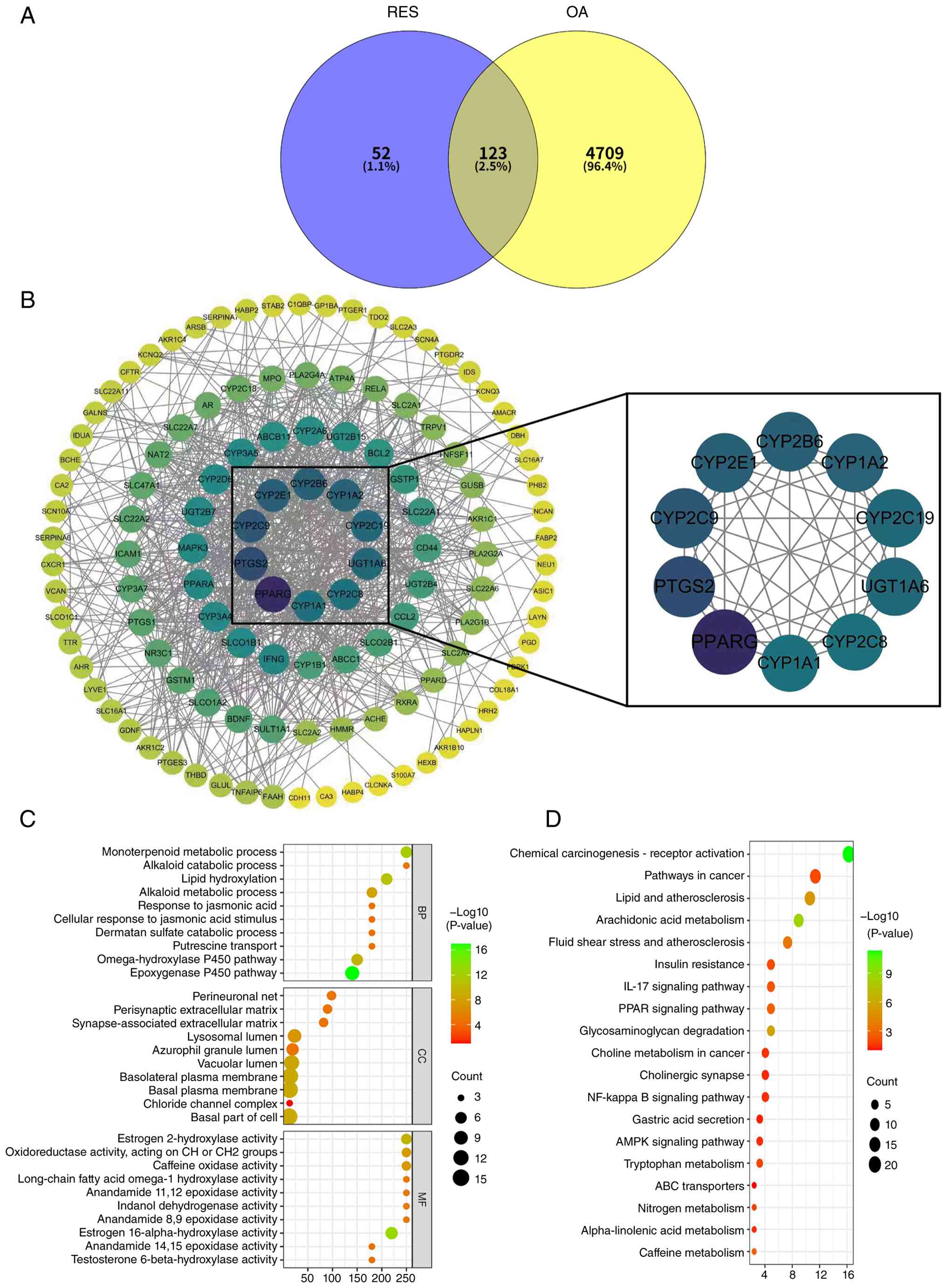
A total of 175 potential targets of RES were
predicted using the SwissTargetPrediction database. In addition,
4,832 OA-related target genes were screened by searching the
GeneCards, DrugBank, OMIM and DisGeNET databases. Finally, 123
overlapping targets were screened using the STRING database
(Fig. 1A). A PPI network was
constructed using a confidence threshold of ≥0.4, and the resulting
data underwent topological analysis based on degree values. The
nodes displayed a centripetal distribution pattern (Fig. 1B). Within this network, targets
with a higher number of adjacent nodes were considered more
critical and thus identified as core targets. These core targets
were implicated in metabolism and inflammation, encompassing
proteins such as multiple cytochrome P450 enzymes (CYP),
prostaglandin-endoperoxide synthase 2 (PTGS2) and PPARγ.
BP terms were predominantly associated with
functions such as 'monoterpenoid metabolic process', 'lipid
hydroxylation' and 'epoxygenase P450 pathway' (Fig. 1C). The CC terms were primarily
associated with structures, including 'basal part of cell', 'basal
plasma membrane' and 'basolateral plasma membrane'. The MF terms
were mainly enriched in 'estrogen 2-hydroxylase activity',
'caffeine oxidase activity' and 'oxidoreductases activity, acting
on CH or CH2 groups'. Among KEGG pathways, 'PPAR
signaling pathway' and 'AMPK signaling pathway', both closely
associated with metabolism, were significantly enriched (Fig. 1D).
RES protects against IL-1β-mediated loss
of GAG and proteoglycan in SW1353 cells
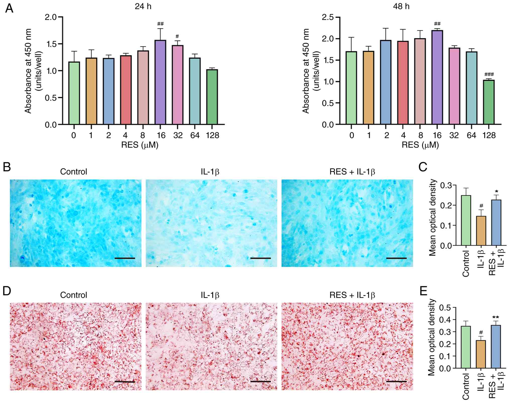
The cytotoxicity of RES on SW1353 cells was assessed
using the CCK-8 assay. RES was administered (0-128 μM) and
cell viability was assessed 24 or 48 h after treatment. Based on
the results, RES exhibited no cytotoxicity at concentrations up to
64, and 16 μM induced the most significant increase in cell
viability at both 24 and 48 h; therefore, this concentration of RES
was selected for subsequent experiments (Fig. 2A). Histological evaluation was
performed to assess the impact of RES on the IL-1β-induced loss of
GAG and proteoglycan in SW1353 cells. IL-1β treatment significantly
diminished AB staining intensity compared with the control
(Fig. 2B and C). Notably,
co-treatment with RES significantly restored AB staining intensity,
indicating its protective effect against the depletion of GAG and
proteoglycan. Consistent with these findings, SO staining
demonstrated that IL-1β significantly decreased the staining
intensity compared to the control, whereas co-treatment with RES
significantly reversed this decline (Fig. 2D and E), thereby confirming the
protective capacity of RES on GAG and proteoglycan levels.
RES activates the AMPK/PPARγ signaling
pathway in SW1353 cells
mRNA and protein expression of AMPK was not
significantly different between the groups (Fig. 3A, C and D). Relative expression
of p-AMPK was significantly decreased in the IL-1β group compared
with that in the control group; pretreatment with RES partially
restored its expression; cotreatment with ComC (RES+ IL-1β + ComC),
an inhibitor of AMPK, suppressed p-AMPK expression, suggesting that
RES activated AMPK and ComC effectively inhibited the activation
(Fig. 3C and E). The mRNA and
protein expression of PPARγ was significantly lower in the IL-1β
group compared with the control group. This decrease was reversed
in the RES+ IL-1β group, and diminished in the RES+ IL-1β + ComC
group (Fig. 3B, C and F). These
findings suggest that RES modulated the AMPK/PPARγ signaling
pathway. Notably, the results of IF staining showed that the in
situ protein expression of AMPK, p-AMPK and PPARγ was
consistent with that in bulk cell lysate of western blotting
(Fig. 3G-L). As evidenced by a
higher nuclear-to-cytoplasmic PPARγ ratio, the nuclear
translocation of PPARγ was enhanced in the RES+ IL-1β compared with
that in the IL-1β group; inhibition of AMPK phosphorylation
diminished this effect, leading to decreased nuclear PPARγ
accumulation (Fig. 3M).
Therefore, RES activated the AMPK/PPARγ pathway in SW1353
cells.
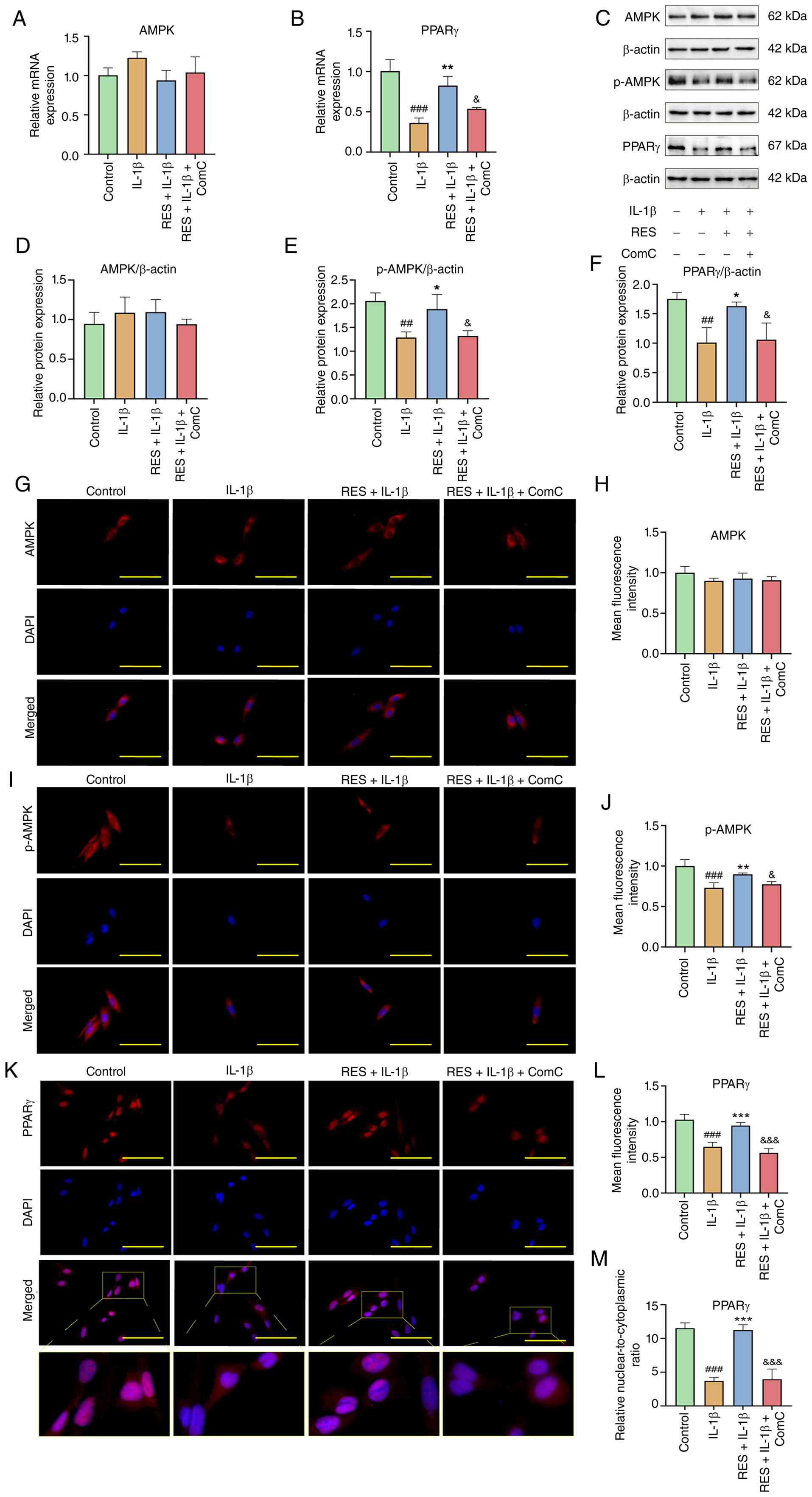 | Figure 3RES activates the AMPK/PPARγ
signaling pathway in SW1353 cells. Relative mRNA expression of (A)
AMPK and (B) PPARγ. (C) Representative western blots of (D) AMPK,
(E) p-AMPK and (F) PPARγ and β-actin. (G) Representative
immunofluorescence staining of AMPK. (H) Quantitative analysis of
AMPK expression. (I) Representative immunofluorescence staining of
p-AMPK. (J) Quantitative analysis of p-AMPK expression. (K)
Representative immunofluorescence staining of PPARγ. (L)
Quantitative analysis of PPARγ expression. (M) Quantitative
analysis of PPARγ nuclear-to-cytoplasmic ratio. Scale bar, 100
μm. ##P<0.01, ###P<0.001 vs.
control group; *P<0.05, **P<0.01,
***P<0.001 vs. IL-1β; &P<0.05,
&&&P<0.001 vs. RES+ IL-1β. RES,
resveratrol; ComC, compound C; p-, phosphorylated; PPARγ,
peroxisome proliferator-activated receptor γ. |
RES regulates the NLRP3 inflammasome
response via the AMPK-dependent signaling pathway in SW1353
cells
RES suppressed the IL-1β-induced upregulation of
NLRP3 mRNA; inhibition of AMPK activation counteracted this effect,
leading to increased NLRP3 expression (Fig. 4A). Western blot analysis of NLRP3
protein expression, along with IF staining, corroborated the qPCR
findings (Fig. 4B, C, F and G).
Western blot analysis of cleaved caspase-1 showed that RES
effectively suppressed IL-1β-induced accumulation, which was
counteracted by inhibition of AMPK activation (Fig. 4B, D and E). Furthermore, the
secretion levels of IL-18, a product of cleaved caspase-1, mirrored
the changes observed in NLRP3 expression across all groups
(Fig. 4H). IL-1β significantly
upregulated the expression of TNF-α and IL-6 and downregulated the
expression of IL-10 at the mRNA level. Treatment with RES reversed
the effects of IL-1β and inhibition of AMPK phosphorylation
suppressed this effect of RES (Fig.
4I-K). The secretion levels of these inflammatory factors
mirrored their intracellular expression patterns (Fig. 4L-N), indicating that RES blocked
the NLRP3 inflammasome response in SW1353 cells by suppressing
NLRP3 expression, inhibiting caspase-1 cleavage and reducing
downstream IL-18 release via the AMPK-dependent signaling
pathway.
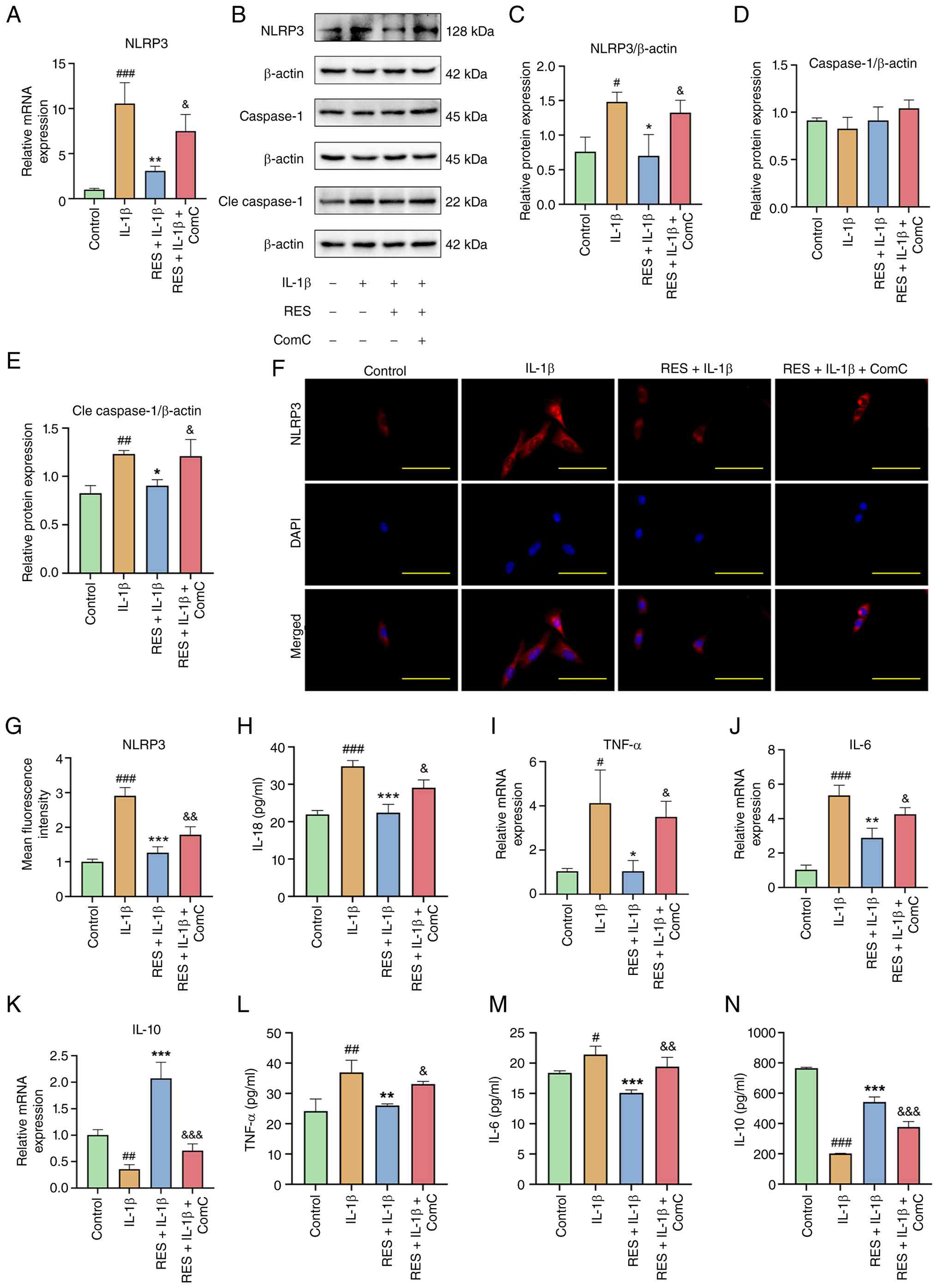 | Figure 4RES regulates the NLRP3 inflammasome
response in SW1353 cells via the AMPK-dependent signaling pathway.
(A) Relative mRNA expression of NLRP3. (B) Representative western
blots of (C) NLRP3, (D) caspase-1, (E) cle caspase-1 and β-actin.
(F) Representative immunofluorescence staining of NLRP3. (G)
Quantitative analysis of NLRP3 expression. (H) Concentration of
IL-18 in the cell culture supernatant. Relative mRNA expression of
(I) TNF-α, (J) IL-6 and (K) IL-10. The concentration of (L) TNF-α,
(M) IL-6 and (N) IL-10 in the cell culture supernatant. Scale bar,
100 μm. #P<0.05, ##P<0.01,
###P<0.001 vs. Control; *P<0.05,
**P<0.01, ***P<0.001 vs. IL-1β group;
&P<0.05, &&P<0.01,
&&&P<0.001 vs. RES+ IL-1β. RES,
resveratrol; ComC, compound C; NLRP3, NOD-like receptor family
pyrin domain containing 3; Cle, cleaved. |
RES regulates ECM metabolism via the
AMPK-dependent signaling pathway in SW1353 cells
The qPCR and western blotting results demonstrated
that IL-1β decreased the mRNA and protein expression of COL2A1 and
elevated the expression of MMP13. Pretreatment with RES
significantly attenuated the effects induced by IL-1β, while
cotreatment with ComC decreased the activity of RES (Fig. 5A-E). The IF staining results were
consistent with those of qPCR and western blotting (Fig. 5F-I). RES-mediated restoration of
COL2A1 and suppression of MMP13 were reversed by the AMPK inhibitor
ComC, suggesting that RES could regulate ECM metabolism via the
AMPK-dependent signaling pathway in SW1353 cells.
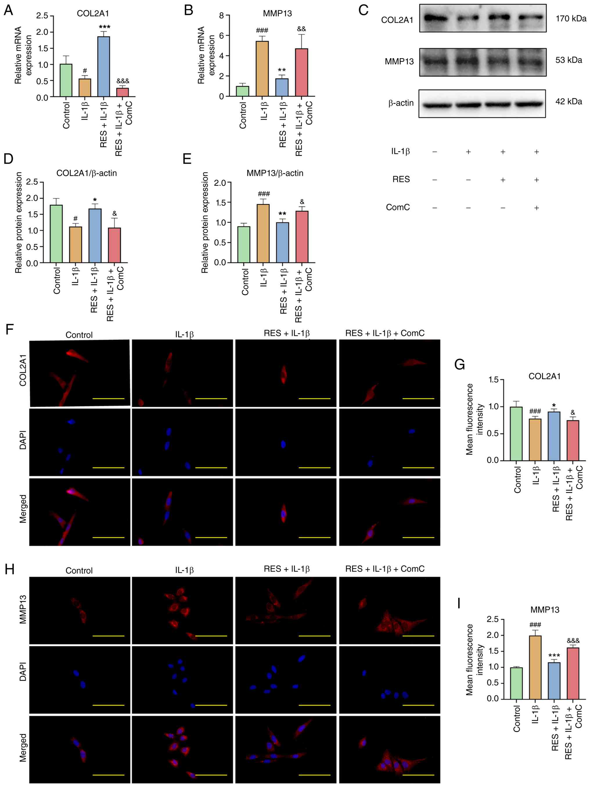 | Figure 5RES regulates extracellular matrix
metabolism via the AMPK-dependent signaling pathway in SW1353
cells. Relative mRNA expression of (A) COL2A1 and (B) MMP13. (C)
Representative western blots of (D) COL2A1, (E) MMP13 and β-actin.
(F) Representative immunofluorescence staining of COL2A1. (G)
Quantitative analysis of COL2A1 expression. (H) Representative
immunofluorescence staining of MMP13. (I) Quantitative analysis of
MMP13 expression. Scale bar, 100 μm. #P<0.05,
###P<0.001 vs. Control; *P<0.05,
**P<0.01, ***P<0.001 vs. IL-1β group;
&P<0.05, &&P<0.01,
&&&P<0.001 vs. RES+ IL-1β. RES,
resveratrol; IL-1β, interleukin-1β; ComC, compound C; COL2A1,
collagen type II α1. |
RES shows no significant hepatotoxicity
in OA mice
No significant differences were observed in the
expression levels of ALT/GPT and AST/GOT among the groups of mice
(Fig. S1A and B). The
pathological examination of liver tissue revealed that the
hepatocytes in the RES group maintained a relatively intact
structure, devoid of notable inflammatory cell infiltration or
degeneration. Furthermore, the overall liver architecture remained
clear and well-preserved (Fig.
S1C). The results indicated that RES, at a dose of 45 mg/kg,
exerted no significant hepatotoxic effects on the liver of OA
mice.
RES attenuates cartilage degeneration and
inhibits inflammatory responses of synovium and serum in OA
mice
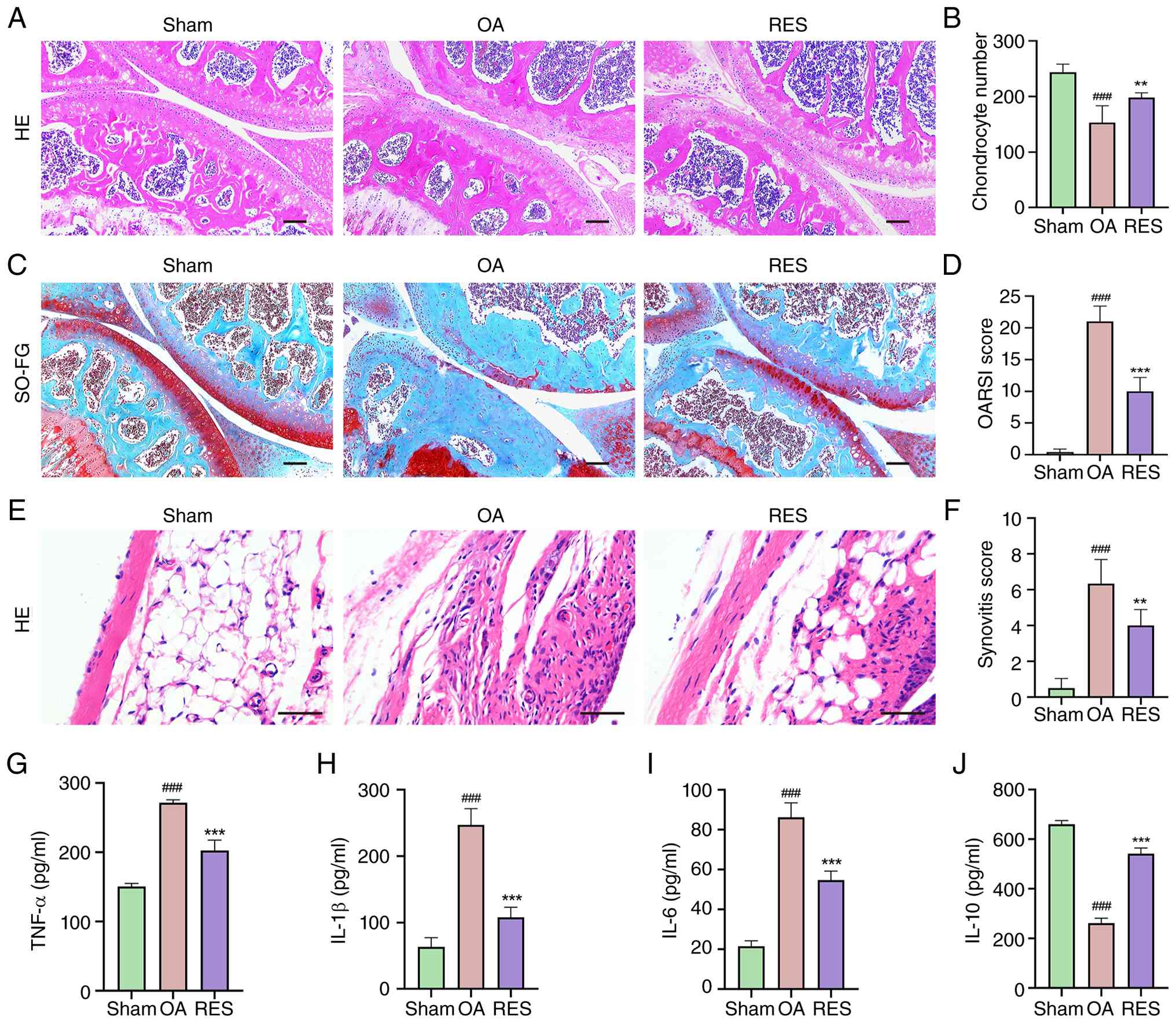
HE staining of cartilage tissue revealed a smooth
and structurally intact cartilage in the Sham group. Conversely,
the articular cartilage in the OA group exhibited notable damage
and a decreased chondrocyte population. RES treatment attenuated
this effect and restored the chondrocyte population (Fig. 6A and B). The SO-FG staining
demonstrated that RES partly reversed proteoglycan loss induced via
the DMM surgery (Fig. 6C). The
OARSI score of the OA group was significantly higher than that of
the Sham group, whereas the score for the RES group was
significantly lower than that of the OA group (Fig. 6D), indicating that RES attenuated
cartilage degeneration in OA mice.
The HE staining of the synovium revealed structural
abnormalities in the OA group, including increased inflammatory
infiltration, pannus formation and elevated cell density. RES
attenuated these pathological changes and reduced synovial scores
in OA mice (Fig. 6E and F).
ELISA demonstrated that the expression levels of IL-1β, TNF-α and
IL-6 were elevated, while IL-10 expression was decreased in the OA
group; these alterations were reversed by RES (Fig. 6G-J). Therefore, RES suppressed
the inflammatory response of synovium and serum in OA mice.
RES regulates the expression of p-AMPK,
PPARγ and NLRP3 and maintains ECM homeostasis in the articular
cartilage in OA mice
IHC staining was performed to detect the expression
levels of proteins associated with the AMPK/PPARγ signaling
pathway, NLRP3 (a key component of the inflammasome complex) and
matrix metabolism. The staining showed a significant decrease in
the protein expression levels of p-AMPK, PPARγ and COL2A1 in the OA
compared with the Sham group. RES partially mitigated these effects
(Fig. 7A, B and D). Conversely,
the OA mice exhibited significantly elevated protein expression
levels of NLRP3 and MMP13 compared with the Sham mice, with RES
inhibiting these increases (Fig. 7C
and E). Therefore, RES modulated the expression of AMPK, PPARγ
and NLRP3, promoting ECM homeostasis in the articular cartilage of
OA mice. These findings align with the in vitro results,
suggesting that RES may exert protective effects in OA via the
AMPK/PPARγ/NLRP3 signaling pathway.
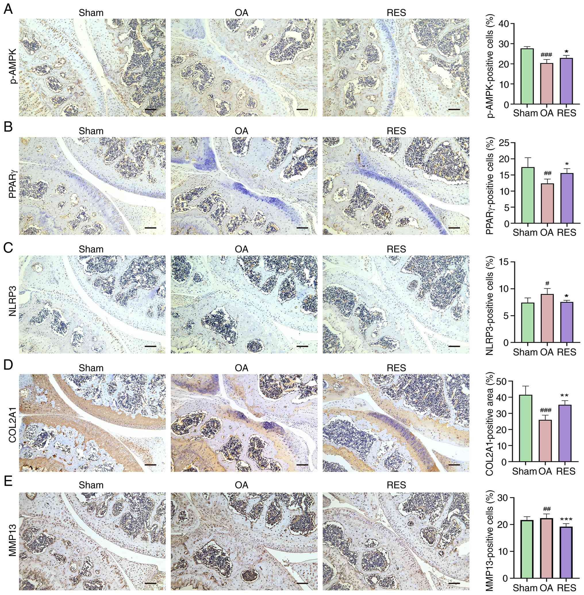 | Figure 7RES regulates the expression of
p-AMPK, PPARγ and NLRP3 and maintains ECM homeostasis in the
articular cartilage in OA mice. Representative immunohistochemical
staining of (A) p-AMPK, (B) PPARγ, (C) NLRP3, (D) COL2A1 and (E)
MMP13 in cartilage. Scale bar, 100 μm.
#P<0.05, ##P<0.01,
###P<0.001 vs. Sham; *P<0.05,
**P<0.01, ***P<0.001 vs. OA. OA,
osteoarthritis; RES, resveratrol; AMPK, adenosine
monophosphate-activated protein kinase; p-, phosphorylated; PPARγ,
peroxisome proliferator-activated receptor γ; NLRP3, NOD-like
receptor family pyrin domain-containing 3; COL2A1, collagen type II
α1. |
Discussion
Network pharmacology analysis is an effective
approach to evaluate the multitarget and multipathway effects of
pharmacological agents (37).
Based on network centrality and OA relevance,
CYP/UDP-glucuronosyltransferase enzymes represent metabolic
determinants of RES bioavailability rather than pharmacodynamic
targets, PTGS2 serves as a downstream inflammatory effector and
PPARγ acts as the primary therapeutic target driving
chondroprotection (38,39). Therefore, the present study
employed network pharmacology analysis, identifying the AMPK/PPARγ
signaling pathway as a key mechanism by which RES exerted effects
on OA. In vitro experiments showed that RES activated the
AMPK/PPARγ signaling pathway and inhibited the activation of NLRP3
inflammasome to regulate the inflammatory response, thereby
preserving the balance between the anabolism and catabolism of ECM
in SW1353 cells. Furthermore, in vivo experiments
demonstrated that RES alleviated OA progression in mice by
suppressing articular cartilage degeneration and inhibiting
inflammatory responses of serum and synovium. Mechanistically, RES
upregulated the expression of proteins associated with the
AMPK/PPARγ signaling pathway, suppressed NLRP3 expression and
enhanced matrix synthesis within the articular cartilage in OA mice
(Fig. 8).
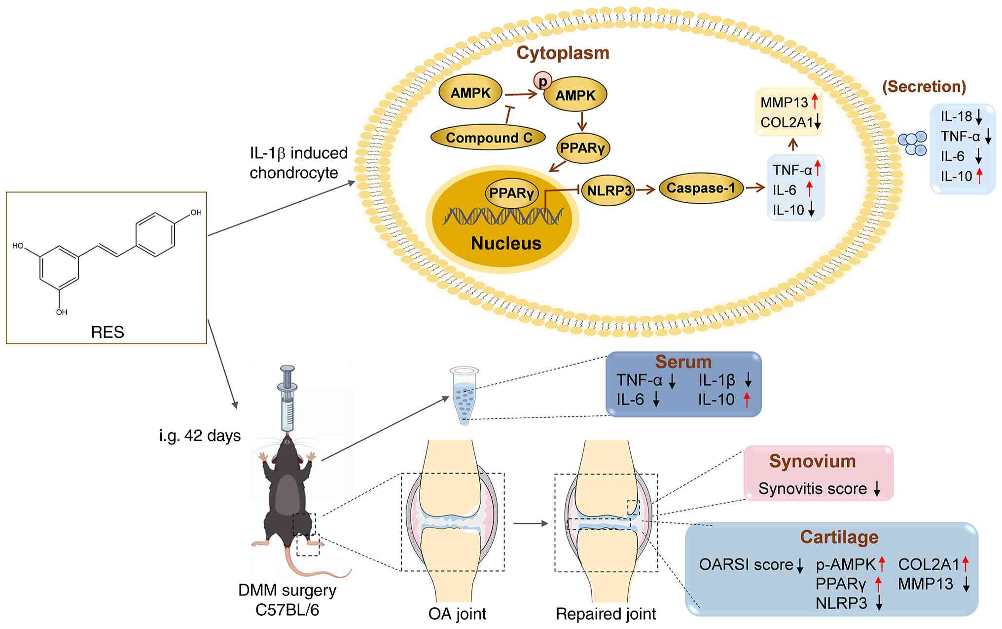 | Figure 8Schematic diagram of the therapeutic
effect and molecular mechanisms of RES on OA. In vitro, RES
inhibits NLRP3 inflammasome activation via the AMPK/PPARγ pathway,
thereby modulating the inflammatory response and the balance of ECM
synthesis and degradation in SW1353 cells. In vivo, RES
alleviates cartilage degeneration, suppresses synovial inflammation
and regulates serum inflammatory factors, thereby alleviating OA
progression in mice. RES modulated the protein expression
associated with AMPK/PPARγ/NLRP3 signaling and matrix metabolism in
OA mice. AMPK, adenosine monophosphate-activated protein kinase;
p-, phosphorylated; NLRP3, NOD-like receptor family pyrin domain
containing 3; COL2A1, collagen type II α1; DMM, destabilization of
the medial meniscus; OARSI, Osteoarthritis Research Society
International; i.g., intragastric. |
Metformin slows the progression of OA in mice
(40). However, this therapeutic
benefit is absent in AMPK knockout mice, suggesting that the
effectiveness of metformin in treating OA relies on AMPK (40). AMPK serves as a cell energy
sensor, and the activation of the AMPK signaling pathway plays a
critical role in modulating metabolism in human primary knee or rat
primary chondrocytes, which is consistent with that in mouse or rat
articular cartilage (41,42).
As a key metabolic regulator, AMPK monitors the AMP/ATP ratio and
modulates autophagy, oxidative stress and inflammation (43,44). AMPK serves a regulatory role in
relieving OA-associated pain in mice by decreasing pain sensitivity
and increasing spontaneous activity, such as locomotion distance,
walking speed, and rearing frequency (41). In C57BL/6 mice, RES has been
shown to delay OA progression by enhancing p-AMPK expression, which
induces autophagy (45).
Similarly, in two-dimensional rat chondrocyte culture, RES
upregulates p-AMPK expression, contributing to resistance against
energy stress (46). The primary
upstream mechanisms responsible for RES-induced AMPK activation
involve mitochondrial metabolic stress. RES has been reported to
partially inhibit mitochondrial respiratory chain complex I,
leading to an increase in the intracellular AMP/ATP ratio (47). Furthermore, RES can activate
SIRT1/liver kinase B1, thereby stimulating AMPK and improving
mitochondrial function (32).
Additionally, RES induces calcium fluxes, which activate
calcium/calmodulin-dependent protein kinase kinase β, a key
upstream kinase of AMPK (48).
The present study, however, concentrated on the downstream
mechanisms of the AMPK pathway induced by RES and investigated the
molecular mechanisms downstream of AMPK, building upon insight from
network pharmacology analysis.
AMPK could enhance the antioxidant stress response,
anti-inflammatory effects and mitochondrial biogenesis in human
knee chondrocytes by activating the expression of PPARγ coactivator
1α, which could interact with PPARγ (49,50). AMPK serves as an upstream
regulator of PPARγ involved in activation of the AMPK/PPARγ
signaling pathway and the modulation of the inflammatory response
in macrophages (51). A recent
study revealed that tenascin-N negatively regulates AMPK/PPARγ
signaling in both OA rats and rat primary chondrocytes stimulated
with IL-1β (52). Pioglitazone,
a PPARγ agonist, has been shown to suppress the production of
pro-inflammatory factors in primary human chondrocytes and protect
against cartilage degradation in OA rats (53,54). To the best of our knowledge, the
present study is the first to demonstrate that RES promotes PPARγ
expression and nuclear translocation in chondrocytes. Notably, the
AMPK inhibitor ComC effectively abrogated the effects of RES.
Therefore, the present study reveals that RES exerts its
anti-inflammatory effects in SW1353 cells through the AMPK/PPARγ
signaling pathway.
Activation of AMPK downregulates NLRP3 (55). The NLRP3 inflammasome, an
inflammatory sensor, is a multi-protein complex composed of NLRP3,
apoptosis-associated speck-like protein (ASC), and caspase-1
(56). The NLRP3 inflammasome is
a key regulator of innate and adaptive host responses. Its
activation typically involves a priming phase of NLRP3
upregulation, followed by an activation phase in which the
enzymatic cleavage of pro-caspase-1 into cleaved caspase-1 serves
as the hallmark of functional inflammasome assembly. This leads to
the maturation and secretion of cytokines such as IL-18 or mature
IL-1β (57,58). NLRP3 inflammasome activation
mediates the release of proinflammatory factors, promoting
pyroptosis in OA rats and oxidative stress in OA mice (8,59). Tang et al (8) found that TNF-α-induced NLRP3
expression results in the elevated release of proinflammatory
factors in primary mouse chondrocytes. Xu et al (60) observed that IL-1β-induced NLRP3
inflammasome activation, characterized by the elevated expression
of NLRP3, ASC and cleaved caspase-1, promotes ECM degradation in
primary mouse chondrocytes. PPARγ has been shown to inhibit the
formation of the NLRP3 inflammasome, thereby exerting
anti-inflammatory effects in mouse macrophages (56). PPARγ downregulates expression of
pro-inflammatory factors and decreases the inflammatory response by
inhibiting NLRP3 inflammasome activation in rats and mice (61,62). Chang et al (63) reported that RES inhibits NLRP3
inflammasome activation by preserving mitochondrial integrity and
augmenting autophagy in mouse macrophages. Li et al
(64) reported that RES
attenuates mitochondrial damage, inhibiting NLRP3 inflammasome
activation. This action regulates the inflammatory response and
decreases cell apoptosis in murine adipocytes, primarily through
the activation of the AMPK pathway (64). Yang et al (65) showed that RES decreases the
expression of NLRP3 in peripheral blood leukocytes of patients with
gout. Combined with the alterations in NLRP3 and cleaved caspase-1
expression, alongside the secretion of IL-18, the present study
demonstrated the inhibitory effect of RES on inflammasome
activation. The present findings indicated that RES inhibited NLRP3
inflammasome activation, attenuated the inflammatory response and
maintained ECM homeostasis via the AMPK/PPARγ signaling pathway in
SW1353 cells. The in vivo data confirmed the expression
alterations of PPARγ, NLRP3, COL2A1 and MMP13 under pathological
conditions and were consistent with the in vitro
results.
In summary, the present study demonstrated that RES
inhibits the NLRP3 inflammasome response in SW1353 cells through
the AMPK-dependent upregulation and nuclear translocation of PPARγ,
revealing that RES used PPARγ as a critical link between energy
metabolism and inflammation in chondrocytes. The causal
relationship within the AMPK/PPARγ pathway was validated using an
AMPK inhibitor. By integrating network pharmacology with both in
vitro and in vivo validation, the present study
demonstrated that RES modulates the AMPK/PPARγ/NLRP3 inflammasome
response pathway to preserve ECM homeostasis, thereby connecting
AMPK activation with the maintenance of matrix balance.
Nonetheless, there are several limitations in the present study.
The present study did not perform functional validation on other
high-ranking predicted pathways. The present study focused on the
AMPK/PPARγ pathway based on network centrality, meaning the
potential involvement of alternative pathways cannot be excluded.
Future high-throughput screenings or multi-pathway knockout studies
are warranted for a more comprehensive validation. Second, the
human chondrosarcoma cell line SW1353 was used for in vitro
study. Although SW1353 cells mimic the inflammatory and catabolic
responses induced by IL-1β in primary chondrocytes (66,67), the absence of primary cell data
remains a limitation. Further verification using primary human or
animal articular chondrocytes is required to validate the
AMPK/PPARγ/NLRP3 inflammasome response pathway and matrix
metabolism. Third, the in vivo study lacked positive or
negative controls, such as known AMPK agonists or antagonists, to
validate the AMPK/PPARγ signaling pathway. While multi-dimensional
assessments confirmed the protective effects of RES on OA, future
in vivo studies employing AMPK-targeted modulators are
necessary to elucidate the molecular mechanisms by which RES
maintains cartilage matrix homeostasis.
The present study demonstrated that RES was a safe
and efficacious compound with therapeutic potential for treating
OA. In SW1353 cells, RES activated the AMPK/PPARγ signaling pathway
to inhibit the NLRP3 inflammasome activation, regulate the
expression and release of TNF-α, IL-6, and IL-10 and effectively
prevent ECM degradation. In OA mice, RES attenuated cartilage
degeneration and conferred protective effects against synovial
inflammation. Furthermore, RES regulated serum levels of
pro-inflammatory cytokines such as IL-1β, TNF-α and IL-6,
concurrently promoting the production of the anti-inflammatory
cytokine IL-10. The in vivo data confirmed that the
expression trends of PPARγ, NLRP3, COL2A1 and MMP13 under both
pathological conditions and RES treatment were consistent with the
in vitro findings. The present study provided experimental
evidence for the molecular mechanisms underlying the protective
effects of RES against OA, demonstrating its potential clinical
utility as a complementary and alternative therapy.
Supplementary Data
Availability of data and materials
The data generated in the present study may be
requested from the corresponding author.
Authors' contributions
JM and RZ confirm the authenticity of all the raw
data. SC performed experiments and wrote the manuscript. ZL
analyzed data. WQ and BC designed the experiments and analyzed
data. QZ and JBM interpreted data. JM analyzed and interpreted the
data. RZ designed the study and edited the manuscript. All authors
have read and approved the final manuscript.
Ethics approval and consent to
participate
The present study was approved by the Animal Care
and Use Committee of Xi'an Jiaotong University (approval no.
XJTUAE2024-1403; Xi'an, China).
Patient consent for publication
Not applicable.
Competing interests
The authors declare that they have no competing
interests.
Acknowledgements
Not applicable.
Funding
The present study was supported by the Natural Science
Foundation of Guizhou Province [grant no. Qiankehejichu-ZK
(2024)yiban588], the Natural Science Basic Research Program
Project-General Project (Youth) of Shaanxi Province (grant no.
2023-JC-QN-0837), the Capacity Building Program of the Shaanxi
Administration of Traditional Chinese Medicine (grant no.
SZY-NLTL-2022-003) and the Scientific Research Team Fund Project of
Guiyang Kangyang University (grant no. Guikangda-TD2025-003).
References
|
1
|
Chen T, Chen W, Xu T, Wang H, Zhang Y,
Wang L, Zhu S, Tao H and Yang X: Osteoarthritis: Epidemiology,
pathogenesis, and treatment. MedComm (2020). 7:e707272026.
View Article : Google Scholar
|
|
2
|
GBD 2021 Osteoarthritis Collaborators:
Global, regional, and national burden of osteoarthritis, 1990-2020
and projections to 2050: A systematic analysis for the global
burden of disease study 2021. Lancet Rheumatol. 5:e508–e522. 2023.
View Article : Google Scholar :
|
|
3
|
Savvari P, Skiadas I, Barmpouni M,
Papadakis SA, Psychogios V, Pastroudis AP, Skarpas GA, Tsoutsanis
A, Garofalakis A, Katsifis G, et al: Moderate to severe
osteoarthritis: What is the economic burden for patients and the
health care system? Insights from the 'PONOS' study. Cartilage.
15:268–277. 2024. View Article : Google Scholar :
|
|
4
|
Rai MF: Appraising the evolving landscape
of protease inhibition in osteoarthritis. Nat Rev Rheumatol.
21:131–132. 2025. View Article : Google Scholar
|
|
5
|
Vélez-Slimani H, Hernández-Montelongo J
and Salazar LA: Recent developments in osteoarthritis research:
Innovative therapeutic approaches and the role of polyphenols and
nanotechnology. Int J Mol Sci. 26:89252025. View Article : Google Scholar :
|
|
6
|
Feng K, Wang F, Chen H, Zhang R, Liu J, Li
X, Xie X and Kang Q: Cartilage progenitor cells derived
extracellular vesicles-based cell-free strategy for osteoarthritis
treatment by efficient inflammation inhibition and extracellular
matrix homeostasis restoration. J Nanobiotechnology. 22:3452024.
View Article : Google Scholar
|
|
7
|
Lee HR, Jeong YJ, Lee JW, Jhun J, Na HS,
Cho KH, Kim SJ, Cho ML and Heo TH: Tannic acid, an IL-1β-direct
binding compound, ameliorates IL-1β-induced inflammation and
cartilage degradation by hindering IL-1β-IL-1R1 interaction. PLoS
One. 18:e02818342023. View Article : Google Scholar
|
|
8
|
Tang H, Gong X, Dai J, Gu J, Dong Z, Xu Y,
Hu Z, Zhao C, Deng J and Dong S: The IRF1/GBP5 axis promotes
osteoarthritis progression by activating chondrocyte pyroptosis. J
Orthop Translat. 44:47–59. 2023. View Article : Google Scholar
|
|
9
|
Wang J, Ma J, Gu JH, Wang FY, Shang XS,
Tao HR and Wang X: Regulation of type II collagen, matrix
metalloproteinase-13 and cell proliferation by interleukin-1β is
mediated by curcumin via inhibition of NF-κB signaling in rat
chondrocytes. Mol Med Rep. 16:1837–1845. 2017. View Article : Google Scholar : PubMed/NCBI
|
|
10
|
Yan W, Li Y, Xie S, Tao WA, Hu J, Liu H,
Zhang G, Liu F, Nie Y, Chen X, et al: Chondrocyte-targeted delivery
system of sortase A-engineered extracellular vesicles silencing
MMP13 for osteoarthritis therapy. Adv Healthc Mater.
13:e23035102024. View Article : Google Scholar : PubMed/NCBI
|
|
11
|
Kim J, Xu M, Xo R, Mates A, Wilson GL,
Pearsall AW IV and Grishko V: Mitochondrial DNA damage is involved
in apoptosis caused by pro-inflammatory cytokines in human OA
chondrocytes. Osteoarthritis Cartilage. 18:424–432. 2010.
View Article : Google Scholar
|
|
12
|
Xu L, Zhang F, Cheng G, Yuan X, Wu Y, Wu
H, Wang Q, Chen J, Kuai J, Chang Y, et al: Attenuation of
experimental osteoarthritis with human adipose-derived mesenchymal
stem cell therapy: Inhibition of the pyroptosis in chondrocytes.
Inflamm Res. 72:89–105. 2023. View Article : Google Scholar
|
|
13
|
Vadhan A, Gupta T and Hsu WL: Mesenchymal
stem cell-derived exosomes as a treatment option for
osteoarthritis. Int J Mol Sci. 25:91492024. View Article : Google Scholar : PubMed/NCBI
|
|
14
|
Li H, Zhao Y, Qi P, Zhang B, Ma J, Xie X
and Li N: Mechanisms of active metabolites from traditional Chinese
medicine in osteoarthritis: A critical review. Front Pharmacol.
17:17861122026. View Article : Google Scholar : PubMed/NCBI
|
|
15
|
Lei R and Zhou C: The role of reactive
oxygen species-mitophagy regulation in the treatment of
osteoarthritis by active Chinese herbal medicine monomers: A
review. Front Med (Lausanne). 12:16861902025. View Article : Google Scholar :
|
|
16
|
Zou X, Xu H and Qian W: The role and
current research status of resveratrol in the treatment of
osteoarthritis and its mechanisms: A narrative review. Drug Metab
Rev. 56:399–412. 2024. View Article : Google Scholar
|
|
17
|
Singh K, Gupta JK and Kumar S: The
pharmacological potential of resveratrol in reducing soft tissue
damage in osteoarthritis patients. Curr Rheumatol Rev. 20:27–38.
2024. View Article : Google Scholar
|
|
18
|
Meng T, Xiao D, Muhammed A, Deng J, Chen L
and He J: Anti-inflammatory action and mechanisms of resveratrol.
Molecules. 26:2292021. View Article : Google Scholar : PubMed/NCBI
|
|
19
|
Berman AY, Motechin RA, Wiesenfeld MY and
Holz MK: The therapeutic potential of resveratrol: A review of
clinical trials. NPJ Precis Oncol. 1:352017. View Article : Google Scholar
|
|
20
|
Dyck GJB, Raj P, Zieroth S, Dyck JRB and
Ezekowitz JA: The effects of resveratrol in patients with
cardiovascular disease and heart failure: A narrative review. Int J
Mol Sci. 20:9042019. View Article : Google Scholar
|
|
21
|
Ren ZQ, Zheng SY, Sun Z, Luo Y, Wang YT,
Yi P, Li YS, Huang C and Xiao WF: Resveratrol: Molecular
mechanisms, health benefits, and potential adverse effects. MedComm
(2020). 6:e702522025. View Article : Google Scholar :
|
|
22
|
Ghasemzadeh Rahbardar M and Sahebkar A:
Resveratrol as a naturally occurring inflammasome modulator:
Implications for health and disease. Iran J Basic Med Sci.
28:1301–1318. 2025.
|
|
23
|
Wei Y, Jia J, Jin X, Tong W and Tian H:
Resveratrol ameliorates inflammatory damage and protects against
osteoarthritis in a rat model of osteoarthritis. Mol Med Rep.
17:1493–1498. 2018.
|
|
24
|
Frischholz S, Berberich O, Böck T, Meffert
RH and Blunk T: Resveratrol counteracts IL-1β-mediated impairment
of extracellular matrix deposition in 3D articular chondrocyte
constructs. J Tissue Eng Regen Med. 14:897–908. 2020. View Article : Google Scholar
|
|
25
|
Zhou M, Wang D and Tang J: Identification
of the resveratrol potential targets in the treatment of
osteoarthritis. Evid Based Complement Alternat Med.
2021:99112862021. View Article : Google Scholar : PubMed/NCBI
|
|
26
|
Zhou X, Jiang P, Tan H, Wang Y and Bai L:
KLF9-GRK5-HDAC6 axis aggravates osteoarthritis pathogenesis by
promoting chondrocyte extracellular matrix degradation and
apoptosis. Commun Biol. 8:232025. View Article : Google Scholar :
|
|
27
|
Pan MH, Li MY, Tsai ML, Pan CY, Badmaev V,
Ho CT and Lai CS: A mixture of citrus polymethoxyflavones, green
tea polyphenols and lychee extracts attenuates adipogenesis in
3T3-L1 adipocytes and obesity-induced adipose inflammation in mice.
Food Funct. 10:7667–7677. 2019. View Article : Google Scholar
|
|
28
|
Xu X, Liu X, Yang Y, He J, Gu H, Jiang M,
Huang Y, Liu X and Liu L: Resveratrol inhibits the development of
obesity-related osteoarthritis via the TLR4 and PI3K/Akt signaling
pathways. Connect Tissue Res. 60:571–582. 2019. View Article : Google Scholar
|
|
29
|
Livak KJ and Schmittgen TD: Analysis of
relative gene expression data using real-time quantitative PCR and
the 2(-Delta Delta C(T)) method. Methods. 25:402–408. 2001.
View Article : Google Scholar
|
|
30
|
Glasson SS, Blanchet TJ and Morris EA: The
surgical destabilization of the medial meniscus (DMM) model of
osteoarthritis in the 129/SvEv mouse. Osteoarthritis Cartilage.
15:1061–1069. 2007. View Article : Google Scholar : PubMed/NCBI
|
|
31
|
Gu H, Li K, Li X, Yu X, Wang W, Ding L and
Liu L: Oral resveratrol prevents osteoarthritis progression in
C57BL/6J mice fed a high-fat diet. Nutrients. 8:2332016. View Article : Google Scholar : PubMed/NCBI
|
|
32
|
Price NL, Gomes AP, Ling AJ, Duarte FV,
Martin-Montalvo A, North BJ, Agarwal B, Ye L, Ramadori G, Teodoro
JS, et al: SIRT1 is required for AMPK activation and the beneficial
effects of resveratrol on mitochondrial function. Cell Metab.
15:675–690. 2012. View Article : Google Scholar : PubMed/NCBI
|
|
33
|
Cano EA, Pasigna L, Perido HG, Rodelas AG,
Sabarez SA, Salinas R and Santos DA: Cytotoxic and cytostatic
activity of resveratrol: An updated scoping review. Bull Natl Inst
Health Sci. 142:3695–3714. 2024.
|
|
34
|
Xiong G, Yang Y and Guo M: Effect of
resveratrol on abnormal bone remodeling and angiogenesis of
subchondral bone in osteoarthritis. Int J Clin Exp Pathol.
14:417–425. 2021.PubMed/NCBI
|
|
35
|
Krenn V, Morawietz L, Burmester GR, Kinne
RW, Mueller-Ladner U, Muller B and Haupl T: Synovitis score:
Discrimination between chronic low-grade and high-grade synovitis.
Histopathology. 49:358–364. 2006. View Article : Google Scholar
|
|
36
|
Glasson SS, Chambers MG, Van Den Berg WB
and Little CB: The OARSI histopathology initiative-recommendations
for histological assessments of osteoarthritis in the mouse.
Osteoarthritis Cartilage. 18(Suppl 3): S17–S23. 2010. View Article : Google Scholar
|
|
37
|
Fan Y, Yin L, Zhong X, He Z, Meng X, Chai
F, Kong M, Zhang Q, Xia C, Tong Y and Bi Q: An integrated network
pharmacology, molecular docking and experiment validation study to
investigate the potential mechanism of Isobavachalcone in the
treatment of osteoarthritis. J Ethnopharmacol. 326:1178272024.
View Article : Google Scholar : PubMed/NCBI
|
|
38
|
Zanger UM and Schwab M: Cytochrome P450
enzymes in drug metabolism: Regulation of gene expression, enzyme
activities, and impact of genetic variation. Pharmacol Ther.
138:103–141. 2013. View Article : Google Scholar
|
|
39
|
Fahmi H, Di Battista JA, Pelletier JP,
Mineau F, Ranger P and Martel-Pelletier J: Peroxisome
proliferator-activated receptor gamma activators inhibit
interleukin-1beta-induced nitric oxide and matrix metalloproteinase
13 production in human chondrocytes. Arthritis Rheum. 44:595–607.
2001. View Article : Google Scholar : PubMed/NCBI
|
|
40
|
Li J, Zhang B, Liu WX, Lu K, Pan H, Wang
T, Oh CD, Yi D, Huang J, Zhao L, et al: Metformin limits
osteoarthritis development and progression through activation of
AMPK signalling. Ann Rheum Dis. 79:635–645. 2020. View Article : Google Scholar
|
|
41
|
Li J, Wang Y, Chen D and Liu-Bryan R: Oral
administration of berberine limits post-traumatic osteoarthritis
development and associated pain via AMP-activated protein kinase
(AMPK) in mice. Osteoarthritis Cartilage. 30:160–171. 2022.
View Article : Google Scholar
|
|
42
|
Jin Z, Chang B, Wei Y, Yang Y, Zhang H,
Liu J, Piao L and Bai L: Curcumin exerts chondroprotective effects
against osteoarthritis by promoting AMPK/PINK1/Parkin-mediated
mitophagy. Biomed Pharmacother. 151:1130922022. View Article : Google Scholar
|
|
43
|
O'Neill LAJ and Hardie DG: Metabolism of
inflammation limited by AMPK and pseudo-starvation. Nature.
493:346–355. 2013. View Article : Google Scholar : PubMed/NCBI
|
|
44
|
Yi D, Yu H, Lu K, Ruan C, Ding C, Tong L,
Zhao X and Chen D: AMPK signaling in energy control, cartilage
biology, and osteoarthritis. Front Cell Dev Biol. 9:6966022021.
View Article : Google Scholar : PubMed/NCBI
|
|
45
|
Qin N, Wei L, Li W, Yang W, Cai L, Qian Z
and Wu S: Local intra-articular injection of resveratrol delays
cartilage degeneration in C57BL/6 mice by inducing autophagy via
AMPK/mTOR pathway. J Pharmacol Sci. 134:166–174. 2017. View Article : Google Scholar
|
|
46
|
Zhang R and Kanki K: Glycolytic inhibition
by resveratrol facilitates chondrocyte survival under
glucose-deprived conditions and improves the viability of
3D-cultured cartilage tissue. J Biosci Bioeng. 140:90–97. 2025.
View Article : Google Scholar : PubMed/NCBI
|
|
47
|
Hawley SA, Ross FA, Chevtzoff C, Green KA,
Evans A, Fogarty S, Towler MC, Brown LJ, Ogunbayo OA, Evans AM and
Hardie DG: Use of cells expressing gamma subunit variants to
identify diverse mechanisms of AMPK activation. Cell Metab.
11:554–565. 2010. View Article : Google Scholar :
|
|
48
|
Park SJ, Ahmad F, Philp A, Baar K,
Williams T, Luo H, Ke H, Rehmann H, Taussig R, Brown AL, et al:
Resveratrol ameliorates aging-related metabolic phenotypes by
inhibiting cAMP phosphodiesterases. Cell. 148:421–433. 2012.
View Article : Google Scholar : PubMed/NCBI
|
|
49
|
Ma CH, Chiua YC, Wu CH, Jou IM, Tu YK,
Hung CH, Hsieh PL and Tsai KL: Homocysteine causes dysfunction of
chondrocytes and oxidative stress through repression of SIRT1/AMPK
pathway: A possible link between hyperhomocysteinemia and
osteoarthritis. Redox Biol. 15:504–512. 2018. View Article : Google Scholar : PubMed/NCBI
|
|
50
|
Wang Y, Zhao X, Lotz M, Terkeltaub R and
Liu-Bryan R: Mitochondrial biogenesis is impaired in osteoarthritis
chondrocytes but reversible via peroxisome proliferator-activated
receptor γ coactivator 1α. Arthritis Rheumatol. 67:2141–2153. 2015.
View Article : Google Scholar
|
|
51
|
Wang Y, Xu Y, Zhang P, Ruan W, Zhang L,
Yuan S, Pang T and Jia AQ: Smiglaside A ameliorates LPS-induced
acute lung injury by modulating macrophage polarization via
AMPK-PPARγ pathway. Biochem Pharmacol. 156:385–395. 2018.
View Article : Google Scholar : PubMed/NCBI
|
|
52
|
Chen Z, Wu G, Fan Y, Li C, Wang C, Yang C,
Wu S, Wu Z, Wang P, Zhang Y and You W: Increased TNN in knee
osteoarthritis accelerates cartilage damage via the negative
regulation of AMPK-PPARγ signalling. J Cell Mol Med. 29:e709812025.
View Article : Google Scholar
|
|
53
|
Ma C, Zhang Y, Li YQ, Chen C, Cai W and
Zeng YL: The role of PPARγ in advanced glycation end
products-induced inflammatory response in human chondrocytes. PLoS
One. 10:e01257762015. View Article : Google Scholar
|
|
54
|
Chen J, Xu W, Dai T, Jiao S, Xue X, Jiang
J, Li S and Meng Q: Pioglitazone-loaded cartilage-targeted
nanomicelles (Pio@C-HA-DOs) for osteoarthritis treatment. Int J
Nanomedicine. 18:5871–5890. 2023. View Article : Google Scholar : PubMed/NCBI
|
|
55
|
Chen Y, Liu Y, Jiang K, Wen Z, Cao X and
Wu S: Linear ubiquitination of LKB1 activates AMPK pathway to
inhibit NLRP3 inflammasome response and reduce chondrocyte
pyroptosis in osteoarthritis. J Orthop Translat. 39:1–11. 2022.
View Article : Google Scholar : PubMed/NCBI
|
|
56
|
Yang CC, Wu CH, Lin TC, Cheng YN, Chang
CS, Lee KT, Tsai PJ and Tsai YS: Inhibitory effect of PPARγ on
NLRP3 inflammasome activation. Theranostics. 11:2424–2441. 2021.
View Article : Google Scholar
|
|
57
|
Strowig T, Henao-Mejia J, Elinav E and
Flavell R: Inflammasomes in health and disease. Nature.
481:278–286. 2012. View Article : Google Scholar
|
|
58
|
Swanson KV, Deng M and Ting JPY: The NLRP3
inflammasome: Molecular activation and regulation to therapeutics.
Nat Rev Immunol. 19:477–489. 2019. View Article : Google Scholar
|
|
59
|
Feng Z, Huang Q, Zhang X, Xu P, Li S, Ma D
and Meng Q: PPAR-γ activation alleviates osteoarthritis through
both the Nrf2/NLRP3 and PGC-1 α/ Δψm pathways by
inhibiting pyroptosis. PPAR Res. 2023:25235362023. View Article : Google Scholar
|
|
60
|
Xu L, Jiang Z and Li H: KLK8 promotes
cartilage degradation and pyroptosis in osteoarthritis by
activating the NF-κB and NLRP3 inflammasome pathways. Tissue Cell.
101:1034272026. View Article : Google Scholar
|
|
61
|
Abu-Risha SE, Mousa MA and Elsisi AE:
Protective role of irbesartan against cyclophosphamide-induced
testicular damage in rats via up-regulating PPAR-γ signaling and
ameliorating NF-κB/NLRP3/IL-18 inflammatory axis. Life Sci.
289:1202182022. View Article : Google Scholar
|
|
62
|
Althagafy HS, Sharawi ZW, Batawi AH,
Almohaimeed HM, Al-Thubiani WS, Hassanein EHM and Rateb A:
Buspirone attenuated methotrexate-induced hippocampal toxicity in
rats by regulating Nrf2/HO-1, PPAR-γ, NF-κB/nNOS, and
ROS/NLRP3/caspase-1 signaling pathways. J Biochem Mol Toxicol.
37:e234142023. View Article : Google Scholar
|
|
63
|
Chang YP, Ka SM, Hsu WH, Chen A, Chao LK,
Lin CC, Hsieh CC, Chen MC, Chiu HW, Ho CL, et al: Resveratrol
inhibits NLRP3 inflammasome activation by preserving mitochondrial
integrity and augmenting autophagy. J Cell Physiol. 230:1567–1579.
2015. View Article : Google Scholar
|
|
64
|
Li A, Zhang S, Li J, Liu K, Huang F and
Liu B: Metformin and resveratrol inhibit Drp1-mediated
mitochondrial fission and prevent ER stress-associated NLRP3
inflammasome activation in the adipose tissue of diabetic mice. Mol
Cell Endocrinol. 434:36–47. 2016. View Article : Google Scholar : PubMed/NCBI
|
|
65
|
Yang QB, He YL, Zhong XW, Xie WG and Zhou
JG: Resveratrol ameliorates gouty inflammation via upregulation of
sirtuin 1 to promote autophagy in gout patients.
Inflammopharmacology. 27:47–56. 2019. View Article : Google Scholar
|
|
66
|
Gebauer M, Saas J, Sohler F, Haag J, Söder
S, Pieper M, Bartnik E, Beninga J, Zimmer R and Aigner T:
Comparison of the chondrosarcoma cell line SW1353 with primary
human adult articular chondrocytes with regard to their gene
expression profile and reactivity to IL-1beta. Osteoarthritis
Cartilage. 13:697–708. 2005. View Article : Google Scholar
|
|
67
|
Pang KL, Chow YY, Leong LM, Law JX, Ghafar
NA, Soelaiman IN and Chin KY: Establishing SW1353 chondrocytes as a
cellular model of chondrolysis. Life (Basel). 11:2722021.PubMed/NCBI
|