Introduction
Ischemic stroke (IS) occurs as a result of cerebral
ischemia and hypoxia caused by multiple factors, leading to
irreversible ischemic necrosis or softening of brain tissue
(1). Notably, IS has high
incidence, mortality and disability rates, with ~6 million cases of
IS-associated death and disability occurring globally each year,
making it one of the main contributors to the global disease burden
(2). Currently, the primary
treatments for IS include administration of tissue plasminogen
activator and mechanical thrombectomy; however, due to time
limitations and the risk of secondary intracerebral hemorrhage,
most patients fail to achieve optimal outcomes in recovery of
neurological function and quality of life improvement (3). Therefore, exploring novel treatment
methods is required.
Mesenchymal stem cells (MSCs) are multipotent
stromal cells with promising therapeutic potential for ischemic
brain injury via paracrine effects. MSC-derived exosomes
(MSC-Exos), the major paracrine products of MSCs, may serve as a
candidate therapeutic agent for IS through mechanisms such as
inhibiting inflammatory responses, promoting angiogenesis,
regulating ferroptosis and maintaining blood-brain barrier (BBB)
integrity, leveraging their unique targeting, transport and
content-processing characteristics (4). Moreover, as nano-sized cell
secretions, MSC-Exos can traverse the BBB and mediate intercellular
communication; therefore, they are considered potential therapeutic
targets for a number of neurological diseases (5). However, exosomes obtained via
different separation methods under varying culture conditions
exhibit notable differences in content, purity and function
(6). Furthermore, the efficacy
of MSC-Exo monotherapy for IS is limited by low exosome yield, low
brain bioavailability and its inability to remain at the lesion
site for an extended period of time to exert an effect (7). In recent years, numerous studies
have shown that combining MSC-Exos with microRNAs (miRNAs/miRs),
traditional Chinese medicine, hydrogels, exosome pretreatment and
electroacupuncture (EA) can enhance the efficacy of MSC-Exo
monotherapy for IS (8,9).
Notably, combining MSC-Exos with various strategies
for the treatment of IS is considered a focus of current research,
and an in-depth exploration of the therapeutic mechanisms may aid
the early diagnosis, treatment and prognosis of IS (10). The present study details the
therapeutic effects of MSC-Exos on IS and systematically evaluates
the mechanisms by which combining MSC-Exos with different
optimization strategies enhances the efficacy of IS monotherapy. In
addition, the progress of clinical research on MSC-Exos for the
treatment of IS, obstacles in clinical translation and
corresponding strategies are discussed, providing a reference for
the treatment and scientific research of IS.
For the present review, a comprehensive literature
search was performed using databases such as PubMed (https://pubmed.ncbi.nlm.nih.gov/), Web of Science
(https://www.webofscience.com/) and
ScienceDirect (https://www.sciencedirect.com/), and search terms
including 'ischemic stroke', 'mesenchymal stem cells-exosomes',
'miRNA', 'drug' and 'programmed cell death', with the aim of
exploring the mechanisms of MSC-Exo treatment for IS. The retrieval
time range was set between January 2013 and June 2026. All
retrieved literature was initially screened by title and abstract,
followed by full-text screening. Studies were included if they
reported original research or they were review articles focusing on
MSC-Exos for IS. The exclusion criteria were as follows: Conference
abstracts, case reports, letters, non-English publications, animal
studies irrelevant to IS and duplicate publications. Unsuitable
literature was progressively excluded, and finally, qualifying
literature was compiled and organized for use in the current
review.
Pathogenesis of IS
IS is primarily caused by narrowing or occlusion of
cerebral blood vessels, leading to cerebral ischemia and hypoxia,
and subsequently resulting in brain cell death (11). The main causes of IS include the
formation and rupture of atherosclerotic plaques, platelet adhesion
and aggregation, and the activation of coagulation factors,
ultimately leading to thrombus formation and vascular obstruction.
In addition, embolisms caused by thrombi detaching from sites such
as the heart or carotid arteries, which enter the cerebral
circulation via blood flow and obstruct distal small arteries, can
also lead to embolic events, namely IS (12).
Following the onset of IS, cerebral tissue initiates
a robust inflammatory cascade, accompanied by neuronal apoptosis,
autophagy and necrotic cell death (13,14). It has been reported that
inflammatory factors, such as C-reactive protein and IL-6, serve
important roles in the occurrence, development and prognosis of
stroke (15). Oxidative stress
is also one of the important mechanisms underlying stroke
pathogenesis, and the excessive generation of free radicals has a
causal relationship with subsequent neurological dysfunction caused
by IS (13). During
ischemia/reperfusion (I/R), free radical generation increases,
causing lipid peroxidation of cell membranes and inducing cellular
ferroptosis (16). Yu et
al (17) investigated the
role of neutrophil extracellular traps (NETs) in a middle cerebral
artery occlusion (MCAO)-induced ischemic brain injury mouse model.
The results indicated that pharmacological intervention inhibiting
peptidyl-arginine deiminase type IV (PAD4) may constitute an
effective strategy to reduce neuroinflammation, neuronal apoptosis
and brain tissue damage, while enhancing long-term neurobehavioral
function in mice. These findings support the pathological role of
NETs in IS, suggesting that targeting PAD4 may be a potential
direction for IS treatment (17). The pathogenesis of IS may also be
associated with multiple risk factors, such as hypertension,
diabetes, hyperlipidemia, smoking, alcohol consumption and genetic
factors (18). Certain specific
diseases (such as cancer) can also increase stroke risk by
promoting a hypercoagulable state or vascular lesions (19). Fig. 1 schematically depicts the core
pathogenetic mechanisms contributing to the onset and progression
of IS.
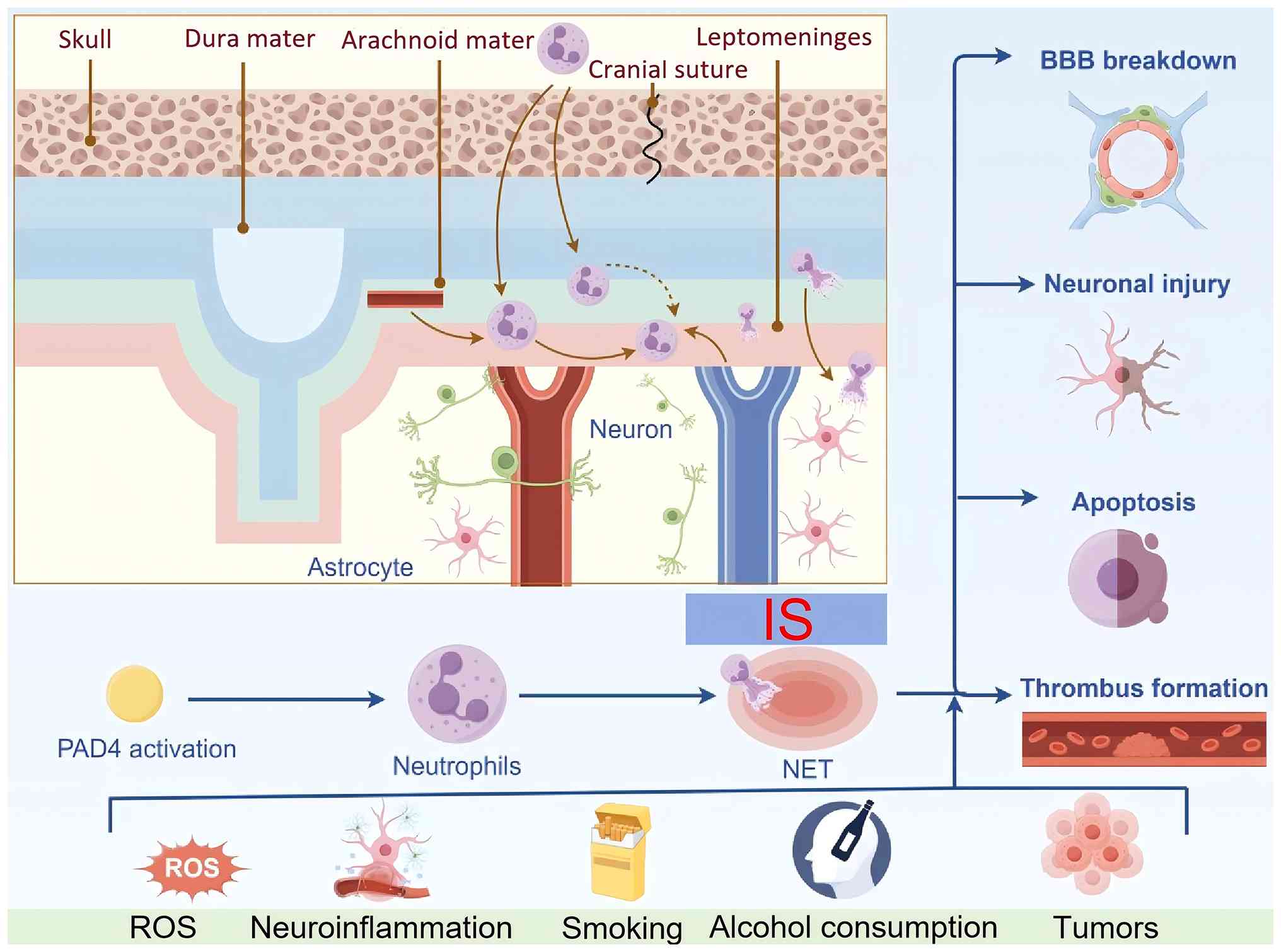 | Figure 1Pathogenesis of IS. The main box
illustrates the anatomical layers extending from the skull to the
brain parenchyma, including the skull, dura mater, arachnoid mater,
cranial sutures and leptomeninges. Neutrophils infiltrate across
the meninges into the cerebral parenchyma, where neurons and
astrocytes are located. Triggered by the risk factors displayed in
the figure: ROS, neuroinflammation, smoking, alcohol consumption
and tumors, multiple pathological events take place, such as BBB
breakdown, neuronal injury, apoptosis, ferroptosis and thrombus
formation. Furthermore, PAD4-dependent NET formation facilitates
the progression of IS. Created with Figdraw.com.
BBB, blood-brain barrier; IS, ischemic stroke; NET, neutrophil
extracellular trap; PAD4, peptidyl-arginine deiminase type IV; ROS,
reactive oxygen species. |
MSC-Exo characteristics, extraction,
identification and therapeutic potential in neurological
diseases
MSC-Exo characteristics
In recent years, MSCs have demonstrated promising
therapeutic effects in neurological diseases, such as IS,
intracerebral hemorrhage, spinal cord injury and Alzheimer's
disease (20,21). However, multiple limitations of
direct MSC transplantation, including potential tumorigenicity,
excessive cell dosage and microvascular embolism risk, have impeded
their clinical translation. Research has revealed that the
paracrine effects of MSCs exert beneficial outcomes that are partly
mediated by soluble factors, particularly exosomes (22). MSC-Exos possess marked biological
properties, such as good biocompatibility, high stability, low
toxicity and efficient exchange of molecular substances, and confer
notable benefits on a variety of diseases and injuries including
IS, myocardial infarction, cutaneous wounds and osteoarthritis, via
bioactive cargos such as growth-associated proteins and miRNAs
(23). In addition to possessing
biological functions similar to those of parent MSCs, MSC-Exos have
been widely utilized due to advantages such as their small volume,
low immunogenicity, and ease of acquisition, storage and
management. Compared with MSCs, the use of MSC-Exos in the human
body offers several potential advantages. i) Nanoscale exosomes can
freely traverse various biological barriers without affecting
microvascular circulation (24);
ii) while the number of transplanted MSCs rapidly decreases, the
delivery of MSC-Exos can sustain their therapeutic effect within
the body (25); and iii) because
MSC-Exos do not exist as living cells in vivo, this may
reduce the risk of tumor formation (although this does not imply
that all tumorigenic risks are eliminated; MSC-Exos may still
inherit the carcinogenic cargo from parent cells) (26). Beyond direct disease
intervention, MSC-Exos can also serve as drug carriers, working
synergistically with medications to achieve therapeutic efficacy
(27). Therefore, studies across
multiple disease models have indicated that MSC-Exos may serve as
an alternative to traditional stem cell therapies.
MSC-Exo extraction
Due to the limitations of traditional separation
methods in extracting MSC-Exos (such as cumbersome operation, low
efficiency, and low exosome yield and purity), a number of novel
separation methods have emerged, including ultrafiltration
separation technology, integrated dual-filter microfluidic chips,
nano-plasma-enhanced scattering, membrane-mediated exosome
separation and chip-based exosome separation (6,28). Table I comprehensively summarizes the
advantages and disadvantages of different extraction methods for
exosomes. These mainstream separation methods exhibit notable
differences in exosome purity, particle yield and impurity content.
The differences in technical parameters partially explain the
inconsistent therapeutic effects reported in different preclinical
studies (28-30). Although advances in new
technologies have improved exosome yield and purity to some extent,
drawbacks such as high costs and stringent equipment requirements
remain. Besides the extraction method, the cultivation method of
MSCs, their surrounding environment and the source of MSC-Exos also
affect MSC-Exo extraction (30-32). MSC-Exos originating from the bone
marrow, adipose tissue and umbilical cord possess distinct cargo
compositions, including unique profiles of proteins, cytokines and
miRNAs, and therapeutic efficacies against ischemic injury
(33); therefore, standardized
selection of MSC sources and exosome isolation protocols is crucial
for reducing experimental heterogeneity and promoting subsequent
clinical translation.
 | Table IAdvantages and disadvantages of
different extraction methods for mesenchymal stem cell-derived
exosome. |
Table I
Advantages and disadvantages of
different extraction methods for mesenchymal stem cell-derived
exosome.
| Authors, year | Extraction
method | Advantages | Disadvantages | (Refs.) |
|---|
| Kurian et
al, 2021 |
Ultracentrifugation | High purity | Equipment
dependency; the operation is cumbersome and takes a long time | (141) |
| Huang et al,
2025 | Density gradient
centrifugation | High purity and few
impurities | Operation is
complex and time-consuming | (28) |
| Luo et al,
2019 |
Ultrafiltration | Fast, easy to
operate and scalable for production | Membrane adsorption
leads to exosomes loss and a relatively low purity | (142) |
| Jiawei et
al, 2022 | Magnetic-activated
cell sorting | High specificity
and high purity | Low efficiency; the
biological activity is easily affected by pH and salt
concentration | (143) |
| Tiwari et
al, 2021 | Polymer-based
precipitation method | Easy to operate;
quick procedure | Low purity | (144) |
| Shirejini et
al, 2022 | Microfluidic chip
technology | High portability
and purity; low consumption of reagents and samples | High cost; complex
equipment | (145) |
| Sidhom et
al, 2020 | Chromatography | Easy to operate;
quick procedure; high purity | Small extraction
volume; equipment requirements are high | (146) |
MSC-Exo identification
MSC-Exo identification is a critical step to ensure
the purity, characteristics and reliability of exosomes in
research. The International Society for Extracellular Vesicles
(ISEV) established minimum experimental requirements for exosome
identification in 2018 (34).
Regarding biochemical characteristics, the expression of specific
positive marker proteins and the absence of contaminating negative
markers must be verified. Specifically, at least two transmembrane
proteins (any two from CD9, CD63 and CD81) and one cytoplasmic
protein (TSG101 or Alix) should be tested. Additionally, calnexin
or GRP94 (endoplasmic reticulum proteins), GM130 (Golgi apparatus
protein), at least one mitochondrial protein [such as cytochrome
c (Cyt-C)] and one nuclear protein (such as histone H3) must
be detected and excluded as contaminants. Regarding individual
characterization, transmission electron microscopy and nanoparticle
tracking analysis are used to observe the peak size and morphology
of exosomes. Exosomes typically appear as saucer-shaped or oval
bilayer membrane structures with varying sizes, featuring a low
electron density component in the center, clear boundaries and
concentrated distribution. The peak particle size distribution
ranges between 30 and 150 nm (35). In summary, the identification of
MSC-Exos is an integrative process involving multiple parameters
and diverse techniques. Only by comprehensively analyzing physical
characteristics (such as size and shape), particle concentration
and biochemical characteristics (for example, positive and negative
marker proteins), and strictly adhering to the core principles of
ISEV guidelines can it be reliably confirmed that the isolated
nanoparticles are indeed exosomes and relatively pure, suitable for
subsequent functional studies and application development.
Therapeutic potential of MSC-Exos in
neurological diseases
To date, extensive research results have confirmed
the broad application prospects of MSC-Exos in treating
neurological diseases. For example, MSC-Exos have been shown to
promote neurovascular remodeling in a rat model of MCAO via
miR-133b transfer and to regulate neurite outgrowth (36). Furthermore, when injected
intravenously into Alzheimer's disease model mice, MSC-Exos
pretreated with hypoxia can effectively increase synaptic proteins
(such as PSD-95 and Synapsin-I) expression; downregulate
inflammatory factors, such as TNF-α and IL-1β; and upregulate
anti-inflammatory factors, including IL-4 and IL-10, thereby
improving learning and memory function (37). Furthermore, Xu et al
(27) comprehensively summarized
the mechanisms of MSC-Exos in treating spinal cord injury and
optimization strategies to enhance therapeutic outcomes,
highlighting that combinatorial interventions can further boost the
anti-inflammatory and neuroprotective potency of MSC-Exos. In
conclusion, MSC-Exos may effectively promote nerve injury repair
and functional reconstruction. Compared with MSCs, MSC-Exos have
achieved marked success in treating neurological diseases due to
their unique advantages, such as improved safety, enhanced
stability and greater targeting properties.
Mechanisms of IS treatment by MSC-Exos
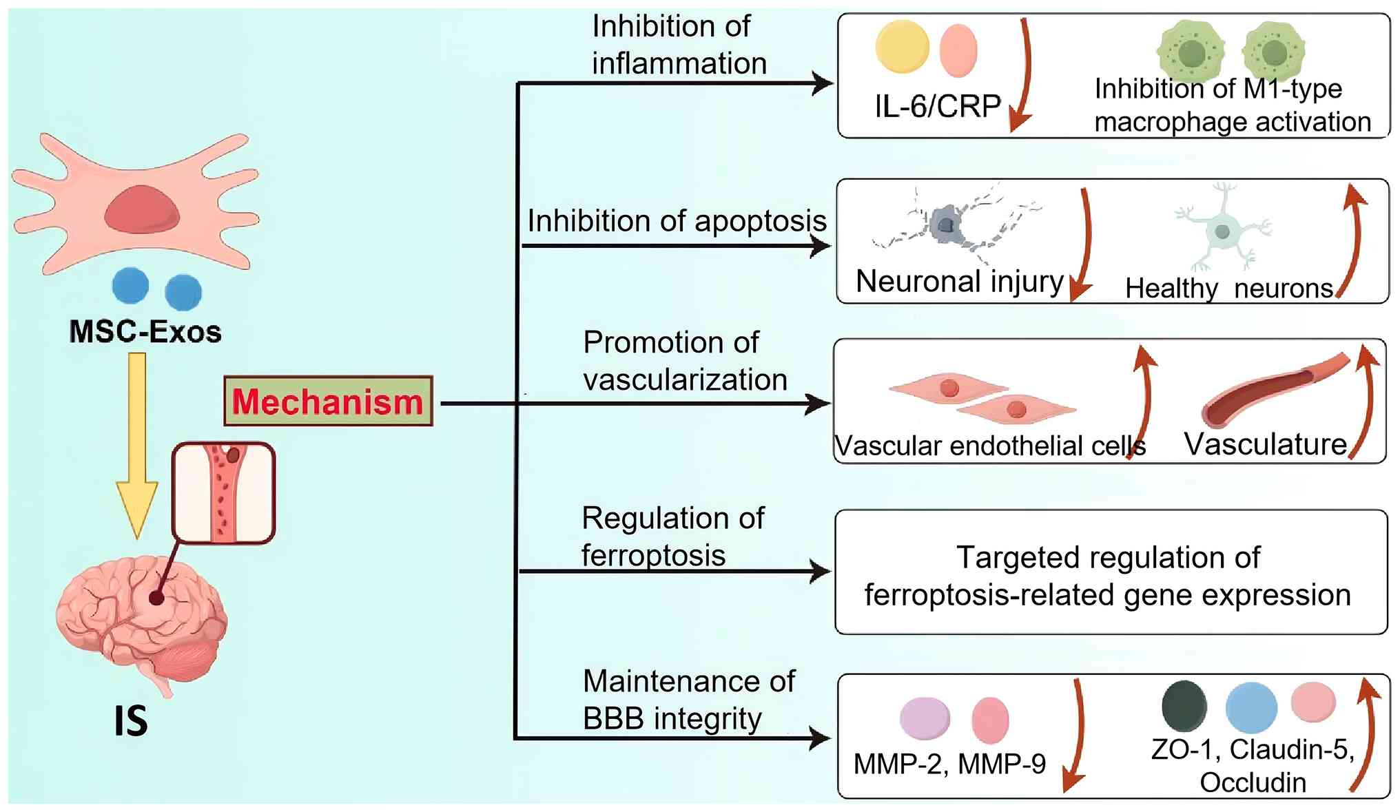
Research has indicated that MSC-Exos are key
effector molecules in the therapeutic action of MSCs, with their
internal components exerting complex biological effects on other
cells (38,39). MSC-Exos can cross the BBB and
demonstrate unique advantages in IS treatment through mechanisms
such as inhibiting inflammation, suppressing apoptosis, promoting
angiogenesis and regulating ferroptosis (Fig. 2).
Inhibition of inflammatory responses
Neuronal apoptosis in ischemic areas typically
induces secondary immune/inflammatory responses, leading to brain
tissue damage. Therefore, inhibiting inflammatory responses is an
important neuroprotective strategy. MSC-Exos can improve post-IS
functional deficits by modulating the activity of immune cells in
the IS lesion area and alleviating local inflammation. For example,
MSC-Exos can alleviate acute brain injury and microglial
inflammation by inhibiting the CysLT2/ERK1/2 signaling pathway and
inhibiting M1 macrophage activation (40). Additionally, MSC-Exos can reduce
neuronal oxidative stress and inflammation levels by inhibiting
ferroptosis and regulating the expression of glutathione peroxidase
4 (GPX4) and solute carrier family 7 member 11 (SLC7A11) (41,42). However, it has been reported that
although MSC-Exos mitigate secondary injuries caused by local
inflammatory responses through their immunomodulatory effects, it
remains unclear as to whether MSC-Exos can promote the repair and
regeneration of neural tissue in the lesion area (43).
Inhibition of apoptosis
Due to ischemia and hypoxia following IS,
neurological function is impaired and neuronal apoptosis is
enhanced. Numerous studies have confirmed that MSC-Exos attenuate
brain injury by inhibiting apoptosis. Hao et al (44) reported that MSC-Exos markedly
reduced the expression levels of Cyt-C and IL-1β, and the apoptotic
index in the neurons of IS rats. Zhao et al (45) discovered that when the Toll-like
receptor 4 (TLR4)/NF-κB signaling pathway was inactivated, the
expression of the key apoptotic protease caspase-3 was also
decreased. The underlying mechanism may involve MSC-Exos modulating
the TLR4/NF-κB signaling pathway to inhibit vascular endothelial
cell apoptosis, thereby exerting anti-cerebral ischemic effects.
MSC-Exos can also inhibit the activity of antigen-presenting cells,
downregulate the expression of B lymphocytes, T lymphocytes and
macrophages at the IS site, participate in the regulation and
balance of immune responses and provide a favorable
microenvironment for subsequent neural regeneration, thus
inhibiting apoptosis (46,47).
Promotion of angiogenesis
Following IS, angiogenesis is a critical step in
restoring brain tissue function. Vessel blockage damages the
structure of the vessels and alters their permeability, resulting
in reduced normal blood supply and oxygen delivery. Notably, it has
been shown that MSC-Exos facilitate cerebral tissue remodeling by
promoting angiogenesis and neuronal survival (48). Doeppner et al (49) demonstrated that when treating
adult male rats with MCAO using bone marrow MSC-derived exosomes
(BMMSC-Exos), endothelial cell proliferation was markedly increased
and a new capillary network was formed, indicating that BMMSC-Exos
could promote post-stroke angiogenesis. In addition, Xin et
al (36) administered
MSC-Exos via tail vein injection 24 h after constructing an MCAO
rat model, and the results showed a notable increase in vascular
endothelial cell numbers, indicating that MSC-Exos can improve
post-stroke vascular regeneration.
Regulation of ferroptosis
It has been show that upon occurrence of IS, due to
cerebral tissue hypoxia and reperfusion injury, iron ion levels are
markedly increased, thus triggering ferroptosis (50). MSC-Exos protect neurons from
damage by targeting and regulating ferroptosis-related pathways
through carrying miRNAs, proteins and other bioactive substances.
For example, miR-214-3p in MSC-Exos can alleviate neuronal injury
in mouse models of cerebral ischemia by modulating the expression
of ferroptosis-regulating genes, including GPX4 and SLC7A11
(41). Deng et al
(51) also revealed that steroid
receptor coactivator-3 in MSC-Exos could significantly inhibit
lipid peroxidation and ferroptosis in hypoxic glucose-deprived
neurons, and reduce infarct volume and brain tissue water content
after cerebral ischemia, thereby improving neurological function.
Although the specific mechanisms by which MSC-Exos regulate
ferroptosis in IS have not been fully elucidated, exosome-mediated
regulation of ferroptosis holds promise as a potential therapeutic
target for future clinical treatment of IS.
Maintenance of BBB integrity
After IS, disruption of the BBB is an important
factor leading to further brain tissue injury. In the early stage
of IS, BBB disruption manifests as increased permeability and
enhanced destruction, subsequently leading to increased penetration
of immune cells and inflammatory substances (52). MSC-Exos can protect the integrity
of the BBB by enhancing the expression of tight junction proteins
(such as ZO-1 and Claudin-5), and reducing the penetration of
inflammatory cells and immune factors. Li et al (53) demonstrated that adipose-derived
MSC-derived exosomes (ADSC-Exos) could increase the distribution of
ZO-1 and Claudin-5 in MCAO rat models, improve the BBB state and
alleviate cerebral edema. In addition, MSC-Exos may reduce brain
tissue edema and lesion size, lower the levels of blood-derived
brain biomarkers (such as S100B and glial fibrillary acidic
protein) and enhance BBB integrity by inhibiting the expression and
enzymatic activity of MMP-9 (54).
Notably, IS can trigger multiple forms of programmed
cell death, not limited to apoptosis and ferroptosis. I/R injury
simultaneously activates novel cell death regulatory pathways, such
as pyroptosis, necroptosis, PANoptosis, autophagy and cuproptosis.
These distinct death pathways interact to amplify oxidative stress
and neuroinflammation, collectively resulting in marked neuronal
loss in the ischemic penumbra (55,56).
MSC-Exos are capable of successfully crossing the
BBB and accumulating at the ischemic lesion site. In addition to
the aforementioned effects of suppressing neuroinflammation,
antagonizing neuronal apoptosis, regulating iron overload and lipid
peroxidation-mediated ferroptosis, and promoting angiogenesis, they
can also broadly regulate multiple programmed death pathways by
carrying various miRNAs, cytokines and other active substances
(55,57,58). Current basic research has also
preliminarily suggested that they may improve copper-induced
neuronal damage by maintaining mitochondrial homeostasis, although
more experimental validation is required (59,60). In conclusion, the synergistic
regulation of multiple programmed cell death pathways constitutes
an important mechanistic basis for MSC-Exos in improving the
prognosis of IS.
Different combination strategies for MSC-Exo
treatment of IS
Although MSC-Exos show promising therapeutic
efficacy in treating IS, they also possess drawbacks such as high
heterogeneity, low yield, storage difficulties and rapid clearance
in vivo, leading to issues including poor organ targeting
and insufficient efficacy when used alone for IS treatment
(61). Moreover, the mechanisms
by which MSC-Exos regulate inflammation, promote angiogenesis and
facilitate neuronal regeneration in IS treatment remain unclear.
For example, it has been reported that MSC-Exos can markedly
promote functional recovery in MCAO model rats without reducing
infarct volume (62). Therefore,
combining MSC-Exos with innovative delivery systems and enhancement
strategies has become a critical direction for overcoming the
obstacles in IS treatment (Fig.
3).
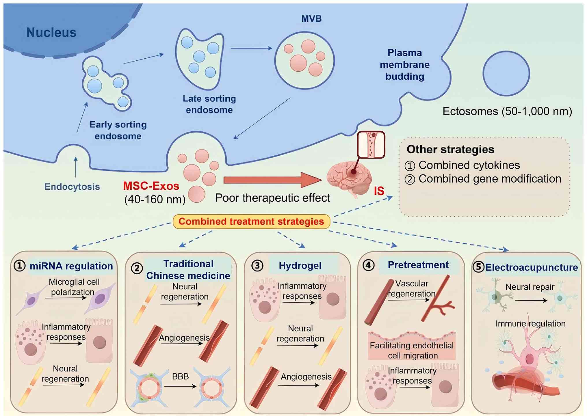 | Figure 3Combination strategies and mechanisms
of MSC-Exos in the treatment of IS. i) MSC-Exos combined with
miRNAs improves the therapeutic effect of IS by regulating
microglial polarization, inhibiting inflammatory responses and
promoting nerve regeneration. ii) MSC-Exos combined with
traditional Chinese medicine improves the therapeutic effect of IS
by promoting nerve regeneration, repairing vascular injury and
regulating the blood-brain barrier. iii) MSC-Exos combined with
hydrogel improves the therapeutic effect on IS by inhibiting
inflammatory response, promoting nerve regeneration and repairing
vascular injury. iv) MSC-Exos pretreatment improves the therapeutic
effect on IS by promoting vascular regeneration, facilitating
endothelial cell migration and inhibiting inflammatory responses.
v) MSC-Exos combined with electroacupuncture improves the
therapeutic effect on IS by inhibiting neuronal apoptosis and
immune regulation. The five boxes at the bottom correspond to the
five combinatorial strategies: miRNA regulation, traditional
Chinese medicine intervention, hydrogel loading, pretreatment and
electroacupuncture. Besides exosomes, extracellular vesicles also
include ectosomes released from the plasma membrane. Combined
cytokines refers to exogenous cytokine-loaded MSC-Exos for
synergistic therapy; combined gene modification refers to
genetically engineered MSC-Exos overexpressing target genes to
enhance therapeutic potency. Created with Figdraw.com. IS, ischemic stroke; miRNA, microRNA;
MSC-Exo, mesenchymal stem cell-derived exosome; MVB, multivesicular
body. |
miRNA regulation
Research has shown that a large number of miRNAs in
MSC-Exos widely participate in oxidative stress, cellular apoptosis
and immune-inflammatory injury after cerebral ischemia, and also
serve important roles in vascular remodeling, neuronal regeneration
and synaptic plasticity post-ischemia (63,64). Based on core detection indicators
and experimental observation focuses from previous studies, the
combination of MSC-Exos and miRNAs may promote functional recovery
after injury by regulating microglial polarization, alleviating
inflammatory responses, promoting angiogenesis and facilitating
nerve regeneration. These mechanisms are closely interrelated and
mutually reinforcing in the pathological process of ischemic brain
injury and inherent biological cross-effects exist (Table II).
| Table IIMSC-Exos combined with miRNAs treat
IS through different mechanisms. |
Table II
MSC-Exos combined with miRNAs treat
IS through different mechanisms.
| Authors, year | Exosome source | miRNA | Biological
effect | Mechanism | (Refs.) |
|---|
| Zhao et al,
2020 | BMMSC-Exos | miR-223-3p | Regulation of
microglia polarization | Inhibition of
microglia M1 polarization-mediated pro-inflammatory responses | (65) |
| Geng et al,
2019 | ADSC-Exos | miR-126 | Regulation of
microglia polarization | Improvement of
neurogenesis and inhibition of microglia activation | (66) |
| Jiang et al,
2018 | ADSC-Exos | miR-30d-5p | Regulation of
microglia polarization | Inhibition of
autophagy-mediated microglia polarization to M1 type | (67) |
| Xin et al,
2020 | MSC-Exos | miR-21a-5p | Regulation of
microglia polarization | Regulation of
microglia/macrophage polarization and attenuation of brain injury
from hypoxic-ischemic injury | (68) |
| Deng et al,
2019 | BMMSC-Exos | miR-138-5p | Reduced
inflammatory response | Inhibition of
lipocalin-2 to promote astrocyte proliferation after IS | (72) |
| Zhou et al,
2020 | BMMSC-Exos | miR-126-3p | Promotion of nerve
regeneration | Altered brain
plasticity and increased neural protrusion growth | (74) |
| Zhang et al,
2017 | MSC-Exos | miR-17-92 | Promotion of nerve
regeneration | Activation of
PTEN/mTOR signaling pathway | (75) |
| Hou et al,
2020 | BMMSC-Exos | miR-29b-3p | Promotion of nerve
regeneration | Activation of
PTEN-mediated Akt signaling pathway to prevent hypoxic-ischemic
injury in rat brain | (76) |
| Xin et al,
2017 | BMMSC-Exos | miR-17-92 | Promotion of nerve
regeneration | Targeting PTEN to
activate the PI3K/Akt/rapamycin/glycogen synthase kinase 3β
signaling pathway | (77) |
| Bao et al,
2024 | BMMSC-Exos | miR-486 | Regulation of
cerebral microvascular endothelial cell function | Activation of
PTEN/Akt signaling pathway | (79) |
| Yang et al,
2018 | ADSC-Exos | miR-181b | Regulation of
cerebral microvascular endothelial cell function | Promotion of
endothelial cell movement and angiogenesis via miR-181b/TRPM7 axis
in vitro | (80) |
Regulation of microglial cell
polarization
Numerous studies have indicated that miRNAs can be
loaded and transported to specific target cells in the form of
exosomes; thus, MSC-Exos carrying miRNAs possess the ability to
regulate microglial activation and anti-inflammatory responses
(65-67). MSC-Exos loaded with miR-223-3p
have been reported to alleviate pro-inflammatory responses mediated
by M1 polarization of microglia, reduce cerebral I/R injury and
thereby accelerate functional recovery after IS (65). MSC-Exos loaded with miR-126 can
also improve functional recovery in IS model rats by promoting
neurogenesis and inhibiting microglial activation (66). Furthermore, MSC-Exos loaded with
miR-30d-5p may prevent brain injury by inhibiting
autophagy-mediated microglial polarization to the M1 phenotype
(67). In addition, MSC-Exos can
regulate microglia/macrophage polarization via targeted delivery of
miR-21a-5p, protecting the brains of newborn mice from
hypoxic-ischemic injury (68).
Reducing inflammatory responses
It has been reported that IL-6 is an important
cytokine involved in pro-inflammatory responses that is closely
associated with immune responses after IS (69). Ji et al (70) demonstrated that the concentration
of serum exosomes, and the median levels of miR-9 and miR-124 in
serum exosomes were notably elevated in patients with IS. In
addition, the levels of miR-9 and miR-124 were positively
associated with National Institutes of Health Stroke Scale (NIHSS)
scores, infarct volume and serum IL-6 concentrations. These results
suggested that serum exosomal miR-9 and miR-124 may be promising
biomarkers for diagnosing IS and assessing the extent of ischemic
injury. Xue et al (71)
discovered that downregulation of miR-9 inhibited the transcription
of inflammatory factors TNF-α and IL-6 by targeting and silencing
silent information regulator 1 in the downstream NF-κB signaling
pathway, thereby alleviating I/R injury. Similarly, miR-138-5p
within BMMSC-Exos has been shown to promote astrocyte proliferation
in the I/R area by targeting lipocalin-2 and to suppress
inflammatory responses to mitigate neuronal injury (72). Although inflammatory mediators
serve as important targets of MSC-Exos at sites of inflammation,
the specific mechanisms by which they promote an immunosuppressive
microenvironment in damaged areas remain unclear.
Promoting neural regeneration
Studies have shown that the number of MSC-Exos in
the infarcted hemisphere increases in a dose-dependent manner, and
MSC-Exos are rich in various proteins and miRNAs related to
neurogenesis (73). In acute IS
(AIS), miR-126-3p in MSC-Exos can alter brain plasticity, increase
neurite growth, and protect nerve cells from damage and apoptosis
(74). Zhang et al
(75) tracked
fluorescence-labeled MSC-Exos and monitored axonal growth of
neurons using time-lapse microscopy, revealing that
miR-17-92-treated MSC-Exos activated the phosphatase and tensin
homolog (PTEN)/mTOR signaling pathway in recipient neurons, further
accelerating the growth of cortical neuron axons. Additionally, Hou
et al (76) observed
apoptosis in the neurons of MCAO model rats; however,
overexpression of miR-29b-3p or silencing of PTEN reversed these
changes. It has also been reported that the use of custom exosomes
enriched with the miR-17-92 cluster can enhance neural plasticity
and functional recovery after IS, a mechanism potentially involving
targeting PTEN to activate the PI3K/Akt/rapamycin/glycogen synthase
kinase 3β signaling pathway (77).
Regulating the function of brain
microvascular endothelial cells
Brain microvascular endothelial cells interact
actively with extracellular matrix proteins forming the basal
lamina, pericytes, astrocytes and neurons, jointly forming the
neurovascular unit (NVU). AIS leads to rapid NVU damage and impairs
neurovascular coupling (78). It
has been shown that BMMSC-Exos can markedly increase microvascular
density after ischemic brain injury by upregulating miR-486 and
activating the PTEN/Akt signaling pathway, thereby improving brain
ischemic injury (79). Yang
et al (80) demonstrated
that ADSC-Exos can promote the migration and angiogenesis of brain
microvascular endothelial cells under in vitro
oxygen-glucose deprivation (OGD) conditions via the miR-181b/TRPM7
axis. Liu et al (81)
reported that elevated levels of miR-122 in the blood can reduce
neurological deficits and cerebral infarction area in a rat
MCAO-induced IS model, by downregulating the expression of
cell-adhesion-related genes such as vascular cell adhesion molecule
1 and intercellular cell adhesion molecule 1, which are involved in
leukocyte extravasation, while maintaining vascular integrity.
In conclusion, MSC-Exos carrying miRNAs may serve as
a means of intercellular communication after cerebral ischemia,
providing novel insights into the pathogenesis and treatment of IS
using MSC-Exos, and facilitating the development of
MSC-Exos-targeted vectors to improve the therapeutic outcomes for
IS. Notably, most preclinical studies rely on young-rodent MCAO
models with variable protocols for modelling, exosome preparation
and outcome assessment, which may produce conflicting results.
Furthermore, small sample sizes and incomplete
blinding/randomization raise reproducibility risks. Collectively,
the current evidence remains preliminary with low certainty.
Combination with traditional Chinese
medicine
The combined application of MSC-Exos with
traditional Chinese medicine ingredients also demonstrates
favorable therapeutic advantages in the treatment of IS (82). Exosomes have emerged as ideal
delivery carriers for herbal active ingredients to target lesion
cells and exert therapeutic effects, making them a focus of
research (82). Multiple studies
have indicated that in the treatment of IS, MSC-Exos loaded with
traditional Chinese medicine components such as curcumin,
tetramethylpyrazine and gastrodin exert neuroprotective, angiogenic
and BBB permeability-regulating effects, with efficacy superior to
that of MSC-Exos or drug therapy alone (Table III).
| Table IIIMSC-Exos combined with traditional
Chinese medicines treat ischemic stroke through different
mechanisms. |
Table III
MSC-Exos combined with traditional
Chinese medicines treat ischemic stroke through different
mechanisms.
| Authors, year | Exosome source | Drug | Biological
effect | Mechanism | (Refs.) |
|---|
| Mao et al,
2023 | BMMSC-Exos |
Tetramethylpyrazine | Promotion of
neuroprotection | Inhibition of
neuronal apoptosis | (83) |
| Zhang et al,
2023 | BMMSC-Exos |
Tetramethylpyrazine | Promotion of
neuroprotection | Inhibition of
neuronal apoptosis and attenuation of oxidative stress levels | (84) |
| Chen et al,
2023 | BMMSC-Exos | Musk ketone | Promotion of
neuroprotection | Decreased
expression of pro-apoptotic proteins and pro-inflammatory factors,
and increased expression of the anti-apoptotic protein Bcl-2 | (85) |
| Kalani et
al, 2017 | MSC-Exos | Curcumin | Promotion of
neuroprotection | Improved
endothelial cell permeability and reduced levels of oxidative
stress | (86) |
| Liu, 2022 | BMMSC-Exos | Xionggui
decoction | Promotion of
neuroprotection | Modulation of the
miR-17-92/PTEN signaling pathway for axonal regeneration | (87) |
| Luo et al,
2023 | BMMSC-Exos | Ginsenoside
Rg1 | Promotion of
angiogenesis | Activation of the
PI3K/Akt pathway and upregulation of VEGF and HIF-1α
expression | (89) |
| Li et al,
2023 | hUCMSC-Exos | Shennao Fuyuan
decoction | Promotion of
angiogenesis | Modulation of the
Wnt3a/β-catenin pathway and attenuation of NSC injury | (90) |
| Yang et al,
2015 | BMMSC-Exos | Buyang Huanwu
decoction | Promotion of
angiogenesis | Induction of VEGF
secretion from endothelial cells and increased vascular
density | (91) |
| Nan et al,
2023 | MSC-Exos | Gastrodin | Regulation of the
BBB | Upregulation of
XIAP expression in the ischemic hemisphere inhibits neuronal
apoptosis | (94) |
| Yao et al,
2024 | BMMSC-Exos | Qixiong Zuogui
compound | Regulation of the
BBB | Inhibition of the
expression of MMP-2 and MMP-9, and promotion of the expression of
tight junction proteins ZO-1 and Occludin | (95) |
| Zhong et al,
2019 | MSC-Exos | Scutellarin | Regulation of the
BBB | Upregulation of
CD63 and BBB tight junction protein expression | (96) |
Promotion of neuroprotection
It has been reported that the combination of
BMMSC-Exos with tetramethylpyrazine can markedly enhance the
neuroprotective effect against IS, a mechanism potentially
associated with the inhibition of neuronal apoptosis (83). Furthermore, the combination of
BMMSC-Exos with tetramethylpyrazine can notably improve
pathological changes in IS, reduce infarct volume, and lower levels
of neuronal apoptosis and oxidative stress (84). The combination of BMMSC-Exos with
musk ketone has been reported to effectively improve neurological
function and reduce infarct volume in MCAO model rats. Further
research revealed that this combined strategy can downregulate the
expression of pro-apoptotic proteins, such as Bax, and
pro-inflammatory factors, including TNF-α and IL-1β, while
promoting the expression of the anti-apoptotic protein Bcl-2,
thereby effectively inhibiting neuronal apoptosis and microglial
activation in rats (85).
Additionally, the combination of MSC-Exos with curcumin exhibits
anti-lipid deposition, antioxidant and anti-inflammatory effects,
protecting against IS by improving endothelial cell permeability
and reducing oxidative stress levels (86). It has also been reported that
Xionggui decoction enhances axonal regeneration in rats following
BMMSC-Exos intervention in IS by regulating the miR-17-92/PTEN
signaling pathway (87). This
effect is achieved through the improvement of neurological deficits
and brain tissue pathological damage, as well as the promotion of
oligodendrocyte differentiation and myelin formation.
Promotion of angiogenesis
Ginsenoside Rg1 is a major active component
extracted from ginseng roots. Due to its multiple biological
functions, such as anti-inflammatory, immune-regulatory and
angiogenesis-promoting effects, it is widely used in the treatment
of neurological diseases (88).
BMMSC-Exos modified with c(RGDyk) peptide, serving as a
multifunctional carrier material, load ginsenoside Rg1 through
co-incubation and have been reported to activate the PI3K/Akt
pathway in a rat model of IS to promote the expression of vascular
endothelial growth factor (VEGF) and hypoxia-inducible factor-1α
(HIF-1α), thereby promoting angiogenesis and effectively reducing
the volume of cerebral infarction (89). Li et al (90) demonstrated that the combination
of human umbilical cord mesenchymal stem cell-derived exosomes
(hUCMSC-Exos) with Shennao Fuyuan decoction alleviates
OGD/reoxygenation-induced damage to neural stem cells by promoting
angiogenesis and inhibiting cell apoptosis, which may be associated
with activation of the Wnt3a/β-catenin pathway. In addition,
compared with treatment with BMMSC-Exos alone, the combination of
BMMSC-Exos with Buyang Huanwu decoction shows stronger effects in
inducing VEGF secretion by brain tissue endothelial cells and
increasing vascular density in cerebral ischemic rats (91). In addition to these drugs, in the
treatment of neurological diseases, catalpol can promote
post-ischemic neurological recovery, protect the NVU and upregulate
VEGF expression (92); however,
to the best of our knowledge, whether the combination of catalpol
with MSC-Exos is beneficial for angiogenesis after IS has not yet
been reported in the literature and requires further
exploration.
Regulation of the BBB
Studies have shown that single herbal medicines or
prescriptions can regulate the BBB through multiple pathways,
targets and effects, thereby exerting antioxidant effects,
inhibiting neuronal apoptosis, improving cerebral blood flow and
other functions. In vivo studies have confirmed that the
combination of MSC-Exos and traditional Chinese medicine
effectively regulates the permeability of the BBB after cerebral
I/R injury, reduces the volume of cerebral infarction and markedly
restores neurological function. Gastrodin can alleviate cerebral
ischemic injury in mice by enhancing antioxidant and
anti-inflammatory activity, and inhibiting the cell apoptosis
pathway (93). Nan et al
(94) demonstrated that
gastrodin can increase the content of miR-20a-5p in circulating
exosomes; these MSC-Exos may then penetrate the BBB and upregulate
the expression of X-linked inhibitor of apoptosis protein (XIAP) in
the ischemic hemisphere, inhibit neuronal apoptosis and promote the
recovery of neurological function in MCAO model rats. Yao et
al (95) confirmed that the
combination of BMMSC-Exos with Qixiong Zuogui compound may markedly
reduce the cerebral infarction area in aged MCAO model rats,
improve neurological deficit scores and enhance cognitive function;
it may also inhibit the expression of MMP-2 and MMP-9, promote the
expression of the tight junction proteins ZO-1 and Occludin, and
reduce BBB permeability. In addition, MSC-Exos, which are
influenced by scutellarin, can protect homocysteine-induced rat
microvascular endothelial cells by upregulating the expression of
CD63 and BBB tight junction proteins (96).
Although the combination of MSC-Exos and traditional
Chinese medicine exhibits certain advantages in treating IS, most
existing preclinical evidence comes from a limited number of
research teams and independent verification across different teams
is still lacking. Almost all data originate from young-adult rodent
MCAO models, and evidence from aged or comorbid animals is scarce.
In addition, variability in herbal and exosome protocols hampers
cross-study comparison and the pre-clinical evidence certainty
remains low. Therefore, these encouraging experimental results
should be interpreted with caution and more research is needed to
translate them into clinical applications to confirm their
reproducibility.
Combination with composite hydrogels
It has been reported that the in vivo
therapeutic efficacy of natural MSC-Exos is limited by their short
half-life, poor lesion accumulation and insufficient tissue
targeting ability. Notably, hydrogels loaded with MSC-Exos have
attracted widespread attention as an emerging therapeutic strategy
in the fields of tissue engineering and regenerative medicine, and
are regarded as an effective means to promote nerve regeneration
and functional recovery in patients following a stroke (97,98). The good biocompatibility and
adjustability of MSC-Exos combined with hydrogels can effectively
deliver exosomes to individual cells, thereby extending their
retention time at the site of tissue injury (99).
Regulation of the inflammatory
microenvironment
As aforementioned, the main pathogenesis of IS
involves ischemia, oxidative stress, inflammatory response and
neuronal apoptosis pathways (100). The novel exosome-hydrogel
combination therapy directly delivers exosomes to the injury site,
providing a scaffold for tissue and nerve repair, thereby exerting
anti-inflammatory, antioxidant, axon growth-stimulating and
myelin-regenerating effects (101). Lu et al (102) confirmed that the combination of
BMMSC-Exos with hydrogel materials can markedly improve
neurological functional recovery in rats with traumatic brain
injury and can partially relieve the occurrence of neural
inflammation. Hydrogels can also mimic different repair
microenvironments after peripheral nerve injury, producing a
synergistic effect with MSC-Exos to promote neurite outgrowth and
neurological defect repair (103). Li et al (104) reported that when using a
hydrogel loaded with 3D-MSC-Exos to treat AIS, a 30% Pluronic-F127
hydrogel had good thermosensitivity, was fluid at low temperatures
and rapidly transformed into a soft gel near body temperature, and
stably released MSC-Exos for >6 days. Notably, this combination
can be safely injected into the ischemic core of MCAO model rats,
effectively alleviating the inflammatory response in the ischemic
hemisphere and promoting the recovery of neurological function in
MCAO model rats.
Promotion of vascular and nerve
regeneration
Functionally, MSC-Exos not only participate in
immune regulation and homeostasis maintenance but also in blood
coagulation, nerve regeneration and intercellular signal
transduction. Zhang et al (22) developed a supramolecular hydrogel
capable of effectively sustained release of exosomes. After
application in an MCAO mouse model, it promoted nerve repair after
stroke. Immunofluorescence staining indicated that the neurological
function in MCAO model mice was markedly improved, and angiogenic
and neuroregenerative capabilities were enhanced. In behavioral
tests, motor coordination and balance abilities of these mice were
notably improved and the number of apoptotic cells was also
reduced. This demonstrates the efficacy of exosomes combined with
hydrogels in promoting post-stroke neurological functional
recovery. Furthermore, the latest research reports that hydrogels
can precisely deliver bioactive molecules to specific regions of
the brain while supporting the survival and proliferation of
transplanted stem cell-derived exosomes, thereby improving
neuroinflammation, vascular remodeling and sensorimotor functional
recovery after IS (105). It
has also been reported that injectable adhesive hydrogels composed
of catechol-grafted hyaluronic acid, prepared via photocuring, can
be combined with exosomes to achieve sustained exosome release,
which may be used to improve neurological function and angiogenesis
after IS (106).
In summary, the combined application of MSC-Exos and
hydrogels represents a revolutionary advancement in the field of IS
repair therapy. By addressing the critical obstacle of rapid
clearance of exosomes in vivo, this approach achieves
synergistic effects on neurovascular repair. Current research has
transitioned from basic mechanistic exploration to clinical
translation, with various innovative hydrogel systems (such as
thermosensitive, adhesive and active interface materials)
demonstrating unique advantages. It is worth noting that currently,
almost all supporting data for hydrogel-assisted MSC-Exos treatment
of IS come from rodent models, and such preclinical results cannot
be directly applied to human stroke. A number of unresolved issues
remain regarding intracranial hydrogel delivery for human IS,
including the determination of safe injection volume, optimization
of injection sites and assessment of in vivo
biocompatibility. Risks such as local inflammation and mass effects
that may occur after hydrogel implantation also need to be fully
validated in large animal models before human trials can be
initiated. Limited sample sizes cause reproducibility concerns and
these findings cannot be directly extrapolated to humans. In the
future, with the development of engineered exosomes, smart
responsive materials and personalized treatment strategies, this
field is expected to break through the limitations of neural repair
and provide novel solutions for the treatment of disabling IS.
Other combination strategies
Pretreatment
It is well known that hypoxia can activate HIF-1α,
thereby regulating the transcription of angiogenic genes, such as
VEGF and VEGF receptors (VEGFRs), and promoting angiogenesis. Hu
et al (107) reported
that, compared with in a normoxia group, MSCs subjected to hypoxic
pretreatment and transplanted into brain tissue showed increased
survival and migrated towards the ischemic area. This mechanism may
be related to the increased expression of CXC chemokine receptor 4
on the surface of MSCs after hypoxic preconditioning. It has also
been reported that hypoxic pretreatment of MSC-Exos can be used for
IS treatment. Han et al (108) revealed that hypoxia-treated
hUCMSC-Exos can enhance angiogenesis by regulating the VEGF/VEGFR
signal. In addition, BMMSC-Exos under low-oxygen conditions promote
angiogenesis by activating the HIF-1α/VEGF/JNK pathway (109).
In addition to hypoxic preconditioning, cholesterol
lipid preconditioning of MSCs can enhance the release and survival
capacity of MSC-Exos, exerting beneficial effects in stroke models
through cerebral blood flow protection (110). Zhang et al (111) loaded cholesterol-modified
miR-210 onto BMMSC-Exos conjugated with RGDyk cyclic peptide; after
intravenous injection, these targeted mouse brain tissues, thereby
promoting VEGF expression and angiogenesis in the ischemic area.
MSC-Exos pretreated with interferon-γ can also induce activated
microglia to secrete fewer pro-inflammatory factors, effectively
inducing oligodendrocyte differentiation and myelin formation,
thereby promoting recovery from stroke (112). Ye et al (113) adopted human umbilical vein
endothelial cells as an in vitro surrogate for cerebral
vascular endothelial cells. Using a scratch-wound healing assay, it
was demonstrated that infarcted tissue extract-preconditioned
hUCMSC-Exos promoted the directional migration of endothelial cells
compared with naïve hUCMSC-Exos. In the same study, it was also
revealed that hUCMSC-Exos pretreated with infarcted tissue extracts
could markedly improve the morphology and viability of endothelial
cells, enhance vascular endothelial remodeling and promote
neurological functional recovery after stroke (113). Lee et al (114) also demonstrated that MSC-Exos
treated with brain extracts had therapeutic effects on rat cerebral
ischemia models. They enhanced behavioral recovery and reduced the
area of cerebral infarction by promoting angiogenesis, neurogenesis
and anti-inflammatory effects.
Combination with EA
Studies have shown that EA can enhance the
survival, migration and neural differentiation of MSCs, ultimately
improving motor function after IS (115). The combination of MSCs and EA
can balance pro-inflammatory and anti-inflammatory factors, thereby
alleviating brain damage after IS (116). Kim et al (117) reported that acupuncture
combined with MSC intervention could improve sensorimotor function
in IS rats, reduce striatal atrophy caused by ischemia and promote
neuronal proliferation. In addition, exosomes act as an important
carrier of EA efficacy, and EA can also regulate the expression and
secretion of MSC-Exos. This combined treatment may more effectively
regulate neurogenesis than monotherapy through synergistic
regulation of brain-derived neurotrophic factor (BDNF), leading to
improved therapeutic outcomes. Xu et al (118) revealed that EA combined with
exosomal miR-210 could promote angiogenesis under hypoxic
conditions via the HIF-1α/VEGF/Notch1 signaling pathway. Moreover,
induced pluripotent stem cell-derived small extracellular vesicles
combined with EA could reduce infarct volume in mice with IS,
improve motor and neural functions, and inhibit neuronal apoptosis.
The mechanism may involve regulating IL-33/ST2-mediated
inflammation and activating the synergistic neuroprotective effects
of microglia and astrocytes against ischemic brain injury. Although
both MSCs and MSC-Exos can be combined with EA to correct
inflammatory states and exert neuroprotective effects, further
research is needed to investigate the impact of factors such as the
timing, frequency and intensity of EA intervention on therapeutic
efficacy. Current indirect evidence has suggested a potential
interaction between EA and MSC-Exos-related pathways; however, the
underlying mechanistic links remain largely speculative and require
further experimental verification (119). Moreover, this combined strategy
is still in the early stages of research. Key parameters such as
the dose-response relationship of stimulation, optimal frequency
and intervention timing have not been systematically
investigated.
Moreover, MSC-Exos can be loaded with exogenous
proteins for brain delivery (120). Pigment epithelium-derived
factor (PEDF) is a multifunctional protein with anti-inflammatory,
antioxidant and neuroprotective properties (121). Huang et al (120) combined PEDF overexpression with
ADSC-Exos to improve I/R injury by activating autophagy and
inhibiting neuronal apoptosis. The results indicated that PEDF may
serve a role in preventing exosome-mediated cerebral I/R injury by
regulating apoptotic factors and promoting autophagy. Additionally,
it has been shown that a novel delivery system, TAT peptide
membrane and rabies virus glycoprotein co-modified MSC-Exos
carrying a miR-15b-5p inhibitor, can break through the BBB to
target the peri-infarct area, markedly inhibiting neuronal
apoptosis by regulating the HTR2C/ERK signaling pathway and
providing a new strategy for stroke treatment (122). Notably, most studies have only
verified the overall neuroprotective effects of MSC-Exos, while
evidence revealing dynamic signaling pathways, exosomal cargo
sorting mechanisms and specific receptor-ligand interactions
mediating biological functions remains limited. A number of studies
have also used different stroke models in rodents, but the
intervention parameters were inconsistent. Positive results are
preferentially reported while neutral or negative data are seldom
published, bringing notable reproducibility risks and low-quality
pre-clinical evidence.
Current status and challenges of clinical
translation
Clinical research progress and
translational trends
Currently, exosome-based combination therapies have
entered early-stage clinical trials. To the best of our knowledge,
the ChiCTR2500099772 trial (registered at Chinese Clinical Trial
Registry, https://www.chictr.org.cn/showproj.html?proj=266284),
registered at The First Affiliated Hospital of Xiamen University
(Xiamen, China), is the first to explore the safety and efficacy of
hUCMSC-Exos nasal spray in post-stroke cerebral infarction
following smoking-related diseases. This trial has employed a
single-arm design with a planned enrollment of eight patients; the
drug is administered via the nasal mucosa to bypass the BBB, and
the primary endpoints are improvements in NIHSS and Modified Rankin
Scale scores. An investigational stem cell product for chronic
stroke developed by Jiuzhitang Maker (Beijing) Cell Technology Co.,
Ltd. has completed Phase II patient enrollment. The preliminary
Phase I data, presented orally at the 2025 Wuzhen Health Congress,
showed a 35% improvement in Fugl-Meyer motor score and 28%
improvement in Barthel index (Jiuzhitang Maker Cell Technology Co.,
Ltd., 2025 Wuzhen Health Congress, Wuzhen, 2025). The conference
proceedings remain unpublished, and the detailed quantitative data
were only presented orally and have not undergone peer review. The
company intends to submit an application for conditional marketing
approval in 2026. Although some early clinical trials have reported
encouraging preliminary results, these should not be
over-interpreted. Current clinical evidence is limited to
small-sample studies, and there is still a lack of safety and
efficacy data from large-scale clinical verification, which
restricts the routine clinical application of MSC-Exos in IS.
Clinical transformation obstacles and
countermeasures
Although MSC-Exos exert notable neuroprotective
effects against IS in preclinical experiments, the overall
methodological quality and experimental rigor of current studies
remain suboptimal, leading to multiple potential risks of bias and
limiting clinical translation (123,124). First, a number of the included
preclinical studies share common methodological flaws, including
limited sample sizes, incomplete blinding and randomization
procedures, and inconsistent exosome identification criteria, which
may introduce potential bias risks and undermine the reliability of
the experimental results. No unified optimal dosage has been
established in either animal or human studies; notably, excessive
dosing may trigger off-target inflammatory responses, whereas
insufficient dosing fails to achieve satisfactory neuroreparative
effects (125). Regarding
administration routes, intravenous injection is the most convenient
option, but intranasal and intracerebral administration exhibit
higher brain targeting efficiency, accompanied by greater
operational risks (126-128).
The therapeutic time window is another critical factor; early
intervention within hours of cerebral ischemia yields a better
prognosis, whereas delayed administration during the rehabilitation
phase primarily focuses on neuroplasticity (59).
In vivo biodistribution and pharmacokinetic
profiles determine the retention time and efficacy of exosomes in
the ischemic penumbra (129).
Furthermore, standardized good manufacturing practice (GMP)-level
production is a prerequisite for clinical trials (130). Differences in cell sources,
culture conditions and isolation methods inevitably lead to
batch-to-batch variability in exosomes (131). Almost all preclinical studies
tend to report positive therapeutic effects, whereas negative or
ineffective experimental data are rarely published, resulting in
publication bias.
Notably, the potential oncogenic risks cannot be
ignored. MSC-Exos may encapsulate oncogenic proteins or oncogenic
miRNAs derived from parent MSCs; these oncogenic cargoes can be
transported to recipient cells, posing potential safety hazards
(132). Therefore, strict
quality assessment of the parent cell source is obligatory prior to
clinical translation. There is a need to establish standardized
GMP-level exosome preparation methods, a unified quality control
system and strictly standardized in vivo experimental
protocols to reduce bias and enhance the experimental rigor of
future research.
Summary and prospects
IS is the most common type of stroke, and the time
window for conventional reperfusion therapy is relatively narrow,
making it difficult to fully repair neurological damage. Against
these conditions, MSC-Exos have shown potential in IS treatment as
an emerging therapeutic approach, leveraging their diverse
neuroprotective effects. The therapeutic efficacy of MSC-Exos is
not determined solely by their intrinsic nucleic acid, protein,
lipid carrier contents or membrane substrate structure; exosomes
themselves possess notable biological plasticity: The culture
microenvironment of parental stem cells can dynamically regulate
exosome secretion levels, surface molecular markers, internal cargo
composition and biological functions (133). Hypoxic preconditioning,
inflammatory microenvironment stimulation, oxidative stress
induction and cytokine intervention can all reshape exosome
content, upregulate anti-inflammatory, antioxidant and
neuroprotective active components, enhance exosome tropism toward
ischemic brain tissue, and thereby achieve endogenous optimization
of exosome efficacy without artificial modification. This inherent
plasticity also provides a biological basis for the artificial
engineering modification of exosomes (134).
Numerous studies have reported that MSC-Exos
improve post-IS neurological functional deficits through mechanisms
such as neuroprotection, promotion of angiogenesis, regulation of
inflammatory responses and maintenance of BBB integrity. The
present review systematically summarizes how various optimization
strategies, such as miRNA regulation combined with MSC-Exos,
traditional Chinese medicine intervention, hydrogel encapsulation,
stem cell pretreatment and EA regulation, enhance the therapeutic
effects of IS. However, a number of core controversies and
knowledge gaps remain unresolved. For example, details such as the
dynamic activation patterns of signaling pathways, cargo sorting
during exosome generation, and specific receptor-ligand recognition
between exosomes and recipient cells remain unclear (135). Divergent research findings,
such as 'functional improvement but no reduction in infarct
volume', also complicate comparisons across different studies,
suggesting that reliance solely on infarct volume metrics may
underestimate the therapeutic potential of MSC-Exos (136). Notably, heterogeneity caused by
varying cell sources and isolation methods leads to inconsistent
therapeutic outcomes across different studies; optimized parameters
for EA stimulation, administration routes for traditional Chinese
medicine and delivery conditions for hydrogels remain undefined,
and there is a lack of large-scale clinical validation and
long-term safety assessment. In addition, few direct comparative
studies exist between different combination regimens, and most
preclinical studies provide only qualitative reports of therapeutic
benefits, rarely offering standardized effect sizes suitable for
comparing different combination strategies. Due to the absence of
unified outcome measures and head-to-head experimental data, it is
currently difficult to quantitatively evaluate the advantages and
disadvantages of various combined intervention methods or to
perform quantitative cross-comparisons based on the available data.
Future research should focus on standardizing experimental designs,
establishing unified quality control systems, promoting
multi-center clinical trials and exploring the potential mechanisms
of synergistic effects.
Engineering modification strategies based on
exosome plasticity can markedly enhance the efficiency of exosome
penetration across the BBB, their ability to target and accumulate
at brain lesions and the overall therapeutic efficacy in stroke
treatment. Current mainstream modification schemes mainly include
four aspects: Pretreatment modification of parent cells, targeted
modification of membrane surfaces, exogenous cargo loading and
genetic engineering modifications. Current preclinical studies
confirm that comprehensive strategies, including exosome surface
engineering, combination with anti-inflammatory and neuroprotective
drugs, and optimization of administration timing, can enhance the
therapeutic effects of MSC-Exos against IS. Among these,
targeted-modified exosomes combined with nasal administration have
become one of the most promising combination therapies due to their
brain-targeting performance and good safety profile (137,138). Kim et al (139) demonstrated that engineered
MSC-Exos loaded with BDNF, prepared via non-destructive methods,
can enhance neuropeptide levels and improve post-stroke recovery
after intranasal delivery by promoting neuroregeneration. However,
uniform dosage specifications, standardized GMP production
processes, stable quality control systems and clear regulatory
standards remain major obstacles hindering their widespread
clinical application (123,124,131). Of note, the widely referenced
Minimal Information for Studies of Extracellular Vesicles
guidelines are voluntary expert consensus recommendations rather
than mandatory regulatory standards and have not been universally
implemented in extracellular vesicle research (140). These guidelines only provide
general reporting frameworks and lack unified protocols adaptable
to all preclinical experimental scenarios, thereby presenting
inherent practical limitations. Therefore, the future development
of standardized preparation systems and research into low-cost,
safe modification technologies will become key directions for
exosome combination therapy in IS.
In conclusion, MSC-Exo combination therapy offers a
novel paradigm for neurological functional recovery in IS. A
qualitative overview comparing the combinatorial strategies is
provided in Table IV. Through
interdisciplinary integration of materials science, genetic
engineering and physical therapy, the efficiency and accuracy of
MSC-Exo therapy are achieving revolutionary breakthroughs. With the
rapid advancement of regenerative medicine technologies,
exosome-based combination therapy holds promise as one of the
standard treatment approaches for IS neurological repair. If these
combination strategies are integrated with traditional vascular
recanalization therapies (such as thrombolysis and thrombectomy),
they may reshape the landscape of stroke treatment and provide new
options for comprehensive patient rehabilitation.
| Table IVQualitative comparison of different
strategies to enhance MSC-Exo therapeutic efficacy in ischemic
stroke. |
Table IV
Qualitative comparison of different
strategies to enhance MSC-Exo therapeutic efficacy in ischemic
stroke.
| Strategy | Core
advantages | Key
limitations | Potential
application contexts |
|---|
| miRNA-modified
MSC-Exos | Targeted regulation
of key pathological pathways (inflammation, neuroregeneration,
vascular repair); high biological specificity; flexible cargo
adjustment | Low in vivo
stability of loaded miRNA; technical barriers for efficient
loading; risk of off-target effects; mostly validated in rodent
models | Mechanistic
research; precise molecularintervention; future development of
engineered extracellular vesicle therapeutics |
| Combination with
traditional Chinese medicine | Synergistic
anti-inflammatory, anti-oxidative and neuroprotective effects;
multi-target regulatory characteristics; relatively low cost of
herbal components | Complex composition
of herbal extracts; undefined active ingredients; poor batch
consistency; scarce large animal validation | Pre-clinical
combinatorial exploratory research; candidate for adjuvant
therapeutic schemes |
| Hydrogel-loaded
MSC-Exos | Prolongs exosome
retention at lesion site; provides physical support for neural
repair; sustained local release | Invasive
intracranial implantation risk; unresolved safety of intracranial
hydrogel; problems with clinical translation; almost all data are
from rodent studies | Local brain injury
repair; pre-clinical exploration for post-stroke neural
regeneration; not ready for routine clinical use |
| Parental cell
pretreatment | Simple operational
workflow; improves exosome bioactivity without complex genetic
modification; enhances targeting to ischemic tissue | Unstable exosome
yield after pre-conditioning; unclear optimal pretreatment
parameters; large inter batch variation | Easy to implement
pre-clinical optimization; low threshold modification for MSC-Exo
products |
| Combined
electroacupuncture | Non-invasive
physical intervention; can modulate endogenous regenerative
microenvironment; good clinical safety record for human
patients | Difficult to
standardize stimulation parameters; indirect synergistic mechanism;
limited mechanistic understanding | Adjuvant
rehabilitation therapy; combined use together with cell-derived
extracellular vesicles intervention in stroke rehabilitation
phase |
Availability of data and materials
Not applicable.
Authors' contributions
YX conceptualized the review, designed its
methodology, prepared the original manuscript, and reviewed and
edited the manuscript. XZ and WZ contributed to the
conceptualization of this review. JW and LZ designed the
methodology, which referred to the strategy for literature
retrieval, inclusion-exclusion criteria and framework construction
of this narrative review. LL and ZZ performed the literature search
and edited the manuscript. CC, YZ and ZH contributed to reviewing
and editing the manuscript. JY conceptualized this review,
administered the research project, supervised the whole study and
revised the manuscript Data authentication is not applicable. All
authors read and approved the final manuscript.
Ethics approval and consent to
participate
Not applicable.
Patient consent for publication
Not applicable.
Competing interests
The authors declare that they have no competing
interests.
Abbreviations:
|
BBB
|
blood-brain barrier
|
|
CRP
|
C-reactive protein
|
|
EA
|
electroacupuncture
|
|
HIF-1α
|
hypoxia-inducible factor-1α
|
|
I/R
|
ischemia/reperfusion
|
|
IS
|
ischemic stroke
|
|
miRNA
|
microRNA
|
|
MSC
|
mesenchymal stem cell
|
|
MSC-Exo
|
MSC-derived exosome
|
|
MCAO
|
middle cerebral artery occlusion
|
|
NET
|
neutrophil extracellular trap
|
|
NSC
|
neural stem cell
|
|
NVU
|
neurovascular unit
|
|
NIHSS
|
National Institutes of Health Stroke
Scale
|
|
OGD
|
oxygen-glucose deprivation
|
|
PEDF
|
pigment epithelium-derived factor
|
|
TLR4
|
Toll-like receptor 4
|
|
VEGF
|
vascular endothelial growth
factor
|
|
XIAP
|
X-linked inhibitor of apoptosis
protein
|
Acknowledgements
Not applicable.
Funding
This work was supported by the National Natural Science
Foundation of China (grant no. 32060232); the Natural Science
Foundation of Jiangxi Province grant no. 20212BAB206075); the
Foundation of Jiangxi Provincial Key Laboratory of Tissue
Engineering (grant no. 2024SSY06291); the Foundation of Technology
Innovation Team of First Affiliated Hospital of Gannan Medical
University (grant no. 2021CXTD-08); the Science and Technology
Project of Ganzhou (grant no. 202101034530); the First Affiliated
Hospital of Gannan Medical University, Doctor Start-up Fund (grant
nos. QD076 and QD066) and the Stem Cell Clinical Research Bi-Filing
Project of First Affiliated Hospital of Gannan Medical University
(grant no. SC-BiFR-001).
References
|
1
|
Otero-Ortega L, Laso-García F, Gómez-de
Frutos M, Fuentes B, Diekhorst L, Díez-Tejedor E and
Gutiérrez-Fernández M: Role of exosomes as a treatment and
potential biomarker for stroke. Transl Stroke Res. 10:241–249.
2019. View Article : Google Scholar
|
|
2
|
GBD 2021 Stroke Risk Factor Collaborators:
Global, regional, and national burden of stroke and its risk
factors, 1990-2021: A systematic analysis for the global burden of
disease study 2021. Lancet Neurol. 23:973–1003. 2024. View Article : Google Scholar : PubMed/NCBI
|
|
3
|
Günkan A, Ferreira MY, Vilardo M, Scarcia
L, Bocanegra-Becerra JE, Cardoso LJC, Fabrini Paleare LF, de
Oliveira Almeida G, Semione G, Ferreira C, et al: Thrombolysis for
ischemic stroke beyond the 4.5-h window: A meta-analysis of
randomized clinical trials. Stroke. 56:580–590. 2025. View Article : Google Scholar
|
|
4
|
Hao S, Chen Y and Guan Y: Research
progress of extracellular vesicles derived from mesenchymal stem
cells in the treatment of ischemic stroke. Chin J Neuroanat.
41:99–103. 2025.In Chinese.
|
|
5
|
Yari H, Mikhailova MV, Mardasi M,
Jafarzadehgharehziaaddin M, Shahrokh S, Thangavelu L, Ahmadi H,
Shomali N, Yaghoubi Y, Zamani M, et al: Emerging role of
mesenchymal stromal cells (MSCs)-derived exosome in
neurodegeneration-associated conditions: A groundbreaking cell-free
approach. Stem Cell Res Ther. 13:4232022. View Article : Google Scholar
|
|
6
|
Yu LL, Zhu J, Liu JX, Jiang F, Ni WK, Qu
LS, Ni RZ, Lu CH and Xiao MB: A comparison of traditional and novel
methods for the separation of exosomes from human samples. Biomed
Res Int. 2018:36345632018. View Article : Google Scholar : PubMed/NCBI
|
|
7
|
Lotfy A, AboQuella NM and Wang H:
Mesenchymal stromal/stem cell (MSC)-derived exosomes in clinical
trials. Stem Cell Res Ther. 14:662023. View Article : Google Scholar : PubMed/NCBI
|
|
8
|
Xin H, Li Y, Liu Z, Wang X, Shang X, Cui
Y, Zhang ZG and Chopp M: MiR-133b promotes neural plasticity and
functional recovery after treatment of stroke with multipotent
mesenchymal stromal cells in rats via transfer of exosome-enriched
extracellular particles. Stem Cells. 31:2737–2746. 2013. View Article : Google Scholar
|
|
9
|
Song J, Zhou D, Cui L, Wu C, Jia L, Wang
M, Li J, Ya J, Ji X and Meng R: Advancing stroke therapy:
Innovative approaches with stem cell-derived extracellular
vesicles. Cell Commun Signal. 22:3692024. View Article : Google Scholar : PubMed/NCBI
|
|
10
|
Nozohouri S, Vaidya B and Abbruscato TJ:
Exosomes in ischemic stroke. Curr Pharm Des. 26:5533–5545. 2020.
View Article : Google Scholar
|
|
11
|
Wang Y, Mulder IA, Westendorp WF, Coutinho
JM and van de Beek D: Immunothrombosis in acute ischemic stroke.
Stroke. 56:553–563. 2025. View Article : Google Scholar
|
|
12
|
Ko D, Chung MK, Evans PT, Benjamin EJ and
Helm RH: Atrial fibrillation: A review. JAMA. 333:329–342. 2025.
View Article : Google Scholar
|
|
13
|
Cao ZJ, Wang QX, Sun Y, Li J and Li FL:
Inflammatory markers in acute ischemic stroke. Clin Chim Acta.
569:1201852025. View Article : Google Scholar
|
|
14
|
Luo Y, Dong W, Yuan L, Zhu YA, Zhang DD,
Ni H and Zhu W: The role of thrombo-inflammation in ischemic
stroke: Focus on the manipulation and clinical application. Mol
Neurobiol. 62:2362–2375. 2025. View Article : Google Scholar
|
|
15
|
Pan A and Wang Y: Research progress of
inflammatory factors C-reactive protein and interleukin-6 in
ischemic stroke. Adv Clin Med. 13:232–237. 2023. View Article : Google Scholar
|
|
16
|
Jiang H, Bai W and Shao J: Research
progress of ferroptosis in the mechanism of cerebral
ischemia-reperfusion injury. Chin J Comp Med. 34:101–109. 2024.In
Chinese.
|
|
17
|
Yu X, Chen Z, Ruan F, Jiang Y, Bao W, Wu
D, Chao L, Wu R and Le K: Inhibition of PAD4-mediated neutrophil
extracellular traps formation attenuates hypoxic-ischemic brain
injury in neonatal mice. Exp Neurol. 384:1150652025. View Article : Google Scholar
|
|
18
|
Feigin VL, Brainin M, Norrving B, Martins
SO, Pandian J, Lindsay PF, Grupper M and Rautalin I: World stroke
organization: Global stroke fact sheet 2025. Int J Stroke.
20:132–144. 2025. View Article : Google Scholar :
|
|
19
|
Lu L and Liang Z: Pathogenesis of
cancer-related ischemic stroke. Int J Cerebrovasc Dis. 26:538–541.
2018.In Chinese.
|
|
20
|
Wu Y, Lu R, Yin H, Peng D, Pan R and Lv L:
Mesenchymal stem cell treatment for peripheral nerve injuries. J
Cell Physiol. 240:e700312025. View Article : Google Scholar : PubMed/NCBI
|
|
21
|
Wang X, Wang Q, Xia Z, Yang Y, Dai X,
Zhang C, Wang J and Xu Y: Mesenchymal stromal cell therapies for
traumatic neurological injuries. J Transl Med. 22:10552024.
View Article : Google Scholar : PubMed/NCBI
|
|
22
|
Zhang X, Zhang Y, Peng X, Yang L, Miao J,
Yue Y, Wang Y, Wang X, Zhu C and Song J: Targeting
neuroinflammation in preterm white matter injury: Therapeutic
potential of mesenchymal stem cell-derived exosomes. Cell Mol
Neurobiol. 45:232025. View Article : Google Scholar
|
|
23
|
Hade MD, Suire CN and Suo Z: Mesenchymal
stem cell-derived exosomes: Applications in regenerative medicine.
Cells. 10:19592021. View Article : Google Scholar : PubMed/NCBI
|
|
24
|
Xiao H, Yu X, Liu Y, Jiang W, Meng X, Dong
Z and Wang F: Mesenchymal stem cell-derived exosomes-a promising
therapeutic approach to improve neurocognitive disorders in chronic
obstructive pulmonary disease. Stem Cell Res Ther. 16:3142025.
View Article : Google Scholar : PubMed/NCBI
|
|
25
|
Xu Y, Zhou X, Wang X, Jin Y, Zhou L and Ye
J: Progress of mesenchymal stem cells (MSCs) & MSC-exosomes
combined with drugs intervention in liver fibrosis. Biomed
Pharmacother. 176:1168482024. View Article : Google Scholar
|
|
26
|
Shen Z, Huang W, Liu J, Tian J, Wang S and
Rui K: Effects of mesenchymal stem cell-derived exosomes on
autoimmune diseases. Front Immunol. 12:7491922021. View Article : Google Scholar : PubMed/NCBI
|
|
27
|
Xu Y, Wang X, Zhou X, Zeng W, Yuan J and
Ye J: Multiple strategies enhance the efficacy of MSC-Exos
transplantation for spinal cord injury. Exp Neurol. 383:1150382025.
View Article : Google Scholar
|
|
28
|
Huang G, Wang S, Zhang Z, Sun S, Guo H and
Lin G: Comparison of different extraction methods for bovine plasma
exosomes. Chin Vet Sci. 55:1248–1253. 2025.In Chinese.
|
|
29
|
Li X, Corbett AL, Taatizadeh E, Tasnim N,
Little JP, Garnis C, Daugaard M, Guns E, Hoorfar M and Li ITS:
Challenges and opportunities in exosome research-Perspectives from
biology, engineering, and cancer therapy. APL Bioeng. 3(3):
0115032019. View Article : Google Scholar :
|
|
30
|
Jafari D, Malih S, Eini M, Jafari R,
Gholipourmalekabadi M, Sadeghizadeh M and Samadikuchaksaraei A:
Improvement, scaling-up, and downstream analysis of exosome
production. Crit Rev Biotechnol. 40:1098–1112. 2020. View Article : Google Scholar : PubMed/NCBI
|
|
31
|
Haraszti RA, Miller R, Stoppato M, Sere
YY, Coles A, Didiot MC, Wollacott R, Sapp E, Dubuke ML, Li X, et
al: Exosomes produced from 3D cultures of MSCs by tangential flow
filtration show higher yield and improved activity. Mol Ther.
26:2838–2847. 2018. View Article : Google Scholar : PubMed/NCBI
|
|
32
|
Wang X, Ou X and Deng B: Comparison of
effects of exosomes secreted by different mesenchymal stem cells
for the treatment of osteoarthritis. Chin J Tissue Eng Res.
26:3980–3985. 2022.In Chinese.
|
|
33
|
Ma ZJ, Yang JJ, Lu YB, Liu ZY and Wang XX:
Mesenchymal stem cell-derived exosomes: Toward cell-free
therapeutic strategies in regenerative medicine. World J Stem
Cells. 12:814–840. 2020. View Article : Google Scholar
|
|
34
|
Théry C, Witwer KW, Aikawa E, Jose Alcaraz
MD, Anderson J, Andriantsitohaina R, Antoniou A, Arab T, Archer F,
Atkin-Smith GK, et al: Minimal information for studies of
extracellular vesicles 2018 (MISEV2018): A position statement of
the international society for extracellular vesicles and update of
the MISEV2014 guidelines. J Extracell Vesicles. 7:15357502018.
View Article : Google Scholar
|
|
35
|
Yang G, Waheed S, Wang C, Shekh M, Li Z
and Wu J: Exosomes and their bioengineering strategies in the
cutaneous wound healing and related complications: Current
knowledge and future perspectives. Int J Biol Sci. 19:1430–1454.
2023. View Article : Google Scholar
|
|
36
|
Xin H, Li Y, Cui Y, Yang JJ, Zhang ZG and
Chopp M: Systemic administration of exosomes released from
mesenchymal stromal cells promote functional recovery and
neurovascular plasticity after stroke in rats. J Cereb Blood Flow
Metab. 33:1711–1715. 2013. View Article : Google Scholar : PubMed/NCBI
|
|
37
|
Cui GH, Wu J, Mou FF, Xie WH, Wang FB,
Wang QL, Fang J, Xu YW, Dong YR, Liu JR and Guo HD: Exosomes
derived from hypoxia-preconditioned mesenchymal stromal cells
ameliorate cognitive decline by rescuing synaptic dysfunction and
regulating inflammatory responses in APP/PS1 mice. FASEB J.
32:654–668. 2018. View Article : Google Scholar
|
|
38
|
Kim HY, Kim TJ, Kang L, Kim YJ, Kang MK,
Kim J, Ryu JH, Hyeon T, Yoon BW, Ko SB and Kim BS: Mesenchymal stem
cell-derived magnetic extracellular nanovesicles for targeting and
treatment of ischemic stroke. Biomaterials. 243:1199422020.
View Article : Google Scholar : PubMed/NCBI
|
|
39
|
Tian T, Zhang HX, He CP, Fan S, Zhu YL, Qi
C, Huang NP, Xiao ZD, Lu ZH, Tannous BA and Gao J: Surface
functionalized exosomes as targeted drug delivery vehicles for
cerebral ischemia therapy. Biomaterials. 150:137–149. 2018.
View Article : Google Scholar
|
|
40
|
Zhao Y, Gan Y, Xu G, Yin G and Liu D:
MSCs-derived exosomes attenuate acute brain injury and inhibit
microglial inflammation by reversing CysLT2R-ERK1/2 mediated
microglia M1 polarization. Neurochem Res. 45:1180–1190. 2020.
View Article : Google Scholar : PubMed/NCBI
|
|
41
|
Yang Y, Gao L, Xi J, Liu X, Yang H, Luo Q,
Xie F, Niu J, Meng P, Tian X, et al: Mesenchymal stem cell-derived
extracellular vesicles mitigate neuronal damage from intracerebral
hemorrhage by modulating ferroptosis. Stem Cell Res Ther.
15:2552024. View Article : Google Scholar :
|
|
42
|
Cui M, Chen F, Shao L, Wei C, Zhang W, Sun
W and Wang J: Mesenchymal stem cells and ferroptosis: Clinical
opportunities and challenges. Heliyon. 10:e252512024. View Article : Google Scholar : PubMed/NCBI
|
|
43
|
Xin H, Li Y and Chopp M: Exosomes/miRNAs
as mediating cell-based therapy of stroke. Front Cell Neurosci.
8:3772014. View Article : Google Scholar : PubMed/NCBI
|
|
44
|
Hao H, Guo T and Yu D: Effect of exosomes
derived from adipose-derived mesenchymal stem cells on neuron
apoptosis and inflammatory cytokines in a rat model of cerebral
ischemia reperfusion. J Third Mil Med Univ. 41:1656–1665. 2019.In
Chinese.
|
|
45
|
Zhao J: The effects and mechanism of
mesenchymal stem cells derived exosomes on macrophage polarization
under myocardial ischemia-reperfusion injury. Thesis. Nanjing Univ;
2019, In Chinese.
|
|
46
|
Wang S and van de Pavert SA: Innate
lymphoid cells in the central nervous system. Front Immunol.
13:8372502022. View Article : Google Scholar :
|
|
47
|
Zhang B, Yeo RWY, Lai RC, Sim EWK, Chin KC
and Lim SK: Mesenchymal stromal cell exosome-enhanced regulatory
T-cell production through an antigen-presenting cell-mediated
pathway. Cytotherapy. 20:687–696. 2018. View Article : Google Scholar : PubMed/NCBI
|
|
48
|
Pratiwi DIN, Alhajlah S, Alawadi A, Hjazi
A, Alawsi T, Almalki SG, Alsalamy A and Kumar A: Mesenchymal stem
cells and their extracellular vesicles as emerging therapeutic
tools in the treatment of ischemic stroke. Tissue Cell.
87:1023202024. View Article : Google Scholar
|
|
49
|
Doeppner TR, Herz J, Görgens A, Schlechter
J, Ludwig AK, Radtke S, de Miroschedji K, Horn PA, Giebel B and
Hermann DM: Extracellular vesicles improve post-stroke
neuroregeneration and prevent postischemic immunosuppression. Stem
Cells Transl Med. 4:1131–1143. 2015. View Article : Google Scholar
|
|
50
|
Zhang Y, Li J, Fu Z, Yan F, Gao Y and Liu
J: Endoplasmic reticulum stress promotes ferroptosis and aggravates
cerebral ischemia-reperfusion injury. Chin J Tissue Eng Res.
30:2806–2813. 2026.
|
|
51
|
Deng M, Hou Y, Liu J, He J, Lan Z and Xiao
H: Mesenchymal stem cell-derived exosomes overexpressing SRC-3
protect mice from cerebral ischemia by inhibiting ferroptosis.
Brain Res Bull. 211:1109482024. View Article : Google Scholar : PubMed/NCBI
|
|
52
|
Lu W and Wen J: The relationship among
H2S, neuroinflammation and MMP-9 in BBB injury following
ischemic stroke. Int Immunopharmacol. 146:1139022025. View Article : Google Scholar
|
|
53
|
Li C, Fei K, Tian F, Gao C and Yang S:
Adipose-derived mesenchymal stem cells attenuate ischemic brain
injuries in rats by modulating miR-21-3p/MAT2B signaling
transduction. Croat Med J. 60:439–448. 2019. View Article : Google Scholar : PubMed/NCBI
|
|
54
|
Williams AM, Bhatti UF, Brown JF,
Biesterveld BE, Kathawate RG, Graham NJ, Chtraklin K, Siddiqui AZ,
Dekker SE, Andjelkovic A, et al: Early single-dose treatment with
exosomes provides neuroprotection and improves blood-brain barrier
integrity in swine model of traumatic brain injury and hemorrhagic
shock. J Trauma Acute Care Surg. 88:207–218. 2020. View Article : Google Scholar
|
|
55
|
Li W, Liu A, Zhang H, Xiao Y, Wan J, Sun
K, Bai Y and Wang Y: Research progress of long non-coding
RNA-microRNA-messenger RNA axes in programmed cell death after
ischemic stroke. Chin J Geriatr Heart Brain Vessel Dis. 27:973–976.
2025.In Chinese.
|
|
56
|
Duan WL, Gu LH, Guo A, Wang XJ, Ding YY,
Zhang P, Zhang BG, Li Q and Yang LX: Molecular mechanisms of
programmed cell death and potential targeted pharmacotherapy in
ischemic stroke (Review). Int J Mol Med. 56:1032025. View Article : Google Scholar : PubMed/NCBI
|
|
57
|
Yang T, An J, Xu X, Li B and Dou Z:
Research progress on pyroptosis regulated by mesenchymal stem
cell-derived exosomes. Front Immunol. 16:17174652025. View Article : Google Scholar : PubMed/NCBI
|
|
58
|
Lee J, Geum D, Park DH and Kim JH:
Molecular targeting of ischemic stroke: The promise of Naïve and
engineered extracellular vesicles. Pharmaceutics. 16:14922024.
View Article : Google Scholar
|
|
59
|
Waseem A, Saudamini, Haque R, Janowski M
and Raza SS: Mesenchymal stem cell-derived exosomes: Shaping the
next era of stroke treatment. Neuroprotection. 1:99–116. 2023.
View Article : Google Scholar
|
|
60
|
Li P, Gao Y and Liu W: Cuproptosis in
stroke: Molecular mechanisms and therapeutic targeting of
copper-mediated cell death. Brain Res Bull. 232:1116142025.
View Article : Google Scholar : PubMed/NCBI
|
|
61
|
Allegretta C, D'Amico E, Manuti V, Avolio
C and Conese M: Mesenchymal stem cell-derived extracellular
vesicles and their therapeutic use in central nervous system
demyelinating disorders. Int J Mol Sci. 23:38292022. View Article : Google Scholar
|
|
62
|
Li C, Wang C, Zhang Y, Alsrouji OK, Chebl
AB, Ding G, Jiang Q, Mayer SA, Lu M, Kole MK, et al: Cerebral
endothelial cell-derived small extracellular vesicles enhance
neurovascular function and neurological recovery in rat acute
ischemic stroke models of mechanical thrombectomy and embolic
stroke treatment with tPA. J Cereb Blood Flow Metab. 41:2090–2104.
2021. View Article : Google Scholar : PubMed/NCBI
|
|
63
|
Mens MMJ, Heshmatollah A, Fani L, Ikram
MA, Ikram MK and Ghanbari M: Circulatory MicroRNAs as potential
biomarkers for stroke risk: The rotterdam study. Stroke.
52:945–953. 2021. View Article : Google Scholar : PubMed/NCBI
|
|
64
|
Sepramaniam S, Tan JR, Tan KS, DeSilva DA,
Tavintharan S, Woon FP, Wang CW, Yong FL, Karolina DS, Kaur P, et
al: Circulating microRNAs as biomarkers of acute stroke. Int J Mol
Sci. 15:1418–1432. 2014. View Article : Google Scholar : PubMed/NCBI
|
|
65
|
Zhao Y, Gan Y, Xu G, Hua K and Liu D:
Exosomes from MSCs overexpressing microRNA-223-3p attenuate
cerebral ischemia through inhibiting microglial M1 polarization
mediated inflammation. Life Sci. 260:1184032020. View Article : Google Scholar : PubMed/NCBI
|
|
66
|
Geng W, Tang H, Luo S, Lv Y, Liang D, Kang
X and Hong W: Exosomes from miRNA-126-modified ADSCs promotes
functional recovery after stroke in rats by improving neurogenesis
and suppressing microglia activation. Am J Transl Res. 11:780–792.
2019.PubMed/NCBI
|
|
67
|
Jiang M, Wang H, Jin M, Yang X, Ji H,
Jiang Y, Zhang H, Wu F, Wu G, Lai X, et al: Exosomes from
MiR-30d-5p-ADSCs reverse acute ischemic stroke-induced,
autophagy-mediated brain injury by promoting M2
microglial/macrophage polarization. Cell Physiol Biochem.
47:864–878. 2018. View Article : Google Scholar
|
|
68
|
Xin D, Li T, Chu X, Ke H, Yu Z, Cao L, Bai
X, Liu D and Wang Z: Mesenchymal stromal cell-derived extracellular
vesicles modulate microglia/macrophage polarization and protect the
brain against hypoxia-ischemic injury in neonatal mice by targeting
delivery of miR-21a-5p. Acta Biomater. 113:597–613. 2020.
View Article : Google Scholar : PubMed/NCBI
|
|
69
|
Xu S, Lu J, Shao A, Zhang JH and Zhang J:
Glial cells: Role of the immune response in ischemic stroke. Front
Immunol. 11:2942020. View Article : Google Scholar : PubMed/NCBI
|
|
70
|
Ji Q, Ji Y, Peng J, Zhou X, Chen X, Zhao
H, Xu T, Chen L and Xu Y: Increased brain-specific MiR-9 and
MiR-124 in the serum exosomes of acute ischemic stroke patients.
PLoS One. 11:e01636452016. View Article : Google Scholar : PubMed/NCBI
|
|
71
|
Xue Y, Li M, Liu D, Zhu Q and Chen H:
Expression of miR-9 in the serum of patients with acute ischemic
stroke and its effect on neuronal damage. Int J Clin Exp Pathol.
11:5885–5892. 2018.
|
|
72
|
Deng Y, Chen D, Gao F, Lv H, Zhang G, Sun
X, Liu L, Mo D, Ma N, Song L, et al: Exosomes derived from
microRNA-138-5p-overexpressing bone marrow-derived mesenchymal stem
cells confer neuroprotection to astrocytes following ischemic
stroke via inhibition of LCN2. J Biol Eng. 13:712019. View Article : Google Scholar : PubMed/NCBI
|
|
73
|
Moon GJ, Sung JH, Kim DH, Kim EH, Cho YH,
Son JP, Cha JM and Bang OY: Application of mesenchymal stem
cell-derived extracellular vesicles for stroke: Biodistribution and
MicroRNA study. Transl Stroke Res. 10:509–521. 2019. View Article : Google Scholar
|
|
74
|
Zhou S, Gao B, Sun C, Bai Y, Cheng D,
Zhang Y, Li X, Zhao J and Xu D: Vascular endothelial cell-derived
exosomes protect neural stem cells against ischemia/reperfusion
injury. Neuroscience. 441:184–196. 2020. View Article : Google Scholar
|
|
75
|
Zhang Y, Chopp M, Liu XS, Katakowski M,
Wang X, Tian X, Wu D and Zhang ZG: Exosomes derived from
mesenchymal stromal cells promote axonal growth of cortical
neurons. Mol Neurobiol. 54:2659–2673. 2017. View Article : Google Scholar
|
|
76
|
Hou K, Li G, Zhao J, Xu B, Zhang Y, Yu J
and Xu K: Bone mesenchymal stem cell-derived exosomal
microRNA-29b-3p prevents hypoxic-ischemic injury in rat brain by
activating the PTEN-mediated Akt signaling pathway. J
Neuroinflammation. 17:462020. View Article : Google Scholar : PubMed/NCBI
|
|
77
|
Xin H, Katakowski M, Wang F, Qian JY, Liu
XS, Ali MM, Buller B, Zhang ZG and Chopp M: MicroRNA cluster
miR-17-92 cluster in exosomes enhance neuroplasticity and
functional recovery after stroke in rats. Stroke. 48:747–753. 2017.
View Article : Google Scholar
|
|
78
|
Yang Y, Tong H, Ye ZF, Xu ZC and Tao T:
Research progress of neurovascular units involved in ischemic
stroke. Ibrain. 11:492–503. 2024. View Article : Google Scholar
|
|
79
|
Bao H, Mao S, Hu X, Li L, Tao H, Zhou J,
Xu L, Fang Y, Zhang Y and Chu L: Exosomal miR-486 derived from bone
marrow mesenchymal stem cells promotes angiogenesis following
cerebral ischemic injury by regulating the PTEN/Akt pathway. Sci
Rep. 14:180862024. View Article : Google Scholar : PubMed/NCBI
|
|
80
|
Yang Y, Cai Y, Zhang Y, Liu J and Xu Z:
Exosomes secreted by adipose-derived stem cells contribute to
angiogenesis of brain microvascular endothelial cells following
oxygen-glucose deprivation in vitro through MicroRNA-181b/TRPM7
axis. J Mol Neurosci. 65:74–83. 2018. View Article : Google Scholar
|
|
81
|
Liu da Z, Jickling GC, Ander BP, Hull H,
Zhan X, Cox C, Shroff N, Dykstra-Aiello C, Stamova B and Sharp FR:
Elevating microRNA-122 in blood improves outcomes after temporary
middle cerebral artery occlusion in rats. J Cereb Blood Flow Metab.
36:1374–1383. 2016. View Article : Google Scholar :
|
|
82
|
Chen N, Fan F, Li S and Zhang Y: Effect
and mechanism of traditional Chinese medicine regulating
mesenchymal stem cell-derived exocrine for ischemic stroke. Chin J
Tissue Eng Res. 26:2081–2086. 2022.In Chinese.
|
|
83
|
Mao S, Li L, Sun Y, Liang H, Jiang W, Xu
J, Yang Y and Chu L: Tetramethylpyrazine pretreatment enhances the
neuroprotective and anti-neuronal apoptosis effects of bone marrow
mesenchymal stem cell-derived exosomes in rats with cerebral
ischemia. J Zhejiang Chin Med Univ. 47:455–462. 2023.In
Chinese.
|
|
84
|
Zhang G, Xu B, Mao J and Liu R: Bone
marrow mesenchymal stem cells-derived exosomes mediated delivery of
tetramethylpyrazine attenuate cerebral ischemic injury. J Stroke
Cerebrovasc Dis. 32:1073692023. View Article : Google Scholar
|
|
85
|
Chen C: Study on neuroprotective effect of
stem cell derived exosome treated with muscone on ischemic stroke
rats. Thesis. Guangxi Univ Tradit Chin Med; 2023, In Chinese.
|
|
86
|
Kalani A and Chaturvedi P: Curcumin-primed
and curcumin-loaded exosomes: Potential neural therapy. Neural
Regen Res. 12:205–206. 2017. View Article : Google Scholar : PubMed/NCBI
|
|
87
|
Liu X: Study on the effect of Xionggui
Prescription and BMSCs-exosomes on axonal regeneration in ischemic
stroke based on miR-17-92/PTEN pathway. Thesis. Tianjin Univ Tradit
Chin Med; 2022, In Chinese.
|
|
88
|
Yang SJ, Wang JJ, Cheng P, Chen LX, Hu JM
and Zhu GQ: Ginsenoside Rg1 in neurological diseases: From bench to
bedside. Acta Pharmacol Sin. 44:913–930. 2023. View Article : Google Scholar
|
|
89
|
Luo H, Huang Z and Shi Y: Functionalized
exosome-loaded ginsenoside Rg1 for the treatment of ischemic
stroke. Chin J Biotech. 39:275–285. 2023.In Chinese.
|
|
90
|
Li X, Shang Y, Xie L and Hu G: Study on
hUC-MSCs derived exosomes combined with Shennao Fuyuan Decoction
reduced NSCs injury through activation of Wnt3a/β-catenin pathway.
Inf Tradit Chin Med. 40:12–20. 2023.In Chinese.
|
|
91
|
Yang J, Gao F, Zhang Y, Liu Y and Zhang D:
Buyang huanwu decoction (BYHWD) enhances angiogenic effect of
mesenchymal stem cell by upregulating VEGF expression after focal
cerebral ischemia. J Mol Neurosci. 56:898–906. 2015. View Article : Google Scholar
|
|
92
|
Zhou X, Zhang W, Liu J, Li L, Liu F and
Liu B: Neovascularization promotion effect and molecular mechanism
of rehmannia catalpol in terms of regulation of vegf and its
receptors. Pract J Card Cereb Pneumal Vasc Dis. 28:64–68. 2020.In
Chinese.
|
|
93
|
Peng Z, Wang S, Chen G, Cai M, Liu R, Deng
J, Liu J, Zhang T, Tan Q and Hai C: Gastrodin alleviates cerebral
ischemic damage in mice by improving anti-oxidant and
anti-inflammation activities and inhibiting apoptosis pathway.
Neurochem Res. 40:661–673. 2015. View Article : Google Scholar : PubMed/NCBI
|
|
94
|
Nan Y, Zhu W, Zhu B and Wang S: Gastrodin
facilitates recovery of neurological function of MCAO rats through
upregulating miR-20a-5p/XIAP pathway via exosome. Neuroreport.
34:685–692. 2023. View Article : Google Scholar : PubMed/NCBI
|
|
95
|
Yao Y, Fan F, Li S, Shi A, Yang J and
Zhang Y: Effects of Qixiong Zuogui Compound Prescription on
blood-brain barrier permeability of rats with ischemic stroke by
interfering in exosomes of bone marrow stem cells and its
anti-aging effects. Chin J Tradit Chin Med. 49:5016–5024. 2024.In
Chinese.
|
|
96
|
Zhong X, Luo C, Deng M and Zhao M:
Scutellarin-treated exosomes increase claudin 5, occludin and ZO1
expression in rat brain microvascular endothelial cells. Exp Ther
Med. 18:33–40. 2019.
|
|
97
|
Brown TE and Anseth KS: Spatiotemporal
hydrogel biomaterials for regenerative medicine. Chem Soc Rev.
46:6532–6552. 2017. View Article : Google Scholar
|
|
98
|
Ren P, Zhang H, Dai Z, Ren F, Wu Y, Hou R,
Zhu Y and Fu J: Stiff micelle-crosslinked hyaluronate hydrogels
with low swelling for potential cartilage repair. J Mater Chem B.
7:5490–5501. 2019. View Article : Google Scholar
|
|
99
|
AL-Madhagi H: The landscape of exosomes
biogenesis to clinical applications. Int J Nanomedicine.
19:3657–3675. 2024. View Article : Google Scholar
|
|
100
|
Zhao W, Tu H, Chen J, Wang J, Liu H, Zhang
F and Li J: Functionalized hydrogels in neural injury repairing.
Front Neurosci. 17:11992992023. View Article : Google Scholar :
|
|
101
|
Tong Y, Jin W and Yang G: Application of
hydrogel-loaded stem cell exosomes in the field of tissue
regeneration. Chin J Biotech. 39:1351–1362. 2023.In Chinese.
|
|
102
|
Lu L: The experimental study on the
efficacy of exosomes combining hydrogel on neural repair after
traumatic brain injury. Thesis. Shantou Univ; 2021, In Chinese.
|
|
103
|
Yang Z, Yang Y, Xu Y, Jiang W, Shao Y,
Xing J, Chen Y and Han Y: Biomimetic nerve guidance conduit
containing engineered exosomes of adipose-derived stem cells
promotes peripheral nerve regeneration. Stem Cell Res Ther.
12:4422021. View Article : Google Scholar : PubMed/NCBI
|
|
104
|
Li X: Neuroprotective effects and
mechanisms of exos derived from 3D culture loaded with hydrogel in
acute ischemic stroke. Thesis. Shandong Univ; 2023, In Chinese.
|
|
105
|
Yin W, Jiang Y, Ma G, Mbituyimana B, Xu J,
Shi Z, Yang G and Chen H: A review: Carrier-based hydrogels
containing bioactive molecules and stem cells for ischemic stroke
therapy. Bioact Mater. 49:39–62. 2025.PubMed/NCBI
|
|
106
|
Gu C, Li Y, Liu J, Liu S, Long J, Zhang Q,
Duan W, Feng T, Huang J, Qiu Y, et al: Neural stem cell-derived
exosomes-loaded adhesive hydrogel controlled-release promotes
cerebral angiogenesis and neurological function in ischemic stroke.
Exp Neurol. 370:1145472023. View Article : Google Scholar
|
|
107
|
Hu Y, Chen W, Wu L, Jiang L, Qin H and
Tang N: Hypoxic preconditioning improves the survival and neural
effects of transplanted mesenchymal stem cells via CXCL12/CXCR4
signalling in a rat model of cerebral infarction. Cell Biochem
Funct. 37:504–515. 2019. View Article : Google Scholar
|
|
108
|
Han Y, Ren J, Bai Y, Pei X and Han Y:
Exosomes from hypoxia-treated human adipose-derived mesenchymal
stem cells enhance angiogenesis through VEGF/VEGF-R. Int J Biochem
Cell Biol. 109:59–68. 2019. View Article : Google Scholar
|
|
109
|
Chen H and Zhang Q: Study on the Mechanism
of BMSCS-derived exosomes activating the HIF-1 α/VEGF/JNK pathway
to promote angiogenesis under hypoxic conditions. Shenzhen J Integr
Tradit Chin West Med. 31:33–35+205. 2021.In Chinese.
|
|
110
|
Barzegar M, Wang Y, Eshaq RS, Yun JW,
Boyer CJ, Cananzi SG, White LA, Chernyshev O, Kelley RE, Minagar A,
et al: Human placental mesenchymal stem cells improve stroke
outcomes via extracellular vesicles-mediated preservation of
cerebral blood flow. EBioMedicine. 63:1031612021. View Article : Google Scholar
|
|
111
|
Zhang H, Wu J, Wu J, Fan Q, Zhou J, Wu J,
Liu S, Zang J, Ye J, Xiao M, et al: Exosome-mediated targeted
delivery of miR-210 for angiogenic therapy after cerebral ischemia
in mice. J Nanobiotechnology. 17:292019. View Article : Google Scholar : PubMed/NCBI
|
|
112
|
Tobin MK, Stephen TKL, Lopez KL, Pergande
MR, Bartholomew AM, Cologna SM and Lazarov O: Activated mesenchymal
stem cells induce recovery following stroke via regulation of
inflammation and oligodendrogenesis. J Am Heart Assoc.
9:e0135832020. View Article : Google Scholar : PubMed/NCBI
|
|
113
|
Ye YC, Chang ZH, Wang P, Wang YW, Liang J,
Chen C, Wang JJ, Sun HT, Wang Y and Li XH: Infarct-preconditioning
exosomes of umbilical cord mesenchymal stem cells promoted vascular
remodeling and neurological recovery after stroke in rats. Stem
Cell Res Ther. 13:3782022. View Article : Google Scholar :
|
|
114
|
Lee JY, Kim E, Choi SM, Kim DW, Kim KP,
Lee I and Kim HS: Microvesicles from brain-extract-treated
mesenchymal stem cells improve neurological functions in a rat
model of ischemic stroke. Sci Rep. 6:330382016. View Article : Google Scholar : PubMed/NCBI
|
|
115
|
Ahn SM, Kim YR, Shin YI, Ha KT, Lee SY,
Shin HK and Choi BT: Therapeutic potential of a combination of
electroacupuncture and TrkB-expressing mesenchymal stem cells for
ischemic stroke. Mol Neurobiol. 56:157–173. 2019. View Article : Google Scholar
|
|
116
|
Xu H, Mu S and Qin W: Microglia TREM2 is
required for electroacupuncture to attenuate neuroinflammation in
focal cerebral ischemia/reperfusion rats. Biochem Biophys Res
Commun. 503:3225–3234. 2018. View Article : Google Scholar : PubMed/NCBI
|
|
117
|
Kim YR, Ahn SM, Pak ME, Lee HJ, Jung DH,
Shin YI, Shin HK and Choi BT: Potential benefits of mesenchymal
stem cells and electroacupuncture on the trophic factors associated
with neurogenesis in mice with ischemic stroke. Sci Rep.
8:20442018. View Article : Google Scholar : PubMed/NCBI
|
|
118
|
Xu SY, Zeng CL, Ni SM and Peng YJ: The
angiogenesis effects of electro-acupuncture treatment via exosomal
miR-210 in cerebral ischemia-reperfusion rats. Curr Neurovasc Res.
19:61–72. 2022. View Article : Google Scholar : PubMed/NCBI
|
|
119
|
Deng P, Wang L, Zhang Q, Chen S, Zhang Y,
Xu H, Chen H, Xu Y, He W, Zhang J and Sun H: Correction: Deng et
al. Therapeutic potential of a combination of electroacupuncture
and human iPSC-derived small extracellular vesicles for ischemic
stroke. Cells 2022, 11, 820. Cells. 13:10152024. View Article : Google Scholar : PubMed/NCBI
|
|
120
|
Huang X, Ding J, Li Y, Liu W, Ji J, Wang H
and Wang X: Exosomes derived from PEDF modified adipose-derived
mesenchymal stem cells ameliorate cerebral ischemia-reperfusion
injury by regulation of autophagy and apoptosis. Exp Cell Res.
371:269–277. 2018. View Article : Google Scholar
|
|
121
|
Riabinska A, Zille M, Terzi MY, Cordell R,
Nieminen-Kelhä M, Klohs J and Piña AL: Pigment epithelium-derived
factor improves paracellular blood-brain barrier integrity in the
normal and ischemic mouse brain. Cell Mol Neurobiol. 40:751–764.
2020. View Article : Google Scholar
|
|
122
|
You G, Chen X, Ma X, Tang X, Zheng Y, Lin
W, Li Y, Yao H, Zan J and Ding Y: TAT and RVG Co-modified
MSC-derived exosomes-mediated delivery of microRNA-15b-5p inhibitor
alleviate cerebral ischemia and reperfusion-induced neuronal
apoptosis by promoting HTR2C-ERK signaling. Mol Neurobiol.
62:15813–15831. 2025. View Article : Google Scholar : PubMed/NCBI
|
|
123
|
Tan F, Li X, Wang Z, Li J, Shahzad K and
Zheng J: Clinical applications of stem cell-derived exosomes.
Signal Transduct Target Ther. 9:172024. View Article : Google Scholar : PubMed/NCBI
|
|
124
|
Maumus M, Rozier P, Boulestreau J,
Jorgensen C and Noël D: Mesenchymal stem cell-derived extracellular
vesicles: Opportunities and challenges for clinical translation.
Front Bioeng Biotechnol. 8:9972020. View Article : Google Scholar :
|
|
125
|
Dehghani L, Hashemi SM, Saadatnia M, Zali
A, Oraee-Yazdani S, Keshel SH, Khojasteh A and Soleimani M:
Correction to: Stem cell-derived exosomes as treatment for stroke:
a systematic review. Stem Cell Rev Rep. 17:4392021. View Article : Google Scholar
|
|
126
|
Bang OY and Kim JE: Stem cell-derived
extracellular vesicle therapy for acute brain insults and
neurodegenerative diseases. BMB Rep. 55:20–29. 2022. View Article : Google Scholar
|
|
127
|
Khan H, Pan JJ, Li Y, Zhang Z and Yang GY:
Native and bioengineered exosomes for ischemic stroke therapy.
Front Cell Dev Biol. 9:6195652021. View Article : Google Scholar : PubMed/NCBI
|
|
128
|
Xu M, Feng T, Liu B, Qiu F, Xu Y, Zhao Y
and Zheng Y: Engineered exosomes: Desirable target-tracking
characteristics for cerebrovascular and neurodegenerative disease
therapies. Theranostics. 11:8926–8944. 2021. View Article : Google Scholar : PubMed/NCBI
|
|
129
|
Choi H, Choi Y, Yim HY, Mirzaaghasi A, Yoo
JK and Choi C: Biodistribution of exosomes and engineering
strategies for targeted delivery of therapeutic exosomes. Tissue
Eng Regen Med. 18:499–511. 2021. View Article : Google Scholar : PubMed/NCBI
|
|
130
|
Grangier A, Branchu J, Volatron J, Piffoux
M, Gazeau F, Wilhelm C and Silva AKA: Technological advances
towards extracellular vesicles mass production. Adv Drug Deliv Rev.
176:1138432021. View Article : Google Scholar
|
|
131
|
Lai JJ, Chau ZL, Chen SY, Hill JJ, Korpany
KV, Liang NW, Lin LH, Lin YH, Liu JK, Liu YC, et al: Exosome
processing and characterization approaches for research and
technology development. Adv Sci (Weinh). 9:e21032222022. View Article : Google Scholar :
|
|
132
|
Lan T, Tang TT, Di JJ, Qin SF, Muzaipaier
A, Zhou QY, Liu BC and Wu QL: Safety evaluation of repeated
administration of umbilical cord mesenchymal stem cell-derived
exosomes. Stem Cell Res Ther. 17:3492026. View Article : Google Scholar :
|
|
133
|
Long R and Wang S: Exosomes from
preconditioned mesenchymal stem cells: Tissue repair and
regeneration. Regen Ther. 25:355–366. 2024. View Article : Google Scholar :
|
|
134
|
Song Y, Liang F, Tian W, Rayhill E, Ye L
and Tian X: Optimizing therapeutic outcomes: Preconditioning
strategies for MSC-derived extracellular vesicles. Front Pharmacol.
16:15094182025. View Article : Google Scholar :
|
|
135
|
Yin W, Ma H, Qu Y, Ren J, Sun Y, Guo ZN
and Yang Y: Exosomes: The next-generation therapeutic platform for
ischemic stroke. Neural Regen Res. 20:1221–1235. 2025. View Article : Google Scholar
|
|
136
|
Dumbrava DA, Surugiu R, Börger V, Ruscu M,
Tertel T, Giebel B, Hermann DM and Popa-Wagner A: Mesenchymal
stromal cell-derived small extracellular vesicles promote
neurological recovery and brain remodeling after distal middle
cerebral artery occlusion in aged rats. Geroscience. 44:293–310.
2022. View Article : Google Scholar
|
|
137
|
Turano E, Scambi I, Virla F, Bonetti B and
Mariotti R: Extracellular vesicles from mesenchymal stem cells:
Towards novel therapeutic strategies for neurodegenerative
diseases. Int J Mol Sci. 24:29172023. View Article : Google Scholar : PubMed/NCBI
|
|
138
|
Yang X, Gao X, Jiang X, Yue K and Luo P:
Targeting capabilities of engineered extracellular vesicles for the
treatment of neurological diseases. Neural Regen Res. 20:3076–3094.
2025. View Article : Google Scholar :
|
|
139
|
Kim JE, Ji YE, Hwang HJ, Go GE, Lim HJ,
Yoo J, Kim J, Park D, Kim EH, Kim D and Bang OY: Engineered MSC-EVs
loaded with BDNF-enhancing neuropeptides via a non-disruptive
method enhance post-stroke neuroregeneration via intranasal
delivery. J Nanobiotechnology. 23:5942025. View Article : Google Scholar : PubMed/NCBI
|
|
140
|
Welsh JA, Goberdhan DCI, O'Driscoll L,
Buzas EI, Blenkiron C, Bussolati B, Cai H, Di Vizio D, Driedonks
TAP, Erdbrügger U, et al: Minimal information for studies of
extracellular vesicles (MISEV2023): From basic to advanced
approaches. J Extracell Vesicles. 13:e124042024. View Article : Google Scholar
|
|
141
|
Kurian TK, Banik S, Gopal D, Chakrabarti S
and Mazumder N: Elucidating methods for isolation and
quantification of exosomes: A review. Mol Biotechnol. 63:249–266.
2021. View Article : Google Scholar
|
|
142
|
Luo J, He H, Guo Y, Huang W and Huang X:
Comparison of differential centrifugation, density gradient
centrifugation, and ultrafiltration of centrifugation in extraction
of exosomes from bone marrow mesenchymal stem cells. Shandong Med
J. 59:48–52. 2019.In Chinese.
|
|
143
|
Jiawei S, Zhi C, Kewei T and Xiaoping L:
Magnetic bead-based adsorption strategy for exosome isolation.
Front Bioeng Biotechnol. 10:9420772022. View Article : Google Scholar : PubMed/NCBI
|
|
144
|
Tiwari S, Kumar V, Randhawa S and Verma
SK: Preparation and characterization of extracellular vesicles. Am
J Reprod Immunol. 85:e133672021. View Article : Google Scholar
|
|
145
|
Shirejini SZ and Inci F: The Yin and Yang
of exosome isolation methods: Conventional practice, microfluidics,
and commercial kits. Biotechnol Adv. 54:1078142022. View Article : Google Scholar
|
|
146
|
Sidhom K, Obi PO and Saleem A: A review of
exosomal isolation methods: Is size exclusion chromatography the
best option? Int J Mol Sci. 21:64662020. View Article : Google Scholar : PubMed/NCBI
|