Introduction
Liver fibrosis is a pathophysiological manifestation
of chronic liver disease induced by viral infection, chemical
insults, autoimmune disorders, alcohol abuse or complications from
other disease. It results from persistent liver injury and is
characterized by excessive extracellular matrix (ECM) accumulation.
This condition represents a dynamic integrated process involving
complex molecular, cell and tissue-level interactions. As a
precursor to cirrhosis and hepatocellular carcinoma, liver fibrosis
poses a threat to human health (1-3).
Currently, chronic liver disease and cirrhosis account for >2
million deaths annually, constituting ~4% of all global deaths
worldwide (4). To date, no
pharmacological therapy has received approval specifically for an
antifibrotic indication. Nonetheless, liver fibrosis is not
necessarily irreversible. Growing clinical evidence, particularly
from patients with chronic hepatitis B or hepatitis C virus
infection, suggests that fibrosis can regress following effective
control or elimination of the underlying cause, lifestyle
modification and antiviral therapy (5,6).
These findings have spurred extensive research into the molecular
and cell mechanisms driving fibrosis resolution, with the aim of
translating these insights into therapeutic strategies for
restoring tissue integrity in human liver disease (5,6).
Recent advancements in single-cell RNA sequencing,
high-dimensional multi-omics technologies (including proteogenomics
and lipidomics) and spatial transcriptomics have enhanced the
understanding of liver cellular heterogeneity in both health and
disease (7,8). These methodologies provide key
insights into various processes associated with fibrosis
regression, such as the removal or suppression of injurious stimuli
(9), the elimination or
inactivation of myofibroblasts (10), the resolution of inflammation
alongside the restoration of anti-inflammatory signaling (11) and the apoptosis or deactivation
of hepatic stellate cells (HSCs). Furthermore, they elucidate
intercellular interactions and intracellular signaling events that
dictate whether fibrosis progresses or regresses (5). Among these mechanisms,
transcription factors (TFs) are key molecular mediators
orchestrating fibrosis resolution. By binding to promoter or
enhancer regions of target genes, TFs regulate gene expression of
key molecular signals involved in liver fibrosis, thereby
modulating downstream gene networks that govern the fibrotic
process (12). During the
development and regression of liver fibrosis, TFs exhibit
pleiotropic roles across different cell compartments, influencing
hepatocyte survival, inflammatory responses in Kupffer cells and
phenotypic transition of HSCs (13). TFs regulate cellular
proliferation and apoptosis to maintain dynamic cell equilibrium
while critically modulating immune and inflammatory responses by
controlling immune cell activation and the release of inflammatory
mediators (12,14). TFs contribute to cellular stress
responses, enabling cells to withstand damaging stimuli, thus
influencing liver fibrogenesis through multiple interconnected
pathways (12,15,16). Notably, the effects of TF
activation or suppression exhibit high cell- and
context-dependence. In non-parenchymal cells, activation of certain
TFs may promote inflammatory and profibrotic responses, while
alterations in TF activity in parenchymal cells may exacerbate or
mitigate liver injury, contingent upon the specific TF, underlying
etiology, disease stage and cellular state (17,18).
The present review aimed to summarize TF roles in
liver fibrosis progression, particularly their compartment-specific
functions and regulatory impact on chronic inflammation and
fibrogenesis. By elucidating the precise molecular mechanisms by
which TFs modulate fibrotic processes across hepatic cell
populations, the present study aimed to identify potential targets
for novel therapeutic strategies against liver fibrosis. A deeper
understanding of these regulatory networks may facilitate the
design of targeted interventions for halting or reversing liver
fibrogenesis.
Origin and functional activity of TFs
TFs are genome-encoded proteins that regulate gene
expression by binding specific DNA sequences in promoters,
enhancers or other regulatory regions and recruiting
transcriptional cofactors. Their activity is influenced by
post-translational modification. In total, ~10% of human genes
encode DNA-binding proteins, many of which function as TFs or
transcriptional cofactors (19).
Gene expression is key to all living systems and is
regulated by complex mechanisms that oversee every stage from RNA
synthesis to protein degradation. Transcription initiation is the
key control point within the gene expression cascade, modulated by
two primary classes of DNA-interacting proteins: Gene-non-specific
factors, such as RNA polymerase II, general TFs and enzymes
involved in histone modifications and DNA methylation and
gene-specific factors that confer regulatory precision at
individual target loci (20).
TFs initiate gene regulatory programs by selectively binding
specific DNA sequences, such as promoters and enhancers, thereby
initiating transcriptional control. This dynamic capacity for
decoding provides a molecular basis for establishing cell type
specificity and developmental programs. Numerous genes serve as
master regulators or selector genes, controlling the establishment
of distinct cell identities of cell types and developmental
patterns (21) while also
modulating specific pathways such as immune responses (22).
Eukaryotic mRNA transcription progresses through
three main steps (Fig. 1):
Binding of transcriptional activators to cis-regulatory DNA
sequences, recruitment of the preinitiation complex to the core
promoter and elongation of the mRNA transcript (23). General TFs (TFIIA, TFIIH) drive
basal transcription and form the preinitiation complex at the core
promoter alongside RNA polymerase II. By contrast, specific TFs
recognize distinct DNA motifs within cis-regulatory elements,
including enhancers and silencers, which may be located proximally
or distally from target promoters. By recruiting co-activator or
-repressor complexes, these TFs contribute to the spatial and
temporal regulation of gene expression (20). A key mechanism of transcriptional
regulation relies on signal-dependent activation: TFs, such as
NF-κB and STAT family members, reside in the cytoplasm in an
inactive state. Following exposure to specific stimuli, including
inflammatory cytokines, growth signals or cellular stress, these
factors undergo conformational changes driven by phosphorylation or
chaperone dissociation, revealing their nuclear localization
signal. This exposure enables their active translocation into the
nucleus, allowing engagement with regulatory elements of target
genes to initiate transcriptional programs (24).
The activity of TFs is primarily regulated through
two mechanisms. First, their functional abundance is controlled at
multiple levels, including transcriptional regulation that dictates
the rate of mRNA synthesis, translational control that modulates
ribosome binding efficiency to mRNA and post-translational
modifications, such as phosphorylation or acetylation, that alter
TF activity (25). Second, the
accessibility of TF binding sites is modulated by epigenetic
processes, including DNA methylation and histone modifications that
influence promoter availability, as well as cell type-specific
chromatin architecture (19,26). Variations in chromatin
conformation and protein binding across cell types determine the
accessibility of TFs to their DNA targets, thereby shaping TF
activity (26). The primary
function of TFs involves regulating gene expression by either
activating or suppressing the transcriptional activity of RNA
polymerase, thus controlling the expression of specific genes.
Therefore, TFs are key for cell differentiation, tissue
development, metabolic regulation and responses to environmental
stimuli. Based on structural and functional characteristics, TFs
are classified into families, including zinc finger,
helix-loop-helix and leucine zipper proteins (27). These DNA-binding domains enable
TFs to recognize specific DNA motifs, exerting control over gene
expression (28,29).
TF activity is tightly regulated by intracellular
signaling pathways, which modify TF function, stability and
subcellular localization through post-translational modification,
such as phosphorylation, ubiquitination and acetylation (30). TFs typically form complexes with
other proteins to coordinate gene expression regulation. While TFs
are key for normal development and homeostasis, their aberrant
expression or dysfunction is associated with organ-specific
diseases. For example, TF abnormalities contribute to diabetes
(pancreatic and duodenal homeobox 1 dysfunction in the pancreas)
(31), cardiomyopathy and heart
failure [GATA-binding factor (GATA)4 or NK2 homeobox 2-5 mutations]
(32), neurodevelopmental defect
and neurodegenerative disorders (PAX6 mutations leading to
congenital eye malformation) (33) and immune dysregulation (STAT3
imbalance resulting in autoimmunity or immunodeficiency) (34). In the liver, TF dysregulation
similarly serves a critical role; abnormal expression or
malfunction of TFs disrupts hepatic metabolic homeostasis,
activates pro-inflammatory and profibrotic cascades and drives
hepatocarcinogenesis, contributing to conditions such as steatosis,
hepatitis, cirrhosis and hepatocellular carcinoma (17,35,36).
Transcriptional regulatory mechanisms in the
liver
Transcription factors (TFs) and their
regulatory networks in hepatic parenchymal cells
Hepatic parenchymal cells sustain their key
metabolic functions through a finely tuned, multilayered regulatory
network composed of core TFs and dynamic signaling pathways. As a
master regulator of liver development and metabolic homeostasis,
hepatocyte nuclear factor (HNF)4α drives the transcriptional
activation of target genes such as transmembrane 4 L six family
member 5 by binding direct repeat 1 (DR1) motifs, thereby
establishing a comprehensive regulatory framework that integrates
lipid metabolism, glucose homeostasis and bile acid synthesis
(37). The functional stability
of HNF4α is protected by dual mechanisms: The Wnt/β-catenin pathway
enhances its protein stability by inhibiting β-transducin
repeat-containing protein-mediated ubiquitin-dependent degradation,
while insulin signaling sustains HNF4α activity by preventing
phosphorylation via the PI3K/Akt/glycogen synthase kinase 3β
pathway. These complementary mechanisms ensure the persistent
activity of HNF4α during liver regeneration and metabolic
adaptation (38,39). HNF4α establishes hierarchical
transcriptional cascades by activating forkhead box A2 (FOXA2), a
critical switch for gluconeogenesis. The nucleocytoplasmic
shuttling of FOXA2 is dynamically regulated by insulin/PI3K/Akt
signaling; postprandial insulin elevation promotes inhibitory
phosphorylation of FOXA2, leading to its nuclear export and
suppression of gluconeogenic genes such as G6Pase, thus
facilitating rapid blood glucose homeostasis (40). mTORC1 signaling suppresses FOXA2
transcriptional activity under nutrient excess by phosphorylating
FOXA2 at Ser318, emphasizing the integration of cell energy-sensing
systems with key metabolic programs (41).
At the intersection of stress response and metabolic
remodeling, CCAAT/enhancer-binding protein (C/EBP) α exhibits a
biphasic regulatory pattern. Acute inflammatory stimuli induce its
expression via the IL-6/JAK/STAT3 pathway, promoting the synthesis
of acute-phase proteins such as C-reactive protein, while
simultaneously reprogramming fatty acid metabolic pathways through
a PPARγ-regulated module (42,43). Under energy stress,
phosphorylation of C/EBPα at Thr222/Thr226 by AMPK impairs its
DNA-binding capacity. This post-translational modification
establishes a negative feedback loop with upstream signaling,
balancing glycogen synthesis and lipid catabolism (44). The spatiotemporal control of
lipid metabolism is exemplified by the ligand-dependent activation
of liver receptor homolog-1 (LRH-1). This nuclear receptor is
inhibited by fibroblast growth factor (FGF)19/FGF receptor (FGFR)4
signaling to limit excessive bile acid synthesis and downregulates
the expression of its target gene 3-hydroxy-3-methylglutaryl-CoA
reductase via the insulin/PI3K/Akt pathway. This interplay between
membrane receptor signaling and metabolic nuclear receptors ensures
precise coupling of peripheral hormonal cues with endogenous
cholesterol biosynthesis (45,46).
In xenobiotic detoxification, the constitutive
androstane receptor (CAR) and pregnane X receptor (PXR) system
exhibits a distinctive ligand-activated regulatory mechanism. Upon
exposure to exogenous compounds such as phenobarbital or
pathological accumulation of endogenous bile acids, these orphan
nuclear receptors undergo conformational changes that reveal
coactivator-binding domains, initiating the cascade expression of
drug-metabolizing enzymes such as cytochrome P450 family 3
subfamily A member 4 (CYP3A4) (47). Notably, the CAR/PXR heterodimer
with retinoid X receptor (RXR) α demonstrates gene selectivity: By
specifically binding the promoter region of CYP7A1, it regulates
the rate of primary bile acid synthesis and participates in
feedback mechanisms that maintain bile acid pool homeostasis, a key
self-regulatory process in preventing cholestatic liver injury
(48). These transcriptional
regulatory modules integrate into dynamic networks through signal
crosstalk. Within these networks, mTOR signaling suppresses FOXA2
activity to decrease gluconeogenesis while enhancing HNF4α-driven
lipid secretion (37,40). AMPK activation inhibits C/EBPα to
limit glycogen accumulation and modulates cholesterol metabolism
via LRH-1 phosphorylation (45,46). This multidimensional signal
integration enables hepatocytes to adapt to changes in nutritional
status, metabolic demand and xenobiotic exposure, maintaining
systemic metabolic homeostasis while providing molecular targets to
elucidate the pathogenesis of metabolic liver disease, such as
non-alcoholic fatty liver disease and drug-induced liver injury
(49). A concise overview of
these interconnected regulatory mechanisms, including TFs and their
regulatory networks in hepatic parenchymal cells, is summarized in
Table I.
 | Table ITranscription factors in hepatic
parenchymal cells. |
Table I
Transcription factors in hepatic
parenchymal cells.
| Transcription
factor | Function | Regulatory
signaling pathways | Impact | (Refs.) |
|---|
| HNF4α | Regulates lipid and
glucose metabolism and bile acid synthesis; activates metabolic
genes (TM4SF5, FOXA2) | Wnt/β-catenin,
insulin | Maintains
hepatocyte proliferation and metabolic function; dysregulation
leads to lipid metabolic disorder and promotes liver fibrosis | (34,35) |
| FOXA2 | Regulates
gluconeogenesis, lipid metabolism and bile acid synthesis;
modulates N-glycosylation-associated genes involved in hepatic
metabolic homeostasis | Insulin/PI3K/Akt,
mTOR | Maintains glucose
homeostasis; inhibition decrease gluconeogenesis; dysregulation
leads to lipid accumulation and oxidative stress | (37) |
| PPARα | Fatty acid
β-oxidation, ketogenesis, anti-inflammatory effects | Fatty acids,
AMPK | Activation by fatty
acids or AMPK, promotes fatty acid catabolism | (40,69) |
| LRH-1 | Regulates bile acid
synthesis and cholesterol metabolism | FGF19/FGFR4,
insulin/PI3K/Akt | Inhibits bile acid
synthesis; dysfunction leads to abnormal cholesterol metabolism,
promoting lipotoxicity and fibrosis | (42,43) |
| CAR | Xenobiotic
detoxification (CYP2B/CYP3A), bilirubin metabolism | Exogenous ligands
(phenobarbital) | Directly activated
by xenobiotic ligands; induces drug-metabolizing enzymes | (44) |
| PXR | Regulates
drug-metabolizing enzymes (CYP3A4), bile acid homeostasis | Xenotoxins
(rifampin), endogenous bile acids | Ligand binding
activates PXR, promoting detoxification and bile acid
excretion | (45) |
| Nrf2 (NFE2L2) | Antioxidant defense
(regulates glutathione synthetase) and detoxification enzyme
expression |
KEAP1/Nrf2/antioxidant response
elements | Oxidative stress
dissociates KEAP1-Nrf2, enabling Nrf2 nuclear translocation to
activate antioxidant genes | (56,155) |
| C/EBPα | Controls hepatocyte
differentiation, glycogen synthesis and acute-phase response | IL-6/JAK/STAT,
AMPK | Promotes
inflammatory response; phosphorylation inhibits activity, causing
abnormal glycogen synthesis and lipid metabolism | (58) |
| C/EBPβ | Liver regeneration,
acute-phase response, lipid metabolism | IL-6/JAK/STAT,
GH | IL-6 activates
C/EBPβ via STAT3; GH promotes C/EBPβ expression via JAK2/STAT5 | (58) |
| HNF1α | Regulates bile acid
synthesis, glucose metabolism and urea cycle gene expression |
HNF4α-dependent | HNF4α activates
HNF1α expression | (61) |
| HNF6 | Controls bile duct
development, glucose metabolism and hepatocyte proliferation | Hedgehog,
Notch | Hedgehog signaling
promotes HNF6 expression; Notch inhibits HNF6 activity. | (64,168) |
| FXR | Regulates bile acid
synthesis (inhibits CYP7A1) and lipid and glucose metabolism | Bile
acid/FXR/RXR | Bile acids activate
FXR, inhibiting bile acid synthesis and promoting excretion | (65) |
| SREBP-1c | Controls fatty acid
synthesis (ACC, FAS) and cholesterol synthesis genes | Insulin/PI3K/Akt,
sterol regulatory element | Insulin activates
SREBP-1c; low sterol levels promote nuclear translocation of
SREBP | (68) |
| CREB | Regulates
gluconeogenesis (PEPCK, G6Pase), hepatocyte survival | cAMP/PKA,
glucagon | cAMP activates PKA,
phosphorylating CREB to promote gluconeogenesis | (167) |
| AhR | Regulates
xenobiotic metabolism (CYP1A1/1A2), immune modulation | Polycyclic aromatic
hydrocarbons (dioxins) | Binding of
exogenous toxins to AhR induces detoxifying enzyme expression | (169) |
TFs and their regulatory mechanisms in
hepatic non-parenchymal cells
Hepatic non-parenchymal cells maintain
microenvironmental homeostasis through a complex network of
transcriptional regulation, where intercellular signaling crosstalk
and intrinsic regulatory mechanisms form a multilayered dynamic
equilibrium. In the regulation of HSC activation, the YAP/TAZ
system exhibits dual responsiveness to biochemical and mechanical
cues. The TGF-β/Smad3 pathway directly phosphorylates YAP/TAZ,
promoting their nuclear accumulation and cooperative activation of
profibrotic gene expression, including α-smooth muscle actin
(α-SMA) and collagen type I (COL1A1) fibers via TEA domain (TEAD)
TFs. Concurrently, changes in ECM stiffness activate the integrin
β1/focal adhesion kinase/Src signaling pathway, leading to F-actin
cytoskeletal remodeling and inactivation of large tumor suppressor
kinases, thereby releasing cytoplasmic YAP/TAZ. This
mechano-chemical coupling mechanism is a core driver of
fibrogenesis (50,51). Conversely, the LIM homeobox
(LHX)2 regulatory network serves as an antifibrotic barrier through
a tripartite mechanism: Its zinc finger domain directly binds the
hepatocyte growth factor (HGF) promoter to drive regenerative
signals, competitively binds Smad4 to suppress TGF-β signaling
efficiency and activates the NRF2/KEAP1 pathway to enhance
glutathione synthesis. The protein stability of LHX2 is regulated
by the Akt/mTORC1 pathway activated under oxidative stress, forming
a dynamic feedback loop that integrates stress adaptation and
tissue repair (52,53).
Lipid metabolic reprogramming serves a central role
in the phenotypic transition of HSCs. PPARγ maintains the quiescent
state by binding lipid-derived ligands such as
15-deoxy-Δ12,14-prostaglandin J2 and suppressing pro-inflammatory
gene expression through the recruitment of the SIRT1 deacetylase
complex. Phosphorylation of AMPKα at Thr172 not only enhances the
DNA-binding ability of PPARγ but also decreases lipotoxic
metabolite accumulation by inhibiting acetyl-CoA carboxylase
phosphorylation, highlighting the interplay between metabolic
sensing and epigenetic regulation in fibrotic remodeling (54,55).
The dynamic balance of the hepatic immune
microenvironment relies on precise regulation of NF-κB signaling in
Kupffer cells. In the canonical pathway, lipopolysaccharide
(LPS)-toll-like receptor (TLR)4 complexes activate a
MyD88-dependent signaling cascade, leading to IKKβ-mediated
phosphorylation and degradation of IκBα, which facilitates the
nuclear translocation of the RelA/p65 dimer and initiates
transcription of pro-inflammatory cytokines such as IL-6 and TNF-α.
By contrast, the non-canonical pathway is driven by NF-κB-inducing
kinase-mediated phosphorylation of IKKα, regulating the p52/RelB
heterodimer to sustain chronic inflammatory states (56,57). Liver sinusoidal endothelial cells
(LSECs) adapt to hypoxia via the hypoxia-inducible factor
(HIF)-1α/Notch/VEGF pathway. Under hypoxic conditions, HIF-1α
evades von Hippel-Lindau-mediated ubiquitination and degradation,
forming a complex with aryl hydrocarbon receptor nuclear
translocator and binding hypoxia response elements in the VEGF
promoter. Concurrently, Notch signaling activates the Delta-like
canonical Notch ligand 4-Jagged1 ligand system through
γ-secretase-mediated release of the Notch intracellular domain.
This dual signaling amplification mechanism not only promotes
angiogenesis but also enhances the antioxidant capacity of LSECs by
inducing the NRF2/glutathione peroxidase 4 pathway (58,59). The polarization state of hepatic
macrophages is governed by the NRF2/C/EBPβ regulatory pathway.
Under oxidative stress, NRF2 dissociates from Keap1, activating
phase II detoxifying enzymes such as heme oxygenase (HO)-1 and
NAD(P)H quinone dehydrogenase (NQO)1 while competitively inhibiting
IKKβ kinase activity to block NF-κB signaling. C/EBPβ undergoes
phosphorylation-dependent conformational changes (phosphorylation
at Thr235), enhancing cooperation with activator protein 1 (AP-1)
to drive the expression of anti-inflammatory mediators such as
IL-10 and arginase 1. This redox-sensitive transcriptional network
serves a pivotal role in inflammation resolution and tissue repair
(60,61,62).
Hepatic non-parenchymal cells establish a cross-cell
regulatory network through paracrine signaling. Activated HSCs
(aHSCs) secrete connective tissue growth factor (CTGF), which
enhances the sensitivity of Kupffer cells to TLR4 signaling
(63,64). Macrophage-derived TGF-β1 induces
endothelial-to-mesenchymal transition in LSECs via the Smad2/3
pathway (65). Endothelial cells
release platelet-derived growth factor (PDGF)-BB, promoting
stellate cell migration through activation of the PI3K/Akt/mTOR
pathway (64,66). This multidirectional crosstalk
drives a cycle contributing to pathological processes such as liver
fibrosis and portal hypertension. TFs and their regulatory
mechanisms in these cells are summarized in Table II.
| Table IITranscription factors in hepatic
non-parenchymal cells. |
Table II
Transcription factors in hepatic
non-parenchymal cells.
| Transcription
factor | Function | Regulatory
signaling pathways | Impact | (Refs.) |
|---|
| GATA3 | Drives Th2
differentiation and antiparasitic immunity | IL-4/STAT6 | IL-4 via STAT6
promotes GATA3 expression to drive Th2 response | (1,121) |
| STAT4 | Promotes Th1
differentiation and antiviral immune response | IL-12/JAK/STAT | IL-12 activates
STAT4, enhancing IFN-γ secretion and cell immunity | (1,134) |
| T-bet | Drives Th1
differentiation and IFN-γ production | IFN-γ/STAT1 | IFN-γ via STAT1
activates T-bet, forming a Th1 positive feedback loop | (1,134) |
| YAP/TAZ | Promotes HSC
activation and fibrosis; responds to mechanical cues | Hippo,
TGF-β/Smad | Upregulates
collagen gene expression upon activation, directly driving
fibrogenesis | (26,46) |
| STAT3 | Regulates Th17
differentiation, inflammation and immune suppression | IL-6/JAK-STAT,
IL-23 | IL-6 activates
STAT3 to promote proinflammatory Th17 differentiation | (39,75) |
| LHX2 | Inhibits HSC
activation; upregulates HGF and SMAD6 | TGF-β/Smad,
oxidative stress | Maintains HSC
quiescence; overexpression suppresses fibrosis, downregulation
promotes aberrant activation | (48) |
| NF-κB | Regulates Kupffer
cell inflammatory cytokine release (IL-6, TNF-α) | TLR4/MyD88,
RIPK1 | Excessive
activation drives chronic inflammation, paracrine activation of
HSCs and fibrosis progression | (52,57) |
| HIF-1α | Regulates LSEC
hypoxia adaptation and angiogenesis | VEGF/Notch,
oxidative stress | Promotes
angiogenesis under hypoxia; dysregulation disrupts the
microenvironment and promotes fibrosis | (54) |
| NRF2 | Regulates
antioxidant gene expression (NQO1, HO-1); suppresses NF-κB
inflammation signaling | Oxidative
stress | Mitigates oxidative
damage and inflammation; deficiency exacerbates ROS accumulation
and fibrosis | (56,155) |
| PPARγ | Maintains HSC
quiescence; regulates lipid metabolism and apoptosis | Fatty acid,
AMPK | Suppresses HSCs
activation upon activation; downregulation promotes myofibroblast
transition and ECM deposition. | (71) |
| ETS-1 | Regulates
angiogenesis, cell migration and inflammation |
Angiopoietin/Tie2 | Angiopoietin-1
activates Tie2, driving ETS-1 nuclear translocation to enhance
vascular stability | (107,110) |
| KLF2 | Maintains
endothelial anticoagulant phenotype, suppresses inflammation | Shear
stress/Nrf2 | Laminar shear
stress via Nrf2 activates KLF2 to inhibit pro-inflammatory
genes | (111) |
| HNF1β | Regulates bile duct
development, bile secretion and cell polarity | Notch,
Hedgehog | Notch signaling
activates HNF1β to maintain bile duct structure; Hedgehog inhibits
HNF1β | (132,168) |
| Sox9 | Regulates
cholangiocyte differentiation, repair and proliferation | FGF/FGFR2,
Wnt/β-catenin | FGF signaling
activates Sox9 to promote bile duct repair; Wnt signaling inhibits
Sox9 activity | (145,146) |
| AhR | Regulates immune
tolerance; suppresses excessive inflammation | Environmental
toxins (dioxins) | Ligand binding to
AhR inhibits NF-κB, decreasing inflammation | (169) |
| Gli2 | Hedgehog pathway
effector; promotes HSCs activation and fibrosis | Hedgehog/SMO | Hedgehog ligand
binding to SMO activates Gli2, inducing fibrotic gene
expression | (170) |
| KLF6 | Inhibits HSC
activation; regulates cell cycle arrest | TGF-β/Smad | TGF-β via Smad3
suppresses KLF6, promoting HSC activation | (171) |
Transcriptional regulation of liver fibrosis
progression
Role of hepatocyte TF families in liver
fibrosis development
The initiation and progression of liver fibrosis are
driven by the dynamic interplay between pathological processes,
including metabolic dysregulation, inflammatory responses and
cellular phenotypic transition (5,52). Abnormal regulation of TF networks
serves as a central driving force. The HNF family, known as the
molecular gatekeepers of hepatocyte differentiation and metabolic
homeostasis, serves a critical role in maintaining hepatic
physiological balance; its dysfunction directly disrupts this
equilibrium. For example, HNF-1α directly binds the promoter of
CYP7A1 to regulate the rate-limiting step of bile acid synthesis.
Downregulation of HNF-1α reduces CYP7A1 transcriptional activity by
>60% (67). This loss of
precise regulation triggers compensatory hyperactivity of the bile
acid synthetic pathway, leading to an abnormal increase in
hydrophobic bile acids such as deoxycholic acid. This increase
activates G protein-coupled bile acid receptor 1 on the hepatocyte
membrane, inducing calcium influx and mitochondrial membrane
potential collapse (67,68). Toxic bile acids upregulate the
TLR4/MyD88 signaling pathway in HSCs, resulting in a 3-5-fold
increase in α-SMA expression and promoting COL1 secretion through
Smad2/3 phosphorylation (69).
HNF-1α regulates key urea cycle enzymes, including
carbamoyl phosphate synthetase 1 and ornithine transcarbamylase, to
maintain hepatocellular detoxification capacity. Loss of HNF-1α
function elevates blood ammonia levels to 2-3 times the normal
concentration, directly inducing excessive mitochondrial reactive
oxygen species (ROS) production in hepatocytes and activating the
NLRP3 inflammasome in Kupffer cells. This activation promotes the
maturation and release of IL-1β and IL-18 (70).
HNF4α is a key determinant of hepatocyte
differentiation and metabolic homeostasis. During liver fibrosis,
decreased HNF4α expression diminishes PPARα/carnitine
palmitoyltransferase 1α-dependent fatty acid β-oxidation, resulting
in lipid droplet accumulation and lipotoxic stress (41). HNF4α downregulation disrupts the
functional cooperation between HNF4α and farnesoid X receptor
(FXR), decreasing the expression of the bile salt export pump
ATP-binding cassette subfamily B member 11 (ABCB11), impairing
canalicular bile acid export and promoting intracellular bile acid
accumulation (68,71). Lipid overload and bile acid
stress activate inflammatory and stress-responsive pathways,
including JNK/c-Jun and NLRP3 signaling, thus enhancing the
secretion of hepatocyte-derived profibrotic mediators such as
TGF-β1 and PDGF, which facilitate HSC activation (72) (Fig. 2A). Additionally, loss of HNF4α
may shift hepatocyte metabolism toward glycolysis, leading to
increased lactate export that acidifies the extracellular
microenvironment, promoting lysyl oxidase (LOX)-dependent collagen
cross-linking and matrix stabilization (73).
 | Figure 2Transcription factor dysregulation in
liver fibrosis. Dysregulated transcriptional networks disturb
hepatic metabolic homeostasis and promote HSC activation. (A)
Decreased HNF4α expression attenuates PPARα/CPT1α-dependent fatty
acid β-oxidation, resulting in lipid droplet accumulation and
lipotoxic stress. HNF4α downregulation compromises ABCB11-dependent
canalicular bile acid export, leading to intracellular bile acid
accumulation. These metabolic disturbances activate inflammatory
and stress-responsive pathways, including the NLRP3 inflammasome,
and promote HSC activation. (B) Loss of FXR activity reduces SHP
expression and relieves CYP7A1 from SHP-mediated transcriptional
suppression, resulting in increased bile acid synthesis and
accumulation. Aberrant LXR activation induces SREBP-1c-dependent
lipogenesis and intracellular lipid accumulation. The resulting
metabolic stress promotes NLRP3 inflammasome activation and
promotes HSC activation through enhanced profibrotic signaling. (C)
Chronic inflammatory stimulation promotes sustained STAT3
activation in HSCs. Activated STAT3 facilitates HDAC3 recruitment
to the PPARγ promoter, increasing local histone deacetylation and
suppressing PPARγ expression. Loss of PPARγ-mediated
transcriptional restraint enhances collagen I production and
maintains HSC activation, thereby promoting extracellular matrix
accumulation and liver fibrosis. HSC, hepatic stellate cell; HNF,
hepatocyte nuclear factor; CPT, carnitine palmitoyltransferase;
ABCB, ATP-binding cassette subfamily B; FXR, farnesoid X receptor;
SHP, small heterodimer partner; CYP7A1, cytochrome P450 family 7
subfamily A member 1; LXR, liver X receptor; SREBP-1c, sterol
regulatory element-binding protein-1c; HDAC, histone
deacetylase. |
The functional balance of nuclear receptor families
is key for maintaining hepatic metabolic and immune homeostasis.
Under physiological conditions, FXR induces small heterodimer
partner (SHP) expression, which suppresses CYP7A1 transcription and
limits bile acid synthesis. Loss of FXR activity decreases SHP
expression and releases CYP7A1 from this inhibitory control,
leading to increased bile acid synthesis and contributing to bile
acid overload (74).
Concurrently, aberrant LXR activation induces SREBP-1c-dependent
lipogenesis and intracellular lipid accumulation. This lipid
overload promotes NLRP3 inflammasome activation and promotes
profibrotic signaling in HSCs (75) (Fig. 2B). The FXR agonist obeticholic
acid may also attenuate TGF-β signaling in HSCs by promoting
Smurf2-mediated degradation of the TGF-β type I receptor (76).
The STAT family serves as a key signaling hub
linking inflammation to fibrogenesis. PPARγ, which is highly
expressed in quiescent HSCs (qHSCs), maintains an antifibrotic
transcriptional program that inhibits collagen production and HSC
activation (77). During chronic
inflammatory stimulation, activated STAT3 facilitates the
recruitment of histone deacetylase (HDAC)3 to the PPARγ promoter,
increasing local histone deacetylation and suppressing PPARγ
transcription. The resulting loss of PPARγ activity removes a key
transcriptional restraint on collagen-related genes, promoting HSC
activation (78,79) (Fig. 2C). Beyond this representative
epigenetic pathway, persistent STAT3 signaling promotes fibrosis by
maintaining tissue inhibitor of metalloproteinase-1 (TIMP-1)
expression, upregulating glycolytic enzymes such as hexokinase 2
and lactate dehydrogenase A (LDHA) and enhancing microRNA
(miR)-21-5p-mediated intercellular communication through
hepatocyte-derived exosomes (80-82). Conversely, STAT1 may exert
distinct stage-dependent effects, promoting inflammatory chemokine
production during the early phase and contributing to sustained
immune activation at later stages (83).
Crosstalk between multiple signaling pathways forms
a cascade amplification network at the molecular, cell and tissue
levels. At the TF interaction level, HNF4α and FXR cooperate
through shared recruitment of the coactivator PPARγ coactivator-1α
(PGC-1a) 1α, forming a regulatory module. The dual loss of HNF4α
and FXR decreases the expression of mitochondrial
biogenesis-associated genes, such as mitochondrial transcription
factor A and ubiquinone oxidoreductase subunit B8, by 70-80%,
resulting in >50% decrease in hepatocellular ATP production
(68). This energy crisis
compels hepatocytes to enhance autophagic flux via the Akt/mTORC1
pathway, however, excessive autophagic activity (30-50
autophagosomes/cell) leads to the lysosome-dependent release of
cathepsin B, promoting fibrogenic signaling by activating
protease-activated receptor 2 receptors in HSCs (84). At the epigenetic level, STAT3
forms a complex with DNA methyltransferase (DNMT)3b, selectively
increasing CpG methylation of the PPARγ promoter by 3-5-fold and
simultaneously recruiting HDAC1 to remove H3K27 histone
acetylation, establishing dual-layered epigenetic silencing
(85). This cross-pathway
epigenetic regulation confers an 'epigenetic memory' of the aHSC
phenotype, sustaining fibrosis progression following the removal of
the primary injurious stimulus.
At the metabolite signaling level, succinate
accumulation (200-400 mM) resulting from HNF1α deficiency inhibits
prolyl hydroxylase, stabilizing HIF-1α, which forms a
transcriptional complex with STAT3 to cooperatively upregulate VEGF
and CTGF expression. This promotes sinusoidal capillarization and
ECM remodeling (86).
Transcriptional regulation in hepatocytes serves a
key role in the development of non-alcoholic steatohepatitis (NASH)
and liver fibrosis (14,87). During NASH progression,
widespread changes in hepatocyte genomic and transcriptional
landscapes lead to a loss of hepatocyte identity (87). This hepatocyte reprogramming is
controlled by a fibrogenic TF network, including factors such as
E74-like ETS transcription factor 3 and the zinc finger protein
GLIS family zinc finger (GLIS)2, which alter hepatocyte gene
expression profiles, impair normal metabolic function and thereby
contribute to fibrogenesis (14). Furthermore, these TFs modulate
the secretion of hepatokines, mediators that influence
communication between hepatocytes and HSCs, promoting HSC
activation and proliferation, thus exacerbating fibrosis. Studies
have also identified X-box binding protein 1 (XBP1) as an
upregulated TF in the liver of patients with NASH (14,88). Conditional deletion of XBP1 in
hepatocytes decreases lipid accumulation in mice, while Xbp1
deletion in macrophages ameliorates diet-induced steatohepatitis
and fibrosis (88,89). Pharmacological inhibition of XBP1
prevent steatohepatitis and fibrosis in experimental models
(88,89). These regulatory actions of TFs in
NASH and liver fibrosis drive hepatocyte dysfunction and altered
intercellular communication, promoting the transition from
steatosis to more severe inflammation, fibrosis, and even
cirrhosis. Consequently, TFs are not only central players in the
molecular mechanisms of liver fibrosis but also promising
therapeutic targets.
Transcriptional regulation of HSC state
and liver fibrosis progression
Transcriptional control of HSC
phenotype during fibrosis development and regression
The development and progression of liver fibrosis is
associated with the phenotypic transition of HSCs. Under
physiological conditions, qHSCs maintain hepatic metabolic
homeostasis by storing vitamin A and expressing neural/adipogenic
markers, including glial fibrillary acidic protein, PPARγ, adipose
differentiation-related protein and adiponectin receptor 1. This
phenotype is governed by a network of TFs such as ETS, GATA, FOX,
IFN-regulatory factor and PPARγ (90). In chronic liver injury, qHSCs
undergo phenotypic reprogramming marked by the depletion of vitamin
A storage, downregulation of neural/adipogenic markers and
transdifferentiation into aHSCs or myofibroblasts, which secrete
profibrotic mediators. This transition is accompanied by the
upregulation of fibrosis-associated markers, including α-SMA and
platelet-derived growth factor receptor β, alongside the
suppression of key adipogenic factors such as PPARγ (91). Notably, shifts in the TF
landscape play a pivotal role in HSC activation. The upregulation
of profibrotic TFs, including AP-1, mesenchyme homeobox 2 (MET2),
CCCTC-binding factor (CTCF), TEA domain transcription factor
(TEAD), NF-κB and regulatory factor X (RFX), promotes cell
proliferation, increased contractility and excessive ECM synthesis,
while impaired ECM degradation exacerbates fibrogenesis (12,74).
At the molecular level, HSC activation entails the
coordinated interaction of multilayered transcriptional regulatory
networks. The upregulation of c-Myb at the α-SMA promoter is a key
step in transitioning to the myofibroblast phenotype, with SMAD and
C/EBP families serving vital roles in regulating Col1 expression
(92). The loss of C/EBPα
expression and its antiproliferative function promotes
fibrogenesis, whereas its overexpression inhibits both HSC
proliferation and ECM synthesis (93). C/EBPβ promotes collagen gene
expression through its p35 splice isoform, while the p20 isoform
serves as a negative regulator (62,93). Additionally, C/EBPδ contributes
to antifibrotic regulation by directly suppressing COL1A1 gene
expression (93). Runx2, another
key transcriptional regulator, is markedly upregulated in aHSCs.
Functional studies have shown that Runx2 knockdown inhibits HSC
activation and liver fibrosis progression, while overexpression
exacerbates the pathological process, highlighting its positive
regulatory role in fibrogenic signaling (94,95). Dynamic balance between these TFs
determines the extent of HSC activation and establishes a molecular
basis for targeted antifibrotic intervention.
Studies on fibrosis regression have revealed dual
pathways of HSC phenotypic plasticity: ~50% of aHSCs undergo
apoptosis, while the remaining cells transition into inactivated
HSCs, reverting to a quiescent-like state (5,6).
This reversal involves the coordinated upregulation of TFs such as
GATA6, PPARγ, SOX, FOX, STAT and transcription factor 21 (TCF21)
with GATA4 serving a key role. GATA4 facilitates HSC inactivation
through a dual mechanism: It downregulates fibrogenic genes [actin
α2, smooth muscle (ACTA2) laminin subunit α1 and COL1A1] and
upregulates antifibrotic genes (STAT1, SMAD7, TCF21) (12,18). GATA4 directly binds to GATA
motifs within the intronic regions of the endothelial PAS domain
protein 1 gene, suppressing HIF2α transcription and blocking
hypoxia-mediated fibrogenic signaling (18). Preclinical studies indicate
transplantation of human aHSCs into the livers of
Rag2−/− γc−/− mice, in the absence of
fibrotic stimuli, leads to downregulation of Col1α1 and α-SMA and
upregulation of PPARγ, confirming human HSCs exhibit similar
plasticity features as those in murine models (6,12). This phenotypic reversibility
provides a key theoretical basis for the development of
antifibrotic therapeutic strategies.
The dynamic transcriptional regulatory network
underlies all stages of liver fibrosis. During the quiescent phase,
ETS, GATA, FOX and PPARγ maintain the differentiated and
homeostatic state of HSCs. In the activation phase, TFs such as
AP-1, MET2, CTCF, TEAD, NF-κB and RFX drive the fibrogenic
phenotype. In the regression phase, GATA, PPARγ, SOX, FOX, STAT and
Tcf21 coordinate the reversion of aHSCs toward a quiescent-like
state (Fig. 3). This
multilayered and temporally orchestrated regulatory pattern
suggests interventions targeting specific TFs or their interaction
networks may offer effective strategies for fibrosis reversal.
Elucidating the spatiotemporal mechanisms governing these TFs may
provide a foundation for individualized antifibrotic therapy based
on HSC phenotypic regulation.
 | Figure 3Schematic representation of the
molecular regulatory network governing HSC state transition during
liver fibrosis progression. qHSCs become activated into aHSCs under
stimulation by factors such as TGFβ1, undergoing profibrotic
epigenetic reprogramming mediated by transcription factors
including AP1, Runx2, NF-κB, TEAD, MEF2 and RFX. During fibrosis
resolution, cessation of fibrogenic stimuli drives activated HSCs
toward apoptosis or inactivation (iHSCs). This process is
coordinated by transcription factors such as GATA6, GATA4, STAT3,
SOX, FOX and PPARγ, which reverse epigenetic modifications, restore
the quiescent phenotype and terminate fibrogenic progression. qHSC,
hepatic stellate cell; a, activated hepatic stellate cell; AP,
activator protein; TEAD, TEA domain transcription factor; MEF,
myocyte enhancer factor; RFX, regulatory factor X; iHSC,
inactivated hepatic stellate cell; ETS, E26
transformation-specific; IRF, IFN-regulatory factor. |
Epigenetic regulation
The phenotypic plasticity of HSCs is governed not
only by TF networks but also by precise epigenetic reprogramming
that orchestrates dynamic transition. Under physiological
conditions, qHSCs are maintained by an epigenomic landscape that
regulates chromatin accessibility at key gene regulatory elements.
This epigenetic blueprint defines the cell lineage-specific
identity and response patterns to injury signals (96). During chronic liver injury,
transdifferentiation of qHSCs into profibrotic myofibroblast-like
cells necessitates global epigenetic remodeling: Adipogenic genes
are silenced, profibrotic regulators are de novo expressed
through epigenetic activation at open chromatin regions and cell
cycle pathways support proliferation (97). This phenotypic switch involves
dynamic changes in the expression profile of hundreds of genes,
driven by the coordinated actions of DNA methylation, histone
modification and chromatin remodeling complexes (98).
DNA methylation, a classical epigenetic regulatory
mechanism, modulates gene activity through the addition of a methyl
group at the 5' position of cytosine within CpG dinucleotides
(99). Hypermethylation of
promoter regions is typically associated with transcriptional
silencing (97,98). During HSC activation, the genome
exhibits an overall trend toward hypomethylation, with the
transcriptional activation of profibrotic genes such as actin gamma
2, smooth muscle (ACTG2), LOXL1 and COL4A1/2 associated with
decreased promoter methylation levels (100-102). The TF GLI-similar 2 (GLIS2)
serves a critical role in maintaining HSC quiescence and its
downregulation is associated with the progression of liver
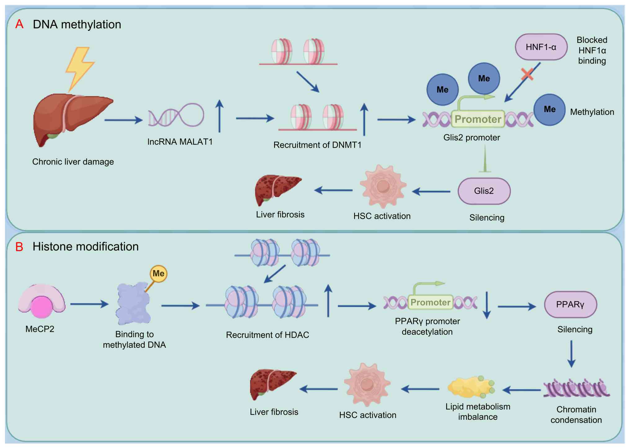
fibrosis. Under pathological conditions, the long non-coding RNA
MALAT1 recruits DNMT1 to the GLIS2 promoter, inducing
hypermethylation within CpG islands, which prevents binding of the
liver-specific TF HNF1A and silences GLIS2 expression. This
suggests that restoring GLIS2 expression by reversing aberrant DNA
methylation may re-establish epigenetic homeostasis in HSCs and
suppress fibrogenesis (103)
(Fig. 4A).
Histone modifications, together with DNA
methylation, underlie epigenetic regulation, modulating chromatin
architecture and TF accessibility. Marks such as histone
acetylation and methylation influence chromatin compaction and
serve as 'molecular anchors' that recruit chromatin remodeling
complexes and transcriptional coactivators or corepressors, forming
a dynamic regulatory network (104). For example, methyl-CpG-binding
protein 2 (MeCP2) has a dual role in HSC activation. MeCP2 binds
methylated DNA while suppressing transcription of specific target
genes. Additionally, MeCP2-mediated silencing of PPARγ is key in
HSC transdifferentiation; Suppression of PPARγ, a master regulator
of HSC quiescence, leads to lipid metabolic imbalance and
diminished inhibition of profibrotic pathways, accelerating
fibrosis progression (105-107). The complexity of this
epigenetic regulation is exemplified by MeCP2 functional
versatility: It serves as a corepressor of antifibrotic genes such
as PPARγ and dynamically coordinates spatial and temporal coupling
of DNA methylation and histone modifications through interaction
with modifying enzymes, including HDACs (108) (Fig. 4B).
The cooperativity and reversibility of epigenetic
events provide a unique perspective for liver fibrosis therapy.
During HSC activation, DNA hypomethylation and chromatin opening at
profibrotic gene promoters reinforce each other, establishing a
positive feedback loop (109).
Regulatory factors such as MeCP2 integrate DNA methylation and
histone modification signals, thereby translating epigenetic
information into specific gene expression programs (98,105). The dynamic balance of this
multilayered regulatory network determines HSC fate decisions. In
the fibrosis regression phase, targeted inhibition of DNMT1 or HDAC
activity restores expression of key genes such as PPARγ, thereby
inducing reversion of aHSCs to a quiescent phenotype (103,107). Preclinical studies suggest that
developing epigenetic therapy requires balancing mechanistic
specificity and global regulatory impact; for example, selective
inhibition of DNMT1 may relieve the silencing of antifibrotic genes
such as Glis2, while targeting the MeCP2-PPARγ axis may
simultaneously modulate DNA methylation and histone modification
(103,105). Collectively, these findings
demonstrate that precise interventions at dynamic nodes within the
epigenetic regulatory network disrupt the self-reinforcing loop of
profibrotic signaling and provide new avenues for reversing liver
fibrosis.
Transcriptional regulation of LSEC state
in liver fibrosis progression
LSECs are key components of the hepatic
microvascular system and serve as critical regulators of liver
fibrosis progression due to their structural and functional
characteristics. LSECs form a discontinuous endothelial barrier in
the sinusoidal wall and engage in the clearance of bloodborne
components via scavenger receptors such as the mannose receptor,
CD32b, lymphatic vessel endothelial hyaluronan receptor 1 (LYVE-1)
and Stabilin-1/2. LSECs secrete vasoactive factors that coordinate
the balance between hepatic immune regulation and regenerative
repair (110-112). Under physiological conditions,
LSECs maintain hepatocyte proliferative capacity and inhibit HSC
activation by releasing regenerative signals, including HGF and
nitric oxide (NO), thereby establishing an antifibrotic
microenvironment (66,113). However, during acute or chronic
liver injury, LSECs undergo a phenotypic switch characterized by
capillarization, which includes basement membrane formation, loss
of fenestrae and decreased regenerative function. This pathological
remodeling disrupts vascular-derived signaling balance, promotes
HSC activation and leads to excessive ECM deposition, driving
fibrosis progression (114).
Recent studies have identified TFs such as Kruppel-like factor
(KLF)2, GATA4 and zinc finger E-box-binding homeobox (ZEB)2 as
pivotal in maintaining endothelial homeostasis and suppressing
fibrogenesis through the regulation of LSEC epigenetic programs and
signaling networks (115,116).
KLF2, a mechanosensitive TF, upholds LSEC functional
integrity through multiple mechanisms. At the molecular level, KLF2
activates the Nrf2 pathway, upregulating antioxidant genes such as
HO-1 and NQO1, which reduces ROS levels and mitigates oxidative
stress-induced damage. KLF2 suppresses TGF-β/Smad signaling to
inhibit the production of profibrotic factors such as endothelin-1
and promotes endothelial NO synthase expression to enhance
NO-mediated vasodilation (117,118). This dual regulation not only
improves hepatic sinusoidal hemodynamics but also sustains LSEC
fenestration through the paracrine secretion of growth factors such
as VEGF, effectively preventing HSC transdifferentiation into
myofibroblasts. KLF2 expression levels are inversely associated
with the severity of liver fibrosis; loss of KLF2 functionality
results in increased LSEC contractility and a proinflammatory
phenotype, positioning KLF2 as a key nexus linking mechanical
stress signals to antifibrotic responses (116,117) (Fig. 5A).
 | Figure 5Pathways by which paracrine signals
from LSECs mediate liver fibrosis. (A) Loss of KLF2 in LSECs leads
to sustained NF-κB activation, which promotes liver sinusoidal
capillarization and fibrosis via VEGF secretion. (B) LSECs secrete
PDGF, which cooperates with the GATA4/Myc signaling pathway to
directly activate HSCs into a profibrotic phenotype, thereby
exacerbating the progression of liver fibrosis. LSEC, liver
sinusoidal endothelial cell; KLF, Kruppel-like factor; PDGF-BB,
platelet-derived growth factor-BB; HSC, hepatic stellate cell;
eNOS, endothelial nitric oxide synthase; ET, endothelin; HO, heme
oxygenase; NQO, NAD(P)H quinone oxidoreductase; ROS, reactive
oxygen species. |
GATA4 preserves LSEC lineage-specific identity,
thereby preventing pathological capillarization. Under homeostatic
conditions, GATA4 directly regulates the expression of endothelial
markers such as stabilin-2 and LYVE-1, maintaining LSEC
fenestration and scavenger function while inhibiting aberrant
activation of the proto-oncogene Myc (17,119). Loss of GATA4 expression leads
to Myc-driven metabolic reprogramming, promoting the
transdifferentiation of LSECs toward a continuous endothelial
phenotype, which is accompanied by increased secretion of
profibrotic factors such as PDGF-BB. This activates HSCs and
induces excessive ECM deposition. The resultant phenotypic switch
fosters a cycle of sinusoidal microvascular disruption: Basement
membrane thickening impairs nutrient exchange and exacerbates
hepatocyte hypoxic injury and sustained PDGFRβ signaling drives
HSCs into an irreversibly activated state. Preclinical studies have
demonstrated that restoring GATA4 expression via adenoviral
delivery reverses LSEC capillarization and significantly decreases
collagen deposition in carbon tetrachloride-induced liver fibrosis
models, affirming the key role of GATA4 in maintaining endothelial
identity (Fig. 5B) (17,115).
In addition to KLF2 and GATA4, TFs such as ZEB2
govern LSEC function through epigenetic remodeling. The loss of
ZEB2 results in aberrant activation of the Notch signaling pathway,
promoting transdifferentiation of LSECs into an arterial-like
endothelial phenotype and increasing the secretion of
proinflammatory cytokines, including IL-6 and TNF-α, thereby
establishing a chronic inflammatory microenvironment (120). This epigenetically driven
endothelial plasticity undermines hepatic immune tolerance and
amplifies profibrotic signaling by recruiting bone marrow-derived
fibroblasts via the CXCL12/CXCR4 pathway (65,120). Notably, crosstalk between TFs
creates a complex regulatory network: KLF2 suppresses ZEB2
expression via inhibition of the NF-κB pathway, while the
antagonistic interaction between GATA4 and Myc modulates Notch
pathway activity by altering the transcriptional regulation of
Notch target genes (17,117). This multilayered regulatory
architecture presents multiple potential nodes for targeted
antifibrotic intervention.
Transcriptional regulation of LSECs exhibits
spatiotemporal specificity and integrative signaling
characteristics. In the early stage of liver injury, KLF2 rapidly
responds to shear stress changes, maintaining endothelial barrier
integrity. During chronic injury, progressive depletion of GATA4
leads to loss of lineage-specific identity and amplifies fibrogenic
signaling cascades. Factors such as ZEB2 contribute to the
maintenance of pathological endothelial phenotypes through
epigenetic memory (115,116).
This dynamic regulatory network indicates that stage-specific
targeted intervention aimed at correcting TF imbalance may optimize
antifibrotic efficacy. In-depth characterization of LSEC-specific
transcriptional regulatory elements and their interaction networks
may establish a theoretical foundation for developing endothelial
phenotype-based antifibrotic therapeutic strategies.
Transcriptional regulation of hepatic
macrophage populations in liver fibrosis progression
The dynamic regulation of hepatic macrophage
populations is a central element in liver fibrosis progression.
Their functional diversity arises from the spatiotemporal interplay
between resident Kupffer cells and monocyte-derived macrophages
(MoMs). As liver-resident 'sentinels', Kupffer cells exhibit
self-renewal capacity and continually monitor the microenvironment
to maintain homeostasis: Under physiological conditions, they
enhance immune tolerance by clearing senescent red blood cells,
pathogens and metabolic byproducts (121,122). Upon parenchymal injury, Kupffer
cells detect damage-associated molecular patterns and secrete
chemokines such as CCL2 and CCL5, facilitating the recruitment of
circulating monocytes that differentiate into MoMs within the
liver, forming a mixed macrophage pool (123). While early proinflammatory
responses initiate tissue repair, persistent activation may
perpetuate a cycle of inflammation and fibrosis. Notably, hepatic
macrophages demonstrate epigenetic plasticity: Their phenotypes are
dynamically reshaped by local microenvironmental cues, including
fatty acid metabolites, apoptotic debris and cell-cell contact
signals (123,124). These stimuli promote
differentiation toward either proinflammatory M1 macrophages
(secreting IL-1β, TNF-α, IL-17) that activate HSCs, or
anti-inflammatory M2 macrophages (secreting IL-10, TGF-β) that
facilitate fibrosis resolution (124,125).
Paracrine crosstalk between macrophages and HSCs is
a central hub in the fibrogenic signaling network. Kupffer cells
directly induce HSC transdifferentiation into myofibroblasts via
the IL-17/STAT3 pathway: IL-17 activates the NF-κB pathway within
HSCs, enhancing TGF-β1 and COL1 expression, while STAT3
phosphorylation drives epigenetic reprogramming that amplifies
α-SMA promoter activity, establishing a profibrotic phenotype
memory (126,127). Simultaneously, a positive
feedback loop is established between Kupffer cells and LSECs: VEGF
released by LSECs during early injury stimulates Kupffer cells to
produce IL-6, which, via JAK3/STAT3 signaling, promotes MoM
polarization toward the M2 phenotype. M2 macrophages secrete PDGF,
activating HSCs and inducing LSEC capillarization (128,129). This cascade of intercellular
interactions demonstrates how macrophage phenotype imbalance
accelerates fibrosis progression: M1 dominance leads to excessive
TNF-α and IL-1β production, inducing mitochondrial dysfunction in
HSCs via ROS-dependent mechanisms and driving hyperproliferation,
whereas excessive M2 activation fosters irreversible fibrotic
scarring via TGF-β/Smad3-mediated ECM crosslinking (130,131).
Precise regulation of the JAK/STAT signaling pathway
underpins macrophage phenotype switching at the molecular level.
IFN-γ drives M1 polarization via the JAK1/STAT1 pathway, inducing
inducible NO synthase expression to enhance bactericidal activity
and recruiting T helper 1 cells through CXCL9/10 production,
thereby exacerbating inflammatory injury (132). Conversely, IL-4 and IL-13
promote M2 differentiation through STAT6-mediated activation of
arginase-1, while IL-6 and IL-10 signal via STAT3 phosphorylation
to upregulate suppressor of cytokine signaling 3 (SOCS3), which
inhibits STAT1 signaling through negative feedback, maintaining an
anti-inflammatory macrophage phenotype (133,134). This balance is disrupted during
fibrosis: Chronic injury leads to SOCS1 downregulation, relieving
the inhibition of STAT1 and driving sustained M1 polarization,
while hypoxia secondary to LSEC capillarization stabilizes HIF-1α,
enhancing STAT3 activity and promoting a profibrotic M2-dominant
microenvironment (134,135) (Fig. 6). Preclinical studies have
demonstrated that targeting STAT1/STAT3 signaling nodes (for
example, using the JAK inhibitor tofacitinib) rebalances macrophage
phenotypes and significantly decreases collagen deposition in
carbon tetrachloride–induced fibrosis models (42,130,131). Furthermore, epigenetic
interventions, including HDAC inhibitors that modulate SOCS
expression, present promising strategies to overcome the M1/M2
polarization impasse.
Macrophage plasticity is dynamically regulated
throughout liver fibrosis. During the early phase of injury,
M1-dominant inflammatory responses facilitate debris clearance and
create conditions conducive to tissue repair. In the progression
phase, M2 macrophages drive ECM remodeling by secreting profibrotic
mediators. In the regression phase, M2 macrophages transition
toward a pro-resolution phenotype, enabling scar degradation via
the regulation of the MMP9/TIMP1 balance (124,125). This dynamic shift is
orchestrated by the interplay of TF networks, including NF-κB,
STAT, and PPARγ, and metabolic reprogramming, exemplified by the
transition from fatty acid oxidation to glycolysis. For example,
PPARγ activation in Kupffer cells enhances lipid scavenging and
suppresses NLRP3 inflammasome assembly, while enhanced AMPK
signaling in MoMs promotes mitochondrial biogenesis and sustains an
anti-inflammatory phenotype (121,129). Elucidating the crosstalk
between metabolic and epigenetic programs across macrophage
subpopulations may provide a foundation for developing
spatiotemporally specific therapeutic intervention.
Transcriptional regulation of other
non-parenchymal liver cells in fibrosis progression
The initiation and progression of liver fibrosis
depend not only on the phenotypical transformation of HSCs but also
on transcriptional regulatory networks and interactions between
various non-parenchymal liver cells. In addition to HSCs, LSECs,
and Kupffer cells, other cell types, including natural killer (NK)
cells, dendritic cells (DCs) and biliary epithelial cells (BECs),
contribute to shaping either a profibrotic or antifibrotic hepatic
microenvironment through signaling pathways and TF-mediated
mechanisms (136-138). NK cells, key components of the
innate immune system, inhibit HSC activation via death
receptor-mediated apoptosis. Upon activation by IL-18 or TLR3
ligands, the p38/PI3K/AKT signaling pathway enhances the expression
of TNF-related apoptosis-inducing ligand and FasL, engaging death
receptors on aHSCs to induce caspase cascades and eliminate
fibrogenic cells (64,139,140). STAT1, a key mediator of
interferon signaling, promotes NK cell cytotoxicity via
phosphorylation while enhancing granzyme B release, directly
targeting HSCs via mitochondrial pathways (Fig. 7A). This dual mechanism decreases
collagen deposition in carbon tetrachloride-induced liver fibrosis
models (140,141).
 | Figure 7Integrated regulatory pathways of
cell types involved in liver fibrosis progression. (A) NK cells are
activated by IL-18 to trigger the PI3K/AKT/STAT11 signaling
pathway, releasing granzyme B, which induces mitochondrial
apoptosis in HSCs, thereby exerting antifibrotic effects. (B) LPS
stimulation activates the PI3K/AKT pathway in DCs, leading to
phosphorylation and inactivation of FoxO1, which relieves
FoxO1-mediated suppression of pro-inflammatory genes transcription,
increases IL-6-mediated Th17 differentiation and promotes HSC
activation (C) BECs, via the YAP-Sox9 axis, induce aberrant
proliferation of LPCs, contributing to peribiliary fibrosis
lesions. Under Hedgehog signaling stimulation, transcription
factors are upregulated to promote BEC EMT and VEGF secretion,
which activates HSCs. NK, natural killer; HSC, hepatic stellate
cell; LPS, lipopolysaccharide; DC, dendritic cell; Th, T helper;
BEC, biliary epithelial cell; LPC, liver progenitor cell; EMT,
epithelial-mesenchymal transition; YAP, Yes-associated protein;
Gli, glioma-associated oncogene. |
DCs regulate fibrosis progression through
immunometabolic reprogramming. Stimulation with LPS activates the
PI3K/AKT pathway in DCs, leading to phosphorylation and
inactivation of forkhead box O1 (FoxO1). This relieves suppression
of proinflammatory gene transcription, promoting DC maturation and
secretion of cytokines such as IL-6 and IL-23, which drive Th17
cell differentiation and HSC activation (142-145) (Fig. 7B). This bidirectional regulation
along the metabolism-immune axis is prominent in cholestatic liver
diseases: Sustained FoxO1 activation upregulates PD-L1 expression,
inducing DC tolerance and inhibiting CD8+ T
cell-mediated HSC killing. The PI3K inhibitor LY294002 can reverse
this effect, highlighting the potential of targeting the
PI3K/AKT/oxO1 network to restore DC functional balance (146-148). Additionally, crosstalk between
DCs and Kupffer cells establishes an inflammatory amplification
loop: Kupffer cell-derived TGF-β suppresses DC antigen-presenting
capacity via Smad3-dependent mechanisms, while DC-derived IL-12
activates the NLRP3 inflammasome in Kupffer cells. This positive
feedback circuit serves a key role in fibrosis associated with NASH
(129,143).
Transcriptional reprogramming of BECs provides a
unique pathological perspective on liver fibrosis. Under
physiological conditions, BECs maintain bile duct integrity and
contribute to liver regeneration through Sox9 activity (149,150). However, in chronic liver
injury, aberrant activation of the Hedgehog (Hh) signaling pathway
induces upregulation of TFs such as GLI family zinc finger 2
(GLI2), driving epithelial-mesenchymal transition (EMT) of BECs
into mesenchymal-like cells with profibrotic properties (149-151). This phenotypic switch is
accompanied by excessive secretion of growth factors, including
VEGF and PDGF, which not only directly activate neighboring HSCs
but also promote aberrant proliferation of liver progenitor cells
(LPCs) via the YAP-Sox9 axis, contributing to periductular fibrotic
lesions. In biliary atresia models, BEC-derived Hh ligands (Sonic
Hh) bind Patched receptors on HSCs, triggering Gli-dependent
upregulation of α-SMA and COL1A1 and enhancing CXCL12 secretion to
recruit bone marrow-derived fibroblasts, thereby establishing a
multicellular profibrotic interaction (152,153) (Fig. 7C). BEC plasticity exhibits
bidirectional regulatory potential: EMT is reversed by inhibiting
YAP nuclear translocation or blocking Hh signaling, while
activation of the nuclear receptor FXR restores epithelial barrier
function by suppressing Sox9 phosphorylation (149,150).
The transcriptional regulatory network between
non-parenchymal liver cells is dynamic and spatially specific. For
example, STAT1 activation in NK cells suppresses IL-17 production
by Kupffer cells, thereby blocking STAT3 phosphorylation in HSCs
(64,140). Conversely, IL-10 derived from
DCs activates STAT6 signaling in BECs to promote biliary repair
while inhibiting the TGF-β/Smad pathway in HSCs (141,148). This integration of
intercellular signals underscores the multidimensional regulatory
nature of liver fibrosis: In the early stage of injury, immune
surveillance by NK cells and DCs limits fibrogenesis through the
clearance of aHSCs; during progression, proinflammatory signals
from Kupffer cells and BECs dominate, establishing a pathological
microenvironment characterized by excessive ECM deposition; in the
regression phase, DCs transition toward a tolerogenic phenotype,
while BEC-LPC transdifferentiation facilitates tissue remodeling
(140,141,148). Targeting specific TFs (STAT1,
Gli2, FoxO1) or epigenetic modifiers (HDACs, DNMTs) allows precise
modulation of functional states across these cell populations
(154). Strategies such as
clustered regularly interspaced short palindromic
repeats/catalytically dead Cas9 (CRISPR/dCas9)-directed activation
of STAT1 signaling in NK cells or pharmacological inhibition of BEC
Hh signaling using smoothened (SMO) inhibitors have shown promise
in preclinical models for reversing fibrosis (64,140,148). Deciphering the transcriptional
dialogue between non-parenchymal liver cells may provide a
theoretical framework for developing cell type-specific,
multitargeted antifibrotic therapy.
Nanomaterial-based targeting of TFs:
Mechanisms and strategies for liver fibrosis therapy
In the molecular pathogenesis of liver fibrosis,
aberrant activation of TFs and disruption of epigenetic homeostasis
are central regulatory hubs (109,147). Innovations in nanotechnology
provide a multiscale intervention paradigm to address the
limitations of conventional therapies. Nanomaterial-based targeted
delivery systems, characterized by precise localization,
stimuli-responsive release and efficacy, enable multilayered
modulation of transcriptional regulatory networks within the
fibrotic microenvironment (155). Surface functionalization
strategies, such as conjugation with retinoic acid receptor
ligands, COL-binding peptides or integrin αvβ3 antibodies, allow
nanocarriers to penetrate the disordered sinusoidal endothelium,
facilitating selective accumulation within aHSCs (156,157). Notably, vitamin
A-A-functionalized liposomes demonstrate an 8-12-fold increase in
targeting efficiency in fibrotic liver tissue compared with normal
liver, attributed to marked upregulation of retinoic acid
receptor-β during HSC activation (157). Microenvironment-responsive
designs, such as ROS-sensitive linkers, MMP-cleavable peptides or
pH-sensitive polymers, enable spatiotemporally specific drug
release. For example, acid-labile poly(lactic-co-glycolic acid)
(PLGA) nanoparticles delivering TGF-β small interfering (si) RNA
rapidly disassemble in the acidic lysosomal environment of HSCs,
decreasing Smad2/3 phosphorylation levels by >70% while
minimizing off-target organ exposure (158). Moreover, multimodal therapeutic
systems have demonstrated superior efficacy compared with
single-agent delivery: Mesoporous silica nanoparticles
co-encapsulating the STAT3 inhibitor cucurbitacin B and miR-29b
mimic simultaneously block the IL-6/STAT3 pathway and activate the
TGF-β/Smad7 negative feedback loop, resulting in a 58% decrease in
COL deposition area and a 64% decrease in α-SMA expression in a
CCl4-induced liver fibrosis model, confirming the
advantage of cross-mechanism combination strategies (159). Existing research emphasizes
four-dimensional intervention approaches: In gene silencing, gold
nanoparticles loaded with TGF-β siRNA block COL1A1/CTGF
transcription by inhibiting Smad4 nuclear translocation, achieving
a 20-fold increase in hepatic delivery efficiency compared with
free siRNA, while decreasing renal accumulation toxicity to
physiological levels (160). In
epigenetic regulation, chitosan nanoparticles delivering miR-146a
target the 3' untranslated region of NF-κB p65, suppressing IKKβ
phosphorylation (thereby downregulating TNF-α and other
inflammatory mediators) and inhibiting DNMT activity to remodel the
HSC epigenome, reversing 80% of hypermethylated loci in an
alcoholic liver disease model (161). In gene editing, lipid
nanoparticles encapsulating CRISPR/Cas9 enhance homologous
recombination to activate the Nrf2 antioxidant pathway, restoring
glutathione levels to 85% of normal and demonstrating a 3.7-fold
increase in homologous recombination efficiency via nanocarrier
delivery (162). For
protein-protein interaction inhibition, PLGA nanoparticles loaded
with the STAT3 inhibitor napabucasin mimic the SH2 domain to block
STAT3 dimerization, increasing hepatic accumulation 12-fold
compared with oral administration while decreasing cardiac tissue
exposure and associated toxicity by 73%, highlighting the value of
nanodelivery in expanding therapeutic windows (163). Biomimetic delivery strategies
are engineered exosome-liposome hybrids that penetrate
intracellular barriers via membrane fusion. For example, hybrid
nanoparticles delivering recombinant Smad7 protein directly into
the HSC cytoplasm competitively inhibit Smad2/3 phosphorylation and
reverse myofibroblast transdifferentiation, achieving
dose-dependent fibrosis reversal (half-maximal effective
concentration=2.3 mg/kg) in preclinical studies, with efficacy
4.5-fold greater than conventional liposomes (156,164).
Despite the promise of nanotherapy, clinical
translation faces three core challenges. First, the long-term
accumulation toxicity associated with inorganic materials such as
carbon nanotubes has prompted a shift toward biodegradable
platforms, such as hyaluronic acid-polycaprolactone copolymers
(156,165). Surface PEGylation and charge
neutralization decrease immunogenicity to levels comparable with
saline controls, however, achieving precise alignment between
material degradation rates and drug release kinetics remains an
obstacle (165). Second,
downregulation of vitamin A receptors during fibrosis progression
necessitates the development of multistage targeting systems. For
example, matrix stiffness-responsive nanorobots equipped with
piezoelectric sensors detect the mechanical properties of aHSCs
(elastic modulus >8 kPa) and release CXCR4 antagonistic peptides
to enhance lesion retention. Recent studies demonstrate that this
strategy prolongs nanoparticle retention in advanced fibrotic
livers for up to 72 h (156,166). Third, development of efficacy
evaluation systems requires the integration of organoid-on-chip
platforms with spatial multi-omics technologies. For example, 3D
bioprinted fibrotic liver models allow high-resolution (up to 5
μm) assessment of nanoparticle permeability, while
single-cell assay for transposase-accessible chromatin using
sequencing analysis of chromatin accessibility demonstrates that
nanocarriers loaded with HDAC inhibitors increase PPARγ promoter
accessibility by 3.2-fold in HSCs, supporting the mechanistic basis
for epigenetic reprogramming (167).
Future studies should focus on the development of
intelligent, integrated diagnostic-therapeutic systems. In
precision-controlled drug release, near-infrared light-responsive
upconversion nanoparticles (inorganic luminescent nanomaterials
with anti-Stokes luminescence properties) facilitate
millimeter-scale spatially resolved drug delivery upon activation
with 808 nm laser irradiation (167,168). A system loaded with the YAP
inhibitor verteporfin decreases YAP nuclear localization by 82%
within the irradiated zone without notable off-target effects in
adjacent normal tissue, offering a novel paradigm for localized
antifibrotic therapy (169). In
personalized treatment strategies, machine learning algorithms
integrating patient-specific TF profiles (covering 12 core nodes,
including STAT3, NF-κB and YAP) with nanoparticle physicochemical
parameters (such as particle size, ζ potential and drug loading)
have optimized STAT3/NF-κB co-inhibition regimens (169,170). Phase II clinical trials
demonstrate that this approach increases therapeutic response rates
from 41% with conventional regimens to 67%, while decreasing the
incidence of serious adverse events by 55% (156,171,172). At the frontier of
immunometabolic modulation, nanoparticles co-delivering PD-1
antibodies and NRF2 activators block T cell exhaustion (restoring
CD8+ T cell IFN-γ secretion to 2.3-fold above baseline)
and enhance macrophage antioxidant capacity (reducing mitochondrial
ROS levels by 68%). This dual-action strategy achieves both
fibrosis reversal and tumor suppression in models of fibrosis with
concurrent steatosis, providing a new path for combined
antifibrotic and antitumor therapy (156,173). These breakthroughs have not
only deepened the mechanistic understanding of TF-targeted
nanoregulation but also propelled therapeutic strategies beyond
single-target intervention toward multiscale network modulation,
establishing a systems biology and bioengineering foundation for
clinical translation.
Conclusion
Liver fibrosis is a common pathological response to
chronic liver injury, presenting treatment challenges due to its
association with metabolic dysfunction, persistent inflammation,
phenotypic changes in hepatic cell population and excessive ECM
deposition. TFs are key in these processes, translating
extracellular and intracellular signals into cell-specific
transcriptional programs. The present review summarizes the
regulatory roles of TFs in liver fibrosis and their potential as
therapeutic targets, with a particular focus on
nanotechnology-based delivery strategies.
TF functions in liver fibrosis vary depending on
cell type and biological context. In hepatocytes, TFs such as
HNF4α, FOXA2 and C/EBPα serve essential roles in metabolic
homeostasis and adaptive responses, interacting with Wnt, insulin,
IL-6 and nutrient-sensing pathways. Dysregulation of these TFs
disrupts glucose, lipid and bile acid metabolism, aggravates
hepatocyte injury and promotes the release of profibrotic
mediators. In non-parenchymal cells, including HSCs, Kupffer cells
and LSECs, TFs such as YAP/TAZ, LHX2, PPARγ, NF-κB and HIF-1α
regulate cellular activation, inflammatory responses, angiogenesis
and matrix remodeling. The activity of an individual TF is not
uniform; it may vary with disease etiology, stage and cellular
state. Moreover, TF programs in different hepatic cell populations
are connected through paracrine signaling, where injured
hepatocytes activate macrophages and HSCs, macrophage-derived
cytokines regulate HSC activation and endothelial inflammatory
responses and endothelial cells and aHSCs further modify the
hepatic microenvironment. Such intercellular crosstalk sustains
fibrosis even when the initial injury has been partially
controlled.
TF activity is determined by multiple factors,
including expression, subcellular localization, DNA binding,
protein stability and interactions with transcriptional cofactors.
These characteristics are dynamically regulated by extracellular
stimuli, intracellular signaling pathways, post-translational
modification and epigenetic mechanisms. This multilayered
regulation partly explains why the same TF may exert opposing
effects across cell types and stages of fibrosis. Advances in
single-cell sequencing and spatial multi-omics approaches have
enhanced the resolution of cell-specific TF networks and
fibrosis-associated cell states. Nonetheless, numerous regulatory
relationships inferred from these analyses do not prove causation,
necessitating complementary loss/gain-of-function and rescue
experiments, alongside validation in human tissue, organoids or
precision-cut liver slices.
The cell-specific nature of TF regulation
underscores the rationale for nanotechnology-based delivery.
Nanocarriers improve the stability, cellular uptake and
pharmacokinetic properties of small-molecule modulators, siRNAs,
mRNAs, decoy oligonucleotides and gene editing components. Surface
modification with cell-selective ligands may enhance delivery to
aHSCs, hepatic macrophages, LSECs or hepatocytes while limiting
systemic exposure. However, the therapeutic value of these systems
should not be inferred solely from hepatic accumulation.
Preferential sequestration by Kupffer cells may impede delivery to
other target populations, while fibrosis-associated changes in
vascular permeability, ECM density and receptor expression can
alter nanoparticle distribution. Additional barriers to clinical
translation include endosomal escape, immunogenicity, manufacturing
reproducibility, long-term toxicity and differences between
experimental models and human disease.
While treating underlying etiology can promote
fibrosis regression in some patients, to the best of our knowledge,
no therapies specifically targeting antifibrotic indications have
been approved. Future studies should identify TFs with functions
that are both cell- and stage-specific, establishing their causal
roles in fibrosis progression or resolution, and determining
whether they can be modulated within an acceptable therapeutic
window. Nanotechnology may facilitate this by enabling more
selective delivery, but its application necessitates evaluation of
biodistribution, pharmacological activity and safety in clinically
relevant models. Overall, TFs represent promising yet complex
therapeutic targets. Integrating mechanistic studies with
human-relevant experimental systems and rational delivery
strategies is essential for translating TF-directed intervention
into effective antifibrotic therapies.
Availability of data and materials
Not applicable.
Authors' contributions
XFZ wrote and edited the manuscript and performed
the literature review. CJY constructed figures and performed the
literature review. ZYW contributed to conceiving and designing the
study and constructed and revised the figures. LHL wrote and edited
the manuscript and conceived the study. Data authentication is not
applicable. All authors have read and approved the final
manuscript.
Ethics approval and consent for
participation
Not applicable.
Patient consent for publication
Not applicable.
Competing interests
The authors declare that they have no competing
interests.
Acknowledgements
Not applicable.
Funding
The present study was supported by the Zhejiang Provincial
Natural Science Foundation of China (grant no. LY23H030002),
Zhejiang Provincial Medical and Health Science and Technology
Project (grant no. 2023KY412) and the Basic Research Project of
Liaoning Provincial Department of Education (grant no.
LJKMZ20221804).
References
|
1
|
Wynn TA: Fibrotic disease and the
T(H)1/T(H)2 paradigm. Nat Rev Immunol. 4:583–594. 2004. View Article : Google Scholar : PubMed/NCBI
|
|
2
|
Bataller R and Brenner DA: Liver fibrosis.
J Clin Invest. 115:209–218. 2005. View Article : Google Scholar : PubMed/NCBI
|
|
3
|
Ginès P, Krag A, Abraldes JG, Solà E,
Fabrellas N and Kamath PS: Liver cirrhosis. Lancet. 398:1359–1376.
2021. View Article : Google Scholar : PubMed/NCBI
|
|
4
|
Devarbhavi H, Asrani SK, Arab JP, Nartey
YA, Pose E and Kamath PS: Global burden of liver disease: 2023
update. J Hepatol. 79:516–537. 2023. View Article : Google Scholar : PubMed/NCBI
|
|
5
|
Kisseleva T and Brenner D: Molecular and
cellular mechanisms of liver fibrosis and its regression. Nat Rev
Gastroenterol Hepatol. 18:151–166. 2021. View Article : Google Scholar
|
|
6
|
Pei Q, Yi Q and Tang L: Liver fibrosis
resolution: From molecular mechanisms to therapeutic opportunities.
Int J Mol Sci. 24:96712023. View Article : Google Scholar : PubMed/NCBI
|
|
7
|
Codotto G, Blarasin B, Tiribelli C,
Bellarosa C and Licastro D: Decoding liver fibrosis: How omics
technologies and innovative modeling can guide precision medicine.
Int J Mol Sci. 26:26582025. View Article : Google Scholar : PubMed/NCBI
|
|
8
|
Li JZ, Yang L, Xiao MX, Li N, Huang X, Ye
LH, Zhang HC, Liu ZQ, Li JQ, Liu YY, et al: Spatial and single-cell
transcriptomics reveals the regional division of the spatial
structure of MASH fibrosis. Liver Int. 45:e161252025. View Article : Google Scholar :
|
|
9
|
Cordero-Espinoza L and Huch M: The
balancing act of the liver: Tissue regeneration versus fibrosis. J
Clin Invest. 128:85–96. 2018. View Article : Google Scholar : PubMed/NCBI
|
|
10
|
Jun JI and Lau LF: Resolution of organ
fibrosis. J Clin Invest. 128:97–107. 2018. View Article : Google Scholar : PubMed/NCBI
|
|
11
|
Krenkel O, Puengel T, Govaere O, Abdallah
AT, Mossanen JC, Kohlhepp M, Liepelt A, Lefebvre E, Luedde T,
Hellerbrand C, et al: Therapeutic inhibition of inflammatory
monocyte recruitment reduces steatohepatitis and liver fibrosis.
Hepatology. 67:1270–1283. 2018. View Article : Google Scholar
|
|
12
|
Liu X, Xu J, Rosenthal S, Zhang LJ,
McCubbin R, Meshgin N, Shang L, Koyama Y, Ma HY, Sharma S, et al:
Identification of lineage-specific transcription factors that
prevent activation of hepatic stellate cells and promote fibrosis
resolution. Gastroenterology. 158:1728–1744.e14. 2020. View Article : Google Scholar : PubMed/NCBI
|
|
13
|
Marx V: Method of the year: Spatially
resolved transcriptomics. Nat Methods. 18:9–14. 2021. View Article : Google Scholar : PubMed/NCBI
|
|
14
|
Loft A, Alfaro AJ, Schmidt SF, Pedersen
FB, Terkelsen MK, Puglia M, Chow KK, Feuchtinger A, Troullinaki M,
Maida A, et al: Liver-fibrosis-activated transcriptional networks
govern hepatocyte reprogramming and intra-hepatic communication.
Cell Metab. 33:1685–1700.e9. 2021. View Article : Google Scholar : PubMed/NCBI
|
|
15
|
Ramachandran P, Dobie R, Wilson-Kanamori
JR, Dora EF, Henderson BEP, Luu NT, Portman JR, Matchett KP, Brice
M, Marwick JA, et al: Resolving the fibrotic niche of human liver
cirrhosis at single-cell level. Nature. 575:512–518. 2019.
View Article : Google Scholar : PubMed/NCBI
|
|
16
|
Tan S, Zhao J, Sun Z, Cao S, Niu K, Zhong
Y, Wang H, Shi L, Pan H, Hu J, et al: Hepatocyte-specific TAK1
deficiency drives RIPK1 kinase-dependent inflammation to promote
liver fibrosis and hepatocellular carcinoma. Proc Natl Acad Sci
USA. 117:14231–14242. 2020. View Article : Google Scholar : PubMed/NCBI
|
|
17
|
Winkler M, Staniczek T, Kürschner SW,
Schmid CD, Schönhaber H, Cordero J, Kessler L, Mathes A, Sticht C,
Neßling M, et al: Endothelial GATA4 controls liver fibrosis and
regeneration by preventing a pathogenic switch in angiocrine
signaling. J Hepatol. 74:380–393. 2021. View Article : Google Scholar
|
|
18
|
Arroyo N, Villamayor L, Díaz I, Carmona R,
Ramos-Rodríguez M, Muñoz-Chápuli R, Pasquali L, Toscano MG, Martín
F, Cano DA and Rojas A: GATA4 induces liver fibrosis regression by
deactivating hepatic stellate cells. JCI Insight. 6:e1500592021.
View Article : Google Scholar : PubMed/NCBI
|
|
19
|
Lambert SA, Jolma A, Campitelli LF, Das
PK, Yin Y, Albu M, Chen X, Taipale J, Hughes TR and Weirauch MT:
The human transcription factors. Cell. 172:650–665. 2018.
View Article : Google Scholar : PubMed/NCBI
|
|
20
|
Vo Ngoc L, Huang CY, Cassidy CJ, Medrano C
and Kadonaga JT: Identification of the human DPR core promoter
element using machine learning. Nature. 585:459–463. 2020.
View Article : Google Scholar : PubMed/NCBI
|
|
21
|
Lee TI and Young RA: Transcriptional
regulation and its misregulation in disease. Cell. 152:1237–1251.
2013. View Article : Google Scholar : PubMed/NCBI
|
|
22
|
Krausgruber T, Fortelny N, Fife-Gernedl V,
Senekowitsch M, Schuster LC, Lercher A, Nemc A, Schmidl C, Rendeiro
AF, Bergthaler A and Bock C: Structural cells are key regulators of
organ-specific immune responses. Nature. 583:296–302. 2020.
View Article : Google Scholar : PubMed/NCBI
|
|
23
|
Suter DM: Transcription factors and DNA
play hide and seek. Trends Cell Biol. 30:491–500. 2020. View Article : Google Scholar : PubMed/NCBI
|
|
24
|
Malz M, Pinna F, Schirmacher P and
Breuhahn K: Transcriptional regulators in hepatocarcinogenesis-key
integrators of malignant transformation. J Hepatol. 57:186–195.
2012. View Article : Google Scholar : PubMed/NCBI
|
|
25
|
Spitz F and Furlong EEM: Transcription
factors: From enhancer binding to developmental control. Nat Rev
Genet. 13:613–626. 2012. View Article : Google Scholar : PubMed/NCBI
|
|
26
|
Qian M, Yan F, Yuan T, Yang B, He Q and
Zhu H: Targeting post-translational modification of transcription
factors as cancer therapy. Drug Discov Today. 25:1502–1512. 2020.
View Article : Google Scholar : PubMed/NCBI
|
|
27
|
Vaquerizas JM, Kummerfeld SK, Teichmann SA
and Luscombe NM: A census of human transcription factors: Function,
expression and evolution. Nat Rev Genet. 10:252–263. 2009.
View Article : Google Scholar : PubMed/NCBI
|
|
28
|
Yang L, Shi P, Zhao G, Xu J, Peng W, Zhang
J, Zhang G, Wang X, Dong Z, Chen F and Cui H: Targeting cancer stem
cell pathways for cancer therapy. Signal Transduct Target Ther.
5:82020. View Article : Google Scholar : PubMed/NCBI
|
|
29
|
Derynck R and Budi EH: Specificity,
versatility, and control of TGF-β family signaling. Sci Signal.
12:eaav51832019. View Article : Google Scholar
|
|
30
|
Dey A, Varelas X and Guan KL: Targeting
the Hippo pathway in cancer, fibrosis, wound healing and
regenerative medicine. Nat Rev Drug Discov. 19:480–494. 2020.
View Article : Google Scholar : PubMed/NCBI
|
|
31
|
Zhang Y, Fang X, Wei J, Miao R, Wu H, Ma K
and Tian J: PDX-1: A promising therapeutic target to reverse
diabetes. Biomolecules. 12:17852022. View Article : Google Scholar : PubMed/NCBI
|
|
32
|
Pirruccello JP, Di Achille P, Nauffal V,
Nekoui M, Friedman SF, Klarqvist MDR, Chaffin MD, Weng LC,
Cunningham JW, Khurshid S, et al: Genetic analysis of right heart
structure and function in 40,000 people. Nat Genet. 54:792–803.
2022. View Article : Google Scholar : PubMed/NCBI
|
|
33
|
Daruich A, Duncan M, Robert MP, Lagali N,
Semina EV, Aberdam D, Ferrari S, Romano V, des Roziers CB,
Benkortebi R, et al: Congenital aniridia beyond black eyes: From
phenotype and novel genetic mechanisms to innovative therapeutic
approaches. Prog Retin Eye Res. 95:1011332023. View Article : Google Scholar
|
|
34
|
Zou S, Tong Q, Liu B, Huang W, Tian Y and
Fu X: Targeting STAT3 in cancer immunotherapy. Mol Cancer.
19:1452020. View Article : Google Scholar : PubMed/NCBI
|
|
35
|
Sun N, Shen C, Zhang L, Wu X, Yu Y, Yang
X, Yang C, Zhong C, Gao Z, Miao W, et al: Hepatic Krüppel-like
factor 16 (KLF16) targets PPARα to improve steatohepatitis and
insulin resistance. Gut. 70:2183–2195. 2021. View Article : Google Scholar :
|
|
36
|
Hao L, Zhong W, Dong H, Guo W, Sun X,
Zhang W, Yue R, Li T, Griffiths A, Ahmadi AR, et al: ATF4
activation promotes hepatic mitochondrial dysfunction by repressing
NRF1-TFAM signalling in alcoholic steatohepatitis. Gut.
70:1933–1945. 2021. View Article : Google Scholar :
|
|
37
|
Xu Y, Zhu Y, Hu S, Xu Y, Stroup D, Pan X,
Bawa FC, Chen S, Gopoju R, Yin L and Zhang Y: Hepatocyte nuclear
factor 4α prevents the steatosis-to-NASH progression by regulating
p53 and bile acid signaling (in mice). Hepatology. 73:2251–2265.
2021. View Article : Google Scholar
|
|
38
|
Lv DD, Zhou LY and Tang H: Hepatocyte
nuclear factor 4α and cancer-related cell signaling pathways: A
promising insight into cancer treatment. Exp Mol Med. 53:8–18.
2021. View Article : Google Scholar : PubMed/NCBI
|
|
39
|
Lin Y, Wang S, Li Z, Zhou Y, Wang R, Wang
Y and Chen Y: Short-term statin therapy induces hepatic insulin
resistance through HNF4α/PAQR9/PPM1α axis regulated AKT
phosphorylation. Adv Sci (Weinh). 11:e24034512024. View Article : Google Scholar
|
|
40
|
Wang P, Cong M, Liu T, Li Y, Liu L, Sun S,
Sun L, Zhu Z, Ma H, You H, et al: FoxA2 inhibits the proliferation
of hepatic progenitor cells by reducing PI3K/Akt/HK2-mediated
glycolysis. J Cell Physiol. 235:9524–9537. 2020. View Article : Google Scholar : PubMed/NCBI
|
|
41
|
Verma AK, Sharma A, Subramaniyam N and
Gandhi CR: Augmenter of liver regeneration: Mitochondrial function
and steatohepatitis. J Hepatol. 77:1410–1421. 2022. View Article : Google Scholar : PubMed/NCBI
|
|
42
|
Bharadwaj U, Kasembeli MM, Robinson P and
Tweardy DJ: Targeting janus kinases and signal transducer and
activator of transcription 3 to treat inflammation, fibrosis, and
cancer: Rationale, progress, and caution. Pharmacol Rev.
72:486–526. 2020. View Article : Google Scholar : PubMed/NCBI
|
|
43
|
Chen H, Tan H, Wan J, Zeng Y, Wang J, Wang
H and Lu X: PPAR-γ signaling in nonalcoholic fatty liver disease:
Pathogenesis and therapeutic targets. Pharmacol Ther.
245:1083912023. View Article : Google Scholar
|
|
44
|
Kim HY, Jang HJ, Muthamil S, Shin UC, Lyu
JH, Kim SW, Go Y, Park SH, Lee HG and Park JH: Novel insights into
regulators and functional modulators of adipogenesis. Biomed
Pharmacother. 177:1170732024. View Article : Google Scholar : PubMed/NCBI
|
|
45
|
Wu T, Lu ZF, Yu HN, Wu XS, Liu Y and Xu Y:
Liver receptor homolog-1: Structures, related diseases, and drug
discovery. Acta Pharmacol Sin. 45:1571–1581. 2024. View Article : Google Scholar : PubMed/NCBI
|
|
46
|
Sun Y, Demagny H and Schoonjans K:
Emerging functions of the nuclear receptor LRH-1 in liver
physiology and pathology. Biochim Biophys Acta Mol Basis Dis.
1867:1661452021. View Article : Google Scholar : PubMed/NCBI
|
|
47
|
Wang Z, Wu Q, Li X and Klaunig JE:
Constitutive androstane receptor (CAR) mediates dieldrin-induced
liver tumorigenesis in mouse. Arch Toxicol. 94:2873–2884. 2020.
View Article : Google Scholar : PubMed/NCBI
|
|
48
|
Li SC, Wang B, Zhang M, Yin Q, Yang ZY, Li
XT and Liang G: Induction of cytochrome P450 via upregulation of
CAR and PXR: A potential mechanism for altered florfenicol
metabolism by macranthoidin B in vivo. Front Pharmacol.
15:14609482024. View Article : Google Scholar : PubMed/NCBI
|
|
49
|
Willson TM and Kliewer SA: PXR, CAR and
drug metabolism. Nat Rev Drug Discov. 1:259–266. 2002. View Article : Google Scholar : PubMed/NCBI
|
|
50
|
Shen H, Huang X, Zhao Y, Wu D, Xue K, Yao
J, Wang Y, Tang N and Qiu Y: The Hippo pathway links adipocyte
plasticity to adipose tissue fibrosis. Nat Commun. 13:60302022.
View Article : Google Scholar : PubMed/NCBI
|
|
51
|
Weiler SME, Lutz T, Bissinger M, Sticht C,
Knaub M, Gretz N, Schirmacher P and Breuhahn K: TAZ target gene
ITGAV regulates invasion and feeds back positively on YAP and TAZ
in liver cancer cells. Cancer Lett. 473:164–175. 2020. View Article : Google Scholar : PubMed/NCBI
|
|
52
|
Tao J, Wu Z, Liang Y, Wang J, Tang M,
Huang S, Jiang F, Zhou G, Guo L, Yuan S, et al: Lhx2 specifically
expressed in HSCs promotes liver regeneration and inhibits liver
fibrosis. Hepatology. 82:683–701. 2025. View Article : Google Scholar
|
|
53
|
Zhong YJ, Luo XM, Liu F, He ZQ, Yang SQ,
Ma WJ, Wang JK, Dai YS, Zou RQ, Hu YF, et al: Integrative analyses
of bulk and single-cell transcriptomics reveals the infiltration
and crosstalk of cancer-associated fibroblasts as a novel predictor
for prognosis and microenvironment remodeling in intrahepatic
cholangiocarcinoma. J Transl Med. 22:4222024. View Article : Google Scholar : PubMed/NCBI
|
|
54
|
Castanho Martins M, Dixon ED, Lupo G,
Claudel T, Trauner M and Rombouts K: Role of PNPLA3 in hepatic
stellate cells and hepatic cellular crosstalk. Liver Int.
45:e161172025. View Article : Google Scholar :
|
|
55
|
Filali-Mouncef Y, Hunter C, Roccio F,
Zagkou S, Dupont N, Primard C, Proikas-Cezanne T and Reggiori F:
The ménage à trois of autophagy, lipid droplets and liver disease.
Autophagy. 18:50–72. 2022. View Article : Google Scholar :
|
|
56
|
Kang Y, Kuang X, Yan H, Ren P, Yang X, Liu
H, Liu Q, Yang H, Kang X, Shen X, et al: A novel synbiotic
alleviates autoimmune hepatitis by modulating the gut
microbiota-liver axis and inhibiting the hepatic TLR4/NF-κB/NLRP3
signaling pathway. mSystems. 8:e01127222023. View Article : Google Scholar
|
|
57
|
Taft J, Markson M, Legarda D, Patel R,
Chan M, Malle L, Richardson A, Gruber C, Martín-Fernández M,
Mancini GMS, et al: Human TBK1 deficiency leads to autoinflammation
driven by TNF-induced cell death. Cell. 184:4447–4463.e20. 2021.
View Article : Google Scholar : PubMed/NCBI
|
|
58
|
Qing Z, Luo Q, Duan J, Lin J, Huang H,
Yang S and Zeng Z: SENP1 attenuates hypoxia-reoxygenation injury in
liver sinusoid endothelial cells by relying on the HIF-1α signaling
pathway. Mol Med Rep. 29:642024. View Article : Google Scholar
|
|
59
|
Feng J, Li J, Wu L, Yu Q, Ji J, Wu J, Dai
W and Guo C: Emerging roles and the regulation of aerobic
glycolysis in hepatocellular carcinoma. J Exp Clin Cancer Res.
39:1262020. View Article : Google Scholar : PubMed/NCBI
|
|
60
|
G Bardallo R, Panisello-Roselló A,
Sanchez-Nuno S, Alva N, Roselló-Catafau J and Carbonell T: Nrf2 and
oxidative stress in liver ischemia/reperfusion injury. FEBS J.
289:5463–5479. 2022. View Article : Google Scholar
|
|
61
|
Ramos-Tovar E and Muriel P: Molecular
mechanisms that link oxidative stress, inflammation, and fibrosis
in the liver. Antioxidants (Basel). 9:12792020. View Article : Google Scholar : PubMed/NCBI
|
|
62
|
Ren Q, Liu Z, Wu L, Yin G, Xie X, Kong W,
Zhou J and Liu S: C/EBPβ: The structure, regulation, and its roles
in inflammation-related diseases. Biomed Pharmacother.
169:1159382023. View Article : Google Scholar
|
|
63
|
Wang L, Huang Y, Chen J, Gao J, Chen S,
Zhao M, Lin J, Zhou S, Shen Y and Cheng Y: Dynamic crosstalk
between HSCs and liver microenvironment: Multicellular interactions
in the regulation of liver fibrosis. Front Cell Dev Biol.
13:16357632025. View Article : Google Scholar : PubMed/NCBI
|
|
64
|
Cai X, Wang J, Wang J, Zhou Q, Yang B, He
Q and Weng Q: Intercellular crosstalk of hepatic stellate cells in
liver fibrosis: New insights into therapy. Pharmacol Res.
155:1047202020. View Article : Google Scholar : PubMed/NCBI
|
|
65
|
Huang R, Deng J, Wang KQ, Zhu CP, Ding CH,
Chen F, Zhang X and Xie WF: Endothelial Tgfbr2 deficiency
ameliorate liver fibrosis through regulating angiocrine signalling
and macrophage accumulation. Liver Int. 45:e702272025. View Article : Google Scholar : PubMed/NCBI
|
|
66
|
Gao J, Lan T, Kostallari E, Guo Y, Lai E,
Guillot A, Ding B, Tacke F, Tang C and Shah VH: Angiocrine
signaling in sinusoidal homeostasis and liver diseases. J Hepatol.
81:543–561. 2024. View Article : Google Scholar : PubMed/NCBI
|
|
67
|
Hassan M, Salem MB, Hammam OA and ElZallat
M: Protective effects of cilostazol via the HNF1α/FXR signalling
pathway and anti-apoptotic mechanisms in a rat model of
estrogen-induced intrahepatic cholestasis. Sci Rep. 14:227512024.
View Article : Google Scholar
|
|
68
|
Yang Z, Danzeng A, Liu Q, Zeng C, Xu L, Mo
J, Pingcuo C, Wang X, Wang C and Zhang B and Zhang B: The role of
nuclear receptors in the pathogenesis and treatment of
non-alcoholic fatty liver disease. Int J Biol Sci. 20:113–126.
2024. View Article : Google Scholar : PubMed/NCBI
|
|
69
|
Yang T, Li L, Pang J, Heng C, Wei C, Wang
X, Xia Z, Huang X, Zhang L and Jiang Z: Modulating intestinal
barrier function by sphingosine-1-phosphate receptor 1 specific
agonist SEW2871 attenuated ANIT-induced cholestatic hepatitis via
the gut-liver axis. Int Immunopharmacol. 125:1111502023. View Article : Google Scholar : PubMed/NCBI
|
|
70
|
Ge W, Wang T, Zhao Y, Yang Y, Sun Q, Yang
X, Gao Y, Xu X and Zhang J: Period1 mediates rhythmic metabolism of
toxins by interacting with CYP2E1. Cell Death Dis. 12:762021.
View Article : Google Scholar : PubMed/NCBI
|
|
71
|
Xu C, Fang X, Song Y, Xiang Z, Xu X and
Wei X: Transcriptional control: A directional sign at the
crossroads of adult hepatic progenitor cells' fates. Int J Biol
Sci. 20:3544–3556. 2024. View Article : Google Scholar : PubMed/NCBI
|
|
72
|
Park H, Lee EJ, Moon D, Yun H, Cha A,
Hwang I and Kim HS: Discovery of 3,7-dimethoxyflavone that inhibits
liver fibrosis based on dual mechanisms of antioxidant and
inhibitor of activated hepatic stellate cell. Free Radic Biol Med.
204:195–206. 2023. View Article : Google Scholar : PubMed/NCBI
|
|
73
|
Müller H, Straßmann JK, Baier AS, von
Bülow V, Stettler F, Hagen MJ, Schmidt FP, Tschuschner A, Schmid
AR, Zahner D, et al: Liver fibrosis is enhanced by a higher egg
burden in younger mice infected with S. mansoni. Cells.
13:16432024. View Article : Google Scholar : PubMed/NCBI
|
|
74
|
Horn P and Tacke F: Metabolic
reprogramming in liver fibrosis. Cell Metab. 36:1439–1455. 2024.
View Article : Google Scholar : PubMed/NCBI
|
|
75
|
Gong M, Yuan Y, Shi X, Huang H, Liu J,
Zhao J and Xu Q: Compound oolong tea ameliorates lipid accumulation
through AMPK-PPAR pathway of hepatic lipid metabolism and modulates
gut microbiota in HFD induced mice. Food Res Int. 196:1150412024.
View Article : Google Scholar : PubMed/NCBI
|
|
76
|
Bendixen SM, Jakobsgaard PR, Hansen D,
Hejn KH, Terkelsen MK, Bjerre FA, Thulesen AP, Eriksen NG,
Hallenborg P, Geng Y, et al: Single cell-resolved study of advanced
murine MASH reveals a homeostatic pericyte signaling module. J
Hepatol. 80:467–481. 2024. View Article : Google Scholar
|
|
77
|
Zhao S, Zhu Q, Lee WH, Funcke JB, Zhang Z,
Wang MY, Lin Q, Field B, Sun XN, Li G, et al: The adiponectin-PPARγ
axis in hepatic stellate cells regulates liver fibrosis. Cell Rep.
44:1151652025. View Article : Google Scholar
|
|
78
|
Qian L, Zhu Y, Deng C, Liang Z, Chen J,
Chen Y, Wang X, Liu Y, Tian Y and Yang Y: Peroxisome
proliferator-activated receptor gamma coactivator-1 (PGC-1) family
in physiological and pathophysiological process and diseases.
Signal Transduct Target Ther. 9:502024. View Article : Google Scholar : PubMed/NCBI
|
|
79
|
Feng Y, Guo S, Zhao Y, Dong H, Qian J, Hu
Y, Wu L, Jia Y and Zhao R: DNA 5mC and RNA m6A
modification successively facilitates the initiation and
perpetuation stages of HSC activation in liver fibrosis
progression. Cell Death Differ. 30:1211–1220. 2023. View Article : Google Scholar : PubMed/NCBI
|
|
80
|
Yang L, Pham K, Xi Y, Jiang S, Robertson
KD and Liu C: Acyl-CoA synthetase medium-chain family member
5-mediated fatty acid metabolism dysregulation promotes the
progression of hepatocellular carcinoma. Am J Pathol.
194:1951–1966. 2024. View Article : Google Scholar : PubMed/NCBI
|
|
81
|
Li Z, Zheng Y, Zhang L and Xu E:
Cryptotanshinone alleviates liver fibrosis via inhibiting
STAT3/CPT1A-dependent fatty acid oxidation in hepatic stellate
cells. Chem Biol Interact. 399:1111192024. View Article : Google Scholar : PubMed/NCBI
|
|
82
|
Lai CY, Yeh KY, Lin CY, Hsieh YW, Lai HH,
Chen JR, Hsu CC and Her GM: RETRACTED Lai, et al MicroRNA-21 plays
multiple oncometabolic roles in the process of NAFLD-related
hepatocellular carcinoma via PI3K/AKT, TGF-β, and STAT3 signaling.
Cancers 2021, 13, 940. Cancers (Basel). 16:15972024. View Article : Google Scholar
|
|
83
|
Martí-Rodrigo A, Alegre F, Moragrega ÁB,
García-García F, Martí-Rodrigo P, Fernández-Iglesias A,
Gracia-Sancho J, Apostolova N, Esplugues JV and Blas-García A:
Rilpivirine attenuates liver fibrosis through selective
STAT1-mediated apoptosis in hepatic stellate cells. Gut.
69:920–932. 2020. View Article : Google Scholar
|
|
84
|
Arfianti A, Pok S, Barn V, Haigh WG, Yeh
MM, Ioannou GN, Teoh NC and Farrell GC: Exercise retards
hepatocarcinogenesis in obese mice independently of weight control.
J Hepatol. 73:140–148. 2020. View Article : Google Scholar : PubMed/NCBI
|
|
85
|
Betto RM, Diamante L, Perrera V, Audano M,
Rapelli S, Lauria A, Incarnato D, Arboit M, Pedretti S, Rigoni G,
et al: Metabolic control of DNA methylation in naive pluripotent
cells. Nat Genet. 53:215–229. 2021. View Article : Google Scholar : PubMed/NCBI
|
|
86
|
Kuo CC, Wu JY and Wu KK: Cancer-derived
extracellular succinate: A driver of cancer metastasis. J Biomed
Sci. 29:932022. View Article : Google Scholar : PubMed/NCBI
|
|
87
|
Zhu C, Tabas I, Schwabe RF and Pajvani UB:
Maladaptive regeneration-the reawakening of developmental pathways
in NASH and fibrosis. Nat Rev Gastroenterol Hepatol. 18:131–142.
2021. View Article : Google Scholar :
|
|
88
|
Wang Q, Bu Q, Liu M, Zhang R, Gu J, Li L,
Zhou J, Liang Y, Su W, Liu Z, et al: XBP1-mediated activation of
the STING signalling pathway in macrophages contributes to liver
fibrosis progression. JHEP Rep. 4:1005552022. View Article : Google Scholar : PubMed/NCBI
|
|
89
|
Wang Q, Zhou H, Bu Q, Wei S, Li L, Zhou J,
Zhou S, Su W, Liu M, Liu Z, et al: Role of XBP1 in regulating the
progression of non-alcoholic steatohepatitis. J Hepatol.
77:312–325. 2022. View Article : Google Scholar : PubMed/NCBI
|
|
90
|
Wang SS, Tang XT, Lin M, Yuan J, Peng YJ,
Yin X, Shang G, Ge G, Ren Z and Zhou BO: Perivenous stellate cells
are the main source of myofibroblasts and cancer-associated
fibroblasts formed after chronic liver injuries. Hepatology.
74:1578–1594. 2021. View Article : Google Scholar : PubMed/NCBI
|
|
91
|
Cogliati B, Yashaswini CN, Wang S, Sia D
and Friedman SL: Friend or foe? The elusive role of hepatic
stellate cells in liver cancer. Nat Rev Gastroenterol Hepatol.
20:647–661. 2023. View Article : Google Scholar : PubMed/NCBI
|
|
92
|
Sharma A, Verma AK, Kofron M, Kudira R,
Miethke A, Wu T, Wang J and Gandhi CR: Lipopolysaccharide reverses
hepatic stellate cell activation through modulation of cMyb, Smad
and C/EBP transcription factors. Hepatology. 72:1800–1818. 2020.
View Article : Google Scholar : PubMed/NCBI
|
|
93
|
Buck M and Chojkier M: A ribosomal S-6
kinase-mediated signal to C/EBP-beta is critical for the
development of liver fibrosis. PLoS One. 2:e13722007. View Article : Google Scholar : PubMed/NCBI
|
|
94
|
Zhong L, Zhao J, Huang L, Liu Y, Pang X,
Zhan K, Li S, Xue Q, Pan X and Deng L: Runx2 activates hepatic
stellate cells to promote liver fibrosis via transcriptionally
regulating Itgav expression. Clin Transl Med. 13:e13162023.
View Article : Google Scholar : PubMed/NCBI
|
|
95
|
Wei C, Lu D, Liu J, Qin JJ, Wang M and Lei
F: EDIL3 deficiency attenuates liver fibrosis through inhibiting
hepatic stellate cells activation via the integrin
αvβ3-ERK1/2-RUNX2 axis in MASH mice. FASEB J. 39:e708622025.
View Article : Google Scholar
|
|
96
|
Bergman DT, Jones TR, Liu V, Ray J, Jagoda
E, Siraj L, Kang HY, Nasser J, Kane M, Rios A, et al: Compatibility
rules of human enhancer and promoter sequences. Nature.
607:176–184. 2022. View Article : Google Scholar : PubMed/NCBI
|
|
97
|
Xue T, Qiu X, Liu H, Gan C, Tan Z, Xie Y,
Wang Y and Ye T: Epigenetic regulation in fibrosis progress.
Pharmacol Res. 173:1059102021. View Article : Google Scholar : PubMed/NCBI
|
|
98
|
Barcena-Varela M, Colyn L and
Fernandez-Barrena MG: Epigenetic mechanisms in hepatic stellate
cell activation during liver fibrosis and carcinogenesis. Int J Mol
Sci. 20:25072019. View Article : Google Scholar : PubMed/NCBI
|
|
99
|
Jones PA: Functions of DNA methylation:
Islands, start sites, gene bodies and beyond. Nat Rev Genet.
13:484–492. 2012. View Article : Google Scholar : PubMed/NCBI
|
|
100
|
Guo T, Gong C, Wu P, Battaglia-Hsu SF,
Feng J, Liu P, Wang H, Guo D, Yao Y, Chen B, et al: LINC00662
promotes hepatocellular carcinoma progression via altering genomic
methylation profiles. Cell Death Differ. 27:2191–2205. 2020.
View Article : Google Scholar : PubMed/NCBI
|
|
101
|
Ding X, He M, Chan AWH, Song QX, Sze SC,
Chen H, Man MKH, Man K, Chan SL, Lai PBS, et al: Genomic and
epigenomic features of primary and recurrent hepatocellular
carcinomas. Gastroenterology. 157:1630–1645.e6. 2019. View Article : Google Scholar : PubMed/NCBI
|
|
102
|
El Taghdouini A, Sørensen AL, Reiner AH,
Coll M, Verhulst S, Mannaerts I, Øie CI, Smedsrød B, Najimi M,
Sokal E, et al: Genome-wide analysis of DNA methylation and gene
expression patterns in purified, uncultured human liver cells and
activated hepatic stellate cells. Oncotarget. 6:26729–26745. 2015.
View Article : Google Scholar : PubMed/NCBI
|
|
103
|
Gong HY, Zhou PC, Zhang HY, Chen LM, Zhou
YM and Liu ZG: Transcriptional regulation of Glis2 in hepatic
fibrosis. Exp Mol Med. 55:1462–1478. 2023. View Article : Google Scholar : PubMed/NCBI
|
|
104
|
Chan JC and Maze I: Nothing Is Yet Set in
(Hi)stone: Novel post-translational modifications regulating
chromatin function. Trends Biochem Sci. 45:829–844. 2020.
View Article : Google Scholar : PubMed/NCBI
|
|
105
|
Moran-Salvador E, Garcia-Macia M,
Sivaharan A, Sabater L, Zaki MYW, Oakley F, Knox A, Page A, Luli S,
Mann J and Mann DA: Fibrogenic activity of MECP2 is regulated by
phosphorylation in hepatic stellate cells. Gastroenterology.
157:1398–1412.e9. 2019. View Article : Google Scholar : PubMed/NCBI
|
|
106
|
Ratziu V, Harrison SA, Francque S, Bedossa
P, Lehert P, Serfaty L, Romero-Gomez M, Boursier J, Abdelmalek M,
Caldwell S, et al: Elafibranor, an agonist of the peroxisome
proliferator-activated receptor-α and -δ, induces resolution of
nonalcoholic steatohepatitis without fibrosis worsening.
Gastroenterology. 150:1147–1159.e5. 2016. View Article : Google Scholar
|
|
107
|
Ortega-Alarcon D, Claveria-Gimeno R, Vega
S, Kalani L, Jorge-Torres OC, Esteller M, Ausio J, Abian O and
Velazquez-Campoy A: Extending MeCP2 interactome: Canonical
nucleosomal histones interact with MeCP2. Nucleic Acids Res.
52:3636–3653. 2024. View Article : Google Scholar : PubMed/NCBI
|
|
108
|
Inge MM, Miller R, Hook H, Bray D, Keenan
JL, Zhao R, Gilmore TD and Siggers T: Rapid profiling of
transcription factor-cofactor interaction networks reveals
principles of epigenetic regulation. Nucleic Acids Res.
52:10276–10296. 2024. View Article : Google Scholar : PubMed/NCBI
|
|
109
|
Gao Y and Mao Y: Noncoding RNA-mediated
epigenetic regulation in hepatic stellate cells of liver fibrosis.
Noncoding RNA. 10:442024.PubMed/NCBI
|
|
110
|
Augustin HG and Koh GY: Organotypic
vasculature: From descriptive heterogeneity to functional
pathophysiology. Science. 357:eaal23792017. View Article : Google Scholar : PubMed/NCBI
|
|
111
|
Shetty S, Lalor PF and Adams DH: Liver
sinusoidal endothelial cells-gatekeepers of hepatic immunity. Nat
Rev Gastroenterol Hepatol. 15:555–567. 2018. View Article : Google Scholar : PubMed/NCBI
|
|
112
|
Leibing T, Riedel A, Xi Y, Adrian M,
Krzistetzko J, Kirkamm C, Dormann C, Schledzewski K, Goerdt S and
Géraud C: Deficiency for scavenger receptors Stabilin-1 and
Stabilin-2 leads to age-dependent renal and hepatic depositions of
fasciclin domain proteins TGFBI and Periostin in mice. Aging Cell.
22:e139142023. View Article : Google Scholar : PubMed/NCBI
|
|
113
|
Koch PS, Lee KH, Goerdt S and Augustin HG:
Angiodiversity and organotypic functions of sinusoidal endothelial
cells. Angiogenesis. 24:289–310. 2021. View Article : Google Scholar : PubMed/NCBI
|
|
114
|
Gracia-Sancho J, Caparrós E,
Fernández-Iglesias A and Francés R: Role of liver sinusoidal
endothelial cells in liver diseases. Nat Rev Gastroenterol Hepatol.
18:411–431. 2021. View Article : Google Scholar : PubMed/NCBI
|
|
115
|
Winkler M, Staniczek T, Suhayda M,
Kürschner-Zacharias SW, Hoffmann J, Cordero J, Kraske L, Maude H,
Nagy D, Manco R, et al: Endothelial c-Maf prevents MASLD-like liver
fibrosis by regulating chromatin accessibility to suppress
pathogenic microvascular cell subsets. JHEP Rep. 7:1014752025.
View Article : Google Scholar : PubMed/NCBI
|
|
116
|
Zhou J, Wang J, Zhang L, Zhang C and Tian
C: Defenestration of liver sinusoidal endothelial cells: The
trigger of liver fibrosis. Pharmaceuticals (Basel). 18:8932025.
View Article : Google Scholar : PubMed/NCBI
|
|
117
|
Chen T, Shi Z, Zhao Y, Meng X, Zhao S,
Zheng L, Han X, Hu Z, Yao Q, Lin H, et al: LncRNA Airn maintains
LSEC differentiation to alleviate liver fibrosis via the
KLF2-eNOS-sGC pathway. BMC Med. 20:3352022. View Article : Google Scholar : PubMed/NCBI
|
|
118
|
Sivinski J, Zhang DD and Chapman E:
Targeting NRF2 to treat cancer. Semin Cancer Biol. 76:61–73. 2021.
View Article : Google Scholar : PubMed/NCBI
|
|
119
|
Poisson J, Lemoinne S, Boulanger C, Durand
F, Moreau R, Valla D and Rautou PE: Liver sinusoidal endothelial
cells: Physiology and role in liver diseases. J Hepatol.
66:212–227. 2017. View Article : Google Scholar
|
|
120
|
de Haan W, Dheedene W, Apelt K,
Décombas-Deschamps S, Vinckier S, Verhulst S, Conidi A, Deffieux T,
Staring MW, Vandervoort P, et al: Endothelial Zeb2 preserves the
hepatic angioarchitecture and protects against liver fibrosis.
Cardiovasc Res. 118:1262–1275. 2022. View Article : Google Scholar :
|
|
121
|
Guo W, Li Z, Anagnostopoulos G, Kong WT,
Zhang S, Chakarov S, Shin A, Qian J, Zhu Y, Bai W, et al: Notch
signaling regulates macrophage-mediated inflammation in metabolic
dysfunction-associated steatotic liver disease. Immunity.
57:2310–2327.e6. 2024. View Article : Google Scholar : PubMed/NCBI
|
|
122
|
De Simone G, Andreata F, Bleriot C,
Fumagalli V, Laura C, Garcia-Manteiga JM, Di Lucia P, Gilotto S,
Ficht X, De Ponti FF, et al: Identification of a Kupffer cell
subset capable of reverting the T cell dysfunction induced by
hepatocellular priming. Immunity. 54:2089–2100.e8. 2021. View Article : Google Scholar
|
|
123
|
Wen Y, Lambrecht J, Ju C and Tacke F:
Hepatic macrophages in liver homeostasis and diseases-diversity,
plasticity and therapeutic opportunities. Cell Mol Immunol.
18:45–56. 2021. View Article : Google Scholar :
|
|
124
|
Cheng D, Chai J, Wang H, Fu L, Peng S and
Ni X: Hepatic macrophages: Key players in the development and
progression of liver fibrosis. Liver Int. 41:2279–2294. 2021.
View Article : Google Scholar : PubMed/NCBI
|
|
125
|
Guilliams M and Scott CL: Liver
macrophages in health and disease. Immunity. 55:1515–1529. 2022.
View Article : Google Scholar : PubMed/NCBI
|
|
126
|
Kim HY, Sakane S, Eguileor A, Carvalho
Gontijo Weber R, Lee W, Liu X, Lam K, Ishizuka K, Rosenthal SB,
Diggle K, et al: The origin and fate of liver myofibroblasts. Cell
Mol Gastroenterol Hepatol. 17:93–106. 2024. View Article : Google Scholar
|
|
127
|
Ruiz de Morales JMG, Puig L, Daudén E,
Cañete JD, Pablos JL, Martín AO, Juanatey CG, Adán A, Montalbán X,
Borruel N, et al: Critical role of interleukin (IL)-17 in
inflammatory and immune disorders: An updated review of the
evidence focusing in controversies. Autoimmun Rev. 19:1024292020.
View Article : Google Scholar
|
|
128
|
Kumar S, Duan Q, Wu R, Harris EN and Su Q:
Pathophysiological communication between hepatocytes and
non-parenchymal cells in liver injury from NAFLD to liver fibrosis.
Adv Drug Deliv Rev. 176:1138692021. View Article : Google Scholar : PubMed/NCBI
|
|
129
|
Hammerich L and Tacke F: Hepatic
inflammatory responses in liver fibrosis. Nat Rev Gastroenterol
Hepatol. 20:633–646. 2023. View Article : Google Scholar : PubMed/NCBI
|
|
130
|
Xin P, Xu X, Deng C, Liu S, Wang Y, Zhou
X, Ma H, Wei D and Sun S: The role of JAK/STAT signaling pathway
and its inhibitors in diseases. Int Immunopharmacol. 80:1062102020.
View Article : Google Scholar : PubMed/NCBI
|
|
131
|
Wang C, Ma C, Gong L, Guo Y, Fu K, Zhang
Y, Zhou H and Li Y: Macrophage polarization and its role in liver
disease. Front Immunol. 12:8030372021. View Article : Google Scholar :
|
|
132
|
Wang F, Zhang S, Jeon R, Vuckovic I, Jiang
X, Lerman A, Folmes CD, Dzeja PD and Herrmann J: Interferon gamma
induces reversible metabolic reprogramming of M1 macrophages to
sustain cell viability and pro-inflammatory activity. EBioMedicine.
30:303–316. 2018. View Article : Google Scholar : PubMed/NCBI
|
|
133
|
Quero L, Tiaden AN, Hanser E, Roux J,
Laski A, Hall J and Kyburz D: miR-221-3p drives the shift of
M2-macrophages to a pro-inflammatory function by suppressing
JAK3/STAT3 activation. Front Immunol. 10:30872020. View Article : Google Scholar : PubMed/NCBI
|
|
134
|
Yu T, Zuo Y, Cai R, Huang X, Wu S, Zhang
C, Chin YE, Li D, Zhang Z, Xia N, et al: SENP1 regulates
IFN-γ-STAT1 signaling through STAT3-SOCS3 negative feedback loop. J
Mol Cell Biol. 9:144–153. 2017. View Article : Google Scholar
|
|
135
|
Körholz J, Chen LS, Strauss T, Schuetz C
and Dalpke AH: One gene to rule them all-clinical perspectives of a
potent suppressor of cytokine signaling-SOCS1. Front Immunol.
15:13851902024. View Article : Google Scholar
|
|
136
|
Wallace SJ, Tacke F, Schwabe RF and
Henderson NC: Understanding the cellular interactome of
non-alcoholic fatty liver disease. JHEP Rep. 4:1005242022.
View Article : Google Scholar : PubMed/NCBI
|
|
137
|
Bernsmeier C, van der Merwe S and Périanin
A: Innate immune cells in cirrhosis. J Hepatol. 73:186–201. 2020.
View Article : Google Scholar : PubMed/NCBI
|
|
138
|
Ceci L, Gaudio E and Kennedy L: Cellular
interactions and crosstalk facilitating biliary fibrosis in
cholestasis. Cell Mol Gastroenterol Hepatol. 17:553–565. 2024.
View Article : Google Scholar : PubMed/NCBI
|
|
139
|
Wolf NK, Kissiov DU and Raulet DH: Roles
of natural killer cells in immunity to cancer, and applications to
immunotherapy. Nat Rev Immunol. 23:90–105. 2023. View Article : Google Scholar
|
|
140
|
Choi WM, Ryu T, Lee JH, Shim YR, Kim MH,
Kim HH, Kim YE, Yang K, Kim K, Choi SE, et al: Metabotropic
glutamate receptor 5 in natural killer cells attenuates liver
fibrosis by exerting cytotoxicity to activated stellate cells.
Hepatology. 74:2170–2185. 2021. View Article : Google Scholar : PubMed/NCBI
|
|
141
|
Majoros A, Platanitis E, Kernbauer-Hölzl
E, Rosebrock F, Müller M and Decker T: Canonical and non-canonical
aspects of JAK-STAT signaling: Lessons from interferons for
cytokine responses. Front Immunol. 8:292017. View Article : Google Scholar : PubMed/NCBI
|
|
142
|
Koyama Y and Brenner DA: Liver
inflammation and fibrosis. J Clin Invest. 127:55–64. 2017.
View Article : Google Scholar : PubMed/NCBI
|
|
143
|
Miao Y, Jiang M, Qi L, Yang D, Xiao W and
Fang F: BCAP regulates dendritic cell maturation through the
dual-regulation of NF-κB and PI3K/AKT signaling during infection.
Front Immunol. 11:2502020. View Article : Google Scholar
|
|
144
|
Mi XJ, Hou JG, Jiang S, Liu Z, Tang S, Liu
XX, Wang YP, Chen C, Wang Z and Li W: Maltol mitigates
thioacetamide-induced liver fibrosis through TGF-β1-mediated
activation of PI3K/Akt signaling pathway. J Agric Food Chem.
67:1392–1401. 2019. View Article : Google Scholar : PubMed/NCBI
|
|
145
|
Li R, Zou X, Huang H, Yu Y, Zhang H, Liu
P, Pan S, Ouyang Y and Shang Y: HMGB1/PI3K/Akt/mTOR signaling
participates in the pathological process of acute lung injury by
regulating the maturation and function of dendritic cells. Front
Immunol. 11:11042020. View Article : Google Scholar : PubMed/NCBI
|
|
146
|
Zhao T, Wang J, He A, Wang S, Chen Y, Lu
J, Lv J, Li S, Wang J, Qian M, et al: Mebhydrolin ameliorates
glucose homeostasis in type 2 diabetic mice by functioning as a
selective FXR antagonist. Metabolism. 119:1547712021. View Article : Google Scholar : PubMed/NCBI
|
|
147
|
Xin Z, Ma Z, Hu W, Jiang S, Yang Z, Li T,
Chen F, Jia G and Yang Y: FOXO1/3: Potential suppressors of
fibrosis. Ageing Res Rev. 41:42–52. 2018. View Article : Google Scholar
|
|
148
|
Xiang M, Liu T, Tian C, Ma K, Gou J, Huang
R, Li S, Li Q, Xu C, Li L, et al: Kinsenoside attenuates liver
fibro-inflammation by suppressing dendritic cells via the
PI3K-AKT-FoxO1 pathway. Pharmacol Res. 177:1060922022. View Article : Google Scholar : PubMed/NCBI
|
|
149
|
Deng X, Zhang X, Li W, Feng RX, Li L, Yi
GR, Zhang XN, Yin C, Yu HY, Zhang JP, et al: Chronic liver injury
induces conversion of biliary epithelial cells into hepatocytes.
Cell Stem Cell. 23:114–122.e3. 2018. View Article : Google Scholar : PubMed/NCBI
|
|
150
|
Liu Y, Zhuo S, Zhou Y, Ma L, Sun Z, Wu X,
Wang XW, Gao B and Yang Y: Yap-Sox9 signaling determines hepatocyte
plasticity and lineage-specific hepatocarcinogenesis. J Hepatol.
76:652–664. 2022. View Article : Google Scholar
|
|
151
|
Rizvi F, Lee YR, Diaz-Aragon R, Bawa PS,
So J, Florentino RM, Wu S, Sarjoo A, Truong E, Smith AR, et al:
VEGFA mRNA-LNP promotes biliary epithelial cell-to-hepatocyte
conversion in acute and chronic liver diseases and reverses
steatosis and fibrosis. Cell Stem Cell. 30:1640–1657.e8. 2023.
View Article : Google Scholar : PubMed/NCBI
|
|
152
|
Sato K, Meng F, Giang T, Glaser S and
Alpini G: Mechanisms of cholangiocyte responses to injury. Biochim
Biophys Acta Mol Basis Dis. 1864:1262–1269. 2018. View Article : Google Scholar
|
|
153
|
Chen Z, Li H, Li Z, Chen S, Huang X, Zheng
Z, Qian X, Zhang L, Long G, Xie J, et al: SHH/GLI2-TGF-β1 feedback
loop between cancer cells and tumor-associated macrophages
maintains epithelial-mesenchymal transition and endoplasmic
reticulum homeostasis in cholangiocarcinoma. Pharmacol Res.
187:1065642023. View Article : Google Scholar
|
|
154
|
Krenkel O and Tacke F: Liver macrophages
in tissue homeostasis and disease. Nat Rev Immunol. 17:306–321.
2017. View Article : Google Scholar : PubMed/NCBI
|
|
155
|
Hao Y, Song K, Tan X, Ren L, Guo X, Zhou
C, Li H, Wen J, Meng Y, Lin M, et al: Reactive oxygen
species-responsive polypeptide drug delivery system targeted
activated hepatic stellate cells to ameliorate liver fibrosis. ACS
Nano. 16:20739–20757. 2022. View Article : Google Scholar : PubMed/NCBI
|
|
156
|
Du X, Niu R, Liu X, Wu F, Yang X, Ma X,
Zhang J, Zhou H, Shao L and Wang S: Nanomedicines in the treatment
of liver fibrosis: A review. Int J Nanomedicine. 20:9641–9665.
2025. View Article : Google Scholar : PubMed/NCBI
|
|
157
|
Li R, Zhang J, Liu Q, Tang Q, Jia Q, Xiong
Y, He J and Li Y: CREKA-modified liposomes target activated hepatic
stellate cells to alleviate liver fibrosis by inhibiting collagen
synthesis and angiogenesis. Acta Biomater. 168:484–496. 2023.
View Article : Google Scholar : PubMed/NCBI
|
|
158
|
Xia S, Liu Z, Cai J, Ren H, Li Q, Zhang H,
Yue J, Zhou Q, Zhou T, Wang L, et al: Liver fibrosis therapy based
on biomimetic nanoparticles which deplete activated hepatic
stellate cells. J Control Release. 355:54–67. 2023. View Article : Google Scholar : PubMed/NCBI
|
|
159
|
Ngamcherdtrakul W, Reda M, Nelson MA, Wang
R, Zaidan HY, Bejan DS, Hoang NH, Lane RS, Luoh SW, Leachman SA, et
al: In situ tumor vaccination with nanoparticle Co-delivering CpG
and STAT3 siRNA to effectively induce whole-body antitumor immune
response. Adv Mater. 33:e21006282021. View Article : Google Scholar : PubMed/NCBI
|
|
160
|
Cui L, Wang X, Zhao X, Sun B, Xia T and Hu
S: CeO2 nanoparticles induce pulmonary fibrosis via
activating S1P pathway as revealed by metabolomics. Nano Today.
45:1015592022. View Article : Google Scholar
|
|
161
|
Eskandani R, Kazempour M, Farahzadi R,
Sanaat Z, Eskandani M, Adibkia K, Vandghanooni S and Mokhtarzadeh
A: Engineered nanoparticles as emerging gene/drug delivery systems
targeting the nuclear factor-κB protein and related signaling
pathways in cancer. Biomed Pharmacother. 156:1139322022. View Article : Google Scholar
|
|
162
|
Shi M, Guo Y, Xu J, Yan L, Li X, Liu R,
Feng Y, Zhang Y, Zhao Y, Zhang C, et al: Gaudichaudione H
ameliorates liver fibrosis and inflammation by targeting NRF2
signaling pathway. Free Radic Biol Med. 224:770–784. 2024.
View Article : Google Scholar : PubMed/NCBI
|
|
163
|
Wang L, Bi S, Li Z, Liao A, Li Y, Yang L,
Zhou X, Gao Y, Liu X, Zou Y, et al: Napabucasin deactivates STAT3
and promotes mitoxantrone-mediated cGAS-STING activation for
hepatocellular carcinoma chemo-immunotherapy. Biomaterials.
313:1227662025. View Article : Google Scholar
|
|
164
|
Jha NK, Arfin S, Jha SK, Kar R, Dey A,
Gundamaraju R, Ashraf GM, Gupta PK, Dhanasekaran S, Abomughaid MM,
et al: Re-establishing the comprehension of phytomedicine and
nanomedicine in inflammation-mediated cancer signaling. Semin
Cancer Biol. 86:1086–1104. 2022. View Article : Google Scholar : PubMed/NCBI
|
|
165
|
Witkowska M, Florek E and Mrówczyński R:
Assessment of pristine carbon nanotubes toxicity in rodent models.
Int J Mol Sci. 23:153432022. View Article : Google Scholar : PubMed/NCBI
|
|
166
|
Kaffe E, Roulis M, Zhao J, Qu R, Sefik E,
Mirza H, Zhou J, Zheng Y, Charkoftaki G, Vasiliou V, et al:
Humanized mouse liver reveals endothelial control of essential
hepatic metabolic functions. Cell. 186:3793–3809.e26. 2023.
View Article : Google Scholar : PubMed/NCBI
|
|
167
|
Du P, Wei Y, Liang Y, An R, Liu S, Lei P
and Zhang H: Near-infrared-responsive rare earth nanoparticles for
optical imaging and wireless phototherapy. Adv Sci (Weinh).
11:e23053082024. View Article : Google Scholar :
|
|
168
|
Nsubuga A, Fayad N, Pini F, Natile MM and
Hildebrandt N: Small upconversion-ruthenium nanohybrids for cancer
theranostics. Nanoscale. 17:3809–3821. 2025. View Article : Google Scholar : PubMed/NCBI
|
|
169
|
Tang Y, Kim JY, Ip CKM, Bahmani A, Chen Q,
Rosenberger MG, Esser-Kahn AP and Ferguson AL: Data-driven
discovery of innate immunomodulators via machine learning-guided
high throughput screening. Chem Sci. 14:12747–12766. 2023.
View Article : Google Scholar : PubMed/NCBI
|
|
170
|
Chou WC, Canchola A, Zhang F and Lin Z:
Machine learning and artificial intelligence in nanomedicine. Wiley
Interdiscip Rev Nanomed Nanobiotechnol. 17:e700272025. View Article : Google Scholar : PubMed/NCBI
|
|
171
|
Wan Q, Zhang X, Zhou D, Xie R, Cai Y,
Zhang K and Sun X: Inhaled nano-based therapeutics for pulmonary
fibrosis: Recent advances and future prospects. J
Nanobiotechnology. 21:2152023. View Article : Google Scholar : PubMed/NCBI
|
|
172
|
Noureddin M, Harrison SA, Loomba R,
Alkhouri N, Chalasani N, Sheikh MY, Tomah S, Gutierrez JA, Urbina
S, Suschak JJ, et al: Safety and efficacy of weekly pemvidutide
versus placebo for metabolic dysfunction-associated steatohepatitis
(IMPACT): 24-week results from a multicentre, randomised,
double-blind, phase 2b study. Lancet. 406:2644–2655. 2025.
View Article : Google Scholar : PubMed/NCBI
|
|
173
|
Liang C, Liu X, Sun Z, Wen L, Wu J, Shi S,
Liu X, Luo N and Li X: Lipid nanosystems for fatty liver therapy
and targeted medication delivery: A comprehensive review. Int J
Pharm. 669:1250482025. View Article : Google Scholar
|