Introduction
Chronic kidney disease (CKD) represents a notable
global health burden and is characterized by progressive structural
and functional damage to the kidneys, ultimately leading to the
irreversible loss of renal function, including waste clearance,
water-electrolyte and acid-base homeostasis, and endocrine
regulation (1). Driven by the
rising prevalence of population aging, which causes progressive
nephron loss, alongside hypertension and diabetes, which induce
microvascular damage and glomerular hyperfiltration, CKD has
emerged as a mounting challenge to global healthcare systems
(1,2). Additionally, patients with CKD
frequently experience systemic complications, which markedly
increase cardiovascular risk and reduce patient quality of life
(2,3). Although renal replacement
therapies, such as dialysis and transplantation, can prolong
patient survival, their efficacy remains limited by prohibitive
financial burdens, severe treatment-related complications and
persistent disparities in global accessibility (3,4).
Therefore, identifying the molecular mechanisms driving CKD
progression and exploring the underlying pathophysiology of this
disease are important for developing strategies to delay CKD
advancement.
Inflammatory responses and regulated cell death are
processes that are central to CKD progression and renal
interstitial fibrosis (5). Among
these processes, pyroptosis-associated signaling cascades, which
trigger cellular stress and amplify inflammation, have attracted
increasing attention (6). For
instance, a previous study demonstrated that canonical pyroptosis
can be initiated by inflammasome activation, such as via absent in
melanoma 2 (AIM2) or NLR family pyrin domain containing 3 (NLRP3),
leading to caspase-1 cleavage and subsequent gasdermin
(GSDM)D-mediated cellular responses, which promote IL-1β and IL-18
release and amplify inflammation (6,7).
Notably, while a previous study highlighted the role of the NLRP3
inflammasome in metabolic inflammation (8), accumulating evidence has further
demonstrated that interferon (IFN) signaling possesses a highly
specific, well-documented evolutionary link with the AIM2
inflammasome pathway (9-11). Consistent with this, in CKD, IFN
signaling transcriptionally upregulates AIM2, while cell
stress-released aberrant cytosolic dsDNA activates this sensor,
together driving GSDM-mediated PANoptosis and renal
inflammation/fibrosis that perpetuate CKD progression (12-14). These findings highlight an
important role for IFN signaling in connecting cell stress or
inflammatory cues to pyroptosis-associated cell injury in renal and
non-renal tissues.
The high mobility group nucleosome-binding (HMGN)
protein family represents a class of chromatin architectural
regulators that modulate transcription by altering nucleosome
accessibility (15,16). HMGN1 has been reported to act as
an alarmin, enhancing innate immune responses through IFN
regulatory factor (IRF)3/IRF7-dependent type I IFN induction
(17). HMGN5 shares structural
similarity with HMGN1 and may exert analogous regulatory effects,
but its role in CKD remains ambiguous (16). Chromatin remodeling is important
in CKD, as it helps sustain a proinflammatory and profibrotic
transcriptional program in renal tubular epithelial cells (18,19). We hypothesize that HMGN5 may
function as an upstream epigenetic regulator that links chromatin
remodeling to IFN signaling, potentially contributing to
inflammasome activation and pyroptosis-associated cellular
responses in renal tubular epithelial cells.
To test this hypothesis, the present study
systematically examined HMGN5 expression in CKD, assessed its role
in renal tubular injury and explored its regulatory effects on the
transcription and signaling of key pyroptosis-related genes. The
present study aimed to provide mechanistic insight into the
HMGN5-IFN-pyroptosis axis and explore its potential as a
pathophysiological pathway of interest for limiting renal
injury.
Materials and methods
Human renal tissue collection and
grouping
A total of 45 participants recruited between April
2024 and May 2025 were enrolled in the present study at
Shijiazhuang People's Hospital (Shijiazhuang, China), comprising 20
individuals in the control group and 25 patients in the CKD group.
The control group consisted of 12 male patients and 8 female
patients, with a median age of 46 years (range, 35-62 years). The
CKD group comprised 14 male patients and 11 female patients, with a
median age of 54 years (range, 42-71 years). Detailed clinical and
pathological characteristics of these patients, including age, sex,
CKD stage (classified according to the KDIGO clinical practice
guidelines) (20), renal
function (estimated glomerular filtration rate), underlying
etiology and comorbidities, have been summarized in Table I.
 | Table IBaseline clinical and pathological
characteristics of the study population. |
Table I
Baseline clinical and pathological
characteristics of the study population.
|
Characteristics | Control group
(n=20) | CKD group
(n=25) | P-value |
|---|
| Age (years), mean ±
SD | 47.2±8.5 | 53.8±9.2 | 0.746 |
| Sex, n (%) | | | 0.789 |
| Male | 12 (60.0) | 14 (56.0) | |
| Female | 8 (40.0) | 11 (44.0) | |
| Serum creatinine
(μmol/l), mean ± SD | 74.3±12.5 | 284.6±78.3 | <0.001 |
| eGFR (ml/min/1.73
m2), mean ± SD | 96.4±10.8 | 22.8±6.5 | <0.001 |
| 24-h urinary
protein (g/24 h), mean ± SD | 0.09±0.05 | 2.45±1.12 | <0.001 |
| CKD stage, n
(%) | | | |
| Stage 3 | | 6 (24.0) | |
| Stage 4 | | 11 (44.0) | |
| Stage 5 | | 8 (32.0) | |
| Underlying renal
disease, n (%) | | | |
| lgA
nephropathy | 0 (0.0) | 9 (36.0) | |
| Membranous
nephropathy | 0 (0.0) | 6 (24.0) | |
| Diabetic kidney
disease | 0 (0.0) | 7 (28.0) | |
| Hypertensive
nephrosclerosis | 0 (0.0) | 3 (12.0) | |
| Comorbidities, n
(%) | | | |
| Hypertension | 0 (0.0) | 21 (84.0) | <0.001 |
| Diabetes
mellitus | 0 (0.0) | 8 (32.0) | 0.026 |
|
Hyperlipidemia | 0 (0.0) | 11 (44.0) | 0.038 |
In the disease group, renal tissue specimens were
collected from patients with CKD who underwent diagnostic biopsy or
surgical intervention. Control renal tissues were obtained from
patients who were hospitalized and undergoing radical or partial
nephrectomy for renal tumors, with samples taken from non-tumorous
renal cortical regions located at least 2 cm away from the tumor
margin. To ensure the integrity of the control group, all control
samples were independently evaluated by two experienced
pathologists and confirmed to be free of glomerulosclerosis,
tubular atrophy, interstitial fibrosis or active inflammatory
lesions, and were thus considered histologically normal. Additional
inclusion criteria for the control group included normal renal
function and the absence of structural renal abnormalities;
furthermore, patients with a clinical history of hypertension or
diabetes mellitus were excluded from the control group to minimize
confounding systemic effects. The CKD and control groups were
approximately age- and sex-matched.
All human tissue collection procedures were
conducted in accordance with the Declaration of Helsinki. Written
informed consent for the use of resected renal tissues in this
scientific research was obtained from all participants or in cases
where patients were unable to communicate or incapacitated, from
their designated legal representatives, and the study protocol was
approved by the Medical Scientific Research Ethics Committee of
Shijiazhuang People's Hospital (Shijiazhuang, China; approval no.
2023-56A). Upon excision, renal tissues were processed immediately:
One portion of these tissues was fixed in 4% paraformaldehyde at
room temperature for 24 h for histological and immunofluorescence
analysis, whereas the remaining sample was snap-frozen in liquid
nitrogen and stored at −80°C for molecular assays.
Animals and experimental design
Specific pathogen-free male C57BL/6 mice purchased
from Beijing Vital River Laboratory Animal Technology Co., Ltd.
(Beijing, China) (aged 6-8 weeks; body weight, 20-25 g, a total of
18 mice) were used for in vivo animal experiments. Mice were
housed under standard conditions (temperature, 22±2°C; humidity,
50±10%; 12-h light/dark cycle) with ad libitum access to
food and water. All animal procedures were approved by the Medical
Scientific Research Ethics Committee of Shijiazhuang People's
Hospital (approval no. 2024-99). Investigators were blinded to
group allocation during data collection and analysis.
Surgical procedure and CKD induction
Chronic renal failure was induced via a two-step
nephrectomy. In the first step, a left total nephrectomy was
performed via a flank incision. A total of 1 week later, a right
partial (2/3) nephrectomy was performed by ligating and resecting
the upper and lower poles. Sham-operated mice underwent identical
surgical exposure without tissue resection. Anesthesia for all
surgical and invasive procedures was induced with 3-4% isoflurane
and maintained with 1.5-2.0% isoflurane in oxygen. The depth of
anesthesia was carefully monitored by checking the pedal withdrawal
reflex and respiratory rate. To ensure post-operative analgesia and
animal welfare, meloxicam (purity, 99.59%; cat. no. HY-B0261;
MedChemExpress) was administered subcutaneously at a dose of 2
mg/kg immediately following the surgical procedures and once daily
for 3 days thereafter. Meloxicam was dissolved in a vehicle
comprising 10% DMSO, 40% polyethylene glycol 300, 5% Tween-80 and
45% saline, following the manufacturer's recommended protocol for
in vivo studies. Mice were monitored daily for distress, and
body weight was recorded twice weekly. Predefined humane endpoints
included rapid weight loss exceeding 20% of baseline body weight,
severe lethargy, inability to access food or water, or signs of
unmanaged pain. During the experimental period, no animals met
these humane endpoints.
Grouping and baseline assessment
To validate the CKD model, serial blood samples
(strictly restricted to 4 and 8 weeks post-surgery; 150-200
μl) were collected via the lateral saphenous vein at two
experimental time points: At 4 weeks post-surgery (when mice were
separated into treatment groups) to confirm the successful
induction of the CKD model, and at the experimental endpoint (8
weeks post-surgery; prior to euthanasia) for comprehensive
biochemical analysis, including measurements of serum creatinine
and blood urea nitrogen levels (and no other blood biochemical
assays were performed). This minimally invasive blood-collection
method was chosen to avoid the severe stress and injury that is
associated with more invasive blood-collection techniques (such as
retro-orbital bleeding), thereby maintaining the physiological
stability of the vulnerable CKD model by minimizing confounding
stress responses. Mice were gently restrained for this procedure,
and no anesthesia was required. The blood samples were allowed to
clot at room temperature for 30 min, followed by centrifugation at
1,500 × g for 15 min at 4°C to isolate serum, which was stored at
−80°C until analysis. At 4 weeks post-surgery, mice were randomly
assigned into the following three groups (n=5 per group) using a
random number table: Control, model + IgG and model + IFN receptor
(IFNAR)-neutralizing antibody (IFNAR Ab). Statistical analysis
indicated no significant differences in baseline renal function
among these groups (P>0.05).
Intervention strategy
To investigate the role of IFNAR signaling in
driving chronic renal inflammation and fibrosis during CKD
pathogenesis, mice were treated with an IFNAR Ab (cat. no. BE0241;
clone, MAR1-5A3; Bio X Cell; diluted in InVivoPure pH 7.0 buffer to
a working dose of 200 μg in 100 μl per injection),
which has been shown to effectively block downstream IFNAR
signaling (21,22), for 4 weeks (250 μg/dose;
intraperitoneal administration; three times per week, optimized to
ensure sustained receptor saturation throughout the chronic disease
model), as previously described (21,22). Mice in the control group received
an equivalent dose of isotype-matched IgG (cat. no. BE0083; Bio X
Cell; prepared and diluted to match the identical concentration)
administered on the same schedule. This antibody treatment
continued for 4 weeks. At the experimental endpoint, a larger
volume of blood (0.8-1.0 ml) was collected via cardiac puncture
under deep anesthesia (5% isoflurane) immediately prior to
euthanasia to facilitate comprehensive biochemical analysis.
Following blood collection, firm pressure was applied to the
cardiac puncture site to ensure complete hemostasis. As a terminal
workflow, exsanguination via cardiac puncture under deep anesthesia
(5% isoflurane) served as the primary procedure, and mice were
subsequently subjected to cervical dislocation as a confirmatory
secondary procedure to ensure death. The absence of heartbeat,
cessation of respiratory rhythm and loss of corneal reflexes were
verified in mice to confirm successful euthanasia. Kidney tissues
were harvested for further evaluation.
Histological assessment
Human and mouse renal tissues were fixed in 4%
paraformaldehyde at room temperature for 24 h, followed by
cryoprotection and embedding in an optimal cutting temperature
compound. Prior to embedding, tissue samples were stored in liquid
nitrogen (-196°C). Frozen sections (8 μm) were prepared
using a cryostat at −20°C and mounted onto glass slides. For each
experimental group (n=5), three independent serial sections per
kidney sample were prepared. Hematoxylin and eosin (H&E)
staining was performed using a commercial kit (cat. no. G1120;
Beijing Solarbio Science & Technology Co., Ltd.) according to
the manufacturer's instructions. Briefly, sections were stained
with hematoxylin at room temperature for 5 min, differentiated
using 1% hydrochloric acid alcohol, rinsed in tap water followed by
incubation in PBS for bluing, and counterstained with eosin at room
temperature for 2 min. After dehydration in graded ethanol and
clearing in xylene, sections were mounted with neutral resin.
Slides were examined using a light microscope (×20 magnification;
Keyence Corporation).
Assessment of renal tubular injury
As an extension of the aforementioned histological
analysis, renal tubular injury was specifically evaluated in mouse
kidney samples. To semi-quantify renal tubular injury, three
independent serial sections were prepared for each kidney sample
(n=5) as aforementioned. For each section, images of 10 random
fields were captured at ×20 magnification using a light microscope.
All images were acquired using identical settings to ensure
inter-group comparability. Any post-acquisition adjustments to
brightness or contrast were applied uniformly to the entire image
and across all comparative groups. Renal tubular injury was
assessed by two blinded investigators using a semi-quantitative
scale (0-4), based on the extent of tubular dilation, urinary cast
formation and epithelial sloughing; the grading system was as
follows: 0, no damage; 1, <25% of the tubular area was affected;
2, 25-50% of the tubular area was affected; 3, 50-75% of the
tubular area was affected; and 4, >75% of the tubular area was
affected. Any discrepancies in scoring between the two
investigators were resolved by re-evaluating the slide together to
reach a consensus, or by consulting a third senior pathologist when
necessary. The average injury score was calculated for each mouse
to represent the overall degree of renal injury.
Masson's trichrome staining
To evaluate renal fibrosis in mouse kidney samples,
three independent serial sections (8 μm) of frozen renal
tissue were prepared using a cryostat set at −20°C and stained
using a Masson's Trichrome Stain Kit (cat. no. G1340; Beijing
Solarbio Science & Technology Co., Ltd.) according to the
manufacturer's protocol. Briefly, sections were fixed in saturated
aqueous Bouin's solution (commercially provided in the kit) at room
temperature overnight, followed by rinsing in 75% ethanol until the
yellow coloration was removed. Cytoplasmic and muscle fibers were
stained with ponceau-fuchsin solution at room temperature for 10
min. Subsequently, sections were treated with phosphomolybdic acid
for differentiation at room temperature for 5 min and stained with
aniline blue at room temperature for 5 min to label collagen
fibers. Finally, sections were dehydrated, cleared in xylene and
mounted with neutral resin.
Quantification of renal fibrosis
To evaluate the degree of tubulointerstitial
fibrosis in mouse kidney samples, three independent serial sections
were prepared for each kidney sample (n=5) as aforementioned. For
each section, images of 10 random fields (excluding major blood
vessels and large renal corpuscles to ensure evaluation of the
tubulointerstitial area) were captured at ×20 magnification using a
light microscope (Keyence Corporation). To ensure inter-group
comparability, all images were acquired under identical settings
(including exposure time, gain and brightness). The fibrotic area
(blue-stained collagen) for each field was quantified using ImageJ
software (version 1.53k; National Institutes of Health). A
standardized color thresholding protocol (with fixed hue,
saturation and brightness parameters consistently applied across
all experimental groups) was applied to distinguish collagen fibers
from background and non-fibrotic tissue. To avoid potential
confounding effects from non-specific luminal staining or casts,
tubular lumens were strictly masked and excluded during the
quantitative measurement. The extent of fibrosis was expressed as
the percentage of the blue-stained area relative to total
tubulointerstitial tissue area (excluding tubular lumens) per
field. All assessments were performed by two blinded investigators
(who were kept unaware of the experimental group assignments during
image selection, threshold setup and quantitative measurements to
eliminate observer bias). Any discrepancies between the two
investigators were resolved by re-evaluating the images together to
reach a consensus, or by consulting a third senior researcher. The
mean value of the fibrotic area for each mouse was used for
statistical analysis.
Immunofluorescence
For histological analysis via immunofluorescence,
three independent serial renal tissue sections were prepared for
each mouse kidney sample (n=5). For paraffin-embedded tissues,
samples were fixed in 4% paraformaldehyde at room temperature for
24 h and sectioned at a thickness of 4 μm using a microtome.
Sections were then deparaffinized in xylene, rehydrated through a
descending ethanol series, and subjected to antigen retrieval using
a specific buffer (citrate buffer, pH 6.0), followed by blocking
with 5% bovine serum albumin (BSA; Beijing Solarbio Science &
Technology Co., Ltd.) at room temperature for 1 h to minimize
non-specific binding. Cultured cells on coverslips were rinsed with
phosphate-buffered saline (PBS), fixed with 4% paraformaldehyde at
room temperature for 15 min and permeabilized with 0.1% Triton
X-100 at room temperature for 10 min, followed by blocking with 5%
BSA at room temperature for 1 h.
Both tissue sections and cells were then incubated
with primary antibodies against connective tissue growth factor
(CTGF; cat. no. ab6992; Abcam; 1:200), IRF3 (cat. no. ab68481;
Abcam; diluted 1:200) and IRF7 (clone SC0617; cat. no. MA5-41165;
Invitrogen; 1:150) overnight at 4°C, followed by incubation with
appropriate ActivAbTMMouse Anti-Rabbit IgG(8A3)/Solar Fluor 568
(Beijing Solarbio Science & Technology Co., Ltd.; 1:500) in the
dark at room temperature for 1 h. Nuclei were counterstained with
DAPI (Beijing Solarbio Science & Technology Co., Ltd.) at room
temperature for 5 min. Samples were mounted with an anti-fade
medium and images were captured using a fluorescence microscope
(Keyence Corporation).
Quantitative image analysis
For histological samples, analysis was performed on
three independent serial sections per kidney (n=5). For cell
cultures, experiments were performed in three independent
biological replicates. In both cases, images were captured of 10
random fields per tissue section or per cell coverslip at ×20
magnification under identical acquisition settings (exposure time,
gain and intensity). Any image adjustments, including global
background subtraction, brightness or contrast, were applied
uniformly across all experimental groups without altering specific
regional signals.
For analyzing CTGF (evaluated as a key downstream
pro-fibrotic effector regulated by TGF-β and IFN signaling
pathways, serving as a classic marker reflecting extracellular
matrix remodeling and tubulointerstitial fibrosis progression in
CKD), mean fluorescence intensity was quantified using ImageJ
software (version: 1.54k, National Institutes of Health) by
applying standardized thresholding. For IRF3 and IRF7 analysis, the
extent of nuclear translocation was assessed by calculating the
nuclear-to-cytoplasmic fluorescence ratio, with DAPI defining the
nuclear mask. Data are expressed as the mean nuclear-to-cytoplasmic
ratio. All analyses were conducted by two blinded investigators to
minimize observation bias, and any quantitative discrepancies
between the two investigators exceeding a predetermined 10%
threshold were resolved by consultation with a third senior
investigator, and the final value for each mouse (serving as an
independent biological replicate) was determined by the mean values
of the captured fields. All data are presented as the mean of three
independent biological replicates.
Cell culture
The human renal proximal tubular epithelial cell
line HK-2 (Procell Life Science & Technology Co., Ltd.) was
cultured in Minimum Essential Medium supplemented with
non-essential amino acids (cat. no. PM150410; Procell Life Science
& Technology Co., Ltd.), 10% fetal bovine serum (cat. no.
164210; Procell Life Science & Technology Co., Ltd.) and 1%
penicillin-streptomycin (cat. no. PB180120; Procell Life Science
& Technology Co., Ltd.). The authenticity of the HK-2 cell line
was verified by short tandem repeat profiling analysis provided by
the supplier. All experimental procedures were conducted using
cells within a restricted passage range (passages 5-15) to ensure
phenotypic stability and consistency. Furthermore, the cultures
were routinely screened and confirmed to be strictly negative for
mycoplasma contamination. Cells were maintained at 37°C in a
humidified incubator with an atmosphere of 95% air and 5%
CO2. Culture medium was replaced every 2-3 days, and
cells were passaged at 70-80% confluence using standard
trypsinization procedures.
Adenoviral transduction
Recombinant adenoviruses were generated using the
pAdEasy system, with homologous recombination performed in BJ5183
bacteria and subsequent rescue in HEK293 packaging cells (Cell Bank
of the Chinese Academy of Sciences). Transfection into packaging
cells was conducted using standard liposome-mediated transfection
at 37°C for 6 h, followed by viral particle expansion, collection
and purification. To manipulate the expression of HMGN5 or IFNAR in
HK-2 cells, adenoviral vectors were employed. For HMGN5
overexpression, the HMGN5 coding sequence was cloned into the
pAdEasy-CMV-ZsGreen backbone (custom-synthesized based on standard
pAdEasy framework; equivalent backbone details available via
Agilent Technologies, Inc.; cat. no. 240009) to generate the HMGN5
overexpression vector (Ad-HMGN5), with the empty vector serving as
the negative control for overexpression (Ad-NC). For knockdown
experiments, short hairpin RNA (shRNA) sequences targeting HMGN5
(5'-TCTATAGGAGATCTTAGTCAG-3') or IFNAR2
(5'-TTATTCCTCCTATTTTGGCAG-3') were cloned into the pAdEasy-U6-EGFP
backbone (custom-synthesized based on standard pAdEasy framework;
equivalent backbone details available via Agilent Technologies,
Inc.; cat. no. 240021), as well as a scrambled negative control
shRNA sequence (Ad-sh-NC; 5'-TTCTCCGAACGTGTCACGT-3'). All
adenoviral vectors were designed and synthesized by Suzhou
GenePharma Co., Ltd. For transduction, HK-2 cells were seeded in
culture plates at a seeding density of 2×10⁵ cells/well
and incubated at 37°C for 24 h and allowed to reach ~80%
confluence. The culture medium was then replaced with fresh medium
containing the respective adenoviruses at a multiplicity of
infection of 20. After 8 h of incubation at 37°C in a humidified
atmosphere with 5% CO2, the virus-containing medium was
aspirated and replaced with complete fresh medium. Cells were
further cultured for 48 h to allow for efficient transgene
expression or knockdown.
Reverse transcription-quantitative PCR
(RT-qPCR)
Total RNA was extracted from renal tissues or
cultured HK-2 cells (including both control and
adenoviral-transduced cells) using TRIzol® reagent
(Invitrogen; Thermo Fisher Scientific, Inc.) for extraction or an
equivalent method according to the manufacturer's instructions of
both the TRIzol reagent and the RT kit. RT-qPCR was performed using
a One-Step RT-PCR Kit (Beyotime Biotechnology; cat. no. HKE045-01A;
utilizing SYBR Green as the fluorophore), which allowed RT and PCR
amplification to be carried out in a single reaction strictly
according to the manufacturer's protocol. Briefly, RNA templates,
primers, 2X One-Step RT-PCR SuperMix, RT Enzyme Mix and RNase-free
water were thawed on ice prior to use. The PCR reaction was
prepared at a total volume of 20 μl, containing 1 μg
RNA template, 10 μl 2X One-Step RT-PCR SuperMix, 1 μl
RT Enzyme Mix, 1 μl each of forward and reverse primers (10
μM), and RNase-free water to volume. The reaction mixture
was gently mixed and briefly centrifuged at 12,000 × g at 4°C for
30 sec to collect the contents at the bottom of the tube. RT and
PCR amplification were performed using the following thermal
cycling conditions: initial RNA denaturation at 65°C for 5 min,
followed by RT at 42°C for 5 min, final enzyme inactivation at 85°C
for 5 min and initial denaturation at 95°C for 5 min, followed by
25-30 cycles of denaturation at 94°C for 20 sec, annealing at
50-60°C for 20 sec and extension at 72°C for 60 sec per kilobase,
with a final extension step at 72°C for 5 min. The reaction was
then held at 4°C indefinitely to preserve sample stability prior to
analysis. The mRNA expression levels of HMGN5, IL-1β, IL-18 and
IL-6 were subsequently analyzed. Gene expression was normalized to
an internal reference gene (β-actin), and relative expression
levels were calculated using the 2−ΔΔCq method (23). All reactions were performed with
technical replicates (n=3) per group), and independent biological
replicates were included according to the experimental design. The
primers used in this protocol have been listed in Table II.
| Table IIPrimers used in the present
study. |
Table II
Primers used in the present
study.
A, RT-qPCR
|
|---|
| Primer | Sequence
(5'-3') |
|---|
| HMGN5 |
| Forward |
TGTGAGTGAATTTCGTGTTGTGT |
| Reverse |
ACCTGCAGAACAAGAATGTTGG |
| β-actin |
| Forward |
ACAGGAAGTCCCTTGCCATC |
| Reverse |
TGTGTGGACTTGGGAGAGGA |
| IFN-β |
| Forward |
CTAGCACTGGCTGGAATGAGA |
| Reverse |
TGGCCTTCAGGTAATGCAGA |
| IRF3 |
| Forward |
TGGGCCCCCAGATCTGATTA |
| Reverse |
AATCCATGCCCTCCACCAAG |
| IRF7 |
| Forward |
CGGCACTAACGACAGGCG |
| Reverse |
CGGCCCTTGTACATGATGGT |
|
| B, ChIP-qPCR |
|
| Primer | Sequence
(5'-3') |
|
| IFNAR2 |
| Forward |
TGCCTATCCCGAAACACGTTG |
| Reverse |
TCCGTTGTAGATTCTCCCCTCT |
| AIM2 |
| Forward |
TCATGGTCACGCTCTTTGGT |
| Reverse |
CTTCAGCCCCACTTCCCATT |
| IFN-β |
| Forward |
GGCTGTGACTGGTTGGTTTG |
| Reverse |
TGTGAGAGGGCCTTGGAAAC |
| IRF3 |
| Forward |
GCCTAAGGAAGGCAGCTAGG |
| Reverse |
GGAGTCCAGTCCCTAACCCT |
| IRF7 |
| Forward |
TTCCGCTGGTCGCATCCAAT |
| Reverse |
GCTTTTATGGTGGCCAGGCG |
Western blotting
Total protein was extracted from renal tissues and
cultured cells using a RIPA buffer (Beijing Solarbio Science &
Technology Co., Ltd.) supplemented with protease and phosphatase
inhibitors (Beijing Solarbio Science & Technology Co., Ltd.).
Lysates were clarified by centrifugation at 12,000 × g for 15 min
at 4°C, and protein concentrations were determined using a BCA
Protein Assay Kit (Beijing Solarbio Science & Technology Co.,
Ltd.) according to the manufacturer's instructions. A total of 30
μg of protein was loaded per lane.
All experiments were performed in at least three
independent biological replicates. Equal amounts of protein were
separated by SDS-PAGE and subsequently transferred onto PVDF
membranes. After transfer, membranes were blocked with 5% bovine
serum albumin (BSA; Beijing Solarbio Science & Technology Co.,
Ltd.) in Tris-buffered saline containing 0.1% Tween-20 (TBST) at
room temperature to reduce non-specific binding. Membranes were
then incubated with primary antibodies against HMGN5 (1:1,000; cat.
no. ab186001; Abcam), AIM2 (1:1,000; cat. no. ab93015; Abcam),
N-terminal pore-forming domain of GSDMD (GSDMD-N; 1:1,000; cat. no.
ab215203; Abcam), GSDMD (1:1,000; cat. no. ab219800; Abcam),
cleaved caspase-1 (1:1,000; cat. no. 89332; Cell Signaling
Technology, Inc.), IFNAR2 (1:1,000; cat. no. ab193410; Abcam),
phosphorylated (p-)IRF3 (1:1,000; cat. no. ab76493; Abcam), p-IRF7
(1:1,000; cat. no. 12390; Abcam) and β-actin (1:5,000; cat. no.
4967; Cell Signaling Technology, Inc.) overnight at 4°C. Molecular
weight markers were utilized to verify protein size. Following
incubation with primary antibodies, membranes were washed with TBST
and incubated with horseradish peroxidase (HRP)-conjugated
secondary antibodies (goat anti-rabbit/mouse IgG, diluted at
1:5,000; cat. no. SE134/SE131; Beijing Solarbio Science &
Technology Co., Ltd.) at room temperature for 1 h. Protein bands
were visualized using an ECL Western Blotting Substrate (Beijing
Solarbio Science & Technology Co., Ltd.) detection system and
images were captured using a chemiluminescence imaging system.
β-actin was used as an internal loading control. Band intensities
were semi-quantified using ImageJ software (version 1.53t, National
Institutes of Health) and normalized to the corresponding β-actin
levels.
EdU cell proliferation assay
Cell proliferation was evaluated using the
BeyoClick™ EdU-488 Cell Proliferation Kit (Beyotime Biotechnology).
Transduced cells were seeded in 6-well plates at a density of
2×10⁵ cells/well and allowed to adhere overnight at 37°C
in a humidified incubator containing 5% CO2 before
experimental treatments. Cells were then incubated with EdU at a
final concentration of 10 μM for 2 h at 37°C. After
labeling, cells were fixed with room-temperature 4%
paraformaldehyde for 15 min, permeabilized with 0.3% Triton X-100
in PBS at room temperature for 15 min and subjected to click
chemistry reactions according to the manufacturer's instructions
provided with the aforementioned kit. Nuclei were counterstained
with Hoechst 33342 (Beyotime Biotechnology) at room temperature for
10 min, and fluorescence images were captured using a fluorescence
microscope. The proportion of EdU-positive cells was quantified
using ImageJ software (version 1.53t, National Institutes of
Health) to assess cell proliferation.
ELISA
IFN-β levels in cell culture supernatants (from HK-2
cells transfected with Ad-HMGN5 or Ad-NC for 24 h) and mouse plasma
were measured using commercial ELISA kits for mouse (cat. no.
SP13645) and human (cat. no. SP10123) IFN-β (Wuhan Saipei
Biotechnology Co., Ltd.) according to the manufacturers'
instructions. Samples and standards were added to antibody-coated
plates and incubated at 37°C for 60 min, followed by incubation
with biotin-labeled detection antibody at 37°C for 30 min and
streptavidin-HRP at 37°C for 30 min (with thorough washing steps
between each incubation). Color development was achieved using the
TMB substrate solution provided in the kit (incubated in the dark
at 37°C for 15 min), and the reaction was terminated with stop
solution. Absorbance was subsequently measured at 450 nm using a
microplate reader (Thermo Fisher Scientific, Inc.). IFN-β
concentrations were calculated based on standard curves.
Assessment of pyroptosis-associated
signaling
To evaluate the involvement of pyroptosis-associated
signaling in HMGN5-mediated renal-tubular injury, the present study
employed a multi-parametric approach based on established molecular
signatures (24,25). These molecular signatures and
criteria were established based on well-documented literature
defining gasdermin-mediated programmed cell death. In the present
study, pyroptosis-associated signaling was defined by the
confluence of the following criteria: i) Activation of the
canonical inflammatory caspase cascade, specifically the presence
of cleaved caspase-1; ii) proteolytic processing of GSDMD into
GSDMD-N; iii) the maturation and release of pro-inflammatory
cytokines, including IL-1β, IL-8 and IFN-β; and iv) the loss of
plasma membrane integrity, as determined by the efflux of lactate
dehydrogenase (LDH) into the culture supernatant (24,25). Although these markers
collectively constitute a robust molecular signature of the
pyroptotic pathway (24,25), the present study has referred to
these findings as 'pyroptosis-associated signaling' throughout the
manuscript to maintain rigor, as absolute morphological
confirmation of this cell death modality, such as real-time
visualization of membrane pore formation via transmission electron
microscopy, was beyond the scope of the present study.
Chromatin immunoprecipitation
(ChIP)-qPCR
To examine the direct binding of HMGN5 protein to
specific genomic regions in vivo, ChIP-qPCR was performed
using the Magna ChIP® A ChIP kit (cat. no. 17-610; Merck
KGaA) according to the manufacturer's protocol. Briefly, HK-2 cells
from the control, Ad-HMGN5 and Ad-NC groups were cross-linked using
1% formaldehyde for 10 min at room temperature. The reaction was
subsequently quenched with 0.125 M glycine. After washing cell
samples with cold PBS, cells were harvested and lysed sequentially
using the cell lysis and nuclear lysis buffers provided in the kit.
Chromatin was subsequently sheared by sonication (optimized to 15
cycles of 30 sec 'ON' and 30 sec 'OFF' on ice) to an average size
of 200-1,000 bp. For immunoprecipitation, chromatin extracts
equivalent to 1×10⁶ cells per IP reaction (derived from
a total lysate input of normalized protein/cell concentration) were
incubated overnight at 4°C with 2 μg anti-HMGN5 antibody
(cat. no. ab186001; 1:50; Abcam), normal IgG (negative control;
cat. no. 12-371; 1:50; Merck KGaA). Protein A magnetic beads (20
μl per IP reaction) were then added and incubated at 4°C for
2 h. Immune complexes were magnetically isolated using a magnetic
separator rack. The immune complexes were subsequently washed
extensively with the provided wash buffers. After reverse
cross-linking (incubation at 65°C for 2 h) and proteinase K
digestion (incubation at 62°C for 45 min), the co-precipitated DNA
was purified. Enrichment of specific genomic regions was analyzed
by qPCR using PowerUp™ SYBR™ Green Master Mix (Applied Biosystems;
Thermo Fisher Scientific Inc.) and primers targeting the
promoter/regulatory regions of the human IFNAR2 and AIM2 genes. The
percentage of input chromatin was calculated using the formula:
Input (%)=2Cq(Input)-Cq(IP) × dilution factor ×100.
Results have been presented as fold-enrichment relative to the IgG
control. All experiments were performed in at least three
independent biological replicates. The primers used have been
listed in Table II.
Dual-luciferase reporter assay
The human IFNAR2 promoter region containing
the predicted HMGN5-binding site (identified by the ChIP assay) was
amplified by PCR and cloned into the pGL3-basic firefly luciferase
reporter vector (Promega Corporation) to generate the wild-type
(WT) IFNAR2 promoter-luciferase plasmid. A mutant construct,
in which the core binding motif was disrupted, was generated using
a site-directed mutagenesis kit (QuikChange II Site-Directed
Mutagenesis Kit, cat. no. 200523; Agilent Technologies, Inc.). HK-2
cells were seeded in 24-well plates at a density of
1×10⁵ cells/well and co-transfected with the respective
firefly luciferase reporter vectors (WT or mutant) and a pRL-TK
Renilla luciferase plasmid (Promega Corporation) using
Lipofectamine® 3000 (Thermo Fisher Scientific, Inc.) at
37°C for 24 h, followed by transduction with Ad-NC or Ad-HMGN5 at
an MOI of 50 at 37°C for another 24 h (where transfection and
transduction protocols were performed sequentially). After a total
of 48 h of incubation at 37°C, firefly and Renilla
luciferase activities were sequentially measured using the
Dual-Luciferase® Reporter Assay System (Promega
Corporation) according to the manufacturer's protocol. Relative
luciferase activity was calculated as the ratio of firefly
luciferase intensity to Renilla luciferase intensity.
LDH cytotoxicity assay
Cell membrane integrity was evaluated using an LDH
cytotoxicity assay kit (cat. no. C0016; Beyotime Biotechnology).
HK-2 cells were seeded into 96-well plates at a density of
1×10⁴ cells/well and cultured until reaching 80-90%
confluence. After HK-2 cells transfected with Ad-HMGN5, Ad-NC or
Ad-sh-NC, Ad-sh-HMGN5 for 24 h, culture supernatants were collected
to assess LDH release. Maximum LDH-release controls were generated
by treating parallel wells with the LDH-release reagent provided in
the kit. Following centrifugation at 400 × g for 5 min at room
temperature, supernatants were transferred to a new 96-well plate
and LDH activity was measured according to the kit manufacturer's
instructions. LDH release was used as an indicator of cell membrane
damage and cytotoxicity.
Caspase-1 activity assay
The enzymatic activity of caspase-1 in HK-2 cells
(seeded at a density of 2×10⁵ cells/well in 6-well
plates) was measured using a commercial Caspase-1 Activity
Detection Kit (cat. no. C1102; Beyotime Biotechnology), strictly
following the manufacturer's instructions. Briefly, HK-2 cells
transfected with Ad-HMGN5, Ad-NC or Ad-sh-NC, Ad-sh-HMGN5 for 24 h
were harvested and lysed using the provided lysis buffer on ice for
15 min. After centrifugation at 16,000 × g at 4°C for 15 min, the
supernatant was collected and the protein concentration was
quantified using the BCA method. Equal amounts of total protein (50
μg) were incubated with the specific synthetic substrate
acetyl-Tyr-Val-Ala-Asp-p-nitroanilide at 37°C for 4 h. Caspase-1 in
the sample specifically cleaves the substrate to release the
chromophore p-nitroaniline. The absorbance of the reaction mixture
was measured at 405 μm using a microplate reader. The
relative caspase-1 activity was normalized to the protein
concentration of each sample, and the results were expressed as a
fold change relative to the control group.
Statistical analysis
All quantitative data from experimental protocols
are presented as the mean ± SEM. Regarding sample sizes, in
vivo animal experiments were conducted with 5 mice per group
and in vitro cellular assays were performed with three
independent biological replicates. Statistical analyses were
performed using GraphPad Prism 9.0 (Dotmatics). For baseline
clinical and pathological characteristics (Table I), continuous variables are
presented as mean ± SD, with data normality formally assessed using
the Shapiro-Wilk test; normally distributed continuous variables
were compared using an unpaired Student's t-test, whereas
non-normally distributed continuous variables were compared using
the Mann-Whitney U test, whereas categorical variables are
presented as n (%) and compared using the χ2 test or
Fisher's exact test. For normally distributed continuous data
meeting these assumptions, differences between two independent
groups were analyzed using an unpaired two-tailed Student's t-test.
Comparisons among three or more groups were evaluated using a
one-way analysis of variance followed by Tukey's post-hoc test for
multiple comparisons. For non-continuous or non-normally
distributed data, such as renal injury scores, the Mann-Whitney U
test was used for two-group comparisons, and the Kruskal-Wallis
test followed by Dunn's post-hoc test was applied for comparisons
among three or more groups. P<0.05 was considered to indicate a
statistically significant difference. To ensure data transparency,
exact P-values have been reported where appropriate, rather than
solely indicating significance thresholds.
Results
HMGN5 is upregulated in CKD renal
tissue
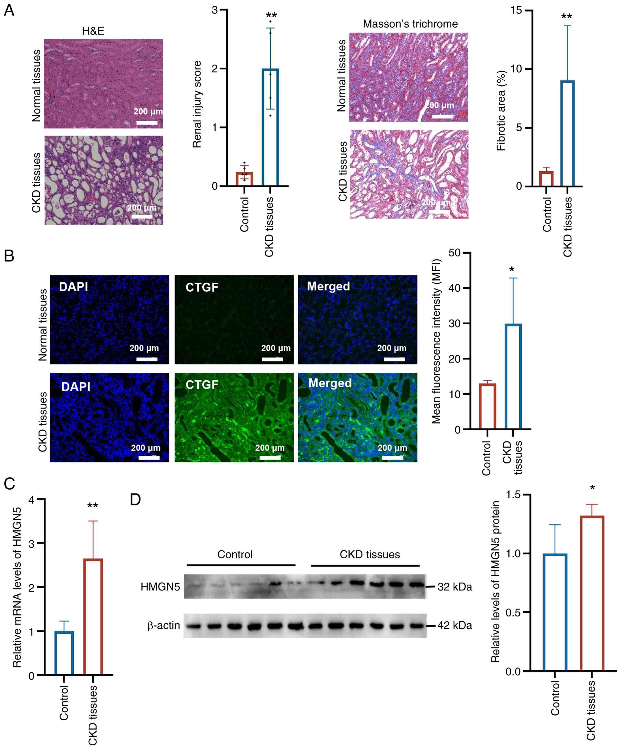
Histopathological analysis revealed marked
structural alterations in renal tissues from patients with CKD
compared with controls. Both H&E staining and Masson's
trichrome staining demonstrated marked histological disruption and
significantly increased fibrosis in the kidney tissues of patients
with CKD, indicating severe pathological changes (Fig. 1A). Immunofluorescence staining
showed that the expression of CTGF was significantly elevated in
CKD renal tissues relative to controls (Fig. 1B). RT-qPCR analysis further
revealed that HMGN5 mRNA was significantly upregulated in CKD
tissues compared with controls (Fig.
1C). Consistently, western blot analysis demonstrated a
significant increase in HMGN5 protein expression in renal tissue
samples from patients with CKD relative to control tissues
(Fig. 1D). Collectively, these
data indicated that HMGN5 expression was significantly elevated at
both the transcriptional and protein levels in the renal tissue of
patients with CKD, in association with enhanced CTGF expression and
notable histopathological alterations.
HMGN5 promotes pyroptosis-associated
signaling in HK-2 cells
To investigate the functional impact of HMGN5 on
pyroptosis-associated signaling, the present study manipulated its
expression in HK-2 cells. Western blotting confirmed the efficiency
of HMGN5 overexpression (Fig.
2A) and knockdown (Fig. 2C).
RT-qPCR analysis showed that, compared with the control group,
HMGN5 overexpression significantly upregulated the mRNA levels of
IL-1β, IL-18 and IL-6 (Fig. 2B),
whereas HMGN5 knockdown significantly suppressed the expression of
these transcripts compared with controls (Fig. 2D).
 | Figure 2HMGN5 promotes the expression of
pyroptosis-associated genes and modulates cell injury and
proliferation in HK-2 cells. (A) Western blot analysis and
semi-quantification of HMGN5 protein levels following the
overexpression of HMGN5 using Ad-HMGN5, confirming the efficiency
of HMGN5 overexpression. (B) Overexpression of HMGN5 significantly
upregulated the mRNA levels of pro-inflammatory cytokines IL-1β,
IL-18 and IL-6 compared with controls. *P<0.05 and
**P<0.01 vs. Ad-NC. (C) Western blot analysis and
semi-quantification of HMGN5 protein levels following the knockdown
of HMGN5 using Ad-sh-HMGN5, confirming knockdown efficiency. (D)
HMGN5 knockdown significantly reduced the expression of IL-1β,
IL-18 and IL-6 at the mRNA level. *P<0.05 and
**P<0.01 vs. Ad-sh-NC. (E) HMGN5 overexpression
promoted LDH leakage, indicating an increase in cell injury
compared with controls. (F) Caspase-1 activity assay demonstrated
that HMGN5 overexpression enhanced inflammasome activation. (G)
Western blot analysis and the semi-quantification of protein levels
showed that HMGN5 overexpression increased the protein levels of
HMGN5, AIM2, cleaved caspase-1 and GSDMD-N. *P<0.05,
**P<0.01 and ***P<0.001 vs. Ad-NC. (H)
HMGN5 knockdown reduced LDH release, suggesting that silencing
HMGN5 attenuated cell injury. (I) Caspase-1 activity assay results
revealed that HMGN5 knockdown suppressed inflammasome activation.
(J) Western blot analysis and the semi-quantification of protein
levels showed that HMGN5 knockdown decreased the protein levels of
HMGN5, AIM2, cleaved caspase-1 and GSDMD-N. For western blot
analyses, proteins and their corresponding controls were detected
using the same batch of total protein samples, with β-actin serving
as an internal loading control to ensure comparability. (K)
Representative images of an EdU incorporation assay and the
quantification of results, demonstrating that HMGN5 knockdown
enhanced the proliferative capacity of HK-2 cells. Scale bar, 50
μm; *P<0.05, and **P<0.01 vs.
Ad-sh-NC. Data are presented as mean ± SEM (n=3 independent
biological replicates). HMGN5, high mobility group
nucleosome-binding 5; AIM2, absent in melanoma 2; GSDMD, gasdermin
D; GSDMD-N, N-terminal pore-forming domain of gasdermin D; LDH,
lactate dehydrogenase; Ad-NC, negative control for overexpression;
Ad-HMGN5, HMGN5 overexpression vector; Ad-sh-NC, negative control
short hairpin RNA sequence; Ad-sh-HMGN5, short hairpin RNA sequence
targeting HMGN5. |
Functionally, the present study assessed
pyroptosis-associated cell injury using LDH release and caspase-1
activity assays. HMGN5 overexpression significantly increased LDH
leakage (Fig. 2E) and caspase-1
enzymatic activity (Fig. 2F).
Consistent with these findings, western blot analysis demonstrated
that, compared with the control group, HMGN5 overexpression
significantly elevated protein levels of AIM2, cleaved caspase-1
and GSDMD-N (Fig. 2G).
Conversely, HMGN5 knockdown significantly reduced LDH release
(Fig. 2H), caspase-1 activity
(Fig. 2I) and the protein levels
of AIM2, cleaved caspase-1 and GSDMD-N compared with the control
group (Fig. 2J). Furthermore,
EdU incorporation assays showed that HMGN5 knockdown significantly
enhanced cell proliferation compared with controls (Fig. 2K), indicating that HMGN5
depletion alleviates growth inhibition associated with
pyroptosis-related cellular stress. Taken together, these results
demonstrated that HMGN5 promoted the activation of
pyroptosis-associated signaling in HK-2 cells.
HMGN5 promotes IFN-β signaling and
IRF3/IRF7 activation in HK-2 cells
To elucidate the mechanism underlying the
HMGN5-mediated regulation of pyroptosis-associated signaling, the
present study performed ChIP-qPCR assays to examine its enrichment
at the promoters of HMGN5-target genes. The results showed that
HMGN5 was significantly enriched at the IFNAR2 promoter,
whereas no significant level of enrichment was detected at the
IFNAR1 IRF3, IRF7 or AIM2 promoters (Fig. 3A). To determine whether this
promoter binding translated into functional transcriptional
regulation, a dual-luciferase reporter assay was performed.
Co-transfection with Ad-HMGN5 significantly increased the
luciferase activity of the WT IFNAR2 promoter reporter
compared with the WT + Ad-NC group (Fig. 3B). Conversely, mutation of the
predicted HMGN5-binding motif within the promoter constructs
attenuated the HMGN5-induced enhancement of luciferase activity
(Fig. 3B), providing further
evidence that HMGN5 positively regulated IFNAR2
transcription through direct promoter interactions. Furthermore,
HMGN5 overexpression markedly increased the mRNA levels of
IFNAR2, IRF7 and IRF3 compared with the
control group (Fig. 3C), and
significantly elevated IFN-β protein secretion as measured by ELISA
(Fig. 3D). Additionally,
LDH-release assays revealed that, compared with the control group,
transduction with Ad-HMGN5 significantly increased LDH leakage in
HK-2 cells (Fig. 3E), which was
indicative of elevated cell-injury levels associated with
pyroptosis-related signaling.
 | Figure 3HMGN5 binds to the IFNAR2
promoter and promotes its expression, leading to IRF3/IRF7
activation and nuclear translocation in HK-2 cells. (A) Chromatin
immunoprecipitation-qPCR analysis showed HMGN5 enrichment at the
promoter region of IFNAR2, with no significant changes in HMGN5
enrichment detected at the AIM2, IFNAR1, IRF3 or IRF7
promoters compared with the IgG control. *P<0.05 vs.
IgG. (B) Dual-luciferase reporter assay in HK-2 cells evaluating
the transcriptional activity of WT or MUT IFNAR2 promoter
constructs under HMGN5 overexpression. **P<0.01 vs.
WT + Ad-NC. (C) Reverse transcription-qPCR analysis of
IFN-β, IRF7 and IRF3 mRNA levels in HK-2
cells. *P<0.05 vs. Ad-NC. (D) ELISA analysis of IFN-β
protein secretion. ***P<0.001 vs. Ad-NC. (E) LDH
release assay in HK-2 cells showed that HMGN5 overexpression
increased LDH release. **P<0.01 vs. Ad-NC. (F)
Western blot analysis and semi-quantification of IFNAR2, p-IRF3,
IRF3, p-IRF7 and IRF7 protein levels in HK-2 cells overexpressing
HMGN5. *P<0.05 vs. Ad-NC. (G) Western blot analysis
and semi-quantification of IFNAR2, p-IRF3, IRF3, p-IRF7 and IRF7
protein levels in HK-2 cells after HMGN5 knockdown.
*P<0.05 vs. Ad-sh-NC. For western blot analyses,
cleaved/phosphorylated proteins and their corresponding total
protein forms were detected using the same batch of protein lysates
in parallel experiments, with β-actin serving as an internal
loading control to ensure comparability. Representative images of
immunofluorescence staining showing the nuclear translocation of
(H) IRF3 and (I) IRF7 in HK-2 cells, which were quantified to
provide nuclear-to-cytoplasmic ratios of each protein. Scale bar,
50 μm; **P<0.01 vs. Ad-NC. Data are presented
as mean ± SEM (n=3 independent biological replicates). HMGN5, high
mobility group nucleosome-binding 5; IFNAR1, interferon receptor 1;
IFNAR2, interferon receptor 2; qPCR, quantitative PCR; AIM2, absent
in melanoma 2; IRF3, interferon regulatory factor 3; IRF7,
interferon regulatory factor 7; WT, wild-type; MUT, mutant; IFN-β,
interferon-β; LDH, lactate dehydrogenase; p-, phosphorylated-;
Ad-NC, negative control for overexpression; Ad-HMGN5, HMGN5
overexpression vector; Ad-sh-NC, negative control short hairpin RNA
sequence; Ad-sh-HMGN5, short hairpin RNA sequence targeting
HMGN5. |
Western blot analysis further showed that, compared
with controls, HMGN5 overexpression significantly upregulated the
protein levels of IFNAR2, p-IRF3 and p-IRF7 (Fig. 3F), whereas HMGN5 knockdown
resulted in a significant reduction in the expression levels of
these markers (Fig. 3G).
Furthermore, immunofluorescence staining demonstrated that,
compared with the control group, HMGN5 overexpression significantly
promoted the nuclear translocation of IRF3 and IRF7 in HK-2 cells
(Fig. 3H and I), suggesting that
HMGN5 modulated the IFN-β signaling cascade through transcriptional
regulation of IFNAR2.
HMGN5 regulates pyroptosis-associated
signaling via an IFNAR2-dependent mechanism
To investigate the role of IFNAR2 signaling in renal
tubular epithelial cell injury, HK-2 cells were transduced with an
adenovirus encoding an shRNA sequence targeting IFNAR2
(Ad-sh-IFNAR2). Compared with Ad-sh-NC-transduced cells,
Ad-sh-IFNAR2 significantly reduced IFNAR2 expression.
Concomitantly, the expression levels of AIM2, cleaved caspase-1 and
GSDMD-N were significantly decreased in the Ad-sh-IFNAR2 group
compared with the Ad-sh-NC group (Fig. 4A). Furthermore, LDH release was
significantly reduced following IFNAR2 knockdown compared with the
control group (Fig. 4B),
indicating that IFNAR2 knockdown attenuated membrane damage, which
was consistent with the suppression of pyroptosis-associated
signaling.
 | Figure 4IFNAR2 signaling contributes to
HMGN5-mediated pyroptosis-associated cellular responses in HK-2
cells. (A) Western blot analysis and semi-quantification of IFNAR2,
AIM2, caspase-1, cleaved caspase-1, GSDMD and GSDMD-N protein
levels in HK-2 cells following IFNAR2 knockdown. (B) LDH
release assay in HK-2 cells showed that IFNAR2 knockdown
reduced LDH release. *P<0.05 and
**P<0.01 vs. Ad-sh-NC. (C) Western blot analysis and
semi-quantification of HMGN5, IFNAR2, IRF3, p-IRF3, IRF7, p-IRF7,
AIM2, caspase-1, cleaved caspase-1, GSDMD and GSDMD-N protein
levels in HK-2 cells overexpressing HMGN5, with or without
concurrent IFNAR2 knockdown. For western blot analyses,
cleaved/phosphorylated proteins and their corresponding total
protein forms were detected using the same batch of protein lysates
in parallel experiments, with β-actin serving as an internal
loading control to ensure comparability. ELISA analysis of (D)
IFN-β, (E) IL-8 and (F) IL-1β secretion in HK-2 cells
overexpressing HMGN5, with or without concurrent IFNAR2
knockdown. (G) LDH release assay in HK-2 cells overexpressing
HMGN5, with or without concurrent IFNAR2 knockdown. Data are
presented as mean ± SEM (n=3 independent biological replicates).
*P<0.05, **P<0.01 and
***P<0.001 vs. Ad-NC; #P<0.05 and
##P<0.01 vs. Ad-HMGN5. IFNAR2, interferon receptor 2;
HMGN5, high mobility group nucleosome-binding 5; AIM2, absent in
melanoma 2; GSDMD, gasdermin D; GSDMD-N, N-terminal pore-forming
domain of gasdermin D; IRF3, interferon regulatory factor 3; IRF7,
interferon regulatory factor 7; IFN-β, interferon-β; LDH, lactate
dehydrogenase; p-, phosphorylated-; Ad-sh-NC, negative control
short hairpin RNA sequence; Ad-sh-IFNAR2, short hairpin RNA
sequence targeting IFNAR2; Ad-NC, negative control for
overexpression; Ad-HMGN5, HMGN5 overexpression vector. |
Subsequently, the present study examined whether
HMGN5 regulated HK-2 cell injury through an IFNAR2-dependent
mechanism. HMGN5 overexpression significantly increased the
expression of IFNAR2, p-IRF3, p-IRF7, AIM2, cleaved caspase-1 and
GSDMD-N compared with Ad-NC-treated cells (Fig. 4C). Notably, concurrent IFNAR2
knockdown partially yet significantly reversed the HMGN5-induced
upregulation of these proteins (Fig.
4C), suggesting that IFNAR2 signaling was a key mediator
required for HMGN5-induced activation of the AIM2 inflammasome and
downstream pyroptosis-associated signaling cascades. In parallel,
ELISA analyses demonstrated that, compared with controls, HMGN5
overexpression significantly increased the secretion of IL-1β, IL-8
and IFN-β. Furthermore, silencing IFNAR2 significantly mitigated
these HMGN5-induced elevations (Fig.
4D-F), suggesting that the HMGN5-IFNAR2 axis contributed to
downstream pyroptosis-associated signaling in renal tubular cells.
Additionally, compared with the control group, a significant
increase in LDH release was induced by HMGN5 overexpression; this
elevation in LDH leakage was significantly reduced by concurrent
IFNAR2 silencing (Fig. 4G),
further supporting an important role for IFNAR2 signaling in
mediating HMGN5-modulated pyroptosis-associated cell injury.
HMGN5 expression and
pyroptosis-associated signaling are upregulated in the kidneys of
CKD-model mice
To establish a mouse model of CKD, mice were
subjected to a two-step nephrectomy involving an initial left total
nephrectomy followed by a right partial (2/3) nephrectomy 1 week
later. Histopathological analysis using H&E staining and
Masson's trichrome staining revealed structural alterations in the
renal tissue of CKD-model mice compared with sham-operated
controls, with renal injury scores and fibrotic quantification
indicating the occurrence of significant damage and fibrosis in
model mice (Fig. 5A). Similarly,
immunofluorescence staining demonstrated a significant increase in
CTGF expression in the kidneys of CKD-model mice compared with
control mice (Fig. 5B).
Biochemical analyses further showed that plasma levels of
creatinine and blood urea nitrogen were significantly elevated in
CKD-model mice compared with sham-operated controls, providing
further evidence of impaired renal function in the model (Fig. 5C and D). In parallel, circulating
levels of IFN-β, IL-8 and IL-1β were significantly increased in
CKD-model mice relative to the sham group (Fig. 5E-G), suggesting that model mice
experienced enhanced systemic inflammatory and IFN responses.
Additionally, LDH release assays revealed a significant increase in
LDH leakage in renal tissue samples from CKD-model mice compared
with sham controls (Fig. 5H),
indicating that model mice experienced an increase in renal cell
injury associated with pyroptosis-related signaling. At the
molecular level, western blot analysis demonstrated that the
protein expression levels of HMGN5, IFNAR2, cleaved caspase-1 and
GSDMD-N were significantly elevated in the kidneys of CKD-model
mice relative to sham-operated mice (Fig. 5I), suggesting that HMGN5-mediated
activation of the IFNAR2 signaling axis may have contributed to the
activation of pyroptosis-related pathways in CKD-model renal
tissues.
 | Figure 5HMGN5 expression and
pyroptosis-associated signaling are increased in the kidneys of
CKD-model mice. (A) Representative images of H&E staining and
Masson's trichrome staining in renal tissues from CKD-model mice
and sham-operated controls. H&E staining was semi-quantified to
calculate renal injury scores, and Masson's trichrome staining was
used to quantify fibrosis in renal tissues. (B) Representative
immunofluorescence staining images and quantification of CTGF
expression in the renal tissues of mice from CKD-model and
sham-operated groups. Plasma levels of (C) creatinine and (D) urea
in CKD-model mice and sham-operated controls. ELISA analysis of
plasma levels of (E) IFN-β, (F) IL-8 and (G) IL-1β. (H) Results of
an LDH release assay performed on the renal tissues of CKD-model
mice and sham-operated controls. (I) Western blot analysis and
semi-quantification of HMGN5, IFNAR2, caspase-1, cleaved caspase-1,
GSDMD and GSDMD-N protein levels in the renal tissues of CKD-model
mice and sham-operated controls. For western blot analyses,
cleaved/phosphorylated proteins and their corresponding total
protein forms were detected using the same batch of protein lysates
in parallel experiments, with β-actin serving as an internal
loading control to ensure comparability. Scale bar, 200 μm.
Data are presented as mean ± SEM (n=5 mice per group; 3 independent
serial sections per sample). *P<0.05,
**P<0.01 and ***P<0.001 vs. sham group.
CKD, chronic kidney disease; H&E, hematoxylin and eosin;
IFNAR2, interferon receptor 2; HMGN5, high mobility group
nucleosome-binding 5; GSDMD, gasdermin D; GSDMD-N, N-terminal
pore-forming domain of gasdermin D; IFN-β, interferon-β; LDH,
lactate dehydrogenase; CTGF, connective tissue growth factor. |
IFNAR2 blockade attenuates renal injury
and suppresses pyroptosis-associated signaling in CKD-model
mice
To evaluate the pathophysiological impact of IFNAR
blockade in vivo, CKD-model mice were treated with an IFNAR
Ab. Histopathological examinations using H&E staining and
Masson's trichrome staining revealed that IFNAR Ab treatment
attenuated renal pathological alterations and significantly reduced
fibrosis compared with untreated CKD-model mice (Fig. 6A). Consistent with these
histological improvements, biochemical analyses demonstrated that,
compared with the CKD-model group, plasma creatinine and urea
levels were significantly reduced following IFNAR Ab treatment
(Fig. 6B and C), indicating that
this treatment improved renal function in CKD-model mice. In
parallel, immunofluorescence staining showed that CTGF expression
was significantly reduced in the renal tissues of IFNAR Ab-treated
CKD-model mice compared with untreated CKD mice (Fig. 6D). ELISA analyses further
revealed that IFNAR Ab treatment significantly reduced the
circulating levels of IL-1β, IL-8 and IFN-β in CKD-model mice
(Fig. 6E-G), suggesting that
this treatment attenuated systemic inflammatory and type I IFN
responses. Furthermore, LDH release assays demonstrated that IFNAR
Ab treatment significantly reduced LDH leakage in renal tissues
compared with CKD-model mice (Fig.
6H), indicating that this treatment attenuated renal cell
injury associated with pyroptosis-related signaling. At the
molecular level, western blot analysis showed that IFNAR Ab
treatment partially yet significantly reversed the upregulation of
IFNAR2, cleaved caspase-1 and GSDMD-N observed in the kidneys of
CKD-model mice (Fig. 6I),
suggesting the suppression of IFNAR2-associated pyroptosis-related
signaling in vivo.
 | Figure 6IFNAR Ab treatment partially
attenuates renal injury and suppresses pyroptosis-associated
changes in CKD-model mice. (A) Representative images of H&E
staining and Masson's trichrome staining showing that IFNAR Ab
treatment markedly attenuated renal pathological alterations and
reduced fibrosis in CKD-model mice compared with untreated
CKD-model controls. H&E staining was semi-quantified to
calculate renal injury scores, and Masson's trichrome staining was
used to quantify fibrosis in renal tissues. (B) Biochemical
analysis showed that plasma creatinine levels were significantly
decreased in CKD-model mice after IFNAR Ab treatment compared with
the untreated CKD-model group. However, IFNAR Ab treatment did not
completely restore plasma creatinine to control levels (C)
Biochemical analysis revealed that plasma urea levels were
significantly reduced, partially reversing the increase observed in
untreated CKD-model mice, following IFNAR Ab treatment. (D)
Representative immunofluorescence staining images and subsequent
fluorescence-intensity quantification showed that CTGF expression
was partially yet significantly reduced in the kidneys of IFNAR
Ab-treated CKD-model mice compared with the untreated CKD-model
group. ELISA analysis demonstrated that the plasma levels of (E)
IL-1β, (F) IL-8 and (G) IFN-β were significantly attenuated in
CKD-model mice after IFNAR Ab treatment compared with untreated
CKD-model control mice. (H) LDH release assay results showed that
LDH leakage in renal tissues was significantly reduced in CKD-model
mice following IFNAR Ab treatment compared with the untreated
CKD-model group. (I) Western blot analysis and semi-quantification
demonstrated that the protein expression levels of IFNAR2, cleaved
caspase-1 and GSDMD-N significantly decreased in the kidneys of
IFNAR Ab-treated CKD-model mice compared with untreated CKD-model
mice; however, the expression of these proteins was not fully
restored to basal levels following this treatment. For western blot
analyses, cleaved/phosphorylated proteins and their corresponding
total protein forms were detected using the same batch of protein
lysates in parallel experiments, with β-actin serving as an
internal loading control to ensure comparability. Scale bar, 200
μm. Data are presented as mean ± SEM (n=5 mice per group; 3
independent serial sections per sample). **P<0.01 and
***P<0.001 vs. sham + IgG group;
#P<0.05, ##P<0.01 and
###P<0.001 vs. model + IgG group. CKD, chronic kidney
disease; H&E, hematoxylin and eosin; IFN-β, interferon-β;
IFNAR, interferon receptor; IFNAR Ab, IFNAR-neutralizing antibody;
LDH, lactate dehydrogenase; HMGN5, high mobility group
nucleosome-binding 5; GSDMD, gasdermin D; GSDMD-N, N-terminal
pore-forming domain of gasdermin D; CTGF, connective tissue growth
factor. |
Discussion
CKD is characterized by persistent inflammation,
tubular epithelial injury and progressive fibrosis, which drive the
gradual loss of renal function (26). Increasing evidence has indicated
that dysregulated innate immune signaling and sustained activation
of inflammatory pathways contribute to CKD progression (1,2).
However, the upstream nuclear events that link cellular stress to
inflammatory amplification in renal tubular epithelial cells remain
incompletely elucidated.
HMGN proteins are non-histone chromatin-binding
proteins that function by competing with histone H1 for nucleosome
binding, thereby reducing the compaction of chromatin and
increasing the accessibility of regulatory DNA sequences for
binding by transcription factors and RNA polymerase II (27). By modifying the global chromatin
landscape, HMGN5 facilitates a permissive epigenetic state,
enabling the efficient recruitment of transcriptional machinery to
specific promoters. Additionally, a previous study identified HMGN1
as an endogenous alarmin capable of modulating innate immune
responses (17). In particular,
HMGN1 has been shown to synergize with Toll-like receptor 7/8
agonists to facilitate the activation of dendritic cells, promoting
the nuclear translocation of IRF3 and IRF7 and the subsequent
production of type I IFNs, including IFN-β (17). In systemic lupus erythematosus,
HMGN family proteins have been implicated as molecular links
between aberrant chromatin remodeling and IFN-associated
inflammation (28). These
findings highlight the potential for HMGN family members to serve
as bridges between chromatin remodeling and inflammatory
responses.
HMGN5 shares structural and functional similarities
with HMGN1, including nucleosome-binding capacity and
transcriptional regulatory potential (29), yet its role in non-immune
parenchymal cells and chronic inflammatory diseases has remained
largely unexplored. Given the importance of IFN signaling and
innate immune activation in CKD, we hypothesized that HMGN5 may
function as an upstream nuclear regulator that links chromatin
dynamics to IFN-dependent inflammatory pathways in renal tubular
epithelial cells.
The present study demonstrated that HMGN5 expression
was elevated in renal tissues from patients with CKD and CKD-model
mice, at both transcriptional and protein levels. This upregulation
associated with structural damage, increased CTGF expression and
indicators of inflammation (such as IL-8 and IL-1β) and renal
dysfunction, suggesting that HMGN5 was closely associated with CKD
progression. Mechanistically, the in vitro experiments of
the present study revealed that HMGN5 overexpression in HK-2 cells
promoted the expression of pyroptosis-associated molecules,
including AIM2, cleaved caspase-1 and GSDMD-N; this upregulation
was accompanied by increased LDH release and pro-inflammatory
cytokine secretion.
Pyroptosis-associated signaling has been
increasingly recognized as an important mechanism contributing to
tubular injury, inflammatory amplification and fibrosis in CKD
(25,30). Several lines of evidence from
diverse disease contexts have supported the involvement of IFN
signaling in pyroptosis-related pathways (12,31,32). For instance, granzyme A from
cytotoxic lymphocytes has been shown to induce GSDMB-mediated
pyroptosis in an IFN-dependent manner, highlighting a link between
IFN signaling and GSDM-mediated inflammatory cell death (31). Similarly, IFN-γ has been shown to
promote pyroptosis-related signaling in corneal epithelial cells
through Janus kinase 2 (JAK2)/STAT1 activation, suggesting that
type II IFNs can prime non-immune cells for inflammatory injury
(12). In cancer models, the
potentiation of type I IFN signaling through ubiquitin specific
protease 18 depletion has been shown to induce pyroptosis-related
signaling via STAT2- and NF-κB-dependent transcriptional programs,
underscoring a broader role for IFN pathways in orchestrating
inflammatory cell death (32).
Regarding immune cells, AIM2 inflammasome activation and
pyroptosis-related pathways in monocytes have been shown to be
regulated by IFN-γ through the JAK2/STAT1 pathway, supporting the
concept that IFN signaling contributes to inflammasome-mediated
cell death across diverse cell types (9). Even during bacterial infection,
type I IFNs have been shown to epigenetically upregulate
pyroptosis-related genes, including GSDMD, further
activating inflammasomes (33).
Collectively, these studies have provided mechanistic and
translational support for the findings of the present study,
specifically that IFNAR2-dependent signaling contributed to
HMGN5-mediated pyroptosis-associated cell injury in renal tubular
epithelial cells.
Building upon these distinctions in inflammatory
cell death pathways, it is important to further contextualize these
findings: While apoptosis represents a non-inflammatory mode of
programmed cell death characterized by cellular shrinkage and
membrane blebbing without rupture, pyroptosis is a notably
pro-inflammatory process driven by GSDM-mediated pore formation in
the plasma membrane. This pore formation results in the rapid
release of intracellular contents, for example LDH and
pro-inflammatory cytokines, which markedly amplifies the
inflammatory microenvironment (34). Previous studies have highlighted
the notable role of GSDM in renal injury, demonstrating that GSDMD
cleavage is a central executioner of pyroptosis in various renal
pathologies, including acute kidney injury and diabetic nephropathy
(34,35). Furthermore, the epigenetic
control of inflammasomes has emerged as an important layer of
regulation in pyroptosis; for instance, histone modification and
chromatin remodeling have been shown to determine the
transcriptional readiness of the NLRP3 and AIM2 loci (36-38). The findings of the present study
extended this concept by proposing that HMGN5 serves as an
epigenetic rheostat, precisely tuning the transcriptional
activation of the IFNAR2 promoter to facilitate downstream
inflammasome assembly.
A key finding of the present study was the
identification of IFNAR2-dependent signaling as a primary pathway
mediating HMGN5-induced pyroptosis-associated responses. ChIP-qPCR
assays demonstrated that HMGN5 was enriched at the IFNAR2
promoter, suggesting a direct role in its transcriptional
regulation. Similarly, HMGN5 overexpression enhanced IFNAR2
expression, promoted IRF3 and IRF7 phosphorylation and nuclear
translocation, and increased IFN-β production in HK-2 cells.
Notably, IFNAR2 silencing mitigated HMGN5-induced activation
of the AIM2 inflammasome, reduced downstream caspase-1 activation
and GSDMD-N cleavage, and attenuated LDH release and inflammatory
cytokine secretion. These findings indicate that HMGN5 functioned
upstream of the IFNAR2 signaling axis, and that intact IFNAR2
signaling contributed to HMGN5-mediated pyroptosis-associated
responses.
From a translational perspective, the in vivo
data obtained in the present study demonstrated that
pharmacological blockade of IFNAR2 signaling contributed to the
mitigation of renal injury and the suppression of
pyroptosis-associated signaling in CKD-model mice. IFNAR2 antibody
treatment was associated with improved renal histopathology,
reduced fibrosis, restored renal function and decreased systemic
inflammatory markers in renal tissues. These findings suggest that
IFNAR2 signaling may represent a potential therapeutic target in
the context of CKD pathogenesis. However, it should be acknowledged
that the 5/6 nephrectomy CKD mouse model used in the present study
primarily reflects a specific context of remnant-kidney injury and
hemodynamic overload (39).
Future studies utilizing alternative etiological models, such as
diabetic nephropathy or unilateral ureteral obstruction, will be
important for determining whether the HMGN5-IFNAR2-IRF-AIM2 axis
represents a generalized mechanism across the biological
heterogeneity of CKD.
Furthermore, without direct in vivo HMGN5
gain- or loss-of-function experiments, the upstream role of HMGN5
in regulating IFNAR2 in the CKD mouse model remains an observation
that warrants further verification. To support this causal link
in vivo, future investigations utilizing tubule-specific
HMGN5-knockout mice are warranted. Although directly
targeting chromatin-binding proteins, such as HMGN5, presents
notable translational challenges, the results of the present study
suggest that the modulation of IFNAR2 signaling could be explored
as a theoretical strategy to attenuate IFN-driven and
pyroptosis-related injury in CKD. Given that type I IFN pathways
are already under investigation as therapeutic targets in
autoimmune and inflammatory diseases (40,41), further research is required to
evaluate whether IFNAR2-targeted approaches could eventually hold
promise for attenuating progression in a subset of patients with
inflammation-driven CKD.
Collectively, the findings of the present study
identified HMGN5 as a potential epigenetic regulator associated
with IFN-related inflammatory signaling in CKD. The data in the
present study suggest that HMGN5 may have regulated IFNAR2
expression via promoter enrichment and that IFNAR2 knockdown
mitigated HMGN5-induced pyroptosis-associated responses. The
proposed HMGN5-IFNAR2-IRF3/IRF7-AIM2 pathway should be interpreted
as a mechanistically supported pathway in CKD pathogenesis rather
than an established signaling cascade. Further studies employing
targeted gain- and loss-of-function approaches for IRF3, IRF7 and
AIM2 will be required to define the precise downstream signaling
hierarchy of this pathway. As such, the HMGN5-IFNAR2 axis warrants
further investigation as a theoretical target for limiting
pyroptosis-associated renal injury in CKD.
Availability of data and materials
The data generated in the present study may be
requested from the corresponding author.
Authors' contributions
XR, JX and LN designed the research, analyzed the
data, performed the experiments, wrote the original draft of the
manuscript and were responsible for making revisions to the
manuscript. JZ, QZ, JW and GZ contributed to performing the animal
experiments. LN secured funding. All authors confirm the
authenticity of all the raw data. All authors read and approved the
final version of the manuscript.
Ethics approval and consent to
participate
The study protocol was approved by the Medical
Scientific Research Ethics Committee of Shijiazhuang People's
Hospital (Shijiazhuang, China; approval no. 2023-56A). Written
informed consent was received for the patient study. All animal
procedures were approved by the Medical Scientific Research Ethics
Committee of Shijiazhuang People's Hospital (approval no.
2024-99).
Patient consent for publication
Not applicable.
Competing interests
The authors declare that they have no competing
interests.
Use of artificial intelligence tools
During the preparation of this work, artificial
intelligence tools (DeepSeek) were used to improve the readability
and language of the manuscript, and subsequently, the authors
revised and edited the content produced by the artificial
intelligence tools as necessary, taking full responsibility for the
ultimate content of the present manuscript.
Acknowledgements
Not applicable.
Funding
The present study was supported by Hebei Provincial Institute of
Medical Research (grant no. 20241271).
References
|
1
|
Rao IR, Bangera A, Nagaraju SP, Shenoy SV,
Prabhu RA, Rangaswamy D and Bhojaraja MV: Chronic kidney disease of
unknown aetiology: A comprehensive review of a global public health
problem. Trop Med Int Health. 28:588–600. 2023. View Article : Google Scholar : PubMed/NCBI
|
|
2
|
Kishi S, Nagasu H, Kidokoro K and
Kashihara N: Oxidative stress and the role of redox signalling in
chronic kidney disease. Nat Rev Nephrol. 20:101–119. 2024.
View Article : Google Scholar
|
|
3
|
Zhu H, Cao C, Wu Z, Zhang H, Sun Z, Wang
M, Xu H, Zhao Z, Wang Y, Pei G, et al: The probiotic L. casei Zhang
slows the progression of acute and chronic kidney disease. Cell
Metab. 33:1926–1942.e8. 2021. View Article : Google Scholar
|
|
4
|
Jiayi H, Ziyuan T, Tianhua X, Mingyu Z,
Yutong M, Jingyu W, Hongli Z and Li S: Copper homeostasis in
chronic kidney disease and its crosstalk with ferroptosis.
Pharmacol Res. 202:1071392024. View Article : Google Scholar : PubMed/NCBI
|
|
5
|
Sanz AB, Sanchez-Niño MD, Ramos AM and
Ortiz A: Regulated cell death pathways in kidney disease. Nat Rev
Nephrol. 19:281–299. 2023. View Article : Google Scholar : PubMed/NCBI
|
|
6
|
Pang Q, Wang P, Pan Y, Dong X, Zhou T,
Song X and Zhang A: Irisin protects against vascular calcification
by activating autophagy and inhibiting NLRP3-mediated vascular
smooth muscle cell pyroptosis in chronic kidney disease. Cell Death
Dis. 13:2832022. View Article : Google Scholar : PubMed/NCBI
|
|
7
|
Ding B, Ma G, Wang Z, Liang W and Gao W:
Mechanisms of kidney cell pyroptosis in chronic kidney disease and
the effects of traditional chinese medicine. Evid Based Complement
Alternat Med. 2021:11733242021. View Article : Google Scholar : PubMed/NCBI
|
|
8
|
Chen Y, Ye X, Escames G, Lei W, Zhang X,
Li M, Jing T, Yao Y, Qiu Z, Wang Z, et al: The NLRP3 inflammasome:
Contributions to inflammation-related diseases. Cell Mol Biol Lett.
28:512023. View Article : Google Scholar : PubMed/NCBI
|
|
9
|
Zhao Y, Liang B, Sheng S, Wang C, Jin B,
Zhang X, Cheng Y, Shen C and Zheng F: AIM2 inflammasome regulated
by the IFN-γ/JAK2/STAT1 pathway promotes activation and pyroptosis
of monocytes in coronary artery disease. Immun Inflamm Dis.
12:e13172024. View Article : Google Scholar
|
|
10
|
Zhang Q, Meng Y, Zhao W, Li W, Dai C,
Cheng J, Li B, Chen F, Yang J, Li H and Li S: IFN-γ-induced
AIM2-PANoptosis in granulosa cells leads to ovulatory dysfunction
in polycystic ovary syndrome. Biochem Pharmacol. 250:1180392026.
View Article : Google Scholar
|
|
11
|
Costa Franco MMS, Marim FM, Alves-Silva J,
Cerqueira D, Rungue M, Tavares IP and Oliveira SC: AIM2 senses
Brucella abortus DNA in dendritic cells to induce IL-1β secretion,
pyroptosis and resistance to bacterial infection in mice. Microbes
Infect. 21:85–93. 2019. View Article : Google Scholar
|
|
12
|
Yang X, Zuo X, Zeng H, Liao K, He D, Wang
B and Yuan J: IFN-γ facilitates corneal epithelial cell pyroptosis
through the JAK2/STAT1 pathway in dry eye. Invest Ophthalmol Vis
Sci. 64:342023.
|
|
13
|
Luo L, Liu M, Fan Y, Zhang J, Liu L, Li Y,
Zhang Q, Xie H, Jiang C, Wu J, et al: Intermittent theta-burst
stimulation improves motor function by inhibiting neuronal
pyroptosis and regulating microglial polarization via
TLR4/NFkappaB/NLRP3 signaling pathway in cerebral ischemic mice. J
Neuroinflammation. 19:1412022. View Article : Google Scholar
|
|
14
|
Jiang J, Shen W, He Y, Liu J, Ouyang J,
Zhang C and Hu K: Overexpression of NLRP12 enhances antiviral
immunity and alleviates herpes simplex keratitis via
pyroptosis/IL-18/IFN-γ signaling. Int Immunopharmacol.
137:1124282024. View Article : Google Scholar
|
|
15
|
Mou J, Huang M, Wang F, Xu X, Xie H, Lu H,
Li M, Li Y, Kong W, Chen J, et al: HMGN5 escorts oncogenic STAT3
signaling by regulating the chromatin landscape in breast cancer
tumorigenesis. Mol Cancer Res. 20:1724–1738. 2022. View Article : Google Scholar : PubMed/NCBI
|
|
16
|
Rochman M, Malicet C and Bustin M:
HMGN5/NSBP1: A new member of the HMGN protein family that affects
chromatin structure and function. Biochim Biophys Acta. 1799:86–92.
2010. View Article : Google Scholar : PubMed/NCBI
|
|
17
|
Alam MM, Yang D, Trivett A, Meyer TJ and
Oppenheim JJ: HMGN1 and R848 synergistically activate dendritic
cells using multiple signaling pathways. Front Immunol. 9:29822018.
View Article : Google Scholar
|
|
18
|
Kulkarni HV, Mulay SR and Gaikwad AB: The
AKI-CKD continuum: Mechanisms and therapeutic interventions.
Nephrol Dial Transplant. Aug 8–2026.Epub ahead of print. View Article : Google Scholar : PubMed/NCBI
|
|
19
|
Hong W, Zhu Y, Lin Y, Tang S, Chen J, Xu
L, Jiang J, Zong Y, Zhang Y, Sun A and Wu X: The chromatin
remodeling protein BRG1 mediates Ang II induced pro-fibrogenic
response in renal fibroblasts. Life Sci. 340:1223202024. View Article : Google Scholar : PubMed/NCBI
|
|
20
|
Kidney Disease: Improving Global Outcomes
(KDIGO) CKD Work Group: KDIGO 2024 clinical practice guideline for
the evaluation and management of chronic kidney disease. Kidney
Int. 105:S117–S314. 2024. View Article : Google Scholar
|
|
21
|
Hackstein CP, Spitzer J, Symeonidis K,
Horvatic H, Bedke T, Steglich B, Klein S, Assmus LM, Odainic A,
Szlapa J, et al: Interferon-induced IL-10 drives systemic T-cell
dysfunction during chronic liver injury. J Hepatol. 79:150–166.
2023. View Article : Google Scholar : PubMed/NCBI
|
|
22
|
Huang MW, Stock AD, Mike EV, Herlitz L,
Kolbeck R and Putterman C: Anti-IFNAR treatment does not reverse
neuropsychiatric disease in MRL/lpr lupus mice. Lupus.
28:1510–1523. 2019. View Article : Google Scholar : PubMed/NCBI
|
|
23
|
Livak KJ and Schmittgen TD: Analysis of
relative gene expression data using real-time quantitative PCR and
the 2(-Delta Delta C(T)) method. Methods. 25:402–408. 2001.
View Article : Google Scholar
|
|
24
|
Liu Y, Pan R, Ouyang Y, Gu W, Xiao T, Yang
H, Tang L, Wang H, Xiang B and Chen P: Pyroptosis in health and
disease: Mechanisms, regulation and clinical perspective. Signal
Transduct Target Ther. 9:2452024. View Article : Google Scholar : PubMed/NCBI
|
|
25
|
Elias EE, Lyons B and Muruve DA:
Gasdermins and pyroptosis in the kidney. Nat Rev Nephrol.
19:337–350. 2023. View Article : Google Scholar : PubMed/NCBI
|
|
26
|
Neuen BL, Yeung EK, Rangaswami J and
Vaduganathan M: Combination therapy as a new standard of care in
diabetic and non-diabetic chronic kidney disease. Nephrol Dial
Transplant. 40(Suppl 1): i59–i69. 2025. View Article : Google Scholar : PubMed/NCBI
|
|
27
|
He B, Zhu I, Postnikov Y, Furusawa T,
Jenkins L, Nanduri R, Bustin M and Landsman D: Multiple epigenetic
factors co-localize with HMGN proteins in A-compartment chromatin.
Epigenetics Chromatin. 15:232022. View Article : Google Scholar : PubMed/NCBI
|
|
28
|
Lindblom J, Lagutkin D, Sherina N, Idborg
H, Beretta L, Borghi MO; PRECISESADS Clinical Consortium; Peyper
JM, Barturen G, Jakobsson PJ, et al: Novel IgG and IgA
autoantibodies validated in two independent cohorts are associated
with disease activity and determine organ manifestations in
systemic lupus erythematosus: Implications for anti-LIN28A,
anti-HMGN5, anti-IRF5, and anti-TGIF1. Ann Rheum Dis. 84:1164–1179.
2025. View Article : Google Scholar : PubMed/NCBI
|
|
29
|
Rodríguez-Ortiz A, Montoya-Villegas JC,
García-Vallejo F and Mina-Paz Y: Spatial and temporal expression of
high-mobility-group nucleosome-binding (HMGN) genes in brain areas
associated with cognition in individuals with down syndrome. Genes
(Basel). 12:20002021. View Article : Google Scholar : PubMed/NCBI
|
|
30
|
Wang Y, Li Y and Xu Y: Pyroptosis in
kidney disease. J Mol Biol. 434:1672902022. View Article : Google Scholar
|
|
31
|
Zhou Z, He H, Wang K, Shi X, Wang Y, Su Y,
Wang Y, Li D, Liu W, Zhang Y, et al: Granzyme A from cytotoxic
lymphocytes cleaves GSDMB to trigger pyroptosis in target cells.
Science. 368:eaaz75482020. View Article : Google Scholar : PubMed/NCBI
|
|
32
|
Arimoto KI, Miyauchi S, Troutman TD, Zhang
Y, Liu M, Stoner SA, Davis AG, Fan JB, Huang YJ, Yan M, et al:
Expansion of interferon inducible gene pool via USP18 inhibition
promotes cancer cell pyroptosis. Nat Commun. 14:2512023. View Article : Google Scholar : PubMed/NCBI
|
|
33
|
Li Y, Guo X, Hu C, Du Y, Guo C, Wang D,
Zhao W, Huang G, Li C, Lu Q, et al: Type I IFN operates pyroptosis
and necroptosis during multidrug-resistant A. Baumannii infection.
Cell Death Differ. 25:1304–1318. 2018. View Article : Google Scholar : PubMed/NCBI
|
|
34
|
Yu P, Zhang X, Liu N, Tang L, Peng C and
Chen X: Pyroptosis: Mechanisms and diseases. Signal Transduct
Target Ther. 6:1282021. View Article : Google Scholar : PubMed/NCBI
|
|
35
|
Zhang N, Zhang J, Yang Y, Shan H, Hou S,
Fang H, Ma M, Chen Z, Tan L and Xu D: A
palmitoylation-depalmitoylation relay spatiotemporally controls
GSDMD activation in pyroptosis. Nat Cell Biol. 26:757–769. 2024.
View Article : Google Scholar : PubMed/NCBI
|
|
36
|
Saller BS, Wöhrle S, Fischer L, Dufossez
C, Ingerl IL, Kessler S, Mateo-Tortola M, Gorka O, Lange F, Cheng
Y, et al: Acute suppression of mitochondrial ATP production
prevents apoptosis and provides an essential signal for NLRP3
inflammasome activation. Immunity. 58:90–107.e11. 2025. View Article : Google Scholar
|
|
37
|
Fu M, Lan Z, Ye Y, Gong Y, Liang Q, Li M,
Feng L, Chen A, Dong Q, Li Y, et al: The metabolite
alpha-ketoglutarate inhibits vascular calcification partially
through modulation of the TET2/NLRP3 inflammasome signaling
pathway. Kidney Int. 108:233–252. 2025. View Article : Google Scholar : PubMed/NCBI
|
|
38
|
Li Y, Wang L, Wanga W, Wangb W, Huang W
and Liu S: Acetylation modification of AIM2 by KAT2B suppresses the
AKT/Wnt/β-catenin signaling pathway activation and inhibits breast
cancer progression. Epigenetics Chromatin. 18:272025. View Article : Google Scholar
|
|
39
|
Tan RZ, Zhong X, Li JC, Zhang YW, Yan Y,
Liao Y, Wen D, Diao H, Wang L and Shen HC: An optimized 5/6
nephrectomy mouse model based on unilateral kidney ligation and its
application in renal fibrosis research. Ren Fail. 41:555–566. 2019.
View Article : Google Scholar : PubMed/NCBI
|
|
40
|
Goel RR, Kotenko SV and Kaplan MJ:
Interferon lambda in inflammation and autoimmune rheumatic
diseases. Nat Rev Rheumatol. 17:349–362. 2021. View Article : Google Scholar : PubMed/NCBI
|
|
41
|
Wu YY, Xing J, Li XF, Yang YL, Shao H and
Li J: Roles of interferon induced protein with tetratricopeptide
repeats (IFIT) family in autoimmune disease. Autoimmun Rev.
22:1034532023. View Article : Google Scholar : PubMed/NCBI
|