Iron homeostasis is key in maintaining cellular
metabolism, redox balance and diverse physiological functions,
relying on the coordinated regulation of iron uptake, storage,
utilization and export. As a major intracellular iron-storage
complex, ferritin serves a central role in buffering iron toxicity
while maintaining iron availability (1,2).
In recent years, ferritinophagy, a selective form of autophagy that
has attracted increasing attention, has been closely associated
with ferritin degradation and has been shown to serve an important
role in regulating cellular iron homeostasis under the mediation of
nuclear receptor coactivator 4 (NCOA4) (3,4).
Acting as a specific cargo receptor, NCOA4 recognizes and binds
ferritin at the early stage of autophagy and mediates its delivery
to the autophagy-lysosome pathway for degradation. This process
promotes the conversion of stored iron into bioavailable iron;
however, under conditions of dysregulated iron control, it may also
exacerbate iron overload and trigger cellular dysfunction (5-7).
Previous evidence has suggested that NCOA4 is a central receptor
involved in ferritinophagy and also exhibits a key regulatory
function in lysosome-mediated transport, degradation and recycling
of ferritin-derived iron (8,9).
NCOA4 was initially identified in association with the RET-fusion
gene and its biological function is currently considered to
encompass broader regulation of cellular stability (10). NCOA4-mediated ferritin
degradation releases Fe2+ into the intracellular labile
iron pool (LIP), which, upon Fenton chemistry, drives reactive
oxygen species (ROS) production and triggers ferroptosis (11,12). As awareness of ferroptosis, as an
example of regulated cell death that involves iron-dependent lipid
peroxidation, has increased, iron homeostasis is expected to be
increasingly regarded as the upper-limit regulator of the
ferroptosis threshold and NCOA4-mediated ferritinophagy serves a
notable role in this regulation.
Fibrosis is a pathological condition characterized
by excessive accumulation of extracellular matrix (ECM) components
(13-15). Previous research regarding
fibrosis has repeatedly demonstrated an association between
ferroptosis and iron metabolism disorder, with the degree of
ferroptosis usually being positively associated with fibrotic
progression (11,14,16). Iron-derived ROS activate
TGF-β/Smad signaling and drive fibroblast-to-myofibroblast
conversion (17-20). However, the upstream regulatory
mechanisms governing iron availability in fibrosis remain
incompletely understood. Ferritinophagy, by controlling ferritin
degradation and iron mobilization, may serve as such an upstream
checkpoint.; however, direct evidence is still fragmented and
ferritinophagy may exert distinct effects depending on cell type
and disease stage, thus underscoring the need for systematic
investigation (21,22).
Therefore, within this context, it is of marked
importance to systematically examine the role of ferritinophagy in
fibrotic diseases from the perspective of upstream iron homeostasis
regulation. The majority of existing studies have focused primarily
on ferroptosis or oxidative stress, whereas the roles of
ferritinophagy in fibrosis across different organs, as well as its
interactions with ferroptosis and classical profibrotic signaling
networks, remain insufficiently summarized or mechanistically
integrated (13,14,21). However, despite increasing
recognition of ferritinophagy as a key regulator of cellular iron
metabolism (4,10), its precise role in fibrotic
diseases remains incompletely understood. It is still unclear
whether ferritinophagy represents a universal driver of fibrotic
progression or rather functions as a context-dependent adaptive
response influenced by cell type, tissue microenvironment and
disease stage. Furthermore, current evidence is primarily derived
from in vitro models and experimental animal studies, while
the causal association between ferritinophagy activation and
fibrosis development, as well as its relative contribution compared
with other iron-regulatory pathways, requires further clarification
(13,14,21). Therefore, the present review
aimed to not only summarize the molecular mechanisms by which
ferritinophagy regulates iron homeostasis, ferroptosis and fibrotic
remodeling, but to also critically evaluate its functional roles
across different fibrotic diseases. By integrating current evidence
and identifying unresolved controversies, the present study sought
to provide a conceptual framework for understanding ferritinophagy
as a metabolic checkpoint in fibrosis and to offer new perspectives
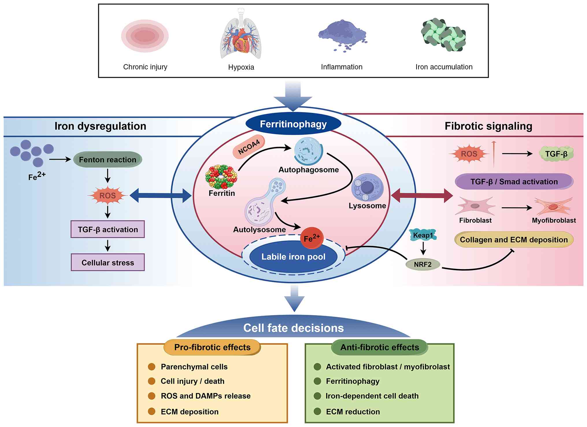
for developing anti-fibrotic strategies (Fig. 1).
Ferritin represents a key intracellular form of iron
storage and serves a central role in maintaining iron homeostasis
by reversibly storing and releasing iron ions to regulate
intracellular iron levels (23,24). Structurally, ferritin forms a
hollow spherical complex composed of 24 subunits, including
ferritin heavy chain (FTH1) and ferritin light chain (FTL). FTH1
possesses ferroxidase activity that catalyzes the conversion of
Fe2+ into the more stable Fe3+ form, whereas
FTL promotes iron core formation and maintains the structural
stability of the ferritin complex (25,26). The coordinated actions of FTH1
and FTL enable ferritin to safely store large quantities of iron
while allowing dynamic iron release when required by the cell,
thereby maintaining intracellular iron homeostasis while preventing
free iron toxicity.
Previous research has shown that the function of the
lysosomal system is important for ferritin degradation and iron
excretion (27,28). In 2014, Mancias et al
(29) first proposed the concept
of ferritinophagy. The authors found that NCOA4 was the main
receptor for ferritin phagocytosis during ferritin engulfment and
that ferritin was delivered to autolysosomes after being recognized
by NCOA4 to fuse with lysosomes to decompose ferritin and release
iron ions. NCOA4 exists as two isoforms, NCOA4α (70 kDa) and NCOA4β
(287 amino acids), both of which share an N-terminal coiled-coil
domain and a portion of the C-terminal region. Notably, the
C-terminal domain of NCOA4α contains binding sites for FTH1 and the
E3 ubiquitin ligase HECT and RLD domain containing E3 ubiquitin
protein ligase 2 (HERC2) (6,29-31). NCOA4 binds to FTH1 through its
C-terminal domain and promotes the directed transport of ferritin
complexes into lysosomes for degradation (32,33). Previous studies regarding
structural biology have shown that selective recognition of NCOA4
for binding to FTH1 is based on the presence of 16 highly conserved
residues in the FTH1 surface, including K180, R183 and D186. Each
ferritin molecule can bind ≤24 NCOA4 molecules (29-31); thus, due to its numerous binding
capabilities, ferritin can be more easily recognized and degraded
in the autophagy pathway. In particular, the NCOA4-FTH1 interaction
is dynamically modulated by intracellular iron content: In an
iron-deficient environment, their affinity rises and ferritin
degrades to liberate iron ions, whereas, under iron-replete
conditions, their binding weakens and the ferritin degradation rate
decreases to reduce free iron accumulation. NCOA4 exhibits a direct
interaction with Golgi-associated ATPase enhancer of 16 kDa
(GATE16)/LC3 family members through two short linear domains at its
C-terminus to anchor ferritin particles onto the autophagosome
membrane (34). These two motifs
exhibit a weak binding affinity for GATE16; however, acting
together, they can achieve a higher degree of binding that allows
NCOA4 to accurately distinguish between the inactive soluble state
of LC3/GABA type A receptor-associated protein and the active
conjugate at the membrane-bound ferritin complex, thus ensuring the
precise delivery of the ferritin complex (35).
When NCOA4 expression is reduced, intracellular iron
release becomes restricted, leading cells to increase iron uptake
and sensing through the upregulation of transferrin receptor 1
(TfR1) and iron regulatory protein 1 (IRP1), reflecting a
compensatory response to insufficient iron supply (36,37). Ferritinophagy is not
constitutively active but is precisely regulated by intracellular
iron levels, forming a dynamic negative feedback loop. Under
iron-replete conditions, the E3 ubiquitin ligase HERC2 recognizes
and binds NCOA4, mediating its K48-linked ubiquitination and
subsequent degradation through the proteasomal pathway, thereby
inhibiting ferritin degradation and limiting excessive iron
release. In 2025, Liu et al (38) elucidated the underlying molecular
mechanism of this regulatory process, revealing that NCOA4 senses
intracellular iron levels by binding a [2Fe-2S] iron-sulfur cluster
through its hybrid-binding domain (38). NCOA4 bound to the iron-sulfur
cluster efficiently binds the cysteine-, proline- and
histidine-rich domain and IRP2-binding domain of HERC2 through a
bivalent mode, whereas NCOA4, lacking the cluster, binds HERC2 only
weakly through a monovalent interaction. Consequently, under
iron-deficient conditions, NCOA4 stability increases, promoting
ferritin degradation and iron release, while, under iron-replete
conditions, NCOA4 is efficiently degraded by HERC2, thereby
inhibiting ferritinophagy to prevent iron overload (29,33). Meanwhile, the classical
IRP1/IRP2-iron-responsive element (IRE) regulatory system also
contributes to maintaining iron homeostasis, primarily by
regulating ferritin synthesis. Under iron-deficient conditions,
IRP1 binds to IREs and inhibits ferritin translation; by contrast,
when intracellular iron levels increase, IRP2 is degraded through
F-box and leucine-rich repeat protein 5-mediated pathways, thereby
relieving the inhibition of ferritin translation (39-41) (Fig. 2). Collectively, NCOA4
specifically recognizes FTH1 through its C-terminal domain to
achieve selective capture of ferritin, binds GATE16/LC3 through
short linear motifs to complete targeted delivery of ferritin to
the autophagic-lysosomal system and senses iron levels through its
[2Fe-2S] cluster to form a negative feedback loop mediated by
HERC2. The coordinated action of these mechanisms establishes NCOA4
as a central regulatory molecule in ferritinophagy. However, the
recognition of ferritin by NCOA4 represents only the initiation
phase of ferritinophagy and subsequent degradation still depends on
the collaborative action of the autophagic execution machinery.
Following NCOA4-mediated cargo recognition, ferritin
is routed through the canonical autophagy-lysosome pathway for
degradation. The execution of ferritinophagy shares the core
autophagic machinery with bulk autophagy, encompassing initiation,
isolation membrane expansion, autophagosome closure and lysosomal
fusion (Fig. 3). Autophagy
initiation is governed by cellular energy status: Under
nutrient-rich conditions, mTOR suppresses autophagy, whereas under
energy stress, AMP-activated protein kinase (AMPK) activates the
UNC-51-like kinase 1/2 (ULK1/2)-autophagy-related protein
(ATG)-13-FAK family kinase-interacting protein of 200 kDa complex,
which subsequently recruits the PI3K complex to generate
phosphatidylinositol-3-phosphate, nucleating the isolation membrane
and mediating lipid delivery to the autophagosomal membrane
(42-47). Subsequently, the
ATG5-ATG12-ATG16L complex facilitates LC3 lipidation to LC3-II,
which anchors onto the autophagosomal membrane to drive its
expansion and closure (48-53). ATG9 and ATG2 further contribute
to membrane lipid transfer and expansion, thereby supporting
autophagosome expansion and closure and ensuring the efficient
engulfment of the NCOA4-ferritin complex for subsequent lysosomal
degradation (54-57).
Once the NCOA4-ferritin complex has been loaded into
the autophagosome, this matures and subsequently fuses with
lysosomes to form autolysosomes. The autophagy adaptor protein
p62/SQSTM1 participates in regulating NCOA4 and facilitates the
localization of the complex to autophagosomes (58). The endosomal sorting complex
required for transport (ESCRT) machinery subsequently mediates the
closure of the autophagosomal membrane (59-61). In parallel, syntaxin 17 and
synaptosomal-associated protein 29 interact with the lysosomal
membrane proteins, vesicle-associated membrane protein 7/8
(VAMP7/8), to form the soluble N-ethylmaleimide-sensitive factor
attachment protein receptor complex, thereby promoting fusion
between autophagosomes and lysosomes (62-64). Ras-related protein Rab-7a (Rab7)
further facilitates autophagosome maturation and lysosomal
trafficking by recruiting effector proteins, such as WD repeat
domain 91 and Rab-interacting lysosomal protein (65,66). Following the fusion of
autophagosomes with lysosomes, the acidic lysosomal environment
promotes ferritin degradation and iron release, thereby completing
the execution stage of ferritinophagy (Fig. 3).
Ferritinophilic degradation of ferroptosis
regulation is regulated at the level of a set of fundamental
elements to maintain iron balance in various states. Nuclear factor
erythroid 2-related factor 2 (NRF2) is an antioxidant-response
regulator promoting cells to defend against oxidative damage
through the regulation of ferritinophagy. Previous research has
indicated that NRF2 may enhance cell antioxidant-defense mechanisms
through activation and stabilization of NCOA4, thus inducing
ferritin degradation (67).
Under conditions of oxidative stress or toxicity exposure, cells
can dissociate NRF2 from Kelch-like ECH-associated protein 1
(Keap1), move into the nucleus and activate the expression of
numerous antioxidant-associated genes, such as heme oxygenase-1,
NAD(P)H quinone dehydrogenase 1 and glutamate-cysteine ligase
modifier subunit (68-71). This transcriptional activation
not only improves the antioxidant capacity of cells, but also
inhibits the stability of NCOA4, inhibits ferritin degradation and
releases fewer free iron ions, thus reducing the risk of iron
overload and ferroptosis and stabilizing cellular redox state and
iron homeostasis. Anandhan et al (72) found that NRF2 regulates
ferritinophagy through HERC2 and VAMP8, with HERC2 participating in
NCOA4 degradation and VAMP8 involved in autophagosome-lysosome
fusion (72).
Complementary to the action of NRF2,
hypoxia-inducible factor 1 (HIF-1) also serves a central role in
ferritinophagy, particularly under hypoxic conditions. Previous
studies have shown that, under hypoxia, HIF-1 influences
ferritinophagy by regulating NCOA4 expression: Enhanced stability
of HIF-1 increases NCOA4 mRNA levels, promoting ferritin
degradation and iron release (10,73). Furthermore, HIF-1α and HIF-2α
bind to hypoxia response elements (HREs) in the NCOA4 promoter,
further enhancing NCOA4 transcription, leading to markedly
increased ferritinophagy and iron release (74). Through this mechanism, HIF-1
promotes ferritin degradation and iron release under hypoxic
conditions to support cellular energy metabolism and other
physiological processes, such as erythropoiesis, mitochondrial
respiration and DNA synthesis (75). In addition to influencing the
levels of NCOA4, HIF-1 can affect iron uptake or storage in cells
through modulation of cell-level changes in intracellular iron
balance. Hypoxia-stimulated expression of HIF-1 causes ferritin
degradation in order to release more iron ions for cellular
utilization as an energy source. HIF-1 and NRF2 may interact to
regulate iron use inside cells under low-oxygen environments to
keep the homeostasis of the cell.
In addition to NRF2 and HIF-1, it has been reported
that the JNK/Jun proto-oncogene (c-Jun) signalling pathway directly
modulates NCOA4 transcription. JNK, one of the members of the MAPK
family, is activated upon various stimulatory factors, including
oxidative stress, inflammatory cytokines and iron overload.
Activated JNK phosphorylates c-Jun at Ser63 and Ser73 to promote
its nuclear transfer. Therefore, it enhances the transcriptional
activity of c-Jun. Nuclear-localized activated c-Jun specifically
binds to activator protein-1-binding sites in the NCOA4 promoter
region and activates transcription by increasing the expression of
NCOA4, thus elevating ferritinophagy-associated proteins, such as
LC3-II and p62/SQSTM1 (76). As
shown by this regulatory axis in numerous disease types, including
osteoarthritis, under light-induced retinal degeneration
conditions, all-trans-retinal activates the JNK/c-Jun signalling
pathway to promote NCOA4 expression and then triggers ferroptosis
through ferritinophagy-mediated mechanisms (77). In osteoarthritic chondrocytes,
the JNK inhibitor SP600125 can block c-Jun phosphorylation to
reduce NCOA4 expression, which reduces the ferroptosis-induced
degradation of the ECM (78,79).
Furthermore, the MiT/TFE family transcription factor
transcription factor EB (TFEB) has also been shown to participate
in ferritinophagy regulation. TFEB is a master transcriptional
regulator of the autophagy-lysosome pathway and serves a key role
in lysosomal biogenesis and autophagic flux regulation. TFEB
participates in ferritinophagy regulation through multiple
mechanisms: It directly binds to coordinated lysosomal expression
and regulation elements in the NCOA4 promoter region to upregulate
NCOA4 transcription. Simultaneously, TFEB transcriptionally
activates iron metabolism-related genes such as TfR1, FTH1 and FTL,
promoting autophagosome-lysosome fusion by regulating
vesicle-associated membrane protein 8 (VAMP8) expression, thereby
ensuring the smooth completion of the ferritinophagy process
(80-82) (Fig. 4). Notably, TFEB exerts fine-tuned
balancing regulation in iron metabolism: On the one hand, it
promotes ferritin degradation through NCOA4 upregulation to release
iron ions, whereas, on the other hand, it enhances
transferrin-mediated iron uptake by upregulating TfR1 and promoting
lysosomal iron storage, while simultaneously increasing ferritin
synthesis to safely store excess iron ions, collectively
maintaining the intracellular LIP at a low level.
In summary, NRF2 and HIF-1 serve central roles
through transcriptional regulation under oxidative stress and
hypoxic conditions, whereas JNK/c-Jun and TFEB complement the
transcriptional regulatory network of ferritinophagy with regard to
stress signal response and autophagy-lysosomal regulation. These
multi-layered regulatory mechanisms coordinate with one another to
collectively maintain iron homeostasis under numerous physiological
conditions and to ensure normal cellular metabolic activities.
Fibrosis is a slowly progressive disease that
ultimately leads to tissue degeneration and can have marked
consequences in multiple organs, including the heart, lung, liver,
kidney and skin (83). It is
characterized by excessive accumulation of fibrous connective
tissue within ECM regions of damaged tissues, with collagen
(particularly type I and III collagen) being the key component of
fibrotic scar tissue. As the disease progresses, tissues remain in
a persistent state of abnormal repair, ultimately resulting in
disruption of organ architecture and functional impairment
(84). Tissue injury activates
both the innate and adaptive immune systems and modulates the
magnitude and direction of inflammatory responses (85). When injurious stimuli such as
chronic inflammation, hypoxia, oxidative stress or metabolic
abnormalities persist, the repair program shifts towards
pathological fibrotic remodeling (14,86), during which inflammatory cells
release profibrotic mediators and cytokines that interact with
oxidative stress and other factors, initiating fibroblast
activation and driving sustained fibrotic progression (87,88).
During the process of fibrosis, due to
differentiation, myofibroblast cells exhibit increased secretion
ability, contraction force and ECM production (86). Previous single-cell sequencing
studies have shown that myofibroblasts display a markedly diverse
gene-expression pattern and evolve dynamically in the context of
fibrosis across numerous organ types (89,90). The differentiation trajectories
of fibroblasts vary across organs: In pulmonary fibrosis, the
differentiation paths of mesenchymal cells differ markedly between
normal and fibrotic states (91), while in the liver, hepatic
stellate cells (HSCs) serve as the principal pro-fibrotic cell type
(92,93). Notably, myofibroblasts can arise
from fibroblasts in other parts of the body. Immune cell
contributions, epithelial-mesenchymal transition (EMT) and
endothelial-mesenchymal transition-induced EMT, have also been
found to increase the number of myofibroblasts and further cause
their activation (94). In
particular, although inhibiting the proliferation and activation of
myofibroblasts is considered a core approach for antifibrosis
treatment, myofibroblasts develop resistance to apoptosis during
differentiation as they mature into fibroblasts, which limits the
effects of programmed-cell-death pathways and may explain why
strategies targeting a reduced number of myofibroblasts fail alone
(95).
During sustained activation of myofibroblasts,
numerous signaling pathways participate in the regulation of
fibrotic responses, among which TGF-β signaling is widely regarded
as the central molecular axis driving fibrosis progression. TGF-β
promotes fibrotic responses through both canonical and
non-canonical signaling pathways (96).
In the canonical Smad signaling pathway, TGF-β first
binds to the TGF-β type II receptor (TGFBR2) on the cell membrane,
inducing the recruitment and phosphorylation of the TGF-β type I
receptor (TGFBR1). Activated TGFBR1 subsequently phosphorylates
Smad2 and Smad3, which form a complex with Smad4 and translocate
into the nucleus to initiate the transcription of pro-fibrotic
target genes such as collagens and α-smooth muscle actin (α-SMA)
(97,98). Smad7 functions as a key negative
feedback regulator by competitively binding to TGFBR1 and
recruiting ubiquitin ligases to promote TGFBR1 degradation, thereby
terminating signal transduction (99-101).
In addition to the classical Smad axis, TGF-β can
also activate a number of non-canonical signaling pathways that are
independent of Smad protein phosphorylation and synergistically
promote fibrotic responses through other signal transduction routes
(102). Among these, the
PI3K/Akt pathway promotes fibroblast survival and myofibroblast
transformation (103,104), while the MAPK pathway regulates
fibroblast proliferation and migration (105,106) and the Janus kinase (JAK)/STAT
pathway enhances inflammatory cytokine release and myofibroblast
differentiation (107,108) (Fig. 5). In summary, TGF-β directly
initiates pro-fibrotic gene transcription through the canonical
Smad pathway while synergistically enhancing fibroblast activation,
proliferation and survival through non-canonical pathways such as
PI3K/Akt, MAPK and JAK/STAT, thereby forming a multi-dimensional,
positive-feedback pro-fibrotic signaling network.
Previous research has identified an oxidative
stress-induced pathogenesis that leads to fibrotic conditions
through certain mechanisms. ROS is not only the downstream effector
of TGF-β to stimulate fibroblast proliferation, but latent forms of
TGF-β are also activated and form an additive-positive-feedback
cycle that promotes persistent growth of fibrotic tissues.
TGF-β-induced oxidative stress primarily arises from the activation
of the NADPH oxidase (Nox) family. Among these, NADPH oxidase 4
(Nox4) has been found to be key in initiating such processes
(109,110). TGF-β1 stimulation can greatly
enhance the level of Nox4 expression in fibroblasts. Furthermore,
it also produces hydrogen peroxide by oxidizing this receptor
through reversible oxidation of its cysteine residue to modulate
pathways subsequently (111).
Previous research has shown that TGF-β1 treatment can enhance the
expression of Nox4 and α-SMA in primary human cardiac fibroblasts.
On the other hand, after removing Nox4 from fibroblasts, a decrease
in TGF-β1-triggered α-SMA production is observed, indicating that
ROS serves as a mediator for this change (112).
ROS-TGF-β bidirectionally regulated the expression
of target genes through a positive-feedback regulatory loop to
sustain fibrotic damage: On the one hand, TGF-β can stimulate the
expression of ROS production through Nox4 and inhibit antioxidant
defense systems to disrupt the balance of intracellular redox
potential (113). On the other
hand, ROS is also activated by several pathways to release latent
TGF-β. ROS can directly oxidize the cysteine residues in the
latency-associated peptide (LAP) of the latent TGF-β complex to
form a conformational change and release free TGF-β. Alternatively,
ROS activates MMPs and promotes TGF-β activity through a
proteolytic pathway (114). In
addition, ROS can activate non-canonically activated TGF-β signal
transduction pathways through the activation of MAPK and PI3K/Akt
to regulate the transcriptional activity of TGF-β1 (115). The positive feedback mechanism
continuously strengthens the fibrotic signal in the local
microenvironment and may become self-perpetuating even in the
absence of continued stimulation.
Collectively, this ROS-TGF-β feedforward loop
constitutes a self-reinforcing network that sustains fibrotic
signaling and ECM deposition. Since ferritinophagy directly
modulates Fe2+-driven ROS generation through
NCOA4-mediated ferritin degradation, this loop provides a direct
molecular entry point for ferritinophagy into the fibrotic
regulatory network. By tuning the LIP, ferritinophagy influences
the intensity of this ROS-TGF-β loop, exerting bidirectional
effects depending on cell type and disease stage, which will be
systematically discussed in subsequent sections with organ-specific
evidence.
Existing evidence has suggested that dysregulated
iron metabolism and iron deposition are common features within the
fibrotic microenvironment across numerous organs. As a key
intracellular pathway responsible for mobilizing stored iron,
ferritinophagy may expand the LIP and amplify oxidative stress,
thereby coordinating inflammatory responses and profibrotic
signaling networks to promote ECM deposition and tissue remodeling
(116,117).
Disruption of iron homeostasis in fibrotic tissues
typically presents as increased iron accumulation, abnormal
expression of iron transport-associated genes or proteins and
elevated concentrations of free iron. Collectively, these changes
lead to an increase in the volume of labile iron inside cells
(116,118). In conditions of iron
overloading, Fe2+ serves as a substrate in the Fenton
reaction to generate ROS continuously through oxidation (119). Persistent oxidative stress is
considered a key feature of the fibrotic microenvironment and can
interact with inflammatory responses, causing activation of
TGF-β/SMAD signaling and fibroblast activation, thus ultimately
driving excessive ECM deposition (117,118).
With regard to pathological circuits, parenchymal
cells are more susceptible to injury and death under conditions of
disrupted iron metabolism and oxidative stress. Chronic injury and
persistent inflammation provide long-term stimuli that promote
fibrosis. Dampened by damaged parenchymal cells releasing
damage-associated molecular patterns (DAMPs) that activate immune
cells and promote the release of profibrotic mediators to
accelerate fibrosis development. For example, in liver fibrosis,
long-term hepatic injury causes the release of DAMPs and ROS by
hepatocytes, which activates Kupffer cells, leading to increased
production of profibrotic mediators such as TGF-β and
platelet-derived growth factor and thus promotes the activation
state of HSCs and excessive deposition of ECM (120,121). Therefore, iron dyshomeostasis
can be considered one of its early components. It increases
oxidative stress and induces a strong cellular stress response,
triggering an inflammatory reaction and pro-fibrogenic signaling
pathways to maintain a persistent amplification state and create
conditions for iron-related autophagy processes within the system
of fibrotic regulation network (122). Given these circumstances,
whether cells can mobilize available iron stores to form a more
stable LIP to alleviate prolonged oxidative damage needs to be
evaluated. Ferritinophagy functions as one such execution mechanism
(123).
The core mechanism of ferritinophagy involves
NCOA4-mediated selective autophagic degradation of ferritin. NCOA4
binds to the FTH1 and delivers ferritin to autophagosomes, where it
is subsequently degraded in lysosomes to release Fe2+
(124). Once released into the
LIP, Fe2+ can generate hydroxyl radicals through the
Fenton reaction and enhance lipid peroxidation, thereby
demonstrating an association between ferritin turnover and
oxidative damage at the molecular level (117,125).
In the context of stress associated with fibrosis,
this process is particularly important given that ferritinophagy
directly influences the size of the LIP. Expansion of the LIP
increases the probability of iron-dependent ROS generation,
transforming oxidative stress from a transient event into a more
sustained state and thereby providing continuous driving forces for
inflammatory responses and tissue remodeling. In addition, previous
studies have indicated that ferritinophagy activity is dynamically
regulated by cellular iron status and exhibits cross-regulation
with mTOR signaling, core autophagy proteins and the NRF2
antioxidant axis (43,58,72). Notably, the role of autophagy
itself in fibrosis remains controversial, with evidence supporting
both pro-fibrotic (such as autophagy-driven epithelial injury in
the kidney) and anti-fibrotic (such as autophagy-mediated
hepatocyte protection) functions, largely depending on cell type
and tissue context. These findings suggest that the influence of
ferritinophagy on oxidative stress is not fixed but rather depends
on the cellular iron load and stress regulatory status (72,126). This provides a framework for
understanding how ferritinophagy-mediated ferroptosis may be either
detrimental or protective.
Within the fibrotic signaling network, activation of
TGF-β signaling and the differentiation of fibroblasts into
myofibroblasts represent one of the classical central axes.
Existing studies have indicated that enhanced ferritinophagy
promotes Fe2+ release and increases ROS levels. ROS
subsequently activates TGF-β signaling pathways, thereby inducing
fibroblast activation, collagen deposition and fibrotic
progression, establishing a close connection between iron
homeostasis dysregulation and classical profibrotic signaling
networks (117,119,124).
Meanwhile, the NRF2-mediated antioxidant pathway
serves a key balancing role within this regulatory network. The
NRF2 antioxidant pathway counteracts this ROS-TGF-β loop by
limiting labile iron expansion and attenuating TGF-β signaling
activation (72,126,127). Consequently, NRF2 signaling
activity is considered an important determinant of the functional
direction of ferritinophagy, as its regulatory status determines
whether ferritinophagy tends to amplify tissue injury or maintain
redox homeostasis.
Conversely, chronic inflammation and persistent
oxidative stress within the fibrotic microenvironment may also
regulate autophagy and iron metabolic reprogramming. DAMPs released
from injured cells enhance immune responses and stimulate TGF-β
expression, accelerating ECM deposition and causing ferritinophagy
to exert different effects at distinct stages of disease
progression (117). During the
early stage of injury, excessive ferritinophagy in parenchymal
cells amplifies tissue injury and profibrotic transformation
through ROS and TGF-β signaling. By contrast, during chronic
stages, induction of iron-dependent cell death in activated
fibroblast populations may reduce the number of pathological
ECM-producing cells and thereby limit fibrotic progression
(121,124). Collectively, the association
between ferritinophagy and TGF-β signaling is not a simple linear
pathway but rather a bidirectionally coupled regulatory module
within the inflammatory and oxidative stress network that is
modulated by the NRF2 antioxidant axis.
By modulating the LIP and redox status,
ferritinophagy reshapes the cellular fate decision network and
exerts bidirectional effects within the fibrotic microenvironment.
In parenchymal cells (such as epithelial cells, renal tubular
epithelial cells, cardiomyocytes and hepatocytes), enhanced
ferritinophagy leads to elevated Fe2+ levels and ROS
generation, predisposing cells to oxidative damage and cell death
(123,128). Damaged or dying cells release
DAMPs and pro-inflammatory factors, such as interleukin (IL)-1β,
IL-6 and tumor necrosis factor-α, which activate immune cells and
amplify profibrotic signals, thereby promoting fibroblast
activation and ECM deposition (117).
Conversely, in activated fibroblasts and
myofibroblasts (which are the primary cellular sources of ECM),
ferritinophagy-associated iron-dependent cell death may exert a
potential antifibrotic effect by reducing the number of effector
cells that continuously produce ECM (129,130). Emerging evidence has suggested
that different cell types exhibit markedly distinct responses to
alterations in iron homeostasis: Enhanced ferritinophagy tends to
exacerbate injury and inflammation in parenchymal cells, whereas,
in overactivated fibroblast populations, it may facilitate
pathological cell clearance, thereby limiting the progression of
fibrosis (127) (Fig. 6). Thus, the role of
ferritinophagy in fibrosis is not unidirectional but exhibits clear
cell type- and disease stage-dependent characteristics. Its overall
impact on the fibrotic process depends on the target cell type, the
stage of the disease and the dynamic balance between oxidative
stress levels and antioxidant regulatory networks.
The biological outcomes of ferritinophagy are
notably context dependent. In parenchymal cells, excessive
ferritinophagy promotes iron release, oxidative stress and
ferroptotic injury, thus accelerating fibrotic remodeling. In
activated fibroblasts, however, sustained ferritinophagy may
increase ferroptotic vulnerability and limit pathological ECM
deposition (133). To reconcile
these opposing effects, three-tiered framework can be proposed: i)
Tier I: Stress sensing, wherein hypoxia, oxidative stress and
inflammation converge on HIF-1α, NRF2 and TFEB to regulate NCOA4;
ii) tier II: Ferritinophagy flux, releasing Fe2+ into
the LIP; and iii) tier III: Effector outputs, driving ROS
generation, TGF-β activation and ferroptosis, with net outcome
determined by cellular antioxidant capacity (Fig. 7). Notably, nearly all current
evidence derives from experimental models, whereas clinical data
from human fibrosis remain scarce. Whether ferritinophagy operates
similarly in human disease awaits investigation using
patient-derived samples, spatial transcriptomics and
cell-type-specific genetic approaches.
In pulmonary fibrosis, particularly idiopathic
pulmonary fibrosis, iron deposition and iron metabolic
reprogramming are considered important contributors to sustained
tissue injury (134,135). Based on current evidence,
ferritinophagy is not just involved in maintaining iron homeostasis
but may participate in pulmonary fibrotic remodeling by regulating
intracellular iron availability. NCOA4-mediated ferritinophagy
promotes ferritin degradation, releasing Fe2+, expanding
the LIP and enhancing lipid peroxidation, thereby rendering
alveolar epithelial cells more susceptible to oxidative damage and
iron-dependent cell death. Experimental inhibition of NCOA4
expression or ferritinophagy activity has been reported to reduce
epithelial cell death and collagen deposition in fibrotic lung
models, supporting a pathogenic role for excessive
ferritinophagy-associated iron mobilization in pulmonary fibrosis
(136,137). Epithelial injury further
triggers inflammatory responses and amplifies TGF-β signaling,
driving persistent fibroblast ECM deposition (123,135,138). Collectively, pulmonary fibrosis
can be understood as a metabolic dysregulation state in which
ferritinophagy represents an important regulatory mechanism
associating iron dyshomeostasis with inflammatory amplification and
fibrotic remodeling. However, whether ferritinophagy acts as an
initiating driver of pulmonary fibrosis or functions primarily as
an amplifier of established injury responses remains to be fully
elucidated. Therapeutically, recent studies are shifting from
targeting terminal fibrotic responses toward modulating upstream
metabolic events associated with ferritinophagy. Reducing labile
iron levels or inhibiting lipid peroxidation alleviates pulmonary
fibrosis in experimental models, suggesting that interventions
targeting ferritinophagy-mediated iron release may have potential
value (135,139). Importantly, ferritinophagy
exhibits marked cell context dependence. In alveolar epithelial
cells, its enhancement often promotes injury progression, whereas
moderate induction of iron-dependent death in activated fibroblasts
may limit pathological matrix accumulation (140). Therefore, the key to future
antifibrotic strategies lies not in simply inhibiting or activating
ferritinophagy, but in achieving cell-type and disease stage
specific precision regulation. Based on this perspective,
ferritinophagy is more likely to function as a regulatory node in
antifibrotic therapy rather than as a single therapeutic
target.
Renal fibrosis typically arises during the
progression of chronic kidney injury, with key pathological events
including prolonged exposure of renal tubular epithelial cells to
oxidative stress and inflammatory stimuli, leading to a profibrotic
phenotype. Previous research has shown that chronic kidney injury
is accompanied by notable iron metabolism dysregulation and
intracellular iron overload, with elevated labile iron levels
contributing to sustained tissue injury (141). Ferritinophagy, through
NCOA4-mediated ferritin degradation and Fe2+ release,
may continually expand the LIP, thereby enhancing lipid
peroxidation, inflammatory responses, mitochondrial dysfunction and
metabolic reprogramming (142,143). During this process, injured
renal tubular epithelial cells become a persistent source of
proinflammatory and profibrotic signals, driving the activation of
interstitial fibroblasts and ECM deposition. However, whether
ferritinophagy directly mediates renal fibrogenesis or represents a
secondary response to chronic kidney injury remains to be
clarified. Ferritinophagy may therefore represent a potential
association between iron dysregulation and renal fibrotic
progression (144,145).
Given the important function of labile iron in
maintaining oxidative stress, recently, there have been numerous
studies (142,143,145) regarding modulating renal
fibrosis by intervening in iron metabolism. Regulating iron
metabolism may alleviate tubular injury and interstitial fibrosis,
although the specific contribution of ferritinophagy modulation
remains incompletely understood (141). Current perspectives emphasize
fine-tuning ferritinophagy activity rather than completely
inhibiting it, aiming to maintain iron availability while
preventing excessive oxidative damage (143,145,146). Further research is needed to
study the functional transformation of ferritinophagy in different
renal cells and stages of disease to provide a theoretical basis
for stage-specific intervention.
Hepatic fibrosis is an injury-driven chronic
pathological process that features persistent inflammation,
oxidative stress and the activation of HSCs and results in ECM
accumulation. Dysregulated iron metabolism exists in numerous
etiologies of liver fibrosis and the extent of hepatic iron
deposition is closely associated with the severity of fibrosis
(147-149). Ferritinophagy, through
NCOA4-mediated selective ferritin degradation and Fe2+
release into the LIP, represents a key mechanism regulating hepatic
labile iron levels. Enhanced ferritinophagy increases
Fe2+ availability and promotes the Fenton reaction,
which produces ROS and causes lipid peroxidation, further
activating the TGF-β/Smad pathway to drive HSCs into the
myofibroblast phenotype (150,151). Simultaneously, iron-dependent
oxidative damage kills hepatocytes and releases inflammatory
mediators that stimulate Kupffer cells, creating an ongoing
pro-fibrotic microenvironment. However, whether this iron release
oxidative stress inflammation amplification cascade is directly
controlled by ferritinophagy remains unclear. Ferritinophagy may
therefore represent a potential metabolic regulator linking iron
dyshomeostasis and hepatic fibrosis progression, although direct
causal evidence remains limited (152).
Ferritinophagy during hepatic fibrosis has a marked
cell-type specificity. In hepatocytes, ferritinophagy-mediated
increase of labile iron enhances oxidative injury and inflammation,
which leads to fibrosis. In activated HSCs, elevated iron-dependent
lipid peroxidation may cause cell death and decrease ECM
production, indicating that ferritinophagy participates in both
fibrotic progression and reversal. However, direct experimental
evidence demonstrating cell-specific effects of ferritinophagy in
hepatic fibrosis remains insufficient (149,153). Reflecting the bidirectionality
of this regulation, existing research has begun to move away from
broad-spectrum antioxidants towards specific iron balance and
ferroptosis points. Reducing the iron load or inhibiting lipid
peroxidation can mitigate hepatic fibrosis and linking autophagy
regulation with antioxidant systems to regulate the stability of
glutathione peroxidase 4 (GPX4) and ferroptosis sensitivity is
regarded as a prospective treatment option (120). Consequently, ferritinophagy is
more likely to be regarded as a key node controlling the
reversibility of hepatic fibrosis, with future studies aiming to
focusing on cell-type- and stage-specific interventions to balance
parenchymal cell protection with removal of pathological HSCs.
Cardiac fibrosis frequently occurs during tissue
remodeling following myocardial infarction or other injuries, with
the core pathology involving persistent inflammation and
interstitial remodeling induced by cardiomyocyte death. Previous
studies have demonstrated that myocardial injury is accompanied by
enhanced iron-dependent lipid peroxidation, and intracellular
Fe2+ accumulation combined with impaired GPX4
antioxidant defense promotes ferroptosis (154-156). Although these findings
highlight the importance of iron-dependent oxidative damage in
cardiac remodeling, direct evidence to demonstrate the association
between NCOA4-mediated ferritinophagy and cardiac fibrosis remains
limited. In this context, ferritinophagy may modulate cardiomyocyte
susceptibility to oxidative stress by regulating ferritin
degradation and labile iron availability. When
ferritinophagy-mediated iron release exceeds cellular antioxidant
capacity, lipid peroxidation is further amplified, promoting
ferroptotic cardiomyocyte death. Injured cardiomyocytes
subsequently release inflammatory signals that paracrinally induce
fibroblast activation, increasing α-SMA expression and collagen
deposition, thereby driving cardiac structural remodeling (124). Downregulation of antioxidant
enzymes such as glutathione S-transferase Mu 1 exacerbates lipid
peroxidation and ferroptosis, whereas inhibition of iron-dependent
oxidative damage notably alleviates myocardial fibrosis, indicating
that ferritinophagy contributes to regulating cardiomyocyte death
thresholds and serves as a key link between metabolic stress and
cardiac remodeling (157).
Based on such mechanistic explorations,
interventions targeting iron-dependent oxidative stress have been
studied as possible anti-fibrotic strategies for the heart. The
findings revealed that ferroptosis inhibitors lessen collagen
accumulation and enhance cardiac function, implying that iron
homeostasis regulation may exert therapeutic effects in the
remodeling period (155).
However, whether selective modulation of ferritinophagy can provide
therapeutic benefits beyond general iron or ferroptosis regulation
remains unclear. The role of ferritinophagy in the heart is to
maintain the balance between life and death of cardiomyocytes,
rather than merely influencing inflammation or preserving chronic
injury. Therefore, in the future, research could further study the
cross-talk mechanism of ferritinophagy and antioxidant systems,
clarify its functional changes at different time points after
injury and identify precise intervention measures to protect
cardiomyocytes while restraining excessive interstitial
remodeling.
Systemic scleroderma is an autoimmune disease where
fibrosis normally happens above ongoing immune events, with the
primary driver being the chronically maintained inflammatory
microenvironment rather than acute parenchymal cell injury.
Previous research has shown that there is widespread iron
metabolism disorder, increased lipid peroxidation and antioxidant
system damage and macrophages serve an important role in iron
homeostasis reprogramming (158). The impact of ferritinophagy on
macrophages exhibits notable inflammatory stage-dependent and
subset-specific characteristics. During acute/early inflammation,
macrophages polarize toward the M1 phenotype, which is accompanied
by elevated LC3B expression, reduced FTH1 levels and enhanced
NCOA4-LC3B colocalization, indicating ferritinophagy activation
(159). Lysine
acetyltransferase 2A (KAT2A) promotes M1 polarization and
inflammatory cytokine release by activating cyclic GMP-AMP synthase
(cGAS)-STING signaling through NCOA4-mediated ferritinophagy
(160). In the chronic stage,
M2 macrophages display increased susceptibility to ferroptosis,
which is regulated by ferritinophagy through the NRF2/ERK pathway.
Suppression of ferritinophagy alleviates M2 macrophage ferroptosis
and attenuates liver injury (161). Thus, cGAS-STING signaling is
primarily involved in the early/acute stage, whereas the NRF2/ERK
pathway is involved in the chronic stage (161-163). Ferritinophagy, through the
regulation of ferritin degradation and labile iron release, changes
the content of free iron in immune cells, thus affecting its
sensitivity to lipid peroxidation and iron-dependent cell death.
The bleomycin-induced systemic sclerosis model has exhibited
upregulation of acyl-CoA synthetase long-chain family member 4
(ACSL4), which promotes polyunsaturated lipid remodeling and
enhances lipid peroxidation, accompanied by macrophage iron
accumulation and ferroptosis increase, forming a metabolic loop to
maintain inflammation and amplify fibrotic signals (158). Ferritinophagy-mediated iron
release may influence fibrosis indirectly through the remodeling of
immune cell metabolism, which promotes fibroblasts activation and
collagen deposition.
Recent research has focused on immune-metabolites
and not simply activated fibroblasts. ACSL4 inhibition decreases
iron and lipid peroxidation and markedly improves tissue fibrosis,
suggesting the possibility of targeting iron homeostasis networks
in therapy. In autoimmune-associated fibrosis, ferritinophagy may
work through influencing the inflammatory microenvironment without
directly determining the extent of tissue damage (164). Future studies should aim to
delineate the differential regulatory mechanisms of ferritinophagy
in M1 vs. M2 macrophages, identify the functional switch points
through which ferritinophagy transitions from promoting M1
polarization in early inflammation to modulating M2 ferroptosis in
chronic stages and further explore novel anti-fibrotic strategies
targeting NCOA4 or associated signaling cascades (such as
cGAS-STING and NRF2/ERK) to achieve immunometabolic
reprogramming.
In malignant tumors, fibrosis typically appears as
tumor-associated stroma remodeling rather than classical organ
scarring and the continuous activation of cancer-associated
fibroblasts (CAFs) and abnormal ECM deposition are its basic
characteristics. Previous studies have shown that iron metabolism
disorder and increased lipid peroxidation exist in the tumor
microenvironment, affecting both the survival of tumor cells and
the function of stromal cells (17,165). Ferritinophagy, through the
regulation of ferritin degradation and labile iron, may regulate
iron homeostasis in the tumor microenvironment and affect cellular
sensitivity to iron-dependent oxidative injury. Current evidence
indicates that iron-dependent cell death may affect CAF activity
and ECM deposition, indicating that the iron regulatory network is
key in tumor stroma construction (166,167). However, whether ferritinophagy
directly controls CAF activation or ECM remodeling remains largely
unexplored. Therefore, ferritinophagy may be involved in shaping
the tumor stroma by regulating the microenvironmental iron content
rather than by directly causing damage to a specific cell type.
With the increasing understanding of tumor
microenvironment metabolism, certain previous studies (17,165,166) have focused on the impact that
modulation of iron homeostasis has on stroma remodeling and
therapy. Regulating iron metabolism and lipid peroxidation would
change fibroblast function and ECM structure, which suggests that
ferritinophagy-associated pathway may be a new method to regulate
tumor microenvironment (168).
However, direct evidence regarding ferritinophagy in the tumor
stroma is limited and the specific cell types and time evolution
have not been thoroughly clarified. Future research combining
spatial omics technology and microenvironment analysis is needed to
systematically clarify the dynamic regulatory associations of
ferritinophagy among tumor, immune and stromal cells and thus
provide a basis for combinatorial strategies targeting
microenvironment remodeling in both cancer treatment and fibrosis
suppression (Table I).
Overall, current evidence suggests that, although
ferritinophagy is important for maintaining iron homeostasis, it
actually serves as a central metabolic node that regulates the
balance between tissue damage and repair (117,169). NCOA4 mediates ferritin
degradation, converting stored iron to labile iron, which changes
redox balance and cell fate (4,32,170,171). Under conditions of chronic
injury or sustained metabolic stress, increased labile iron
promotes lipid peroxidation and maintains oxidative stress in an
autoamplified network with inflammatory mediators and profibrotic
signals such as TGF-β. This drives tissue repair progressively away
from regenerative processes toward fibrotic remodeling. This way,
ferritinophagy may serve as an upstream checkpoint that influences
whether fibrosis is initiated and which pathogenic program
subsequently follows, rather than functioning merely as a
downstream bystander. Importantly, ferritinophagy exhibits dual
effects on fibrosis (154).
This bidirectional effect is cell type- and disease
stage-dependent. In parenchymal cells, including alveolar
epithelial cells, renal tubular epithelial cells, hepatocytes and
cardiomyocytes, enhanced ferritinophagy during the early injury
phase increases labile iron and ROS, exacerbating oxidative damage
and inflammation and promoting fibrotic initiation (36,142). By contrast, in activated
effector cells, such as myofibroblasts, HSCs and CAFs,
ferritinophagy during the chronic phase may induce iron-dependent
cell death, reducing ECM-producing cells and potentially limiting
fibrosis progression (149).
Macrophages also exhibit stage-specific responses: ferritinophagy
promotes M1 polarization during acute inflammation through the
KAT2A-cGAS-STING axis, whereas it regulates M2 ferroptosis during
chronic inflammation through the NRF2/ERK pathway. A closer
examination of this 'dual-role' paradigm, however, reveals numerous
key issues that warrant further elaboration. The mechanistic basis
of this divergence lies in three interconnected variables: i)
Baseline antioxidant buffering capacity; ii) lipid peroxidation
susceptibility (such as ACSL4 expression) (172,173) and ferroportin-mediated iron
efflux, all of which differ markedly across cell lineages; and iii)
disease stages. Methodologically, the majority of previous evidence
derives from broad-spectrum ferroptosis inhibitors or global NCOA4
ablation, which are approaches that cannot dissect cell-autonomous
ferritinophagy effects from secondary inflammatory modulation.
Macrophage NCOA4 deletion, for instance, may indirectly alter
fibroblast behavior through cytokine changes (159). The therapeutic assumption that
myofibroblast ferroptosis is uniformly beneficial is also tempered
by the observation that ferroptotic debris can activate toll-like
receptor 4 signaling, potentially offsetting the benefit of ECM
clearance. Furthermore, nearly all data originate from acute animal
models, leaving it uncertain whether ferritinophagy operates
similarly in human fibrosis, which is a process spanning decades
amid comorbidities and medications. In addition, ferritinophagy
flux reporters have yet to be validated in human tissues (174-176). Collectively, these
considerations suggest that the net effect of ferritinophagy should
be viewed not as a binary switch but as a threshold-governed
rheostat, whose direction depends on the integrated cellular stress
response and redox equilibrium, a nuance that calls for
quantitative, spatiotemporally resolved investigations in future
studies.
Despite the aforementioned caveats, cross-organ
evidence indicates that iron metabolic re-programming is nearly
ubiquitous in fibrotic disease and, thus, ferritinophagy may
provide a common metabolic basis for fibrosis (128). However, its biological outcomes
are highly affected by the tissue-specific cell lineages and
microenvironment context. This is particularly evident for NCOA4:
In the kidney, NCOA4 drives tubulointerstitial fibrosis, whereas in
the liver, NCOA4-mediated ferritinophagy promotes hepatic stellate
cell senescence and alleviates fibrosis, while, in the heart and
lung, NCOA4 upregulation exacerbates fibrotic remodeling. This
indicates that ferroptosis induction through ferritinophagy can
have opposite outcomes depending on the organ. In pulmonary, renal
and cardiac fibrosis, ferritinophagy is mainly associated with
parenchymal cell damage and inflammatory amplification. Hepatic
fibrosis participates in hepatocyte damage and HSC fate
decision-making, affecting the reversibility of fibrosis.
Ferritinophagy mainly alters the immune metabolic state in
autoimmune and tumor-associated fibrosis, thus influencing the
formation of matrix, which shows that ferritinophagy is an adaptive
expression of the common metabolic strategy among different tissue
ecosystems. Besides organ-specific differences, disease stage also
decides ferritinophagy function (177). Early in the injury,
ferritinophagy enhancement increases oxidative stress and initiates
fibrosis formation. During the chronic phase, if the majority of
iron-dependent damage occurs on pathological effector cells, then
ferritinophagy can decrease abnormal matrix accumulation and impact
results.
The aforementioned hierarchical framework positions
ferritinophagy as an attractive upstream checkpoint for
antifibrotic intervention. However, clinical translation faces key
challenges. Systemic toxicity is a primary concern, as global NCOA4
inhibition may impair erythropoiesis and mitochondrial function,
necessitating cell-type-selective delivery strategies (178). The therapeutic window is also
stage-dependent, since attenuating ferritinophagy may protect
parenchymal cells in early injury, whereas activating it could
eliminate pathological myofibroblasts in established fibrosis.
Pharmacologically, although preclinical agents targeting the
NCOA4-FTH1 interaction (such as compound 9a, a first-in-class
inhibitor that directly binds NCOA4383-522 to disrupt
ferritin binding and reduce labile iron) or modulating
ferritinophagy (such as curcumol, which exerts bidirectional
effects depending on cell type or schisandrin B, an NCOA4-mediated
ferritinophagy inducer that promotes activated HSC senescence) have
shown efficacy, to the best of our knowledge, none of them have
advanced to clinical trials for fibrosis (129,179). Beyond these, a
proteolysis-targeting chimera-based NCOA4 degrader (V3) has
recently been developed as a tool compound for ferritinophagy
inhibition, while natural products such as morroniside and
daphnetin have been identified as selective ferritinophagy
activators that promote ferroptosis in activated HSCs to alleviate
liver fibrosis in preclinical models (180-182). In addition, ferroptosis
inhibitors have been shown to reduce collagen accumulation and
improve cardiac function after myocardial infarction, indicating
that inhibition of iron-dependent oxidative damage can also be
therapeutically beneficial (183). Future efforts should aim to
prioritize cell-type-selective nanocarriers, patient stratification
based on iron overload status and combinatorial regimens that
integrate ferritinophagy modulation with antioxidant or
TGF-β-targeted therapies (184,185). Emerging tools such as spatial
transcriptomics and ferritinophagy flux reporters will be key in
defining the optimal intervention window.
Ferritophagy is important in connecting metabolic
pathways, including iron metabolism, cellular stress response and
tissue remodeling and its functionality relies on different cell
types and disease states. The spatiotemporal regulation of this
process may provide a novel understanding of the commonalities
among multiple organ fibrosis and set forth a next-generation
anti-fibrotic approach.
Ferritinophagy is a key way of controlling iron in
cells and its association with iron dyshomeostasis, oxidative
stress and multiple organ fibrosis, as well as cell fate change.
Ferritinophagy is known to participate in both initiating and
developing fibrosis; it can also influence the reversibility of
tissue remodeling through altering effector cell survival, which
makes ferritinophagy have a dual effect, stage-specificity and
cellular-type-dependent nature. Combining ferritinophagy with the
classical profibrotic signaling pathway provides a metabolic
perspective for the reinterpretation of fibrosis and lays the
theoretical basis for precise regulation of iron homeostasis in
future antifibrosis strategies.
Not applicable.
All authors contributed to the present review. SS,
XL, JY and HL were responsible for study conceptualization. SS, XL
and JY were responsible for original manuscript draft preparation.
XL and HL were responsible for writing, reviewing and editing the
manuscript. SS was responsible for visualization, including the
design and preparation of the figures and schematic illustrations.
All authors read and approved the final version of the manuscript.
Data authentication is not applicable.
Not applicable.
Not applicable.
The authors declare that they have no competing
interests.
Not applicable.
The present study was supported by the National Natural Science
Foundation of China (grant no. 82560214).
|
1
|
Fu P, Chen Y, Wu M, Bao B, Yin X, Chen Z
and Zhang M: Effect of ferroptosis on chronic cerebral
hypoperfusion in vascular dementia. Exp Neurol. 370:1145382023.
View Article : Google Scholar : PubMed/NCBI
|
|
2
|
Dixon SJ and Olzmann JA: The cell biology
of ferroptosis. Nat Rev Mol Cell Biol. 25:424–442. 2024. View Article : Google Scholar : PubMed/NCBI
|
|
3
|
Dixon SJ, Lemberg KM, Lamprecht MR, Skouta
R, Zaitsev EM, Gleason CE, Patel DN, Bauer AJ, Cantley AM, Yang WS,
et al: Ferroptosis: An iron-dependent form of nonapoptotic cell
death. Cell. 149:1060–1072. 2012. View Article : Google Scholar : PubMed/NCBI
|
|
4
|
Santana-Codina N, Gikandi A and Mancias
JD: The role of NCOA4-Mediated ferritinophagy in ferroptosis. Adv
Exp Med Biol. 1301:41–57. 2021. View Article : Google Scholar : PubMed/NCBI
|
|
5
|
Zhang M, Cui Y, Jia R, Cai Z, Shen Y, Yu
W, Pang Z, Ding S, Gao X, Huang Y, et al: Hesperidin alleviated
dendritic spines through inhibiting ferritinophagy via HERC2-NCOA4
ubiquitination in CUMS mice. Phytomedicine. 135:1561322024.
View Article : Google Scholar : PubMed/NCBI
|
|
6
|
Wu H, Liu Q, Shan X, Gao W and Chen Q: ATM
orchestrates ferritinophagy and ferroptosis by phosphorylating
NCOA4. Autophagy. 19:2062–2077. 2023. View Article : Google Scholar : PubMed/NCBI
|
|
7
|
Dai Y, Zhu C, Xiao W, Huang K, Wang X, Shi
C, Lin D, Zhang H, Liu X, Peng B, et al: Mycobacterium tuberculosis
hijacks host TRIM21- and NCOA4-dependent ferritinophagy to enhance
intracellular growth. J Clin Invest. 133:e1599412023. View Article : Google Scholar : PubMed/NCBI
|
|
8
|
Huang G, Cai Y, Ren M, Zhang X, Fu Y,
Cheng R, Wang Y, Miao M, Zhu L and Yan T: Salidroside sensitizes
Triple-negative breast cancer to ferroptosis by SCD1-mediated
lipogenesis and NCOA4-mediated ferritinophagy. J Adv Res.
74:589–607. 2025. View Article : Google Scholar :
|
|
9
|
Zhu M, Peng L, Huo S, Peng D, Gou J, Shi
W, Tao J, Jiang T, Jiang Y, Wang Q, et al: STAT3 signaling promotes
cardiac injury by upregulating NCOA4-mediated ferritinophagy and
ferroptosis in high-fat-diet fed mice. Free Radic Biol Med.
201:111–125. 2023. View Article : Google Scholar : PubMed/NCBI
|
|
10
|
Galy B, Conrad M and Muckenthaler M:
Mechanisms controlling cellular and systemic iron homeostasis. Nat
Rev Mol Cell Biol. 25:133–155. 2024. View Article : Google Scholar
|
|
11
|
Fang Y, Chen X, Tan Q, Zhou H, Xu J and Gu
Q: Inhibiting ferroptosis through disrupting the NCOA4-FTH1
interaction: A new mechanism of action. ACS Cent Sci. 7:980–989.
2021. View Article : Google Scholar : PubMed/NCBI
|
|
12
|
Li K, Chen B, Xu A, Shen J, Li K, Hao K,
Hao R, Yang W, Jiang W, Zheng Y, et al: TRIM7 modulates
NCOA4-mediated ferritinophagy and ferroptosis in glioblastoma
cells. Redox Biol. 56:1024512022. View Article : Google Scholar : PubMed/NCBI
|
|
13
|
Savin IA, Zenkova MA and Sen'kova AV:
Pulmonary fibrosis as a result of acute lung inflammation:
Molecular mechanisms, relevant in vivo models, prognostic and
therapeutic approaches. Int J Mol Sci. 23:149592022. View Article : Google Scholar : PubMed/NCBI
|
|
14
|
Antar SA, Ashour NA, Marawan ME and
Al-Karmalawy AA: Fibrosis: Types, effects, markers, mechanisms for
disease progression, and its relation with oxidative stress,
immunity, and inflammation. Int J Mol Sci. 24:40042023. View Article : Google Scholar : PubMed/NCBI
|
|
15
|
Suri GS, Kaur G, Jha CK and Tiwari M:
Understanding idiopathic pulmonary fibrosis - Clinical features,
molecular mechanism and therapies. Exp Gerontol. 153:1114732021.
View Article : Google Scholar : PubMed/NCBI
|
|
16
|
Wang H, Jiang C, Yang Y, Li J, Wang Y,
Wang C and Gao Y: Resveratrol ameliorates iron overload induced
liver fibrosis in mice by regulating iron homeostasis. PeerJ.
10:e135922022. View Article : Google Scholar : PubMed/NCBI
|
|
17
|
Peng D, Fu M, Wang M, Wei Y and Wei X:
Targeting TGF-β signal transduction for fibrosis and cancer
therapy. Mol Cancer. 21:1042022. View Article : Google Scholar
|
|
18
|
Ko T, Nomura S, Yamada S, Fujita K, Fujita
T, Satoh M, Oka C, Katoh M, Ito M, Katagiri M, et al: Cardiac
fibroblasts regulate the development of heart failure via
Htra3-TGF-β-IGFBP7 axis. Nat Commun. 13:32752022. View Article : Google Scholar
|
|
19
|
Song Y, Wei J, Li R, Fu R, Han P, Wang H,
Zhang G, Li S, Chen S, Liu Z, et al: Tyrosine kinase receptor B
attenuates liver fibrosis by inhibiting TGF-β/SMAD signaling.
Hepatology. 78:1433–1447. 2023. View Article : Google Scholar : PubMed/NCBI
|
|
20
|
Milara J, Ballester B, Safont MJ, Artigues
E, Escrivá J, Morcillo E and Cortijo J: MUC4 is overexpressed in
idiopathic pulmonary fibrosis and collaborates with transforming
growth factor β inducing fibrotic responses. Mucosal Immunol.
14:377–388. 2021. View Article : Google Scholar
|
|
21
|
Di X, Li Y, Wei J, Li T and Liao B:
Targeting fibrosis: From molecular mechanisms to advanced
therapies. Adv Sci (Weinh). 12:e24104162025. View Article : Google Scholar :
|
|
22
|
Zhong Y, Wei B, Wang W, Chen J, Wu W,
Liang L, Huang XR, Szeto CC, Yu X, Nikolic-Paterson DJ and Lan HY:
Single-Cell RNA-Sequencing identifies bone marrow-derived
progenitor cells as a main source of extracellular matrix-producing
cells across multiple organ-based fibrotic diseases. Int J Biol
Sci. 20:5027–5042. 2024. View Article : Google Scholar : PubMed/NCBI
|
|
23
|
Li JY, Feng YH, Li YX, He PY, Zhou QY,
Tian YP, Yao RQ and Yao YM: Ferritinophagy: A novel insight into
the double-edged sword in ferritinophagy-ferroptosis axis and human
diseases. Cell Prolif. 57:e136212024. View Article : Google Scholar : PubMed/NCBI
|
|
24
|
Knovich MA, Storey JA, Coffman LG, Torti
SV and Torti FM: Ferritin for the clinician. Blood Rev. 23:95–104.
2009. View Article : Google Scholar
|
|
25
|
Pantopoulos K, Porwal SK, Tartakoff A and
Devireddy L: Mechanisms of mammalian iron homeostasis.
Biochemistry. 51:5705–5724. 2012. View Article : Google Scholar : PubMed/NCBI
|
|
26
|
Wu K, Zhao W, Hou Z, Zhang W, Qin L, Qiu
J, Wang D, Zhuang L, Xue X and Sun D: Ferritinophagy: Multifaceted
roles and potential therapeutic strategies in liver diseases. Front
Cell Dev Biol. 13:15510032025. View Article : Google Scholar : PubMed/NCBI
|
|
27
|
Kidane TZ, Sauble E and Linder MC: Release
of iron from ferritin requires lysosomal activity. Am J Physiol
Cell Physiol. 291:C445–C455. 2006. View Article : Google Scholar : PubMed/NCBI
|
|
28
|
Santana-Codina N, Del Rey MQ, Kapner KS,
Zhang H, Gikandi A, Malcolm C, Poupault C, Kuljanin M, John KM,
Biancur DE, et al: NCOA4-Mediated Ferritinophagy Is a pancreatic
cancer dependency via maintenance of iron bioavailability for
iron-sulfur cluster proteins. Cancer Discov. 12:2180–2197. 2022.
View Article : Google Scholar : PubMed/NCBI
|
|
29
|
Mancias JD, Wang X, Gygi SP, Harper JW and
Kimmelman AC: Quantitative proteomics identifies NCOA4 as the cargo
receptor mediating ferritinophagy. Nature. 509:105–109. 2014.
View Article : Google Scholar : PubMed/NCBI
|
|
30
|
Le Y, Liu Q, Yang Y and Wu J: The emerging
role of nuclear receptor coactivator 4 in health and disease: A
novel bridge between iron metabolism and immunity. Cell Death
Discov. 10:3122024. View Article : Google Scholar : PubMed/NCBI
|
|
31
|
Ohshima T, Yamamoto H, Sakamaki Y, Saito C
and Mizushima N: NCOA4 drives ferritin phase separation to
facilitate macroferritinophagy and microferritinophagy. J Cell
Biol. 221:e2022031022022. View Article : Google Scholar : PubMed/NCBI
|
|
32
|
Zhao H, Lu Y, Zhang J, Sun Z, Cheng C, Liu
Y, Wu L, Zhang M, He W, Hao S and Li K: NCOA4 requires a [3Fe-4S]
to sense and maintain the iron homeostasis. J Biol Chem.
300:1056122024. View Article : Google Scholar : PubMed/NCBI
|
|
33
|
Mancias JD, Pontano Vaites L, Nissim S,
Biancur DE, Kim AJ, Wang X, Liu Y, Goessling W, Kimmelman AC and
Harper JW: Ferritinophagy via NCOA4 is required for erythropoiesis
and is regulated by iron dependent HERC2-mediated proteolysis.
Elife. 4:e103082015. View Article : Google Scholar : PubMed/NCBI
|
|
34
|
Lee A and Davis JH: NCOA4 initiates
ferritinophagy by binding GATE16 using two highly avid short linear
interaction motifs. bioRxiv [Preprint].2024.06.09.597909. 2024.
|
|
35
|
Gryzik M, Srivastava A, Longhi G, Bertuzzi
M, Gianoncelli A, Carmona F, Poli M and Arosio P: Expression and
characterization of the ferritin binding domain of Nuclear Receptor
Coactivator-4 (NCOA4). Biochim Biophys Acta Gen Subj.
1861:2710–2716. 2017. View Article : Google Scholar : PubMed/NCBI
|
|
36
|
Zhao H, Wang Z and Wang H: The role of
NCOA4-mediated ferritinophagy in the ferroptosis of hepatocytes: A
mechanistic viewpoint. Pathol Res Pract. 270:1559962025. View Article : Google Scholar : PubMed/NCBI
|
|
37
|
Bellelli R, Federico G, Matte A, Colecchia
D, Iolascon A, Chiariello M, Santoro M, De Franceschi L and
Carlomagno F: NCOA4 deficiency impairs systemic iron homeostasis.
Cell Rep. 14:411–421. 2016. View Article : Google Scholar : PubMed/NCBI
|
|
38
|
Liu H, Shen L, Gong X, Zhou X, Huang Y,
Zhou Y, Guo Z, Guo H, Wang S and Pan L: Mechanistic insights into
the iron-sulfur cluster-dependent interaction of the autophagy
receptor NCOA4 with the E3 ligase HERC2. Proc Natl Acad Sci USA.
122:e25102691222025. View Article : Google Scholar : PubMed/NCBI
|
|
39
|
Lee A and Davis JH: NCOA4 initiates
ferritinophagy by avidly binding GATE16 using two short linear
interaction motifs. Life Sci Alliance. 9:e2025036112026. View Article : Google Scholar : PubMed/NCBI
|
|
40
|
Tong WH and Rouault TA: Metabolic
regulation of citrate and iron by aconitases: Role of iron-sulfur
cluster biogenesis. Biometals. 20:549–564. 2007. View Article : Google Scholar : PubMed/NCBI
|
|
41
|
Zhang DL, Ghosh MC and Rouault TA: The
physiological functions of iron regulatory proteins in iron
homeostasis - an update. Front Pharmacol. 5:1242014. View Article : Google Scholar : PubMed/NCBI
|
|
42
|
Kim DH: Contrasting views on the role of
AMPK in autophagy. Bioessays. 46:e23002112024. View Article : Google Scholar : PubMed/NCBI
|
|
43
|
Bao L, Zhao C, Feng L, Zhao Y, Duan S, Qiu
M, Wu K, Zhang N, Hu X and Fu Y: Ferritinophagy is involved in
Bisphenol A-induced ferroptosis of renal tubular epithelial cells
through the activation of the AMPK-mTOR-ULK1 pathway. Food Chem
Toxicol. 163:1129092022. View Article : Google Scholar : PubMed/NCBI
|
|
44
|
Nähse V, Raiborg C, Tan KW, Mørk S,
Torgersen ML, Wenzel EM, Nager M, Salo VT, Johansen T, Ikonen E, et
al: ATPase activity of DFCP1 controls selective autophagy. Nat
Commun. 14:40512023. View Article : Google Scholar : PubMed/NCBI
|
|
45
|
Nähse V, Stenmark H and Schink KO:
Omegasomes control formation, expansion, and closure of
autophagosomes. Bioessays. 46:e24000382024. View Article : Google Scholar : PubMed/NCBI
|
|
46
|
Barboza E, Hudson J, Chang WP, Kovats S,
Towner RA, Silasi-Mansat R, Lupu F, Kent C and Griffin TM:
Profibrotic infrapatellar fat pad remodeling without M1 macrophage
polarization precedes knee osteoarthritis in mice with diet-induced
obesity. Arthritis Rheumatol. 69:1221–1232. 2017. View Article : Google Scholar : PubMed/NCBI
|
|
47
|
Gaugel A, Bakula D, Hoffmann A and
Proikas-Cezanne T: Defining regulatory and phosphoinositide-binding
sites in the human WIPI-1 β-propeller responsible for
autophagosomal membrane localization downstream of mTORC1
inhibition. J Mol Signal. 7:162012. View Article : Google Scholar
|
|
48
|
Rao S, Skulsuppaisarn M, Strong LM, Ren X,
Lazarou M, Hurley JH and Hummer G: Three-step docking by WIPI2,
ATG16L1, and ATG3 delivers LC3 to the phagophore. Sci Adv.
10:eadj80272024. View Article : Google Scholar : PubMed/NCBI
|
|
49
|
Gubas A, Attridge E, Jefferies HB,
Nishimura T, Razi M, Kunzelmann S, Gilad Y, Mercer TJ, Wilson MM,
Kimchi A and Tooze SA: WIPI2b recruitment to phagophores and
ATG16L1 binding are regulated by ULK1 phosphorylation. EMBO Rep.
25:3789–3811. 2024. View Article : Google Scholar : PubMed/NCBI
|
|
50
|
Jensen LE, Rao S, Schuschnig M, Cada AK,
Martens S, Hummer G and Hurley JH: Membrane curvature sensing and
stabilization by the autophagic LC3 lipidation machinery. Sci Adv.
8:eadd14362022. View Article : Google Scholar : PubMed/NCBI
|
|
51
|
Wilson MI, Dooley HC and Tooze SA: WIPI2b
and Atg16L1: Setting the stage for autophagosome formation. Biochem
Soc Trans. 42:1327–1334. 2014. View Article : Google Scholar : PubMed/NCBI
|
|
52
|
Noda T and Yoshimori T: Molecular basis of
canonical and bactericidal autophagy. Int Immunol. 21:1199–1204.
2009. View Article : Google Scholar : PubMed/NCBI
|
|
53
|
Mizushima N: The ATG conjugation systems
in autophagy. Curr Opin Cell Biol. 63:1–10. 2020. View Article : Google Scholar : PubMed/NCBI
|
|
54
|
Obara K, Sekito T, Niimi K and Ohsumi Y:
The Atg18-Atg2 complex is recruited to autophagic membranes via
phosphatidylinositol 3-phosphate and exerts an essential function.
J Biol Chem. 283:23972–23980. 2008. View Article : Google Scholar : PubMed/NCBI
|
|
55
|
Osawa T and Noda NN: Atg2: A novel
phospholipid transfer protein that mediates de novo autophagosome
biogenesis. Protein Sci. 28:1005–1012. 2019. View Article : Google Scholar : PubMed/NCBI
|
|
56
|
Wei B, Fu Y, Li X, Chen F, Zhang Y, Chen
H, Tong M, Li L, Pan Y, Zhang S, et al: ANKFY1 bridges
ATG2A-mediated lipid transfer from endosomes to phagophores. Cell
Discov. 10:432024. View Article : Google Scholar : PubMed/NCBI
|
|
57
|
Javed R, Mari M, Trosdal E, Duque T,
Paddar MA, Allers L, Mudd MH, Claude-Taupin A, Akepati PR, Hendrix
E, et al: ATG9A facilitates the closure of mammalian
autophagosomes. J Cell Biol. 224:e2024040472025. View Article : Google Scholar : PubMed/NCBI
|
|
58
|
Liu L, Zheng B, Luo M, Du J, Yang F, Huang
C, Ma Z, Li C, Guo D and Peng H: Suppression of USP8 sensitizes
cells to ferroptosis via SQSTM1/p62-mediated ferritinophagy.
Protein Cell. 14:230–234. 2023.PubMed/NCBI
|
|
59
|
Jiang W, Chen X, Ji C, Zhang W, Song J, Li
J and Wang J: Key regulators of autophagosome closure. Cells.
10:28142021. View Article : Google Scholar : PubMed/NCBI
|
|
60
|
Hamamoto K, Liang X, Ito A, Lanza M, Bui
V, Zhang J, Opozda DM, Hattori T, Chen L, Haddock D, et al:
Unveiling the physiological impact of ESCRT-dependent autophagosome
closure by targeting the VPS37A ubiquitin E2 variant-like domain.
Cell Rep. 43:1150162024. View Article : Google Scholar : PubMed/NCBI
|
|
61
|
Lefebvre C, Legouis R and Culetto E: ESCRT
and autophagies: Endosomal functions and beyond. Semin Cell Dev
Biol. 74:21–28. 2018. View Article : Google Scholar
|
|
62
|
Zheng D, Tong M, Zhang S, Pan Y, Zhao Y,
Zhong Q and Liu X: Human YKT6 forms priming complex with STX17 and
SNAP29 to facilitate autophagosome-lysosome fusion. Cell Rep.
43:1137602024. View Article : Google Scholar : PubMed/NCBI
|
|
63
|
Ke PY: Molecular mechanism of
autophagosome-lysosome fusion in mammalian cells. Cells.
13:5002024. View Article : Google Scholar : PubMed/NCBI
|
|
64
|
Tian X, Teng J and Chen J: New insights
regarding SNARE proteins in autophagosome-lysosome fusion.
Autophagy. 17:2680–2688. 2021. View Article : Google Scholar :
|
|
65
|
Xing R, Zhou H, Jian Y, Li L, Wang M, Liu
N, Yin Q, Liang Z, Guo W and Yang C: The Rab7 effector WDR91
promotes autophagy-lysosome degradation in neurons by regulating
lysosome fusion. J Cell Biol. 220:e2020070612021. View Article : Google Scholar : PubMed/NCBI
|
|
66
|
Ao X, Zou L and Wu Y: Regulation of
autophagy by the Rab GTPase network. Cell Death Differ. 21:348–358.
2014. View Article : Google Scholar : PubMed/NCBI
|
|
67
|
Dong Y, Zheng M, Ding W, Guan H, Xiao J
and Li F: Nrf2 activators for the treatment of rare iron overload
diseases: From bench to bedside. Redox Biol. 81:1035512025.
View Article : Google Scholar : PubMed/NCBI
|
|
68
|
Fan Z, Wirth AK, Chen D, Wruck CJ, Rauh M,
Buchfelder M and Savaskan N: Nrf2-Keap1 pathway promotes cell
proliferation and diminishes ferroptosis. Oncogenesis. 6:e3712017.
View Article : Google Scholar : PubMed/NCBI
|
|
69
|
Suzuki T, Motohashi H and Yamamoto M:
Toward clinical application of the Keap1-Nrf2 pathway. Trends
Pharmacol Sci. 34:340–346. 2013. View Article : Google Scholar : PubMed/NCBI
|
|
70
|
Han Y, Gao X, Wu N, Jin Y, Zhou H, Wang W,
Liu H, Chu Y, Cao J, Jiang M, et al: Long noncoding RNA LINC00239
inhibits ferroptosis in colorectal cancer by binding to Keap1 to
stabilize Nrf2. Cell Death Dis. 13:7422022. View Article : Google Scholar : PubMed/NCBI
|
|
71
|
Li Y, Xu B, Ren X, Wang L, Xu Y, Zhao Y,
Yang C, Yuan C, Li H, Tong X, et al: Inhibition of CISD2 promotes
ferroptosis through ferritinophagy-mediated ferritin turnover and
regulation of p62-Keap1-NRF2 pathway. Cell Mol Biol Lett.
27:812022. View Article : Google Scholar : PubMed/NCBI
|
|
72
|
Anandhan A, Dodson M, Shakya A, Chen J,
Liu P, Wei Y, Tan H, Wang Q, Jiang Z, Yang K, et al: NRF2 controls
iron homeostasis and ferroptosis through HERC2 and VAMP8. Sci Adv.
9:eade95852023. View Article : Google Scholar : PubMed/NCBI
|
|
73
|
Wu Y, Li Z, Ding T, Yang Y, Wei C, Zhang S
and Fan X: Bidirectional regulation of neuronal autophagy in
ischemic stroke: Mechanisms and therapeutic potential. Ageing Res
Rev. 111:1028422025. View Article : Google Scholar : PubMed/NCBI
|
|
74
|
Li X, Lozovatsky L, Sukumaran A, Gonzalez
L, Jain A, Liu D, Ayala-Lopez N and Finberg KE: NCOA4 is regulated
by HIF and mediates mobilization of murine hepatic iron stores
after blood loss. Blood. 136:2691–2702. 2020.PubMed/NCBI
|
|
75
|
Jin X, Jiang C, Zou Z, Huang H, Li X, Xu S
and Tan R: Ferritinophagy in the etiopathogenic mechanism of
related diseases. J Nutr Biochem. 117:1093392023. View Article : Google Scholar : PubMed/NCBI
|
|
76
|
Fuhrmann DC, Mondorf A, Beifuß J, Jung M
and Brüne B: Hypoxia inhibits ferritinophagy, increases
mitochondrial ferritin, and protects from ferroptosis. Redox Biol.
36:1016702020. View Article : Google Scholar : PubMed/NCBI
|
|
77
|
Yang B, Yang K, Xi R, Li S, Chen J and Wu
Y: Inhibition of JNK signaling attenuates photoreceptor ferroptosis
caused by all-trans-retinal. Free Radic Biol Med. 227:179–189.
2025. View Article : Google Scholar
|
|
78
|
Sun K, Hou L, Guo Z, Wang G, Guo J, Xu J,
Zhang X and Guo F: JNK-JUN-NCOA4 axis contributes to chondrocyte
ferroptosis and aggravates osteoarthritis via ferritinophagy. Free
Radic Biol Med. 200:87–101. 2023. View Article : Google Scholar : PubMed/NCBI
|
|
79
|
Ding N, Zeng MY, Song WJ, Xiao CX, Li EM
and Wei B: SP600125 restored TNF-α-induced impaired chondrogenesis
in bone mesenchymal stem cells and its antiosteoarthritis effect in
mice. Stem Cells Dev. 30:1028–1036. 2021. View Article : Google Scholar : PubMed/NCBI
|
|
80
|
Chen L, Ma Y, Ma X, Liu L, Jv X, Li A,
Shen Q, Jia W, Qu L, Shi L and Xie J: TFEB regulates cellular
labile iron and prevents ferroptosis in a TfR1-dependent manner.
Free Radic Biol Med. 208:445–457. 2023. View Article : Google Scholar : PubMed/NCBI
|
|
81
|
Wang J, Song X, Hui Y, Dong B, Gong J,
Zhao Y, Ji H, Qiu Y, Jiang S, Guo D and Gao X: TFEB orchestrates
ferritinophagy and ferroptosis in ionophore drug-induced
hepatotoxicity: unveiling a novel therapeutic avenue. Free Radic
Biol Med. 236:116–133. 2025. View Article : Google Scholar : PubMed/NCBI
|
|
82
|
Chen J, Li W, Zhang C, Wen D and Jiao C:
Tyrosine phosphatase SHP2 promoted the progression of CRC via
modulating the PI3K/BRD4/TFEB signaling induced ferroptosis. Discov
Oncol. 15:7932024. View Article : Google Scholar : PubMed/NCBI
|
|
83
|
Conte E: Targeting monocytes/macrophages
in fibrosis and cancer diseases: Therapeutic approaches. Pharmacol
Ther. 234:1080312022. View Article : Google Scholar
|
|
84
|
Davidson MD, Burdick JA and Wells RG:
Engineered biomaterial platforms to study fibrosis. Adv Healthc
Mater. 9:e19016822020. View Article : Google Scholar : PubMed/NCBI
|
|
85
|
Prasse A, Pechkovsky DV, Toews GB,
Jungraithmayr W, Kollert F, Goldmann T, Vollmer E, Müller-Quernheim
J and Zissel G: A vicious circle of alveolar macrophages and
fibroblasts perpetuates pulmonary fibrosis via CCL18. Am J Respir
Crit Care Med. 173:781–792. 2006. View Article : Google Scholar : PubMed/NCBI
|
|
86
|
Moon Y, Hong J, Chung J and Baek JH:
Macrophage plasticity and regulatory networks during the transition
from inflammation to fibrosis in the kidney. Life (Basel).
16:522025.
|
|
87
|
Zhao M, Wang L, Wang M, Zhou S, Lu Y, Cui
H, Racanelli AC, Zhang L, Ye T, Ding B, et al: Targeting fibrosis,
mechanisms and cilinical trials. Signal Transduct Target Ther.
7:2062022. View Article : Google Scholar : PubMed/NCBI
|
|
88
|
Al-Hattab DS, Chattopadhyaya S and Czubryt
MP: Canadian contributions in fibroblast biology. Cells.
11:22722022. View Article : Google Scholar : PubMed/NCBI
|
|
89
|
Farbehi N, Patrick R, Dorison A, Xaymardan
M, Janbandhu V, Wystub-Lis K, Ho JW, Nordon RE and Harvey RP:
Single-cell expression profiling reveals dynamic flux of cardiac
stromal, vascular and immune cells in health and injury. Elife.
8:e438822019. View Article : Google Scholar : PubMed/NCBI
|
|
90
|
Tabib T, Morse C, Wang T, Chen W and
Lafyatis R: SFRP2/DPP4 and FMO1/LSP1 define major fibroblast
populations in human skin. J Invest Dermatol. 138:802–810. 2018.
View Article : Google Scholar
|
|
91
|
Xie T, Wang Y, Deng N, Huang G, Taghavifar
F, Geng Y, Liu N, Kulur V, Yao C, Chen P, et al: Single-cell
deconvolution of fibroblast heterogeneity in mouse pulmonary
fibrosis. Cell Rep. 22:3625–3640. 2018. View Article : Google Scholar : PubMed/NCBI
|
|
92
|
Yang W, He H, Wang T, Su N, Zhang F, Jiang
K, Zhu J, Zhang C, Niu K, Wang L, et al: Single-cell transcriptomic
analysis reveals a hepatic stellate cell-activation roadmap and
myofibroblast origin during liver fibrosis in mice. Hepatology.
74:2774–2790. 2021. View Article : Google Scholar : PubMed/NCBI
|
|
93
|
Trivedi P, Wang S and Friedman SL: The
power of plasticity-metabolic regulation of hepatic stellate cells.
Cell Metab. 33:242–257. 2021. View Article : Google Scholar
|
|
94
|
Yuan Q, Tan RJ and Liu Y: Myofibroblast in
kidney fibrosis: Origin, activation, and regulation. Adv Exp Med
Biol. 1165:253–283. 2019. View Article : Google Scholar : PubMed/NCBI
|
|
95
|
Kato K, Logsdon NJ, Shin YJ, Palumbo S,
Knox A, Irish JD, Rounseville SP, Rummel SR, Mohamed M, Ahmad K, et
al: Impaired myofibroblast dedifferentiation contributes to
nonresolving fibrosis in aging. Am J Respir Cell Mol Biol.
62:633–644. 2020. View Article : Google Scholar : PubMed/NCBI
|
|
96
|
Meng XM, Nikolic-Paterson DJ and Lan HY:
TGF-β: the master regulator of fibrosis. Nat Rev Nephrol.
12:325–338. 2016. View Article : Google Scholar : PubMed/NCBI
|
|
97
|
Gong J, Xu J, Zhang J, Shen Y, Sun H and
Chen B: Molecular mechanisms of skeletal muscle fibrosis and
potential targeted therapeutic strategies. Front Immunol.
17:17142382026. View Article : Google Scholar : PubMed/NCBI
|
|
98
|
Gu YY, Liu XS, Huang XR, Yu XQ and Lan HY:
Diverse role of TGF-β in kidney disease. Front Cell Dev Biol.
8:1232020. View Article : Google Scholar
|
|
99
|
Yan X and Chen YG: Smad7: Not only a
regulator, but also a cross-talk mediator of TGF-β signalling.
Biochem J. 434:1–10. 2011. View Article : Google Scholar : PubMed/NCBI
|
|
100
|
Hanna A, Humeres C and Frangogiannis NG:
The role of Smad signaling cascades in cardiac fibrosis. Cell
Signal. 77:1098262021. View Article : Google Scholar
|
|
101
|
Gifford CC, Tang J, Costello A, Khakoo NS,
Nguyen TQ, Goldschmeding R, Higgins PJ and Samarakoon R: Negative
regulators of TGF-β1 signaling in renal fibrosis; pathological
mechanisms and novel therapeutic opportunities. Clin Sci (Lond).
135:275–303. 2021. View Article : Google Scholar : PubMed/NCBI
|
|
102
|
Finnson KW, Almadani Y and Philip A:
Non-canonical (non-SMAD2/3) TGF-β signaling in fibrosis: Mechanisms
and targets. Semin Cell Dev Biol. 101:115–122. 2020. View Article : Google Scholar
|
|
103
|
Glaviano A, Foo ASC, Lam HY, Yap KCH,
Jacot W, Jones RH, Eng H, Nair MG, Makvandi P, Geoerger B, et al:
PI3K/AKT/mTOR signaling transduction pathway and targeted therapies
in cancer. Mol Cancer. 22:1382023. View Article : Google Scholar : PubMed/NCBI
|
|
104
|
Wang J, Hu K, Cai X, Yang B, He Q, Wang J
and Weng Q: Targeting PI3K/AKT signaling for treatment of
idiopathic pulmonary fibrosis. Acta Pharm Sin B. 12:18–32. 2022.
View Article : Google Scholar : PubMed/NCBI
|
|
105
|
Lee JH and Massagué J: TGF-β in
developmental and fibrogenic EMTs. Semin Cancer Biol. 86(Pt 2):
136–145. 2022. View Article : Google Scholar : PubMed/NCBI
|
|
106
|
Zhang Y, Jin D, Kang X, Zhou R, Sun Y,
Lian F and Tong X: Signaling pathways involved in diabetic renal
fibrosis. Front Cell Dev Biol. 9:6965422021. View Article : Google Scholar : PubMed/NCBI
|
|
107
|
Montero P, Milara J, Roger I and Cortijo
J: Role of JAK/STAT in interstitial lung diseases; molecular and
cellular mechanisms. Int J Mol Sci. 22:62112021. View Article : Google Scholar : PubMed/NCBI
|
|
108
|
Yuan Q, Tang B and Zhang C: Signaling
pathways of chronic kidney diseases, implications for therapeutics.
Signal Transduct Target Ther. 7:1822022. View Article : Google Scholar : PubMed/NCBI
|
|
109
|
Magnani F and Mattevi A: Structure and
mechanisms of ROS generation by NADPH oxidases. Curr Opin Struct
Biol. 59:91–97. 2019. View Article : Google Scholar : PubMed/NCBI
|
|
110
|
Arendshorst WJ, Vendrov AE, Kumar N,
Ganesh SK and Madamanchi NR: Oxidative stress in kidney injury and
hypertension. Antioxidants (Basel). 13:14542024. View Article : Google Scholar :
|
|
111
|
Cucoranu I, Clempus R, Dikalova A, Phelan
PJ, Ariyan S, Dikalov S and Sorescu D: NAD(P)H oxidase 4 mediates
transforming growth factor-beta1-induced differentiation of cardiac
fibroblasts into myofibroblasts. Circ Res. 97:900–907. 2005.
View Article : Google Scholar : PubMed/NCBI
|
|
112
|
Aminzadehanboohi M, Makridakis M, Rasti D,
Cambet Y, Krause KH, Vlahou A and Jaquet V: Redox mechanisms
driving skin fibroblast-to-myofibroblast differentiation.
Antioxidants (Basel). 14:4862025. View Article : Google Scholar : PubMed/NCBI
|
|
113
|
Sampson N, Berger P and Zenzmaier C: Redox
signaling as a therapeutic target to inhibit myofibroblast
activation in degenerative fibrotic disease. Biomed Res Int.
2014:1317372014. View Article : Google Scholar : PubMed/NCBI
|
|
114
|
Liu RM and Desai LP: Reciprocal regulation
of TGF-β and reactive oxygen species: A perverse cycle for
fibrosis. Redox Biol. 6:565–577. 2015. View Article : Google Scholar : PubMed/NCBI
|
|
115
|
Zhang Y, Zhong Y, Zeng Q and Tang M:
Redox-architected signaling networks in cardiac fibrosis. Biochem
Biophys Res Commun. 781:1525532025. View Article : Google Scholar : PubMed/NCBI
|
|
116
|
Maus M, López-Polo V, Mateo L, Lafarga M,
Aguilera M, De Lama E, Meyer K, Sola A, Lopez-Martinez C,
López-Alonso I, et al: Iron accumulation drives fibrosis,
senescence and the senescence-associated secretory phenotype. Nat
Metab. 5:2111–2130. 2023. View Article : Google Scholar : PubMed/NCBI
|
|
117
|
Hu Y, Huang Y, Zong L, Lin J, Liu X and
Ning S: Emerging roles of ferroptosis in pulmonary fibrosis:
Current perspectives, opportunities and challenges. Cell Death
Discov. 10:3012024. View Article : Google Scholar : PubMed/NCBI
|
|
118
|
Sun Y, Ren Y, Song LY, Wang YY, Li TG, Wu
YL, Li L and Yang ZS: Targeting iron-metabolism:a potential
therapeutic strategy for pulmonary fibrosis. Biomed Pharmacother.
172:1162702024. View Article : Google Scholar : PubMed/NCBI
|
|
119
|
Takahashi M, Mizumura K, Gon Y, Shimizu T,
Kozu Y, Shikano S, Iida Y, Hikichi M, Okamoto S, Tsuya K, et al:
Iron-Dependent mitochondrial dysfunction contributes to the
pathogenesis of pulmonary fibrosis. Front Pharmacol. 12:6439802022.
View Article : Google Scholar : PubMed/NCBI
|
|
120
|
Pan Q, Luo Y, Xia Q and He K: Ferroptosis
and liver fibrosis. Int J Med Sci. 18:3361–3366. 2021. View Article : Google Scholar : PubMed/NCBI
|
|
121
|
Wang Y, Kuang S, Li K, Chen S, Yang M,
Deng K, Li M, Xie S, Chen Q, Wen J, et al: Astragalin promotes HSCs
ferroptosis through NCOA4 mediated ferritinophagy to alleviate
liver fibrosis in zebrafish and mice. Commun Biol. 8:10812025.
View Article : Google Scholar : PubMed/NCBI
|
|
122
|
Li S, Zhang H, Chang J, Li D and Cao P:
Iron overload and mitochondrial dysfunction orchestrate pulmonary
fibrosis. Eur J Pharmacol. 912:1746132021. View Article : Google Scholar : PubMed/NCBI
|
|
123
|
Yuan L, Sun Y, Zhou N, Wu W, Zheng W and
Wang Y: Dihydroquercetin attenuates silica-induced pulmonary
fibrosis by inhibiting ferroptosis signaling pathway. Front
Pharmacol. 13:8456002022. View Article : Google Scholar : PubMed/NCBI
|
|
124
|
Sun Q, Wang M, Liu L, Ding R, Yan K, Liu
S, Ren X, Xu Q, Sun Z, Liu Q, et al: PM(2) (.5) induce
endothelial-mesenchymal transition and cardiac fibrosis via the
NCOA4-Mediated ferritinophagy. Adv Sci (Weinh). 12:e075362025.
View Article : Google Scholar : PubMed/NCBI
|
|
125
|
Shi Y, Shi X, Zhao M and Zhang Y, Zhang Q,
Liu J, Duan H, Yang B and Zhang Y: Ferroptosis is involved in focal
segmental glomerulosclerosis in rats. Sci Rep. 13:222502023.
View Article : Google Scholar : PubMed/NCBI
|
|
126
|
Chen Y, Jiang Z and Li X: New insights
into crosstalk between Nrf2 pathway and ferroptosis in lung
disease. Cell Death Dis. 15:8412024. View Article : Google Scholar : PubMed/NCBI
|
|
127
|
Zeng Q, Wen BB, Liu X, Luo YY, Hu ZG,
Huang L, Zhang XH, Huang XT, Zhou TT, Sang XX, et al: NBR1-p62-Nrf2
mediates the anti-pulmonary fibrosis effects of protodioscin. Chin
Med. 19:602024. View Article : Google Scholar : PubMed/NCBI
|
|
128
|
Gao B, Gong T, Qi Z, Li L, Liu P, Xia R,
Li L and Wang J: Excessive autophagy of myocardial cells promotes
ferroptosis and exacerbates heart failure in the state of
myocardial infarction. Arch Med Sci. 21:944–963. 2024.PubMed/NCBI
|
|
129
|
Zheng Y, Zhao T and Wang J, Jiang R, Huang
J, Li W and Wang J: Curcumol alleviates liver fibrosis through
inducing autophagy and ferroptosis in hepatic stellate cells. FASEB
J. 36:e226652022. View Article : Google Scholar : PubMed/NCBI
|
|
130
|
Zheng Y, Wang L and Wang J, Zhao T and
Wang J: Modulation of the HIF-1α-NCOA4-FTH1 signaling axis
regulating ferroptosis-induced hepatic stellate cell senescence to
explore the anti-hepatic fibrosis mechanism of curcumol. Curr Med
Chem. 31:2821–2837. 2024. View Article : Google Scholar
|
|
131
|
Shi Z, Chen K, Wang Y and Du H: The
crosstalk between ferritinophagy and ferroptosis in ischemic
stroke: regulatory mechanisms and therapeutic implications. Cell
Mol Neurobiol. 45:732025. View Article : Google Scholar : PubMed/NCBI
|
|
132
|
Ning J, Wen L and Qiao L: Ferritinophagy:
Molecular mechanisms and its crosstalk with ferroptosis in chronic
respiratory diseases. Cell Biol Toxicol. 42:312026. View Article : Google Scholar : PubMed/NCBI
|
|
133
|
Chabane T, Bouscary D and Grignano E:
NCOA4 and ferritinophagy in hematological malignancies: A
double-edged regulator of iron metabolism and cell fate. Front
Oncol. 15:17174352026. View Article : Google Scholar : PubMed/NCBI
|
|
134
|
Yao M, Liu Z, Zhao W, Song S, Huang X and
Wang Y: Ferroptosis in idiopathic pulmonary fibrosis: Mechanisms,
impact, and therapeutic opportunities. Front Immunol.
16:15679942025. View Article : Google Scholar : PubMed/NCBI
|
|
135
|
Song L, Gao F and Man J: Ferroptosis: The
potential key roles in idiopathic pulmonary fibrosis. Eur J Med
Res. 30:3412025. View Article : Google Scholar : PubMed/NCBI
|
|
136
|
Zhang J, Du J, Liu D, Zhuo J, Chu L, Li Y,
Gao L, Xu M, Chen W, Huang W, et al: Polystyrene microplastics
induce pulmonary fibrosis by promoting alveolar epithelial cell
ferroptosis through cGAS/STING signaling. Ecotoxicol Environ Saf.
277:1163572024. View Article : Google Scholar : PubMed/NCBI
|
|
137
|
Du J, Yu L, Yang X, Shao F, Xia J, Jin W,
Zhang Y, Lei G, Wang Y, Li Y, et al: Regulation of NCOA4-mediated
iron recycling ameliorates paraquat-induced lung injury by
inhibiting ferroptosis. Cell Commun Signal. 22:1462024. View Article : Google Scholar : PubMed/NCBI
|
|
138
|
Yue D, Zhang Q, Zhang J, Liu W, Chen L,
Wang M, Li R, Qin S, Song X and Ji Y: Diesel exhaust PM2.5 greatly
deteriorates fibrosis process in pre-existing pulmonary fibrosis
via ferroptosis. Environ Int. 171:1077062023. View Article : Google Scholar
|
|
139
|
Feng L, Han S, Li Y, Zhan S, Li L, Wang Y
and Feng J: Roflumilast mitigates idiopathic pulmonary fibrosis via
inhibition of iron accumulation and ferroptosis. Eur J Pharmacol.
1008:1783852025. View Article : Google Scholar : PubMed/NCBI
|
|
140
|
Huang X, Feng J, Xu Q, Tang R, Peng Y,
Yang W, Yang X, Xing S, Qian G, Gao Y, et al: NCOA4-mediated
ferritinophagy induces ferroptosis and enriches ferritin-containing
EVs via ferritin phase separation to promote mechanical
ventilation-induced pulmonary fibrosis. J Adv Res. 82:449–467.
2026. View Article : Google Scholar :
|
|
141
|
Zhou L, Xue X, Hou Q and Dai C: Targeting
ferroptosis attenuates interstitial inflammation and kidney
fibrosis. Kidney Dis (Basel). 8:57–71. 2021. View Article : Google Scholar
|
|
142
|
Jin L, Yu B, Wang H, Shi L, Yang J, Wu L,
Gao C, Pan H, Han F, Lin W, et al: STING promotes ferroptosis
through NCOA4-dependent ferritinophagy in acute kidney injury. Free
Radic Biol Med. 208:348–360. 2023. View Article : Google Scholar : PubMed/NCBI
|
|
143
|
Zhang L, Chen F, Dong J, Wang R, Bi G, Xu
D, Zhang Y, Deng Y, Lin W, Yang Z and Cao W: HDAC3
aberration-incurred GPX4 suppression drives renal ferroptosis and
AKI-CKD progression. Redox Biol. 68:1029392023. View Article : Google Scholar : PubMed/NCBI
|
|
144
|
Liu Z, Zhou S, Wang F, Xie H, Zhang J, Wu
C, Xu D and Zhu Q: C5b-9 promotes ferritinophagy leading to
ferroptosis in renal tubular epithelial cells of
trichloroethylene-sensitized mice. Sci Total Environ.
923:1713782024. View Article : Google Scholar : PubMed/NCBI
|
|
145
|
Li X, Ma TK, Wang P, Shi H, Hai S, Qin Y,
Zou Y, Zhu WT, Li HM, Li YN, et al: HOXD10 attenuates renal
fibrosis by inhibiting NOX4-induced ferroptosis. Cell Death Dis.
15:3982024. View Article : Google Scholar : PubMed/NCBI
|
|
146
|
Li J, Yang J, Zhu B, Fan J, Hu Q and Wang
L: Tectorigenin protects against unilateral ureteral obstruction by
inhibiting Smad3-mediated ferroptosis and fibrosis. Phytother Res.
36:475–487. 2022. View Article : Google Scholar
|
|
147
|
Amengual J, Alay A, Vaquero J,
Gonzalez-Sanchez E, Bertran E, Sánchez A, Herrera B, Meyer K, Maus
M, Serrano M, et al: Iron chelation as a new therapeutic approach
to prevent senescence and liver fibrosis progression. Cell Death
Dis. 15:6802024. View Article : Google Scholar : PubMed/NCBI
|
|
148
|
Li X, Jiang F, Hu Y, Lang Z, Zhan Y, Zhang
R, Tao Q, Luo C, Yu J and Zheng J: Schisandrin B promotes hepatic
stellate cell ferroptosis via wnt pathway-mediated Ly6C(lo)
Macrophages. J Agric Food Chem. Nov 3–2023.Epub ahead of print.
|
|
149
|
Zhu W, Fu L, Cui Y, Tang Y, Liu K, Shi L,
Gao Y, Li M and Huang L: Ferroptosis in liver fibrosis and its
potential intervention strategy. Cell Biol Toxicol. 41:1522025.
View Article : Google Scholar : PubMed/NCBI
|
|
150
|
Bi Y, Liu S, Qin X, Abudureyimu M, Wang L,
Zou R, Ajoolabady A, Zhang W, Peng H, Ren J and Zhang Y: FUNDC1
interacts with GPx4 to govern hepatic ferroptosis and fibrotic
injury through a mitophagy-dependent manner. J Adv Res. 55:45–60.
2024. View Article : Google Scholar :
|
|
151
|
Deng Y, Lu L, Zhu D, Zhang H, Fu Y, Tan Y,
Tan X, Guo M, Zhang Y, Yang H, et al: MafG/MYH9-LCN2 axis promotes
liver fibrosis through inhibiting ferroptosis of hepatic stellate
cells. Cell Death Differ. 31:1127–1139. 2024. View Article : Google Scholar : PubMed/NCBI
|
|
152
|
Yuan S, Wei C, Liu G, Zhang L, Li J, Li L,
Cai S and Fang L: Sorafenib attenuates liver fibrosis by triggering
hepatic stellate cell ferroptosis via HIF-1α/SLC7A11 pathway. Cell
Prolif. 55:e131582022. View Article : Google Scholar
|
|
153
|
Pei Q, Yi Q and Tang L: Liver fibrosis
resolution: From molecular mechanisms to therapeutic opportunities.
Int J Mol Sci. 24:96712023. View Article : Google Scholar : PubMed/NCBI
|
|
154
|
Ning ZH, Wang XH, Zhao Y, Ou Y, Yang JY,
Tang HF and Hu HJ: Ferroptosis in organ fibrosis: Mechanisms and
therapeutic approaches. Int Immunopharmacol. 151:1143412025.
View Article : Google Scholar : PubMed/NCBI
|
|
155
|
Huang X, Song Y, Wei L, Guo J, Xu W and Li
M: The emerging roles of ferroptosis in organ fibrosis and its
potential therapeutic effect. Int Immunopharmacol. 116:1098122023.
View Article : Google Scholar : PubMed/NCBI
|
|
156
|
Cheng P, Wang X, Liu Q, Yang T, Dai E, Sha
W, Qu H and Zhou H: LuQi formula attenuates Cardiomyocyte
ferroptosis via activating Nrf2/GPX4 signaling axis in heart
failure. Phytomedicine. 125:1553572024. View Article : Google Scholar : PubMed/NCBI
|
|
157
|
Chen KJ, Zhang Y, Zhu XY, Yu S, Xie Y, Jin
CJ, Shen YM, Zhou SY, Dai XC, Su SA, et al: GSTM1 suppresses
cardiac fibrosis post-myocardial infarction through inhibiting
lipid peroxidation and ferroptosis. Mil Med Res.
12:262025.PubMed/NCBI
|
|
158
|
Cao D, Zheng J, Li Z, Yu Y, Chen Z and
Wang Q: ACSL4 inhibition prevents macrophage ferroptosis and
alleviates fibrosis in bleomycin-induced systemic sclerosis model.
Arthritis Res Ther. 25:2122023. View Article : Google Scholar : PubMed/NCBI
|
|
159
|
Li J, Wang X, Shi X, Jiang Y and Shen R:
Association between ferritinophagy and M1 macrophage polarization
during the acute phase of childhood allergic asthma. J Inflamm Res.
19:5699562026.PubMed/NCBI
|
|
160
|
Zuo Z, Gao S, Wang J, Liu Z, Lu Z, Wu H,
Huang X, Yang C, Chen J, Liu H, et al: Inhibitors of KAT2A
Alleviate the Progression of AKI by Alleviating Macrophage
Ferritinophagy. Immunotargets Ther. 14:1515–1529. 2025. View Article : Google Scholar :
|
|
161
|
Li J, Qian Z, Li Z, Mao X, Tian A, Peng X,
Yang Z and Yang K: Annexin A5 ameliorates immune-mediated liver
injury by regulating ferritinophagy-ferroptosis in M2 macrophages
via the NRF2/ERK pathway. Cell Signal. 141:1123662026. View Article : Google Scholar : PubMed/NCBI
|
|
162
|
Li B, Wang W, Li Y, Wang S, Liu H, Xia Z,
Gao W and Zhao B: cGAS-STING pathway aggravates early cerebral
ischemia-reperfusion injury in mice by activating NCOA4-mediated
ferritinophagy. Exp Neurol. 359:1142692023. View Article : Google Scholar
|
|
163
|
Wu J, Liu Q, Zhang X, Tan M, Li X, Liu P,
Wu L, Jiao F, Lin Z, Wu X, et al: The interaction between STING and
NCOA4 exacerbates lethal sepsis by orchestrating ferroptosis and
inflammatory responses in macrophages. Cell Death Dis. 13:6532022.
View Article : Google Scholar : PubMed/NCBI
|
|
164
|
Ruan L, Cai X, Qian R, Bei S, Wu L, Cao J
and Shen S: Live macrophages loaded with Fe(3)O(4) and
sulfasalazine for ferroptosis and photothermal therapy of
rheumatoid arthritis. Mater Today Bio. 24:1009252023. View Article : Google Scholar
|
|
165
|
Lesage J, DiMauro A, Schab AM, Stidham S,
Mullen MM, Fuh KC and Longmore GD: DDR2 confers ferroptosis
resistance to cancer-associated fibroblasts and attenuates PARPi
sensitivity of ovarian tumor cells. Mol Cancer Res. 23:1012–1024.
2025. View Article : Google Scholar : PubMed/NCBI
|
|
166
|
Desterke C, Cosialls E, Xiang Y, Elhage R,
Duruel C, Chang Y and Hamaï A: Adverse crosstalk between
extracellular matrix remodeling and ferroptosis in basal breast
cancer. Cells. 12:21762023. View Article : Google Scholar : PubMed/NCBI
|
|
167
|
Ye Z, Cheng M, Lian W, Leng Y, Qin X, Wang
Y, Zhou P, Liu X, Peng T, Wang R, et al: GPX4 deficiency-induced
ferroptosis drives endometrial epithelial fibrosis in polycystic
ovary syndrome. Redox Biol. 83:1036152025. View Article : Google Scholar : PubMed/NCBI
|
|
168
|
Wei J, Zhao X, Wu B, Tan Z, Xie Y, Wei M
and Wu L: Centipeda minima extract exhibits anti-liver cancer
effects via the ER stress/HMOX1/Fe(2+)/ROS pathway. Phytomedicine.
139:1564872025. View Article : Google Scholar : PubMed/NCBI
|
|
169
|
Lai W, Wang B, Huang R, Zhang C, Fu P and
Ma L: Ferroptosis in organ fibrosis: From mechanisms to therapeutic
medicines. J Transl Int Med. 12:22–34. 2024. View Article : Google Scholar : PubMed/NCBI
|
|
170
|
Zhu L, Liu Z, Liu J, Li Z, Bao Y, Sun X,
Zhao W, Zhou A and Wu H: NCOA4 linked to endothelial cell
ferritinophagy and ferroptosis:a key regulator aggravate aortic
endothelial inflammation and atherosclerosis. Redox Biol.
79:1034652025. View Article : Google Scholar :
|
|
171
|
Wang X, Xu L, Meng Y, Chen F, Zhuang J,
Wang M, An W, Han Y, Chu B, Chai R, et al: FOXO1-NCOA4 Axis
Contributes to Cisplatin-Induced Cochlea Spiral Ganglion Neuron
Ferroptosis via Ferritinophagy. Adv Sci (Weinh). 11:e24026712024.
View Article : Google Scholar : PubMed/NCBI
|
|
172
|
Cheng Y, Song T, Yao J, Wang Q, Meng C and
Feng F: Study on the mechanism of hsa_circ_0074763 regulating the
miR-3667-3P/ACSL4 axis in liver fibrosis. Sci Rep. 15:105482025.
View Article : Google Scholar : PubMed/NCBI
|
|
173
|
Li Y, Lu M, Wang Y, Han S, Feng L, Zhan S,
Zhang J and Feng J: Empagliflozin alleviates iron accumulation and
ferroptosis to mitigate bleomycin-induced pulmonary fibrosis
through TFEB-mediated regulation of ACSL4 and GPX4. Eur J
Pharmacol. 1010:1783752026. View Article : Google Scholar
|
|
174
|
Antoniu SA and Kolb MR: Update on models
of pulmonary fibrosis therapy for preclinical drug research. Expert
Opin Drug Discov. 4:939–946. 2009. View Article : Google Scholar : PubMed/NCBI
|
|
175
|
Brazee P, Allen N, Knipe R, Redente EF and
Le Saux CJ: Peeling back the layers of the bleomycin model of lung
fibrosis: lessons learned, factors to consider, and future
directions. Semin Respir Crit Care Med. 46:330–346. 2025.
View Article : Google Scholar : PubMed/NCBI
|
|
176
|
Kempelingaiah SK, Straus AJ, Mavodza G and
Senkal CE: Detection and quantitation of ferritinophagy using
HaloTag tracing. J Biol Chem. 302:1110162026. View Article : Google Scholar :
|
|
177
|
Zhang HY, Cheng M, Zhang L and Wang YP:
Ferroptosis and renal fibrosis: A new target for the future
(Review). Exp Ther Med. 25:132022. View Article : Google Scholar : PubMed/NCBI
|
|
178
|
Loke J, Kim PG, Nguyen TTP, Boileau M,
McConkey M, Miller A, Shin W, Hergott CB, Ericsson M, Nordstrom A,
et al: An in vivo barcoded CRISPR-Cas9 screen identifies
Ncoa4-mediated ferritinophagy as a dependence in Tet2-deficient
hematopoiesis. Blood. 146:1174–1186. 2025. View Article : Google Scholar : PubMed/NCBI
|
|
179
|
Ma M, Wei N, Yang J, Ding T, Song A, Chen
L, Zheng S and Jin H: Schisandrin B promotes senescence of
activated hepatic stellate cell via NCOA4-mediated ferritinophagy.
Pharm Biol. 61:621–629. 2023. View Article : Google Scholar : PubMed/NCBI
|
|
180
|
Zhu L, Wu Y, Chang S and Ci X: Daphnetin
alleviates liver fibrosis by inducing ferritinophagy-mediated
ferroptosis in activated hepatic stellate cells. Phytother Res.
40:473–487. 2026. View Article : Google Scholar
|
|
181
|
Zhang C, Song Y, Huang Y, Luo Y, Zhao H,
Huang Y, Zhao H and Qiu Z: Morroniside ameliorates hepatic fibrosis
in mice by targeting ACYP1 to promote NCOA4-mediated ferritinophagy
and ferroptosis. Phytomedicine. 160:1586732026. View Article : Google Scholar : PubMed/NCBI
|
|
182
|
Ji J, Jin Y, Ma S, Zhu Y, Bi X, You Q and
Jiang Z: Discovery of a NCOA4 degrader for labile iron-dependent
ferroptosis inhibition. J Med Chem. 67:12521–12533. 2024.
View Article : Google Scholar : PubMed/NCBI
|
|
183
|
Zhang F, Li L, Liu Q, Wang WL, Wang ZX,
Song RQ, Sun J, Zhang YY, Ren XC, Wang D and Wu YT: Tanshinone I
promotes angiogenesis and improves ventricular remodeling
post-myocardial infarction via ALDH2 signaling-mediated ferroptosis
inhibition. Front Pharmacol. 16:16010172025. View Article : Google Scholar : PubMed/NCBI
|
|
184
|
Xiang D, Zhou L, Yang R, Yuan F, Xu Y,
Yang Y, Qiao Y and Li X: Advances in ferroptosis-inducing agents by
targeted delivery system in cancer therapy. Int J Nanomedicine.
19:2091–2112. 2024. View Article : Google Scholar : PubMed/NCBI
|
|
185
|
Gao J, Ma N, Ni S and Han X: Intersection
of ferroptosis and nanomaterials brings benefits to breast cancer.
Cell Biol Toxicol. 41:1192025. View Article : Google Scholar : PubMed/NCBI
|