Introduction
Postmenopausal osteoporosis (PMO) is a metabolic
bone disease caused primarily by estrogen deficiency, which
manifests as an accelerated bone remodeling imbalance (1). The sudden drop in estrogen levels
during the menopausal transition promotes differentiation and
activation of osteoclasts, resulting in a rate of bone resorption
that exceeds the rate of bone formation (2). This hormonal change leads to rapid
bone loss during the perimenopausal period, ultimately resulting in
reduced bone mineral density (BMD), deterioration of
microarchitecture, impaired bone strength, and susceptibility to
fragility fracture. Densitometric osteoporosis is defined by a
dual-energy X-ray absorptiometry (DXA) T-score of -2.5 or lower; a
hip or vertebral fragility fracture can also establish a clinical
diagnosis irrespective of BMD (3,4).
However, as the population ages, osteoporosis and associated
fractures are rapidly becoming an important public health problem,
and epidemiological data indicate a high burden of disease: ~20% of
women aged 50 years or older in the United States, and 30% of women
aged ≥65 years, meet the criteria for osteoporosis as defined by
the DXA (5). In China, the
prevalence of osteoporosis was 20.6% among women aged 40 years or
older in a nationwide population-based study (6). Fragility fractures can cause
persistent pain, functional impairment and loss of independence.
Hip fractures are particularly serious and are associated with
increased short-term mortality in older women (7).
Current research suggests that the underlying
mechanism causing PMO is estrogen deficiency in menopausal women
(8). Under physiological
conditions, bone homeostasis is maintained by a dynamic remodeling
process that balances osteoblast-mediated bone formation and
osteoclast-mediated bone resorption. Estrogen preserves this
balance primarily by inhibiting osteoclastogenesis through
regulation of the receptor activator of nuclear factor-κB ligand
(RANKL)/osteoprotegerin (OPG) system and suppression of
pro-inflammatory cytokines. Meanwhile, estrogen promotes osteoblast
differentiation and survival through osteogenic signaling pathways
such as Wnt/β-catenin. In addition, estrogen contributes to
systemic calcium homeostasis by enhancing intestinal calcium
absorption and reducing renal calcium excretion. Consequently,
estrogen deficiency disrupts bone remodeling equilibrium, resulting
in increased bone resorption, reduced bone formation, and
progressive bone loss characteristic of PMO (9,10). Based on this mechanism,
researchers in the Women's Health Initiative (WHI) trial found that
hormone replacement therapy (HRT) in healthy postmenopausal women
was effective in reducing the risk of bone fractures (11), but that the therapy may increase
the risk of cardiovascular disease and breast cancer (12). However, with further research, it
has been observed that even with normal estrogen levels, women in
the menopausal transition also experience bone loss (13), suggesting that PMO may not be due
to estrogen deficiency alone. Other endocrine pathways implicated
in postmenopausal skeletal regulation include follicle-stimulating
hormone (FSH) (14), oxytocin
(OT) (15), the parathyroid
hormone (PTH)-vitamin D axis (16,17) and calcitonin (CT) (18). Recent single-cell and multi-omics
studies have characterized cellular and molecular changes during
ovarian aging in humans and non-human primates (19-21). These findings inform upstream
endocrine remodeling but do not directly establish a causal link
between individual ovarian cell programs and postmenopausal
skeletal outcomes.
The present narrative review synthesizes evidence on
postmenopausal hormonal remodeling and its skeletal consequences,
focusing mainly on studies published between 2015 and 2025, with
selected updates from 2026 and earlier landmark studies where
relevant. Rather than treating individual hormones as independent
determinants of bone loss, the available evidence was organized
within an integrated endocrine-network framework. In this
framework, ovarian aging and the loss of estrogenic feedback
represent upstream endocrine changes that interact with alterations
in FSH, OT, PTH, CT and vitamin D, while these hormonal signals
converge on shared skeletal effector systems, including the basic
multicellular unit, osteoimmune pathways, calcium homeostasis and
the gut-bone axis. This framework is used to distinguish
well-established mechanisms from unresolved controversies, compare
the translational maturity of emerging interventions, and identify
stage-specific priorities for future research.
Hormonal changes in postmenopausal
women
Decrease in estrogen levels
The postmenopausal decline in estrogen reflects
follicular depletion and age-related changes in granulosa-cell
function and intraovarian signaling. The central feature of ovarian
ageing is the irreversible reduction of the primordial follicular
pool. As the number of follicles decreases to a critical threshold
(<1,000), the level of estradiol (E2) secreted by the granulosa
cells declines precipitously, and the serum E2 concentration in
postmenopausal women is usually less than 20 pg/ml (22). Granulosa cells are the main
functional unit of estrogen synthesis. Using single-nuclei
multi-omics analyses, Jin et al (21) found that the abundance of
granulosa cells in human aging ovaries was reduced compared with
that in young ovaries and that the expression of apoptosis-related
genes in granulosa cells was increased. Further studies revealed
that this is mainly due to CCAAT/enhancer-binding protein delta
(CEBPD)-mediated regulation of senescence genes (21) and mechanistic target of rapamycin
(mTOR) signaling (23) leading
to an ovarian-specific senescence pathway. In addition to a
decrease in granulosa cell number, aging also leads to granulosa
cell dysfunction and impaired estrogen synthesis. In a single-cell
study of goat ovarian aging, Xu et al (24) identified pseudotime-dependent
changes in hydroxysteroid 17-beta dehydrogenase 1 (HSD17B1)
expression in granulosa cells. These findings suggest altered
steroidogenic regulation but do not establish HSD17B1 as a causal
driver of estrogen decline in human menopause. The expression level
of circular RNA DDX10 (circDDX10) gradually decreased with age,
inhibiting the expression of factors related to steroid hormone
synthesis in ovarian granulosa cells, leading to insufficient
estrogen secretion in the aged ovary (25). Aging also alters cellular
communication, with FSH and growth differentiation factor (GDF)
signaling depleted in aged granulosa cells and oocytes, reducing
the ovarian regulatory effects of gonadotropins (21). New research found that changes in
ovarian follicular fluid (FF) cytokine profiles and hormone
profiles during the natural human physiological cycle are
age-related and independent of endogenous gonadotropin regulation,
with interleukin (IL)-2 and IL-12 in FF correlating with E2 and
estrone (E1) in FF in young women (26).
Increase in follicle stimulating hormone
(FSH) levels
Hormonal changes in menopausal women include a
decrease in estrogen and a corresponding change in FSH. Elevated
FSH concentrations during the menopausal transition were documented
as early as 1975 (27). This
phenomenon is highly correlated with the decrease in estrogen
during menopause (28).
Follicular depletion reduces ovarian estrogen and inhibin feedback
to the hypothalamic-pituitary system, thereby increasing pituitary
FSH secretion (28,29). Epidemiological analyses indicate
that hormonal shifts characterized by declining E2 and elevated FSH
are strongly associated with reduced BMD and increased risk of
osteopenia and osteoporosis in postmenopausal populations (30).
Decrease in OT levels
OT is a neuropeptide primarily produced by neurons
in the hypothalamus. Lower circulating OT concentrations have been
reported in some postmenopausal populations (31). The underlying mechanisms for this
decline may include changes in sexual activity, decreased vagal
activity, and lower levels of estrogen (32).
Increase in PTH levels
PTH concentrations may increase with aging and
disturbances in calcium and vitamin D homeostasis, but secondary
hyperparathyroidism is not an inevitable consequence of menopause
(33). Eastell et al
(33) found a 35% increase in
PTH with age by collecting blood and testing serum PTH in 51
healthy women between the ages of 26 and 88 years (P<0.02).
CT-related changes
CT inhibits osteoclast-mediated bone resorption, but
the pattern and skeletal significance of circulating CT changes
after menopause remain less well established than those of estrogen
deficiency. Age-related changes in thyroid C cells have been
reported in male rats, and estrogen-responsive CT secretion has
been demonstrated in fish; these findings cannot be directly
extrapolated to postmenopausal women (34,35). Accordingly, a consistent
menopause-related decline in CT should not be assumed.
Vitamin D deficiency
Vitamin D synthesis and metabolism can be used to
regulate calcium, phosphorus, and bone metabolism, with regulation
of the efficiency of intestinal calcium absorption by its active
form, 1,25-dihydroxyvitamin D [1,25(OH)2D], being the
most important function (36). A
cross-sectional National Health and Nutrition Examination Survey
(NHANES) analysis examined associations between vitamin D status
and menopausal timing, but it did not establish that vitamin D
deficiency causes earlier menopause (37). Vitamin D status remains relevant
to calcium homeostasis and skeletal health after menopause
(38). Unfortunately, vitamin D
deficiency is very common in menopausal women. Vitamin D deficiency
in menopausal women may have several contributing factors. First,
aging reduces 7-dehydrocholesterol (7-DHC), the precursor for
vitamin D3 synthesis (39). Reduced sun exposure may
contribute to low vitamin D status, although exposure patterns vary
among populations (40). In
addition, vitamin D transport relies on vitamin D binding protein
(DBP) and albumin, whose levels can be influenced by the lower
estrogen levels typical of menopause (41). The detailed mechanisms underlying
the various hormonal changes that occur in postmenopausal women are
shown in Fig. 1.
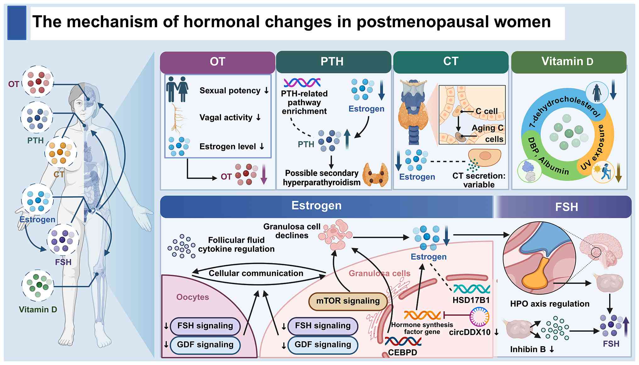 | Figure 1Mechanisms of hormonal changes in
postmenopausal women. Ovarian aging drives the decline in estrogen
production through multiple interrelated mechanisms: Progressive
granulosa cell loss mediated by mTOR signaling and CEBPD-regulated
senescence programs; impaired steroidogenic function of granulosa
cells caused by downregulated circDDX10 and altered HSD17B1
expression; and disrupted oocyte-granulosa cell communication due
to depleted FSH and GDF signaling, combined with age-related
changes in follicular fluid cytokine profiles. The attenuated
negative feedback of estrogen on the HPO axis, together with
reduced inhibin B secretion, collectively leads to elevated
circulating FSH levels. Decreased circulating OT levels are
associated with reduced sexual activity, attenuated vagal activity
and lower estrogen concentrations. Age-related enrichment of
PTH-related pathways and loss of direct estrogenic suppression may
result in possible secondary hyperparathyroidism. CT secretion
patterns vary across individuals, which is attributable to
age-related morphological and functional changes in thyroid C cells
and withdrawal of estrogenic stimulation. Vitamin D status is
regulated by reduced cutaneous 7-dehydrocholesterol, insufficient
UV exposure and altered levels of carrier proteins DBP and albumin.
CEBPD, CCAAT/enhancer-binding protein delta; circDDX10, circular
RNA DDX10; CT, calcitonin; DBP, vitamin D-binding protein; FSH,
follicle-stimulating hormone; GDF, growth differentiation factor;
HPO, hypothalamic-pituitary-ovarian; HSD17B1, hydroxysteroid
17-beta dehydrogenase 1; mTOR, mechanistic target of rapamycin; OT,
oxytocin; PTH, parathyroid hormone; UV, ultraviolet. |
Effect of hormonal changes on the
development of osteoporosis in postmenopausal women
Effect of reduced estrogen levels on
osteoporosis
Bone mechanical homeostasis mediated
by reduced estrogen
Bone remodeling requires the coordinated activity of
multiple cell types, including osteocytes, osteoblasts,
osteoclasts, bone lining cells, and bone marrow mesenchymal stem
cells (BMSCs). Osteoclasts, osteoblasts, osteocytes and bone lining
cells, together with the local vasculature, form the basic
multicellular unit (BMU), with osteal macrophages (osteomacs) also
contributing to the local remodeling environment. Communication
among these cells through direct contact and autocrine/paracrine
signaling coordinates bone resorption and formation. Under
mechanical loading, these cellular interactions contribute to bone
remodeling, fracture repair and regeneration (42,43).
Bone tissue contains numerous osteocytes, which form
an interconnected network via extended dendrites to facilitate
mechanotransduction. Estrogen deficiency post-menopause increases
osteocyte apoptosis. However, selective estrogen receptor (ER)
modulators (SERMs), as reported by Huber et al (44), can protect osteocytes from
apoptosis after estrogen withdrawal. Estrogen deficiency also
impairs osteocyte function by altering their paracrine activity, as
shown by Geoghegan et al (45), leading to an increased RANKL/OPG
ratio. Lewis et al (46)
used ovariectomy to model menopausal ovarian failure and found that
reduced estrogen lowers osteocyte mechanosome numbers and weakens
osteocyte Ca2+ signaling. This explains the diminished
bone adaptability to mechanical stimulation. Osteocytes are also
the principal source of sclerostin, a glycoprotein encoded by the
SOST gene. Sclerostin binds to the Wnt co-receptors low-density
lipoprotein receptor-related proteins 5 and 6 (LRP5/6) and inhibits
canonical Wnt/β-catenin signaling, thereby suppressing osteoblast
differentiation and bone formation. Because estrogen deficiency
alters osteocyte viability, mechanosensation and paracrine
signaling, dysregulation of the osteocyte-sclerostin axis may
contribute to impaired bone formation and remodeling imbalance
after menopause (47,48). Osteoblasts serve as bone forming
cells and mineralizers (49).
Estrogen enhances alkaline phosphatase activity (50) and boosts osteocalcin (OCN)
expression (51), thereby
contributing to the normal functioning of osteoblasts. Osteoclasts
are essential for bone remodeling through their bone-resorbing
function. Experimental work by Guo et al (52) showed that estrogen suppresses
osteoclast formation and bone resorption through microRNA
(miR)-27a-mediated regulation of peroxisome proliferator-activated
receptor γ and adenomatous polyposis coli. When subjected to
mechanical stress, BMSCs can differentiate into osteoblasts or
chondrocytes. Estrogen supports the osteogenic differentiation of
BMSCs through several transcriptional and epigenetic mechanisms. In
cultured human BMSCs, including cells obtained from women with PMO,
17β-estradiol increased runt-related transcription factor 2 (RUNX2)
expression and promoted early osteogenic differentiation, partly
through Notch signaling (53).
Separately, experiments in human dental mesenchymal stem cells
showed that estrogen induced lysine demethylase 6B (KDM6B) through
estrogen receptor α (ERα) and promoted osteogenic differentiation
through bone morphogenetic protein 2 (BMP2)- and homeobox C6
(HOXC6)-related regulation (54). These findings support distinct
estrogen-responsive osteogenic mechanisms in experimental stem-cell
models (Fig. 2).
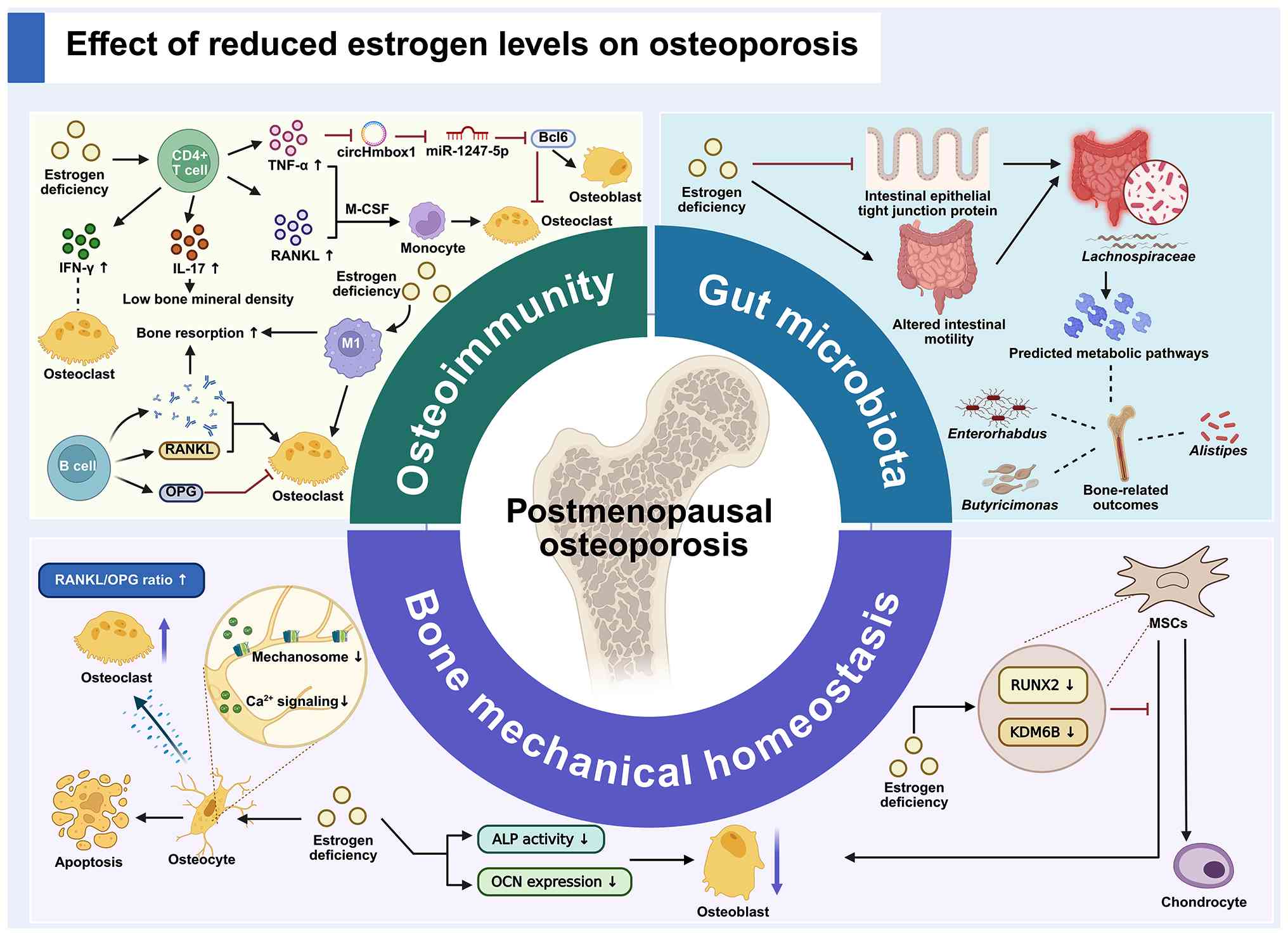 | Figure 2Effect of reduced estrogen levels on
osteoporosis. Estrogen deficiency promotes postmenopausal
osteoporosis via three interconnected pathophysiological modules.
i) Osteoimmunity: Estrogen deficiency increases CD4+ T
cell-derived TNF-α, IL-17 and IFN-γ; TNF-α suppresses the
circHmbox1/miR-1247-5p/Bcl6 axis to impair osteoblast
differentiation, while TNF-α and RANKL, together with M-CSF, drive
osteoclastogenesis from monocytes. IFN-γ exerts context-dependent
effects on osteoclast formation. B cells regulate osteoclast
activity via RANKL and OPG secretion, and M1-polarized macrophages
enhance bone resorption. ii) Gut microbiota: Estrogen deficiency
reduces intestinal epithelial tight junction protein expression and
alters intestinal motility, leading to microbial dysbiosis.
Lachnospiraceae and its predicted metabolic pathways are associated
with bone-related outcomes, while Enterorhabdus, Alistipes and
Butyricimonas show associations with bone alterations. iii) Bone
mechanical homeostasis: Estrogen deficiency increases osteocyte
apoptosis, reduces osteocyte mechanosome abundance and
Ca2+ signaling, and elevates the RANKL/OPG ratio to
activate osteoclasts; it also decreases osteoblast ALP activity and
OCN expression. The MSC module summarizes separate
estrogen-responsive osteogenic mechanisms involving RUNX2 in human
bone marrow MSCs and KDM6B in human dental MSCs. ALP, alkaline
phosphatase; Bcl6, B-cell lymphoma 6; Ca2+, calcium ion;
circHmbox1, circular RNA Hmbox1; IFN-γ, interferon-gamma; IL-17,
interleukin-17; KDM6B, lysine demethylase 6B; M1, classically
activated macrophage; M-CSF, macrophage colony-stimulating factor;
OCN, osteocalcin; OPG, osteoprotegerin; RANKL, receptor activator
of nuclear factor-κB ligand; RUNX2, runt-related transcription
factor 2; TNF-α, tumor necrosis factor-alpha; MSCs, mesenchymal
stem cells; miR, microRNA. |
The effects of estrogen on different bone cell
populations do not occur in isolation but within an interconnected
remodeling network. In addition to the aforementioned direct
effects, several coupling mechanisms operate within the BMU.
Osteocyte-derived RANKL and sclerostin can regulate both
osteoclasts and osteoblasts, bone lining cells contribute to
RANKL-mediated osteoclast regulation, osteomacs support osteoblast
mineralization, and vascular endothelial cells participate in
remodeling through the local blood supply and signals such as
vascular endothelial growth factor (VEGF). Thus, the disruption of
bone homeostasis associated with estrogen deficiency should be
considered in the context of both individual cellular responses and
multicellular communication within the BMU (43).
Osteoimmunity mediated by reduced
estrogen
Osteoimmunity plays a critical role in maintaining
bone homeostasis through cytokines and antibodies secreted by
immune cells. Estrogen deficiency not only directly affects
osteoblasts and osteoclasts, leading to bone loss, but also
disrupts bone homeostasis by influencing osteoimmune cells,
including T cells, B cells and macrophages.
In postmenopausal women, estrogen deficiency has
been associated with alterations in CD4+ T cell subsets,
including increased T helper 1 (Th1) and T helper 17 (Th17)
activity and elevated circulating levels of tumor necrosis factor-α
(TNF-α), interferon-γ (IFN-γ), IL-17 and RANKL (55-57). Importantly, while some evidence
is derived from clinical observations in postmenopausal women, the
causal signaling pathways have been primarily validated in
experimental models. RANKL or TNF-α, together with macrophage
colony-stimulating factor (M-CSF), can induce osteoclastogenesis
from bone marrow monocytes (58). TNF-α can also regulate bone
remodeling through the circular RNA Hmbox1
(circHmbox1)/miR-1247-5p/B-cell lymphoma 6 (Bcl6) axis. TNF-α
reduces circHmbox1, which increases miR-1247-5p-mediated
suppression of Bcl6, thereby favoring osteoclast differentiation
while impairing osteoblast differentiation (59). IFN-γ has context-dependent
effects: It can directly inhibit osteoclast precursors, while
indirectly promoting osteoclastogenesis through T-cell activation
and increased osteoclastogenic cytokine production. In experimental
estrogen-deficiency models, the net effect can favor bone
resorption (60). IL-17 is
associated with low BMD in postmenopausal women, suggesting that it
may be a key target for osteoporosis (61). In collaboration with T cells, B
cells are equally involved in the progression of PMO. In PMO, B
cells contribute through two main mechanisms: secreting RANKL and
OPG to modulate osteoclast differentiation and activation (62) as well as producing antibodies
that stimulate bone resorption (63). Estrogen influences the M1/M2
macrophage ratio by inhibiting M1 macrophage polarization and
promoting M2 macrophage polarization through its interaction with
the ER. When estrogen levels are sufficient, it helps maintain a
balanced macrophage profile. However, in its absence [for example,
in ovariectomized (OVX) models], M1 macrophages dominate, which can
lead to increased osteoclastogenesis and bone resorption (1) (Fig.
2).
Bone loss induced by estrogen
deficiency through the gut microbiota
Gut flora is a key microecosystem in the body. In
recent years, researchers have proposed the concept of the
'gut-bone' axis, revealing a close relationship between gut flora
and PMO (64). Human
observational data link the menopausal transition to changes in
intestinal permeability and inflammation (65). Experimental studies also suggest
altered gastrointestinal motility after estrogen withdrawal
(66). These changes can alter
gut flora composition, potentially leading to dysbiosis. Meng et
al (67) demonstrated that
estrogen deficiency markedly reduces gut microbiota species
richness and alters β-diversity. Notably, certain bacteria,
including Helicobacter rodentium, Lachnospiraceae
10-1, Lachnospiraceae A4 and Lactobacillus
reuteri, show significantly enhanced relative abundance.
Interestingly, Chen et al (68) found that recipient mice receiving
fecal transplants from postmenopausal patients with normal bone
mass exhibited Lachnospiraceae and Oscillibacter
enrichment, whereas recipient mice receiving fecal transplants from
patients with PMO exhibited Halobiforma,
Enterorhabdus, Alistipes and Butyricimonas,
suggesting donor- and model-dependent associations between
microbial composition and skeletal outcomes. In recipient mice,
enrichment of Halobiforma, Enterorhabdus, Alistipes and
Butyricimonas was also associated with altered TNF-α and
IL-17A expression in serum and bone tissue, linking gut dysbiosis
to the osteoimmune pathways (68). Using Mendelian randomization,
Zeng et al (69) reported
associations of Lachnospiraceae NK4A136 and
Oxalobacteraceae with lower osteoporosis risk, and
associations of Pasteurellaceae, Pasteurellales and
Ruminococcaceae UCG004 with higher risk. These findings
depend on the assumptions of the genetic instruments and require
validation across populations. Lachnospiraceae was
associated with alterations in purine metabolism, including changes
in inosine, xanthine and uric acid levels in the OVX model. These
metabolic changes were correlated with BMD. Functional prediction
suggested enrichment of purine-metabolism-related enzymes (70). Associations between microbial
taxa, predicted metabolic functions, and bone-related outcomes have
also been reported in experimental and human studies (70-72). These findings do not yet
establish uniform protective or pathogenic roles for individual
taxa or demonstrate that the inferred metabolic pathways mediate
bone loss in humans (Fig. 2).
Associations between specific microbial taxa and bone health are
not consistent across studies. A previous systematic review showed
substantial variation in osteoporosis-associated microbial profiles
across cohorts, including population-dependent differences in
Alistipes (73). Such
heterogeneity may be related to differences in population
characteristics, diet, menopausal stage, sequencing methods and
analytical approaches (73).
Therefore, individual taxa cannot yet be consistently classified as
protective or pathogenic in PMO.
In addition to microbial composition, increasing
attention has been directed toward microbiota-derived metabolites,
particularly short-chain fatty acids (SCFAs) such as acetate,
propionate and butyrate. These metabolites are produced through
microbial fermentation of dietary fibers and are considered key
mediators linking gut microbiota to host metabolic regulation.
SCFAs have been shown to modulate bone metabolism by inhibiting
osteoclast differentiation, promoting osteoblast activity, and
regulating immune responses involved in bone remodeling.
Experimental studies suggest that SCFAs may reduce bone loss by
suppressing osteoclastogenesis and modulating immune pathways such
as the regulatory T cell (Treg)/Th17 balance, thereby contributing
to the maintenance of bone homeostasis (74-76). Research has shown that the
composition of the gut microbiota is significantly altered in women
with PMO, accompanied by reduced levels of certain SCFAs (77). These findings support the
hypothesis that microbial metabolites act as functional
intermediates in the gut-bone axis and may represent potential
biomarkers or therapeutic targets in PMO. Several clinical and
translational studies have investigated probiotic interventions as
a strategy to modulate the gut microbiota and promote bone health.
Meta-analyses of probiotic trials report effects on selected
bone-turnover and BMD outcomes, but findings vary by strain,
formulation, population and treatment duration (78,79). These results do not establish
fracture prevention or justify replacing standard osteoporosis
therapy with probiotics.
The gut-bone axis may connect endocrine, immune and
metabolic changes in PMO, but taxon-specific mechanisms and
clinical benefits of microbiota-directed interventions remain
incompletely established. These interactions are summarized in
Fig. 2.
Effect of increased FSH levels on
osteoporosis
Using NHANES data (1999-2002), Liu et al
(80) analyzed serum FSH
associations with lumbar spine and femoral neck BMD in women aged
35-60. They found a significant non-linear inverse association
between serum FSH and lumbar spine BMD exclusively in
postmenopausal women (P for non-linearity=0.013), and a comparable
link between FSH and osteoporosis risk (P=0.05) (80). Zheng et al (81) identified follitropin subunit beta
among proteins with high importance in a multivariable predictive
model. Predictive importance does not establish a causal skeletal
effect of circulating FSH. Mechanistically, FSH binds to the FSH
receptor (FSHR) and activates G protein/CCAAT/enhancer-binding
protein β signaling in adipocytes, which promotes heat shock
protein 90α (HSP90α) secretion. Increased extracellular HSP90α
subsequently enhances osteoclast activity and may contribute to
increased bone resorption (82).
Although observational and genetic analyses support an association
between elevated FSH and bone loss, direct human interventional
evidence is still lacking. Most mechanistic evidence remains
experimental; therefore, the causal skeletal effect of elevated FSH
in humans has not yet been established and requires further
longitudinal and interventional validation.
Effect of decreased OT levels on
osteoporosis
Indeed, OT receptors are widely present on bone
tissue and OT regulates bone remodeling by binding to OT receptors
on osteoblasts and osteoclasts (83). In normal conditions, E2 triggers
the mitogen-activated protein kinase/extracellular signal-regulated
kinase (MAPK/ERK) pathway and OT receptor expression in
osteoblasts, boosting OT production. OT then upregulates BMP2 and
activating transcription factor 4 promoting osteoblast
differentiation and function and forming a synergistic feed-forward
loop (15). Reduced estrogen
signaling may weaken this feed-forward loop, although its
contribution to skeletal outcomes after menopause remains to be
established (15). OT has
distinct effects on osteoclast formation and mature cell resorptive
activity. In experimental models, OT promotes osteoclastogenesis
directly and indirectly, including through increased RANKL and
reduced OPG expression in osteoblasts. Conversely, OT can suppress
resorption by mature osteoclasts through intracellular
Ca2+ release and nitric oxide synthesis (84). These stage- and cell-dependent
effects help explain why changes in osteoclast number do not
necessarily translate into equivalent changes in bone resorption;
the net therapeutic effect in postmenopausal women remains
unproven.
In human studies, Du et al (85) linked OT to bone mass (β=0.59,
P=0.044) and after age adjustment, to osteoporosis risk (OR: 0.75;
95% CI: 0.63-0.98; P=0.019) in a cross-sectional study of healthy
community dwelling postmenopausal women aged 50-90. In an
ovariectomized rat model, OT attenuated bone loss and altered
bone-turnover markers and OPG/RANKL mRNA expression (86). Thus, current human evidence
supports an association between OT and skeletal status but does not
yet establish the therapeutic efficacy of OT supplementation.
Effect of increased PTH levels on
osteoporosis
PTH is a key regulator of bone remodeling and
exhibits context-dependent effects on skeletal homeostasis. At the
cellular level, PTH promotes bone formation by enhancing osteoblast
precursor differentiation, inhibiting osteoblast apoptosis, and
suppressing sclerostin production, a negative regulator of bone
formation (87). However,
prolonged elevation of PTH can shift bone remodeling toward
resorption by stimulating osteoblast- and osteocyte-derived RANKL
expression and reducing OPG production, thereby promoting
osteoclast differentiation. By contrast, intermittent PTH
stimulation used in anabolic therapies preferentially enhances
osteoblast activity and bone formation (16). In experimental models, PTH
reduced miR-19a/b expression, while antagonizing miR-19a/b
increased its target TG-interacting factor 1 (Tgif1) and augmented
PTH-induced bone gain (88).
Apart from the direct effects of PTH, genetic polymorphisms in the
patient's own PTH and PTH 1 receptor (PTHR1) genes also influence
susceptibility to osteoporosis in menopausal women. By analyzing
case-control genetic associations in a cohort of South Indian
women, Fernandez et al (89) revealed lower heel BMD in women
with osteoporosis who had the BstBI-AA recessive genotype. On the
other hand, Abdi et al (90) selected Arab women as their
subjects and discovered that the PTHR1 single-nucleotide
polymorphism rs1138518 C/T genotype serves as a risk factor for
osteoporosis in postmenopausal women. Furthermore, it was observed
that the C/T and T/T genotypes are associated with different
outcomes in 25-hydroxyvitamin D [25(OH)D] levels, suggesting that
PTHR1 rs1138518 may play a regulatory role in modulating 25(OH)D
levels (90). These
population-specific genetic associations require replication and do
not establish a functional mechanism or a clinically validated
prediction tool.
CT and osteoporosis
CT suppresses osteoclast-mediated bone resorption,
which can be reflected by reduced urinary hydroxyproline excretion
(91). For osteoclasts, CT
rapidly disrupts the osteoclast ruffled border, inhibits osteoclast
motility and promotes detachment from bone (18). Although CT has a well-established
role in regulating osteoclasts, its effects on osteoblasts remain
debated. Naot et al (92)
reported the absence of CT receptors in osteoblasts, whereas Farley
et al (93) demonstrated
CT-mediated promotion of osteoblast proliferation. By contrast,
Cornish et al (94)
showed no increase in mineralized bone area following CT treatment
in mice. These studies examined different models and endpoints,
including receptor expression, cell proliferation and mineralized
bone area, which are not interchangeable measures of a direct
osteoanabolic effect. This discrepancy may arise from differences
in CT concentrations and incubation durations between studies,
highlighting the need for concentration- and time-dependent
gradient experiments to characterize CT effects on osteoblasts.
Several investigations indicate that CT primarily acts by
inhibiting osteoclast activity, thereby altering the coupling
signals between osteoclasts and osteoblasts rather than directly
stimulating osteoblast proliferation (92).
However, although CT has well-established inhibitory
effects on osteoclast-mediated bone resorption, its clinical
efficacy in the treatment of osteoporosis in humans appears to be
relatively limited. Randomized controlled trials (RCTs) have shown
that intranasal salmon CT (sCT) can reduce the risk of vertebral
fractures, but it does not significantly decrease the incidence of
non-vertebral or hip fractures (95). Accordingly, most current clinical
guidelines no longer recommend CT as a first-line therapy for PMO.
These findings suggest that although CT plays an important role in
the regulation of bone metabolism, its clinical utility in the
treatment of osteoporosis remains limited and warrants further
investigation. Clarifying the cellular targets and signaling
mechanisms of CT will be important for determining its therapeutic
relevance in PMO.
Effect of Vitamin D deficiency on
osteoporosis
Vitamin D, a fat-soluble vitamin, is crucial for
bone metabolism as it enhances intestinal calcium absorption. Chen
et al (96) investigated
the association between vitamin D levels and BMD, bone turnover
markers (BTMs), as well as PTH in 8,532 Chinese postmenopausal
women. Vitamin D deficiency or insufficiency was present in ~81% of
the study population (96). In
osteoporotic patients, higher vitamin D levels were positively
correlated with BMD at all measured sites and negatively correlated
with BTMs, including β-C-terminal telopeptide of type I collagen
(β-CTX) and OCN, as well as with PTH levels (96). In line with this result, the
study by Al-Rawaf et al (97) also identified vitamin D as a key
predictor of osteoporosis in postmenopausal women. Current evidence
links vitamin D to osteoporosis through three key mechanisms:
Regulation of calcium-phosphorus homeostasis (17), immune-mediated modulation of
pro-inflammatory cytokine production (17), and vitamin D receptor gene
polymorphisms that influence susceptibility to the disease
(98). Selected skeletal actions
of FSH, OT, PTH, CT and vitamin D are summarized in Fig. 3. The regulatory effects of
different hormones and their receptors on postmenopausal bone loss
are summarized in Table I.
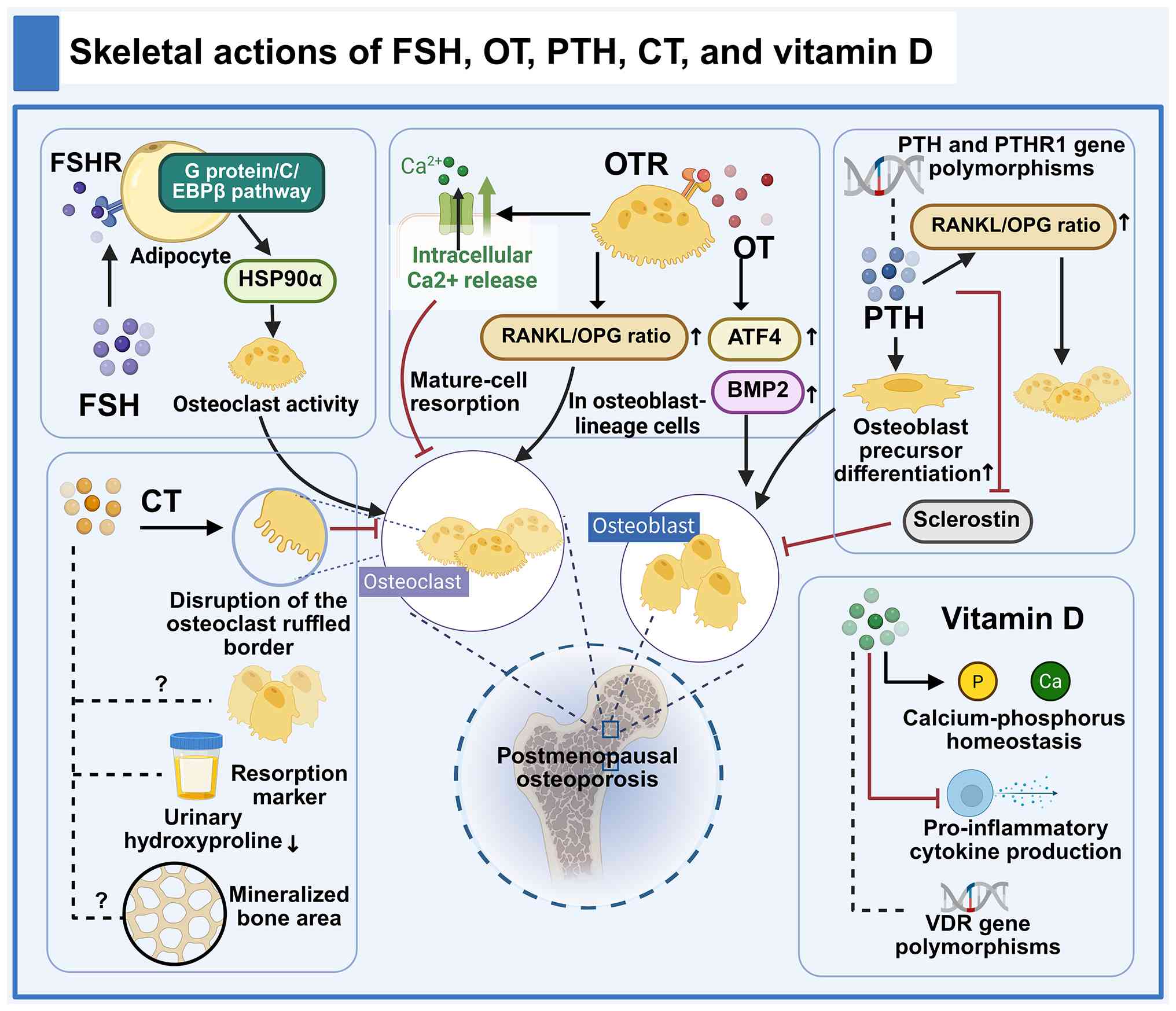 | Figure 3Skeletal actions of FSH, OT, PTH, CT
and vitamin D. This diagram summarizes the regulatory effects of
five hormonal systems on bone remodeling. FSH binds to FSH
receptors on adipocytes and activates the G protein/C/EBPβ pathway,
which promotes HSP90α secretion and thereby enhances osteoclast
activity. OT signaling upregulates ATF4 and BMP2 to promote
osteoblast differentiation; meanwhile, it increases the RANKL/OPG
ratio in osteoblast-lineage cells to favor osteoclastogenesis, and
on the other hand, induces intracellular Ca2+ release in
mature osteoclasts to suppress their resorptive function. PTH
exerts exposure-dependent biphasic effects: Sustained elevation
upregulates the RANKL/OPG ratio to promote osteoclastogenesis,
while intermittent administration enhances osteoblast precursor
differentiation and inhibits sclerostin to exert anabolic effects.
Polymorphisms in the PTH and PTHR1 genes modulate individual
susceptibility to osteoporosis. CT inhibits osteoclast-mediated
bone resorption by disrupting the osteoclast ruffled border, as
reflected by reduced levels of urinary hydroxyproline, a bone
resorption marker; the direct effects of CT on osteoblast
proliferation and mineralized bone area remain controversial.
Vitamin D maintains calcium-phosphorus homeostasis and modulates
pro-inflammatory cytokine production; polymorphisms in the VDR gene
are genetic susceptibility factors for osteoporosis. FSH,
follicle-stimulating hormone; OT, oxytocin; PTH, parathyroid
hormone; CT, calcitonin; HSP90α, heat shock protein 90 alpha; ATF4,
activating transcription factor 4; RANKL, receptor activator of
nuclear factor-κB ligand; OPG, osteoprotegerin; VDR, vitamin D
receptor. |
 | Table IMajor hormones and receptors involved
in the regulation of postmenopausal bone loss. |
Table I
Major hormones and receptors involved
in the regulation of postmenopausal bone loss.
| Authors, year | Hormone | Major receptor | Postmenopausal
change | Predominant
skeletal action of the hormone or pathway | Major regulatory
features and supporting evidence | (Refs.) |
|---|
| Khosla et
al, 2012; Kular et al, 2012; Fan et al, 2014; Liu
et al, 2022 | Estrogen (E2) | ER | ↓ | Inhibits bone
loss | Maintains osteocyte
survival and osteoblast function and suppresses osteoclastogenesis
through mechanisms including RANKL/OPG regulation. Estrogen also
supports osteogenic differentiation in mesenchymal stem-cell models
through distinct RUNX2-related and ERα/KDM6B-related mechanisms
demonstrated in different experimental cell sources | (2,43,53,54) |
| Liu et al,
2023; Zheng et al, 2025; Huang et al, 2024 | FSH | FSHR | ↑ | Potentially
promotes bone loss | Higher FSH levels
are associated with lower BMD and greater osteoporosis risk in some
human studies. Experimental evidence suggests that FSH/FSHR
signaling may enhance osteoclast-related activity through an
adipocyte FSHR/G protein/C/EBPβ/HSP90α pathway, although a direct
causal role in humans remains to be established | (80-82) |
| Tamma et al,
2009; Du et al, 2021; Moghazy et al, 2020 | Oxytocin | OTR | Lower levels
reported in some studies; variable | Context- and
stage-dependent | Promotes osteoblast
differentiation and function in experimental models and has
stage-dependent effects on osteoclasts, promoting differentiation
while suppressing the resorptive activity of mature cells. Human
observational studies report associations between circulating OT
and skeletal measures, but therapeutic efficacy in humans remains
unestablished | (84-86) |
| Silva et al,
2015; Eastell et al, 1991; Fernandez et al, 2022;
Abdi et al, 2021 | PTH | PTHR1 | May increase with
aging, vitamin D deficiency, or altered calcium balance |
Exposure-pattern-dependent | Regulates
osteoblast differentiation, cell survival, and sclerostin
production. Sustained elevation may favor increased bone turnover
and bone loss, whereas intermittent pharmacological exposure is
predominantly anabolic. Selected PTH/PTHR1 variants have been
associated with osteoporosis susceptibility in population-specific
studies | (16,33,89,90) |
| Xie et al,
2020 | CT | CTR | Variable; a
consistent menopause-specific decrease is not established | Inhibits
osteoclast-mediated bone resorption | Primarily
suppresses osteoclast-mediated bone resorption by disrupting the
ruffled border, reducing osteoclast motility, and promoting
detachment from bone | (18) |
| Bellavia et
al, 2024; Chen et al, 2023; Mondockova et al,
2023 | Vitamin D | VDR |
Deficiency/insufficiency is common | Adequate vitamin D
status is generally protective; deficiency favors bone loss | Supports
calcium-phosphorus homeostasis. Observational studies report
associations of vitamin D status with BMD, bone turnover markers,
and PTH; reported VDR polymorphism associations require
population-specific interpretation | (17,96,98) |
Hormonal crosstalk and regulatory
networks
To integrate the aforementioned hormone-specific
evidence, postmenopausal endocrine remodeling can be considered at
three analytically distinct but interacting levels. At the upstream
level, ovarian aging and the loss of estrogenic feedback establish
the endocrine context of menopause. At an intermediate level,
alterations involving FSH, the PTH-vitamin D axis, OT and CT modify
systemic and local skeletal regulation. At the effector level,
these endocrine signals converge on the basic multicellular unit,
osteoimmune pathways, calcium homeostasis, and the gut-bone axis,
ultimately disturbing osteoblast-osteoclast coupling and favoring
net bone loss. This hierarchy should be regarded as an integrative
conceptual framework rather than a strictly linear cascade, because
several components are linked through feedback loops and the
strength of evidence differs among individual pathways. Within this
framework, the decline in estrogen is widely regarded as the key
endocrine event. Under physiological conditions, estrogen provides
negative feedback to the pituitary and helps regulate gonadotropin
secretion. With progressive ovarian insufficiency and the
accompanying fall in estrogen levels, this feedback regulation
becomes attenuated, leading to a pronounced rise in FSH (99). As a result, in the postmenopausal
state, reduced estrogen and elevated FSH may jointly influence bone
cells and favor a shift in bone remodeling toward increased bone
resorption.
In addition, alterations in estrogen levels also
disturb the balance among calcium-regulating hormones. Reduced
calcium absorption and low vitamin D status may contribute to
compensatory PTH elevation, particularly with aging (33,100,101). At the same time, vitamin D
metabolism is influenced by both aging and changes in estrogen
status. Insufficient vitamin D further impairs intestinal calcium
absorption and stimulates compensatory PTH secretion, forming a
vitamin D-PTH regulatory loop that ultimately contributes to
accelerated bone loss (100,101). At the cellular level, PTH and
1,25(OH)2D3 can also increase RANKL
expression in osteoblasts, thereby linking hormonal changes to
osteoblast-osteoclast communication within the BMU. Vitamin
D-related signaling can further stimulate osteoblast-derived VEGF,
providing a link between bone formation and vascular regulation
(43). Beyond the classical
endocrine axes, several neuroendocrine factors also participate in
this regulatory network. For instance, estrogen has been shown to
regulate the synthesis of OT and the expression of its receptor.
Consequently, the postmenopausal decline in estrogen may indirectly
weaken the protective effects of OT on bone tissue (102). These interactions illustrate
how distinct endocrine changes can converge on shared skeletal
regulatory pathways. However, the strength of supporting evidence
differs among individual pathways; the major hormone-related
findings are therefore summarized according to their evidence types
in Table II.
| Table IIEvidence types supporting
hormone-related findings in postmenopausal osteoporosis. |
Table II
Evidence types supporting
hormone-related findings in postmenopausal osteoporosis.
| Authors, year | Hormonal
factor | Hormone-related
findings | Evidence type and
main limitation | (Refs.) |
|---|
| Khosla et
al, 2012; Fan et al, 2014; Liu et al, 2022;
Lorentzon et al, 2022 | Estrogen/ER
signaling | Estrogen deficiency
contributes to remodeling imbalance; estrogen-based therapy reduces
fracture risk in selected postmenopausal populations. | Mechanistic
evidence summarized in a review, with primary experiments in
distinct human stem-cell models. Therapeutic fracture outcomes:
pooled analysis of randomized trials. Clinical efficacy does not
independently validate every proposed molecular pathway. | (2,53, 54,109) |
| Liu et al,
2023; Zheng et al, 2025; Huang et al, 2024 Gera et
al, 2022 | FSH/FSHR
signaling | Higher FSH is
associated with lower BMD in some human studies; FSH-related
pathways and FSH blockade have been investigated
experimentally. | Human observational
associations and proteomic prediction; a small human comparison and
animal and in vitro experiments; preclinical FSH-blocking
antibody studies. A direct skeletal benefit of FSH blockade has not
been established in human osteoporosis trials. | (80-82,136) |
| Tamma et al,
2009; Du et al, 2021; Moghazy et al, 2020 | OT/OTR
signaling | OT supports
osteoblast function and has stage-dependent effects on osteoclast
formation and resorption in experimental models; circulating OT is
associated with skeletal measures in some populations. | Human observational
studies; animal and in vitro studies. Human therapeutic
efficacy, dosing, and long-term skeletal safety remain
unestablished. | (84-86) |
| LeBoff et
al, 2022; Silva et al, 2015; Fernandez et al,
2022; Abdi et al, 2021 | PTH/PTHR1
signaling | Sustained PTH
excess and intermittent PTH-receptor stimulation have different
skeletal effects. Selected PTH/PTHR1 variants have been associated
with BMD or osteoporosis susceptibility. | Experimental
mechanisms summarized in a review; clinical trial outcomes
summarized in a clinical guide; population-specific genetic
association studies. Therapeutic evidence should not be conflated
with evidence for individual genetic variants. | (4,16,89,90) |
| Xie et al,
2020; Chesnut et al, 2000 | CT/CTR
signaling | CT directly
inhibits osteoclast-mediated resorption. Pharmacological salmon CT
has shown vertebral antifracture activity, but evidence for
nonvertebral or hip fracture benefit is limited. | Experimental
mechanisms summarized in a review; primary randomized trial
evidence for intranasal salmon CT. Drug effects do not establish
that endogenous CT deficiency is a major cause of PMO. | (18,95) |
| Bellavia et
al, 2024; Fleet et al, 2022; Chen et al, 2023;
Mondockova et al, 2023 | Vitamin D/VDR
signaling | Vitamin D regulates
calcium homeostasis; vitamin D status and selected VDR variants
have been associated with skeletal outcomes. | Human observational
studies; physiological and mechanistic evidence summarized in
reviews; genetic association studies. Association does not
establish an added antifracture benefit of supplementation in
vitamin-D-replete individuals. | (17,36,96,98) |
Therapeutic strategies for PMO
HRT
In 2002, the WHI found that the use of combined
conjugated equine estrogen and medroxyprogesterone acetate
regimens, aimed at improving osteoporosis, led to an elevated risk
of cardiovascular events and an increased risk of breast cancer
(12). This puts the use of HRT
in the limelight. In fact, HRT components are primarily estrogens
and/or progestins and are primarily used to treat perimenopausal
and early menopausal vasomotor symptoms. Systemic hormone therapy
may prevent bone loss in appropriately selected postmenopausal
women, with formulation, route, and duration individualized
according to menopausal symptoms and the benefit-risk profile
(103). Stevenson et al
(104) employed two HRT
regimens. The first regimen consisted of 1 mg of 17β-estradiol and
either 5 or 10 mg of dydrogesterone administered daily for 14
consecutive days, with a 28-day cycle. The second regimen involved
2 mg of 17β-estradiol and either 10 or 20 mg of dydrogesterone
given daily for 14 consecutive days, also with a 28-day cycle. They
found that, compared with baseline, HRT significantly increased
total disc height in both the 1-mg E2 group (0.16±0.65 cm, P=0.015)
and the 2-mg E2 group (0.21±0.86 cm, P=0.006). However,
between-group differences were not statistically significant, and
this analysis did not directly establish a reduction in fracture
risk (104). Gamsjaeger et
al (105) administered a
combination of 2 mg E2 and 1 mg norethindrone acetate to
postmenopausal women over a two-year period. They observed that HRT
with this regimen altered osteoid composition and
mineralization-related characteristics (105). The 2022 North American
Menopause Society (NAMS) position statement emphasizes that the
benefit-risk profile of HRT is generally more favorable in healthy
women younger than 60 years or within 10 years of menopause onset
(103). By contrast, initiation
at an older age or more than 10 years after menopause is associated
with higher absolute risks of coronary heart disease, stroke and
venous thromboembolism. Risk also varies according to the route and
regimen of treatment. Oral estrogen is associated with a higher
risk of venous thromboembolism than transdermal estrogen, although
direct comparative RCT evidence remains limited. Breast cancer risk
also differs according to the type and duration of hormone therapy
(106-108). Systemic HRT is generally
avoided in women with estrogen-sensitive malignancies, unexplained
vaginal bleeding, active liver disease, previous venous
thromboembolism, or established cardiovascular disease. Therefore,
treatment should be individualized according to age, time since
menopause, route of administration, and baseline risk profile
(103).
Recent studies continue to refine the evidence base
for hormone therapy in PMO. A reanalysis of the WHI randomized
trials reported fracture-risk reductions across baseline risk
strata (109). Separately, a
2025 nested case-control study examined fracture risk after
discontinuation of menopausal hormone therapy (110). Additionally, emerging evidence
suggests that combining HRT with structured exercise can further
enhance BMD (111).
Contemporary analyses also highlight that the risk profile of HRT
varies with formulation and route, with observational evidence
suggesting lower venous thromboembolic risk with transdermal than
oral estrogen (108,112). In summary, the individual
benefit risk balance of HRT hinges on the type, dose and duration
of HRT, along with each woman's unique characteristics. Further
studies are required to investigate these variables
comprehensively.
Pharmacological therapies other than
menopausal hormone therapy
Bisphosphonates
Bisphosphonates, characterized by two phosphonate
groups, function by inhibiting osteoclast activity and thereby
reducing bone resorption, positioning them as a potent class of
antiresorptive agents. Following estrogen deficiency, the
expression of osteoclastogenic factors increases, leading to
enhanced osteoclast activity and excessive bone resorption.
Bisphosphonates counteract this process by directly inhibiting
osteoclast function, thereby compensating for the estrogen
deficiency-induced increase in bone resorption (4). US Food and Drug Administration
(FDA)-approved bisphosphonates encompass alendronate, ibandronate,
risedronate and zoledronic acid (4). Nevertheless, bisphosphonates are
associated with potential adverse effects such as renal impairment,
gastrointestinal problems, and hypocalcemia, which restricts their
application in osteoporosis treatment (113). Oral bisphosphonates should be
avoided in patients with major esophageal abnormalities or those
unable to remain upright after administration, while hypocalcemia
should be corrected before treatment. Renal function should also be
considered when selecting individual bisphosphonates (4). Rare but important adverse events
include osteonecrosis of the jaw and atypical femoral fractures,
which should be considered during long-term treatment and
reassessment (4).
PTH and PTH-related protein
analogs
Intermittent PTH receptor stimulation promotes bone
formation, whereas sustained PTH elevation can favor bone
resorption. Teriparatide is a PTH (1-34) analog, and abaloparatide is a
PTH-related protein analog; both are used as anabolic treatments
for osteoporosis (114,115). Nevertheless, these agents are
associated with side effects. Common adverse effects of PTH analogs
include nausea, dizziness and disturbances in calcium metabolism.
An osteosarcoma signal was observed in preclinical rodent studies;
however, subsequent large observational studies have not
demonstrated an increased incidence of osteosarcoma in humans
(116). Nevertheless, these
agents should be avoided in patients at increased baseline risk of
osteosarcoma, including those with Paget's disease, skeletal
malignancies or bone metastases, and previous skeletal radiation
exposure. Caution is also warranted in patients with active or
recent urolithiasis (117).
Denosumab (RANKL inhibitor)
Denosumab is a monoclonal antibody that binds RANKL
and prevents its interaction with RANK, thereby inhibiting
osteoclast formation and activity. In postmenopausal women, reduced
estrogen levels result in an increased RANKL/OPG ratio, which
promotes osteoclastogenesis and enhances bone resorption. Denosumab
directly targets this pathway by binding to RANKL and blocking its
interaction with RANK, thereby suppressing osteoclast activation
and counteracting the excessive osteoclast activity caused by
estrogen deficiency (118,119). Owing to its remarkable capacity
to increase BMD, it has been introduced into clinical practice.
Denosumab is generally well tolerated, but hypocalcemia remains an
important safety concern, particularly in patients with impaired
renal function. Severe hypocalcemia is a particular concern in
advanced chronic kidney disease (CKD), especially in patients
receiving dialysis or with CKD-related mineral and bone disorder
(120). Delayed dosing or
discontinuation can result in rebound bone turnover, rapid BMD loss
and multiple vertebral fractures. Therefore, denosumab should not
be discontinued without subsequent antiresorptive therapy to reduce
rebound fracture risk (4,121).
Raloxifene (SERM)
Raloxifene, a selective ER modulator approved by
FDA, is indicated for the prevention and treatment of osteoporosis
in postmenopausal women (4).
Raloxifene exerts estrogen agonist effects in bone. By activating
estrogen receptor-mediated signaling, it inhibits osteoclast
activity, reduces bone resorption, and increases BMD, thereby
correcting the bone metabolic imbalance caused by estrogen
deficiency (122,123). Guidelines from the Endocrine
Society recommend raloxifene for postmenopausal women at low risk
of deep vein thrombosis who are unsuitable for bisphosphonates or
denosumab, or those at high risk for breast cancer, to prevent
vertebral fractures (115).
Notably, raloxifene reduces vertebral fracture risk, while a
reduction in non-vertebral fracture risk has not been demonstrated
(4). However, Raloxifene has
been linked to an increased risk of venous thromboembolism and
possibly fatal stroke, without a corresponding increase in total
stroke events (124).
Sclerostin inhibitor
(romosozumab)
Romosozumab is a humanized monoclonal antibody that
targets sclerostin, an osteocyte-derived inhibitor of canonical
Wnt/β-catenin signaling. By neutralizing sclerostin, romosozumab
relieves inhibition of LRP5/6-mediated Wnt signaling and exerts a
dual effect by increasing bone formation while reducing bone
resorption (4). Current guidance
supports a 12-month course of romosozumab in patients at very high
fracture risk, followed by antiresorptive therapy to maintain
skeletal gains (4,125). Recent clinical evidence has
confirmed substantial increases in BMD and reductions in fracture
risk with romosozumab. However, cardiovascular safety remains an
important consideration, and treatment should be individualized
according to the patient's fracture and cardiovascular risk
profiles (126). Under US
labeling, romosozumab should not be initiated in patients with
myocardial infarction or stroke within the preceding year (127).
Current therapies are not interchangeable because
they address different aspects of the imbalance in bone remodeling.
Bisphosphonates and denosumab primarily suppress excessive bone
resorption, while PTH analogs and romosozumab offer anabolic
options for patients at very high fracture risk. Decisions about
HRT also depend on age, time since menopause, and systemic safety
alongside skeletal benefit. Treatment selection should therefore
account for fracture risk, mechanism of action, and
patient-specific safety rather than efficacy alone.
Non-pharmacological treatment
Beyond the aforementioned HRT and non-HRT
pharmacological treatments, non-pharmacological approaches play a
crucial role in preventing and ameliorating PMO. Lifestyle measures
include smoking cessation, avoidance of excessive alcohol intake,
adequate nutrition, and weight-bearing, resistance, and balance
exercise adapted to individual fracture risk and physical capacity
(4,128). In addition to its effects on
skeletal loading, regular exercise can help preserve muscle
function and physical performance, which are important components
of fall and fracture prevention (4). An updated systematic review and
meta-analysis of 80 controlled studies involving 5,581
postmenopausal women further demonstrated beneficial effects of
exercise on BMD at the lumbar spine, femoral neck and total hip
(128). Besides lifestyle
modifications, adequate calcium and vitamin D intake is also
advantageous. Calcium and vitamin D intake should follow applicable
guidance and individual assessment. For example, the Bone Health
and Osteoporosis Foundation recommends a total calcium intake of
1,200 mg/day for women aged 51 years or older and vitamin D
supplementation of 800-1,000 IU/day when needed (4). Treatment of established vitamin D
deficiency should be individualized rather than equated with
routine maintenance supplementation. Together, these lifestyle and
nutritional measures should be considered integral components of
comprehensive osteoporosis management and are generally used
alongside pharmacological therapy in women at elevated fracture
risk (4). The representative
mechanisms, clinical efficacy, and key limitations of current
therapeutic strategies for PMO are summarized in Table III.
| Table IIITherapeutic strategies for
postmenopausal osteoporosis. |
Table III
Therapeutic strategies for
postmenopausal osteoporosis.
| Therapeutic
strategy | Representative
agents | Main mechanism of
action | Representative
clinical efficacy | Key adverse
effects/limitations |
|---|
| HRT | Estradiol;
conjugated estrogens; estrogen-progestin combinations | Compensates for
estrogen deficiency; activates estrogen receptor signaling in bone;
suppresses osteoclast activity; reduces bone resorption and
fracture risk | In a pooled
analysis of the WHI hormone therapy trials, relative risks of any
clinical fracture, major osteoporotic fracture, and hip fracture
were reduced by 28, 40 and 34%, respectively, vs. placebo over a
mean follow-up of 4.3 years (109). | Venous
thromboembolism and stroke; breast cancer risk varies by regimen
and duration. Selection depends on age, time since menopause,
symptoms, and contraindications (103,107). |
|
Bisphosphonates | Alendronate;
risedronate; ibandronate; zoledronic acid | Inhibit
osteoclast-mediated bone resorption. | The clinical guide
summarizes ~50% lower spine and hip fracture incidence with
alendronate over 3 years in patients with prior vertebral fracture
or osteoporosis by hip BMD. Corresponding estimates for zoledronic
acid are ~62-70, 41, and 21-25% lower vertebral, hip, and
nonvertebral fracture incidence, respectively, over 3 years
(4). | Gastrointestinal
adverse effects with oral agents; renal restrictions and
hypocalcemia; rare osteonecrosis of the jaw and atypical femoral
fractures. Avoid oral treatment in patients with major esophageal
disorders or inability to remain upright; agent-specific renal
thresholds apply (4). |
| PTH/PTHrP
analogs | Teriparatide;
abaloparatide | Intermittent
PTH1-receptor activation promotes osteoblast differentiation and
survival and increases bone formation; suppression of sclerostin
contributes to the anabolic response (114). | The clinical guide
summarizes ~65-77 and 35-53% lower vertebral and nonvertebral
fracture risks, respectively, with teriparatide after an average of
18 months. Abaloparatide reduced new vertebral and nonvertebral
fracture risks by ~86% and 43%, respectively, vs. placebo over 18
months (4). | Nausea, dizziness,
and calcium-related adverse effects; caution with active or recent
urolithiasis. Avoid use in patients at increased baseline risk of
osteosarcoma. A rodent osteosarcoma signal has not translated into
an increased incidence in human observational studies of
teriparatide (4,116). |
| RANKL
inhibitor | Denosumab | Binds RANKL,
prevents its interaction with RANK, and inhibits osteoclast
formation and activity | In the FREEDOM
trial, denosumab reduced new radiographic vertebral, hip, and
non-vertebral fracture risks by ~68, 40 and 20%, respectively, vs.
placebo over 3 years (144). | Hypocalcemia,
including severe hypocalcemia in advanced chronic kidney disease;
rebound bone loss and multiple vertebral fractures after delayed
dosing or discontinuation. Subsequent antiresorptive therapy is
required when treatment is stopped (4,121). FDA warning for advanced kidney
disease (120). |
| SERM | Raloxifene | Estrogen agonist
effect in bone; suppresses osteoclast activity; increases bone
mineral density; reduces vertebral fracture risk | The clinical guide
summarizes ~30-40% lower vertebral fracture incidence with
raloxifene in patients with a prior vertebral fracture and ~55%
lower incidence in those without a prior vertebral fracture. A
reduction in non-vertebral fracture risk has not been demonstrated
(4). | Venous
thromboembolism; increased fatal-stroke risk in women with coronary
disease or elevated coronary risk. Contraindicated in women with a
current or previous venous thromboembolism (115,124). |
| Sclerostin
inhibitor | Romosozumab | Neutralizes
sclerostin; relieves inhibition of LRP5/6-mediated Wnt/β-catenin
signaling; increases bone formation and decreases bone
resorption | Reduced vertebral
and clinical fracture risks by ~73% and 36%, respectively, at 12
months vs. placebo (125) | Cardiovascular risk
assessment is required; under US labeling, do not initiate
treatment within 1 year after myocardial infarction or stroke.
Limit treatment to 12 monthly doses and follow with antiresorptive
therapy (4). US prescribing
information (127). |
| Non-pharmacological
interventions | Lifestyle
modification; calcium; vitamin D; exercise | Improve bone health
through adequate nutrient supply and mechanical loading; support
bone strength and remodeling | Supports bone
health and fall prevention; a single fracture-risk reduction
estimate is not applicable to this heterogeneous group (4,128). | Adjunctive care;
does not replace indicated pharmacotherapy in women at high
fracture risk. Avoid excessive calcium or vitamin D supplementation
(4,145). |
Despite their established clinical utility, current
therapeutic strategies are constrained by patient-specific safety
considerations, limitations of long-term treatment,
discontinuation-related effects, or incomplete applicability across
different risk groups. Together with increasing recognition of the
broader endocrine remodeling that accompanies menopause, these
limitations provide a rationale for exploring complementary
strategies that target additional hormone-related pathways and
mechanisms of bone loss.
Future research directions regarding
treatment based on hormonal changes during post-menopause
As aforementioned, HRT is effective in preventing
and alleviating PMO, but the long-term use of estrogen- and
progestin-based regimens may be associated with adverse effects.
This has prompted increasing interest in natural bioactive
compounds as potential complementary strategies for modulating bone
remodeling. Some natural bioactive compounds have been reported to
exert both pro-anabolic and antiresorptive effects by supporting
osteoblast function and suppressing osteoclast activity, with
additional mechanisms involving estrogen-like, anti-inflammatory,
antioxidant and bone signaling effects (129). Recent experimental evidence
supports this dual regulatory concept. In 2024, a
formononetin-derived phytoestrogen was shown to promote osteoblast
formation through BMP/Smad signaling while simultaneously
inhibiting RANKL-induced osteoclastogenesis through regulation of
the RANK/RANKL/OPG axis in vitro and in an ovariectomized
mouse model (130).
Additionally, several ER-targeted plant-derived compounds have also
shown potential osteoprotective effects. Chen et al
(131) generated a PMO rat
model through ovariectomy. Subsequently, upon administering
Secoisolariciresinol diglucoside (SDG) to the rats, it was observed
that SDG regulated the expression of E2 and ER. This regulation led
to a reduction in injury and inflammation, and an improvement in
bone formation indices (131).
Hyperoside, a flavonoid constituent of Cuscutae Semen, has shown
osteoprotective effects in OVX mice through the ERα/integrin β3
(ITGβ3) signaling pathway (132). Siwu decoction (SWD), composed
of Rehmannia glutinosa (Gaertn.) DC., Angelica
sinensis (Oliv.) Diels, Paeonia lactiflora Pall., and
Ligusticum striatum DC., has been investigated for its
potential effects on PMO. Network pharmacology identified the
ER/phosphatidylinositol 3-kinase (PI3K)/protein kinase B (AKT)
pathway as a potential mechanism, which was subsequently examined
in both in vivo and in vitro experiments (133). Although the aforementioned
specific ER-targeted plant-derived compounds are supported
predominantly by preclinical evidence, the clinical evidence is not
uniform across all classes of phytoestrogens. A 2024 systematic
review and meta-analysis of 63 RCTs reported that isoflavone
interventions were associated with improvements in BMD at the
lumbar spine, femoral neck and distal radius in postmenopausal
women (134). However, these
findings cannot be directly extrapolated to compounds such as SDG,
hyperoside, or SWD, for which robust randomized clinical evidence
and long-term safety data remain limited.
FSH-targeted approaches have been investigated
experimentally, although the direct skeletal contribution of FSH in
humans remains unresolved (135). In 2022, Gera et al
(136) developed a humanized
epitope specific FSH blocking antibody, designated as MS-Hu6. They
reported on the efficacy of MS-Hu6 in preventing and treating
osteoporosis in mice, along with safety parameters from monkey
experiments. These preclinical findings support further evaluation
of MS-Hu6 for the treatment of PMO. Ning et al (137) reported that knockdown of the
long non-coding RNA designated MRF enhanced BMSC osteogenic
differentiation and bone-defect repair through FSHR-associated
cyclic adenosine monophosphate (cAMP)/protein kinase A (PKA)/cAMP
response element-binding protein signaling. This identifies MRF as
an experimental regulatory target, but it does not establish that
MRF inhibition is equivalent to systemic FSH blockade or that it is
an effective treatment for PMO. Although preclinical studies of the
FSH-blocking antibody MS-Hu6 have shown both preventive and
therapeutic effects in animal models, its translation to humans
requires further investigation. Key issues, including long-term
safety, immunogenicity, optimal dosing and cost-effectiveness,
remain unresolved. The cited study provides preclinical evidence
and does not establish antifracture efficacy or long-term safety in
humans (136).
Lower circulating OT concentrations have been
associated with skeletal outcomes in some postmenopausal
populations (85). Exogenous OT
supplementation has been shown to exert anti-osteoporotic effects
in OVX rats (86). A 2022 study
in senescent female rats further examined the effects of OT on bone
remodeling (138). Although
animal studies have shown osteoprotective effects of OT, human
evidence remains largely observational. A recent systematic review
similarly found that intervention evidence is dominated by animal
studies, whereas human studies mainly report associations between
circulating OT and skeletal outcomes; clinical trials are still
needed to establish therapeutic efficacy and optimal dosing
(139). Well-designed,
large-scale RCTs are therefore needed to clarify the appropriate
dose, long-term safety, and its efficacy compared with established
anti-osteoporotic treatments.
Anabolic treatment is generally followed by
antiresorptive therapy to preserve skeletal gains (115). Treatment duration should follow
agent-specific and jurisdiction-specific recommendations;
teriparatide use beyond two years is reserved under US labeling for
patients who remain at or return to high fracture risk (116). This clinical context provides
the impetus for the current studies, which aim to investigate the
advantages of PTH combination therapy in postmenopausal women with
osteoporosis. Wei et al (140) reported that, following six
months of treatment of patients with PMO with teriparatide alone or
a combination of teriparatide and zoledronic acid, visual analogue
scale (VAS) scores, along with serum levels of osteopontin (OPN)
and C-terminal telopeptide of type I collagen (S-CTX), were
significantly lower in the combination-treatment group compared
with the teriparatide-monotherapy group (VAS: P=0.001; OPN:
P<0.001; S-CTX: P<0.001) (140). Compared with vitamin K2 alone,
combined recombinant human PTH (1-34) [rhPTH(1-34)] and vitamin K2 improved BMD and
selected bone-turnover markers in a small, randomized study
(141). This comparison does
not establish the additional benefit of vitamin K2 over PTH
monotherapy. These results should not be interpreted as established
superiority for fracture prevention without adequately powered
comparative trials.
sCT, a peptide hormone, exhibits clinical efficacy
in inhibiting osteoclast activity, thereby regulating bone
metabolism. A meta-analysis reported changes in selected
biochemical outcomes with intranasal sCT but did not establish
clear superiority for BMD outcomes or for combination treatment
over conventional therapy alone (142). Furthermore, sCT is susceptible
to rapid degradation, attributed to its short half-life and
inherent instability. This characteristic substantially restricts
its clinical utility. Zhang et al (143) engineered a microsphere
formulation for encapsulating sCT. This innovation has achieved a
decrease in the dosing frequency and an extension of the
sustained-release duration, suggesting that this formulation may
improve sCT delivery for PMO treatment (143). Formulation improvements do not
establish superior antifracture efficacy. CT has a limited role in
contemporary osteoporosis management and is generally considered
only when more effective alternatives are unsuitable (115).
Discussion
PMO is a globally prevalent skeletal disorder with
far reaching implications for women's health, intricately
intertwined with the hormonal perturbations that accompany
menopause. The present review has provided an in-depth exploration
of the complex interplay between hormonal changes and the
development of PMO, as well as the current and emerging therapeutic
modalities.
Overall, current evidence supports estrogen
deficiency as a central, but not isolated, endocrine event in PMO.
Ovarian aging is accompanied by broader hormonal remodeling
involving FSH, the PTH-vitamin D axis, OT and CT, and these changes
converge on shared regulatory systems including the basic
multicellular unit, osteoimmune signaling, calcium homeostasis, and
the gut-bone axis. This network-based perspective helps explain why
postmenopausal bone loss cannot be fully attributed to a single
hormone or signaling pathway. However, the maturity of evidence
differs substantially among these components. While several
endocrine relationships are well established, the direct skeletal
contributions of FSH, OT, CT, and specific gut microbial signals
remain incompletely validated in humans. A key unresolved question
is therefore how to distinguish endocrine changes that are
associated with bone loss from those that have a causal role in
skeletal deterioration. Future studies should place greater
emphasis on longitudinal human data and interventional validation
of these pathways.
In the realm of therapeutic strategies, HRT has
been a cornerstone in the management of PMO. It has demonstrated
efficacy in preventing and ameliorating osteoporosis by
replenishing estrogen levels. However, the use of HRT is marred by
concerns over an increased risk of cardiovascular disease and
breast cancer. This risk benefit ratio is a critical consideration
in clinical decision making. Future research should focus on
identifying reliable biomarkers that can accurately predict an
individual woman's risk of developing these adverse events with
HRT. This could enable the selection of women who are more likely
to benefit from HRT while minimizing potential risks. Established
tissue-selective estrogen complexes combine estrogen with a SERM;
their suitability still depends on the treatment indication and
individual risk profile (103).
Pharmacological therapies other than HRT, including
bisphosphonates, PTH analogs, denosumab, raloxifene, and the
sclerostin inhibitor romosozumab, each have their own unique
advantages and limitations. Bisphosphonates, potent inhibitors of
osteoclast activity, are associated with potential adverse effects
such as renal impairment, gastrointestinal disturbances, and
osteonecrosis of the jaw. PTH analogs, while effective in
stimulating osteogenesis, carry the risk of side effects such as
leg cramps, nausea, dizziness, and an increased risk of kidney
stones. An osteosarcoma signal was also observed in preclinical
studies (116). Denosumab, a
monoclonal antibody targeting RANKL, has shown remarkable efficacy
in increasing BMD, but its use is complicated by a risk of
vertebral fractures upon discontinuation. Raloxifene, a selective
ER modulator, is effective in reducing the risk of vertebral
fractures but has limited efficacy in preventing non-vertebral
fractures. Romosozumab provides a distinct osteoanabolic strategy
by targeting osteocyte-derived sclerostin, although its use
requires careful consideration of cardiovascular risk. These
limitations highlight the need for continuous innovation in drug
development. For instance, the development of novel drug delivery
systems, such as nanoparticle-based carriers or targeted
drug-delivery platforms, could potentially mitigate the adverse
effects of these drugs by enhancing their specificity and reducing
off-target effects.
Non-pharmacological treatments, such as lifestyle
modifications (including smoking cessation, avoidance of excessive
alcohol intake and regular exercise) and calcium and vitamin D
supplementation, are integral components of osteoporosis
management. However, the optimal combination and dosage of these
interventions for individual patients remain ill-defined. Future
research should aim to develop personalized non-pharmacological
treatment plans that consider a patient's age, menopausal status,
genetic background, lifestyle factors and comorbidities.
The emerging potential therapies, including plant
extracts, FSH-blockade therapy, OT-related interventions, PTH
combination therapy and modified sCT therapy, are supported by
varying levels of preclinical and clinical evidence. Taken
together, these approaches span different stages of translational
development. The aforementioned specific plant-derived compounds,
together with FSH blockade and OT-related approaches, remain
predominantly supported by preclinical evidence. OT supplementation
has shown osteoprotective effects in animal models; however,
whether these findings can be translated into effective therapies
for PMO requires further clinical validation. By contrast,
PTH-based combination therapy has begun to accumulate clinical
evidence, whereas modified sCT has mainly been explored through
formulation-based optimization. This heterogeneity in evidence
maturity indicates that future studies should be tailored to the
specific stage of each approach, with greater emphasis on human
validation, long-term safety, comparative efficacy and appropriate
patient selection.
Future studies should move beyond single-factor
models toward experimentally testable designs. Factorial animal
models combining estrogen withdrawal with controlled perturbation
of FSH and the PTH-vitamin D axis could help distinguish
independent from interactive endocrine effects on bone remodeling.
For the gut-bone axis, candidate strains should be tested in
germ-free or antibiotic-depleted models using defined colonization,
rescue experiments and metabolomic profiling to establish
causality. Clinical studies should stratify perimenopausal and
older postmenopausal women, given their distinct endocrine
profiles, fracture risks and comorbidity burdens. For FSH- and
OT-targeted therapies, dose-response relationships,
pharmacokinetics, pharmacodynamics, and long-term safety should be
established before large efficacy trials; immunogenicity is an
additional concern for antibody-based FSH blockade.
In conclusion, while significant strides have been
made in elucidating the influence of postmenopausal hormone level
changes on osteoporosis and developing treatment strategies,
numerous knowledge gaps persist. Key gaps remain in fully
elucidating the molecular mechanisms linking hormonal changes to
bone remodeling, integrating multi-system regulatory interactions,
and establishing long-term safety and individualized treatment
strategies. Future research should be directed towards a more
comprehensive understanding of the underlying molecular mechanisms,
the improvement of existing therapies through innovative drug
design and delivery systems, and the acceleration of the
translation of novel treatments from bench to bedside. This will be
essential for effectively managing PMO and reducing its associated
morbidity and mortality, thereby improving the quality of life for
postmenopausal women worldwide.
Availability of data and materials
Not applicable.
Authors' contributions
CZ conceived and designed the study. WL, ZL and PW
contributed equally to this work and were primarily responsible for
drafting the manuscript. JZ, HZ, YX, RY, LY and SL participated in
literature collection, data organization, and manuscript
preparation. WL performed the language editing of the manuscript.
CZ provided important intellectual input and critical comments
during manuscript preparation. CZ supervised the overall study and
performed the final critical revision of the manuscript. All
authors contributed to the article, read and approved the final
version of the manuscript. Data authentication is not
applicable.
Ethics approval and consent to
participate
Not applicable.
Patient consent for publication
Not applicable.
Competing interests
The authors declare that they have no competing
interests.
Abbreviations:
|
PMO
|
postmenopausal osteoporosis
|
|
BMD
|
bone mineral density
|
|
OT
|
oxytocin
|
|
OTR
|
OT receptor
|
|
CT
|
calcitonin
|
|
FSH
|
follicle-stimulating hormone
|
|
PTH
|
parathyroid hormone
|
|
PTHR1
|
PTH 1 receptor
|
|
HRT
|
hormone replacement therapy
|
|
WHI
|
Women's Health Initiative
|
|
FF
|
follicular fluid
|
|
HPO
|
hypothalamic-pituitary-ovarian
|
|
BMSCs
|
bone marrow MSCs
|
|
SERMs
|
selective estrogen receptor
modulators
|
|
OCN
|
osteocalcin
|
|
OPG
|
osteoprotegerin
|
|
BTMs
|
bone turnover markers
|
|
FDA
|
Food and Drug Administration
|
|
SDG
|
Secoisolariciresinol diglucoside
|
|
SWD
|
Siwu decoction
|
|
TCM
|
traditional Chinese medicine
|
|
VAS
|
visual analogue scale
|
|
rhPTH
|
recombinant human PTH
|
|
sCT
|
Salmon CT
|
|
OVX
|
ovariectomized
|
|
SCFAs
|
short-chain fatty acids
|
|
MSCs
|
mesenchymal stem cells
|
|
ERα
|
estrogen receptor α
|
|
KDM6B
|
lysine demethylase 6B
|
|
HOXC6
|
homeobox C6
|
|
BMP2
|
bone morphogenetic protein 2
|
|
Treg
|
regulatory T cell
|
|
FSHR
|
FSH receptor
|
|
HSP90α
|
heat shock protein 90α
|
|
RANKL
|
receptor activator of nuclear
factor-κB ligand
|
Acknowledgements
Not applicable.
Funding
The present review was supported by the Youth Science and
Technology Program of the Jiangxi Provincial Department of Science
and Technology (grant no. 20252BAC200538) and the Science and
Technology Program of the Jiangxi Provincial Health Commission
(grant no. 202310505).
References
|
1
|
Yao Y, Cai X, Chen Y, Zhang M and Zheng C:
Estrogen deficiency-mediated osteoimmunity in postmenopausal
osteoporosis. Med Res Rev. 45:561–575. 2025. View Article : Google Scholar
|
|
2
|
Khosla S, Oursler MJ and Monroe DG:
Estrogen and the skeleton. Trends Endocrinol Metab. 23:576–581.
2012. View Article : Google Scholar : PubMed/NCBI
|
|
3
|
Kanis JA, Melton LJ III, Christiansen C,
Johnston CC and Khaltaev N: The diagnosis of osteoporosis. J Bone
Miner Res. 9:1137–1141. 1994. View Article : Google Scholar : PubMed/NCBI
|
|
4
|
LeBoff MS, Greenspan SL, Insogna KL,
Lewiecki EM, Saag KG, Singer AJ and Siris ES: The clinician's guide
to prevention and treatment of osteoporosis. Osteoporos Int.
33:2049–2102. 2022. View Article : Google Scholar : PubMed/NCBI
|
|
5
|
Noel SE, Santos MP and Wright NC: Racial
and ethnic disparities in bone health and outcomes in the United
States. J Bone Miner Res. 36:1881–1905. 2021. View Article : Google Scholar : PubMed/NCBI
|
|
6
|
Wang L, Yu W, Yin X, Cui L, Tang S, Jiang
N, Cui L, Zhao N, Lin Q, Chen L, et al: Prevalence of osteoporosis
and fracture in China: The China osteoporosis prevalence study.
JAMA Netw Open. 4:e21211062021. View Article : Google Scholar : PubMed/NCBI
|
|
7
|
LeBlanc ES, Hillier TA, Pedula KL, Rizzo
JH, Cawthon PM, Fink HA, Cauley JA, Bauer DC, Black DM, Cummings SR
and Browner WS: Hip fracture and increased short-term but not
long-term mortality in healthy older women. Arch Intern Med.
171:1831–1837. 2011. View Article : Google Scholar : PubMed/NCBI
|
|
8
|
Almeida M, Laurent MR, Dubois V, Claessens
F, O'Brien CA, Bouillon R, Vanderschueren D and Manolagas SC:
Estrogens and androgens in skeletal physiology and pathophysiology.
Physiol Rev. 97:135–187. 2017. View Article : Google Scholar
|
|
9
|
Aibar-Almazán A, Voltes-Martínez A,
Castellote-Caballero Y, Afanador-Restrepo DF, Carcelén-Fraile MDC
and López-Ruiz E: Current status of the diagnosis and management of
osteoporosis. Int J Mol Sci. 23:94652022. View Article : Google Scholar : PubMed/NCBI
|
|
10
|
Wang LT, Chen LR and Chen KH:
Hormone-related and drug-induced osteoporosis: A cellular and
molecular overview. Int J Mol Sci. 24:58142023. View Article : Google Scholar : PubMed/NCBI
|
|
11
|
Cauley JA, Robbins J, Chen Z, Cummings SR,
Jackson RD, LaCroix AZ, LeBoff M, Lewis CE, McGowan J, Neuner J, et
al: Effects of estrogen plus progestin on risk of fracture and bone
mineral density: The Women's Health Initiative randomized trial.
JAMA. 290:1729–1738. 2003. View Article : Google Scholar : PubMed/NCBI
|
|
12
|
Rossouw JE, Anderson GL, Prentice RL,
LaCroix AZ, Kooperberg C, Stefanick ML, Jackson RD, Beresford SA,
Howard BV, Johnson KC, et al: Risks and benefits of estrogen plus
progestin in healthy postmenopausal women: Principal results From
the Women's Health Initiative randomized controlled trial. JAMA.
288:321–333. 2002. View Article : Google Scholar : PubMed/NCBI
|
|
13
|
Sowers MR, Greendale GA, Bondarenko I,
Finkelstein JS, Cauley JA, Neer RM and Ettinger B: Endogenous
hormones and bone turnover markers in pre- and perimenopausal
women: SWAN. Osteoporos Int. 14:191–197. 2003. View Article : Google Scholar : PubMed/NCBI
|
|
14
|
Mattick LJ, Bea JW, Hovey KM,
Wactawski-Wende J, Cauley JA, Crandall CJ, Tian L and Ochs-Balcom
HM: Follicle-stimulating hormone is associated with low bone mass
in postmenopausal women. Osteoporos Int. 34:693–701. 2023.
View Article : Google Scholar : PubMed/NCBI
|
|
15
|
Breuil V, Trojani MC and Ez-Zoubir A:
Oxytocin and bone: Review and perspectives. Int J Mol Sci.
22:85512021. View Article : Google Scholar : PubMed/NCBI
|
|
16
|
Silva BC and Bilezikian JP: Parathyroid
hormone: Anabolic and catabolic actions on the skeleton. Curr Opin
Pharmacol. 22:41–50. 2015. View Article : Google Scholar : PubMed/NCBI
|
|
17
|
Bellavia D, Costa V, De Luca A, Maglio M,
Pagani S, Fini M and Giavaresi G: Vitamin D level between
calcium-phosphorus homeostasis and immune system: New perspective
in osteoporosis. Curr Osteoporos Rep. 22:599–610. 2024. View Article : Google Scholar
|
|
18
|
Xie J, Guo J, Kanwal Z, Wu M, Lv X,
Ibrahim NA, Li P, Buabeid MA, Arafa EA and Sun Q: Calcitonin and
bone physiology: In vitro, in vivo, and clinical investigations.
Int J Endocrinol. 2020:32368282020. View Article : Google Scholar : PubMed/NCBI
|
|
19
|
Wu M, Tang W, Chen Y, Xue L, Dai J, Li Y,
Zhu X, Wu C, Xiong J, Zhang J, et al: Spatiotemporal transcriptomic
changes of human ovarian aging and the regulatory role of FOXP1.
Nat Aging. 4:527–545. 2024. View Article : Google Scholar : PubMed/NCBI
|
|
20
|
Wang S, Zheng Y, Li J, Yu Y, Zhang W, Song
M, Liu Z, Min Z, Hu H, Jing Y, et al: Single-cell transcriptomic
atlas of primate ovarian aging. Cell. 180:585–600.e19. 2020.
View Article : Google Scholar : PubMed/NCBI
|
|
21
|
Jin C, Wang X, Yang J, Kim S, Hudgins AD,
Gamliel A, Pei M, Contreras D, Devos M, Guo Q, et al: Molecular and
genetic insights into human ovarian aging from single-nuclei
multi-omics analyses. Nat Aging. 5:275–290. 2025. View Article : Google Scholar :
|
|
22
|
Talaulikar V: Menopause transition:
Physiology and symptoms. Best Pract Res Clin Obstet Gynaecol.
81:3–7. 2022. View Article : Google Scholar : PubMed/NCBI
|
|
23
|
Guo Z and Yu Q: Role of mTOR signaling in
female reproduction. Front Endocrinol (Lausanne). 10:6922019.
View Article : Google Scholar : PubMed/NCBI
|
|
24
|
Xu D, Song S, Wang F, Li Y, Li Z, Yao H,
Zhao Y and Zhao Z: Single-cell transcriptomic atlas of goat ovarian
aging. J Anim Sci Biotechnol. 14:1512023. View Article : Google Scholar : PubMed/NCBI
|
|
25
|
Cai H, Chang T, Li Y, Jia Y, Li H, Zhang
M, Su P, Zhang L and Xiang W: Circular DDX10 is associated with
ovarian function and assisted reproductive technology outcomes
through modulating the proliferation and steroidogenesis of
granulosa cells. Aging (Albany NY). 13:9592–9612. 2021. View Article : Google Scholar : PubMed/NCBI
|
|
26
|
Piccinni MP, Vicenti R, Logiodice F,
Fabbri R, Kullolli O, Pallecchi M, Paradisi R, Danza G, Macciocca
M, Lombardelli L and Seracchioli R: Description of the follicular
fluid cytokine and hormone profiles in human physiological natural
cycles. J Clin Endocrinol Metab. 106:e721–e738. 2021. View Article : Google Scholar :
|
|
27
|
Sherman BM and Korenman SG: Hormonal
characteristics of the human menstrual cycle throughout
reproductive life. J Clin Invest. 55:699–706. 1975. View Article : Google Scholar : PubMed/NCBI
|
|
28
|
Kawakita T, Yasui T, Yoshida K, Matsui S
and Iwasa T: Associations of LH and FSH with reproductive hormones
depending on each stage of the menopausal transition. BMC Womens
Health. 23:2862023. View Article : Google Scholar : PubMed/NCBI
|
|
29
|
Burger HG, Hale GE, Dennerstein L and
Robertson DM: Cycle and hormone changes during perimenopause: The
key role of ovarian function. Menopause. 15:603–612. 2008.
View Article : Google Scholar : PubMed/NCBI
|
|
30
|
Rasul S, Mashayekhi Y, Javaid M, Merie S,
Khalaf MA, Ahmed T, Haris M and Mustafa I: Hormonal changes during
menopause and their impact on bone health: Insights from orthopedic
and reproductive medicine. Cureus. 17:e932242025.PubMed/NCBI
|
|
31
|
Korkmaz H, Deveci CD, Üstün Y and
Pehlivanoğlu B: Comparison of plasma oxytocin level in women with
natural and surgical menopause. Endocrine. 82:209–214. 2023.
View Article : Google Scholar : PubMed/NCBI
|
|
32
|
Liu N, Yang H, Han L and Ma M: Oxytocin in
women's health and disease. Front Endocrinol (Lausanne).
13:7862712022. View Article : Google Scholar : PubMed/NCBI
|
|
33
|
Eastell R, Yergey AL, Vieira NE, Cedel SL,
Kumar R and Riggs BL: Interrelationship among vitamin D metabolism,
true calcium absorption, parathyroid function, and age in women:
Evidence of an age-related intestinal resistance to
1,25-dihydroxyvitamin D action. J Bone Miner Res. 6:125–132. 1991.
View Article : Google Scholar : PubMed/NCBI
|
|
34
|
Filipović B, Ajdžanović V, Živanović J,
Trifunović S, Ristić N, Milošević V and Šošić-Jurjević B:
Age-related changes in calcitonin-producing thyroid C-cells of male
wistar rats. Microsc Microanal. 1–9. 2022.Epub ahead of print.
|
|
35
|
Suzuki N, Yamamoto K, Sasayama Y, Suzuki
T, Kurokawa T, Kambegawa A, Srivastav AK, Hayashi S and Kikuyama S:
Possible direct induction by estrogen of calcitonin secretion from
ultimobranchial cells in the goldfish. Gen Comp Endocrinol.
138:121–127. 2004. View Article : Google Scholar : PubMed/NCBI
|
|
36
|
Fleet JC: Vitamin D-mediated regulation of
intestinal calcium absorption. Nutrients. 14:33512022. View Article : Google Scholar : PubMed/NCBI
|
|
37
|
Alinia T, Sabour S, Hashemipour M,
Hovsepian S, Pour HR and Jahanfar S: Relationship between vitamin D
levels and age of menopause and reproductive lifespan: Analysis
based on the National health and nutrition examination survey
(NHANES) 2001-2018. Eur J Obstet Gynecol Reprod Biol. 289:183–189.
2023. View Article : Google Scholar : PubMed/NCBI
|
|
38
|
Sharma M and Kalra S: Vitamin D and
menopause. J Pak Med Assoc. 74:815–817. 2024. View Article : Google Scholar : PubMed/NCBI
|
|
39
|
Holick MF, Matsuoka LY and Wortsman J:
Age, vitamin D, and solar ultraviolet. Lancet. 2:1104–1105. 1989.
View Article : Google Scholar : PubMed/NCBI
|
|
40
|
Jo JY, Lee YJ, Seo HK, Kang MJ, Cho IA,
Choi WJ, Kim SC and Cho MC: Vitamin D status and bone health in
postmenopausal women working in greenhouses. J Menopausal Med.
28:70–77. 2022. View Article : Google Scholar : PubMed/NCBI
|
|
41
|
Ciarambino T, Crispino P, Minervini G and
Giordano M: Vitamin D: Can gender medicine have a role?
Biomedicines. 11:17622023. View Article : Google Scholar : PubMed/NCBI
|
|
42
|
Ma Q, Miri Z, Haugen HJ, Moghanian A and
Loca D: Significance of mechanical loading in bone fracture
healing, bone regeneration, and vascularization. J Tissue Eng.
14:204173142311725732023. View Article : Google Scholar : PubMed/NCBI
|
|
43
|
Kular J, Tickner J, Chim SM and Xu J: An
overview of the regulation of bone remodelling at the cellular
level. Clin Biochem. 45:863–873. 2012. View Article : Google Scholar : PubMed/NCBI
|
|
44
|
Huber C, Collishaw S, Mosley JR, Reeve J
and Noble BS: Selective estrogen receptor modulator inhibits
osteocyte apoptosis during abrupt estrogen withdrawal: Implications
for bone quality maintenance. Calcif Tissue Int. 81:139–144. 2007.
View Article : Google Scholar : PubMed/NCBI
|
|
45
|
Geoghegan IP, McNamara LM and Hoey DA:
Estrogen withdrawal alters cytoskeletal and primary ciliary
dynamics resulting in increased Hedgehog and osteoclastogenic
paracrine signalling in osteocytes. Sci Rep. 11:92722021.
View Article : Google Scholar : PubMed/NCBI
|
|
46
|
Lewis KJ, Cabahug-Zuckerman P,
Boorman-Padgett JF, Basta-Pljakic J, Louie J, Stephen S, Spray DC,
Thi MM, Seref-Ferlengez Z, Majeska RJ, et al: Estrogen depletion on
in vivo osteocyte calcium signaling responses to mechanical
loading. Bone. 152:1160722021. View Article : Google Scholar : PubMed/NCBI
|
|
47
|
Iwamoto R, Koide M, Udagawa N and
Kobayashi Y: Positive and negative regulators of sclerostin
expression. Int J Mol Sci. 23:48952022. View Article : Google Scholar : PubMed/NCBI
|
|
48
|
McNamara LM: Osteocytes and estrogen
deficiency. Curr Osteoporos Rep. 19:592–603. 2021. View Article : Google Scholar : PubMed/NCBI
|
|
49
|
Ponzetti M and Rucci N: Osteoblast
differentiation and signaling: Established concepts and emerging
topics. Int J Mol Sci. 22:66512021. View Article : Google Scholar : PubMed/NCBI
|
|
50
|
Naqvi SM, Panadero Pérez JA, Kumar V,
Verbruggen ASK and McNamara LM: A novel 3D osteoblast and osteocyte
model revealing changes in mineralization and pro-osteoclastogenic
paracrine signaling during estrogen deficiency. Front Bioeng
Biotechnol. 8:6012020. View Article : Google Scholar : PubMed/NCBI
|
|
51
|
Zuo HL, Xin H, Yan XN, Huang J, Zhang YP
and Du H: 17β-Estradiol improves osteoblastic cell function through
the Sirt1/NF-κB/MMP-8 pathway. Climacteric. 23:404–409. 2020.
View Article : Google Scholar : PubMed/NCBI
|
|
52
|
Guo L, Chen K, Yuan J, Huang P, Xu X, Li
C, Qian N, Qi J, Shao Z, Deng L, et al: Estrogen inhibits
osteoclasts formation and bone resorption via microRNA-27a
targeting PPARγ and APC. J Cell Physiol. 234:581–594. 2018.
View Article : Google Scholar : PubMed/NCBI
|
|
53
|
Fan JZ, Yang L, Meng GL, Lin YS, Wei BY,
Fan J, Hu HM, Liu YW, Chen S, Zhang JK, et al: Estrogen improves
the proliferation and differentiation of hBMSCs derived from
postmenopausal osteoporosis through notch signaling pathway. Mol
Cell Biochem. 392:85–93. 2014. View Article : Google Scholar : PubMed/NCBI
|
|
54
|
Liu Z, Lee HL, Suh JS, Deng P, Lee CR,
Bezouglaia O, Mirnia M, Chen V, Zhou M, Cui ZK, et al: The
ERα/KDM6B regulatory axis modulates osteogenic differentiation in
human mesenchymal stem cells. Bone Res. 10:32022. View Article : Google Scholar
|
|
55
|
Zhang W, Dang K, Huai Y and Qian A:
Osteoimmunology: The regulatory roles of T lymphocytes in
osteoporosis. Front Endocrinol (Lausanne). 11:4652020. View Article : Google Scholar : PubMed/NCBI
|
|
56
|
Zhang J, Fu Q, Ren Z, Wang Y, Wang C, Shen
T, Wang G and Wu L: Changes of serum cytokines-related Th1/Th2/Th17
concentration in patients with postmenopausal osteoporosis. Gynecol
Endocrinol. 31:183–190. 2015. View Article : Google Scholar
|
|
57
|
Sang C, Zhang J, Zhang Y, Chen F, Cao X
and Guo L: TNF-α promotes osteoclastogenesis through JNK
signaling-dependent induction of Semaphorin3D expression in
estrogen-deficiency induced osteoporosis. J Cell Physiol.
232:3396–3408. 2017. View Article : Google Scholar : PubMed/NCBI
|
|
58
|
Li CH, Ma ZZ, Jian LL, Wang XY, Sun L, Liu
XY, Yao ZQ and Zhao JX: Iguratimod inhibits osteoclastogenesis by
modulating the RANKL and TNF-α signaling pathways. Int
Immunopharmacol. 90:1072192021. View Article : Google Scholar
|
|
59
|
Liu Z, Li C, Huang P, Hu F, Jiang M, Xu X,
Li B, Deng L, Ye T and Guo L: CircHmbox1 targeting miRNA-1247-5p is
involved in the regulation of bone metabolism by TNF-α in
postmenopausal osteoporosis. Front Cell Dev Biol. 8:5947852020.
View Article : Google Scholar
|
|
60
|
Li S, Liu G and Hu S: Osteoporosis:
Interferon-gamma-mediated bone remodeling in osteoimmunology. Front
Immunol. 15:13961222024. View Article : Google Scholar : PubMed/NCBI
|
|
61
|
Bhadricha H, Patel V, Singh AK, Savardekar
L, Patil A, Surve S and Desai M: Increased frequency of Th17 cells
and IL-17 levels are associated with low bone mineral density in
postmenopausal women. Sci Rep. 11:161552021. View Article : Google Scholar : PubMed/NCBI
|
|
62
|
Zhang Z, Tao Z, Zhang Z, Zhang W, Zhang X,
Xu X, Gao R, Tao X and Zhou X: Single-cell transcriptomic analysis
reveals AP-1 downregulation remodels bone marrow environment and
contributes to osteopenia in ovariectomized mice. J Orthop
Translat. 52:1–13. 2025. View Article : Google Scholar : PubMed/NCBI
|
|
63
|
Lagerquist MK, Gupta P, Sehic E, Horkeby
KL, Scheffler JM, Nordqvist J, Lawenius L, Islander U, Corciulo C,
Henning P, et al: Reduction of mature B cells and immunoglobulins
results in increased trabecular bone. JBMR Plus. 6:e106702022.
View Article : Google Scholar : PubMed/NCBI
|
|
64
|
Xu Q, Li D, Chen J, Yang J, Yan J, Xia Y,
Zhang F, Wang X and Cao H: Crosstalk between the gut microbiota and
postmenopausal osteoporosis: Mechanisms and applications. Int
Immunopharmacol. 110:1089982022. View Article : Google Scholar : PubMed/NCBI
|
|
65
|
Shieh A, Epeldegui M, Karlamangla AS and
Greendale GA: Gut permeability, inflammation, and bone density
across the menopause transition. JCI Insight. 5:e1340922020.
View Article : Google Scholar :
|
|
66
|
Sprouse JC, Sampath C and Gangula PR:
Supplementation of 17β-estradiol normalizes rapid gastric emptying
by restoring impaired Nrf2 and nNOS function in obesity-induced
diabetic ovariectomized mice. Antioxidants (Basel). 9:5822020.
View Article : Google Scholar
|
|
67
|
Meng Q, Ma M, Zhang W, Bi Y, Cheng P, Yu
X, Fu Y, Chao Y, Ji T, Li J, et al: The gut microbiota during the
progression of atherosclerosis in the perimenopausal period shows
specific compositional changes and significant correlations with
circulating lipid metabolites. Gut Microbes. 13:1–27. 2021.
View Article : Google Scholar : PubMed/NCBI
|
|
68
|
Chen T, Wang N, Hao Y and Fu L: Fecal
microbiota transplantation from postmenopausal osteoporosis human
donors accelerated bone mass loss in mice. Front Cell Infect
Microbiol. 14:14880172024. View Article : Google Scholar : PubMed/NCBI
|
|
69
|
Zeng HQ, Li G, Zhou KX, Li AD, Liu W and
Zhang Y: Causal link between gut microbiota and osteoporosis
analyzed via Mendelian randomization. Eur Rev Med Pharmacol Sci.
28:542–555. 2024.PubMed/NCBI
|
|
70
|
Wei Q, Zhou Y, Hu Z, Shi Y, Ning Q, Ren K,
Guo X, Zhong R, Xia Z, Yin Y, et al: Function-oriented mechanism
discovery of coumarins from Psoralea corylifolia L. in the
treatment of ovariectomy-induced osteoporosis based on multi-omics
analysis. J Ethnopharmacol. 329:1181302024. View Article : Google Scholar : PubMed/NCBI
|
|
71
|
Zhang J, Zhang Q, Liu H, Liu X, Yu Y, Han
D, He X, Zeng P and Wang J: Soy-whey dual-protein alleviates
osteoporosis of ovariectomized rats via regulating bone fat
metabolism through gut-liver-bone axis. Nutrition.
103-104:1117232022. View Article : Google Scholar : PubMed/NCBI
|
|
72
|
Liang Z, Hao Y, Yang L, Yuan P, Kang W,
Liang T, Gu B and Dong B: The potential of Klebsiella and
Escherichia-Shigella and amino acids metabolism to monitor patients
with postmenopausal osteoporosis in northwest China. BMC Microbiol.
23:1992023. View Article : Google Scholar : PubMed/NCBI
|
|
73
|
Huang R, Liu P, Bai Y, Huang J, Pan R, Li
H, Su Y, Zhou Q, Ma R, Zong S and Zeng G: Changes in the gut
microbiota of osteoporosis patients based on 16S rRNA gene
sequencing: A systematic review and meta-analysis. J Zhejiang Univ
Sci B. 23:1002–1013. 2022. View Article : Google Scholar : PubMed/NCBI
|
|
74
|
Feng B, Lu J, Han Y, Han Y, Qiu X and Zeng
Z: The role of short-chain fatty acids in the regulation of
osteoporosis: New perspectives from gut microbiota to bone health:
A review. Medicine (Baltimore). 103:e394712024. View Article : Google Scholar : PubMed/NCBI
|
|
75
|
Tyagi AM, Yu M, Darby TM, Vaccaro C, Li
JY, Owens JA, Hsu E, Adams J, Weitzmann MN, Jones RM and Pacifici
R: The microbial metabolite butyrate stimulates bone formation via
T regulatory cell-mediated regulation of WNT10B expression.
Immunity. 49:1116–1131.e7. 2018. View Article : Google Scholar : PubMed/NCBI
|
|
76
|
Lucas S, Omata Y, Hofmann J, Böttcher M,
Iljazovic A, Sarter K, Albrecht O, Schulz O, Krishnacoumar B,
Krönke G, et al: Short-chain fatty acids regulate systemic bone
mass and protect from pathological bone loss. Nat Commun. 9:552018.
View Article : Google Scholar : PubMed/NCBI
|
|
77
|
Li S, Wang J, Zhang Y, Wang J, Zhou T, Xie
Y, Zhou Y, Tang L, Hu L, Dong Q and Sun P: Gut microbiota and
short-chain fatty acids signatures in postmenopausal osteoporosis
patients: A retrospective study. Medicine (Baltimore).
103:e405542024. View Article : Google Scholar :
|
|
78
|
Yuan Y, Li J, Wang K, Li J and Wei H:
Meta-analysis of the effects of probiotic supplementation on bone
turnover markers in middle-aged and elderly patients with
osteoporosis. Front Cell Infect Microbiol. 15:17383782026.
View Article : Google Scholar : PubMed/NCBI
|
|
79
|
Yu J, Cao G, Yuan S, Luo C, Yu J and Cai
M: Probiotic supplements and bone health in postmenopausal women: A
meta-analysis of randomised controlled trials. BMJ Open.
11:e0413932021. View Article : Google Scholar : PubMed/NCBI
|
|
80
|
Liu Z, Tong H, Su B, Yu M and Chen Y: The
non-linear associations of serum follicle-stimulating hormone
levels with bone mineral density and the risk of osteoporosis
during menopausal transition: An analysis of national health and
nutrition examination survey data. Endocrine. 80:212–220. 2023.
View Article : Google Scholar
|
|
81
|
Zheng Y, Li J, Li Y, Wang J, Suo C, Jiang
Y, Jin L, Xu K and Chen X: Plasma proteomic profiles reveal
proteins and three characteristic patterns associated with
osteoporosis: A prospective cohort study. J Adv Res. 75:491–503.
2025. View Article : Google Scholar :
|
|
82
|
Huang J, Zhang Z, He P and Zhou J:
Possible mechanisms underlying the regulation of postmenopausal
osteoporosis by follicle-stimulating hormone. Heliyon.
10:e354052024. View Article : Google Scholar : PubMed/NCBI
|
|
83
|
Dunietz GL, Tittle LJ, Mumford SL, O'Brien
LM, Baylin A, Schisterman EF, Chervin RD and Young LJ: Oxytocin and
women's health in midlife. J Endocrinol. 262:e2303962024.
View Article : Google Scholar : PubMed/NCBI
|
|
84
|
Tamma R, Colaianni G, Zhu LL, DiBenedetto
A, Greco G, Montemurro G, Patano N, Strippoli M, Vergari R, Mancini
L, et al: Oxytocin is an anabolic bone hormone. Proc Natl Acad Sci
USA. 106:7149–7154. 2009. View Article : Google Scholar : PubMed/NCBI
|
|
85
|
Du Y, Xu C, Shi H, Jiang X, Tang W, Wu X,
Chen M, Li H, Zhang X and Cheng Q: Serum concentrations of
oxytocin, DHEA and follistatin are associated with osteoporosis or
sarcopenia in community-dwelling postmenopausal women. BMC Geriatr.
21:5422021. View Article : Google Scholar : PubMed/NCBI
|
|
86
|
Moghazy H, Abdeen Mahmoud A, Elbadre H and
Abdel Aziz HO: Protective effect of oxytocin against bone loss in a
female rat model of osteoporosis. Rep Biochem Mol Biol. 9:147–155.
2020.PubMed/NCBI
|
|
87
|
Martin TJ: Bone biology and anabolic
therapies for bone: Current status and future prospects. J Bone
Metab. 21:8–20. 2014. View Article : Google Scholar : PubMed/NCBI
|
|
88
|
Taipaleenmäki H, Saito H, Schröder S,
Maeda M, Mettler R, Ring M, Rollmann E, Gasser A, Haasper C, Gehrke
T, et al: Antagonizing microRNA-19a/b augments PTH anabolic action
and restores bone mass in osteoporosis in mice. EMBO Mol Med.
14:e136172022. View Article : Google Scholar : PubMed/NCBI
|
|
89
|
Fernandez C, Tennyson J and Priscilla AS:
Osteoporosis and its association with vitamin D receptor, oestrogen
α receptor, parathyroid receptor and collagen type I alpha receptor
gene polymorphisms with bone mineral density: A pilot study from
South Indian postmenopausal women of Tamil Nadu. Biochem Genet.
60:2015–2036. 2022. View Article : Google Scholar : PubMed/NCBI
|
|
90
|
Abdi S, Almiman AA, Ansari MGA, Alnaami
AM, Mohammed AK, Aljohani NJ, Alenad A, Alghamdi A, Alokail MS and
Al-Daghri NM: PTHR1 genetic polymorphisms are associated with
osteoporosis among postmenopausal arab women. Biomed Res Int.
2021:29937612021. View Article : Google Scholar :
|
|
91
|
Martin TJ, Robinson CJ and MacIntyre I:
The mode of action of thyrocalcitonin. Lancet. 1:900–902. 1966.
View Article : Google Scholar : PubMed/NCBI
|
|
92
|
Naot D and Cornish J: The role of peptides
and receptors of the calcitonin family in the regulation of bone
metabolism. Bone. 43:813–818. 2008. View Article : Google Scholar : PubMed/NCBI
|
|
93
|
Farley JR, Tarbaux NM, Hall SL, Linkhart
TA and Baylink DJ: The anti-bone-resorptive agent calcitonin also
acts in vitro to directly increase bone formation and bone cell
proliferation. Endocrinology. 123:159–167. 1988. View Article : Google Scholar : PubMed/NCBI
|
|
94
|
Cornish J, Callon KE, Cooper GJ and Reid
IR: Amylin stimulates osteoblast proliferation and increases
mineralized bone volume in adult mice. Biochem Biophys Res Commun.
207:133–139. 1995. View Article : Google Scholar : PubMed/NCBI
|
|
95
|
Chesnut CH III, Silverman S, Andriano K,
Genant H, Gimona A, Harris S, Kiel D, LeBoff M, Maricic M, Miller
P, et al: A randomized trial of nasal spray salmon calcitonin in
postmenopausal women with established osteoporosis: The prevent
recurrence of osteoporotic fractures study. PROOF Study Group. Am J
Med. 109:267–276. 2000. View Article : Google Scholar : PubMed/NCBI
|
|
96
|
Chen X, Shen L, Gao C, Weng R, Fan Y, Xu
S, Zhang Z and Hu W: Vitamin D status and its associations with
bone mineral density, bone turnover markers, and parathyroid
hormone in Chinese postmenopausal women with osteopenia and
osteoporosis. Front Nutr. 10:13078962024. View Article : Google Scholar : PubMed/NCBI
|
|
97
|
Al-Rawaf HA, Alghadir AH and Gabr SA:
Circulating MicroRNA expression, vitamin D, and hypercortisolism as
predictors of osteoporosis in elderly postmenopausal women. Dis
Markers. 2021:37199192021. View Article : Google Scholar : PubMed/NCBI
|
|
98
|
Mondockova V, Kovacova V, Zemanova N,
Babikova M, Martiniakova M, Galbavy D and Omelka R: Vitamin D
receptor gene polymorphisms affect osteoporosis-related traits and
response to antiresorptive therapy. Genes (Basel). 14:1932023.
View Article : Google Scholar : PubMed/NCBI
|
|
99
|
Santoro N and Randolph JF Jr: Reproductive
hormones and the menopause transition. Obstet Gynecol Clin North
Am. 38:455–466. 2011. View Article : Google Scholar : PubMed/NCBI
|
|
100
|
Gallagher JC, Riggs BL and DeLuca HF:
Effect of estrogen on calcium absorption and serum vitamin D
metabolites in postmenopausal osteoporosis. J Clin Endocrinol
Metab. 51:1359–1364. 1980. View Article : Google Scholar : PubMed/NCBI
|
|
101
|
Gallagher JC and Sai AJ: Vitamin D
insufficiency, deficiency, and bone health. J Clin Endocrinol
Metab. 95:2630–2633. 2010. View Article : Google Scholar : PubMed/NCBI
|
|
102
|
Colaianni G, Sun L, Di Benedetto A, Tamma
R, Zhu LL, Cao J, Grano M, Yuen T, Colucci S, Cuscito C, et al:
Bone marrow oxytocin mediates the anabolic action of estrogen on
the skeleton. J Biol Chem. 287:29159–29167. 2012. View Article : Google Scholar : PubMed/NCBI
|
|
103
|
'The 2022 Hormone Therapy Position
Statement of The North American Menopause Society' Advisory Panel:
The 2022 hormone therapy position statement of The North American
menopause society. Menopause. 29:767–794. 2022. View Article : Google Scholar : PubMed/NCBI
|
|
104
|
Stevenson TEJ, Brincat MP, Pollacco J and
Stevenson JC: Effect of hormone replacement therapy on
intervertebral disc height. Climacteric. 26:110–113. 2023.
View Article : Google Scholar : PubMed/NCBI
|
|
105
|
Gamsjaeger S, Eriksen EF and Paschalis EP:
Effect of hormone replacement therapy on bone formation quality and
mineralization regulation mechanisms in early postmenopausal women.
Bone Rep. 14:1010552021. View Article : Google Scholar : PubMed/NCBI
|
|
106
|
Vigneswaran K and Hamoda H: Hormone
replacement therapy-current recommendations. Best Pract Res Clin
Obstet Gynaecol. 81:8–21. 2022. View Article : Google Scholar : PubMed/NCBI
|
|
107
|
Gosset A, Pouillès JM and Trémollieres F:
Menopausal hormone therapy for the management of osteoporosis. Best
Pract Res Clin Endocrinol Metab. 35:1015512021. View Article : Google Scholar : PubMed/NCBI
|
|
108
|
Weller SC, Davis JW, Porterfield L, Chen L
and Wilkinson G: Hormone exposure and venous thromboembolism in
commercially insured women aged 50 to 64 years. Res Pract Thromb
Haemost. 7:1001352023. View Article : Google Scholar : PubMed/NCBI
|
|
109
|
Lorentzon M, Johansson H, Harvey NC, Liu
E, Vandenput L, Crandall CJ, Cauley JA, LeBoff MS, McCloskey EV and
Kanis JA: Menopausal hormone therapy reduces the risk of fracture
regardless of falls risk or baseline FRAX probability-results from
the women's health initiative hormone therapy trials. Osteoporos
Int. 33:2297–2305. 2022. View Article : Google Scholar : PubMed/NCBI
|
|
110
|
Vinogradova Y, Iyen B, Masud T, Taylor L
and Kai J: Discontinuation of menopausal hormone therapy and risk
of fracture: Nested case-control studies using routinely collected
primary care data. Lancet Healthy Longev. 6:1007292025. View Article : Google Scholar : PubMed/NCBI
|
|
111
|
Platt O, Bateman J and Bakour S: Impact of
menopause hormone therapy, exercise, and their combination on bone
mineral density and mental wellbeing in menopausal women: A scoping
review. Front Reprod Health. 7:15427462025. View Article : Google Scholar : PubMed/NCBI
|
|
112
|
Arnautu AM, Nimigean VR, Nacea-Radu CA,
Tilici DM and Paun DL: Menopausal hormone therapy-risks, benefits
and emerging options: A narrative review. Int J Mol Sci.
26:110982025. View Article : Google Scholar : PubMed/NCBI
|
|
113
|
Yong EL and Logan S: Menopausal
osteoporosis: Screening, prevention and treatment. Singapore Med J.
62:159–166. 2021. View Article : Google Scholar : PubMed/NCBI
|
|
114
|
Leder BZ: Parathyroid hormone and
parathyroid hormone-related protein analogs in osteoporosis
therapy. Curr Osteoporos Rep. 15:110–119. 2017. View Article : Google Scholar : PubMed/NCBI
|
|
115
|
Eastell R, Rosen CJ, Black DM, Cheung AM,
Murad MH and Shoback D: Pharmacological management of osteoporosis
in postmenopausal women: An endocrine society* clinical practice
guideline. J Clin Endocrinol Metab. 104:1595–1622. 2019. View Article : Google Scholar : PubMed/NCBI
|
|
116
|
Krege JH, Gilsenan AW, Komacko JL and
Kellier-Steele N: Teriparatide and osteosarcoma risk: History,
science, elimination of boxed warning, and other label updates.
JBMR Plus. 6:e106652022. View Article : Google Scholar : PubMed/NCBI
|
|
117
|
Ayers C, Kansagara D, Lazur B, Fu R, Kwon
A and Harrod C: Effectiveness and safety of treatments to prevent
fractures in people with low bone mass or primary osteoporosis: A
living systematic review and network meta-analysis for the american
college of physicians. Ann Intern Med. 176:182–195. 2023.
View Article : Google Scholar : PubMed/NCBI
|
|
118
|
Eghbali-Fatourechi G, Khosla S, Sanyal A,
Boyle WJ, Lacey DL and Riggs BL: Role of RANK ligand in mediating
increased bone resorption in early postmenopausal women. J Clin
Invest. 111:1221–1230. 2003. View Article : Google Scholar : PubMed/NCBI
|
|
119
|
Chen Y, Zhu J, Zhou Y, Peng J and Wang B:
Efficacy and safety of denosumab in osteoporosis or low bone
mineral density post-menopausal women. Front Pharmacol.
12:5880952021. View Article : Google Scholar
|
|
120
|
U.S Food and Drug Administration (FDA):
FDA adds Boxed Warning for increased risk of severe hypocalcemia in
patients with advanced chronic kidney disease taking osteoporosis
medicine Prolia (denosumab). FDA; 2024, https://www.fda.gov/drugs/drug-safety-communications/fda-adds-boxed-warning-increased-risk-severe-hypocalcemia-patients-advanced-chronic-kidney-disease.
Accessed October 8, 2026
|
|
121
|
Reid IR and Billington EO: Drug therapy
for osteoporosis in older adults. Lancet. 399:1080–1092. 2022.
View Article : Google Scholar : PubMed/NCBI
|
|
122
|
Ho WC, Chang CC, Wu WT, Lee RP, Yao TK,
Peng CH and Yeh KT: Effect of osteoporosis treatments on
osteoarthritis progression in postmenopausal women: A review of the
literature. Curr Rheumatol Rep. 26:188–195. 2024. View Article : Google Scholar : PubMed/NCBI
|
|
123
|
Snyder KR, Sparano N and Malinowski JM:
Raloxifene hydrochloride. Am J Health Syst Pharm. 57:1669–1678.
2000. View Article : Google Scholar : PubMed/NCBI
|
|
124
|
Lewiecki EM, Miller PD, Harris ST, Bauer
DC, Davison KS, Dian L, Hanley DA, McClung MR, Yuen CK and Kendler
DL: Understanding and communicating the benefits and risks of
denosumab, raloxifene, and teriparatide for the treatment of
osteoporosis. J Clin Densitom. 17:490–495. 2014. View Article : Google Scholar
|
|
125
|
Cosman F, Crittenden DB, Adachi JD,
Binkley N, Czerwinski E, Ferrari S, Hofbauer LC, Lau E, Lewiecki
EM, Miyauchi A, et al: Romosozumab treatment in postmenopausal
women with osteoporosis. N Engl J Med. 375:1532–1543. 2016.
View Article : Google Scholar : PubMed/NCBI
|
|
126
|
Wong RMY, Wong PY, Liu C, Wong HY, Fong
MK, Zhang N, Cheung WH and Law SW: Treatment effects, adverse
outcomes and cardiovascular safety of romosozumab-existing
worldwide data: A systematic review and meta-analysis. J Orthop
Translat. 48:107–122. 2024. View Article : Google Scholar : PubMed/NCBI
|
|
127
|
National Library of Medicine: EVENITY
(romosozumab-aqqg) injection solution. https://dailymed.nlm.nih.gov/dailymed/drugInfo.cfm?setid=471baba2-7154-4488-9891-0db2f46791e7.
Accessed October 8, 2026
|
|
128
|
Mohebbi R, Shojaa M, Kohl M, von Stengel
S, Jakob F, Kerschan-Schindl K, Lange U, Peters S, Thomasius F,
Uder M and Kemmler W: Exercise training and bone mineral density in
postmenopausal women: An updated systematic review and
meta-analysis of intervention studies with emphasis on potential
moderators. Osteoporos Int. 34:1145–1178. 2023. View Article : Google Scholar : PubMed/NCBI
|
|
129
|
Wang T, Liu Q, Tjhioe W, Zhao J, Lu A,
Zhang G, Tan RX, Zhou M, Xu J and Feng HT: Therapeutic potential
and outlook of alternative medicine for osteoporosis. Curr Drug
Targets. 18:1051–1068. 2017. View Article : Google Scholar : PubMed/NCBI
|
|
130
|
Yan XJ, Wang ZJ, Wang H, Wei MZ, Chen YC,
Zhao YL and Luo XD: Formononetin derivative for osteoporosis by
simultaneous regulating osteoblast and osteoclast. J Nat Prod.
87:2004–2013. 2024. View Article : Google Scholar : PubMed/NCBI
|
|
131
|
Chen G, Chen Y, Hong J, Gao J and Xu Z:
Secoisolariciresinol diglucoside regulates estrogen receptor
expression to ameliorate OVX-induced osteoporosis. J Orthop Surg
Res. 18:7922023. View Article : Google Scholar : PubMed/NCBI
|
|
132
|
Wei Q, Ouyang M, Guo X, Fu X, Liu T, Luo
Y, Tang H, Yang Y, Gao X and Mao H: Effect of hyperoside on
osteoporosis in ovariectomized mice through estrogen receptor
α/ITGβ3 signaling pathway. Eur J Pharmacol. 977:1766662024.
View Article : Google Scholar
|
|
133
|
Yang J, Cong N, Shi D, Chen S, Zhang Z and
Zhao P: Siwu decoction exerts a phytoestrogenic osteoprotective
effect on postmenopausal osteoporosis via the estrogen
receptor/phosphatidylinositol 3-kinase/serine/threonine protein
kinase pathway. J Ethnopharmacol. 332:1183662024. View Article : Google Scholar : PubMed/NCBI
|
|
134
|
Inpan R, Na Takuathung M, Sakuludomkan W,
Dukaew N, Teekachunhatean S and Koonrungsesomboon N: Isoflavone
intervention and its impact on bone mineral density in
postmenopausal women: A systematic review and meta-analysis of
randomized controlled trials. Osteoporos Int. 35:413–430. 2024.
View Article : Google Scholar
|
|
135
|
Zaidi M, Yuen T and Kim SM: Pituitary
crosstalk with bone, adipose tissue and brain. Nat Rev Endocrinol.
19:708–721. 2023. View Article : Google Scholar : PubMed/NCBI
|
|
136
|
Gera S, Kuo TC, Gumerova AA, Korkmaz F,
Sant D, DeMambro V, Sudha K, Padilla A, Prevot G, Munitz J, et al:
FSH-blocking therapeutic for osteoporosis. Elife. 11:e780222022.
View Article : Google Scholar : PubMed/NCBI
|
|
137
|
Ning Q, Li M, Liao Z, Chen E, Liu H, Liang
Y, Chen Y, Li Y and Huang L: LncRNA MRF targeting FSHR inhibits the
osteogenic differentiation of BMSCs and bone defect repair through
the regulation of the cAMP-PKA-CREB signaling pathway. Stem Cell
Res Ther. 16:2002025. View Article : Google Scholar : PubMed/NCBI
|
|
138
|
Santos LFG, Fernandes-Breitenbach F, Silva
RAS, Santos DR, Peres-Ueno MJ, Ervolino E, Chaves-Neto AH and
Dornelles RCM: The action of oxytocin on the bone of senescent
female rats. Life Sci. 297:1204842022. View Article : Google Scholar : PubMed/NCBI
|
|
139
|
Mohammadi SM, Karimi S, Saniee N, Kamyari
N and Radmanesh E: The effect of oxytocin on osteoporosis
improvement: A systematic review. Med J Islam Repub Iran.
38:1402024.
|
|
140
|
Wei K, Qu Y, Gao Y and Ma Y: Comparison of
efficacy of teriparatide (parathyroid hormone 1-34) alone and in
combination with zoledronic acid for osteoporosis in postmenopausal
women. J Coll Physicians Surg Pak. 31:240–242. 2021. View Article : Google Scholar : PubMed/NCBI
|
|
141
|
Zhang Z, Xia K, Gong W, Mai R, Liu P and
Lu Z: Efficacy of recombinant human parathyroid hormone 1-34 and
vitamin K2 combination therapy in postmenopausal osteoporosis. Horm
Metab Res. 57:33–38. 2025. View Article : Google Scholar
|
|
142
|
Li N, Gong YC and Chen J: A meta-analysis
of the therapeutic effect of intranasal salmon calcitonin on
osteoporosis. Eur J Med Res. 26:1402021. View Article : Google Scholar : PubMed/NCBI
|
|
143
|
Zhang X, Chen J, Cui Y, Cui Y, Yan G, Tang
H, Man Y, Yang J, Bi Y and Teng L: A size-switchable microsphere
loaded with salmon calcitonin as two-weekly dosing for osteoporosis
therapy. Eur J Pharm Biopharm. 205:1145652024. View Article : Google Scholar : PubMed/NCBI
|
|
144
|
Cummings SR, San Martin J, McClung MR,
Siris ES, Eastell R, Reid IR, Delmas P, Zoog HB, Austin M, Wang A,
et al: Denosumab for prevention of fractures in postmenopausal
women with osteoporosis. N Engl J Med. 361:756–765. 2009.
View Article : Google Scholar : PubMed/NCBI
|
|
145
|
Burt LA, Billington EO, Rose MS, Raymond
DA, Hanley DA and Boyd SK: Effect of high-dose vitamin D
supplementation on volumetric bone density and bone strength: A
randomized clinical trial. JAMA. 322:736–745. 2019. View Article : Google Scholar : PubMed/NCBI
|