Metabolic dysfunction-associated steatotic liver
disease (MASLD), formerly known as non-alcoholic fatty liver
disease (NAFLD), encompasses a spectrum of liver disorders ranging
from steatosis to metabolic dysfunction-associated steatohepatitis
(MASH), which can progress to fibrosis, cirrhosis and
hepatocellular carcinoma (HCC). Because of the global rise in
metabolic risk factors such as obesity and type 2 diabetes (T2DM),
MASLD affects >30% of the worldwide population and represents a
notable public health burden (1-3).
Among all histological features, the stage of liver fibrosis is the
strongest independent predictor of adverse long-term outcomes
(4,5). In the United States, resmetirom has
been approved for MASH with stage F2-F3 fibrosis. However, further
research is needed to evaluate its long-term efficacy, safety and
optimal criteria for patient selection (6-8).
More recently, semaglutide, a glucagon-like peptide-1 (GLP-1)
receptor agonist, received Food and Drug Administration approval in
2025 for non-cirrhotic MASH with F2-F3 fibrosis, making it the
second approved pharmacotherapy for this condition. Several other
agents are in late-stage development, including GLP-1-based
multi-agonists, fibroblast growth factor 21 analogues,
pan-peroxisome proliferator-activated receptor (pan-PPAR) agonists
and sodium-glucose cotransporter 2 (SGLT2) inhibitors. Combination
strategies are increasingly explored, although validated biomarkers
of treatment response and long-term outcome data remain lacking
(9,10). These advances have changed the
clinical landscape of MASLD/MASH and underscore the need for
mechanism-based therapeutic strategies. A deeper understanding of
the principal drivers of MASLD is required to identify new
therapeutic targets.
Hepatocyte death is a central event in MASLD
pathogenesis. It directly causes liver injury and promotes
inflammation and fibrosis (11,12). While apoptosis, necroptosis and
pyroptosis have been extensively studied, ferroptosis has recently
emerged as a distinct, metabolism-driven mechanism contributing to
MASLD progression (13,14). First characterized in 2012,
ferroptosis is an iron-dependent form of cell death that results
from excessive peroxidation of polyunsaturated fatty acids (PUFAs)
in membrane phospholipids (PLs) (12,15-17). Its regulation is complex and
involves the interplay of iron metabolism, lipid homeostasis and
redox homeostasis. Redox homeostasis is controlled by the
glutathione (GSH)-GSH peroxidase 4 (GPX4) axis, as well as by the
ferroptosis suppressor protein 1 (FSP1)/coenzyme Q10 (CoQ10) and
GTP cyclohydrolase 1 (GCH1)/tetrahydrobiopterin (BH4) pathways
(18-21). As the principal organ responsible
for iron metabolism and metabolic balance, the liver is vulnerable
to ferroptosis. In MASLD, iron homeostasis is typically disrupted
and oxidative stress is elevated, creating an environment that
favors ferroptosis (22-24).
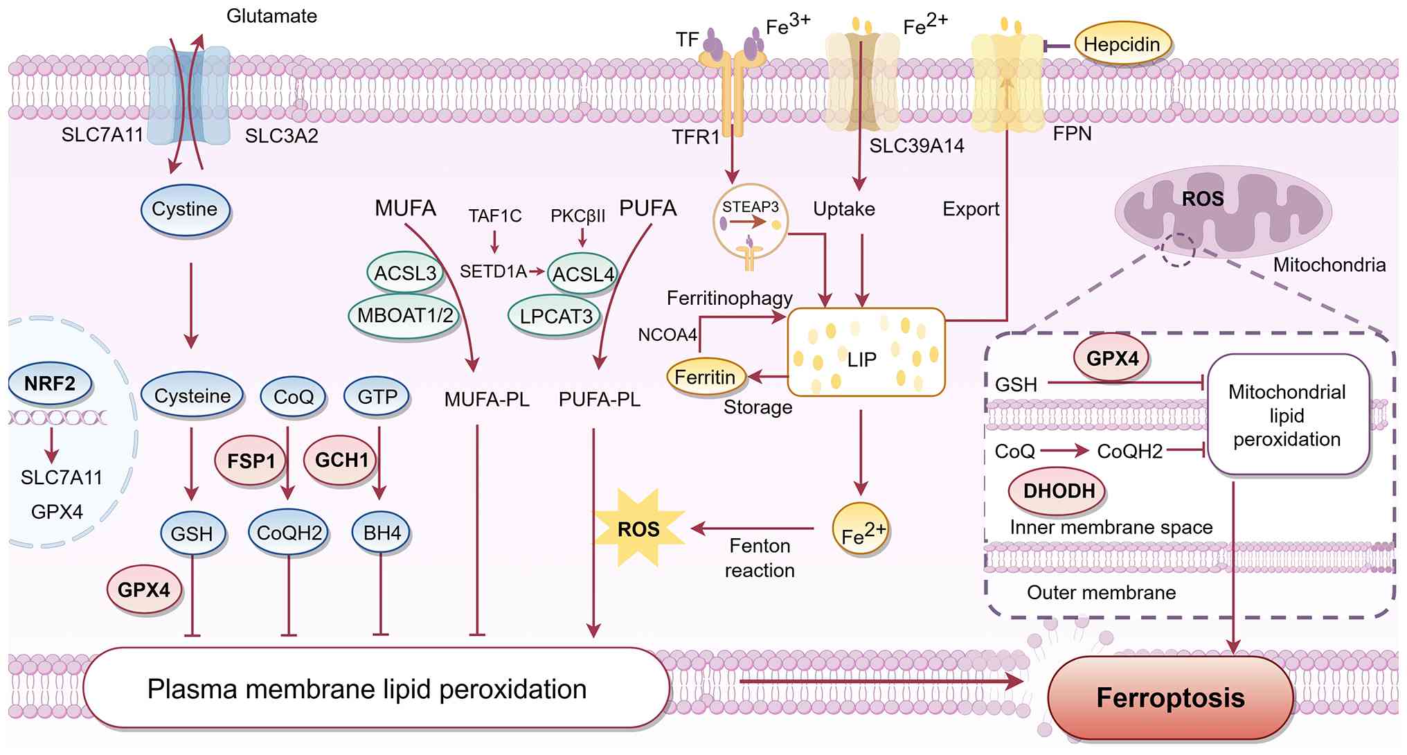
The enrichment of PUFAs in membrane PLs provides the
necessary substrates for lipid peroxidation (22,31) (Fig. 1). Central to this process is
acyl-CoA synthetase long-chain family member 4 (ACSL4), which is
key to PUFA activation. In collaboration with
lysophosphatidylcholine acyltransferase 3, ACSL4 esterifies FAs
into membrane PLs, thereby increasing membrane susceptibility to
peroxidation. ACSL4 activity is a well-recognized determinant of
ferroptosis sensitivity and its inhibition confers substantial
protection, making it a potential preclinical target for MASLD
therapy (32-35). This pro-ferroptotic program is
amplified by the protein kinase C βII-ACSL4 axis (36). Beyond these direct regulators,
recent genome-wide clustered regularly interspaced short
palindromic repeats screening has identified the TATA box binding
protein associated factor RNA polymerase I subunit C (TAF1C) as an
upstream epigenetic determinant of ferroptosis in MASLD (37). TAF1C is upregulated during
disease progression and directly interacts with SET
domain-containing 1A to remodel histone methylation and
acetylation, thereby enhancing ACSL4 expression. The resulting
increase in ACSL4 promotes lipid peroxidation and sensitizes
hepatocytes to ferroptosis (37). Conversely, incorporation of
monounsaturated FAs (MUFAs) into PLs shifts the lipid composition
toward ferroptosis resistance. This protective lipid remodeling is
mediated by acyl-CoA synthetase long-chain family member 3 (ACSL3),
which preferentially incorporates MUFAs into PLs and decreases
susceptibility to lipid peroxidation (38,39). Membrane-bound O-acyltransferase
domain-containing proteins 1 and 2 are key regulators of PL
remodeling and modulate ferroptosis by increasing MUFA-PL while
decreasing PUFA-PL levels (40).
These findings have direct therapeutic implications, as
non-specific manipulation of lipid metabolism may produce
unintended effects. Future studies should include comprehensive
lipidomic profiling to define the specific PL patterns that
determine ferroptosis susceptibility in the liver in MASLD.
Iron homeostasis comprises three interconnected
compartments: Systemic/intestinal handling, macrophage/Kupffer cell
(KC) recycling and hepatocyte iron homeostasis (27). At the systemic level, dietary
iron is absorbed by enterocytes and exported into the circulation
via ferroportin (FPN). Hepcidin binds FPN and promotes its
internalization and degradation, thereby decreasing both intestinal
iron absorption and iron release from macrophages and hepatocytes
(41,42). The hepatic hepcidin/intestinal
hypoxia-inducible factor-2α pathway modulates iron absorption in
response to systemic iron status (43). Hepcidin transcription is
regulated by bone morphogenetic protein (BMP)/SMAD signaling in
hepatocytes, with BMP6 serving as the key iron-responsive ligand
and transmembrane serine protease 6 serving as a negative regulator
(41,44-46). In inflammatory states, IL-6
activates STAT3, which directly stimulates hepcidin transcription
(47,48). Elevated hepcidin promotes FPN
degradation, thereby trapping iron within splenic and hepatic
macrophages, duodenal enterocytes and hepatocytes (49). In MASLD, hepcidin levels are
typically low relative to hepatic iron stores (50), although inflammatory signals may
simultaneously stimulate hepcidin production. The relative
contribution of hepcidin suppression vs. inflammation-driven
hepcidin elevation may vary across disease stages and patient
subgroups. For example, hepcidin suppression may predominate in
earlier or less inflammatory disease, whereas inflammation-driven
hepcidin induction may become more prominent in advanced disease
and in patients with a higher inflammatory burden. Beyond the liver
and intestine, the kidney contributes to systemic iron homeostasis
by reabsorbing filtered iron and clearing hepcidin (51), although its specific role in
MASLD remains to be defined.
KCs, liver-resident macrophages, recycle iron from
senescent erythrocytes and export it back into the circulation via
FPN. Under conditions of low hepcidin, this export is enhanced,
contributing to the circulating iron pool. Conversely, when
hepcidin is elevated, FPN is degraded and iron is retained within
macrophages (49). In MASLD,
dysregulated iron handling leads to iron accumulation in KCs.
Iron-rich KCs exhibit phenotypical changes toward a
pro-inflammatory and pro-fibrotic state during MASH progression
(52). A recent study (53) identified neutrophil cytosolic
factor (NCF)1 as a key regulator. Its upregulation in liver
macrophages causes iron overload and ferroptosis in KCs.
NCF1-induced oxidized PLs activate toll-like receptor
(TLR)4-dependent hepatocyte hepcidin production, which increases
iron deposition in KCs, forming a positive feedback loop that
amplifies ferroptosis and MASH (53). Notably, not all hepatic
macrophages experience iron overload in MASLD. Embryonic KCs
exhibit decreased labile iron pools (LIPs) and mitochondrial
dysfunction in MASH and KC-specific FPN knockout aggravates MASH
(54). This highlights the
heterogeneity of iron handling across macrophage populations.
Hepatocytes take up transferrin-bound iron via
transferrin receptor 1. Within acidified endosomes, six
transmembrane epithelial antigen of prostate 3 converts
Fe3+ to Fe2+, which is transported into the
cytosol by solute carrier family 11 member 2 (SLC11A2) (55,56). Under iron overload conditions,
uptake of non-transferrin-bound iron via SLC39A14 increases
(57). Intracellularly, excess
iron is sequestered in ferritin, which serves both as a reservoir
and as a protective buffer against the Fenton reaction (58). When iron demand increases or
oxidative stress rises, ferritin is selectively degraded through
nuclear receptor coactivator 4-dependent ferritinophagy, releasing
Fe2+ into the LIP (59,60). Expansion of the LIP facilitates
ferroptosis by supplying the redox-active iron required for the
Fenton reaction and subsequent lipid peroxidation (61-63). The mitochondrial LIP acts as a
major catalyst of Fenton reactions, generating reactive oxygen
species (ROS) that trigger and sustain lipid peroxidation, thereby
directly promoting ferroptosis (64-66). Iron efflux from hepatocytes is
mediated by FPN, which is downregulated by hepcidin (41,42). These alterations in iron handling
manifest as elevated serum ferritin concentrations and increased
hepatic iron levels (67-69).
Notably, a recent study (70)
demonstrated that decreased FPN expression in hepatocytes leads to
iron accumulation and systemic metabolic disturbance, identifying
fetuin-A and leukocyte cell-derived chemotaxin 2 as key
hepatokines. These findings are crucial for elucidating the
association between MASLD and metabolic disorders and suggest
therapeutic strategies such as hepatokine silencing, FPN
overexpression or iron chelation (70). In addition, iron serves as a
potent pro-oxidant, and its accumulation in the liver increases ROS
production, lipid peroxidation and subsequent cell damage (71,72). The resulting oxidative stress and
inflammation exacerbate the progression of liver fibrosis and
cirrhosis, underscoring the key role of iron metabolism in MASLD
pathogenesis (73).
Ferroptosis is controlled by antioxidant pathways,
including the GSH-GPX4 axis, the FSP1/CoQ10 pathway and the
GCH1-BH4 axis. These pathways are key for maintaining cell redox
homeostasis and preventing the accumulation of harmful lipid
peroxides (74). Central to the
GSH-GPX4 axis is the cystine/glutamate antiporter SLC7A11, which
imports cystine, a key component for synthesizing GSH (75). GPX4 uses GSH to transform PL
hydroperoxides into non-toxic lipid alcohols, thereby preventing
ferroptosis (76). However, in
MASLD, GSH depletion and GPX4 inactivation are typically observed
(31). In parallel, FSP1 reduces
CoQ10 to ubiquinol at the plasma membrane and other compartments,
trapping lipid radicals independently of GSH (77,78). The GCH-BH4 axis also serves a key
role in ferroptosis regulation. GCH1 and its metabolic derivative
BH4 are involved in lipid remodeling that maintains the pool of PLs
containing PUFA tails, counteracting their loss during ferroptosis
and thereby conferring resistance to ferroptosis (79). NRF2 is key for orchestrating the
antioxidant response and preventing ferroptosis. Upon activation,
NRF2 upregulates genes associated with GSH synthesis and iron
metabolism such as SLC7A11 and GPX4, protecting against oxidative
stress and ferroptosis (80,81).
Mitochondrial dysfunction is a key feature of MASLD.
Iron overload causes mitochondrial damage, characterized by
increased ROS production and impaired mitochondrial respiration,
which may exacerbate liver injury (82,83). Mitochondria are also involved in
controlling ferroptosis. Mitochondrial ROS, particularly those
derived from the electron transport chain (ETC), are key
contributors to lipid peroxidation and ferroptosis (84,85), which are associated with
mitochondrial metabolism. The tricarboxylic acid cycle and ETC
maintain mitochondrial membrane potential and redox balance, which
are essential for ferroptosis (86). Mitochondria also employ a key
defense network against ferroptosis. Central to this system is the
transport of GSH into the mitochondrial matrix, mediated by
specific carrier proteins such as the dicarboxylate carrier and the
oxoglutarate carrier. This import is required for the proper
functioning of mitochondrial GPX4. Blocking these carriers
increases ferroptosis by elevating mitochondrial ROS and depleting
GSH (87). In addition,
dihydroorotate dehydrogenase operates within the inner
mitochondrial membrane, where it catalyzes the reduction of
ubiquinone to ubiquinol, which directly mitigates lipid
peroxidation independently of the GSH-GPX4 axis (88,89).
Collectively, the metabolic environment in MASLD is
characterized by a lipid composition that permits ferroptosis,
excess active iron and weakened antioxidant defenses. These factors
increase the likelihood of ferroptosis (Fig. 1).
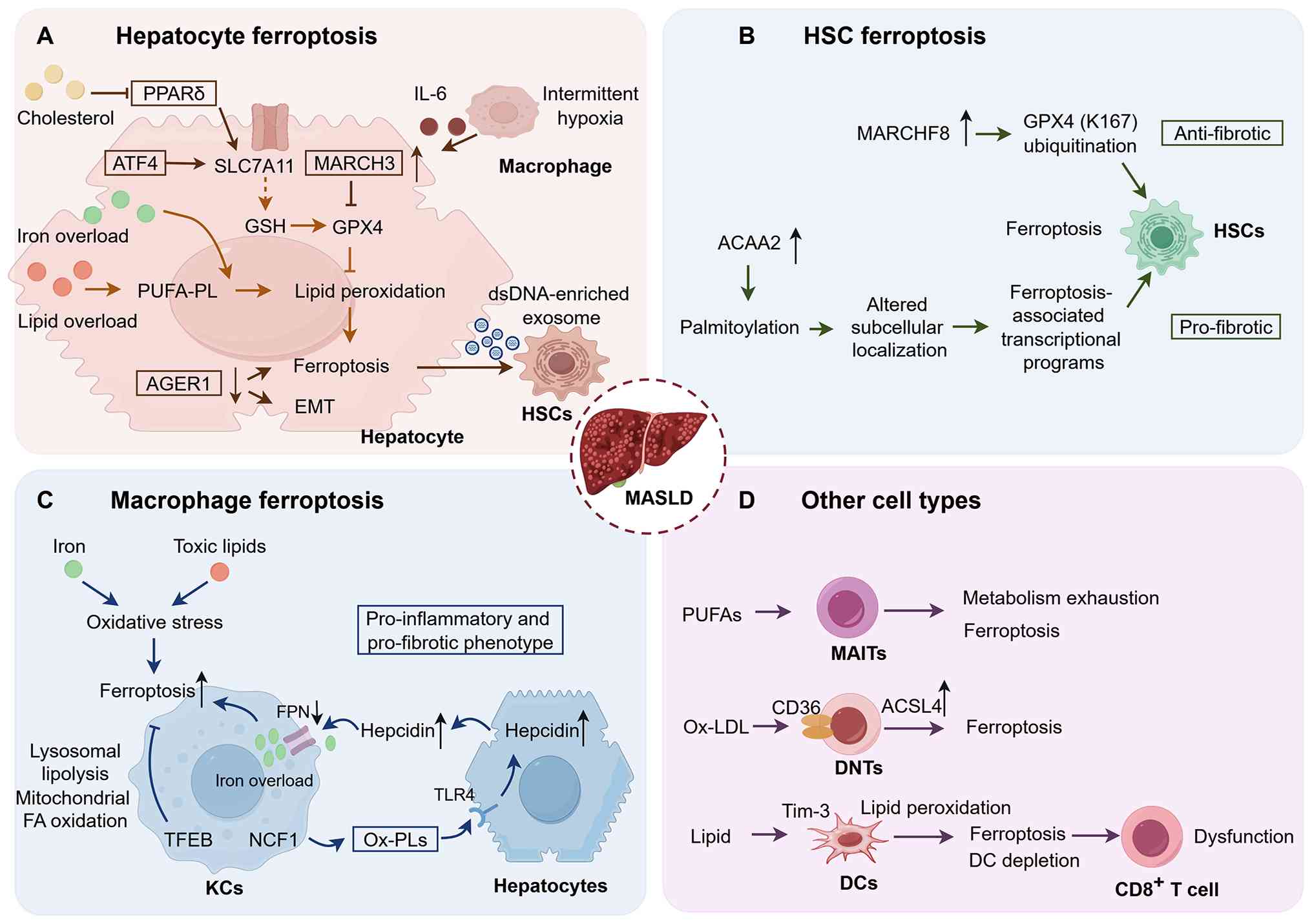
In MASLD, hepatocyte ferroptosis is a key
contributor to liver inflammation and fibrosis (15,83,90). Studies highlight its role in
amplifying oxidative stress and activating fibrogenic pathways
(15,83,90). In particular, the accumulation of
free FAs (FFAs) in hepatocytes is a hallmark of MASLD. This
accumulation triggers lipid peroxidation and ferroptosis, thereby
intensifying liver damage (91)
(Fig. 2). Notably, another study
demonstrated the protective role of activating transcription factor
4 in normal hepatocytes, where it maintains GSH production and
inhibits ferroptosis-dependent inflammatory cell death (92).
In MASLD, decreased GPX4 levels are associated with
increased vulnerability to ferroptosis, consequently enhancing
hepatic oxidative stress and promoting fibrogenesis (15). Moreover, the vulnerability of
hepatocytes to ferroptosis is modulated by immune-derived signals.
Under intermittent hypoxia, a prevalent comorbidity in MASLD, IL-6
produced by macrophages induces upregulation of the E3 ubiquitin
ligase membrane-associated RING finger protein 3 (MARCH3) in
hepatocytes. MARCH3 facilitates ubiquitination and degradation of
GPX4, increasing hepatocyte susceptibility to ferroptosis (94). Similarly, in MASH with T2DM,
advanced glycation end product receptor 1 (AGER1) deficiency
initiates hepatocyte ferroptosis and promotes
epithelial-mesenchymal transition, thereby promoting fibrogenesis.
Conversely, restoring AGER1 mitigates these effects (95). Collectively, hepatocyte
ferroptosis serves a key role in the development of MASLD as it
promotes oxidative stress, inflammation and fibrogenesis, thereby
contributing to disease progression and complications.
HSCs are the primary cause of liver fibrosis, as
their transdifferentiation into myofibroblasts leads to excessive
extracellular matrix production (96). The role of ferroptosis in HSCs is
context-dependent, with evidence pointing to opposing outcomes
(97,98). Ferroptosis in HSCs can inhibit
HSC activation and reduce liver fibrosis. On the other hand,
ferroptosis-related programs in HSCs can exacerbate fibrogenesis.
Quercetagetin (QG), the active compound in the Tibetan medicine
Huagan Tongluo Fang, induces ferroptosis in HSCs by promoting
ubiquitin-proteasome-mediated degradation of GPX4. This is mediated
by MARCHF8 and ultimately prevents HSC activation and reduces liver
fibrosis (97). Conversely,
other ferroptosis-related programs in HSCs promote fibrogenesis.
Han et al (98) showed
that acetyl-CoA acyltransferase 2 (ACAA2), a mitochondrial enzyme
involved in FA β-oxidation, is markedly upregulated in HSCs during
MASLD-associated fibrosis. ACAA2 undergoes palmitoylation, a
modification that controls its subcellular localization and
enhances ferroptosis-associated transcriptional programs, thereby
exacerbating hepatic fibrosis. Inhibiting ACAA2 or blocking its
palmitoylation attenuates both ferroptosis and fibrogenesis, an
effect associated with AMP-activated protein kinase (AMPK) pathway
activation and suppression of HSC activation (98). Rather than merely eliminating
HSCs, ACAA2-driven ferroptosis triggers a pro-inflammatory and
pro-fibrotic cascade, or occurs within a metabolic state that that
enhances fibrogenic signaling following cellular death. These
divergent outcomes may be determined by several contextual factors,
including the HSC activation state, disease stage, experimental
model and local microenvironment. These observations demonstrate
that inducing HSC ferroptosis cannot yet be regarded as a generally
applicable antifibrotic strategy until the biological determinants
governing its net tissue effect are defined.
KCs are liver-resident macrophages that serve a key
role in maintaining hepatic homeostasis and responding to liver
injury (99). In MASLD,
iron-rich KCs exhibit alterations associated with inflammation and
fibrosis, such as upregulated CD11c and proinflammatory and
profibrotic genes (Tnfa, Ccl3, and Timp1) and activation of MiT/TFE
transcription factors (52).
NCF1 is elevated in liver macrophages and dendritic cells (DCs) in
patients with MASLD and in MASH mice. This upregulation promotes
iron overload and ferroptosis in KCs, exacerbating MASH progression
(53). Oxidized PLs released by
macrophages activate TLR4-dependent production of hepcidin in
hepatocytes, which in turn increases iron accumulation in KCs and
initiates ferroptosis (53).
These findings suggest targeting NCF1 may improve KC function and
limit MASH progression.
KCs exhibit abnormal lipid metabolism in MASLD,
leading to the accumulation of toxic lipids and a pro-inflammatory
state (100). Lipid-induced
stress promotes ferroptosis and further contributes to liver
injury. Importantly, induction of transcription factor EB in KCs
protects against ferroptosis by enhancing lysosomal lipolysis and
mitochondrial FA oxidation, decreasing oxidative stress and
maintaining KC function (101).
This highlights the potential of targeting lipid metabolism in KCs
to prevent ferroptosis in MASLD.
Beyond hepatocytes, HSCs and KCs, ferroptosis may
also influence other liver cell populations; for example,
accumulation of PUFAs in mucosal-associated invariant T (MAIT)
cells causes ferroptosis-driven metabolic exhaustion, impairing
their anti-tumor surveillance and accelerating disease progression
(22). Similarly, oxidized
low-density lipoprotein induces ferroptosis in double-negative
regulatory T cells via CD36-mediated lipid uptake, decreasing their
survival and immunosuppressive capacity and thereby exacerbating
liver inflammation (102).
DCs are the main antigen-presenting cells and are
particularly susceptible to ferroptosis in MASLD. Li et al
(103) demonstrated that, in
MASLD-related HCC, a lipid-rich environment promotes lipid
peroxidation and ferroptosis in DCs via a T cell immunoglobulin and
mucin domain-containing protein 3 (Tim-3)-dependent pathway. This
depletes DCs, impairs CD8+ T cell response and
facilitates tumor growth. Genetic or pharmacological inhibition of
Tim-3 decreases lipid peroxidation, improves DC survival,
reactivates CD8+ T cells and slows tumor growth. Tim-3
blockade enhances the efficacy of anti-PD-1 therapy and lenvatinib,
achieving sustained tumor control in preclinical models (103,104). These findings identify Tim-3 as
a key regulator of DC ferroptosis in metabolic liver disease.
A disease stage-specific framework may clarify when
ferroptosis should be inhibited or induced. In early MASLD and in
MASH with fibrosis, ferroptosis in hepatocytes and KCs is
detrimental and should be inhibited. By contrast, selective
induction of ferroptosis in activated HSCs may promote fibrosis
resolution, although this strategy requires caution because HSC
ferroptosis can be pro-fibrotic depending on context. During HCC
progression, ferroptosis in immune cells such as DCs and MAIT and
regulatory T cells impairs anti-tumor immunity. At this stage,
protecting these immune cells or inducing ferroptosis specifically
in malignant hepatocytes may be preferable. The consequences of
ferroptosis vary by cell type and disease stage; therapeutic
strategies should be designed accordingly.
A challenge in linking ferroptosis to MASLD is the
lack of specific and tractable markers. At the tissue level, Cui
et al (105) identified
hyperoxidized peroxiredoxin 3 (PRDX3) as a direct ferroptosis
marker in chronic liver diseases (Table I). In liver tissue sections,
PRDX3 hyperoxidation is associated with disease severity (105), providing a tool for identifying
ferroptotic lesions. This is valuable for staging MASLD. However,
because it requires invasive tissue sampling, its use is confined
to research settings and serial monitoring is not feasible.
Recent studies have increasingly focused on
inhibiting ferroptosis in hepatocytes as a strategy to decrease
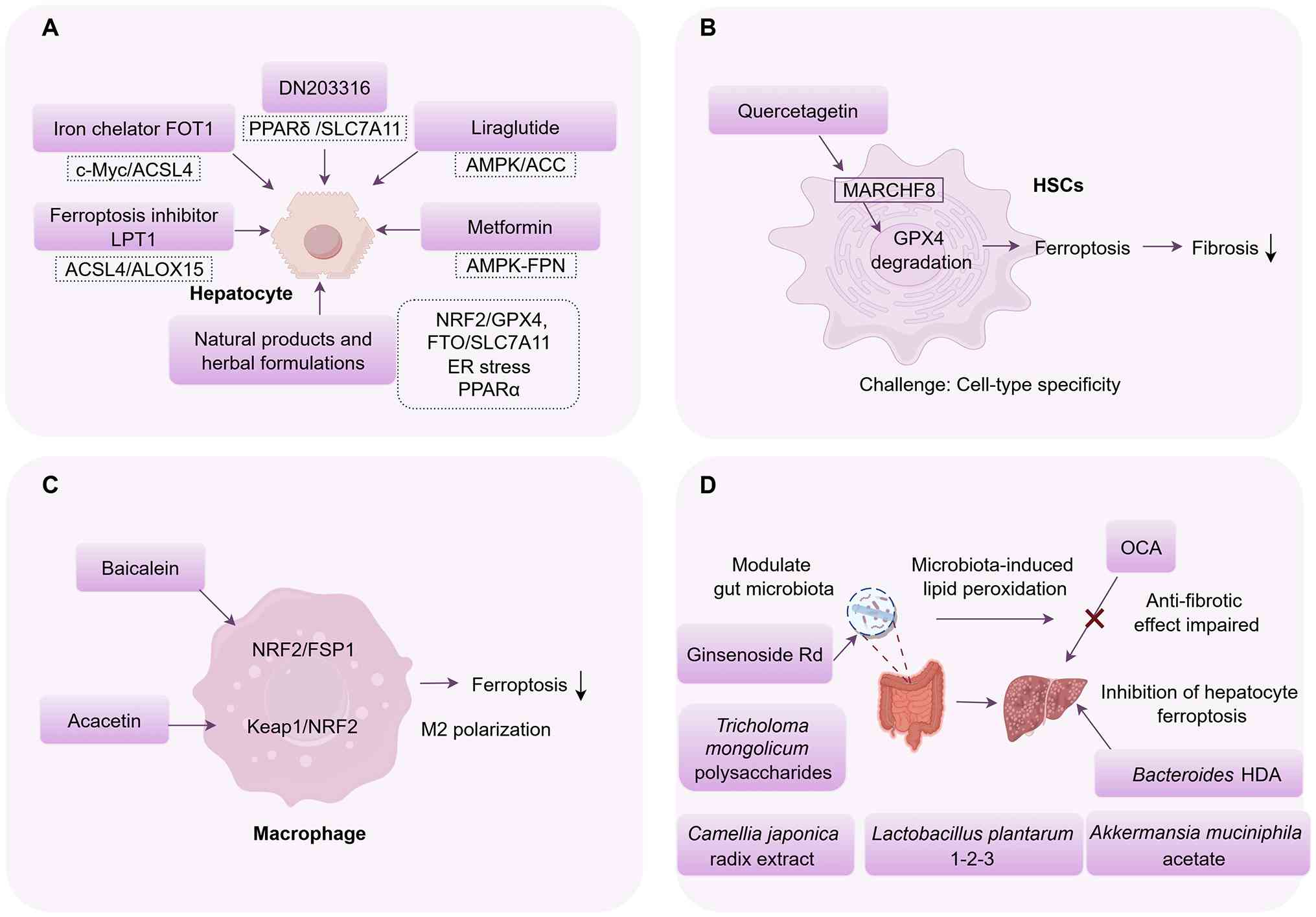
liver injury in MASLD (15,26,67). Several natural products and
herbal formulations attenuate ferroptosis in MASLD models (112-114), although the strength of
evidence varies (Fig. 3). Direct
evidence of ferroptosis has been provided for a limited number of
compounds, including betaine, plant sterol ester of α-linolenic
acid, protocatechuic acid and arbutin. These compounds decrease
erastin- or RAS-selective lethal 3-induced ferroptosis and modulate
NRF2/GPX4 or fat mass and obesity-associated protein/SLC7A11
signaling in hepatocytes (115-118). By contrast, other natural
products and herbal formulations including Hu Gan Tang, diosgenin,
puerarin and gastrodin have primarily been shown to attenuate
ferroptosis-associated molecular changes (112,119-121). For these compounds, the
evidence supports an association with ferroptosis-related pathways
rather than establishing ferroptosis as the causal mechanism. A
common limitation of most natural products is the lack of
information on oral bioavailability, tissue distribution and
long-term safety, which hinders clinical translation. Additionally,
many studies use the methionine-choline-deficient (or high-fat diet
models that do not fully recapitulate human MASLD (Table II) (113-114,122-127).
Clinically approved drugs with anti-ferroptotic
properties have translation potential. Liraglutide improves MASLD
in mouse models (128,129) by activating the AMPK/acetyl-CoA
carboxylase (ACC) pathway and attenuating ferroptosis-associated
markers (129). Similarly,
metformin, a first-line antidiabetic agent, mitigates hepatic iron
overload via the AMPK/FPN pathway, reducing iron availability for
Fenton reactions and lipid peroxidation (130). To the best of our knowledge,
however, their anti-ferroptotic effects in MASLD have been
demonstrated only in animal models, and clinical validation is
lacking.
Although inhibiting ferroptosis in hepatocytes is
key to limiting liver injury and MASLD progression, selectively
inducing ferroptosis in activated HSCs may offer a strategy to
reverse fibrosis. As aforementioned, QG induces ferroptosis in HSCs
by promoting ubiquitination and degradation of GPX4, thereby
decreasing collagen deposition in a MASLD mouse model (97). However, the effect of HSC
ferroptosis depends on multiple factors, such as HSC activation
state, disease stage and the local microenvironment. In addition,
most preclinical studies on HSC ferroptosis in liver fibrosis use
carbon tetrachloride or thioacetamide-induced models (132-138), which do not fully recapitulate
the metabolic and inflammatory context of MASLD. Achieving cell
type specificity also remains a challenge, and future work should
focus on HSC-targeted delivery systems. Therefore, induction of HSC
ferroptosis should not be regarded as a generally applicable
antifibrotic strategy. Its therapeutic potential requires
validation in more disease-relevant models before clinical
translation.
In a mouse model of MASH, baicalein, a traditional
Chinese medicine extracted from Scutellaria baicalensis,
improves metabolic dysfunction and slows disease progression.
Baicalein restores hepatic iron balance and decreases iron overload
and lipid peroxidation (139,140). Its anti-ferroptotic effects
have been confirmed in Raw264.7 macrophages exposed to FFAs
(140). Mechanically, baicalein
suppresses M1 macrophage polarization and promotes M2 polarization
by activating the NRF2/FSP1 signaling pathway, thereby decreasing
ferroptosis (140). Similarly,
acacetin, a natural flavonoid, alleviates MASLD through mechanisms
involving two distinct cell types: In hepatocytes, acacetin reduces
Notch1-mediated lipogenesis and necroptosis; in macrophages, it
blocks ferroptosis through the Keap1/NRF2 signaling pathway and
promotes M2 polarization (141). These findings suggest targeting
macrophage ferroptosis may represent a potential therapeutic
strategy for MASLD.
The gut microbiome serves a key role in the onset
and progression of MASLD and its complications. Changes in
microbiome composition are associated with disease progression and
interventions targeting the microbiome can influence disease
outcomes through several mechanisms, such as increasing gut
permeability, decreasing levels of endotoxin-producing bacteria and
modulating bile acid metabolism (142-146).
Metabolites from certain gut microbial strains
directly or indirectly inhibit hepatic ferroptosis. For example,
Zhang et al (150)
demonstrated that hexadecanedioic acid, a metabolite produced by
Bacteroides uniformis, is transported to the liver via the
portal vein, where it inhibits the inositol-requiring enzyme
1α/spliced X-box binding protein 1 signaling pathway. This
inhibition alleviates liver fibrosis and suppresses
ferroptosis-associated molecular changes (150). The aforementioned study
provides novel insight into the links between gut microbiota,
endoplasmic reticulum stress and hepatocyte ferroptosis. In
addition, Lactobacillus plantarum 1-2-3 has beneficial
effects on lipid metabolism and inflammation and protects against
MASLD by modulating gut microbiota and metabolic pathways (151). Furthermore, acetate produced by
Akkermansia muciniphila activates the hepatic
AMPK/SIRT1/PPAR gamma coactivator 1 alpha (PGC-1α) signaling
pathway, thereby suppressing ferroptosis-associated signaling and
ameliorating MASLD (152).
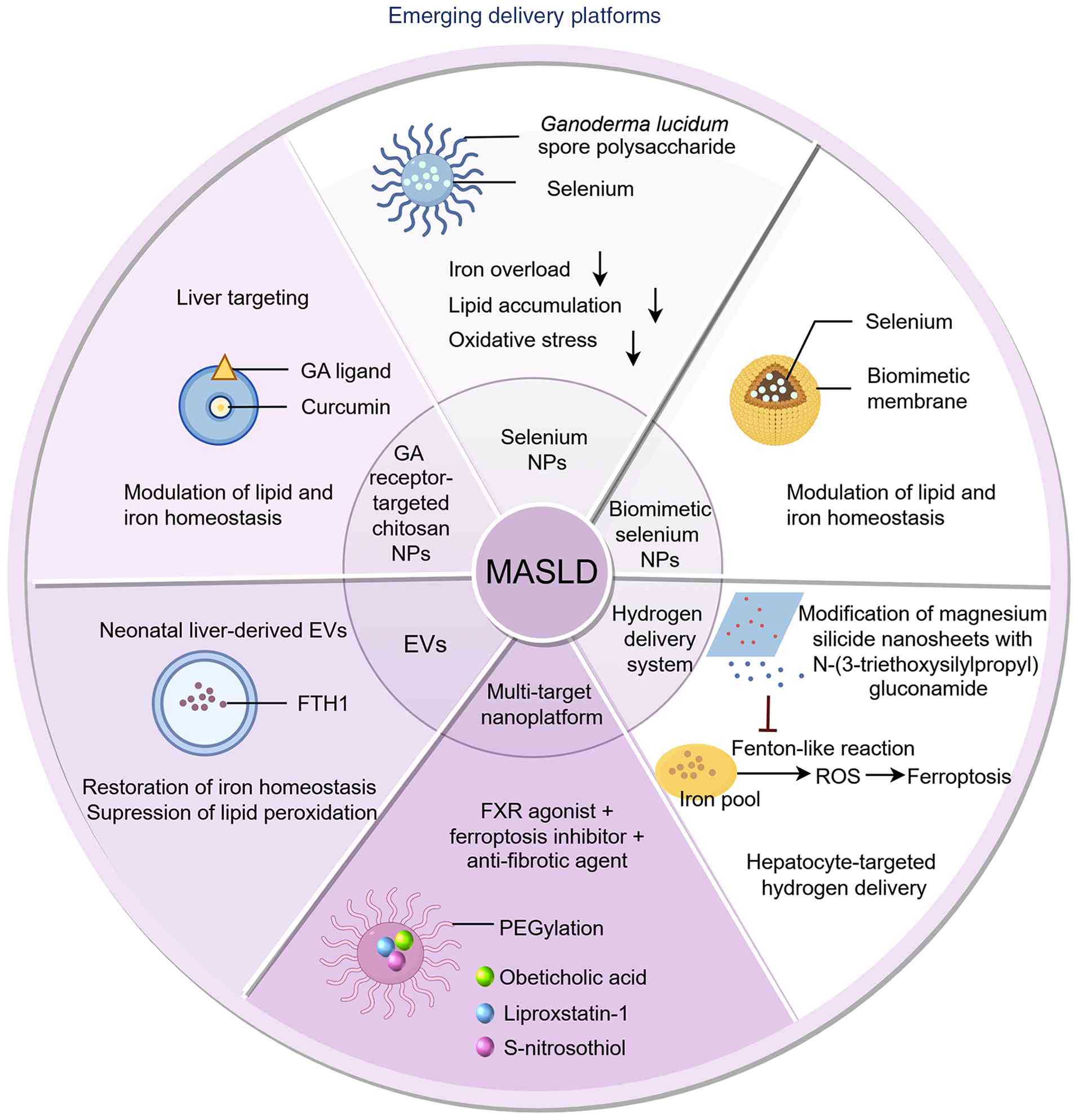
Nanotechnology-based delivery systems have been
developed to facilitate the delivery of multiple therapeutic agents
and improve therapeutic outcomes. For example, selenium
nanoparticles (NPs) stabilized with Ganoderma lucidum spore
polysaccharides prevent MASLD in HepG2 cells, primarily through
their antioxidant properties (155). Similarly, biomimetic selenium
nanocomposites that modulate lipid and iron homeostasis alleviate
MASH in choline-deficient, L-amino acid-defined, high-fat diet
induced MASH mice (156). These
selenium-based systems exploit the natural redox properties of
selenium and improve hepatic distribution. Targeted nanocarriers
have also been developed. For example, glycyrrhetinic acid
receptor-targeted chitosan oligosaccharide NPs improve MASLD by
inhibiting ferroptosis-associated signaling, although the exact
anti-ferroptotic mechanism remains incompletely understood
(157). In a more advanced
approach, a single nanotherapeutic platform was designed to
co-deliver an farnesoid X receptor agonist, a ferroptosis inhibitor
and an anti-fibrotic agent for combination treatment of MASH,
achieving greater efficacy than any single component alone
(158). This multi-target
strategy reflects the simultaneous involvement of metabolic,
oxidative and fibrotic processes in MASLD and demonstrates how
nanotechnology integrates these components into a single
formulation.
Hepatocyte-targeted hydrogen delivery has also been
developed to neutralize free radicals and inhibit the Fenton
reaction, thereby preventing heme-induced lipid peroxidation
(159). However, these
strategies remain at the preclinical stage and represent a
potential non-drug approach, although clinical feasibility remains
to be established. In addition, a recent study reported the
therapeutic potential of extracellular vesicles enriched with
ferritin heavy chain 1 in mitigating ferroptosis-associated
molecular changes and treating MASLD (160) (Fig. 4). This is consistent with a
growing body of literature (161-163) highlighting the role of EVs in
modulating cell processes and influencing disease outcomes.
Targeting ferroptosis with iron chelators and
inhibitors has promise in restoring redox and lipid homeostasis,
suggesting a potential therapeutic strategy for MASLD (83). However, several barriers must be
addressed before ferroptosis-targeted therapies can be translated
into clinical practice.
A notable challenge is the incomplete understanding
of the molecular mechanisms and regulatory networks governing
ferroptosis in MASLD. Recent studies (15,83,164) have identified several pathways
linking ferroptosis with MASLD progression, but how ferroptosis
influences different disease stages remains poorly defined.
Moreover, MASLD is not a single condition but a spectrum of liver
disorders and ferroptosis may exert distinct effects at different
stages. Treatment development therefore requires a stage-specific
approach. In addition, systemic interactions between the liver,
adipose tissue and gut microbiota further modulate ferroptosis
susceptibility, offering opportunities for precision therapy.
Identifying reliable biomarkers of ferroptosis in
MASLD is an active area of research. New biomarkers may enable
early detection and monitoring of ferroptosis, but their clinical
validation remains at an early stage and further studies are needed
to establish their efficacy and reliability (83). Advanced tools such as imaging
probes that detect ferroptosis within specific cell compartments
would also substantially advance the field. For example, a
viscosity-sensitive near-infrared fluorescent probe has been
developed for sensitive visualization of lipid peroxidation in
ferroptotic hepatocytes and dual-channel probes have been designed
to detect multiple oxidative stress markers during ferroptosis
(165,166). These platforms remain
preclinical, but may accelerate target validation and drug efficacy
assessment in MASLD research.
The development of ferroptosis-targeting therapy
shows promise, but clinical evaluation is key to ascertain safety
and efficacy. Future therapeutic strategies should not aim merely
to modulate ferroptosis systemically but should achieve
cell-specific modulation with precise spatial and temporal control.
This requires improved targeted delivery systems, dynamic
biomarkers and a detailed map of communication pathways between
diseased cells and their neighbors. For example, recent advances in
nanotechnology have produced nanoagents that induce ferroptosis in
tumor cells while sparing immune cells, providing a model for
analogous strategies in MASLD, where immune cell preservation is
key (167). In addition, the
interactions between ferroptosis and necroptosis, pyroptosis and
autophagy in MASLD remain largely unknown. Understanding of how
these cell death pathways reinforce or compensate for one another
is needed to predict the net effect of ferroptosis-targeting
therapy.
Notably, the daidzein intervention on liver
histopathology in patients with MASLD trial (trial no.
NCT07623044), a Phase 2 study that is not yet recruiting, aims to
evaluate the potential of short-term perioperative daidzein
intervention in improving liver tissue health in patients with
MASLD. The primary endpoint is the NAFLD Activity Score (168), with exploratory analyses
examining GPX4, ACSL4 and intrahepatic CD8+ T cell
infiltration. These secondary assessments aim to clarify whether
daidzein exerts measurable effects on ferroptosis-associated
pathways and the hepatic immune microenvironment. If successfully
completed, this trial may provide human tissue-level evidence for
ferroptosis modulation by a natural product in MASLD. Although
preclinical findings are promising, the clinical use of
ferroptosis-targeting strategies for MASLD is in its early stages.
More clinical trials are needed to evaluate the therapeutic
benefits of ferroptosis-targeting intervention for patients with
MASLD, which is essential for translating basic findings into
clinical practice.
In summary, although preclinical studies have
demonstrated promising results, clinical evidence for
ferroptosis-targeted therapy in MASLD remains limited, with most
interventions still in early preclinical development. A deeper
understanding of how ferroptosis interacts with other cell
processes across different cell types, organs and disease stages is
needed. Such work will help identify more precise molecular targets
and support the development of cell-specific interventions that
mitigate the harmful effects of ferroptosis while preserving its
physiological roles. Clinical translation requires rigorous
evaluation of efficacy and safety in well-designed trials, but this
must follow adequate preclinical validation and should not be
interpreted as imminent clinical readiness. These efforts may
advance understanding of MASLD pathogenesis and inform the
development of novel therapeutic strategies to improve patient
outcomes.
Not applicable.
XT wrote and revised the manuscript. ML revised the
manuscript. All authors have read and approved the final
manuscript. Data authentication is not applicable.
Not applicable.
Not applicable.
The authors declare that they have no competing
interests.
Not applicable.
The present study was supported by Hubei Provincial Natural
Science Foundation of China (grant no. 2026AFB252) and Research
Foundation of Tongji Hospital of Tongji Medical College, Huazhong
University of Science and Technology (grant no. 2024B32).
|
1
|
Huang DQ, Wong VWS, Rinella ME, Boursier
J, Lazarus JV, Yki-Järvinen H and Loomba R: Metabolic
dysfunction-associated steatotic liver disease in adults. Nat Rev
Dis Primers. 11:142025. View Article : Google Scholar : PubMed/NCBI
|
|
2
|
Stefan N, Yki-Järvinen H and
Neuschwander-Tetri BA: Metabolic dysfunction-associated steatotic
liver disease: Heterogeneous pathomechanisms and effectiveness of
metabolism-based treatment. Lancet Diabetes Endocrinol. 13:134–148.
2025. View Article : Google Scholar
|
|
3
|
Younossi ZM, Kalligeros M and Henry L:
Epidemiology of metabolic dysfunction-associated steatotic liver
disease. Clin Mol Hepatol. 31(Suppl): S32–S50. 2025. View Article : Google Scholar :
|
|
4
|
Tincopa MA, Speliotes EK, Valenti L and
Loomba R: Metabolic dysfunction-associated steatotic liver disease
and metabolic dysfunction-associated steatohepatitis-related
fibrosis: Therapeutic options and approaches to treatment. Annu Rev
Med. 77:103–115. 2026. View Article : Google Scholar : PubMed/NCBI
|
|
5
|
European Association for the Study of the
Liver (EASL); European Association for the Study of Diabetes
(EASD); European Association for the Study of Obesity (EASO):
EASL-EASD-EASO clinical practice guidelines on the management of
metabolic dysfunction-associated steatotic liver disease (MASLD). J
Hepatol. 81:492–542. 2024. View Article : Google Scholar : PubMed/NCBI
|
|
6
|
Do A, Zahrawi F and Mehal WZ: Therapeutic
landscape of metabolic dysfunction-associated steatohepatitis
(MASH). Nat Rev Drug Discov. 24:171–189. 2025. View Article : Google Scholar
|
|
7
|
Arvanitakis K, Koufakis T, Cholongitas E,
Francque S and Germanidis G: Insights into the results of
resmetirom trials: Can a thyroid hormone receptor agonist be the
holy grail of MASH therapy? Pharmacol Ther. 268:1088112025.
View Article : Google Scholar : PubMed/NCBI
|
|
8
|
Noureddin M, Charlton MR, Harrison SA,
Bansal MB, Alkhouri N, Loomba R, Sanyal AJ and Rinella ME: Expert
panel recommendations: Practical clinical applications for
initiating and monitoring resmetirom in patients With MASH/NASH and
moderate to noncirrhotic advanced fibrosis. Clin Gastroenterol
Hepatol. 22:2367–2377. 2024. View Article : Google Scholar : PubMed/NCBI
|
|
9
|
Gallage S, Govaere O, Anstee QM, Shulman
GI, Tacke F and Heikenwalder M: Current therapeutic landscape and
future treatment perspectives of MASH. Nature. 657:76–91. 2026.
View Article : Google Scholar : PubMed/NCBI
|
|
10
|
Desjardins EM, Wu J, Lavoie DCT, Ahmadi E,
Townsend LK, Morrow MR, Wang D, Tsakiridis EE, Batchuluun B,
Fayyazi R, et al: Combination of an ACLY inhibitor with a GLP-1R
agonist exerts additive benefits on nonalcoholic steatohepatitis
and hepatic fibrosis in mice. Cell Rep Med. 4:1011932023.
View Article : Google Scholar : PubMed/NCBI
|
|
11
|
Mederacke I, Filliol A, Affo S, Nair A,
Hernandez C, Sun Q, Hamberger F, Brundu F, Chen Y, Ravichandra A,
et al: The purinergic P2Y14 receptor links hepatocyte death to
hepatic stellate cell activation and fibrogenesis in the liver. Sci
Transl Med. 14:eabe57952022. View Article : Google Scholar : PubMed/NCBI
|
|
12
|
Gaul S, Leszczynska A, Alegre F, Kaufmann
B, Johnson CD, Adams LA, Wree A, Damm G, Seehofer D, Calvente CJ,
et al: Hepatocyte pyroptosis and release of inflammasome particles
induce stellate cell activation and liver fibrosis. J Hepatol.
74:156–167. 2021. View Article : Google Scholar
|
|
13
|
Xu HL, Wan SR, An Y, Wu Q, Xing YH, Deng
CH, Zhang PP, Long Y, Xu BT and Jiang ZZ: Targeting cell death in
NAFLD: mechanisms and targeted therapies. Cell Death Discov.
10:3992024. View Article : Google Scholar : PubMed/NCBI
|
|
14
|
Zhong J, Zhang M, Zheng Y, Gong C, Deng Y,
Sun L, Zhou D, Zhou Q and Chen Q: Programmed cell death in
metabolic syndrome: From molecular mechanisms to therapeutic
strategies (Review). Int J Mol Med. 58:3062026. View Article : Google Scholar : PubMed/NCBI
|
|
15
|
Peleman C, Hellemans S, Veeckmans G, Arras
W, Zheng H, Koeken I, Van San E, Hassannia B, Walravens M,
Kayirangwa E, et al: Ferroptosis is a targetable detrimental factor
in metabolic dysfunction-associated steatotic liver disease. Cell
Death Differ. 31:1113–1126. 2024. View Article : Google Scholar : PubMed/NCBI
|
|
16
|
Preston SP, Stutz MD, Allison CC, Nachbur
U, Gouil Q, Tran BM, Duvivier V, Arandjelovic P, Cooney JP,
Mackiewicz L, et al: Epigenetic silencing of RIPK3 in hepatocytes
prevents MLKL-mediated necroptosis from contributing to liver
pathologies. Gastroenterology. 163:1643–1657.e14. 2022. View Article : Google Scholar : PubMed/NCBI
|
|
17
|
Lei Z, Yu J, Wu Y, Shen J, Lin S, Xue W,
Mao C, Tang R, Sun H, Qi X, et al: CD1d protects against hepatocyte
apoptosis in non-alcoholic steatohepatitis. J Hepatol. 80:194–208.
2024. View Article : Google Scholar : PubMed/NCBI
|
|
18
|
Dixon SJ, Lemberg KM, Lamprecht MR, Skouta
R, Zaitsev EM, Gleason CE, Patel DN, Bauer AJ, Cantley AM, Yang WS,
et al: Ferroptosis: an iron-dependent form of nonapoptotic cell
death. Cell. 149:1060–1072. 2012. View Article : Google Scholar : PubMed/NCBI
|
|
19
|
Ru Q, Li Y, Chen L, Wu Y, Min J and Wang
F: Iron homeostasis and ferroptosis in human diseases: Mechanisms
and therapeutic prospects. Signal Transduct Target Ther. 9:2712024.
View Article : Google Scholar : PubMed/NCBI
|
|
20
|
Yang WS and Stockwell BR: Ferroptosis:
Death by lipid peroxidation. Trends Cell Biol. 26:165–176. 2016.
View Article : Google Scholar
|
|
21
|
Liu Y, Lu S, Wu LL, Yang L, Yang L and
Wang J: The diversified role of mitochondria in ferroptosis in
cancer. Cell Death Dis. 14:5192023. View Article : Google Scholar : PubMed/NCBI
|
|
22
|
Deschler S, Pohl-Topcu J, Ramsauer L,
Meiser P, Erlacher S, Schenk RP, Maurer HC, Shen P, Kager J, Zink
J, et al: Polyunsaturated fatty acid-induced metabolic exhaustion
and ferroptosis impair the anti-tumour function of MAIT cells in
MASLD. J Hepatol. 83:1364–1378. 2025. View Article : Google Scholar : PubMed/NCBI
|
|
23
|
Zhang Z, Zhou Q, Li Z, Huang F, Mo K, Shen
C, Niu X, Hou B, Zhang C and Huang S: DTX2 attenuates
Lenvatinib-induced ferroptosis by suppressing docosahexaenoic acid
biosynthesis through HSD17B4-dependent peroxisomal β-oxidation in
hepatocellular carcinoma. Drug Resist Updat. 81:1012242025.
View Article : Google Scholar
|
|
24
|
Gensluckner S, Wernly B, Datz C and Aigner
E: Iron, oxidative stress, and metabolic dysfunction-associated
steatotic liver disease. Antioxidants (Basel). 13:2082024.
View Article : Google Scholar : PubMed/NCBI
|
|
25
|
Gao H, Jin Z, Bandyopadhyay G, Wang G,
Zhang D, Rocha KCE, Liu X, Zhao H, Kisseleva T, Brenner DA, et al:
Aberrant iron distribution via hepatocyte-stellate cell axis drives
liver lipogenesis and fibrosis. Cell Metab. 34:1201–1213.e5. 2022.
View Article : Google Scholar : PubMed/NCBI
|
|
26
|
Peleman C, Francque S and Berghe TV:
Emerging role of ferroptosis in metabolic dysfunction-associated
steatotic liver disease: Revisiting hepatic lipid peroxidation.
EBioMedicine. 102:1050882024. View Article : Google Scholar : PubMed/NCBI
|
|
27
|
Chen J, Li X, Ge C, Min J and Wang F: The
multifaceted role of ferroptosis in liver disease. Cell Death
Differ. 29:467–480. 2022. View Article : Google Scholar : PubMed/NCBI
|
|
28
|
Tong J, Lan XT, Zhang Z, Liu Y, Sun DY,
Wang XJ, Ou-Yang SX, Zhuang CL, Shen FM, Wang P and Li DJ:
Ferroptosis inhibitor liproxstatin-1 alleviates metabolic
dysfunction-associated fatty liver disease in mice: Potential
involvement of PANoptosis. Acta Pharmacol Sin. 44:1014–1028. 2023.
View Article : Google Scholar
|
|
29
|
Park GC, Bang SY, Kim JM, Shin SC, Cheon
YI, Kim KM, Park H, Sung ES, Lee M, Lee JC and Lee BJ: Inhibiting
ferroptosis prevents the progression of steatotic liver disease in
obese mice. Antioxidants (Basel). 13:13362024. View Article : Google Scholar : PubMed/NCBI
|
|
30
|
Li M, Sun Y, Wei Y, Li Y, Shao JJ, Guo M,
Zheng S and Zhang Z: Artemether relieves liver fibrosis by
triggering ferroptosis in hepatic stellate cells via
DHHC12-mediated S-palmitoylation of the BECN1 protein. Free Radic
Biol Med. 231:120–135. 2025. View Article : Google Scholar : PubMed/NCBI
|
|
31
|
Tang Y, Gao Z, Yang J, Li C, Wang W, Wu C,
Wu M, Li M, Wu H, Sun Y, et al: Breaking the synergism of iron
overload and miR-122 to rescue lipid accumulation and peroxidation
in MASLD. Pharmacol Res. 215:1077282025. View Article : Google Scholar : PubMed/NCBI
|
|
32
|
Doll S, Proneth B, Tyurina YY, Panzilius
E, Kobayashi S, Ingold I, Irmler M, Beckers J, Aichler M, Walch A,
et al: ACSL4 dictates ferroptosis sensitivity by shaping cellular
lipid composition. Nat Chem Biol. 13:91–98. 2017. View Article : Google Scholar :
|
|
33
|
Jiang Y, Zhang M and Sun M: ACSL4 at the
helm of the lipid peroxidation ship: A deep-sea exploration towards
ferroptosis. Front Pharmacol. 16:15944192025. View Article : Google Scholar : PubMed/NCBI
|
|
34
|
Cui J, Wang Y, Tian X, Miao Y, Ma L, Zhang
C, Xu X, Wang J, Fang W and Zhang X: LPCAT3 is transcriptionally
regulated by YAP/ZEB/EP300 and collaborates with ACSL4 and YAP to
determine ferroptosis sensitivity. Antioxid Redox Signal.
39:491–511. 2023. View Article : Google Scholar : PubMed/NCBI
|
|
35
|
Lagrost L and Masson D: The expanding role
of lyso-phosphatidylcholine acyltransferase-3 (LPCAT3), a
phospholipid remodeling enzyme, in health and disease. Curr Opin
Lipidol. 33:193–198. 2022. View Article : Google Scholar : PubMed/NCBI
|
|
36
|
Zhang HL, Hu BX, Li ZL, Du T, Shan JL, Ye
ZP, Peng XD, Li X, Huang Y, Zhu XY, et al: PKCβII phosphorylates
ACSL4 to amplify lipid peroxidation to induce ferroptosis. Nat Cell
Biol. 24:88–98. 2022. View Article : Google Scholar : PubMed/NCBI
|
|
37
|
Gong Y and Chen J, Zhang F, Huang R, Mai
L, Xu Y, Zhao W, Wei X, Zheng J, Liu X and Chen J: Genome-wide
CRISPR screen identifies TAF1C as an epigenetic determinant of
lipid deposition via ACSL4-dependent ferroptosis in MASLD. J Adv
Res. S2090-1232(26)00355-3. 2026.Epub ahead of print. View Article : Google Scholar
|
|
38
|
Magtanong L, Ko PJ, To M, Cao JY, Forcina
GC, Tarangelo A, Ward CC, Cho K, Patti GJ, Nomura DK, et al:
Exogenous monounsaturated fatty acids promote a
ferroptosis-resistant cell state. Cell Chem Biol. 26:420–432.e9.
2019. View Article : Google Scholar : PubMed/NCBI
|
|
39
|
Li Z, Liao X, Hu Y, Li M, Tang M, Zhang S,
Mo S, Li X, Chen S, Qian W, et al: SLC27A4-mediated selective
uptake of mono-unsaturated fatty acids promotes ferroptosis defense
in hepatocellular carcinoma. Free Radic Biol Med. 201:41–54. 2023.
View Article : Google Scholar : PubMed/NCBI
|
|
40
|
Liang D, Feng Y, Zandkarimi F, Wang H,
Zhang Z, Kim J, Cai Y, Gu W, Stockwell BR and Jiang X: Ferroptosis
surveillance independent of GPX4 and differentially regulated by
sex hormones. Cell. 186:2748–2764.e22. 2023. View Article : Google Scholar : PubMed/NCBI
|
|
41
|
Ganz T: Hepcidin and iron regulation, 10
years later. Blood. 117:4425–4433. 2011. View Article : Google Scholar : PubMed/NCBI
|
|
42
|
Ganz T and Nemeth E: Hepcidin and
disorders of iron metabolism. Annu Rev Med. 62:347–360. 2011.
View Article : Google Scholar
|
|
43
|
Schwartz AJ, Das NK, Ramakrishnan SK, Jain
C, Jurkovic MT, Wu J, Nemeth E, Lakhal-Littleton S, Colacino JA and
Shah YM: Hepatic hepcidin/intestinal HIF-2α axis maintains iron
absorption during iron deficiency and overload. J Clin Invest.
129:336–348. 2019. View Article : Google Scholar :
|
|
44
|
Babitt JL, Huang FW, Wrighting DM, Xia Y,
Sidis Y, Samad TA, Campagna JA, Chung RT, Schneyer AL, Woolf CJ, et
al: Bone morphogenetic protein signaling by hemojuvelin regulates
hepcidin expression. Nat Genet. 38:531–539. 2006. View Article : Google Scholar : PubMed/NCBI
|
|
45
|
Andriopoulos B Jr, Corradini E, Xia Y,
Faasse SA, Chen S, Grgurevic L, Knutson MD, Pietrangelo A,
Vukicevic S, Lin HY and Babitt JL: BMP6 is a key endogenous
regulator of hepcidin expression and iron metabolism. Nat Genet.
41:482–487. 2009. View
Article : Google Scholar : PubMed/NCBI
|
|
46
|
Du X, She E, Gelbart T, Truksa J, Lee P,
Xia Y, Khovananth K, Mudd S, Mann N, Moresco EM, et al: The serine
protease TMPRSS6 is required to sense iron deficiency. Science.
320:1088–1092. 2008. View Article : Google Scholar : PubMed/NCBI
|
|
47
|
Nemeth E, Rivera S, Gabayan V, Keller C,
Taudorf S, Pedersen BK and Ganz T: IL-6 mediates hypoferremia of
inflammation by inducing the synthesis of the iron regulatory
hormone hepcidin. J Clin Invest. 113:1271–1276. 2004. View Article : Google Scholar : PubMed/NCBI
|
|
48
|
Wrighting DM and Andrews NC: Interleukin-6
induces hepcidin expression through STAT3. Blood. 108:3204–3209.
2006. View Article : Google Scholar : PubMed/NCBI
|
|
49
|
Sagar P, Angmo S, Sandhir R, Rishi V,
Yadav H and Singhal NK: Effect of hepcidin antagonists on anemia
during inflammatory disorders. Pharmacol Ther. 226:1078772021.
View Article : Google Scholar : PubMed/NCBI
|
|
50
|
Barisani D, Pelucchi S, Mariani R,
Galimberti S, Trombini P, Fumagalli D, Meneveri R, Nemeth E, Ganz T
and Piperno A: Hepcidin and iron-related gene expression in
subjects with dysmetabolic hepatic iron overload. J Hepatol.
49:123–133. 2008. View Article : Google Scholar : PubMed/NCBI
|
|
51
|
Mohammad G, Matakidou A, Robbins PA and
Lakhal-Littleton S: The kidney hepcidin/ferroportin axis controls
iron reabsorption and determines the magnitude of kidney and
systemic iron overload. Kidney Int. 100:559–569. 2021. View Article : Google Scholar : PubMed/NCBI
|
|
52
|
Kanamori Y, Tanaka M, Itoh M, Ochi K, Ito
A, Hidaka I, Sakaida I, Ogawa Y and Suganami T: Iron-rich Kupffer
cells exhibit phenotypic changes during the development of liver
fibrosis in NASH. iScience. 24:1020322021. View Article : Google Scholar : PubMed/NCBI
|
|
53
|
Zhang J, Wang Y, Fan M, Guan Y, Zhang W,
Huang F, Zhang Z, Li X, Yuan B, Liu W, et al: Reactive oxygen
species regulation by NCF1 governs ferroptosis susceptibility of
Kupffer cells to MASH. Cell Metab. 36:1745–1763.e6. 2024.
View Article : Google Scholar : PubMed/NCBI
|
|
54
|
Wang K, An G, Park J, Cunha E, Rocha K,
Zeng Z, Qin H, Farrell A, Schlotthauer A, Lim W and Ying W: Labile
iron starvation in embryonic Kupffer cells aggravates MASH via
mitochondrial failure and macrophage dysfunction. Cell Death Dis.
17:8792026. View Article : Google Scholar : PubMed/NCBI
|
|
55
|
Zhang F, Tao Y, Zhang Z, Guo X, An P, Shen
Y, Wu Q, Yu Y and Wang F: Metalloreductase Steap3 coordinates the
regulation of iron homeostasis and inflammatory responses.
Haematologica. 97:1826–1835. 2012. View Article : Google Scholar : PubMed/NCBI
|
|
56
|
Sendamarai AK, Ohgami RS, Fleming MD and
Lawrence CM: Structure of the membrane proximal oxidoreductase
domain of human Steap3, the dominant ferrireductase of the
erythroid transferrin cycle. Proc Natl Acad Sci USA. 105:7410–7415.
2008. View Article : Google Scholar : PubMed/NCBI
|
|
57
|
Anderson GJ and Frazer DM: Current
understanding of iron homeostasis. Am J Clin Nutr. 106(Suppl 6):
1559S–1566S. 2017. View Article : Google Scholar : PubMed/NCBI
|
|
58
|
Zhang J, Chen X, Hong J, Tang A, Liu Y,
Xie N, Nie G, Yan X and Liang M: Biochemistry of mammalian
ferritins in the regulation of cellular iron homeostasis and
oxidative responses. Sci China Life Sci. 64:352–362. 2021.
View Article : Google Scholar
|
|
59
|
Hou W, Xie Y, Song X, Sun X, Lotze MT, Zeh
HJ III, Kang R and Tang D: Autophagy promotes ferroptosis by
degradation of ferritin. Autophagy. 12:1425–1428. 2016. View Article : Google Scholar : PubMed/NCBI
|
|
60
|
Ajoolabady A, Aslkhodapasandhokmabad H,
Libby P, Tuomilehto J, Lip GYH, Penninger JM, Richardson DR, Tang
D, Zhou H, Wang S, et al: Ferritinophagy and ferroptosis in the
management of metabolic diseases. Trends Endocrinol Metab.
32:444–462. 2021. View Article : Google Scholar : PubMed/NCBI
|
|
61
|
Zhang X, Xiang Y, Wang Q, Bai X, Meng D,
Wu J, Sun K, Zhang L, Qiang R, Liu W, et al: Regulation of iron
metabolism in ferroptosis: From mechanism research to clinical
translation. J Pharm Anal. 15:1013042025. View Article : Google Scholar : PubMed/NCBI
|
|
62
|
Bayir H, Anthonymuthu TS, Tyurina YY,
Patel SJ, Amoscato AA, Lamade AM, Yang Q, Vladimirov GK, Philpott
CC and Kagan VE: Achieving life through death: redox biology of
lipid peroxidation in ferroptosis. Cell Chem Biol. 27:387–408.
2020. View Article : Google Scholar : PubMed/NCBI
|
|
63
|
Ma M, Wang R and Xu M: Thorium(IV)
triggers ferroptosis through disrupting iron homeostasis and heme
metabolism in the liver following oral ingestion. J Hazard Mater.
452:1312172023. View Article : Google Scholar : PubMed/NCBI
|
|
64
|
Lei L, Yuan J, Dai Z, Xiang S, Tu Q, Cui
X, Zhai S, Chen X, He Z, Fang B, et al: Targeting the labile iron
pool with engineered DFO nanosheets to inhibit ferroptosis for
Parkinson's disease therapy. Adv Mater. 36:e24093292024. View Article : Google Scholar : PubMed/NCBI
|
|
65
|
Lin L, Wang S, Deng H, Yang W, Rao L, Tian
R, Liu Y, Yu G, Zhou Z, Song J, et al: Endogenous labile iron
pool-mediated free radical generation for cancer chemodynamic
therapy. J Am Chem Soc. 142:15320–15330. 2020. View Article : Google Scholar : PubMed/NCBI
|
|
66
|
Liu Y and Chang YZ: The regulation of
mitochondrial ferritin on mitochondrial redox balance is essential
to cell fate decision. Redox Biol. 92:1041242026. View Article : Google Scholar : PubMed/NCBI
|
|
67
|
Tao L, Yang X, Ge C, Zhang P, He W, Xu X,
Li X, Chen W, Yu Y, Zhang H, et al: Integrative clinical and
preclinical studies identify FerroTerminator1 as a potent
therapeutic drug for MASH. Cell Metab. 36:2190–2206.e5. 2024.
View Article : Google Scholar : PubMed/NCBI
|
|
68
|
Shao T and Chung RT: Ironing out MAFLD:
Therapeutic targeting of liver ferroptosis. Cell Metab.
36:2167–2169. 2024. View Article : Google Scholar : PubMed/NCBI
|
|
69
|
Amangurbanova M, Huang DQ, Noureddin N,
Tesfai K, Bettencourt R, Siddiqi H, Lopez SJ, Cervantes V, Madamba
E and Loomba R: A Prospective study on the prevalence of MASLD in
patients with Type 2 diabetes and hyperferritinaemia. Aliment
Pharmacol Ther. 61:456–464. 2025. View Article : Google Scholar
|
|
70
|
Jo HJ, Kim A, Rho H, Park AK, Kim GH, Jo
SJ, Yuxin H, Hong YJ, Yeon JM, Yu HC, et al: Iron overload in
steatotic hepatocytes drives systemic metabolic dysfunction via
alterations in hepatokine production. J Clin Invest.
136:e1963742026. View Article : Google Scholar : PubMed/NCBI
|
|
71
|
Fernández-Real JM and Manco M: Effects of
iron overload on chronic metabolic diseases. Lancet Diabetes
Endocrinol. 2:513–526. 2014. View Article : Google Scholar : PubMed/NCBI
|
|
72
|
Wang Y, Wang M, Liu Y, Tao H, Banerjee S,
Srinivasan S, Nemeth E, Czaja MJ and He P: Integrated regulation of
stress responses, autophagy and survival by altered intracellular
iron stores. Redox Biol. 55:1024072022. View Article : Google Scholar : PubMed/NCBI
|
|
73
|
Mehta KJ, Farnaud SJ and Sharp PA: Iron
and liver fibrosis: Mechanistic and clinical aspects. World J
Gastroenterol. 25:521–538. 2019. View Article : Google Scholar : PubMed/NCBI
|
|
74
|
Zheng J and Conrad M: Ferroptosis: When
metabolism meets cell death. Physiol Rev. 105:651–706. 2025.
View Article : Google Scholar
|
|
75
|
Parker JL, Deme JC, Kolokouris D, Kuteyi
G, Biggin PC, Lea SM and Newstead S: Molecular basis for redox
control by the human cystine/glutamate antiporter system xc. Nat
Commun. 12:71472021. View Article : Google Scholar
|
|
76
|
Chen X, Li J, Kang R, Klionsky DJ and Tang
D: Ferroptosis: Machinery and regulation. Autophagy. 17:2054–2081.
2021. View Article : Google Scholar :
|
|
77
|
Bersuker K, Hendricks JM, Li Z, Magtanong
L, Ford B, Tang PH, Roberts MA, Tong B, Maimone TJ, Zoncu R, et al:
The CoQ oxidoreductase FSP1 acts parallel to GPX4 to inhibit
ferroptosis. Nature. 575:688–692. 2019. View Article : Google Scholar : PubMed/NCBI
|
|
78
|
Doll S, Freitas FP, Shah R, Aldrovandi M,
da Silva MC, Ingold I, Goya Grocin A, Xavier da Silva TN, Panzilius
E, Scheel CH, et al: FSP1 is a glutathione-independent ferroptosis
suppressor. Nature. 575:693–698. 2019. View Article : Google Scholar : PubMed/NCBI
|
|
79
|
Kraft VAN, Bezjian CT, Pfeiffer S,
Ringelstetter L, Müller C, Zandkarimi F, Merl-Pham J, Bao X,
Anastasov N, Kössl J, et al: GTP cyclohydrolase
1/tetrahydrobiopterin counteract ferroptosis through lipid
remodeling. ACS Cent Sci. 6:41–53. 2020. View Article : Google Scholar : PubMed/NCBI
|
|
80
|
Dodson M, Castro-Portuguez R and Zhang DD:
NRF2 plays a critical role in mitigating lipid peroxidation and
ferroptosis. Redox Biol. 23:1011072019. View Article : Google Scholar : PubMed/NCBI
|
|
81
|
Liu P, Anandhan A, Chen J, Shakya A,
Dodson M, Ooi A, Chapman E, White E, Garcia JG and Zhang DD:
Decreased autophagosome biogenesis, reduced NRF2, and enhanced
ferroptotic cell death are underlying molecular mechanisms of
non-alcoholic fatty liver disease. Redox Biol. 59:1025702023.
View Article : Google Scholar
|
|
82
|
Pardo Andreu GL, Inada NM, Vercesi AE and
Curti C: Uncoupling and oxidative stress in liver mitochondria
isolated from rats with acute iron overload. Arch Toxicol.
83:47–53. 2009. View Article : Google Scholar
|
|
83
|
Zhou XD, Fan QY, Targher G, Byrne CD, Xie
W, Chen QF, Dong Y, Wang F, Tang D and Zheng MH: Ferroptosis in
MASLD: From molecular mechanisms to therapeutic opportunities. Med.
7:1010992026. View Article : Google Scholar : PubMed/NCBI
|
|
84
|
Jang SK, Ahn SH, Kim G, Kim S, Hong J,
Park KS, Park IC and Jin HO: Inhibition of VDAC1 oligomerization
blocks cysteine deprivation-induced ferroptosis via mitochondrial
ROS suppression. Cell Death Dis. 15:8112024. View Article : Google Scholar : PubMed/NCBI
|
|
85
|
Song X, Hao X and Zhu BT: Role of
mitochondrial reactive oxygen species in chemically-induced
ferroptosis. Free Radic Biol Med. 223:473–492. 2024. View Article : Google Scholar : PubMed/NCBI
|
|
86
|
Gao M, Yi J, Zhu J, Minikes AM, Monian P,
Thompson CB and Jiang X: Role of mitochondria in ferroptosis. Mol
Cell. 73:354–363.e3. 2019. View Article : Google Scholar
|
|
87
|
Jang S, Chapa-Dubocq XR, Tyurina YY, St
Croix CM, Kapralov AA, Tyurin VA, Bayır H, Kagan VE and Javadov S:
Elucidating the contribution of mitochondrial glutathione to
ferroptosis in cardiomyocytes. Redox Biol. 45:1020212021.
View Article : Google Scholar : PubMed/NCBI
|
|
88
|
Cao J, Chen X, Chen L, Lu Y, Wu Y, Deng A,
Pan F, Huang H, Liu Y, Li Y, et al: DHODH-mediated mitochondrial
redox homeostasis: A novel ferroptosis regulator and promising
therapeutic target. Redox Biol. 85:1037882025. View Article : Google Scholar : PubMed/NCBI
|
|
89
|
Mao C, Liu X, Zhang Y, Lei G, Yan Y, Lee
H, Koppula P, Wu S, Zhuang L, Fang B, et al: DHODH-mediated
ferroptosis defence is a targetable vulnerability in cancer.
Nature. 593:586–590. 2021. View Article : Google Scholar : PubMed/NCBI
|
|
90
|
Tsurusaki S, Tsuchiya Y, Koumura T,
Nakasone M, Sakamoto T, Matsuoka M, Imai H, Yuet-Yin Kok C, Okochi
H, Nakano H, et al: Hepatic ferroptosis plays an important role as
the trigger for initiating inflammation in nonalcoholic
steatohepatitis. Cell Death Dis. 10:4492019. View Article : Google Scholar : PubMed/NCBI
|
|
91
|
Li S, Rao P, Yu W, Tang Y, Jiang X and Liu
J: Melatonin prevents the progression of MASLD via inhibiting
FFAs-induced ferroptosis through KEAP1/NRF2/HO-1 pathway. Biomol
Ther (Seoul). 33:876–889. 2025. View Article : Google Scholar : PubMed/NCBI
|
|
92
|
He F, Zhang P, Liu J, Wang R, Kaufman RJ,
Yaden BC and Karin M: ATF4 suppresses hepatocarcinogenesis by
inducing SLC7A11 (xCT) to block stress-related ferroptosis. J
Hepatol. 79:362–377. 2023. View Article : Google Scholar : PubMed/NCBI
|
|
93
|
Kim YJ, Kim J, An DY, Park M, Jeong GH,
Jin J, Kim MK, Chin J, Choi YK and Park KG: DN203316, a novel PPARδ
agonist, suppresses ferroptotic signaling and fibrogenesis in
metabolic dysfunction-associated steatohepatitis. Exp Mol Med.
58:1900–1913. 2026. View Article : Google Scholar : PubMed/NCBI
|
|
94
|
Cai W, Wu S, Ming X, Li Z, Pan D, Yang X,
Yang M, Yuan Y and Chen X: IL6 derived from macrophages under
intermittent hypoxia exacerbates NAFLD by promoting ferroptosis via
MARCH3-Led ubiquitylation of GPX4. Adv Sci (Weinh).
11:e24022412024. View Article : Google Scholar : PubMed/NCBI
|
|
95
|
Gong Y, Liu Z, Zhang Y, Zhang J, Zheng Y
and Wu Z: AGER1 deficiency-triggered ferroptosis drives fibrosis
progression in nonalcoholic steatohepatitis with type 2 diabetes
mellitus. Cell Death Discov. 9:1782023. View Article : Google Scholar : PubMed/NCBI
|
|
96
|
Tsuchida T and Friedman SL: Mechanisms of
hepatic stellate cell activation. Nat Rev Gastroenterol Hepatol.
14:397–411. 2017. View Article : Google Scholar : PubMed/NCBI
|
|
97
|
Qiu Y, Li S, Jiang M, Huang A, Yang Y,
Chen X, Li H, Yang Z, Wei J and Xuan J: Quercetagetin alleviates
liver fibrosis in non-alcoholic fatty liver disease by promoting
ferroptosis of hepatic stellate cells through GPX4 ubiquitination.
Chin Med. 20:892025. View Article : Google Scholar : PubMed/NCBI
|
|
98
|
Han J, Yan Z, Sun Z, Dang W, Li B, Li S,
Lv X, Ni L, He A, Gu P, et al: Acetyl-CoA acyltransferase 2
palmitoylation drives liver fibrosis by inducing hepatic stellate
cell ferroptosis. Redox Biol. 90:1040352026. View Article : Google Scholar : PubMed/NCBI
|
|
99
|
Araujo David B, Andreata F, Blériot C,
Ginhoux F, Kubes P and Iannacone M: Kupffer cells in liver
homeostasis and disease: From immune sentinels to metabolic
gatekeepers. Nat Rev Immunol. 26:538–554. 2026. View Article : Google Scholar : PubMed/NCBI
|
|
100
|
Leroux A, Ferrere G, Godie V, Cailleux F,
Renoud ML, Gaudin F, Naveau S, Prévot S, Makhzami S, Perlemuter G,
et al: Toxic lipids stored by Kupffer cells correlates with their
pro-inflammatory phenotype at an early stage of steatohepatitis. J
Hepatol. 57:141–149. 2012. View Article : Google Scholar : PubMed/NCBI
|
|
101
|
Chan MM, Daemen S, Beatty W, Byrnes K, Cho
K, Ferguson D, Feldstein N, Park C, Guo Z, Park AC, et al:
Induction of TFEB promotes Kupffer cell survival and reduces lipid
accumulation in MASLD. Hepatol Commun. 9:e08532025. View Article : Google Scholar :
|
|
102
|
Wei Y, Jiang Y, Zhu J, Zhang Z, Li M,
Zheng S, Wang X, Sun J, Li C, Shi W, et al: CD36-mediated uptake of
oxidized LDL induces double-negative regulatory T cell ferroptosis
in metabolic dysfunction-associated steatotic liver disease.
Metabolism. 164:1561272025. View Article : Google Scholar : PubMed/NCBI
|
|
103
|
Li N, Song X, Peng X, Li M, Zhu M, Xiao R,
Wang L, Ling L, Zhao Y, Wang T, et al: Tim-3 facilitates dendritic
cell ferroptosis and impairs antitumor immunity in
steatohepatitis-related HCC. J Hepatol. 85:331–344. 2026.
View Article : Google Scholar : PubMed/NCBI
|
|
104
|
Zhang XS, Zhou HC, Wei P, Chen L, Ma WH,
Ding L, Liang SC and Chen BD: Combined TIM-3 and PD-1 blockade
restrains hepatocellular carcinoma development by facilitating CD4+
and CD8+ T cell-mediated antitumor immune responses. World J
Gastrointest Oncol. 15:2138–2149. 2023. View Article : Google Scholar
|
|
105
|
Cui S, Ghai A, Deng Y, Li S, Zhang R,
Egbulefu C, Liang G, Achilefu S and Ye J: Identification of
hyperoxidized PRDX3 as a ferroptosis marker reveals ferroptotic
damage in chronic liver diseases. Mol Cell. 83:3931–3939.e5. 2023.
View Article : Google Scholar : PubMed/NCBI
|
|
106
|
Suresh D, Li A, Miller MJ, Wijarnpreecha K
and Chen VL: Associations between metabolic hyperferritinaemia,
fibrosis-promoting alleles and clinical outcomes in steatotic liver
disease. Liver Int. 44:389–398. 2024. View Article : Google Scholar
|
|
107
|
Matsuoka Y, Katsumata Y, Chu PS, Morikawa
R, Nakamoto N, Iguchi K, Takahashi K, Kou T, Ito R, Taura K, et al:
Monitoring ferroptosis in vivo: Iron-driven volatile oxidized
lipids as breath biomarkers. Redox Biol. 86:1038582025. View Article : Google Scholar : PubMed/NCBI
|
|
108
|
Yang M, Xu Q, Li F, Chen C, Wang T, Tang
M, Yang M, He J, Pan Y, Dai R, et al: Exploring the correlation
between ferroptosis-associated biomarkers and MRI-derived
parameters in liver fibrosis associated with metabolic
dysfunction-associated steatotic liver disease. Clin Exp Med.
26:1172026. View Article : Google Scholar : PubMed/NCBI
|
|
109
|
Anastasopoulos NA, Barbouti A, Goussia AC,
Christodoulou DK and Glantzounis GK: Exploring the role of
metabolic hyperferritinaemia (MHF) in steatotic liver disease (SLD)
and hepatocellular carcinoma (HCC). Cancers (Basel). 17:8422025.
View Article : Google Scholar : PubMed/NCBI
|
|
110
|
Matboli M, Hamady S, Saad M, Khaled R,
Khaled A, Barakat EM, Sayed SA, Agwa SA and Youssef I: Innovative
approaches to metabolic dysfunction-associated steatohepatitis
diagnosis and stratification. Noncoding RNA Res. 10:206–222. 2024.
View Article : Google Scholar
|
|
111
|
Ciociola E, Dutta T, Sasidharan K, Kovooru
L, Noto FR, Pennisi G, Petta S, Mirarchi A, Maurotti S, Scopacasa
B, et al: Downregulation of the MARC1 p.A165 risk allele reduces
hepatocyte lipid content by increasing beta-oxidation. Clin Mol
Hepatol. 31:445–459. 2025. View Article : Google Scholar :
|
|
112
|
Yang M, Xia L, Song J, Hu H, Zang N, Yang
J, Zou Y, Wang L, Zheng X, He Q, et al: Puerarin ameliorates
metabolic dysfunction-associated fatty liver disease by inhibiting
ferroptosis and inflammation. Lipids Health Dis. 22:2022023.
View Article : Google Scholar : PubMed/NCBI
|
|
113
|
Xue X, Wang L, Wu R, Li Y, Liu R, Ma Z,
Jia K, Zhang Y and Li X: Si-Wu-Tang alleviates metabolic
dysfunction-associated fatty liver disease by inhibiting
ACSL4-mediated arachidonic acid metabolism and ferroptosis in MCD
diet-fed mice. Chin Med. 19:792024. View Article : Google Scholar : PubMed/NCBI
|
|
114
|
Wang Z, Zhu M, Li Q, Cao J, Zhong Q, Jin
Z, Huang Y, Lan Q, Gao Y and Xiong Z: Lycorine ameliorates liver
steatosis, oxidative stress, ferroptosis and intestinal homeostasis
imbalance in MASLD mice. Mol Med. 30:2352024. View Article : Google Scholar : PubMed/NCBI
|
|
115
|
Zhai XJ, Xiao C, Yang CR, Ding ZR, Wang
DM, Liu JY and Miao Y: Betaine attenuates high-fat-diet-induced
metabolism-associated steatotic liver disease via the inhibition of
ferroptosis through the Nrf2/GPX4 axis. FASEB J. 39:e713152025.
View Article : Google Scholar : PubMed/NCBI
|
|
116
|
Han H, Pei L, Dong Y, Han Y, Ji S, Wang X
and Zheng M: Plant sterol ester of α-linolenic acid protects
against ferroptosis in metabolic dysfunction-associated fatty liver
disease via activating the Nrf2 signaling pathway. J Nutr Biochem.
147:1100982026. View Article : Google Scholar
|
|
117
|
Feng Y, Shi M, Zhang Y, Li X, Yan L, Xu J,
Liu C, Li M, Bai F, Yuan F, et al: Protocatechuic acid relieves
ferroptosis in hepatic lipotoxicity and steatosis via regulating
NRF2 signaling pathway. Cell Biol Toxicol. 40:1042024. View Article : Google Scholar : PubMed/NCBI
|
|
118
|
Jiang T, Xiao Y, Zhou J, Luo Z, Yu L, Liao
Q, Liu S, Qi X, Zhang H, Hou M, et al: Arbutin alleviates fatty
liver by inhibiting ferroptosis via FTO/SLC7A11 pathway. Redox
Biol. 68:1029632023. View Article : Google Scholar : PubMed/NCBI
|
|
119
|
Cui L, Lu M, Li W, Yin Y, Li C and Chen M:
Hu Gan Tang ameliorates metabolic-associated fatty liver disease by
inhibiting ferroptosis through the Nrf2/GPX4 pathway. J
Ethnopharmacol. 353:1203422025. View Article : Google Scholar : PubMed/NCBI
|
|
120
|
Zhang X, Yin G, Chen S, Meng D, Yu W, Liu
H, Wang L and Zhang F: Diosgenin ameliorating non-alcoholic fatty
liver disease via Nrf2-mediated regulation of oxidative stress and
ferroptosis. Diabetes Obes Metab. 26:5745–5756. 2024. View Article : Google Scholar : PubMed/NCBI
|
|
121
|
Zhang Q, Chen X, Liao M, Lu X, Luo Q, Li
S, Ran T, Fan Y, Pan D, Pan X, et al: Gastrodin ameliorates
nonalcoholic fatty liver disease via inhibiting hepatic ferroptosis
by the Keap1/Nrf2 signaling pathway. Phytomedicine. 151:1577622026.
View Article : Google Scholar : PubMed/NCBI
|
|
122
|
Guan Q, Wang Z, Hu K, Cao J, Dong Y and
Chen Y: Melatonin ameliorates hepatic ferroptosis in NAFLD by
inhibiting ER stress via the MT2/cAMP/PKA/IRE1 signaling pathway.
Int J Biol Sci. 19:3937–3950. 2023. View Article : Google Scholar : PubMed/NCBI
|
|
123
|
Wang Z, Fu Y, Zheng W, Zeng X, Xu Z, Chen
J, Liu X and Qin H: Curcumol alleviates ferroptosis in
metabolic-associated fatty liver disease by inhibiting
EGFR-endoplasmic reticulum stress axis. Phytother Res.
40:1886–1902. 2026. View Article : Google Scholar : PubMed/NCBI
|
|
124
|
Wang S, Du R, Liu J, Zhong W, Zhang C,
Jiang X, Wang X, Wu Q, Tong G and Luo L: Multi-approach analysis
reveals the mechanism by which Shugan Xiaozhi decoction protects
against metabolic dysfunction-associated steatohepatitis.
Phytomedicine. 141:1567122025. View Article : Google Scholar : PubMed/NCBI
|
|
125
|
Qiu J, Le Y, Liu N, Chen L, Jiang Y, Wang
Y, Fan X, Rong X, Yu Z, Li S and Dou X: Nuciferine alleviates
high-fat diet- and ApoE−/−-induced hepatic steatosis and
ferroptosis in NAFLD Mice via the PPARα signaling pathway. J Agric
Food Chem. 72:24417–24431. 2024. View Article : Google Scholar : PubMed/NCBI
|
|
126
|
Miao Y, Jiang Z, Song H, Zhang Y, Chen H,
Liu W, Wei X, Li L, Li W and Li X: Vitamin D supplementation
alleviates high fat diet-induced metabolic associated fatty liver
disease by inhibiting ferroptosis pathway. Eur J Nutr. 64:502024.
View Article : Google Scholar : PubMed/NCBI
|
|
127
|
Liu Y, Zhou F, Zhao H, Song J, Song M, Zhu
J, Wang Y, Man Hoi MP, Lin L and Zhang Q: Dimeric guaianolide
sesquiterpenoids from the flowers of Chrysanthemum indicum
ameliorate hepatic steatosis through mitigating SIRT1-mediated
lipid accumulation and ferroptosis. J Adv Res. 76:345–370. 2025.
View Article : Google Scholar : PubMed/NCBI
|
|
128
|
Chen Y, Liu C, Yang Q, Yang J, Zhang H,
Zhang Y, Feng Y, Liu J, Li L and Li D: Signatures of proteomics and
glycoproteomics revealed liraglutide ameliorates MASLD by
regulating specific metabolic homeostasis in mice. J Pharm Anal.
15:1012732025. View Article : Google Scholar : PubMed/NCBI
|
|
129
|
Guo T, Yan W, Cui X, Liu N, Wei X, Sun Y,
Fan K, Liu J, Zhu Y, Wang Z, et al: Liraglutide attenuates type 2
diabetes mellitus-associated non-alcoholic fatty liver disease by
activating AMPK/ACC signaling and inhibiting ferroptosis. Mol Med.
29:1322023. View Article : Google Scholar : PubMed/NCBI
|
|
130
|
Yue F, Shi Y, Wu S, Xing L, He D, Wei L,
Qiu A, Russell R and Zhang D: Metformin alleviates hepatic iron
overload and ferroptosis through AMPK-ferroportin pathway in
HFD-induced NAFLD. iScience. 26:1085602023. View Article : Google Scholar : PubMed/NCBI
|
|
131
|
Maddie N, Chacko N, Matatov D and
Carrillo-Sepulveda MA: Western diet promotes the progression of
metabolic dysfunction-associated steatotic liver disease in
association with ferroptosis in male mice. Physiol Rep.
12:e701392024. View Article : Google Scholar : PubMed/NCBI
|
|
132
|
Lin L, Li X, Li Y, Lang Z, Li Y and Zheng
J: Ginsenoside Rb1 induces hepatic stellate cell ferroptosis to
alleviate liver fibrosis via the BECN1/SLC7A11 axis. J Pharm Anal.
14:1009022024. View Article : Google Scholar : PubMed/NCBI
|
|
133
|
Luo P, Liu D, Zhang Q, Yang F, Wong YK,
Xia F, Zhang J, Chen J, Tian Y, Yang C, et al: Celastrol induces
ferroptosis in activated HSCs to ameliorate hepatic fibrosis via
targeting peroxiredoxins and HO-1. Acta Pharm Sin B. 12:2300–2314.
2022. View Article : Google Scholar : PubMed/NCBI
|
|
134
|
Liu G, Wei C, Yuan S, Zhang Z, Li J, Zhang
L, Wang G and Fang L: Wogonoside attenuates liver fibrosis by
triggering hepatic stellate cell ferroptosis through
SOCS1/P53/SLC7A11 pathway. Phytother Res. 36:4230–4243. 2022.
View Article : Google Scholar : PubMed/NCBI
|
|
135
|
Lai Q, Li W, Hu D, Huang Z, Wu M, Feng S
and Wan Y: Hepatic stellate cell-targeted chemo-gene therapy for
liver fibrosis using fluorinated peptide-lipid hybrid
nanoparticles. J Control Release. 376:601–617. 2024. View Article : Google Scholar : PubMed/NCBI
|
|
136
|
Wang Z, Yuan M, Yao L, Xiong Z, Dai K, Liu
P, Chen P, Sun M, Shu K, Xia Y and Jiang Y: Exosomal miR-499a-5p
from human umbilical cord mesenchymal stem cells attenuates liver
fibrosis via targeting ETS1/GPX4-mediated ferroptosis in hepatic
stellate cells. J Nanobiotechnology. 23:2222025. View Article : Google Scholar : PubMed/NCBI
|
|
137
|
Jiang F, Li X, Li M, Zhang W, Li Y, Lin L,
He L and Zheng J: Polyphyllin VII promotes hepatic stellate cell
ferroptosis via the HIC1/CX3CL1/GPX4 axis. J Pharm Anal.
15:1011472025. View Article : Google Scholar : PubMed/NCBI
|
|
138
|
Xia Y, Wang S, Zhu J, Chen Y, Liu X and
Zhang C: Carvedilol triggers ferroptosis in hepatic stellate cells
via the ATF4/SAT1 axis promoting spermidine depletion to ameliorate
liver fibrosis. Cell Death Dis. 17:6412026. View Article : Google Scholar : PubMed/NCBI
|
|
139
|
Guo C, Li Q, Chen R, Fan W, Zhang X, Zhang
Y, Guo L, Wang X, Qu X and Dong H: Baicalein alleviates
non-alcoholic fatty liver disease in mice by ameliorating
intestinal barrier dysfunction. Food Funct. 14:2138–2148. 2023.
View Article : Google Scholar : PubMed/NCBI
|
|
140
|
Wang Y, Zhao Y, Zhao W, Chen H, Cao Z, Li
H, Tang Y, Zhu X and Yao P: Baicalein attenuates metabolic
dysfunction-associated steatohepatitis by regulating macrophage
ferroptosis through nuclear factor (erythroid-derived 2)-like 2
pathway. Free Radic Biol Med. 240:238–252. 2025. View Article : Google Scholar : PubMed/NCBI
|
|
141
|
Xu J, Ying H, Wang Y, Zeng Y, Zhao Q, Yu
X, Jiang H, Zhu D, Xia Z, Zhou R and Yu F: Acacetin ameliorates
MASLD by inhibiting the Notch1 pathway in hepatocytes and
reprogramming macrophage polarization via Keap1-Nrf2-mediated
restraint of ferroptosis. Metabolism. 182:1566702026. View Article : Google Scholar : PubMed/NCBI
|
|
142
|
Lau HC, Zhang X and Yu J: Gut microbiome
in metabolic dysfunction-associated steatotic liver disease and
associated hepatocellular carcinoma. Nat Rev Gastroenterol Hepatol.
22:619–638. 2025. View Article : Google Scholar : PubMed/NCBI
|
|
143
|
Zhou H, Huang Y, Chen C, Song M and
Hylemon PB: Gut microbiome and bile acid metabolism in liver
disease: Mechanisms, clinical implications, and therapeutic
opportunities. Pharmacol Rev. 78:1001202026. View Article : Google Scholar : PubMed/NCBI
|
|
144
|
Maher S, Rajapakse J, El-Omar E and Zekry
A: Role of the gut microbiome in metabolic dysfunction-associated
steatotic liver disease. Semin Liver Dis. 44:457–473. 2024.
View Article : Google Scholar : PubMed/NCBI
|
|
145
|
Schnabl B, Damman CJ and Carr RM:
Metabolic dysfunction-associated steatotic liver disease and the
gut microbiome: Pathogenic insights and therapeutic innovations. J
Clin Invest. 135:e1864232025. View Article : Google Scholar : PubMed/NCBI
|
|
146
|
Saeed H, Díaz LA, Gil-Gómez A, Burton J,
Bajaj JS, Romero-Gomez M, Arrese M, Arab JP and Khan MQ:
Microbiome-centered therapies for the management of metabolic
dysfunction-associated steatotic liver disease. Clin Mol Hepatol.
31(Suppl): S94–S111. 2025. View Article : Google Scholar :
|
|
147
|
Gu S, Chen C, Wang J, Wang Y, Zhao L,
Xiong Z, Zhang H, Deng T, Pan Q, Zheng Y and Li Y: Camellia
Japonica Radix modulates gut microbiota and 9(S)-HpODE-mediated
ferroptosis to alleviate oxidative stress against MASLD.
Phytomedicine. 143:1568062025. View Article : Google Scholar : PubMed/NCBI
|
|
148
|
Ma C, Bao Y, Hereid S, Zhang H, Bai X, Bai
Q, Zhao L, Zhang X, Lian H, Dai L, et al: Mechanistic elucidation
of Tricholoma mongolicum polysaccharides in treating MAFLD via
regulation of the gut microbiota-metabolite-ferroptosis axis: A
multi-omics perspective. J Agric Food Chem. 73:17040–17056. 2025.
View Article : Google Scholar : PubMed/NCBI
|
|
149
|
Liu W, Zhou X, Xiao L, Huang X, Chang D,
Zhong X, Zeng M, Xian Y, Zheng Y, Huang W, et al: The gut
microbiota-mediated ferroptosis pathway: A key mechanism of
ginsenoside Rd against metabolism-associated fatty liver disease.
Chin Med. 20:832025. View Article : Google Scholar : PubMed/NCBI
|
|
150
|
Zhang DY, Li D, Chen SJ, Zhang LJ, Zhu XL,
Chen FD, Chen C, Wang Q, Du Y, Xiong JX, et al: Bacteroides
uniformis-generated hexadecanedioic acid ameliorates
metabolic-associated fatty liver disease. Gut Microbes.
17:25084332025. View Article : Google Scholar : PubMed/NCBI
|
|
151
|
Zhang M, Zhang S, Wu H, Xu L, Gao P, Gao
Y, Fan W and Song S: Lactobacillus plantarum 1-2-3 inhibits
ferroptosis by regulating dysregulated fatty acid metabolism to
protect mice from high-fat diet-induced MAFLD. Free Radic Biol Med.
238:137–151. 2025. View Article : Google Scholar : PubMed/NCBI
|
|
152
|
Zhuge A, Li S, Han S, Yuan Y, Shen J, Wu
W, Wang K, Xia J, Wang Q, Gu Y, et al: Akkermansia
muciniphila-derived acetate activates the hepatic AMPK/SIRT1/PGC-1
α axis to alleviate ferroptosis in metabolic-associated fatty liver
disease. Acta Pharm Sin B. 15:151–167. 2025. View Article : Google Scholar : PubMed/NCBI
|
|
153
|
Wang K, Zhang Y, Wang G, Hao H and Wang H:
FXR agonists for MASH therapy: Lessons and perspectives from
obeticholic acid. Med Res Rev. 44:568–586. 2024. View Article : Google Scholar
|
|
154
|
Zhuge A, Li S, Yuan Y, Han S, Xia J, Wang
Q, Wang S, Lou P, Li B and Li L: Microbiota-induced lipid
peroxidation impairs obeticholic acid-mediated antifibrotic effect
towards nonalcoholic steatohepatitis in mice. Redox Biol.
59:1025822023. View Article : Google Scholar :
|
|
155
|
Xiao Z, Zhou J, Chen H, Chen X, Wang L,
Liu D and Kang X: Synthesis, characterization and MAFLD prevention
potential of Ganoderma lucidum spore polysaccharide-stabilized
selenium nanoparticles. Int J Biol Macromol. 282:1369622024.
View Article : Google Scholar : PubMed/NCBI
|
|
156
|
Zhang Z, Fan B, Zhou C, Zhang P, Liu C,
Xiang Y, Xia W, Sun G, You Y, Li D, et al: Biomimetic
selenium-nanocomposites alleviate MASH by modulating lipid and iron
homeostasis. Adv Healthc Mater. 14:e25004672025. View Article : Google Scholar : PubMed/NCBI
|
|
157
|
Yu Y, Wang Q, Huang X and Li Z: GA
receptor targeted chitosan oligosaccharide polymer nanoparticles
improve non-alcoholic fatty liver disease by inhibiting
ferroptosis. Int J Biol Macromol. 278:1347792024. View Article : Google Scholar : PubMed/NCBI
|
|
158
|
Fu J, Zhang P, Sun Z, Lu G, Cao Q, Chen Y,
Wu W, Zhang J, Zhuang C, Sheng C, et al: A combined nanotherapeutic
approach targeting farnesoid X receptor, ferroptosis, and fibrosis
for nonalcoholic steatohepatitis treatment. Acta Pharm Sin B.
14:2228–2246. 2024. View Article : Google Scholar : PubMed/NCBI
|
|
159
|
Zhao M, Jin Z, Xia C, Chen S, Zeng L, Qin
S and He Q: Inhibition of free heme-catalyzed Fenton-like reaction
prevents non-alcoholic fatty liver disease by hepatocyte-targeted
hydrogen delivery. Biomaterials. 301:1222302023. View Article : Google Scholar : PubMed/NCBI
|
|
160
|
Zeng X, Jiang W, Wu T, Li L, Fu F, Yao H
and Wu D: Neonatal liver-derived FTH1-enriched extracellular
vesicles attenuate ferroptosis and ameliorate MASLD pathogenesis.
Free Radic Biol Med. 240:693–703. 2025. View Article : Google Scholar : PubMed/NCBI
|
|
161
|
Buzas EI: The roles of extracellular
vesicles in the immune system. Nat Rev Immunol. 23:236–250. 2023.
View Article : Google Scholar
|
|
162
|
Zhu J, Wang S, Yang D, Xu W and Qian H:
Extracellular vesicles: Emerging roles, biomarkers and therapeutic
strategies in fibrotic diseases. J Nanobiotechnology. 21:1642023.
View Article : Google Scholar : PubMed/NCBI
|
|
163
|
Kalluri R and McAndrews KM: The role of
extracellular vesicles in cancer. Cell. 186:1610–1626. 2023.
View Article : Google Scholar : PubMed/NCBI
|
|
164
|
El-Sehrawy AAMA, Rashid TA, Ullah MI,
Uthirapathy S, Ganesan S, Singh A, Devi A, Joshi KK, Jasim AS and
Kadhim AJ: Cutting edge: Ferroptosis in metabolic
dysfunction-associated steatotic liver disease (MASLD) pathogenesis
and therapy. Funct Integr Genomics. 25:712025. View Article : Google Scholar : PubMed/NCBI
|
|
165
|
Pham LBH, Kim T, Kim S, Kim YS, Kim J, Kim
K, Lim H, Shim WS, Kim B, Yoo SY, et al: Novel near-infrared probe
for monitoring lipid peroxidation-mediated viscosity change in
ferroptotic hepatocytes. Clin Mol Hepatol. 32:318–338. 2026.
View Article : Google Scholar :
|
|
166
|
Zan Q, Fan L, Wang R, Wang H, Huang Y, Yu
X, Zhang Y, Dong C and Shuang S: Dual-channel fluorescent probe for
simultaneously visualizing ONOO− and viscosity in
epilepsy, non-alcoholic fatty liver and tumoral ferroptosis models.
Biosens Bioelectron. 282:1174952025. View Article : Google Scholar
|
|
167
|
Wang Y, Han Y, Li J, Wang Z, Li K, Bai T,
Wang L, Sun Y, Chen C and Yu M: Cell-specific ferroptosis targeting
tumors while sparing immune cells. Biomaterials. 323:1234572025.
View Article : Google Scholar : PubMed/NCBI
|
|
168
|
Kleiner DE, Brunt EM, Van Natta M, Behling
C, Contos MJ, Cummings OW, Ferrell LD, Liu YC, Torbenson MS,
Unalp-Arida A, et al: Design and validation of a histological
scoring system for nonalcoholic fatty liver disease. Hepatology.
41:1313–1321. 2005. View Article : Google Scholar : PubMed/NCBI
|