Introduction
Metabolic dysfunction-associated steatotic liver
disease (MASLD), affecting ~25% of the global population, is one of
the most prevalent chronic liver diseases and is closely associated
with metabolic dysregulation (1,2).
The pathological hallmark of MASLD is excessive hepatic lipid
accumulation, which may progress to metabolic
dysfunction-associated steatohepatitis (MASH), cirrhosis and
hepatocellular carcinoma (HCC). MASLD is currently recognised as
the fastest-growing aetiology of HCC worldwide (2). Early studies of MASLD largely
relied on the 'two-hit hypothesis' to explain its pathogenesis. In
this model, the first hit involves insulin resistance leading to
hepatic steatosis, whereas the second hit comprises factors such as
oxidative stress that promote inflammation and fibrosis (3). However, subsequent investigations
have demonstrated that the pathogenesis of MASLD is substantially
more complex than suggested by the two-hit model, leading to the
development of the 'multiple-hit hypothesis' (4).
The multiple-hit hypothesis departs from the linear
framework of the two-hit model and instead proposes an integrated
pathogenic network (4,5). According to this concept, multiple
pathogenic factors, including insulin resistance, adipose tissue
dysfunction, nutritional influences, gut microbiota dysbiosis and
genetic and epigenetic alterations, act in a coordinated and
dynamic manner rather than independently or sequentially. Through
complex interactions, these factors collectively drive disease
progression from simple steatosis to MASH, hepatic fibrosis and
ultimately HCC. This framework plausibly explains the substantial
interindividual variability in disease severity and progression
observed among patients with MASLD. Since it more suitably reflects
the multifactorial and dynamic nature of MASLD pathophysiology, the
multiple-hit hypothesis has become widely accepted and has largely
replaced the oversimplified two-hit model. Consequently, future
preventive and therapeutic strategies are expected to adopt more
comprehensive and individualised approaches that target the
specific constellation of pathogenic factors present in each
patient.
Non-coding RNAs (ncRNAs) comprise a diverse group of
functional RNA molecules that lack protein-coding capacity,
including long ncRNAs (lncRNAs), microRNAs (miRNAs) and circular
RNAs (circRNAs) (6). These
molecules play critical roles in a wide range of human malignancies
and other diseases (7-9), and growing evidence shows their
pivotal regulatory roles in MASLD-driven HCC pathogenesis (10,11). Rather than being regarded as
'junk' RNA, ncRNAs are now recognised as key regulators of disease
initiation and progression. By modulating gene expression, ncRNAs
participate in virtually every stage of MASLD development, from
hepatic steatosis and steatohepatitis to hepatic fibrosis,
cirrhosis and eventual malignant transformation into HCC. Their
regulatory mechanisms are highly complex, enabling them to function
as either oncogenes or tumour suppressors, depending on the
biological context.
The present review first summarises the
epidemiological characteristics of MASLD-driven HCC and then
discusses the underlying pathogenic mechanisms. It then reviews
animal models of MASH-driven hepatocarcinogenesis, followed by an
examination of the relationships between ncRNAs and both MASLD and
HCC. The present review places particular emphasis on the roles of
ncRNAs in the progression of MASLD-driven HCC. Research into ncRNAs
has provided important insights into the pathogenesis of
MASLD-driven HCC and has highlighted their potential as novel
biomarkers and therapeutic targets. Although challenges remain,
including functional redundancy among ncRNAs and the complexity of
their regulatory networks, further elucidation of their molecular
mechanisms will provide a robust foundation for the early diagnosis
of MASLD-driven HCC and the development of precision therapeutic
strategies.
MASLD-driven HCC
MASLD-driven HCC
MASLD-driven HCC refers to HCC that develops within
the MASLD spectrum. Progression from MASLD to HCC is a complex,
multifactorial process involving chronic inflammation, oxidative
stress, immune dysregulation, gut microbiota imbalance,
lipotoxicity and glucotoxicity (12).
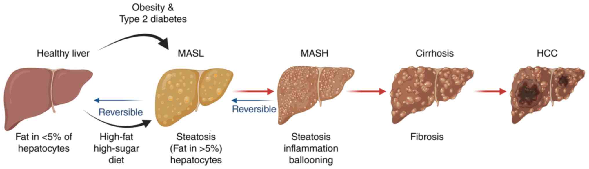
The earliest stage of MASLD is simple steatosis, now
termed metabolic dysfunction-associated steatotic liver (MASL).
Histopathologically, this stage corresponds to the entity formerly
termed non-alcoholic fatty liver (NAFL), which is characterised by
macrovesicular steatosis affecting >5% of hepatocytes in the
absence of hepatocellular injury (13). This initial stage is primarily
driven by persistent exposure to metabolic risk factors, including
high-fat and high-sugar diets, obesity and type 2 diabetes
(14). Notably, MASL is
generally reversible, and management at this stage focuses on
preventing disease progression through lifestyle interventions such
as weight reduction and dietary modification.
A subset of patients subsequently progresses to
MASH, a more advanced and severe pathological stage. The
histological diagnosis of MASH requires hepatic steatosis involving
>5% of hepatocytes, accompanied by lobular inflammation and
hepatocyte ballooning, with or without fibrosis (15). At this stage, timely intervention
may still reverse disease progression. However, persistent
metabolic injury promotes the activation of fibrogenic signalling
pathways, leading to progressive hepatic fibrosis. The final stage
of fibrotic progression is cirrhosis, which is characterised by
extensive architectural remodelling of the liver and confers a
substantially increased risk of HCC (Fig. 1).
Notably, MASLD-driven HCC does not arise exclusively
in cirrhotic livers. HCC may also develop in patients with advanced
fibrosis who have not yet progressed to cirrhosis. In a cohort of
13,371 patients with MASLD-driven HCC, 37% of cases occurred
without cirrhosis (16). These
findings highlight the importance of maintaining vigilance for HCC
risk even among patients with non-cirrhotic MASLD.
Epidemiology of MASLD-driven HCC
Although the incidence of numerous cancer types is
declining worldwide, the burden of primary liver cancer continues
to increase. According to GLOBOCAN 2024 global cancer statistics,
~843,000 new cases and 732,000 deaths from primary liver cancer
were reported worldwide, ranking it among the leading causes of
cancer-related incidence and mortality (17). Markov model-based projections
further suggest that both the number of new cases and the incidence
rate of primary liver cancer will continue to rise in both men and
women through 2030 (18).
HCC is the predominant histological subtype of
primary liver cancer, accounting for 75-85% of all cases (19). In the United States, for example,
the annual number of newly diagnosed HCC cases is projected to
increase from 5,160 to 12,240 by 2030, representing a 137% increase
(20).
Cirrhosis remains the strongest independent risk
factor for MASLD-driven HCC. A large cohort study of >290,000
individuals found that patients with MASLD and cirrhosis have an
annual HCC incidence of 10.6 per 1,000 person-years, notably higher
than in patients without cirrhosis (21). Furthermore, systematic reviews
have shown that the annual risk of HCC development in cirrhotic
individuals is at least 10-fold higher than that in non-cirrhotic
individuals (22).
Nevertheless, cirrhosis is not a prerequisite for
MASLD-driven HCC. Epidemiological studies indicate that only ~50%
of MASLD-driven HCC cases occur in the presence of cirrhosis,
contrasting sharply with hepatitis C virus (HCV)-related HCC, where
cirrhosis is present in the majority of cases (23). This distinctive feature poses
challenges for risk stratification and surveillance strategies in
patients with MASLD.
Although hepatitis B virus and HCV infections remain
the leading causes of HCC worldwide (24), the burden of viral
hepatitis-related HCC has gradually declined (25) owing to advances in antiviral
therapies and the widespread implementation of vaccination
programmes. By contrast, MASLD has emerged as the fastest-growing
cause of HCC. An analysis of 2,106 patients with HCC revealed that,
although viral hepatitis remained the dominant aetiology accounting
for ~80% of cases, the proportion of HCC attributable to MASH
increased by 68% over time (26). Furthermore, a large-scale study
involving >900,000 patients with MASLD demonstrated that the
disease significantly increases both the incidence of HCC and
HCC-related mortality (27).
Metabolic disorders, particularly diabetes and
obesity, are also major risk factors for HCC. The risk of
developing HCC is ~87% higher in individuals with diabetes, and
HCC-related mortality is increased by 1.88-fold in this population
(28). Similarly, a
meta-analysis reported a positive association between body mass
index (BMI) and liver cancer risk, with risk increasing
progressively as BMI rises (29). Notably, the prevalence of MASLD
exceeds 50% among individuals with type 2 diabetes or severe
obesity, further underscoring the close relationship between MASLD
and metabolic dysfunction (30).
With ongoing socioeconomic development, lifestyle
changes and the increasing prevalence of metabolic risk factors,
the global burden of diabetes and obesity is expected to continue
rising. Consequently, MASLD is likely to remain one of the
fastest-growing causes of HCC worldwide.
Pathogenesis of MASLD-driven HCC
Chronic inflammation in MASLD-driven
HCC
Chronic inflammation is widely recognised as a
fundamental driver of tumour initiation and progression in numerous
cancer types. For instance, persistent inflammation in inflammatory
bowel disease is a major contributor to colorectal carcinogenesis
(31). Similarly, inflammation
plays a central role in MASLD pathogenesis. Systematic reviews and
meta-analyses show that elevated levels of inflammatory markers,
including C-reactive protein, tumour necrosis factor-α (TNF-α) and
interleukin (IL)-1β, are significantly associated with both
increased MASLD risk and greater disease severity, including
steatohepatitis and liver fibrosis (32,33). These findings highlight chronic
inflammation as a critical pathogenic mechanism underlying
MASLD-driven HCC.
In MASLD and MASH, hepatic steatosis and metabolic
stress, including insulin resistance, endoplasmic reticulum (ER)
stress and mitochondrial dysfunction, together with gut microbiota
dysbiosis and microbial metabolites such as lipopolysaccharide
(LPS), collectively initiate chronic inflammatory responses
(34,35). These stimuli activate innate
immunity through damage-associated molecular patterns (DAMPs) and
pathogen-associated molecular patterns, which pattern-recognition
receptors in the liver recognise. Activation of intracellular
signalling pathways, including nuclear factor-κB (NF-κB),
mitogen-activated protein kinase (MAPK) and Janus kinase
(JAK)-signal transducer and activator of transcription (STAT),
promotes production of pro-inflammatory cytokines such as TNF-α,
IL-1β and IL-6, along with chemokines including C-C motif chemokine
ligand (CCL) 2/monocyte chemoattractant protein-1 and CCL5
(36,37).
Activation of the NF-κB pathway represents a pivotal
mechanism driving inflammation and disease progression in MASLD
(4,38). After pathway activation, TNF-α
binds its receptor, recruits adaptor proteins and activates the IκB
kinase complex. Phosphorylation and degradation of IκBα allow NF-κB
nuclear translocation, activating the transcription of genes
encoding pro-inflammatory cytokines and chemokines (39). These mediators further sustain
NF-κB activation, establishing a positive feedback loop
characterised by TNF-α production, immune-cell recruitment and
subsequent amplification of inflammatory signalling. Furthermore,
NF-κB-C-X-C chemokine (CXC) receptor type 4 and 7 axis-mediated
Toll-like receptor (TLR) 3 and TLR4 signalling has been implicated
in the progression from MASH to HCC (40). An experimental study has shown
that Huazhi Fugan Granule attenuates liver injury in MASLD models
by suppressing TNF-α and NF-κB signalling (41). Consequently, the NF-κB pathway
has emerged as a promising therapeutic target for MASLD.
Chemokines promote the recruitment of circulating
immune cells into the liver. Within the chronic inflammatory
microenvironment, infiltrating monocytes differentiate into
macrophages and polarise toward the pro-inflammatory M1 phenotype.
These cells release additional inflammatory mediators, including
TNF-α, IL-1β and IL-6, thereby exacerbating inflammation and
hepatocellular injury (42).
IL-6 binding to its receptor activates JAK, which phosphorylates
and activates STAT3. Activated STAT3 subsequently induces
downstream pathways associated with resistance to apoptosis,
enhanced proliferation, invasion, metastasis, angiogenesis and
therapeutic resistance (43).
Persistent STAT3 activation is recognised as a key driver of HCC
initiation and progression (44).
Macrophage polarisation also contributes to disease
evolution. Although M2 macrophages can suppress M1-mediated
inflammatory activity by inducing programmed cell death, persistent
inflammatory stimulation in MASH may lead to dysregulated immune
responses and aberrant tissue repair, ultimately promoting fibrosis
and establishing a pro-tumorigenic microenvironment (45). In addition, neutrophils
contribute to hepatic injury by releasing proteases and neutrophil
extracellular traps, further amplifying inflammation and tissue
damage (46,47).
A critical event in the progression to fibrosis and
cirrhosis is the activation of hepatic stellate cells (HSCs), which
secrete large quantities of extracellular matrix (ECM) components
(48). Fibrosis and cirrhosis
are well-established risk factors for HCC development (49). Beyond their fibrogenic role,
activated HSCs secrete pro-inflammatory cytokines and chemokines
that recruit additional immune cells and sustain the
inflammation-fibrosis cycle. HSC-derived matrix metalloproteinases
(MMPs) further facilitate tumour cell migration, invasion and
immune evasion (50,51). Consequently, HSCs have attracted
considerable interest as potential therapeutic targets in liver
cancer (52). Nevertheless, the
precise mechanisms governing HSC activation and functional
regulation remain incompletely understood, and targeted
drug-delivery strategies directed at HSCs require further
optimisation (53).
Overall, chronic inflammation is a central driver of
progression from MASLD to HCC. Persistent activation of signalling
pathways such as NF-κB and JAK-STAT promotes sustained production
of pro-inflammatory cytokines and chemokines, leading to continuous
immune-cell recruitment and M1 macrophage polarisation.
Simultaneously, activated HSCs contribute to ECM deposition,
fibrosis and cirrhosis while fostering a pro-tumorigenic
microenvironment that supports tumour-cell migration and immune
evasion. Collectively, these events establish a self-perpetuating
cascade of inflammation, fibrosis and carcinogenesis that
ultimately culminates in HCC development (Fig. 2).
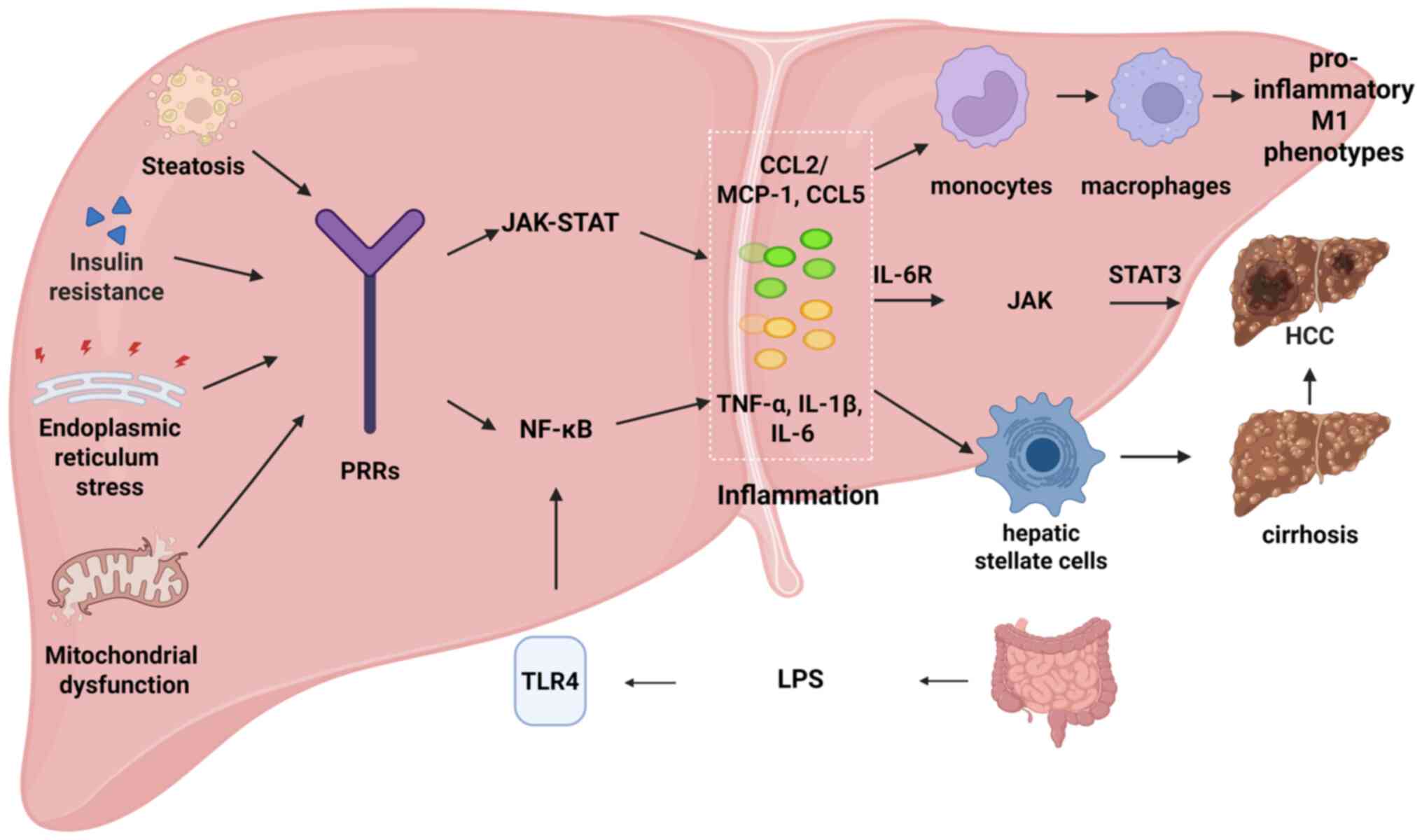 | Figure 2Schematic illustration of the
mechanism by which chronic inflammation drives the progression of
MASLD to HCC. Metabolic disorders (such as steatosis and insulin
resistance), along with TLR4 and other pathways, activate JAK-STAT
and NF-κB signalling, increase proinflammatory factor production
and recruit and polarize macrophages, driving inflammation and
eventually progression to liver fibrosis, cirrhosis and HCC. MASLD,
metabolic dysfunction-associated steatotic liver disease; HCC,
hepatocellular carcinoma; TLR4, Toll-like receptor 4; JAK, Janus
kinase; STAT, signal transducer and activator of transcription;
NF-κB, nuclear factor κB; LPS, lipopolysaccharide; PRRs, pattern
recognition receptors. Created with BioRender.com. |
Oxidative stress in MASLD-driven
HCC
Mitochondria function not only as the cellular
'powerhouse' but also as a major source of reactive oxygen species
(ROS). Under physiological conditions, oxidative phosphorylation
produces ATP and is a fundamental mitochondrial function. However,
this process involves a small degree of electron leakage (0.2-2%)
from the electron transport chain (ETC), resulting in direct oxygen
reduction and ROS generation (54). Although low levels of ROS have
important physiological and protective functions (55), excessive ROS production induces
oxidative stress, leading to cellular injury, DNA damage and
disease development (56).
In MASLD, hepatic steatosis-associated fatty acid
accumulation increases mitochondrial metabolic burden and impairs
ETC function. This dysfunction directly increases electron leakage
and superoxide anion generation. In addition, altered mitochondrial
membrane fluidity may further exacerbate electron leakage,
initiating oxidative stress and cellular damage (57). More notably, lipid overload,
particularly excess saturated fatty acids and cholesterol, induces
ER stress and the accumulation of unfolded proteins. Activation of
ER stress and the unfolded protein response (UPR) generate large
amounts of ROS. Concurrently, ER stress, for example through
activation of the inositol-requiring enzyme 1α pathway, disrupts
the function of the sarco/ER Ca2+-ATPase pump, resulting
in leakage of ER calcium stores. The subsequent influx of
Ca2+ into mitochondria impairs mitochondrial function
and further amplifies ROS production (58). Consequently, a self-perpetuating
cycle develops between ER stress and mitochondrial dysfunction,
leading to sustained oxidative stress.
Excessive ROS attack polyunsaturated fatty acids,
generating cytotoxic lipid peroxidation products, including
4-hydroxynonenal (4-HNE) and malondialdehyde (MDA). These products
directly contribute to MASH progression and, together with ROS,
induce damage to mitochondrial DNA (mtDNA). Since mtDNA encodes
essential components of the respiratory chain, its impairment
further compromises ETC function and promotes additional ROS
production (59). This process
establishes a pathological positive feedback loop centred on
mitochondrial dysfunction and ROS accumulation: Mitochondrial
injury leads to increased ROS production, which results in lipid
peroxidation, mtDNA damage and further mitochondrial dysfunction,
culminating in further ROS generation. Continuous activation of
this cycle sustains MASH progression and chronic inflammation.
Furthermore, mtDNA acts as a key mitochondrial-derived DAMP.
Following hepatocellular injury, mtDNA is released from damaged
mitochondria and directly activates HSCs through receptors such as
TLR9, thereby promoting liver fibrogenesis (60).
Oxidative stress-derived ROS also induce nuclear DNA
damage. In hepatocytes, such damage may result in mutations of the
tumour suppressor gene TP53 (59). Notably, mutant p53 not only loses
its tumour-suppressive activity but may also acquire oncogenic
functions and further enhance ROS production, thereby establishing
an additional positive feedback loop that facilitates malignant
transformation (61,62). In parallel, ROS function as
potent activators of inflammatory signalling. By activating
pathways such as NF-κB and the NOD-like receptor protein 3
inflammasome, ROS stimulate the production of pro-inflammatory
cytokines, including TNF-α and IL-6, thereby aggravating hepatic
inflammation and hepatocellular injury (63).
As MASLD progresses to MASH and HCC, the
nicotinamide adenine dinucleotide phosphate (NADPH) oxidase (NOX)
family is another major source of ROS, alongside mitochondria and
the ER. NOX-mediated amplification of oxidative stress occurs
through a characteristic three-step process. First, lipid
peroxidation products such as 4-HNE and MDA act as DAMPs and
activate innate immune receptors, including TLRs and the receptor
for advanced glycation end products. Second, receptor activation
markedly upregulates NOX expression and activity, particularly NOX2
and NOX4, resulting in a burst of ROS production. Third, excessive
ROS activate transcription factors such as NF-κB, leading to the
release of pro-inflammatory cytokines. These cytokines further
stimulate NOX activity and ROS generation, establishing a
self-sustaining positive feedback loop linking inflammation and
oxidative stress that continuously exacerbates liver injury
(64).
Overall, lipid overload in MASLD promotes ER stress
and mitochondrial dysfunction, which mutually reinforce one another
to sustain oxidative stress. Excessive ROS drive a vicious cycle
centred on mitochondrial injury by inducing mtDNA damage, lipid
peroxidation and TP53 mutations. Simultaneously, ROS activate
NOX-dependent signalling pathways and establish a self-amplifying
feedback network with inflammatory cytokines. Collectively, these
interconnected mechanisms promote hepatic inflammation, fibrosis
and malignant transformation, ultimately contributing to HCC
development and progression (Fig.
3).
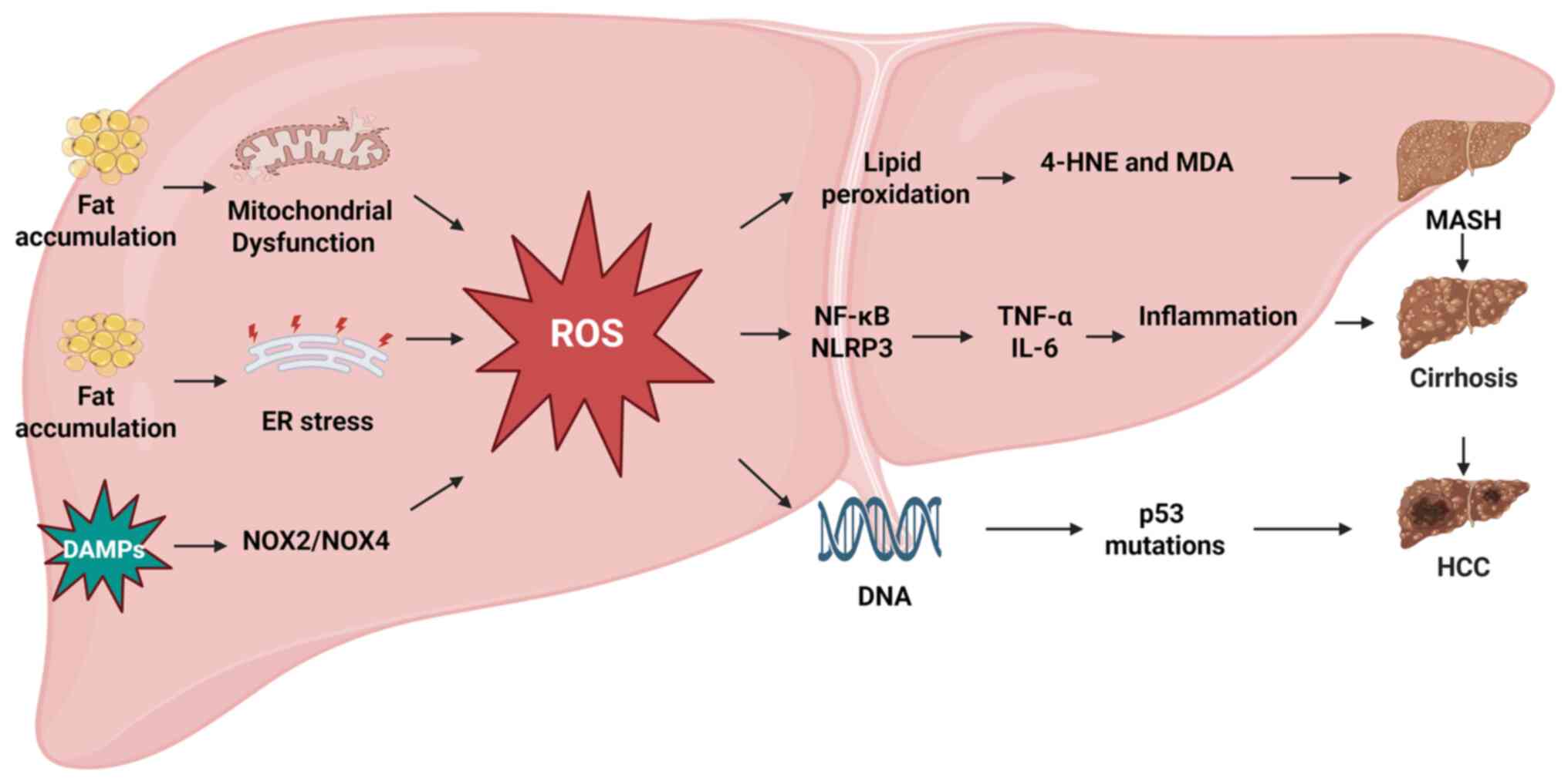 | Figure 3Pathogenic mechanism of oxidative
stress in the progression of MASLD to HCC. Fat accumulation,
coupled with mitochondrial and ER dysfunction, triggers ROS
production. As a key hub, ROS trigger inflammation by activating
the NF-κB/NLRP3 pathway, leading to MASH. Furthermore, ROS cause
lipid peroxidation and p53 mutations, which together promote
progression from MASH to liver cirrhosis and HCC. MASLD, metabolic
dysfunction-associated steatotic liver disease; HCC, hepatocellular
carcinoma; ER, endoplasmic reticulum; ROS, reactive oxygen species;
NF-κB, nuclear factor κB; NLRP3, NOD-, LRR- and pyrin
domain-containing protein 3; MASH, metabolic dysfunction-associated
steatohepatitis; p53, tumour protein p53; DAMPs, damage-associated
molecular patterns; 4-HNE, 4-hydroxynonenal; MDA, malondialdehyde;
IL, interleukin. Created with BioRender.com. |
Immune dysregulation in MASLD-driven
HCC
Under physiological conditions, the immune system
maintains homeostasis through finely regulated bidirectional
control mechanisms that enable the efficient elimination of
pathogens and abnormal cells while preventing excessive tissue
damage through immune tolerance. As a specialised immune organ, the
liver contains a large and diverse population of resident immune
cells, including Kupffer cells, natural killer (NK) cells and T
lymphocytes, which collectively preserve hepatic immune homeostasis
and immune tolerance. Under normal circumstances, Kupffer cells
remove blood-borne pathogens, such as bacteria and viruses,
phagocytose senescent or damaged hepatocytes and support immune
surveillance, immunoregulation and systemic immune tolerance
(65).
However, during MASLD progression, chronic metabolic
disturbances and lipotoxicity induce persistent hepatic
inflammation, leading to profound alterations in the immune
microenvironment. Kupffer cells preferentially polarise towards the
pro-inflammatory M1 phenotype under chronic inflammatory
conditions, producing high levels of cytokines and chemokines,
including TNF-α, IL-1β and IL-6. Activated Kupffer cells also
secrete profibrogenic mediators, such as transforming growth
factor-β (TGF-β) and platelet-derived growth factor, which induce
HSC transdifferentiation into collagen-producing myofibroblasts and
promote liver fibrosis (66,67).
Cluster of differentiation 8 (CD8)+ T
cells play a crucial role in immune surveillance by recognising
viral and tumour antigens presented on major histocompatibility
complex class I molecules through their T-cell receptors. Upon
activation, they eliminate infected or transformed cells by
releasing perforin and granzymes, thereby protecting the host
against malignant transformation (68). However, in MASLD, persistent
antigenic stimulation within the chronically inflamed liver
promotes CD8+ T-cell exhaustion. This dysfunctional
state is characterised by reduced production of cytotoxic molecules
and cytokines together with increased expression of inhibitory
receptors, including programmed cell death protein 1, T-cell
immunoglobulin and mucin domain-containing protein 3, cytotoxic
T-lymphocyte-associated protein 4 and lymphocyte activation gene 3
(69). Evidence from both human
cirrhotic liver samples and experimental MASH models supports this
exhausted phenotype (70).
Exhausted CD8+ T cells exhibit impaired
antitumour activity and fail to eliminate preneoplastic and
malignant cells efficiently, compromising immune surveillance and
facilitating tumour immune escape. Furthermore, in
obesity-associated MASH and cirrhosis, increased CD8+ T
cell infiltration correlates positively with HSC activation, as
reflected by elevated α-smooth muscle actin expression. Since
activated HSCs are the principal source of collagen during hepatic
fibrogenesis, this association suggests that CD8+ T
cells directly contribute to fibrosis progression. Supporting this
concept, experimental depletion of CD8+ T cells
attenuates hepatic inflammation and suppresses HSC activation
(71).
CD4+ T cells differentiate into distinct
functional subsets according to the surrounding antigenic and
cytokine environment, including T helper 17 (Th17) cells and
regulatory T (Treg) cells. Under physiological conditions, immune
homeostasis depends on a dynamic balance between pro-inflammatory
Th17 cells and immunosuppressive Treg cells. However, in MASH, this
equilibrium is disrupted. Treg cells expand substantially and
infiltrate, further suppressing effector T-cell function and
establishing an immune-tolerant microenvironment. Conversely, Th17
cells and their signature cytokine IL-17 promote inflammatory and
fibrotic responses while directly facilitating tumorigenesis by
stimulating angiogenesis and cellular proliferation (72).
Myeloid-derived suppressor cells (MDSCs) constitute
a heterogeneous population of immature myeloid cells characterised
by potent immunosuppressive activity. In MASLD, chronic
inflammation drives marked MDSC expansion, with cells recruited
from the bone marrow and spleen to the liver in response to
chemokines released by injured hepatocytes and activated Kupffer
cells. MDSCs mediate immunosuppression through several
complementary mechanisms. First, they deplete arginine, an amino
acid essential for T-cell proliferation and function, leading to
T-cell inactivation and cell-cycle arrest (73). Second, they generate ROS and
reactive nitrogen species that disrupt T-cell signalling pathways
and induce apoptosis (74).
Third, they inhibit lymphocyte migration (75). Fourth, they actively promote the
induction and expansion of immunosuppressive Treg cells (76). Collectively, these actions help
establish a profoundly immunosuppressive microenvironment.
In addition, a study in murine models showed that
MDSCs suppress T-cell and NK-cell function through ROS production
mediated by NOX2 and inducible nitric oxide synthase (76). MDSCs can also promote macrophage
polarisation towards an immunosuppressive M2-like phenotype and
facilitate tumour growth by maintaining low levels of IL-12
secretion (77). Beyond their
immunomodulatory functions, MDSCs support tumour progression
through multiple non-immune mechanisms. These include promoting
tumour angiogenesis through vascular endothelial growth factor
secretion, enhancing nutrient supply to tumour tissues and
remodelling the tumour microenvironment through the release of MMPs
and additional cytokines, thereby facilitating tumour invasion and
metastasis (78).
Overall, chronic inflammation during MASLD
progression notably disrupts hepatic immune homeostasis. Kupffer
cells acquire a pro-inflammatory, profibrotic phenotype;
CD8+ T cells become exhausted; the Th17/Treg cell
balance is disturbed; and MDSCs expand substantially. Together,
these alterations establish a fibrogenic, immunosuppressive
microenvironment that promotes immune evasion, fibrosis and tumour
development, ultimately driving progression from MASLD to HCC
(Fig. 4).
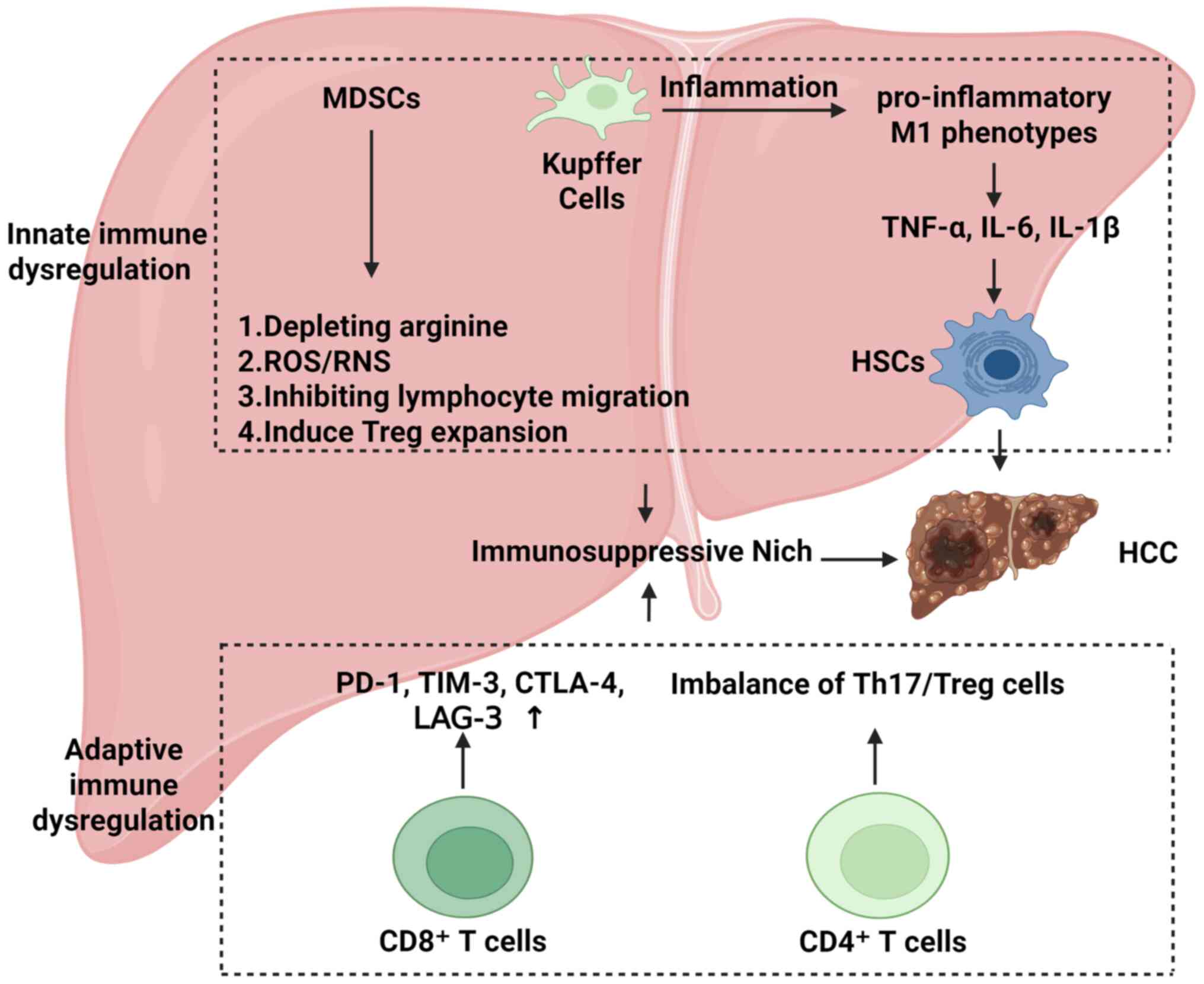 | Figure 4Schematic of how dysregulation of the
liver immune microenvironment promotes HCC. After activation,
Kupffer cells polarize into a pro-inflammatory M1 phenotype,
release TNF-α and other factors and activate HSCs, driving
inflammation and forming an immunosuppressive microenvironment.
CD8+ T cells become exhausted, while CD4+ T
cell helper function becomes imbalanced, leading to failed immune
surveillance. These two pathways work together to provide the key
conditions for hepatocellular carcinogenesis. TNF-α, tumour
necrosis factor-α; HSCs, hepatic stellate cells; CD4/8, cluster of
differentiation 4/8; HCC, hepatocellular carcinoma; MDSCs,
myeloid-derived suppressor cells; ROS, reactive oxygen species;
RNS, reactive nitrogen species; IL, interleukin; PD-1, programmed
cell death protein 1; TIM-3, T-cell immunoglobulin and mucin
domain-containing protein 3; LAG-3, lymphocyte activation gene 3;
Th17, T helper 17 cells; Treg, regulatory T cells. Created with
BioRender.com. |
Gut microbiome dysbiosis in
MASLD-driven HCC
The human gut harbours an immense microbial
community comprising trillions of bacteria, viruses, fungi and
other microorganisms that actively participate in numerous
physiological processes. The gut and liver are connected by the
portal vein, a direct, unidirectional and exclusive conduit that
links them closely anatomically and functionally. Substances
absorbed from the intestine, including nutrients, potentially
harmful agents and bacterial metabolites, are transported via the
portal vein to the liver for metabolic processing before entering
the systemic circulation. Consequently, disruption of the gut
microbiota can impair normal hepatic function, contributing to
liver injury and related diseases.
The progression from MASLD to HCC is a complex,
multistep process in which gut microbiota dysbiosis has emerged as
a key driver. The principal mechanism involves dysbiosis-induced
impairment of intestinal barrier function, which facilitates the
translocation of bacterial metabolites, such as LPS and secondary
bile acids, to the liver. This process triggers chronic
inflammation and immune dysregulation, ultimately promoting HCC
development (79). The
intestinal barrier is a multilayered and highly coordinated defence
system that regulates the selective absorption of nutrients and
water while preventing the translocation of harmful substances,
including bacteria, toxins and antigens, from the gut lumen into
the circulation. The intestinal barrier comprises four major
components: i) The physical barrier, formed by intestinal
epithelial cells and tight junctions, which prevents excessive
permeability; ii) the chemical barrier, consisting of the mucus
layer and antimicrobial secretions, which provides lubrication,
separation and bactericidal activity; iii) the biological barrier,
represented by the commensal microbial community that suppresses
pathogen colonisation through competitive exclusion and nutrient
competition; and iv) the immune barrier, which includes
gut-associated lymphoid tissue and associated immune mediators that
recognise and eliminate pathogens (80).
Dysbiosis is characterised by reduced beneficial
bacteria, including short-chain fatty acid-producing species,
leading to decreased production of key microbial metabolites such
as butyrate. This reduction disrupts epithelial tight junctions and
increases intestinal permeability (81,82). Simultaneously, the expansion of
harmful bacterial populations and the production of toxic
metabolites and enzymes, including ethanol and secondary bile
acids, directly damage the mucus layer and intestinal epithelial
cells (83,84), further disrupting physical and
chemical barriers. Furthermore, harmful bacteria can occupy
ecological niches, deplete nutrients and release toxins, further
disturbing microbial homeostasis and weakening the biological
barrier. The normal microbiota contributes to immune education by
enabling the immune system to distinguish commensals from
pathogens. Consequently, dysbiosis may promote inappropriate immune
responses against harmless antigens or commensal microorganisms,
disrupting immune tolerance and impairing immune barrier function
(85).
Following intestinal barrier disruption, large
quantities of microbe-associated molecular patterns, particularly
LPS derived from the cell wall of Gram-negative bacteria, enter the
portal circulation. As a potent inflammatory stimulus, LPS binds to
TLR4, activating the NF-κB and MAPK signalling pathways. This
activation promotes the release of proinflammatory cytokines,
including TNF-α and IL-6, thereby driving hepatic inflammation and
cellular injury (86). This
mechanism is considered central to the transition from NAFLD to
MASH.
In addition, secondary bile acids reaching the liver
can induce HSC senescence and the formation of a
senescence-associated secretory phenotype (SASP) (87), while also upregulating
cyclooxygenase-2 (COX-2), a key enzyme involved in prostaglandin
synthesis in HSCs (88). The
SASP comprises a complex array of proinflammatory factors,
chemokines, growth factors and proteases secreted by senescent
cells. Through paracrine and autocrine signalling, these factors
influence neighbouring cells and help establish a tumour-promoting
microenvironment. An animal study has demonstrated that hepatic
SASP activity markedly promotes HCC development (89). COX-2 is highly expressed in
multiple malignancies and contributes to tumour cell proliferation,
angiogenesis, immune evasion and metastasis (90). In MASH-driven HCC, COX-2-derived
prostaglandin E2 can suppress antitumour immune responses, thereby
promoting tumour progression (91).
Given the pivotal role of the gut microbiota in the
initiation and progression of MASLD and its transition to HCC,
therapeutic strategies targeting the gut microbiota represent a
promising clinical approach. A previous review has systematically
summarised preclinical and clinical studies evaluating
microbiota-targeted interventions in MASLD, MASH and HCC, including
probiotics, prebiotics, dietary interventions, faecal microbiota
transplantation and antibiotics (92). These approaches have shown
benefits for intestinal barrier integrity, inflammation and disease
progression. However, limitations remain, including small sample
sizes, incomplete mechanistic understanding and uncertain long-term
efficacy. Further investigation is therefore required to optimise
and validate these therapeutic strategies. Overall, the progression
from MASLD to HCC is a complex, multistep process in which gut
microbiota dysbiosis plays a central pathogenic role (Fig. 5).
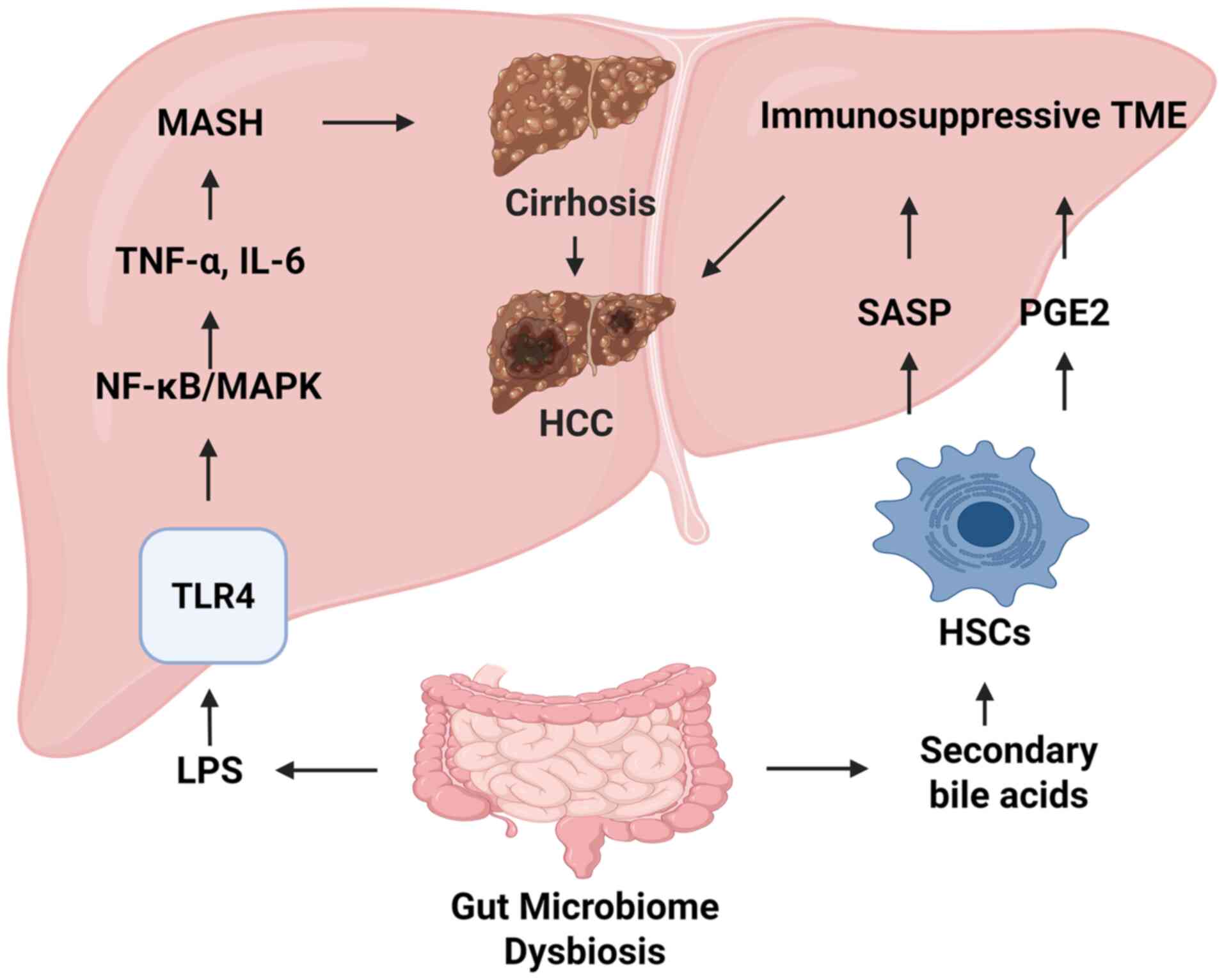 | Figure 5Mechanism by which gut microbiome
dysbiosis triggers inflammation and fibrosis, leading to HCC
development. Gut microbiota dysbiosis increases harmful products,
such as LPS and secondary bile acids, which enter the liver through
the portal vein. LPS can activate the TLR4 receptor on hepatocytes,
trigger inflammatory signalling pathways such as NF-κB/MAPK and
produce pro-inflammatory factors such as TNF-α and IL-6, driving
progression from MASH to cirrhosis. In liver cirrhosis, activated
HSCs secrete SASP and PGE2, which together create an
immunosuppressive TME and ultimately promote HCC development. LPS,
lipopolysaccharide; TLR4, Toll-like receptor 4; NF-κB, nuclear
factor κB; MAPK, mitogen-activated protein kinase; TNF-α, tumour
necrosis factor-α; IL-6, interleukin-6; MASH, metabolic
dysfunction-associated steatohepatitis; HSCs, hepatic stellate
cells; SASP, senescence-associated secretory phenotype; PGE2,
prostaglandin E2; TME, tumour microenvironment; HCC, hepatocellular
carcinoma. Created with BioRender.com. |
Role of lipotoxicity and glucotoxicity in
MASH and HCC development
Lipotoxicity is a pathological process in which the
abnormal accumulation of toxic lipid species in organs and tissues
leads to cellular dysfunction and, ultimately, cell death (93). As a major target organ of
lipotoxicity, the liver normally esterifies free fatty acids (FFAs)
with glycerol to form triglycerides, which are safely stored within
lipid droplets under physiological conditions. However, when FFA
influx exceeds the hepatic storage capacity, excess FFAs are
diverted into non-esterification pathways, generating a range of
harmful lipid derivatives (94).
Palmitic acid is a saturated fatty acid that the
liver can synthesise de novo. Under physiological
conditions, palmitic acid synthesis occurs in the cytoplasm, where
fatty acid synthase (FASN) converts acetyl-CoA and malonyl-CoA into
palmitic acid. This multistep process requires NADPH and proceeds
through sequential condensation, reduction and dehydration
reactions (95). Physiological
levels of palmitic acid are essential for normal cellular function.
However, under pathological conditions, including insulin
resistance, obesity and high-fat diets, excessive palmitic acid
accumulation can transform palmitic acid into a potent lipotoxic
molecule that disrupts ER lipid homeostasis, impairs protein
folding and induces ER stress. Mechanistically, UPR activation
contributes to insulin resistance and hepatic steatosis through its
central role in cellular stress regulation (96). In addition, palmitic acid induces
mitochondrial membrane depolarisation, abnormal mitochondrial
fission, impaired ATP production and suppression of autophagy,
resulting in the accumulation of dysfunctional mitochondria.
Palmitic acid-induced oxidative stress and calcium overload trigger
opening of the mitochondrial permeability transition pore,
initiating caspase activation and apoptosis (97). An experimental study has
demonstrated that mitochondria-targeted antioxidants can interrupt
this process, thereby alleviating ER stress and cell death
(98). Furthermore, palmitic
acid activates pattern-recognition receptors, including TLR4,
thereby stimulating inflammatory pathways such as NF-κB and
promoting hepatocyte secretion of proinflammatory cytokines,
including TNF-α and IL-1β. These events recruit and activate immune
cells, contributing to MASH development (99).
The accumulation of other toxic lipids, including
ceramides and free cholesterol, further promotes ER stress,
oxidative injury and mitochondrial dysfunction, ultimately leading
to hepatocyte death (100). A
critical mechanism underlying MASH progression involves immune cell
activation by DAMPs released from dying hepatocytes, resulting in
persistent inflammation that exacerbates disease progression.
Sustained inflammation activates HSCs, promoting collagen
deposition and the development of hepatic fibrosis and cirrhosis
(101). With ongoing DNA damage
and dysregulated cellular proliferation, these pathological changes
may ultimately culminate in HCC.
Glucotoxicity refers to the progressive cellular and
tissue injury caused by chronic hyperglycaemia and its associated
metabolic disturbances, including hyperinsulinaemia and insulin
resistance, ultimately resulting in dysfunction and cell death. Its
central mechanism involves disruption of metabolic homeostasis
rather than a direct toxic effect of glucose itself. As MASLD
progresses to HCC, glucotoxicity contributes through several
interconnected mechanisms. First, it exacerbates lipid metabolic
dysfunction. Sterol regulatory element-binding protein-1c
(SREBP-1c), a key transcription factor regulating hepatic
lipogenesis, promotes the expression of genes involved in fatty
acid synthesis (102). Insulin
activates SREBP-1c and thereby enhances hepatic fatty acid
synthesis (103). Under
hyperinsulinaemic conditions, excessive SREBP-1c activation
markedly increases lipogenesis, worsening hepatic steatosis and
lipotoxicity. Simultaneously, insulin resistance and
hyperinsulinaemia suppress fatty acid oxidation, further promoting
lipid accumulation within hepatocytes (104).
Second, chronic hyperglycaemia drives the formation
of advanced glycation end products, a process closely associated
with oxidative stress. Advanced glycation end products binding to
receptors on hepatocytes and Kupffer cells activates multiple
signalling pathways, notably NF-κB, MAPK and JAK/STAT, while also
stimulating NOX activity. These events promote excessive ROS
generation (105), thereby
amplifying oxidative stress, inflammatory cytokine production and
cellular injury, which collectively contribute to MASH development.
In parallel, chronic inflammation further activates HSCs,
accelerating the progression of hepatic fibrosis and cirrhosis.
During hepatocarcinogenesis, the combined effects of
hyperinsulinaemia and a proinflammatory microenvironment promote
DNA damage, enhanced cellular proliferation and immune evasion.
Collectively, these processes facilitate HCC development (106).
In summary, glucotoxicity contributes to the
malignant progression of liver disease from MASLD to HCC through
multiple mechanisms, including disruption of lipid metabolism,
induction of oxidative stress and the promotion of chronic
inflammation (Fig. 6).
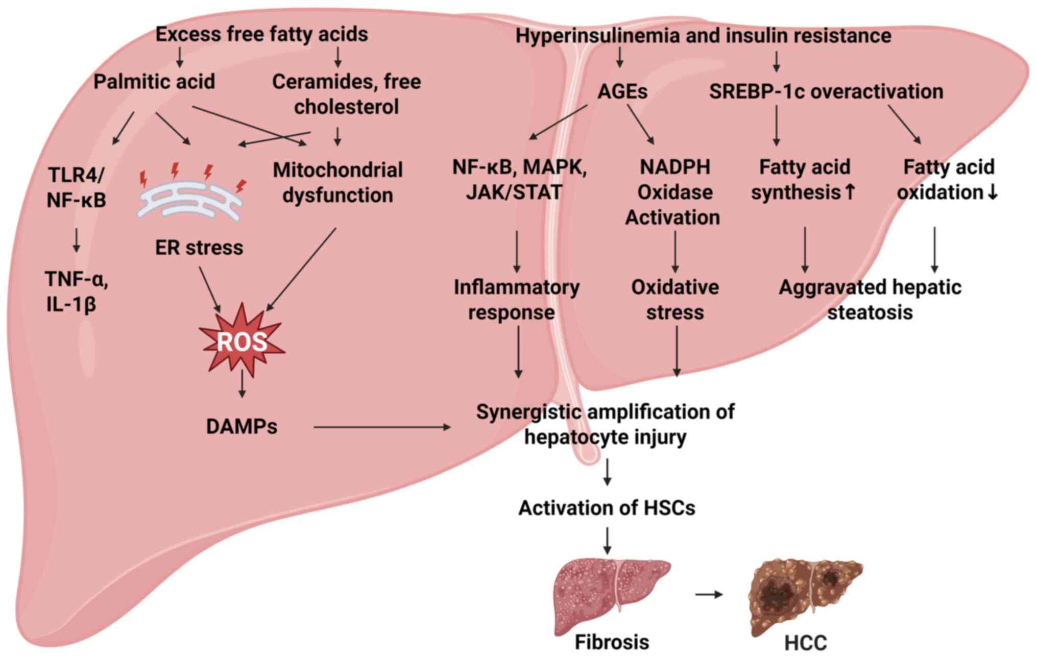 | Figure 6Schematic illustration of the key
molecular mechanisms by which lipotoxicity and glucotoxicity drive
the progression of MASLD to HCC. Excess FFAs (such as palmitic
acid) and their derivatives (ceramide and free cholesterol) trigger
a strong inflammatory response (production of TNF-α and IL-1β) by
activating the TLR4/NF-κB pathway and inducing ER stress. These
stresses, together with hyperinsulinemia, increase ROS production
and drive oxidative stress. Hyperinsulinemia and insulin resistance
worsen hepatic steatosis by overactivating SREBP-1c and disrupting
lipid metabolism (fat synthesis increases and fatty acid oxidation
decreases). Damage signals (such as ROS and DAMPs) released by
these three pathways synergistically amplify and jointly activate
HSCs, ultimately driving liver fibrosis and HCC. MASLD, metabolic
dysfunction-associated steatotic liver disease; HCC, hepatocellular
carcinoma; FFAs, free fatty acids; TNF-α, tumour necrosis factor-α;
IL-1β, interleukin-1β; TLR4, Toll-like receptor 4; NF-κB, nuclear
factor κB; ER, endoplasmic reticulum; ROS, reactive oxygen species;
SREBP-1c, sterol regulatory element-binding protein 1c; DAMPs,
damage-associated molecular patterns; HSCs, hepatic stellate cells;
MAPK, mitogen-activated protein kinase; JAK, Janus kinase; STAT,
signal transducer and activator of transcription; AGEs, advanced
glycation end products. Created with BioRender.com. |
Animal models of MASH-driven HCC
Due to the complex pathophysiology of MASLD, its
precise pathogenic mechanisms remain incompletely understood, and
effective therapeutic options remain limited. Animal models are
indispensable tools for investigating disease mechanisms,
evaluating therapeutic efficacy and screening potential drug
candidates. Accordingly, appropriate MASLD models are essential for
drug discovery, validation of novel therapeutic strategies and
improving the understanding of the biological processes underlying
MASH and MASH-driven HCC.
A variety of animal models have been developed to
study MASLD, most of which are based on dietary interventions
(107). Among these, the
Western diet model, characterised by high fat and high sugar
intake, has been widely recommended (108). This model typically
incorporates a diet containing high fat content (usually 40-45% of
total caloric intake), elevated levels of sugars, particularly
fructose and sucrose, and high cholesterol content (1-2%). It
effectively recapitulates several features of human metabolic
syndrome, including obesity, insulin resistance and diabetes, while
reproducing the full histopathological spectrum of MASH, including
hepatic steatosis, inflammation, hepatocellular ballooning and
fibrosis. Following prolonged feeding for >12 months, male
C57BL/6J mice may spontaneously develop HCC, making this model a
benchmark for investigating the natural history of MASH-driven HCC
(109).
Nevertheless, no animal model can fully reproduce
all aspects of human MASH, and each model has inherent limitations.
Although the Western diet model is widely used, some investigators
have argued that it inadequately reproduces the immunosuppressive
microenvironment that contributes to progression from MASH to HCC
(110). Consequently, these
researchers advocate the use of the major urinary protein
promoter-driven urokinase-type plasminogen activator (MUP-uPA)
transgenic model (111). The
MUP-uPA model is considered one of the current reference models for
studying MASH-driven HCC due to its spontaneous disease
progression, accurate representation of the tumour immune
microenvironment, conservation of relevant molecular pathways and
capacity to predict therapeutic responses. However, this model is
also subject to several limitations, including relatively slow
tumour development, substantial tumour heterogeneity, inadequate
reproduction of the intensity of human inflammatory responses,
incomplete representation of the diversity of human disease
subtypes, difficulties in recapitulating the human gut microbiome
and dependence on transgenic uPA overexpression. Consequently, it
may not fully reflect the natural pathogenesis of human
disease.
At present, an ideal animal model capable of
reproducing the entire spectrum of human MASH-driven HCC has not
been established. Therefore, investigators should select the most
appropriate model according to the specific scientific objectives
and pathological features under investigation.
ncRNAs and MASLD
Classification and functional modalities
of ncRNAs
ncRNAs are conventionally categorized according to
two complementary frameworks: By functional role into housekeeping
and regulatory ncRNAs or by transcript length into small ncRNAs
(<200 nucleotides) and lncRNAs (≥200 nucleotides) (112,113). As a detailed account of ncRNA
classification and biogenesis falls outside the scope of the
present review, the reader is referred to dedicated summaries of
this topic (112-114). Briefly, the classes most
pertinent to hepatic pathophysiology are miRNAs (~22 nucleotides;
mediators of post-transcriptional gene silencing), lncRNAs (RNA
polymerase II transcripts that lack a significant open reading
frame) and circRNAs, which are generated by back-splicing into
covalently closed loops and are thus highly stable and resistant to
exonucleolytic degradation (114).
Although other ncRNA classes, including small
nucleolar RNAs, PIWI-interacting RNAs and tRNA-derived small RNAs,
have been implicated in liver pathophysiology and
hepatocarcinogenesis (115,116), a recent systematic review of
MASLD and MASH reported that, among 134 eligible preclinical
studies, 96 focused on miRNAs, 38 on lncRNAs and 11 on circRNAs
(117). These findings indicate
that these three classes currently possess the strongest evidence
base in the MASLD-driven HCC field. Accordingly, the present review
focuses on the three ncRNA classes supported by the strongest
evidence in MASLD-driven HCC.
Transcriptional regulation is a well-established
functional modality of lncRNAs. Nuclear lncRNAs act as modular
scaffolds or guides that recruit chromatin-modifying complexes,
function as transcriptional decoys that sequester transcription
factors from their cognate promoters and act either in cis on
neighbouring genes or in trans at distal loci, while enhancer RNAs
promote enhancer-promoter looping through chromatin-looping
factors. As these classical paradigms have been reviewed
extensively elsewhere, the reader is referred to dedicated
summaries (118-121). In steatotic livers, this
modality has direct pathophysiological relevance, as illustrated by
several MASLD-specific examples: The lncRNA Blnc1 serves as a core
component of the liver X receptor (LXR) transcriptional complex and
licenses pathological hepatic lipogenesis by activating the
SREBP-1c lipogenic program (122); the steroid receptor RNA
activator (SRA) lncRNA facilitates hepatic lipid accumulation
partly by directly activating insulin receptor gene transcription
(123,124); H19, which is upregulated in
MASLD, promotes a protumorigenic lipogenic programme through the
H19/PTBP1/SREBP1 feed-forward loop and mechanistic target of
rapamycin (mTOR) complex 1 (mTORC1) signalling (125,126); and the circRNA-derived
micropeptide circ-SLC9A6-126aa promotes hepatic lipid uptake by
activating CD36 transcription in an H4K16ac-dependent manner
(127). Numerous other lncRNAs
couple epigenetic dysregulation to metabolic dysfunction by
sustaining lipogenic transcription-factor networks or repressing
tumour-suppressive programs (119,122).
Post-transcriptional regulation is the canonical
functional modality of miRNAs, which are processed by Drosha/DGCR8
and Dicer and loaded into the RNA-induced silencing complex to
repress target mRNAs through partial base pairing with their 3'UTRs
(128). As single miRNAs
co-ordinately regulate hundreds of transcripts, they act as
network-level rheostats over hub metabolic and oncogenic pathways,
a property of direct relevance to MASLD-driven HCC. miRNA networks
calibrate lipogenesis, oxidative stress and inflammatory signalling
in hepatocytes, with miR-21-5p promoting lipid accumulation and
tumorigenesis via peroxisome proliferator-activated receptor α
(PPARα), miR-378 impairing fatty acid oxidation and loss of the
liver-intrinsic tumour suppressors miR-223 and miR-122 accelerating
MASLD progression and increasing the incidence of spontaneous HCC,
respectively (129). Comparable
post-transcriptional crosstalk also operates across ncRNA classes.
circRNA_0046366 sponges miR-34a to relieve PPARα-mediated
suppression and enhance fatty acid β-oxidation (130), whereas GAS5 sponges miR-29a-3p
to upregulate NOTCH2 and aggravate steatohepatitis (131), illustrating that competing
endogenous RNA (ceRNA)-mediated derepression is a recurring
post-transcriptional motif in MASLD progression.
Another functional modality involves assembling
ribonucleoprotein complexes. Certain lncRNAs function as molecular
scaffolds that physically bridge multiple proteins into complexes
with distinct signalling outputs (120). In hepatic malignancy, for
example, the lncRNA SNHG6 promotes the interaction between
ER-anchored FAF2 and lysosomal mTORC1, thereby activating mTORC1
signalling and driving MASLD-driven HCC progression (132). Similarly, LINC01468 acts as a
molecular scaffold that promotes CUL4A-mediated ubiquitination and
degradation of the phosphatase SHIP2, thereby activating the
oncogenic protein kinase B (AKT)/mTOR pathway (133). Blnc1 likewise assembles a
ribonucleoprotein complex with lipogenic transcription factors to
enhance lipogenic gene transcription (122) and LncSHGL recruits hnRNPA1 to
enhance calmodulin translation and activate hepatic
phosphatidylinositol 3-kinase (PI3K)/AKT signalling (134). circRNAs often function as miRNA
sponges (or ceRNAs) sequestering miRNAs from their target mRNAs and
thereby derepressing downstream oncogenes. They can also act as
protein decoys or molecular scaffolds (114,135). Furthermore, some circRNAs
containing internal ribosome entry sites or N6-methyladenosine
(m6A) modifications can undergo cap-independent translation to
generate regulatory micropeptides, adding another layer of
non-canonical protein-coding activity within the non-coding
transcriptome (114). Through
these mechanisms, the abundance and accessibility of signalling
proteins become directly linked to the expression of specific
ncRNAs, providing an additional axis through which ncRNAs influence
the malignant transformation of hepatocytes.
miRNAs and MASLD
miRNAs are a class of ncRNAs, typically 21-24
nucleotides in length, that primarily regulate gene expression
through post-transcriptional gene silencing. They exert their
effects through partial base pairing with the 3'UTR of target
mRNAs, resulting in mRNA degradation or translational repression
(136).
Accumulating evidence indicates that miRNAs are
intimately involved in the initiation, development and progression
of MASLD, largely through the regulation of hepatic lipid
metabolism and insulin sensitivity. Dysregulation of specific
miRNAs has been shown to contribute to MASLD pathogenesis. For
instance, increased miR-124-3p expression promotes hepatic
steatosis and inflammation by suppressing Pref-1 (137), whereas miR-130b-5p exacerbates
lipid accumulation and insulin resistance by targeting IGFBP2 and
inhibiting the AKT signalling pathway (138). Conversely, reduced miR-338-3p
expression diminishes suppression of PP4R1, impairing PP4R1/PP4
axis function and disrupting insulin signalling (139).
In addition, miR-378 and nuclear respiratory factor
1 (NRF1) form a pathogenic positive feedback loop. miR-378 inhibits
NRF1-mediated fatty acid oxidation, while reduced NRF1 activity
further enhances miR-378 transcription, thereby aggravating hepatic
steatosis (140). The oncogenic
miRNA miR-21 promotes de novo lipogenesis during the early
stages of MASLD by suppressing the HBP1/p53 axis, relieving
repression of the master lipogenic regulator SREBP-1c (141). By contrast, miR-223 protects
against steatohepatitis by targeting inflammatory and oncogenic
mediators, including CXCL10 and Taz (142).
lncRNAs and MASLD
lncRNAs are characterised by their lack of
protein-coding capacity, and their transcriptional products are
typically RNA molecules >200 nucleotides that lack functional
open reading frames (143).
Accumulating evidence indicates that lncRNAs serve important
regulatory roles in a range of diseases, with particularly
prominent involvement in the initiation and progression of MASLD. A
study has shown that lncRNAs modulate signalling pathways
associated with hepatic lipid metabolism, steatohepatitis,
steatohepatitis-associated fibrosis and HCC, thereby contributing
to MASLD pathogenesis (144).
During MASLD initiation and progression, lncRNAs
play central roles in regulating lipid metabolism and inflammatory
responses. A study has demonstrated that lncRNA-AK026 expression is
markedly upregulated in MASLD models and may promote intracellular
lipid accumulation in hepatocytes through the PPAR signalling
pathway (145). GAS5 has been
shown to aggravate hepatic steatosis by acting as a molecular
sponge for miR-29a-3p, thereby relieving its inhibitory effect on
NOTCH2 (131). In parallel,
formation of the miR-378/Nrf1 negative feedback loop further
suppresses fatty acid oxidation, leading to lipid accumulation
(140). As the disease
progresses, the cholesterol-sensing molecule SNHG6 assembles a
SNHG6-FAF2-mTOR complex, which promotes cholesterol-dependent
lysosomal recruitment and activation of mTORC1. This establishes a
vicious cycle that enhances lipid synthesis and signal
transduction, thereby driving progression from simple steatosis to
MASH (146). Notably, loss of
Dicer in hepatocytes reduces miR-192-3p expression, increasing
insulin-like growth factor 2 (IGF2) expression, which promotes M1
macrophage polarisation and exacerbates inflammation (147). Therapeutic approaches targeting
these pathways, such as RORA agonists, can increase miR-122
expression and subsequently suppress key lipogenic enzymes,
representing a potential strategy for reversing hepatic steatosis
(148).
circRNAs and MASLD
circRNAs are a distinct class of single-stranded RNA
molecules characterised by a covalently closed loop structure that
distinguishes them from linear RNAs. This unique conformation
confers remarkable stability, rendering them resistant to
degradation by RNA exonucleases as they lack free 5' and 3'termini
(149). Growing evidence
suggests that circRNAs contribute to MASLD pathogenesis by
regulating diverse biological processes, including lipid
metabolism, autophagy, inflammatory responses, oxidative stress and
fibrosis (150,151). A study has further demonstrated
that circRNAs can regulate hepatic lipid metabolism through
signalling pathways such as JAK2/STAT5 (152). RNA sequencing and bioinformatic
analyses have identified 93 differentially expressed circRNAs in a
mouse model of MASLD, highlighting their potential utility as
cellular biomarkers (153).
Consequently, circRNAs have emerged as promising blood-based
biomarkers for early MASLD diagnosis.
Inflammation is a key mechanism driving progression
from MASLD to MASH and is characterised by immune cell infiltration
within hepatic lobules and the release of pro-inflammatory
cytokines. circRNAs are involved in this process by regulating HSC
activation and macrophage function. For example, circRSF1 promotes
inflammatory responses (150),
whereas circPSD3 exerts anti-inflammatory effects (154). However, some of these
mechanistic findings come from non-MASH models, and their clinical
relevance requires further validation.
ncRNAs and HCC
miRNAs and HCC
During the transition from MASLD to HCC, alterations
in miRNA expression profiles drive hepatocarcinogenesis by
promoting cell proliferation, survival and the establishment of a
tumour-promoting microenvironment. One of the earliest critical
events is the silencing of miR-122, the most abundant
liver-specific miRNA, which occurs before malignant transformation
and serves as an important molecular marker (155). Loss of miR-148a accelerates
hepatocarcinogenesis by derepressing multiple target genes,
including HMGCR, PGC1α, SIRT7 and YBX1, disrupting lipid
homeostasis (156). Similarly,
miR-27a promotes obesity-associated HCC by suppressing the tumour
suppressor BTG2, resulting in mitochondrial dysfunction and
enhanced cellular proliferation (157). In addition, miR-27a mediates
the transgenerational transmission of HCC susceptibility. Maternal
obesity increases miR-27a-3p expression, which represses protective
genes, including ACSL1 and ALDH2, in the livers of offspring
(158). Conversely, loss of
miR-4310 promotes HCC growth and metastasis by releasing the
inhibitory control of lipogenic enzymes FASN and SCD1, thereby
providing tumour cells with substrates required for lipid
biosynthesis (159).
Activation of HSCs and fibrosis development are
critical precancerous events in hepatocarcinogenesis. miR-98
suppresses these processes by targeting hepatic leukaemia factor
and inhibiting the HIF-1α/TGF-β signalling axis (160). By contrast, the protective
miRNA miR-223 retains its tumour-suppressive activity in HCC by
repressing oncogenic mediators such as TAZ (142).
lncRNAs and HCC
In the malignant transition from MASLD to HCC,
lncRNAs contribute to tumour development by modulating the tumour
microenvironment and regulating oncogenic signalling pathways.
Downregulation of the tumour-suppressive lncRNA FTX reduces its
ability to promote M1 polarisation of Kupffer cells, thereby
weakening hepatic antitumour immune surveillance (161). Concurrently, persistent
activation of the miR-192-3p/IGF2 axis, resulting from
hepatocyte-specific Dicer deletion, sustains the infiltration of
protumorigenic M1-like macrophages (147). During tumour initiation,
LINC00624 promotes hepatocarcinogenesis by disrupting the
HDAC6/TRIM28/ZNF354C transcriptional repressor complex, activating
neighbouring oncogenes, including CHD1L and BCL9 (162).
These findings are consistent with mechanisms
described in previous reviews, which indicate that lncRNAs exert
their functions through diverse modes of action, including serving
as molecular scaffolds, miRNA sponges and epigenetic regulators
(163). Among these, LINC01468
enhances de novo lipogenesis and tumour proliferation by
recruiting CUL4A to promote the ubiquitin-mediated degradation of
SHIP2, thereby relieving suppression of the PI3K/AKT/mTOR
signalling pathway (133).
circRNAs and HCC
The biological functions of circRNAs in HCC have
been extensively characterised over the past decade. Accumulating
evidence indicates that circRNAs primarily act as miRNA sponges,
representing a central mechanism through which they exert both
tumour-suppressive and oncogenic effects during HCC development.
Tumour-suppressive circRNAs, including circLIFR and circ_0005986,
inhibit tumour progression by sequestering oncogenic miRNAs and
consequently suppressing key signalling pathways, such as β-catenin
and Notch (164,165). By contrast, oncogenic circRNAs,
including circTMEM45A and hsa_circRNA_104348, promote HCC
progression by releasing miRNA target genes from repression and
aberrantly activating protumorigenic pathways, including IGF2 and
Wnt/β-catenin signalling (166,167).
circGPR137B is a tumour-suppressive circRNA that
acts as a sponge for miR-4739, thereby attenuating its inhibitory
effect on the tumour suppressor fat mass and obesity-associated
protein (FTO). Moreover, FTO enhances circGPR137B stability through
m6A demethylation, forming a positive feedback loop that
collectively suppresses HCC initiation and metastasis (168). These mechanistic findings
highlight the considerable potential of circRNAs as prognostic
biomarkers and therapeutic targets in HCC.
ncRNAs and the progression of MASLD-driven
HCC
miRNAs and the progression of
MASLD-driven HCC
MASLD has emerged as a global health challenge,
with disease progression spanning a continuum from simple hepatic
steatosis to MASH, fibrosis, cirrhosis and ultimately HCC. This
progression is driven by complex interactions among metabolic
stress, chronic inflammation, oxidative injury and genetic
alterations. Although aberrant ncRNA expression has been
extensively documented in both MASLD and HCC, the specific
regulatory mechanisms underlying the transition from MASLD to HCC
remain incompletely elucidated.
Upregulation of pro-steatotic miRNAs is a major
contributor to early-stage simple steatosis. For example, miR-34a
impairs fatty acid oxidation by suppressing PPARα (169), whereas miR-21 disrupts lipid
homeostasis by targeting PPARα (170). Similarly, miR-22 promotes
hepatic steatosis by directly targeting SIRT1 and suppressing PPARα
expression, thereby driving lipid accumulation and amplifying TNF-α
and IL-6 production (171).
Evidence has shown that NF-κB induces miR-155-5p expression, which
suppresses STC1 and leads to mitochondrial dysfunction in
hepatocytes, thereby accelerating MASLD progression (172). By contrast, downregulation of
protective miRNAs such as miR-192-5p promotes lipid accumulation by
derepressing its target gene, SCD1 (173).
Progression to MASH is characterised by intensified
inflammatory responses. Under lipotoxic conditions,
hepatocyte-derived extracellular vesicles deliver miR-9-5p to
macrophages, promoting a pro-inflammatory M1 phenotype by
inhibiting transglutaminase 2 (TGM2) and amplifying IL-1β, IL-6 and
TNF-α production, thereby exacerbating hepatic inflammation
(174). miR-21 further
amplifies inflammatory signalling through repression of SMAD7 and
PDCD4 (170). By contrast,
miR-146a and miR-690 exert anti-inflammatory effects by promoting
M2 polarization, with miR-146a targeting IRAK1/TRAF6 and miR-690
targeting NADK, thereby attenuating inflammation (175). Furthermore, loss of miR-122
increases inflammatory cytokine expression, including IL-6 and
TNF-α, accelerating MASH progression (176). Notably, inhibition of miR-375
exerts protective effects in MASLD through AdipoR2-dependent
regulation of adipokines and suppression of inflammatory cytokine
production (177). Of
particular interest, genetic deletion of miR-33 prevents
progression from MASH to HCC by promoting favourable metabolic
reprogramming, highlighting its therapeutic potential (178).
HSC activation is a pivotal event in hepatic
fibrogenesis. Hepatic miR-21 is upregulated in NASH/MASLD and
contributes to hepatocellular injury, inflammation and
fibrogenesis, chiefly by repressing PPARα in inflammatory and
biliary cells and by targeting phosphatase and tensin homolog
(PTEN) (170); sustained
upregulation reinforces a lipotoxic and profibrotic programme that
can progress toward MASH-related HCC (179,180). Likewise, increased miR-188-5p
expression exacerbates fibrosis by activating the PTEN/PI3K/AKT
signalling pathway (181).
Conversely, protective miRNAs such as miR-29a exert anti-fibrotic
effects by directly targeting fibrogenic mediators such as COL1A1
and by dampening TGF-β/SMAD3-driven hepatic stellate cell
activation (182).
Downregulation of miR-372-3p in patients with MASH promotes
fibrosis through loss of repression of AEBP1, a critical driver of
HSC activation (183).
The final transition to HCC is accompanied by
marked miRNA dysregulation. Oncogenic miRNAs, including miR-182-5p,
promote tumour development by inhibiting tumour suppressors such as
CYLD and FOXO1 (184).
Conversely, loss of tumour-suppressive miRNA activity plays a
critical role in hepatocarcinogenesis. Silencing miR-122 is
recognised as an early event in malignant transformation (155), whereas loss of miR-223
accelerates progression from MASH to HCC by derepressing
inflammatory and oncogenic pathways (142). Other miRNAs, including miR-132,
have been associated with disease severity by simultaneously
promoting steatosis and hyperlipidaemia through multiple downstream
targets (185).
In summary, miRNAs form an intricate regulatory
network throughout the MASLD-MASH-HCC disease continuum and
represent promising biomarkers for disease stratification, as well
as attractive therapeutic targets. A preclinical study
demonstrating the efficacy of miR-22 inhibition (186) and miR-29a supplementation
strongly support the development of miRNA-based therapeutic
strategies aimed at halting or reversing disease progression.
Furthermore, recent single-cell analyses have characterised the
distinct tumour microenvironment of MASLD-driven HCC and identified
FABP4 as a prognostic biomarker (187). These findings provide valuable
insights into prognostic stratification and targeted therapeutic
development for this increasingly prevalent form of liver
cancer.
lncRNAs and the progression of
MASLD-driven HCC
Once regarded as 'transcriptional noise', lncRNAs
are now recognised as pivotal epigenetic regulators in the
pathogenesis of MASLD and its progression to HCC. These RNAs
coordinate gene expression at both the transcriptional and
translational levels, thereby driving key pathological processes,
including metabolic dysregulation, inflammation, fibrosis and
malignant transformation.
During the initiation of hepatic steatosis,
pro-steatotic lncRNAs promote aberrant de novo lipogenesis.
For example, SRA facilitates lipid accumulation by modulating the
p38/JNK phosphorylation balance and directly activating the insulin
receptor gene (123,124). Similarly, Blnc1 forms a
ribonucleoprotein complex with transcription factors to enhance the
transcription of lipogenic genes through the LXR-SREBP1c pathway,
and its liver-specific deletion ameliorates steatosis and insulin
resistance in obese mice (122). The lncRNA Gm15622, which is
upregulated in obesity, functions as a ceRNA by sequestering
miR-742-3p, thereby derepressing its target SREBP-1c and
accelerating lipogenesis. Notably, metformin alleviates steatosis,
in part through suppression of Gm15622 (188). Furthermore, NEAT1 promotes
hepatocellular lipid accumulation by sponging miR-146a-5p and
upregulating ROCK1 expression (189). MALAT1 has also been reported to
enhance hepatic lipid production and drive MASLD progression by
inhibiting miR-206, thereby increasing ARNT expression and
activating the PPARα/CD36 pathway (190). Conversely, NEAT1 downregulation
inhibits mTOR/S6K1 signalling, effectively reducing hepatic lipid
deposition, inflammation and injury, highlighting its potential as
a therapeutic target in MASLD (191). NEAT1 also binds miR-129-5p,
regulates PEG3 expression and contributes to HSC activation in MASH
(192). By contrast, the
protective lncRNA Mirt2 is downregulated in MASLD. Its upregulation
attenuates steatosis by sponging miR-34a-5p, resulting in USP10
upregulation and subsequent inhibition of the master lipogenic
transcription factor SREBP-1 (193).
Chronic inflammation and HSC activation drive
progression to steatohepatitis and fibrosis, and specific lncRNAs
influence both. The protective lncRNA LncSHGL is downregulated
during this stage. Restoration of LncSHGL recruits hnRNPA1,
enhancing calmodulin translation and activating the PI3K/AKT
pathway. This activation suppresses gluconeogenesis while
inhibiting the mTOR/SREBP-1 pathway, thereby reducing lipogenesis
(134). The role of GAS5
appears to be context dependent; it is highly expressed in MASLD,
where it sponges miR-29a-3p, upregulates NOTCH2 expression and
promotes disease progression (131). In experimental fibrosis models,
however, GAS5 upregulation suppresses HSC activation by sponging
miR-23a and restoring PTEN expression (194).
The transition to HCC involves impaired antitumour
surveillance and activation of oncogenic pathways, processes
strongly influenced by lncRNAs. H19 is highly expressed during
liver development and is re-induced in NAFLD-related steatosis. It
also enhances hepatic lipogenesis through distinct mechanisms: The
H19/PTBP1/SREBP1 feed-forward loop stabilizes SREBP1c and promotes
its nuclear translocation (125), whereas H19 also activates the
MLXIPL transcriptional network and the mTORC1 signalling axis to
drive lipid accumulation (126). In addition, H19 acts as a ceRNA
for miR-130a, increasing PPARγ expression (195). The inflammatory lncRNA lncTNF,
which is upregulated in MASLD, exacerbates chronic inflammation
through NF-κB activation (196). Remodelling of the immune
microenvironment is also critical. SNHG20 promotes the polarisation
of Kupffer cells from the antitumour M1 phenotype to the pro-tumour
M2 phenotype through STAT6 activation (197). By contrast, FTX functions as a
tumour suppressor and impedes MASLD-driven HCC progression. This
lncRNA acts as an immunometabolic checkpoint by regulating the
balance between M1 and M2 Kupffer-cell polarisation, suppressing
the protumorigenic M2 phenotype, reducing inflammation and
inhibiting carcinogenesis (161). Moreover, LINC01468 functions as
a molecular scaffold that promotes ubiquitination and degradation
of the phosphatase SHIP2, thereby activating the oncogenic AKT/mTOR
pathway and driving NAFLD-HCC tumorigenesis (133). Recently, lnc-OXAR was
identified as a key mediator of oxaliplatin resistance in MASH-HCC.
Regulated by the m6A-WTAP/IGF2BP2 axis, this lncRNA enhances DNA
damage repair through Ku70/CSTA and represents a promising
therapeutic target (198).
In summary, lncRNAs form a dynamic, interconnected
regulatory network that orchestrates each stage of the MASLD-driven
HCC continuum. A comprehensive understanding of this network not
only provides insight into the underlying pathogenic mechanisms but
also identifies promising stage-specific diagnostic biomarkers and
therapeutic targets.
circRNAs and the progression of
MASLD-driven HCC
In the initiation and progression of hepatic
steatosis, circRNAs primarily regulate early-stage disease
phenotypes by modulating lipid metabolism and oxidative stress
homeostasis. circRNA_0046366 alleviates hepatic steatosis by
sponging miR-34a and enhancing PPARα-mediated fatty acid
β-oxidation (130). Conversely,
reduced circRNA_0046367 expression weakens its protective effect
against hepatocyte lipid peroxidation, thereby promoting lipid
accumulation (199).
Gain-of-function experiments show that nanodrug delivery of
circRNA_0001805 ameliorates hepatic steatosis in mouse models by
regulating the miR-106a-5p/miR-320a and ABCA1/CPT1 axes (200). Notably, the functional peptide
circ-SLC9A6-126aa, translated from circ-SLC9A6, acts as a key
driver of the steatosis/MASLD stage by activating CD36
transcription in an H4K16ac-dependent manner, thereby directly
promoting hepatic lipid uptake and deposition (127).
During MASH development, inflammatory responses and
cellular stress are the predominant pathogenic events, and circRNAs
help regulate both the immune microenvironment and cellular
homeostasis. Downregulation of CiR-EIS reduces its ability to
sponge miR-548m, suppressing IGF1 expression and impairing
macrophage polarisation toward an anti-inflammatory M2-like
phenotype, thereby exacerbating MASH-associated inflammation
(201). Through a non-canonical
mechanism, circPI4KB facilitates extracellular export of miR-122,
reducing its intracellular abundance and indirectly aggravating
hepatic lipid accumulation and injury within the inflammatory
microenvironment (202).
Furthermore, lipids suppress circRNA SCAR expression through the ER
stress/PGC-1α axis; its downregulation results in mitochondrial
dysfunction and accumulation of mitochondrial ROS, directly
promoting fibroblast-mediated metaflammation and establishing a
critical link between metabolic stress and inflammation (10).
As disease progresses to fibrosis, HSC activation
becomes a central event in fibrogenesis. In a methionine and
choline-deficient-diet-induced mouse NASH model, circRNA_29981 is
markedly upregulated and bioinformatic analysis suggests it may
regulate HSC activation by sequestering miRNAs, including
miR-181b-5p, indicating a potential role in fibrosis. However, this
proposed ceRNA mechanism remains to be experimentally validated
(203).
In MASH-associated hepatocarcinogenesis, circRNAs
function as either oncogenic or tumour-suppressive regulators.
circSMEK1 is significantly downregulated in MASH-HCC, and
disruption of its tumour-suppressive activity through the
hnRNPK-IGF2-AKT signalling axis markedly promotes
hepatocarcinogenesis. Serum circSMEK1 levels have demonstrated
diagnostic potential, while therapeutic strategies targeting this
pathway may provide new opportunities for HCC prevention and
treatment (204).
ncRNA-metabolism interplay in
MASLD-driven HCC progression
MASLD is fundamentally a metabolic disorder, and
metabolic dysregulation represents a defining feature of the
progression from MASLD to HCC. As discussed in previous sections,
ncRNAs actively regulate lipid metabolism, glucose metabolism,
inflammatory responses and fibrogenesis throughout this disease
continuum. Notably, this interaction is bidirectional, not
unidirectional. Metabolic intermediates and nutrient availability
can reciprocally influence ncRNA expression and function,
establishing self-perpetuating feedback loops that sustain
metabolic dysfunction. Emerging evidence shows a complex interplay
between metabolic reprogramming and epigenetic regulation,
including ncRNA-mediated mechanisms, which collectively drive
MASLD-driven HCC progression (205).
Within this regulatory framework, lipid metabolism
occupies a central role. During hepatic steatosis development,
miR-34a and miR-21 target and suppress PPARα, a key transcription
factor that promotes fatty acid β-oxidation, thereby reducing fatty
acid catabolism (169,170). Simultaneously, miR-22
upregulation suppresses FGF21 and FGFR1, reduces AMP-activated
protein kinase (AMPK; and ERK1/2) activation, and thereby promotes
hepatic lipogenesis and fat accumulation (steatosis) (206). Together, these miRNAs cooperate
to drive hepatic lipid accumulation. By contrast, circRNA_0046366
functions as a molecular sponge for miR-34a, relieving PPARα
suppression and enhancing PPARα-mediated fatty acid β-oxidation,
thereby attenuating hepatic steatosis (130). Furthermore, MALAT1 sponges
miR-206, increasing ARNT expression and activating the PPARα/CD36
pathway, which promotes lipogenesis (190). Collectively, these findings
indicate that distinct classes of ncRNAs converge on common
metabolic regulators through mechanisms including
post-transcriptional repression, transcriptional co-activation and
molecular sponging, thereby forming a multilayered regulatory
network.
In the MASLD liver, insulin resistance and
persistent hyperglycaemia further reshape the ncRNA expression
landscape. The aforementioned miR-22-mediated suppression of AMPK
not only enhances lipogenesis but also aggravates insulin
resistance and glucose dysregulation (171,206-208). As disease progresses toward
HCC, oncogenic miR-182-5p enhances glucose uptake and lactate
production by targeting Cyld and Foxo1 (184), whereas lncRNA H19 promotes
glycolysis through mTORC1 signalling (125,126). This metabolic reprogramming not
only supports rapid tumour growth but also creates an acidic,
immunosuppressive microenvironment, reinforcing metabolic-immune
crosstalk.
Notably, the relationship between ncRNAs and
metabolism is reciprocal. As aforementioned, lipotoxic stress
induces hepatocytes to release extracellular vesicles containing
miR-9-5p, which drive macrophage polarisation toward a
pro-inflammatory M1 phenotype by inhibiting TGM2 and amplifying
IL-1β, IL-6 and TNF-α production (174). Cholesterol overload activates
the SNHG6-FAF2-mTOR complex, establishing a feedforward loop that
further amplifies lipogenic signalling (146). In addition,
metabolite-responsive transcription factors, including PPAR, LXR
and SREBP, can directly regulate ncRNA transcription through
promoter binding, thereby linking nutrient availability to ncRNA
expression (209-211). Metabolic cofactors such as
NAD+/NADH and acetyl-CoA also modulate epigenetic enzyme
activity, adding another layer of metabolic-epigenetic-ncRNA
interaction. For example, the m6A demethylase FTO
stabilises circGPR137B, establishing a positive feedback loop that
suppresses HCC progression (168). Collectively, these findings
demonstrate that ncRNAs are dynamically regulated by the metabolic
environment that they simultaneously shape.
From a translational perspective, the
ncRNA-metabolism axis provides several therapeutically actionable
targets with considerable clinical potential. Metformin, a widely
used first-line antidiabetic agent, partially alleviates MASLD by
suppressing the Gm15622/miR-742-3p/SREBP-1c axis (188), illustrating that approved
metabolic therapies can exert part of their therapeutic effect
through ncRNA modulation. A recent review systematically summarised
multiple clinical studies and highlighted the therapeutic potential
of targeting the ncRNA-metabolism axis (212). AZD4076 (RG-125), a
GalNAc-conjugated anti-miR-103/107 oligonucleotide designed to
relieve caveolin-1 suppression and improve insulin sensitivity, has
undergone Phase I evaluation. Trial NCT02612662 assessed its safety
and pharmacokinetic profile in healthy volunteers, while
NCT02826525 completed Phase I evaluation in patients with NAFLD and
type 2 diabetes; however, subsequent development was discontinued.
Although the planned enrolment of this study was 51, only 14
subjects were ultimately enrolled, and development of RG-125 was
halted in Phase II in 2017 after serious adverse events were
reported for one of the other miRNA-targeting agents under clinical
development by the company alliance behind RG-125; the
discontinuation therefore reflected safety findings related to a
different programme within the alliance rather than any reported
failure of AZD4076 itself (212). RES-010, a locked nucleic
acid-modified anti-miRNA oligonucleotide targeting miR-22-5p, has
been developed to restore lipid metabolic regulation and promote
white-to-brown adipose tissue conversion for the treatment of
MASLD/MASH, and entered Phase I clinical evaluation in 2024
(EudraCT No.2024-514871-17-00). In addition, MRX34, a
liposome-encapsulated miR-34a mimic (NCT01829971), was tested in
completed a Phase I study on patients with advanced solid tumours,
including HCC. Although the trial was terminated due to
immune-related adverse events, it provided the first clinical proof
of concept for miRNA mimic therapy and generated valuable insights
for developing next-generation ncRNA delivery platforms.
Nevertheless, efficient delivery, off-target effects and the
context-dependent nature of ncRNA function remain major challenges,
and each is exemplified by specific clinical experience:
Delivery-related toxicity of MRX34 was observed, whose liposomal
vehicle was implicated in the immune-related serious adverse events
that terminated the trial; however, a distinct delivery trade-off
is seen with hepatocyte-selective GalNAc conjugation, which
improves hepatocyte targeting but limits access to Kupffer cells
and hepatic stellate cells, an issue not addressed by the MRX34
experience, where the primary problem was vehicle-related toxicity
rather than cell-type selectivity; sequence-dependent off-target
regulation is exemplified by the miR-103/107 family, which
regulates additional metabolic transcripts beyond caveolin-1
(212); and context-dependent
activity is exemplified by GAS5, which promotes MASLD progression
by sponging miR-29a-3p yet restrains fibrosis by sponging miR-23a
(131,194), and by miR-34a, which drives
hepatocyte steatosis yet is exploited as a tumour-suppressive mimic
in HCC, whereby the same molecule may exert distinct or even
opposing effects at different disease stages (213,214). These limitations highlight the
need for precisely designed ncRNA-targeted interventions tailored
to both disease stage and cellular context.
In summary, this section highlights the regulatory
roles of the three major classes of ncRNAs, namely miRNAs, lncRNAs
and circRNAs, in the progression from MASLD to HCC (Fig. 7 and Table SI). These ncRNAs coordinate a
complex regulatory network by modulating key pathological
processes, including lipid metabolism, inflammatory responses,
fibrogenesis and malignant transformation. At a broader level, the
interplay between ncRNAs and metabolic reprogramming constitutes a
critical driver of MASLD-driven HCC progression. Consequently,
these ncRNAs, together with the signalling pathways they regulate,
represent promising biomarkers for disease-stage assessment and
attractive therapeutic targets. Ultimately, targeting this
regulatory axis represents a promising avenue for developing
precision strategies for the prevention, diagnosis and treatment of
MASLD-driven HCC.
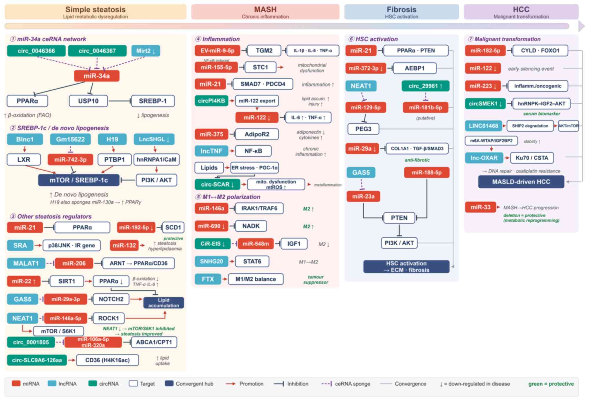 | Figure 7Key regulatory network of ncRNAs
during the progression from MASLD to HCC. The diagram is organised
into four disease stages (simple steatosis, MASH, fibrosis and HCC)
and illustrates how miRNAs (red), lncRNAs (blue) and circRNAs
(green), together with their target molecules (white) and the
convergent signalling hubs (dark blue), play crucial roles in this
disease process, either promoting or inhibiting it through complex
interactions. Regulatory mechanisms shared by different ncRNA
classes are merged into convergent hubs, including the miR-34a
ceRNA axis, the SREBP-1c lipogenic hub, the lipid-accumulation
node, the PTEN/PI3K/AKT node, the M1/M2 macrophage-polarisation hub
and the HSC-activation hub, thereby highlighting the convergent
regulatory networks underlying MASLD-driven HCC progression. Among
them, certain lncRNAs and circRNAs can function as ceRNAs that
modulate miRNA activity via a sponge mechanism (purple dashed
lines), thereby influencing downstream target genes and signalling
pathways and, collectively, driving the evolution of disease
phenotypes. Red arrows indicate promotion/activation, dark T-bars
indicate inhibition, grey lines denote convergence onto
stage-specific hubs, ↓ denotes downregulation in disease, and green
labels mark protective factors. ncRNAs, non-coding RNAs; MASLD,
metabolic dysfunction-associated steatotic liver disease; HCC,
hepatocellular carcinoma; miRNAs, microRNAs; lncRNAs, long
non-coding RNAs; circRNAs, circular RNAs; ceRNAs, competing
endogenous RNAs; MASH, metabolic dysfunction-associated
steatohepatitis; HSC, hepatic stellate cell. Created with
figdraw.com. |
Conclusions and future perspectives
The progression from MASLD to HCC is a complex,
multifactorial pathological process driven by the interplay of
metabolic dysregulation, chronic inflammation, oxidative stress,
immune dysfunction and gut microbiota imbalance. The present review
systematically highlights the central regulatory roles of ncRNAs,
including miRNAs, lncRNAs and circRNAs, across this disease
continuum. By orchestrating the fine-tuned regulation of key
signalling pathways, particularly NF-κB, JAK/STAT and
PI3K/AKT/mTOR, these ncRNAs exert notable effects on lipid
metabolism, inflammatory responses, fibrogenesis and hepatocellular
malignant transformation, thereby promoting disease progression
across the entire spectrum from simple steatosis to MASH, liver
fibrosis/cirrhosis and ultimately HCC.
Mechanistically, MASLD-driven HCC progression is
driven by multiple interconnected processes, including chronic
inflammation, oxidative stress, immune dysregulation, gut
microbiota imbalance, lipotoxicity and glucotoxicity. ncRNAs play
critical regulatory roles at different stages of disease
development through diverse mechanisms, including molecular
sponging, scaffold formation and, in some cases, the encoding of
functional micropeptides that enable precise control of cellular
processes. Despite substantial progress, most studies have focused
on individual ncRNAs, while interactions among different classes of
ncRNAs remain poorly understood. Future investigations should
therefore move beyond a single-molecule perspective and adopt a
network-based approach to elucidate potential synergistic, additive
or antagonistic relationships among ncRNAs and define their
integrated downstream signalling networks.
The close association between ncRNA expression
profiles and disease severity across the MASLD-driven HCC continuum
highlights their considerable translational potential. Future
advances are likely to focus on two major directions. First,
developing non-invasive biomarkers for diagnosis and dynamic
disease monitoring is a key priority. Several ncRNAs, including
circulating miR-122 and miR-34a, have demonstrated strong
associations with disease progression and may be incorporated into
biomarker panels following validation in large-scale clinical
cohorts. Such panels could provide non-invasive tools for early
detection, risk stratification, disease monitoring and therapeutic
response assessment, overcoming limitations of current diagnostic
approaches. Second, precision therapeutic strategies targeting
ncRNAs represent a promising avenue for disease intervention.
Silencing pathogenic ncRNAs, such as miR-21 and miR-155, with
chemically modified inhibitors (including antagomirs) or restoring
the activity of protective ncRNAs, such as miR-122, through
synthetic mimics, could modulate disease progression at the
molecular level.
Recent advances in oligonucleotide engineering,
delivery technologies and targeted RNA therapeutics have further
accelerated the clinical development of ncRNA-based interventions.
Nevertheless, several substantial challenges remain, including
efficient and tissue-specific delivery, long-term safety,
off-target effects and the context-dependent functions of ncRNAs
across different disease stages and cellular environments.
Addressing these limitations will be essential for successful
clinical translation.
In conclusion, ncRNAs are not merely passive
biomarkers but active regulators of the molecular events underlying
MASLD-driven HCC progression. Their extensive involvement in
metabolic regulation, inflammation, fibrosis and
hepatocarcinogenesis places them at the centre of disease
pathogenesis. A more comprehensive understanding of ncRNA
regulatory networks will not only provide deeper insight into the
mechanisms driving MASLD-associated hepatocarcinogenesis but also
facilitate the development of innovative diagnostic, prognostic and
therapeutic strategies. Ultimately, integration of ncRNA-based
approaches with existing clinical management paradigms may enable
more precise and effective prevention and treatment of MASLD-driven
HCC.
Supplementary Data
Availability of data and materials
Not applicable.
Authors' contributions
HW conceived the topic of review. WZ, CA, LX, HW,
YW and JZ wrote, reviewed and edited the manuscript. Data
authentication is not applicable. All authors read and approved the
final version of the manuscript.
Ethics approval and consent to
participate
Not applicable.
Patient consent for publication
Not applicable.
Competing interests
The authors declare that they have no competing
interests.
Abbreviations:
|
MASLD
|
metabolic dysfunction-associated
steatotic liver disease
|
|
MASH
|
metabolic dysfunction-associated
steatohepatitis
|
|
HCC
|
hepatocellular carcinoma
|
|
ncRNA
|
non-coding RNA
|
|
DAMPs
|
damage-associated molecular
patterns
|
|
HSCs
|
hepatic stellate cells
|
|
MDSCs
|
myeloid-derived suppressor cells
|
|
SASP
|
senescence-associated secretory
phenotype
|
|
SREBP-1c
|
sterol regulatory element-binding
protein-1c
|
|
uPA
|
urokinase-type plasminogen
activator
|
|
lncRNA
|
long ncRNA
|
|
LXR
|
liver X receptor
|
|
miRNA
|
microRNA
|
|
PPARα
|
peroxisome proliferator-activated
receptor α
|
|
mTORC1
|
mTOR complex 1
|
|
m6A
|
N6-methyladenosine
|
|
NRF1
|
nuclear respiratory factor 1
|
|
mTOR
|
mechanistic target of rapamycin
|
|
IGF2
|
insulin-like growth factor 2
|
|
circRNA
|
circular RNA
|
|
FTO
|
fat mass and obesity-associated
protein
|
|
ceRNA
|
competing endogenous RNA
|
Acknowledgements
Not applicable.
Funding
The present review was funded by the Hohhot Youth Talent Plan
Project (grant no. 2023003), the Hohhot Science and Technology
Bureau Applied Research and Development Funds (grant no.
2023-she-14), the Science and Technology Project of High-level
Clinical Specialty Construction in Hohhot Public Hospital of Inner
Mongolia Autonomous Region Health Commission (grant no.
2023SGGZ029), the Beijing Natural Science Foundation (grant no.
7252174), the General Project of Liaoning Province Education
Department (grant no. LJ212510164017) and the Talent Development
Plan for the Future in Medical-Engineering Integration by BRA-CDCHE
and ZTA (grant no. MBRC0012025029).
References
|
1
|
Wong VWS, Zelber-Sagi S, Cusi K, Carrieri
P, Wright E, Crespo J and Lazarus JV: Management of NAFLD in
primary care settings. Liver Int. 42:2377–2389. 2022. View Article : Google Scholar : PubMed/NCBI
|
|
2
|
Hwang SY, Danpanichkul P, Agopian V, Mehta
N, Parikh ND, Abou-Alfa GK, Singal AG and Yang JD: Hepatocellular
carcinoma: Updates on epidemiology, surveillance, diagnosis and
treatment. Clin Mol Hepatol. 31(Suppl): S228–S254. 2025. View Article : Google Scholar :
|
|
3
|
Ramai D, Tai W, Rivera M, Facciorusso A,
Tartaglia N, Pacilli M, Ambrosi A, Cotsoglou C and Sacco R: Natural
progression of Non-alcoholic steatohepatitis to hepatocellular
carcinoma. Biomedicines. 9:1842021. View Article : Google Scholar : PubMed/NCBI
|
|
4
|
Buzzetti E, Pinzani M and Tsochatzis EA:
The multiple-hit pathogenesis of non-alcoholic fatty liver disease
(NAFLD). Metabolism. 65:1038–1048. 2016. View Article : Google Scholar : PubMed/NCBI
|
|
5
|
Kim BK: Metabolic dysfunction-associated
steatotic liver disease and obesity: Pathogenesis, diagnostics,
risk stratification, and therapeutic approach. Kaohsiung J Med
Scie. e702272026. View Article : Google Scholar : Epub ahead of
print.
|
|
6
|
Nemeth K, Bayraktar R, Ferracin M and
Calin GA: Non-coding RNAs in disease: From mechanisms to
therapeutics. Nat Rev Genet. 25:211–232. 2024. View Article : Google Scholar
|
|
7
|
Li B, Li X, Ma M, Wang Q, Shi J and Wu C:
Analysis of long non-coding RNAs associated with disulfidptosis for
prognostic signature and immunotherapy response in uterine corpus
endometrial carcinoma. Sci Rep. 13:222202023. View Article : Google Scholar : PubMed/NCBI
|
|
8
|
Wang D, Gao H, Li Y, Jiang S, Yong Y and
Yang X: Genome-Scale expression pattern of long Non-Coding RNAs in
chinese uyghur patients with Parkinson's disease. Med Sci Monit.
26:e9258882020.PubMed/NCBI
|
|
9
|
Ogata H, Higasa K, Kageyama Y, Tahara H,
Shimamoto A, Takekita Y, Koshikawa Y, Nonen S, Kato T, Kinoshita T
and Kato M: Relationship between circulating mitochondrial DNA and
microRNA in patients with major depression. J Affect Disord.
339:538–546. 2023. View Article : Google Scholar : PubMed/NCBI
|
|
10
|
Zhao Q, Liu J, Deng H, Ma R, Liao JY,
Liang H, Hu J, Li J, Guo Z, Cai J, et al: Targeting
Mitochondria-located circRNA SCAR alleviates NASH via reducing mROS
output. Cell. 183:76–93.e22. 2020. View Article : Google Scholar : PubMed/NCBI
|
|
11
|
Ji Y, Luo Z, Gao H, Dos Reis FCG,
Bandyopadhyay G, Jin Z, Manda KA, Isaac R, Yang M, Fu W, et al:
Hepatocyte-derived exosomes from early onset obese mice promote
insulin sensitivity through miR-3075. Nat Metab. 3:1163–1174. 2021.
View Article : Google Scholar : PubMed/NCBI
|
|
12
|
Wang Y, Fleishman JS, Li T, Li Y, Ren Z,
Chen J and Ding M: Pharmacological therapy of metabolic
dysfunction-associated steatotic liver disease-driven
hepatocellular carcinoma. Front Pharmacol. 14:13362162024.
View Article : Google Scholar : PubMed/NCBI
|
|
13
|
Rinella ME, Neuschwander-Tetri BA,
Siddiqui MS, Abdelmalek MF, Caldwell S, Barb D, Kleiner DE and
Loomba R: AASLD Practice Guidance on the clinical assessment and
management of nonalcoholic fatty liver disease. Hepatology.
77:1797–1835. 2023. View Article : Google Scholar : PubMed/NCBI
|
|
14
|
Dezan MGF, Oliveira CP and Cotrim HP:
Metabolic Dysfunction-associated steatotic liver disease: Update.
Arq Gastroenterol. 62:e241172025. View Article : Google Scholar : PubMed/NCBI
|
|
15
|
Zhang X, Zheng MH, Liu D, Lin Y, Song SJ,
Chu ES, Liu D, Singh S, Berman M, Lau HC, et al: A blood-based
biomarker panel for non-invasive diagnosis of metabolic
dysfunction-associated steatohepatitis. Cell Metab. 37:59–68.e3.
2025. View Article : Google Scholar
|
|
16
|
Castellana M, Donghia R, Lampignano L,
Castellana F, Zupo R, Sardone R, Pergola G and Giannelli G:
Prevalence of the absence of cirrhosis in subjects with
NAFLD-Associated hepatocellular carcinoma. J Clin Med. 10:46382021.
View Article : Google Scholar : PubMed/NCBI
|
|
17
|
Sung H, Filho AM, Laversanne M, Ferlay J,
Siegel RL, Soerjomataram I, Jemal A and Bray F: Global cancer
statistics 2024: GLOBOCAN estimates of incidence and mortality
worldwide for 34 cancers in 186 countries. CA Cancer J Clin.
76:e700902026.PubMed/NCBI
|
|
18
|
Valery PC, Laversanne M, Clark PJ, Petrick
JL, McGlynn KA and Bray F: Projections of primary liver cancer to
2030 in 30 countries worldwide. Hepatology. 67:600–611. 2018.
View Article : Google Scholar
|
|
19
|
Kim H, Jang M and Park YN:
Histopathological variants of hepatocellular carcinomas: An update
according to the 5th edition of the WHO classification of digestive
system tumors. J Liver Cancer. 20:17–24. 2020. View Article : Google Scholar : PubMed/NCBI
|
|
20
|
Estes C, Razavi H, Loomba R, Younossi Z
and Sanyal AJ: Modeling the epidemic of nonalcoholic fatty liver
disease demonstrates an exponential increase in burden of disease.
Hepatology. 67:123–133. 2018. View Article : Google Scholar :
|
|
21
|
Kanwal F, Kramer JR, Mapakshi S, Natarajan
Y, Chayanupatkul M, Richardson PA, Li L, Desiderio R, Thrift AP,
Asch SM, et al: Risk of hepatocellular cancer in patients With
Non-Alcoholic fatty liver disease. Gastroenterology.
155:1828–1837.e2. 2018. View Article : Google Scholar : PubMed/NCBI
|
|
22
|
Ueno M, Takeda H, Takai A and Seno H: Risk
factors and diagnostic biomarkers for nonalcoholic fatty liver
disease-associated hepatocellular carcinoma: Current evidence and
future perspectives. World J Gastroenterol. 28:3410–3421. 2022.
View Article : Google Scholar : PubMed/NCBI
|
|
23
|
Piscaglia F, Svegliati-Baroni G, Barchetti
A, Pecorelli A, Marinelli S, Tiribelli C and Bellentani S;
HCC-NAFLD Italian Study Group: Clinical patterns of hepatocellular
carcinoma in nonalcoholic fatty liver disease: A multicenter
prospective study. Hepatology. 63:827–838. 2016. View Article : Google Scholar
|
|
24
|
McGlynn KA, Petrick JL and El-Serag HB:
Epidemiology of hepatocellular carcinoma. Hepatology. 73(Suppl 1):
S4–S13. 2021. View Article : Google Scholar
|
|
25
|
Liu Z, Jiang Y, Yuan H, Fang P, Cai N, Suo
C, Xiong H and Chen Q: The trends in death of primary liver cancer
caused by specific etiologies worldwide: Results from the Global
Burden of Disease Study 2019 and implications for liver cancer
management. BMC Med. 23:5982023.
|
|
26
|
Kim NG, Nguyen PP, Dang H, Kumari R,
Garcia G, Esquivel CO and Nguyen MH: Temporal trends in disease
presentation and survival of patients with hepatocellular
carcinoma: A real-world experience from 1998 to 2015. Cancer.
124:2588–2598. 2018. View Article : Google Scholar : PubMed/NCBI
|
|
27
|
Petrelli F, Manara M, Colombo S, De Santi
G, Ghidini M, Mariani M, Iaculli A, Rausa E, Rampulla V and Arru M:
Hepatocellular carcinoma in patients with nonalcoholic fatty liver
disease: A systematic review and meta-analysis: HCC and Steatosis
or Steatohepatitis. Neoplasia. 30:1008092022. View Article : Google Scholar : PubMed/NCBI
|
|
28
|
Yang WS, Va P, Bray F, Gao S, Gao J, Li HL
and Xiang YB: The role of pre-existing diabetes mellitus on
hepatocellular carcinoma occurrence and prognosis: A meta-analysis
of prospective cohort studies. PLoS One. 6:e273262011. View Article : Google Scholar : PubMed/NCBI
|
|
29
|
Sohn W, Lee HW, Lee S, Lim JH, Lee MW,
Park CH and Yoon SK: Obesity and the risk of primary liver cancer:
A systematic review and meta-analysis. Clin Mol Hepatol.
27:157–174. 2021. View Article : Google Scholar :
|
|
30
|
Younossi ZM and Henry L: Epidemiology of
non-alcoholic fatty liver disease and hepatocellular carcinoma.
JHEP Rep. 3:1003052021. View Article : Google Scholar : PubMed/NCBI
|
|
31
|
Papadakos SP, Georgiadou C, Argyrou A,
Michailidou E, Thanos C, Vogli S, Siakavellas SI, Manolakopoulos S
and Theocharis S: Inflammatory bowel disease (IBD)-Associated
colorectal cancer (CRC): Is cGAS-STING pathway targeting the key to
chemoprevention? Int J Mol Sci. 26:49792025. View Article : Google Scholar : PubMed/NCBI
|
|
32
|
Duan Y, Pan X, Luo J, Xiao X, Li J,
Bestman PL and Luo M: Association of inflammatory cytokines with
Non-alcoholic fatty liver disease. Front Immunol. 13:8802982022.
View Article : Google Scholar : PubMed/NCBI
|
|
33
|
Seo YY, Cho YK, Bae JC, Seo MH, Park SE,
Rhee EJ, Park CY, Oh KW, Park SW and Lee WY: Tumor necrosis
Factor-alpha as a predictor for the development of nonalcoholic
fatty liver disease: A 4-year follow-up study. Endocrinol Metab
(Seoul). 28:41–45. 2013. View Article : Google Scholar
|
|
34
|
Cannito S, Dianzani U, Parola M, Albano E
and Sutti S: Inflammatory processes involved in NASH-related
hepatocellular carcinoma. Biosci Rep. 43:BSR202212712023.
View Article : Google Scholar : PubMed/NCBI
|
|
35
|
Michelotti A, de Scordilli M, Palmero L,
Guardascione M, Masala M, Roncato R, Foltran L, Ongaro E and
Puglisi F: NAFLD-related hepatocarcinoma: The malignant side of
metabolic syndrome. Cells. 10:20342021. View Article : Google Scholar : PubMed/NCBI
|
|
36
|
Raza S, Mahamood R, Medhe P, Shahi A,
Yadav A, Tewari A and Sinha RA: Sterile inflammation in MASH:
Emerging role of extracellular RNA and therapeutic strategies. NPJ
Metab Health Dis. 3:392025. View Article : Google Scholar : PubMed/NCBI
|
|
37
|
Yu L, Gao F, Li Y, Zhang X and Feng Z:
Role of pattern recognition receptors in the development of MASLD
and potential therapeutic applications. Biomed Pharmacother.
175:1167242024. View Article : Google Scholar : PubMed/NCBI
|
|
38
|
Heida A, Gruben N, Catrysse L, Koehorst M,
Koster M, Kloosterhuis NJ, Gerding A, Havinga R, Bloks VW,
Bongiovanni L, et al: The hepatocyte IKK:NF-kappaB axis promotes
liver steatosis by stimulating de novo lipogenesis and cholesterol
synthesis. Mol Metab. 54:1013492021. View Article : Google Scholar
|
|
39
|
Rius-Perez S, Perez S, Marti-Andres P,
Monsalve M and Sastre J: Nuclear factor Kappa B signaling complexes
in acute inflammation. Antioxid Redox Signal. 33:145–165. 2020.
View Article : Google Scholar
|
|
40
|
Balsano C, Porcu C, Sideri S and Tavolaro
S: Fat and hepatocellular carcinoma. Hepatoma Res. 4:2018.
View Article : Google Scholar
|
|
41
|
Ye M, Tang Y, He J, Yang Y, Cao X, Kou S,
Wang L, Sheng L and Xue J: Alleviation of non-alcoholic fatty liver
disease by Huazhi Fugan Granules is associated with suppression of
TLR4/NF-κB signaling pathway. Clin Investig Arterioscler.
33:257–266. 2021.PubMed/NCBI
|
|
42
|
Duarte N, Coelho IC, Patarrao RS, Almeida
JI, Penha-Goncalves C and Macedo MP: How Inflammation impinges on
NAFLD: A role for kupffer cells. Biomed Res Int. 2015:9845782015.
View Article : Google Scholar : PubMed/NCBI
|
|
43
|
Xu J, Lin H, Wu G, Zhu M and Li M:
IL-6/STAT3 is a promising therapeutic target for hepatocellular
carcinoma. Front Oncol. 11:7609712021. View Article : Google Scholar :
|
|
44
|
Lee C and Cheung ST: STAT3: An emerging
therapeutic target for hepatocellular carcinoma. Cancers (Basel).
11:16462019. View Article : Google Scholar : PubMed/NCBI
|
|
45
|
Yu S, Wang J, Zheng H, Wang R, Johnson N,
Li T, Li P, Lin J, Li Y, Yan J, et al: Pathogenesis from
Inflammation to Cancer in NASH-Derived HCC. J Hepatocell Carcinoma.
9:855–867. 2022. View Article : Google Scholar : PubMed/NCBI
|
|
46
|
Adrover JM, McDowell SAC, He XY, Quail DF
and Egeblad M: NETworking with cancer: The bidirectional interplay
between cancer and neutrophil extracellular traps. Cancer Cell.
41:505–526. 2023. View Article : Google Scholar : PubMed/NCBI
|
|
47
|
Zhu S, Yu Y, Ren Y, Xu L, Wang H, Ling X,
Jin L, Hu Y, Zhang H, Miao C and Guo K: The emerging roles of
neutrophil extracellular traps in wound healing. Cell Death Dis.
12:9842021. View Article : Google Scholar : PubMed/NCBI
|
|
48
|
Peverill W, Powell LW and Skoien R:
Evolving concepts in the pathogenesis of NASH: Beyond steatosis and
inflammation. Int J Mol Sci. 15:8591–8638. 2014. View Article : Google Scholar : PubMed/NCBI
|
|
49
|
Singal AG, Llovet JM, Yarchoan M, Mehta N,
Heimbach JK, Dawson LA, Jou JH, Kulik LM, Agopian VG and Marrero
JA: AASLD Practice Guidance on prevention, diagnosis, and treatment
of hepatocellular carcinoma. Hepatology. 78:1922–1965. 2023.
View Article : Google Scholar : PubMed/NCBI
|
|
50
|
Han S, Han L, Yao Y, Sun H, Zan X and Liu
Q: Activated hepatic stellate cells promote hepatocellular
carcinoma cell migration and invasion via the activation of
FAK-MMP9 signaling. Oncol Rep. 31:641–648. 2014. View Article : Google Scholar
|
|
51
|
Zhou J, Liu L, Hu X, Feng R, Zhao N, Zhang
L, Hu W, Zhang J, Huang S, Liu L, et al: Matrix
metalloproteinase-21 promotes metastasis via increasing the
recruitment and M2 polarization of macrophages in HCC. Cancer Sci.
114:423–435. 2023. View Article : Google Scholar :
|
|
52
|
Niu ZS, Wang WH and Niu XJ: Recent
progress in molecular mechanisms of postoperative recurrence and
metastasis of hepatocellular carcinoma. World J Gastroenterol.
28:6433–6477. 2022. View Article : Google Scholar : PubMed/NCBI
|
|
53
|
Ali E, Trailin A, Ambrozkiewicz F, Liska V
and Hemminki K: Activated hepatic stellate cells in hepatocellular
carcinoma: Their role as a potential target for future therapies.
Int J Mol Sci. 23:152922022. View Article : Google Scholar : PubMed/NCBI
|
|
54
|
Zhao RZ, Jiang S, Zhang L and Yu ZB:
Mitochondrial electron transport chain, ROS generation and
uncoupling (Review). Int J Mol Med. 44:3–15. 2019.PubMed/NCBI
|
|
55
|
Lettieri-Barbato D: Redox control of
non-shivering thermogenesis. Mol Metab. 25:11–19. 2019. View Article : Google Scholar : PubMed/NCBI
|
|
56
|
Huang X, Zeng Z, Li S, Xie Y and Tong X:
The therapeutic strategies targeting mitochondrial metabolism in
cardiovascular disease. Pharmaceutics. 14:27602022. View Article : Google Scholar : PubMed/NCBI
|
|
57
|
Li YF, Xie ZF, Song Q and Li JY:
Mitochondria homeostasis: Biology and involvement in hepatic
steatosis to NASH. Acta Pharmacol Sin. 43:1141–1155. 2022.
View Article : Google Scholar : PubMed/NCBI
|
|
58
|
Rives C, Fougerat A, Ellero-Simatos S,
Loiseau N, Guillou H, Gamet-Payrastre L and Wahli W: Oxidative
stress in NAFLD: Role of nutrients and food contaminants.
Biomolecules. 10:17022020. View Article : Google Scholar : PubMed/NCBI
|
|
59
|
Brahma MK, Gilglioni EH, Zhou L, Trepo E,
Chen P and Gurzov EN: Oxidative stress in obesity-associated
hepatocellular carcinoma: Sources, signaling and therapeutic
challenges. Oncogene. 40:5155–5167. 2021. View Article : Google Scholar : PubMed/NCBI
|
|
60
|
An P, Wei LL, Zhao S, Sverdlov DY, Vaid
KA, Miyamoto M, Kuramitsu K, Lai M and Popov YV: Hepatocyte
mitochondria-derived danger signals directly activate hepatic
stellate cells and drive progression of liver fibrosis. Nat Commun.
11:23622020. View Article : Google Scholar : PubMed/NCBI
|
|
61
|
Torrens-Mas M, Cordani M, Mullappilly N,
Pacchiana R, Riganti C, Palmieri M, Pons DG, Roca P, Oliver J and
Donadelli M: Mutant p53 induces SIRT3/MnSOD axis to moderate ROS
production in melanoma cells. Arch Biochem Biophys. 679:1082192020.
View Article : Google Scholar
|
|
62
|
Trachootham D, Lu W, Ogasawara MA, Nilsa
RD and Huang P: Redox regulation of cell survival. Antioxid Redox
Signal. 10:1343–1374. 2008. View Article : Google Scholar : PubMed/NCBI
|
|
63
|
Liu J, Han X, Zhang T, Tian K, Li Z and
Luo F: Reactive oxygen species (ROS) scavenging biomaterials for
anti-inflammatory diseases: From mechanism to therapy. J Hematol
Oncol. 16:1162023. View Article : Google Scholar : PubMed/NCBI
|
|
64
|
Gabbia D, Cannella L and De Martin S: The
role of oxidative stress in NAFLD-NASH-HCC Transition-Focus on
NADPH oxidases. Biomedicines. 9:6872021. View Article : Google Scholar : PubMed/NCBI
|
|
65
|
Nikitina IA, Razenkova VA, Fedorova EA,
Kirik OV and Korzhevskii DE: Technology of combined identification
of macrophages and collagen fibers in liver samples. Sovrem
Tekhnologii Med. 16:24–29. 2024. View Article : Google Scholar : PubMed/NCBI
|
|
66
|
Roohani S and Tacke F: Liver injury and
the macrophage issue: Molecular and mechanistic facts and their
clinical relevance. Int J Mol Sci. 22:72492021. View Article : Google Scholar : PubMed/NCBI
|
|
67
|
Roehlen N, Crouchet E and Baumert TF:
Liver fibrosis: Mechanistic concepts and therapeutic perspectives.
Cells. 9:8752020. View Article : Google Scholar : PubMed/NCBI
|
|
68
|
Al Moussawy M and Abdelsamed HA:
Non-cytotoxic functions of CD8 T cells: 'Repentance of a serial
killer'. Front Immunol. 13:10011292022. View Article : Google Scholar
|
|
69
|
Huang Y, Jia A, Wang Y and Liu G: CD8+ T
cell exhaustion in anti-tumour immunity: The new insights for
cancer immunotherapy. Immunology. 168:30–48. 2023. View Article : Google Scholar
|
|
70
|
Burtis AEC, DeNicola DMC, Ferguson ME,
Santos RG, Pinilla C, Kriss MS, Orlicky DJ, Tamburini BAJ, Gillen
AE and Burchill MA: Ag-driven CD8 + T cell clonal expansion is a
prominent feature of MASH in humans and mice. Hepatology.
81:591–608. 2025. View Article : Google Scholar :
|
|
71
|
Breuer DA, Pacheco MC, Washington MK,
Montgomery SA, Hasty AH and Kennedy AJ: CD8+ T cells regulate liver
injury in obesity-related nonalcoholic fatty liver disease. Am J
Physiol Gastrointest Liver Physiol. 318:G211–G224. 2020. View Article : Google Scholar
|
|
72
|
Pan Y, Yang W, Tang B, Wang X, Zhang Q, Li
W and Li L: The protective and pathogenic role of Th17 cell
plasticity and function in the tumor microenvironment. Front
Immunol. 14:11923032023. View Article : Google Scholar : PubMed/NCBI
|
|
73
|
Law AMK, Valdes-Mora F and Gallego-Ortega
D: Myeloid-derived suppressor cells as a therapeutic target for
cancer. Cells. 9:5612020. View Article : Google Scholar : PubMed/NCBI
|
|
74
|
Gabrilovich DI and Nagaraj S:
Myeloid-derived suppressor cells as regulators of the immune
system. Nat Rev Immunol. 9:162–174. 2009. View Article : Google Scholar : PubMed/NCBI
|
|
75
|
Liu H, Wang Z, Zhou Y and Yang Y: MDSCs in
breast cancer: An important enabler of tumor progression and an
emerging therapeutic target. Front Immunol. 14:11992732023.
View Article : Google Scholar : PubMed/NCBI
|
|
76
|
Vanhaver C, van der Bruggen P and Bruger
AM: MDSC in mice and men: Mechanisms of immunosuppression in
cancer. J Clin Med. 10:28722021. View Article : Google Scholar : PubMed/NCBI
|
|
77
|
Groth C, Hu X, Weber R, Fleming V,
Altevogt P, Utikal J and Umansky V: Immunosuppression mediated by
myeloid-derived suppressor cells (MDSCs) during tumour progression.
Br J Cancer. 120:16–25. 2019. View Article : Google Scholar :
|
|
78
|
Vetsika EK, Koukos A and Kotsakis A:
Myeloid-derived suppressor cells: Major figures that shape the
immunosuppressive and angiogenic network in cancer. Cells.
8:16472019. View Article : Google Scholar : PubMed/NCBI
|
|
79
|
Kumar AR, Nair B, Kamath AJ, Nath LR,
Calina D and Sharifi-Rad J: Impact of gut microbiota on metabolic
dysfunction-associated steatohepatitis and hepatocellular
carcinoma: Pathways, diagnostic opportunities and therapeutic
advances. Eur J Med Res. 29:4852024. View Article : Google Scholar : PubMed/NCBI
|
|
80
|
Macura B, Kiecka A and Szczepanik M:
Intestinal permeability disturbances: Causes, diseases and therapy.
Clin Exp Med. 24:2322024. View Article : Google Scholar : PubMed/NCBI
|
|
81
|
Yang Z, Lin S, Liu Y, Song Z, Ge Z, Fan Y,
Chen L, Bi Y, Zhao Z, Wang X, et al: Targeting intestinal
microecology: Potential intervention strategies of traditional
Chinese medicine for managing hypertension. Front Pharmacol.
14:11711192023. View Article : Google Scholar : PubMed/NCBI
|
|
82
|
Fu Q, Song T, Ma X and Cui J: Research
progress on the relationship between intestinal microecology and
intestinal bowel disease. Animal Model Exp Med. 5:297–310. 2022.
View Article : Google Scholar : PubMed/NCBI
|
|
83
|
Altves S, Yildiz HK and Vural HC:
Interaction of the microbiota with the human body in health and
diseases. Biosci Microbiota Food Health. 39:23–32. 2020. View Article : Google Scholar : PubMed/NCBI
|
|
84
|
Islam MM, Mahbub NU, Hong ST and Chung HJ:
Gut bacteria: An etiological agent in human pathological
conditions. Front Cell Infect Microbiol. 14:12911482024. View Article : Google Scholar : PubMed/NCBI
|
|
85
|
Caricilli AM, Castoldi A and Camara NO:
Intestinal barrier: A gentlemen's agreement between microbiota and
immunity. World J Gastrointest Pathophysiol. 5:18–32. 2014.
View Article : Google Scholar : PubMed/NCBI
|
|
86
|
Nishimura N, Kaji K, Kitagawa K, Sawada Y,
Furukawa M, Ozutsumi T, Fujinaga Y, Tsuji Y, Takaya H, Kawaratani
H, et al: Intestinal permeability is a mechanical rheostat in the
pathogenesis of liver cirrhosis. Int J Mol Sci. 22:69212021.
View Article : Google Scholar : PubMed/NCBI
|
|
87
|
Yoshimoto S, Loo TM, Atarashi K, Kanda H,
Sato S, Oyadomari S, Iwakura Y, Oshima K, Morita H, Hattori M, et
al: Obesity-induced gut microbial metabolite promotes liver cancer
through senescence secretome. Nature. 499:97–101. 2013. View Article : Google Scholar : PubMed/NCBI
|
|
88
|
Loo TM, Kamachi F, Watanabe Y, Yoshimoto
S, Kanda H, Arai Y, Nakajima-Takagi Y, Iwama A, Koga T, Sugimoto Y,
et al: Gut microbiota promotes obesity-associated liver cancer
through PGE2-mediated suppression of antitumor immunity. Cancer
Discov. 7:522–538. 2017. View Article : Google Scholar : PubMed/NCBI
|
|
89
|
Huang Y, Yang X, Meng Y, Shao C, Liao J,
Li F, Li R, Jing Y and Huang A: The hepatic senescence-associated
secretory phenotype promotes hepatocarcinogenesis through
Bcl3-dependent activation of macrophages. Cell Biosci. 11:1732021.
View Article : Google Scholar : PubMed/NCBI
|
|
90
|
Liu B, Qu L and Yan S: Cyclooxygenase-2
promotes tumor growth and suppresses tumor immunity. Cancer Cell
Int. 15:1062015. View Article : Google Scholar : PubMed/NCBI
|
|
91
|
Ohtani N, Kamiya T and Kawada N: Recent
updates on the role of the gut-liver axis in the pathogenesis of
NAFLD/NASH, HCC, and beyond. Hepatol Commun. 7:e02412023.
View Article : Google Scholar : PubMed/NCBI
|
|
92
|
Lau HCH, Zhang X and Yu J: Gut microbiome
in metabolic dysfunction-associated steatotic liver disease and
associated hepatocellular carcinoma. Nat Rev Gastroenterol Hepatol.
22:619–638. 2025. View Article : Google Scholar : PubMed/NCBI
|
|
93
|
Rockenfeller P and Gourlay CW: Lipotoxicty
in yeast: A focus on plasma membrane signalling and membrane
contact sites. FEMS Yeast Res. 18:foy0342018. View Article : Google Scholar : PubMed/NCBI
|
|
94
|
Engin AB: Mechanism of obesity-related
lipotoxicity and clinical perspective. Adv Exp Med Biol.
1460:131–166. 2024. View Article : Google Scholar : PubMed/NCBI
|
|
95
|
Chandel NS: Lipid metabolism. Cold Spring
Harb Perspect Biol. 13:a0405762021. View Article : Google Scholar : PubMed/NCBI
|
|
96
|
Maiers JL and Malhi H: Endoplasmic
reticulum stress in metabolic liver diseases and hepatic fibrosis.
Semin Liver Dis. 39:235–248. 2019. View Article : Google Scholar : PubMed/NCBI
|
|
97
|
Ly LD, Xu S, Choi SK, Ha CM, Thoudam T,
Cha SK, Wiederkehr A, Wollheim CB, Lee IK and Park KS: Oxidative
stress and calcium dysregulation by palmitate in type 2 diabetes.
Exp Mol Med. 49:e2912017. View Article : Google Scholar : PubMed/NCBI
|
|
98
|
Xu S, Nam SM, Kim JH, Das R, Choi SK,
Nguyen TT, Quan X, Choi SJ, Chung CH, Lee EY, et al: Palmitate
induces ER calcium depletion and apoptosis in mouse podocytes
subsequent to mitochondrial oxidative stress. Cell Death Dis.
6:e19762015. View Article : Google Scholar : PubMed/NCBI
|
|
99
|
Korbecki J and Bajdak-Rusinek K: The
effect of palmitic acid on inflammatory response in macrophages: An
overview of molecular mechanisms. Inflamm Res. 68:915–932. 2019.
View Article : Google Scholar : PubMed/NCBI
|
|
100
|
Yamamoto Y and Gaynor RT: Pathogenesis of
nonalcoholic steatohepatitis and Hormone-based therapeutic
approaches. Front Endocrinol (Lausanne). 9:9052018.
|
|
101
|
Steinberg GR, Carpentier AC and Wang D:
MASH: The nexus of metabolism, inflammation, and fibrosis. J Clin
Invest. 135:e1864202025. View Article : Google Scholar : PubMed/NCBI
|
|
102
|
Hodson L and Gunn PJ: The regulation of
hepatic fatty acid synthesis and partitioning: The effect of
nutritional state. Nat Rev Endocrinol. 15:689–700. 2019. View Article : Google Scholar : PubMed/NCBI
|
|
103
|
Samuel VT and Shulman GI: Nonalcoholic
fatty liver disease as a nexus of metabolic and hepatic diseases.
Cell Metab. 27:22–41. 2018. View Article : Google Scholar
|
|
104
|
Chavez JA and Summers SAL: Lipid
oversupply, selective insulin resistance, and lipotoxicity:
Molecular mechanisms. Biochim Biophys Acta. 1801:252–265. 2010.
View Article : Google Scholar
|
|
105
|
Ott C, Jacobs K, Haucke E, Navarrete
Santos A, Grune T and Simm A: Role of advanced glycation end
products in cellular signaling. Redox Biol. 2:411–429. 2014.
View Article : Google Scholar : PubMed/NCBI
|
|
106
|
Wang X, Zhang L and Dong B: Molecular
mechanisms in MASLD/MASH-related HCC. Hepatology. 79:206–224.
2024.
|
|
107
|
Im YR, Hunter H, de Gracia Hahn D, Duret
A, Cheah Q, Dong J, Fairey M, Hjalmarsson C, Li A, Lim HK, et al: A
systematic review of animal models of NAFLD finds High-Fat,
High-Fructose diets most closely resemble human NAFLD. Hepatology.
74:1884–1901. 2021. View Article : Google Scholar : PubMed/NCBI
|
|
108
|
Tsuchida T, Lee YA, Fujiwara N, Ybanez M,
Allen B, Martins S, Fiel MI, Goossens N, Chou HI, Hoshida Y and
Friedman SL: A simple diet- and chemical-induced murine NASH model
with rapid progression of steatohepatitis, fibrosis and liver
cancer. J Hepatol. 69:385–395. 2018. View Article : Google Scholar : PubMed/NCBI
|
|
109
|
Gallage S, Avila JEB, Ramadori P, Focaccia
E, Rahbari M, Ali A, Malek NP, Anstee QM and Heikenwalder M: A
researcher's guide to preclinical mouse NASH models. Nat Metab.
4:1632–1649. 2022. View Article : Google Scholar : PubMed/NCBI
|
|
110
|
Setayesh T, Hu Y, Vaziri F, Wei D and
Yvonne Wan Y-J: The spatial impact of a Western diet in enriching
Galectin-1-regulated Rho, ECM, and SASP signaling in a novel
MASH-HCC mouse model. Biomark Res. 12:1222024. View Article : Google Scholar : PubMed/NCBI
|
|
111
|
Febbraio MA, Reibe S, Shalapour S, Ooi GJ,
Watt MJ and Karin M: Preclinical models for studying NASH-Driven
HCC: How useful are they? Cell Metab. 29:18–26. 2019. View Article : Google Scholar
|
|
112
|
Bhatti GK, Khullar N, Sidhu IS, Navik US,
Reddy AP, Reddy PH and Bhatti JS: Emerging role of non-coding RNA
in health and disease. Metab Brain Dis. 36:1119–1134. 2021.
View Article : Google Scholar : PubMed/NCBI
|
|
113
|
Zhang P, Wu W, Chen Q and Chen M:
Non-coding RNAs and their integrated networks. J Integr Bioinform.
16:201900272019. View Article : Google Scholar : PubMed/NCBI
|
|
114
|
Kristensen LS, Andersen MS, Stagsted LVW,
Ebbesen KK, Hansen TB and Kjems J: The biogenesis, biology and
characterization of circular RNAs. Nat Rev Genet. 20:675–691. 2019.
View Article : Google Scholar : PubMed/NCBI
|
|
115
|
Huang QY, Zhou ZY, Zhang YL, Zhou Y, Duan
SW and Dong JY: Transfer RNA-derived small RNAs in liver disease.
Hepatobiliary Pancreat Dis Int. 25:332–329. 2026. View Article : Google Scholar
|
|
116
|
Manna D and Sarkar D: Non-coding RNAs:
Regulating disease progression and therapy resistance in
hepatocellular carcinoma. Cancers (Basel). 12:12432020. View Article : Google Scholar : PubMed/NCBI
|
|
117
|
Naseer QA, Xuexian C, Yimai D, Khan MA and
Chen S: Diagnostic and therapeutic relevance of non-coding RNAs in
metabolic dysfunction-associated steatotic liver disease and
steatohepatitis: A systematic review. J The Korean Med Assoc.
68:682–698. 2025. View Article : Google Scholar
|
|
118
|
Guttman M and Rinn JL: Modular regulatory
principles of large non-coding RNAs. Nature. 482:339–346. 2012.
View Article : Google Scholar : PubMed/NCBI
|
|
119
|
Statello L, Guo CJ, Chen LL and Huarte M:
Gene regulation by long non-coding RNAs and its biological
functions. Nat Rev Mol Cell Biol. 22:96–118. 2021. View Article : Google Scholar
|
|
120
|
Kopp F and Mendell JT: Functional
classification and experimental dissection of long noncoding RNAs.
Cell. 172:393–407. 2018. View Article : Google Scholar : PubMed/NCBI
|
|
121
|
Tsai PF, Dell'Orso S, Rodriguez J, Vivanco
KO, Ko KD, Jiang K, Juan AH, Sarshad AA, Vian L, Tran M, et al: A
Muscle-Specific enhancer RNA mediates cohesin recruitment and
regulates transcription in trans. Mol Cell. 71:129–141.e8. 2018.
View Article : Google Scholar : PubMed/NCBI
|
|
122
|
Zhao XY, Xiong X, Liu T, Mi L, Peng X, Rui
C, Guo L, Li S, Li X and Lin JD: Long noncoding RNA licensing of
obesity-linked hepatic lipogenesis and NAFLD pathogenesis. Nat
Commun. 9:England 9. 29862018. View Article : Google Scholar : PubMed/NCBI
|
|
123
|
Sheng L, Ye L, Zhang D, Cawthorn WP and Xu
B: New insights into the long Non-coding RNA SRA: Physiological
functions and mechanisms of action. Front Med (Lausanne).
5:2442018. View Article : Google Scholar : PubMed/NCBI
|
|
124
|
Liu S, Xu R, Gerin I, Cawthorn WP,
Macdougald OA, Chen XW, Saltiel AR, Koenig RJ and Xu B: SRA
regulates adipogenesis by modulating p38/JNK phosphorylation and
stimulating insulin receptor gene expression and downstream
signaling. PLoS One. 9:e954162014. View Article : Google Scholar : PubMed/NCBI
|
|
125
|
Liu C, Yang Z, Wu J, Zhang L, Lee S, Shin
DJ, Tran M and Wang L: Long noncoding RNA H19 interacts with
polypyrimidine tract-binding protein 1 to reprogram hepatic lipid
homeostasis. Hepatology. 67:1768–1783. 2018. View Article : Google Scholar
|
|
126
|
Wang H, Cao Y, Shu L, Zhu Y, Peng Q, Ran
L, Wu J, Luo Y, Zuo G, Luo J, et al: Long non-coding RNA (lncRNA)
H19 induces hepatic steatosis through activating MLXIPL and mTORC1
networks in hepatocytes. J Cell Mol Med. 24:1399–1412. 2020.
View Article : Google Scholar :
|
|
127
|
Wang Y, Tian X, Wang Z, Liu D, Zhao X, Sun
X, Tu Z, Li Z, Zhao Y, Zheng S and Yao J: A novel peptide encoded
by circ-SLC9A6 promotes lipid dyshomeostasis through the regulation
of H4K16ac-mediated CD36 transcription in NAFLD. Clin Transl Med.
14:e18012024. View Article : Google Scholar : PubMed/NCBI
|
|
128
|
Gebert LFR and MacRae IJ: Regulation of
microRNA function in animals. Nat Rev Mol Cell Biol. 20:21–37.
2019. View Article : Google Scholar :
|
|
129
|
Chen P, Li Y, Dai Y, Wang Z, Zhou Y, Wang
Y and Li G: Advances in the pathogenesis of metabolic liver
Disease-Related hepatocellular carcinoma. J Hepatocell Carcinoma.
11:581–594. 2024. View Article : Google Scholar : PubMed/NCBI
|
|
130
|
Guo XY, Sun F, Chen JN, Wang YQ, Pan Q and
Fan JG: circRNA_0046366 inhibits hepatocellular steatosis by
normalization of PPAR signaling. World J Gastroenterol. 24:323–337.
2018. View Article : Google Scholar : PubMed/NCBI
|
|
131
|
Cui J, Wang Y and Xue H: Long non-coding
RNA GAS5 contributes to the progression of nonalcoholic fatty liver
disease by targeting the microRNA-29a-3p/NOTCH2 axis.
Bioengineered. 13:8370–8381. 2022. View Article : Google Scholar : PubMed/NCBI
|
|
132
|
Shah M and Sarkar D: HCC-related lncRNAs:
Roles and mechanisms. Int J Mol Sci. 25:5972024. View Article : Google Scholar : PubMed/NCBI
|
|
133
|
Wang H, Wang Y, Lai S, Zhao L, Liu W, Liu
S, Chen H, Wang J, Du G and Tang B: LINC01468 drives NAFLD-HCC
progression through CUL4A-linked degradation of SHIP2. Cell Death
Discov. 8:4492022. View Article : Google Scholar : PubMed/NCBI
|
|
134
|
Wang J, Yang W, Chen Z, Chen J, Meng Y,
Feng B, Sun L, Dou L, Li J, Cui Q and Yang J: Long noncoding RNA
lncSHGL recruits hnRNPA1 to suppress hepatic gluconeogenesis and
lipogenesis. Diabetes. 67:581–593. 2018. View Article : Google Scholar : PubMed/NCBI
|
|
135
|
Salmena L, Poliseno L, Tay Y, Kats L and
Pandolfi PP: A ceRNA hypothesis: The rosetta stone of a hidden RNA
language? Cell. 146:353–358. 2011. View Article : Google Scholar : PubMed/NCBI
|
|
136
|
Lin S and Gregory RI: MicroRNA biogenesis
pathways in cancer. Nat Rev Cancer. 15:321–333. 2015. View Article : Google Scholar : PubMed/NCBI
|
|
137
|
Wang G, Zou H, Lai C, Huang X, Yao Y and
Xiang G: Repression of MicroRNA-124-3p alleviates High-Fat
Diet-induced hepatosteatosis by targeting Pref-1. Front Endocrinol
(Lausanne). 11:5899942020. View Article : Google Scholar : PubMed/NCBI
|
|
138
|
Liu X, Chen S and Zhang L: Downregulated
microRNA-130b-5p prevents lipid accumulation and insulin resistance
in a murine model of nonalcoholic fatty liver disease. Am J Physiol
Endocrinol Metab. 319:E34–E42. 2020. View Article : Google Scholar : PubMed/NCBI
|
|
139
|
Dou L, Wang S, Sun L, Huang X, Zhang Y,
Shen T, Guo J, Man Y, Tang W and Li J: Mir-338-3p Mediates
Tnf-A-Induced hepatic insulin resistance by targeting PP4r1 to
regulate PP4 expression. Cell Physiol Biochem. 41:2419–2431. 2017.
View Article : Google Scholar : PubMed/NCBI
|
|
140
|
Zhang T, Zhao X, Steer CJ, Yan G and Song
G: A negative feedback loop between microRNA-378 and Nrf1 promotes
the development of hepatosteatosis in mice treated with a high fat
diet. Metabolism. 85:183–191. 2018. View Article : Google Scholar : PubMed/NCBI
|
|
141
|
Wu H, Ng R, Chen X, Steer CJ and Song G:
MicroRNA-21 is a potential link between non-alcoholic fatty liver
disease and hepatocellular carcinoma via modulation of the
HBP1-p53-Srebp1c pathway. Gut. 65:1850–1860. 2016. View Article : Google Scholar :
|
|
142
|
He Y, Hwang S, Cai Y, Kim SJ, Xu M, Yang
D, Guillot A, Feng D, Seo W, Hou X and Gao B: MicroRNA-223
ameliorates nonalcoholic steatohepatitis and cancer by targeting
multiple inflammatory and oncogenic genes in hepatocytes.
Hepatology. 70:1150–1167. 2019. View Article : Google Scholar : PubMed/NCBI
|
|
143
|
Li K, Tian Y, Yuan Y, Fan X, Yang M, He Z
and Yang D: Insights into the functions of LncRNAs in drosophila.
Int J Mol Sci. 20:46462019. View Article : Google Scholar : PubMed/NCBI
|
|
144
|
Tao Q, Xie J, Wu Y and Jin Y: Long
non-coding RNAs as modulators and therapeutic targets in
non-alcoholic fatty liver disease (NAFLD). Gastroenterol Hepatol.
47:506–516. 2024. View Article : Google Scholar
|
|
145
|
Chen X, Xu Y, Zhao D, Chen T, Gu C, Yu G,
Chen K, Zhong Y, He J, Liu S, et al: LncRNA-AK012226 is involved in
fat accumulation in db/db mice fatty liver and Non-alcoholic fatty
liver disease cell model. Front Pharmacol. 9:8882018. View Article : Google Scholar : PubMed/NCBI
|
|
146
|
Liu F, Tian T, Zhang Z, Xie S, Yang J, Zhu
L, Wang W, Shi C, Sang L, Guo K, et al: Long non-coding RNA SNHG6
couples cholesterol sensing with mTORC1 activation in
hepatocellular carcinoma. Nat Metab. 4:1022–1040. 2022. View Article : Google Scholar : PubMed/NCBI
|
|
147
|
Hu C, Li X, Sui Y, Chen Y, Zhao Y, Yang Y,
Zhou Q, Ni H, Sun L, Gan W, et al: Dicer deletion in hepatocytes
promotes macrophages M1 polarization through dysregulated
miR-192-3p/IGF2 in non-alcoholic steatohepatitis and hepatocellular
carcinoma. Cancer Gene Ther. 29:1252–1262. 2022. View Article : Google Scholar : PubMed/NCBI
|
|
148
|
Chai C, Cox B, Yaish D, Gross D, Rosenberg
N, Amblard F, Shemuelian Z, Gefen M, Korach A, Tirosh O, et al:
Agonist of RORA attenuates nonalcoholic fatty liver progression in
mice via Up-regulation of MicroRNA 122. Gastroenterology.
159:999–1014.e9. 2020. View Article : Google Scholar : PubMed/NCBI
|
|
149
|
Teng M, Xia ZJ, Lo N, Daud K and He HH:
Assembling the RNA therapeutics toolbox. Med Rev. 4:110–128. 2024.
View Article : Google Scholar
|
|
150
|
Yepmo M, Potier JB, Pinget M, Grabarz A,
Bouzakri K and Dumond Bourie A: Discussing the role of circular RNA
in the pathogenesis of non-alcoholic fatty liver disease and its
complications. Front Endocrinol (Lausanne). 13:10351592022.
View Article : Google Scholar : PubMed/NCBI
|
|
151
|
Lu HJ and Zhou H: Circular RNAs in
metabolic liver diseases: Biomarkers, mechanisms, and therapeutic
perspectives. Metab Dis. 1:1000222026. View Article : Google Scholar
|
|
152
|
Li P, Shan K, Liu Y, Zhang Y and Xu L and
Xu L: CircScd1 promotes fatty liver disease via the janus kinase
2/Signal transducer and activator of transcription 5 pathway. Dig
Dis Sci. 64:113–122. 2019. View Article : Google Scholar
|
|
153
|
Yuan X, Diao J, Du A, Wen S, Zhou L and
Pan Y: Circular RNA expression profiles and features in NAFLD mice:
A study using RNA-seq data. J Transl Med. 18:4762020. View Article : Google Scholar : PubMed/NCBI
|
|
154
|
Bu FT, Zhu Y, Chen X, Wang A, Zhang YF,
You HM, Yang Y, Yang YR, Huang C and Li J: Circular RNA circPSD3
alleviates hepatic fibrogenesis by regulating the miR-92b-3p/Smad7
axis. Mol Ther Nucleic Acids. 23:847–862. 2021. View Article : Google Scholar : PubMed/NCBI
|
|
155
|
Takaki Y, Saito Y, Takasugi A, Toshimitsu
K, Yamada S, Muramatsu T, Kimura M, Sugiyama K, Suzuki H, Arai E,
et al: Silencing of microRNA-122 is an early event during
hepatocarcinogenesis from non-alcoholic steatohepatitis. Cancer
Sci. 105:1254–1260. 2014. View Article : Google Scholar : PubMed/NCBI
|
|
156
|
Cheng L, Zhu Y, Han H, Zhang Q, Cui K,
Shen H, Zhang J, Yan J, Prochownik E and Li Y: MicroRNA-148a
deficiency promotes hepatic lipid metabolism and
hepatocarcinogenesis in mice. Cell Death Dis. 8:e29162017.
View Article : Google Scholar : PubMed/NCBI
|
|
157
|
Su Q, Xu ZX, Xiong ML, Li HY, Xu MY and
Luo SZ: The oncogenic miR-27a/BTG2 axis promotes obesity-associated
hepatocellular carcinoma by mediating mitochondrial dysfunction.
Neoplasma. 69:820–831. 2022. View Article : Google Scholar : PubMed/NCBI
|
|
158
|
Sun Y, Wang Q, Zhang Y, Geng M, Wei Y, Liu
Y, Liu S, Petersen RB, Yue J, Huang K and Zheng L:
Multigenerational maternal obesity increases the incidence of HCC
in offspring via miR-27a-3p. J Hepatol. 73:603–615. 2020.
View Article : Google Scholar : PubMed/NCBI
|
|
159
|
Li H, Chen Z, Zhang Y, Yuan P, Liu J, Ding
L and Ye Q: MiR-4310 regulates hepatocellular carcinoma growth and
metastasis through lipid synthesis. Cancer Lett. 519:161–171. 2021.
View Article : Google Scholar : PubMed/NCBI
|
|
160
|
Wang Q, Wei S, Zhou H, Li L, Zhou S, Shi
C, Shi Y, Qiu J and Lu L: MicroRNA-98 inhibits hepatic stellate
cell activation and attenuates liver fibrosis by regulating HLF
expression. Front Cell Dev Biol. 8:5132020. View Article : Google Scholar : PubMed/NCBI
|
|
161
|
Wu H, Zhong Z, Wang A, Yuan C, Ning K, Hu
H, Wang C and Yin X: LncRNA FTX represses the progression of
non-alcoholic fatty liver disease to hepatocellular carcinoma via
regulating the M1/M2 polarization of Kupffer cells. Cancer Cell
Int. 20:2662020. View Article : Google Scholar : PubMed/NCBI
|
|
162
|
Li Z, Lu X, Liu Y, Zhao J, Ma S, Yin H,
Huang S, Zhao Y and He X: Gain of LINC00624 enhances liver cancer
progression by disrupting the histone Deacetylase 6/Tripartite
motif Containing 28/Zinc finger protein 354C corepressor complex.
Hepatology. 73:1764–1782. 2021. View Article : Google Scholar
|
|
163
|
Uchida S and Kauppinen S: Long Non-coding
RNAs in liver cancer and nonalcoholic steatohepatitis. Noncoding
RNA. 6:342020.PubMed/NCBI
|
|
164
|
Yang L, Tan W, Wei Y, Xie Z, Li W, Ma X,
Wang Q, Li H, Zhang Z and Shang C: CircLIFR suppresses
hepatocellular carcinoma progression by sponging miR-624-5p and
inactivating the GSK-3β/β-catenin signaling pathway. Cell Death
Dis. 13:4642022. View Article : Google Scholar
|
|
165
|
Fu L, Chen Q, Yao T, Li T, Ying S, Hu Y
and Guo J: Hsa_ circ_0005986 inhibits carcinogenesis by acting as a
miR-129-5p sponge and is used as a novel biomarker for
hepatocellular carcinoma. Oncotarget. 8:43878–43888. 2017.
View Article : Google Scholar : PubMed/NCBI
|
|
166
|
Zhang T, Jing B, Bai Y, Zhang Y and Yu H:
Circular RNA circTMEM45A acts as the sponge of MicroRNA-665 to
promote hepatocellular carcinoma progression. Mol Ther Nucleic
Acids. 22:285–297. 2020. View Article : Google Scholar : PubMed/NCBI
|
|
167
|
Huang G, Liang M, Liu H, Huang J, Li P,
Wang C, Zhang Y, Lin Y and Jiang X: CircRNA hsa_circRNA_104348
promotes hepatocellular carcinoma progression through modulating
miR-187-3p/RTKN2 axis and activating Wnt/beta-catenin pathway. Cell
Death Dis. 11:10652020. View Article : Google Scholar
|
|
168
|
Liu L, Gu M, Ma J, Wang Y, Li M, Wang H,
Yin X and Li X: CircGPR137B/miR-4739/FTO feedback loop suppresses
tumorigenesis and metastasis of hepatocellular carcinoma. Mol
Cancer. 21:1492022. View Article : Google Scholar : PubMed/NCBI
|
|
169
|
Ding J, Li M, Wan X, Jin X, Chen S, Yu C
and Li Y: Effect of miR-34a in regulating steatosis by targeting
PPARα expression in nonalcoholic fatty liver disease. Sci Rep.
5:137292015. View Article : Google Scholar
|
|
170
|
Liu M, Lu T, Bai Y, Han X, Zhang W, Zhang
L, Chen S, Lin C, Liu C and Yuan C: The critical role of
microRNA-21 in Non-alcoholic fatty liver disease pathogenesis. Curr
Pharm Des. 29:904–913. 2023. View Article : Google Scholar : PubMed/NCBI
|
|
171
|
Yang Z, Qin W, Huo J, Zhuo Q, Wang J and
Wang L: MiR-22 modulates the expression of lipogenesis-related
genes and promotes hepatic steatosis in vitro. FEBS Open Bio.
11:322–332. 2021. View Article : Google Scholar :
|
|
172
|
Shen M, Pan H, Ke J and Zhao F:
NF-κB-upregulated miR-155-5p promotes hepatocyte mitochondrial
dysfunction to accelerate the development of nonalcoholic fatty
liver disease through downregulation of STC1. J Biochem Mol
Toxicol. 36:e230252022. View Article : Google Scholar
|
|
173
|
Liu XL, Cao HX, Wang BC, Xin FZ, Zhang RN,
Zhou D, Yang RX, Zhao ZH, Pan Q and Fan JG: miR-192-5p regulates
lipid synthesis in non-alcoholic fatty liver disease through SCD-1.
World J Gastroenterol. 23:8140–8151. 2017. View Article : Google Scholar :
|
|
174
|
Liu H, Niu Q, Wang T, Dong H and Bian C:
Lipotoxic hepatocytes promote nonalcoholic fatty liver disease
progression by delivering microRNA-9-5p and activating macrophages.
Int J Biol Sci. 17:3745–3759. 2021. View Article : Google Scholar
|
|
175
|
Ge C, Yuan Y, Li X, Zhang J, Ling J, Zheng
X, Wu Y, Huang X, Yang P, Liu X, et al: Advances in understanding
the mechanism of noncoding RNA (ncRNA)-mediated regulation of
macrophage polarization in NAFLD. Biochem Pharmacol.
242:1173722025. View Article : Google Scholar : PubMed/NCBI
|
|
176
|
Rusu I, Pirlog R, Chiroi P, Nutu A, Puia
VR, Fetti AC, Rusu DR, Berindan-Neagoe I and Al Hajjar N: The
Implications of noncoding RNAs in the evolution and progression of
nonalcoholic fatty liver disease (NAFLD)-Related HCC. Int J Mol
Sci. 23:123702022. View Article : Google Scholar : PubMed/NCBI
|
|
177
|
Lei L, Zhou C, Yang X and Li L:
Down-regulation of microRNA-375 regulates adipokines and inhibits
inflammatory cytokines by targeting AdipoR2 in non-alcoholic fatty
liver disease. Clin Exp Pharmacol Physiol. 45:819–831. 2018.
View Article : Google Scholar : PubMed/NCBI
|
|
178
|
Fernandez-Tussy P, Cardelo MP, Zhang H,
Sun J, Price NL, Boutagy NE, Goedeke L, Cadena-Sandoval M,
Xirouchaki CE, Brown W, et al: miR-33 deletion in hepatocytes
attenuates MASLD-MASH-HCC progression. JCI Insight. 9:e1684762024.
View Article : Google Scholar : PubMed/NCBI
|
|
179
|
Aspichueta P and Zeisel MB: miR-21p-5p
coordinates biological pathways to promote MASLD progression. Liver
Int. 43:2343–2345. 2023. View Article : Google Scholar : PubMed/NCBI
|
|
180
|
Loyer X, Paradis V, Hénique C, Vion AC,
Colnot N, Guerin CL, Devue C, On S, Scetbun J, Romain M, et al:
Liver microRNA-21 is overexpressed in non-alcoholic steatohepatitis
and contributes to the disease in experimental models by inhibiting
PPARα expression. Gut. 65:1882–1894. 2016. View Article : Google Scholar :
|
|
181
|
Riaz F, Chen Q, Lu K, Osoro EK, Wu L, Feng
L, Zhao R, Yang L, Zhou Y, He Y, et al: Inhibition of miR-188-5p
alleviates hepatic fibrosis by significantly reducing the
activation and proliferation of HSCs through PTEN/PI3K/AKT pathway.
J Cell Mol Med. 25:4073–4087. 2021. View Article : Google Scholar : PubMed/NCBI
|
|
182
|
Lin HY, Yang YL, Wang PW, Wang FS and
Huang YH: The emerging role of MicroRNAs in NAFLD: Highlight of
MicroRNA-29a in modulating oxidative stress, inflammation, and
beyond. Cells. 9:10412020. View Article : Google Scholar : PubMed/NCBI
|
|
183
|
Gerhard GS, Hanson A, Wilhelmsen D, Piras
IS, Still CD, Chu X, Petrick AT and DiStefano JK: AEBP1 expression
increases with severity of fibrosis in NASH and is regulated by
glucose, palmitate, and miR-372-3p. PLoS One. 14:e02197642019.
View Article : Google Scholar : PubMed/NCBI
|
|
184
|
Compagnoni C, Capelli R, Zelli V, Corrente
A, Vecchiotti D, Flati I, Di Vito Nolfi M, Angelucci A, Alesse E,
Zazzeroni F and Tessitore A: MiR-182-5p is upregulated in hepatic
tissues from a Diet-induced NAFLD/NASH/HCC C57BL/6J mouse model and
modulates cyld and foxo1 expression. Int J Mol Sci. 24:92392023.
View Article : Google Scholar : PubMed/NCBI
|
|
185
|
Hanin G, Yayon N, Tzur Y, Haviv R, Bennett
ER, Udi S, Krishnamoorthy YR, Kotsiliti E, Zangen R, Efron B, et
al: miRNA-132 induces hepatic steatosis and hyperlipidaemia by
synergistic multitarget suppression. Gut. 67:1124–1134. 2018.
View Article : Google Scholar :
|
|
186
|
Panella R, Zanderigo F, Morandini F,
Federico D, Vicentini E, Andreetta F, Toniolo A and Kauppinen S:
Assessment of immunostimulatory responses to the antimiR-22
oligonucleotide compound RES-010 in human peripheral blood
mononuclear cells. Front Pharmacol. 14:11256542023. View Article : Google Scholar : PubMed/NCBI
|
|
187
|
Zhang S, Xu H, Li M, Yuan G, Nie J, Zhang
Q, Shi J, Liu D, Meng H, Dong W, et al: An etiology-stratified
single-cell atlas identifies FABP4 as a prognostic marker for
MASLD-related HCC. J Hepatol. 84:920–932. 2025. View Article : Google Scholar : PubMed/NCBI
|
|
188
|
Ma M, Duan R, Shen L, Liu M, Ji Y, Zhou H,
Li C, Liang T, Li X and Guo L: The lncRNA Gm15622 stimulates
SREBP-1c expression and hepatic lipid accumulation by sponging the
miR-742-3p in mice. J Lipid Res. 61:1052–1064. 2020. View Article : Google Scholar : PubMed/NCBI
|
|
189
|
Chen X, Tan XR, Li SJ and Zhang XX: LncRNA
NEAT1 promotes hepatic lipid accumulation via regulating
miR-146a-5p/ROCK1 in nonalcoholic fatty liver disease. Life Sci.
235:1168292019. View Article : Google Scholar : PubMed/NCBI
|
|
190
|
Xiang J, Deng YY, Liu HX and Pu Y: LncRNA
MALAT1 promotes PPARα/CD36-Mediated hepatic lipogenesis in
nonalcoholic fatty liver disease by modulating miR-206/ARNT axis.
Front Bioeng Biotechnol. 10:8585582022. View Article : Google Scholar
|
|
191
|
Wang X: Down-regulation of lncRNA-NEAT1
alleviated the non-alcoholic fatty liver disease via mTOR/S6K1
signaling pathway. J Cell Biochem. 119:1567–1574. 2018. View Article : Google Scholar
|
|
192
|
Zhang Z, Wen H, Peng B, Weng J and Zeng F:
Downregulated microRNA-129-5p by long Non-coding RNA NEAT1
upregulates PEG3 expression to aggravate Non-alcoholic
Steatohepatitis. Front Genet. 11:5632652020. View Article : Google Scholar
|
|
193
|
Zhang B, Li H, Li D, Sun H, Li M and Hu H:
Long noncoding RNA Mirt2 upregulates USP10 expression to suppress
hepatic steatosis by sponging miR-34a-5p. Gene. 700:139–148. 2019.
View Article : Google Scholar : PubMed/NCBI
|
|
194
|
Dong Z, Li S, Wang X, Si L, Ma R, Bao L
and Bo A: lncRNA GAS5 restrains CCl4-induced hepatic fibrosis by
targeting miR-23a through the PTEN/PI3K/Akt signaling pathway. Am J
Physiol Gastrointest Liver Physiol. 316:G539–G550. 2019. View Article : Google Scholar
|
|
195
|
Liu J, Tang T, Wang GD and Liu B:
LncRNA-H19 promotes hepatic lipogenesis by directly regulating
miR-130a/PPARgamma axis in non-alcoholic fatty liver disease.
Biosci Rep. 39:BSR201817222019. View Article : Google Scholar
|
|
196
|
Atanasovska B, Rensen SS, Marsman G,
Shiri-Sverdlov R, Withoff S, Kuipers F, Wijmenga C, van de Sluis B
and Fu J: Long Non-Coding RNAs involved in progression of
Non-alcoholic fatty liver disease to steatohepatitis. Cells.
10:18832021. View Article : Google Scholar : PubMed/NCBI
|
|
197
|
Wang B, Li X, Hu W, Zhou Y and Din Y:
Silencing of lncRNA SNHG20 delays the progression of nonalcoholic
fatty liver disease to hepatocellular carcinoma via regulating
liver Kupffer cells polarization. IUBMB Life. 71:1952–1961. 2019.
View Article : Google Scholar : PubMed/NCBI
|
|
198
|
Lin Z, Huang Z, Qiu J, Shi Y, Zuo D, Qiu
Z, He W, Niu Y, Yuan Y and Li B: m6A-mediated lnc-OXAR promotes
oxaliplatin resistance by enhancing Ku70 stability in non-alcoholic
steatohepatitis-related hepatocellular carcinoma. J Exp Clin Cancer
Res. 43:2062024. View Article : Google Scholar
|
|
199
|
Guo XY, Chen JN, Sun F, Wang YQ, Pan Q and
Fan JG: circRNA_0046367 prevents hepatoxicity of lipid
peroxidation: An inhibitory role against hepatic steatosis. Oxid
Med Cell Longev. 2017:39601972017. View Article : Google Scholar : PubMed/NCBI
|
|
200
|
Li J, Qi J, Tang Y, Liu H, Zhou K, Dai Z,
Yuan L and Sun C: A nanodrug system overexpressed circRNA_0001805
alleviates nonalcoholic fatty liver disease via
miR-106a-5p/miR-320a and ABCA1/CPT1 axis. J Nanobiotechnology.
19:3632021. View Article : Google Scholar : PubMed/NCBI
|
|
201
|
Chen X, Wan Z, Wu L, Cai X, Si H, Liu X,
Zhao Q, Xu F and Deng H: CiR-EIS alleviates metabolic
dysfunction-associated steatohepatitis by modulating macrophage
polarization involving the miR-548m/IGF1 axis. Clin Nutr.
50:104–116. 2025. View Article : Google Scholar : PubMed/NCBI
|
|
202
|
Liu CH, Jiang W, Zeng Q, Wu D, Li H, Zhou
L, Bai L and Tang H: CircRNA-PI4KB induces hepatic lipid deposition
in Non-alcoholic fatty liver disease by transporting miRNA-122 to
Extra-hepatocytes. Int J Mol Sci. 24:12972023. View Article : Google Scholar : PubMed/NCBI
|
|
203
|
Ou Q, Zhao Y, Zhou J and Wu X:
Comprehensive circular RNA expression profiles in a mouse model of
nonalcoholic steatohepatitis. Mol Med Rep. 19:2636–2648.
2019.PubMed/NCBI
|
|
204
|
Guo P, Jia X, Wang S, Li X, Liu Y, Lin L,
Wang Z, Liu F, Wolczynski S, Rahman N, et al: CircSMEK1 suppresses
HCC via the hnRNPK-IGF2-AKT Axis: A diagnostic biomarker and
therapeutic target. Adv Sci (Weinh). 12:e052672025. View Article : Google Scholar : PubMed/NCBI
|
|
205
|
Aiello C, Felli E, Musarra T, Nevi L,
Altomare A, Gracia-Sancho J, Baiocchini A and Carotti S: Rewriting
the MASLD-associated hepatocellular carcinoma script: Targeting
epigenetics and metabolism. Int J Cancer. 157:1991–2003. 2025.
View Article : Google Scholar : PubMed/NCBI
|
|
206
|
Hu Y, Liu H-X, Jena PK, Sheng L, Ali MR
and Wan YJY: miR-22 inhibition reduces hepatic steatosis via FGF21
and FGFR1 induction. JHEP Rep. 2:1000932020. View Article : Google Scholar : PubMed/NCBI
|
|
207
|
Thibonnier M, Esau C, Ghosh S, Wargent E
and Stocker C: Metabolic and energetic benefits of microRNA-22
inhibition. BMJ Open Diabetes Res Care. 8:e0014782020. View Article : Google Scholar : PubMed/NCBI
|
|
208
|
Tomasini S, Vigo P, Margiotta F, Scheele
US, Panella R and Kauppinen S: The role of microRNA-22 in
metabolism. Int J Mol Sci. 26:7822025. View Article : Google Scholar : PubMed/NCBI
|
|
209
|
Wang H, Luo J, He Q, Yao D, Wu J and Loor
JJ: miR-26b promoter analysis reveals regulatory mechanisms by
lipid-related transcription factors in goat mammary epithelial
cells. J Dairy Sci. 7:5837–5849. 2017. View Article : Google Scholar
|
|
210
|
Zailaie SA, Khoja BB, Siddiqui JJ, Mawardi
MH, Heaphy E, Aljagthmi A and Sergi CM: Investigating the role of
non-coding RNA in non-alcoholic fatty liver disease. Noncoding RNA.
10:102024.PubMed/NCBI
|
|
211
|
Fernández-Alvarez A, Alvarez MS, Gonzalez
R, Cucarella C, Muntané J and Casado M: Human SREBP1c expression in
liver is directly regulated by peroxisome proliferator-activated
receptor alpha (PPARα). J Biol Chem. 286:21466–21477. 2011.
View Article : Google Scholar
|
|
212
|
Carpi S, Daniele S, de Almeida JFM and
Gabbia D: Recent advances in miRNA-based therapy for MASLD/MASH and
MASH-associated HCC. Int J Mol Sci. 25:122292024. View Article : Google Scholar : PubMed/NCBI
|
|
213
|
Xu Y, Zhu Y, Hu S, Pan X, Bawa FC, Wang
HH, Wang DQH, Yin L and Zhang Y: Hepatocyte miR-34a is a key
regulator in the development and progression of non-alcoholic fatty
liver disease. Mol Metab. 49:1012442021. View Article : Google Scholar
|
|
214
|
Beg MS, Brenner AJ, Sachdev J, Borad M,
Kang YK, Stoudemire J, Smith S, Bader AG, Kim S and Hong DS: Phase
I study of MRX34, a liposomal miR-34a mimic, administered twice
weekly in patients with advanced solid tumors. Invest New Drugs.
35:180–188. 2017. View Article : Google Scholar
|