Introduction
Pulmonary fibrosis (PF) is a chronic, progressive
interstitial lung disease characterized primarily by pathologically
defined aberrant tissue repair, accumulation of activated
fibroblasts and excessive deposition of extracellular matrix (ECM),
which collectively lead to irreversible architectural distortion
and loss of lung function (1).
PF is not a single disease entity but rather a heterogeneous
pathological phenotype. As a basket term encompassing a spectrum of
fibrotic lung diseases, PF includes idiopathic PF (IPF), of unknown
etiology with a usual interstitial pneumonia pattern, as well as
secondary forms attributable to known causes, including
occupational and environmental exposures, drug toxicities,
connective tissue diseases and infections (2). PF represents the final common
pathway of multiple interstitial lung diseases (ILDs) and is
associated with substantial morbidity and mortality worldwide.
Recent global estimates indicate that PF affects ≤0.1% of the
global population, with the global annual incidence of ILD-related
PF having more than doubled between 1990 and 2019. Conservative
estimates suggest a current incidence of 15-20 cases per 100,000
population (3). PF carries a
poor prognosis, with substantial variation in clinical course,
disease progression and therapeutic response across its
heterogeneous subtypes. In 2014, the Food and Drug Administration
(FDA) granted approval for nintedanib and pirfenidone as treatments
for IPF (4). Both medications,
while effective in slowing the decline in lung function and
reducing disease progression, are not curative, and their overall
efficacy is limited (5).
Nerandomilast is the first new therapy for IPF in over a decade,
with its indication expanded in December 2025 to include
progressive PF. Despite these advancements, the side effects and
restricted efficacy of current antifibrotic agents, coupled with
the lack of therapies that can halt or reverse fibrotic lung
damage, highlight the pressing need for innovative therapeutic
strategies.
The pathogenesis of PF is initiated by various
etiological factors, including genetic susceptibility,
environmental exposures and lifestyle factors (6). A widely accepted model posits that
PF arises from repeated injury to alveolar epithelial cells, which
subsequently initiate maladaptive repair mechanisms that fail to
restore homeostasis (7). As this
injury persists and becomes chronic, unresolved inflammation
maintains a pathological microenvironment rich in pro-fibrotic
cytokines, such as transforming growth factor (TGF)-β, tumor
necrosis factor-α and interleukin (IL)-1β, collectively augmenting
fibrogenic signaling and tissue remodeling (8). Within this environment, alveolar
epithelial cells may undergo phenotypic transformations, including
processes akin to epithelial-mesenchymal transition (EMT), while
resident fibroblasts are stimulated to activate and differentiate
into myofibroblasts responsible for ECM production (9,10). Concurrently, the accumulation of
senescent epithelial cells and fibroblasts amplifies pro-fibrotic
signaling via the senescence-associated secretory phenotype,
thereby further reinforcing the pro-fibrotic milieu and sustaining
aberrant repair pathways (11,12). In summary, inflammation,
epithelial dysfunction, fibroblast activation and cellular
senescence constitute an interconnected and self-perpetuating
network involving multiple effector cells and signaling pathways
(13). Importantly, maintaining
this persistent profibrotic state requires substantial bioenergetic
and biosynthetic support, which is primarily provided by cellular
metabolic reprogramming (14).
In recent years, there has been an increasing focus
on cellular metabolic reprogramming as a fundamental aspect of
pulmonary fibrogenesis. A growing body of evidence correlates
metabolic dysregulation, particularly altered glucose metabolism,
with the progression of IPF (15). Notably, upregulation of aerobic
glycolysis has been recognized as a vital metabolic adaptation in
activated fibroblasts, injured alveolar epithelial cells and
pro-fibrotic immune cells (16).
A notable indicator of this metabolic change is the 'Warburg
effect', wherein epithelial cells and macrophages convert glucose
into lactate even under oxygen-rich conditions (17). Beyond fulfilling bioenergetic and
biosynthetic demands, enhanced glycolysis also functions as a
signaling node that actively shapes the profibrotic
microenvironment (14,18). Mechanistically, increased
glycolytic flux elevates lactate production and contributes to
local acidification, which in turn can promote myofibroblast
differentiation and reinforce canonical pathways such as TGF-β
signaling, thereby establishing feed-forward circuits that sustain
fibroblast activation and ECM deposition (19,20). Simultaneously, glycolytic
rewiring influences the inflammatory programs of immune cells,
further stabilizing a niche conducive to persistent tissue repair
and pathological scarring (21,22). Collectively, these mechanisms
underscore the crucial role of glycolysis in driving and
maintaining PF.
Considering the pivotal role of glycolytic
reprogramming in fueling the fibrotic cascade, the present review
offers a comprehensive overview of the diverse contributions of
glycolysis to PF. First, the major risk factors and fundamental
pathogenic mechanisms of PF are summarized. Subsequently, the focus
is on how pro-fibrotic signals remodel glycolysis, as well as on
the regulatory mechanisms of transcriptional and
post-transcriptional networks at key nodes of the glycolytic
pathway. Building on this mechanistic understanding, the current
study highlights metabolic targets with drug development potential,
including glucose transporters (GLUTs), glycolytic rate-limiting
enzymes and critical metabolic intermediates. Finally, it
summarizes anti-fibrotic intervention strategies targeting the
glycolytic pathway and its upstream regulatory factors, along with
the latest preclinical evidence. Ultimately, the present study
underscores the therapeutic potential of non-coding RNAs (ncRNAs)
as modulators of glycolytic pathways and examines the translational
challenges and future prospects for metabolism-based interventions
in PF.
Risk factors of PF
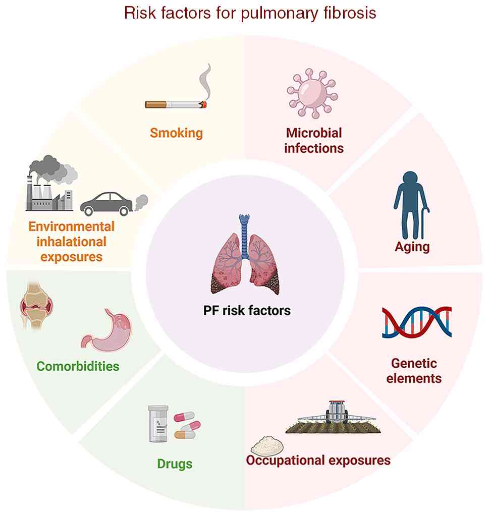
Multiple risk factors can initiate the progression
of PF, including genetic factors, aging, smoking, occupational
exposure, inhalation from the environment, microbial infection,
systemic disease and drug-induced factors. The primary risk factors
involved in PF are further illustrated in Fig. 1.
Genetic factors
Genetic factors significantly influence the onset
and progression of PF (23).
Familial interstitial pneumonia (FIP), also referred to as familial
PF, typically exhibits autosomal-dominant inheritance with
incomplete penetrance and variable clinical expressivity, thereby
emphasizing the contribution of rare, high-impact genetic factors
to disease etiology (24).
Genome-wide association studies (GWAS) have further confirmed the
significant polygenic inheritance effect in PF. Specifically, a
previous meta-analysis of GWAS on IPF has identified five new
susceptibility signaling loci, with the associated genes primarily
involved in biological processes such as the mTOR signaling
pathway, spindle assembly and regulation of telomere homeostasis
(25). Among these common
susceptibility loci, the promoter variant rs35705950 within the
mucin 5B, oligomeric mucus/gel-forming gene remains the most
consistently replicated and potent genetic risk factor, markedly
increasing susceptibility to both sporadic IPF and FIP (26). Additionally, previous research
has identified three genes associated with telomeres, which had
previously been linked to familial PF. These findings provide
evidence supporting the hypothesis that telomere malfunction
contributes to the pathogenesis of IPF (27).
Aging
Aging is associated with a progressive decline in
tissue homeostasis and regenerative capacity, thereby increasing
the likelihood of multiple chronic disorders, notably fibrotic
diseases affecting various organs (28). In the pulmonary system,
physiological aging correlates with an extensive range of molecular
and structural modifications that impair epithelial repair,
diminish regenerative responses following injury, and elevate
susceptibility to both acute and chronic pulmonary conditions such
as chronic obstructive pulmonary disease and interstitial lung
fibrosis (29). A total of two
cardinal hallmarks of this aging process, namely cellular
senescence and telomere attrition, are particularly relevant to
IPF. These phenomena significantly contribute to both the onset and
advancement of IPF (30).
Consistent with this mechanistic link, IPF is predominantly
diagnosed in middle-aged and elderly individuals, with age being
widely recognized as the most significant risk factor for disease
onset and progression (31).
Furthermore, epidemiological and experimental evidence further
indicates that aged lungs demonstrate a predisposition for
disrepair, which may exacerbate abnormal wound-healing responses
and facilitate persistent ECM remodeling following injury (32). Mechanistically, aging hallmarks,
including cellular senescence and telomere attrition, are
increasingly recognized as key contributors to IPF pathogenesis. At
the molecular and cellular levels, senescent epithelial cells and
fibroblasts acquire a senescence-associated secretory phenotype,
sustaining profibrotic signaling, activating fibroblasts and
promoting ECM deposition (11,33). In summary, cellular senescence
represents a critical risk factor that significantly contributes to
the pathogenesis and progression of PF.
Smoking
Cigarette smoke, a complex mixture of thousands of
toxic chemicals and reactive oxidants, induces persistent
epithelial injury and chronic pulmonary stress, thereby
constituting a significant and modifiable risk factor for IPF and
other smoking-related ILDs (34). Mechanistically, cigarette smoke
promotes fibrogenesis through convergent pathways involving
oxidative stress, persistent inflammatory signaling, disruption of
the epithelial barrier, and activation of fibroblasts, thereby
lowering the threshold for maladaptive tissue repair (35). At the cellular level, previous
experimental evidence indicates that cigarette smoke-mediated
inactivation of sirtuin (SIRT)1 induces autophagy-dependent
senescence of alveolar epithelial type 2 (AT2) cells, impairing
epithelial regeneration and facilitating ECM deposition (36). Concurrently, cigarette smoke may
further augment fibrotic remodeling by enhancing mitochondrial
oxidative stress and metabolic dysregulation within stromal cells,
reinforcing myofibroblast activation and matrix accumulation
(37). Emerging data also
suggest that smoking-related epigenetic and telomere-associated
alterations may exacerbate AT2 cell senescence and accelerate the
progression of fibrosis (38).
Collectively, these multi-level findings reinforce the
understanding of cigarette smoking as a key environmental trigger
that drives PF.
Occupational exposure
Occupational exposures are increasingly recognized
as significant and potentially preventable determinants of PF
(39). A previous comprehensive
meta-analysis that synthesized data from numerous international
case-control studies found that occupational exposures account for
~16% of the population-attributable risk among the causative
factors underlying PF (40).
These occupational hazards encompass a diverse range of agents.
Epidemiological studies consistently associate IPF with workplace
exposures to vapors, gases, dusts and fumes, including metal
dust/fumes, wood and agricultural dust and crystalline silica
(41). Among these hazards,
respirable crystalline silica serves as a quintessential fibrogenic
agent; it not only incites classical silicosis, which is
characterized by progressive and often irreversible fibrotic
remodeling, but may also exacerbate fibrosis through the alteration
of lung microbiota composition and associated metabolic pathways
(42,43). Another well-established
fibrogenic agent is asbestos, whose role warrants further scrutiny;
chronic inhalation of its fibers, such as chrysotile and
crocidolite, can cause asbestosis, a fibrotic lung disease
distinguished by interstitial fibrosis and pleural thickening
(44). Additionally, coal
workers' pneumoconiosis is a prevalent occupational disease to
which coal miners are highly susceptible owing to prolonged
exposure to coal dust particles, resulting in PF and impaired
immune function (45). Exposures
related to metallic substances form another critical category; in
industries producing hard metals, inhalation of tungsten
carbide-cobalt particulates may provoke epithelial and macrophage
injury, accompanied by significant cytokine release, thereby
providing mechanistic evidence for subsequent fibrotic remodeling
(46). Beyond these traditional
occupational hazards, emerging engineered nanomaterials such as
carbon nanotubes have been shown to promote
fibroblast-to-myofibroblast transition and stromal expansion in
experimental models, thus raising concerns regarding their
fibrogenic potential (47).
Inhalation from the environment
Environmental inhalational exposures, most notably
ambient and indoor air pollution, are increasingly regarded as
significant risk factors for IPF. In urban settings, diesel exhaust
is a substantial source of ambient fine particulate matter (PM2.5)
and, as a key traffic-related component, a primary contributor to
respiratory morbidity (48).
Mechanistically, multiple experimental studies have demonstrated
that PM2.5 can penetrate deeply into the distal lung and deposit in
the alveolar region, triggering a cascade of events, including
inflammation, production of reactive oxygen species and damage to
alveolar type 2 epithelial (AT2) cells, ultimately lung remodeling
and fibrosis (49). In addition
to particulate pollution, traffic-related gaseous pollutants such
as nitrogen dioxide (NO2) have been associated with
increased mortality and a higher risk of disease progression in
IPF, emphasizing the clinical importance of multipollutant exposure
burdens (50,51). NO2 primarily induces
PF by initiating sustained oxidative stress in lung tissues, which
in turn mediates epithelial cell injury, facilitates inflammatory
cell infiltration and ultimately promotes fibroblast activation
along with excessive ECM deposition (52). Emerging evidence further
indicates that indoor air pollution contributes to adverse IPF
outcomes, highlighting the importance of accumulated lifetime
inhalational injury that extends beyond outdoor sources (53). Additionally, long-term personal
exposure to elevated levels of air pollutants constitutes an
independent risk factor for acute exacerbation of IPF. In this
regard, it has been hypothesized that this may be associated with
air pollution, potentially serving as a repetitive stimulus capable
of disrupting telomere shortening and oxidative stress pathways
(54).
Microbial infection
An increasing body of evidence indicates that
microbial infections, particularly viral infections, may
potentially contribute to both the initiation and exacerbation of
PF. Viral invasion can cause immediate harm to pulmonary tissues,
thereby triggering a cascade of events that lead to persistent lung
injury and/or impaired tissue repair mechanisms. This
pathophysiological process ultimately results in the progression of
PF. Furthermore, viral infections possess the capacity to induce
immune-mediated lung pathology. The interaction between viral
components and pro-inflammatory and pro-fibrotic mediators,
including cytokines and other bioactive factors, contributes to the
progression of ongoing and severe pulmonary damage, thereby
promoting fibrotic alterations within the lung parenchyma (55). It is noteworthy that fibrotic
changes may manifest following the acute phase of viral infections.
Specific viruses, including cytomegalovirus, avian influenza virus
and coronaviruses such as severe acute respiratory syndrome
(SARS)-coronavirus (CoV), Middle East respiratory syndrome-CoV and
SARS-CoV-2, are capable of inducing persistent lung damage and
facilitating the onset of fibrosis subsequent to the acute
infection period (56). Beyond
viral pathogens, recent research has clarified the involvement of
bacterial infections in the pathogenic mechanisms underlying IPF.
Notably, previous research has demonstrated that individuals with
IPF possess a higher bacterial load within their bronchoalveolar
lavage fluid compared with healthy subjects or patients with
chronic obstructive pulmonary disease (57). Building on these epidemiological
findings, the colonization and subsequent infection by
opportunistic pathogens are posited to potentially facilitate the
persistence of a fibrotic microenvironment. This occurs through
persistent inflammatory stimulation and aberrant immune responses.
A paradigmatic example is Pseudomonas aeruginosa, a
facultatively anaerobic, Gram-negative bacterium that is one of the
principal causative agents of severe infections. It exhibits a
notably high prevalence among individuals afflicted with chronic
lung conditions, such as cystic fibrosis, thereby underscoring the
substantial clinical ramifications associated with its colonization
and infection within the setting of chronic pulmonary ailments
(58).
Systemic disease
Systemic disorders constitute a significant
etiological category for progressive PF, characterized by a
convergence of recurrent epithelial injury, dysregulated immunity
and impaired inflammatory resolution that sustains a
self-perpetuating fibroproliferative response. One such disorder,
gastro-esophageal reflux disease (GERD), is commonly observed in
IPF and is hypothesized to exacerbate disease progression through
chronic microaspiration of gastric contents, thereby sustaining
alveolar epithelial injury and promoting profibrotic repair
mechanisms (59). Beyond
mechanical factors such as GERD, connective tissue
disease-associated ILD constitutes another primary category of
systemic disorders driving PF. Within this category, systemic
sclerosis serves as a prototypical immune-driven fibrotic disorder
characterized by vascular damage, immune activation and progressive
fibrosis of the skin and internal organs, most notably the lungs
(60). Specifically, systemic
sclerosis-ILD is a leading cause of morbidity and mortality and
often progresses into a fibrosing phenotype marked by declining
lung function and increasing radiographic fibrosis (61). Extending beyond systemic
sclerosis, rheumatoid arthritis (RA), a systemic inflammatory
condition, frequently manifests with pulmonary involvement. RA-ILD
represents a major extra-articular cause of morbidity and mortality
among affected individuals (62).
Drug-induced PF (DIPF)
DIPF, a serious adverse reaction associated with
various medication classes, including bleomycin, amiodarone and
methotrexate, is driven by a complex cascade involving alveolar
epithelial cell (AEC) injury, inflammatory dysregulation, metabolic
reprogramming, fibroblast activation and excessive deposition of
ECM (63). Chemotherapeutic
agents such as bleomycin, a typical fibrogenic agent, lead to an
increase in programmed death-ligand 1 expression, initiate EMT and
cause fibrosis-like morphological changes in human pulmonary AECs
(64). Beyond chemotherapeutics,
amiodarone, a first-line antiarrhythmic agent, also induces DIPF
through a distinct pathological mechanism characterized by the
intracellular accumulation of phospholipid complexes within lung
histiocytes and type II pneumocytes, which further exacerbates
epithelial injury and pro-fibrotic inflammation (65). In addition, disease-modifying
antirheumatic drugs (DMARDs), which constitute the cornerstone of
therapy for autoimmune disorders such as RA, systemic lupus
erythematosus and psoriatic arthritis, also present a documented
risk of DIPF. An analysis of pharmacovigilance data sourced from
the FDA Adverse Event Reporting System database indicates that
DMARDs are among the drug categories most frequently implicated in
reports concerning PF. This finding underscores the critical
necessity for vigilant clinical monitoring of medication usage
(66). Additionally,
nitrofurantoin, a commonly prescribed antibiotic for urinary tract
infections, has been associated with rare but potentially severe
pulmonary toxicity. Specifically, its chronic toxicity can induce
ILD accompanied by progressive fibrosis, which may result in
substantial and occasionally irreversible pulmonary impairment
(67).
Cellular and molecular mechanisms of PF
The pathogenesis of PF involves multiple cell types,
including AECs, fibroblasts, macrophages and ECs. This section
elaborates on the respective roles of these cells in PF, as
illustrated in Fig. 2.
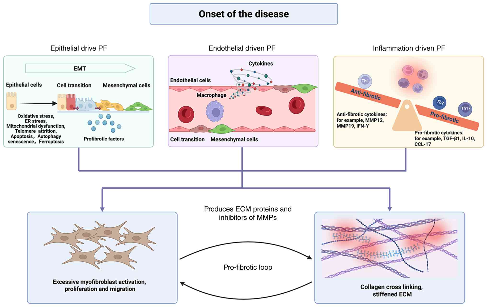 | Figure 2Cellular and molecular mechanisms
driving PF. PF involves multiple cell types and interconnected
pathways. Epithelial cells contribute via EMT, driven by stressors
such as oxidative stress, ER stress, mitochondrial dysfunction,
telomere attrition, apoptosis, autophagy, senescence and
ferroptosis. Endothelial cells and macrophages are also involved,
with macrophages secreting pro-fibrotic and anti-fibrotic cytokines
(e.g., TGF-β1, IL-10, CCL-17, MMP12, MMP19, IFN-γ). These pathways
converge on excessive myofibroblast activation, proliferation and
migration, leading to ECM deposition, collagen cross-linking and
ECM stiffening, forming a pro-fibrotic loop that sustains disease
progression. The image was generated utilizing Biorender
(https://BioRender.com). ECM, extracellular
matrix; EMT, epithelial-mesenchymal transition; ER, endoplasmic
reticulum; PF, pulmonary fibrosis. |
AECs
Alveolar epithelial integrity is fundamental to lung
homeostasis, and its injury is widely recognized as a critical
initiating and driving event in the pathogenesis of IPF (68). In the healthy alveolus, squamous
type 1 alveolar epithelial AT1 cells form the primary gas-exchange
surface in close apposition to ECs, whereas cuboidal AT2 cells
produce phospholipid-rich surfactant and serve as facultative
progenitors that replenish the epithelium by generating AT1 cells
after injury (69). In striking
contrast, in IPF, repetitive micro-injuries and a failure of normal
repair mechanisms lead to a profound dysfunction of this
regenerative program. Consequently, this results in AT2-cell
dysfunction and impaired differentiation into AT1 cells, while also
promoting aberrant epithelial-mesenchymal interactions-a
constellation of defects that collectively propel the lung into a
state of persistent, maladaptive wound healing (69-71). Within this deleterious
microenvironment, AT2 cells demonstrate increased sensitivity to
disturbances in intracellular homeostasis, encompassing oxidative
stress, endoplasmic reticulum stress, mitochondrial dysfunction and
telomere attrition, which collectively compromise epithelial cell
viability and regenerative potential (72,73). As a result, AT2 cells undergo
various injury and death pathways, such as apoptosis,
autophagy-related damage, cellular senescence and ferroptosis,
further limiting their ability to proliferate and restore the
epithelial barrier. Furthermore, epithelial cells exhibit increased
plasticity, undergoing EMT-like transitions that enable them to
directly produce ECM components and secrete pro-fibrotic mediators,
thereby actively fueling interstitial remodeling (74). Beyond these well-established
cellular outcomes, epithelial injury in fibrotic conditions is also
associated with organelle and metabolic stress. For instance,
abnormal mitochondrial iron accumulation has been observed in AT2
cells within bleomycin-induced fibrosis models and in epithelial
cells exposed to bleomycin. This indicates that dysregulation of
mitochondrial iron may be a pivotal factor in the damage and
depletion of AT2 cells, consequently accelerating fibrotic
remodeling (75). In summary,
dysfunction of AECs is intricately associated with PF pathogenesis,
operating through a multifaceted network of cellular stress, cell
fate dysregulation and metabolic dysfunction.
Fibroblasts
In PF, lung fibroblasts secrete substantial
quantities of collagen, which is integral to the progression of the
disease. Within the activated fibroblast population,
myofibroblasts-differentiated cells characterized by smooth
muscle-like contractile properties and α-smooth muscle actin (SMA)
expression-are regarded as the primary effector cells responsible
for fibrotic remodeling (76).
The master driver of this transition is TGF-β1, which is largely
secreted by injured AECs and other cellular sources within the
fibrotic microenvironment (77).
Through this transformation, fibroblasts directly fuel the
excessive deposition of ECM, the pathological hallmark and critical
endpoint in the progression of PF (78). Meanwhile, the buildup of
myofibroblasts in fibroblastic foci causes lung tissue contraction
and thickening of the alveolar-capillary membrane. These structural
alterations, in turn, result in a lasting reduction in pulmonary
diffusing capacity, restrictive ventilatory failure and PF
(79). Previous research on
mouse models indicate that periostin (Postn) functions as a
reliable and specific biomarker for pulmonary myofibroblasts.
Furthermore, the removal of Postn-positive myofibroblasts following
injury has been demonstrated to mitigate lung fibrosis, implying
that targeting these cells could represent a promising strategy for
future therapeutic interventions in PF (80).
Macrophages
In the context of fibrotic progression, the immune
microenvironment serves as a crucial factor that significantly
amplifies the entire cascade of fibrotic events. In the context of
healthy lungs, macrophages represent the predominant immune cell
population and can be categorized into two principal subsets:
Alveolar macrophages (AMs) and interstitial macrophages. Among
these, AMs serve as the primary innate immune cells residing in the
lungs, playing a pivotal role in maintaining tissue homeostasis
(81). Based on the distinct
local microenvironments they encounter, macrophages have the
capacity to undergo polarization, adopting either a classically
activated (M1) or an alternatively activated (M2) phenotype.
Typically, M1 macrophages are primarily involved in the initial
phases of wound repair subsequent to alveolar epithelial damage,
whereas M2 macrophages are mainly tasked with facilitating the
resolution of wound healing or promoting fibrosis (82). Macrophages of both M1 and M2
phenotypes are implicated in the onset of lung injury and its
subsequent progression towards PF. An overabundance of M1
macrophages can induce epithelial cell apoptosis and hinder the
repair process, whereas an excessive presence of M2 macrophages
results in aberrant and poorly regulated repair mechanisms, which
are hallmarks of progressive fibrotic conditions (83). M2 macrophages secrete profibrotic
mediators such as TGF-β1, IL-10 and C-C motif chemokine ligand 17
(CCL17), which enhance fibroblast proliferation, myofibroblast
differentiation and ECM deposition, thereby amplifying fibrogenesis
(84). Furthermore, macrophage
polarization is tightly shaped by T helper (Th)-cell cytokines:
Th1-derived IFN-γ tends to restrain profibrotic macrophage
programs, whereas Th2 cytokines (IL-4/IL-13) promote M2-like
polarization and reinforce TGF-β-driven matrix accumulation. Th17
responses further sustain macrophage inflammatory circuits and
tissue injury, while Th22/IL-22 primarily supports epithelial
integrity and indirectly dampens macrophage-driven profibrotic
remodeling (85,86). Previous research findings
indicate that anti-fibrotic therapy employing pirfenidone
interferes with both the polarization process and mechanical
activation of macrophages that promote fibrosis, thereby
suppressing fibrotic progression (87).
ECs
In addition to the contributions of epithelial,
stromal and immune cells, the vascular system undergoes notable
structural and functional alterations that play a profound role in
the progression of fibrosis. ECs constitute the innermost lining of
the vasculature and are essential for maintaining pulmonary
homeostasis through the regulation of vascular tone, permeability
and leukocyte trafficking (88).
In PF, however, this homeostatic function is disrupted. Increasing
evidence suggests that PF is associated with persistent EC
activation and dysfunction of the vascular barrier, as evidenced by
abnormal vascular endothelial cadherin/CD31 signaling, increased
adhesion between ECs and immune cells and an overall reduction in
barrier integrity (89). These
functional alterations, in turn, can amplify fibro-inflammatory
signaling and facilitate ECM remodeling (90). Importantly, aging further
predisposes the lung to fibrosis by inducing EC dysfunction, which
compromises vascular repair capacity after injury and facilitates
the persistence of fibrotic remodeling (91). A central mechanism linking these
vascular changes to fibrosis is endothelial-to-mesenchymal
transition (EndMT), a process during which ECs undergo
transcription factor-driven reprogramming, losing their
characteristic cobblestone morphology and endothelial identity, and
acquiring spindle-shaped, mesenchymal or myofibroblastic features
(92). This transition is
promoted by profibrotic pathways, including TGF-β/mothers against
decapentaplegic homolog (Smad) and Wnt/β-catenin signaling
cascades, and can be further exacerbated by cigarette smoke
exposure and pro-inflammatory cytokines, thereby aggravating
endothelial dysfunction and vascular remodeling in fibrotic lungs
(74). Consistent with this,
mediators derived from ECs have been demonstrated to actively
direct the process of fibrosis. For instance, matrix
metalloproteinase (MMP)19 facilitates PF by inducing EndMT and
promoting monocyte infiltration (93). Conversely, macrophage-derived
MMP12 sustains endothelial injury, thereby reinforcing a positive
feedback loop between vascular damage and fibrogenesis (94). In cases of silica-associated
fibrosis, senescent EC-derived galectin-3 further accelerates
disease progression through crosstalk between ECs, fibroblasts and
macrophages (95). Reinforcing
these observations at the molecular level, recent single-cell and
genetic studies further substantiate an active vasculofibrotic
interaction, demonstrating that EC-intrinsic programs, such as
forkhead box F1 (FOXF1)/related RAS viral oncogene homolog
signaling, serve to inhibit fibroblast activation, whereas the loss
of endothelial FOXF1 amplifies inflammation and collagen deposition
(96). To summarize, a series of
interconnected processes, encompassing endothelial damage,
impairment of barrier function and EndMT, are intricately linked
with epithelial injury, immune-mediated inflammation and remodeling
of the interstitial tissue. These processes collectively contribute
to the formation and perpetuation of the intricate pathological
network that underlies the progression of PF.
Glycolysis-related factors and PF
Aerobic glycolysis, traditionally known as the
Warburg effect, describes the metabolic tendency of cells to
convert glucose into lactate despite the presence of sufficient
oxygen, a phenomenon initially identified in cancer cells. The
biological importance of this mechanism of reprogramming extends
beyond merely providing a swift energy source; it is of even
greater importance in preserving the pool of glycolytic
intermediates. This preservation of glycolytic intermediates plays
a pivotal role by channeling a portion of the carbon flux towards
the pentose phosphate pathway (PPP), as well as anabolic routes
including lipid, amino acid and nucleotide biosynthesis.
Consequently, this orchestrated shunting of carbon flux underpins
the accumulation of biomass that is essential for cellular
proliferation and ECM synthesis (97,98). Specifically, glucose-6-phosphate
(G-6-P) enters the PPP to support nucleotide synthesis, while
dihydroxyacetone phosphate and 3-phosphoglycerate act as precursors
for lipid and amino acid biosynthesis, respectively. At the
molecular level, this metabolic shift is regulated through the
coordinated upregulation and activation of rate-limiting glycolytic
enzymes and transporters, including hexokinase (HKs),
phosphofructokinase (PFK)-1 and pyruvate kinases (PKs), as well as
GLUTs such as GLUT1. In PF, activated fibroblasts, damaged AECs and
inflammatory cells exhibit increased glycolytic activity, driven by
the upregulation of HK2, PFK1, PKM2 and GLUT1, to fulfill their
heightened bioenergetic and biosynthetic demands.
Glucose transport
Glucose transport across the plasma membrane is
mediated by facilitative GLUTs, a family of integral membrane
proteins that regulate cellular glucose uptake and thereby maintain
metabolic homeostasis (99).
Once transported into the cytoplasm by GLUTs, glucose is channeled
into glycolysis, a cytosolic pathway consisting of 10
enzyme-catalyzed reactions that ultimately convert glucose to
pyruvate, with concomitant production of adenosine triphosphate
(ATP) and nicotinamide adenine dinucleotide (reduced) (NADH)
(100). This GLUT family
encompasses a total of 14 distinct members. Each GLUT variant
exhibits unique transporter kinetics and tissue-specific
distribution patterns, thereby conferring individual
characteristics to every member within this family. Among them,
GLUT1 is the most ubiquitously expressed isoform and is frequently
upregulated under stress conditions. Increased GLUT1 expression
enhances glucose influx, augments glycolytic flux and elevates
lactate production, thereby favoring glucose-dependent metabolic
programs (101). Consistent
with this role, GLUT1-driven aerobic glycolysis provides both ATP
and anabolic intermediates required for tumor cell proliferation
and metastatic dissemination (102). In contrast to GLUT1's
high-affinity and proliferation-supporting role, GLUT2 functions as
a low-affinity, high-capacity transporter, which is highly
expressed in the liver, intestine and kidney, as well as in
pancreatic β-cells, where it contributes to hepatic glucose export,
intestinal and renal glucose handling and glucose-sensing-dependent
insulin secretion (103).
GLUT3, which exhibits a particularly high affinity for glucose,
supports basal glucose uptake in tissues with high metabolic demand
(104). GLUT4 is a GLUT
responsive to insulin and exercise, which translocates to the
plasma membrane in skeletal muscle and adipose tissue, thereby
enhancing glucose uptake for glycolysis and ATP synthesis (105). Considering their key role in
supporting glycolysis, GLUTs are promising therapeutic targets,
with numerous GLUT-focused strategies currently being explored in
the context of oncology.
HKs
HKs are the initial rate-limiting enzymes in the
glycolytic pathway, catalyzing the irreversible phosphorylation of
glucose to G-6-P immediately following its entry into the cell via
GLUTs (106). Previous studies
have identified four distinct HK isozymes (HK1, HK2, HK3 and HK4),
each exhibiting unique tissue-specific expression profiles and
enzymatic kinetic characteristics (107,108). Among these, HK2 has garnered
particular attention for its crucial role in mediating aerobic
glycolysis, a metabolic hallmark widely observed in cancer cells,
and its upregulation is closely linked to tumor growth and
progression (109).
Functionally, HK2 can channel G-6-P into glycogen synthesis and the
PPP within the cytoplasm. However, when associated with
mitochondria, HK2 preferentially supports glycolytic flux and
oxidative phosphorylation processes (110). Emerging evidence suggests that
this HK2-centered metabolic switch is also active in PF. In
fibroblasts derived from patients with IPF and in bleomycin-induced
PF models, TGF-β1 signaling drives HK2 accumulation (via the MYC
proto-oncogene) to enhance aerobic glycolysis, promote
myofibroblast differentiation and increase collagen synthesis.
Genetic silencing of HK2 or pharmacological inhibition with
lonidamine reduces fibrogenic gene expression and alleviates
bleomycin-induced lung fibrosis in vivo (111). Further underscoring the
pathogenic relevance of HK2, vitamin D alleviates silica-induced PF
partly by inhibiting signal transducer and activator of
transcription (STAT)3/HK2-mediated glycolysis in fibroblasts
(112). Consistently, in
TGF-β1-stimulated human lung fibroblasts in vitro,
strategies that suppress HK2 expression-such as
alamandine/Mas-related G protein-coupled receptor D (MrgD)
signaling-disrupt the lactate-HK2 feed-forward loop, restrain
fibroblast activation and produce anti-fibrotic effects (113). Collectively, these findings
highlight HK2 as a promising metabolic target for the development
of therapies against PF.
PFK-1
PFK-1 functions as the second primary rate-limiting
enzyme in glycolysis, catalyzing the irreversible conversion of
fructose-6-phosphate to fructose-1,6-bisphosphate and thereby
committing glucose to glycolytic degradation (114). Although PFK-1 is encoded by
three distinct genes (PFKL, PFKM and PFKP), its enzymatic activity
is further regulated by the PFKFB family of bifunctional enzymes
(namely PFKFB1-PFKFB4), which produce fructose-2,6-bisphosphate, a
potent allosteric activator of PFK-1. Among the PFKFB isozymes,
PFKFB3 is the most extensively studied and is frequently
overexpressed in various cancer types, where it enhances glycolytic
flux and facilitates the Warburg effect, thereby supporting tumor
cell proliferation and survival (115). Beyond its established role in
cancer, emerging evidence indicates that the pathogenic role of
PFKFB3-driven glycolysis extends to fibrotic diseases. For
instance, in IPF, PFKFB3-mediated glycolysis has been
mechanistically linked to fibroblast activation, and
pharmacological inhibition of PFKFB3 reduces bleomycin-induced and
lipopolysaccharide (LPS)-induced PF in animal models (116,117). In silica-induced fibrosis,
activation of glucagon-like peptide-1 receptor disrupts the
interaction between the NLR family pyrin domain containing 3
inflammasome and PFKFB3-driven glycolysis, leading to decreased
lactate production and histone lactylation, thereby inhibiting
fibroblast activation (118).
Furthermore, the natural compound peimine, extracted from
Fritillaria, has been demonstrated to inhibit fibroblast
activation and alleviate bleomycin-induced PF by suppressing
glycolysis via the phosphoinositide 3-kinase (PI3K)/protein kinase
B (Akt)/PFKFB3 signaling pathway (119).
PK
PK serves as a pivotal regulatory enzyme at the
terminal stage of the glycolytic pathway, where it assumes a
critical function in sustaining the rate of glycolytic flux and
facilitating energy generation (120). Specifically, PK catalyzes the
conversion of phosphoenolpyruvate and ADP into pyruvate and ATP,
thereby functioning as a critical metabolic checkpoint that links
glycolytic flux to cellular energy demands (121). In mammals, two genes encode
four major isoforms (PKL/PKR and PKM1/PKM2). Among these, PKM2
stands out for its pronounced conformational plasticity, switching
between a low-activity dimer and a high-activity tetramer to direct
glucose carbon toward either ATP production or anabolic
biosynthesis (122). During
fibrotic remodeling in IPF and related PF models, PKM2 is
increasingly recognized as a pathogenic metabolic hub:
Myofibroblast differentiation prompts the upregulation of PKM2 and
favors its dimeric form, which reroutes glycolytic intermediates
toward de novo glycine synthesis, thereby supporting
collagen biosynthesis and the maintenance of activated
myofibroblasts (123).
Consistent with this pathogenic role, genetic deletion of PKM2
significantly attenuates bleomycin-induced PF and suppresses
myofibroblast activation by dampening TGF-β1 signaling (124). Beyond its metabolic functions,
PKM2 can perform non-canonical signaling roles, including
stress-induced nuclear translocation in myeloid cells, potentially
amplifying inflammatory responses relevant to fibrotic progression
(125). Together, these
observations position PKM2 as a tractable glycolytic regulator
linking metabolic rewiring to matrix accumulation in PF.
Lactate dehydrogenase (LDH)
LDH, predominantly the A isoform (LDHA), catalyzes
the terminal reaction of glycolysis by reducing pyruvate to lactate
while oxidizing NADH to nicotinamide adenine dinucleotide
(oxidized) (NAD+), thereby maintaining cytosolic redox
balance and sustaining glycolytic flux when mitochondrial oxidation
is constrained or biosynthetic demand is high (126). Under physiological conditions,
high LDH activity maintains the pyruvate-lactate equilibrium and
supports lactate as a diffusible metabolite within tissues,
consistent with its role as a physiological end product of
glycolysis (127). By contrast,
under pathological conditions such as IPF, lactate is enriched in
diseased lungs and functions as an active profibrotic signal rather
than a passive by-product (20).
Mechanistically, elevated lactate promotes extracellular
acidification, which can activate latent TGF-β1 in a pH-dependent
manner and induce fibroblast-to-myofibroblast differentiation,
thereby creating a feed-forward loop of matrix accumulation
(19). Furthermore, lactate
derived from myofibroblasts can induce histone lactylation in
macrophages and amplify profibrotic transcriptional programs,
thereby further supporting fibro-inflammatory remodeling (21). Accordingly, pharmacological
inhibition of LDHA decreases lactate production and mitigates
bleomycin-induced PF, thereby highlighting the LDH-lactate axis as
a potential therapeutic target in PF (128).
Glycolysis-related signaling pathways and
PF
Multiple signaling pathways are involved in the
pathogenesis of PF, including TGF-β1, hypoxia-inducible factor
(HIF)-1α, PI3K/Akt/mTOR, Hippo, AMP-activated protein kinase (AMPK)
and SIRT. This section will elaborate on the respective roles of
these pathways in PF, as illustrated in Fig. 3.
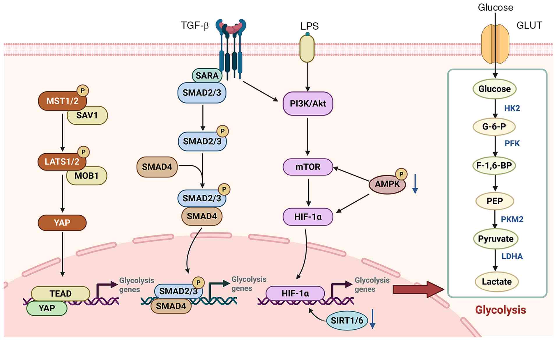 | Figure 3Signaling pathways regulating
glycolysis in PF. Multiple signaling cascades converge to modulate
glycolytic metabolism in the context of PF. The Hippo pathway
(MST1/2-LATS1/2-YAP/TEAD), TGF-β/SMAD signaling (SMAD2/3-SMAD4) and
LPS-induced PI3K/Akt-mTOR-HIF-1α axis collectively drive the
transcription of glycolysis-related genes (e.g., HK2,
PFK, PKM2, LDHA), thereby enhancing glucose
uptake and conversion to lactate. AMPK and SIRT1/6 act as negative
regulators, inhibiting mTOR and HIF-1α activity, respectively, to
counteract glycolytic flux. This coordinated signaling network
highlights the central role of augmented glycolysis in the
pathogenesis of PF, positioning it as a potential therapeutic
target. The image was generated utilizing Biorender (https://BioRender.com). PF, pulmonary fibrosis; HIF,
hypoxia-inducible factor; HK2, hexokinase 2; LPS,
lipopolysaccharide; LATS1/2, large tumor suppressor kinase 1/2;
LDHA: Lactate dehydrogenase A ; YAP, Yes-associated protein; TEAD,
TEA domain transcription factor; TGF-β, transforming growth
factor-β; MST1/2, mammalian sterile 20-like kinase 1/2; SAV1,
Salvador family WW domain-containing protein 1; MOB1, Mps one
binder kinase activator protein 1; SARA, SMAD anchor for receptor
activation; SMAD2/3, sons of mothers against decapentaplegic 2/3;
PI3K, phosphatidylinositol 3-kinase; Akt, protein kinase B; mTOR,
mammalian target of rapamycin; AMPK, AMP-activated protein kinase;
SIRT1/6, silent information regulator 1/6; GLUT, glucose
transporter; G-6-P, glucose-6-phosphate; PFK, phosphofructokinase;
F-1,6-BP, fructose-1,6-bisphosphate; PEP, phosphoenolpyruvate;
PKM2, pyruvate kinase M2; LDHA, lactate dehydrogenase A; P,
phosphorylation. |
TGF-β
TGF-β is a pleiotropic cytokine that orchestrates
tissue repair and immune homeostasis; however, when persistently
activated, it is widely regarded as a master profibrotic program in
fibrosis, driving fibroblast-to-myofibroblast differentiation,
epithelial plasticity and excessive ECM deposition (129,130). In mammals, TGF-β exists in
three subtypes: TGF-β1, TGF-β2 and TGF-β3, with TGF-β1 playing a
dominant role in PF, as demonstrated in IPF lung tissues,
bleomycin-induced PF models and silicosis models, through its
integration with glycolysis (131). In human IPF lung tissues and in
bleomycin- and silica-induced PF models, TGF-β1 serves as a crucial
profibrotic signal generated by damaged epithelial and immune
cells, including macrophages. Through canonical Smad2/3 signaling,
it facilitates the transition of fibroblasts to myofibroblasts and
promotes ECM accumulation (132). At the molecular level, TGF-β1
interacts with its receptors, TGF-β receptor I and II, consequently
inducing the phosphorylation and activation of Smad2 and Smad3.
Notably, Smad7 serves as an inhibitory regulatory element,
providing negative feedback in this pathway. Upon activation,
phosphorylated Smad2 and Smad3 form complexes with Smad4 and
translocate to the nucleus, where they play a critical role in
regulating the expression of essential target genes (1). Beyond this canonical signaling
cascade, recent research highlights that TGF-β signaling is tightly
coupled to glycolytic reprogramming during myofibroblast
differentiation, enabling fibroblasts to meet the increased
bioenergetic and biosynthetic demands of collagen production
(133). Specifically, TGF-β
enhances glucose uptake and glycolytic flux by upregulating pivotal
glycolytic nodes such as GLUT1, HK2 and PFKFB3, which leads to
increased lactate production and fosters a profibrotic metabolic
milieu (20,111,133). This lactate, in turn, is not
merely a metabolic by-product; it contributes to the acidification
of the extracellular space and may induce the activation of latent
TGF-β1 through pH alterations. Thus, a feed-forward mechanism
involving 'TGF-β-glycolysis-lactate' is established, which
maintains myofibroblast viability and perpetuates ECM remodeling
(19).
HIF
HIF serves as a transcriptional activator that is
frequently triggered under hypoxic conditions (134). Structurally, HIF is a
heterodimeric transcription factor composed of α and β subunits.
The α subunits include HIF-1α, HIF-2α and HIF-3α, while the β
subunits comprise aryl hydrocarbon receptor nuclear translocator
(ARNT1) (also known as HIF-1β) and ARNT2 (135). Under normoxic conditions,
HIF-1α undergoes hydroxylation facilitated by prolyl hydroxylase
domain enzymes (PHDs), which are recognized by the von
Hippel-Lindau E3 ligase complex, leading to ubiquitination and
subsequent degradation. Conversely, hypoxic conditions inhibit PHD
activity, resulting in the rapid stabilization and nuclear
accumulation of HIF-1α, which heterodimerizes with HIF-1β and binds
to hypoxia-responsive elements to initiate the transcription of
downstream genes (136). Beyond
its involvement in angiogenesis and ECM remodeling, HIF-1α is a
critical factor in metabolic reprogramming from oxidative
phosphorylation toward glycolysis. At the metabolic level, HIF-1α
enhances glucose uptake by inducing the GLUT family and broadly
upregulates the transcription of glycolytic enzymes, thereby
augmenting glycolytic flux. Key glycolytic enzymes upregulated by
HIF-1α include HK2, PFK and PFKFB3, which collectively facilitate
increased glucose utilization and lactate production (136). In addition, HIF-1α promotes the
diversion of pyruvate away from mitochondrial oxidation by inducing
pyruvate dehydrogenase kinase 1 (PDK1), which inhibits the pyruvate
dehydrogenase complex and further reinforces the glycolytic
phenotype (137). Thus,
enhanced glycolysis and lactate accumulation serve as functional
readouts of HIF-1α-dependent transcriptional activation, providing
a direct link between this transcription factor and the metabolic
phenotype of fibrotic tissues.
Consistent with the aforementioned link,
accumulating evidence supports a close association between aberrant
HIF-1α activation and PF pathogenesis (138). Within IPF lung tissues,
HIF-1α-mediated metabolic dysfunction in AECs results in elevated
lactate production. This lactate, in turn, triggers the activation
of interstitial fibroblasts, ultimately expediting the progression
of PF (139). Elevated HIF-1α
expression has been observed in AECs in bleomycin-induced models
and in human fibrotic lung tissues, indicating its critical role in
disease progression (140).
Consistently, targeting the HIF-1α-glycolysis axis, particularly
the HIF-1α/PDK1 metabolic switch and lactate-associated signaling,
has emerged as a promising strategy to mitigate PF by disrupting
profibrotic metabolic reprogramming.
PI3K/Akt/mTOR
The PI3K/Akt signaling cascade constitutes a crucial
intracellular pathway that plays a central role in the regulation
of the cell cycle. To date, extensive investigations have been
undertaken to elucidate the structural attributes and functional
mechanisms of PI3Ks, including class I, class II and class III
PI3Ks. Upon activation, PI3K generates PIP3, which subsequently
activates Akt and stimulates mTOR signaling, thereby driving
profibrotic cellular programs (141). Notably, the PI3K/Akt/mTOR
pathway also facilitates metabolic reprogramming towards
glycolysis. One critical mechanism linking this pathway to
glycolysis is its regulation of HIF-1α. HIF-1α is stabilized and
activated by growth factors and cytokines via PI3K/Akt/mTOR
signaling. Specifically, by activating mTOR complex (mTORC)1, this
pathway enhances protein translation and stabilizes HIF-1α, leading
to the transcriptional activation of glycolytic genes such as
GLUT1, HK2 and PFKFB3. Previous research findings indicate that the
activation of the PI3K-Akt-mTOR signaling axis plays a role in
modulating cellular aerobic glycolysis. For instance, in the lung,
LPS may induce aerobic glycolysis in pulmonary fibroblasts by
activating the PI3K-Akt-mTOR/PFKFB3 pathway, thereby contributing
to the pathogenesis of LPS-induced PF (117). Beyond fibroblasts, the
PI3K/Akt/mTOR axis also mediates metabolic crosstalk between immune
and stromal cells: In neodymium oxide-induced PF, glycolytic
reprogramming in macrophages leads to lactate-mediated histone
lactylation, which activates the PI3K/Akt/mTOR pathway and promotes
TGF-β1 secretion, thereby facilitating macrophage-fibroblast
crosstalk and fibrotic progression (142). Providing a more detailed
mechanistic view in PF, in bleomycin-induced models, hyperactivated
PI3K/Akt/HIF-1α signaling modulates aberrant cell proliferation and
apoptosis through oxidative stress mechanisms (143). Recent comprehensive reviews
have further consolidated the central role of PI3K/Akt, mTOR and
HIF-1α in the network-based regulation of glycolysis in PF
(144).
Hippo pathway
The Hippo pathway is a conserved kinase cascade that
governs tissue growth and regeneration. Its core regulatory module
comprises mammalian STE20-like kinase 1/2 (MST1/2), Salvador
homolog 1, large tumor suppressor kinase 1/2 (LATS1/2) and the
transcriptional co-activators Yes-associated protein (YAP) and
transcriptional coactivator with PDZ-binding motif (TAZ) (145). In the canonical setting, MST1/2
activates LATS1/2, which phosphorylates YAP/TAZ, promoting their
cytoplasmic retention and proteasomal degradation, thereby
suppressing YAP/TAZ-dependent transcriptional programs. Conversely,
when Hippo signaling is inhibited, dephosphorylated YAP/TAZ
accumulate in the nucleus and preferentially cooperate with TEA
domain transcription factor (TEAD) to induce pro-proliferative and
pro-remodeling genes, including connective tissue growth factor and
cysteine-rich angiogenic inducer 61, positioning YAP/TAZ as central
effectors of Hippo output (146,147). In IPF, aberrant activation and
nuclear localization of YAP/TAZ represent a major profibrotic
driver across multiple cell types (148). In fibroblasts, mechanical cues
such as ECM stiffening can sustain YAP/TAZ nuclear activity,
reinforcing fibroblast contractility and ECM synthesis and thereby
promoting progressive scarring (149). In macrophages, YAP/TAZ
activation is markedly enriched in lung macrophages during
bleomycin-induced injury, where it shapes macrophage heterogeneity
and profibrotic functions that facilitate fibrosis progression
(150). In AECs, Hippo/YAP
signaling is implicated in alveolar epithelial regeneration,
particularly in AT2 self-renewal and differentiation into AT1
cells, underscoring its context-dependent roles in repair vs.
fibrotic remodeling (151).
Beyond its cell-type-specific roles in fibrosis,
Hippo/YAP/TAZ signaling is intricately associated with cellular
metabolism and aerobic glycolysis. Mechanistically, under
conditions of glucose deprivation or energy stress, AMPK and
kinases linked to the Hippo pathway phosphorylate and inhibit the
activity of YAP (145,152), whereas YAP activation sustains
glycolytic metabolism and glucose homeostasis, partly through the
regulation of glucose transport mechanisms (153). Conversely, glycolytic activity
can directly enhance the transcriptional activity of YAP/TAZ. For
example, during aerobic glycolysis, PFK1 interacts with TEAD1 and
stabilizes the YAP/TAZ-TEAD transcriptional complex, thereby
augmenting the expression of genes related to glycolysis;
conversely, inhibition of glucose metabolism diminishes
YAP/TAZ-driven transcription (154). This bidirectional regulation is
further modulated by lactate, a glycolytic end-product enriched in
fibrotic lungs, which can facilitate the activation of YAP/TAZ and
the subsequent expression of profibrotic genes (20). In conclusion, persistent YAP/TAZ
activation fosters a glycolysis-boosting environment, providing
vital resources for fibroblast activation, ECM buildup and immune
tissue remodeling, thus advancing PF.
AMPK
AMPK traditionally serves as a crucial cellular
energy sensor. It is triggered into activation when the cellular
energy level declines. Upon activation, AMPK functions to
re-establish energy balance within cells. It achieves this by
facilitating catabolic pathways that generate ATP and,
concurrently, suppressing processes that consume energy (155). Through phosphorylation of
various downstream substrates, AMPK orchestrates lipid catabolism,
glucose uptake, glycolytic flux, autophagy and insulin sensitivity,
thereby maintaining cellular energy homeostasis (156). Previous research has shown that
the phosphorylation of AMPK can initiate the activation of the
mTORC2-AKT signaling cascade, leading to phosphorylation of
cytosolic glycolytic enzymes and increased HIF-1α expression, which
collectively promote the transcription of glycolysis-related genes
and enhance aerobic glycolysis (157). However, in the context of
fibrotic stress, AMPK predominantly acts as an anti-fibrotic
metabolic checkpoint, restraining pathological glycolytic
reprogramming and limiting profibrotic signaling.
For example, in sepsis-associated PF, LPS
stimulation promotes fibroblast activation, which is accompanied by
AMPK inactivation, HIF-1α induction and increased aerobic
glycolysis. Conversely, AMPK activation or HIF-1α inhibition
suppresses glycolysis and fibroblast activation, attenuating
fibrosis in vivo (158).
Supporting this anti-fibrotic role, AMPK activation has been shown
to mitigate pulmonary inflammation and fibrosis by antagonizing
TGF-β1 signaling and promoting autophagy via the AMPK-mTOR axis
(159). Clinical observations
further reveal that fibrotic regions within IPF lungs, such as
fibroblastic foci, show reduced AMPK activation, and impaired AMPK
signaling may exacerbate mitochondrial dysfunction and metabolic
reprogramming, thereby supporting the transition of fibroblasts to
myofibroblasts and excessive ECM production (160). Beyond stromal remodeling, AMPK
also exerts disease-relevant metabolic control in the alveolar
epithelium. Furthermore, impaired regulation of AT2 cell metabolism
by AMPK has been directly associated with PF in both murine models
and human IPF samples, a condition in which deficient AMPK
signaling correlates with increased glycolysis, decreased
mitochondrial biogenesis, disrupted fatty acid oxidation (FAO),
accumulation of dysfunctional mitochondria and loss of AT2
progenitor capacity. Pharmacological modulation of AMPK activity
restores metabolic health and reduces fibrosis in vivo
(161). Finally, in the context
of aging-associated lung fibrosis, increased GLUT1-dependent
glycolysis is observed. Both genetic and pharmacological inhibition
of GLUT1 suppress fibroblast activation markers (such as α-SMA) and
alleviate bleomycin-induced PF, further emphasizing glycolysis as a
key driver of fibrogenesis (101).
SIRTs
SIRTs constitute a highly conserved family of seven
NAD+ -dependent deacetylases and deacylases
(SIRT1-SIRT7), each with distinct subcellular localization: SIRT1,
SIRT6 and SIRT7 are predominantly nuclear, while SIRT2 is primarily
cytosolic, and SIRT3, SIRT4 and SIRT5 are mainly mitochondrial
(162). These proteins are
involved in the regulation of cellular metabolism and participate
in various biological processes, including cell viability, DNA
repair, cellular aging and response to oxidative stress. Therefore,
SIRTs may be implicated in the pathogenesis of fibrotic diseases
(163-165). Notably, SIRT signaling
intersects with glycolytic regulation, particularly through the
modulation of the HIF-1α axis. For instance, SIRT1 directly
deacetylates HIF-1α, restraining its transcriptional activity and
consequently suppressing HIF-1α-dependent glycolytic gene
expression under hypoxic conditions (166). Similarly, SIRT6 functions as a
corepressor of HIF-1α by deacetylating histone H3K9 at promoters of
glycolytic genes. Loss of SIRT6 enhances glucose uptake and
glycolytic flux while diminishing mitochondrial respiration,
underscoring its role as a critical regulator of the
glycolysis-mitochondrial function balance (167,168). Collectively, these mechanisms
suggest that SIRTs can influence fibrogenesis through the
modulation of pathological glycolytic remodeling and metabolic
stress signaling.
Building on this metabolic connection, it is well
established that SIRT1, SIRT3, SIRT6 and SIRT7 play protective
roles in preventing the onset and progression of IPF. However, the
regulatory functions of the remaining members of the SIRT family in
this pathological process necessitate further investigation
(169). SIRT1 and SIRT3 serve
as crucial modulators in the context of lung fibrosis, given their
capacity to mitigate the profibrotic actions triggered by TGF-β, as
evidenced by both in vitro and in vivo experimental
studies (163). By contrast,
SIRT2 appears to exert pro-fibrotic effects: Gong et al
(170) demonstrated that SIRT2
is upregulated in TGF-β1-stimulated human lung fibroblasts and in
bleomycin-treated mouse lungs, and pharmacological inhibition of
SIRT2 alleviates fibroblast activation and PF, at least in part via
suppression of Smad2/3 signaling. Specifically, a deficiency of
mitochondrial SIRT3 has been identified in AECs in cases of IPF,
and genetic ablation of SIRT3 aggravates experimental lung
fibrosis, concomitant with mitochondrial DNA damage in AECs and
activation of intrinsic apoptotic pathways, thereby indicating that
impaired mitochondrial quality control may heighten epithelial
susceptibility and promote remodeling (171). Additionally, SIRT6 confers
epithelial protection and reduces bleomycin-induced PF by
facilitating lipid catabolism, augmenting cellular energy supplies
and decreasing lipid peroxidation-related injury (172). Finally, in fibroblasts, a broad
reduction in SIRT expression has been observed in PF, with SIRT7
showing the most marked decrease. Accordingly, overexpression of
SIRT7 inhibits TGF-β-induced collagen synthesis and α-SMA
expression, suggesting that SIRT7 functions as a negative regulator
of fibroblast activation (173). In essence, SIRTs exert either
protective or pathogenic roles in various aspects of PF, including
epithelial damage, interstitial activation and immune-metabolic
remodeling, by integrating energy sensing, epigenetic regulation
and metabolic pathways.
Interaction between glycolysis and other
metabolic pathways
Glycolysis-FAO metabolic switch
FAO constitutes a principal mitochondrial catabolic
mechanism that transforms fatty acids into acetyl-CoA via
β-oxidation, thereby providing energy for the tricarboxylic acid
cycle and oxidative ATP synthesis (174). In cigarette smoke-induced PF,
emerging evidence indicates an association with lipid metabolic
dysregulation and a maladaptive shift in substrate utilization. For
instance, cigarette smoke induces mitochondrial oxidative stress
and disrupts autophagy flux, suppressing peroxisome
proliferator-activated receptor α/carnitine palmitoyltransferase
1a-mediated FAO and lipid droplet autophagy. Consequently, this
enhances lipid accumulation and fibroblast activation in the lungs,
suggesting that abnormal lipid metabolism along with mitochondrial
dysfunction jointly promote fibrotic remodeling (37).
Beyond these environmental triggers, a key feature
of fibrotic metabolic remodeling is the reciprocal association
between enhanced glycolysis and suppressed FAO, which appears to
couple energy metabolism to ECM homeostasis. Supporting this
reciprocal association, in fibroblasts, inhibition of HIF-1α blunts
the TGF-β1-driven glycolytic program and is accompanied by
restoration of FAO, suggesting that the TGF-β/HIF-1α axis
participates in coordinating the glycolysis-FAO switch during
fibroblast activation (175).
Consistent with the functional importance of FAO, impaired FAO in
aging AT2 cells compromises injury-induced alveolar regeneration
and promotes the accumulation of intermediate epithelial states
with aberrant basaloid/secretory features, which are associated
with increased fibrotic susceptibility (176). Other previous research findings
suggest that fibroblast-intrinsic metabolic rewiring may also be
driven by metabolite signaling. For example, enhanced glycolysis
together with reduced FAO has been linked to succinate
accumulation, and targeting succinate dehydrogenase (SDH) in
fibroblasts attenuates bleomycin-induced lung fibrosis,
highlighting the succinate-SDH axis as a potential therapeutic
entry point to counteract pro-fibrotic metabolic programming
(177).
Effects of energy homeostasis
imbalance
Mitochondria are indispensable to cellular energy
metabolism, as they generate the majority of intracellular ATP.
This ATP production maintains a high ATP-to-ADP ratio, which is
vital for cellular homeostasis and function (178,179). Conversely, disruptions in
mitochondrial dynamics lead to loss of mitochondrial homeostasis
and subsequently impair mitochondrial functionality. For example,
dysregulation of mitochondrial fusion and fission destabilizes
mitochondrial networks, diminishes respiratory capacity and
amplifies stress responses, collectively contributing to the
pathogenesis of pulmonary diseases, including fibrosis (180). Within this framework,
mitochondrial dysfunction has been widely recognized as a principal
pathogenic contributor to various pulmonary disorders, exhibiting a
strong correlation with both the initiation and progression of IPF
(181). At the cellular level,
mitochondrial dynamics and bioenergetic capacity are closely linked
to myofibroblast differentiation (182). Furthermore, mitochondria offer
an additional layer of quality control, with the phosphatase and
tensin homolog-induced kinase 1/Parkin pathway serving as a key
mediator of mitophagy. Deficiency in this pathway results in
inefficient clearance of damaged mitochondria, thereby exacerbating
oxidative stress and pro-fibrotic signaling. Conversely, enhanced
mitophagy, such as through the upregulation of acyl-CoA synthetase
long-chain family member 1, may serve to alleviate fibrosis
(183). These findings
highlight mitochondrial quality control as a promising target for
intervention to restore energy homeostasis and mitigate fibrotic
progression.
Therapeutic strategies and challenges of
targeting glycolysis in PF
Throughout this section, interventions that
directly inhibit defined glycolytic enzymes or transporters are
distinguished from those that indirectly modulate glycolytic flux
through upstream signaling. Genetic evidence, including knockdown,
knockout and rescue-of-function experiments, provides
higher-confidence target-dependency validation compared with
pharmacological treatment alone, given that numerous small-molecule
compounds exert pleiotropic off-target effects.
Direct inhibitors of glycolytic enzymes
and transporters
Inhibition of glycolytic enzymes has become a
mechanism-based adjunct to antifibrotic therapy, as restricting
glucose flux can limit the bioenergetic and biosynthetic inputs
that support myofibroblast differentiation and ECM deposition
(116). A range of natural and
synthetic small-molecule agents has been developed as potential
inhibitors targeting distinct stages of glycolysis, including
glucose uptake, its rate-limiting step, and lactate generation.
Numerous glycolytic nodes have been validated as legitimate
therapeutic targets through complementary genetic and
pharmacological methods (Table
I).
 | Table ITherapeutic targeting of glycolysis
in PF with small-molecule compounds: Targets, models, mechanisms
and translational outlook. |
Table I
Therapeutic targeting of glycolysis
in PF with small-molecule compounds: Targets, models, mechanisms
and translational outlook.
| Metabolic
target | Targeting mode | Compound | Relevant cell
type(s) | Major
mechanism | PF model | Evidence level | Clinical
development status | Key
limitations | (Refs.) |
|---|
| HK2 | Direct | Lonidamine | Lung
fibroblasts | Inhibits HK2;
reduces glycolysis, suppresses myofibroblast differentiation and
collagen synthesis | TGF-β1-stimulated
human lung fibroblasts; BLM-induced mouse PF | In vitro;
animal | Preclinical
(oncology history; not in PF trials) | Non-selective HK
inhibitor; may affect other HK isoforms; hepatotoxicity
reported | (111) |
| HK2 | Direct | 2-DG | Lung
myofibroblasts; alveolar macrophages in silica model | Glucose analog;
competitively inhibits HK and blocks the initial step of
glycolysis; suppresses myofibroblast proliferation, contractility
and collagen accumulation; inhibits HIF-1α in alveolar macrophages
in silicosis | BLM-induced PF;
silica-induced PF; other preclinical PF models | In vitro;
animal | Preclinical
(various diseases; not in PF trials) | Broad metabolic
inhibitor; low oral bioavailability; short half-life; off-target
effects | (116,204,205) |
| PFKFB3 | Direct | 3PO | Lung
fibroblasts/myofibroblasts | Inhibits PFKFB3;
reduces F2,6BP production and PFK-1 activity, thereby suppressing
glycolytic flux and collagen synthesis | LPS- and
BLM-induced PF | Animal | Preclinical | Poor water
solubility; rapid clearance; high or frequent dosing needed | (116,117) |
| PFKFB3 | Direct | Anlotinib | Lung
fibroblasts | Multi-target TKI;
downregulates PCBP3 and inhibits PFKFB3 translation, reducing
glycolysis and reversing PF | BLM-induced PF;
TGF-β1-stimulated cells | In vitro;
animal | Approved for
cancer; preclinical for PF | Approved for
cancer, but PF use is off-label; antifibrotic mechanism still needs
verification | (184) |
| PFKFB3 | Direct (via
PI3K/AKT/PFKFB3) | Peimine | Lung
fibroblasts | Natural
isosteroidal alkaloid; inhibits the PI3K/Akt/PFKFB3 pathway,
reducing glycolysis and fibroblast activation | BLM-induced PF;
TGF-β1-stimulated cells | In vitro;
animal | Preclinical | Natural product;
possible multi-target effects; selectivity and in vivo PK
data limited | (119) |
| PKM2 | Direct | Compound 3k | Lung
fibroblasts | Directly targets
glycolytic enzyme PKM2 and disrupts PKM2 tetramer formation;
suppresses PKM2-driven aerobic glycolysis and reduces biosynthetic
precursor supply in lung fibroblasts | BLM-induced PF | Animal | Preclinical | Target selectivity
against PKM isoforms requires further validation; off-target
effects cannot be fully excluded; only validated in
bleomycin-induced PF model, no human data available | (124,185) |
| LDHA | Direct | Gossypol | Primary human lung
fibroblasts | Directly inhibits
glycolytic terminal enzyme LDHA, curtails glycolytic flux and
lactate overproduction; reduced lactate limits pH-dependent latent
TGF-β activation and myofibroblast differentiation | BLM-induced PF;
radiation-induced PF | Animal | Preclinical
(oncology history; not in PF trials) | Not LDHA-specific;
multiple off-target cytotoxic effects; only preclinical, no human
data | (128,186) |
| LDHA | Direct | GNE-140 | Macrophages | Selective LDHA
inhibitor; reduces lactate production and histone lactylation,
suppressing profibrotic macrophage phenotypes | PM2.5-induced
PF | Animal | Preclinical | Limited in
vivo PF model data; lung-targeting delivery challenge | (187) |
| GLUT1 | Direct | BAY876 | Lung
fibroblasts | Selective GLUT1
inhibition; reduces glucose uptake and suppresses TGF-β-induced
fibroblast-to-myofibroblast differentiation and ECM production | TGF-β1-stimulated
human lung fibroblasts | In vitro;
animal | Preclinical | No in vivo
PF data; target selectivity and lung delivery remain to be
evaluated | (190,191) |
| GLUT1/other | Direct
(GLUT1/pleiotropic) | Quercetin | Multiple cell
types | Natural flavonoid;
binds an exofacial site on GLUT1 and inhibits glucose
transport | Not reported in the
cited PF context | In vitro
(mechanistic) | Preclinical | Multi-target
effects; GLUT1 specificity questionable; low oral
bioavailability | (189) |
| TGF-β/Smad | Indirect | Albendazole | Lung
fibroblasts | Suppresses upstream
TGF-β1/Smad profibrotic signaling; reduces ECAR and lactate
production and down-regulates PFKFB3 to dampen TGF-β-driven aerobic
glycolysis | TGF-β1-stimulated
human lung fibroblasts; bleomycin-induced mouse PF | In vitro;
animal | Preclinical | Broad-spectrum
antiparasitic agent with multiple off-target bioactivities; exact
primary molecular target in fibrotic contexts remains incompletely
defined; no dedicated clinical trials for PF | (192) |
| YAP/TEAD | Indirect | Verteporfin | Alveolar epithelial
cells | Inhibits YAP-TEAD
protein-protein interaction. Based on established transcriptional
regulation of glycolytic genes by YAP-TEAD, glycolysis suppression
is theoretically predicted as an indirect downstream consequence;
direct glycolysis-related experimental evidence in PF is
lacking | BLM-induced PF | Animal | Preclinical
(off-label; preclinical for PF) | Targets YAP-TEAD
transcriptional complex rather than glycolytic machinery; predicted
glycolysis-modulating effect lacks direct experimental data in PF
models; broad cellular off-target effects | (213) |
| AMPK | Indirect | Metformin | Alveolar epithelial
cells, fibroblasts | AMPK activator;
inhibits HIF-1α-induced aerobic glycolysis, antagonizes TGF-β1
signaling, promotes autophagy and attenuates fibrosis | Sepsis-associated
PF; silica-induced PF | Animal | Approved for T2DM;
early-phase/repurposing exploration for PF | Non-specific AMPK
activation; broad metabolic effects; gastrointestinal effects | (158,159) |
| AMPK | Indirect | Fenbendazole | Lung
fibroblasts | Activates AMPK
signaling; reduces glucose consumption and ECAR to dampen aerobic
glycolysis | TGF-β1-stimulated
lung fibroblasts; bleomycin-induced mouse PF | In vitro;
animal | Preclinical | Veterinary-use
anthelmintic; off-target bioactivities cannot be fully excluded; no
dedicated human clinical trials for PF | (193) |
| SIRT3 | Indirect | Baicalein | Lung fibroblasts;
macrophages | Restores
mitochondrial SIRT3 signaling and inhibits TGF-β1/Smad activation;
suppresses aerobic glycolysis in both fibroblasts and
macrophages | TGF-β1-stimulated
lung fibroblasts; bleomycin-induced mouse PF | In vitro;
animal | Preclinical | Broad biological
activities; multiple off-target pathways; no dedicated clinical
trials for PF | (195) |
The pathogenic role of HK2 in PF has been firmly
established through both genetic and pharmacological approaches. In
models of TGF-β1-stimulated human lung fibroblasts and
bleomycin-induced PF, TGF-β1 signaling drives the accumulation of
HK2 in a MYC-dependent manner, enhancing aerobic glycolysis,
promoting myofibroblast differentiation and increasing collagen
synthesis. Notably, genetic silencing of HK2 or pharmacological
inhibition with lonidamine significantly reduces fibrogenic gene
expression and alleviates bleomycin-induced lung fibrosis in
vivo (111). Furthermore,
evidence indicates that vitamin D mitigates silica-induced PF, in
part by inhibiting STAT3/HK2-mediated glycolysis in fibroblasts
(112). Additionally,
alamandine/MrgD signaling disrupts the lactate-HK2 feedback loop,
thereby restraining fibroblast activation (113). Collectively, these studies
provide compelling genetic evidence that HK2 acts as a causal
mediator rather than merely a correlate of fibrotic glycolysis. In
glycolysis, PFKFB3 boosts glycolytic flux by upregulating PFK1
activity via fructose-2,6-bisphosphate production. Multiple studies
indicate its pathogenic role in PF remodeling and fibroblast
activation. In PF models, multiple PFKFB3 inhibitors have
demonstrated efficacy. The PFKFB3 inhibitor
3-(3-pyridinyl)-1-(4-pyridinyl)-2-propen-1-one (3PO) reduces
glycolysis and mitigates fibrotic phenotypes. For instance, in
LPS-induced PF, the PI3K-Akt-mTOR/PFKFB3 pathway drives fibroblast
aerobic glycolysis and collagen synthesis, effects that are
mitigated by 3PO (117). In
pulmonary myofibroblasts, 2-deoxy-D-glucose (2-DG) blocks
HK-initiated glycolysis, while 3PO inhibits PFKFB3 to reduce PFK-1
activity. Both interventions suppress myofibroblast proliferation,
contractility and collagen accumulation, which are key fibrotic
features. In mice with bleomycin-induced PF, 3PO also reduces
fibrosis (116). Anlotinib has
emerged as a novel candidate for PF treatment, inhibiting
PFKFB3-driven glycolysis in myofibroblasts (184). Peimine suppresses fibroblast
activation and ameliorates bleomycin-induced PF by inhibiting
glycolysis through the PI3K/Akt/PFKFB3 signaling pathway (119). PKM2 is a core glycolytic
checkpoint that shapes metabolic reprogramming in profibrotic lung
cells (124). Compound 3k is a
PKM2-targeted small-molecule inhibitor that directly disrupts PKM2
tetramer assembly. Pharmacological or genetic suppression of PKM2
curbs aerobic glycolysis, lowers the supply of biosynthetic
precursors and attenuates PF in bleomycin-challenged mice (124,185). Of note, tetramer-disrupting
compound 3k also indirectly dampens profibrotic TGF-β-dependent
signaling as a secondary downstream consequence of altered PKM2
protein conformation, though this is not its primary mechanism of
action. Genetic knockdown of PKM2 recapitulates the antifibrotic
effects, further confirming the pathogenic contribution of
PKM2-driven glycolysis. These findings collectively establish PKM2
as a therapeutic target with strong genetic validation.
As the terminal glycolytic enzyme, LDHA catalyzes
pyruvate-to-lactate conversion to regenerate cytosolic
NAD+, sustaining high glycolytic flux in profibrotic
lung cells. Pharmacological inhibition of LDHA interrupts this
late-stage glycolytic step and curtails lactate overproduction. The
LDHA inhibitor gossypol dampens aerobic glycolysis and lactate
accumulation in primary human lung fibroblasts, which in turn
limits pH-dependent latent TGF-β activation and myofibroblast
differentiation (128). In
vivo, gossypol reduces collagen deposition in both bleomycin-
and radiation-induced PF, and retains efficacy even when
intervention is delayed (128,186). Pre-administration of the
selective LDHA inhibitor GNE-140 effectively mitigated
PM2.5-provoked pulmonary inflammation and fibrosis in a murine
model via suppression of glycolysis and downstream histone
lactylation (187). While
genetic studies specifically targeting LDHA in PF models are
limited, the consistent efficacy of structurally distinct LDHA
inhibitors and the well-defined role of lactate in activating
latent TGF-β1 and promoting histone lactylation provide strong
pharmacological validation for this axis (19). In the context of glycolytic
substrate provision, the transmembrane movement of glucose
predominantly relies on the GLUT protein family. Consequently,
therapeutic approaches aimed at modulating GLUT activity possess a
clearly delineated mechanistic rationale. A diverse array of GLUT
inhibitors has been identified, encompassing both naturally
occurring substances from plants or microorganisms and synthetic
small molecules (188). For
example, quercetin inhibits GLUT1 by binding at an exofacial site,
demonstrating the feasibility of pharmacologically targeting
GLUT-mediated glucose uptake (189). In addition, in TGF-β-stimulated
human lung fibroblasts in vitro, treatment with the
selective GLUT1 inhibitor BAY876 markedly attenuates
TGF-β-triggered profibrotic responses in lung fibroblasts, as
evidenced by suppressed myofibroblast differentiation and reduced
expression levels of the signature profibrotic proteins α-SMA,
collagen and fibronectin (190,191) (Fig. 4).
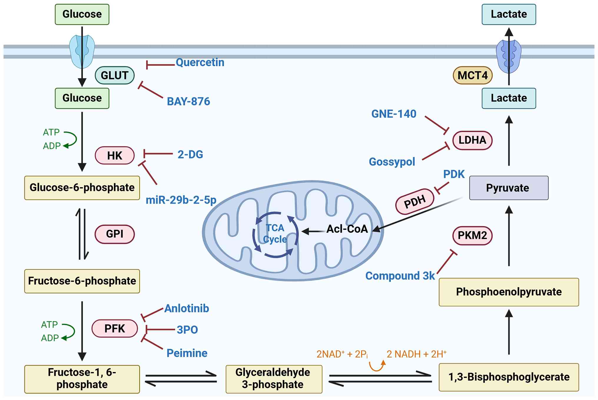 | Figure 4Therapeutic targets in PF. This
schematic illustrates key glycolytic pathways involved in PF and
highlights various pharmacological inhibitors targeting these
metabolic nodes, including glucose and lactate transporters (e.g.,
BAY-876, GNE-140) and glycolytic enzymes (e.g., 2-DG, gossypol,
shikonin, Compound 3k, 3PO), as well as natural compounds such as
quercetin and curcumin. The image was generated utilizing Biorender
(https://BioRender.com). PF, pulmonary fibrosis,
2-DG, 2-deoxy-D-glucose; 3PO,
3-(3-pyridinyl)-1-(4-pyridinyl)-2-propen-1-one. |
It should be noted, however, that quercetin and
curcumin are broad-spectrum natural compounds with multiple
reported targets; their antifibrotic effects are accompanied by
alterations in glycolytic signaling but have not been confirmed by
target-dependency experiments (such as genetic knockdown/knockout
or rescue studies), and therefore, glycolysis should not yet be
regarded as their primary causal therapeutic target.
Indirect modulators of glycolysis via
upstream signaling pathways
A large number of interventions that exhibit
reported anti-fibrotic effects and associated alterations in
glycolysis do not function by directly inhibiting glycolytic
enzymes. Instead, they modulate upstream signaling pathways that
regulate the expression of glycolytic genes. In these instances,
the observed changes in glycolytic flux may result from more
extensive effects on cell proliferation, differentiation or
survival, and should not be interpreted as evidence that glycolysis
is the primary therapeutic target (Table I).
One such upstream target is the TGF-β pathway.
Albendazole is a small-molecule compound that suppresses upstream
TGF-β1/Smad profibrotic signaling in human lung fibroblasts. Both
in-vitro fibroblast experiments and bleomycin-challenged
mouse PF models demonstrate that albendazole dampens
TGF-β-triggered aerobic glycolysis. It reduces the extracellular
acidification rate (ECAR) and lactate production, accompanied by
downregulation of the key glycolytic effector PFKFB3. Since
albendazole primarily targets the upstream TGF-β/Smad cascade
rather than glycolytic enzymes, glycolysis suppression represents
an indirect downstream biological consequence of TGF-β pathway
inhibition (192). In mice with
bleomycin-induced PF, inhibition of epithelial YAP-TEAD/lysyl
oxidase signaling, including pharmacological blockade of YAP-TEAD
with verteporfin, reduces ECM crosslinking and remodeling, thereby
attenuating fibrosis. Biochemical studies have established that
YAP-TEAD transcriptional activity drives the expression of multiple
glycolytic genes (154).
Therefore, verteporfin treatment is theoretically expected to
indirectly dampen glycolytic reprogramming by disrupting
YAP-TEAD-dependent transcription. Nevertheless, direct experimental
evidence demonstrating that verteporfin alters glycolytic flux or
glycolytic-enzyme abundance in PF cellular or animal models remains
absent. In PF, AMPK activation has emerged as a promising
anti-glycolytic strategy. Metformin, an AMPK activator, suppresses
HIF-1α expression and alleviates sepsis-associated PF by inhibiting
HIF-1α-induced aerobic glycolysis (158). Another AMPK-activating compound
is fenbendazole. In vitro experiments in TGF-β1-stimulated
lung fibroblasts and bleomycin-challenged mouse PF models
demonstrate that fenbendazole activates AMPK signaling, reduces
glucose consumption and ECAR and dampens aerobic glycolysis. Since
fenbendazole primarily triggers AMPK activation rather than
directly targeting glycolytic enzymes, glycolysis suppression
represents an indirect downstream metabolic consequence of
AMPK-mediated signaling reprogramming (193). Similarly, AMPK-mediated
suppression of HIF-1α represents an indirect mechanism of
glycolytic downregulation. SIRTs, NAD+-dependent
deacylases, integrate the metabolic state with stress responses and
transcriptional regulation (194). For example, restoring
mitochondrial SIRT3 signaling with baicalein intervention inhibits
TGF-β1/Smad activation and fibroblast-macrophage metabolic
crosstalk, thus diminishing PF. Baicalein suppresses aerobic
glycolysis in both lung fibroblasts and macrophages in
bleomycin-induced PF models (195). Since baicalein primarily
targets upstream SIRT3/TGF-β1/Smad signaling rather than glycolytic
enzymes, glycolysis inhibition represents an indirect downstream
metabolic outcome. SIRT7 levels are decreased in PF and can
suppress the fibrotic phenotype of lung fibroblasts by
downregulating Smad3-dependent profibrotic outputs, supporting
SIRTs as viable targets beyond canonical glycolytic enzymes
(196). SIRTs play a crucial
role in the regulation of glycolysis, exerting their influence
indirectly through deacetylation of HIF-1α, modifications of
histones and activation of metabolic sensor pathways (166,167). Consequently, it is important to
note that the observed anti-fibrotic effects of SIRT modulators
cannot be solely ascribed to the inhibition of glycolytic
processes.
Therapeutic potential of ncRNAs
ncRNAs, including microRNAs (miRNAs or miRs), long
ncRNAs (lncRNAs) and circular RNAs (circRNAs), have emerged as
critical regulators of glycolytic reprogramming in PF. Notably,
several ncRNAs have been validated through genetic approaches that
provide strong evidence for target dependency. By modulating the
expression of key glycolytic enzymes, upstream transcription
factors and metabolic sensors, ncRNAs influence core fibrotic
processes such as myofibroblast activation, excessive ECM
deposition and inflammatory amplification. This pivotal regulatory
role emphasizes their translational potential as innovative
therapeutic targets. Among ncRNAs, miRNAs function as short,
post-transcriptional repressors that directly target glycolytic
regulators. For example, genetic knockout of miR-21 abolishes
arsenate-induced Akt activation and glycolytic upregulation,
thereby inhibiting myofibroblast differentiation in vivo
(197). Similarly, in a Dahuang
Zhechong pill-treated rodent model of PF, miR-29b-2-5p suppressed
glycolysis and fibroblast activation by targeting HK2, contributing
to attenuated fibrosis (198).
Another key miRNA, miR-30a-3p, in mice with silica-induced PF, acts
as a glycolytic brake by directly targeting the transcriptional
activator FOXK2. Overexpression of miR-30a-3p reduces glycolytic
flux in fibroblasts and mitigates PF, further supporting its
therapeutic potential (199).
In contrast to miRNAs, circRNAs and lncRNAs primarily exert their
effects through competing endogenous RNA (ceRNA) networks, where
they sequester miRNAs to modulate downstream signaling pathways.
For instance, circ0066187, which is upregulated in fibrotic models,
promotes STAT3-mediated glycolytic signaling. Silencing circ0066187
disrupts this pathway and inhibits fibroblast-to-myofibroblast
transition (200). Similarly,
circHIPK3 enhances FOXK2 expression and fibroblast glycolysis by
sponging miR-30a-3p. In vivo, adeno-associated
virus-mediated silencing of circHIPK3 alleviates silica-induced PF
(199). Regarding lncRNAs, in
the same pathological context, lncRNA PVT1 is transcriptionally
activated by FOXM1 and promotes fibroblast proliferation by acting
as a ceRNA for miR-497-5p. Inhibition of the FOXM1/PVT1 axis
reduces fibrosis, further underscoring lncRNAs as promising targets
for disrupting pro-fibrotic metabolic reprogramming (201). Collectively, these findings
illustrate a multi-layered ncRNA network that precisely regulates
glycolytic metabolism in PF. Therefore, targeting specific ncRNAs
presents a promising strategy to reverse pathological metabolic
remodeling and halt disease progression (Fig. 4).
Translational challenges and future
directions for glycolysis-targeting therapies
Despite the promising preclinical efficacy of the
aforementioned glycolysis-targeting strategies, translating these
findings into clinically viable therapies for PF faces substantial
challenges that warrant critical consideration.
First, target selectivity and cell-type specificity
are challenging. An important consideration emerging from the
current literature is the cell-type- and context-dependent nature
of glycolytic metabolism in PF. While enhanced glycolysis is
predominantly pathogenic in activated fibroblasts and pro-fibrotic
macrophages, its role in AECs is more nuanced. Fibroblasts undergo
metabolic reprogramming characterized by upregulated glycolytic
flux to support the bioenergetic and biosynthetic demands of
myofibroblast differentiation and ECM production (116). Macrophages exhibit
polarization-dependent metabolic profiles, with classically
activated M1 cells favoring glycolysis and alternatively activated
M2 cells relying more on oxidative phosphorylation (202). Furthermore, lactate derived
from activated fibroblasts can shape macrophage function through
histone lactylation, reinforcing a pro-fibrotic metabolic crosstalk
(21). In ECs, glycolytic
activation promotes EndMT and contributes to vascular remodeling.
In AECs, however, glycolysis serves a dual role: A tightly
regulated glycolytic flux supports progenitor cell function and
regenerative repair, whereas dysregulated glycolysis in the setting
of metabolic dysfunction contributes to pathogenic epithelial
alterations and fibrosis (203). The varying roles of different
cell types highlight a marked therapeutic issue: Global inhibition
of glycolysis may effectively reduce profibrotic fibroblast
activity, but it could also hinder epithelial regeneration and
other essential adaptive responses. Therefore, future therapeutic
approaches should focus on selective targeting of specific cell
types, by utilizing isoform-specific inhibitors, lung-targeted
delivery systems or interventions tailored to the disease stage, in
order to enhance antifibrotic effectiveness while minimizing any
adverse effects on epithelial repair and immune homeostasis.
Secondly, there are pharmacokinetic and
delivery-related challenges. Numerous glycolytic inhibitors
demonstrate low oral bioavailability, short half-lives or
restricted penetration into lung tissue. For example, 2-DG, a
glucose analog that inhibits glycolysis, has been extensively
utilized in preclinical PF models; however, its suboptimal
pharmacokinetics hinder its therapeutic effectiveness (204,205). Similarly, 3PO and its analogs
have demonstrated efficacy in animal models but encounter
challenges related to formulation and systemic exposure, such as
poor water solubility and rapid clearance (116). Anlotinib, which inhibits
PFKFB3-driven glycolysis in myofibroblasts by downregulating
poly(rC) binding protein 3 (PCBP3), has shown strong antifibrotic
effects in bleomycin-induced mouse models; however, its clinical
application for PF remains unestablished (184). Lung-targeted drug delivery
methods, such as inhalation, aerosolized nanoparticles or
exosome-based delivery, offer a promising approach to achieve
elevated local concentrations while minimizing systemic toxicity
(206). Recent advancements
include inhalable myofibroblast-targeting nanoparticles, which
achieved a 117.8% increase in pulmonary drug delivery and a 71.5%
increase specifically aimed at fibroblast activation
protein-positive myofibroblasts (207), nebulized multi-modular stem
cell-derived exosomes for targeted pulmonary delivery and inhalable
nanoparticles that restore airway-alveolar homeostasis by
inhibiting bronchiolarization and EMT (208). However, the thickened
interstitium and reduced vascularity of fibrotic lungs may hinder
drug penetration, and the inflammatory microenvironment could
affect nanoparticle stability and clearance (209). Addressing these physical and
biological barriers is essential for advancing glycolysis-targeting
therapies toward clinical translation.
Third, the dependence on disease stage and the
optimal timing for intervention are vital considerations. The role
of glycolysis in PF is not fixed but rather adapts with disease
progression. During the early stages of lung injury, metabolic
reprogramming in AECs, including enhanced glycolysis coupled with
FAO and glutamine catabolism, meets the energetic demands necessary
for the differentiation of AT2 into AT1 and supports alveolar
regeneration (210). However,
as fibrosis becomes established, the metabolic needs of effector
cells change. Activated myofibroblasts develop a marked glycolytic
dependency that fuels their bioenergetic and biosynthetic
requirements for ECM production. Furthermore, targeting the
HIF-1α/PDK1-mediated glycolytic switch with dichloroacetate has
been shown to suppress bleomycin-induced PF, highlighting the
therapeutic potential of metabolic interventions (137). In models of established
fibrosis, inhibiting lactate production through LDHA or blocking
lactate transporters has demonstrated efficacy, even when initiated
after fibrosis has developed (211). These observations suggest that,
while glycolysis in the early disease stage supports epithelial
repair, the fibrotic phase presents a therapeutic window for
selectively targeting glycolysis and lactate transport in
pathogenic fibroblasts and myofibroblasts. Consequently, delayed
intervention aimed at addressing the glycolytic dependence of
established myofibroblasts, rather than indiscriminate early
inhibition, may represent a more effective and safer therapeutic
strategy. Accurate patient stratification based on disease stage,
distinguishing between the early inflammatory/repair phase and the
established fibrotic phase, will be critical for optimizing the
timing of interventions targeting glycolysis.
Fourth, there is a potential for interference with
immune defense. As discussed in previous sections, glycolysis
serves various critical functions across different cell types in
PF. While it drives pathogenic activation in fibroblasts, it also
plays vital roles in immune cells. Macrophages display metabolic
profiles that depend on their polarization: Classically activated
M1 cells favor glycolysis, whereas alternatively activated M2 cells
preferentially utilize oxidative phosphorylation (85). Additionally, lactate produced by
myofibroblasts can lead to histone lactylation in macrophages and
enhance profibrotic transcriptional programs (21). This complexity indicates that
therapies targeting glycolysis may have varying effects on
different immune-cell subsets, depending on their polarization
state and the stage of the disease. Patients with PF, who are
already more vulnerable to respiratory infections, face an
increased risk of pneumonia and other pulmonary infections, which
are significant contributors to morbidity and mortality in this
population (212). Therefore,
although targeting glycolysis shows promise for inhibiting
fibroblast activation, it is essential to carefully assess its
impact on immune defense in future research.
Fifth, the developmental status of candidate
interventions must be considered. Current glycolysis-targeting
interventions for PF remain predominantly at the preclinical stage,
with evidence largely derived from in vitro studies and
animal models (bleomycin-, silica-, LPS- and radiation-induced PF
models). Among the agents discussed, only a few have progressed
toward clinical evaluation, mostly in oncology contexts rather than
PF. Gossypol (an LDHA inhibitor) has demonstrated efficacy in
preclinical models of radiation- and bleomycin-induced PF, with
subcutaneous treatment inhibiting collagen accumulation,
myofibroblast differentiation and pro-fibrotic gene expression in
the lung, but has not been evaluated in PF clinical trials
(128). GNE-140 (an LDHA
inhibitor) pretreatment was found effective in alleviating histone
lactylation and thus PF in PM2.5-induced models (187). Metformin (an AMPK activator) is
an approved antidiabetic agent being repurposed for PF in
early-phase trials. Verteporfin (a YAP inhibitor) is approved for
ophthalmological use and is being explored preclinically for PF
(213). Anlotinib has shown
potent antifibrotic effects by downregulating PCBP3, reducing
PFKFB3 translation and inhibiting glycolysis in myofibroblasts.
Other agents, such as 2-DG, 3PO, compound 3k and BAY876, remain at
the in vitro or animal model stage, with no human data in
PF. The translational gap between preclinical efficacy and clinical
benefit remains substantial, and safety, tolerability and
pharmacokinetic profiles in patients with PF are largely
unknown.
Sixth, strategies to address translational barriers
must be explored. Despite the aforementioned challenges, several
strategies could enhance the translational potential of therapies
targeting glycolysis, including: i) Development of
isoform-selective inhibitors that offer improved target
specificity, such as HK2- and PFKFB3-selective compounds; ii) use
of lung-targeted delivery systems (such as inhalation,
nanoparticles and exosomes) to achieve organ-specific exposure
while minimizing systemic toxicity; iii) biomarker-driven patient
selection based on glycolytic activity in fibrotic tissue or plasma
lactate levels; iv) disease-stage stratification to differentiate
between the early inflammatory/repair and established fibrotic
phases, allowing for intervention at the optimal therapeutic
window; v) combination regimens with current standard-of-care
agents, such as nintedanib and pirfenidone, to achieve additive or
synergistic effects; and vi) adaptive trial designs that
incorporate close monitoring for infectious complications and
facilitate early detection of safety signals, particularly those
related to immune function and epithelial repair.
In summary, while glycolysis-targeting therapies
hold considerable promise for PF, their successful translation
requires careful attention to target selectivity, delivery
strategies, therapeutic timing and patient stratification.
Addressing these challenges through innovative drug design,
formulation technology and personalized medicine approaches will be
essential to realize the full therapeutic potential of metabolic
reprogramming in PF.
Conclusions and perspective
PF is a progressive and often fatal chronic lung
disease characterized by complex pathogenesis and limited
therapeutic options. In recent years, metabolic reprogramming,
particularly aberrant activation of glycolysis, has been identified
as a critical factor in the initiation and progression of PF. The
present review has systematically elucidated the central role of
glycolysis in PF, including the upregulation of key metabolic
enzymes such as GLUTs, HK2, PFKFB3, PKM2 and LDHA, as well as
lactate accumulation and the regulatory mechanisms of associated
signaling pathways, including TGF-β1, HIF-1α, PI3K/AKT/mTOR, Hippo,
AMPK and SIRT. Collectively, these metabolic shifts facilitate
fibroblast-to-myofibroblast differentiation, promote ECM deposition
and disrupt immune cell functions and energy homeostasis, thereby
driving the fibrotic process.
A more nuanced understanding of glycolysis in PF
has emerged, moving beyond the earlier perspective that enhanced
glycolysis serves as a universal pathogenic driver. Increasing
evidence indicates that its functional consequences differ across
various cell types, microenvironmental contexts and disease stages.
In summary, enhanced glycolysis in mesenchymal and pro-fibrotic
immune cells generally promotes activation, amplifies inflammation
and facilitates matrix deposition. Conversely, in AECs, it plays a
role in progenitor function and epithelial repair. This functional
duality presents a remarkable therapeutic challenge: While global
suppression of glycolysis may diminish fibrotic activation, it
could also hinder alveolar regeneration, compromise barrier
integrity and weaken antimicrobial immune responses. Consequently,
the central challenge lies not in whether to target glycolysis, but
in how to selectively inhibit it within pathogenic cell populations
and at the appropriate disease stage, all while preserving
essential protective metabolic processes. Progress in this area may
be enhanced by using isoform-selective inhibitors, implementing
lung-targeted and spatiotemporally controlled delivery systems,
stratifying by disease stage and employing reliable biomarkers of
glycolytic activity. These unmet needs set the stage for the
therapeutic strategies and translational challenges outlined
below.
Building on these mechanistic insights, inhibitors
targeting key glycolytic nodes have demonstrated promising
anti-fibrotic efficacy in preclinical studies. Concurrently,
ncRNAs, which serve as crucial regulators of both glycolysis and
fibrotic signaling pathways, have emerged as prospective diagnostic
biomarkers and therapeutic targets. Addressing metabolic
reprogramming, with particular emphasis on glycolysis, offers novel
perspectives and strategies for the treatment of PF. Furthermore,
fibrotic diseases are also associated with other metabolic
pathways, such as glutaminolysis and lipid metabolism. The
interplay and crosstalk between glycolysis and these pathways
warrant further comprehensive investigation. As the current
understanding of the metabolic mechanisms underpinning PF continues
to advance, future research is expected to facilitate the
development of more effective and less toxic personalized
therapies, ultimately improving patient outcomes.
Availability of data and materials
Not applicable.
Authors' contributions
All authors contributed to the study conception and
design. CY was involved in conceptualization and writing-original
draft preparation. HY was responsible for methodology, software and
writing-reviewing and editing. MH provided visualization and
supervision. QX provided guidance on writing and performed funding
acquisition and supervision. Data authentication is not applicable.
All authors read and approved the final manuscript.
Ethics approval and consent to
participate
Not applicable.
Patient consent for publication
Not applicable.
Competing interests
The authors declare that they have no competing
interests.
Abbreviations:
|
AEC
|
alveolar epithelial cell
|
|
AMPK
|
AMP-activated protein kinase
|
|
AT2
|
alveolar type 2 cell
|
|
EC
|
endothelial cell
|
|
ECM
|
extracellular matrix
|
|
EMT
|
epithelial-mesenchymal transition
|
|
FAO
|
fatty acid oxidation
|
|
GLUT
|
glucose transporter
|
|
HIF-1α
|
hypoxia-inducible factor 1α
|
|
HK2
|
hexokinase 2
|
|
IPF
|
idiopathic pulmonary fibrosis
|
|
LDHA
|
lactate dehydrogenase A
|
|
ncRNA
|
non-coding RNA
|
|
PF
|
pulmonary fibrosis
|
|
PFKFB3
|
6-phosphofructo-2-kinase/fructose-2,6-biphosphatase 3
|
|
PI3K
|
phosphoinositide 3-kinase
|
|
PKM2
|
pyruvate kinase M2
|
|
PPP
|
pentose phosphate pathway
|
|
SIRT
|
sirtuin
|
|
TGF-β1
|
transforming growth factor-β1
|
|
YAP
|
Yes-associated protein
|
|
α-SMA
|
α-smooth muscle actin
|
Acknowledgements
Not applicable.
Funding
This study was supported by the National Natural Science
Foundation of China (grant no. 82204008) and the Natural Science
Foundation of Shandong Province (grant no. ZR2022QH136).
References
|
1
|
Jiang M, Bu W, Wang X, Ruan J, Shi W, Yu
S, Huang L, Xue P, Tang J, Zhao X, et al: Pulmonary fibrosis: From
mechanisms to therapies. J Transl Med. 23:5152025. View Article : Google Scholar : PubMed/NCBI
|
|
2
|
Koudstaal T, Funke-Chambour M, Kreuter M,
Molyneaux PL and Wijsenbeek MS: Pulmonary fibrosis: From
pathogenesis to clinical decision-making. Trends Mol Med.
29:1076–1087. 2023. View Article : Google Scholar : PubMed/NCBI
|
|
3
|
Dhooria S, Inoue Y, Rathi K and Takimoto
T: Global perspective on pulmonary fibrosis: Epidemiology, regional
variations, gaps in care and future directions. Thorax Mar. 9:2026.
View Article : Google Scholar
|
|
4
|
Raghu G and Selman M: Nintedanib and
pirfenidone. New antifibrotic treatments indicated for idiopathic
pulmonary fibrosis offer hopes and raises questions. Am J Respir
Crit Care Med. 191:252–254. 2015. View Article : Google Scholar : PubMed/NCBI
|
|
5
|
Vancheri C, Sciacca E, Muscato G, Spicuzza
L, Fruciano M, Gili E, Sambataro G, Palmucci S and Libra A:
Pharmacological treatment in Idiopathic Pulmonary Fibrosis: Current
issues and future perspectives. Multidiscip Respir Med. 19:9822024.
View Article : Google Scholar : PubMed/NCBI
|
|
6
|
Wang J, Li K, Hao D, Li X, Zhu Y, Yu H and
Chen H: Pulmonary fibrosis: Pathogenesis and therapeutic
strategies. MedComm (2020). 5:e7442024. View Article : Google Scholar : PubMed/NCBI
|
|
7
|
Katzen J and Beers MF: Contributions of
alveolar epithelial cell quality control to pulmonary fibrosis. J
Clin Invest. 130:5088–5099. 2020. View Article : Google Scholar : PubMed/NCBI
|
|
8
|
Wynn TA: Integrating mechanisms of
pulmonary fibrosis. J Exp Med. 208:1339–1350. 2011. View Article : Google Scholar : PubMed/NCBI
|
|
9
|
Kim KK, Kugler MC, Wolters PJ, Robillard
L, Galvez MG, Brumwell AN, Sheppard D and Chapman HA: Alveolar
epithelial cell mesenchymal transition develops in vivo during
pulmonary fibrosis and is regulated by the extracellular matrix.
Proc Natl Acad Sci USA. 103:13180–13185. 2006. View Article : Google Scholar : PubMed/NCBI
|
|
10
|
Wolters PJ, Collard HR and Jones KD:
Pathogenesis of idiopathic pulmonary fibrosis. Annu Rev Pathol.
9:157–179. 2014. View Article : Google Scholar
|
|
11
|
Schafer MJ, White TA, Iijima K, Haak AJ,
Ligresti G, Atkinson EJ, Oberg AL, Birch J, Salmonowicz H, Zhu Y,
et al: Cellular senescence mediates fibrotic pulmonary disease. Nat
Commun. 8:145322017. View Article : Google Scholar : PubMed/NCBI
|
|
12
|
Yao C, Guan X, Carraro G, Parimon T, Liu
X, Huang G, Mulay A, Soukiasian HJ, David G, Weigt SS, et al:
Senescence of alveolar type 2 cells drives progressive pulmonary
fibrosis. Am J Respir Crit Care Med. 203:707–717. 2021. View Article : Google Scholar
|
|
13
|
Li J, Zhai X, Sun X, Cao S, Yuan Q and
Wang J: Metabolic reprogramming of pulmonary fibrosis. Front
Pharmacol. 13:10318902022. View Article : Google Scholar : PubMed/NCBI
|
|
14
|
Selvarajah B, Azuelos I, Anastasiou D and
Chambers RC: Fibrometabolism-An emerging therapeutic frontier in
pulmonary fibrosis. Sci Signal. 14:eaay10272021. View Article : Google Scholar : PubMed/NCBI
|
|
15
|
Wang Y, Wang X, Du C, Wang Z, Wang J, Zhou
N, Wang B, Tan K, Fan Y and Cao P: Glycolysis and beyond in glucose
metabolism: Exploring pulmonary fibrosis at the metabolic
crossroads. Front Endocrinol (Lausanne). 15:13795212024. View Article : Google Scholar : PubMed/NCBI
|
|
16
|
Miguel V, Alcalde-Estévez E, Sirera B,
Rodríguez-Pascual F and Lamas S: Metabolism and bioenergetics in
the pathophysiology of organ fibrosis. Free Radic Biol Med.
222:85–105. 2024. View Article : Google Scholar : PubMed/NCBI
|
|
17
|
Liberti MV and Locasale JW: The warburg
effect: How does it benefit cancer cells? Trends Biochem Sci.
41:211–218. 2016. View Article : Google Scholar : PubMed/NCBI
|
|
18
|
Lunt SY and Vander Heiden MG: Aerobic
glycolysis: Meeting the metabolic requirements of cell
proliferation. Annu Rev Cell Dev Biol. 27:441–464. 2011. View Article : Google Scholar : PubMed/NCBI
|
|
19
|
Kottmann RM, Kulkarni AA, Smolnycki KA,
Lyda E, Dahanayake T, Salibi R, Honnons S, Jones C, Isern NG, Hu
JZ, et al: Lactic acid is elevated in idiopathic pulmonary fibrosis
and induces myofibroblast differentiation via pH-dependent
activation of transforming growth factor-β. Am J Respir Crit Care
Med. 186:740–751. 2012. View Article : Google Scholar : PubMed/NCBI
|
|
20
|
Yan P, Liu J, Li Z, Wang J, Zhu Z, Wang L
and Yu G: Glycolysis reprogramming in idiopathic pulmonary
fibrosis: Unveiling the mystery of lactate in the lung. Int J Mol
Sci. 25:3152023. View Article : Google Scholar
|
|
21
|
Cui H, Xie N, Banerjee S, Ge J, Jiang D,
Dey T, Matthews QL, Liu RM and Liu G: Lung myofibroblasts promote
macrophage profibrotic activity through Lactate-induced histone
lactylation. Am J Respir Cell Mol Biol. 64:115–125. 2021.
View Article : Google Scholar
|
|
22
|
Maher TM: Aerobic glycolysis and the
warburg effect. An unexplored realm in the search for fibrosis
therapies? Am J Respir Crit Care Med. 192:1407–1409. 2015.
View Article : Google Scholar : PubMed/NCBI
|
|
23
|
Adegunsoye A, Kropski JA, Behr J,
Blackwell TS, Corte TJ, Cottin V, Glanville AR, Glassberg MK,
Griese M, Hunninghake GM, et al: Genetics and genomics of pulmonary
fibrosis: Charting the molecular landscape and shaping precision
medicine. Am J Respir Crit Care Med. 210:401–423. 2024. View Article : Google Scholar : PubMed/NCBI
|
|
24
|
Mei Q, Liu Z, Zuo H, Yang Z and Qu J:
Idiopathic pulmonary fibrosis: An update on pathogenesis. Front
Pharmacol. 12:7972922021. View Article : Google Scholar
|
|
25
|
Allen RJ, Stockwell A, Oldham JM,
Guillen-Guio B, Schwartz DA, Maher TM, Flores C, Noth I, Yaspan BL,
Jenkins RG, et al: Genome-wide association study across five
cohorts identifies five novel loci associated with idiopathic
pulmonary fibrosis. Thorax. 77:829–833. 2022. View Article : Google Scholar : PubMed/NCBI
|
|
26
|
Seibold MA, Wise AL, Speer MC, Steele MP,
Brown KK, Loyd JE, Fingerlin TE, Zhang W, Gudmundsson G, Groshong
SD, et al: A common MUC5B promoter polymorphism and pulmonary
fibrosis. N Engl J Med. 364:1503–1512. 2011. View Article : Google Scholar : PubMed/NCBI
|
|
27
|
Petrovski S, Todd JL, Durheim MT, Wang Q,
Chien JW, Kelly FL, Frankel C, Mebane CM, Ren Z, Bridgers J, et al:
An exome sequencing study to assess the role of rare genetic
variation in pulmonary fibrosis. Am J Respir Crit Care Med.
196:82–93. 2017. View Article : Google Scholar : PubMed/NCBI
|
|
28
|
Parimon T, Hohmann MS and Yao C: Cellular
senescence: Pathogenic mechanisms in lung fibrosis. Int J Mol Sci.
22:62142021. View Article : Google Scholar : PubMed/NCBI
|
|
29
|
Cho SJ and Stout-Delgado HW: Aging and
lung disease. Annu Rev Physiol. 82:433–459. 2020. View Article : Google Scholar
|
|
30
|
Stuart BD, Choi J, Zaidi S, Xing C,
Holohan B, Chen R, Choi M, Dharwadkar P, Torres F, Girod CE, et al:
Exome sequencing links mutations in PARN and RTEL1 with familial
pulmonary fibrosis and telomere shortening. Nat Genet. 47:512–517.
2015. View Article : Google Scholar : PubMed/NCBI
|
|
31
|
Selman M, Buendía-Roldán I and Pardo A:
Aging and pulmonary fibrosis. Rev Invest Clin. 68:75–83.
2016.PubMed/NCBI
|
|
32
|
Sueblinvong V, Neujahr DC, Mills ST,
Roser-Page S, Ritzenthaler JD, Guidot D, Rojas M and Roman J:
Predisposition for disrepair in the aged lung. Am J Med Sci.
344:41–51. 2012. View Article : Google Scholar :
|
|
33
|
Bhardwaj S, Gautam RK and Kushwaha S: From
senescence to scarring: Exploring TGF-β signaling in cellular
aging, fibrotic remodeling, and pulmonary fibrosis. Cytokine Growth
Factor Rev. 86:29–39. 2025. View Article : Google Scholar : PubMed/NCBI
|
|
34
|
Cheng S and Mohammed TL: Diffuse
smoking-related lung disease: Emphysema and interstitial lung
disease. Semin Roentgenol. 50:16–22. 2015. View Article : Google Scholar
|
|
35
|
Cha SR, Jang J, Park SM, Ryu SM, Cho SJ
and Yang SR: Cigarette Smoke-Induced respiratory response: Insights
into cellular processes and biomarkers. Antioxidants (Basel).
12:12102023. View Article : Google Scholar : PubMed/NCBI
|
|
36
|
Zhang Y, Huang W, Zheng Z, Wang W, Yuan Y,
Hong Q, Lin J, Li X and Meng Y: Cigarette smoke-inactivated SIRT1
promotes autophagy-dependent senescence of alveolar epithelial type
2 cells to induce pulmonary fibrosis. Free Radic Biol Med.
166:116–127. 2021. View Article : Google Scholar : PubMed/NCBI
|
|
37
|
Zhang Y, Li T, Pan M, Wang W, Huang W,
Yuan Y, Xie Z, Chen Y, Peng J, Li X and Meng Y: SIRT1 prevents
cigarette smoking-induced lung fibroblasts activation by regulating
mitochondrial oxidative stress and lipid metabolism. J Transl Med.
20:2222022. View Article : Google Scholar : PubMed/NCBI
|
|
38
|
Shi M, Wang W, Wan P, Shi J, Cui H, Sun Z,
Chen X, Chen J, Wang J and Shi X: Smoking promotes AT2 cell
senescence and exacerbates pulmonary fibrosis by downregulating
POT1 via integratively inducing CpG methylation and MECP2-mediated
FOXP2 transcriptional binding inhibition. Aging Cell.
24:e701742025. View Article : Google Scholar : PubMed/NCBI
|
|
39
|
Ranzieri S, Illica Magrini E, Mozzoni P,
Andreoli R, Pelà G, Bertorelli G and Corradi M: Idiopathic
pulmonary fibrosis and occupational risk factors. Med Lav.
110:407–436. 2019.PubMed/NCBI
|
|
40
|
Walters GI: Occupational exposures and
idiopathic pulmonary fibrosis. Curr Opin Allergy Clin Immunol.
20:103–111. 2020. View Article : Google Scholar : PubMed/NCBI
|
|
41
|
Gandhi S, Tonelli R, Murray M, Samarelli
AV and Spagnolo P: Environmental causes of idiopathic pulmonary
fibrosis. Int J Mol Sci. 24:16482023. View Article : Google Scholar
|
|
42
|
Huo C, Jiao X, Wang Y, Jiang Q, Ning F,
Wang J, Jia Q, Zhu Z and Tian L: Silica aggravates pulmonary
fibrosis through disrupting lung microbiota and amino acid
metabolites. Sci Total Environ. 945:1740282024. View Article : Google Scholar : PubMed/NCBI
|
|
43
|
Zhang Y, Rajaram N, Lau A, Mehta K,
Holness DL, Tarlo SM and Arrandale VH: Silicosis, asbestosis, and
pulmonary fibrosis in Ontario, Canada from 1996 to 2019. Am J Ind
Med. 66:670–678. 2023. View Article : Google Scholar : PubMed/NCBI
|
|
44
|
Wagner GR: Asbestosis and silicosis.
Lancet. 349:1311–1315. 1997. View Article : Google Scholar : PubMed/NCBI
|
|
45
|
Li B, Wang J, Zhao Y, Zou Y, Cao H, Jin H,
Tao X and Mu M: Vitamin D3 reverses immune tolerance and enhances
the cytotoxicity of effector T cells in coal pneumoconiosis.
Ecotoxicol Environ Saf. 271:1159722024. View Article : Google Scholar : PubMed/NCBI
|
|
46
|
Yan W, Ma D, Liu Y, Sun W, Cheng D, Li G,
Zhou S, Wang Y, Wang H and Ni C: PTX3 alleviates hard metal-induced
acute lung injury through potentiating efferocytosis. Ecotoxicol
Environ Saf. 230:1131392022. View Article : Google Scholar : PubMed/NCBI
|
|
47
|
Dong J and Ma Q: Myofibroblasts and lung
fibrosis induced by carbon nanotube exposure. Part Fibre Toxicol.
13:602016. View Article : Google Scholar : PubMed/NCBI
|
|
48
|
Yue D, Zhang Q, Zhang J, Liu W, Chen L,
Wang M, Li R, Qin S, Song X and Ji Y: Diesel exhaust PM2.5 greatly
deteriorates fibrosis process in pre-existing pulmonary fibrosis
via ferroptosis. Environ Int. 171:1077062023. View Article : Google Scholar
|
|
49
|
Cui F, Sun Y, Xie J, Li D, Wu M, Song L,
Hu Y and Tian Y: Air pollutants, genetic susceptibility and risk of
incident idiopathic pulmonary fibrosis. Eur Respir J.
61:22007772023. View Article : Google Scholar
|
|
50
|
Yoon HY, Kim SY, Kim OJ and Song JW:
Nitrogen dioxide increases the risk of mortality in idiopathic
pulmonary fibrosis. Eur Respir J. 57:20018772021. View Article : Google Scholar
|
|
51
|
Yoon HY, Kim SY, Kim OJ and Song JW:
Nitrogen dioxide increases the risk of disease progression in
idiopathic pulmonary fibrosis. Respirology. 28:254–261. 2023.
View Article : Google Scholar
|
|
52
|
Persinger RL, Poynter ME, Ckless K and
Janssen-Heininger YM: Molecular mechanisms of nitrogen dioxide
induced epithelial injury in the lung. Mol Cell Biochem.
234-235:71–80. 2002. View Article : Google Scholar : PubMed/NCBI
|
|
53
|
Lan D, Fermoyle CC, Troy LK, Knibbs LD and
Corte TJ: The impact of air pollution on interstitial lung disease:
A systematic review and meta-analysis. Front Med (Lausanne).
10:13210382023. View Article : Google Scholar
|
|
54
|
Tomos I, Dimakopoulou K, Manali ED,
Papiris SA and Karakatsani A: Long-term personal air pollution
exposure and risk for acute exacerbation of idiopathic pulmonary
fibrosis. Environ Health. 20:992021. View Article : Google Scholar : PubMed/NCBI
|
|
55
|
Huang WJ and Tang XX: Virus infection
induced pulmonary fibrosis. J Transl Med. 19:4962021. View Article : Google Scholar : PubMed/NCBI
|
|
56
|
Li S, Pan M, Zhao H and Li Y: Role of
CCL2/CCR2 axis in pulmonary fibrosis induced by respiratory
viruses. J Microbiol Immunol Infect. 58:397–405. 2025. View Article : Google Scholar : PubMed/NCBI
|
|
57
|
Moghoofei M, Mostafaei S, Kondori N,
Armstrong ME and Babaei F: Bacterial and viral coinfection in
idiopathic pulmonary fibrosis patients: The prevalence and possible
role in disease progression. BMC Pulm Med. 22:602022. View Article : Google Scholar : PubMed/NCBI
|
|
58
|
Wan X, Wang W, Zhu J and Xiao Y:
Antibacterial peptide Reg4 ameliorates Pseudomonas
aeruginosa-induced pulmonary inflammation and fibrosis. Microbiol
Spectr. 12:e03905232024. View Article : Google Scholar : PubMed/NCBI
|
|
59
|
Baqir M, Vasirreddy A, Vu AN, Moua T,
Chamberlain AM, Frank RD and Ryu JH: Idiopathic pulmonary fibrosis
and gastroesophageal reflux disease: A population-based,
case-control study. Respir Med. 178:1063092021. View Article : Google Scholar : PubMed/NCBI
|
|
60
|
Mattoo H and Pillai S: Idiopathic
pulmonary fibrosis and systemic sclerosis: Pathogenic mechanisms
and therapeutic interventions. Cell Mol Life Sci. 78:5527–5542.
2021. View Article : Google Scholar : PubMed/NCBI
|
|
61
|
Roofeh D, Lescoat A and Khanna D:
Treatment for systemic sclerosis-associated interstitial lung
disease. Curr Opin Rheumatol. 33:240–248. 2021. View Article : Google Scholar : PubMed/NCBI
|
|
62
|
Sullivan DI and Ascherman DP: Rheumatoid
Arthritis-Associated interstitial lung disease (RA-ILD): Update on
prevalence, risk factors, pathogenesis, and therapy. Curr Rheumatol
Rep. 26:431–449. 2024. View Article : Google Scholar : PubMed/NCBI
|
|
63
|
Li S, Shi J and Tang H: Animal models of
drug-induced pulmonary fibrosis: An overview of molecular
mechanisms and characteristics. Cell Biol Toxicol. 38:699–723.
2022. View Article : Google Scholar
|
|
64
|
Li Q, Deng MS, Wang RT, Luo H, Luo YY,
Zhang DD, Chen KJ, Cao XF, Yang GM, Zhao TM, et al: Corrigendum to
'PD-L1 upregulation promotes drug-induced pulmonary fibrosis by
inhibiting vimentin degradation' [Pharmacol. Res. (2023)
106636/PMID: 36586643]. Pharmacol Res. 221:1079562025. View Article : Google Scholar
|
|
65
|
Budin CE, Cocuz IG, Sabău AH, Niculescu R,
Ianosi IR, Ioan V and Cotoi OS: Pulmonary fibrosis related to
Amiodarone-Is it a standard pathophysiological pattern? A
Case-Based literature review. Diagnostics (Basel). 12:32172022.
View Article : Google Scholar : PubMed/NCBI
|
|
66
|
McCall KL, Hennig KR, Abe ZT, Dattler DN,
Hurd KL, Portnoy SL and Zagoria ZJ: Rising cases of drug-induced
pulmonary fibrosis: Analysis of the food and drug administration
adverse event reporting system (FAERS) database, 2000-2022.
Pharmacoepidemiol Drug Saf. 33:e57972024. View Article : Google Scholar : PubMed/NCBI
|
|
67
|
Kaye AD, Shah SS, LaHaye L, Hennagin JA,
Ardoin AK, Dubuisson A, Ahmadzadeh S and Shekoohi S:
Nitrofurantoin-induced pulmonary toxicity: Mechanisms, diagnosis,
and management. Toxics. 13:3822025. View Article : Google Scholar : PubMed/NCBI
|
|
68
|
Enzel D, Kriventsov M, Sataieva T and
Malygina V: Cellular and molecular genetic mechanisms of lung
fibrosis development and the role of vitamin D: A review. Int J Mol
Sci. 25:89462024. View Article : Google Scholar : PubMed/NCBI
|
|
69
|
Parimon T, Yao C, Stripp BR, Noble PW and
Chen P: Alveolar epithelial type II cells as drivers of lung
fibrosis in idiopathic pulmonary fibrosis. Int J Mol Sci.
21:22692020. View Article : Google Scholar : PubMed/NCBI
|
|
70
|
Winters NI, Burman A, Kropski JA and
Blackwell TS: Epithelial injury and dysfunction in the pathogenesis
of idiopathic pulmonaryfibrosis. Am J Med Sci. 357:374–378. 2019.
View Article : Google Scholar : PubMed/NCBI
|
|
71
|
Barkauskas CE, Cronce MJ, Rackley CR,
Bowie EJ, Keene DR, Stripp BR, Randell SH, Noble PW and Hogan BL:
Type 2 alveolar cells are stem cells in adult lung. J Clin Invest.
123:3025–3036. 2013. View Article : Google Scholar : PubMed/NCBI
|
|
72
|
Wang Y, Wang L, Ma S, Cheng L and Yu G:
Repair and regeneration of the alveolar epithelium in lung injury.
FASEB J. 38:e236122024. View Article : Google Scholar : PubMed/NCBI
|
|
73
|
Zhu W, Tan C and Zhang J: Alveolar
epithelial type 2 cell dysfunction in idiopathic pulmonary
fibrosis. Lung. 200:539–547. 2022. View Article : Google Scholar : PubMed/NCBI
|
|
74
|
Lu W, Teoh A, Waters M, Haug G, Shakeel I,
Hassan I, Shahzad AM, Callerfelt AL, Piccari L and Sohal SS:
Pathology of idiopathic pulmonary fibrosis with particular focus on
vascular endothelium and epithelial injury and their therapeutic
potential. Pharmacol Ther. 265:1087572025. View Article : Google Scholar
|
|
75
|
Shao M, Cheng H, Li X, Qiu Y, Zhang Y,
Chang Y, Fu J, Shen M, Xu X, Feng D, et al: Abnormal mitochondrial
iron metabolism damages alveolar type II epithelial cells involved
in bleomycin-induced pulmonary fibrosis. Theranostics.
14:2687–2705. 2024. View Article : Google Scholar : PubMed/NCBI
|
|
76
|
Savin IA, Zenkova MA and Sen'kova AV:
Pulmonary fibrosis as a result of acute lung inflammation:
Molecular mechanisms, relevant in vivo models, prognostic and
therapeutic approaches. Int J Mol Sci. 23:149592022. View Article : Google Scholar : PubMed/NCBI
|
|
77
|
Sharma P, Kumari K, Yadav R, Verma K,
Paliwal S, Paliwal S and Sharma S: Molecular mechanisms and
emerging therapeutics in pulmonary fibrosis: A recent update. Eur J
Pharmacol. 1006:1781592025. View Article : Google Scholar : PubMed/NCBI
|
|
78
|
Goodwin AT and Jenkins G: Molecular
endotyping of pulmonary fibrosis. Chest. 149:228–237. 2016.
View Article : Google Scholar
|
|
79
|
Harada T, Watanabe K, Nabeshima K,
Hamasaki M and Iwasaki H: Prognostic significance of fibroblastic
foci in usual interstitial pneumonia and non-specific interstitial
pneumonia. Respirology. 18:278–283. 2013. View Article : Google Scholar
|
|
80
|
Xu L, Li T, Cao Y, He Y, Shao Z, Liu S,
Wang B, Su A, Tian H, Li Y, et al: PIEZO1 mediates periostin+
myofibroblast activation and pulmonary fibrosis in mice. J Clin
Invest. 135:e1841582025. View Article : Google Scholar : PubMed/NCBI
|
|
81
|
Perrot CY, Karampitsakos T and Herazo-Maya
JD: Monocytes and macrophages: Emerging mechanisms and novel
therapeutic targets in pulmonary fibrosis. Am J Physiol Cell
Physiol. 325:C1046–C1057. 2023. View Article : Google Scholar : PubMed/NCBI
|
|
82
|
Zhang L, Wang Y, Wu G, Xiong W, Gu W and
Wang CY: Macrophages: Friend or foe in idiopathic pulmonary
fibrosis? Respir Res. 19:1702018. View Article : Google Scholar : PubMed/NCBI
|
|
83
|
Misharin AV, Morales-Nebreda L, Reyfman
PA, Cuda CM, Walter JM, McQuattie-Pimentel AC, Chen CI, Anekalla
KR, Joshi N, Williams KJN, et al: Monocyte-derived alveolar
macrophages drive lung fibrosis and persist in the lung over the
life span. J Exp Med. 214:2387–2404. 2017. View Article : Google Scholar : PubMed/NCBI
|
|
84
|
Cheng P, Li S and Chen H: Macrophages in
lung injury, repair, and fibrosis. Cells. 10:4362021. View Article : Google Scholar : PubMed/NCBI
|
|
85
|
Kolahian S, Fernandez IE, Eickelberg O and
Hartl D: Immune mechanisms in pulmonary fibrosis. Am J Respir Cell
Mol Biol. 55:309–322. 2016. View Article : Google Scholar : PubMed/NCBI
|
|
86
|
Xu Y, Lan P and Wang T: The role of immune
cells in the pathogenesis of idiopathic pulmonary fibrosis.
Medicina (Kaunas). 59:19842023. View Article : Google Scholar : PubMed/NCBI
|
|
87
|
Xu Y, Ying L, Lang JK, Hinz B and Zhao R:
Modeling mechanical activation of macrophages during pulmonary
fibrogenesis for targeted anti-fibrosis therapy. Sci Adv.
10:eadj95592024. View Article : Google Scholar : PubMed/NCBI
|
|
88
|
Yun E, Kook Y, Yoo KH, Kim KI, Lee MS, Kim
J and Lee A: Endothelial to mesenchymal transition in pulmonary
vascular diseases. Biomedicines. 8:6392020. View Article : Google Scholar : PubMed/NCBI
|
|
89
|
Fließer E, Jandl K, Lins T, Birnhuber A,
Valzano F, Kolb D, Foris V, Heinemann A, Olschewski H, Evermann M,
et al: Lung fibrosis is linked to increased endothelial cell
activation and dysfunctional vascular barrier integrity. Am J
Respir Cell Mol Biol. 71:318–331. 2024. View Article : Google Scholar
|
|
90
|
Probst CK, Montesi SB, Medoff BD, Shea BS
and Knipe RS: Vascular permeability in the fibrotic lung. Eur
Respir J. 56:19001002020. View Article : Google Scholar : PubMed/NCBI
|
|
91
|
Raslan AA, Pham TX, Lee J, Kontodimas K,
Tilston-Lunel A, Schmottlach J, Hong J, Dinc T, Bujor AM,
Caporarello N, et al: Lung injury-induced activated endothelial
cell states persist in aging-associated progressive fibrosis. Nat
Commun. 15:54492024. View Article : Google Scholar
|
|
92
|
Gorelova A, Berman M and Al Ghouleh I:
Endothelial-to-Mesenchymal transition in pulmonary arterial
hypertension. Antioxid Redox Signal. 34:891–914. 2021. View Article : Google Scholar
|
|
93
|
Zhao W, Wang L, Yang J, Chen X, Guo X, Xu
K, Wang N, Zhao W, Xia C, Lian H, et al: Endothelial cell-derived
MMP19 promotes pulmonary fibrosis by inducing E(nd)MT and monocyte
infiltration. Cell Commun Signal. 21:562023. View Article : Google Scholar : PubMed/NCBI
|
|
94
|
Zhou X, Zhang C, Yang S, Yang L, Luo W,
Zhang W, Zhang X and Chao J: Macrophage-derived MMP12 promotes
fibrosis through sustained damage to endothelial cells. J Hazard
Mater. 461:1327332024. View Article : Google Scholar
|
|
95
|
Cheng D, Lian W, Jia X, Wang T, Sun W, Jia
Z, Liu Y and Ni C: Senescent endothelial cell-derived Galectin 3
promotes silicosis through endothelial-fibroblast and
endothelial-macrophage crosstalk. J Hazard Mater. 489:1376052025.
View Article : Google Scholar : PubMed/NCBI
|
|
96
|
Bian F, Lan YW, Zhao S, Deng Z, Shukla S,
Acharya A, Donovan J, Le T, Milewski D, Bacchetta M, et al: Lung
endothelial cells regulate pulmonary fibrosis through FOXF1/R-Ras
signaling. Nat Commun. 14:25602023. View Article : Google Scholar : PubMed/NCBI
|
|
97
|
Koppenol WH, Bounds PL and Dang CV: Otto
Warburg's contributions to current concepts of cancer metabolism.
Nat Rev Cancer. 11:325–337. 2011. View Article : Google Scholar : PubMed/NCBI
|
|
98
|
Vander Heiden MG, Cantley LC and Thompson
CB: Understanding the Warburg effect: The metabolic requirements of
cell proliferation. Science. 324:1029–1033. 2009. View Article : Google Scholar : PubMed/NCBI
|
|
99
|
Yadav D, Yadav A, Bhattacharya S, Dagar A,
Kumar V and Rani R: GLUT and HK: Two primary and essential key
players in tumor glycolysis. Semin Cancer Biol. 100:17–27. 2024.
View Article : Google Scholar : PubMed/NCBI
|
|
100
|
Chandel NS: Glycolysis. Cold Spring Harb
Perspect Biol. 13:2021. View Article : Google Scholar
|
|
101
|
Cho SJ, Moon JS, Lee CM, Choi AM and
Stout-Delgado HW: Glucose transporter 1-dependent glycolysis is
increased during Aging-related lung fibrosis, and phloretin
inhibits lung fibrosis. Am J Respir Cell Mol Biol. 56:521–531.
2017. View Article : Google Scholar
|
|
102
|
Zhu P, Liu G, Wang X, Lu J, Zhou Y, Chen
S, Gao Y, Wang C, Yu J, Sun Y and Zhou P: Transcription factor
c-Jun modulates GLUT1 in glycolysis and breast cancer metastasis.
BMC Cancer. 22:12832022. View Article : Google Scholar : PubMed/NCBI
|
|
103
|
Thorens B: GLUT2, glucose sensing and
glucose homeostasis. Diabetologia. 58:221–232. 2015. View Article : Google Scholar
|
|
104
|
Thorens B and Mueckler M: Glucose
transporters in the 21st Century. Am J Physiol Endocrinol Metab.
298:E141–E145. 2010. View Article : Google Scholar
|
|
105
|
Chang YC, Chan MH, Yang YF, Li CH and
Hsiao M: Glucose transporter 4: Insulin response mastermind,
glycolysis catalyst and treatment direction for cancer progression.
Cancer Lett. 563:2161792023. View Article : Google Scholar : PubMed/NCBI
|
|
106
|
Zuo J, Tang J, Lu M, Zhou Z, Li Y, Tian H,
Liu E, Gao B, Liu T and Shao P: Glycolysis Rate-Limiting enzymes:
Novel potential regulators of rheumatoid arthritis pathogenesis.
Front Immunol. 12:7797872021. View Article : Google Scholar : PubMed/NCBI
|
|
107
|
De Jesus A, Keyhani-Nejad F, Pusec CM,
Goodman L, Geier JA, Stoolman JS, Stanczyk PJ, Nguyen T, Xu K,
Suresh KV, et al: Hexokinase 1 cellular localization regulates the
metabolic fate of glucose. Mol Cell. 82:1261–1277.e9. 2022.
View Article : Google Scholar : PubMed/NCBI
|
|
108
|
Fukushi A, Kim HD, Chang YC and Kim CH:
Revisited metabolic control and reprogramming cancers by means of
the warburg effect in tumor cells. Int J Mol Sci. 23:100372022.
View Article : Google Scholar : PubMed/NCBI
|
|
109
|
Wang J, Shao F, Yang Y, Wang W, Yang X, Li
R, Cheng H, Sun S, Feng X, Gao Y, et al: A non-metabolic function
of hexokinase 2 in small cell lung cancer: promotes cancer cell
stemness by increasing USP11-mediated CD133 stability. Cancer
Commun (Lond). 42:1008–1027. 2022. View Article : Google Scholar : PubMed/NCBI
|
|
110
|
Chen S, Zou Y, Song C, Cao K, Cai K, Wu Y,
Zhang Z, Geng D, Sun W, Ouyang N, et al: The role of glycolytic
metabolic pathways in cardiovascular disease and potential
therapeutic approaches. Basic Res Cardiol. 118:482023. View Article : Google Scholar : PubMed/NCBI
|
|
111
|
Yin X, Choudhury M, Kang JH, Schaefbauer
KJ, Jung MY, Andrianifahanana M, Hernandez DM and Leof EB:
Hexokinase 2 couples glycolysis with the profibrotic actions of
TGF-β. Sci Signal. 12:eaax40672019. View Article : Google Scholar
|
|
112
|
Yang H, Zhang L, Chen G, Wu Q, Li M, Zhang
C, Han M and Xu Q: Vitamin D alleviates silica-induced pulmonary
fibrosis partly by inhibiting STAT3/hexokinase 2-mediated
fibroblast glycolysis. Toxicol Appl Pharmacol. 503:1174562025.
View Article : Google Scholar : PubMed/NCBI
|
|
113
|
Wang W, Zhang Y, Huang W, Yuan Y, Hong Q,
Xie Z, Li L, Chen Y, Li X and Meng Y: Alamandine/MrgD axis prevents
TGF-β1-mediated fibroblast activation via regulation of aerobic
glycolysis and mitophagy. J Transl Med. 21:242023. View Article : Google Scholar
|
|
114
|
Lynch EM, Hansen H, Salay L, Cooper M,
Timr S, Kollman JM and Webb BA: Structural basis for allosteric
regulation of human phosphofructokinase-1. Nat Commun. 15:73232024.
View Article : Google Scholar : PubMed/NCBI
|
|
115
|
Fernandes PM, Kinkead J, McNae I, Michels
PAM and Walkinshaw MD: Biochemical and transcript level differences
between the three human phosphofructokinases show optimisation of
each isoform for specific metabolic niches. Biochem J.
477:4425–4441. 2020. View Article : Google Scholar : PubMed/NCBI
|
|
116
|
Xie N, Tan Z, Banerjee S, Cui H, Ge J, Liu
RM, Bernard K, Thannickal VJ and Liu G: Glycolytic reprogramming in
myofibroblast differentiation and lung fibrosis. Am J Respir Crit
Care Med. 192:1462–1474. 2015. View Article : Google Scholar : PubMed/NCBI
|
|
117
|
Hu X, Xu Q, Wan H, Hu Y, Xing S, Yang H,
Gao Y and He Z: PI3K-Akt-mTOR/PFKFB3 pathway mediated lung
fibroblast aerobic glycolysis and collagen synthesis in
lipopolysaccharide-induced pulmonary fibrosis. Lab Invest.
100:801–811. 2020. View Article : Google Scholar : PubMed/NCBI
|
|
118
|
Liu C, Zhang Q, Zhou H, Jin L, Liu C, Yang
M, Zhao X, Ding W, Xie W and Kong H: GLP-1R activation attenuates
the progression of pulmonary fibrosis via disrupting NLRP3
inflammasome/PFKFB3-driven glycolysis interaction and histone
lactylation. J Transl Med. 22:9542024. View Article : Google Scholar : PubMed/NCBI
|
|
119
|
Li K, Liu X, Lu R, Zhao P, Tian Y and Li
J: Bleomycin pollution and lung health: The therapeutic potential
of peimine in bleomycin-induced pulmonary fibrosis by inhibiting
glycolysis. Ecotoxicol Environ Saf. 289:1174512025. View Article : Google Scholar
|
|
120
|
Israelsen WJ and Vander Heiden MG:
Pyruvate kinase: Function, regulation and role in cancer. Semin
Cell Dev Biol. 43:43–51. 2015. View Article : Google Scholar : PubMed/NCBI
|
|
121
|
Lara-Núñez A, Guerrero-Molina ED,
Vargas-Cortez T and Vázquez-Ramos JM: Interplay of CDKs and cyclins
with glycolytic regulatory enzymes PFK and PK. J Plant Physiol.
303:1543782024. View Article : Google Scholar : PubMed/NCBI
|
|
122
|
Nieborak A, Lukauskas S, Capellades J,
Heyn P, Santos GS, Motzler K, Zeigerer A, Bester R, Protzer U,
Schelter F, et al: Depletion of pyruvate kinase (PK) activity
causes glycolytic intermediate imbalances and reveals a PK-TXNIP
regulatory axis. Mol Metab. 74:1017482023. View Article : Google Scholar : PubMed/NCBI
|
|
123
|
Satyanarayana G, Turaga RC, Sharma M, Wang
S, Mishra F, Peng G, Deng X, Yang J and Liu ZR: Pyruvate kinase M2
regulates fibrosis development and progression by controlling
glycine auxotrophy in myofibroblasts. Theranostics. 11:9331–9341.
2021. View Article : Google Scholar : PubMed/NCBI
|
|
124
|
Gao S, Li X, Jiang Q, Liang Q, Zhang F, Li
S, Zhang R, Luan J, Zhu J, Gu X, et al: PKM2 promotes pulmonary
fibrosis by stabilizing TGF-β1 receptor I and enhancing TGF-β1
signaling. Sci Adv. 8:eabo09872022. View Article : Google Scholar
|
|
125
|
Wang H, Fan C, Chen X, Zhou W, Guo L, Zhao
F, Ye S, He S and Chen Y: Pyruvate Kinase M2 nuclear translocation
regulate Ferroptosis-associated acute lung injury in cytokine
storm. Inflammation. 47:1667–1684. 2024. View Article : Google Scholar : PubMed/NCBI
|
|
126
|
Forkasiewicz A, Dorociak M, Stach K,
Szelachowski P, Tabola R and Augoff K: The usefulness of lactate
dehydrogenase measurements in current oncological practice. Cell
Mol Biol Lett. 25:352020. View Article : Google Scholar : PubMed/NCBI
|
|
127
|
Rogatzki MJ, Ferguson BS, Goodwin ML and
Gladden LB: Lactate is always the end product of glycolysis. Front
Neurosci. 9:222015. View Article : Google Scholar : PubMed/NCBI
|
|
128
|
Judge JL, Nagel DJ, Owens KM, Rackow A,
Phipps RP, Sime PJ and Kottmann RM: Prevention and treatment of
bleomycin-induced pulmonary fibrosis with the lactate dehydrogenase
inhibitor gossypol. PLoS One. 13:e01979362018. View Article : Google Scholar : PubMed/NCBI
|
|
129
|
Ong CH, Tham CL, Harith HH, Firdaus N and
Israf DA: TGF-β-induced fibrosis: A review on the underlying
mechanism and potential therapeutic strategies. Eur J Pharmacol.
911:1745102021. View Article : Google Scholar
|
|
130
|
Peng D, Fu M, Wang M, Wei Y and Wei X:
Targeting TGF-β signal transduction for fibrosis and cancer
therapy. Mol Cancer. 21:1042022. View Article : Google Scholar
|
|
131
|
Deng Z, Fan T, Xiao C, Tian H, Zheng Y, Li
C and He J: TGF-β signaling in health, disease, and therapeutics.
Signal Transduct Target Ther. 9:612024. View Article : Google Scholar
|
|
132
|
Liu TT, Sun HF, Tang MZ, Shen HR, Shen Z,
Han YX, Zhan Y and Jiang JD: Bicyclol attenuates pulmonary fibrosis
with silicosis via both canonical and non-canonical TGF-β1
signaling pathways. J Transl Med. 22:6822024. View Article : Google Scholar
|
|
133
|
Hamanaka RB and Mutlu GM: Metabolic
requirements of pulmonary fibrosis: Role of fibroblast metabolism.
FEBS J. 288:6331–6352. 2021. View Article : Google Scholar : PubMed/NCBI
|
|
134
|
Liu D, Wang L, Ha W, Li K, Shen R and Wang
D: HIF-1α: A potential therapeutic opportunity in renal fibrosis.
Chem Biol Interact. 387:1108082024. View Article : Google Scholar
|
|
135
|
Wang X, Wei L, Li Q and Lai Y: HIF-1α
protects osteoblasts from ROS-induced apoptosis. Free Radic Res.
56:143–153. 2022. View Article : Google Scholar : PubMed/NCBI
|
|
136
|
Kierans SJ and Taylor CT: Regulation of
glycolysis by the hypoxia-inducible factor (HIF): Implications for
cellular physiology. J Physiol. 599:23–37. 2021. View Article : Google Scholar
|
|
137
|
Goodwin J, Choi H, Hsieh MH, Neugent ML,
Ahn JM, Hayenga HN, Singh PK, Shackelford DB, Lee IK, Shulaev V, et
al: Targeting Hypoxia-inducible Factor-1α/pyruvate dehydrogenase
kinase 1 axis by dichloroacetate suppresses Bleomycin-induced
pulmonary fibrosis. Am J Respir Cell Mol Biol. 58:216–231. 2018.
View Article : Google Scholar
|
|
138
|
Thapa R, Marianesan AB, Rekha A, Ganesan
S, Kumari M, Bhat AA, Ali H, Singh SK, Chakraborty A, MacLoughlin
R, et al: Hypoxia-inducible factor and cellular senescence in
pulmonary aging and disease. Biogerontology. 26:642025. View Article : Google Scholar : PubMed/NCBI
|
|
139
|
Yan P, Yang K, Xu M, Zhu M, Duan Y, Li W,
Liu L, Liang C, Li Z, Pan X, et al: CCT6A alleviates pulmonary
fibrosis by inhibiting HIF-1α-mediated lactate production. J Mol
Cell Biol. 16:mjae0212024. View Article : Google Scholar
|
|
140
|
Wang Z, Li X, Chen H, Han L, Ji X, Wang Q,
Wei L, Miu Y, Wang J, Mao J and Zhang Z: Resveratrol alleviates
bleomycin-induced pulmonary fibrosis via suppressing HIF-1α and
NF-κB expression. Aging (Albany NY). 13:4605–4616. 2021. View Article : Google Scholar : PubMed/NCBI
|
|
141
|
Su WY, Tian LY, Guo LP, Huang LQ and Gao
WY: PI3K signaling-regulated metabolic reprogramming: From
mechanism to application. Biochim Biophys Acta Rev Cancer.
1878:1889522023. View Article : Google Scholar : PubMed/NCBI
|
|
142
|
Li W, Chen X, Zhao Y, Su X, Wu K, Zhang Y,
Dong Y, Gao Y and Wang S: Histone lactylation at H4K12 promotes
macrophage-fibroblast crosstalk in neodymium oxide-induced
pulmonary fibrosis via the PIK3R2/PI3K/Akt/mTOR axis. Chem Biol
Interact. 436:1122162026. View Article : Google Scholar : PubMed/NCBI
|
|
143
|
Wang J, Hu K, Cai X, Yang B, He Q, Wang J
and Weng Q: Targeting PI3K/AKT signaling for treatment of
idiopathic pulmonary fibrosis. Acta Pharm Sin B. 12:18–32. 2022.
View Article : Google Scholar : PubMed/NCBI
|
|
144
|
Gao F, Pu R, Pan G, Ma Y, Zhao J, Wang R,
Cao X and Liang Q: Advances in regulatory mechanisms of glycolysis
in the pathological process of pulmonary fibrosis. Eur J Med Res.
31:9692026. View Article : Google Scholar
|
|
145
|
Fu M, Hu Y, Lan T, Guan KL, Luo T and Luo
M: The Hippo signalling pathway and its implications in human
health and diseases. Signal Transduct Target Ther. 7:3762022.
View Article : Google Scholar : PubMed/NCBI
|
|
146
|
Kwon H, Kim J and Jho EH: Role of the
Hippo pathway and mechanisms for controlling cellular localization
of YAP/TAZ. FEBS J. 289:5798–5818. 2022. View Article : Google Scholar
|
|
147
|
Wang W, Xiao ZD, Li X, Aziz KE, Gan B,
Johnson RL and Chen J: AMPK modulates Hippo pathway activity to
regulate energy homeostasis. Nat Cell Biol. 17:490–499. 2015.
View Article : Google Scholar : PubMed/NCBI
|
|
148
|
Kizawa R, Araya J and Fujita Y: Divergent
roles of the Hippo pathway in the pathogenesis of idiopathic
pulmonary fibrosis: Tissue homeostasis and fibrosis. Inflamm Regen.
43:452023. View Article : Google Scholar : PubMed/NCBI
|
|
149
|
Gokey JJ, Patel SD and Kropski JA: The
role of Hippo/YAP signaling in alveolar repair and pulmonary
fibrosis. Front Med (Lausanne). 8:7523162021. View Article : Google Scholar : PubMed/NCBI
|
|
150
|
Mia MM, Ghani SABA, Cibi DM, Bogireddi H,
Nilanthi U, Selvan A, Wong WSF and Singh MK: YAP/TAZ are crucial
regulators of macrophage-mediated pulmonary inflammation and
fibrosis after bleomycin-induced injury. Eur Respir J.
65:23015442025. View Article : Google Scholar : PubMed/NCBI
|
|
151
|
Sun M, Sun Y, Feng Z, Kang X, Yang W, Wang
Y and Luo Y: New insights into the Hippo/YAP pathway in idiopathic
pulmonary fibrosis. Pharmacol Res. 169:1056352021. View Article : Google Scholar : PubMed/NCBI
|
|
152
|
Mo JS, Meng Z, Kim YC, Park HW, Hansen CG,
Kim S, Lim DS and Guan KL: Cellular energy stress induces
AMPK-mediated regulation of YAP and the Hippo pathway. Nat Cell
Biol. 17:500–510. 2015. View Article : Google Scholar : PubMed/NCBI
|
|
153
|
Zheng X, Han H, Liu GP, Ma YX, Pan RL,
Sang LJ, Li RH, Yang LJ, Marks JR, Wang W and Lin A: LncRNA wires
up Hippo and Hedgehog signaling to reprogramme glucose metabolism.
EMBO J. 36:3325–3335. 2017. View Article : Google Scholar : PubMed/NCBI
|
|
154
|
Enzo E, Santinon G, Pocaterra A, Aragona
M, Bresolin S, Forcato M, Grifoni D, Pession A, Zanconato F, Guzzo
G, et al: Aerobic glycolysis tunes YAP/TAZ transcriptional
activity. EMBO J. 34:1349–1370. 2015. View Article : Google Scholar : PubMed/NCBI
|
|
155
|
Steinberg GR and Hardie DG: New insights
into activation and function of the AMPK. Nat Rev Mol Cell Biol.
24:255–272. 2023. View Article : Google Scholar
|
|
156
|
Langer HT, Rohm M, Goncalves MD and Sylow
L: AMPK as a mediator of tissue preservation: Time for a shift in
dogma? Nat Rev Endocrinol. 20:526–540. 2024. View Article : Google Scholar : PubMed/NCBI
|
|
157
|
Zhang P, Fu HJ, Lv LX, Liu CF, Han C, Zhao
XF and Wang JX: WSSV exploits AMPK to activate mTORC2 signaling for
proliferation by enhancing aerobic glycolysis. Commun Biol.
6:3612023. View Article : Google Scholar : PubMed/NCBI
|
|
158
|
Zhong H, Tang R, Feng JH, Peng YW, Xu QY,
Zhou Y, He ZY, Mei SY and Xing SP: Metformin mitigates
sepsis-associated pulmonary fibrosis by promoting AMPK activation
and inhibiting HIF-1α-induced aerobic glycolysis. Shock.
61:283–293. 2024. View Article : Google Scholar
|
|
159
|
Li N, Lin Z, Zhou Q, Chang M, Wang Y, Guan
Y, Li H, Zhao Y, Liu N, Jin Y and Yao S: Metformin alleviates
crystalline silica-induced pulmonary fibrosis by remodeling
endothelial cells to mesenchymal transition via autophagy
signaling. Ecotoxicol Environ Saf. 245:1141002022. View Article : Google Scholar : PubMed/NCBI
|
|
160
|
Tirunavalli SK and Andugulapati SB:
Geneticin ameliorates pulmonary fibrosis by attenuating the
TGF-β/Smad via modulating AMPK/SIRT1 signaling. Life Sci.
346:1226262024. View Article : Google Scholar
|
|
161
|
Rodríguez LR, Alysandratos KD, Katzen J,
Murthy A, Roque Barboza W, Tomer Y, Bui S, Acín-Pérez R, Petcherski
A, Minakin K, et al: Impaired AMPK control of alveolar epithelial
cell metabolism promotes pulmonary fibrosis. JCI Insight.
10:e1825782025. View Article : Google Scholar : PubMed/NCBI
|
|
162
|
Shahgaldi S and Kahmini FR: A
comprehensive review of Sirtuins: With a major focus on redox
homeostasis and metabolism. Life Sci. 282:1198032021. View Article : Google Scholar : PubMed/NCBI
|
|
163
|
Perrotta F, D'Agnano V, Mariniello DF,
Castaldo G, Vitale M, Cazzola M, Bianco A and Scialò F: Potential
role of SIRT-1 and SIRT-3 as biomarkers for the diagnosis and
prognosis of idiopathic pulmonary fibrosis. Respir Res. 25:1892024.
View Article : Google Scholar : PubMed/NCBI
|
|
164
|
Lee SH, Lee JH, Lee HY and Min KJ: Sirtuin
signaling in cellular senescence and aging. BMB Rep. 52:24–34.
2019. View Article : Google Scholar :
|
|
165
|
Vazquez BN, Thackray JK and Serrano L:
Sirtuins and DNA damage repair: SIRT7 comes to play. Nucleus.
8:107–115. 2017. View Article : Google Scholar : PubMed/NCBI
|
|
166
|
Lim JH, Lee YM, Chun YS, Chen J, Kim JE
and Park JW: Sirtuin 1 modulates cellular responses to hypoxia by
deacetylating hypoxia-inducible factor 1alpha. Mol Cell.
38:864–878. 2010. View Article : Google Scholar : PubMed/NCBI
|
|
167
|
Zhong L, D'Urso A, Toiber D, Sebastian C,
Henry RE, Vadysirisack DD, Guimaraes A, Marinelli B, Wikstrom JD,
Nir T, et al: The histone deacetylase Sirt6 regulates glucose
homeostasis via Hif1alpha. Cell. 140:280–293. 2010. View Article : Google Scholar : PubMed/NCBI
|
|
168
|
Sebastián C, Zwaans BM, Silberman DM,
Gymrek M, Goren A, Zhong L, Ram O, Truelove J, Guimaraes AR, Toiber
D, et al: The histone deacetylase SIRT6 is a tumor suppressor that
controls cancer metabolism. Cell. 151:1185–1199. 2012. View Article : Google Scholar : PubMed/NCBI
|
|
169
|
D'Agnano V, Mariniello DF, Pagliaro R,
Perrotta F, Ottaiano A, Santone A, D'Agnano V, Mormile R, Gallipoli
D'Errico A, Bianco A and Picone C: Sirtuins and cellular senescence
in patients with idiopathic pulmonary fibrosis and systemic
autoimmune disorders. Drugs. 84:491–501. 2024. View Article : Google Scholar : PubMed/NCBI
|
|
170
|
Gong H, Zheng C, Lyu X, Dong L, Tan S and
Zhang X: Inhibition of Sirt2 alleviates fibroblasts activation and
pulmonary fibrosis via Smad2/3 pathway. Front Pharmacol.
12:7561312021. View Article : Google Scholar : PubMed/NCBI
|
|
171
|
Cheresh P, Kim SJ, Jablonski R, Watanabe
S, Lu Z, Chi M, Helmin KA, Gius D, Budinger GRS and Kamp DW: SIRT3
Overexpression ameliorates Asbestos-induced pulmonary fibrosis,
mt-DNA damage, and lung fibrogenic monocyte recruitment. Int J Mol
Sci. 22:68562021. View Article : Google Scholar : PubMed/NCBI
|
|
172
|
He J, Yu C, Shen Y, Huang J, Zhou Y, Gu J,
Cao Y and Zheng Q: Sirtuin 6 ameliorates bleomycin-induced
pulmonary fibrosis via activation of lipid catabolism. J Cell
Physiol. 239:e310272024. View Article : Google Scholar
|
|
173
|
Huang P, Qin D, Qin Y, Tao S and Liu G:
SIRT3/6/7: Promising therapeutic targets for pulmonary fibrosis.
Front Cell Dev Biol. 13:15573842025. View Article : Google Scholar : PubMed/NCBI
|
|
174
|
Bang S, Choi SH and Jeong SM: Beyond
bioenergetics: Emerging roles of mitochondrial fatty acid oxidation
in stress response and aging. Cells. 14:19562025. View Article : Google Scholar : PubMed/NCBI
|
|
175
|
Sun W, Zhou S, Peng L, Wang W, Liu Y, Wang
T, Cheng D, Li Z, Xiong H, Jia X, et al: Fatty acid
Oxidation-Glycolysis metabolic transition affects ECM homeostasis
in Silica-induced pulmonary fibrosis. Adv Sci (Weinh).
12:e24071342025. View Article : Google Scholar :
|
|
176
|
Angeles-Lopez QD, Rodriguez-Lopez J,
Agudelo Garcia P, Calyeca J, Álvarez D, Bueno M, Tu LN,
Salazar-Terreros M, Vanegas-Avendaño N, Krull JE, et al: Regulation
of lung progenitor plasticity and repair by fatty acid oxidation.
JCI Insight. 10:e1658372025. View Article : Google Scholar : PubMed/NCBI
|
|
177
|
Wang Z, Chen L, Huang Y, Luo M, Wang H,
Jiang Z, Zheng J, Yang Z, Chen Z, Zhang C, et al: Pharmaceutical
targeting of succinate dehydrogenase in fibroblasts controls
bleomycin-induced lung fibrosis. Redox Biol. 46:1020822021.
View Article : Google Scholar : PubMed/NCBI
|
|
178
|
Liu S, Xi Q, Li X and Liu H: Mitochondrial
dysfunction and alveolar type II epithelial cell senescence: The
destroyer and rescuer of idiopathic pulmonary fibrosis. Front Cell
Dev Biol. 13:15356012025. View Article : Google Scholar : PubMed/NCBI
|
|
179
|
Spinelli JB and Haigis MC: The
multifaceted contributions of mitochondria to cellular metabolism.
Nat Cell Biol. 20:745–754. 2018. View Article : Google Scholar : PubMed/NCBI
|
|
180
|
Li H, Dai X, Zhou J, Wang Y, Zhang S, Guo
J, Shen L, Yan H and Jiang H: Mitochondrial dynamics in pulmonary
disease: Implications for the potential therapeutics. J Cell
Physiol. 239:e313702024. View Article : Google Scholar : PubMed/NCBI
|
|
181
|
Pokharel MD, Garcia-Flores A, Marciano D,
Franco MC, Fineman JR, Aggarwal S, Wang T and Black SM:
Mitochondrial network dynamics in pulmonary disease: Bridging the
gap between inflammation, oxidative stress, and bioenergetics.
Redox Biol. 70:1030492024. View Article : Google Scholar : PubMed/NCBI
|
|
182
|
Guo T, Jiang CS, Yang SZ, Zhu Y, He C,
Carter AB, Antony VB, Peng H and Zhou Y: Mitochondrial fission and
bioenergetics mediate human lung fibroblast durotaxis. JCI Insight.
8:e1573482023. View Article : Google Scholar :
|
|
183
|
Lin Q, Lin Y, Liao X, Chen Z, Deng M and
Zhong Z: ACSL1 improves pulmonary fibrosis by reducing
mitochondrial damage and activating PINK1/Parkin mediated
mitophagy. Sci Rep. 14:265042024. View Article : Google Scholar : PubMed/NCBI
|
|
184
|
Chen W, Zhang J, Zhong W, Liu Y, Lu Y,
Zeng Z, Huang H, Wan X, Meng X, Zou F, et al: Anlotinib inhibits
PFKFB3-driven glycolysis in myofibroblasts to reverse pulmonary
fibrosis. Front Pharmacol. 12:7448262021. View Article : Google Scholar : PubMed/NCBI
|
|
185
|
Mei S, Xu Q, Hu Y, Tang R, Feng J, Zhou Y,
Xing S, Gao Y and He Z: Integrin β3-PKM2 pathway-mediated aerobic
glycolysis contributes to mechanical ventilation-induced pulmonary
fibrosis. Theranostics. 12:6057–6068. 2022. View Article : Google Scholar
|
|
186
|
Judge JL, Lacy SH, Ku WY, Owens KM,
Hernady E, Thatcher TH, Williams JP, Phipps RP, Sime PJ and
Kottmann RM: The lactate dehydrogenase inhibitor gossypol inhibits
Radiation-Induced pulmonary fibrosis. Radiat Res. 188:35–43. 2017.
View Article : Google Scholar : PubMed/NCBI
|
|
187
|
Li J, Zeng G, Zhang Z, Wang Y, Shao M, Li
C, Lu Z, Zhao Y, Zhang F and Ding W: Urban airborne PM2.5 induces
pulmonary fibrosis through triggering glycolysis and subsequent
modification of histone lactylation in macrophages. Ecotoxicol
Environ Saf. 273:1161622024. View Article : Google Scholar
|
|
188
|
Reckzeh ES and Waldmann H: Small-molecule
inhibition of glucose transporters GLUT-1-4. Chembiochem. 21:45–52.
2020. View Article : Google Scholar :
|
|
189
|
Hamilton KE, Rekman JF, Gunnink LK,
Busscher BM, Scott JL, Tidball AM, Stehouwer NR, Johnecheck GN,
Looyenga BD and Louters LL: Quercetin inhibits glucose transport by
binding to an exofacial site on GLUT1. Biochimie. 151:107–114.
2018. View Article : Google Scholar : PubMed/NCBI
|
|
190
|
Gan PXL, Zhang S and Fred Wong WS:
Targeting reprogrammed metabolism as a therapeutic approach for
respiratory diseases. Biochem Pharmacol. 228:1161872024. View Article : Google Scholar : PubMed/NCBI
|
|
191
|
Andrianifahanana M, Hernandez DM, Yin X,
Kang JH, Jung MY, Wang Y, Yi ES, Roden AC, Limper AH and Leof EB:
Profibrotic up-regulation of glucose transporter 1 by TGF-β
involves activation of MEK and mammalian target of rapamycin
complex 2 pathways. FASEB J. 30:3733–3744. 2016. View Article : Google Scholar : PubMed/NCBI
|
|
192
|
Zeng C, Yue H, Wang C, Ju X, Wang T, Fu X,
Zhou Q, Zhang H, He L, Yu J and Wang Y: Albendazole ameliorates
aerobic glycolysis in myofibroblasts to reverse pulmonary fibrosis.
J Transl Med. 22:9102024. View Article : Google Scholar : PubMed/NCBI
|
|
193
|
Wang L, Xu K, Wang N, Ding L, Zhao W, Wan
R, Zhao W, Guo X, Pan X, Yang J, et al: Fenbendazole attenuates
Bleomycin-induced pulmonary fibrosis in mice via suppression of
Fibroblast-to-myofibroblast differentiation. Int J Mol Sci.
23:140882022. View Article : Google Scholar : PubMed/NCBI
|
|
194
|
Fiorentino F, Fabbrizi E, Mai A and Rotili
D: Activation and inhibition of sirtuins: From bench to bedside.
Med Res Rev. 45:484–560. 2025. View Article : Google Scholar :
|
|
195
|
Zhang W, Yuan LL, Li JR, Zheng TS and Fan
LH: Metabolic reprogramming by baicalein disrupts
fibroblast-macrophage crosstalk in idiopathic pulmonary fibrosis.
Chem Biol Interact. 431:1120292026. View Article : Google Scholar : PubMed/NCBI
|
|
196
|
Wyman AE, Noor Z, Fishelevich R, Lockatell
V, Shah NG, Todd NW and Atamas SP: Sirtuin 7 is decreased in
pulmonary fibrosis and regulates the fibrotic phenotype of lung
fibroblasts. Am J Physiol Lung Cell Mol Physiol. 312:L945–Ll958.
2017. View Article : Google Scholar : PubMed/NCBI
|
|
197
|
Wang P, Xiao T, Li J, Wang D, Sun J, Cheng
C, Ma H, Xue J, Li Y, Zhang A and Liu Q: miR-21 in EVs from
pulmonary epithelial cells promotes myofibroblast differentiation
via glycolysis in arsenic-induced pulmonary fibrosis. Environ
Pollut. 286:1172592021. View Article : Google Scholar : PubMed/NCBI
|
|
198
|
He XY, Liang JT, Xiao JY, Li X, Zhang XB,
Chen DY and Wu LJ: Dahuang zhechong pill improves pulmonary
fibrosis through miR-29b-2-5p/HK2 mediated glycolysis pathway. Chin
J Integr Med. 31:600–612. 2025. View Article : Google Scholar
|
|
199
|
Xu Q, Cheng D, Li G, Liu Y, Li P, Sun W,
Ma D and Ni C: CircHIPK3 regulates pulmonary fibrosis by
facilitating glycolysis in miR-30a-3p/FOXK2-dependent manner. Int J
Biol Sci. 17:2294–2307. 2021. View Article : Google Scholar : PubMed/NCBI
|
|
200
|
Liu B, Liu W, Li H, Zhai N, Lv C, Song X
and Yang S: circ0066187 promotes pulmonary fibrogenesis through
targeting STAT3-mediated metabolism signal pathway. Cell Mol Life
Sci. 82:792025. View Article : Google Scholar : PubMed/NCBI
|
|
201
|
Li Y, Sun W, Pan H, Yuan J, Xu Q, Xu T, Li
P, Cheng D, Liu Y and Ni C: LncRNA-PVT1 activates lung fibroblasts
via miR-497-5p and is facilitated by FOXM1. Ecotoxicol Environ Saf.
213:1120302021. View Article : Google Scholar : PubMed/NCBI
|
|
202
|
Kuang H, Chen Y, Yang X, Sun R, Chen Y, Hu
Z, Ye L, Deng C, Li X and Wu J: Macrophage polarization as a
therapeutic target in lung diseases: From pathogenesis to precision
intervention. Int Immunopharmacol. 180:1166602026. View Article : Google Scholar : PubMed/NCBI
|
|
203
|
Wang Z, Wei D, Bin E, Li J, Jiang K, Lv T,
Mao X, Wang F, Dai H and Tang N: Enhanced glycolysis-mediated
energy production in alveolar stem cells is required for alveolar
regeneration. Cell Stem Cell. 30:1028–1042.e7. 2023. View Article : Google Scholar : PubMed/NCBI
|
|
204
|
Sołtyka-Krajewska M, Ziemniak M,
Zawadzka-Kazimierczuk A, Skrzypczyk P, Siwiak-Niedbalska E,
Jaśkiewicz A, Zieliński R, Fokt I, Skóra S, Koźmiński W, et al:
Potent biological activity of fluorinated derivatives of
2-Deoxy-d-Glucose in a glioblastoma model. Biomedicines.
12:22402024. View Article : Google Scholar
|
|
205
|
Lu Y, Mu M, RenChen X, Wang W, Zhu Y,
Zhong M, Jiang Y and Tao X: 2-Deoxy-D-glucose ameliorates
inflammation and fibrosis in a silicosis mouse model by inhibiting
hypoxia-inducible factor-1α in alveolar macrophages. Ecotoxicol
Environ Saf. 269:1157672024. View Article : Google Scholar
|
|
206
|
Wan Q, Zhang X, Zhou D, Xie R, Cai Y,
Zhang K and Sun X: Inhaled nano-based therapeutics for pulmonary
fibrosis: Recent advances and future prospects. J
Nanobiotechnology. 21:2152023. View Article : Google Scholar : PubMed/NCBI
|
|
207
|
Chen Z, Yang J, Zhang Q, Zeng W, Liu Y, Hu
W, Chen L, Shen J, Miao Y, Xiao Z, et al: Inhalable myofibroblast
targeting nanoparticles for synergistic treatment of pulmonary
fibrosis. Sci Adv. 11:eadv95712025. View Article : Google Scholar : PubMed/NCBI
|
|
208
|
Gao X, Sun C, Yang Y, Li L, Sha S, Cai S,
Li L, Hang Z, Wang L, Xing C and Du H: Nebulized delivery of
multi-modular stem cell-derived exosomes for the treatment of
pulmonary fibrosis. Nanomedicine. 74:1029252026. View Article : Google Scholar : PubMed/NCBI
|
|
209
|
Zou J, Jiang C, Zhang J, Wan S, Yang Y,
Leng Y, Wang S, Jia X, Mao Y, Zhang D and Li Y: Inhalable
Mucus-penetrating engineered hybrid nanomedicine for enhanced
treatment of idiopathic pulmonary fibrosis. Nano Lett.
25:11929–11939. 2025. View Article : Google Scholar : PubMed/NCBI
|
|
210
|
Pantuck MA and Tian Y: Metabolic control
of alveolar epithelial regeneration: Implications for lung repair
and pulmonary fibrosis. Am J Respir Cell Mol Biol. Aug 3–2026.Epub
ahead of print. View Article : Google Scholar
|
|
211
|
Kottmann RM, Trawick E, Judge JL, Wahl LA,
Epa AP, Owens KM, Thatcher TH, Phipps RP and Sime PJ: Pharmacologic
inhibition of lactate production prevents myofibroblast
differentiation. Am J Physiol Lung Cell Mol Physiol.
309:L1305–1312. 2015. View Article : Google Scholar : PubMed/NCBI
|
|
212
|
Yamazaki R, Nishiyama O, Sano H, Iwanaga
T, Higashimoto Y, Kume H and Tohda Y: Clinical features and
outcomes of IPF patients hospitalized for pulmonary infection: A
Japanese cohort study. PLoS One. 11:e01681642016. View Article : Google Scholar : PubMed/NCBI
|
|
213
|
Wagner DE, Alsafadi HN, Mitash N, Justet
A, Hu Q, Pineda R, Staab-Weijnitz C, Korfei M, Gvazava N, Wannemo
K, et al: Inhibition of epithelial cell YAP-TEAD/LOX signaling
attenuates pulmonary fibrosis in preclinical models. Nat Commun.
16:70992025. View Article : Google Scholar : PubMed/NCBI
|