Introduction
Kidney diseases, including acute kidney injury (AKI)
and chronic kidney disease (CKD), impose a growing global burden
and often converge on inflammation, fibrosis and loss of functional
nephron mass. Diabetic nephropathy (DN), immune-mediated
glomerulonephritis, ischemia-reperfusion injury (IRI) and toxic
tubular injury differ in their initiating triggers, but each can
activate sterile inflammatory programs that contribute to
persistent tubulointerstitial remodeling and kidney failure
(1-3). Damage-associated molecular patterns
(DAMPs), released by stressed, activated or necrotic cells, can
mediate this transition, although their relative contribution
varies across disease models and patient populations.
The S100 family of calcium-binding proteins includes
selected extracellular DAMPs implicated in kidney disease. Members
such as S100A8/A9, S100A12, S100A4, S100A6, S100A7A, S100A10,
S100A16 and S100B have distinct intracellular and extracellular
functions; alarmin activity is best characterized for selected
members such as S100A8/A9 and S100A12 (4-7).
Extracellular forms of selected S100 proteins engage
pattern-recognition receptors, especially the receptor for advanced
glycation end-products (RAGE) and Toll-like receptor 4 (TLR4),
thereby activating nuclear factor κB (NF-κB) and mitogen-activated
protein kinase (MAPK) pathways depending on cell type and disease
context; intracellular signal transducer and activator of
transcription 3 (STAT3), Wnt/β-catenin and endoplasmic reticulum
stress (ERS) mechanisms do not necessarily require RAGE (1,5,7,8).
Experimental studies show that the effects of selected S100
pathways can be injurious or reparative. In diabetic kidney disease
(DKD), circulating high-molecular-weight RAGE ligands activate
pathways associated with nephropathy (1). In cisplatin nephrotoxicity, S100A7A
promotes macrophage infiltration via RAGE in male mice (6). After ischemia-reperfusion,
S100A8/A9 influences macrophage-mediated inflammation and repair
(5).
Clinically, this biology has created interest in
S100 proteins as biomarkers and therapeutic targets. Serum
creatinine may rise after tissue injury has occurred, while
albuminuria provides limited information about the underlying
inflammatory pathway. Urinary and circulating S100 proteins may
reflect leukocyte activation, tubular inflammation, vascular
inflammation or fibrotic remodeling. The present review examines
three aspects of S100 biology in nephrology: Their diagnostic and
prognostic potential, their context-dependent roles in kidney and
vascular injury, and their therapeutic potential in carefully
selected patients. The conceptual framework linking injury
triggers, cellular S100 sources, receptor engagement, downstream
signaling and renal pathological outcomes is summarized in Fig. 1. Throughout the present review,
S100/RAGE and S100/TLR4 are used only when involvement of the
corresponding receptor is supported, whereas the broader term
S100-associated mechanisms encompasses intracellular functions.
Clinical kidney disease was also distinguished from tissue injury
and the renal or vascular compartment relevant to each mechanism
was identified. This framework guides our assessment of the
settings in which S100 measurements may have clinical value, the
mechanisms that may justify target selection, and the evidence
still needed for therapeutic translation.
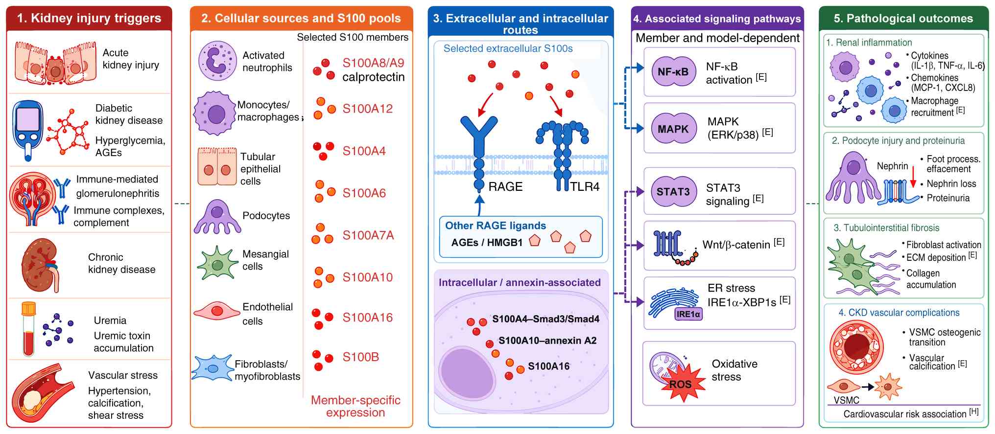 | Figure 1Overview of S100-associated
mechanisms in kidney disease. Selected extracellular S100-receptor
routes are distinguished from intracellular and annexin-associated
pathways. Cellular sources and proteins are illustrative groups:
Expression alone does not establish secretion, and connections
depend on the S100 member and experimental model. Non-S100 RAGE
ligands are shown separately. Dashed connectors indicate conceptual
relationships across studies. H, human observational association;
E, functional experimental support. Labels apply to adjacent
findings, without validating every grouped connection or
establishing clinical benefit. AGEs, advanced glycation
end-products; CKD, chronic kidney disease; ECM, extracellular
matrix; ER, endoplasmic reticulum; HMGB1, high-mobility group box
1; MAPK, mitogen-activated protein kinase; NF-κB, nuclear factor
κB; RAGE, receptor for advanced glycation end-products; STAT3,
signal transducer and activator of transcription 3; TLR4, Toll-like
receptor 4; VSMC, vascular smooth muscle cell. |
Biology of the S100 protein family and
receptor signaling
The S100 protein family comprises more than 20
small, acidic calcium-binding proteins characterized by two EF-hand
calcium-binding motifs. These proteins serve diverse intracellular
functions, and some members can act as DAMPs after extracellular
release (9). Myeloid cells such
as neutrophils and activated macrophages are major sources of
S100A8/A9 (calprotectin), which can engage cell surface receptors
to propagate inflammatory responses (9,10). Members implicated in kidney
disease include S100A8/A9, S100A12, S100A4, S100A6 and S100B.
Non-myeloid sources include stressed tubular cells, mesangial
cells, podocytes and interstitial or vascular cells; expression
alone does not establish extracellular release (6,11-14). S100A2 is an emerging regulator of
tubular stress involving forkhead box O1 (FoxO1) (15).
S100A8/A9, a heterodimer of S100A8 and S100A9, is
the best-characterized S100 complex in kidney disease. In
nephrotoxic nephritis, S100A9 deficiency protects mice from
glomerular injury, and in vitro S100A8/A9 stimulation
induces interleukin-6 (IL-6), tumor necrosis factor-α (TNF-α) and
C-X-C motif chemokine ligand 1 (CXCL1) production by macrophages
and mesangial cells (10). These
data support a pathogenic role in interactions between leukocytes
and renal cells. The effects nevertheless depend on context:
S100A8/A9 may participate in both injury amplification and
repair-associated macrophage responses depending on timing,
cellular source and disease model (5,10). Recent studies also implicate
calprotectin in CKD-associated vascular calcification and diabetic
renal interstitial fibrosis, but clinical causality remains to be
established (11,16).
S100A12, a pro-inflammatory S100 protein released by
activated neutrophils, is elevated in
myeloperoxidase-antineutrophil cytoplasmic antibody
(MPO-ANCA)-associated glomerulonephritis (17) and has also been associated with
vascular pathology in CKD (18,19). Serum S100A12 levels are elevated
in patients with CKD stages 4-5, especially in those with
cardiovascular comorbidity (18). Increased S100A12 mRNA
transcription in peripheral leukocytes supports increased
production without excluding altered renal clearance (18). Animal models suggest that S100A12
can contribute to vascular remodeling and arterial calcification in
the uremic environment (19).
S100A4, also known as fibroblast-specific protein 1
(FSP1), has been used as a marker of fibroblasts and
myofibroblasts, although it is not specific to fibroblasts
(20). Increased numbers of
S100A4-positive interstitial cells are observed in renal fibrosis
across kidney diseases, including immunoglobulin A (IgA)
nephropathy, obstructive nephropathy and polycystic kidney disease
(20-22). S100A4/FSP1 has also been examined
in vascular-access smooth-muscle remodeling (23), podocyte or tubular
epithelial-to-mesenchymal transition (EMT)-like phenotypes
(24,25), and CKD arteriovenous-fistula
endothelial remodeling (26).
Lineage-tracing studies challenge the interpretation of complete
epithelial conversion into myofibroblasts (27), while transplant-biopsy SNAI1
findings support interpreting mesenchymal-marker expression as a
diseased epithelial phenotype rather than proof of lineage
conversion (28).
Other S100 proteins also play distinct roles. S100A6
(calcyclin) expression is upregulated in the glomeruli of diabetic
mice (29). In acute renal
failure, S100A6 mRNA and protein levels are markedly elevated in
the renal cortex during the peak of tubular injury and early
recovery, supporting its investigation as a marker of tubular
injury and recovery (30).
S100B, a protein best known as a marker of glial cell activity, is
also a RAGE ligand (8,12,31). In kidney disease, elevated serum
S100B has been investigated as a biomarker for cognitive impairment
in patients with end-stage renal disease (ESRD) (32). S100B has also been studied after
cardiac arrest, a distinct prognostic setting (33). Mechanistically, high glucose
stimulates S100B expression in mesangial cells; S100B then drives
pro-fibrotic gene expression, including transforming growth
factor-β1 (TGF-β1) and type IV collagen, and induces cellular
hypertrophy (12).
RAGE mediates some of the pro-inflammatory and
pro-fibrotic effects of selected extracellular S100 proteins. RAGE
is a multi-ligand transmembrane receptor belonging to the
immunoglobulin superfamily, expressed on numerous cell types,
including macrophages, endothelial cells and podocytes (12,31). Its ligands are not limited to
S100 proteins but also include advanced glycation end-products
(AGEs) and high-mobility group box 1, both of which are elevated in
conditions such as diabetes and CKD (1). High-molecular-weight RAGE ligands
isolated from the serum of patients with diabetes are particularly
potent activators of downstream signaling (1). Because RAGE recognizes non-S100
ligands, protection following RAGE blockade alone does not identify
the responsible S100 protein.
After ligand engagement, the short cytoplasmic
domain of RAGE triggers intracellular signaling that sustains
inflammatory gene expression. The best-established downstream
effects include NF-κB activation, MAPK signaling through
extracellular signal-regulated kinases 1/2 (ERK1/2) and p38,
oxidative stress, cytokine and chemokine release, and extracellular
matrix (ECM) production (1,8,12,31). For example, RAGE ligand
preparations and S100B stimulation promote profibrotic responses in
mesangial cells (1,12,31). These findings support a
feed-forward model in which tissue injury releases S100 ligands,
receptor activation amplifies inflammation, and persistent
inflammation accelerates fibrosis and vascular damage.
S100 proteins as clinical biomarkers in
nephrology
Selected S100 proteins, including members with
extracellular DAMP activity, are candidate biomarkers in several
kidney diseases. Blood and urine measurements of selected S100
proteins have been investigated as indicators of acute tubular
injury, chronic inflammation, fibrosis and vascular damage. This
section reviews the clinical evidence for key S100 proteins across
nephrological settings. The disease-specific evidence is summarized
in Fig. 2, and representative
biomarker applications are listed in Table I. Blood, urine and tissue
measurements are not interchangeable, and an association with
disease does not establish clinical decision value.
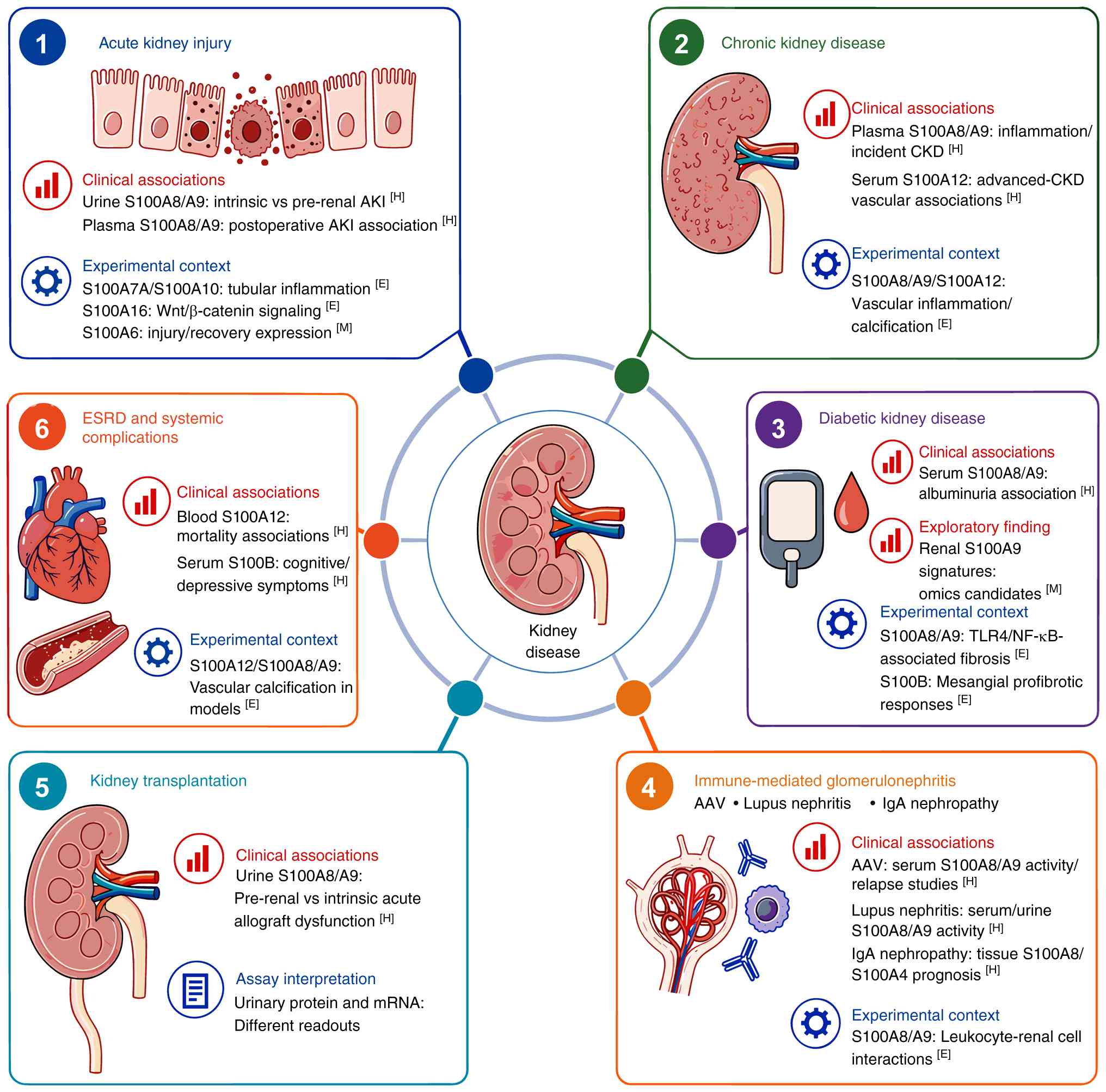 | Figure 2Clinical associations and
experimental contexts of S100 proteins across kidney diseases. H
denotes human observational evidence, including diagnostic cohorts;
E, functional experimental support; M, exploratory or model-limited
evidence, including expression and omics findings. These labels
describe adjacent findings rather than formal evidence grades.
Urine, serum, plasma, tissue and mRNA measurements are distinct
readouts. Transplant findings concern acute graft dysfunction;
human S100B associations concern systemic complications. Tissue
S100A8 and S100A4 findings in IgA nephropathy derive from separate
studies, and S100A9 omics candidates require protein-level
validation. Ring connections group disease settings without
implying causality. Clinical utility requires further validation.
AKI, acute kidney injury; CKD, chronic kidney disease; ESRD,
end-stage renal disease; AAV, antineutrophil cytoplasmic
antibody-associated vasculitis; IgA, immunoglobulin A; mRNA,
messenger RNA. |
 | Table IS100 measurements in kidney disease:
specimen, clinical question and evidence limitations. |
Table I
S100 measurements in kidney disease:
specimen, clinical question and evidence limitations.
| S100 protein | Sample type | Clinical
question | Key supporting
evidence | Evidence and
interpretation limit | (Refs.) |
|---|
| S100A8/A9 | Urine protein | Intrinsic vs.
pre-renal AKI | Adult, pediatric
and allograft diagnostic cohorts | H: candidate
adjunct; UTI and assay thresholds limit transferability | (34,35,38-41) |
| S100A8/A9 | Plasma protein | Postoperative AKI
risk | Cardiopulmonary
bypass and aortic dissection cohorts | H: timing-specific
association; does not validate urine thresholds | (44,45) |
| S100A8/A9 | Urine protein | Adverse outcomes in
pediatric AKI | Mortality AUC
0.55-0.61; kidney replacement therapy AUC 0.72 | H: poor mortality
prediction; endpoint-specific performance | (43) |
| S100A8/A9 | Urine protein |
Cisplatin-associated kidney injury | Exploratory
nephrotoxicity cohort | H: limited
validation; not an established treatment rule | (48) |
| S100A8/A9 | Plasma or
feces | Incident CKD or
intestinal inflammation | Plasma CKD
association; fecal mortality association | H: distinct
compartments; confounding and causality unresolved | (50,51,53) |
| S100A8/A9 | Serum; urine CD163
in a panel | Active AAV kidney
disease | Disease-activity
cohort and combined biomarker model | H: external
validation and incremental value needed | (54,55) |
| S100A8/A9 | Serum and urine
protein | Lupus nephritis
activity | Associations with
clinical kidney activity | H: systemic
autoimmune activity can confound blood results | (56,57) |
| S100A8 | Glomerular
macrophages | IgAN
progression | MRP8-positive cells
associated with sclerotic change | H: biopsy
association; not a non-invasive test | (58) |
| S100A8/A9 | Serum; kidney
immune-cell transcripts | DKD severity or
early inflammatory signatures | Albuminuria
associations and omics candidates | H/M: protein
measurements and transcripts are not equivalent | (4,61-63) |
| S100A12 | Serum or
plasma | Vascular risk in
advanced CKD | Associations with
vascular disease and mortality | H: account for
inflammation, kidney function and dialysis timing | (18,75-78) |
| S100A4/FSP1 | Kidney biopsy | IgAN prognosis and
fibrosis | Cell density
associated with kidney survival and steroid response | H: historical name
does not confer fibroblast specificity | (21,74) |
| S100B | Serum protein | Systemic neurologic
complications | Cognitive,
depressive or neuropathy associations | H: not specific for
kidney injury or DKD progression | (32,65-67) |
| S100A6 and annexin
A2 | Kidney tissue | Tubular injury and
recovery | Co-expression over
experimental injury and recovery | M: experimental
marker association, not a validated clinical assay | (30) |
S100A8/A9 in AKI
S100A8/A9 is abundantly expressed in neutrophils and
monocytes and serves as a sensitive marker of innate immune
activation. Its most extensively studied clinical application in
nephrology is differentiating intrinsic (structural) from pre-renal
(functional) AKI. This distinction is clinically useful, but
urinary calprotectin should be interpreted as a marker of urinary
tract or kidney inflammation rather than as a stand-alone
diagnostic test. Its performance may be influenced by urinary tract
infection, systemic inflammation, sampling time and the assay
platform (34-36).
Multiple studies have evaluated urinary calprotectin
for differentiating intrinsic from pre-renal AKI. An adult
comparison study evaluated calprotectin together with neutrophil
gelatinase-associated lipocalin (NGAL) (35). A cardiopulmonary-bypass
proteomics study detected urinary S100A8 and provides perioperative
biomarker data, but it did not evaluate calprotectin diagnostic
accuracy (37). A meta-analysis
and critical-care cohorts report favorable discrimination, although
thresholds differ and external validation and standardization
remain limited (38,39). In renal transplant recipients,
urinary calprotectin can help distinguish pre-renal graft
dysfunction from intrinsic acute allograft injury (40).
S100A8/A9 has also been studied in several clinical
settings associated with AKI. In pediatric populations, urinary
calprotectin has been investigated for differentiating AKI
etiologies (41,42) and for predicting adverse
outcomes. However, one pediatric AKI cohort reported poor mortality
discrimination, with areas under the receiver operating
characteristic curve (AUCs) of 0.55 for 30-day and 0.61 for 3-month
mortality, whereas prediction of renal replacement therapy (RRT)
yielded an AUC of 0.72 (43).
After cardiac surgery, elevated plasma S100A8/A9 levels immediately
following cardiopulmonary bypass are associated with subsequent AKI
(44). Plasma S100A8/A9 has also
been investigated for predicting postoperative AKI after high-risk
procedures such as acute type A aortic dissection surgery (45). Mechanistic experiments in this
setting implicate S100A8/A9-high macrophages in renal tubular
epithelial cell damage (46). In
sepsis-associated AKI, S100A8/A9 expression is time-dependently
elevated in the kidney, and its inhibition ameliorates renal
dysfunction in murine models, supporting a pathogenic role in that
model (47). In drug-induced
nephrotoxicity, urinary calprotectin has also been evaluated for
detecting acute cisplatin-induced nephrotoxicity (48). Urinary calprotectin has
additionally been studied in relation to kidney damage and
mortality in COVID-19 (49).
These plasma and urine studies use different sampling times and
endpoints; thus a common diagnostic threshold cannot be
assumed.
S100A8/A9 in CKD and
glomerulonephritis
In CKD, S100A8/A9 has been studied primarily as a
marker of systemic low-grade inflammation associated with disease
progression and complications. Higher plasma calprotectin levels
are prospectively associated with an increased risk of new-onset
CKD in the general population, particularly when defined by urinary
albumin excretion (50).
CKD-related systemic inflammation has also been examined in
relation to the gut-kidney axis: Elevated fecal calprotectin, a
marker of intestinal inflammation, has been associated with higher
mortality in patients with CKD, independent of traditional
inflammatory markers (51).
Related exploratory findings in children on RRT and elderly
patients with moderate CKD support further investigation of this
relationship (52,53). Intestinal barrier dysfunction and
microbial products or uremic solutes may increase systemic
inflammatory exposure. However, fecal calprotectin indicates
intestinal inflammation; it does not establish transport of S100
proteins to the kidney or causation of CKD progression.
S100A8/A9 has also been studied in inflammatory
glomerular diseases. In ANCA-associated vasculitis (AAV), serum and
leukocyte surface S100A8/A9 reflect disease activity, decrease with
treatment and may help predict relapse (54). A predictive model combining serum
calprotectin, urinary soluble CD163 and hematuria has been proposed
to improve detection of active renal vasculitis (55). In lupus nephritis, serum and
urinary S100A8/A9 levels correlate with renal activity in the
available cohorts (56,57). In IgA nephropathy, the
accumulation of myeloid-related protein 8 (MRP8; S100A8)-expressing
macrophages in glomeruli has been associated with sclerotic
progression and unfavorable prognosis in children (58). These observations support
S100A8/A9 as a marker of active myeloid inflammation, but larger
prospective studies are needed to define its role in routine
decision-making.
Evidence linking calprotectin to vascular
calcification may be relevant to CKD-associated cardiovascular
risk. A primary experimental study indicates that S100A8/A9 can
promote osteochondrogenic differentiation of vascular smooth muscle
cells (VSMCs) through RAGE and TLR4 signaling (16). Reviews and commentary provide
broader mechanistic and clinical context (59,60). The extent to which circulating
calprotectin is a causal mediator, a marker of vascular
inflammation or both may vary among patient subgroups and requires
prospective interventional study.
S100 proteins in DKD
In DN, S100A8/A9 and S100B have been associated with
disease status, progression and systemic complications. The
evidence is most consistent for associations with albuminuria,
inflammatory cell infiltration and diabetic complications, whereas
their incremental value beyond established clinical markers remains
insufficiently defined. DKD is used for the clinical disease
category; DN is retained when describing studies that use that
designation.
Serum S100A8/A9 levels increase across worsening
stages of DN, from normo-albuminuria to macroalbuminuria, and
correlate positively with urinary albumin-to-creatinine ratio and
serum creatinine (4). The
association with microalbuminuria has prompted evaluation of
calprotectin as an early marker of microvascular damage and
inflammation in type 2 diabetes (61). Single-cell sequencing studies
have identified S100A9 as a candidate early molecular marker that
is highly expressed in pro-inflammatory myeloid-derived suppressor
cells that infiltrate the kidney in early DN (62). Machine-learning analyses have
also nominated S100A9 as a candidate diagnostic biomarker for DKD
(63). The gut-kidney axis is
also relevant in DN, but a study of patients with type 1 diabetes
found that fecal calprotectin did not differ significantly between
progressive and stable DKD groups (64). These omics-derived signatures
require independent protein-level validation and comparison with
estimated glomerular filtration rate (eGFR) and albuminuria.
S100B, which is primarily expressed in glial cells,
has been studied in relation to systemic complications of diabetes
(65). Elevated serum S100B has
been associated with diabetic peripheral neuropathy, a common
comorbidity of DN (65). In
patients with ESRD, which can result from DN, higher serum S100B
levels are associated with neurologic and psychiatric
complications, including cognitive impairment and depression
(32,65-67). In high-glucose-stimulated
mesangial cells, S100B contributes to pro-fibrotic responses
(12). S100B is also under
investigation as a potential marker for long-term complications in
patients with DN who have a history of COVID-19 (68). These systemic associations do not
establish S100B as a kidney-specific predictor of DKD progression.
Studies of activated leukocyte cell adhesion molecule in DN and IgA
nephropathy provide non-S100 biomarker comparators (69,70), not evidence of S100B
signaling.
S100A4 and S100A12 as prognostic
markers
S100A4 and S100A12 are two S100 proteins with
distinct candidate prognostic roles in kidney disease, related
primarily to fibrosis and cardiovascular complications,
respectively.
S100A4/FSP1 is a widely used marker of fibroblast
activation in fibrotic kidneys, but its biological interpretation
requires caution. In animal models of kidney fibrosis, including
obstructive and remnant-kidney models, S100A4/FSP1 expression
increases in interstitial fibroblast-like cells (71,72). Hypoxia and fibroblast-epithelial
crosstalk may further promote FSP1-positive cell accumulation and
fibrotic remodeling (71,73).
In IgA nephropathy, interstitial FSP1-positive cell density has
been associated with renal survival and corticosteroid
responsiveness (21,74). However, extracellular FSP1 may
also exert context-dependent protective effects through antioxidant
pathways, indicating that FSP1 does not act solely as a
fibrosis-promoting factor (13).
Intracellular fibrogenic functions and extracellular protective
effects should therefore be distinguished.
S100A12 (also known as EN-RAGE) is a
pro-inflammatory RAGE ligand and a candidate marker of
cardiovascular risk in advanced CKD. Plasma S100A12 levels are
elevated in patients with ESRD undergoing hemodialysis or
peritoneal dialysis (75,76),
and several cohorts link higher levels to all-cause or
cardiovascular mortality (77,78). These findings may have prognostic
value, but they do not show that S100A12-directed therapy will
improve outcomes. ABCC6 genetics provides non-S100 calcification
context (79); the distinction
between S100A12 as a risk marker and a causal factor remains
important (80). Experimental
studies support S100-associated vascular remodeling and
calcification mechanisms (19,81).
Comparative value of S100 biomarkers
The potential clinical value of S100 proteins should
be assessed against established biomarkers, both individually and
in combined panels. In AKI, urinary calprotectin provides
information about inflammatory parenchymal injury that differs from
tubular stress markers such as NGAL, kidney injury molecule-1
(KIM-1) and interleukin-18 (IL-18) (35,41,43). Its principal value may be
etiological classification rather than universal AKI prediction. In
CKD, S100 proteins are unlikely to replace albuminuria, estimated
glomerular filtration rate or histology, but may add pathway-level
information on inflammation, fibrosis or vascular risk.
In chronic and inflammatory kidney diseases,
combining S100 proteins with other markers may improve diagnostic
performance. In AAV, a model combining serum calprotectin with the
macrophage marker urinary CD163 performed better for detecting
active renal disease than either biomarker alone (55). In renal transplant recipients,
urinary mRNA levels of NGAL, KIM-1 and IL-18 correlated with
histological findings of rejection, whereas S100A8/A9 mRNA did not,
indicating that biomarker choice may depend on the clinical
question within the same patient population (82). In stable CKD, plasma calprotectin
levels correlate with other inflammatory markers such as IL-6 and
monocyte chemoattractant protein-1, as well as with levels of
protein-bound uremic toxins, supporting its investigation as an
indicator of the systemic inflammatory state associated with uremia
(83). Integration of S100
proteins into panels for AKI differentiation, CKD assessment or
prediction of inflammatory flares requires external validation and
evidence of clinical benefit. Urinary transcript findings should
not be extrapolated directly to calprotectin protein assays.
Across biomarker studies, the evidence base is
heterogeneous. Urinary S100A8/A9 in AKI is supported by multiple
clinical cohorts and meta-analytic evidence, whereas several other
S100 members are supported mainly by small cohorts, tissue studies,
animal models or bioinformatic analyses. Future studies should
therefore report standardized assays, prespecified cutoffs,
external validation cohorts and incremental predictive value beyond
conventional markers.
Assay standardization and
confounders
Most urinary AKI studies used enzyme-linked
immunosorbent assays with cohort- and assay-specific cut-offs
(34,35,38). The 300 ng/ml value from the
original adult study is a diagnostic threshold, not a normal range
or treatment-selection threshold (34). Antibody specificity, calibrators,
molecular forms, dilution, analytical range and urine-matrix
effects limit comparability. A serum assay comparison required
substantial calibration adjustment; this finding demonstrates the
need for harmonization but does not quantify inter-assay bias in
urine (84). Validation should
address precision, recovery, linearity, interference and
quantification limits, followed by independent validation of
clinical thresholds and incremental value. Urinary concentrations
and creatinine-normalized values should be reported separately.
Urinary tract infection, pyuria and contamination
can confound interpretation. Bacteriuria can raise urinary
calprotectin even without dipstick pyuria, thus a negative dipstick
alone does not exclude infection-related elevation (36). Studies should document the
collection method, catheter status, hematuria and suspected
contamination, with urine culture or repeat sampling when
indicated. Systemic infection, surgery and autoimmune activity also
affect circulating markers independently of kidney injury (44,54,85). Studies should record eGFR,
residual kidney function, dialysis status and sampling relative to
dialysis; increased leukocyte S100A12 transcription does not
exclude altered clearance (18).
Serum and plasma results should not be pooled:
Specimen-dependent concentrations and delayed-processing effects
have been demonstrated for circulating calprotectin (86). Collection-to-processing time,
temperature, centrifugation, storage and freeze-thaw exposure
should be controlled for each matrix and assay. Findings in blood
cannot establish analyte stability in urine. Sampling should be
defined relative to injury, surgery, treatment and recovery because
these time points address different clinical questions (30,43,44).
S100-associated mechanisms in kidney injury
and progression
S100-associated signaling is best viewed as one
component of a broader sterile inflammatory network in kidney
disease. After release from stressed or necrotic cells, selected
extracellular S100 proteins act as DAMPs by engaging
pattern-recognition receptors, including RAGE and TLR4. These
interactions can activate downstream signaling events that promote
inflammation, fibrosis and cellular injury and thereby contribute
to AKI and CKD progression. However, the magnitude and direction of
S100 effects depend on ligand concentration, cellular source,
receptor availability and disease stage. The principal
compartment-specific mechanisms are illustrated in Fig. 3. Similar endpoints may also arise
from intracellular S100 functions without established RAGE
dependence.
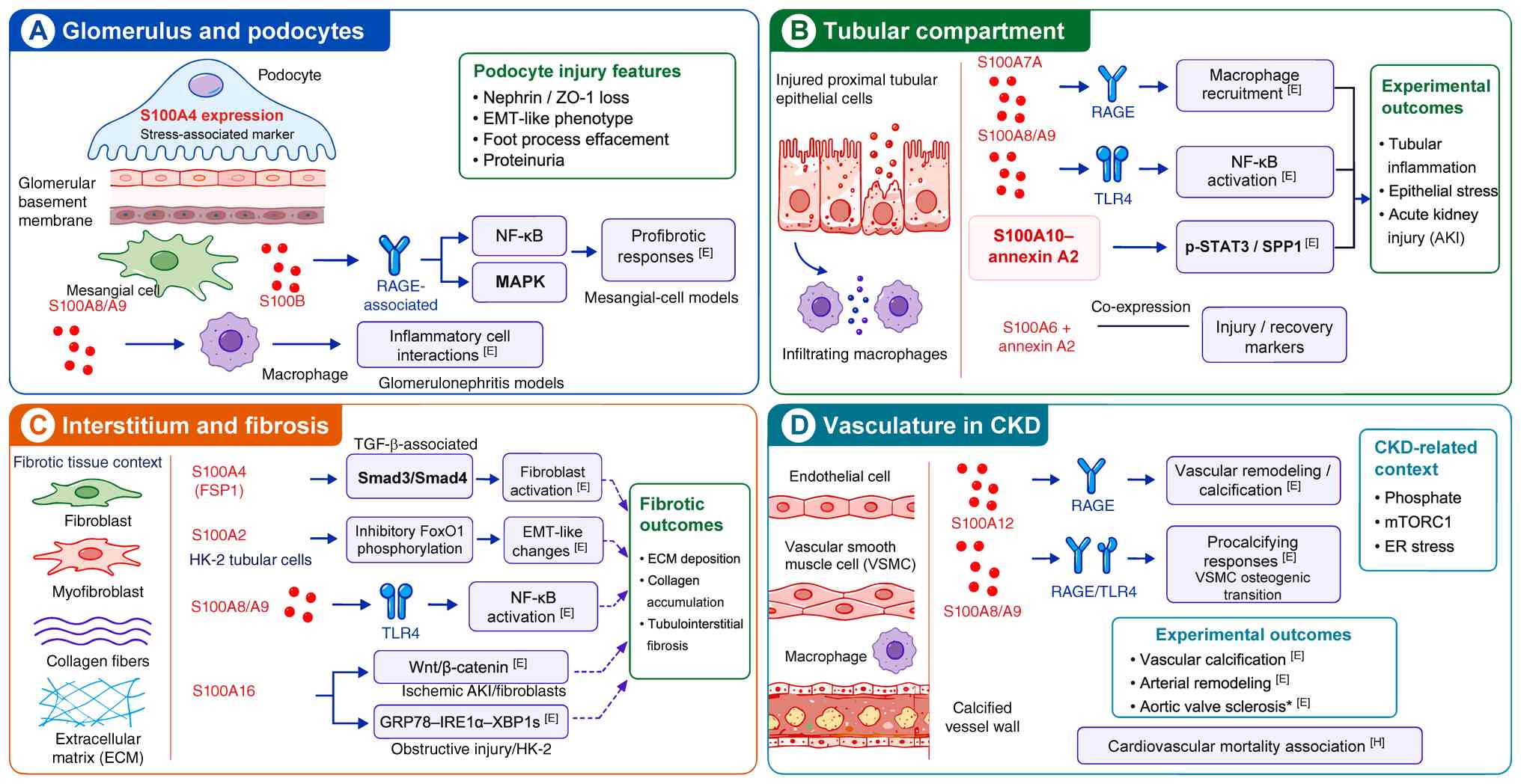 | Figure 3S100-associated mechanisms in
glomerular, tubular, interstitial and vascular compartments. (A)
Podocyte S100A4 expression does not establish extracellular release
or S100A4-RAGE causality. (B) S100A7A findings derive from
male-mouse cisplatin AKI. S100A10-annexin A2 signaling has no
established RAGE requirement, whereas S100A6/annexin A2 denotes
co-expression. (C) S100A2-associated inhibitory FoxO1
phosphorylation and EMT-like changes concern HK-2 cells.
S100A16-Wnt findings derive from ischemic AKI/interstitial
fibroblast models, and GRP78/IRE1α/XBP1s findings from obstructive
injury/HK-2 models. Tissue illustrations do not assign a shared
cellular origin or demonstrate epithelial conversion into
fibroblasts. (D) The asterisk denotes aortic valve sclerosis in a
humanized S100A8/A9/A12 cluster model. CKD-related factors provide
context from separate studies; human mortality associations remain
observational. Solid arrows indicate effects supported within the
stated models; dashed connectors indicate cross-study synthesis. H,
human association; E, functional experimental support. Labels apply
to adjacent findings and are not formal grades or evidence of
clinical benefit. CKD, chronic kidney disease; ECM, extracellular
matrix; EMT, epithelial-to-mesenchymal transition; ER, endoplasmic
reticulum; FSP1, fibroblast-specific protein 1; RAGE, receptor for
advanced glycation end-products; SPP1, secreted phosphoprotein 1;
TLR4, Toll-like receptor 4; VSMC, vascular smooth muscle cell; AKI,
acute kidney injury. |
Kidney inflammation
A hallmark of kidney injury is inflammatory cell
recruitment and activation. S100A8/A9 and S100A12 can act as
chemo-attractants and immune activators, but their functions are
not uniformly deleterious. In glomerulonephritis models, S100A8/A9
promotes inflammatory leukocyte-renal cell interactions, whereas in
IRI it has been linked to macrophage-mediated repair responses
(5,10). Distinguishing injury-amplifying
from repair-associated S100 signaling is therefore essential for
interpreting therapeutic studies.
These mechanisms occur across several nephropathies.
In systemic lupus erythematosus, serum S100A8/A9 and S100A12 are
elevated in patients with high disease activity and are associated
with the presence of glomerulonephritis (85). In DKD, S100A8/A9-associated
TLR4/NF-κB signaling contributes to inflammation and interstitial
fibrosis in experimental models (11). In septic AKI, blockade of the
S100A8/A9-TLR4 interaction with paquinimod ameliorates renal
dysfunction and inflammatory injury (47). Human biomarker associations and
experimental receptor effects provide different levels of evidence.
S100A8- and S100A9-derived B-cell epitopes have also been evaluated
as serological biomarkers in DN (87), but that diagnostic study does not
establish a pathogenic signaling mechanism.
Recent single-cell transcriptomic studies have
provided a more detailed cellular map of S100 expression. In
IRI-induced AKI, inflammatory macrophage subsets with high S100A9
expression have been proposed as potential therapeutic targets
(88). In DKD, S100A8/A9-high
macrophages are also enriched in inflammatory niches (89). These datasets are valuable for
hypothesis generation, but transcript abundance does not
necessarily establish protein activity or causal function.
Validation at the protein level and with spatial and functional
studies remains important.
Together, these studies implicate myeloid
recruitment and tubular stress as distinct contributors to
inflammation. The relevant receptor, cell population and timing
vary; an intervention should therefore be assessed for both early
injury and later recovery (5,6,10,88).
Tubulointerstitial fibrosis (TIF)
TIF is a common feature of progressive kidney
disease and contributes to irreversible loss of renal function. It
is characterized by excessive accumulation of ECM, produced
primarily by activated myofibroblasts. S100-associated mechanisms
are implicated in this fibrotic process, with S100A4 and S100A8/A9
playing prominent roles.
S100A4/FSP1 is associated with fibroblast activation
in fibrotic kidneys, although its expression is not
fibroblast-specific. Niclosamide treatment, together with genetic
S100A4 perturbation, attenuates fibroblast activation, collagen
deposition and ECM accumulation in experimental renal fibrosis
(90). In vitro, S100A4
overexpression promotes TGF-β1-induced fibroblast activation,
whereas S100A4 knockdown is protective (90). S100A2 and S100A8/A9 are
associated with additional profibrotic mechanisms: S100A2 promotes
inhibitory FoxO1 phosphorylation and EMT-like changes in HK-2
cells, S100A8/A9 promotes fibrosis in obstructive nephropathy, and
S100A8/A9 inhibition ameliorates diabetic renal interstitial
fibrosis by suppressing TLR4/NF-κB signaling (11,15,91). S100A4 interacts with Smad3,
stabilizes the Smad3/Smad4 complex and promotes its nuclear
translocation, although niclosamide is not a selective mechanistic
probe (90). S100A2-associated
FoxO1 inhibition does not establish a S100A2-RAGE pathway.
The cellular origin of scar-producing myofibroblasts
remains a matter of debate. S100A4/FSP1 expression in tubular
epithelial cells or podocytes has often been interpreted as
evidence for EMT in DN and human immunodeficiency virus-associated
podocyte injury (24,92,93). However, lineage-tracing studies
have shown that resident interstitial cells, particularly
pericytes, rather than epithelial cells, are the dominant source of
myofibroblasts in several models of kidney fibrosis (27). Thus, S100A4 expression should be
interpreted as a marker of injury, dedifferentiation and
profibrotic signaling rather than proof that epithelial cells
become matrix-producing myofibroblasts in vivo.
S100A4-Smad3/Smad4, S100A2-FoxO1 and
S100A16-associated Wnt or ERS mechanisms contribute to fibroblast
activation, epithelial stress and tissue remodeling (14,15,90,94). This conclusion synthesizes
findings across studies and does not demonstrate direct
interactions among all S100 proteins. The kidney studies discussed
in the present review do not establish direct S100-specific
coupling to Hippo/YAP/TAZ; this relationship remains a testable
hypothesis.
Podocyte injury and proteinuria
Podocytes are essential for maintaining the
glomerular filtration barrier, and injury to these cells can result
in foot-process effacement, detachment, proteinuria and progressive
glomerulosclerosis. RAGE signaling is implicated in glomerular
injury, although AGE-containing ligand preparations do not isolate
S100-specific effects (1,8).
In diabetic settings, RAGE signaling intersects with oxidative
stress, inflammatory cytokines and ECM remodeling, contributing to
podocyte vulnerability.
An EMT-like podocyte phenotype links S100A4
expression to proteinuria-associated cellular stress. TGF-β1 can
reduce podocyte junctional proteins such as nephrin and ZO-1 while
inducing mesenchymal markers including desmin and S100A4/FSP1;
these changes are associated with increased albumin permeability
(24). In DN, podocyte FSP1
expression correlates with podocyte depletion and
glomerulosclerosis (92). These
findings support a stress-associated phenotype but neither prove
complete lineage conversion nor establish extracellular S100A4-RAGE
causality. PLA2R binding to the annexin A2-S100A10 complex in human
podocytes is a separate molecular observation whose contribution to
barrier injury remains unresolved (95).
These observations link S100A4 expression to
podocyte stress but do not establish a causal extracellular
ligand-receptor mechanism. Cell-specific receptor perturbation and
ligand rescue would be needed to establish whether a specific S100
pathway directly contributes to proteinuria.
Vascular complications in CKD
Cardiovascular disease is the leading cause of
mortality in patients with CKD, driven in part by accelerated
atherosclerosis and vascular calcification (VC). The uremic
environment, characterized by inflammatory mediators and mineral
dysregulation, favors vascular remodeling. Within this setting,
S100/RAGE-related signaling has been implicated in
uremia-associated VC, an active, cell-mediated process that
involves the osteogenic transition of VSMCs.
Mechanistic evidence supports a causal contribution
of S100/RAGE signaling to vascular calcification. In CKD models,
transgenic S100A12 expression directly mediates vascular remodeling
and arterial calcification through RAGE-dependent pathways
(19). Humanized mice expressing
the S100A8/A9/A12 cluster develop left ventricular hypertrophy and
aortic valve sclerosis with calcification during CKD, and this
phenotype requires RAGE (81).
Separate studies implicate RAGE in phosphate-associated
calcification and mechanistic target of rapamycin complex 1
(mTORC1)/ERS in the uremic vascular environment; these findings do
not establish one linear S100 pathway (96,97).
Calprotectin has emerged as a candidate mediator and
therapeutic target in CKD-associated vascular calcification
(16). Mechanistic experiments
showed that calprotectin exacerbates calcification in human VSMCs
and mouse aortic rings, whereas paquinimod reduced calcification by
dampening inflammatory signaling (16). These findings link calprotectin
measurements to an experimentally modifiable vascular mechanism,
but clinical trials are needed before calprotectin can be regarded
as a validated therapeutic target in CKD vascular disease.
Pharmacological RAGE and TLR4 inhibition reduced pro-calcifying
effects in that experimental setting, supporting receptor
involvement without demonstrating treatment benefit in patients
(16).
The vascular evidence links inflammatory ligand
exposure to mineral stress in vascular cells. Human outcome
associations remain observational, whereas intervention experiments
provide support that is specific to each model; reduced
inflammation or calcification in a model is not equivalent to
improved cardiovascular outcomes.
Stress-regulated and intracellular S100
pathways
Although RAGE and TLR4 are central to the current
framework, S100 biology in the kidney extends beyond a single
inflammatory axis. Recent studies link S100 proteins to tubular
stress responses, gasdermin regulation, annexin complexes,
Wnt/β-catenin signaling and ERS. The position of each S100 protein
within these pathways varies with the specific protein and
experimental model. These findings broaden the mechanistic scope of
the field and may explain disease-context specificity, but several
remain early observations from animal or cell models and should be
interpreted as emerging mechanisms rather than established clinical
targets.
A recent study identified a protective,
non-pyroptotic function of gasdermin E (GSDME) in cisplatin-induced
nephrotoxicity (6). In this
male-mouse AKI model, GSDME expression was reduced in kidney
tissue, and tubule-specific GSDME deficiency exacerbated renal
injury. Mechanistic experiments indicate that GSDME in proximal
tubular cells normally prevents the degradation of O-linked
N-acetylglucosamine (O-GlcNAc) transferase. Consequently, GSDME
loss reduces STAT3 O-GlcNAcylation, which enhances STAT3
phosphorylation and activation. Activated STAT3 then drives the
expression of its downstream target, S100a7a. The resulting
increase in S100A7A protein promotes macrophage infiltration via
RAGE activation, amplifying renal inflammation. These findings
support a protective role for tubular GSDME through suppression of
STAT3-dependent S100A7A production in this model (6). The non-pyroptotic upstream function
of GSDME should therefore be distinguished from the downstream
RAGE-associated S100A7A effect. Janus kinase 2
(JAK2)/STAT3-directed treatment in DN provides broader pathway
context (98), without
establishing S100-specific efficacy.
Interactions between S100 proteins and annexin A2
(Anxa2), another calcium-dependent protein, further illustrate the
context dependence of S100-related pathways in tubular injury.
S100A6 and Anxa2 have been identified as markers associated with
tubular injury and recovery in AKI (30). In experimental toxic and ischemic
AKI, both proteins increase during injury and early recovery,
consistent with roles in the injury response rather than uniformly
pathogenic effects (30). By
contrast, S100A10/Anxa2 complex activation was reported to
aggravate cisplatin- and ischemia-reperfusion-induced AKI through
phosphorylated STAT3 and secreted phosphoprotein 1 signaling
(99). Co-expression does not
establish a pathogenic S100A6-annexin A2 complex. The
S100A10-annexin A2 experiments included complex inhibition and
proximal-tubule knockdown, but do not establish a RAGE requirement
(99).
S100A16, a less extensively characterized member of
the S100 family, has been implicated in several stress pathways
relevant to AKI and fibrosis. In experimental ischemic AKI and
interstitial fibroblasts, S100A16 promotes Wnt/β-catenin activation
by facilitating the degradation of glycogen synthase kinase 3β and
casein kinase 1α, two negative regulators of β-catenin signaling,
through the E3 ubiquitin ligase HRD1 (14). S100A16 also interacts with the
endoplasmic reticulum chaperone glucose-regulated protein 78
(GRP78) and activates the inositol-requiring enzyme 1α/spliced
X-box binding protein 1 pathway in renal TIF models (94). These findings support S100A16 as
a mechanistic candidate, but its clinical relevance and
druggability remain to be defined. The Wnt study used heterozygous
S100A16 deficiency; the GRP78 study examined obstructive injury and
HK-2 cells. Neither mechanism should be considered RAGE-dependent
without receptor-specific evidence.
Therapeutic implications and future
perspectives
The involvement of S100 proteins in inflammation,
fibrosis and vascular injury supports their investigation as
therapeutic targets, although the supporting evidence varies across
diseases. Current evidence consists mainly of preclinical studies
and human biomarker associations, while kidney-specific clinical
trial evidence remains limited. Studies have examined inhibition of
S100 ligands, RAGE signaling and downstream inflammatory pathways.
Current therapeutic strategies and evidence gaps are summarized in
Table II.
| Table IIClassification of interventions
affecting S100-associated mechanisms in kidney disease. |
Table II
Classification of interventions
affecting S100-associated mechanisms in kidney disease.
| Intervention
class | Agent or
manipulation | Target or
process | Experimental
setting and finding | Main
limitation | (Refs.) |
|---|
| Ligand-directed
pharmacology | Paquinimod | S100A9 and
calprotectin signaling | VSMCs, aortic rings
and CKD mice: reduced calcification | E: preclinical
efficacy; selectivity and kidney dosing require definition | (16) |
| Ligand-directed
pharmacology | Paquinimod |
S100A8/A9-associated inflammation | Septic AKI mice:
improved injury; nephrotoxic nephritis: no successful
prevention | E:
disease-dependent response; not broad kidney protection | (47,100) |
| Receptor
blockade | Pharmacological
RAGE or TLR4 inhibition |
Calprotectin-associated receptor
effects | VSMCs and aortic
rings: reduced pro-calcifying effects | E: receptors have
other ligands; receptor efficacy is not S100 specificity | (16) |
| Genetic test and
expression modulation | S100A8/A9
knockdown; AB38b | S100A8/A9; TLR4 and
NF-κB | Diabetic models:
reduced epithelial stress and fibrosis | E: knockdown and a
compound reducing expression are distinct interventions | (11) |
| Pleiotropic
pharmacology with genetic support | Niclosamide; S100A4
knockdown | S100A4;
Smad3/Smad4 | Folic acid
nephropathy and fibroblasts: reduced activation and matrix | E: niclosamide is
not a selective probe; extracellular S100A4 may protect
tubules | (13,90) |
| Indirect downstream
modulation | KIOM-79 | S100B-stimulated
NF-κB/MAPK outputs | Cultured mesangial
cells: reduced TGF-β1 and fibronectin | E: in vitro
extract data; direct RAGE antagonism not established | (31) |
| Expression
modulation | Scrophularia
striata extract | RAGE and S100A8
expression | Diabetic rats:
improved parameters and reduced expression | E: mixed
constituents; expression change does not demonstrate binding | (101) |
| Intracellular
complex disruption | A2ti; proximal
tubular S100a10 knockdown | S100A10-annexin
A2/STAT3/SPP1 | Toxic and ischemic
AKI models: reduced injury | M: limited models;
no established RAGE requirement | (99) |
| Genetic mechanistic
test | S100A16
heterozygous deficiency or knockdown | HRD1/GSK3β/CK1α and
Wnt/β-catenin | Ischemic AKI and
interstitial fibroblasts | M: no validated
selective drug or human efficacy | (14) |
| Mechanistic
perturbation | S100A16 expression
manipulation |
GRP78/IRE1α/XBP1s | Obstructive injury
and HK-2 cells: ER-stress mechanism | M: mechanistic
evidence, not demonstrated drug efficacy | (94) |
| Broad immune
modulation | C4BP(β-) | Complement-related
immune regulation | Experimental lupus
nephritis: reduced disease | E: not an
S100-directed inhibitor | (103) |
| Multi-component
treatment and omics | Benincasa
hispida extract | Candidate
S100A6-associated changes | Diabetic mice:
structural and molecular responses | M: candidate
association; selective S100A6 inhibition unproven | (104) |
Interventions affecting S100 proteins and
receptor signaling
Specific S100 proteins are potential therapeutic
targets, but this strategy remains at an early stage. The S100A8/A9
heterodimer has been studied most extensively because of its
biomarker performance and mechanistic links to vascular
calcification and inflammation. Paquinimod reduced
calprotectin-driven vascular calcification in experimental systems,
providing preclinical proof of principle (16). However, the absence of clear
efficacy in immune-mediated glomerulonephritis models indicates
that S100A8/A9 inhibition may be disease- and compartment-specific
rather than broadly renoprotective (100).
Preclinical studies have also examined
pharmacological modulation of S100A4-associated renal fibrosis. In
a mouse model of folic acid-induced nephropathy, niclosamide, an
anthelmintic investigated as an S100A4 inhibitor, attenuated
fibroblast activation, collagen deposition and ECM protein
expression. S100A4 facilitated TGF-β1-induced fibroblast activation
in this model, and niclosamide blocked this effect, supporting a
link between S100A4 and a central fibrotic pathway (90). The pleiotropic effects of
niclosamide limit attribution to selective S100A4 inhibition, and
extracellular S100A4 can protect tubular cells in cisplatin injury
models (13).
RAGE or its downstream pathways provide additional
therapeutic targets. Preclinical studies have examined natural
compounds that modulate RAGE-related signaling. For example,
KIOM-79, a herbal extract, prevented S100B-induced expression of
TGF-β1 and fibronectin in mouse mesangial cells by suppressing the
activation of downstream effectors, including NF-κB and the MAP
kinases p38 and ERK (31). An
ethanolic extract from Scrophularia striata also produced
protective effects in a rat model of DN by suppressing the renal
expression of both RAGE and its ligand S100A8 (101). These studies show that
RAGE-related expression or signaling can be reduced experimentally.
These extracts are indirect modulators; reduced expression or
downstream signaling does not establish direct receptor antagonism.
Studies of sepsis-associated autophagy decline (102), β-chain-deficient complement
component 4 binding protein [C4BP(β-)] and Benincasa hispida
(103,104) provide broader injury and
treatment context; none establishes selective S100 inhibition.
Challenges to clinical translation
Despite the promise of targeting S100-associated
mechanisms, several challenges remain. One concern is inhibitor
specificity. Numerous small molecules, including paquinimod and
niclosamide, may have off-target effects that contribute to their
observed efficacy or toxicity. The discrepancy in paquinimod's
effectiveness against vascular calcification vs. glomerulonephritis
illustrates the challenge of translating findings from animal
models to human disease (16,100). Animal models often represent a
single, acute pathological process, which may not fully
recapitulate the multifactorial nature and chronicity of human
kidney disease. Furthermore, S100-associated signaling involves
multiple ligands and receptors, and this redundancy may allow other
components to compensate when one is inhibited.
Elevated S100 concentrations may reflect systemic
inflammation rather than a treatment-responsive kidney pathway.
Biomarker associations therefore require independent evidence of
target dependence before they can justify treatment selection.
Roadmap for clinical translation
A staged research strategy should begin with assay
qualification and validation of repeated measurements against
relevant clinical variables. No kidney-specific S100 threshold for
selecting treatment is established. A prespecified upper quantile
defined for the assay and study population could be an exploratory
enrichment criterion, but it would require validation and
continuous biomarker analysis. Candidate populations include CKD
with documented vascular calcification and DKD with persistent
albuminuria and inflammatory evidence despite stable
standard-of-care therapy. Infection, mineral metabolism, eGFR and
dialysis-related confounding should be assessed before
enrollment.
Early randomized, placebo-controlled add-on studies
should prioritize safety, pharmacokinetics in kidney impairment and
target engagement. Prespecified high- and low-biomarker strata
would allow treatment-by-biomarker interaction analysis; a study
restricted to the high-biomarker group cannot establish
differential treatment benefit. Adaptive enrichment would require
prespecified rules, adequate sample size and control of
false-positive results. Ligand inhibition, RAGE blockade,
expression modulation and intracellular complex disruption should
be selected according to the demonstrated mechanism, rather than
treated as interchangeable interventions (Table II).
For receptor-directed agents, pharmacodynamic
assessment should measure ligand-receptor engagement or receptor
activity using a functional assay. For intracellular targets,
pharmacodynamic assessment should measure pathway responses in the
relevant tissue. Total circulating S100 concentration may not fall
after ligand blockade. eGFR trajectory, albuminuria and
standardized calcification imaging can provide exploratory
outcomes, with clinical benefit tested in later trials with
adequate power. Infection, hematologic effects and impaired injury
recovery should be monitored. This roadmap is a research proposal,
not a validated clinical algorithm.
Conclusion
S100 proteins should not be treated as
interchangeable biomarkers or therapeutic targets. Clinical
translation depends on resolving ligand identity, cellular source,
receptor dependence and whether the evidence supports an
association or a causal experimental effect.
Priorities include assay standardization,
prospective validation beyond conventional clinical markers and
appropriately controlled intervention studies. Target engagement,
safety and benefit beyond existing care must be demonstrated before
S100 measurements or inhibition can guide routine nephrology
practice.
Availability of data and materials
Not applicable.
Authors' contributions
HW wrote the original draft, organized the
literature and prepared the figures and tables. ZL wrote the
original draft and assisted with figure and table preparation. QL
and YN contributed to literature collection and manuscript
drafting. JL and LM wrote, reviewed and edited the manuscript,
supervised the study and acquired funding. All authors read and
approved the final version of the manuscript. Data authentication
is not applicable.
Ethics approval and consent to
participate
Not applicable.
Patient consent for publication
Not applicable.
Competing interests
The authors declare that they have no competing
interests.
Abbreviations:
|
AAV
|
ANCA-associated vasculitis
|
|
AKI
|
acute kidney injury
|
|
CKD
|
chronic kidney disease
|
|
DAMP
|
damage-associated molecular
pattern
|
|
DKD
|
diabetic kidney disease
|
|
DN
|
diabetic nephropathy
|
|
eGFR
|
estimated glomerular filtration
rate
|
|
EMT
|
epithelial-to-mesenchymal
transition
|
|
ESRD
|
end-stage renal disease
|
|
FSP1
|
fibroblast-specific protein 1
|
|
GSDME
|
gasdermin E
|
|
IgA
|
immunoglobulin A
|
|
MAPK
|
mitogen-activated protein kinase
|
|
NF-κB
|
nuclear factor κB
|
|
NGAL
|
neutrophil gelatinase-associated
lipocalin
|
|
RAGE
|
receptor for advanced glycation
end-products
|
|
STAT3
|
signal transducer and activator of
transcription 3
|
|
TLR4
|
Toll-like receptor 4
|
Acknowledgements
Not applicable.
Funding
The present study was supported by the National Natural Science
Foundation of China (grant no. 82675432), the Sichuan Science and
Technology Program (grant nos. 2026NSFSC1827 and 2026NSFSC0638),
the China Association of Chinese Medicine Joint Research Project
(grant no. 2023DYPLHGG-05), the Joint Project of Luzhou Municipal
People's Government and Southwest Medical University (grant no.
2024LZXNYDJ020) and The Funding Program of Southwest Medical
University (grant no. 2024ZXYZX05).
References
|
1
|
Penfold SA, Coughlan MT, Patel SK,
Srivastava PM, Sourris KC, Steer D, Webster DE, Thomas MC, MacIsaac
RJ, Jerums G, et al: Circulating high-molecular-weight RAGE ligands
activate pathways implicated in the development of diabetic
nephropathy. Kidney Int. 78:287–295. 2010. View Article : Google Scholar : PubMed/NCBI
|
|
2
|
Fujiu K, Manabe I and Nagai R: Renal
collecting duct epithelial cells regulate inflammation in
tubulointerstitial damage in mice. J Clin Invest. 121:3425–3441.
2011. View Article : Google Scholar : PubMed/NCBI
|
|
3
|
Zhou J, Wu R, High AA, Slaughter CA,
Finkelstein D, Rehg JE, Redecke V and Häcker H: A20-binding
inhibitor of NF-κB (ABIN1) controls Toll-like receptor-mediated
CCAAT/enhancer-binding protein β activation and protects from
inflammatory disease. Proc Natl Acad Sci USA. 108:E998–E1006. 2011.
View Article : Google Scholar
|
|
4
|
Gu W, Cui J, Shi M, Zhang R, Tan D, Zhang
X and Zhang H: Serum myeloid-related protein 8/14 is a potential
predictor of diabetic kidney disease. Discov Med. 30:97–105.
2020.
|
|
5
|
Dessing MC, Tammaro A, Pulskens WP, Teske
GJ, Butter LM, Claessen N, van Eijk M, van der Poll T, Vogl T, Roth
J, et al: The calcium-binding protein complex S100A8/A9 has a
crucial role in controlling macrophage-mediated renal repair
following ischemia/reperfusion. Kidney Int. 87:85–94. 2015.
View Article : Google Scholar
|
|
6
|
Chen Q, Sun P, Zhou J, Long T, Xiao A, Liu
Z, Xu S, Lei W, Zhang R, Tian J, et al: Renal tubular GSDME
protects cisplatin nephrotoxicity by impeding OGT-STAT3-S100A7A
axis in male mice. Nat Commun. 16:68072025. View Article : Google Scholar : PubMed/NCBI
|
|
7
|
Nazari A, Khorramdelazad H, Hassanshahi G,
Day AS, Sardoo AM, Fard ET, Abedinzadeh M and Nadimi AE: S100A12 in
renal and cardiovascular diseases. Life Sci. 191:253–258. 2017.
View Article : Google Scholar : PubMed/NCBI
|
|
8
|
Curran CS and Kopp JB: RAGE pathway
activation and function in chronic kidney disease and COVID-19.
Front Med (Lausanne). 9:9704232022. View Article : Google Scholar : PubMed/NCBI
|
|
9
|
Ometto F, Friso L, Astorri D, Botsios C,
Raffeiner B, Punzi L and Doria A: Calprotectin in rheumatic
diseases. Exp Biol Med (Maywood). 242:859–873. 2017. View Article : Google Scholar :
|
|
10
|
Pepper RJ, Wang HH, Rajakaruna GK,
Papakrivopoulou E, Vogl T, Pusey CD, Cook HT and Salama AD:
S100A8/A9 (calprotectin) is critical for development of
glomerulonephritis and promotes inflammatory leukocyte-renal cell
interactions. Am J Pathol. 185:1264–1274. 2015. View Article : Google Scholar : PubMed/NCBI
|
|
11
|
Du L, Chen Y, Shi J, Yu X, Zhou J, Wang X,
Xu L, Liu J, Gao J, Gu X, et al: Inhibition of S100A8/A9
ameliorates renal interstitial fibrosis in diabetic nephropathy.
Metabolism. 144:1553762023. View Article : Google Scholar
|
|
12
|
Chuang CT, Guh JY, Lu CY, Chen HC and
Chuang LY: S100B is required for high glucose-induced pro-fibrotic
gene expression and hypertrophy in mesangial cells. Int J Mol Med.
35:546–552. 2015. View Article : Google Scholar
|
|
13
|
Takahashi N, Yokoi S, Kimura H, Naiki H,
Matsusaka T, Yamamoto Y, Nakatani K, Kasuno K and Iwano M:
Renoprotective effects of extracellular fibroblast specific protein
1 via nuclear factor erythroid 2-related factor-mediated
antioxidant activity. Sci Rep. 13:225402023. View Article : Google Scholar : PubMed/NCBI
|
|
14
|
Sun Y, Fan Y, Wang Z, Li M, Su D, Liu Y
and Liang X: S100A16 promotes acute kidney injury by activating
HRD1-induced ubiquitination and degradation of GSK3β and CK1α. Cell
Mol Life Sci. 79:1842022. View Article : Google Scholar
|
|
15
|
Yang X, Zheng F, Yan P, Liu X, Chen X, Du
X, Zhang Y, Wang P, Chen C, Lu H and Bai Y: S100A2 activation
promotes interstitial fibrosis in kidneys by FoxO1-mediated
epithelial-mesenchymal transition. Cell Biol Toxicol. 40:862024.
View Article : Google Scholar : PubMed/NCBI
|
|
16
|
Amaya-Garrido A, Brunet M, Buffin-Meyer B,
Piedrafita A, Grzesiak L, Agbegbo E, Del Bello A, Ferrandiz I,
Ardeleanu S, Bermudez-Lopez M, et al: Calprotectin is a contributor
to and potential therapeutic target for vascular calcification in
chronic kidney disease. Sci Transl Med. 15:eabn59392023. View Article : Google Scholar : PubMed/NCBI
|
|
17
|
Komatsuda A, Ohtani H, Wakui H, Chyzh KA,
Hatakeyama T, Iwamoto K, Maki N, Kimura T, Hitomi J and Sawada K:
Increased serum levels of S100A12 in patients with
MPO-ANCA-associated glomerulonephritis. Clin Nephrol. 66:315–321.
2006. View Article : Google Scholar : PubMed/NCBI
|
|
18
|
Hara M, Ando M, Morito T, Nokiba H, Iwasa
Y, Tsuchiya K and Nitta K: S100A12 gene expression is increased in
peripheral leukocytes in chronic kidney disease stage 4-5 patients
with cardiovascular disease. Nephron Clin Pract. 123:202–208. 2013.
View Article : Google Scholar : PubMed/NCBI
|
|
19
|
Gawdzik J, Mathew L, Kim G, Puri TS and
Hofmann Bowman MA: Vascular remodeling and arterial calcification
are directly mediated by S100A12 (EN-RAGE) in chronic kidney
disease. Am J Nephrol. 33:250–259. 2011. View Article : Google Scholar : PubMed/NCBI
|
|
20
|
Okada H, Ban S, Nagao S, Takahashi H,
Suzuki H and Neilson EG: Progressive renal fibrosis in murine
polycystic kidney disease: An immunohistochemical observation.
Kidney Int. 58:587–597. 2000. View Article : Google Scholar : PubMed/NCBI
|
|
21
|
Nishitani Y, Iwano M, Yamaguchi Y, Harada
K, Nakatani K, Akai Y, Nishino T, Shiiki H, Kanauchi M, Saito Y and
Neilson EG: Fibroblast-specific protein 1 is a specific prognostic
marker for renal survival in patients with IgAN. Kidney Int.
68:1078–1085. 2005. View Article : Google Scholar : PubMed/NCBI
|
|
22
|
Muñoz-Félix JM, López-Novoa JM and
Martínez-Salgado C: Heterozygous disruption of activin
receptor-like kinase 1 is associated with increased renal fibrosis
in a mouse model of obstructive nephropathy. Kidney Int.
85:319–332. 2014. View Article : Google Scholar
|
|
23
|
Liang M, Wang Y, Liang A, Mitch WE,
Roy-Chaudhury P, Han G and Cheng J: Migration of smooth muscle
cells from the arterial anastomosis of arteriovenous fistulas
requires Notch activation to form neointima. Kidney Int.
88:490–502. 2015. View Article : Google Scholar : PubMed/NCBI
|
|
24
|
Li Y, Kang YS, Dai C, Kiss LP, Wen X and
Liu Y: Epithelial-to-mesenchymal transition is a potential pathway
leading to podocyte dysfunction and proteinuria. Am J Pathol.
172:299–308. 2008. View Article : Google Scholar : PubMed/NCBI
|
|
25
|
Kie JH, Kapturczak MH, Traylor A, Agarwal
A and Hill-Kapturczak N: Heme oxygenase-1 deficiency promotes
epithelial-mesenchymal transition and renal fibrosis. J Am Soc
Nephrol. 19:1681–1691. 2008. View Article : Google Scholar : PubMed/NCBI
|
|
26
|
Wang Y, Liang A, Luo J, Liang M, Han G,
Mitch WE and Cheng J: Blocking Notch in endothelial cells prevents
arteriovenous fistula failure despite CKD. J Am Soc Nephrol.
25:773–783. 2014. View Article : Google Scholar : PubMed/NCBI
|
|
27
|
Humphreys BD, Lin SL, Kobayashi A, Hudson
TE, Nowlin BT, Bonventre JV, Valerius MT, McMahon AP and Duffield
JS: Fate tracing reveals the pericyte and not epithelial origin of
myofibroblasts in kidney fibrosis. Am J Pathol. 176:85–97. 2010.
View Article : Google Scholar
|
|
28
|
Prunotto M, Chaykovska L, Bongiovanni M,
Frattini M, Cagarelli T, Weibel F, Bruschi M, de Herreros AG and
Moll S: Tubular cytoplasmic expression of zinc finger protein SNAI1
in renal transplant biopsies: A sign of diseased epithelial
phenotype? Am J Pathol. 187:55–69. 2017. View Article : Google Scholar
|
|
29
|
Wang Y, Zhou J, Minto AW, Hack BK,
Alexander JJ, Haas M, Li YC, Heilig CW and Quigg RJ: Altered
vitamin D metabolism in type II diabetic mouse glomeruli may
provide protection from diabetic nephropathy. Kidney Int.
70:882–891. 2006. View Article : Google Scholar : PubMed/NCBI
|
|
30
|
Cheng CW, Rifai A, Ka SM, Shui HA, Lin YF,
Lee WH and Chen A: Calcium-binding proteins annexin A2 and S100A6
are sensors of tubular injury and recovery in acute renal failure.
Kidney Int. 68:2694–2703. 2005. View Article : Google Scholar : PubMed/NCBI
|
|
31
|
Jung DH, Kim YS and Kim JS: KIOM-79
prevents S100b-induced TGF-beta1 and fibronectin expression in
mouse mesangial cells. J Ethnopharmacol. 125:374–379. 2009.
View Article : Google Scholar : PubMed/NCBI
|
|
32
|
Park BS, Lee HW, Lee YJ, Park S, Kim YW,
Kim SE, Kim IH, Park JH and Park KM: Serum S100B represents a
biomarker for cognitive impairment in patients with end-stage renal
disease. Clin Neurol Neurosurg. 195:1059022020. View Article : Google Scholar : PubMed/NCBI
|
|
33
|
Elmer J, Jeong K, Abebe KZ, Guyette FX,
Murugan R, Callaway CW and Rittenberger JC; Pittsburgh Post-Cardiac
Arrest Service: Serum Neutrophil Gelatinase-associated lipocalin
predicts survival after resuscitation from cardiac arrest. Crit
Care Med. 44:111–1119. 2016. View Article : Google Scholar
|
|
34
|
Heller F, Frischmann S, Grünbaum M, Zidek
W and Westhoff TH: Urinary calprotectin and the distinction between
prerenal and intrinsic acute kidney injury. Clin J Am Soc Nephrol.
6:2347–2355. 2011. View Article : Google Scholar : PubMed/NCBI
|
|
35
|
Seibert FS, Pagonas N, Arndt R, Heller F,
Dragun D, Persson P, Schmidt-Ott K, Zidek W and Westhoff TH:
Calprotectin and neutrophil gelatinase-associated lipocalin in the
differentiation of pre-renal and intrinsic acute kidney injury.
Acta Physiol (Oxf). 207:700–708. 2013. View Article : Google Scholar : PubMed/NCBI
|
|
36
|
Waldecker-Gall S, Waldecker CB, Babel N,
Baraliakos X, Seibert F and Westhoff TH: Urinary calprotectin as a
diagnostic tool for detecting significant bacteriuria. Sci Rep.
14:122302024. View Article : Google Scholar : PubMed/NCBI
|
|
37
|
Dwivedi RC, Navarrete M, Choi N, Spicer V,
Rigatto C, Arora RC, Krokhin O, Ho J and Wilkins JA: A proteomic
evaluation of urinary changes associated with cardiopulmonary
bypass. Clin Proteomics. 13:172016. View Article : Google Scholar : PubMed/NCBI
|
|
38
|
Chen JJ, Fan PC, Kou G, Chang SW, Chen YT,
Lee CC and Chang CH: Meta-analysis: Urinary calprotectin for
discrimination of intrinsic and prerenal acute kidney injury. J
Clin Med. 8:742019. View Article : Google Scholar : PubMed/NCBI
|
|
39
|
John JS, Deepthi RV, Rebekah G, Prabhu SB,
Ajitkumar P, Mathew G and Agarwal I: Usefulness of urinary
calprotectin as a novel marker differentiating functional from
structural acute kidney injury in the critical care setting. J
Nephrol. 36:695–704. 2023. View Article : Google Scholar : PubMed/NCBI
|
|
40
|
Seibert FS, Rosenberger C, Mathia S, Arndt
R, Arns W, Andrea H, Pagonas N, Bauer F, Zidek W and Westhoff TH:
Urinary calprotectin differentiates between prerenal and intrinsic
acute renal allograft failure. Transplantation. 101:387–394. 2017.
View Article : Google Scholar
|
|
41
|
Westhoff JH, Fichtner A, Waldherr S,
Pagonas N, Seibert FS, Babel N, Tönshoff B, Bauer F and Westhoff
TH: Urinary biomarkers for the differentiation of prerenal and
intrinsic pediatric acute kidney injury. Pediatr Nephrol.
31:2353–2363. 2016. View Article : Google Scholar : PubMed/NCBI
|
|
42
|
Rivetti G, Gizzone P, Petrone D, Di Sessa
A, Miraglia Del Giudice E, Guarino S and Marzuillo P: Acute kidney
injury in children: A focus for the general pediatrician. Children
(Basel). 11:10042024.PubMed/NCBI
|
|
43
|
Westhoff JH, Seibert FS, Waldherr S, Bauer
F, Tönshoff B, Fichtner A and Westhoff TH: Urinary calprotectin,
kidney injury molecule-1, and neutrophil gelatinase-associated
lipocalin for the prediction of adverse outcome in pediatric acute
kidney injury. Eur J Pediatr. 176:745–755. 2017. View Article : Google Scholar : PubMed/NCBI
|
|
44
|
Nikolakopoulou Z, Hector LR, Creagh-Brown
BC, Evans TW, Quinlan GJ and Burke-Gaffney A: Plasma S100A8/A9
heterodimer is an early prognostic marker of acute kidney injury
associated with cardiac surgery. Biomark Med. 13:205–218. 2019.
View Article : Google Scholar : PubMed/NCBI
|
|
45
|
Wang Z, Xu J, Zhang Y, Chen C, Kong C,
Tang L, Jiang Y, Yu R, Zong Q, Zhang L and Wang D: Prediction of
acute kidney injury incidence following acute type A aortic
dissection surgery with novel biomarkers: A prospective
observational study. BMC Med. 21:5032023. View Article : Google Scholar : PubMed/NCBI
|
|
46
|
Cai X, Li X, Shi J, Tang L, Yang J, Yu R,
Wang Z and Wang D: S100A8/A9 high-expression macrophages mediate
renal tubular epithelial cell damage in acute kidney injury
following acute type A aortic dissection surgery. Front Mol Biosci.
12:15307412025. View Article : Google Scholar : PubMed/NCBI
|
|
47
|
Shi W, Wan TT, Li HH and Guo SB: Blockage
of S100A8/A9 ameliorates septic nephropathy in mice. Front
Pharmacol. 14:11723562023. View Article : Google Scholar : PubMed/NCBI
|
|
48
|
Koçak G, Bilik G, Yeşilova A, Oyman F, Can
M and Cihan Ş: The evaluation of urinary calprotectin levels for
prediction of acute cisplatin-induced nephrotoxicity. Medicine
(Baltimore). 101:e298142022. View Article : Google Scholar : PubMed/NCBI
|
|
49
|
Racovitan D, Hogeweg M, Doevelaar AA,
Seidel M, Rohn B, Bettag S, Rieckmann S, Babel N, Seibert FS and
Westhoff TH: Urinary biomarkers to predict acute kidney damage and
mortality in COVID-19. Clin Nephrol. 99:161–171. 2023. View Article : Google Scholar : PubMed/NCBI
|
|
50
|
Bourgonje AR, Bourgonje MF, la Bastide-van
Gemert S, Nilsen T, Hidden C, Gansevoort RT, Mulder DJ, Hillebrands
JL, Bakker SJL, Dullaart RPF, et al: A prospective study of the
association between plasma calprotectin levels and New-Onset CKD in
the general population. Kidney Int Rep. 9:1265–1275. 2024.
View Article : Google Scholar : PubMed/NCBI
|
|
51
|
Lee SY, Han K, Kwon HS, Koh ES and Chung
S: Fecal calprotectin as a prognostic biomarker for mortality and
renal outcomes in chronic kidney disease. Biomolecules. 15:5572025.
View Article : Google Scholar : PubMed/NCBI
|
|
52
|
Samaey A, Vázquez-Castellanos JF,
Caenepeel C, Evenepoel P, Vermeire S, Raes J and Knops N: Effects
of fecal microbiota transplantation for recurrent Clostridium
difficile infection in children on kidney replacement therapy: A
pilot study. Pediatr Nephrol. 39:1201–1212. 2024. View Article : Google Scholar
|
|
53
|
Aquilani R, Bolasco P, Murtas S, Maestri
R, Iadarola P, Testa C, Deiana ML, Esposito MP, Contu R, Cadeddu M,
et al: Effects of a metabolic mixture on gut inflammation and
permeability in elderly patients with chronic kidney disease: A
Proof-of-concept study. Metabolites. 12:9872022. View Article : Google Scholar : PubMed/NCBI
|
|
54
|
Pepper RJ, Hamour S, Chavele KM, Todd SK,
Rasmussen N, Flint S, Lyons PA, Smith KG, Pusey CD, Cook HT and
Salama AD: Leukocyte and serum S100A8/S100A9 expression reflects
disease activity in ANCA-associated vasculitis and
glomerulonephritis. Kidney Int. 83:1150–1158. 2013. View Article : Google Scholar : PubMed/NCBI
|
|
55
|
Anton-Pampols P, Martínez Valenzuela L,
Fernández Lorente L, Quero Ramos M, Gómez Preciado F, Martín Capón
I, Morandeira F, Manrique Escola J, Fulladosa X, Cruzado JM, et al:
Combining neutrophil and macrophage biomarkers to detect active
disease in ANCA vasculitis: A combinatory model of calprotectin and
urine CD163. Clin Kidney J. 16:693–700. 2023. View Article : Google Scholar : PubMed/NCBI
|
|
56
|
Tantivitayakul P, Benjachat T, Somparn P,
Leelahavanichkul A, Kittikovit V, Hirankarn N, Pisitkun T and
Avihingsanon Y: Elevated expressions of myeloid-related proteins-8
and -14 are danger biomarkers for lupus nephritis. Lupus. 25:38–45.
2016. View Article : Google Scholar
|
|
57
|
Turnier JL, Fall N, Thornton S, Witte D,
Bennett MR, Appenzeller S, Klein-Gitelman MS, Grom AA and Brunner
HI: Urine S100 proteins as potential biomarkers of lupus nephritis
activity. Arthritis Res Ther. 19:2422017. View Article : Google Scholar : PubMed/NCBI
|
|
58
|
Kawasaki Y, Suyama K, Go H, Imamura T,
Ushijima Y, Sakai N, Hashimoto K and Hosoya M: Accumulation of
macrophages expressing myeloid-related protein 8 associated with
the progression of sclerotic changes in children with IgA
nephropathy. Tohoku J Exp Med. 218:49–55. 2009. View Article : Google Scholar : PubMed/NCBI
|
|
59
|
Amaya-Garrido A and Klein J: The role of
calprotectin in vascular calcification. Curr Opin Nephrol
Hypertens. 34:276–283. 2025. View Article : Google Scholar : PubMed/NCBI
|
|
60
|
Drueke TB and Massy ZA: Calprotectin, a
misnomer for another player in vascular calcification. Kidney Int.
105:915–918. 2024. View Article : Google Scholar : PubMed/NCBI
|
|
61
|
Schmaderer C, Kemmner S, Burkhardt K,
Heemann U and Baumann M: Serum myeloid-related protein 8/14 complex
is associated with microalbuminuria in patients with type 2
diabetes. Ther Adv Cardiovasc Dis. 8:80–88. 2014. View Article : Google Scholar : PubMed/NCBI
|
|
62
|
Tsai YC, Dai CY, Tsai MJ, Hwang SJ, Yu PS,
Hung WW, Pan SF, Wu LY, Tsai PH, Tsai HP, et al: Immune cell
infiltration and kynurenine pathway activation define early injury
and progression in diabetic nephropathy. Int J Biol Sci.
22:1389–1406. 2026. View Article : Google Scholar : PubMed/NCBI
|
|
63
|
Wang Y, Zhou X, Jiang Y, Jiang L, Gao L,
Liu X, Wang X, Sun C and Wu Y: Diagnostic immune-related markers
for diabetic kidney disease: A bioinformatics and machine learning
approach. Ren Fail. 47:25254672025. View Article : Google Scholar : PubMed/NCBI
|
|
64
|
Fedulovs A, Tzivian L, Zalizko P, Ivanova
S, Bumane R, Janeviča J, Krūzmane L, Krustins E and Sokolovska J:
Progression of diabetic kidney disease and gastrointestinal
symptoms in patients with type I diabetes. Biomedicines.
11:26792023. View Article : Google Scholar : PubMed/NCBI
|
|
65
|
Celikbilek A, Akyol L, Sabah S, Tanik N,
Adam M, Celikbilek M, Korkmaz M and Yilmaz N: S100B as a glial cell
marker in diabetic peripheral neuropathy. Neurosci Lett. 558:53–57.
2014. View Article : Google Scholar
|
|
66
|
Kim JK, Kim SG, Kim HJ and Song YR: Serum
S100B protein is associated with depressive symptoms in patients
with end-stage renal disease. Clin Biochem. 45:1573–1577. 2012.
View Article : Google Scholar : PubMed/NCBI
|
|
67
|
Al-Hakeim HK, Twaij BAA-R, Al-Naqeeb TH,
Moustafa SR and Maes M: Neuronal damage and inflammatory biomarkers
are associated with the affective and chronic fatigue-like symptoms
due to end-stage renal disease. J Affect Disord. 347:220–229. 2024.
View Article : Google Scholar
|
|
68
|
Rabczyński M, Chwałek S, Adamiec-Mroczek
J, Lewandowski Ł, Trocha M, Nowak B, Misiuk-Hojło M,
Bednarska-Chabowska D, Kuźnik E, Lubieniecki P, et al: Neutrophil
Elastase, Neuron-Specific Enolase, and S100B protein as potential
markers of Long-term complications caused by COVID-19 in patients
with type 2 diabetes mellitus (T2DM) and advanced stage of diabetic
nephropathy (NfT2DM)-Observational studies. Int J Mol Sci.
25:117912024. View Article : Google Scholar
|
|
69
|
Sulaj A, Kopf S, Gröne E, Gröne HJ,
Hoffmann S, Schleicher E, Häring HU, Schwenger V, Herzig S, Fleming
T, et al: ALCAM a novel biomarker in patients with type 2 diabetes
mellitus complicated with diabetic nephropathy. J Diabetes
Complications. 31:1058–1065. 2017. View Article : Google Scholar : PubMed/NCBI
|
|
70
|
Luo Z, Zhang L, Wang Z, Xiong Q, Tang J,
Yang Y, Hu X, Ge H, Xiao X, Tang R and Lin W: Serum ALCAM is a
potential novel non-invasive marker for predicting the progression
of IgA nephropathy. BMC Nephrol. 26:7042025. View Article : Google Scholar : PubMed/NCBI
|
|
71
|
Lin L, Shi C, Sun Z, Le NT, Abe JI and Hu
K: The Ser/Thr kinase p90RSK promotes kidney fibrosis by modulating
fibroblast-epithelial crosstalk. J Biol Chem. 294:9901–9910. 2019.
View Article : Google Scholar : PubMed/NCBI
|
|
72
|
Yang B, Vohra PK, Janardhanan R, Misra KD
and Misra S: Expression of profibrotic genes in a murine remnant
kidney model. J Vasc Interv Radiol. 22:1765–1772.e1. 2011.
View Article : Google Scholar : PubMed/NCBI
|
|
73
|
Higgins DF, Kimura K, Bernhardt WM,
Shrimanker N, Akai Y, Hohenstein B, Saito Y, Johnson RS, Kretzler
M, Cohen CD, et al: Hypoxia promotes fibrogenesis in vivo via HIF-1
stimulation of epithelial-to-mesenchymal transition. J Clin Invest.
117:3810–3820. 2007.PubMed/NCBI
|
|
74
|
Harada K, Akai Y, Yamaguchi Y, Kimura K,
Nishitani Y, Nakatani K, Iwano M and Saito Y: Prediction of
corticosteroid responsiveness based on fibroblast-specific protein
1 (FSP1) in patients with IgA nephropathy. Nephrol Dial Transplant.
23:3152–3159. 2008. View Article : Google Scholar : PubMed/NCBI
|
|
75
|
Shiotsu Y, Mori Y, Hatta T, Maki N, Iida
K, Matsuoka E, Kado H, Ishida R, Kishimoto N, Tamagaki K, et al:
Plasma S100A12 levels and peripheral arterial disease in end-stage
renal disease. Nephron Extra. 1:242–250. 2011. View Article : Google Scholar : PubMed/NCBI
|
|
76
|
Isoyama N, Machowska A, Qureshi AR,
Yamamoto T, Anderstam B, Heimburger O, Barany P, Stenvinkel P and
Lindholm B: Elevated Circulating S100A12 associates with vascular
disease and worse clinical outcome in peritoneal dialysis patients.
Perit Dial Int. 36:269–276. 2016. View Article : Google Scholar
|
|
77
|
Isoyama N, Leurs P, Qureshi AR, Bruchfeld
A, Anderstam B, Heimburger O, Bárány P, Stenvinkel P and Lindholm
B: Plasma S100A12 and soluble receptor of advanced glycation end
product levels and mortality in chronic kidney disease Stage 5
patients. Nephrol Dial Transplant. 30:84–91. 2015. View Article : Google Scholar
|
|
78
|
Shiotsu Y, Mori Y, Nishimura M, Hatta T,
Imada N, Maki N, Iida K, Iwamoto N, Matsuoka E, Tamagaki K and
Kosaki A: Prognostic utility of plasma S100A12 levels to establish
a novel scoring system for predicting mortality in maintenance
hemodialysis patients: A two-year prospective observational study
in Japan. BMC Nephrol. 14:162013. View Article : Google Scholar : PubMed/NCBI
|
|
79
|
Schott C, Dilliott AA, Wang J, McIntyre
AD, Son S, Colaiacovo S, Baker C, Gunaratnam L, House AA, Susan
Huang SH, et al: Vascular calcification in chronic kidney disease
associated with pathogenic variants in ABCC6. Gene. 927:1487312024.
View Article : Google Scholar : PubMed/NCBI
|
|
80
|
Lindholm B: Serum S100A12: A risk marker
or risk factor of vascular calcification in chronic kidney disease.
Am J Nephrol. 42:1–3. 2015. View Article : Google Scholar : PubMed/NCBI
|
|
81
|
Yan L, Mathew L, Chellan B, Gardner B,
Earley J, Puri TS and Hofmann Bowman MA: S100/Calgranulin-mediated
inflammation accelerates left ventricular hypertrophy and aortic
valve sclerosis in chronic kidney disease in a receptor for
advanced glycation end products-dependent manner. Arterioscler
Thromb Vasc Biol. 34:1399–1411. 2014. View Article : Google Scholar : PubMed/NCBI
|
|
82
|
Szeto CC, Kwan BC, Lai KB, Lai FM, Chow
KM, Wang G, Luk CC and Li PK: Urinary expression of kidney injury
markers in renal transplant recipients. Clin J Am Soc Nephrol.
5:2329–2337. 2010. View Article : Google Scholar : PubMed/NCBI
|
|
83
|
Borges NA, Barros AF, Nakao LS, Dolenga
CJ, Fouque D and Mafra D: Protein-bound uremic toxins from gut
microbiota and inflammatory markers in chronic kidney disease. J
Ren Nutr. 26:396–400. 2016. View Article : Google Scholar : PubMed/NCBI
|
|
84
|
Nilsen T, Sunde K and Larsson A: A new
turbidimetric immunoassay for serum calprotectin for fully
automatized clinical analysers. J Inflamm (Lond). 12:452015.
View Article : Google Scholar : PubMed/NCBI
|
|
85
|
Tydén H, Lood C, Gullstrand B, Jönsen A,
Ivars F, Leanderson T and Bengtsson AA: Pro-inflammatory S100
proteins are associated with glomerulonephritis and anti-dsDNA
antibodies in systemic lupus erythematosus. Lupus. 26:139–149.
2017. View Article : Google Scholar
|
|
86
|
Mylemans M, Nevejan L, Van Den Bremt S,
Stubbe M, Cruyssen BV, Moulakakis C, Berthold H, Konrad C, Bossuyt
X and Van Hoovels L: Circulating calprotectin as biomarker in
neutrophil-related inflammation: Pre-analytical recommendations and
reference values according to sample type. Clin Chim Acta.
517:149–155. 2021. View Article : Google Scholar : PubMed/NCBI
|
|
87
|
Yu C, Dong Y, Zhong F, Zeng J and Fang Z:
Discovery and serological validation of DAMP-derived B-cell
epitopes as diagnostic biomarkers for diabetic nephropathy. Front
Endocrinol (Lausanne). 16:16529222025. View Article : Google Scholar : PubMed/NCBI
|
|
88
|
Yao W, Chen Y, Li Z, Ji J, You A, Jin S,
Ma Y, Zhao Y, Wang J, Qu L, et al: Single Cell RNA Sequencing
identifies a unique inflammatory macrophage subset as a druggable
target for alleviating acute kidney injury. Adv Sci (Weinh).
9:e21036752022. View Article : Google Scholar : PubMed/NCBI
|
|
89
|
Bai Y, Chi K, Zhao D, Shen W, Liu R, Hao
J, Cai G, Chen X and Hong Q: Identification of functional
heterogeneity of immune cells and tubular-immune cellular interplay
action in diabetic kidney disease. J Transl Int Med. 12:395–405.
2024. View Article : Google Scholar : PubMed/NCBI
|
|
90
|
Wen J, Jiao B, Tran M and Wang Y:
Pharmacological Inhibition of S100A4 Attenuates Fibroblast
Activation and Renal Fibrosis. Cells. 11:27622022. View Article : Google Scholar : PubMed/NCBI
|
|
91
|
Tammaro A, Florquin S, Brok M, Claessen N,
Butter LM, Teske GJD, de Boer OJ, Vogl T, Leemans JC and Dessing
MC: S100A8/A9 promotes parenchymal damage and renal fibrosis in
obstructive nephropathy. Clin Exp Immunol. 193:361–375. 2018.
View Article : Google Scholar : PubMed/NCBI
|
|
92
|
Yamaguchi Y, Iwano M, Suzuki D, Nakatani
K, Kimura K, Harada K, Kubo A, Akai Y, Toyoda M, Kanauchi M, et al:
Epithelial-mesenchymal transition as a potential explanation for
podocyte depletion in diabetic nephropathy. Am J Kidney Dis.
54:653–664. 2009. View Article : Google Scholar : PubMed/NCBI
|
|
93
|
Yadav A, Vallabu S, Kumar D, Ding G,
Charney DN, Chander PN and Singhal PC: HIVAN phenotype: Consequence
of epithelial mesenchymal transdifferentiation. Am J Physiol Renal
Physiol. 298:F734–F744. 2010. View Article : Google Scholar
|
|
94
|
Jin R, Zhao A, Han S, Zhang D, Sun H, Li
M, Su D and Liang X: The interaction of S100A16 and GRP78 actives
endoplasmic reticulum stress-mediated through the IRE1α/XBP1
pathway in renal tubulointerstitial fibrosis. Cell Death Dis.
12:9422021. View Article : Google Scholar
|
|
95
|
Fresquet M, Jowitt TA, McKenzie EA, Ball
MD, Randles MJ, Lennon R and Brenchley PE: PLA2R binds to the
annexin A2-S100A10 complex in human podocytes. Sci Rep. 7:68762017.
View Article : Google Scholar
|
|
96
|
Belmokhtar K, Ortillon J, Jaisson S, Massy
ZA, Boulagnon Rombi C, Doué M, Maurice P, Fritz G, Gillery P,
Schmidt AM, et al: Receptor for advanced glycation end products: A
key molecule in the genesis of chronic kidney disease vascular
calcification and a potential modulator of sodium phosphate
co-transporter PIT-1 expression. Nephrol Dial Transplant.
34:2018–2030. 2019. View Article : Google Scholar : PubMed/NCBI
|
|
97
|
Panda DK, Bai X, Sabbagh Y, Zhang Y, Zaun
HC, Karellis A, Koromilas AE, Lipman ML and Karaplis AC: Defective
interplay between mTORC1 activity and endoplasmic reticulum
stress-unfolded protein response in uremic vascular calcification.
Am J Physiol Renal Physiol. 314:F1046–F1061. 2018. View Article : Google Scholar : PubMed/NCBI
|
|
98
|
Wen W, Sun J, Ma Y, Shi S, Zhang W, Li J
and Guo H: Renal protective effect of boeravinone B against
diabetic nephropathy rats via inhibition of the inflammatory and
JAK2/STAT3 signalling pathway. Cell J. 26:351–360. 2024.PubMed/NCBI
|
|
99
|
Chen Z, Ye L, Zhu M, Lin X, Wu J, Zhang X,
Wang Y, Hong X, Li Z, Hu S, et al: Multi-omics analysis identifies
S100a10/Anxa2 complex within proximal tubule aggravates acute
kidney injury through p-Stat3/Spp1 signaling. Transl Res.
284:57–68. 2025. View Article : Google Scholar : PubMed/NCBI
|
|
100
|
Draibe J, Pepper RJ and Salama AD:
Treatment with Quinoline-3-carboxamide does not successfully
prevent immune-mediated glomerulonephritis in mice. Nefrologia.
36:687–693. 2016. View Article : Google Scholar : PubMed/NCBI
|
|
101
|
Alaee M, Akbari A, Karami H, Salemi Z,
Amri J and Panahi M: Antidiabetic and protective effects of
Scrophularia striata ethanolic extract on diabetic nephropathy via
suppression of RAGE and S100A8 expression in kidney tissues of
streptozotocin-induced diabetic rats. J Basic Clin Physiol
Pharmacol. 31:201901862020. View Article : Google Scholar
|
|
102
|
Hsiao HW, Tsai KL, Wang LF, Chen YH,
Chiang PC, Chuang SM and Hsu C: The decline of autophagy
contributes to proximal tubular dysfunction during sepsis. Shock.
37:289–96. 2012. View Article : Google Scholar
|
|
103
|
Luque A, Serrano I, Ripoll E, Malta C,
Gomà M, Blom AM, Grinyó JM, Rodríguez de Córdoba S, Torras J and
Aran JM: Noncanonical immunomodulatory activity of complement
regulator C4BP(β-) limits the development of lupus nephritis.
Kidney Int. 97:551–566. 2020. View Article : Google Scholar : PubMed/NCBI
|
|
104
|
Singh N, Kesharwani A, Gajbhiye RL, Sankar
Sreenikethanam Hari H, Bharathavikru RS, Ravichandiran V and
Parihar VK: Hydroalcoholic extract of Benincasa hispida (Thunb.)
Cogn. reinstates renal structural and functional dynamics
compromised in diabetic mice: Molecular insights through
integration of transcriptomics and proteomics. J Ethnopharmacol.
362:1211322026. View Article : Google Scholar : PubMed/NCBI
|