Neuromyelitis optica spectrum disorder (NMOSD) is a
relapsing autoimmune disease of the central nervous system (CNS)
that predominantly affects female patients and typically manifests
as optic neuritis and myelitis, often resulting in disability
(1). Its annual incidence in
China is ~0.278/100,000 individuals (2). Binding of aquaporin (AQP)4-IgG to
astrocytic end-feet triggers complement-dependent cytotoxicity
(CDC) and antibody-dependent cell-mediated cytotoxicity (ADCC),
leading to astrocyte injury, blood-brain barrier (BBB) disruption
and secondary damage to oligodendrocytes and neurons (3,4)
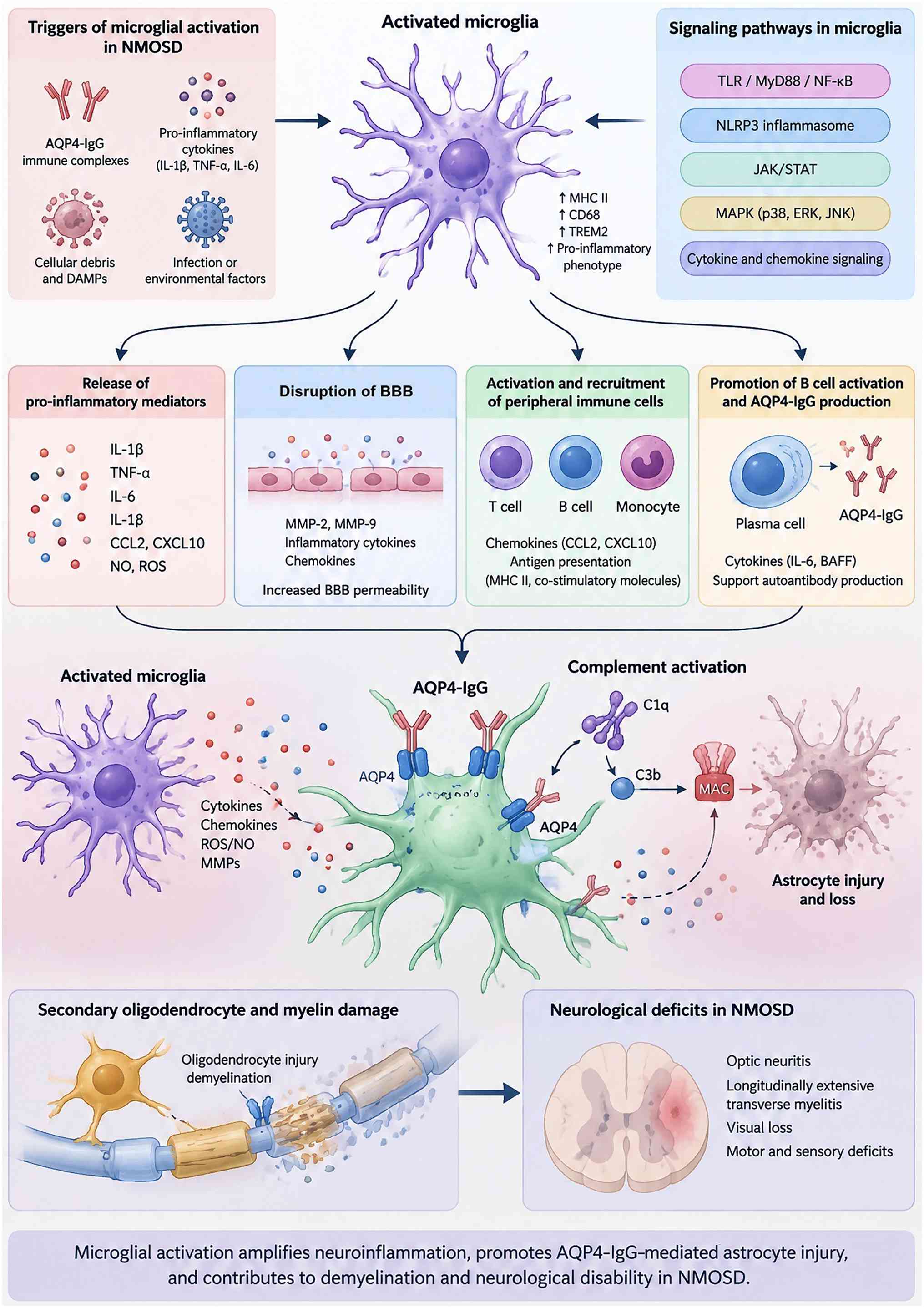
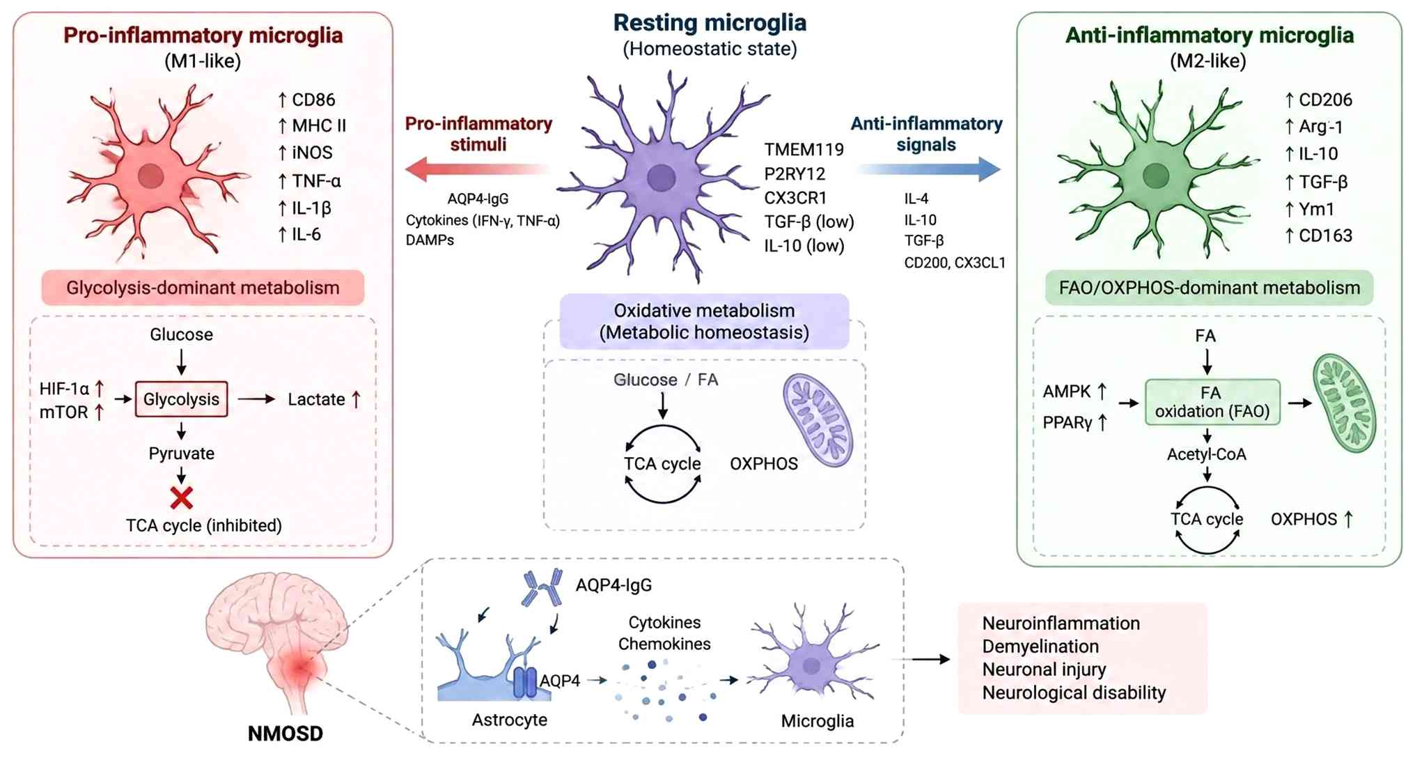
(Fig. 1). Astrocytic injury
releases damage-associated molecular patterns (DAMPs), including
ATP, high mobility group B1 (HMGB1) and mitochondrial DNA, which
activate microglial pattern recognition receptors and NF-κB
signaling (5,6). The mechanisms underlying the
initial production of AQP4-IgG are incompletely understood, with
genetic and environmental factors potentially contributing to its
development. Subsequent AQP4-IgG attacks involve complement
activation, IL-6 signaling and BBB disruption (7).
Microglia are not primary targets of AQP4-IgG but
contribute to the amplification of astrocyte-initiated injury
(Fig. 1; Table I) (8-11). Complement signals, Fc receptor
engagement and DAMPs stimulate microglia to release TNF-α, IL-1β,
IL-6, chemokines, reactive oxygen species (ROS) and MMPs. These
mediators further compromise the BBB, promote peripheral immune
cell recruitment and contribute to oligodendrocyte and axonal
injury. Activated microglia also upregulate major
histocompatibility complex class II and costimulatory molecules,
potentially sustaining autoreactive T cell responses through
antigen presentation (12).
Serum from patients with NMOSD impairs astrocytic
glutamate uptake and glutamine synthetase activity, contributing to
secondary oligodendrocyte injury (13). Under physiological conditions,
astrocytes and microglia exchange trophic and immunoregulatory
signals that maintain cell homeostasis (14). In NMOSD, injured astrocytes
release complement fragments, ATP, DAMPs and cytokines that
activate microglia, whereas microglial inflammatory mediators
exacerbate astrocyte injury. This reciprocal amplification loop
promotes persistent neuroinflammation, demyelination and axonal
damage (15).
The present review aimed to examine how complement,
IL, IFN and metabolic pathways shape microglial responses in NMOSD
and how these responses may be modulated to limit inflammation and
support tissue repair. The present study aimed to distinguish
mechanistic opportunities from the experimental and translational
limitations that constrain the development of microglia-targeted
therapies.
As the resident immune cells of the CNS, microglia
provide continuous surveillance of the brain through dynamic
interactions with neurons and other cell components. Upon detecting
pathogens or tissue injury, microglia respond by phagocytosing
cellular debris and releasing cytokines and chemokines, thereby
enabling timely recognition and containment of threats within the
CNS (16). Microglia are key for
synaptic maintenance and remodeling. During neurodevelopment, they
mediate synaptic pruning, facilitate synaptogenesis and myelination
and contribute to the maturation and synchronization of neuronal
activity (17). In addition,
microglia support neuronal survival and repair through the release
of neurotrophic factors (NFs), such as brain-derived NF (BDNF)
(18).
Rather than existing as a simple pro-inflammatory or
reparative binary, microglia occupy a continuous spectrum of
functional states (Table II)
(19-27). These states encompass
homeostatic, inflammatory, immunoregulatory and disease-associated
microglial (DAM) phenotypes and are shaped by local signals and
disease stage (Fig. 2) (28). In AQP4-IgG-driven pathology,
complement, FcγR, NF-κB and STAT1 signaling can amplify tissue
injury (12), whereas triggering
receptor expressed on myeloid cells (TREM)2, PPARγ, STAT6 and Nrf2
pathways may promote phagocytosis, trophic support and barrier
repair (29-31). Complement fragments, glutamate,
metabolic substrates, iron, myelin debris and apoptotic cell
signals further influence microglial states across lesion cores,
peri-lesional regions and distinct stages of injury. Accordingly,
therapeutic strategies should aim to restrain detrimental
microglial responses while preserving their reparative function,
rather than treating microglial activation as uniformly harmful.
The timing and cell specificity of such interventions are therefore
key determinants of their therapeutic potential.
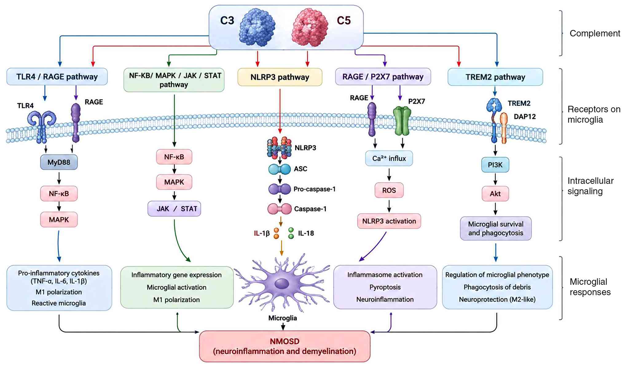
Complement C3 is a key component of the complement
cascade and a convergence point for the classical, alternative and
lectin pathways in NMOSD, with key effects on microglial activation
and function (Fig. 2) (48). Dysregulated C3 expression
sustains microglial activation and contributes to synaptic
dysfunction and cognitive impairment. Systemic inflammation, for
example, promotes microglial activation through increased C3
expression, leading to synaptic loss and cognitive deficit
(8). In NMOSD, excessive C3
activation is associated with optic nerve and spinal cord injury,
whereas C3 inhibition decreases complement deposition and
demyelination and improves motor function in experimental models
(49,50).
Under physiological conditions, astrocytes express
little or no C3. In pathological states, NMO-IgG induces robust C3
production by astrocytes (51),
followed by its cleavage into C3a and C3b. C3a binds C3aR and
promotes NLRP3 inflammasome assembly, facilitating the maturation
and release of IL-1β and IL-18 and enhancing microglial
inflammatory responses (Fig. 2)
(8). CDC and ADCC are also
established mechanisms of astrocyte injury in NMOSD (52,53). Complement activation promotes
macrophage recruitment, while macrophage-derived cytokines and ROS
injure neurons and oligodendrocytes, contributing to astrocyte
damage and secondary demyelination (54).
AQP4-IgG promotes AQP4 internalization and astrocyte
injury through CDC and ADCC (63). Activated microglia secrete C1q,
contributing to local neuronal and oligodendrocyte injury (64). Astrocyte-derived ATP, HMGB1 and
S100B activate microglial receptors, including P2Y12 and
TLR4/receptor for advanced glycation end products (RAGE), whereas
microglial IL-1α, TNF and ROS can further impair astrocyte function
(65). Complement signaling also
regulates microglial processing of myelin debris (66). Excessive complement activation
may compromise effective debris clearance and sustain
demyelination, potentially via NF-κB signaling (66). Thus, complement-dependent
phagocytosis should be considered in the context of both tissue
injury and repair.
Several key questions remain unresolved, including
the interplay between C3a and C5a signaling, their relevance to
AQP4-IgG-seronegative disease and their potential utility as
biomarkers. Longitudinal studies are needed to define their
association with relapse and treatment response, while
pathway-selective interventions should distinguish detrimental
inflammatory signaling from the protective functions of
complement.
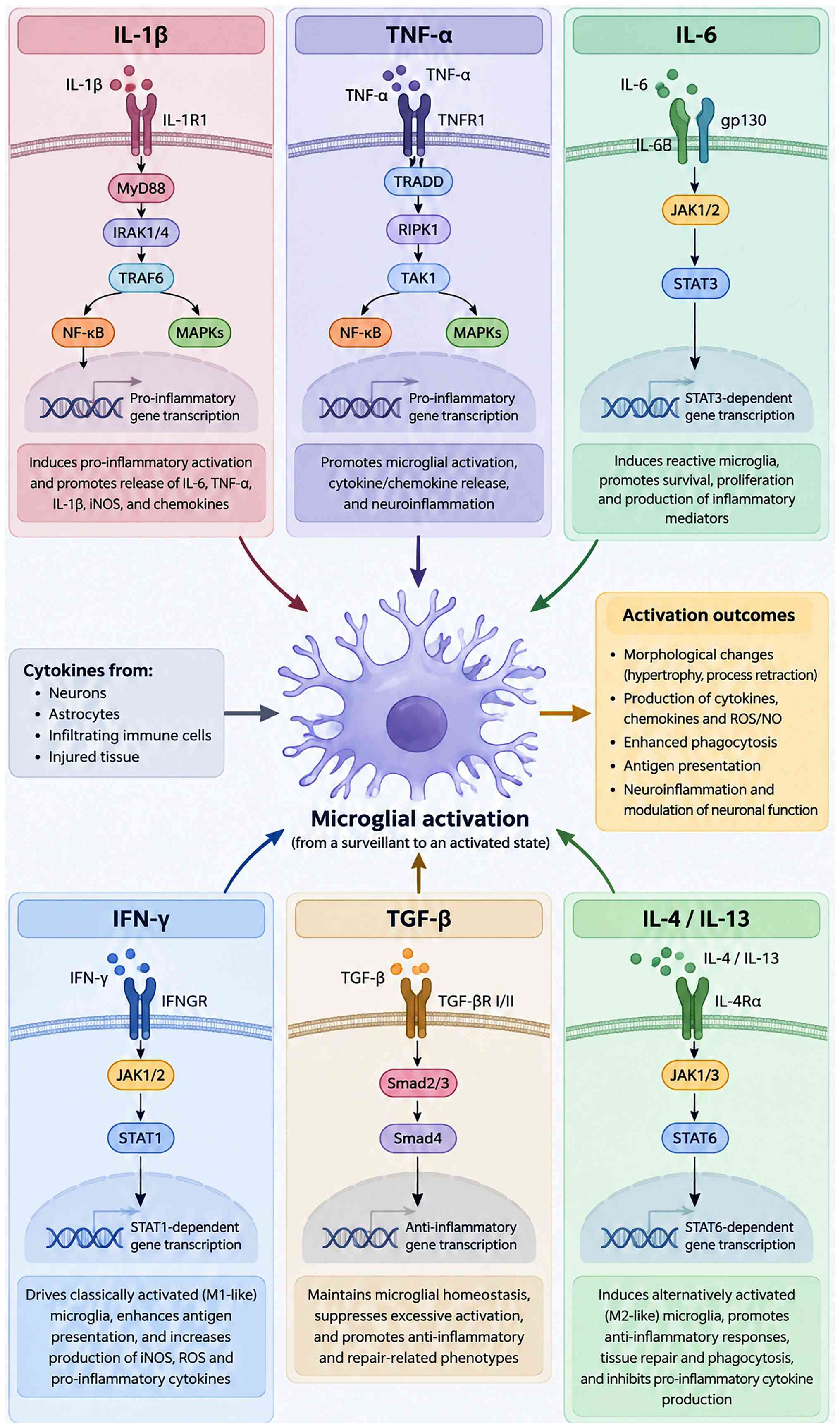
ILs are multifunctional cytokines that regulate
immune responses and contribute to microglial activation and
neuroinflammation (Fig. 3). IL-6
is involved in the initiation and regulation of NMOSD and can
modulate microglial activation (67). IL-17A activates microglia and
astrocytes, inducing the production of inflammatory mediators and
exacerbating neuronal injury and dysfunction (68). The IL-20 family regulates BBB
integrity, oxidative stress and autophagy partly through effects on
microglial activation, linking these processes with
neuroinflammatory and neurodegenerative pathology (69). IL-2 exhibits region- and cell
type-specific changes across neurological diseases, including
multiple sclerosis (MS), Alzheimer's disease (AD), ischemic stroke
and encephalitis (70). By
contrast, IL-10 is a key anti-inflammatory regulator that
suppresses glial activation and protects against
inflammation-associated neuronal degeneration (71). Collectively, these findings
underscore the complex and context-dependent functions of ILs in
NMOSD.
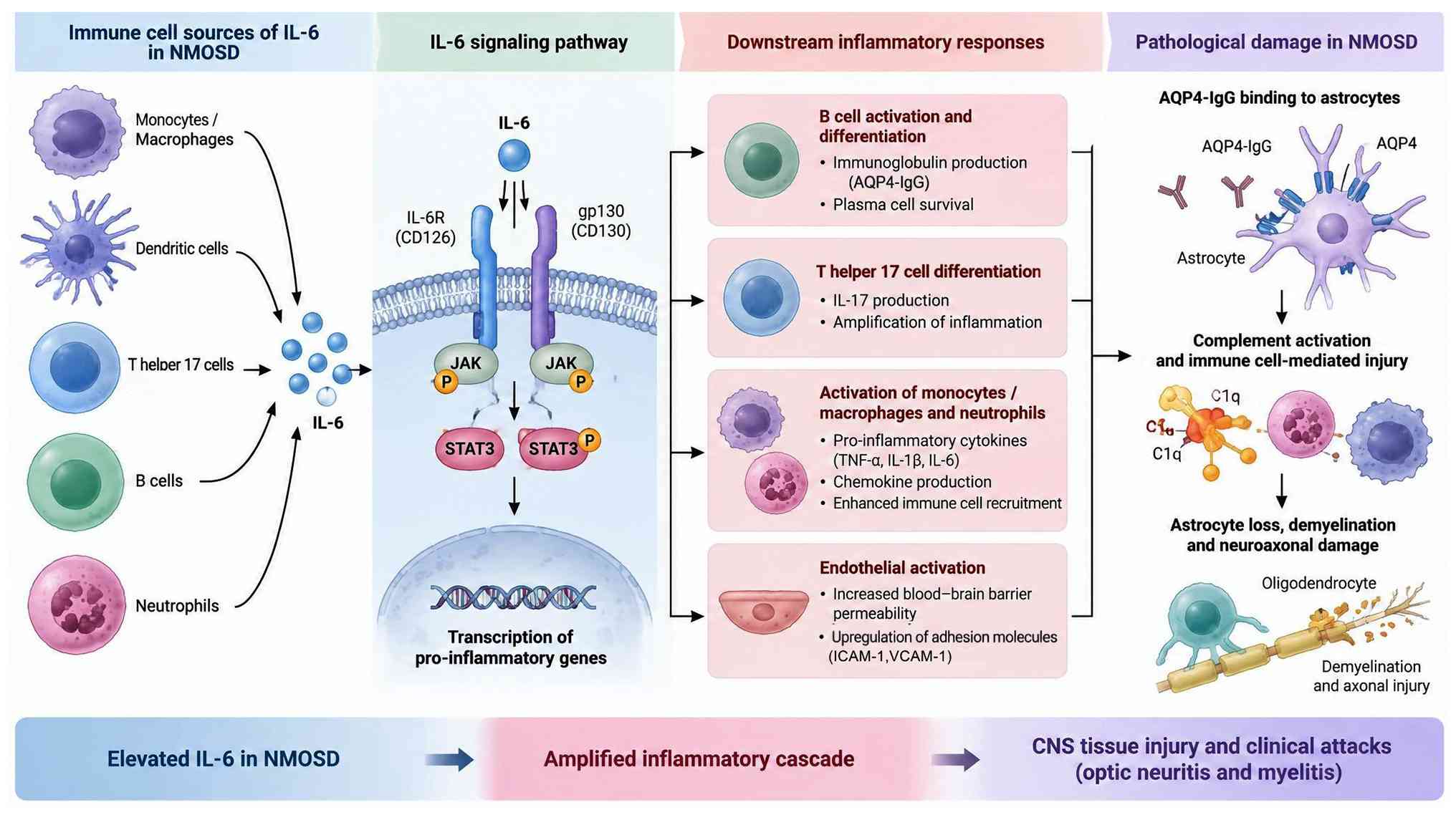
IL-6 is produced by microglia, lymphocytes,
macrophages, fibroblasts and endothelial cells in response to
infection, tissue damage and inflammatory stimuli (72) (Fig. 4). Its effects on immune cell
survival, inflammatory signaling and barrier function make it
particularly relevant to NMOSD.
IL-6 is produced by microglia, lymphocytes,
macrophages, fibroblasts and endothelial cells in response to
infection, tissue injury and inflammatory stimuli (72) (Fig. 4). Its effects on plasmablast
survival, stimulates AQP4 antibody production and perpetuates an
autoimmune cycle make it particularly relevant to NMOSD (73). IL-6 is a regulator of
neuroinflammation, with key effects on microglial activation and
function (74). In microglia,
IL-6 signals via the JAK/STAT pathway to regulate proliferation and
inflammatory responses (74).
IL-6 receptor engagement activates downstream signaling pathways
that enhance microglial migration and phagocytic activity (75) (Fig. 4).
Elevated IL-6 levels during NMOSD relapses suggest
its potential use as a biomarker of disease activity (76). IL-6 may promote disease
progression by modulating other cytokines and immune cell
populations (76). For example,
IL-6 and IL-17A impair BBB integrity, thereby exacerbating CNS
inflammation (77). Increasing
evidence implicates dysregulated microglial activation and IL-6
production in NMOSD pathogenesis (78-80). Elevated serum and cerebrospinal
fluid (CSF) IL-6 levels during relapse are associated with disease
severity and brain atrophy, suggesting that IL-6 may serve not only
as a marker of disease activity but also as a mediator of
pathological progression (81).
IL-6 promotes plasmablast survival and AQP4 antibody production,
thereby sustaining the autoimmune response in NMOSD (82). It can also work with TNF-α to
increase the expression of endothelial adhesion molecules,
including intercellular adhesion molecule (ICAM)-1, facilitating
neutrophil infiltration across the BBB (83). This compromises BBB integrity
(84), while neutrophil-derived
MMP-9 disrupts myelin structure (85). In addition, IL-6 inhibits nuclear
translocation of oligodendrocyte transcription factor 2 in
oligodendrocyte precursor cells (OPCs), impairing their
differentiation into mature oligodendrocytes and decreasing myelin
basic protein (MBP) synthesis (86,87). IL-6 also activates suppressor of
cytokine signaling 3 in OPCs (88), interfering with insulin-like
growth factor-1 (IGF-1)-mediated repair signaling (89) and restricting OPC differentiation
and remyelination. Because OPC proliferation and differentiation
are key for myelin regeneration, impaired microglia-OPC
interactions may further compromise repair in the inflamed CNS
(90). IL-6 is directly involved
in inflammatory signaling (91).
Binding of IL-6 to IL-6R promotes gp130 homodimerization and
formation of the functional receptor complex, leading to activation
of JAK1 and JAK2 and subsequent phosphorylation and activation of
STAT3. Activated STAT3 translocates to the nucleus and regulates
the expression of inflammation-associated genes (Fig. 4) (92). In activated microglia, IL-6
signaling via the IL-6R/gp130 complex induces downstream
pro-inflammatory mediators, including TNF-α and IL-1β, establishing
a positive feedback loop that sustains inflammation and neuronal
injury (93). This pathway
increases microglial expression of inducible nitric oxide synthase
and cyclooxygenase-2, promoting the production of ROS and
prostaglandin E2 and thereby aggravating oxidative stress (93). IL-6 may cooperate with TGF-β to
promote differentiation of CD4+ T cells toward the T
helper (Th)17 lineage, enhancing autoimmune responses, increasing
BBB permeability and intensifying inflammation in NMOSD (94).
Key questions remain regarding the interaction
between IL-6 signaling and AQP4-IgG-mediated injury, its relevance
to seronegative NMOSD and the determinants of treatment response.
Longitudinal comparative studies and biomarker-guided patient
stratification are needed to refine IL-6-targeted therapy and
assess combination approaches for refractory disease.
IL-1β primarily promotes inflammation by activating
immune cells and inducing the release of additional inflammatory
mediators (95). IL-1β
stimulates astrocytes and microglia to produce chemokines, thereby
recruiting peripheral immune cells to sites of inflammation and
amplifying neuronal injury (Fig.
3) (95). IL-1β levels are
notably elevated during the acute phase of inflammatory
demyelinating disease, including myelin oligodendrocyte
glycoprotein antibody-associated disease (MOGAD) and
AQP4-IgG-positive NMOSD (96,97). This increase is particularly
pronounced during acute attacks, suggesting a role for IL-1β in
early disease pathology. CSF IL-1β levels are elevated in NMOSD and
associated with disease severity, supporting its involvement in
NMOSD pathogenesis (98). IL-1β
can act in concert with other inflammatory mediators to reinforce
microglial activation. It promotes the production of
pro-inflammatory cytokines, including TNF-α and IL-6, and
chemokines such as CCL2 and CXCL10, thereby intensifying local
inflammation (99). IL-1β may
promote steroid resistance through activation of T helper 17 cells,
further sustaining inflammatory responses (100). In astrocytes, IL-1β binding to
IL-1 receptor activates NF-κB and MAPK signaling, inducing the
production of CXCL10 and CCL2. These chemokines facilitate the
recruitment of peripheral neutrophils and monocytes to the CNS,
thereby amplifying inflammation and contributing to
IL-1β-associated neuronal and oligodendrocyte injury (101). This cytokine-driven cascade may
promote NMOSD pathology. Beyond its inflammatory effects, IL-1β
influences tissue repair and cellular differentiation (102). Increased BBB permeability
facilitates peripheral immune cell infiltration, while experimental
evidence linking IL-1β with caspase-1 activation, pyroptosis and
synaptic dysfunction suggests additional mechanisms of neuronal
injury (103). The relative
contribution of these pathways to human NMOSD remains to be
established. IL-1β may contribute to NMOSD by regulating immune
cell activation and trafficking. It enhances T and other immune
cell activation (104) and
increases the expression of endothelial adhesion molecules,
including vascular CAM-1 and ICAM-1, facilitating transendothelial
immune cell migration and compromising BBB integrity (105). IL-1β may promote the production
of AQP4-targeting autoantibodies, potentially reinforcing the
autoimmune response characteristic of NMOSD. IL-1β can inhibit OPC
proliferation and differentiation through the
microRNA-202-3p/β-catenin/glioma-associated oncogene 1 pathway,
decreasing the expression of myelin proteins such as MBP and
impairing remyelination (106).
In neurons, IL-1β induces calcium overload and mitochondrial ROS
production, contributing to axonal degeneration (107). It may also act with TNF-α to
increase neutrophil-derived MMP-9, disrupting myelin integrity and
promoting neuronal injury (108). Evidence supporting a role for
IL-1β in NMOSD is predominantly preclinical or based on
cross-sectional observations (96). Its association with
AQP4-IgG-mediated astrocyte injury and seronegative disease
requires further clarification. Longitudinal biomarker studies and
controlled evaluations of pathway-specific inhibition are needed to
determine whether IL-1β serves as a reliable marker of disease
activity or a viable therapeutic target.
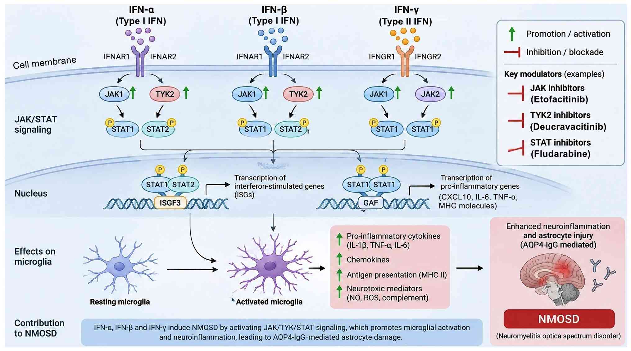
IFNs regulate antimicrobial defense, antigen
presentation and immune cell activation (109,110). Their effects in NMOSD vary
according to IFN subtype, cell context and experimental model, with
potential roles in either inflammatory amplification or maintenance
of immune tolerance (111)
(Fig. 5).
IFN-β, a type I IFN, is key for antiviral defense
and is produced by various resident CNS cells, including microglia,
astrocytes, neurons and endothelial cells (28). Microglia are responsive to type I
IFN signaling in the CNS (112). Type I IFNs have been
investigated in immune-mediated demyelinating disease, particularly
MS (113), in which IFN-β
exerts therapeutic effects by modulating immune cell functions
(108). However, excessive or
context-dependent type I IFN signaling may produce adverse effects.
In a mouse model of NMOSD lesions, IFN-β exacerbates NMO-like
pathological changes, including increased AQP4 and glial fibrillary
acidic protein expression and expansion of the CD11c+
microglial subset (114).
Transcriptomic analysis have identified notable type I IFN
signatures in affected tissue and microglial populations (115). Microglial depletion attenuates
these pathological changes without substantially altering IFN-β
expression, supporting a key role for microglia as downstream
responders to type I IFN signaling (116). Type I IFN-responsive microglia
may contribute to the inflammatory milieu and aggravate CNS injury
(116). In NMOSD, type I IFN
may exacerbate disease by suppressing Th1 cell function (117).
The aforementioned findings support further
investigation of the disease-specific IFN-microglia axis.
Single-cell and spatial transcriptomic approaches may identify
IFN-responsive microglial populations, whereas epigenetic analyses
may clarify persistent inflammatory programming. CNS-penetrant JAK
inhibitors and IL-6 antagonists are potential mechanistic
interventions, although proposed CSF biomarkers, including IL-6 and
BAFF, require independent validation.
IFN-β engages microglia through the cyclic GMP-AMP
synthase/stimulator of interferon genes/type I interferon (IFN-I)
signaling pathway and plays a paradoxical role in NMOSD (135), in contrast to its therapeutic
effect on multiple sclerosis. Activated microglia-derived IFN-β
promotes expansion and infiltration of AQP4-specific autoreactive T
cells, exacerbates astrocyte injury and inflammatory pathology in
NMOSD (133). Nevertheless, the
functional heterogeneity of microglial subsets, species differences
between rodent and human systems and stage-dependent effects of IFN
signaling must be resolved before pathway modulation can be
translated into NMOSD therapy.
IFN-γ is the sole member of the type II IFN family
and is a notable regulator of inflammatory responses. Dysregulated
IFN-γ signaling is implicated in several autoinflammatory and
autoimmune diseases such as MS and NMOSD, reflecting its dual
immunostimulatory and immunoregulatory functions (136). Primarily produced by Th1,
CD8+ T and natural killer cells, IFN-γ promotes the
polarization of microglia toward an inflammatory state (137). In NMOSD, IFN-γ is typically
considered pathogenic due to its role in enhancing inflammatory
responses and activating immune pathways targeting the CNS
(Fig. 5) (108). Th1-derived IFN-γ promotes
inflammatory microglial polarization via JAK1/STAT1 signaling,
suppresses reparative markers such as arginase-1 and CD206 and
increases the expression of pro-inflammatory cytokines and
chemokines, thereby aggravating NMOSD-associated pathology
(138). IFN-γ can activate the
NLRP3 inflammasome through STAT1, intensifying microglial
inflammatory responses (139).
In addition, IFN-γ promotes CD8+ T cell and monocyte
recruitment through the CXCL10/CXCR3 pathway, facilitating
inflammatory cell accumulation within lesions and potentially
exacerbating optic nerve and spinal cord injury (140). IFN-γ may induce Fas expression
on oligodendrocytes, enhancing FasL-mediated apoptotic signaling
(141), while activating NADPH
oxidase 2 in microglia and increasing superoxide production, which
contribute to myelin injury (142). IFN-γ-associated BBB disruption
may facilitate the entry of autoantibodies and peripheral immune
cells into the CNS, thereby promoting demyelination and neuronal
injury (143).
Recent findings, however, indicate that type I and
II IFNs may exert opposing effects in
AQP4201-220-induced disease (138). In this model, type II IFN
decreases disease incidence and severity, challenging the
predominant view of IFN-γ as pathogenic (144). Further studies suggest IFN-γ
contributes to immune tolerance toward AQP4, whereas its deficiency
promotes autoimmune inflammation (108,145). Decreased IFN-γ signaling may
increase IL-6 expression and enhance Th17 responses, thereby
facilitating autoimmune activation (136). The effects of IFN-γ are
associated with peripheral immune-tolerance mechanisms that
regulate autoreactive B and T cell activation (146). These observations suggest IFN-γ
may exert protective effects under specific immunological
conditions and highlight the context dependence of IFN signaling in
NMOSD.
The divergent effects of IFN-γ underscore the
limitations of extrapolating findings across experimental models
and disease stages. Regional microglial heterogeneity, species
differences, interactions with other cytokines and limited access
to human lesion tissue complicate interpretation. Spatial profiling
and functional studies are needed to distinguish inflammatory
effects within CNS lesions from peripheral mechanisms by which
IFN-γ contributes to AQP4-specific immune tolerance.
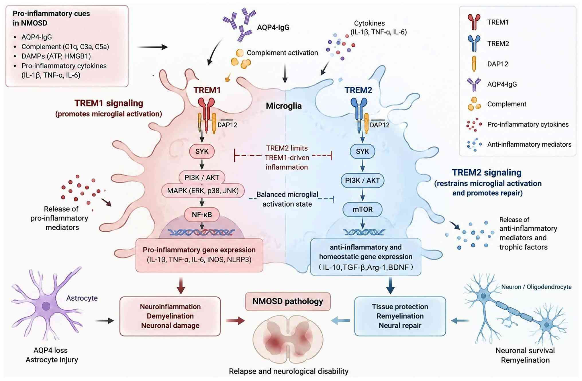
TREM family regulates inflammation, immune response
and tissue homeostasis. TREM1 and TREM2 are of particular interest
because they link myeloid cell activation with inflammatory
signaling, phagocytosis and metabolic adaptation (151).
TREM1 signals through the adaptor protein tyrosine
kinase-binding protein 12 (DAP12) to activate spleen tyrosine
kinase (SYK) and NF-κB, promoting the release of IL-1β and TNF-α
(152) (Fig. 6). TREM1 contributes to
inflammatory signaling and macrophage polarization in numerous
types of inflammatory disorder such as NMOSD, MS, and ankylosing
spondylitis (153). By
contrast, TREM2 recognizes lipid-associated signals and damaged
cell material and supports microglial survival and phagocytosis
(154). Through DAP12 and
PI3K/Akt signaling, TREM2 enhances phagocytic activity and favors
reparative microglial responses (Fig. 6) (155). Microglia in these states
produce IL-10 and TGF-β, which restrain inflammation and support
neuronal survival (156).
Recognition of myelin debris activates SYK/PI3K-dependent lysosomal
programs, facilitating debris processing in demyelinated regions
and limiting the accumulation of potentially toxic lipid material
(157). TREM2 may suppress
TLR4/NF-κB signaling and IL-1β production, linking debris clearance
with control of excessive inflammation (158). These functions are relevant to
tissue repair because effective phagocytosis requires not only
recognition and uptake of damaged material but also the metabolic
capacity to process its lipid content (159). However, TREM2 expression alone
does not define a uniform reparative state and its functional
effects depend on lesion stage and the surrounding inflammatory
milieu.
TREM2 is a transmembrane receptor predominantly
expressed by microglia and serves as an important regulator of cell
metabolism and inflammatory responses through extracellular lipid
sensing (160). In AD, TREM2
signaling enhances the metabolic adaptability and survival of
microglia, thereby shaping the response to amyloid-β plaques
(161). TREM2 is implicated in
other neurodegenerative disease; in AD and MS, it contributes to
the balance between protective and detrimental inflammatory
responses (162). Its effects
extend to lipid metabolism, through which TREM2 maintains cell
energy homeostasis and functional adaptation (163). TREM2 deficiency causes
metabolic abnormality characterized by autophagosome accumulation
and impaired mitochondrial function (20). TREM2 regulates mTOR signaling,
thereby influencing microglial metabolism and autophagy, processes
that are essential for cellular energy production and biosynthetic
activity (164). In NMOSD,
TREM2 deficiency impairs microglial response and exacerbates
demyelinating injury (165).
Acute TREM2 inhibition decreases the population of v-maf avian
musculoaponeurotic fibrosarcoma oncogene homolog B-expressing
microglia and impairs remyelination (166). Loss of this microglial
population is associated with decreased oligodendrocyte generation,
an essential process for myelin restoration (167). During NMOSD pathology, TREM2
expression is associated with microglial aggregation, proliferation
and phagocytic activity (157).
TREM2 deficiency aggravates tissue injury and neurological
dysfunction while decreasing microglial accumulation and impairing
myelin debris clearance (157),
supporting a neuroprotective role for TREM2 during demyelination.
TREM2-activated microglia promote OPC differentiation into mature
oligodendrocytes through the release of platelet-derived growth
factor BB, while enhancing the synthesis and secretion of myelin
proteins such as proteolipid protein and MBP, thereby facilitating
remyelination (168).
TREM2-dependent debris clearance and secretion of NFs, including
BDNF and glial cell line-derived neurotrophic factor (GDNF), may
also contribute to extracellular matrix remodeling, reduce glial
scar formation and provide a permissive environment for OPC
migration and remyelination (169). TREM2 is associated with
microenvironmental changes in NMOSD. Elevated CSF levels of soluble
TREM2 (sTREM2) are associated with neuronal injury and other
neuroinflammatory markers (neurofilament light chain, intercellular
adhesion molecule 1 and IFN-γ) in patients with NMOSD (170). sTREM2 may influence microglial
dysfunction through NF-κB signaling, potentially contributing to
disease progression (31,171).
In animal models of NMOSD, TREM2 deficiency decreases microglial
phagocytic activity and myelin debris degradation, supporting its
role in maintaining microglial function during demyelination
(165,166,172). The interaction between TREM2
and apolipoprotein (Apo)E is key for microglial activation and
lipid handling. A biphasic TREM2 response is associated with
microglial state transitions, raising the possibility that TREM2
may support inflammatory responses during early inflammation while
promoting repair and inflammation resolution at later stages
(173). TREM1 may similarly
regulate microglial activity through interconnected inflammatory
pathways, but its specific contribution to NMOSD is poorly defined.
TREM2 signaling in AQP4-IgG-mediated astrocyte injury and
seronegative NMOSD is incompletely characterized. Larger
longitudinal cohorts are needed to determine associations between
TREM2 expression, sTREM2 levels, genetic variation, disease
activity and prognosis. Whether TREM2 activation or inhibition is
therapeutically beneficial may depend on disease stage and the
specific microglial function being targeted.
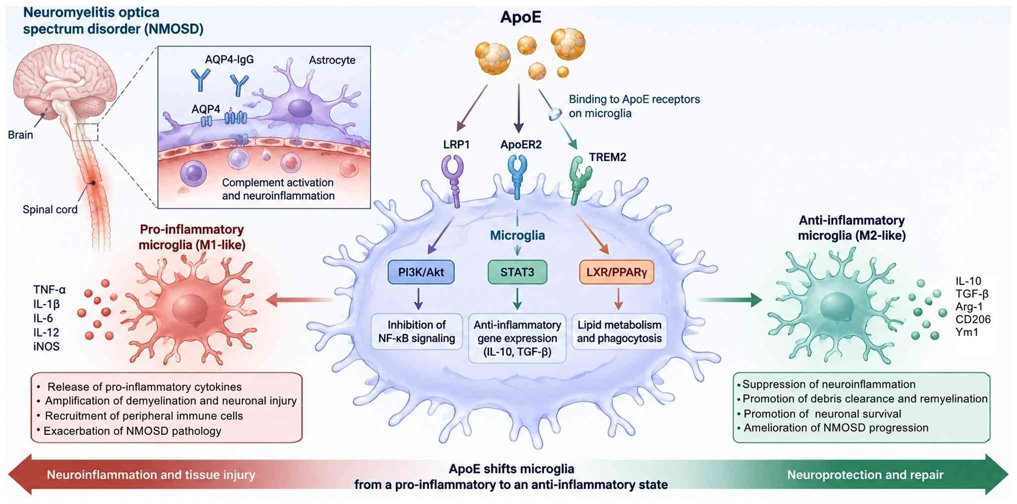
ApoE is a major regulator of lipid and cholesterol
transport in the CNS. Its three notable alleles, ε2, ε3 and ε4,
differ in their effects on lipid handling and disease
susceptibility, with ε4 associated with late-onset AD (174,175). These established functions in
lipid metabolism and neuroinflammation provide a rationale for
investigating ApoE in NMOSD, although genetic associations
established in AD cannot be directly extrapolated to optic-spinal
autoimmunity.
The timing and direction of APOE/TREM2 signaling in
NMOSD are uncertain. Independent human cohorts should examine
associations between ApoE genetic variation, pathway activity,
relapse risk and disease severity, including in seronegative NMOSD.
Stage-specific therapeutic strategies require evidence that
modifying lipid handling enhances debris clearance and repair
without sustaining detrimental inflammatory responses.
TREM2/DAP12/Syk signaling connects recognition of damaged material
with phagocytosis, metabolic adaptation and regulation of
complement and inflammatory pathways (Fig. 8) (189). Its protective capacity may be
insufficient under intense AQP4-IgG-driven inflammation (189). AQP4-IgG-triggered complement
cascade and rapid infiltration of neutrophils/eosinophils generate
tissue destruction far exceeding the pathway's physiological
clearance and anti-inflammatory threshold; the highly oxidative
NMOSD lesion microenvironment induces excessive TREM2 ectodomain
shedding, depleting functional membrane TREM2 and abrogating
downstream Syk signaling; and the pathway's myeloid-restricted
regulatory scope cannot directly counteract the core pathological
drivers of AQP4-IgG-mediated astrocyte cytotoxicity and persistent
autoantibody production. Defining the conditions under which TREM2
signaling supports recovery and determining whether genetic or
metabolic factors modify these effects in NMOSD is key for
evaluating its therapeutic potential.
Metabolic reprogramming is a key determinant of
microglial functional state and has emerged as an important focus
in neuroscience (Fig. 9)
(190,191). Pro-inflammatory microglia
typically exhibit increased glycolytic metabolism, whereas
reparative microglia rely more heavily on oxidative phosphorylation
(OXPHOS) to meet their energetic demands (192). These metabolic states are
coupled to microglial function: Glycolysis provides rapid ATP
production and biosynthetic intermediates to support inflammatory
activation, whereas OXPHOS provides sustained energy for
phagocytosis, tissue repair and homeostatic functions. The mTOR
signaling pathway is a key metabolic regulator that connects
cellular energy status with microglial functional phenotype
(193). Modulation of
mTOR-associated metabolic pathways promotes the transition from
inflammatory toward reparative microglial states and may alleviate
neuroinflammation and neurodegenerative pathology (193). Lipid metabolism is key for CNS
homeostasis and myelin maintenance; its disruption can increase
myelin vulnerability and contribute to neurological dysfunction
(194). In NMOSD, impaired
lipid handling may further compromise myelin integrity and nerve
conduction. Microglial metabolic reprogramming may contribute to
remyelination failure in NMOSD through interconnected alterations
in energy metabolism, inflammatory state persistence and
intercellular signaling (195).
In acute NMOSD lesions, AQP4-IgG-mediated astrocyte injury
activates microglial purinergic receptor P2X 7 (P2X7) and RAGE)
receptors, promoting glycolytic metabolism via NF-κB and
hypoxia-inducible factor (HIF)-1α signaling (196,197). Increased glycolytic activity
and lactate accumulation may impair OPC differentiation and enhance
inflammatory gene expression through lactylation, thereby
reinforcing local inflammation (198). At later stages, mitochondrial
dysfunction and impaired OXPHOS may decrease ATP availability and
compromise the efficient clearance of myelin debris, limiting OPC
maturation (199). Persistent
metabolic imbalance may stabilize a glycolysis-dependent
inflammatory state through epigenetic mechanisms, including histone
lactylation and DNA methylation, restricting the transition toward
an OXPHOS-supported reparative phenotype (200). Metabolic reprogramming may
alter microglia-oligodendrocyte and microglia-astrocyte
communication. Lactate released by glycolytic microglia activates G
protein-coupled receptor 81 on OPCs and inhibits differentiation
through ERK and PI3K/Akt signaling, whereas CXCL10/CXCR3 signaling
between microglia and reactive astrocytes may promote glial scar
formation and restrict OPC migration (201,202). Together, these metabolic and
intercellular alterations may establish a persistent inflammatory
microenvironment that limits remyelination in NMOSD. Direct
characterization of microglial metabolism in NMOSD remains limited,
with evidence derived from MS, AD and spinal cord injury models
(191,203). Current experimental systems
incompletely reproduce AQP4-IgG-mediated astrocytopathy (30,204), while lesion-stage heterogeneity
(202,206), static metabolic measurement
(207), difficulty in
distinguishing resident microglia from infiltrating macrophages
(208) and limited access to
human lesion tissue (209)
constrain mechanistic interpretation. Future studies should
stratify NMOSD by disease phenotype and lesion stage and
distinguish NMOSD from MOGAD rather than assuming shared metabolic
mechanisms (210). Single-cell
and spatial profiling, metabolic-flux analysis, induced pluripotent
stem cell (iPSC)-derived models, and neurovascular co-culture
systems may provide more direct evidence of cell-specific metabolic
changes. Candidate interventions include itaconate derivatives,
HIF-1α modulation, AMPK activation, PPARγ agonism and TREM2
modulation (211). Their
therapeutic potential depends on disease stage, cell specificity
and compatibility with existing immunotherapies, which require
investigation in NMOSD.
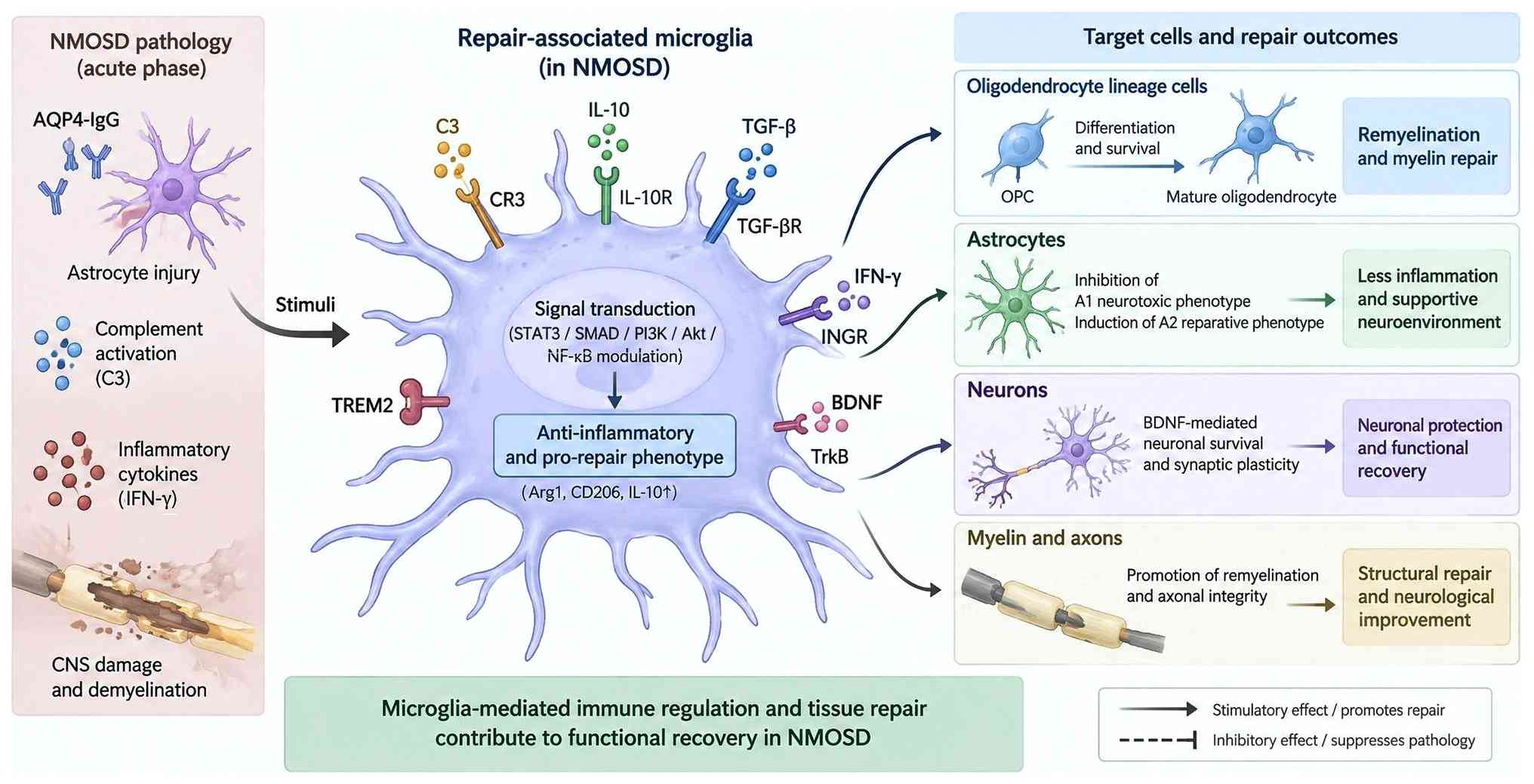
Microglia contribute to tissue repair and recovery
in addition to their roles in acute inflammatory injury (Fig. 10; Table IV) (165,212-223). TREM2-, CR3- and MER
proto-oncogene tyrosine kinase-mediated phagocytosis facilitates
the removal of myelin and cell debris that impede tissue
regeneration (165,224-227). IL-33/growth stimulation
expressed gene 2, TGF-β/Smad3 and PPARγ signaling may promote
homeostatic or reparative microglial states and support oxidative
metabolism, although these phenotypes are heterogeneous and
context-dependent (228-230).
Microglia release trophic mediators, including BDNF, GDNF, IGF-1,
fibroblast growth factor 2, ciliary neurotrophic factor and stromal
cell-derived factor 1(SDF-1), which support neuronal and glial
survival and recovery (231-235). Efficient debris clearance, OPC
recruitment and metabolic support may facilitate oligodendrocyte
maturation and remyelination (235,236). Microglia may contribute to
neurovascular repair by promoting endothelial junction restoration,
supporting pericyte function, limiting MMP-9 activity and
facilitating recovery of astrocytic end-feet (237,238). Through synaptic pruning and
trophic support for axonal growth, microglia may participate in
circuit remodeling and functional recovery (239). In addition, microglia modulate
adaptive immunity through regulatory T cell (Treg) recruitment and
signals that regulate B cell responses (240). These reparative functions are
shaped by interactions with other immune cells: Th1/Th17-derived
cytokines typically favor inflammatory activation (241), whereas Th2/Treg-associated
signals may promote reparative responses (242,243). Direct cell-cell contact
provides an additional mode of reciprocal regulation between
microglia and neighboring cells (244). Therapeutic strategies should
preserve beneficial debris clearance and trophic function while
limiting cytotoxic activation. However, mechanisms established in
other types of CNS injury should not be regarded as definitive
restorative pathways in human NMOSD without disease-specific
validation. Repair-oriented studies face many of the same model and
species limitations as investigations of inflammatory microglial
responses. Phagocytosis and synaptic pruning exert beneficial or
detrimental effects depending on their target, timing and cellular
context (245). Human-relevant
and spatially resolved studies should therefore assess functional
recovery alongside inflammatory and repair-associated markers and
distinguish protective microglial responses from excessive removal
of viable neural elements.
Potential microglia-directed strategies include
colony-stimulating factor 1 receptor inhibition, modulation of
NF-κB/NLRP3 signaling, disruption of C3-C3aR or cytokine-mediated
astrocyte-microglia communication and promotion of reparative
responses through TREM2 or PPARγ (Table V) (246-253). Inhibition of P2X7 or NADPH
oxidase may attenuate inflammatory activation and oxidative stress
(246-248). These approaches must be
balanced against potential impairment of immune surveillance
following microglial depletion, systemic effects of broadly
expressed signaling pathways, increased infection risk and delayed
clearance of cell debris. Most evidence is preclinical and the
therapeutic relevance of these pathways to human NMOSD requires
validation (254,255). Current therapies targeting
complement (C3aR antagonists and anti-C1q), IL-6R or B cells
primarily act on upstream immune mechanisms rather than selectively
modulating microglial response (249-251). Future microglia-directed
adjunctive therapy should demonstrate adequate CNS exposure,
cellular specificity and long-term safety while preserving key
phagocytic and reparative functions. Candidate monitoring
approaches, including 18 kDa translocator protein-positron emission
tomography (256,257) and sTREM2, require validation
because their signals do not reliably distinguish beneficial from
detrimental microglial states.
Despite increasing interest in microglial function
and immune-regulatory mechanisms in NMOSD (Table VI) (79,258-260), several limitations remain in
disease modeling, cell resolution, functional assessment and
clinical translation. Rodent microglia differ notably from their
human counterparts, while passive transfer and active immunization
models do not fully reproduce the chronic, relapsing features of
NMOSD (Table VII) (12,99,188,261-266). The conventional M1/M2 framework
oversimplifies microglial heterogeneity and fails to capture
dynamic interactions with astrocytes, neurons and peripheral immune
cells. MRI and PET provide valuable information at the tissue level
but lack sufficient spatial resolution to resolve individual cell
responses, whereas single-cell sequencing may under-represent rare
populations or low-abundance transcripts. Shared molecular pathways
between resident microglia and infiltrating myeloid cells
complicate cell attribution and target specificity. Reliable
biomarkers that reflect microglial function and treatment response
are limited, and available human tissue samples typically represent
advanced or end-stage lesions. These constraints restrict causal
interpretation and indicate that decreased inflammatory markers
alone cannot establish neuroprotection or tissue repair.
Future studies should integrate human iPSC-derived
microglia, neurovascular co-culture systems and improved chronic or
relapsing models with single-cell and spatial multi-omics. Such
approaches may resolve microglial subsets, characterize their
interactions with neighboring cells and define the temporal
transition from inflammatory activation to tissue repair.
Longitudinal imaging and functional assays should be combined with
patient-derived samples to validate candidate biomarkers rather
than infer microglial activity from non-specific inflammatory
signals. Translational studies should prioritize selective
intervention with adequate CNS exposure and assess outcomes that
capture both relapse control and tissue recovery. Human tissue
biobanks, standardized experimental protocols and collaborative
data sharing may improve reproducibility and enable validation
across independent cohorts. Metabolic interventions are promising,
but direct evidence is needed to establish whether altering
microglial energy metabolism can restore remyelination in
NMOSD.
Complement- and IL-6R-directed therapy underscore
the therapeutic relevance of targeting pathways involved in NMOSD
pathogenesis. Microglia-directed approaches, including modulation
of cytokine signaling and TREM2-dependent phagocytosis, are
primarily experimental. Their development should complement control
of pathogenic autoimmunity while preserving endogenous mechanisms
of tissue repair.
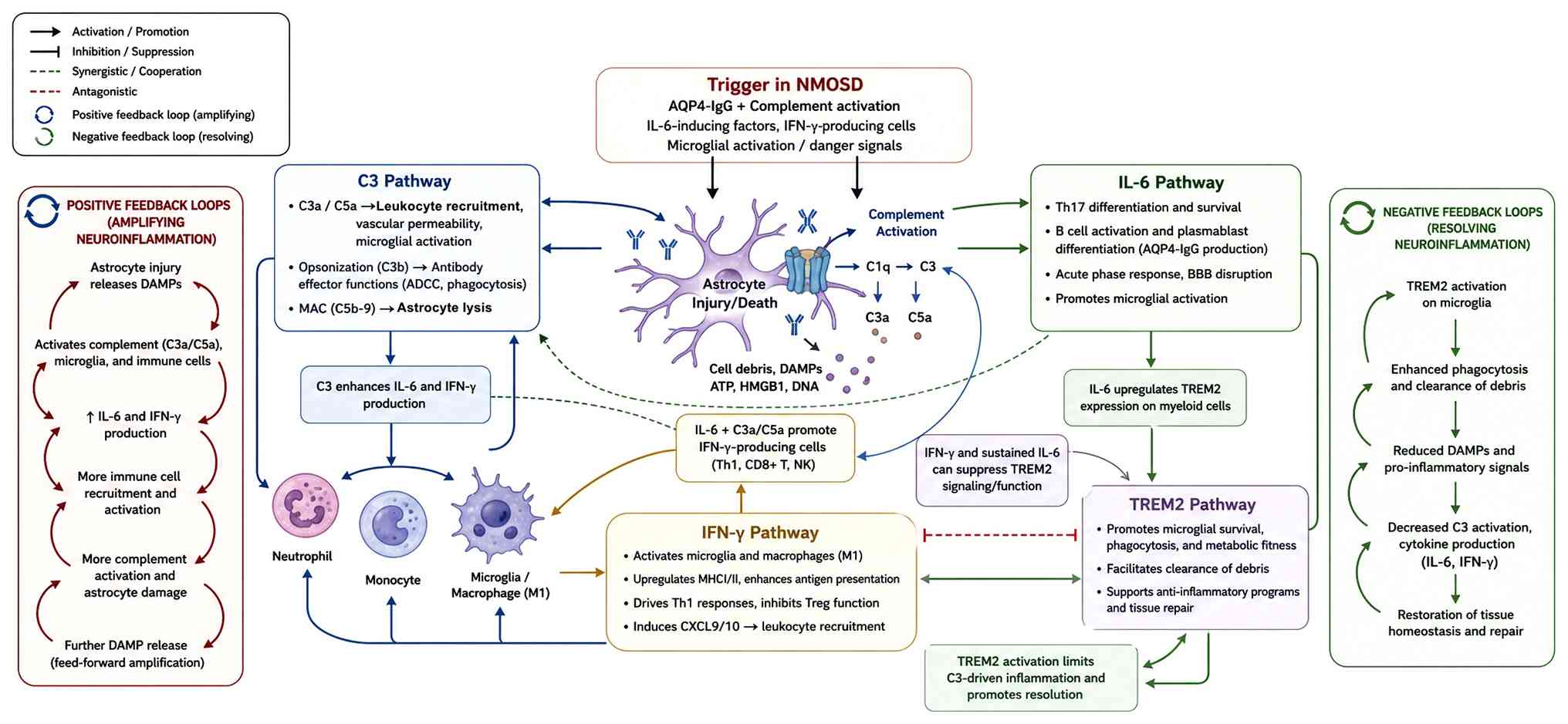
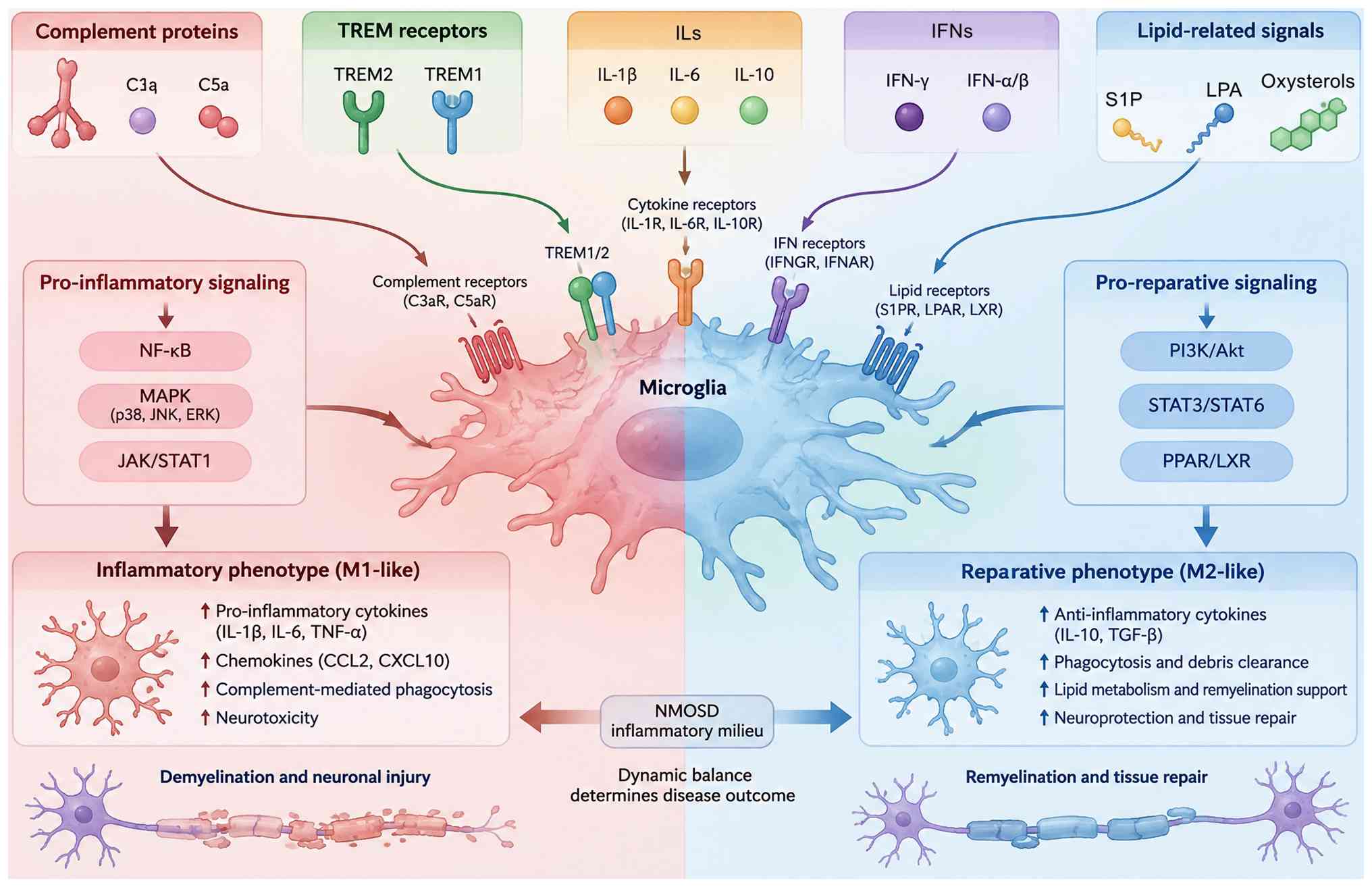
Complement proteins, ILs, IFNs and lipid-associated
signals shape inflammatory and reparative microglial function
(Fig. 11). IL-6 and IL-1β can
amplify inflammatory responses, whereas IFN effects vary with
disease context; the association between IFN-β and NMOSD
exacerbation cautions against extrapolating its therapeutic effects
in MS. C3a/C5a signaling and C5b-9-mediated astrocyte injury
provide links between complement activation and microglial
response, whereas TREM2-associated lipid handling and phagocytosis
may contribute to debris clearance and recovery. These pathways
provide plausible therapeutic targets, but their stage-dependent
effects and relevance to human NMOSD require validation.
Defining the temporal and molecular determinants of
microglial transition between injury-promoting and reparative
states may reveal biomarkers and therapeutic opportunities.
Progress depends on NMOSD-specific experimental models,
longitudinal human evidence and interventions that selectively
limit detrimental inflammation while preserving the phagocytic,
trophic and metabolic functions required for CNS repair.
Not applicable.
YZ, LMZ and MWL performed the literature review and
drafted the manuscript. JBH and YZ edited the manuscript. Data
authentication is not applicable. All authors have read and
approved the final manuscript.
Not applicable.
Not applicable.
The authors declare that they have no competing
interests.
Not applicable.
The present study was supported by the National Natural Science
Foundation of China (grant no. 32560188), Yunnan Revitalization
Talent Support Program (grant no. XDYC-QNRC-2022-0328), 535 Talent
Project of First Affiliated Hospital of Kunming Medical University
(grant no. 2025535Q05), Yunnan Health Training Project of High
Level Talents (grant no. D-2024038), Collaborative Innovation
Special Project of Dali University Affiliated Hospital Alliance
(grant no. ZDXM202602), Yunnan Clinical Research Center for
Neurological Disease (grant no. 202505AJ310009) and Yunnan Clinical
Center for Neurological and Cardiovascular Diseases (grant no.
YNLCYXZX2023300077).
|
1
|
Jarius S, Paul F, Weinshenker BG, Levy M,
Kim HJ and Wildemann B: Neuromyelitis optica. Nat Rev Dis Primers.
22:852020. View Article : Google Scholar
|
|
2
|
Yang Y, Li J, Li T, Li Z, Zhuo Z, Han X,
Duan Y, Cao G, Zheng F, Tian D, et al: Cerebellar connectome
alterations and associated genetic signatures in multiple sclerosis
and neuromyelitis optica spectrum disorder. J Transl Med.
21:3522023. View Article : Google Scholar : PubMed/NCBI
|
|
3
|
Guo Y, Lennon VA, Parisi JE, Popescu B,
Vasquez C, Pittock SJ, Howe CL and Lucchinetti CF: Spectrum of
sublytic astrocytopathy in neuromyelitis optica. Brain.
145:1379–1390. 2022. View Article : Google Scholar :
|
|
4
|
Sun J, Guo M, Chai L, Xu S, Lizhu Y, Li Y,
Duan Y, Xu X, Lv S, Weng J, et al: Distinct virtual histology of
grey matter atrophy in four neuroinflammatory diseases. Brain.
147:3906–3917. 2024. View Article : Google Scholar : PubMed/NCBI
|
|
5
|
Nakajima A, Yanagimura F, Saji E, Shimizu
H, Toyoshima Y, Yanagawa K, Arakawa M, Hokari M, Yokoseki A,
Wakasugi T, et al: Stage-dependent immunity orchestrates AQP4
antibody-guided NMOSD pathology: a role for netting neutrophils
with resident memory T cells in situ. Acta Neuropathol. 147:762024.
View Article : Google Scholar : PubMed/NCBI
|
|
6
|
Wang X and Bao L: Comparison of ocular
changes in multiple sclerosis and neuromyelitis optica spectrum
disorder patients. Front Neurol. 15:14178142024. View Article : Google Scholar : PubMed/NCBI
|
|
7
|
Xue B, Li J, Xie D, Zhang X, Xia J, Li X
and Lin J: The role of neuroimmunology subspecialty in disease
management of patients with neuromyelitis optica spectrum disorder.
Orphanet J Rare Dis. 21:2892026. View Article : Google Scholar : PubMed/NCBI
|
|
8
|
Peng X, Ju J, Li Z, Liu J, Jia X, Wang J,
Ren J and Gao F: C3/C3aR bridges spinal astrocyte-microglia
crosstalk and accelerates neuroinflammation in morphine-tolerant
rats. CNS Neurosci Ther. 31:e702162025. View Article : Google Scholar : PubMed/NCBI
|
|
9
|
Li T, Qiu T, Jiang F, Cai H and Le W:
Microglia in the crosstalk between peripheral and central nervous
systems in Parkinson's disease. Transl Neurodegener. 14:662025.
View Article : Google Scholar : PubMed/NCBI
|
|
10
|
Sun Y, Wei K, Liao X, Wang J, Gao L and
Pang B: Lipid metabolism in microglia: Emerging mechanisms and
therapeutic opportunities for neurodegenerative diseases (Review).
Int J Mol Med. 56:1392025. View Article : Google Scholar : PubMed/NCBI
|
|
11
|
Fang M, Zhou Y, He K, Lu Y, Tao F and
Huang H: Glucose metabolic reprogramming in microglia: Implications
for neurodegenerative diseases and targeted therapy. Mol Neurobiol.
62:8204–8221. 2025. View Article : Google Scholar : PubMed/NCBI
|
|
12
|
Li W, Liu J, Tan W and Zhou Y: The role
and mechanisms of microglia in neuromyelitis optica spectrum
disorders. Int J Med Sci. 18:3059–3065. 2021. View Article : Google Scholar : PubMed/NCBI
|
|
13
|
Jiang L, Wang Y, Wu QZ, Yu JC, Huang YL,
Xu R, Ni K, Gu XP and Ma ZL: Microglia modulate integrity of myelin
and proliferation of oligodendrocyte precursor cells in murine
model of bone cancer pain. Eur J Pharmacol. 996:1775852025.
View Article : Google Scholar : PubMed/NCBI
|
|
14
|
Fan X, Cao S, Fang Y, Zhu L and Wang X:
Pro-inflammatory microglia exacerbate high-altitude-induced
cognitive impairment by driving lipid droplet accumulation in
astrocytes. Antioxidants (Basel). 14:9182025. View Article : Google Scholar : PubMed/NCBI
|
|
15
|
Jitprapaikulsan J, Fryer JP, Majed M,
Smith CY, Jenkins SM, Cabre P, Hinson SR, Weinshenker BG, Mandrekar
J, Chen JJ, et al: Clinical utility of AQP4-IgG titers and measures
of complement-mediated cell killing in NMOSD. Neurol Neuroimmunol
Neuroinflamm. 7:e7272020. View Article : Google Scholar : PubMed/NCBI
|
|
16
|
Wei S, Li C, Li W, Yuan F, Kong J, Su X,
Huang P, Guo H, Xu J and Sun H: Glial changes and gene expression
in Alzheimer's disease from snRNA-Seq and spatial transcriptomics.
J Alzheimers Dis. 105:646–665. 2025. View Article : Google Scholar : PubMed/NCBI
|
|
17
|
Zorec R, Verkhratsky A and Parpura V: The
role of neuroglia in cognitive longevity. Neurochem Res.
50:2922025. View Article : Google Scholar : PubMed/NCBI
|
|
18
|
Xie M, Wang T, Feng J, Ma D, Feng L and
Hao Y: Roles of microglia in synaptogenesis, synaptic pruning, and
synaptic plasticity in physiological conditions and central nervous
system disorders. Curr Neuropharmacol. 24:2–35. 2026.
|
|
19
|
Guzmán-Ruíz MA, Guerrero Vargas NN,
Ramírez-Carreto RJ, González-Orozco JC, Torres-Hernández BA,
Valle-Rodríguez M, Guevara-Guzmán R and Chavarría A: Microglia in
physiological conditions and the importance of understanding their
homeostatic functions in the arcuate nucleus. Front Immunol.
15:13920772024. View Article : Google Scholar : PubMed/NCBI
|
|
20
|
Baligács N, Albertini G, Borrie SC,
Serneels L, Pridans C, Balusu S and De Strooper B: Homeostatic
microglia initially seed and activated microglia later reshape
amyloid plaques in Alzheimer's disease. Nat Commun. 15:106342024.
View Article : Google Scholar : PubMed/NCBI
|
|
21
|
Felice F, De Falco P, Milani M, Castelli
S, Ragnini-Wilson A, Lazzarino G, D'Ambrosi N, Ciccarone F and
Ciriolo MR: N-acetylaspartate mitigates pro-inflammatory responses
in microglial cells by intersecting lipid metabolism and
acetylation processes. Cell Commun Signal. 22:5642024. View Article : Google Scholar : PubMed/NCBI
|
|
22
|
Stojanović NM, Ranđelović PJ, Simonović M,
Radić M, Todorović S, Corrigan M, Harkin A and Boylan F: Essential
oil constituents as anti-inflammatory and neuroprotective agents:
An insight through microglia modulation. Int J Mol Sci.
25:51682024. View Article : Google Scholar
|
|
23
|
Ajoolabady A, Kim B, Abdulkhaliq AA, Ren
J, Bahijri S, Tuomilehto J, Borai A, Khan J and Pratico D: Dual
role of microglia in neuroinflammation and neurodegenerative
diseases. Neurobiol Dis. 216:1071332025. View Article : Google Scholar : PubMed/NCBI
|
|
24
|
Miyake S, Katsura Y, Baba M,
Tomizawa-Shinohara H, Matsumoto Y and Serizawa K: Anti-IL-6R
antibody treatment changes microglial phenotype in AQP4
peptide-immunized mice, leading to suppression of myelitis
severity. J Neuroimmunol. 405:5786442025. View Article : Google Scholar : PubMed/NCBI
|
|
25
|
Sapana T and Zhuo Z: Biomarkers for
diagnosis and prognosis of myelin oligodendrocyte glycoprotein
antibody-associated disease-review article. Front Immunol.
16:15949602025. View Article : Google Scholar
|
|
26
|
Huang TL, Wang JK, Chang PY, Hsu YR, Lin
CH, Lin KH and Tsai RK: Neuromyelitis optica spectrum disorder:
From basic research to clinical perspectives. Int J Mol Sci.
23:79082022. View Article : Google Scholar : PubMed/NCBI
|
|
27
|
Corbali O and Chitnis T: Pathophysiology
of myelin oligodendrocyte glycoprotein antibody disease. Front
Neurol. 14:11379982023. View Article : Google Scholar : PubMed/NCBI
|
|
28
|
Cao T, Hemati-Gourabi M, Liu Y, Mills AE,
Baur L, Rice EP, Klippel T, Yan Y, Xu X, Fenske WK, et al:
Astrocyte-microglia crosstalk via CSF1 and IFN-β promotes central
nervous system repair. Cell Rep. 45:1174182026. View Article : Google Scholar
|
|
29
|
Yick LW, Chan EYY, Zou W, Yau KX, Kwan
JSC, Lee CY and Chan KH: PPAR-γ activation by pioglitazone inhibits
microglia-mediated neuroinflammation and alleviates disease in
neuromyelitis optica spectrum disorders. Acta Neuropathol Commun.
13:2572025. View Article : Google Scholar
|
|
30
|
Kaneko K, Kuroda H, Matsumoto Y, Sakamoto
N, Yamazaki N, Yamamoto N, Umezawa S, Namatame C, Ono H, Takai Y,
et al: Different complement activation patterns following C5
cleavage in MOGAD and AQP4-IgG+NMOSD. Neurol Neuroimmunol
Neuroinflamm. 11:e2002932024. View Article : Google Scholar : PubMed/NCBI
|
|
31
|
Xu S, Dai W, Wang T, Cao S, Du J, Pang X,
Li Q and Tian Y: Temporal dynamics of APOE and TREM2 expression in
microglial activation of NMOSD mouse models. Mol Neurobiol.
62:13587–13600. 2025. View Article : Google Scholar : PubMed/NCBI
|
|
32
|
Asavapanumas N, Tradtrantip L and Verkman
AS: Targeting the complement system in neuromyelitis optica
spectrum disorder. Expert Opin Biol Ther. 21:1073–1086. 2021.
View Article : Google Scholar : PubMed/NCBI
|
|
33
|
Yang C, Yang L and Liu Y: Soluble
complement complex C5b-9 promotes microglia activation. J
Neuroimmunol. 267:16–19. 2014. View Article : Google Scholar : PubMed/NCBI
|
|
34
|
Ramarao S, Pang Y, Carter K and Bhatt A:
Azithromycin protects oligodendrocyte progenitor cells against
lipopolysaccharide-activated microglia-induced damage. Dev
Neurosci. 44:1–12. 2022. View Article : Google Scholar
|
|
35
|
Liang Z, Damianou A, Vendrell I, Jenkins
E, Lassen FH, Washer SJ, Grigoriou A, Liu G, Yi G, Lou H, et al:
Proximity proteomics reveals UCH-L1 as an essential regulator of
NLRP3-mediated IL-1β production in human macrophages and microglia.
Cell Rep. 43:1141522024. View Article : Google Scholar
|
|
36
|
Wang SS, Jin X, Ma WD, Li WY, Dou MM, Yan
TY, Liu N, Liu C, Zhao XY, Zhang ML, et al: Oxymatrine regulates
microglia to produce IFN-β by activating the STING/TBK1/IRF3
pathway against experimental autoimmune encephalomyelitis. Eur J
Pharmacol. 1009:1783802025. View Article : Google Scholar
|
|
37
|
Lee JH, Han JH, Woo JH and Jou I:
25-Hydroxycholesterol suppress IFN-γ-induced inflammation in
microglia by disrupting lipid raft formation and caveolin-mediated
signaling endosomes. Free Radic Biol Med. 179:252–265. 2022.
View Article : Google Scholar
|
|
38
|
Yeh FL, Hansen DV and Sheng M: TREM2,
microglia, and neurodegenerative diseases. Trends Mol Med.
23:512–533. 2017. View Article : Google Scholar : PubMed/NCBI
|
|
39
|
Chen G, Wang M, Zhang Z, Hong DK, Ahn EH,
Liu X, Kang SS and Ye K: ApoE3 R136S binds to Tau and blocks its
propagation, suppressing neurodegeneration in mice with Alzheimer's
disease. Neuron. 113:719–736.e5. 2025. View Article : Google Scholar : PubMed/NCBI
|
|
40
|
Haney MS, Pálovics R, Munson CN, Long C,
Johansson PK, Yip O, Dong W, Rawat E, West E, Schlachetzki JCM, et
al: APOE4/4 is linked to damaging lipid droplets in Alzheimer's
disease microglia. Nature. 628:154–161. 2024. View Article : Google Scholar : PubMed/NCBI
|
|
41
|
Cacciaguerra L and Flanagan EP: Updates in
NMOSD and MOGAD diagnosis and treatment: A tale of two central
nervous system autoimmune inflammatory disorders. Neurol Clin.
42:77–114. 2024. View Article : Google Scholar
|
|
42
|
Takai Y, Misu T, Suzuki H, Takahashi T,
Okada H, Tanaka S, Okita K, Sasou S, Watanabe M, Namatame C, et al:
Staging of astrocytopathy and complement activation in
neuromyelitis optica spectrum disorders. Brain. 144:2401–2415.
2021. View Article : Google Scholar : PubMed/NCBI
|
|
43
|
Yang J, Ding H, Shuai B and Zhang Y and
Zhang Y: Mechanism and effects of STING-IFN-I pathway on
nociception: A narrative review. Front Mol Neurosci.
15:10812882023. View Article : Google Scholar : PubMed/NCBI
|
|
44
|
Baik SH, Kang S, Lee W, Choi H, Chung S,
Kim JI and Mook-Jung I: A breakdown in metabolic reprogramming
causes microglia dysfunction in Alzheimer's disease. Cell Metab.
30:493–507.e6. 2019. View Article : Google Scholar : PubMed/NCBI
|
|
45
|
Lo MW and Woodruff TM: Complement:
Bridging the innate and adaptive immune systems in sterile
inflammation. J Leukoc Biol. 108:339–351. 2020. View Article : Google Scholar : PubMed/NCBI
|
|
46
|
Soteros BM and Sia GM: Complement and
microglia dependent synapse elimination in brain development. WIREs
Mech Dis. 14:e15452022. View Article : Google Scholar
|
|
47
|
Tillmon H, Soteros BM, Shen L, Cong Q,
Wollet M, General J, Chin H, Lee JB, Carreno FR, Morilak DA, et al:
Complement and microglia activation mediate stress-induced synapse
loss in layer 2/3 of the medial prefrontal cortex in male mice. Nat
Commun. 15:98032024. View Article : Google Scholar : PubMed/NCBI
|
|
48
|
Xu L, Xu H, Chen S, Jiang W, Afridi SK,
Wang Y, Ren X, Zhao Y, Lai S, Qiu X, et al: Inhibition of
complement C3 signaling ameliorates locomotor and visual
dysfunction in autoimmune inflammatory diseases. Mol Ther.
31:2715–2733. 2023. View Article : Google Scholar : PubMed/NCBI
|
|
49
|
Nagano T, Tsuda N, Fujimura K, Ikezawa Y,
Higashi Y and Kimura SH: Prostaglandin E2 increases the
expression of cyclooxygenase-2 in cultured rat microglia. J
Neuroimmunol. 361:5777242021. View Article : Google Scholar
|
|
50
|
Lin L, Wu Y, Hang H, Lu J and Ding Y:
Plasma complement 3 and complement 4 are promising biomarkers for
distinguishing NMOSD from MOGAD and are associated with the
blood-brain-barrier disruption in NMOSD. Front Immunol.
13:8538912022. View Article : Google Scholar : PubMed/NCBI
|
|
51
|
Zhu F, He P, Jiang W, Afridi SK, Xu H,
Alahmad M, Alvin Huang YW, Qiu W, Wang G and Tang C:
Astrocyte-secreted C3 signaling impairs neuronal development and
cognition in autoimmune diseases. Prog Neurobiol. 240:1026542024.
View Article : Google Scholar : PubMed/NCBI
|
|
52
|
Siriratnam P, Huda S, Butzkueven H, van
der Walt A, Jokubaitis V and Monif M: A comprehensive review of the
advances in neuromyelitis optica spectrum disorder. Autoimmun Rev.
22:1034652023. View Article : Google Scholar : PubMed/NCBI
|
|
53
|
Yick LW, Tang CH, Ma OK, Kwan JSC and Chan
KH: Memantine ameliorates motor impairments and pathologies in a
mouse model of neuromyelitis optica spectrum disorders. J
Neuroinflammation. 17:2362020. View Article : Google Scholar : PubMed/NCBI
|
|
54
|
Wang Y, Bian J, Yao M, Du L, Xu Y, Chang
H, Cong H, Wei Y, Xu W, Wang H, et al: Targeting chemoattractant
chemokine (C-C motif) ligand 2 derived from astrocytes is a
promising therapeutic approach in the treatment of neuromyelitis
optica spectrum disorders. Front Immunol. 14:11445322023.
View Article : Google Scholar : PubMed/NCBI
|
|
55
|
Kobayashi S, Kokubun N, Aoki R, Hamaguchi
M, Matsuda H and Suzuki K: Possible role of neutrophils in
astrocyte injury in neuromyelitis optica spectrum disorder. J
Neurol Sci. 438:1202932022. View Article : Google Scholar : PubMed/NCBI
|
|
56
|
Liu J, Shao X, Fan J, Wang Y, Cao Y, Tan
G, Sugimoto K, Li B and Jia Z: Association of plasma sPD-1 and
sPD-L1 with disease status and future relapse in AQP4-IgG (+)
NMOSD. Ann Clin Transl Neurol. 11:436–449. 2024. View Article : Google Scholar :
|
|
57
|
Kalluri SR, Srivastava R, Kenet S, Tanti
GK, Dornmair K, Bennett JL, Misgeld T, Hemmer B, Wyss MT and
Herwerth M: P2R inhibitors prevent antibody-mediated complement
activation in an animal model of neuromyelitis optica: P2R
inhibitors prevent autoantibody injury. Neurotherapeutics.
19:1603–1616. 2022. View Article : Google Scholar : PubMed/NCBI
|
|
58
|
Carvalho K, Schartz ND,
Balderrama-Gutierrez G, Liang HY, Chu SH, Selvan P, Gomez-Arboledas
A, Petrisko TJ, Fonseca MI, Mortazavi A and Tenner AJ: Modulation
of C5a-C5aR1 signaling alters the dynamics of AD progression. J
Neuroinflammation. 19:1782022. View Article : Google Scholar : PubMed/NCBI
|
|
59
|
Al-Sayyar A and Rua R: Microglial C5aR1
defines a pathogenic inflammatory axis driving cerebral edema.
Neuron. 114:378–380. 2026. View Article : Google Scholar : PubMed/NCBI
|
|
60
|
Schartz ND, Liang HY, Carvalho K, Chu SH,
Mendoza-Arvilla A, Petrisko TJ, Gomez-Arboledas A, Mortazavi A and
Tenner AJ: C5aR1 antagonism suppresses inflammatory glial responses
and alters cellular signaling in an Alzheimer's disease mouse
model. Nat Commun. 15:70282024. View Article : Google Scholar : PubMed/NCBI
|
|
61
|
Schmidt CQ and Smith RJH: Protein
therapeutics and their lessons: Expect the unexpected when
inhibiting the multi-protein cascade of the complement system.
Immunol Rev. 313:376–401. 2023. View Article : Google Scholar
|
|
62
|
Yadav MK, Singh SP and Egwuagu CE:
IL-6/IL-12 superfamily of cytokines and regulatory lymphocytes play
critical roles in the etiology and suppression of CNS autoimmune
diseases. Front Immunol. 16:15140802025. View Article : Google Scholar : PubMed/NCBI
|
|
63
|
Zhu DD, Huang YL, Guo SY, Li N, Yang XW,
Sui AR, Wu Q, Zhang Y, Kong Y, Li QF, et al: AQP4 aggravates
cognitive impairment in sepsis-associated encephalopathy through
inhibiting Nav 1.6-mediated astrocyte autophagy. Adv Sci
(Weinh). 10:e22058622023. View Article : Google Scholar
|
|
64
|
Dejanovic B, Wu T, Tsai MC, Graykowski D,
Gandham VD, Rose CM, Bakalarski CE, Ngu H, Wang Y, Pandey S, et al:
Complement C1q-dependent excitatory and inhibitory synapse
elimination by astrocytes and microglia in Alzheimer's disease
mouse models. Nat Aging. 2:837–850. 2022. View Article : Google Scholar
|
|
65
|
Ikegami A, Kato D and Wake H: Microglial
process dynamics depend on astrocyte and synaptic activity. Nagoya
J Med Sci. 85:772–778. 2023.
|
|
66
|
Liu Y, Wu C, Hou Z, Fu X, Yuan L, Sun S,
Zhang H, Yang D, Yao X and Yang J: Pseudoginsenoside-F11
accelerates microglial phagocytosis of myelin debris and attenuates
cerebral ischemic injury through complement receptor 3.
Neuroscience. 426:33–49. 2020. View Article : Google Scholar
|
|
67
|
Zheng MY and Luo LZ: The role of IL-17A in
mediating inflammatory responses and progression of
neurodegenerative diseases. Int J Mol Sci. 26:25052025. View Article : Google Scholar : PubMed/NCBI
|
|
68
|
Goleij P, Amini A, Tabari MAK, Hadipour M,
Rezaee A, Daglia M, Aschner M, Sanaye PM, Kumar AP and Khan H:
Unraveling the role of the IL-20 cytokine family in
neurodegenerative diseases: Mechanisms and therapeutic insights.
Int Immunopharmacol. 152:1143992025. View Article : Google Scholar : PubMed/NCBI
|
|
69
|
Zhang Y, Kong Q, Fan J and Zhao H:
Interleukin-2 and its receptors: Implications and therapeutic
prospects in immune-mediated disorders of central nervous system.
Pharmacol Res. 213:1076582025. View Article : Google Scholar : PubMed/NCBI
|
|
70
|
Porro C, Cianciulli A and Panaro MA: The
regulatory role of IL-10 in neurodegenerative diseases.
Biomolecules. 10:10172020. View Article : Google Scholar : PubMed/NCBI
|
|
71
|
Haramati A, Rechtman A, Zveik O, Haham N,
Brill L and Vaknin-Dembinsky A: IL-6 as a marker for NMOSD disease
activity. J Neuroimmunol. 370:5779252022. View Article : Google Scholar : PubMed/NCBI
|
|
72
|
Setiadi AF, Abbas AR, Jeet S, Wong K,
Bischof A, Peng I, Lee J, Bremer M, Eggers EL, DeVoss J, et al:
IL-17A is associated with the breakdown of the blood-brain barrier
in relapsing-remitting multiple sclerosis. J Neuroimmunol.
332:147–154. 2019. View Article : Google Scholar : PubMed/NCBI
|
|
73
|
Li X and Zhao C: Interleukin-6 in
neuroimmunological disorders: Pathophysiology and therapeutic
advances with satralizumab. Autoimmun Rev. 24:1038262025.
View Article : Google Scholar : PubMed/NCBI
|
|
74
|
Wu C, Ge J, Yang M, Yan Q, Wang Y, Yu H,
Yang H and Zou J: Resveratrol protects human nucleus pulposus cells
from degeneration by blocking IL-6/JAK/STAT3 pathway. Eur J Med
Res. 26:812021. View Article : Google Scholar : PubMed/NCBI
|
|
75
|
Mora P, Laisné M, Bourguignon C, Rouault
P, Jaspard-Vinassa B, Maître M, Gadeau AP, Renault MA, Horng S,
Couffinhal T and Chapouly C: Astrocytic DLL4-NOTCH1 signaling
pathway promotes neuroinflammation via the IL-6-STAT3 axis. J
Neuroinflammation. 21:2582024. View Article : Google Scholar : PubMed/NCBI
|
|
76
|
Carnero Contentti E and Correale J:
Neuromyelitis optica spectrum disorders: From pathophysiology to
therapeutic strategies. J Neuroinflammation. 18:2082021. View Article : Google Scholar : PubMed/NCBI
|
|
77
|
Yuan WQ, Huang WP, Jiang YC, Xu H, Duan
CS, Chen NH, Liu YJ and Fu XM: The function of astrocytes and their
role in neurological diseases. Eur J Neurosci. 58:3932–3961. 2023.
View Article : Google Scholar : PubMed/NCBI
|
|
78
|
Cho S, Lee H, Jung M, Hong K, Woo SH, Lee
YS, Kim BJ, Jeon MY, Seo J and Mun JY: Neuromyelitis optica
(NMO)-IgG-driven organelle reorganization in human iPSC-derived
astrocytes. FASEB J. 35:e218942021. View Article : Google Scholar : PubMed/NCBI
|
|
79
|
Elosua-Bayés I and Cobo-Calvo A:
Therapeutic updates in NMOSD and MOGAD: From present practice to
future promise. Rev Neurol (Paris). 182:396–403. 2026. View Article : Google Scholar : PubMed/NCBI
|
|
80
|
Erdal M, Arı A, Emekli AS, Yılmaz NÇ and
Kürtüncü M: Clinical use of interleukin-6 blockade in
neuroimmunological disorders. Noro Psikiyatr Ars. 63:482–494.
2026.PubMed/NCBI
|
|
81
|
Becher B, Derfuss T and Liblau R:
Targeting cytokine networks in neuroinflammatory diseases. Nat Rev
Drug Discov. 23:862–879. 2024. View Article : Google Scholar : PubMed/NCBI
|
|
82
|
Chen J, Zhu Z, Li Q, Lin Y, Dang E, Meng
H, Sha N, Bai H, Wang G, An S and Shao S: Neutrophils enhance
cutaneous vascular dilation and permeability to aggravate psoriasis
by releasing matrix metallopeptidase 9. J Invest Dermatol.
141:787–799. 2021. View Article : Google Scholar
|
|
83
|
Paul S, Mondal GP, Bhattacharyya R, Ghosh
KC and Bhat IA: Neuromyelitis optica spectrum disorders. J Neurol
Sci. 420:1172252021. View Article : Google Scholar
|
|
84
|
Wei H, Dong X, You Y, Hai B, Duran RCD, Wu
X, Kharas N and Wu JQ: OLIG2 regulates lncRNAs and its own
expression during oligodendrocyte lineage formation. BMC Biol.
19:1322021. View Article : Google Scholar : PubMed/NCBI
|
|
85
|
Khawaja RR, Agarwal A, Fukaya M, Jeong HK,
Gross S, Gonzalez-Fernandez E, Soboloff J, Bergles DE and Kang SH:
GluA2 overexpression in oligodendrocyte progenitors promotes
postinjury oligodendrocyte regeneration. Cell Rep. 35:1091472021.
View Article : Google Scholar : PubMed/NCBI
|
|
86
|
Zhang S, Wang Y, Zhu X, Song L, Zhan X, Ma
E, McDonough J, Fu H, Cambi F, Grinspan J and Guo F: The Wnt
effector TCF7l2 promotes oligodendroglial differentiation by
repressing autocrine BMP4-mediated signaling. J Neurosci.
41:1650–1664. 2021. View Article : Google Scholar : PubMed/NCBI
|
|
87
|
Maghbooli Z, Naser Moghadasi A,
Rezaeimanesh N, Omidifar A, Varzandi T and Sahraian MA: The
possible role of Interleukin-6 as a regulator of insulin
sensitivity in patients with neuromyelitis optica spectrum
disorder. BMC Neurol. 21:1672021. View Article : Google Scholar : PubMed/NCBI
|
|
88
|
Prineas JW and Parratt JDE: Multiple
sclerosis: Microglia, monocytes, and macrophage-mediated
demyelination. J Neuropathol Exp Neurol. 80:975–996. 2021.
View Article : Google Scholar : PubMed/NCBI
|
|
89
|
Fujihara K, Bennett JL, de Seze J,
Haramura M, Kleiter I, Weinshenker BG, Kang D, Mughal T and
Yamamura T: Interleukin-6 in neuromyelitis optica spectrum disorder
pathophysiology. Neurol Neuroimmunol Neuroinflamm. 7:e8412020.
View Article : Google Scholar : PubMed/NCBI
|
|
90
|
West PK, McCorkindale AN, Guennewig B,
Ashhurst TM, Viengkhou B, Hayashida E, Jung SR, Butovsky O,
Campbell IL and Hofer MJ: The cytokines interleukin-6 and
interferon-α induce distinct microglia phenotypes. J
Neuroinflammation. 19:962022. View Article : Google Scholar
|
|
91
|
Du L, Chang H, Xu W, Wei Y, Wang Y, Yin L
and Zhang X: Effect of NMO-IgG on the interleukin-6 cascade in
astrocytes via activation of the JAK/STAT3 signaling pathway. Life
Sci. 258:1182172020. View Article : Google Scholar : PubMed/NCBI
|
|
92
|
Jang JH, Kim DH, Lim JM, Lee JW, Jeong SJ,
Kim KP and Surh YJ: Breast cancer cell-derived soluble CD44
promotes tumor progression by triggering macrophage IL1β
production. Cancer Res. 80:1342–1356. 2020. View Article : Google Scholar : PubMed/NCBI
|
|
93
|
Shafiey SI, Ahmed KA, Abo-Saif AA,
Abo-Youssef AM and Mohamed WR: Galantamine mitigates testicular
injury and disturbed spermatogenesis in adjuvant arthritic rats via
modulating apoptosis, inflammatory signals, and
IL-6/JAK/STAT3/SOCS3 signaling. Inflammopharmacology. 32:405–418.
2024. View Article : Google Scholar :
|
|
94
|
Li Z, Xiao J, Xu X, Li W, Zhong R, Qi L,
Chen J, Cui G, Wang S, Zheng Y, et al: M-CSF, IL-6, and TGF-β
promote generation of a new subset of tissue repair macrophage for
traumatic brain injury recovery. Sci Adv. 7:eabb62602021.
View Article : Google Scholar
|
|
95
|
Garcia-Bonilla L, Sciortino R, Shahanoor
Z, Racchumi G, Janakiraman M, Montaner J, Zhou P, Anrather J and
Iadecola C: Role of microglial and endothelial CD36 in
post-ischemic inflammasome activation and interleukin-1β-induced
endothelial activation. Brain Behav Immun. 95:489–501. 2021.
View Article : Google Scholar : PubMed/NCBI
|
|
96
|
Bruno A, Borrelli A, Lauritano G, Di Lemme
S, Di Caprio V, Fantozzi R, Dolcetti E, Azzolini F, Gilio L, Galifi
G, et al: IL-1β contributes to neurological disability in NMOSD
AQP4 + Patients. Neurol Sci. 46:6687–6695. 2025. View Article : Google Scholar : PubMed/NCBI
|
|
97
|
Wang X, Zhao R, Fan J, Liu C, Zhang L,
Yang H and Wang W: Assessing the inflammation in pediatric MOGAD:
Significance of CSF HMGB1 and related biomarkers. Front Immunol.
16:15341722025. View Article : Google Scholar : PubMed/NCBI
|
|
98
|
Gao R, Song SJ, Tian MY, Wang LB, Zhang Y
and Li X: Myelin debris phagocytosis in demyelinating disease.
Glia. 72:1934–1954. 2024. View Article : Google Scholar : PubMed/NCBI
|
|
99
|
Zakani M, Nigritinou M, Ponleitner M,
Takai Y, Hofmann D, Hillebrand S, Höftberger R, Bauer J, Lasztoczi
B, Misu T, et al: Paths to hippocampal damage in neuromyelitis
optica spectrum disorders. Neuropathol Appl Neurobiol.
49:e128932023. View Article : Google Scholar : PubMed/NCBI
|
|
100
|
Long J, Sun Y, Liu S, Yang S, Chen C,
Zhang Z, Chu S, Yang Y, Pei G, Lin M, et al: Targeting pyroptosis
as a preventive and therapeutic approach for stroke. Cell Death
Discov. 9:1552023. View Article : Google Scholar : PubMed/NCBI
|
|
101
|
Li X, Zhang J, Zhang S, Shi S, Lu Y, Leng
Y and Li C: Biomarkers for neuromyelitis optica: A visual analysis
of emerging research trends. Neural Regen Res. 19:2735–2749. 2024.
View Article : Google Scholar : PubMed/NCBI
|
|
102
|
Van Den Eeckhout B, Tavernier J and Gerlo
S: Interleukin-1 as innate mediator of T cell immunity. Front
Immunol. 11:6219312021. View Article : Google Scholar : PubMed/NCBI
|
|
103
|
Hauptmann J, Johann L, Marini F, Kitic M,
Colombo E, Mufazalov IA, Krueger M, Karram K, Moos S, Wanke F, et
al: Interleukin-1 promotes autoimmune neuroinflammation by
suppressing endothelial heme oxygenase-1 at the blood-brain
barrier. Acta Neuropathol. 140:549–567. 2020. View Article : Google Scholar : PubMed/NCBI
|
|
104
|
Li Y, Liu L, Ding X, Liu Y, Yang Q and Ren
B: Interleukin-1β attenuates the proliferation and differentiation
of oligodendrocyte precursor cells through regulation of the
microRNA-202-3p/β-catenin/Gli1 axis. Int J Mol Med. 46:1217–1224.
2020. View Article : Google Scholar : PubMed/NCBI
|
|
105
|
Li F, Guan Z, Gao Y, Bai Y, Zhan X, Ji X,
Xu J, Zhou H and Rao Z: ER stress promotes mitochondrial calcium
overload and activates the ROS/NLRP3 axis to mediate fatty liver
ischemic injury. Hepatol Commun. 8:e03992024. View Article : Google Scholar : PubMed/NCBI
|
|
106
|
Guadalupi L, Vanni V, Balletta S, Caioli
S, De Vito F, Fresegna D, Sanna K, Nencini M, Donninelli G, Volpe
E, et al: Interleukin-9 protects from microglia- and TNF-mediated
synaptotoxicity in experimental multiple sclerosis. J
Neuroinflammation. 21:1282024. View Article : Google Scholar : PubMed/NCBI
|
|
107
|
Castro LS, Lobo GS, Pereira P, Freire MG,
Neves MC and Pedro AQ: Interferon-based biopharmaceuticals:
Overview on the production, purification, and formulation. Vaccines
(Basel). 9:3282021. View Article : Google Scholar : PubMed/NCBI
|
|
108
|
Arellano G, Loda E, Chen Y, Neef T,
Cogswell AC, Primer G, Joy G, Kaschke K, Wills S, Podojil JR, et
al: Interferon-γ controls aquaporin 4-specific Th17 and B cells in
neuromyelitis optica spectrum disorder. Brain. 147:1344–1361. 2024.
View Article : Google Scholar :
|
|
109
|
Bunkofske ME and Hunter CA: Getting on
your last nerve: IFNs and resistance to infection. J Immunol.
215:vkag1692026. View Article : Google Scholar : PubMed/NCBI
|
|
110
|
Guo H, Miao L and Zhou F:
Type-I-interferon-responsive microglia: Participates in cerebral
development and disease. MedComm (2020). 5:e6292024. View Article : Google Scholar : PubMed/NCBI
|
|
111
|
Wlodarczyk A, Khorooshi R, Marczynska J,
Holtman IR, Burton M, Jensen KN, Blaabjerg M, Meyer M, Thomassen M,
Eggen BJL, et al: Type I interferon-activated microglia are
critical for neuromyelitis optica pathology. Glia. 69:943–953.
2021. View Article : Google Scholar
|
|
112
|
Crow YJ: CNS disease associated with
enhanced type I interferon signalling. Lancet Neurol. 23:1158–1168.
2024. View Article : Google Scholar : PubMed/NCBI
|
|
113
|
Li L, Olcer M, Wang Z, Song Y, Ke J, Feng
X and Reder AT: IFN-β therapy rescues dysregulated IFN-stimulated
proteins, serum cytokines, and neurotrophic factors in multiple
sclerosis: Multiplex analysis of short-term and long-term IFN
responses. PLoS One. 20:e03308672025. View Article : Google Scholar
|
|
114
|
Kim SH, Kim W, Li XF, Jung IJ and Kim HJ:
Does interferon beta treatment exacerbate neuromyelitis optica
spectrum disorder? Mult Scler. 18:1480–1483. 2012. View Article : Google Scholar : PubMed/NCBI
|
|
115
|
Quan P, Li X, Si Y, Sun L, Ding FF, Fan Y,
Liu H, Wei C, Li R, Zhao X, et al: Single cell analysis reveals the
roles and regulatory mechanisms of type-I interferons in
Parkinson's disease. Cell Commun Signal. 22:2122024. View Article : Google Scholar : PubMed/NCBI
|
|
116
|
Faustmann TJ, Wawrzyniak M, Faustmann PM,
Corvace F and Ismail FS: Inhibition of microglial activation by
amitriptyline and doxepin in interferon-β pre-treated
astrocyte-microglia co-culture model of inflammation. Brain Sci.
13:4932023. View Article : Google Scholar
|
|
117
|
Wu S, Zhang X, Hu C, Zhong Y, Chen J and
Chong WP: CD8+ T cells reduce neuroretina inflammation
in mouse by regulating autoreactive Th1 and Th17 cells through
IFN-γ. Eur J Immunol. 53:e23505742023. View Article : Google Scholar
|
|
118
|
Gao D, He M, Xu Q, Huang D, Wei S and Tian
Y: Neuromyelitis optica spectrum disorder occurred after interferon
alpha therapy in malignant melanoma. Mult Scler Relat Disord.
32:33–36. 2019. View Article : Google Scholar : PubMed/NCBI
|
|
119
|
Xu W, Xing H and Hou L: Neuromyelitis
optica spectrum disorder overlapping with Sjögren's disease:
Immunopathological links and therapeutic implications. Front
Immunol. 17:19317292026. View Article : Google Scholar
|
|
120
|
Zhang C, Zhang TX, Liu Y, Jia D, Zeng P,
Du C, Yuan M, Liu Q, Wang Y and Shi FD: B-cell compartmental
features and molecular basis for therapy in autoimmune disease.
Neurol Neuroimmunol Neuroinflamm. 8:e10702021. View Article : Google Scholar : PubMed/NCBI
|
|
121
|
Chen R, Wang J, Dai X, Wu S, Huang Q,
Jiang L and Kong X: Augmented PFKFB3-mediated glycolysis by
interferon-γ promotes inflammatory M1 polarization through the
JAK2/STAT1 pathway in local vascular inflammation in Takayasu
arteritis. Arthritis Res Ther. 24:2662022. View Article : Google Scholar
|
|
122
|
Ke J, Moss C, Feng X and Reder AT:
Long-term IFN-β therapy enhances and prolongs IFN responses in
multiple sclerosis. J Interferon Cytokine Res. 46:253–263. 2026.
View Article : Google Scholar : PubMed/NCBI
|
|
123
|
Rojas JI, Gracia F, Patrucco L, Alonso R,
Carnero Contentti E and Cristiano E: Multiple sclerosis and
neuromyelitis optica spectrum disorder testing and treatment
availability in Latin America. Neurol Res. 43:1081–1086. 2021.
View Article : Google Scholar : PubMed/NCBI
|
|
124
|
Jarernsook B, Siritho S and Prayoonwiwat
N: Efficacy and safety of beta-interferon in Thai patients with
demyelinating diseases. Mult Scler. 19:585–592. 2013. View Article : Google Scholar
|
|
125
|
Maurya S, Lin M, Karnam S, Singh T, Kumar
M, Ward E, Sivak J, Flanagan JG and Gronert K: Regulation of
disease-associated microglia in the optic nerve by lipoxin
B4 and ocular hypertension. Mol Neurodegener. 19:862024.
View Article : Google Scholar
|
|
126
|
Ma H, Wang C, Han L, Kong F, Liu Z, Zhang
B, Chu W, Wang H, Wang L, Li Q, et al: Tofacitinib promotes
functional recovery after spinal cord injury by regulating
microglial polarization via JAK/STAT signaling pathway. Int J Biol
Sci. 19:4865–4882. 2023. View Article : Google Scholar : PubMed/NCBI
|
|
127
|
Wang Q, Yang S, Zhang X, Zhang S, Chen L,
Wang W, Chen N and Yan J: Inflammasomes in neurodegenerative
diseases. Transl Neurodegener. 13:652024. View Article : Google Scholar : PubMed/NCBI
|
|
128
|
West PK, Viengkhou B, Campbell IL and
Hofer MJ: Microglia responses to interleukin-6 and type I
interferons in neuroinflammatory disease. Glia. 67:1821–1841. 2019.
View Article : Google Scholar : PubMed/NCBI
|
|
129
|
Yong KP and Kim HJ: Demystifying MOGAD and
double seronegative NMOSD further with IL-6 blockade. Neurol
Neuroimmunol Neuroinflamm. 9:e11102021. View Article : Google Scholar : PubMed/NCBI
|
|
130
|
Shajarian M, Alsahebfosoul F and
Etemadifar M: The effect of IFN-β treatment on plasma levels of
BDNF and IL-6 in relapsing-remitting multiple sclerosis patients.
Neuroimmunomodulation. 28:150–157. 2021. View Article : Google Scholar
|
|
131
|
de Brito CB, Ascenção FR, Arifa RDN, Lima
RL, Garcia ZM, Fagundes M, Resende BG, Bezerra RO, Queiroz-Junior
CM, Santos ACPMD, et al: Fcγ RIIb protects from
reperfusion injury by controlling antibody and type I IFN-mediated
tissue injury and death. Immunology. 167:428–442. 2022. View Article : Google Scholar : PubMed/NCBI
|
|
132
|
Zhang S, Meng R, Jiang M, Qing H and Ni J:
Emerging roles of microglia in blood-brain barrier integrity in
aging and neurodegeneration. Curr Neuropharmacol. 22:1189–1204.
2024. View Article : Google Scholar
|
|
133
|
Dai L, Cheng G, Cui T, He S, Wang R, Kang
J, Li P, Zhang X, Ding X, Xue X, et al: Activation status of
astrocytes drives the MS/NMOSD therapeutic paradox: Insights from
IFNAR1 signaling. Cell Rep. 45:1169132026. View Article : Google Scholar : PubMed/NCBI
|
|
134
|
Li K, Yu W, Cao R, Zhu Z and Zhao G:
Microglia-mediated BAFF-BAFFR ligation promotes neuronal survival
in brain ischemia injury. Neuroscience. 363:87–96. 2017. View Article : Google Scholar : PubMed/NCBI
|
|
135
|
Liu J, Chen H, Lin X, Zhu X, Huang J, Xu
W, Tan M and Su J: Melatonin suppresses cyclic GMP-AMP
synthase-stimulator of interferon genes signaling and delays the
development of hearing loss in the C57BL/6J presbycusis mouse
model. Neuroscience. 517:84–95. 2023. View Article : Google Scholar : PubMed/NCBI
|
|
136
|
Goala P, Zhang Y, Beatty N, Pavy A, McSain
S, Sailer C, Tariq MJ, Hamid S, Cortes Gomez E, Wang J, et al:
IFN-γ-driven skewing towards Th1 over Th17 differentiation
underlies CRS and neutropenia in CAR-T therapy. J Clin Invest.
136:e1946312025. View Article : Google Scholar
|
|
137
|
Britt RD Jr, Thompson MA, Sasse S,
Pabelick CM, Gerber AN and Prakash YS: Th1 cytokines TNF-α and
IFN-γ promote corticosteroid resistance in developing human airway
smooth muscle. Am J Physiol Lung Cell Mol Physiol. 316:L71–L81.
2019. View Article : Google Scholar
|
|
138
|
Malorny N, Chausse B, Khodaie B, Elgez A,
Söder L, Lewen A, Egorov AV and Kann O: TNF-α and IFN-γ impair
neural oscillations and induce neurodegeneration by microglial
nitric oxide, metabolic and oxidative stress. J Neuroinflammation.
23:1602026. View Article : Google Scholar
|
|
139
|
He H, Zhang X, He H, Xu G, Li L, Yang C,
Liu YE, You Z and Zhang J: Microglial priming by IFN-γ involves
STAT1-mediated activation of the NLRP3 inflammasome. CNS Neurosci
Ther. 30:e700612024. View Article : Google Scholar
|
|
140
|
Andrews LP, Butler SC, Cui J, Cillo AR,
Cardello C, Liu C, Brunazzi EA, Baessler A, Xie B, Kunning SR, et
al: LAG-3 and PD-1 synergize on CD8+ T cells to drive T
cell exhaustion and hinder autocrine IFN-γ-dependent anti-tumor
immunity. Cell. 187:4355–4372.e22. 2024. View Article : Google Scholar
|
|
141
|
Rackov G, Tavakoli Zaniani P, Colomo Del
Pino S, Shokri R, Monserrat J, Alvarez-Mon M, Martinez-A C and
Balomenos D: Mitochondrial reactive oxygen is critical for
IL-12/IL-18-induced IFN-γ production by CD4+ T cells and
is regulated by Fas/FasL signaling. Cell Death Dis. 13:5312022.
View Article : Google Scholar
|
|
142
|
Cheng WT, Lin MS, Chan TY, Wu CH and Shieh
CC: The type 1 and type 3 immune responses underlying the tissue
inflammation associated with NOX2 deficiency. Front Immunol.
17:18489162026. View Article : Google Scholar : PubMed/NCBI
|
|
143
|
Huang H, Lin L, Wu T, Wu C, Zhou L, Li G,
Su F, Liang F, Guo W, Chen W, et al: Phosphorylation of AQP4 by
LRRK2 R1441G impairs glymphatic clearance of IFNγ and aggravates
dopaminergic neurodegeneration. NPJ Parkinsons Dis. 10:312024.
View Article : Google Scholar
|
|
144
|
Su N, Jiang L, Wang Y, Dong Y, Yong Y,
Yang K and Gao S: Expressions of type I-III interferons in
Sjögren's syndrome and non-Sjögren's dry eye. Ophthalmic Res.
67:470–477. 2024.
|
|
145
|
Mishra B and Ivashkiv LB: Interferons and
epigenetic mechanisms in training, priming and tolerance of
monocytes and hematopoietic progenitors. Immunol Rev. 323:257–275.
2024. View Article : Google Scholar : PubMed/NCBI
|
|
146
|
Peng JJ, Ding JY, Xu Y, Shih HP, Lin YN,
Wu TY, Lo YF, Lo CC, Yeh CF, Kuo CY, et al: Chimeric autoantibody
receptor T cells clonally eliminate B cells producing
autoantibodies against IFN-γ. Sci Immunol. 10:eadm81862025.
View Article : Google Scholar
|
|
147
|
Kawade N and Yamanaka K: Novel insights
into brain lipid metabolism in Alzheimer's disease:
Oligodendrocytes and white matter abnormalities. FEBS Open Bio.
14:194–216. 2024. View Article : Google Scholar :
|
|
148
|
Xie T, Fok HT, Quan Z, Ku CY, Akefe IO and
Schweitzer D: Bidirectional relationship between mitochondrial
dysfunction and dysregulated lipid metabolism in multiple
sclerosis: Potential mechanisms and therapeutic implications.
Neurobiol Dis. 217:1071592025. View Article : Google Scholar
|
|
149
|
Wen H, Zi Y, Liu Z, Bai Y, Lin J and Wang
H, Xu B, Wang J and Wang H: ACSL4-mediated astrocyte ferroptosis
augments neuroinflammation and exacerbates NMOSD pathology. Cell
Death Differ. 33:1846–1862. 2026. View Article : Google Scholar : PubMed/NCBI
|
|
150
|
Wang Z, Huang T, Cui B, Cheng J, Pang X,
Zhang M, Guo J and Meng H: Remnant cholesterol and lipid ratios
predict the relapse of neuromyelitis optica spectrum disorder.
Lipids Health Dis. 25:342026. View Article : Google Scholar : PubMed/NCBI
|
|
151
|
Natale G, Biagioni F, Busceti CL,
Gambardella S, Limanaqi F and Fornai F: TREM receptors connecting
bowel inflammation to neurodegenerative disorders. Cells.
8:11242019. View Article : Google Scholar : PubMed/NCBI
|
|
152
|
Shen T, Cui G, Chen H, Huang L, Song W, Zu
J, Zhang W, Xu C, Dong L and Zhang Y: TREM-1 mediates interaction
between substantia nigra microglia and peripheral neutrophils.
Neural Regen Res. 19:1375–1384. 2024. View Article : Google Scholar :
|
|
153
|
Liu X, Wang X, Wang Y, Zhu H, Xiang Q, Li
Q, Wang X, Xu W, Jiang M, Wang W, et al: Rhubarb ameliorates
ischemic stroke-induced tryptophan-kynurenine metabolic
reprogramming and neuroinflammation via modulation of the
IDO-1/TREM-1 pathway. Phytomedicine. 155:1581332026. View Article : Google Scholar : PubMed/NCBI
|
|
154
|
Jung ES, Choi H and Mook-Jung I: Decoding
microglial immunometabolism: A new frontier in Alzheimer's disease
research. Mol Neurodegener. 20:372025. View Article : Google Scholar : PubMed/NCBI
|
|
155
|
Li C and Xu Y, Tang H, Cao X, Sun L, Shao
T, Xia S, Bao X, Zhu Y and Xu Y: Isoschaftoside improves white
matter injury via TREM2-mediated phagocytosis and anti-inflammatory
effects. Int Immunopharmacol. 189:1173882026. View Article : Google Scholar : PubMed/NCBI
|
|
156
|
Bido S, Nannoni M, Muggeo S, Gambarè D,
Ruffini G, Bellini E, Passeri L, Iaia S, Luoni M, Provinciali M, et
al: Microglia-specific IL-10 gene delivery inhibits
neuroinflammation and neurodegeneration in a mouse model of
Parkinson's disease. Sci Transl Med. 16:eadm85632024. View Article : Google Scholar : PubMed/NCBI
|
|
157
|
Wang Y, Kyauk RV, Shen YAA, Xie L,
Reichelt M, Lin H, Jiang Z, Ngu H, Shen K, Greene JJ, et al:
TREM2-dependent microglial function is essential for remyelination
and subsequent neuroprotection. Glia. 71:1247–1258. 2023.
View Article : Google Scholar : PubMed/NCBI
|
|
158
|
Zhu L, Guo S, Wang Z, Huang M, Liu M, Ruan
L, Zou Y, Lin L and Wang X: FGF20 alleviates neuroinflammation in
ischemic stroke by modulating microglial polarization via
TREM2-TLR4/NF-κB pathway. Cell Immunol. 423-424:1050952026.
View Article : Google Scholar
|
|
159
|
Li L, Zheng X, Ma H, Zhu M, Li X, Sun X
and Feng X: TREM2 in neurodegenerative diseases: Mechanisms and
therapeutic potential. Cells. 14:13872025. View Article : Google Scholar : PubMed/NCBI
|
|
160
|
Hou J, Chen Y, Grajales-Reyes G and
Colonna M: TREM2 dependent and independent functions of microglia
in Alzheimer's disease. Mol Neurodegener. 17:842022. View Article : Google Scholar : PubMed/NCBI
|
|
161
|
Zhang L, Xiang X, Li Y, Bu G and Chen XF:
TREM2 and sTREM2 in Alzheimer's disease: From mechanisms to
therapies. Mol Neurodegener. 20:432025. View Article : Google Scholar : PubMed/NCBI
|
|
162
|
Shi Q, Gutierrez RA and Bhat MA:
Microglia, Trem2, and neurodegeneration. Neuroscientist.
31:159–176. 2025. View Article : Google Scholar :
|
|
163
|
Shi S, Zhou Y, Zhang H and Zhang J: TREM2
in MASH: Integrating lipid metabolism and immune response. Front
Immunol. 16:16048372025. View Article : Google Scholar : PubMed/NCBI
|
|
164
|
Shi Q, Chang C, Saliba A and Bhat MA:
Microglial mTOR activation upregulates Trem2 and enhances β-amyloid
plaque clearance in the 5XFAD Alzheimer's disease model. J
Neurosci. 42:5294–5313. 2022. View Article : Google Scholar : PubMed/NCBI
|
|
165
|
You YF, Chen M, Tang Y, Yu WX, Pang XW,
Chu YH, Zhang H, Shang K, Deng G, Zhou LQ, et al: TREM2 deficiency
inhibits microglial activation and aggravates demyelinating injury
in neuromyelitis optica spectrum disorder. J Neuroinflammation.
20:892023. View Article : Google Scholar : PubMed/NCBI
|
|
166
|
Hou J, Magliozzi R, Chen Y, Wu J, Wulf J
II, Strout G, Fang X and Colonna M: Acute TREM2 inhibition depletes
MAFB-high microglia and hinders remyelination. Proc Natl Acad Sci
USA. 122:e24267861222025. View Article : Google Scholar : PubMed/NCBI
|
|
167
|
Wang L, Zhang L, Wang K, He J, Yuan L,
Wang Y, Lv W, Zhang Z, Feng Y, Zhang H, et al: Microglial Lcn2
knockout enhances chronic intracerebral hemorrhage recovery by
restoring myelin and reducing inflammation. Theranostics.
15:4763–4784. 2025. View Article : Google Scholar : PubMed/NCBI
|
|
168
|
Chuquisana O, Spatola M, Dinoto A,
Sepúlveda M, Mariotto S, Tintore M, Montalban X, Comabella M and
Lünemann JD: Soluble TREM2 distinguishes neuromyelitis optica
spectrum disorder from MOG antibody disease. Brain. 148:e4–e7.
2025. View Article : Google Scholar
|
|
169
|
Yan J, Zhang Y, Wang L, Li Z, Tang S, Wang
Y, Gu N, Sun X and Li L: TREM2 activation alleviates neural damage
via Akt/CREB/BDNF signalling after traumatic brain injury in mice.
J Neuroinflammation. 19:2892022. View Article : Google Scholar : PubMed/NCBI
|
|
170
|
Qin C, Chen M, Dong MH, Yang S, Zhang H,
You YF, Zhou LQ, Chu YH, Tang Y, Pang XW, et al: Soluble TREM2
triggers microglial dysfunction in neuromyelitis optica spectrum
disorders. Brain. 147:163–176. 2024. View Article : Google Scholar
|
|
171
|
Senerchia G, Iuzzolino VV, Spisto M,
Panico F, Polito C, Aruta L, Fiorenza M, Sirica R, Terracciano D
and Dubbioso R: Elevated plasma sTREM2 reflects microglial
activation but lacks diagnostic and clinical relevance in
amyotrophic lateral sclerosis. Neurobiol Dis. 224:1074302026.
View Article : Google Scholar : PubMed/NCBI
|
|
172
|
Hwang M, Savarin C, Kim J, Powers J, Towne
N, Oh H and Bergmann CC: Trem2 deficiency impairs recovery and
phagocytosis and dysregulates myeloid gene expression during
virus-induced demyelination. J Neuroinflammation. 19:2672022.
View Article : Google Scholar : PubMed/NCBI
|
|
173
|
Morini R, Tagliatti E, Bizzotto M and
Matteoli M: Microglial and TREM2 dialogues in the developing brain.
Immunity. 58:1068–1084. 2025. View Article : Google Scholar : PubMed/NCBI
|
|
174
|
Belaidi AA, Bush AI and Ayton S:
Apolipoprotein E in Alzheimer's disease: Molecular insights and
therapeutic opportunities. Mol Neurodegener. 20:472025. View Article : Google Scholar : PubMed/NCBI
|
|
175
|
Rasmussen KL, Luo J, Nordestgaard BG,
Tybjærg-Hansen A and Frikke-Schmidt R: APOE and vascular disease:
Sequencing and genotyping in general population cohorts.
Atherosclerosis. 385:1172182023. View Article : Google Scholar : PubMed/NCBI
|
|
176
|
Shin S, Walz KA, Archambault AS, Sim J,
Bollman BP, Koenigsknecht-Talboo J, Cross AH, Holtzman DM and Wu
GF: Apolipoprotein E mediation of neuro-inflammation in a murine
model of multiple sclerosis. J Neuroimmunol. 271:8–17. 2014.
View Article : Google Scholar : PubMed/NCBI
|
|
177
|
Jiang S, Li X, Li Y, Chang Z, Yuan M,
Zhang Y, Zhu H, Xiu Y, Cong H, Yin L, et al: APOE from
patient-derived astrocytic extracellular vesicles alleviates
neuromyelitis optica spectrum disorder in a mouse model. Sci Transl
Med. 16:eadg51162024. View Article : Google Scholar : PubMed/NCBI
|
|
178
|
Pineda-Lopez L, Aguillon D, Villar-Vesga
J, Valderrama-Carmona P, Guerrero A, Upreti A, Arevalo-Alquichire
S, Correa L, Mendez-Castellanos K, Castaño D, et al: Plasma
extracellular vesicles from APOE3 Christchurch carriers display a
protective phenotype in early stages of autosomal dominant
Alzheimer's disease. Alzheimers Dement. 22:e711482026. View Article : Google Scholar : PubMed/NCBI
|
|
179
|
Fernández-Calle R, Konings SC,
Frontiñán-Rubio J, García-Revilla J, Camprubí-Ferrer L, Svensson M,
Martinson I, Boza-Serrano A, Venero JL, Nielsen HM, et al: APOE in
the bullseye of neurodegenerative diseases: Impact of the APOE
genotype in Alzheimer's disease pathology and brain diseases. Mol
Neurodegener. 17:622022. View Article : Google Scholar : PubMed/NCBI
|
|
180
|
Strickland MR, Wang Z, Golden LR, Wang H,
Lu P, Ren Y, Tabor GT, Zhao N, Ulrich JD, Han X, et al: Lipidome
and proteome of astrocyte and microglia ApoE lipoprotein reveal
differences based on cell type and ApoE isoform. J Lipid Res.
67:1010002026. View Article : Google Scholar : PubMed/NCBI
|
|
181
|
Kim YK, Hong HK, Yoo HS, Park SP and Park
KH: AICAR upregulates ABCA1/ABCG1 expression in the retinal pigment
epithelium and reduces Bruch's membrane lipid deposit in ApoE
deficient mice. Exp Eye Res. 213:1088542021. View Article : Google Scholar : PubMed/NCBI
|
|
182
|
Lü SL, Dang GH, Deng JC, Liu HY, Liu B,
Yang J, Ma XL, Miao YT, Jiang CT, Xu QB, et al: Shikonin attenuates
hyperhomocysteinemia-induced CD4+ T cell inflammatory
activation and atherosclerosis in ApoE−/− mice by
metabolic suppression. Acta Pharmacol Sin. 41:47–55. 2020.
View Article : Google Scholar
|
|
183
|
Barros PO, Dias ASO, Kasahara TM, Ornelas
AMM, Aguiar RS, Leon SA, Ruiz A, Marignier R, Araújo ACRA,
Alvarenga R and Bento CAM: Expansion of IL-6+ Th17-like
cells expressing TLRs correlates with microbial translocation and
neurological disabilities in NMOSD patients. J Neuroimmunol.
307:82–90. 2017. View Article : Google Scholar : PubMed/NCBI
|
|
184
|
Preman P, Moechars D, Fertan E, Wolfs L,
Serneels L, Shah D, Lamote J, Poovathingal S, Snellinx A, Mancuso
R, et al: APOE from astrocytes restores Alzheimer's Aβ-pathology
and DAM-like responses in APOE deficient microglia. EMBO Mol Med.
16:3113–3141. 2024. View Article : Google Scholar : PubMed/NCBI
|
|
185
|
Chou Y, Sun Z, Wang Y, Wang Y, Ma J, Zhang
D and Zhong Y: Genetic polymorphisms of apolipoprotein E in
nonarteritic anterior ischemic optic neuropathy. Graefes Arch Clin
Exp Ophthalmol. 260:2717–2726. 2022.PubMed/NCBI
|
|
186
|
Yen JJ and Yu II: The role of
ApoE-mediated microglial lipid metabolism in brain aging and
disease. Immunometabolism (Cobham). 5:e000182023. View Article : Google Scholar : PubMed/NCBI
|
|
187
|
Mrdjen D, Cannon BJ, Amouzgar M, Kim Y,
Liu C, Vijayaragavan K, Camacho C, Spence A, McCaffrey EF,
Bharadwaj A, et al: Spatial proteomics of Alzheimer's
disease-specific human microglial states. Nat Immunol.
26:1397–1410. 2025. View Article : Google Scholar : PubMed/NCBI
|
|
188
|
Liu Z, Bai Y, Xu B, Wen H, Chen K, Lin J,
Wang Y, Xu J, Wang H, Shi F, et al: TDP43 augments astrocyte
inflammatory activity through mtDNA-cGAS-STING axis in NMOSD. J
Neuroinflammation. 22:142025. View Article : Google Scholar : PubMed/NCBI
|
|
189
|
Luoying W, Xingcheng Y, Donghang C,
Mengtuan L, Ping W, Lijuan Z, Yupeng L, Tianyi Z, Hongyu C, Song W,
et al: Macrophage activation of the TREM2-DAP12-SYK pathway shapes
the adipose tissue microenvironment in obesity and unveils the
therapeutic potential of natural compounds egcg and SMRR. Front
Immunol. 16:16949852025. View Article : Google Scholar : PubMed/NCBI
|
|
190
|
Zhang R, Gao X, Wang H and Wang S:
Acupuncture regulates neuroinflammation: microglial glucose
metabolism reprogramming as a potential mediating mechanism. Front
Immunol. 17:18532572026. View Article : Google Scholar : PubMed/NCBI
|
|
191
|
Zhang L, Wang Z, Yang C, Wang X, Liu X,
Zhang H, Liu H, Liao H, Chen J and Wang H: RUVBL2 regulates
microglia metabolic reprogramming to mediate stress granules
aggregation exacerbating postoperative delirium in aged mild
cognitive impairment rats. Aging Cell. 25:e704582026. View Article : Google Scholar : PubMed/NCBI
|
|
192
|
Wang L, Pavlou S, Du X, Bhuckory M, Xu H
and Chen M: Glucose transporter 1 critically controls microglial
activation through facilitating glycolysis. Mol Neurodegener.
14:22019. View Article : Google Scholar : PubMed/NCBI
|
|
193
|
Xiong L, Shu J, Gao H, Qin Y, Zhang Y,
Chang X, Dong Q and Chen H: Advanced glycation end products induce
inflammaging in periodontal ligament fibroblasts through
RAGE/AKT/mTOR/glycolysis pathway. Acta Odontol Scand. 84:479–490.
2025. View Article : Google Scholar : PubMed/NCBI
|
|
194
|
Wang Y, Wang B, Hou J, Huo X, Liu C, Guan
R, Chen H, Zhou Y, Zhang J, Zhuang C and Zou Y: Lipid droplets in
astrocytes: Key organelles for CNS homeostasis and disease
(Review). Int J Mol Med. 57:202026.
|
|
195
|
Miao J, Chen L, Pan X, Li L, Zhao B and
Lan J: Microglial metabolic reprogramming: Emerging insights and
therapeutic strategies in neurodegenerative diseases. Cell Mol
Neurobiol. 43:3191–3210. 2023. View Article : Google Scholar : PubMed/NCBI
|
|
196
|
Tang Z, Yang G, Liao Z, Chen F, Chen S,
Wang W, Huo G, Sun X and Wang X: Tanshinone IIA reduces AQP4
expression and astrocyte swelling after OGD/R by inhibiting the
HMGB1/RAGE/NF-κB/IL-6 pro-inflammatory axis. Sci Rep. 12:141102022.
View Article : Google Scholar
|
|
197
|
Ishikura T, Kinoshita M, Shimizu M,
Yasumizu Y, Motooka D, Okuzaki D, Yamashita K, Murata H, Beppu S,
Koda T, et al: Anti-AQP4 autoantibodies promote ATP release from
astrocytes and induce mechanical pain in rats. J Neuroinflammation.
18:1812021. View Article : Google Scholar : PubMed/NCBI
|
|
198
|
Petralla S, Peña-Altamira LE, Poeta E,
Massenzio F, Virgili M, Barile SN, Sbano L, Profilo E, Corricelli
M, Danese A, et al: Deficiency of mitochondrial aspartate-glutamate
carrier 1 leads to oligodendrocyte precursor cell proliferation
defects both in vitro and in vivo. Int J Mol Sci. 20:44862019.
View Article : Google Scholar : PubMed/NCBI
|
|
199
|
Juryńczyk M: Mitochondrial diseases
mimicking NMOSD. Mult Scler. 29:897–898. 2023. View Article : Google Scholar
|
|
200
|
Hua T, Kong E, Zhang H, Lu J, Huang K,
Ding R, Wang H, Li J, Han C and Yuan H: PRMT6 deficiency or
inhibition alleviates neuropathic pain by decreasing glycolysis and
inflammation in microglia. Brain Behav Immun. 118:101–114. 2024.
View Article : Google Scholar : PubMed/NCBI
|
|
201
|
Hu J, Wu M, Xu Y, Dai Z, Wu Y, Gao X, Lian
L, Wu J, Huang Y and Zhou S: Microglia-astrocyte crosstalk-driven
metabolic-inflammatory imbalance and cerebrovascular frailty in
exacerbating stroke injury during aging. Exp Neurol.
405:1159132026. View Article : Google Scholar : PubMed/NCBI
|
|
202
|
Shi G, Tan R, Cao Y, Zheng X and Zhan R:
Piezo1 in the central nervous system: decoding the mechanical
signature of neuroinflammation. J Neuroinflammation. 23:2372026.
View Article : Google Scholar : PubMed/NCBI
|
|
203
|
Cheng J, Zhang R, Xu Z, Ke Y, Sun R, Yang
H, Zhang X, Zhen X and Zheng LT: Early glycolytic reprogramming
controls microglial inflammatory activation. J Neuroinflammation.
18:1292021. View Article : Google Scholar : PubMed/NCBI
|
|
204
|
Lin PH, Yao HY, Huang L, Fu CC, Yao XL,
Lian C, Zhang SF, Lai WD, Lin GY, Liao S, et al: Autoimmune
astrocytopathy double negative for AQP4-IgG and GFAP-IgG:
Retrospective research of clinical practice, biomarkers, and
pathology. CNS Neurosci Ther. 30:e700422024. View Article : Google Scholar : PubMed/NCBI
|
|
205
|
Hasel P, Aisenberg WH, Bennett FC and
Liddelow SA: Molecular and metabolic heterogeneity of astrocytes
and microglia. Cell Metab. 35:555–570. 2023. View Article : Google Scholar : PubMed/NCBI
|
|
206
|
Chen Z, Wang Q, Xiang R, Ding R, Tao J,
Peng Q, Wang S, Cheng N, Zhao M, Li J, et al: Single-nucleus
transcriptomic profiling reveals temporal dynamics of
neuroinflammation and myelin repair after intracerebral
haemorrhage. Clin Transl Med. 15:e704862025. View Article : Google Scholar : PubMed/NCBI
|
|
207
|
Yu J, Li F, Chen XJ, Mou C, Yao D, Bi Z,
Chen X, Du L, Feng Z, Zhang X, et al: Characterizing the
metabolomes of microglia, astrocytes and neurons in ageing and
Alzheimer's brains. Nat Cell Biol. 28:1031–1048. 2026. View Article : Google Scholar : PubMed/NCBI
|
|
208
|
Li L, Jiang W, Yu B, Liang H, Mao S, Hu X,
Feng Y, Xu J and Chu L: Quercetin improves cerebral
ischemia/reperfusion injury by promoting microglia/macrophages M2
polarization via regulating PI3K/Akt/NF-κB signaling pathway.
Biomed Pharmacother. 168:1156532023. View Article : Google Scholar
|
|
209
|
Lorefice L and Cortese R: Brain and spinal
cord atrophy in NMOSD and MOGAD: Current evidence and future
perspectives. Mult Scler Relat Disord. 85:1055592024. View Article : Google Scholar : PubMed/NCBI
|
|
210
|
Margoni M, Preziosa P, Rocca MA and
Filippi M: Anti-CD20 therapies in pediatric acquired demyelinating
syndromes: evidence across MS, AQP4-IgG-positive NMOSD and MOGAD.
CNS Drugs. 40:1051–1071. 2026. View Article : Google Scholar : PubMed/NCBI
|
|
211
|
Yu M, Yang D, Chen X, Yang Y, Zhang B,
Jiang X, Xing L, Yang Y, Sun Y and Li N: Metabolic reprogramming in
cancer: Dysregulation of glucose, lipid, and amino acid pathways
and therapeutic opportunities. Mol Biomed. 7:252026. View Article : Google Scholar : PubMed/NCBI
|
|
212
|
Liu Y, Zhai Y, Ma L, Wang Z, Wang J, Hu B
and Tang Y: Colchicine alleviates ischemic white matter lesions and
cognitive deficits by inhibiting microglia inflammation via the
TAK1/MAPK/NF-κB signaling pathway. Behav Brain Res. 490:1156192025.
View Article : Google Scholar
|
|
213
|
Kumar N, Sharma N and Mehan S: Connection
between JAK/STAT and PPARγ signaling during the progression of
multiple sclerosis: Insights into the modulation of T-cells and
immune responses in the brain. Curr Mol Pharmacol. 14:823–837.
2021. View Article : Google Scholar
|
|
214
|
Given KS, Acker EG, Macklin WB, Carlin D,
Owens GP and Bennett JL: Complement inhibition rapidly blocks
lesion extension and facilitates remyelination in neuromyelitis
optica. Acta Neuropathol Commun. 13:1292025. View Article : Google Scholar : PubMed/NCBI
|
|
215
|
Fan H, Tang HB, Chen Z, Wang HQ, Zhang L,
Jiang Y, Li T, Yang CF, Wang XY, Li X, et al: Inhibiting HMGB1-RAGE
axis prevents pro-inflammatory macrophages/microglia polarization
and affords neuroprotection after spinal cord injury. J
Neuroinflammation. 17:2952020. View Article : Google Scholar : PubMed/NCBI
|
|
216
|
Tastan B, Cotuk A, Arioz BI, Karacicek B,
Ture B, Tastan N and Genc S: Long non-coding RNA NEAT1 modulates
microglial NLRP3 inflammasome activation. J Neuroimmunol.
409:5787702025. View Article : Google Scholar : PubMed/NCBI
|
|
217
|
Asl ER, Hosseini SE, Tahmasebi F, Bolandi
N and Barati S: MiR-124 and MiR-155 as therapeutic targets in
microglia-mediated inflammation in multiple sclerosis. Cell Mol
Neurobiol. 45:632025. View Article : Google Scholar : PubMed/NCBI
|
|
218
|
Cao S, Yang J, Chen L, Li Z, Jia L, Huang
Y, Xu Z, Lu P, Liu J, Liu Q and Liu M: IL-4-JAK1-STAT6 pathway
mediates electroacupuncture's effect on microglial M2 polarization
to treat inflammatory bowel disease with comorbid depression. CNS
Neurosci Ther. 31:e705722025. View Article : Google Scholar : PubMed/NCBI
|
|
219
|
Zhao X, Li J and Sun H: CD200-CD200R
interaction: An important regulator after stroke. Front Neurosci.
13:8402019. View Article : Google Scholar : PubMed/NCBI
|
|
220
|
Ma Z, Dang HA, Yang J, Rodella G, Mwema A,
De Lombaerde E, Chen Y, De Geest BG, van Pesch V, Muccioli GG and
des Rieux A: Lipid nanoparticle-mediated delivery of microRNA-124
reduces neuroinflammation. Biomaterials. 325:1235892026. View Article : Google Scholar
|
|
221
|
Wang DJ, Wang X, Li SL, Zhang TT, Yang YC,
Wang YM, Zhao XQ, Li KY, Wang YQ, Li Y, et al: Sanguinarine
modulates microglial function via PPARγ activation and protects
against CNS demyelination. Int Immunopharmacol. 127:1114082024.
View Article : Google Scholar
|
|
222
|
Zhou Z, Deng Z, Huang Y, Chen J, Yang J
and Cao Y: Microglial estrogen receptor 1 signal designates sexual
dimorphism of AQP4 antibody-induced neuroinflammation and
demyelination. Cell Rep. 44:1166362025. View Article : Google Scholar : PubMed/NCBI
|
|
223
|
Jin LW, Di Lucente J, Ruiz Mendiola U,
Suthprasertporn N, Tomilov A, Cortopassi G, Kim K, Ramsey JJ and
Maezawa I: The ketone body β-hydroxybutyrate shifts microglial
metabolism and suppresses amyloid-β oligomer-induced inflammation
in human microglia. FASEB J. 37:e232612023. View Article : Google Scholar
|
|
224
|
Sánchez-Sanz A, Coronado-Albi MJ,
Muñoz-Viana R, García-Merino A and Sánchez-López AJ:
Neuroprotective and anti-inflammatory effects of dimethyl fumarate,
monomethyl fumarate, and cannabidiol in neurons and microglia. Int
J Mol Sci. 25:130822024. View Article : Google Scholar : PubMed/NCBI
|
|
225
|
Var SR, Shetty AV, Grande AW, Low WC and
Cheeran MC: Microglia and macrophages in neuroprotection,
neurogenesis, and emerging therapies for stroke. Cells.
10:35552021. View Article : Google Scholar : PubMed/NCBI
|
|
226
|
Nguyen LT, Aprico A, Nwoke E, Walsh AD,
Blades F, Avneri R, Martin E, Zalc B, Kilpatrick TJ and Binder MD:
Mertk-expressing microglia influence oligodendrogenesis and myelin
modelling in the CNS. J Neuroinflammation. 20:2532023. View Article : Google Scholar : PubMed/NCBI
|
|
227
|
Gockel N, Nieves-Rivera N, Druart M, Jaako
K, Fuhrmann F, Musacchio F, Antony H, Mittag M, Crux-Daseking S,
Poll S, et al: Schizophrenia-associated complement C4 impairs
synaptic connectivity and decreases microglia-synapse interactions
through CR3 signaling. Cell Rep. 45:1171612026. View Article : Google Scholar : PubMed/NCBI
|
|
228
|
Shehata AH, Anter AF, Mohamed Naguib Abdel
Hafez S, Rn Ibrahim A, Kamel ES and Ahmed AF: Pioglitazone
ameliorates sepsis-associated encephalopathy through SIRT1
signaling pathway. Int Immunopharmacol. 139:1127572024. View Article : Google Scholar : PubMed/NCBI
|
|
229
|
Zhang L, Wei W, Ai X, Kilic E, Hermann DM,
Venkataramani V, Bähr M and Doeppner TR: Extracellular vesicles
from hypoxia-preconditioned microglia promote angiogenesis and
repress apoptosis in stroke mice via the TGF-β/Smad2/3 pathway.
Cell Death Dis. 12:10682021. View Article : Google Scholar
|
|
230
|
Wang M, Dufort C, Du Z, Shi R, Xu F, Huang
Z, Sigler AR, Leak RK and Hu X: IL-33/ST2 signaling in
monocyte-derived macrophages maintains blood-brain barrier
integrity and restricts infarctions early after ischemic stroke. J
Neuroinflammation. 21:2742024. View Article : Google Scholar : PubMed/NCBI
|
|
231
|
Liang Y, Luo M, Xu Q, Zhang S, Niu S, Li
X, Sun W, Song M, Wang L, Xing X, et al: Microglia-neuronal
communication mediated by P2X4R-BDNF-TrkB promotes synaptic
plasticity and anterior cingulate cortex hyperactivity in muscle
pain chronicity. Br J Anaesth. 135:710–722. 2025. View Article : Google Scholar : PubMed/NCBI
|
|
232
|
Yu D, Jain S, Wangzhou A, Zhu B, Shao W,
Coley-O'Rourke EJ, De Florencio S, Kim J, Choi JJ, Paredes MF, et
al: Microglia regulate GABAergic neurogenesis in prenatal human
brain through IGF1. Nature. 646:676–686. 2025. View Article : Google Scholar : PubMed/NCBI
|
|
233
|
Ding A, Duan G, Zhu M, Chen L, Luo J, Tan
T, Li Z, Wang W, Wang J, Chen Y and Tang YP: Microglial inhibition
promotes proliferation and differentiation of neural stem cells via
STAT3/SDF-1/CXCR4 signaling pathway in hypoxic-ischemic
encephalopathy. Glia. 74:e701492026. View Article : Google Scholar : PubMed/NCBI
|
|
234
|
Hu Z, Deng N, Liu K, Zhou N, Sun Y and
Zeng W: CNTF-STAT3-IL-6 axis mediates neuroinflammatory cascade
across schwann cell-neuron-microglia. Cell Rep. 31:1076572020.
View Article : Google Scholar : PubMed/NCBI
|
|
235
|
Kang W, Liu K, Pan X, Le Y and Wang L:
Epigallocatechin gallate ameliorates oxaliplatin-induced peripheral
neuropathy via upregulation of IGF-1 signaling and suppression of
neuroinflammation. Neuropharmacology. 280:1106602025. View Article : Google Scholar : PubMed/NCBI
|
|
236
|
Falomir-Lockhart E, Dolcetti FJC, Herrera
ML, Pennini J, Zappa Villar MF, Salinas G, Portiansky E, Spittau B,
Lacunza E, Hereñú CB and Bellini MJ: IGF-1 gene transfer modifies
inflammatory environment and gene expression in the caudate-putamen
of aged female rat brain. Mol Neurobiol. 59:3337–3352. 2022.
View Article : Google Scholar : PubMed/NCBI
|
|
237
|
Liu G, Shu W, Chen Y, Fu Y, Fang S, Zheng
H, Cheng W, Lin Q, Hu Y, Jiang N and Yu B: Bone-derived PDGF-BB
enhances hippocampal non-specific transcytosis through
microglia-endothelial crosstalk in HFD-induced metabolic syndrome.
J Neuroinflammation. 21:1112024. View Article : Google Scholar : PubMed/NCBI
|
|
238
|
Kempuraj D, Tsilioni I, Aenlle KK, Klimas
NG and Theoharides TC: Long COVID elevated MMP-9 and release from
microglia by SARS-CoV-2 Spike protein. Transl Neurosci.
15:202203522024. View Article : Google Scholar : PubMed/NCBI
|
|
239
|
Werneburg S, Feinberg PA, Johnson KM and
Schafer DP: A microglia-cytokine axis to modulate synaptic
connectivity and function. Curr Opin Neurobiol. 47:138–145. 2017.
View Article : Google Scholar : PubMed/NCBI
|
|
240
|
Jucá PM, de Almeida Duque É, Covre LHH,
Mariano KAA and Munhoz CD: Microglia and systemic immunity. Adv
Neurobiol. 37:287–302. 2024. View Article : Google Scholar : PubMed/NCBI
|
|
241
|
Wang J, Wu Y, Chen J, Zhang Q, Liu Y, Long
H, Yu J, Wu Q and Feng L: Th1/Th2 imbalance in peripheral blood
echoes microglia state dynamics in CNS during TLE progression. Adv
Sci (Weinh). 11:e24053462024. View Article : Google Scholar : PubMed/NCBI
|
|
242
|
Liu R, Yan H, Liu X, Xie Y, Li Y, Wang Z,
Huang H, Yu Z, Qu W, Wang M and Luo X: Regulatory T cells promote
microglia-mediated synapse engulfment and functional recovery via
the OPN-CD74 axis after spinal cord injury in mice. J
Neuroinflammation. 23:212025. View Article : Google Scholar : PubMed/NCBI
|
|
243
|
Subasi Turgut F, Öztürk M, Bulut M,
Gökcüoğlu Nakipoğlu S, Kızıl N and Mete N: Profile of
Th1/Th2/Th17/Treg signature cytokines and microglia regulatory
cytokines in ADHD and healthy controls: A neuroimmunological
approach. J Atten Disord. 30:807–821. 2026. View Article : Google Scholar : PubMed/NCBI
|
|
244
|
Huo A, Wang J, Li Q, Li M, Qi Y, Yin Q,
Luo W, Shi J and Cong Q: Molecular mechanisms underlying microglial
sensing and phagocytosis in synaptic pruning. Neural Regen Res.
19:1284–1290. 2024. View Article : Google Scholar :
|
|
245
|
Bastian C, Quinn J, Doherty C, Franke C,
Faris A, Brunet S and Baltan S: Role of brain glycogen during
ischemia, aging and cell-to-cell interactions. Adv Neurobiol.
23:347–361. 2019. View Article : Google Scholar : PubMed/NCBI
|
|
246
|
Chadarevian JP, Lombroso SI, Peet GC,
Hasselmann J, Tu C, Marzan DE, Capocchi J, Purnell FS, Nemec KM,
Lahian A, et al: Engineering an inhibitor-resistant human CSF1R
variant for microglia replacement. J Exp Med. 220:e202208572023.
View Article : Google Scholar :
|
|
247
|
Yan L, Li Y, Fan F, Gou M, Xuan F, Feng W,
Chithanathan K, Li W, Huang J, Li H, et al: CSF1R regulates
schizophrenia-related stress response and vascular association of
microglia/macrophages. BMC Med. 21:2862023. View Article : Google Scholar : PubMed/NCBI
|
|
248
|
Martín-Colomo S, Ontoria A, Tapeinos C,
Beloqui A, Pineda JR and Larrañaga A: Microglia encapsulation in
nanozyme-loaded hydrogels: Response to oxidative stress and
inflammation. Nanomedicine (Lond). 21:1437–1457. 2026. View Article : Google Scholar : PubMed/NCBI
|
|
249
|
Gong M, Lan J and Miao J:
Microglia-astrocyte crosstalk as a key organizing principle of
Alzheimer's disease: From homeostatic cooperation to maladaptive
signaling loops. Front Aging Neurosci. 18:19085742026. View Article : Google Scholar : PubMed/NCBI
|
|
250
|
Gnanaguru G, Tabor SJ, Bonilla GM,
Sadreyev R, Yuda K, Köhl J and Connor KM: Microglia refine
developing retinal astrocytic and vascular networks through the
complement C3/C3aR axis. Development. 150:dev2010472023. View Article : Google Scholar : PubMed/NCBI
|
|
251
|
Hu WF, Lee CH, Huang HY, Pang CY, Wu YF,
Lin TJ, Lin PBC, Tsai ST, Lin CH and Liew HK: immunoproteasome
inhibition modulates microglial polarization to facilitate
anti-inflammatory responses and hematoma resolution after
intracerebral hemorrhage. Cells. 15:6642026. View Article : Google Scholar : PubMed/NCBI
|
|
252
|
Brown GC, St George-Hyslop P, Paolicelli
RC and Lemke G: Microglial phagocytosis in Alzheimer disease. Nat
Rev Neurol. 22:54–69. 2026. View Article : Google Scholar
|
|
253
|
Hu J, Chen Y, Xu L, Dai A, Li J and Liu X:
Polystyrene nanoplastics induce hippocampal damage and cognitive
deficits through oxidative stress-triggered microglial
extracellular traps and neuronal ferroptosis. Chem Biol Interact.
434:1121202026. View Article : Google Scholar : PubMed/NCBI
|
|
254
|
Carnero Contentti E, Lopez PA, Pettinicchi
JP, Miguez J, Patrucco L, Cristiano E, Liwacki S, Tkachuk V,
Balbuena ME, Vrech C, et al: Treatment strategies and responses for
attacks of neuromyelitis optica spectrum disorder: A real-world
retrospective cohort study. J Neurol Sci. 462:1230992024.
View Article : Google Scholar : PubMed/NCBI
|
|
255
|
Anderson M and Levy M: Advances in the
long-term treatment of neuromyelitis optica spectrum disorder. J
Cent Nerv Syst Dis. 16:117957352412310942024. View Article : Google Scholar : PubMed/NCBI
|
|
256
|
Szilasiová J, Fedičová M, Vitková M,
Gdovinová Z, Szilasi J, Mikula P and Maretta M: Plasma NfL and GFAP
as candidate biomarkers of disease activity in NMOSD and MOGAD.
Medicina (Kaunas). 61:18732025. View Article : Google Scholar : PubMed/NCBI
|
|
257
|
Xu T, Han B, Zheng G, Jiang W, Zhang H and
Zhang G: Serum SERPINA3 as a candidate non-invasive biomarker for
neuromyelitis optica spectrum disorder: Proteomic discovery and
same-center validation. Diagnostics (Basel). 16:26602026.
View Article : Google Scholar : PubMed/NCBI
|
|
258
|
Saadoun S, Waters P, Bell BA, Vincent A,
Verkman AS and Papadopoulos MC: Intra-cerebral injection of
neuromyelitis optica immunoglobulin G and human complement produces
neuromyelitis optica lesions in mice. Brain. 133:349–361. 2010.
View Article : Google Scholar : PubMed/NCBI
|
|
259
|
Wingerchuk DM, Banwell B, Bennett JL,
Cabre P, Carroll W, Chitnis T, de Seze J, Fujihara K, Greenberg B,
Jacob A, et al: International consensus diagnostic criteria for
neuromyelitis optica spectrum disorders. Neurology. 85:177–189.
2015. View Article : Google Scholar : PubMed/NCBI
|
|
260
|
Jiang Y, Dai S, Pang R, Qin L, Zhang M,
Liu H, Wang X, Zhang J, Peng G, Wang Y and Li W: Single-cell RNA
sequencing reveals cell type-specific immune regulation associated
with human neuromyelitis optica spectrum disorder. Front Immunol.
15:13221252024. View Article : Google Scholar : PubMed/NCBI
|
|
261
|
Huang L, Shao Y, Yang H, Guo C, Wang Y,
Zhao Z and Gong Y: A joint model for lesion segmentation and
classification of MS and NMOSD. Front Neurosci. 18:13513872024.
View Article : Google Scholar : PubMed/NCBI
|
|
262
|
Seok JM, Cho W, Chung YH, Ju H, Kim ST,
Seong JK and Min JH: Differentiation between multiple sclerosis and
neuromyelitis optica spectrum disorder using a deep learning model.
Sci Rep. 13:116252023. View Article : Google Scholar : PubMed/NCBI
|
|
263
|
Zhang W, Han Y, Huang H, Su Y, Ren H, Qi
C, Li J, Yang H, Xu J, Chang G, et al: CD22 blockade exacerbates
neuroinflammation in Neuromyelitis optica spectrum disorder. J
Neuroinflammation. 21:3132024. View Article : Google Scholar : PubMed/NCBI
|
|
264
|
Xu Z, Chen M, Liang F, Song S, Lei J,
Huang X, Hu D, Tang L, Xu P and Lu L: Single-cell sequencing
analysis and machine learning model reveal aberrant TIM-3
expression in microglia during Alzheimer's disease progression. J
Transl Med. 24:2672026. View Article : Google Scholar : PubMed/NCBI
|
|
265
|
Zhang Z, Li F, Li X and Wei H:
Glial-mediated pathogenesis and intervention strategies in
neuromyelitis optica spectrum disorder. Mult Scler Relat Disord.
103:1066662025. View Article : Google Scholar : PubMed/NCBI
|
|
266
|
Wang J, Liu J, Li R and Wang C: Research
and progress on biomarkers of neuromyelitis optica spectrum
disorders. J Recept Signal Transduct Res. 41:417–424. 2021.
View Article : Google Scholar
|
|
267
|
Chan KH and Lee CY: Treatment of
neuromyelitis optica spectrum disorders. Int J Mol Sci.
22:86382021. View Article : Google Scholar : PubMed/NCBI
|