Introduction
Overview of the fatty acid-binding
protein (FABP) family
FABPs are a family of small (14-15 kDa)
intracellular lipid-binding proteins abundantly expressed in
tissues with high fatty acid metabolic activity (1). To date, 10 mammalian FABP isoforms
(FABP1-9 and FABP12) have been identified, each exhibiting
tissue-specific expression patterns while sharing a highly
conserved tertiary structure (1). They form a β-barrel composed of two
nearly orthogonal β-sheets capped by two short α-helical segments,
creating an internal ligand-binding pocket that accommodates
long-chain fatty acids (LCFAs) and other hydrophobic ligands
(2,3). The conserved β-barrel architecture
and the ligand-binding pocket common to all isoforms are
illustrated in Fig. S1A, and
the tissue distribution and functional grouping of the
inflammation- and cancer-relevant isoforms are summarized in
Table SI.
FABP isoforms differ not only in tissue
distribution, but also in ligand-binding properties and
intracellular functions. Liver-type FABP (FABP1 or L-FABP) is the
only family member that can simultaneously bind two fatty acids or
various other hydrophobic ligands due to its uniquely large binding
pocket (4). By contrast,
heart-type FABP (FABP3 or H-FABP), adipocyte-type FABP (FABP4 or
A-FABP/aP2), epidermal-type FABP (FABP5 or E-FABP) and brain-type
FABP (FABP7 or B-FABP) bind a single fatty acid with high affinity
and perform distinct metabolic and signaling roles in their
respective tissues (1). Notably,
while FABPs were traditionally classified as intracellular
proteins, studies have since demonstrated that several isoforms,
including FABP4, are released extracellularly via non-classical
secretion pathways and function as circulating adipokines (3,5).
Link between chronic inflammation and
cancer
Over the past two decades, metabolic and
inflammatory pathways have come to be regarded as tightly
integrated, a relationship for which Hotamisligil (6) introduced the term 'metaflammation'
and which has since been examined extensively in relation to FABP
biology (7). Chronic metabolic
disorders such as obesity, type 2 diabetes and atherosclerosis are
accompanied by persistent low-grade inflammation, which serves as a
common substrate for the development and progression of various
cancer types (3). Signaling
networks such as the PI3K/AKT axis further integrate metabolic and
inflammatory inputs (8). Among
the molecular nodes that connect this
metabolism-inflammation-cancer axis, FABPs, particularly FABP4 and
FABP5, which are co-expressed in adipocytes and macrophages, have
emerged as central regulators that mediate both lipid metabolism
and immune responses (3,9).
FABP4 knockout mice exhibit protection against
insulin resistance and atherosclerosis under high-fat diet or
genetic obesity conditions, indicating that FABP4 directly
contributes to the pathophysiology of metabolic disease (9,10). Furthermore, in macrophages, FABP4
coordinates cholesterol trafficking and inflammatory activity by
regulating the peroxisome proliferator-activated receptor (PPAR)γ
and IKK pathways (11). Elevated
circulating FABP4 levels correlate not only with obesity, insulin
resistance and cardiovascular disease, but also with increased risk
of obesity-associated cancer types, including breast cancer
(7,12). Meanwhile, FABP5 is elevated
relative to matched normal tissue in several tumor types, where
higher expression has generally been associated with a poorer
outcome, although the reported magnitude varies by tumor type and
study (13). FABP5 promotes
tumor growth and metastasis by transferring ligands to nuclear
receptors such as PPARβ/δ and acts as a protumorigenic factor in
prostate, cervical, breast and pancreatic neuroendocrine tumors
(13,14). Notably, FABP5 also regulates
lipid metabolism and T cell differentiation within the tumor
microenvironment (TME), suggesting that FABP functions as a
metabolic-immune integrator beyond its role as a simple lipid
transporter (15).
However, the role of FABPs is neither uniform nor
straightforward. Within the same family, isoforms can exhibit
opposite functions depending on the tissue, cell type or disease
stage. For example, FABP5 supports tumor growth in several tumor
types, yet in colorectal cancer (CRC) the reported direction is
contested, with one study identifying it as a tumor suppressor
acting through FASN degradation and autophagy, and another
describing tumor-intrinsic FABP5 as growth-promoting through
hypoxia-inducible factor (HIF)-1α under hypoxia (16,17). Such context-dependent duality
demands cautious interpretation when approaching FABPs as singular
therapeutic targets.
Aims and structure
Previous FABP-related reviews have provided
important foundations but have generally emphasized either a single
isoform, a single disease category or a broad structural overview
(3,7,9,13,14,18,19). This has left three practical gaps
for molecular medicine. First, the same isoform can have divergent
effects across cell types, for example, FABP5 in T helper 17 (Th17)
cells, regulatory T cells (Tregs) and tumor cells. Second,
circulating FABP4 is both a metabolic biomarker and an
endocrine-like adipokine; but these two roles are often discussed
separately. Third, therapeutic discussion has frequently grouped
clinically observed biomarkers, preclinical inhibitors and future
technologies into the same translational tier.
The present review addresses these gaps by
organizing FABP biology as a context-specific evidence map across
the inflammation-cancer interface rather than as a simple isoform
catalogue. The primary emphasis is on FABP4, FABP5 and FABP7 as the
mechanistic literature on inflammatory signaling, TME remodeling
and tumor-cell plasticity is the most extensive and includes the
most in vivo perturbation work. FABP1, FABP3 and FABP6 are
discussed as contextual isoforms, whereas FABP2, FABP8, FABP9 and
FABP12 are summarized in Table
SI to avoid overextending claims beyond the available
evidence.
The present review had three objectives. First, to
map the isoform-specific and cell-type-specific roles of FABPs in
inflammatory disease and cancer. Second, to evaluate FABPs as
candidate metabolic-immune signaling integrators that may
contribute to, rather than fully explain, transitions from
metabolic stress to chronic inflammation and cancer. Third, to
stratify clinical translation into observed biomarkers, preclinical
therapeutic targets and conceptual future technologies, while
highlighting therapeutic risks such as the FABP5-Treg paradox.
Earlier syntheses have addressed the FABP family
from complementary angles, including metabolic and cardiovascular
disease with an emphasis on FABP4 and FABP5 (3,9),
peritumoral adipose tissue (18), FABP5 across tumor types (13) and the structural and
ligand-binding biology of the family as a whole (1). The present review differs in scope
rather than in quality of coverage as it examines the level of
individual claims across isoforms, cell types and disease contexts,
and reports for each claim what was measured and in what
system.
The present review was prepared as a narrative
synthesis supported by a structured, targeted literature search.
PubMed/MEDLINE (https://pubmed.ncbi.nlm.nih.gov), Web of Science Core
Collection (https://www.webofscience.com) and Scopus (https://www.scopus.com) were searched for
peer-reviewed English language articles published from January 1990
to March 2026, with an emphasis on literature published after
January 2020 and on landmark earlier studies that established FABP
structure, knockout phenotypes, secretion biology or clinical
biomarker associations. Search terms combined 'fatty acid-binding
protein' or 'FABP' with individual isoforms (FABP1-FABP9 and
FABP12) and disease or mechanism terms including 'inflammation',
'tumor microenvironment', 'cancer', 'adipokine', 'PPAR', 'NLRP3',
'MASLD', 'MASH', 'MAFLD', 'NAFLD', 'NASH', 'inflammatory bowel
disease', 'colorectal cancer', 'psoriasis', 'skin cancer', 'breast
cancer', 'biomarker' and 'therapeutic target'. Reference lists of
relevant reviews and mechanistic primary articles were also
screened.
Studies were prioritized if they provided the
following direct mechanistic or translational evidence: i) Genetic
or pharmacological perturbation of FABPs in vivo; ii) human
tissue, circulating biomarkers or cohort data; iii)
cell-type-specific in vitro validation; or iv) integrative
reviews that clarified isoform function, ligand routing or
therapeutic targeting. Conference abstracts without full
peer-reviewed articles, non-English articles without accessible
translations and studies lacking a direct connection to FABP
biology were not considered central to the synthesis. The search
aimed to support a focused conceptual review to balance breadth
with mechanistic relevance, rather than to provide exhaustive
enumerative coverage.
As the present article is a conceptual narrative
review rather than a systematic review or meta-analysis, no
prospective protocol, PRISMA flow diagram, formal risk-of-bias
scoring or effect-size pooling were performed, and records of the
number of items screened and excluded were not kept. Therefore,
there was no attempt to express the weight of the evidence as a
summary grade. A label such as 'strong' would have to be earned by
studies that agree on the disease, on the cell type and on the
outcome measured, and for most FABP circuits the available human
association data, animal perturbation experiments and cell-based
mechanistic work do not agree on all three. In place of grading,
each principal claim in the present review was reported together
with the system in which it was obtained, the manipulation
performed, the outcome that was actually measured and the point at
which extrapolation stops. Measured outcomes were kept distinct
throughout and not combined, including inflammatory or immune
phenotype, growth or spread of an already established tumor,
premalignant change, cancer incidence and biomarker association in
the absence of any perturbation. Table I sets out this mapping for the
principal claims, and Table SII
gives the study-level extraction from which it was built.
 | Table IClaim-level evidence map for FABPs at
the inflammation-cancer interface. |
Table I
Claim-level evidence map for FABPs at
the inflammation-cancer interface.
| Claim and
contexta | Experimental
evidence (system, manipulation, outcome measured) | Human data | What is not
established | (Refs.) |
|---|
| FABP4 amplifies
macrophage inflammatory output (inflammatory phenotype) | Mouse macrophages;
FABP4 deletion and BMS309403. Outcomes measured: NF-κB- and
AP-1-dependent cytokine production, UCP2-dependent mitochondrial
ROS, NLRP3 priming and IL-1β release. | Circulating FABP4
correlates with obesity, insulin resistance, type 2 diabetes and
cardiovascular disease. | That this
macrophage phenotype contributes to human cancer initiation. All
perturbation data are murine and the outcome measured is
inflammation, not tumor incidence. | (11,46-53) |
| FABP4 is released
non-classically and acts as a circulating adipokine (inflammatory
phenotype; biomarker association) | Mouse adipocytes;
lipolytic stimulation and fasting; antibody neutralization.
Outcomes measured: Secretion, hepatic glucose production and
glucose tolerance. | Serum FABP4 rises
with adiposity. Mendelian randomization supports adiposity as a
cause of higher FABP4, placing the protein downstream of the
adipose state. | Whether the tumor
state itself raises circulating FABP4. Post-resection decline in
colorectal cancer is confounded by surgery, resolving inflammation
and altered metabolic state. | (5,71-75,123) |
| Circulating FABP4
and colorectal cancer risk (cancer incidence) | Not applicable;
human observational and genetic epidemiology. | Prospective nested
case-control, 1,324 incident cases: RR 1.01 (0.92-1.12) per SD
after adjustment for body size. Two-sample Mendelian randomization,
58,131 cases: OR 1.10 (0.95-1.27); cis-only estimate 1.56
(1.09-2.23) in women, not surviving correction for multiple
testing. | A causal
contribution of circulating FABP4 to colorectal cancer risk. The
analysis was powered to detect an OR of about 1.17 per SD, so a
small effect is not excluded. No equivalent analysis exists for
breast cancer or for colitis-associated disease. | (119) |
| FABP4 in the
adipocyte-macrophage-tumor axis supports growth of established
tumors (established tumor growth) | Mouse mammary tumor
models; FABP4 deficiency; TAM subset analysis; adipocyte
co-culture. Outcomes measured: Tumor growth, metastasis, IL-6/STAT3
signaling, triglyceride synthesis and lipid droplet accumulation.
In a direct test using mice lacking mature peritoneal adipocytes,
ovarian tumor growth and carboplatin response were unaffected;
endothelial-specific FABP4 deletion reduced omental tumor growth
whereas myeloid-specific deletion did not. | Higher tumoral
FABP4 associates with a poorer breast cancer survival. | Any contribution to
tumor initiation. Every perturbation experiment measures growth or
spread of an already established tumor; none measures incidence.
Whether adipocyte-derived supply is required, as distinct from
sufficient; the dominant FABP4-expressing compartment differs
between tumor types. | (12,44,84-86) |
| FABP4-mediated
lipid uptake drives CD8+ T-cell senescence in
adipocyte-rich tumors (established tumor growth; inflammatory
phenotype) | Ovarian cancer
models; BMS309403 or FABP4 knockdown. Outcomes measured: Lipid
peroxidation, senescence markers and antitumor function in
combination with chemotherapy. | Not established
beyond descriptive association in human tissue. | Generalization
beyond adipocyte-rich tumors. BMS309403 engages FABP3, FABP4 and
FABP5, so the effect is not attributable to FABP4 alone. In the
same niche, mature adipocytes are not required for tumor growth, so
the adipocyte-to-T-cell fatty acid route has not been shown to be
necessary. | (147) |
| FABP5 routes
retinoic acid and fatty acids to PPARβ/δ and supports tumor-cell
growth in several tumor types (established tumor growth) | Prostate, cervical
and breast cancer cell lines and xenografts; FABP5 knockdown or
knockout; manipulation of the FABP5:CRABPII ratio. Outcomes
measured: Proliferation, invasion, VEGF and MMP expression and
xenograft growth. | Tissue FABP5
associates with poor prognosis in several tumor types. | That this applies
across tumor types; see the two rows below. Ligand routing was
established largely in cell systems and no human intervention data
exist. | (13,14,34,36-38,87-90) |
| FABP5 in colorectal
cancer: reported direction is opposite between studies (established
tumor growth) | Normoxic monolayer
and subcutaneous xenograft (HCT116, SW620): stable FABP5
overexpression increases FASN ubiquitination, lowers FASN,
suppresses mTOR, induces autophagy and reduces tumor growth; stable
knockdown does the reverse. Hypoxic monolayer and three-dimensional
spheroid (HCT116, WiDr): transient FABP5 silencing lowers
hypoxia-induced HIF-1α and its target genes and reduces spheroid
growth. Both studies used HCT116, so cell line does not explain the
difference. A third report adds a distinct axis: p53-induced FBXO2
directs FABP5 to chaperone-mediated autophagy, lowering
polyunsaturated fatty acids and conferring resistance to
ferroptosis, so FABP5 sensitizes colorectal cells to ferroptosis;
the in vivo arm manipulated FBXO2 rather than FABP5. | FABP5 protein is
lower in 90 paired carcinoma specimens than in adjacent tissue and
higher FABP5 associates with longer survival. FABP5 mRNA is higher
in the paired series used by the opposing study, but that series
(GSE8671) is a colorectal adenoma set, not carcinoma. | Which variable sets
the direction. Disease stage, analyte, oxygen tension and
perturbation kinetics all differ and none have been tested as the
determining factor. The HIF-1α arm has no in vivo experiment
and tested loss of function only. | (16,17,92) |
| FABP5 in myeloid
cells restricts tumor growth (established tumor growth) | Mouse tumor models;
E-FABP knockout and a small-molecule E-FABP activator. Outcomes
measured: Tumor growth and IFN-β response in tumor-associated
macrophages. | Not established in
human tissue. | How this antitumor
myeloid direction relates to the protumorigenic roles described for
tumor-cell FABP5. The two have not been tested in the same system
and should not be summed. | (59,61) |
| Acute loss of FABP5
function in regulatory T cells raises IL-10-dependent suppressive
capacity (inflammatory phenotype) | Mouse in
vitro-differentiated and ex vivo regulatory T cells;
acute BMS309403 (engaging FABP3, FABP4 and FABP5) or FABP5
knockdown; dependence on cGAS, STING and IFNAR shown by knockdown
or knockout. Outcomes measured: oxidative phosphorylation, cristae
structure, cytosolic mtDNA, IL-10 and suppression assay. | In
vitro-expanded human regulatory T cells show the same metabolic
and suppressive shift in four donors. Reanalysis of published human
breast-carcinoma regulatory T-cell transcriptomes shows higher
FABP5, IL-10 and interferon-stimulated genes than blood regulatory
T cells. | Tumor regulatory T
cells express more, not less, FABP5; the reconciling proposal is
that lipid scarcity in the tumor interstitium produces a functional
deficit. FABP5 was not perturbed in vivo in tumor-bearing
animals, direct type I interferon production by tumor regulatory T
cells was not shown, tumor data rest on one transplantable model,
and no independent replication was identified in the literature
searched. | (67) |
| FABP7 delivers
polyunsaturated fatty acids to RXRα and supports glioblastoma
stem-like plasticity (established tumor growth) | Patient-derived
glioblastoma stem-like cells and cell lines; FABP7 knockdown;
arachidonic-acid-rich vs. docosahexaenoic-acid-rich conditions.
Outcomes measured: SOX2 and ZEB1 induction, proliferation and
migration. | FABP7 expression
and subcellular localization associate with glioma grade and with
selected breast cancer phenotypes. | Any role outside
neural-lineage tumors. The mechanism has not been tested in
premalignant lesions of liver, colon, skin or breast and must not
be presented as a shared premalignant pathway. | (96-102) |
| FABP6 in the
intestine: The physiological and the cancer-associated observations
are not connected (inflammatory phenotype; biomarker
association) |
Intestinal-epithelium-specific FABP6
deletion; rescue by Ligilactobacillus murinus or
taurodeoxycholic acid. Outcome measured: Colitis severity, which is
worsened by loss of FABP6. | FABP6 mRNA is
elevated in most paired colorectal cancer samples and is detectable
from the adenoma stage; expression only, with no perturbation. | Any link between
the two. No experiment has tested FABP6 manipulation in a
colitis-associated carcinogenesis model, and the only
loss-of-function phenotype available runs opposite to the direction
a driver role would predict. | (116,117) |
Section 5 (FABP networks at the inflammation-cancer
interface: A context-specific evidence map) compares three disease
contexts in which a preceding inflammatory or metabolic state has
been linked to cancer: Metabolic dysfunction-associated steatotic
liver disease (MASLD)/metabolic dysfunction-associated
steatohepatitis (MASH) and hepatocellular carcinoma (HCC), obesity
and breast cancer and inflammatory bowel disease (IBD) and CRC.
These contexts permit comparison of where FABP-specific
observations connect and where the evidence remains incomplete.
They are not presented as three established FABP-mediated routes
from inflammation to cancer. In the intestinal context, the
inflammatory and neoplastic observations have not been
experimentally connected in the studies discussed in the present
review.
Section 6 (Clinical translation: Biomarkers,
therapeutic targeting and readiness) distinguishes human biomarker
observations, related early clinical registry records, preclinical
interventions and conceptual approaches. This classification
describes the setting and stage of investigation; it does not grade
mechanistic certainty, clinical validity or efficacy. A common
evidence grade was not assigned since the available studies differ
in their biological systems, interventions and outcomes.
Structural and functional foundations of
FABPs
Conserved β-barrel structure
Although FABPs share only 20-70% sequence identity,
their tertiary structures are nearly superimposable and highly
conserved (20-23). The first cytosolic FABP was
identified in intestinal and hepatic tissue by Ockner et al
(24), and subsequent work
revealed the broader physiological importance of this protein
family in cellular fatty acid uptake and partitioning (25). The conserved β-barrel fold,
evolutionary relationships among the 10 mammalian isoforms and
their chromosomal organization are summarized in Fig. S1. All FABP isoforms are small
proteins of 126-134 amino acids (14-15 kDa) that form a β-barrel
composed of 10 antiparallel β-strands organized into two nearly
orthogonal β-sheets (20,21).
One end of the β-barrel is capped by a short helix-loop-helix
motif, and the interior contains a water-filled cavity that
accommodates hydrophobic ligands (4,20,26).
The gene structures of FABPs are also highly
conserved. Most FABP genes consist of four exons and three introns,
and several isoforms cluster within the same chromosomal region
(20). For example, FABP4,
FABP5, FABP8, FABP9 and FABP12 form a gene cluster on the same
chromosome, suggesting that they evolved through tandem duplication
of a common ancestral gene (1,27). Phylogenetic analysis indicates
that the FABP family arose ~1 billion years ago, after the
divergence of animals from plants and fungi, and diversified into
tissue-specific isoforms through successive gene duplications
(1,28,29).
Ligands are hypothesized to enter the binding pocket
through the dynamic region of the β-barrel, specifically the
entrance formed between α-helix II and the loops between βC-βD and
βE-βF; a model termed the 'portal hypothesis' (21). The majority of FABPs bind a
single LCFA (C16-C20) with high affinity and binding affinity,
increasing with ligand hydrophobicity (21,30). FABP1 is a notable exception,
possessing a relatively wide binding pocket that can simultaneously
accommodate two LCFAs or other hydrophobic ligands such as bile
acids, heme, monoacylglycerols and endocannabinoids (4,31).
FABP5 carries six cysteine residues, two of which,
Cys120 and Cys127, form a disulfide bridge within the
ligand-binding cavity; at present this is the only intramolecular
disulfide reported among mammalian intracellular lipid-binding
proteins (32). Cys120 is also
the principal site at which the lipid peroxidation product
4-hydroxynonenal covalently modifies the protein, a reaction
potentiated by bound fatty acid. Cells lacking FABP5 show increased
4-hydroxynonenal modification of other proteins, which has been
taken to indicate a redox-buffering function (33). This evidence comes from retinal
and retinal pigment epithelial tissue; whether the same function
operates in epidermis has not been established directly.
Tissue distribution and ligand
preferences
FABP isoforms have historically been named after the
tissues in which they were first identified or are most abundant
(9,20). Currently, the 10 mammalian
isoforms include FABP1 (L-FABP), FABP2 (I-FABP), FABP3 (H-FABP),
FABP4 (A-FABP/aP2), FABP5 (E-FABP), FABP6 (Il-FABP), FABP7
(B-FABP), FABP8 (M-FABP/PMP2), FABP9 (T-FABP) and the more recently
identified FABP12 (1,27). The numbering is not continuous,
and FABP10 and FABP11 were described in teleost fish rather than in
mammals (29). However, this
nomenclature does not imply exclusivity of expression. In fact,
most tissues express multiple FABP isoforms, which has previously
caused confusion (9,34).
FABP1, FABP3, FABP4, FABP5, FABP6 and FABP7 are of
relevance to the inflammation-cancer axis. FABP1 is expressed in
the liver, small intestine, pancreas and kidney, and contributes to
hepatic β-oxidation, bile acid metabolism and very low-density
lipoprotein (VLDL) biogenesis through its unique capacity to bind
two fatty acids simultaneously (4,20). FABP3 is abundantly expressed in
the heart, skeletal muscle and mammary gland, where it transports
LCFAs for mitochondrial β-oxidation (4). FABP4 and FABP5 are primarily
expressed in adipocytes and epidermal cells, respectively, but
their co-expression in immune and vascular cells, including
macrophages, dendritic cells and endothelial cells, is of
pathophysiological importance (3,9).
Double-knockout mice for FABP4 and FABP5 exhibit more pronounced
protection against insulin resistance, metabolic syndrome and
various inflammatory diseases compared with single-knockout mice,
indicating functional compensation between the two isoforms
(4,35). FABP6 mediates ileal bile acid
reabsorption and has been implicated in CRC (20). FABP7 is expressed in the brain
and neural stem cells, where it regulates neurodevelopment and
contributes to the maintenance of cancer stem cells in glioblastoma
(GBM) and other neural tumors (1,4).
Table I summarizes the principal
claims by experimental system and measured outcome; Table SI provides a broader family
overview.
The ligand spectrum of FABPs extends beyond LCFAs.
Studies have shown that FABPs bind various bioactive lipids,
including eicosanoids, retinoic acid (RA), endocannabinoids, heme,
bile acids and lysophosphatidic acid (1,3).
Notably, FABP5 binds RA with high affinity and delivers it to the
nuclear receptor PPARβ/δ, thereby inducing genes related to cell
survival (34,36). The FABP5/CRABPII expression ratio
determines the fate of RA; a higher ratio directs RA toward the
PPARβ/δ pathway, whereas a lower ratio favors the classical RA
receptor (RAR) pathway (34,37,38). This ligand-specific partitioning
mechanism provides a molecular basis for the protumorigenic role of
FABP5 in cancer such as in triple-negative breast cancer (TNBC)
(34,37).
Subcellular localization and
secretion
The functional diversity of FABPs is expanded by
their dynamic subcellular localization. Although FABPs were
previously classified as cytosolic proteins, several isoforms have
been shown to translocate to the nucleus and directly participate
in transcriptional regulation. In particular, FABP1, FABP4 and
FABP5 act as 'ligand delivery' proteins that activate PPAR family
nuclear receptors under specific conditions (4,39,40). FABP4, in its ligand-bound holo
form, undergoes continuous nucleocytoplasmic shuttling to
transcriptionally activate PPARγ (3,41), and acts as an inter-compartmental
shuttle for PPARγ agonists. Ligand binding promotes its nuclear
accumulation, but its interaction with PPARγ is ligand-independent
and involves both the DNA- and ligand-binding regions of the
receptor (42). By contrast,
FABP5 transfers RA to PPARβ/δ, a pathway closely associated with
cell survival, proliferation and tumor progression (4,13).
FABP4 has been reported both to activate PPARγ and
reduce its abundance. These findings are not measured on the same
variable and describing them only as 'opposing' does not include
information regarding the manner in which they coexist.
Ligand-loaded FABP4 raises PPARγ transcriptional output (3,41). Separately, FABP4 promotes the
ubiquitination of PPARγ, shifting it from mono- to
polyubiquitinated forms and accelerating its proteasomal
degradation. FABP4 upregulation lowers both basal and
agonist-induced PPARγ transactivation, and PPARγ protein is higher
in FABP4-null adipocytes (43).
This is consistent with the earlier observation that PPARγ activity
is elevated in FABP4-null macrophages (11).
A total of two structural observations make a single
architecture plausible. The FABP4-PPARγ interaction does not
require a ligand and engages regions of the receptor outside the
ligand-binding domain, so binding is not conditional on FABP4 being
in the holo form. Therefore, ligand binding alters the nuclear
accumulation of FABP4 itself (42). The interaction also occurs
through a site on FABP4 distinct from its fatty-acid-binding pocket
(43). Together these data are
consistent with FABP4 acting as a constitutive partner of PPARγ
that both delivers the ligand to the receptor and marks it for
turnover, a negative-feedback arrangement in which net
transcriptional output depends on the ratio of ligand supply to
receptor degradation rather than on either function alone (43). This is offered as an
interpretation that accommodates the published measurements rather
than as a tested model. The experiment that would test it,
measuring PPARγ occupancy, transactivation and protein half-life in
parallel across a range of FABP4:PPARγ ratios and ligand
concentrations in a single cell system, has not been reported.
The non-classical secretion pathway of FABPs is a
particularly important discovery for understanding inter-organ FABP
signaling. Despite lacking a typical secretory signal peptide,
FABP4 is released from adipocytes via a non-classical secretion
pathway upon lipolytic stimulation or fasting (5,7).
Circulating FABP4 functions as an adipokine that stimulates hepatic
glucose production and impairs systemic insulin sensitivity,
exerting widespread effects on distant tissues (3,5).
In obese mice and humans, circulating FABP4 levels are markedly
elevated and independently correlate with insulin resistance, type
2 diabetes, cardiovascular disease and an increased risk of
obesity-associated cancer types, including breast cancer (7,44). Therefore, circulating FABP4 has
been re-evaluated as an active signaling mediator linking the
metabolism-inflammation-tumor axis, beyond its role as a mere
biomarker (5).
In summary, FABPs perform a central role at the
intersection of lipid metabolism and signal transduction through
three layers of functional diversity: i) Binding of diverse lipid
ligands via the highly conserved β-barrel structure; ii)
fine-tuning of tissue-specific expression and ligand preference
among isoforms; and iii) dynamic localization spanning the
cytoplasm, nucleus and extracellular compartments. This molecular
foundation provides the structural rationale for the divergent or
convergent isoform-specific roles of FABPs in inflammation and
tumorigenesis.
FABPs and inflammation
The key feature of the FABP family in inflammation
is that function varies depending on the isoform and, even within a
single isoform, on the cell of expression and biological context.
This section organizes these properties along four axes: i) FABP4,
a proinflammatory member; ii) FABP5, which modulates inflammation
differently across cell contexts; iii) FABP1 and FABP3, which can
play protective roles under certain conditions; and iv) circulating
FABP4, which mediates inflammation at distant tissues following
secretion. The divergent isoform- and context-specific inflammatory
mechanisms discussed below are summarized schematically in Fig. 1.
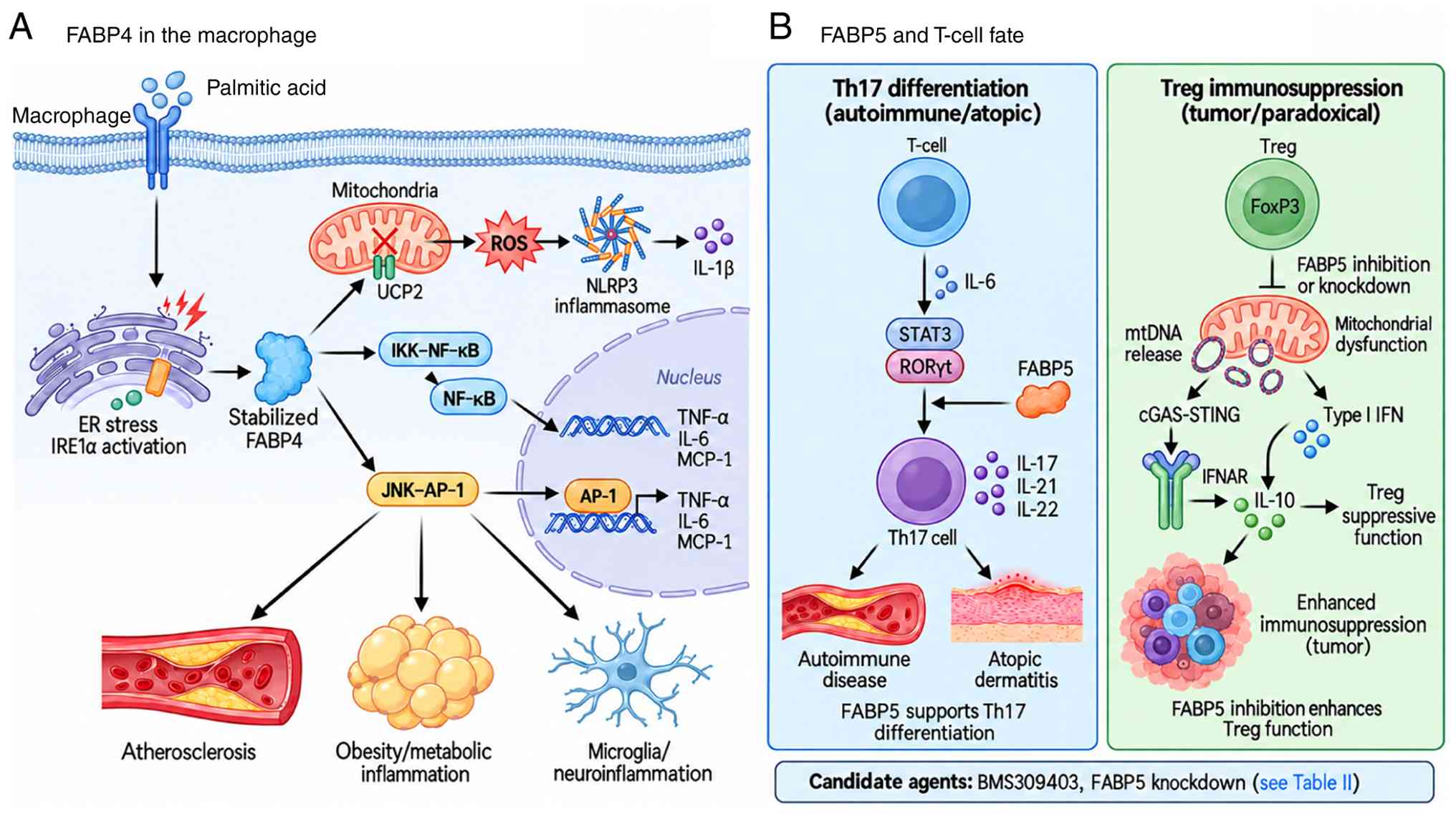 | Figure 1Isoform- and context-specific roles
of FABPs in inflammation. (A) FABP4 in the macrophage. Palmitic
acid induces ER stress and IRE1α activation, stabilizing FABP4
(56-58). FABP4 suppresses UCP2, raising
mitochondrial reactive oxygen species and priming the NLRP3
inflammasome for IL-1β release (52,53), and activates IKK/NF-κB and
JNK/AP-1 to increase TNF-α, IL-6 and MCP-1 (7,11,46,47). This circuit has been described in
atherosclerotic plaque macrophages (11,48,50), obesity-associated metabolic
inflammation (3,9,10) and microglia (55). (B) FABP5 and T-cell fate,
reported here in three separate evidence layers. Observed:
Tumor-infiltrating Tregs express high FABP5, and tumor interstitial
fluid is lipid-poor (67).
Functional experiment: Acute loss of FABP5 function in
differentiated Tregs, by BMS309403 (which engages FABP3, FABP4 and
FABP5) or by FABP5 knockdown, impairs mitochondrial integrity and
triggers mtDNA release, cGAS-STING-dependent type I IFN signaling
and IFNAR-dependent IL-10 production, increasing suppressive
capacity in vitro (67).
Not yet tested: whether lipid scarcity phenocopies this in
vivo, and the net effect on antitumor immunity. On the left,
IL-6/STAT3/RORγt-driven FABP5 supports Th17 differentiation in
autoimmune and atopic settings (63-65,153,166). Not shown: In macrophages, FABP5
activity has instead been reported to restrict tumor growth through
IFN-β (59,61). Candidate agents are listed in
Table II. Arrows and symbols:
Solid black, direction of signaling or of a downstream consequence;
blunt-ended connector, inhibition (pharmacological inhibition or
knockdown of FABP5); cross on UCP2 in (A), FABP4-dependent
suppression of UCP2. AP-1, activator protein 1; cGAS, cyclic
GMP-AMP synthase; ER, endoplasmic reticulum; FABP, fatty
acid-binding protein; IFNAR, IFN α/β receptor; IKK, IκB kinase;
IRE1α, inositol-requiring enzyme 1α; MCP-1, monocyte
chemoattractant protein 1; mtDNA, mitochondrial DNA; NLRP3, NLR
family pyrin domain containing 3; RORγt, retinoic acid
receptor-related orphan receptor γt; ROS, reactive oxygen species;
STING, stimulator of interferon genes; Th17, T helper 17; Treg,
regulatory T cell; UCP2, uncoupling protein 2. |
FABP4 in macrophage-adipocyte
inflammation
FABP4 has been firmly established as a
representative 'proinflammatory' FABP co-expressed in adipocytes
and macrophages (3,9,45); it functions as a key molecular
node in metaflammation, integrating lipid accumulation, lipolysis
and the production of inflammatory cytokines (7).
In macrophages, FABP4 inhibits the PPARγ/liver X
receptor (LXR)α/ABCA1 axis to promote cholesterol ester
accumulation and foam cell formation, while simultaneously
activating the IKK/NF-κB and JNK/AP-1 pathways to upregulate
proinflammatory cytokines such as TNF-α, IL-6 and MCP-1 (7,11,46,47). The transrepressive activity of
PPARγ on inflammatory gene promoters, which is disrupted when FABP4
sequesters PPARγ ligands, provides an additional molecular basis
for the proinflammatory consequences of FABP4 induction (48,49). Macrophage-specific FABP4 deletion
induces an anti-inflammatory phenotype that confers marked
protection against atherosclerotic plaque formation in
ApoE-deficient mouse models (7,50). Pharmacological inhibition of
aP2/FABP4 with small-molecule inhibitors similarly protects against
diabetes and atherosclerosis in mouse models, providing the
earliest preclinical evidence that FABP4 is a druggable target
(51). Mechanistically, FABP4
uncouples lipid metabolism from inflammation by regulating
mitochondrial redox signaling through uncoupling protein 2 (UCP2)
(52).
Previous work has identified FABP4 as an upstream
regulator of NLR family pyrin domain containing 3 (NLRP3)
inflammasome activation. Steen et al (53) demonstrated that loss of FABP4 in
macrophages reduces oxidative modification of mitochondrial
cysteine residues and upregulates antioxidant enzymes (SOD2,
catalase and methionine sulfoxide reductase A). This redox
rebalancing attenuates the mitochondrial unfolded protein response,
thereby reducing cleaved caspase-1 and NLRP3 expression and
substantially suppressing IL-1β secretion. Notably, this
anti-inflammatory effect is partially reversed upon UCP2 silencing,
indicating that the FABP4/UCP2 axis is a key pathway regulating
mitochondrial reactive oxygen species (ROS) and inflammasome
activation.
The proinflammatory activity of FABP4 has been
confirmed in additional vascular contexts. In homocysteine-induced
macrophage inflammation, FABP4 activates the JAK2/STAT2 pathway via
Rap1a-dependent c-Src Tyr416 phosphorylation and membrane
translocation; an effect negatively regulated by SOCS1 (54). Microglia, the resident
macrophages of the central nervous system, also exhibit
FABP4-mediated ROS and TNF-α production, JNK phosphorylation,
increased Toll-like receptor (TLR) 4 expression and decreased UCP2
in response to lipopolysaccharide (LPS), all of which are reversed
by genetic silencing or BMS309403 treatment (55). These findings indicate that the
proinflammatory function of FABP4 is conserved across the
macrophage lineage.
Moreover, FABP4 expression itself is amplified by
inflammatory stimuli in a positive feedback loop, and macrophage
FABP4 acts as a central hub linking endoplasmic reticulum (ER)
stress, lipotoxicity and autophagy dysregulation (56,57). Palmitic acid increases macrophage
FABP4 protein levels (independent of mRNA levels) via ER stress and
IRE1α activation, and elevated FABP4, in turn, inhibits PPARγ and
LXRα function, suppressing SIRT3, UCP2 and PGC-1α expression, and
amplifying mitochondrial ROS production (58). This circuit provides a molecular
basis for the mechanism by which saturated fatty acids sustain
chronic low-grade inflammation in obesity.
FABP5 and T cell differentiation
FABP5 (E-FABP) adopts a fold that is nearly
superimposable on that of FABP4 but is expressed far more widely,
including in macrophages, dendritic cells and T cells (7,59). The inflammatory regulatory
function of FABP5 resembles that of FABP4 in macrophages but
displays a markedly more complex, context-dependent behavior in T
cells.
In macrophages, FABP5 expression is induced by TLR4
(LPS) and TLR2 (zymosan) agonists (60) and has been linked to fatty acid
oxidation (FAO), ROS production, inflammasome activation, IL-1β
upregulation and lipid droplet formation (59,61). However, the same studies report
an antitumor consequence of this activity, whereby FABP5 promotes
IFN-β responses in tumor-associated macrophages (TAMs) and
restricts tumor growth, and a small-molecule activator of FABP5
inhibits tumor growth in mouse models (59,61). This myeloid antitumor direction
is not readily reconciled with the protumorigenic roles described
for tumor-cell FABP5 below, and the two should not be summed into a
single directional statement for the isoform. FABP5 also
participates in cross-talk between dietary polyunsaturated fatty
acid (PUFA) availability and macrophage polarization. FABP5
deletion suppresses atherosclerosis in VLDL receptor-deficient mice
by reducing IL-6 and COX-2 expression and by CCR2-dependent
inhibition of macrophage recruitment (62).
However, in T cells FABP5 function is far more
nuanced. Li et al (63)
reported that E-FABP-deficient CD4+ T cells exhibit
markedly reduced Th17 differentiation and increased
FoxP3+ Treg differentiation. Mechanistically, E-FABP
deficiency impairs IL-6-induced expression of IL-21, RORγt and RORα
while increasing PPARγ, which in turn suppresses STAT3 activity and
blocks the Th17 program (63).
In experimental autoimmune encephalomyelitis, FABP5-deficient mice
are protected from disease through reduced Th17 differentiation
(63,64). However, FABP5 expression promotes
Th17 polarization in mouse models and patient samples of atopic
dermatitis (65), whereas FABP5
deficiency has no effect on antigen-specific CD4+ or
CD8+ T cell generation in a Listeria
monocytogenes infection model (66). These divergent results illustrate
that the T cell-regulatory role of FABP5 depends on stimulus,
tissue context and disease stage.
From a Treg perspective, FABP5 creates one of the
most important therapeutic paradoxes in the FABP field. Field et
al (67) demonstrated that
pharmacological or genetic inhibition of FABP5 in Tregs causes
mitochondrial dysfunction, including decreased oxidative
phosphorylation, impaired lipid metabolism and loss of cristae
structure. These mitochondrial defects promote mtDNA release and
cGAS-STING-dependent type I interferon signaling, thereby
increasing IL-10 production and enhancing Treg suppressive
function. Thus, the same FABP5 blockade that may attenuate
Th17-driven autoimmune inflammation could reinforce
immunosuppression in a tumor-bearing host.
This paradox means that FABP5 inhibition cannot be
interpreted as uniformly anti-inflammatory or antitumor. The
desired effect may be Th17 suppression in psoriasis, atopic
dermatitis or autoimmunity, whereas the undesired oncological
effect may be strengthened Treg-mediated immune evasion.
Preclinical evaluation of FABP5-targeted agents should therefore
include immunocompetent syngeneic tumor models, Treg depletion or
rescue experiments, cell-type-specific knockout approaches and
combination studies with immune checkpoint blockade before clinical
translation in cancer or premalignant high-risk settings.
FABP1 and FABP3
Not all FABPs act in a proinflammatory manner. Some
isoforms attenuate inflammation under specific conditions or
protect tissues from oxidative stress. FABP1 is highly expressed in
the liver, small intestine and kidney, and serves as a fatty acid
buffering and safe transport protein owing to its unique ability to
bind two fatty acids simultaneously (4). FABP1-deficient mice display reduced
hepatic β-oxidation efficiency and bile acid homeostasis
disturbances. However, paradoxically, these animals can also
exhibit exacerbated oxidative stress and lipotoxicity, suggesting
that FABP1 functions as a 'buffer protein' that safely partitions
excess fatty acids (4,68).
FABP3 (H-FABP) plays a central role in delivering
LCFAs to mitochondria for β-oxidation in the heart and skeletal
muscle (4). FABP3 deficiency
switches cardiac fuel selection from fatty acids to glucose and
induces cardiac hypertrophy in aged mice (4). During myocardial
ischemia-reperfusion injury, FABP3 is rapidly released into the
bloodstream and serves as an early biomarker of acute myocardial
infarction (1). Recent work has
shown that FABP3, FABP7 and FABP8 also bind cholesterol and
facilitate its egress from lysosomes, contributing to intracellular
lipid trafficking homeostasis (69). However, in the nervous system,
FABP3 has been implicated in pathogenic processes such as
α-synuclein aggregation and mitochondrial damage in Parkinson's
disease, illustrating that even within a single isoform,
tissue-specific functional divergence is pronounced (70).
These isoform-specific functional divergences
indicate that a binary 'proinflammatory vs. protective'
classification of the FABP family is untenable. Rather, each
isoform appears optimized for the physiological demands of its
expressing tissue (fuel supply, toxicity buffering and signal
transduction), and the directionality of inflammation is determined
when these normal functions become deregulated under pathological
conditions (1).
Circulating FABP4 as an adipokine
The most recently described aspect of FABP-mediated
inflammatory regulation is the existence of extracellular
circulating FABP. Despite lacking a typical secretory signal
peptide, FABP4 is released into the extracellular space via a
non-classical secretion pathway upon lipolytic stimulation or
fasting (71-73). Secreted FABP4 is a bioactive
adipokine that stimulates hepatic glucose production and influences
systemic insulin sensitivity in experimental models (3,5).
Circulating FABP4 levels are markedly elevated in
obese humans and mice, and correlate strongly and independently
with type 2 diabetes, hypertension, heart failure and
atherosclerosis (5,7). Notably, circulating FABP4 is not
merely a disease biomarker but an active mediator that proactively
induces inflammation in distant tissues; it upregulates the
ERK/JNK/STAT1 pathway and downregulates eNOS and SDF-1 signaling in
human coronary endothelial cells, inducing endothelial dysfunction
(74). Moreover,
antibody-mediated neutralization of aP2 improves glucose tolerance
and insulin sensitivity in obese mice, demonstrating that
circulating FABP4 is a neutralizable therapeutic target (3,75).
The concept of circulating FABP4 supports the view
of FABPs as metabolic-immune signaling integrators. FABP4 is
secreted from obese adipose tissue and reaches the liver,
vasculature and potentially distant TMEs to coordinate inflammation
and metabolic rewiring across organs, providing an integrative
framework explaining the obesity-chronic inflammation-cancer
continuum. Specifically, Hao et al (12) reported that circulating FABP4 is
a novel link underlying obesity-associated breast cancer
development; a topic that directly connects with the role of FABPs
in the TME discussed below.
A total of two features of this section carry into
the next. The directionality of FABP-dependent inflammation is set
by the combination of ligand, cell type and disease context, so an
isoform cannot be labeled proinflammatory or anti-inflammatory on
its own; and circulating FABP4 extends this biology beyond the cell
in which the protein is made. The next sections examine how far
these circuits operate in tumors, and where the evidence for that
stops.
FABPs in cancer
The aforementioned inflammatory regulatory functions
of FABPs acquire new dimensions in the context of tumor biology.
Beyond their use as biomarkers in cancer, FABPs also act as drivers
that mediate intercellular lipid shuttling and metabolic rewiring
within the TME. FABP4, FABP5 and FABP7 each have distinct
pathological roles in cancer due to their isoform-specific
properties. A schematic overview of the isoform-specific mechanisms
discussed below, covering FABP4 in adipocyte-macrophage-tumor
crosstalk and the omental endothelial niche, FABP5 ligand routing
in tumor cells and FABP7 in GBM stem-like cells, is provided in
Fig. 2.
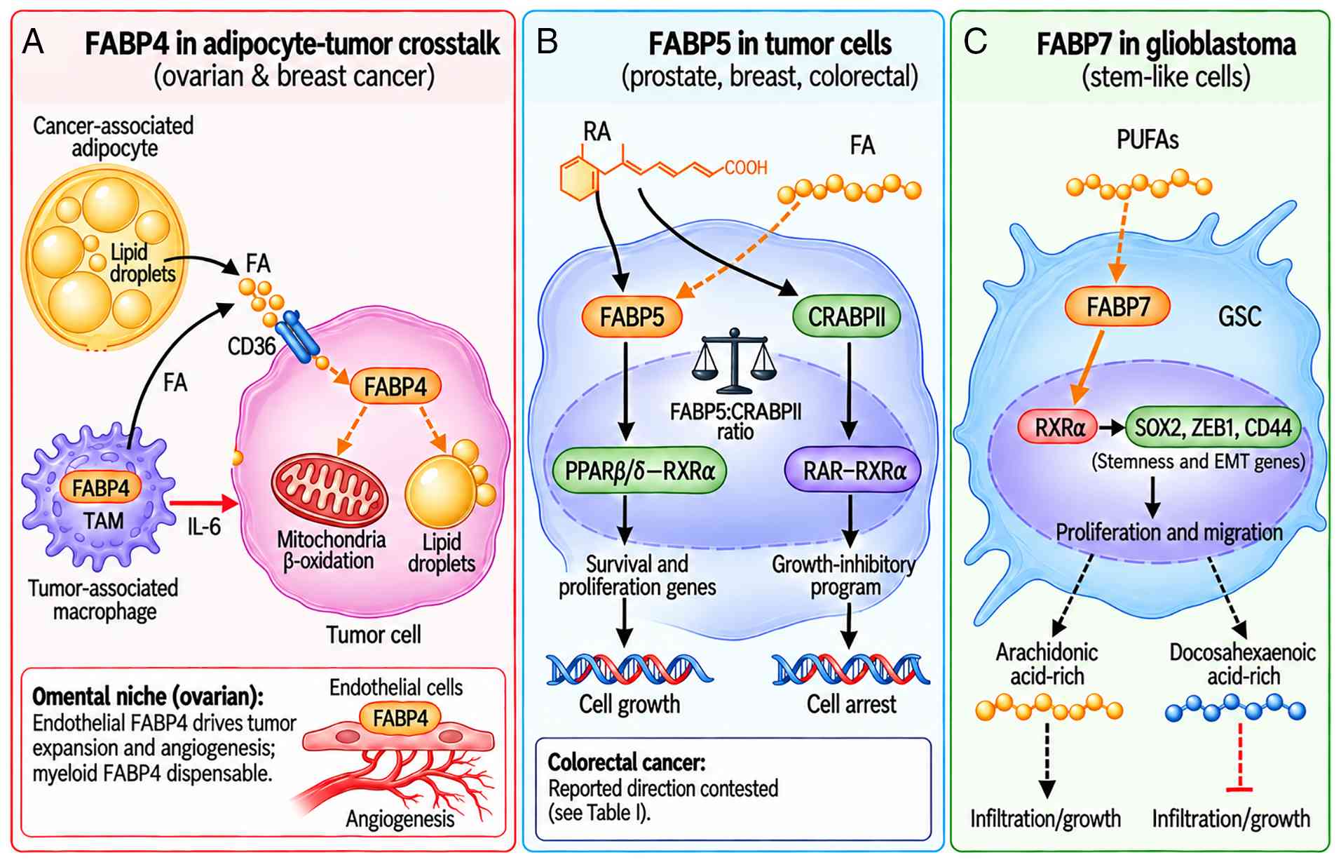 | Figure 2Isoform-specific contexts in cancer.
Three contexts are shown separately since the evidence does not
support a single shared pathway. (A) FABP4 acts in more than one
compartment, and which one dominates differs by tumor type.
Cancer-associated adipocytes and FABP4-high TAMs supply FAs to
tumor cells, and the TAMs also supply IL-6 (44,79,80,86); within tumor cells CD36-mediated
uptake feeds FABP4, which partitions FAs to β-oxidation and lipid
droplets (84). However, in the
omental niche, mature adipocytes are not required for ovarian tumor
growth. Endothelial cells are the principal FABP4-expressing
population and endothelial-specific deletion reduces tumor
expansion and angiogenic branching whereas myeloid-specific
deletion does not (85). (B)
FABP5 in tumor cells. FABP5 delivers RA and FAs to PPARβ/δ-RXRα,
inducing survival and proliferation genes, whereas RA routed
instead to CRABPII-RAR activates a growth-inhibitory program; the
FABP5:CRABPII ratio sets which of the two receptors the ligand
reaches, and lowering it suppresses tumor growth in mammary
carcinoma models (13,14,34,36-38,87). In colorectal cancer the reported
direction is contested (Table
I). (C) FABP7 in glioblastoma. FABP7 delivers PUFAs to RXRα,
inducing SOX2, ZEB1 and CD44 and supporting proliferation and
migration of GSCs; arachidonic-acid-rich and
docosahexaenoic-acid-rich environments have opposite effects
(96,101). This has been demonstrated in
GSCs and has not been tested in premalignant lesions of other
tissues. Arrows and symbols: Solid black, release of FAs from
adipocytes and macrophages in (A), movement of RA in (B) and
downstream transcriptional or phenotypic output in (B) and (C);
dashed orange, uptake or intracellular movement of FA; solid orange
in (C), delivery of PUFAs to RXRα; solid red in (A), IL-6
signaling; dashed black in (C), an outcome conditional on the
surrounding lipid composition; dashed red blunt-ended connector in
(C), suppression of that outcome; balance symbol in (B), the
FABP5:CRABPII ratio that determines which receptor the ligand
reaches. CRABPII, cellular retinoic acid-binding protein II; EMT,
epithelial-mesenchymal transition; FA, fatty acid; FABP, fatty
acid-binding protein; GSC, GBM stem-like cell; PPARβ/δ, peroxisome
proliferator-activated receptor β/δ; PUFA, polyunsaturated fatty
acid; RA, retinoic acid; RAR, retinoic acid receptor; RXRα,
retinoid X receptor α; TAM, tumor-associated macrophage; ZEB1, zinc
finger E-box binding homeobox 1. |
FABP4 in obesity-associated cancer
types
FABP4 has emerged as the most potent protumorigenic
FABP in various obesity-associated cancer types. FABP4 is
upregulated in numerous solid tumors, including breast, ovarian,
prostate, hepatic, pancreatic and lung cancer, and this
upregulation is generally associated with a poor prognosis
(18,76,77). Dysregulated adipose-tumor
crosstalk is a shared feature of these obesity-associated
malignancies (78). Notably,
FABP4 is expressed not only in tumor cells, but also across stromal
components of the TME, including adipocytes, TAMs and endothelial
cells, where it contributes to intercellular lipid exchange
(19). In ovarian cancer,
adipocyte-induced FABP4 expression in tumor cells promotes
metastasis and carboplatin resistance (76).
The adipocyte-tumor cell fatty acid shuttle
represents the most representative model of FABP4-mediated
tumorigenic mechanisms and was originally delineated in the omental
niche of ovarian cancer metastasis (79,80). Adipocytes supply fatty acids to
tumor cells during metastasis to the omentum, inducing FABP4
expression in tumor cells and simultaneously increasing both
metastatic potential and carboplatin resistance (19,76). A parallel CD36-driven fatty acid
import axis, induced by adipocyte coculture, further amplifies this
metastasis-initiating program (81,82). Intraperitoneal injection of
FABP4-high ovarian cancer cells into FABP4−/−
mice markedly reduces tumor growth and metastasis, and FABP4
inhibitors such as BMS309403 effectively suppress both early and
established metastases (76). A
similar pattern is observed in breast cancer, where adipocyte
cancer cell co-culture simultaneously upregulates CD36 and FABP4
expression, with their direct interaction integrally coordinating
fatty acid uptake, intracellular trafficking and delivery to
mitochondria and peroxisomes (82-84). This process promotes metabolic
reprogramming toward FAO and is associated with adipocyte-induced
epithelial-mesenchymal transition (EMT) and stemness, in part
through CD36-FABP4 interaction (84).
This model has since been tested directly and
requires verification. In mice congenitally lacking mature
adipocytes within the peritoneal cavity, ovarian tumors still
seeded adipose-associated regions, peritoneal expansion was not
impaired and the response to carboplatin was unchanged, whereas
removal of the adipocyte-free omentum did reduce tumor burden
(85). The earlier evidence
rested on whole-body FABP4 deletion, which cannot separate
adipocyte FABP4 from its expression in other compartments.
Single-cell analysis of mouse and human omentum placed the highest
FABP4 expression in endothelial cells. Endothelial-specific
deletion reduced omental tumor expansion by ~29% during exponential
growth and lowered tumor-associated vascular branching, whereas
myeloid-specific deletion had no effect in this model (85). Therefore, adipocyte-derived fatty
acid supply may be sufficient to support tumor growth in this niche
without being required for it, and the dominant FABP4-expressing
compartment appears to differ between tumor types, whereby myeloid
FABP4 is required in breast models (44,86), but not in this ovarian model.
FABP4 in TAMs illustrates the mechanisms by which
the inflammatory functions of immune-cell FABPs may be repurposed
in the TME. Hao et al (44) reported that a
CD11b+F4/80+MHCII−Ly6C−
TAM subset in breast tumors expresses high levels of FABP4, and
that FABP4 in these cells is associated with activation of the
NF-κB/microRNA (miR)-29b/IL-6/STAT3 axis. In the same study, FABP4
deficiency reduced mammary tumor growth and metastasis, and higher
FABP4 expression was associated with poorer breast cancer survival
(44). More recently, Yorek
et al (86) demonstrated
that unsaturated fatty acids, particularly linoleic acid, promote
FABP4/CEBPα-dependent triglyceride synthesis and lipid droplet
accumulation in TAMs, and that FABP4-dependent lipid mobilization
in TAMs supports breast cancer cell migration and metastasis.
Together, these findings support a role for FABP4-positive TAMs in
metabolic crosstalk that contributes to breast cancer progression
(44,86).
Circulating FABP4 may extend the fatty acid shuttle
model to a systemic context. Hao et al (12) reported that obesity-associated
adipose-derived FABP4 was linked to the development of distant
breast/mammary tumors, suggesting a potential molecular connection
between obesity and breast cancer. These observations support the
possibility that the aforementioned 'FABP4 as adipokine' concept
(circulating FABP4 as an adipokine) may be relevant not only to
metabolic disease but also to systemic processes that influence
tumor progression.
FABP5 and the PPARβ/δ axis
FABP5, along with FABP4, is among the most studied
isoforms in cancer research. FABP5 is elevated relative to matched
normal tissue in several tumor types, and higher expression has
generally been associated with a poorer outcome, although the
reported magnitude varies by tumor type and study (13). The protumorigenic mechanisms of
FABP5 can be summarized along two main axes: i) Ligand-nuclear
receptor delivery; and ii) metabolic rewiring.
The core of the ligand-nuclear receptor delivery
axis is the function of FABP5 to deliver fatty acids and RA to
PPARβ/δ, thereby inducing genes involved in tumor survival,
proliferation and angiogenesis (VEGF, MMPs and Akt) (13,14,87). This pathway is particularly
well-established in prostate cancer. In castration-resistant
prostate cancer, the FABP5/PPARγ/VEGF axis progressively replaces
the androgen receptor (AR)-mediated pathway, and FABP5 knockout
markedly suppresses proliferation, invasion and colony formation of
PC3-M cells (88). Notably,
deletion of FABP5 results in a complete loss of expression of the
AR splice variant ARV7, which is characterized by the absence of
the ligand-binding domain. This finding highlights FABP5 as a
promising target for addressing resistance to antiandrogen therapy
(88).
In breast cancer, FABP5 plays a complementary role
to FABP4. Both isoforms are prominently expressed in breast tissue
and exhibit distinct expression patterns across molecular subtypes
(19). The FABP5/CRABPII ratio
is a key switch that determines the fate of RA, whereby a higher
ratio directs RA toward the protumorigenic PPARβ/δ pathway, and a
lower ratio favors the tumor-suppressive RAR pathway (36). Reducing the FABP5/CRABPII ratio
in TNBC redirects RA from PPARβ/δ to RAR, thereby suppressing tumor
growth (38).
Along the metabolic rewiring axis, FABP5 shows
tight connections with FASN and hypoxia signaling. In cervical
cancer, FABP5 facilitates lymph node metastasis by reprogramming
fatty acid metabolism, which correlates with elevated MMP-2 and
MMP-9 expression (89,90). In pancreatic neuroendocrine
tumors, FABP5 interacts directly with FASN, modulating its
stability via the ubiquitin-proteasome pathway and activating the
WNT/β-catenin signaling cascade to promote tumor progression
(91). Notably, the direction of
this coupling is the opposite of that reported in colorectal cells,
where FABP5 stabilizes FASN in pancreatic neuroendocrine neoplasia,
but promotes its ubiquitination in CRC (16,91). The two reports come from the same
group and appeared within weeks of each other, share the same
interaction partner and ubiquitin-proteasome read-out and neither
addresses the other. Rather than a contradiction, this is the
clearest available indication that the FABP5-FASN relationship is
tumor-type dependent and cannot be stated as a single rule.
FABP5 in CRC: An unresolved directional
conflict
Reports on FABP5 in CRC have opposing results, and
the discrepancy is informative rather than incidental. Seo et
al (17) showed that
transient FABP5 silencing lowered hypoxia-induced HIF-1α protein
and its target transcripts (VEGF, CA9, LOX, PDK1 and BNIP3L) in
HCT116 and WiDr cells, and that FABP5-silenced HCT116 spheroids
grown on an oxygen-permeable three-dimensional chip remained
smaller than the controls over 7 days. Ye et al (16), also working with the HCT116 cell
line, reported the converse, whereby FABP5 protein was lower in 90
paired colorectal carcinoma specimens than in adjacent tissue,
higher FABP5 was associated with longer survival, stable FABP5
overexpression suppressed proliferation, migration and xenograft
growth while stable knockdown enhanced them, and the effect was
traced to FABP5-dependent ubiquitination of FASN with downstream
inhibition of mTOR and induction of autophagy.
These are four differences between the two studies
which may account for the discrepancies; however, none have been
further studied. First, the lesions differ, whereby the paired
expression series analyzed by Seo et al (17) (GSE8671) comprised colorectal
adenomas, while Ye et al (16) sampled established carcinoma.
Therefore, the two tissue observations may describe different
points on the adenoma-carcinoma sequence, rather than opposite
findings in the same lesion. Second, the analytes differ, with
transcript vs. protein. Third, oxygen tension is central to one
mechanism and absent from the other, in which the FABP5/HIF-1α axis
was defined under hypoxia and in spheroids that develop an internal
hypoxic region, whereas the FASN/mTOR/autophagy axis was
characterized in normoxic monolayers and subcutaneous xenografts.
Fourth, the perturbations differ in kinetics and in direction, with
transient silencing in one case against stable overexpression and
knockdown in the other; only the latter tested both directions, and
no in vivo experiment has been reported for the HIF-1α
arm.
A third report adds a distinct axis rather than
adjudicating between these two. In colorectal cells, p53 induces
FBXO2, which binds FABP5 and directs it to chaperone-mediated
autophagy. The resulting fall in PUFAs reduces lipid peroxidation
and confers resistance to ferroptosis; arachidonic acid (AA)
supplementation reverses it (92). FABP5 sensitizes cells to
ferroptosis, which again places it on the tumor-restraining side,
although the outcome measured is ferroptosis susceptibility rather
than proliferation and the in vivo arm manipulated FBXO2
rather than FABP5. Therefore, two of the three colorectal reports
describe a restraining role, but the one that does not was
conducted under hypoxia and in three-dimensional culture, so oxygen
tension remains the most testable candidate for the
discrepancy.
Neither direction of the FABP5 effect is therefore
treated as established across CRC. A comparison using the same
colorectal cell models, bidirectional FABP5 perturbation and
matched oxygen conditions, with FASN, HIF-1α and autophagy assessed
in parallel, could test whether oxygen tension or pathway
interactions explain the discrepancy. Stage-matched human tissue
would address the separate question of disease-stage associations.
These explanations remain untested.
In cervical cancer, FABP5 promotes EMT,
lymphangiogenesis and lymph node metastasis by reprogramming fatty
acid metabolism. This includes FABP5-driven lipolysis and de
novo fatty acid synthesis which increase intracellular fatty
acid levels that activate the NF-κB signaling pathway and FABP5
expression, which independently predicts lymph node metastasis and
poor prognosis (90).
Additionally, FABP5 serves as an intracellular carrier of
anandamide and related N-acylethanolamines in the brain, delivering
them to fatty acid amide hydrolase for hydrolysis, such that
pharmacological FABP inhibition elevates brain anandamide levels
and produces analgesia (93).
From the perspective of antitumor immune evasion, FABP5 illustrates
the manner in which the aforementioned Treg-regulatory function is
exploited oncologically. In tumor-infiltrating Tregs, FABP5
inhibition activates the mtDNA leakage cGAS/STING/IL-10 pathway,
paradoxically enhancing the Treg immunosuppressive function
(67). This raises a fundamental
dilemma in the clinical application of FABP5 inhibitors in
autoimmune disease (where inhibition is beneficial) vs. cancer
(where inhibition may reinforce immunosuppression). Meanwhile, in
CD8+ T cells, FABP5 promotes linoleic acid uptake and
induces ROS-mediated T cell death, thereby reducing TME
CD8+ T cells and accelerating tumor growth under
high-fat-diet conditions (15,94).
FABP7 in nervous system tumors
FABP7 (B-FABP) is the most studied FABP in nervous
system tumors, including GBM. In the normal brain, FABP7 functions
as a marker for radial glial cells and neural stem cells and is
essential for establishing the radial glial fiber system during
neurogenesis (95,96). The transformation of this normal
neurodevelopmental role into the maintenance of 'cancer stemness'
in oncological contexts is a unique mechanism of FABP7.
Although FABP7 is predominantly expressed in neural
lineage tissues, aberrant FABP7 expression has also been reported
in breast cancer subtypes, where its subcellular localization
correlates with clinical outcomes (97). FABP7 is most characteristically
associated with GBM stem-like cells (GSCs). FABP7 is highly
enriched in patient-derived GSCs and localizes to both the
cytoplasm and nucleus (98).
Differentiation of GSCs by serum addition markedly reduces FABP7
expression, and FABP7+Sox2+ tumor cells are
significantly increased in WHO grade IV GBM compared with grade II
and III tumors (98).
siRNA-mediated FABP7 downregulation reduces both proliferation and
migration of GBM-derived neurospheres, and FABP7 expression
increases after radiation, suggesting an association with
radioresistance (99). This
behavior places FABP7 among the lipid-handling mediators that
contribute to immune evasion by cancer stem cells (100).
Mechanistically, recent work has demonstrated that
FABP7 delivers PUFAs to the nuclear receptor retinoid X receptor
(RXR)α, activating a PUFA/FABP7/RXRα neurogenic pathway (96). This pathway induces SOX2 (a key
stemness regulator) and ZEB1 (an EMT transcription factor), driving
the 'stationary-to-migratory' transition in GSCs (96). Notably, the role of FABP7 is
bidirectional depending on the fatty acid microenvironment. In an
AA-rich environment, FABP7 promotes GBM growth and infiltration,
while in a docosahexaenoic acid (DHA)-rich environment, FABP7 may
inhibit tumor infiltration through increased DHA uptake and
utilization (101). Therefore,
FABP7 has been proposed as a candidate vulnerability of GBM neural
stem-like cells, suggesting that FABP7-targeted therapy may need to
be combined with manipulation of the ligand environment (101).
Limitations in FABP7 inhibitor development have
also been noted, as the existing drug SBFI-26 displays low
selectivity, targeting both FABP5 and FABP7, and its blood-brain
barrier permeability has not been evaluated (102). This underscores the need for
isoform-selective and tissue-specific FABP inhibitors.
FABP1 and FABP6 in gastrointestinal
cancer
FABP1 (L-FABP) shows opposing roles in liver and
CRC depending on tissue context. In some HCC cases, FABP1 promotes
tumor growth and angiogenesis by upregulating VEGF-A through the
VEGFR2/Akt/mTOR pathway (103),
whereas in another study, FABP1 expression is downregulated in
poorly differentiated HCC and associated with poor prognosis
(104). In CRC, FABP1 has been
reported as a differentiation marker downregulated at the adenoma
stage but is upregulated again in advanced cancer (1). Such stage- and context-dependent
expression changes indicate that FABP1 cannot be classified simply
as a tumor-promoting or -suppressing factor. Therefore, FABP1 and
FABP6 exert mechanistically distinct gastrointestinal effects,
whereby FABP1 modifies tumor cell phenotype in a stage-dependent
manner through cell-intrinsic lipid buffering, whereas FABP6
contributes through an epithelial bile acid-microbiota interaction
that is more clearly tied to chronic intestinal inflammation. Both
isoforms are summarized alongside FABP4/FABP5/FABP7 in Table I, which states for each claim the
system in which it was obtained, the outcome measured and what
remains untested.
A total of two points from this section are
relevant to the subsequent discussion. FABPs act both within tumor
cells and in the surrounding compartments, and the compartment that
matters is not fixed, as it differs between tumor types. As the
same isoform can alter tumor metabolism and immune function in
opposite directions, antitumor and immunomodulatory effects must be
assessed separately rather than assumed to align.
FABP networks at the inflammation-cancer
interface: A context-specific evidence map
The previous sections outlined that FABPs can
regulate inflammatory signaling and tumor metabolism in a highly
isoform- and cell-type-dependent manner. The present section
integrates these observations but does not claim a deterministic
linear pathway from inflammation to cancer. Instead, it sets out
for each context what has been measured and in what system, so that
the point at which the chain of evidence stops is visible.
Each context is considered using the same logic: i)
The established inflammation-cancer link; ii) the FABP isoforms
implicated; iii) the relevant cell types; iv) what has been
measured, in which system, and what has not been measured; v)
unresolved questions; and vi) translational implications. This
structure is intended to separate causal in vivo evidence
from associative biomarker evidence and hypotheses that remain to
be directly tested. Of these, three disease contexts were selected
for comparison. Each has a chronic inflammatory or metabolic
precondition that is epidemiologically linked to cancer, each has
FABP-specific mechanistic evidence and each has human data that can
be set against the experimental work rather than simply added to
it. Other clinically important FABP-related cancer contexts, such
as adipocyte-rich ovarian cancer and FABP7-dependent GBM, are
addressed primarily in previous sections and Fig. 2 rather than as integrative
contexts in this section.
Metabolic liver axis:
MASLD/MASH-to-HCC
Terminology requires
clarification
The current nomenclature uses MASLD and MASH,
replacing much of the older non-alcoholic fatty liver disease
(NAFLD)/non-alcoholic steatohepatitis (NASH) terminology; metabolic
dysfunction-associated fatty liver disease (MAFLD) is a related but
not identical metabolic-definition framework (105,106). In the present review,
MASLD/MASH is used for current conceptual discussion, while
NAFLD/NASH is retained when referring to the terminology used in
older studies.
Established link
MASLD can progress through steatosis, MASH,
fibrosis, cirrhosis and HCC, particularly in the presence of
obesity, insulin resistance and type 2 diabetes (106-108). This makes the metabolic liver
axis one of the strongest examples of an inflammation-cancer
continuum discussed in the present review.
FABP evidence and what has been
measured
Human association studies indicate that hepatic and
circulating FABP4 levels rise during NAFLD/NASH and correlate with
inflammation and fibrosis, supporting FABP4 as a biomarker of
disease activity rather than of cancer (109,110). Mechanistic evidence is stronger
in experimental models, whereby the LPL/FABP4/CPT1 fatty acid
metabolic axis is activated during the MASH/NASH-to-HCC transition
in stelic animal model (STAM) mice, and pharmacological inhibition
of this axis reduces progression to HCC (111). In addition, adipocyte-derived
FABP4 promotes HCC stemness and drug resistance through
ITGB1-mediated β-catenin activation, and FABP4 deficiency delays
tumor development in diet- and carcinogen-driven models (112). In both cases the outcome
measured was tumor development in mice. For FABP4, this is the
context in which three types of study, namely human cohort
association, pharmacological inhibition in mice and genetic
deficiency in mice, come closest to addressing the same question.
However, no human study has tested whether altering FABP4 changes
HCC incidence, so the human and causal evidence remain on different
outcomes. For FABP1, the hepatic homeostatic functions are well
characterized (4,68), but no experiment has tested a
stage-specific role in the transition to HCC, and the cancer
associations are based on expression data.
Unresolved issues and
implications
It remains unclear whether FABP4 is necessary for
all metabolic liver cancer contexts or primarily modifies risk in
obesity- and lipotoxicity-dominant subtypes. FABP4-directed
interventions should therefore be evaluated as risk-modifying or
progression-modifying strategies in defined metabolic settings
rather than as general HCC therapeutics.
Intestinal axis: IBD-to-CRC
Established link
Chronic IBD, especially long-standing ulcerative
colitis, increases the risk of CRC and represents a classical
inflammation-driven cancer model (113,114). In this setting, epithelial
barrier disruption, dysbiosis, bile acid remodeling and cytokine
signaling converge on epithelial regeneration and malignant
transformation.
FABP evidence and what has been
measured
FABP6 is the most relevant local intestinal isoform
as it mediates ileal bile acid transport and enterohepatic
recycling (1,115). Intestinal epithelium-specific
FABP6 knockout alters bile acid composition, shifts microbiota
composition and exacerbates dextran sulfate sodium-induced colitis;
rescue by Ligilactobacillus murinus or taurodeoxycholic acid
supports a functional bile acid-microbiota-barrier mechanism
(116). The outcome measured
was colitis severity, and loss of FABP6 makes the condition worse.
Separately, FABP6 mRNA is elevated in most paired human CRC samples
and is detectable from the adenoma stage (117); the measured outcome was
expression, with no perturbation. The two FABP6 observations on
this axis point in different directions and have not been joined.
No experiment has tested FABP6 manipulation in a colitis-associated
carcinogenesis model. Therefore, there is no evidence that FABP6
drives the transition, and the only loss-of-function phenotype
available runs opposite to the direction such a role would
predict.
For FABP4 the evidence on this axis is of a
different type. Circulating FABP4 and FABP6 are elevated in
patients with CRC and fall after radical resection (118), which is a biomarker observation
that is compatible with the tumor state raising FABP4 rather than
the reverse. In a prospective analysis of 1,324 incident cases and
a two-sample Mendelian randomization study using 58,131 cases,
neither measured nor genetically predicted that circulating FABP4
was associated with CRC risk overall (119). These analyses concern the
circulating protein and CRC as a whole; they were not designed to
address colitis-associated disease, and do not speak to tissue
FABP4.
Unresolved issues and
implications
The major unresolved question is whether FABP6 is a
causal driver of colitis-associated tumor initiation or a marker of
epithelial bile acid stress. Future studies should test FABP6
manipulation in colitis-associated CRC models and evaluate
microbiota, bile acid species and immune infiltration as linked
readouts.
Obesity-breast cancer axis
Established link
The epidemiological association between obesity and
breast cancer risk, recurrence and outcomes is well-established,
particularly in postmenopausal disease (120-122). The molecular connection
involves chronic adipose inflammation, adipokine signaling,
insulin/IGF signaling, estrogenic effects and altered lipid
availability.
FABP evidence and what has been
measured
FABP4 accounts for most of the FABP-specific work
on this axis. Circulating FABP4 secreted from obese adipose tissue
promotes mammary tumor stemness and aggressiveness through an
IL-6/STAT3/ALDH1-linked mechanism (12). In the TME, FABP4 expression in a
CD11b+F4/80+MHCII−Ly6C−
TAM subpopulation promotes breast cancer progression through a
CEBPα/NF-κB/miR-29b/IL-6/STAT3 circuit (44). FABP4 also participates in
adipocyte-tumor cell fatty acid transfer and CD36-linked fatty acid
uptake in breast cancer cells (84,86). For FABP4 the different types of
study converge more closely than elsewhere. Every perturbation
experiment on this axis measures the growth or spread of an already
established tumor, and the adipocyte-to-tumor supply route on which
the mechanism rests has been shown to be dispensable in the
analogous ovarian niche (85);
none measures tumor incidence, and no human study has tested
whether altering FABP4 changes breast cancer risk. Circulating
FABP4 is also not cancer-specific, as it is confounded by body mass
index, insulin resistance, diabetes, age, sex and cardiovascular
disease, and Mendelian randomization indicates that adiposity
raises FABP4 rather than the reverse (123), so an elevated level is at least
partly a consequence of the adipose state it is being used to
interrogate.
Convergent signaling pathways
As aforementioned, across these disease contexts,
FABP isoforms appear to converge on four recurring signaling
modules and are summarized in the present review to highlight
cross-context recurrence rather than to re-describe individual
mechanisms. First, FABP-nuclear receptor routing links lipid
availability to transcriptional programs, whereby FABP4 interacts
with PPARγ-related signaling, FABP5 can route RA or fatty acids
toward PPARβ/δ and FABP7 can regulate ligand-dependent RXRα
signaling (13,34,41,43,96). Second, inflammatory cytokine
modules involving NF-κB, IL-6 and STAT3 recur in macrophage and TAM
contexts (11,44,124,125). Third, mitochondrial metabolism,
ROS and inflammasome activation connect lipid chaperoning with
innate immune activation (53,67). Fourth, hypoxia-linked metabolic
rewiring has been described for FABP5 and HIF-1α in hepatic and CRC
cells (17,126). In colorectal cells this has
been shown under hypoxia and in three-dimensional culture, without
in vivo confirmation, and it coexists with a report of the
opposite direction under normoxia (16); it should therefore be read as a
condition-specific observation rather than a general property of
FABP5 in this tumor type.
These convergent modules provide a mechanistic
rationale for the model in Fig.
3, but they should be interpreted as context-dependent routes
rather than universal FABP-driven cascades. In some settings, FABPs
appear causal, in others, they are modifiers or may serve mainly as
biomarkers of metabolic or inflammatory stress.
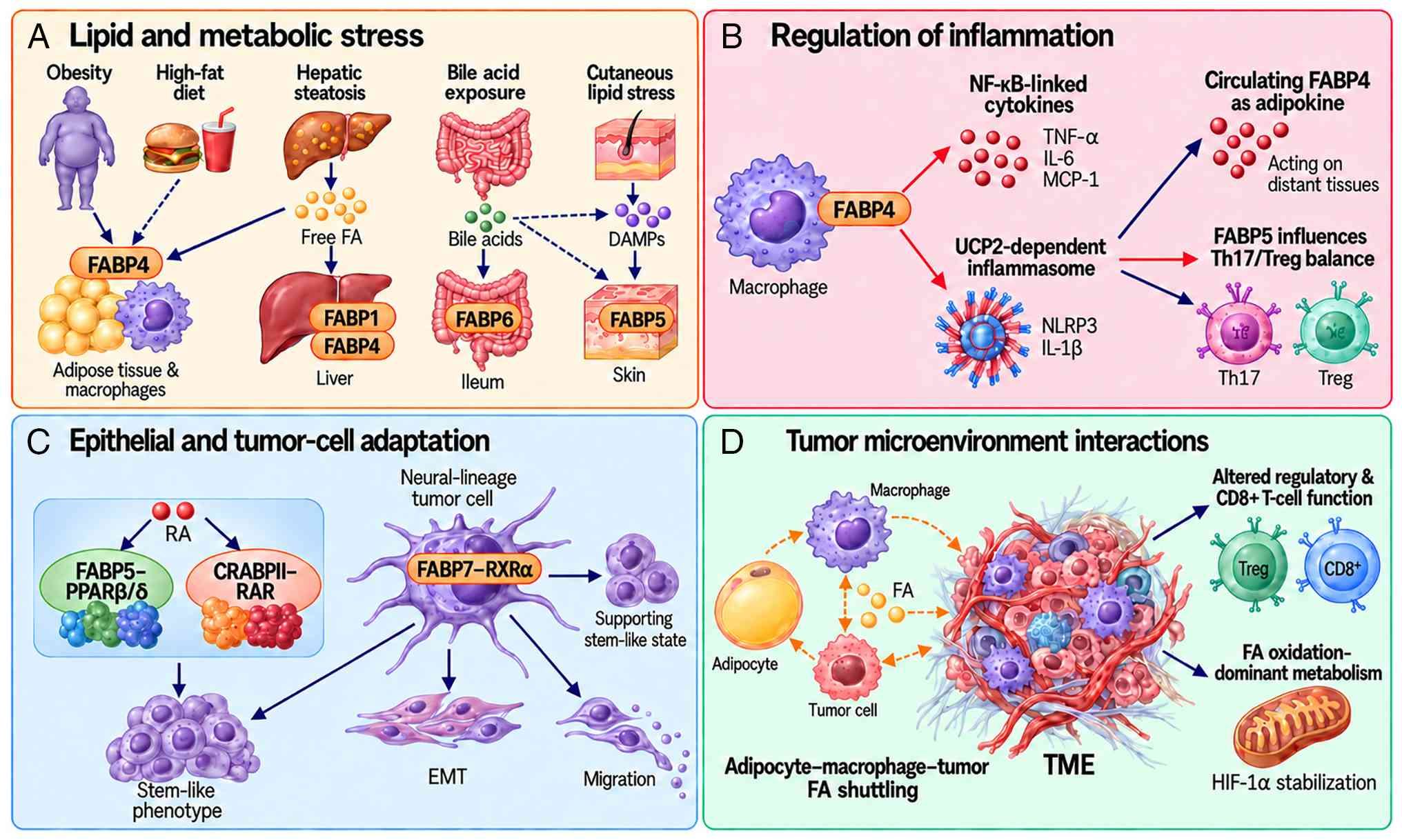 | Figure 3Domains in which FABPs have been
implicated at the inflammation-cancer interface. The four domains
are shown as related areas rather than as sequential stages, as the
evidence does not establish a single ordered progression. Solid
connectors mark links supported by perturbation experiments; dashed
connectors mark links that have not been directly tested. (A) Lipid
and metabolic stress. Obesity, high-fat diet, hepatic steatosis,
bile acid exposure and cutaneous lipid stress are associated with
tissue-specific FABP induction: FABP4 in adipose tissue and
macrophages, FABP1 and FABP4 in liver, FABP6 in the ileum and FABP5
in skin. Principal mediators are free fatty acids, bile acids and
damage-associated molecular patterns. (B) Regulation of
inflammation. FABP4 amplifies NF-κB-linked cytokine production and
UCP2-dependent inflammasome activation, circulating FABP4 acts at a
distance as an adipokine, and FABP5 influences the Th17/Treg
balance. (C) Epithelial and tumor-cell adaptation. RA is
partitioned between FABP5-PPARβ/δ and CRABPII-RAR; in
neural-lineage tumors FABP7-RXRα supports a stem-like state.
Reported outputs are stem-like phenotype, epithelial-mesenchymal
transition and migration. (D) Tumor microenvironment interactions.
Adipocyte-macrophage-tumor fatty acid shuttling, altered regulatory
and CD8+ T-cell function and
fatty-acid-oxidation-dominant metabolism with HIF-1α stabilization.
Arrows: dark blue, induction of a FABP or a downstream consequence,
with solid and dashed forms as defined above; red in (B),
FABP-dependent amplification of inflammatory output; dashed orange
in (D), fatty acid movement between adipocytes, macrophages and
tumor cells, where the dashed form denotes lipid movement rather
than evidence status. CRABPII, cellular retinoic acid-binding
protein II; DAMP, damage-associated molecular pattern; EMT,
epithelial-mesenchymal transition; FA, fatty acid; FABP, fatty
acid-binding protein; HIF-1α, hypoxia-inducible factor 1α; MCP-1,
monocyte chemoattractant protein 1; NLRP3, NLR family pyrin domain
containing 3; PPARβ/δ, peroxisome proliferator-activated receptor
β/δ; RA, retinoic acid; RAR, retinoic acid receptor; RXRα, retinoid
X receptor α; Th17, T helper 17; TME, tumor microenvironment; Treg,
regulatory T cell; UCP2, uncoupling protein 2. |
Integrative framework
Synthesizing these observations, Fig. 3 presents them as a conceptual
framework. Fig. 3A involves
lipid and metabolic stress, including obesity, lipotoxicity, bile
acid exposure or cutaneous lipid stress, which may induce
tissue-specific FABP expression, including FABP4 in adipose tissue
and macrophages, FABP1/FABP4 in the liver, FABP6 in the ileum and
FABP5 in skin. Fig. 3B involves
the regulation of inflammation, whereby selected FABPs can amplify
inflammatory signaling through PPAR-dependent, NF-κB-linked or
inflammasome-related pathways, with circulating FABP4 providing a
systemic adipokine route. Fig.
3C involves epithelial and tumor-cell adaptation, whereby
FABP-linked lipid routing and nuclear receptor signaling may
support survival, stemness or plasticity of stressed epithelial or
tumor cells. Fig. 3D involves
TME interactions, whereby FABP4/5/7-related circuits may support
fatty acid shuttling, immune evasion, TAM activity, Treg function
or tumor-cell plasticity.
The clinical implication is not that FABP targeting
is already a validated stage-specific therapy, but that the stage
of disease, dominant cell type and immune context should determine
the manner in which FABP biomarkers or inhibitors are tested. FABP4
inhibition for MASLD/MASH-HCC prevention, FABP5 inhibition for
Th17-driven inflammation or selected PPARβ/δ-driven tumors and
FABP7 targeting for neural-lineage tumor plasticity are testable
hypotheses rather than clinical recommendations.
Clinical translation: Biomarkers,
therapeutic targeting and readiness
The maturity of the clinical translation evidence
differs sharply by application. Therefore, this section separates
the three categories of evidence that are easily conflated: i)
Biomarkers observed in humans; ii) agents tested only
preclinically; and iii) approaches that remain conceptual. Table II gives the development status
of each strategy and the next validation requirement.
| Table IIFABP-directed biomarkers and
therapeutic strategies: development status, limitations and next
validation requirements. |
Table II
FABP-directed biomarkers and
therapeutic strategies: development status, limitations and next
validation requirements.
| Strategya | Target and
level | Development
statusb | Principal
limitation | Next validation
requirement |
|---|
| Circulating FABP4
measurement | Secreted adipokine;
systemic, not tissue | Early translational
for cardiometabolic risk; exploratory in oncology | Not
cancer-specific. Confounded by body mass index, insulin resistance,
diabetes, age, sex, renal function and cardiovascular disease.
Mendelian randomization gives no support for a causal contribution
to colorectal cancer risk and indicates that adiposity raises FABP4
rather than the reverse. | Prospective cohorts
with standardized assay platforms, predefined cut-offs,
cancer-specific sensitivity and specificity and adjustment for
cardiometabolic confounders, before any cancer-specific claim is
made. |
| Tissue FABP5 or
FABP7 expression | Tumor tissue;
immunohistochemistry or transcript | Exploratory | Association only,
with no perturbation. Assay platform and scoring are not
standardized. For FABP5 the prognostic direction is contested in
colorectal cancer. | Independent cohorts
with standardized immunohistochemistry or transcriptomic scoring,
analyzed separately by tumor type rather than pooled. |
| BMS309403 | Pocket inhibitor;
engages FABP3, FABP4 and FABP5 | Preclinical | Not
isoform-selective, so effects cannot be attributed to a single
FABP. Off-target effects reported. No human safety data. | Isoform-selective
comparators, and cell-type-specific genetic models to establish
which isoform carries each effect in each tissue. |
| FABP4-neutralizing
monoclonal antibody | Extracellular and
circulating FABP4 only | Preclinical | Does not reach
intracellular tumor-cell FABP4, so it addresses only the adipokine
pool. | Immunocompetent
tumor models in which the dominant FABP4-expressing cell type has
been defined in advance, with metabolic safety assessed over a
relevant duration. |
| SBFI-26 and related
compounds | Dual FABP5 and
FABP7 | Preclinical | Low selectivity
between the two target isoforms. Blood-brain barrier penetration
has not been independently confirmed, so on-target neural effects
cannot be separated from peripheral FABP5-mediated immune
effects. | Separation of the
FABP5 and FABP7 contributions using cell-type-specific deletion and
confirmed central nervous system exposure before any glioma
study. |
| FABP5-directed
intervention in an immune context | FABP5 in T
cells | Preclinical
hypothesis | Acute loss of FABP5
function raises regulatory T-cell suppressive capacity in
vitro, which would be unwanted in a tumor-bearing host. The
evidence rests on a single primary report; its pharmacological arm
used a non-selective inhibitor; and FABP5 was not perturbed in
vivo in tumor-bearing animals. | Independent
replication first, then immunocompetent syngeneic tumors with
T-cell-specific deletion, regulatory T-cell depletion or rescue and
combination with immune checkpoint blockade. |
ClinicalTrials.gov was searched again at revision
(Data S1). No study has
evaluated an agent developed as an FABP-directed therapeutic, in
any indication. However, one interventional oncology study is
relevant. Specifically, XL114, a CARD11/BCL10/MALT1 pathway
inhibitor registered by its sponsor under the key word 'Fatty Acid
Binding Protein 5', was tested in a first-in-human phase I study in
non-Hodgkin lymphoma (trial no. NCT05144347). It was terminated
after enrolling two participants for sponsor reasons and has not
posted results and therefore provides no clinical information about
FABP5 as a target. Only one registered study addresses an FABP as a
cancer-related biomarker; it is recruiting and has no results
(trial no. NCT07114614).
Biomarkers and interpretive
limitations
FABP4 is the most developed circulating FABP
biomarker, and also the most difficult to interpret. Serum FABP4 is
consistently associated with obesity, insulin resistance, type 2
diabetes and cardiovascular disease (5,7,127-132), which makes it a useful index of
systemic metabolic-inflammatory burden, but limits its specificity
for cancer.
A total of three directional questions must be
distinguished, since conflating them would allow an association
that is downstream of adiposity to be read as evidence that FABP4
contributes to cancer. First, adiposity is a cause of higher
circulating FABP4 rather than the reverse, as shown by Mendelian
randomization of body mass index against the plasma proteome
(123); therefore, an elevated
level is in part a consequence of the adipose state it is being
used to interrogate. Second, whether the tumor itself raises
circulating FABP4 has not been established. Levels fall after
radical resection in CRC (118), which is compatible with a tumor
contribution but is equally compatible with the effects of surgery,
of resolving inflammation and of altered metabolic state. Third,
the circulating protein and tissue FABP4 are different
compartments, and genetic epidemiology of the former does not speak
to the latter.
Where the causal question has been tested directly,
the result was null. Neither measured nor genetically predicted
circulating FABP4 was associated with CRC risk in a prospective
analysis of 1,324 incident cases combined with two-sample Mendelian
randomization in 58,131 cases (119). That analysis was powered to
detect an odds ratio of ~1.17/standard deviation. Therefore, a
small effect is not excluded, but a large causal contribution
is.
Tissue FABP5 and FABP7 are more effectively
supported as expression markers than as circulating ones, and both
remain exploratory pending standardized assays and independent
cohorts (13,14,87-90,96-102); for FABP5 the prognostic
direction is contested in CRC (Table
I). FABP3 and FABP1 have established roles in myocardial and
renal or hepatic injury (133-139), but this established biomarker
status does not extend to oncological applications.
Preclinical therapeutic approaches
BMS309403 remains the best-characterized
small-molecule FABP inhibitor, although it engages the binding
pockets of FABP3, FABP4 and FABP5 rather than FABP4 alone (140-143). In mouse models it improves
atherosclerosis and endothelial function, reduces ovarian cancer
burden and carboplatin resistance, prevents progression from
steatohepatitis to HCC, and reduces FABP4-dependent CD8+
T-cell senescence in adipocyte-rich tumors (51,76,111,142,144-147). As the compound is not
isoform-selective, none of these results attributes the effect to a
single FABP with confidence.
FABP4-neutralizing antibodies are mechanistically
distinct as they act on the extracellular pool while leaving
intracellular FABP4 intact (3,75,112,148). SBFI-26 and related compounds
are the main tools for FABP5 and FABP7, but their limited
selectivity between those two isoforms, and the absence of
independently confirmed blood-brain barrier penetration, mean that
the reported effects in prostate cancer and GBM are proof of
concept rather than isoform-specific validation (14,102).
Intervening on FABP5 in an immune context requires
particular caution. Acute loss of FABP5 function in Tregs increases
IL-10-dependent suppressive capacity in vitro, which would
be an unwanted effect in a tumor-bearing host (67). That finding rests on a single
primary report, whereby its pharmacological arm used the
aforementioned non-selective inhibitor, and FABP5 was not perturbed
in vivo in tumor-bearing animals; independent replication is
the first requirement.
Barriers to translation
Isoform selectivity is the most fundamental
barrier. FABP3, FABP4, FABP5 and FABP7 share a conserved β-barrel
and partially overlapping ligand pockets. Therefore, complete
selectivity is difficult, and dual or pan-FABP inhibition risks
disrupting cardiac FAO, hepatic lipid buffering, epithelial barrier
function or neural lipid handling (1,143).
Compensation between isoforms is a distinct problem
and not confined to the FABP4-FABP5 pair. FABP5 rises when FABP4 is
deleted in adipocytes yet does not substitute for it in macrophages
(3,35,76). In Tregs, FABP4/FABP5
double-knockout cells show a metabolic phenotype opposite to that
of acute pharmacological inhibition, attributed to compensatory
FABP3 (67). Therefore, germline
deletion and acute inhibition are not interchangeable read-outs,
and a target validated by one may behave differently under the
other.
Delivery separates the strategies, whereby
antibody-mediated neutralization reaches only the extracellular
pool, whereas intracellular FABP5 or FABP7 targeting requires
exposure inside tumor or immune cells, with central nervous system
penetration decisive for GBM and hepatic exposure for metabolic
liver disease.
Safety is structural rather than incidental,
whereby FABPs are homeostatic lipid chaperones, not cancer-specific
oncoproteins, and sustained inhibition could alter PPAR signaling,
mitochondrial fuel selection, lipid storage, barrier function or
immune-cell differentiation.
The limitations of isoform selectivity, delivery
and safety require the target isoform, target cell and disease
context to be defined before an agent is tested. Antitumor effects
should be measured together with immune and metabolic consequences
in the same experimental system.
Therapeutic paradoxes, open questions and
future perspectives
The aforementioned framework remains incomplete.
Future FABP research should prioritize mechanistic tests that
distinguish driver, modifier, biomarker and downstream-correlate
roles. A total of four challenges are particularly important,
including therapeutic paradoxes, isoform compensation, single-cell
heterogeneity and emerging microbiota/drug-design technologies. The
minimum preclinical checkpoints required to address the most
pressing of these paradoxes are summarized in Table II, which is intended as a
decision aid for FABP-targeted preclinical development.
Therapeutic paradoxes as a design
principle
The FABP5-Treg paradox should be treated as a
design principle rather than a minor caveat. Desired
anti-inflammatory effects, such as Th17 suppression, may conflict
with antitumor immunity if FABP5 inhibition enhances
IL-10-producing Tregs. Conversely, FABP4 inhibition may restore
exhausted or senescent CD8+ T-cell function in
adipocyte-rich tumors (147).
These opposing immune effects require preclinical testing in
immunocompetent systems, with cell-type-specific FABP deletion,
Treg depletion/rescue and immune checkpoint combinations before
human cancer studies.
Unresolved questions about
compensation
It has not yet been elucidated in which tissues and
under what conditions inter-isoform compensation operate, and
whether compensatory upregulation limits the durability of
therapeutic inhibition. This can only be addressed with conditional
tissue-specific deletion and simultaneous silencing of more than
one isoform. At present, whether the less-studied FABP9 and FABP12
can substitute in any context remains unknown (1).
FABP heterogeneity at single-cell
resolution
Existing FABP research has predominantly been
conducted at the bulk tissue level, failing to capture
heterogeneity in cell-by-cell FABP expression within the same
tissue. However, recent single-cell RNA sequencing studies have
revealed notable differences in FABP expression by cell
subpopulation. For example, only the
CD11b+F4/80+MHCII−Ly6C−
subpopulation of TAMs in breast cancer highly expresses FABP4
(44), and only the
CD8+ T cell subpopulation adjacent to the adipocyte-rich
microenvironment in ovarian cancer exhibits the FABP4-mediated
senescence phenotype (147).
Single-cell analysis of GBM has revealed notable differences in
FABP7 expression between tumor core and infiltrating margins
(102).
This cell subpopulation-level FABP heterogeneity
provides an opportunity to refine the 'inflammation-cancer
continuum' model with spatial and temporal resolution. At present,
integrated single-cell RNA sequencing analysis pipelines enable
unified comparison of FABP expression patterns across disease
contexts (149), and the rapid
maturation of spatial transcriptomics technologies (150,151) provides an unprecedented
opportunity to map FABP-mediated niches. Multicellular program
discovery approaches such as DIALOGUE (152) could be particularly powerful in
identifying coordinated FABP-driven states across adipocytes,
macrophages and tumor cells. Future research should capture the
spatial distribution of FABP+ cells within the TME,
interaction patterns with neighboring cells and dynamic changes
during treatment response through spatial transcriptomics and
multiplex protein imaging techniques such as CODEX/MIBI. Such data
will form the foundation of patient stratification and precision
FABP-targeted therapy.
Gut microbiota/bile acid/FABP axis
The aforementioned FABP6/bile acid/microbiota axis
is an actively expanding emerging area of FABP research. The
intestinal dysbiosis-exacerbated colitis phenotype in Fabp6ΔIEC
mice (116) suggests that FABPs
may participate in host-microbe interactions, extending their role
beyond intracellular lipid handling. However, this axis is not
limited to FABP6. FABP4 may also be involved in the systemic
circulation of microbial metabolites (short-chain fatty acids and
lipopolysaccharides) through intestinal lymphatics and adipose
tissue, and FABP5 may be a link between cutaneous microbiota
changes and psoriasis progression (153).
Causal evidence from humanized gnotobiotic models
demonstrates that gut microbiota from obese hosts is sufficient to
transmit metabolic phenotypes (154), and microbial metabolites have
been established as central mediators of host-microbiota
communication (155,156). Bile acid derivatives have been
shown to directly control the Th17/Treg axis, which is also central
to FABP5 biology (157). Future
research should address: i) Which microbial metabolites act as
ligands for each FABP isoform; ii) the mechanisms by which gut
microbiota composition regulate FABP expression and function; and
iii) whether probiotics, prebiotics or fecal microbiota
transplantation can modulate FABP-mediated inflammation-cancer
pathways. A study by Kwon et al (116) demonstrated alleviation of
colitis in Fabp6ΔIEC mice through oral administration of L.
murinus, which represents the first practical example of such
microbiota-mediated modulation of the FABP pathway and suggests the
possibility of therapeutic strategies combined with dietary
interventions.
Emerging approaches
Interface-directed inhibition, targeted protein
degradation, selective delivery and artificial
intelligence-assisted structural analysis are possible research
approaches (158-165). The cited technology literature
motivates their consideration but does not, by itself, establish
FABP-specific target engagement or antitumor efficacy. Predicted
binding requires biochemical and cellular validation, followed by
appropriate in vivo exposure and safety studies. These
approaches are therefore discussed as future research
directions.
Cutaneous FABP5: An instructive
limit
FABP5 amplifies IL-17-associated inflammation in
psoriasis models (153,166), which has invited the inference
that it also promotes psoriasis-associated skin cancer. However, a
cutaneous carcinogenesis experiment reported a different direction,
whereby E-FABP-deficient mice developed DMBA/TPA-induced tumors
earlier and in greater number than wild-type littermates, whereas
FABP4-deficient mice did not, with protection traced to
E-FABP-dependent IFN induction, enhanced p53 responses and
suppression of SOX2 in keratinocytes (167). Inflammatory amplification and
carcinogenic direction are separate questions for this isoform, and
cutaneous inflammation is not treated as a FABP5-driven route to
skin cancer.
FABPs in endocrine-metabolic aging
Finally, emerging evidence suggests that FABP
function may extend beyond the aforementioned inflammation-cancer
continuum. The role of FABP4 as an aging biomarker (168), together with observations that
other FABP isoforms participate in neural and behavioral regulation
(1,70), hints at broader biological roles.
These emerging dimensions intersect with the metaflammation
framework, particularly via the concept of inflammaging in
obesity-associated endocrine dysfunction. Further investigation of
the FABP network as a common molecular substrate of metaflammation
and inflammaging represents a logical extension of the
aforementioned framework.
Conclusion
The main conclusions of the present review are
three-fold. First, FABP biology at the inflammation-cancer
interface is isoform- and cell-type-specific, whereby FABP4 most
strongly links adipose inflammation, macrophage activation and
obesity-associated tumor biology; FABP5 connects epithelial/T-cell
lipid metabolism with tumor growth and immune balance; and FABP7
contributes to neural-lineage tumor plasticity in a
ligand-dependent manner.
Second, FABPs should not be framed uniformly as
oncogenic drivers. Depending on context they may act as causal
contributors, risk modifiers, biomarkers or downstream correlates,
and the present review has separated those possibilities rather
than summing them into a single verdict/isoform. A total of three
cases illustrate the range of these possibilities. For FABP4 in
obesity-associated breast cancer, human association, circulating
biomarker data and in vivo perturbation all point the same
way, yet every perturbation experiment measures the growth of an
established tumor and none measures incidence. For FABP5 in CRC the
reported direction is opposite between studies and remains
unresolved, and myeloid FABP5 has been reported to restrict rather
than promote tumor growth. For FABP6 in the intestine, the
inflammatory and the neoplastic observations have not been
connected by any experiment, and the available loss-of-function
phenotype is the opposite of what a tumor-driving role would
predict.
Third, clinical translation requires restraint.
FABP4, FABP5 and FABP7 remain promising molecular targets, but
isoform selectivity, biomarker specificity, metabolic safety and
the FABP5-Treg paradox must be resolved before FABP-targeted
diagnostics or therapeutics can be used in precision oncology.
Supplementary Data
Availability of data and materials
Not applicable.
Authors' contributions
YSH contributed to the conceptualization of the
present review, performed the literature search and wrote, reviewed
and edited the manuscript. YTJ and SJL performed the literature
search, screened and extracted data from the included articles and
wrote, reviewed and edited the manuscript. JH performed the
literature search, designed and constructed the figures and tables
and wrote, reviewed and edited the manuscript. SJK contributed to
the conceptualization of the present review, supervised and
administered the project and wrote, reviewed and edited the
manuscript. All authors read and approved the final version of the
manuscript. Data authentication is not applicable.
Ethics approval and consent to
participate
Not applicable.
Patient consent for publication
Not applicable.
Competing interests
The authors declare that they have no competing
interests.
Use of artificial intelligence tools
During the preparation of this work, artificial
intelligence tools were used to improve the readability and
language of the manuscript or to generate images, and subsequently,
the authors revised and edited the content produced by the
artificial intelligence tools as necessary, taking full
responsibility for the ultimate content of the present
manuscript.
Abbreviations:
|
AA
|
arachidonic acid
|
|
CRC
|
colorectal cancer
|
|
DHA
|
docosahexaenoic acid
|
|
EMT
|
epithelial-mesenchymal transition
|
|
FABP
|
fatty acid-binding protein
|
|
FAO
|
fatty acid oxidation
|
|
GBM
|
glioblastoma
|
|
GSC
|
GBM stem-like cell
|
|
HCC
|
hepatocellular carcinoma
|
|
IBD
|
inflammatory bowel disease
|
|
LCFA
|
long-chain fatty acid
|
|
LPS
|
lipopolysaccharide
|
|
LXR
|
liver X receptor
|
|
MAFLD
|
metabolic dysfunction-associated
fatty liver disease
|
|
MASH
|
metabolic dysfunction-associated
steatohepatitis
|
|
MASLD
|
metabolic dysfunction-associated
steatotic liver disease
|
|
NAFLD
|
non-alcoholic fatty liver disease
|
|
NASH
|
non-alcoholic steatohepatitis
|
|
NLRP3
|
NLR family pyrin domain containing
3
|
|
PPAR
|
peroxisome proliferator-activated
receptor
|
|
PUFA
|
polyunsaturated fatty acid
|
|
RA
|
retinoic acid
|
|
RXR
|
retinoid X receptor
|
|
STAM
|
stelic animal model
|
|
TAM
|
tumor-associated macrophage
|
|
TLR
|
Toll-like receptor
|
|
TME
|
tumor microenvironment
|
|
Treg
|
regulatory T cell
|
|
UCP2
|
uncoupling protein 2
|
|
VLDL
|
very low-density lipoprotein
|
Acknowledgements
Not applicable.
Funding
The present work was supported by a National Research Foundation
of Korea grant funded by the Korean government (Ministry of Science
and ICT; grant no. RS-2026-25485640, 2026).
References
|
1
|
Storch J and Corsico B: The
multifunctional family of mammalian fatty Acid-binding proteins.
Annu Rev Nutr. 43:25–54. 2023. View Article : Google Scholar : PubMed/NCBI
|
|
2
|
Corsico B, Cistola DP, Frieden C and
Storch J: The helical domain of intestinal fatty acid binding
protein is critical for collisional transfer of fatty acids to
phospholipid membranes. Proc Natl Acad Sci USA. 95:12174–12178.
1998. View Article : Google Scholar : PubMed/NCBI
|
|
3
|
Hotamisligil GS and Bernlohr DA: Metabolic
functions of FABPs-mechanisms and therapeutic implications. Nat Rev
Endocrinol. 11:592–605. 2015. View Article : Google Scholar : PubMed/NCBI
|
|
4
|
Storch J and Thumser AE: Tissue-specific
functions in the fatty acid-binding protein family. J Biol Chem.
285:32679–32683. 2010. View Article : Google Scholar : PubMed/NCBI
|
|
5
|
Prentice KJ, Saksi J and Hotamisligil GS:
Adipokine FABP4 integrates energy stores and counterregulatory
metabolic responses. J Lipid Res. 60:734–740. 2019. View Article : Google Scholar : PubMed/NCBI
|
|
6
|
Hotamisligil GS: Inflammation and
metabolic disorders. Nature. 444:860–867. 2006. View Article : Google Scholar : PubMed/NCBI
|
|
7
|
Furuhashi M, Saitoh S, Shimamoto K and
Miura T: Fatty Acid-binding protein 4 (FABP4): Pathophysiological
insights and potent clinical biomarker of metabolic and
cardiovascular diseases. Clin Med Insights Cardiol. 8:23–33.
2014.
|
|
8
|
Huang X, Liu G, Guo J and Su Z: The
PI3K/AKT pathway in obesity and type 2 diabetes. Int J Biol Sci.
14:1483–1496. 2018. View Article : Google Scholar : PubMed/NCBI
|
|
9
|
Furuhashi M and Hotamisligil GS: Fatty
acid-binding proteins: Role in metabolic diseases and potential as
drug targets. Nat Rev Drug Discov. 7:489–503. 2008. View Article : Google Scholar : PubMed/NCBI
|
|
10
|
Hotamisligil GS, Johnson RS, Distel RJ,
Ellis R, Papaioannou VE and Spiegelman BM: Uncoupling of obesity
from insulin resistance through a targeted mutation in aP2, the
adipocyte fatty acid binding protein. Science. 274:1377–1379. 1996.
View Article : Google Scholar : PubMed/NCBI
|
|
11
|
Makowski L, Brittingham KC, Reynolds JM,
Suttles J and Hotamisligil GS: The fatty acid-binding protein, aP2,
coordinates macrophage cholesterol trafficking and inflammatory
activity. Macrophage expression of aP2 impacts peroxisome
proliferator-activated receptor gamma and IkappaB kinase
activities. J Biol Chem. 280:12888–12895. 2005. View Article : Google Scholar : PubMed/NCBI
|
|
12
|
Hao J, Zhang Y, Yan X, Yan F, Sun Y, Zeng
J, Waigel S, Yin Y, Fraig MM, Egilmez NK, et al: Circulating
adipose fatty acid binding protein is a new link underlying
Obesity-Associated Breast/Mammary tumor development. Cell Metab.
28:689–705.e5. 2018. View Article : Google Scholar : PubMed/NCBI
|
|
13
|
George Warren W, Osborn M, Yates A, Wright
K and O'Sullivan SE: The emerging role of fatty acid binding
protein 5 (FABP5) in cancers. Drug Discov Today. 28:1036282023.
View Article : Google Scholar : PubMed/NCBI
|
|
14
|
O'Sullivan SE and Kaczocha M: FABP5 as a
novel molecular target in prostate cancer. Drug Discov Today.
25:2056–2061. 2020. View Article : Google Scholar : PubMed/NCBI
|
|
15
|
Jin R, Hao J, Yu J, Wang P, Sauter ER and
Li B: Role of FABP5 in T cell lipid metabolism and function in the
tumor microenvironment. Cancers (Basel). 15:6572023. View Article : Google Scholar : PubMed/NCBI
|
|
16
|
Ye M, Hu C, Chen T, Yu P, Chen J, Lu F, Xu
L, Zhong Y, Yan L, Kan J, et al: FABP5 suppresses colorectal cancer
progression via mTOR-mediated autophagy by decreasing FASN
expression. Int J Biol Sci. 19:3115–3127. 2023. View Article : Google Scholar : PubMed/NCBI
|
|
17
|
Seo J, Yun J, Fukuda J and Chun YS:
Tumor-intrinsic FABP5 is a novel driver for colon cancer cell
growth via the HIF-1 signaling pathway. Cancer Genet.
258-259:151–156. 2021. View Article : Google Scholar : PubMed/NCBI
|
|
18
|
Guaita-Esteruelas S, Gumà J, Masana L and
Borràs J: The peritumoural adipose tissue microenvironment and
cancer. The roles of fatty acid binding protein 4 and fatty acid
binding protein 5. Mol Cell Endocrinol. 462:107–118. 2018.
View Article : Google Scholar
|
|
19
|
Liu S, Wu D, Fan Z, Yang J, Li Y, Meng Y,
Gao C and Zhan H: FABP4 in obesity-associated carcinogenesis: Novel
insights into mechanisms and therapeutic implications. Front Mol
Biosci. 9:9739552022. View Article : Google Scholar : PubMed/NCBI
|
|
20
|
Chmurzyńska A: The multigene family of
fatty acid-binding proteins (FABPs): Function, structure and
polymorphism. J Appl Genet. 47:39–48. 2006. View Article : Google Scholar
|
|
21
|
Smathers RL and Petersen DR: The human
fatty acid-binding protein family: Evolutionary divergences and
functions. Hum Genomics. 5:170–191. 2011. View Article : Google Scholar : PubMed/NCBI
|
|
22
|
Veerkamp JH and Maatman RG: Cytoplasmic
fatty acid-binding proteins: Their structure and genes. Prog Lipid
Res. 34:17–52. 1995. View Article : Google Scholar : PubMed/NCBI
|
|
23
|
Zimmerman AW and Veerkamp JH: New insights
into the structure and function of fatty acid-binding proteins.
Cell Mol Life Sci. 59:1096–1116. 2002. View Article : Google Scholar : PubMed/NCBI
|
|
24
|
Ockner RK, Manning JA, Poppenhausen RB and
Ho WK: A binding protein for fatty acids in cytosol of intestinal
mucosa, liver, myocardium, and other tissues. Science. 177:56–58.
1972. View Article : Google Scholar : PubMed/NCBI
|
|
25
|
Glatz JF and van der Vusse GJ: Cellular
fatty acid-binding proteins: Their function and physiological
significance. Prog Lipid Res. 35:243–282. 1996. View Article : Google Scholar : PubMed/NCBI
|
|
26
|
Hanhoff T, Lücke C and Spener F: Insights
into binding of fatty acids by fatty acid binding proteins. Mol
Cell Biochem. 239:45–54. 2002. View Article : Google Scholar : PubMed/NCBI
|
|
27
|
Liu RZ, Li X and Godbout R: A novel fatty
acid-binding protein (FABP) gene resulting from tandem gene
duplication in mammals: Transcription in rat retina and testis.
Genomics. 92:436–445. 2008. View Article : Google Scholar : PubMed/NCBI
|
|
28
|
Haunerland NH and Spener F: Fatty
acid-binding proteins-insights from genetic manipulations. Prog
Lipid Res. 43:328–349. 2004. View Article : Google Scholar : PubMed/NCBI
|
|
29
|
Zhang Y, Zhang J, Ren Y, Lu R, Yang L and
Nie G: Tracing the evolution of fatty acid-binding proteins (FABPs)
in organisms with a heterogeneous fat distribution. FEBS Open Bio.
10:861–872. 2020. View Article : Google Scholar : PubMed/NCBI
|
|
30
|
Richieri GV, Ogata RT, Zimmerman AW,
Veerkamp JH and Kleinfeld AM: Fatty acid binding proteins from
different tissues show distinct patterns of fatty acid
interactions. Biochemistry. 39:7197–7204. 2000. View Article : Google Scholar : PubMed/NCBI
|
|
31
|
Thompson J, Winter N, Terwey D, Bratt J
and Banaszak L: The crystal structure of the liver fatty
acid-binding protein. A complex with two bound oleates. J Biol
Chem. 272:7140–7150. 1997. View Article : Google Scholar : PubMed/NCBI
|
|
32
|
Hohoff C, Börchers T, Rüstow B, Spener F
and van Tilbeurgh H: Expression, purification, and crystal
structure determination of recombinant human epidermal-type fatty
acid binding protein. Biochemistry. 38:12229–12239. 1999.
View Article : Google Scholar : PubMed/NCBI
|
|
33
|
Bennaars-Eiden A, Higgins L, Hertzel AV,
Kapphahn RJ, Ferrington DA and Bernlohr DA: Covalent modification
of epithelial fatty acid-binding protein by 4-hydroxynonenal in
vitro and in vivo. Evidence for a role in antioxidant biology. J
Biol Chem. 277:50693–50702. 2002. View Article : Google Scholar : PubMed/NCBI
|
|
34
|
Xu B, Chen L, Zhan Y, Marquez KNS, Zhuo L,
Qi S, Zhu J, He Y, Chen X, Zhang H, et al: The biological functions
and regulatory mechanisms of fatty acid binding protein 5 in
various diseases. Front Cell Dev Biol. 10:8579192022. View Article : Google Scholar : PubMed/NCBI
|
|
35
|
Maeda K, Cao H, Kono K, Gorgun CZ,
Furuhashi M, Uysal KT, Cao Q, Atsumi G, Malone H, Krishnan B, et
al: Adipocyte/macrophage fatty acid binding proteins control
integrated metabolic responses in obesity and diabetes. Cell Metab.
1:107–119. 2005. View Article : Google Scholar : PubMed/NCBI
|
|
36
|
Schug TT, Berry DC, Shaw NS, Travis SN and
Noy N: Opposing effects of retinoic acid on cell growth result from
alternate activation of two different nuclear receptors. Cell.
129:723–733. 2007. View Article : Google Scholar : PubMed/NCBI
|
|
37
|
Liu RZ, Graham K, Glubrecht DD, Germain
DR, Mackey JR and Godbout R: Association of FABP5 expression with
poor survival in triple-negative breast cancer: Implication for
retinoic acid therapy. Am J Pathol. 178:997–1008. 2011. View Article : Google Scholar : PubMed/NCBI
|
|
38
|
Schug TT, Berry DC, Toshkov IA, Cheng L,
Nikitin AY and Noy N: Overcoming retinoic acid-resistance of
mammary carcinomas by diverting retinoic acid from PPARbeta/delta
to RAR. Proc Natl Acad Sci USA. 105:7546–7551. 2008. View Article : Google Scholar : PubMed/NCBI
|
|
39
|
Schroeder F, Petrescu AD, Huang H,
Atshaves BP, McIntosh AL, Martin GG, Hostetler HA, Vespa A,
Landrock D, Landrock KK, et al: Role of fatty acid binding proteins
and long chain fatty acids in modulating nuclear receptors and gene
transcription. Lipids. 43:1–17. 2008. View Article : Google Scholar
|
|
40
|
Tan NS, Shaw NS, Vinckenbosch N, Liu P,
Yasmin R, Desvergne B, Wahli W and Noy N: Selective cooperation
between fatty acid binding proteins and peroxisome
proliferator-activated receptors in regulating transcription. Mol
Cell Biol. 22:5114–5127. 2002. View Article : Google Scholar : PubMed/NCBI
|
|
41
|
Ayers SD, Nedrow KL, Gillilan RE and Noy
N: Continuous nucleocytoplasmic shuttling underlies transcriptional
activation of PPARgamma by FABP4. Biochemistry. 46:6744–6752. 2007.
View Article : Google Scholar : PubMed/NCBI
|
|
42
|
Adida A and Spener F: Adipocyte-type fatty
acid-binding protein as inter-compartmental shuttle for peroxisome
proliferator activated receptor gamma agonists in cultured cell.
Biochim Biophys Acta. 1761:172–181. 2006. View Article : Google Scholar : PubMed/NCBI
|
|
43
|
Garin-Shkolnik T, Rudich A, Hotamisligil
GS and Rubinstein M: FABP4 attenuates PPARγ and adipogenesis and is
inversely correlated with PPARγ in adipose tissues. Diabetes.
63:900–911. 2014. View Article : Google Scholar
|
|
44
|
Hao J, Yan F, Zhang Y, Triplett A, Zhang
Y, Schultz DA, Sun Y, Zeng J, Silverstein KAT, Zheng Q, et al:
Expression of Adipocyte/Macrophage fatty Acid-binding protein in
Tumor-associated macrophages promotes breast cancer progression.
Cancer Res. 78:2343–2355. 2018. View Article : Google Scholar : PubMed/NCBI
|
|
45
|
Hertzel AV and Bernlohr DA: The mammalian
fatty acid-binding protein multigene family: Molecular and genetic
insights into function. Trends Endocrinol Metab. 11:175–180. 2000.
View Article : Google Scholar : PubMed/NCBI
|
|
46
|
Glass CK and Saijo K: Nuclear receptor
transrepression pathways that regulate inflammation in macrophages
and T cells. Nat Rev Immunol. 10:365–376. 2010. View Article : Google Scholar : PubMed/NCBI
|
|
47
|
Tontonoz P and Spiegelman BM: Fat and
beyond: The diverse biology of PPARgamma. Annu Rev Biochem.
77:289–312. 2008. View Article : Google Scholar : PubMed/NCBI
|
|
48
|
Makowski L, Boord JB, Maeda K, Babaev VR,
Uysal KT, Morgan MA, Parker RA, Suttles J, Fazio S, Hotamisligil GS
and Linton MF: Lack of macrophage fatty-acid-binding protein aP2
protects mice deficient in apolipoprotein E against
atherosclerosis. Nat Med. 7:699–705. 2001. View Article : Google Scholar : PubMed/NCBI
|
|
49
|
Pascual G, Fong AL, Ogawa S, Gamliel A, Li
AC, Perissi V, Rose DW, Willson TM, Rosenfeld MG and Glass CK: A
SUMOylation-dependent pathway mediates transrepression of
inflammatory response genes by PPAR-gamma. Nature. 437:759–763.
2005. View Article : Google Scholar : PubMed/NCBI
|
|
50
|
Cao H, Maeda K, Gorgun CZ, Kim HJ, Park
SY, Shulman GI, Kim JK and Hotamisligil GS: Regulation of metabolic
responses by adipocyte/macrophage Fatty Acid-binding proteins in
leptin-deficient mice. Diabetes. 55:1915–1922. 2006. View Article : Google Scholar : PubMed/NCBI
|
|
51
|
Furuhashi M, Tuncman G, Görgün CZ,
Makowski L, Atsumi G, Vaillancourt E, Kono K, Babaev VR, Fazio S,
Linton MF, et al: Treatment of diabetes and atherosclerosis by
inhibiting fatty-acid-binding protein aP2. Nature. 447:959–965.
2007. View Article : Google Scholar : PubMed/NCBI
|
|
52
|
Xu H, Hertzel AV, Steen KA, Wang Q,
Suttles J and Bernlohr DA: Uncoupling lipid metabolism from
inflammation through fatty acid binding protein-dependent
expression of UCP2. Mol Cell Biol. 35:1055–1065. 2015. View Article : Google Scholar : PubMed/NCBI
|
|
53
|
Steen KA, Xu H and Bernlohr DA: FABP4/aP2
regulates macrophage redox signaling and inflammasome activation
via control of UCP2. Mol Cell Biol. 37:e00282–16. 2017. View Article : Google Scholar :
|
|
54
|
Xu L, Zhang H, Wang Y, Yang A, Dong X, Gu
L, Liu D, Ding N and Jiang Y: FABP4 activates the JAK2/STAT2
pathway via Rap1a in the homocysteine-induced macrophage
inflammatory response in ApoE(-/-) mice atherosclerosis. Lab
Invest. 102:25–37. 2022. View Article : Google Scholar :
|
|
55
|
Kagawa Y, Low YL, Pyun J, Doglione U,
Short JL, Pan Y and Nicolazzo JA: Fatty Acid-Binding protein 4 is
essential for the inflammatory and metabolic response of microglia
to lipopolysaccharide. J Neuroimmune Pharmacol. 18:448–461. 2023.
View Article : Google Scholar : PubMed/NCBI
|
|
56
|
Erbay E, Babaev VR, Mayers JR, Makowski L,
Charles KN, Snitow ME, Fazio S, Wiest MM, Watkins SM, Linton MF and
Hotamisligil GS: Reducing endoplasmic reticulum stress through a
macrophage lipid chaperone alleviates atherosclerosis. Nat Med.
15:1383–1391. 2009. View Article : Google Scholar : PubMed/NCBI
|
|
57
|
Hoo RL, Shu L, Cheng KK, Wu X, Liao B, Wu
D, Zhou Z and Xu A: Adipocyte fatty acid binding protein
potentiates toxic Lipids-induced endoplasmic reticulum stress in
macrophages via inhibition of janus kinase 2-dependent autophagy.
Sci Rep. 7:406572017. View Article : Google Scholar : PubMed/NCBI
|
|
58
|
Korbecki J and Bajdak-Rusinek K: The
effect of palmitic acid on inflammatory response in macrophages: An
overview of molecular mechanisms. Inflamm Res. 68:915–932. 2019.
View Article : Google Scholar : PubMed/NCBI
|
|
59
|
Rao E, Singh P, Zhai X, Li Y, Zhu G, Zhang
Y, Hao J, Chi YI, Brown RE, Cleary MP and Li B: Inhibition of tumor
growth by a newly-identified activator for epidermal fatty acid
binding protein. Oncotarget. 6:7815–7827. 2015. View Article : Google Scholar : PubMed/NCBI
|
|
60
|
Kazemi MR, McDonald CM, Shigenaga JK,
Grunfeld C and Feingold KR: Adipocyte fatty acid-binding protein
expression and lipid accumulation are increased during activation
of murine macrophages by toll-like receptor agonists. Arterioscler
Thromb Vasc Biol. 25:1220–1224. 2005. View Article : Google Scholar : PubMed/NCBI
|
|
61
|
Zhang Y, Sun Y, Rao E, Yan F, Li Q, Zhang
Y, Silverstein KA, Liu S, Sauter E, Cleary MP and Li B: Fatty
acid-binding protein E-FABP restricts tumor growth by promoting
IFN-β responses in tumor-associated macrophages. Cancer Res.
74:2986–2998. 2014. View Article : Google Scholar : PubMed/NCBI
|
|
62
|
Babaev VR, Runner RP, Fan D, Ding L, Zhang
Y, Tao H, Erbay E, Görgün CZ, Fazio S, Hotamisligil GS and Linton
MF: Macrophage Mal1 deficiency suppresses atherosclerosis in
low-density lipoprotein receptor-null mice by activating peroxisome
proliferator-activated receptor-γ-regulated genes. Arterioscler
Thromb Vasc Biol. 31:1283–1290. 2011. View Article : Google Scholar : PubMed/NCBI
|
|
63
|
Li B, Reynolds JM, Stout RD, Bernlohr DA
and Suttles J: Regulation of Th17 differentiation by epidermal
fatty acid-binding protein. J Immunol. 182:7625–7633. 2009.
View Article : Google Scholar : PubMed/NCBI
|
|
64
|
Rao E, Singh P, Li Y, Zhang Y, Chi YI,
Suttles J and Li B: Targeting epidermal fatty acid binding protein
for treatment of experimental autoimmune encephalomyelitis. BMC
Immunol. 16:282015. View Article : Google Scholar : PubMed/NCBI
|
|
65
|
Lee J, Kim B, Chu H, Zhang K, Kim H, Kim
JH, Kim SH, Pan Y, Noh JY, Sun Z, et al: FABP5 as a possible
biomarker in atopic march: FABP5-induced Th17 polarization, both in
mouse model and human samples. EBioMedicine. 58:1028792020.
View Article : Google Scholar : PubMed/NCBI
|
|
66
|
Li B and Schmidt NW: Epidermal fatty acid
binding protein (E-FABP) is not required for the generation or
maintenance of effector and memory T cells following infection with
Listeria monocytogenes. PLoS One. 11:e01624272016. View Article : Google Scholar : PubMed/NCBI
|
|
67
|
Field CS, Baixauli F, Kyle RL, Puleston
DJ, Cameron AM, Sanin DE, Hippen KL, Loschi M, Thangavelu G,
Corrado M, et al: Mitochondrial integrity regulated by lipid
metabolism is a Cell-intrinsic checkpoint for treg suppressive
function. Cell Metab. 31:422–437.e5. 2020. View Article : Google Scholar :
|
|
68
|
Wang G, Bonkovsky HL, de Lemos A and
Burczynski FJ: Recent insights into the biological functions of
liver fatty acid binding protein 1. J Lipid Res. 56:2238–2247.
2015. View Article : Google Scholar : PubMed/NCBI
|
|
69
|
Fang XX, Wei P, Zhao K, Sheng ZC, Song BL,
Yin L and Luo J: Fatty acid-binding proteins 3, 7, and 8 bind
cholesterol and facilitate its egress from lysosomes. J Cell Biol.
223:e2022110622024. View Article : Google Scholar : PubMed/NCBI
|
|
70
|
Cheng A, Jia W, Kawahata I and Fukunaga K:
Impact of fatty Acid-binding proteins in α-Synuclein-induced
mitochondrial injury in synucleinopathy. Biomedicines. 9:5602021.
View Article : Google Scholar
|
|
71
|
Cao H, Sekiya M, Ertunc ME, Burak MF,
Mayers JR, White A, Inouye K, Rickey LM, Ercal BC, Furuhashi M, et
al: Adipocyte lipid chaperone AP2 is a secreted adipokine
regulating hepatic glucose production. Cell Metab. 17:768–778.
2013. View Article : Google Scholar : PubMed/NCBI
|
|
72
|
Ertunc ME, Sikkeland J, Fenaroli F,
Griffiths G, Daniels MP, Cao H, Saatcioglu F and Hotamisligil GS:
Secretion of fatty acid binding protein aP2 from adipocytes through
a nonclassical pathway in response to adipocyte lipase activity. J
Lipid Res. 56:423–434. 2015. View Article : Google Scholar
|
|
73
|
Villeneuve J, Bassaganyas L, Lepreux S,
Chiritoiu M, Costet P, Ripoche J, Malhotra V and Schekman R:
Unconventional secretion of FABP4 by endosomes and secretory
lysosomes. J Cell Biol. 217:649–665. 2018. View Article : Google Scholar :
|
|
74
|
Wu YW, Chang TT, Chang CC and Chen JW:
Fatty-acid-binding protein 4 as a novel contributor to mononuclear
cell activation and endothelial cell dysfunction in
atherosclerosis. Int J Mol Sci. 21:92452020. View Article : Google Scholar : PubMed/NCBI
|
|
75
|
Burak MF, Inouye KE, White A, Lee A,
Tuncman G, Calay ES, Sekiya M, Tirosh A, Eguchi K, Birrane G, et
al: Development of a therapeutic monoclonal antibody that targets
secreted fatty acid-binding protein aP2 to treat type 2 diabetes.
Sci Transl Med. 7:319ra2052015. View Article : Google Scholar : PubMed/NCBI
|
|
76
|
Mukherjee A, Chiang CY, Daifotis HA,
Nieman KM, Fahrmann JF, Lastra RR, Romero IL, Fiehn O and Lengyel
E: Adipocyte-induced FABP4 expression in ovarian cancer cells
promotes metastasis and mediates carboplatin resistance. Cancer
Res. 80:1748–1761. 2020. View Article : Google Scholar : PubMed/NCBI
|
|
77
|
Uehara H, Takahashi T, Oha M, Ogawa H and
Izumi K: Exogenous fatty acid binding protein 4 promotes human
prostate cancer cell progression. Int J Cancer. 135:2558–2568.
2014. View Article : Google Scholar : PubMed/NCBI
|
|
78
|
Lengyel E, Makowski L, DiGiovanni J and
Kolonin MG: Cancer as a matter of fat: The Crosstalk between
adipose tissue and tumors. Trends Cancer. 4:374–384. 2018.
View Article : Google Scholar : PubMed/NCBI
|
|
79
|
Nieman KM, Kenny HA, Penicka CV, Ladanyi
A, Buell-Gutbrod R, Zillhardt MR, Romero IL, Carey MS, Mills GB,
Hotamisligil GS, et al: Adipocytes promote ovarian cancer
metastasis and provide energy for rapid tumor growth. Nat Med.
17:1498–1503. 2011. View Article : Google Scholar : PubMed/NCBI
|
|
80
|
Nieman KM, Romero IL, Van Houten B and
Lengyel E: Adipose tissue and adipocytes support tumorigenesis and
metastasis. Biochim Biophys Acta. 1831:1533–1541. 2013. View Article : Google Scholar : PubMed/NCBI
|
|
81
|
Ladanyi A, Mukherjee A, Kenny HA, Johnson
A, Mitra AK, Sundaresan S, Nieman KM, Pascual G, Benitah SA, Montag
A, et al: Adipocyte-induced CD36 expression drives ovarian cancer
progression and metastasis. Oncogene. 37:2285–2301. 2018.
View Article : Google Scholar : PubMed/NCBI
|
|
82
|
Pascual G, Avgustinova A, Mejetta S,
Martín M, Castellanos A, Attolini CS, Berenguer A, Prats N, Toll A,
Hueto JA, et al: Targeting metastasis-initiating cells through the
fatty acid receptor CD36. Nature. 541:41–45. 2017. View Article : Google Scholar
|
|
83
|
Corbet C and Feron O: Emerging roles of
lipid metabolism in cancer progression. Curr Opin Clin Nutr Metab
Care. 20:254–260. 2017.PubMed/NCBI
|
|
84
|
Gyamfi J, Yeo JH, Kwon D, Min BS, Cha YJ,
Koo JS, Jeong J, Lee J and Choi J: Interaction between CD36 and
FABP4 modulates adipocyte-induced fatty acid import and metabolism
in breast cancer. NPJ Breast Cancer. 7:1292021. View Article : Google Scholar : PubMed/NCBI
|
|
85
|
Mintz RL, Han J, Butka EG, Gallerand A,
Gao S, Kim A, Ning S, Yiew NKH, Wohltmann M, Fedotova G, et al:
Peritoneal ovarian cancer growth in the omentum proceeds
independently of mature adipocytes. Nat Commun. 17:96732026.
View Article : Google Scholar : PubMed/NCBI
|
|
86
|
Yorek M, Jiang X, Liu S, Hao J, Yu J,
Avellino A, Liu Z, Curry M, Keen H, Shao J, et al: FABP4-mediated
lipid accumulation and lipolysis in tumor-associated macrophages
promote breast cancer metastasis. Elife. 13:RP1012212024.
View Article : Google Scholar : PubMed/NCBI
|
|
87
|
Kannan-Thulasiraman P, Seachrist DD,
Mahabeleshwar GH, Jain MK and Noy N: Fatty acid-binding protein 5
and PPARbeta/delta are critical mediators of epidermal growth
factor receptor-induced carcinoma cell growth. J Biol Chem.
285:19106–19115. 2010. View Article : Google Scholar : PubMed/NCBI
|
|
88
|
Naeem AA, Abdulsamad SA, Zeng H, He G, Jin
X, Zhang J, Alenezi BT, Ma H, Rudland PS and Ke Y: FABP5 can
substitute for androgen receptor in malignant progression of
prostate cancer cells. Int J Oncol. 64:182024. View Article : Google Scholar :
|
|
89
|
Wang W, Chu HJ, Liang YC, Huang JM, Shang
CL, Tan H, Liu D, Zhao YH, Liu TY and Yao SZ: FABP5 correlates with
poor prognosis and promotes tumor cell growth and metastasis in
cervical cancer. Tumour Biol. 37:14873–14883. 2016. View Article : Google Scholar : PubMed/NCBI
|
|
90
|
Zhang C, Liao Y, Liu P, Du Q, Liang Y, Ooi
S, Qin S, He S, Yao S and Wang W: FABP5 promotes lymph node
metastasis in cervical cancer by reprogramming fatty acid
metabolism. Theranostics. 10:6561–6580. 2020. View Article : Google Scholar : PubMed/NCBI
|
|
91
|
Lu F, Ye M, Hu C, Chen J, Yan L, Gu D, Xu
L, Tian Y, Bai J and Tang Q: FABP5 regulates lipid metabolism to
facilitate pancreatic neuroendocrine neoplasms progression via FASN
mediated Wnt/β-catenin pathway. Cancer Sci. 114:3553–3567. 2023.
View Article : Google Scholar : PubMed/NCBI
|
|
92
|
Tong J, Han T, Deng J, Gan Y, Ruan R, Zhao
W, Xiong C, Liao Q, Chen S, Bu H, et al: p53 and fatty acids
collaborate to trigger ferroptosis via the FBXO2-FABP5 axis in
colorectal cancer. Redox Biol. 90:1040432026. View Article : Google Scholar : PubMed/NCBI
|
|
93
|
Kaczocha M, Rebecchi MJ, Ralph BP, Teng
YH, Berger WT, Galbavy W, Elmes MW, Glaser ST, Wang L, Rizzo RC, et
al: Inhibition of fatty acid binding proteins elevates brain
anandamide levels and produces analgesia. PLoS One. 9:e942002014.
View Article : Google Scholar : PubMed/NCBI
|
|
94
|
Jin R, Hao J, Yi Y, Yin D, Hua Y, Li X,
Bao H, Han X, Egilmez NK, Sauter ER and Li B: Dietary fats high in
linoleic acids impair antitumor T-cell responses by inducing
E-FABP-Mediated mitochondrial dysfunction. Cancer Res.
81:5296–5310. 2021. View Article : Google Scholar : PubMed/NCBI
|
|
95
|
Arai Y, Funatsu N, Numayama-Tsuruta K,
Nomura T, Nakamura S and Osumi N: Role of Fabp7, a downstream gene
of Pax6, in the maintenance of neuroepithelial cells during early
embryonic development of the rat cortex. J Neurosci. 25:9752–9761.
2005. View Article : Google Scholar : PubMed/NCBI
|
|
96
|
Liu RZ, Choi WS, Jain S, Xu X, Elsherbiny
ME, Glubrecht DD, Tessier AG, Easaw JC, Fallone BG and Godbout R:
Stationary-to-migratory transition in glioblastoma stem-like cells
driven by a fatty acid-binding protein 7-RXRα neurogenic pathway.
Neuro Oncol. 25:2177–2190. 2023. View Article : Google Scholar : PubMed/NCBI
|
|
97
|
Alshareeda AT, Rakha EA, Nolan CC, Ellis
IO and Green AR: Fatty acid binding protein 7 expression and its
sub-cellular localization in breast cancer. Breast Cancer Res
Treat. 134:519–529. 2012. View Article : Google Scholar : PubMed/NCBI
|
|
98
|
Morihiro Y, Yasumoto Y, Vaidyan LK,
Sadahiro H, Uchida T, Inamura A, Sharifi K, Ideguchi M, Nomura S,
Tokuda N, et al: Fatty acid binding protein 7 as a marker of glioma
stem cells. Pathol Int. 63:546–553. 2013. View Article : Google Scholar : PubMed/NCBI
|
|
99
|
De Rosa A, Pellegatta S, Rossi M, Tunici
P, Magnoni L, Speranza MC, Malusa F, Miragliotta V, Mori E,
Finocchiaro G and Bakker A: A radial glia gene marker, fatty acid
binding protein 7 (FABP7), is involved in proliferation and
invasion of glioblastoma cells. PLoS One. 7:e521132012. View Article : Google Scholar
|
|
100
|
Tsuchiya H and Shiota G: Immune evasion by
cancer stem cells. Regen Ther. 17:20–33. 2021. View Article : Google Scholar : PubMed/NCBI
|
|
101
|
Choi WS, Xu X, Goruk S, Wang Y, Patel S,
Chow M, Field CJ and Godbout R: FABP7 Facilitates uptake of
docosahexaenoic acid in glioblastoma neural Stem-like cells.
Nutrients. 13:26642021. View Article : Google Scholar : PubMed/NCBI
|
|
102
|
Sun Y, Mu G, Xue Z, Wang S, Li X, Ni S and
Han M: Polyunsaturated fatty acid-binding protein FABP7, an
attractive metabolic target for inhibition of glioblastoma stem
cells. Neuro Oncol. 26:587–589. 2024. View Article : Google Scholar : PubMed/NCBI
|
|
103
|
Ku CY, Liu YH, Lin HY, Lu SC and Lin JY:
Liver fatty acid-binding protein (L-FABP) promotes cellular
angiogenesis and migration in hepatocellular carcinoma. Oncotarget.
7:18229–18246. 2016. View Article : Google Scholar : PubMed/NCBI
|
|
104
|
Wang B, Tao X, Huang CZ, Liu JF, Ye YB and
Huang AM: Decreased expression of liver-type fatty acid-binding
protein is associated with poor prognosis in hepatocellular
carcinoma. Hepatogastroenterology. 61:1321–1326. 2014.PubMed/NCBI
|
|
105
|
Rinella ME, Lazarus JV, Ratziu V, Francque
SM, Sanyal AJ, Kanwal F, Romero D, Abdelmalek MF, Anstee QM, Arab
JP, et al: A multisociety Delphi consensus statement on new fatty
liver disease nomenclature. J Hepatol. 79:1542–1556. 2023.
View Article : Google Scholar : PubMed/NCBI
|
|
106
|
European Association for the Study of the
Liver (EASL); European Association for the Study of Diabetes
(EASD); European Association for the Study of Obesity (EASO):
EASL-EASD-EASO Clinical Practice Guidelines on the management of
metabolic dysfunction-associated steatotic liver disease (MASLD). J
Hepatol. 81:492–542. 2024. View Article : Google Scholar : PubMed/NCBI
|
|
107
|
Anstee QM, Reeves HL, Kotsiliti E, Govaere
O and Heikenwalder M: From NASH to HCC: Current concepts and future
challenges. Nat Rev Gastroenterol Hepatol. 16:411–428. 2019.
View Article : Google Scholar : PubMed/NCBI
|
|
108
|
Diehl AM and Day C: Cause, pathogenesis,
and treatment of nonalcoholic steatohepatitis. N Engl J Med.
377:2063–2072. 2017. View Article : Google Scholar : PubMed/NCBI
|
|
109
|
Coilly A, Desterke C, Guettier C, Samuel D
and Chiappini F: FABP4 and MMP9 levels identified as predictive
factors for poor prognosis in patients with nonalcoholic fatty
liver using data mining approaches and gene expression analysis.
Sci Rep. 9:197852019. View Article : Google Scholar : PubMed/NCBI
|
|
110
|
Milner KL, van der Poorten D, Xu A,
Bugianesi E, Kench JG, Lam KS, Chisholm DJ and George J: Adipocyte
fatty acid binding protein levels relate to inflammation and
fibrosis in nonalcoholic fatty liver disease. Hepatology.
49:1926–1934. 2009. View Article : Google Scholar : PubMed/NCBI
|
|
111
|
Yang H, Deng Q, Ni T, Liu Y, Lu L, Dai H,
Wang H and Yang W: Targeted inhibition of LPL/FABP4/CPT1 fatty acid
metabolic axis can effectively prevent the progression of
nonalcoholic steatohepatitis to liver cancer. Int J Biol Sci.
17:4207–4222. 2021. View Article : Google Scholar : PubMed/NCBI
|
|
112
|
Leung CON, Gurung S, Chung KPS, Leung RWH,
Lei MML, Chan MSM, Muliawan GK, Khan SA, Wu XQ, Yu J, et al:
Adipocyte-derived FABP4 promotes metabolism-associated steatotic
liver-induced hepatocellular carcinoma by driving ITGB1-mediated
β-catenin activation. J Clin Invest. 135:e1823222025. View Article : Google Scholar
|
|
113
|
Terzić J, Grivennikov S, Karin E and Karin
M: Inflammation and colon cancer. Gastroenterology.
138:2101–2114.e5. 2010. View Article : Google Scholar
|
|
114
|
Ullman TA and Itzkowitz SH: Intestinal
inflammation and cancer. Gastroenterology. 140:1807–1816. 2011.
View Article : Google Scholar : PubMed/NCBI
|
|
115
|
Praslickova D, Torchia EC, Sugiyama MG,
Magrane EJ, Zwicker BL, Kolodzieyski L and Agellon LB: The ileal
lipid binding protein is required for efficient absorption and
transport of bile acids in the distal portion of the murine small
intestine. PLoS One. 7:e508102012. View Article : Google Scholar : PubMed/NCBI
|
|
116
|
Kwon K, Kim M, Jung Y, Yoon MY, Lee JY,
Yoon SS, Rho M, Chung YW and Ryu JH: Intestinal dysbiosis caused by
epithelial Fabp6 gene disruption exacerbates gut inflammatory
disease. Immune Netw. 25:e352025. View Article : Google Scholar : PubMed/NCBI
|
|
117
|
Ohmachi T, Inoue H, Mimori K, Tanaka F,
Sasaki A, Kanda T, Fujii H, Yanaga K and Mori M: Fatty acid binding
protein 6 is overexpressed in colorectal cancer. Clin Cancer Res.
12:5090–5095. 2006. View Article : Google Scholar : PubMed/NCBI
|
|
118
|
Zhang Y, Zhao X, Deng L, Li X, Wang G, Li
Y and Chen M: High expression of FABP4 and FABP6 in patients with
colorectal cancer. World J Surg Oncol. 17:1712019. View Article : Google Scholar : PubMed/NCBI
|
|
119
|
Nimptsch K, Aleksandrova K, Pham TT,
Papadimitriou N, Janke J, Christakoudi S, Heath A, Olsen A,
Tjønneland A, Schulze MB, et al: Prospective and Mendelian
randomization analyses on the association of circulating fatty acid
binding protein 4 (FABP-4) and risk of colorectal cancer. BMC Med.
21:3912023. View Article : Google Scholar : PubMed/NCBI
|
|
120
|
Calle EE and Kaaks R: Overweight, obesity
and cancer: Epidemiological evidence and proposed mechanisms. Nat
Rev Cancer. 4:579–591. 2004. View Article : Google Scholar : PubMed/NCBI
|
|
121
|
Park J, Morley TS, Kim M, Clegg DJ and
Scherer PE: Obesity and cancer-mechanisms underlying tumour
progression and recurrence. Nat Rev Endocrinol. 10:455–465. 2014.
View Article : Google Scholar : PubMed/NCBI
|
|
122
|
Iyengar NM, Gucalp A, Dannenberg AJ and
Hudis CA: Obesity and cancer mechanisms: Tumor microenvironment and
inflammation. J Clin Oncol. 34:4270–4276. 2016. View Article : Google Scholar : PubMed/NCBI
|
|
123
|
Goudswaard LJ, Bell JA, Hughes DA, Corbin
LJ, Walter K, Davey Smith G, Soranzo N, Danesh J, Di Angelantonio
E, Ouwehand WH, et al: Effects of adiposity on the human plasma
proteome: Observational and Mendelian randomisation estimates. Int
J Obes (Lond). 45:2221–2229. 2021. View Article : Google Scholar : PubMed/NCBI
|
|
124
|
Grivennikov SI, Greten FR and Karin M:
Immunity, inflammation, and cancer. Cell. 140:883–899. 2010.
View Article : Google Scholar : PubMed/NCBI
|
|
125
|
Mantovani A, Allavena P, Sica A and
Balkwill F: Cancer-related inflammation. Nature. 454:436–444. 2008.
View Article : Google Scholar : PubMed/NCBI
|
|
126
|
Seo J, Jeong DW, Park JW, Lee KW, Fukuda J
and Chun YS: Fatty-acid-induced FABP5/HIF-1 reprograms lipid
metabolism and enhances the proliferation of liver cancer cells.
Commun Biol. 3:6382020. View Article : Google Scholar : PubMed/NCBI
|
|
127
|
Ishimura S, Furuhashi M, Watanabe Y,
Hoshina K, Fuseya T, Mita T, Okazaki Y, Koyama M, Tanaka M, Akasaka
H, et al: Circulating levels of fatty acid-binding protein family
and metabolic phenotype in the general population. PLoS One.
8:e813182013. View Article : Google Scholar : PubMed/NCBI
|
|
128
|
Xu A, Wang Y, Xu JY, Stejskal D, Tam S,
Zhang J, Wat NM, Wong WK and Lam KS: Adipocyte fatty acid-binding
protein is a plasma biomarker closely associated with obesity and
metabolic syndrome. Clin Chem. 52:405–413. 2006. View Article : Google Scholar : PubMed/NCBI
|
|
129
|
Tso AW, Xu A, Sham PC, Wat NM, Wang Y,
Fong CH, Cheung BM, Janus ED and Lam KS: Serum adipocyte fatty acid
binding protein as a new biomarker predicting the development of
type 2 diabetes: A 10-year prospective study in a Chinese cohort.
Diabetes Care. 30:2667–2672. 2007. View Article : Google Scholar : PubMed/NCBI
|
|
130
|
von Eynatten M, Breitling LP, Roos M,
Baumann M, Rothenbacher D and Brenner H: Circulating adipocyte
fatty acid-binding protein levels and cardiovascular morbidity and
mortality in patients with coronary heart disease: A 10-year
prospective study. Arterioscler Thromb Vasc Biol. 32:2327–2335.
2012. View Article : Google Scholar : PubMed/NCBI
|
|
131
|
Cabré A, Lázaro I, Girona J, Manzanares
JM, Marimón F, Plana N, Heras M and Masana L: Fatty acid binding
protein 4 is increased in metabolic syndrome and with
thiazolidinedione treatment in diabetic patients. Atherosclerosis.
195:e150–e158. 2007. View Article : Google Scholar : PubMed/NCBI
|
|
132
|
Kralisch S and Fasshauer M: Adipocyte
fatty acid binding protein: A novel adipokine involved in the
pathogenesis of metabolic and vascular disease? Diabetologia.
56:10–21. 2013. View Article : Google Scholar
|
|
133
|
Rezar R, Jirak P, Gschwandtner M, Derler
R, Felder TK, Haslinger M, Kopp K, Seelmaier C, Granitz C, Hoppe UC
and Lichtenauer M: Heart-Type Fatty Acid-Binding protein (H-FABP)
and its role as a biomarker in heart failure: What Do We Know So
Far? J Clin Med. 9:1642020. View Article : Google Scholar : PubMed/NCBI
|
|
134
|
Vupputuri A, Sekhar S, Krishnan S,
Venugopal K and Natarajan KU: Heart-type fatty acid-binding protein
(H-FABP) as an early diagnostic biomarker in patients with acute
chest pain. Indian Heart J. 67:538–542. 2015. View Article : Google Scholar : PubMed/NCBI
|
|
135
|
O'Donoghue M, de Lemos JA, Morrow DA,
Murphy SA, Buros JL, Cannon CP and Sabatine MS: Prognostic utility
of heart-type fatty acid binding protein in patients with acute
coronary syndromes. Circulation. 114:550–557. 2006. View Article : Google Scholar : PubMed/NCBI
|
|
136
|
Ho SK, Wu YW, Tseng WK, Leu HB, Yin WH,
Lin TH, Chang KC, Wang JH, Yeh HI, Wu CC and Chen JW: The
prognostic significance of heart-type fatty acid binding protein in
patients with stable coronary heart disease. Sci Rep. 8:144102018.
View Article : Google Scholar : PubMed/NCBI
|
|
137
|
Doi K, Noiri E and Sugaya T: Urinary
L-type fatty acid-binding protein as a new renal biomarker in
critical care. Curr Opin Crit Care. 16:545–549. 2010.PubMed/NCBI
|
|
138
|
Kamijo-Ikemori A, Sugaya T, Ichikawa D,
Hoshino S, Matsui K, Yokoyama T, Yasuda T, Hirata K and Kimura K:
Urinary liver type fatty acid binding protein in diabetic
nephropathy. Clin Chim Acta. 424:104–108. 2013. View Article : Google Scholar : PubMed/NCBI
|
|
139
|
Eguchi A and Iwasa M: The role of elevated
Liver-type fatty acid-binding proteins in liver diseases. Pharm
Res. 38:89–95. 2021. View Article : Google Scholar : PubMed/NCBI
|
|
140
|
Sulsky R, Magnin DR, Huang Y, Simpkins L,
Taunk P, Patel M, Zhu Y, Stouch TR, Bassolino-Klimas D, Parker R,
et al: Potent and selective biphenyl azole inhibitors of adipocyte
fatty acid binding protein (aFABP). Bioorg Med Chem Lett.
17:3511–3515. 2007. View Article : Google Scholar : PubMed/NCBI
|
|
141
|
Barf T, Lehmann F, Hammer K, Haile S, Axen
E, Medina C, Uppenberg J, Svensson S, Rondahl L and Lundbäck T:
N-Benzyl-indolo carboxylic acids: Design and synthesis of potent
and selective adipocyte fatty-acid binding protein (A-FABP)
inhibitors. Bioorg Med Chem Lett. 19:1745–1748. 2009. View Article : Google Scholar : PubMed/NCBI
|
|
142
|
Furuhashi M: Fatty Acid-binding protein 4
in cardiovascular and metabolic diseases. J Atheroscler Thromb.
26:216–232. 2019. View Article : Google Scholar : PubMed/NCBI
|
|
143
|
Floresta G, Pistarà V, Amata E, Dichiara
M, Marrazzo A, Prezzavento O and Rescifina A: Adipocyte fatty acid
binding protein 4 (FABP4) inhibitors. A comprehensive systematic
review. Eur J Med Chem. 138:854–873. 2017. View Article : Google Scholar : PubMed/NCBI
|
|
144
|
Lee MY, Li H, Xiao Y, Zhou Z, Xu A and
Vanhoutte PM: Chronic administration of BMS309403 improves
endothelial function in apolipoprotein E-deficient mice and in
cultured human endothelial cells. Br J Pharmacol. 162:1564–1576.
2011. View Article : Google Scholar
|
|
145
|
Lin W, Huang X, Zhang L, Chen D, Wang D,
Peng Q, Xu L, Li J, Liu X, Li K, et al: BMS309403 stimulates
glucose uptake in myotubes through activation of AMP-activated
protein kinase. PLoS One. 7:e445702012. View Article : Google Scholar : PubMed/NCBI
|
|
146
|
Yan F, Shen N, Pang JX, Zhao N, Zhang YW,
Bode AM, Al-Kali A, Litzow MR, Li B and Liu SJ: A vicious loop of
fatty acid-binding protein 4 and DNA methyltransferase 1 promotes
acute myeloid leukemia and acts as a therapeutic target. Leukemia.
32:865–873. 2018. View Article : Google Scholar :
|
|
147
|
Yu C, Li X, Qian X, Zhang H, Li X, Wang B,
Li M, Liu Z, Du W, Chen S, et al: Fatty acid uptake mediated by
FABP4 promotes the formation of CD8+T cell senescence through lipid
peroxidation in the adipocyte-rich microenvironment of Ovarian
Cancer. Oncogenesis. 15:92026. View Article : Google Scholar
|
|
148
|
Jiang X, Xiong Y, Yu J, Avellino A, Liu S,
Han X, Wang Z, Shilyansky JS, Curry MA, Hao J, et al: Expression
profiles of FABP4 and FABP5 in breast cancer: Clinical implications
and perspectives. Discov Oncol. 16:3572025. View Article : Google Scholar : PubMed/NCBI
|
|
149
|
Stuart T, Butler A, Hoffman P, Hafemeister
C, Papalexi E, Mauck WM III, Hao Y, Stoeckius M, Smibert P and
Satija R: Comprehensive integration of Single-cell data. Cell.
177:1888–1902.e21. 2019. View Article : Google Scholar : PubMed/NCBI
|
|
150
|
Lewis SM, Asselin-Labat ML, Nguyen Q,
Berthelet J, Tan X, Wimmer VC, Merino D, Rogers KL and Naik SH:
Spatial omics and multiplexed imaging to explore cancer biology.
Nat Methods. 18:997–1012. 2021. View Article : Google Scholar : PubMed/NCBI
|
|
151
|
Moses L and Pachter L: Museum of spatial
transcriptomics. Nat Methods. 19:534–546. 2022. View Article : Google Scholar : PubMed/NCBI
|
|
152
|
Jerby-Arnon L and Regev A: DIALOGUE maps
multicellular programs in tissue from single-cell or spatial
transcriptomics data. Nat Biotechnol. 40:1467–1477. 2022.
View Article : Google Scholar : PubMed/NCBI
|
|
153
|
Nowowiejska J, Baran A and Flisiak I:
Fatty Acid-binding proteins in psoriasis-A review. Metabolites.
12:8332022. View Article : Google Scholar : PubMed/NCBI
|
|
154
|
Ridaura VK, Faith JJ, Rey FE, Cheng J,
Duncan AE, Kau AL, Griffin NW, Lombard V, Henrissat B, Bain JR, et
al: Gut microbiota from twins discordant for obesity modulate
metabolism in mice. Science. 341:12412142013. View Article : Google Scholar : PubMed/NCBI
|
|
155
|
Chang PV: Chemical mechanisms of
colonization resistance by the gut microbial metabolome. ACS Chem
Biol. 15:1119–1126. 2020. View Article : Google Scholar : PubMed/NCBI
|
|
156
|
Schroeder BO and Bäckhed F: Signals from
the gut microbiota to distant organs in physiology and disease. Nat
Med. 22:1079–1089. 2016. View Article : Google Scholar : PubMed/NCBI
|
|
157
|
Hang S, Paik D, Yao L, Kim E, Trinath J,
Lu J, Ha S, Nelson BN, Kelly SP, Wu L, et al: Bile acid metabolites
control T(H)17 and T(reg) cell differentiation. Nature.
576:143–148. 2019. View Article : Google Scholar : PubMed/NCBI
|
|
158
|
Jumper J, Evans R, Pritzel A, Green T,
Figurnov M, Ronneberger O, Tunyasuvunakool K, Bates R, Žídek A,
Potapenko A, et al: Highly accurate protein structure prediction
with AlphaFold. Nature. 596:583–589. 2021. View Article : Google Scholar : PubMed/NCBI
|
|
159
|
Abramson J, Adler J, Dunger J, Evans R,
Green T, Pritzel A, Ronneberger O, Willmore L, Ballard AJ, Bambrick
J, et al: Accurate structure prediction of biomolecular
interactions with AlphaFold 3. Nature. 630:493–500. 2024.
View Article : Google Scholar : PubMed/NCBI
|
|
160
|
Gu S, Shen C, Zhang X, Sun H, Cai H, Luo
H, Zhao H, Liu B, Du H, Zhao Y, et al: Benchmarking AI-powered
docking methods from the perspective of virtual screening. Nat Mach
Intell. 7:509–520. 2025. View Article : Google Scholar
|
|
161
|
Nam KH: Evaluation of AlphaFold3 for the
fatty acids docking to human fatty acid-binding proteins. J Mol
Graph Model. 133:1088722024. View Article : Google Scholar : PubMed/NCBI
|
|
162
|
Békés M, Langley DR and Crews CM: PROTAC
targeted protein degraders: The past is prologue. Nat Rev Drug
Discov. 21:181–200. 2022. View Article : Google Scholar : PubMed/NCBI
|
|
163
|
Burslem GM and Crews CM:
Proteolysis-targeting chimeras as therapeutics and tools for
biological discovery. Cell. 181:102–114. 2020. View Article : Google Scholar : PubMed/NCBI
|
|
164
|
Mitchell MJ, Billingsley MM, Haley RM,
Wechsler ME, Peppas NA and Langer R: Engineering precision
nanoparticles for drug delivery. Nat Rev Drug Discov. 20:101–124.
2021. View Article : Google Scholar
|
|
165
|
Tsuchikama K and An Z: Antibody-drug
conjugates: Recent advances in conjugation and linker chemistries.
Protein Cell. 9:33–46. 2018. View Article : Google Scholar :
|
|
166
|
Yu J, Hao J, Yorek MS, Jiang X, Avellino
A, Liu S, Han X, Shilyansky J, Wang Z, Wang Y, et al: FABP5 in skin
macrophages mediates saturated fat-induced IL-1β signaling in
psoriatic inflammation. Cell Rep. 44:1162542025. View Article : Google Scholar
|
|
167
|
Zhang Y, Hao J, Zeng J, Li Q, Rao E, Sun
Y, Liu L, Mandal A, Landers VD, Morris RJ, et al: Epidermal FABP
prevents Chemical-Induced skin tumorigenesis by regulation of
TPA-Induced IFN/p53/SOX2 pathway in keratinocytes. J Invest
Dermatol. 138:1925–1934. 2018. View Article : Google Scholar : PubMed/NCBI
|
|
168
|
Lv J, Hu Y, Li L, He Y, Wang J, Guo N,
Fang Y, Chen Q, Cai C, Tong J, et al: Targeting FABP4 in elderly
mice rejuvenates liver metabolism and ameliorates aging-associated
metabolic disorders. Metabolism. 142:1555282023. View Article : Google Scholar : PubMed/NCBI
|

















