Introduction
In recent years, T cell-mediated cancer
immunotherapies, notably immune checkpoint inhibitors (ICIs) and
chimeric antigen receptor (CAR) T cell-based therapies, have
markedly transformed the therapeutic landscape of oncology,
yielding durable clinical responses and sustained survival benefits
in a subset of patients (1-3).
However, their broader clinical application remains limited by
intrinsically heterogeneous response rates, the development of
primary and acquired resistance as well as immune-related and
off-tumor toxicities (4-6). These challenges are largely shaped
by the tumor microenvironment (TME), which extensively reprograms
the functional states of tumor-infiltrating T cells, driving
exhaustion and impairing effector capacity and phenotypic
plasticity, collectively limiting effective antitumor immunity
(7,8). Therefore, deciphering the molecular
networks that orchestrate T cell state transitions and developing
strategies for precise, reversible immune reprogramming represents
critical priorities for advancing next-generation cancer
immunotherapies.
Within the signaling networks that orchestrate T
cell activation and cell-state transitions, the subcellular
localization of signaling proteins represents an actively regulated
and functionally decisive layer of signal control, rather than a
passive spatial feature (9,10). Among the diverse
post-translational modifications that regulate protein distribution
and spatial organization, S-palmitoylation has emerged as a
reversible lipid modification in which palmitic acid (C16:0) is
covalently conjugated to cysteine residues via thioester bond
formation (11). This process is
primarily mediated by the zinc-finger Asp-His-His-Cys (ZDHHC)
family of palmitoyl acyltransferases, whose catalytic activity is
regulated by transcriptional regulation, post-translational
modification and dynamic membrane compartmentalization (12,13). Their expression patterns,
subcellular distribution and enzymatic properties are precisely
controlled by integrated transcriptional, translational and
post-translational regulatory networks, enabling selective and
context-dependent modulation of substrate S-palmitoylation events
(14,15). By enhancing protein
hydrophobicity and membrane association, S-palmitoylation functions
as a reversible regulatory mechanism that facilitates the targeting
of proteins to specialized membrane microdomains, promotes the
formation of membrane-proximal signaling assemblies and fine-tunes
the activation and propagation of intracellular signaling pathways
(16-18). Conversely, the dynamic removal of
palmitoyl groups by acyl-protein thioesterases (APTs), including
APT1 and APT2, establishes a reversible acylation-deacylation cycle
that permits rapid and stimulus-dependent reorganization of protein
localization, stability and signaling activity (17,19,20). Collectively, the coordinated
regulation of S-palmitoylation and depalmitoylation provides a
molecular framework that confers spatiotemporal precision to immune
receptor signaling and cellular functional adaptation.
Building on these findings, the present review
provides a comprehensive overview of recent advances in elucidating
the molecular mechanisms through which protein S-palmitoylation
regulates T cell-mediated antitumor immunity. By integrating
diverse mechanistic and functional findings, the present review
aims to establish a coherent post-translational regulatory
framework in which S-palmitoylation orchestrates multiple
dimensions of immune regulation, encompassing the initiation of T
cell receptor (TCR) signaling, integration of co-stimulatory and
co-inhibitory pathways and dynamic modulation of immune checkpoint
signaling networks. Furthermore, the present review explores
emerging therapeutic strategies aimed at pharmacologically or
genetically targeting the S-palmitoylation cycle to restore T cell
effector functionality and reshape immunosuppressive TMEs toward a
more immunogenic state. Collectively, these advances provide a
conceptual foundation for future precision immunotherapy approaches
pending further preclinical and clinical validation.
Several recent reviews have addressed protein
S-palmitoylation in tumor immunity, the TME and cancer therapy
(21-23); however, the present review offers
distinct contributions by adopting a T-cell-centered,
cell-type-resolved framework, providing an evidence-grade matrix
for ZDHHC-substrate relationships (Table SI), analyzing the bidirectional
immune effects of S-palmitoylation, integrating recent (2024-2026)
immune-checkpoint studies and outlining a translational roadmap
emphasizing cell-, enzyme- and substrate-selective intervention. A
narrative search was performed in PubMed (https://pubmed.ncbi.nlm.nih.gov/) using terms
including 'S-palmitoylation', 'protein palmitoylation', 'ZDHHC', 'T
cell', 'antitumor immunity', 'immune checkpoint', 'tumor
microenvironment' and 'cancer immunotherapy', with the final search
on August 31, 2026; priority was given to mechanistic studies
published within the last 6 years (2020-2026), particularly those
providing endogenous loss-of-function evidence, catalytic-dead
rescue or in vivo immune validation.
Fundamentals and methodological
considerations in protein S-palmitoylation
Protein S-palmitoylation is a reversible
post-translational lipid modification in which long-chain fatty
acids, predominantly palmitate (C16:0), are covalently attached to
the cysteine residues of substrate proteins via labile thioester
bonds (24). This modification
affects ~25% of the human proteome and plays a critical role in
regulating protein trafficking, protein-protein interactions and
protein stability (25). Unlike
other lipid modifications such as N-myristoylation and
S-prenylation, protein S-palmitoylation is fully reversible, a
property that enables cells to dynamically adjust protein membrane
affinity, subcellular distribution and functional activity in
response to changing physiological demands (25).
S-palmitoylation-depalmitoylation
cycle
The forward reaction of S-palmitoylation is
catalyzed by a family of ZDHHC-containing palmitoyl
acyltransferases, commonly referred to as ZDHHC enzymes, which
transfer palmitate from palmitoyl-CoA to the thiol group of
cysteine residues (24,26). The reverse reaction (removal of
the palmitate moiety) is mediated by APTs, palmitoyl protein
thioesterases (PPTs) and α/β hydrolase domain (ABHD)-containing
proteins of the ABHD17 family (27). This dynamic cycle operates on
timescales ranging from seconds to hours, enabling proteins to
rapidly traffic between specialized intracellular membrane
compartments (24). The
reversible nature of S-palmitoylation distinguishes it from other
lipid modifications and renders it particularly suited to the
regulation of rapid cellular signaling events (19).
ZDHHC family of palmitoyl
acyltransferases
The ZDHHC family comprises 23 mammalian members,
each containing a conserved DHHC motif within a cysteine-rich
domain that is essential for catalytic activity. ZDHHC enzymes are
multi-pass transmembrane proteins that are primarily localized in
the Golgi apparatus, endoplasmic reticulum (ER) and plasma membrane
(28,29). The Golgi apparatus serves as the
primary site where most palmitoylated substrates are first modified
before being transported to other subcellular compartments
(26,30).
Structural and mechanistic studies have provided
important insights into the mechanisms by which ZDHHC enzymes
recognize and modify their substrates. ZDHHC proteins form an
autoacylation intermediate that attaches a fatty acid to a cysteine
residue in the target protein (31). Substrate recruitment by ZDHHCs
may involve co-localization through protein interactions,
membrane-associated domains or prior lipid post-translational
modifications (32). The
ZDHHC13/ZDHHC17 subfamily, for example, participates in various
pathological and physiological processes, including
S-palmitoylation, Mg2+ transport and calcium binding and
coiledcoil domain containing 1-mediated Golgiphagy (24).
Depalmitoylating enzymes
Depalmitoylation is mediated by several members of
the thioesterase family, each displaying distinct subcellular
distributions and substrate preferences. APT1 and APT2 are
cytosolic depalmitoylases that share ~60% sequence identity. APT1
mainly localizes to the cytosol, but has also been reported in the
plasma membrane, Golgi, ER, mitochondria and nuclear membrane
(27). Both APT1 and APT2 are
palmitoylated at Cys2, and APT1 can depalmitoylate itself and APT2,
dynamically regulating both enzymes (27). Emerging evidence suggests that
APTs display substrate selectivity: APT1, but not APT2, reverses
agonist-induced S-palmitoylation of β2-adrenergic
receptors (33), whereas APT2
rather than APT1 mediates depalmitoylation of GAP-43 and ZDHHC6
(34,35).
In addition to cytosolic APTs, PPT1 and PPT2
localize in lysosomes and participate in protein depalmitoylation
(36). PPT1 targets lysosomes
via the mannose 6-phosphate receptor-mediated pathway and
facilitates the degradation of palmitoylated proteins (27). The ABHD17 family (ABHD17A/B/C)
represents another class of depalmitoylating enzymes that promote
the turnover of palmitic acid on substrates such as postsynaptic
density protein 95 and N-Ras (27).
Methodological approaches for studying
protein S-palmitoylation
The study of protein S-palmitoylation has advanced
significantly with the development of complementary biochemical and
chemical approaches. The most widely used methods for detecting and
characterizing palmitoylated proteins include acyl-biotin exchange
(ABE), acyl resin-assisted capture (acyl-RAC), acyl-polyethylene
glycol (PEG) exchange (APE) and metabolic labeling with fatty acid
analogs.
ABE is a cysteine-centric approach in which free
thiols are first blocked with a thiol-reactive reagent, followed by
hydroxylamine-mediated cleavage of the thioester bond to expose
palmitoylated cysteine residues. The newly exposed thiols are then
labeled with a biotinylated reagent for subsequent detection or
enrichment. ABE enables the identification of S-acylated proteins
or peptides through the enrichment of previously S-acylated
cysteines (37,38).
Acyl-RAC directly captures palmitoylated proteins
onto a thiol-reactive resin following hydroxylamine-mediated
cleavage, providing a straightforward method for enrichment and
subsequent mass spectrometry-based identification. Both ABE and
acyl-RAC are cysteine-centric approaches that do not require
metabolic labeling of cells (39).
The APE assay is a complementary approach for
detecting and quantifying protein S-palmitoylation. Based on the
same principle as ABE, free cysteine thiols are first blocked with
N-ethylmaleimide, after which hydroxylamine cleaves the thioester
bonds of palmitoylated cysteines. The newly exposed thiols then
react with thiol-reactive methoxy-PEG maleimide of defined
molecular mass (40). This
covalent attachment of a PEG moiety produces a discrete mass shift
that can be readily detected by western blotting or gel
electrophoresis, permitting visualization and semiquantitative
assessment of the palmitoylation status of specific proteins
(41). Unlike ABE and acyl-RAC,
which rely on biotin-based enrichment, APE does not require
affinity purification and is particularly useful for analyzing the
palmitoylation of low-abundance proteins or for the rapid screening
of candidate substrates (40).
The APE assay can also be adapted for quantitative determination of
palmitoylation stoichiometry.
Metabolic labeling with ω-alkynyl fatty acid analogs
(such as 17-octadecynoic acid) enables the identification of
palmitoylated proteins in living cells. The alkyne moiety serves as
a bio-orthogonal handle for subsequent conjugation to the detection
tags via copper-catalyzed azide-alkyne cycloaddition (click
chemistry), which supports fluorescence visualization, affinity
purification and mass spectrometry-based identification (42,43). This palmitate-centric approach
has been widely used for proteome-scale profiling of palmitoylated
proteins (44).
Recent advances in mass spectrometry-based
strategies have substantially expanded the toolbox for studying
protein S-palmitoylation introducing improved workflows that enable
sensitive, site-specific and quantitative analyses. These include
both direct and indirect mass spectrometry-based strategies such as
ABE, lipid metabolic labeling and novel enrichment and
fragmentation methods.
T cell-intrinsic S-palmitoylation during TCR
signaling, activation, differentiation, exhaustion and
apoptosis
S-palmitoylation initiates TCR signaling
in the activation of naive T cells
The transition of naïve T cells into functionally
specialized effector populations relies on the precise coordination
of antigen receptor signaling and co-stimulatory inputs. This
process is initiated by antigen recognition and executed through a
highly organized intracellular signaling network commonly known as
TCR signaling. According to the classical two-signal paradigm,
productive T cell activation requires the coordinated integration
of two distinct but complementary signaling events: The first
signal is initiated by engagement of the TCR-CD3 complex with
peptide-major histocompatibility complex (pMHC), whereas the second
signal is provided through co-stimulatory receptors, particularly
CD28. The integration of these signaling pathways establishes the
molecular basis for T cell activation, clonal expansion, metabolic
reprogramming and the acquisition of effector functions (43).
The TCR-CD3 complex constitutes the central
signaling apparatus of adaptive immunity (45); it comprises a clonotypic TCRαβ
heterodimer non-covalently associated with the invariant CD3
subunits (CD3γ, CD3δ, CD3ε and CD3ζ) (46,47). Whereas the TCRαβ heterodimer,
through its variable and constant domains, mediates specific
recognition of pMHC complexes, the CD3 subunits serve as
indispensable signaling modules that couple extracellular antigen
recognition to intracellular signal transduction (47-49). The signaling competence of the
CD3 complex arises from its structural organization, including
transmembrane domains that ensure stable receptor assembly and
relatively long cytoplasmic tails containing immunoreceptor
tyrosine-based activation motifs (ITAMs), which function as the
principal signaling platforms of the receptor complex (45,50).
Following the engagement of the TCR with the cognate
pMHC, conformational rearrangements expose ITAMs, permitting their
phosphorylation by lipid raft-associated Src-family kinases,
principally Lck and Fyn (47).
Fully phosphorylated (biphosphorylated) ITAMs subsequently recruit
ζ-chain-associated protein kinase 70 (ZAP-70) through its tandem
Src homology 2 (SH2) domains (51). ZAP-70 is then activated by
Lck-dependent phosphorylation, enabling it to function as a central
amplifier of proximal TCR signaling (52). Activated ZAP-70 phosphorylates
multiple adaptor proteins, most notably the linker for the
activation of T cells (LAT) and SH2 domain-containing leukocyte
protein of 76 kDa (SLP-76), which together nucleate the assembly of
a multi-component signaling complex, commonly referred to as the
LAT signalosome (53). This
signaling platform coordinates and amplifies proximal TCR signaling
through multiple downstream pathways, including the phospholipase
Cγ1 (PLCγ1), Ras/mitogen-activated protein kinase (MAPK),
phosphoinositide 3-kinase (PI3K)/AKT and nuclear factor-κB (NF-κB)
cascades, culminating in extensive transcriptional and functional
reprogramming that supports T cell activation, proliferation,
survival and effector differentiation (Fig. 1) (46,54,55). Taken together, these
membrane-proximal signaling events form a highly integrated
signaling network that ensures efficient signal initiation,
propagation and amplification, ultimately enabling functional
maturation of antigen-responsive naïve T cells.
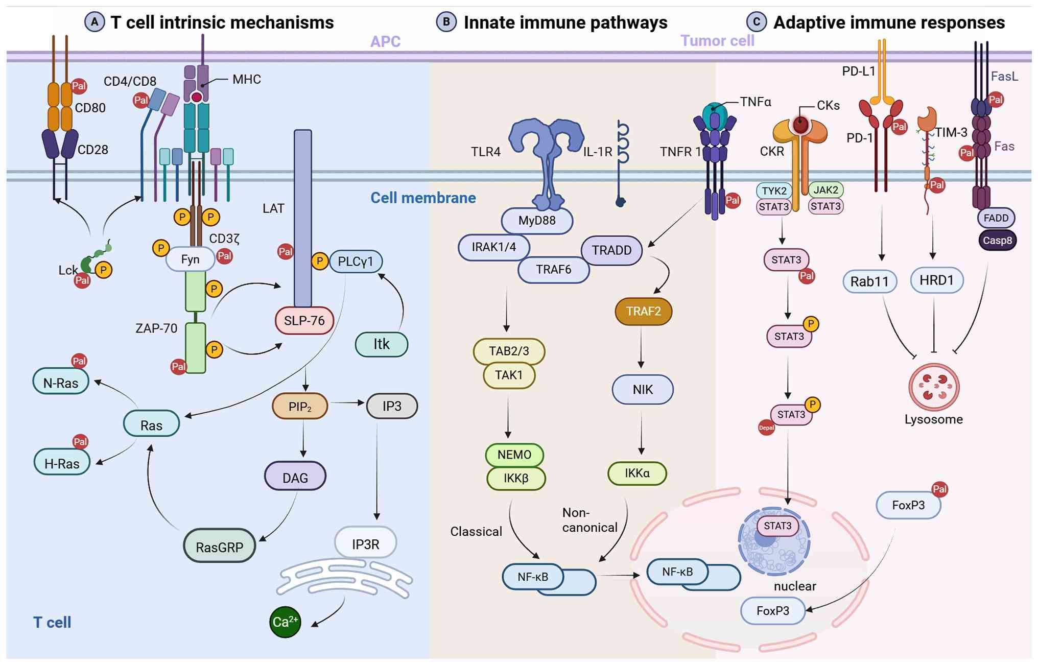 | Figure 1S-palmitoylation orchestrates T cell
signaling activation, proliferation, differentiation, exhaustion
and apoptosis. (A) T cell-intrinsic mechanisms. S-palmitoylation
promotes the membrane localization and lipid raft partitioning of
key T cell receptor-associated signaling molecules, including Lck,
Fyn, LAT and CD4/CD8, facilitating ZAP-70 recruitment and LAT
signalosome assembly. These membrane-proximal events activate
downstream pathways, including PLCγ1, Ras-MAPK and
calcium-dependent signaling cascades, which ultimately drive T cell
activation, proliferation and effector differentiation.
Palmitoylated Ras isoforms (N-Ras and H-Ras) are depicted. (B)
Innate immune pathways. S-palmitoylation dynamically regulates the
localization, trafficking, stability and signaling activity of
innate immune regulatory proteins. TLR4/IL-1R signaling through
MyD88-IRAK1/4-TRAF6 converges on the classical (TAK1/NEMO/IKKβ)
NF-κB activation pathway, whereas TNFR1 signaling through
TRADD/TRAF2 converges on the non-canonical (NIK/IKKα) NF-κB
activation pathway. (C) Adaptive immune responses. S-palmitoylation
of immune checkpoint molecules, including PD-1 and TIM-3, enhances
receptor stability by promoting receptor recycling (such as Rab11)
or preventing lysosomal degradation (such as HRD1), thereby
sustaining inhibitory signaling and contributing to T cell
exhaustion and tumor immune evasion. S-palmitoylation also
regulates TNFR1 and CKR/JAK2/STAT3 signaling (CKs include TGF-β,
IL-6 and IL-23; CKR denotes the corresponding receptors), as well
as Fas/FasL membrane organization, to modulate apoptotic signaling
and immune homeostasis. FoxP3 S-palmitoylation facilitates its
nuclear accumulation and maintains regulatory T cell suppressive
function. Figure created with BioRender.com. ZAP-70, ζ-chain-associated protein
kinase 70; LAT, linker for activation of T cells; PLCγ1,
phospholipase C γ1; NF-κB, nuclear factor κB; STAT3, signal
transducer and activator of transcription 3; FoxP3, forkhead box
P3; PD-1, programmed cell death protein 1; PD-L1, programmed
death-ligand 1; TIM-3, T cell immunoglobulin and mucin-domain
containing-3; TNFR1, tumor necrosis factor receptor 1; CKs,
cytokines; CKR, cytokine receptor; JAK2, Janus kinase 2; Fas, Fas
cell surface death receptor; FasL, Fas ligand; Pal,
S-palmitoylation; Depal, depalmitoylation. |
S-palmitoylation and membrane
localization of Src kinases
The earliest events of T cell activation are
orchestrated by a network of membrane-proximal signaling molecules
associated with the TCR complex. Among these, the Src-family
kinases Lck and Fyn function as key initiators of proximal TCR
signaling and play complementary roles in thymocyte development,
antigen recognition and mature T cell activation (56-59). A defining feature of Lck and Fyn
is their regulated association with the cytoplasmic leaflet of the
plasma membrane, which is mediated by sequential N-myristoylation
and reversible S-palmitoylation. Although N-myristoylation provides
an initial membrane-targeting signal, S-palmitoylation enhances
membrane affinity and directs these kinases into specialized lipid
raft microdomains, a process essential for their optimal
participation in proximal TCR signaling (60-63).
The role of S-palmitoylation in membrane targeting
has been well characterized for Fyn. Fyn undergoes S-palmitoylation
at two N-terminal cysteine residues, Cys3 and Cys6. A site-directed
mutagenesis study has demonstrated that modification of Cys3
represents the predominant and functionally indispensable
S-palmitoylation event required for its stable localization within
lipid rafts and cholesterol- and sphingolipid-enriched membrane
microdomains that function as signaling platforms (64). This modification is mediated by
the palmitoyl acyltransferase DHHC21 (65). Covalent attachment of the
palmitate moiety increases its hydrophobicity, facilitating stable
retention of Fyn within lipid rafts and enabling efficient
engagement with proximal signaling components, particularly the TCR
ζ-chain, where it phosphorylates ITAM tyrosines and cooperates with
Lck to initiate early TCR signaling (Fig. 1A) (57,66).
Similarly, the lymphocyte-specific kinase Lck, a
master regulator of proximal TCR signaling, is subject to stringent
regulation by reversible S-palmitoylation. Lck is also
palmitoylated at Cys3 and Cys5 (57,67,68). Using acyl-RAC to profile protein
lipidation, Fan et al (65) demonstrated that DHHC21 is
indispensable for Lck S-palmitoylation and for the propagation of
Lck-dependent apoptotic signaling in T cells, identifying DHHC21 as
a common regulatory node controlling multiple Src-family kinases.
Following antigen-induced TCR engagement, palmitoylated Lck is
constitutively enriched within the TCR-CD3 complex, where it
performs its canonical signaling function by phosphorylating paired
tyrosine residues within the ITAMs of CD3 subunits. Phosphorylated
ITAMs subsequently recruit the Syk family kinase ZAP-70, which is
further activated by Lck-mediated phosphorylation (64,69). Activated ZAP-70 propagates
proximal TCR signaling by phosphorylating downstream adaptor
proteins and signaling effectors, leading to robust amplification
of the TCR signaling cascade (Fig.
1A) (53).
The phosphorylation cascade initiated by Lck and Fyn
constitutes the molecular foundation of proximal TCR signal
transduction. Nevertheless, efficient propagation of these
signaling events depends not only on the catalytic activities of
these Src-family kinases but also on precise membrane localization,
dynamic assembly and coordinated regulation of downstream signaling
complexes that relay and amplify activation signals throughout the
T cell signaling network.
ZAP-70: A core kinase for signal
amplification and its regulation by S-palmitoylation
ZAP-70 serves as a central tyrosine kinase that
coordinates T cell immune responses. Dysregulated expression or
functional impairment of ZAP-70 has been implicated in diverse
human diseases, including immunodeficiencies, autoimmune disorders
and leukemia (70). Within the
TCR signaling cascade, antigen engagement activates the Src-family
kinase Lck, which subsequently mediates dual phosphorylation of
ITAMs within the TCR-associated CD3ζ chain (71). These phosphorylated ITAMs
function as high-affinity docking platforms for the SH2 domains of
ZAP-70, thereby promoting their recruitment to the TCR-CD3
signaling complex. ITAM binding induces conformational
rearrangements in ZAP-70 that enhances its accessibility to
Lck-mediated phosphorylation, ultimately resulting in
autophosphorylation-dependent full activation (70). Once activated, ZAP-70 drives the
assembly of proximal signaling complexes by phosphorylating key
adaptor proteins, including LAT and SLP-76, which subsequently
initiate PLCγ1 activation and downstream signaling cascades. This
signaling axis plays an essential role in T cell development,
activation and effector differentiation (55,72). In ZAP-70-deficient T cells,
defective phosphorylation of LAT and SLP-76 disrupts the formation
of the LAT signalosome and impairs downstream signal propagation,
further underscoring the central role of ZAP-70 in TCR signal
transduction (53,73,74).
Emerging evidence has indicated that ZAP-70 activity
is regulated by S-palmitoylation. Using the ABE assay, Cys564 was
identified as the principal S-palmitoylation site and this
modification was dependent on TCR stimulation. S-palmitoylation
mediates the stimulus-dependent translocation of ZAP-70 to discrete
plasma membrane microdomains, facilitating its interaction with
downstream effector molecules (Fig.
1A) (75). Therefore, these
findings demonstrate that S-palmitoylation of ZAP-70 is essential
for its function in the proximal TCR signaling pathway. Notably,
downregulation of DHHC21 concurrently suppresses the S-acylation of
Lck, Fyn and LAT, suggesting that this acyltransferase may serve as
a shared regulatory hub in the proximal TCR signaling network
(65,67). However, whether DHHC21 directly
catalyzes ZAP-70 S-palmitoylation, and the molecular determinants
underlying its substrate selectivity, remain unclear.
LAT: Signal integration and downstream
conduction
LAT, a transmembrane adaptor protein modified by
dual palmitoylation, is indispensable for T cell development and
activation (76). Within the
juxtamembrane cytoplasmic region, dual cysteine residues (Cys26 and
Cys29) undergo S-palmitoylation, directing LAT to the plasma
membrane to ensure proper TCR signal transduction (67,77). Although both residues are
modified, evidence from a LAT-deficient Jurkat cell system
indicates that S-palmitoylation at Cys26 is both necessary and
sufficient for plasma membrane localization and TCR signaling,
whereas the lipid raft partitioning reported in microdomain studies
is dispensable for its function (76,78).
A study by Hundt et al (76) demonstrated that anergic T cells
exhibit selective defects in LAT S-palmitoylation, underscoring the
fact that intact S-palmitoylation is required not only for T cell
development but also for productive TCR-driven activation. The loss
of LAT S-palmitoylation promotes its dissociation from the plasma
membrane and impairs its recruitment to immunological synapses.
This mis-localization prevents LAT from efficiently engaging
downstream signaling partners, resulting in attenuated signal
propagation and the consequent suppression of T cell activation
(69,79). However, the precise mechanism
through which S-palmitoylation supports LAT function remains
unclear. While S-palmitoylation is required for efficient plasma
membrane delivery and spatial organization of LAT, the extent to
which its function depends on its partitioning into biochemically
defined lipid rafts rather than broader membrane
sub-compartmentalization remains controversial. Hundt et al
(76) reported that although LAT
S-palmitoylation is essential for plasma membrane transport,
LAT-dependent signaling proceeds independently of lipid raft
integrity, indicating that its functional role may extend beyond
classical raft targeting. These results highlight the distinction
between the well-established requirement for S-palmitoylation for
membrane localization and the more nuanced assay-dependent evidence
concerning lipid raft partitioning. Further studies are warranted
to reconcile these observations and define the precise molecular
mechanisms through which LAT S-palmitoylation coordinates TCR
signal initiation and propagation.
Following TCR stimulation, multiple cytoplasmic
tyrosine residues within LAT are phosphorylated primarily by
Syk-family kinases, enabling LAT to serve as a central scaffold for
the recruitment of key signaling effectors, including PLCγ1 and
Grb2 (71,80). These adaptor proteins are
subsequently activated via ZAP-70-dependent signaling cascades.
Activated PLCγ1 hydrolyzes phosphatidylinositol 4,5-bisphosphate
into inositol 1,4,5-trisphosphate (IP3) and
diacylglycerol (DAG), initiating two major signaling branches:
IP3 triggers calcium release from the ER via its
receptor, whereas DAG activates downstream NF-κB and Ras/MAPK
pathways (Fig. 1A). As signal
transduction progresses, S-palmitoylation-dependent spatial
organization continues to modulate downstream signaling modules,
including MAPK and NF-κB cascades, ultimately driving T cell
activation, proliferation and cytokine production (67,71). Thus, these findings establish LAT
S-palmitoylation as a critical molecular determinant of both the
initiation and propagation of TCR signaling. Aberrant regulation of
this lipid modification represents a key mechanism underlying T
cell hypo-responsiveness
S-palmitoylation and T cell signal
transduction
CD4/CD8 co-receptors
CD4 and CD8 function as essential co-receptors in T
cell activation, selectively binding to MHC class II and class I
molecules, respectively, through their extracellular domains. This
interaction stabilizes the physical interface between T cells and
antigen-presenting cells, enhancing TCR engagement with
peptide-loaded MHC complexes and facilitating the efficient
initiation of antigen recognition (45,81). In addition to their roles in
peripheral T cell activation, both co-receptors are critically
involved in thymic selection, assisting the TCR complex in
discriminating between MHC class II and class I molecules,
ultimately directing lineage commitment toward the CD4+
or CD8+ T cell fate (82,83). The functional competence of these
co-receptors is tightly linked to their precise plasma membrane
localization and stability, processes centrally regulated by
S-palmitoylation.
CD4 undergoes site-specific S-palmitoylation at two
membrane-proximal cysteine residues (Cys396 and Cys399) in its
cytoplasmic tail (84-86). Although this lipid modification
is dispensable for CD4 trafficking and surface expression, it
significantly enhances CD4 partitioning into lipid raft
microdomains. This spatial organization positions CD4 in close
proximity to the TCR complex, enabling rapid co-receptor engagement
following antigen stimulation and promoting efficient propagation
of downstream signaling events (56,84). Furthermore, S-palmitoylation
enhances CD4 enrichment within lipid raft microdomains and promotes
its association with Lck, facilitating the assembly of
signaling-competent co-receptor complexes and sustaining efficient
TCR signal propagation (Fig. 1A)
(67,84).
CD8 predominantly exists as a disulfide-linked CD8αβ
heterodimer in conventional CD8+ T cells. S-acylation of
the CD8β cytoplasmic tail promotes its partitioning into lipid raft
microdomains, a process essential for optimal co-receptor function
(87,88). CD8 S-palmitoylation exhibits
marked species-specific divergence. In human T cells, both CD8α and
CD8β chains undergo S-palmitoylation, whereas in murine T cells,
S-palmitoylation is restricted primarily to CD8β, which is
essential for lipid raft targeting and optimal CD8 co-receptor
function (69,89). Functional studies have revealed
species-dependent mechanisms underlying CD8 S-palmitoylation. In
murine T cells, CD8β S-palmitoylation promotes CD8αβ partitioning
into lipid raft microdomains, facilitating its association with
raft-resident Lck and enhancing downstream TCR signaling. By
contrast, although both CD8α and CD8β undergo S-palmitoylation in
human T cells, this modification appears dispensable for raft
targeting; instead, CD8 localization is primarily dictated by
CD8β-mediated structural organization, whereas S-palmitoylation may
contribute to the assembly and stabilization of signaling-competent
CD8-TCR/CD3 complexes (Fig. 1A)
(67,69,87,88).
These findings establish S-palmitoylation as a
critical mechanistic node within the regulatory network governing T
cell immune responses. By dynamically controlling membrane
microdomain partitioning, protein stability and Lck coupling of CD4
and CD8, S-palmitoylation fine tunes antigen sensitivity and the
efficiency of T cell signaling, ultimately shaping the magnitude
and quality of adaptive immune responses.
Co-stimulatory signaling
Optimal T cell activation requires the integration
of dual-signal inputs; recognition of pMHC complexes by the TCR
delivers the primary activation signal, whereas co-stimulatory
molecules provide the essential secondary signal required for full
T cell activation (90). Among
these, CD28 is a central co-stimulatory receptor constitutively
expressed on the T cell surface. The engagement of CD28 with its
ligands B7.1 (CD80) or B7.2 (CD86) on antigen-presenting cells is
indispensable for initiating productive immune responses in naïve T
cells (91,92). Although CD28 possesses a
canonical extracellular immunoglobulin-like domain, its signaling
competence is largely dictated by its spatial organization within
specialized plasma membrane microdomains. The recruitment of CD28
to lipid rafts and immunological synapse-associated signaling
compartments facilitates the assembly of proximal signaling
complexes and amplifies downstream co-stimulatory responses
(93,94).
In parallel, the CD28 ligand CD80 is subject to
stringent regulation by S-palmitoylation. CD80 is a B7 family
transmembrane glycoprotein that critically modulates T cell
responses through interactions with CD28 and CTLA-4 (95,96). Using a metabolic labeling
strategy in 293T cells, Lu et al (97) demonstrated that CD80 undergoes
S-palmitoylation at multiple cysteine residues within its
transmembrane and intracellular regions, specifically at Cys261,
Cys262, Cys266 and Cys271 (Fig.
1A). This study, employing proximity-dependent biotin
identification and co-immunoprecipitation in 293T cells identified
ZDHHC20 as the principal palmitoyltransferase that catalyzes this
modification. In the same study, mechanistic experiments using
cycloheximide chase and lysosomal/proteasomal inhibition assays in
a 293T stable cell line overexpressing wild-type CD80-Flag or the
palmitoylation-deficient CD80-4CS-Flag mutant demonstrated that
S-palmitoylation protects CD80 from ubiquitination-mediated
degradation, tightly controlling its protein stability and ensuring
accurate trafficking and retention at the plasma membrane (97). By contrast,
S-palmitoylation-deficient CD80 mutants in CHO-K1 cells, as well as
endogenous CD80 in human Raji B-lymphoma cells treated with the
palmitoylation inhibitor tunicamycin, exhibit disrupted plasma
membrane localization, reduced protein stability and impaired
co-stimulatory capacity (97).
These findings from co-culture assays of CHO-K1 cells expressing
membrane-bound OKT3 single-chain variable fragment (anti-CD3
monoclonal antibody) and CD80 with human Jurkat T cells
collectively demonstrate that S-palmitoylation functions as a key
regulatory mechanism controlling CD80 function, ultimately
optimizing co-stimulatory signal transmission and reinforcing
effective T cell activation.
Nevertheless, whether CD28 itself undergoes direct
S-palmitoylation remains unknown, as the available evidence is
primarily derived from studies on CD80. Although CD28 partitions
into lipid rafts and its membrane organization is critical for
signaling, its direct S-palmitoylation, along with the responsible
residues and acyltransferases, remains unknown. Thus, while the
role of CD80 S-palmitoylation in co-stimulation is established,
whether CD28 undergoes direct S-palmitoylation remains unresolved
and warrants further investigation.
Taken together, S-palmitoylation has emerged as a
critical regulatory node in the molecular circuitry underlying T
cell immune responses. By dynamically coordinating membrane
microdomain partitioning, protein stability, and the co-receptor
organization of CD4 and CD8, this lipid modification fine-tunes
antigen recognition sensitivity and enhances the efficiency of T
cell signaling.
Ras signaling
Ras proteins are canonical small GTPases that
function as molecular switches, transitioning between an active
GTP-bound conformation and an inactive GDP-bound state to control
downstream signaling (98-101). TCR-mediated Ras activation is
essential for T cell development, proliferation and functional
differentiation by activating the Ras/RAF/MEK/ERK signaling cascade
(102-104). The Ras superfamily comprises
three major isoforms, H-Ras, N-Ras and K-Ras, with K-Ras existing
as two splice variants, K-Ras4A and K-Ras4B. All Ras isoforms share
a highly conserved G-domain, whereas sequence divergence is
primarily localized within the hypervariable region. These
structural differences, together with distinct lipid modifications,
confer isoform-specific membrane targeting and subcellular
distribution, ultimately shaping divergent downstream signaling
outputs (105-107).
Reversible S-palmitoylation is the central mechanism
that controls the subcellular localization of H- and N-Ras
(108). Following
S-palmitoylation by Golgi-resident palmitoyl acyltransferases, H-
and N-Ras are trafficked through the secretory pathway to the
plasma membrane (109). This
cycle precisely regulates Ras membrane residence and spatial
redistribution between the plasma membrane and Golgi compartments,
as demonstrated in 293T cells using fluorescence resonance energy
transfer and live cell imaging (108). Beyond determining gross
subcellular localization, lipid modifications also control lateral
partitioning within membrane microdomains, ensuring spatially
restricted and efficient signal propagation (67,105). In particular, palmitoylated
N-Ras preferentially associates with raft/non-raft boundary regions
across the plasma membrane, which is consistent with its distinct
signaling behavior and functional specificity (Fig. 1A) (105). Collectively, the
isoform-specific structural features, reversible S-palmitoylation
dynamics and compartmentalized trafficking behavior of Ras proteins
confer functional diversification within the Ras family,
positioning Ras as a key fate-determining signaling hub that
orchestrates T cell biology and directs cellular differentiation
programs.
Proliferation and differentiation of T
cells
Precise regulation of T cell proliferation and
differentiation constitutes a prerequisite for the acquisition and
execution of effector functions. Throughout these dynamic
developmental processes, S-palmitoylation has emerged as an
important post-translational regulatory mechanism that orchestrates
protein subcellular distribution and signaling activity.
Maintaining an appropriate balance of S-palmitoylation is crucial
for preserving the physiological proliferative potential and
directing lineage-specific differentiation programs, ultimately
supporting effective T cell-mediated immune responses and
sustaining immune homeostasis.
NF-κB signaling in T cell
proliferation: ZDHHC11/TRAF6 axis
NF-κB signaling is essential for T cell
proliferation, survival and effector differentiation (110,111). TRAF6, a member of the TNF
receptor-associated factor family, functions as a key adaptor
linking IL-1β and Toll-like receptor (TLR) signaling to downstream
NF-κB activation (112). A
study has identified ZDHHC11 as a positive modulator of NF-κB
signaling in response to IL-1β, lipopolysaccharide (LPS) and DNA
virus infection. Mechanistically, ZDHHC11 interacts with TRAF6
through its C-terminal region (amino acids 198-412) in a
domain-dependent manner. In 293T cells overexpressing these
proteins, this association promotes TRAF6 oligomerization, enhances
E3 ubiquitin ligase activity and facilitates TGF-β-activated kinase
1 (TAK1) activation. Functionally, in in 293 C6 cells (a
293-derived cell line ectopically expressing IL-1R; not PER.C6
cells) ectopically expressing IL-1R, ZDHHC11 overexpression
increased TRAF6-dependent IL-8 mRNA expression, whereas TRAF6
knockdown attenuated ZDHHC11-induced NF-κB activation (113). In mouse embryonic fibroblasts
and bone marrow-derived macrophages from Zdhhc11-knockout
(Zdhhc11-/-) mice, ZDHC11 deficiency reduced IL-1β- and
LPS-stimulated phosphorylation of TAK1, IKKα/β and p65, as well as
IL-6 and TNFα production. In vivo, Zdhhc11-/-
mice treated with LPS/D-galactosamine or infected with herpes
simplex virus 1 showed significantly lower serum IL-6 levels than
wild-type controls, confirming the physiological relevance of this
axis in acute inflammation and antiviral responses (113). Collectively, this regulatory
axis provides a mechanistic link between S-palmitoylation and
NF-κB-driven transcriptional programs that support T cell expansion
and functional differentiation (Fig.
1B) (113). However,
whether ZDHHC11 directly palmitoylates TRAF6 to drive this
regulation or if direct TRAF6 palmitoylation underlies this
regulation remains unclear.
STAT3
S-palmitoylation-depalmitoylation cycle drives Th17
differentiation
TH17 cells are a pro-inflammatory CD4+ T
cell subset characterized by the production of IL-17A, IL-17F and
other inflammatory mediators. Dysregulated expansion and
differentiation of TH17 cells, driven primarily by IL-6, TGF-β and
IL-23 signaling, contribute substantially to intestinal
inflammation and the pathogenesis of inflammatory bowel disease
(IBD) (114,115). Consistent with this, TH17 cell
abundance correlates positively with disease severity in both
experimental models of IBD and in patients (116-120). STAT3 is a master transcription
factor that integrates cytokine signals to regulate inflammatory
and malignant processes. In naïve CD4+ T cells, cytokine
stimulation activates Janus kinase (JAK) -dependent STAT3
phosphorylation, resulting in its nuclear translocation and the
transcriptional activation of Th17-associated genes, including RORC
and IL17A, thereby promoting Th17-cell differentiation (116,121-123).
Membrane recruitment of STAT3 is a prerequisite for
its efficient phosphorylation. A study has identified DHHC7 as the
principal palmitoyl acyltransferase that catalyzes S-palmitoylation
of STAT3 at Cys108 in both 293T cells and primary mouse
splenocytes. As a membrane-targeting lipid modification,
S-palmitoylation promotes the translocation of STAT3 to the plasma
membrane, facilitating its interaction with JAK2 and subsequent
phosphorylation. Functional analyses demonstrated that the
S-palmitoylation-deficient C108S mutant exhibited markedly reduced
phosphorylation compared with wild-type STAT3 in 293T cells and
mouse splenocytes under TH17-polarizing conditions. Conversely, the
phosphorylation-deficient STAT3 Y705F mutant retained normal
S-palmitoylation, indicating that DHHC7-mediated S-palmitoylation
promotes STAT3 activation, whereas STAT3 phosphorylation does not
reciprocally regulate its S-palmitoylation status (Fig. 1C) (116).
Following cytokine-induced activation,
phosphorylated STAT3 is selectively depalmitoylated by acyl protein
thioesterase 2 (APT2/LYPLA2) in 293T cells and mouse splenocytes,
facilitating its release from the plasma membrane and subsequent
nuclear translocation (116).
Thus, STAT3 activity is controlled by a reversible
S-palmitoylation-depalmitoylation cycle coordinated by DHHC7 and
APT2. DHHC7-mediated S-palmitoylation promotes membrane recruitment
and JAK-dependent phosphorylation, whereas APT2-mediated
depalmitoylation enables nuclear accumulation of activated STAT3
(116). This lipidation cycle
is essential for optimal STAT3 signaling and TH17-cell
differentiation in mouse splenocytes cultured under TH17-inducing
conditions, as the disruption of either modification step
significantly impairs TH17 lineage development, as assessed by flow
cytometry and qPCR (116). In a
dextran sulfate sodium (DSS)-induced mouse model of colitis
(C57BL/6J mice), pharmacological inhibition of APT2 using ML349 or
genetic knockout of Zdhhc7 alleviated colitis symptoms and reduced
TH17 cell frequencies in splenocytes (116). Moreover, in human peripheral
blood mononuclear cells from patients with IBD, including those
with Crohn's disease and ulcerative colitis, ZDHHC7 and LYPLA2 mRNA
levels were upregulated and correlated with STAT3 target gene
expression and p-STAT3 levels, supporting the pathophysiological
relevance of this cycle in human IBD (116). Overall, these findings
establish that the STAT3 S-palmitoylation-depalmitoylation cycle is
a critical regulatory mechanism controlling TH17-cell
differentiation and highlight its important contribution to the
pathogenesis of IBD in both murine models and human patients.
Loss of FoxP3 S-palmitoylation attenuates
the immunosuppressive function of regulatory T cells (Tregs)
Tregs are a specialized CD4+ T cell
subset that maintain immune homeostasis and prevent excessive
immune activation. The transcription factor FoxP3 functions as a
lineage-defining regulator of Tregs and is essential for their
development, stable identity and suppressive function (124-126). Accumulating evidence indicates
that FoxP3 activity is extensively regulated by diverse
post-translational modifications, including S-palmitoylation,
phosphorylation, ubiquitination, glycosylation and acetylation
(127,128).
To define the S-palmitoylation landscape of FoxP3,
Zhou et al (129)
generated FoxP3-Myc and FoxP3-Flag expression constructs,
overexpressed and affinity-purified the corresponding proteins in
293T cells and subsequently performed mass spectrometric analysis.
They identified five S-palmitoylation sites within FoxP3 (Cys204,
Cys218, Cys280, Cys281 and Cys424), each of which produced a
characteristic mass increase of 238 Da in the corresponding peptide
fragment. Subsequent co-immunoprecipitation analyses demonstrated
that ectopic expression of DHHC2, DHHC3 or DHHC7 markedly enhanced
FoxP3 S-palmitoylation. Mechanistically, S-palmitoylation-mediated
regulation of FoxP3 subcellular localization may promote its
nuclear enrichment and transcriptional activity, contributing to
the maintenance of Treg suppressive programs (Fig. 1C). Accordingly, ABE and acyl-RAC
assays confirmed that FoxP3 is palmitoylated in primary Tregs
isolated from the lymph nodes of C57BL/6 mice. In parallel, flow
cytometric analyses revealed that genetic deletion of Dhhc2 in
C57BL/6 mice, generated by CRISPR/Cas9 genome editing,
significantly increased the frequencies of activated
CD4+ and CD8+ T cells in the spleens.
Moreover, in a YUMM3.3 melanoma syngeneic mouse model,
T-cell-specific deletion of Dhhc2 (Dhhc2Flox/Flox;
Dhhc2-T cell KO) suppressed tumor growth, reduced intranuclear
FoxP3 expression in tumor-infiltrating Tregs and enhanced IFN-γ
secretion by tumor-infiltrating CD8+ T cells (129). These observations suggest that
FoxP3 S-palmitoylation is closely associated with the maintenance
of Treg suppressive function and may consequently modulate
antitumor T cell immunity.
Integrating these observations, deficiency of DHHC
enzymes responsible for FoxP3 S-palmitoylation markedly reduced the
nuclear accumulation of FoxP3 in both peripheral lymphoid tissues
and tumor-infiltrating Treg cells, resulting in impaired
immunosuppressive activity within the TME. These findings further
support the concept that S-palmitoylation-dependent regulation of
FoxP3 constitutes an important molecular mechanism controlling Treg
stability and function, with potential implications for antitumor
immune regulation.
S-palmitoylation in T cell exhaustion and
apoptosis
Following T cell proliferation and differentiation,
chronic antigen exposure progressively induces a state of T cell
exhaustion, characterized by diminished effector capacity and
persistent upregulation of inhibitory receptors. Negative
co-stimulatory receptors, including programmed cell death protein 1
(PD-1) and T cell immunoglobulin and mucin-domain containing-3
(TIM-3), serve as essential regulators of T cell responses by
constraining their magnitude and duration, thus preserving immune
equilibrium and preventing uncontrolled immune activation (130-132). Accordingly, immune checkpoint
pathways have emerged as key therapeutic targets for the treatment
of malignancies, autoimmune diseases and other immune-related
disorders.
Accumulating evidence indicates that
S-palmitoylation is closely associated with tumor initiation,
progression and therapeutic responsiveness (130,132,133). Given its multifaceted roles in
tumor biology, elucidating how S-palmitoylation regulates immune
checkpoint signaling in T cells is of considerable mechanistic and
translational significance. A more comprehensive understanding of
this regulatory axis is expected to provide a molecular framework
for the development of next-generation immunotherapeutic strategies
and further improve the efficacy of current immune checkpoint-based
therapies.
Tumor-cell-intrinsic PD-1
S-palmitoylation: Established evidence and unresolved relevance to
exhausted T cells
PD-1, a critical inhibitory receptor expressed on
the surface of T cells, is progressively upregulated following
effector T cell activation. Engagement of PD-1 by its ligands,
particularly programmed cell death ligand-1 (PD-L1), triggers the
recruitment of SHP-1/SHP-2 phosphatases to immunoreceptor
tyrosine-based switch motif, thereby attenuating proximal TCR/CD28
signaling, cytokine production, proliferation and cytotoxic
effector functions (130,131,133,134). Yao et al (135) identified Cys192, located at the
junction between the transmembrane domain and cytoplasmic tail of
PD-1, as a bona fide S-palmitoylation site, which was
subsequently validated by metabolic labeling assays in human
colorectal cancer (CRC) RKO and HCT116, melanoma A375 and
hepatoblastoma-derived HepG2 cells. In addition to serving as a
post-translational modification, S-palmitoylation also plays an
essential role in maintaining PD-1 protein stability. Elevated PD-1
expression has been observed in certain tumors, where it
contributes to tumor progression independent of adaptive immune
surveillance. Mechanistically, S-palmitoylation promotes the
interaction of PD-1 with Rab11, as demonstrated by
co-immunoprecipitation assays in HCT116, RKO, and LoVo cells,
facilitating PD-1 transport to recycling endosomes while limiting
its lysosomal degradation (Fig.
1C) (135). This process
results in sustained PD-1 expression and prolonged inhibitory
signaling. Consistently, treatment with the palmitoylation
inhibitor 2-bromopalmitate (2-BP) or a mutation of Cys192 to serine
disrupted PD-1-Rab11 binding and accelerated PD-1 degradation,
which was rescued by the lysosomal inhibitor chloroquine in RKO and
A375 cells. Although PD-1 S-palmitoylation stabilizes PD-1 and
enhances tumor-intrinsic mTOR signaling, as evidenced by the
increased phosphorylation of S6 and eIF4E following DHHC9
overexpression or palmostatin-B treatment in HepG2, NB4 and LoVo
cells (135), whether this
lipid modification regulates PD-1 function and metabolic
reprogramming in exhausted T cells remains unclear. Thus, these
results identify PD-1 S-palmitoylation as an important regulatory
mechanism controlling immune checkpoint signaling and highlight its
pharmacological targeting as a promising therapeutic strategy in
cancer immunotherapy.
DHHC9-mediated TIM-3 S-palmitoylation
stabilizes immune checkpoint molecules and drives immune
exhaustion
TIM-3 was initially identified as a cell-surface
receptor preferentially expressed in Th1 cells, in which
interaction with its ligand galectin-9 triggers inhibitory signals
that drive Th1-cell apoptosis (136,137). TIM-3 has emerged as a broadly
expressed immune checkpoint receptor in diverse immune cell
subsets, and its interactions with multiple ligands orchestrate
immune tolerance, inflammatory responses and immune homeostasis
(132,138,139).
Zhang et al (132) demonstrated that human TIM-3
undergoes S-palmitoylation at Cys296, a modification catalyzed by
the palmitoyl acyltransferase DHHC9. Mechanistically,
S-palmitoylation inhibits the interaction between TIM-3 and the E3
ubiquitin ligase HRD1, preventing TIM-3 polyubiquitination and
subsequent proteasomal degradation and ultimately stabilizing TIM-3
protein (Fig. 1C). Conversely,
depletion of DHHC9 alleviates CAR-T cell exhaustion, whereas
peptide-mediated inhibition of TIM-3 S-palmitoylation accelerates
TIM-3 turnover and enhances the antitumor activity of both CAR-T
and natural killer (NK) cells. In summary, these studies revealed
that S-palmitoylation serves as a critical regulatory axis
controlling TIM-3 stability and checkpoint function, which, in
turn, shapes the balance between immune suppression and antitumor
immunity. Targeting the DHHC9/TIM-3 S-palmitoylation pathway may
provide a promising strategy for reprogramming exhausted immune
cells and improving the efficacy of next-generation adoptive cell
therapies.
S-palmitoylation-depalmitoylation of
tumor necrosis factor receptor 1 (TNFR1): A binary switch steering
prosurvival vs. apoptotic signaling
As a pleiotropic cytokine, TNF-α coordinates
diverse biological processes, including inflammation, cellular
proliferation, differentiation and multiple forms of programmed
cell death (140-142). The biological activities of
TNF-α are primarily mediated through two receptors, TNFR1 and
TNFR2. Among these, TNFR1 contains a C-terminal death domain and
belongs to the death receptor superfamily, mediating the majority
of characterized TNF-induced cellular responses (142,143). Following ligand binding, TNFR1
undergoes oligomerization and serves as a signaling scaffold that
recruits multiple adaptor proteins to form a membrane-associated
signaling complex. This complex subsequently activates the NF-κB
and MAPK pathways to promote cell survival or, alternatively,
undergoes endocytosis to initiate apical caspase activation and
apoptotic signaling (141,144). The balance between survival and
apoptosis is determined by the selective assembly of distinct
signaling complexes that are closely linked to the subcellular
localization of TNFR1. TNFR1 retention at the plasma membrane
preferentially supports pro-survival signaling, whereas receptor
internalization redirects signaling toward apoptotic pathways
(145-147). Consequently, the
internalization and intracellular trafficking of TNFR1 constitute
essential steps in signal diversification, suggesting that
S-palmitoylation participates in coordinating receptor trafficking
to distinct signaling platforms.
S-palmitoylation of TNFR1 occurs during its
trafficking from the ER and Golgi apparatus to the plasma membrane.
In human monocytic U937 cells, although Cys248 is not the only
S-palmitoylation site, it is indispensable for efficient receptor
delivery to the plasma membrane and subsequent activation of NF-κB
signaling (Fig. 1C), as
demonstrated by reduced surface expression and impaired IκB
degradation in TNF-R1-knockout U937 cells reconstituted with the
C248S mutant (145). Activation
of TNFR1 requires depalmitoylation mediated by APT2, which permits
receptor redistribution into distinct lipid raft microdomains and
facilitates the recruitment of membrane-associated signaling
complex I, comprising TRADD, RIPK1 and TRAF2. In U937 cells, APT2
is transiently recruited to the TNFR1 signaling complex within 5-10
min of TNF stimulation, and its inhibition by the selective
inhibitor ML349 or shRNA-mediated knockdown blocks TNF-induced IκB
degradation (145). Conversely,
APT2 inhibition prevents the redistribution of activated TNFR1 and
retains the receptor within the plasma membrane caveolae, resulting
in enhanced neutral sphingomyelinase (nSMase) activity, increased
ceramide production and a subsequent shift toward apoptotic
signaling (145). In U937
cells, APT2 inhibition also enhances TNF-induced ceramide
generation and caspase-3 activation, effects that are reversed by
the nSMase inhibitor GW4869. These findings indicate that the
dynamic S-palmitoylation-depalmitoylation cycle of TNFR1 functions
as a molecular switch that determines cellular fate and is
indispensable for maintaining TNF-mediated signaling
homeostasis.
Fas/FasL S-palmitoylation dynamically
regulates apoptotic signaling and cellular homeostasis
Fas (also known as CD95; encoded by FAS, formerly
TNFRSF6) is a prototypical death receptor within the TNFR
superfamily. Following interaction with its cognate ligand FasL
(formerly TNFSF6), Fas initiates a well-orchestrated cascade of
signaling events. In primary cells, Fas transduces apoptotic
signals through the assembly of the death-inducing signaling
complex, which comprises the adaptor protein FADD, caspase
regulator c-FLIP and initiator protease caspase-8 (148-150).
Accumulating evidence indicates that efficient
propagation of Fas-mediated apoptotic signaling requires receptor
clustering within defined plasma membrane subdomains. Fas is
palmitoylated at an intracellular cysteine residue proximal to its
transmembrane domain (Cys199 in humans), which is essential for
inducing cell death (151,152). Mechanistic studies by Rossin
et al (153),
Chakrabandhu et al (151) and Hueber et al (154) demonstrated that the
pro-apoptotic function of Fas S-palmitoylation depends on two
interrelated processes: The formation of higher-order Fas
supramolecular clusters and the partitioning of Fas into
cholesterol- and sphingolipid-enriched membrane nanodomains. Both
processes are required for the optimal activation of Fas-triggered
apoptotic signaling. Loss of Fas S-palmitoylation leads to impaired
receptor internalization and attenuated activation of downstream
caspase cascades (151,154).
In human colorectal adenocarcinoma SW480 and SW620
cells, reduced Fas expression following the disruption of
S-palmitoylation suggests that S-palmitoylation-deficient Fas is
more susceptible to degradation. Indeed, compared with wild-type
Fas, both the total and plasma membrane-associated pools of
palmitoylation-deficient Fas C199V mutant exhibits markedly reduced
stability, indicating that S-palmitoylation stabilizes Fas at the
cell surface and prevents premature receptor turnover (153). Palmitoyl acyltransferase DHHC7
was identified as the principal enzyme catalyzing Fas
S-palmitoylation, as demonstrated by siRNA-mediated knockdown and
overexpression experiments coupled with ABE assays. DHHC7-mediated
S-palmitoylation preserves Fas expression by limiting lysosomal
degradation, as evidenced by the restoration of Fas protein levels
following treatment with the lysosomal inhibitors chloroquine and
bafilomycin A1, which in turn sustains Fas-dependent apoptotic
signaling (Fig. 1C) (153).
FasL is a type II transmembrane protein belonging
to the TNF superfamily. Upon binding to Fas, FasL induces apoptosis
in Fas-expressing target cells (155). FasL is predominantly expressed
in hematopoietic compartments in vivo, where it plays a
critical role in activation-induced cell death of T lymphocytes and
contributes to the cytotoxic effector functions of T and NK cells
(156). Using human WSU B cells
stably transfected with hFasL and co-cultured with Fas-expressing
human JH6.2 Jurkat T cells, Guardiola-Serrano et al
(156) demonstrated that human
FasL undergoes S-palmitoylation at Cys82, located within the
N-terminal region of its transmembrane domain (Fig. 1C). Using WSU cells expressing the
palmitoylation-deficient hFasLC82S mutant, subsequent experiments
revealed that ADAM10-mediated cleavage and processing of FasL
require intact cholesterol- and sphingolipid-rich membrane
nanodomains (lipid rafts) as well as Fas-FasL engagement. In WSU
cells expressing the receptor-binding-deficient hFasLA247E mutant,
which cannot interact with Fas, FasL processing by ADAM10 was
markedly reduced, indicating that receptor binding is required for
efficient cleavage. Proteolytic processing of FasL following
receptor binding confines its cytotoxic activity to engaged target
cells, thereby limiting bystander cell damage. S-palmitoylation of
FasL modulates its partitioning into lipid rafts, as demonstrated
by fluorescence correlation spectroscopy analysis of GFP-tagged
hFasL, hFasLC82S and hFasLA247E expressed in COS-7 cells,
representing a key regulatory mechanism that fine-tunes
ADAM10-dependent FasL processing and the magnitude of FasL-mediated
cytotoxicity (156).
Therefore, the dynamic
S-palmitoylation-depalmitoylation cycles of Fas and FasL critically
regulate membrane compartmentalization, protein stability and
signal output. This regulatory axis determines the balance between
cell survival and apoptosis, functioning as a molecular rheostat
that shapes immune homeostasis and contributes to cancer
pathogenesis.
Tumor-cell-intrinsic S-palmitoylation
regulating T cell immunity
S-palmitoylation serves as an important molecular
interface that couples metabolic dysregulation with immune escape
within the tumor immune microenvironment. Tumor cells exploit
ZDHHC3- and ZDHHC9-mediated S-palmitoylation to enhance PD-L1
stability and attenuate CD8+ T cell effector activity.
In parallel, ZDHHC24-dependent S-palmitoylation of B7 homolog 3
(B7H3) protein prevents its autophagic turnover, consequently
maintaining an immunosuppressive tumor milieu (157). Collectively,
tumor-cell-intrinsic S-palmitoylation orchestrates a multifaceted
immuno-evasive program by stabilizing immune checkpoint proteins at
the post-translational level while simultaneously reinforcing
metabolic adaptations that support tumor growth and immune evasion
(22,158).
In addition to PD-L1 and B7H3, emerging evidence
has extended the regulatory scope of tumor-cell-intrinsic
S-palmitoylation to other immunomodulatory targets. B7-H4
S-palmitoylation at Cys130, catalyzed by ZDHHC3, protects this
checkpoint molecule from lysosomal degradation, sustains its
immunosuppressive activity and promotes tumor progression (159). Likewise, S-palmitoylation of
IFN-γ receptor 1 (IFNGR1) at Cys122 governs receptor trafficking
and turnover, with its disruption compromising IFN-γ signaling
integrity and conferring intrinsic resistance to immune checkpoint
blockade (ICB) in CRC (160).
These findings collectively establish tumor-cell-intrinsic
S-palmitoylation as a key regulatory hub that links
post-translational lipid modifications to immune checkpoint
stability, metabolic adaptation and therapeutic responsiveness,
thereby offering multiple nodes for therapeutic intervention in
cancer immunotherapy (Fig.
2).
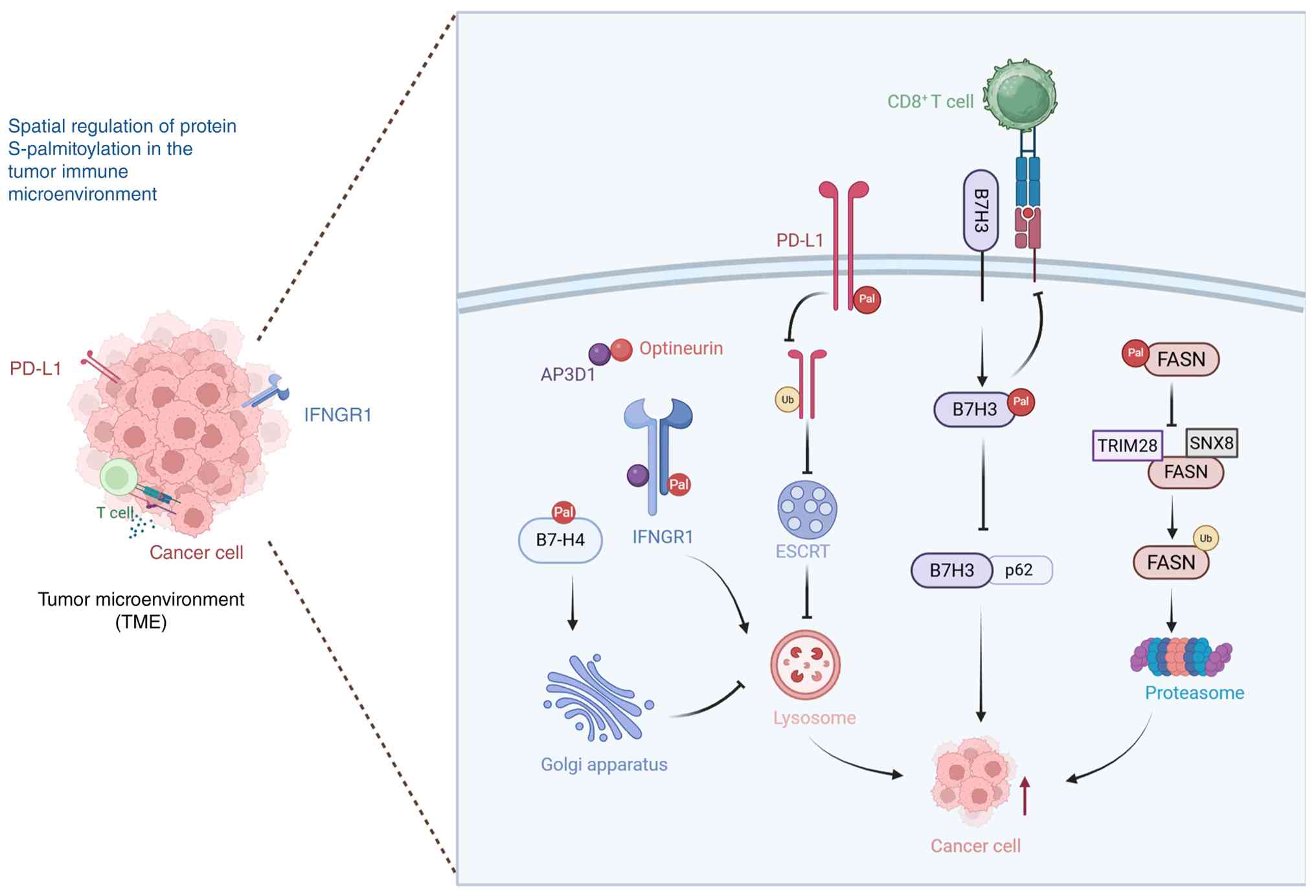 | Figure 2Spatial regulation of protein
S-palmitoylation in the tumor immune microenvironment. Left panel:
schematic overview of the TME, illustrating the interaction between
cancer cells and CD8+ T cells. Right panel: magnified
view of S-palmitoylation-mediated regulation of key
immune-regulatory and metabolic proteins in tumor cells. B7-H4 is
palmitoylated in the Golgi apparatus, which protects it from
lysosomal degradation and sustains its immunosuppressive function.
IFNGR1 S-palmitoylation, in coordination with optineurin, prevents
AP3D1-mediated lysosomal sorting and degradation, preserving IFN-γ
signaling integrity. S-palmitoylation of PD-L1 prevents its
ubiquitination and subsequent ESCRT-mediated lysosomal degradation,
thereby sustaining PD-L1 surface expression and attenuating
CD8+ T cell-mediated antitumor immunity.
S-palmitoylation of B7H3 disrupts its interaction with the
autophagy receptor SQSTM1/p62, limiting autophagic degradation and
enhancing B7H3 stability, which further suppresses CD8+
T cell activity. In addition, S-palmitoylation of FASN disrupts its
interaction with the TRIM28-SNX8 E3 ubiquitin ligase complex,
thereby preventing ubiquitin-proteasome-mediated degradation and
promoting de novo fatty acid synthesis. Collectively, these
spatially regulated S-palmitoylation events promote tumor immune
evasion and metabolic adaptation. Figure created with BioRender.com. PD-L1, programmed death-ligand 1;
B7H3, B7 homolog 3; B7-H4, B7 homolog 4; IFNGR1, interferon γ
receptor 1; FASN, fatty acid synthase; ESCRT, endosomal sorting
complex required for transport; Ub, ubiquitin; Pal,
S-palmitoylation; TME, tumor microenvironment. |
PD-L1 S-palmitoylation: A lipidic
rheostat tuning tumor immune evasion
PD-L1 is a type I transmembrane glycoprotein that
is widely expressed across diverse tumor types (161). Under physiological conditions,
activated antigen-specific cytotoxic T lymphocytes (CTLs) recognize
and directly eliminate malignant cells. However, tumor cells with
elevated PD-L1 expression effectively escape immune surveillance by
interacting with PD-1 on infiltrating immune cells. The PD-1/PD-L1
interaction induces exhaustion of infiltrating CD8+
CTLs, compromises tumor-reactive CTL responses and suppresses the
function of Tregs, which collectively attenuate immune activation
and ultimately facilitate tumor progression into the surrounding
tissues (162,163). Beyond its immunosuppressive
activity, PD-L1 provides intrinsic protection against apoptosis in
tumor cells, underscoring the PD-1/PD-L1 signaling axis as an
attractive target for cancer immunotherapy (163,164).
Studies have shown that S-palmitoylation is a
critical determinant of PD-L1 protein stability. Yang et al
(165) demonstrated that in
breast cancer cells, palmitoyl acyltransferase ZDHHC9 catalyzes
PD-L1 S-palmitoylation at Cys272, thus enhancing PD-L1 stability.
Similarly, Yao et al (166) reported that ZDHHC3 mediates
PD-L1 S-palmitoylation at the same residue in CRC. This
modification sustains elevated PD-L1 expression by suppressing
ubiquitination and preventing its trafficking into multivesicular
bodies via the endosomal sorting complex required for transport
pathway, which reduces lysosomal degradation and sustains immune
checkpoint activity (Fig. 2). In
human CRC RKO and HCT116 cells, treatment with the marine natural
product benzosceptrin C (BC) inhibited DHHC3, blocked PD-L1
palmitoylation at Cys272 and promoted PD-L1 degradation through the
lysosomal pathway. Consistently, in MC38 tumor-bearing C57BL/6
mice, BC administration reduced tumoral PD-L1 expression, increased
tumor-infiltrating CD8+ T cells and suppressed tumor
growth (163). Together, these
findings demonstrate that S-palmitoylation represents a key
post-translational regulatory mechanism controlling PD-L1 protein
stability. Furthermore, it highlights the broader role of
S-palmitoylation in shaping tumor immune escape and remodeling of
the tumor immune microenvironment.
B7H3 S-palmitoylation: Maintenance of
immunosuppression through the inhibition of autophagy-mediated
degradation
B7H3 (also known as CD276) is a type I
transmembrane protein that belongs to the B7 immunoglobulin
superfamily (167). B7H3 is
aberrantly upregulated across a broad spectrum of malignancies and
its elevated expression is closely associated with unfavorable
clinical outcomes and poor patient prognosis (168-170). Accumulating evidence indicates
that therapeutic blockade of B7H3 enhances CD8+ T cell
infiltration, restores effector function and restrains tumor
progression, highlighting B7H3 as a promising target for cancer
immunotherapy (168,171,172).
Rao et al (157) demonstrated that palmitoyl
acyltransferase ZDHHC24 catalyzes the S-palmitoylation of B7H3 at
Cys496. In human (RKO and SW480) and murine (CT26 and MC38) CRC
cell lines, this lipid modification disrupts the B7H3-SQSTM1/p62,
interaction, limiting autophagic degradation and enhancing B7H3
protein stability, as demonstrated by acyl-biotin exchange,
cycloheximide chase and co-immunoprecipitation assays. Increased
B7H3 expression suppressed CD8+ T cell-mediated
antitumor immunity and promoted an immunosuppressive TME (Fig. 2). In a colitis-associated CRC
mouse model using intestinal epithelial cell-specific
Zdhhc24-deficient (Zdhhc24ΔIEC) mice on a C57BL/6J
background, disruption of this regulatory axis significantly
enhanced CD8+ T cell-dependent antitumor responses. This
effect was observed either through mutation of the C496 residue in
CT26 and MC38 cells implanted subcutaneously in immunocompetent
BALB/c and C57BL/6J mice, or through genetic deletion of ZDHHC24,
and was evidenced by increased tumor-infiltrating CD8+ T
cells, elevated granzyme B and IFN-γ expression as well as reduced
tumor growth (157). At the
mechanistic level, Cys496 S-palmitoylation stabilizes B7H3 by
preventing autophagic turnover, allowing B7H3 to maintain its
immunosuppressive activity within the TME and facilitating tumor
immune escape. Collectively, these findings establish B7H3
S-palmitoylation as a critical post-translational mechanism that
regulates immune evasion by controlling protein stability and
identify the ZDHHC24/B7H3 axis as a potential therapeutic target
for restoring CD8+ T cell-mediated antitumor
responses.
B7-H4 S-palmitoylation: Evading lysosomal
degradation and enhancing immune suppression
B7-H4 (also known as VTCN1, B7S1 or B7x) is a
type-I transmembrane glycoprotein belonging to the B7 family.
Through interactions with an unidentified receptor expressed on
activated T cells, B7-H4 suppresses T cell proliferation and
cytokine production (173-175). In addition to its
immunomodulatory activity, B7-H4 exerts diverse pro-tumorigenic
functions by promoting tumor cell proliferation, invasion,
metastasis and resistance to apoptosis. Aberrant upregulation of
B7-H4 has been documented across multiple human malignancies,
including breast, ovarian and uterine cancer. In these settings,
B7-H4 functions as an immune checkpoint molecule that reinforces
the immunosuppressive TME (176,177).
Yan et al (159) demonstrated that the palmitoyl
acyltransferase ZDHHC3 catalyzes S-palmitoylation of B7-H4 at
Cys130 in human (MDAMB468 and T47D) and mouse (4H11 and Py230)
breast cancer cell lines. Mechanistically, this lipid modification
protected B7-H4 from lysosomal degradation, resulting in sustained
protein stability and persistent immunosuppressive activity. As
ZDHHC3 is predominantly localized in the Golgi apparatus, B7-H4
S-palmitoylation likely occurs during Golgi-associated protein
trafficking. Consistent with these observations, genetic depletion
of ZDHHC3 in 4H11 mouse breast cancer cells elicited robust
antitumor immune responses and markedly suppressed tumor
progression in subcutaneous syngeneic C57BL/6 mouse models.
In addition, the FDA-approved cyclin-dependent
kinase 4 and 6 (CDK4/6) inhibitor, abemaciclib, reduced B7-H4
protein abundance independent of CDK4/6 inhibition or cell cycle
arrest, leading to enhanced antitumor immunity and reduced tumor
growth (159). Given that
ZDHHC3-mediated S-palmitoylation stabilizes B7-H4 by preventing
lysosomal degradation, the selective inhibition of ZDHHC3 or
pharmacological manipulation of the lysosomal degradation pathway
may destabilize B7-H4 and accelerate its turnover, ultimately
enhancing antitumor immune responses (Fig. 2) (159). Thus, these findings identify
B7-H4 S-palmitoylation as an important post-translational mechanism
that supports tumor immune evasion. They further highlight
therapeutic targeting of the ZDHHC3/B7-H4 axis, together with
repurposing of abemaciclib, as a promising strategy for the
treatment of B7-H4-positive malignancies.
IFNGR1 S-palmitoylation: Maintaining
signal integrity to counteract immunotherapy resistance
Although ICB has achieved durable clinical
responses in multiple malignancies, its efficacy in CRC remains
largely limited to molecularly-defined subsets of patients. For
instance, ~15% of CRCs exhibit microsatellite instability-high or
deficient mismatch repair phenotypes, which are characterized by
elevated neoantigen burden and enhanced responsiveness to
PD-1/PD-L1 blockade. However, the majority of microsatellite-stable
CRCs remain refractory to current immunotherapeutic approaches
(178-180).
IFNγ signaling represents a central axis of
antitumor immunity; it enhances tumor immunogenicity, promotes
antigen presentation and coordinates immune cell recruitment. IFNγ
induces the expression of MHC class I and class II molecules,
stimulates IL-12 production by antigen-presenting cells, reinforces
Th1 differentiation through the STAT1/T-bet pathway and drives
CXCL9/CXCL10-mediated recruitment of CTLs and NK cells into the
TME, thereby facilitating effective immune-mediated tumor control
(181,182).
IFNGR1 undergoes S-palmitoylation at Cys122. Under
physiological conditions, the adaptor protein AP3D1 recognizes
palmitoylated IFNGR1 and directs the receptor for lysosomal
degradation. Conversely, optineurin, an immune regulatory adaptor
protein, interacts with AP3D1 and prevents lysosomal trafficking of
palmitoylated IFNGR1, preserving receptor stability and maintaining
the integrity of IFNγ signaling as well as MHC class I
antigen-presentation pathways (Fig.
2) (160). In human (LS174T
and DLD1) and murine (MC38 and CT26) colon cancer cell lines,
optineurin deficiency (via shRNA knockdown or CRISPR/Cas9 knockout)
compromises these processes, promotes immune evasion and confers
intrinsic resistance to ICB. By contrast, loss of IFNGR1
S-palmitoylation via the C122A mutation abolishes the IFNGR1-AP3D1
interaction, stabilizes IFNGR1 and enhances IFNγ/MHC-I signaling
(160). In syngeneic mouse
models (C57BL/6 MC38 and BALB/c CT26 tumor-bearing mice) and an
azoxymethane/DSS-induced colitis-associated CRC model with
intestinal epithelial cell-specific optineurin deletion
(optineurinΔIEC mice), optineurin deficiency accelerated
tumor growth, impaired CD8+ T cell effector function and
reduced the efficacy of anti-PD-L1 therapy (160). Based on these results, Du et
al (160) proposed in a
proof-of-concept study that therapeutic strategies aimed at
stabilizing IFNGR1 via modulation of its S-palmitoylation status,
may overcome intrinsic immunotherapy resistance in CRC. In
preclinical models, pharmacological inhibition of IFNGR1
S-palmitoylation using the palmitoylation inhibitor cerulenin
enhanced antitumor immune responses and sensitized previously
resistant tumors to ICB. Together, these findings establish dynamic
regulation of IFNGR1 S-palmitoylation as a critical determinant of
IFNγ signaling integrity and responsiveness to immune checkpoint
therapy in CRC. In addition, they highlight the IFNGR1-optineurin
signaling axis as an attractive therapeutic target for overcoming
intrinsic resistance to cancer immunotherapy.
S-palmitoylation in dendritic cells (DCs)
and other antigen-presenting or myeloid cells
DCs are the most potent professional
antigen-presenting cells and serve as central orchestrators of
adaptive immune responses while maintaining critical functions in
innate immune surveillance (183,184). Therefore, a comprehensive
understanding of post-translational modifications that regulate DC
biology is crucial for elucidating the complex molecular mechanisms
underlying immune regulation. Utilizing click chemistry-based
proteomic profiling combined with high-resolution mass
spectrometry, researchers have identified >550 palmitoylated
proteins in resting bone marrow-derived DCs, including key
components of TLR signaling pathways (TLR2 and TLR9),
co-stimulatory molecules (CD80 and CD86), transcriptional
regulators (NF-κB1 and NEMO) and metabolic enzymes (185,186). Among these substrates, the
S-palmitoylation of TLR2 at Cys609 is essential for proper membrane
localization and functional responsiveness to microbial ligands
(69).
Notably, a 2024 study by Ni et al (187) revealed a distinct cyclical
mechanism of TLR9 regulation. In this model ZDHHC3 mediates
S-palmitoylation of TLR9 in the Golgi apparatus, whereas PPT1
catalyzes its depalmitoylation in lysosomes. In murine RAW264.7
macrophages and 293T cells overexpressing TLR9, as well as in mouse
Flt3L-derived plasmacytoid DCs (pDCs) and bone marrow-derived
macrophages, this dynamic cycle precisely controls TLR9
trafficking, its release from the chaperone UNC93B1 and subsequent
ligand responsiveness, thereby providing a direct regulatory layer
for TLR9 function at the level of the receptor itself (187). The physiological significance
of this regulatory axis was further validated in a B6.Sle1yaa mouse
model of systemic lupus erythematosus (SLE), in which genetic
deficiency of PPT1 or pharmacological inhibition with HDSF reduced
antinuclear antibody levels and attenuated nephritis. Furthermore,
in vitro treatment with HDSF suppressed IFNα production by
pDCs in both patients with SLE and healthy donors, confirming the
translational relevance of this mechanism in human immune cells
(187). Notably, this mechanism
differs from TLR9-driven palmitoylome remodeling in DCs, which
results from broad alterations in the cellular S-palmitoylation
landscape rather than the direct modification of TLR9.
Activation of TLR9 induces extensive remodeling of
the DC S-palmitoylation landscape. This remodeling results in
increased modification of 185 proteins, which are significantly
enriched in pathways associated with NF-κB signaling, the cyclic
GMP-AMP synthase (cGAS)-stimulator of interferon genes (STING)
pathway and mitochondrial respiratory chain function (183). These findings indicate that
S-palmitoylation contributes to metabolic reprogramming associated
with DC activation. Notably, the palmitoyl transferase ZDHHC9
displays substrate-selective activity in DCs; its deficiency
selectively reduces TLR9-induced S-palmitoylation of NF-κB1, NEMO,
interferon-induced transmembrane protein 3 and several metabolic
enzymes, including NADH dehydrogenase ubiquinone flavoprotein 1 and
ubiquinol-cytochrome c reductase iron-sulfur protein 1. However,
depletion of ZDHHC9 does not affect DC differentiation, maturation
or their capacity to activate T cells, suggesting functional
compensation among members of the ZDHHC enzyme family (185,186).
In addition to functioning as an endogenous
post-translational modification, S-palmitoylation can also be
exploited exogenously as a chemical tool to modulate antigen
presentation. Exogenously palmitoylated antigenic peptides provide
an innovative approach to enhance DC-mediated cross-presentation by
virtue of their intrinsic membrane-integrating properties. The
covalent attachment of a single palmitic acid moiety (C16:0) to the
N-terminus of antigenic peptides enables efficient
receptor-independent incorporation into the DC lipid bilayer,
facilitating rapid cellular uptake. Following internalization,
these peptides undergo processing through both cytosolic
proteasome-dependent degradation and vacuolar processing pathways,
substantially enhancing MHC class I peptide loading and subsequent
CD8+ T cell activation (188). Notably, in Batf3-deficient
mice, palmitoylated antigenic peptides are efficiently
cross-presented by DCs, demonstrating that this process does not
strictly depend on conventional cross-presenting DC subsets.
Moreover, this strategy can be directly applied to tumor cells or
tumor-derived apoptotic vesicles, increasing the abundance of
MHC-I/peptide complexes on tumor cell surfaces and enhancing
CTL-mediated recognition and elimination. These modifications
ultimately confer robust tumor-suppressive effects in vivo
(188).
This exogenous application is mechanistically
distinct from the endogenous cysteine S-acylation catalyzed by
ZDHHC palmitoyltransferases. Although both involve covalent
palmitate attachment, the exogenous conjugation of palmitic acid to
the N-terminus of antigenic peptides does not rely on enzymatic
catalysis or target endogenous cysteine residues. Instead, it
exploits the physicochemical properties of the lipid moiety to
facilitate membrane integration and antigen uptake. Although both
strategies rely on palmitate modification, their mechanisms,
downstream effects and applications differ fundamentally.
Collectively, endogenous S-palmitoylation
represents an important regulatory mechanism controlling DC
signaling and functional activation, whereas exogenous
palmitoylated antigenic peptides provide an effective strategy for
enhancing antigen presentation. Together, these findings establish
S-palmitoylation as a fundamental regulatory axis in DC-mediated
immune regulation and highlight its potential for therapeutic
manipulation of antitumor immunity.
Metabolism-S-palmitoylation crosstalk in the
TME
Aberrant fatty acid metabolism within the TME is
characterized by enhanced de novo lipogenesis and increased
uptake of extracellular fatty acids, resulting in substantial local
accumulation of palmitate (189,190). Metabolic reprogramming reshapes
cellular lipid availability and palmitoyl-CoA metabolism by
modulating global protein S-palmitoylation profiles. Conversely,
S-palmitoylation dynamically regulates the localization, stability
and functional activity of metabolic enzymes and transporters,
revealing an interconnected regulatory network linking lipid
metabolism with post-translational modification (11,191-193). A well-characterized example of
this regulatory interplay was provided by Hao et al
(194), who elucidated the
critical role of dynamic CD36 S-palmitoylation in controlling fatty
acid uptake in murine 3T3-L1 adipocytes and primary adipocytes
derived from stromal vascular fractions of wild-type and
Cd36-knockout mice, as well as in high-fat-diet-induced obesity
models. Under basal conditions, S-palmitoylation maintains CD36
enrichment within the plasma membrane lipid rafts, ensuring its
functional availability for fatty acid sensing and transport.
Following binding to long-chain fatty acids, CD36 activates the
downstream kinase, LYN, which subsequently phosphorylates and
inhibits DHHC5, a palmitoyl acyltransferase responsible for CD36
modification. This inhibition triggers APT1-mediated CD36
depalmitoylation. The depalmitoylated CD36 then recruits SYK kinase
and initiates VAV- and JNK-dependent caveolae-mediated endocytosis,
ultimately promoting fatty acid internalization (194). This reversible
S-palmitoylation-depalmitoylation cycle enables CD36 to dynamically
adapt to fluctuations in extracellular fatty acid availability and
precisely adjust fatty acid uptake efficiency. However, it should
be noted that this model was primarily established in adipocytes
and validated in the context of highfatdietinduced metabolic
stress. Its direct applicability to tumor or immune cells within
the TME remains to be experimentally verified.
S-palmitoylation of fatty acid synthase (FASN) is
another important regulatory mechanism involved in metabolic
reprogramming. Mo et al (195) demonstrated that, in human
hepatocellular carcinoma (HCC) cell lines (Huh7, HCCLM3 and Hep3B)
and the hepatoblastoma-derived HepG2 cell line, as well as in
diethylnitrosamine (DEN) and DEN/carbon tetrachloride-induced HCC
mouse models, the palmitoyl transferase ZDHHC20 catalyzed FASN
S-palmitoylation at cysteine residues 1471 and 1881. This
modification prevented ubiquitin-proteasome-mediated degradation of
FASN by disrupting its interaction with the E3 ubiquitin ligase
complex SNX8-TRIM28, consequently stabilizing FASN protein
abundance and sustaining de novo fatty acid synthesis
(Fig. 2). Genetic deletion of
ZDHHC20 or mutation of FASN S-palmitoylation sites accelerated FASN
turnover and markedly suppressed chemically-induced
hepatocarcinogenesis. Overall, these findings demonstrated that the
palmitate-driven equilibrium between S-palmitoylation and
depalmitoylation coordinates two fundamental aspects of fatty acid
metabolism: Transport mediated by CD36 and synthesis mediated by
FASN. This reciprocal regulatory network integrates metabolic
adaptation with lipid modification, linking tumor metabolic
reprogramming to S-palmitoylation-dependent signaling regulation;
it further identifies this axis as a potential therapeutic
vulnerability for targeting tumor metabolism.
Therapeutic strategies, supporting evidence
and translational barriers
Therapeutic strategies targeting S-palmitoylation
in cancer immunotherapy primarily involve two complementary
approaches: Broad-spectrum inhibition of S-palmitoylation and
selective targeting of individual ZDHHC palmitoyltransferases.
Although the pan-S-palmitoylation inhibitor, 2-BP, exhibits potent
antitumor activity by suppressing tumor cell proliferation,
inducing apoptosis and displaying synergistic effects in
combination with chemo- or immunotherapy, its clinical application
remains limited owing to poor target specificity, substantial
off-target effects, and unfavorable pharmacokinetic properties
(196-201). Consequently, the development of
subtype-selective ZDHHC inhibitors has become a major research
focus. Several ZDHHC enzymes, including ZDHHC3, ZDHHC12 and
ZDHHC15, have been identified as potential therapeutic targets;
however, selective small-molecule inhibitors of these enzymes
remain largely confined to preclinical investigations (197,198).
Targeting S-palmitoylation also provides
opportunities for synergistic integration with the established
anticancer therapies. In combination with an ICB, transmembrane
peptides designed to disrupt B7H3 S-palmitoylation accelerate B7H3
degradation and enhance the efficacy of anti-PD-1 monoclonal
antibodies by restoring CD8+ T cell effector activity.
In the context of metabolic intervention, enhanced carnitine
palmitoyltransferase 1A (CPT1A) expression promotes mitochondrial
antiviral signaling protein (MAVS) S-palmitoylation and type I
interferon signaling, whereas pharmacological inhibition of ZDHHC8
by PF-670462 induces glutathione peroxidase 4 (GPX4) degradation
and triggers ferroptotic cell death, both of which improve tumor
responsiveness to PD-1-based immunotherapy (202,203). Furthermore, combining
S-palmitoylation-targeted approaches with chemotherapy or molecular
targeted therapies may overcome treatment resistance. For example,
siRNA-mediated inhibition of ZDHHC12 increases cisplatin
sensitivity through reactive oxygen species (ROS) accumulation and
targeting ZDHHC2 reverses enzalutamide resistance (204,205). In addition, pharmacological
inhibition of FASN with orlistat reduces PD-L1 S-palmitoylation and
membrane stability, consequently enhancing antitumor immune
responses (206). Thus, these
combination strategies exploit the multifaceted functions of
S-palmitoylation in regulating immune checkpoint stability,
metabolic adaptation and tumor cell survival. By promoting
immunogenic cell death (ICD), reshaping the tumor immune
microenvironment and restoring metabolic homeostasis, therapeutic
modulation of the S-palmitoylation machinery represents a promising
avenue for overcoming acquired resistance and expanding the
efficacy of current cancer treatment modalities.
Monotherapy targeting
S-palmitoylation
2-BP is the most widely used broad-spectrum
inhibitor of protein S palmitoylation. As a palmitate analog, 2-BP
inhibits the DHHC family of palmitoyl acyltransferases by
covalently modifying the catalytic cysteine within the DHHC motif,
thereby blocking auto-S-palmitoylation and subsequent substrate
acylation. However, its broad reactivity toward multiple cellular
targets should be considered when interpreting the experimental
results (196,207). Several studies have
demonstrated the antitumor activity of 2-BP in multiple cancer
types. For instance, in Burkitt's lymphoma cells, 2-BP reduced cell
viability in a dose-dependent manner by triggering a marked
increase in apoptosis, with similar outcomes observed in other
B-cell lymphoma subtypes (196,197). In glioma models, 2-BP inhibited
GL261 cell growth in a time- and dose-dependent manner, promoted
apoptotic cell death and attenuated autophagic activity (198). Furthermore, 2-BP reduced PD-L1
abundance in tumor cells and tumor-derived exosomes, alleviated
immunosuppressive signaling and enhanced the antitumor efficacy of
cisplatin-based therapy (199).
In HCC, 2-BP suppressed GSDME S-palmitoylation, thereby limiting
lenvatinib-induced pyroptotic responses (200).
Despite its broad experimental utility, the
clinical translation of 2-BP remains severely restricted by
intrinsic limitations, notably its lack of target selectivity.
Proteomic profiling of 2-BP-treated Burkitt's lymphoma cells
revealed 1,089 differentially expressed proteins, with only a
proportion directly attributable to known
S-palmitoylation-dependent pathways, highlighting extensive
downstream perturbations and potential off-target effects
associated with global S-palmitoylation inhibition (197,201). Significant cytotoxicity has
also been reported; for example, treatment with 200 μM 2-BP
induced extensive cell death in B-cell lymphoma, resulting in the
exclusion of the highest concentration from subsequent analyses
(197). Moreover, certain tumor
models exhibit limited responsiveness to 2-BP, as demonstrated by
the minimal sensitivity of glioma C6 cells, which may be attributed
to insufficient drug uptake or elevated expression of S100 proteins
(198). Thus, these limitations
have confined 2-BP primarily to use as a preclinical research tool
with limited potential for clinical applications. As a
broad-spectrum inhibitor, 2-BP has been used primarily as a
mechanistic tool, with antitumor effects demonstrated in
vitro and in animal models. However, it has not yet been
evaluated in humanized models, pharmacokinetic/pharmacodynamic
studies or clinical trials.
Given these challenges, the development of
selective inhibitors that target individual ZDHHC isoforms has
become an important direction in this field. In B-cell lymphoma,
Liu et al (197)
evaluated cyano-myracrylamide (CMA), another broad-spectrum
S-palmitoylation inhibitor, and found that it induced apoptosis at
relatively low concentrations while displaying reduced inhibitory
activity toward ZDHHC3 and ZDHHC12. Although CMA exhibits partial
subtype preference, it lacks sufficient selectivity for therapeutic
applications. Thus, CMA also remains a broad mechanistic tool with
limited subtype selectivity and has not advanced beyond in
vitro pharmacological validation. Tang et al (198) identified ZDHHC12, ZDHHC15,
ZDHHC22 and ZDHHC23 as important palmitoyltransferases involved in
glioma biology. Elevated expression of ZDHHC12 and ZDHHC15 was
correlated with poor prognosis and activation of the PI3K/AKT
signaling pathway, suggesting their potential value as druggable
targets. However, selective small-molecule inhibitors against these
ZDHHC subtypes have not yet been developed and current functional
investigations remain largely dependent on genetic approaches,
including gene knockdown and overexpression strategies. These
results constitute genetic proof-of-concept. However, no selective
small-molecule inhibitors are currently available, and in
vivo validation in immunocompetent models remains lacking.
Synergistic strategies combining
S-palmitoylation modification with antitumor therapy
Targeting S-palmitoylation has emerged as a
promising therapeutic strategy for enhancing the antitumor efficacy
of conventional chemotherapeutic agents, metabolic interventions
and ICB, exhibiting substantial synergistic potential across
diverse cancer models (Fig.
3).
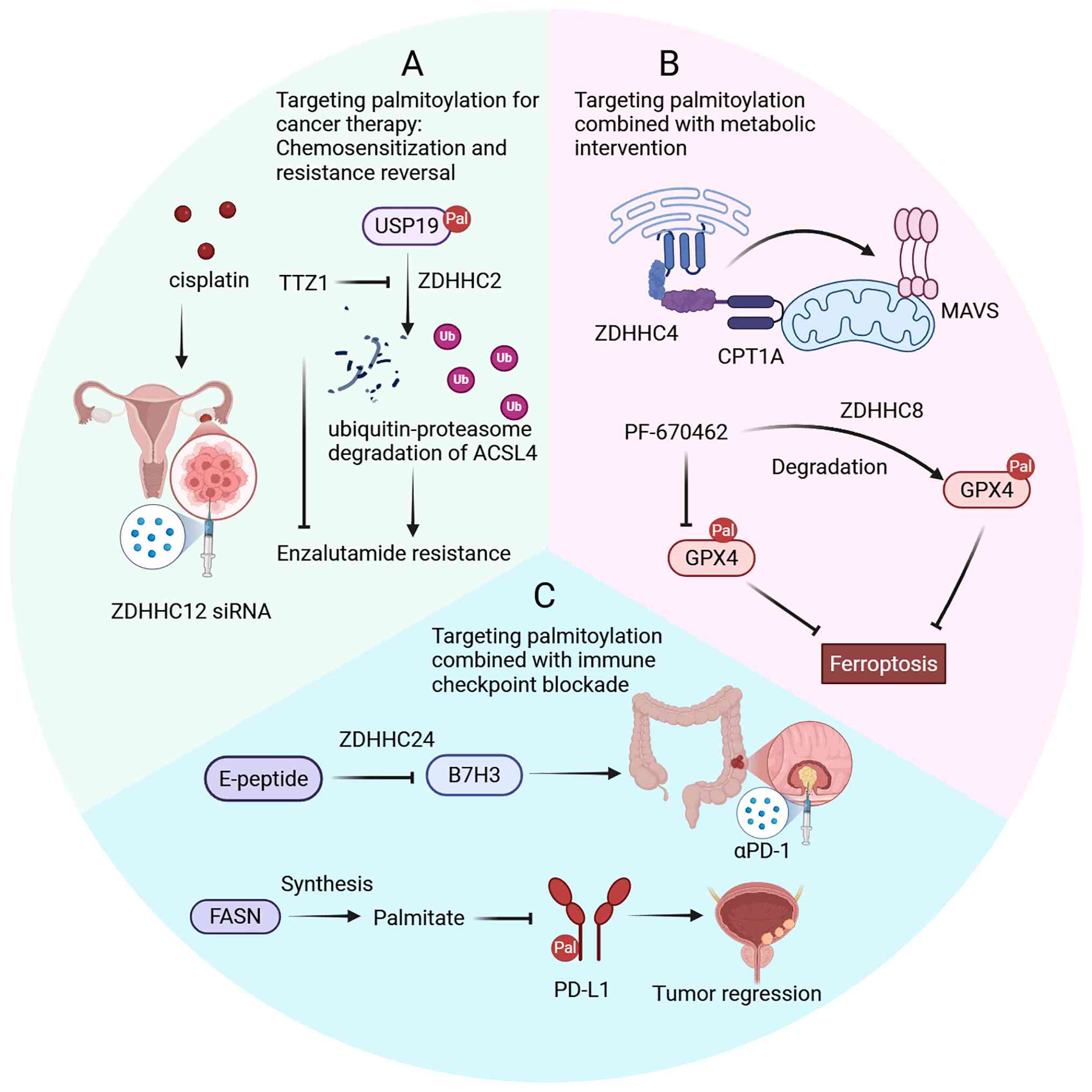 | Figure 3Therapeutic strategies targeting
S-palmitoylation to enhance antitumor responses. S-palmitoylation
is a promising target that can be combined with chemotherapy,
metabolic intervention and immune checkpoint blockade to improve
cancer treatment efficacy. (A) Chemosensitization and resistance
reversal. siRNA-mediated inhibition of ZDHHC12 increases cisplatin
sensitivity in ovarian cancer. Targeting ZDHHC2 with TTZ1 reduces
USP19 S-palmitoylation, thereby preventing ubiquitin-proteasome
degradation of ACSL4, promoting ferroptosis and reversing
enzalutamide resistance in prostate cancer. (B) Metabolic
intervention. The CPT1A/ZDHHC4/MAVS axis promotes MAVS
S-palmitoylation and type I interferon signaling. PF-670462
promotes ZDHHC8 degradation, thereby reducing GPX4
S-palmitoylation, leading to GPX4 degradation and enhanced
ferroptosis. (C) Immune checkpoint blockade. The E-peptide disrupts
ZDHHC24-mediated B7H3 S-palmitoylation, promoting B7H3 degradation
and improving anti-PD-1 responses. Inhibition of FASN reduces
palmitate synthesis, thereby decreasing PD-L1 S-palmitoylation and
restoring sensitivity to immune checkpoint blockade. Figure created
with BioRender.com. ICB, immune checkpoint
blockade; ZDHHC, zinc finger DHHC-type palmitoyltransferase; CPT1A,
carnitine palmitoyltransferase 1A; MAVS, mitochondrial antiviral
signaling protein; GPX4, glutathione peroxidase 4; FASN, fatty acid
synthase; PD-1, programmed cell death protein 1; PD-L1, programmed
death-ligand 1; B7H3, B7 homolog 3; ACSL4, acyl-CoA synthetase
long-chain family member 4. |
Platinum-based chemotherapeutic agents exert
cytotoxic effects, primarily by inducing DNA damage. A recent
studies has demonstrated that the palmitoyl transferase ZDHHC12
facilitates ovarian cancer progression by modulating mitochondrial
function and maintaining ROS homeostasis. Consistently, its
expression is markedly elevated in ovarian malignancies. In the
human high-grade serous ovarian cancer cell lines SNU119 and
OVSAHO, genetic depletion of ZDHHC12 or pharmacological inhibition
of S-palmitoylation using the broad-spectrum inhibitor 2-BP
increased intracellular ROS accumulation, which enhanced
cisplatin-induced cytotoxicity (Fig.
3A) in a ROS-dependent manner across two-dimensional cultures,
three-dimensional Matrigel spheroid models and SNU119 xenograft
tumors in immunodeficient mice (204). This approach has been validated
at both the genetic (knockdown) and pharmacological (2-BP) levels
in cell-based assays, 3D cultures and immunodeficient xenograft
models; it currently resides at the stage of genetic
proof-of-concept and in vitro pharmacological validation,
with early in vivo efficacy demonstrated in immunodeficient
xenograft models. Furthermore, emerging platinum-based compounds
have been reported to induce ICD by activating ER stress pathways
and eliciting sustained antitumor immune responses. Given that
S-palmitoylation critically regulates cellular stress responses,
mitochondrial metabolism and immune-related signaling pathways,
pharmacological modulation of S-palmitoylation represents a
potential strategy to enhance platinum-based therapies. However,
whether such approaches directly potentiate platinum-induced ICD
requires further investigation (208-210).
In prostate cancer, ZDHHC2 mediates
S-palmitoylation of the deubiquitinase USP19 at Cys152, which in
turn weakens the USP19-ACSL4 interaction and promotes
ubiquitin-proteasome degradation of ACSL4, consequently suppressing
lipid peroxidation and ferroptosis. ZDHHC2 is transcriptionally
upregulated by a FOXA1/CXXC5/TET2 complex and is implicated in
enzalutamide resistance in castration-resistant prostate cancer
(CRPC) (Fig. 3A) (205). In human CRPC cell lines (C4-2,
22Rv1 and LNCaP) and enzalutamide-resistant patient-derived
xenograft (PDX) models, treatment with the ZDHHC2-selective
inhibitor TTZ1 (as well as the broad-spectrum inhibitor 2-BP)
substantially restored drug sensitivity, whereas proteomic analyses
revealed that differentially palmitoylated proteins were
significantly enriched in ferroptosis-related and lipid metabolic
pathways, highlighting the importance of metabolic-epigenetic
interactions in the development of therapeutic resistance (205). In a ZDHHC2-knockout mouse model
of spontaneous prostate cancer (CRISPR/Cas9, immunocompetent),
ZDHHC2 depletion significantly limited tumor growth and enhanced
enzalutamide sensitivity. Similar to the ovarian cancer strategy,
this CRPC-directed intervention has been demonstrated in cell lines
and PDX models, placing it at the stages of genetic
proof-of-concept (ZDHHC2 knockout in immunocompetent mice) and
in vitro pharmacological validation (TTZ1 in cell lines),
with early in vivo pharmacological proof-of-concept in
immunodeficient PDX models. However, it has not yet advanced to
humanized animal validation, pharmacokinetic/pharmacodynamic
development or clinical evaluation (Fig. 3A). Overall, these observations
establish a mechanistic connection between chemoresistance and
lipid metabolic abnormalities, providing a potential framework for
the development of S-palmitoylation-targeted combination
therapies.
Given the extensive interplay between
S-palmitoylation and lipid metabolic regulation, the targeting of
metabolic pathways has emerged as a promising strategy for
improving therapeutic responses. Zhang et al (202) demonstrated that mitochondrial
CPT1A recruits ER-associated ZDHHC4 to mediate MAVS
S-palmitoylation at Cys79, resulting in MAVS stabilization and
amplification of IFN-I signaling. Pharmacological activation of
CPT1A enhanced the efficacy of combined epigenetic therapy and PD-1
blockade in refractory tumor models, highlighting the therapeutic
potential of manipulating metabolism-S-palmitoylation crosstalk in
cancer immunotherapy (Fig. 3B).
This pharmacological activation strategy has been evaluated in
immunocompetent tumor models (with anti-PD-1), thus achieving
immune-competent animal validation.
Within the ferroptosis regulatory network,
ZDHHC8-mediated GPX4 S-palmitoylation is a critical mechanism that
preserves tumor cell resistance to ferroptotic stress. Zhou et
al (203) identified
PF-670462 as a ZDHHC8-targeting compound that promotes ZDHHC8
degradation, reduces GPX4 S-palmitoylation and enhances
susceptibility to ferroptosis. Notably, PF-670462 synergized with
anti-PD-1 therapy to prolong the survival of tumor-bearing mice
without apparent toxicity, highlighting the therapeutic potential
of targeting S-palmitoylation-dependent metabolic vulnerabilities
to enhance cancer immunotherapy. PF-670462 has been tested in
combination with anti-PD-1 in tumor-bearing mice, reaching the
stage of immune-competent animal validation with preliminary
pharmacodynamic assessment (Fig.
3B). Together, these strategies integrate the regulation of
lipid metabolism, susceptibility to ferroptosis and protein
S-palmitoylation, thereby enabling coordinated metabolic remodeling
and immune activation.
Advances highlighting the interplay between
metabolic regulation and immune surveillance have stimulated
growing interest in combining S-palmitoylation-targeted approaches
with PD-1/PD-L1 ICB. B7H3 (CD276) is highly expressed in multiple
solid tumors and promotes malignant progression by suppressing T
cell-mediated antitumor immunity and inducing tumor-associated
macrophage polarization toward an immunosuppressive M2 phenotype
(211,212). In microsatellite-stable CRC,
ZDHHC24-mediated S-palmitoylation of B7H3 at Cys496 prevents its
interaction with the autophagy receptor SQSTM1/p62, consequently
limiting selective autophagic degradation and resulting in B7H3
accumulation, which ultimately impairs CD8+ T
cell-mediated antitumor immunity. A cell-penetrating peptide
(E-peptide) targeting the ZDHHC24-B7H3 interaction interface
disrupts B7H3 S-palmitoylation and promotes its degradation, which,
in combination with anti-PD-1 antibodies, effectively restores
CD8+ T cell-dependent antitumor responses in vivo
(Fig. 3C) (157). This peptide-based intervention
has been validated in immune-competent mouse models, placing it at
the immune-competent animal validation stage.
Moreover, in bladder cancer, FASN inhibition
disrupts PD-L1 S-palmitoylation and reduces PD-L1 protein
stability, revealing a metabolic mechanism linking lipid synthesis
to immune checkpoint regulation and providing a rationale for the
combined therapeutic targeting of FASN and PD-L1 (Fig. 3C) (206,213). Currently, this FASN/PD-L1 axis
is supported by in vitro and biochemical evidence, thus
placing it at the in vitro pharmacological validation stage.
Therefore, therapeutic interventions targeting immune checkpoint
S-palmitoylation, particularly when integrated with PD-1/PD-L1
blockade, represent a promising avenue for advancing
next-generation cancer immunotherapy.
Collectively, S-palmitoylation modulation, either
alone or in combination with chemotherapy, metabolic agents or
ICIs, has progressed from genetic proof-of-concept to validation in
immune-competent animal models. However, it remains confined to the
preclinical stage. Although these strategies show promise in
overcoming resistance and sustaining antitumor immunity, they
require pharmacokinetic/pharmacodynamic optimization and humanized
model testing before their transformative potential can be
confirmed in clinical translation.
Challenges for clinical translation
Targeting S-palmitoylation has emerged as a
promising therapeutic strategy for cancer immunotherapy; however,
its clinical translation remains constrained by the limited
availability of selective inhibitors, an incomplete understanding
of ZDHHC substrate specificity and a lack of validated predictive
biomarkers for patient stratification (21,214,215). From the perspective of
biomarker development, accumulating evidence indicates that the
expression patterns of ZDHHC family members may serve as potential
predictors of immunotherapeutic responsiveness. In pancreatic
cancer, elevated ZDHHC9 expression is associated with an
immunosuppressive TME, whereas the genetic or pharmacological
inhibition of ZDHHC9 remodels immune suppression and enhances tumor
responsiveness to anti-PD-L1 therapy (216-218). Similarly, ZDHHC3 is markedly
upregulated in pancreatic cancer and has emerged as a potential
immunotherapeutic target; its disruption substantially improves the
efficacy of anti-PD-1/PD-L1 blockade (219). In breast cancer, increased
ZDHHC9 expression is associated with an unfavorable prognosis and
diminished responsiveness to immunotherapy, whereas reduced ZDHHC9
abundance is correlated with improved therapeutic outcomes
(220). In lung adenocarcinoma,
high ZDHHC18 expression is linked to impaired immune cell
activation and enhanced malignant progression, whereas ZDHHC18
depletion effectively strengthens antitumor immune responses
(221). Collectively, these
findings indicate that aberrant activation of ZDHHC-mediated
S-palmitoylation networks, particularly in pancreatic cancer, is
strongly associated with reduced CD8+ T cell
infiltration and the establishment of an immunosuppressive TME.
Furthermore, emerging prognostic models based on
S-palmitoylation-associated gene signatures have demonstrated
potential utility for the molecular stratification of patients and
the prediction of immunotherapy responsiveness across multiple
cancer types (222-225). Thus, the expression profiles of
specific ZDHHC enzymes and their corresponding S-palmitoylation
landscapes may serve as informative biomarkers for optimizing the
selection of individualized cancer immunotherapy strategies.
Although 2-BP remains the most widely used
pharmacological inhibitor for suppressing global protein
S-palmitoylation, its broad reactivity toward multiple DHHC
palmitoyltransferases, depalmitoylating enzymes and un-related
metabolic pathways markedly limits its therapeutic applicability
(21,196,201,215,226). In addition, owing to its
structural similarity to endogenous fatty acids, 2-BP exhibits
broad metabolic activity beyond inhibition of protein
S-palmitoylation, perturbing lipid metabolic pathways and
generating substantial off-target effects that limit its
translational potential (29,60,201,226). Accordingly, the development of
ZDHHC isoform-selective inhibitors has become an important
direction in this field, with several encouraging advances reported
to date. Zhou et al (203) identified the ZDHHC8-selective
inhibitor, PF-670462, using small-molecule screening approaches.
This compound facilitated ZDHHC8 degradation, decreased GPX4
S-palmitoylation and increased the susceptibility of tumor cells to
ferroptosis. In preclinical animal models, combined treatment with
PF-670462 and anti-PD-1 antibodies markedly suppressed tumor growth
without causing apparent systemic toxicity. Nevertheless, the
development of selective ZDHHC inhibitors remains challenging, as
the 23-member ZDHHC family shares a highly conserved catalytic DHHC
domain. This conservation complicates the achievement of
isoform-specific inhibition while maintaining favorable
pharmacological properties (215). Future investigations should
integrate complementary approaches, including high-throughput
compound screening, structure-based drug optimization and targeted
protein degradation technologies, to accelerate the development of
S-palmitoylation-modulating therapeutics. However, successful
clinical translation critically depends on addressing several key
challenges, namely, achieving tumor cell-selective delivery to
avoid off-target effects on T cells, establishing broad substrate
selectivity to preserve essential physiological palmitoylation
events, such as TCR signaling components, rigorously demonstrating
target engagement and favorable pharmacokinetic properties in
vivo and identifying predictive biomarkers to stratify
responsive patient populations. In particular, inhibiting
checkpoint palmitoylation in tumor cells may inadvertently suppress
physiological TCR signaling. This, makes careful evaluation of T
cell toxicity and immune synapse integrity essential.
Conclusions and perspectives
The regulation of T cell-mediated antitumor
immunity is a fundamental biological principle that underpins the
development and optimization of modern cancer immunotherapies.
S-palmitoylation, a dynamic and reversible lipid-based
post-translational modification, has emerged as an important
regulatory mechanism that integrates multiple dimensions of T cell
function, ranging from proximal signaling events to metabolic
adaptation and immune effector responses. The present review
systematically summarized the diverse functions of S-palmitoylation
in shaping T cell-mediated antitumor immunity and highlighted its
emerging therapeutic potential in cancer immunotherapy.
During T cell activation, S-palmitoylation
facilitates precise membrane compartmentalization of essential
signaling molecules, including Lck, Fyn, ZAP-70 and LAT, within
lipid raft microdomains, ensuring efficient initiation and
amplification of TCR signaling. S-palmitoylation fine-tunes the
antigen responsiveness and activation threshold of T cells by
regulating the plasma membrane localization and protein stability
of CD4/CD8 co-receptors and the co-stimulatory receptor CD28. In
addition, this modification modulates the activity, intracellular
distribution and nuclear accumulation of critical transcriptional
regulators, including STAT3, NF-κB and Foxp3, consequently shaping
the differentiation equilibrium of effector T cell subsets, such as
Th17 cells and Tregs. Simultaneously, S-palmitoylation participates
in the regulation of T cell exhaustion and apoptosis through the
stabilization of immune checkpoint molecules, including PD-1 and
TIM-3, as well as the modulation of Fas/FasL-mediated death
receptor signaling.
Within the tumor immune microenvironment, malignant
cells exploit S-palmitoylation-dependent mechanisms to enhance the
stability and functional persistence of immune checkpoint proteins,
such as PD-L1 and B7H3, ultimately impairing CD8+ T cell
activity and facilitating immune escape. Metabolic abnormalities in
the TME, characterized by increased local accumulation of palmitic
acid, establish a reciprocal regulatory relationship with protein
S-palmitoylation. Mechanistically, the reversible S-palmitoylation
cycle of CD36 controls fatty acid uptake, whereas FASN
S-palmitoylation stabilizes the metabolic enzyme and promotes de
novo lipid biosynthesis. Furthermore, endogenous
S-palmitoylation events in DCs, including modifications of TLR2 and
TLR9, contribute to the regulation of DC activation and metabolic
remodeling. Conversely, exogenous palmitoylated antigenic peptides
enhance the DC cross-presentation efficiency, providing an
additional strategy for potentiating antitumor immune
responses.
From a therapeutic perspective, pharmacological
interventions targeting S-palmitoylation have demonstrated the
potential to remodel immunosuppressive TMEs and promote ICD,
consequently improving the efficacy of chemo- and immunotherapy in
multiple cancer models, including ovarian, prostate and bladder
cancer. Despite these advances, several obstacles have impeded
clinical translation. The highly conserved catalytic domains among
ZDHHC family members present substantial challenges for the
development of isoform-selective inhibitors. In addition, the
currently available broad-spectrum inhibitors are limited by
insufficient specificity, off-target effects and disruption of
physiological lipid metabolic processes. Furthermore, the
regulatory landscape of S-palmitoylation exhibits considerable
tissue specificity, cell type dependence and tumor heterogeneity,
and clinically validated biomarkers for patient selection are
lacking.
Several key research directions are required to
address these challenges. The integration of high-throughput
screening platforms, structure-guided drug discovery and targeted
protein-degradation technologies may accelerate the development of
highly selective and low-toxicity ZDHHC subtype-specific
inhibitors, thereby overcoming the limitations associated with
non-specific inhibition. In parallel, comprehensive
characterization of S-palmitoylation profiles across distinct tumor
types and cellular populations using multi-omics approaches will
facilitate the discovery of robust prognostic and predictive
biomarkers, enabling more precise patient stratification and
therapeutic decision-making. Furthermore, exploring rational
combination strategies that integrate S-palmitoylation-targeted
interventions with emerging immunotherapeutic approaches, including
CAR-T cells, bispecific antibodies and oncolytic viruses, may
broaden the therapeutic potential of this strategy. As the
molecular mechanisms underlying S-palmitoylation-mediated immune
regulation continue to be elucidated, current strategies remain in
preclinical stages, ranging from broad mechanistic tools and
genetic proof-of-concept to in vitro pharmacological
validation and immunocompetent animal validation. Therapies
targeting S-palmitoylation may eventually contribute to cancer
immunotherapy. However, further pharmacokinetic/pharmacodynamic
optimization, humanized model testing and clinical evaluations are
required.
Supplementary Data
Availability of data and materials
Not applicable.
Authors' contributions
SW and XW conceived and designed the review. SW
performed the literature search, collected, screened and
categorized relevant studies and drafted the manuscript. RX and SJ
contributed to literature collection, evidence synthesis and
manuscript preparation. TY contributed to literature organization,
figure preparation and writing and editing the manuscript. MQ
participated in manuscript revision. LX and XW critically revised
the manuscript and provided guidance throughout the preparation
process. All authors read and approved the final version of the
manuscript. Data authentication is not applicable.
Ethics approval and consent to
participate
Not applicable.
Patient consent for publication
Not applicable.
Competing interests
The authors declare that they have no competing
interests.
Acknowledgements
Not applicable.
Funding
This work was supported by the National Natural Science
Foundation of China (grant no. 82360044), the '12345 Future Talent
Training Plan' of Zunyi Medical University (Future Science and
Technology Elite Talent Project (grant no. ZYSE-2024-04), the
Guizhou Provincial Natural Science Foundation [grant no. Qian Ke He
Ji Chu-ZK(2024)-269] and the Zunyi Science and Technology Bureau
Project (grant no. 2026-302).
References
|
1
|
Rui R, Zhou L and He S: Cancer
immunotherapies: advances and bottlenecks. Front Immunol.
14:12124762023. View Article : Google Scholar : PubMed/NCBI
|
|
2
|
Hegde UP and Mukherji B: Current status of
chimeric antigen receptor engineered T cell-based and immune
checkpoint blockade-based cancer immunotherapies. Cancer Immunol
Immunother. 66:1113–1121. 2017. View Article : Google Scholar : PubMed/NCBI
|
|
3
|
Johnson DB, Nebhan CA, Moslehi JJ and
Balko JM: Immune-checkpoint inhibitors: Long-term implications of
toxicity. Nat Rev Clin Oncol. 19:254–267. 2022. View Article : Google Scholar : PubMed/NCBI
|
|
4
|
Tang S, Qin C, Hu H, Liu T, He Y, Guo H,
Yan H, Zhang J, Tang S and Zhou H: Immune checkpoint inhibitors in
non-small cell lung cancer: Progress, challenges, and prospects.
Cells. 11:3202022. View Article : Google Scholar : PubMed/NCBI
|
|
5
|
Zhang X, Zhu L, Zhang H, Chen S and Xiao
Y: CAR-T cell therapy in hematological malignancies: Current
opportunities and challenges. Front Immunol. 13:9271532022.
View Article : Google Scholar : PubMed/NCBI
|
|
6
|
Sharma P, Hu-Lieskovan S, Wargo JA and
Ribas A: Primary, adaptive, and acquired resistance to cancer
immunotherapy. Cell. 168:707–723. 2017. View Article : Google Scholar : PubMed/NCBI
|
|
7
|
Wherry EJ and Kurachi M: Molecular and
cellular insights into T cell exhaustion. Nat Rev Immunol.
15:486–499. 2015. View Article : Google Scholar : PubMed/NCBI
|
|
8
|
Binnewies M, Roberts EW, Kersten K, Chan
V, Fearon DF, Merad M, Coussens LM, Gabrilovich DI,
Ostrand-Rosenberg S, Hedrick CC, et al: Understanding the tumor
immune microenvironment (TIME) for effective therapy. Nat Med.
24:541–550. 2018. View Article : Google Scholar : PubMed/NCBI
|
|
9
|
Soares H, Henriques R, Sachse M,
Ventimiglia L, Alonso MA, Zimmer C, Thoulouze MI and Alcover A:
Regulated vesicle fusion generates signaling nanoterritories that
control T cell activation at the immunological synapse. J Exp Med.
210:2415–2433. 2013. View Article : Google Scholar : PubMed/NCBI
|
|
10
|
Joshi RN, Stadler C, Lehmann R, Lehtiö J,
Tegnér J, Schmidt A and Vesterlund M: TcellSubC: An atlas of the
subcellular proteome of human T cells. Front Immunol. 10:27082019.
View Article : Google Scholar : PubMed/NCBI
|
|
11
|
Zhou B, Hao Q, Liang Y and Kong E: Protein
palmitoylation in cancer: Molecular functions and therapeutic
potential. Mol Oncol. 17:3–26. 2023. View Article : Google Scholar :
|
|
12
|
Zmuda F and Chamberlain LH: Regulatory
effects of post-translational modifications on zDHHC
S-acyltransferases. J Biol Chem. 295:14640–14652. 2020. View Article : Google Scholar : PubMed/NCBI
|
|
13
|
Mitchell DA, Vasudevan A, Linder ME and
Deschenes RJ: Protein palmitoylation by a family of DHHC protein
S-acyltransferases. J Lipid Res. 47:1118–1127. 2006. View Article : Google Scholar : PubMed/NCBI
|
|
14
|
Zhang MM and Hang HC: Protein
S-palmitoylation in cellular differentiation. Biochem Soc Trans.
45:275–285. 2017. View Article : Google Scholar : PubMed/NCBI
|
|
15
|
Stix R, Lee CJ, Faraldo-Gómez JD and
Banerjee A: Structure and mechanism of DHHC protein
acyltransferases. J Mol Biol. 432:4983–4998. 2020. View Article : Google Scholar : PubMed/NCBI
|
|
16
|
Yu T, Hou D, Zhao J, Lu X, Greentree WK,
Zhao Q, Yang M, Conde DG, Linder ME and Lin H: NLRP3 Cys126
palmitoylation by ZDHHC7 promotes inflammasome activation. Cell
Rep. 43:1140702024. View Article : Google Scholar : PubMed/NCBI
|
|
17
|
Salaun C, Greaves J and Chamberlain LH:
The intracellular dynamic of protein palmitoylation. J Cell Biol.
191:1229–1238. 2010. View Article : Google Scholar : PubMed/NCBI
|
|
18
|
Li H, Yuan Q, Wang S, Yu T and Qi X: Role
of S-palmitoylation in digestive system diseases. Cell Death
Discov. 11:3312025. View Article : Google Scholar : PubMed/NCBI
|
|
19
|
Cai J, Cui J and Wang L: S-palmitoylation
regulates innate immune signaling pathways: molecular mechanisms
and targeted therapies. Eur J Immunol. 53:e23504762023. View Article : Google Scholar : PubMed/NCBI
|
|
20
|
Vartak N, Papke B, Grecco HE, Rossmannek
L, Waldmann H, Hedberg C and Bastiaens PI: The autodepalmitoylating
activity of APT maintains the spatial organization of palmitoylated
membrane proteins. Biophys J. 106:93–105. 2014. View Article : Google Scholar : PubMed/NCBI
|
|
21
|
Han M, Lv Y, Chen Y, Li Z, Tian J, Zhou H,
Wang Y, Su W and Zhong J: Advances in targeting protein
S-palmitoylation in tumor immunity and therapy. Front Oncol.
15:15476362025. View Article : Google Scholar : PubMed/NCBI
|
|
22
|
Chen Y, Li Y and Wu L: Protein
S-palmitoylation modification: Implications in tumor and tumor
immune microenvironment. Front Immunol. 15:13374782024. View Article : Google Scholar : PubMed/NCBI
|
|
23
|
Yang Y, Zhang E, Mao X, Liu G and Pan Y:
Targeting palmitoylation: A novel frontier in cancer biology and
immunotherapy. Biochim Biophys Acta Rev Cancer. 1881:1895092026.
View Article : Google Scholar
|
|
24
|
Zhang Y, Fan S, He L and Li L: The
ZDHHC13/ZDHHC17 subfamily: From biological functions to therapeutic
targets of diseases. Pharmacol Res. 209:1074182024. View Article : Google Scholar : PubMed/NCBI
|
|
25
|
Nederstigt AE, Sardana S and Baggelaar MP:
Deciphering protein long-chain S-acylation using mass spectrometry
proteomics strategies. RSC Chem Biol. 6:1532–1545. 2025. View Article : Google Scholar : PubMed/NCBI
|
|
26
|
Liao D, Huang Y, Liu D, Zhang H, Shi X, Li
X and Luo P: The role of s-palmitoylation in neurological diseases:
Implication for zDHHC family. Front Pharmacol. 14:13428302024.
View Article : Google Scholar : PubMed/NCBI
|
|
27
|
Yuan Y, Li P, Li J, Zhao Q, Chang Y and He
X: Protein lipidation in health and disease: Molecular basis,
physiological function and pathological implication. Signal
Transduct Target Ther. 9:602024. View Article : Google Scholar : PubMed/NCBI
|
|
28
|
Liu Z, Xiao M, Mo Y, Wang H, Han Y, Zhao
X, Yang X, Liu Z and Xu B: Emerging roles of protein palmitoylation
and its modifying enzymes in cancer cell signal transduction and
cancer therapy. Int J Biol Sci. 18:3447–3457. 2022. View Article : Google Scholar : PubMed/NCBI
|
|
29
|
Hao S, Mei Y, Chen S, Liu J, Zhang Y, Zhu
Z and Zuo K: Potential therapeutic target in oncology: Protein
palmitoylation (Review). Oncol Rep. 54:1172025. View Article : Google Scholar : PubMed/NCBI
|
|
30
|
Qu M, Zhou X, Wang X and Li H:
Lipid-induced S-palmitoylation as a vital regulator of cell
signaling and disease development. Int J Biol Sci. 17:4223–4237.
2021. View Article : Google Scholar : PubMed/NCBI
|
|
31
|
Elliot Murphy R and Banerjee A: In vitro
reconstitution of substrate S-acylation by the zDHHC family of
protein acyltransferases. Open Biol. 12:2103902022. View Article : Google Scholar : PubMed/NCBI
|
|
32
|
Malgapo MIP and Linder ME: Substrate
recruitment by zDHHC protein acyltransferases. Open Biol.
11:2100262021. View Article : Google Scholar : PubMed/NCBI
|
|
33
|
Adachi N, Hess DT, McLaughlin P and
Stamler JS: S-Palmitoylation of a novel site in the β2-Adrenergic
receptor associated with a novel intracellular itinerary. J Biol
Chem. 291:20232–20246. 2016. View Article : Google Scholar : PubMed/NCBI
|
|
34
|
Abrami L, Dallavilla T, Sandoz PA, Demir
M, Kunz B, Savoglidis G, Hatzimanikatis V and van der Goot FG:
Identification and dynamics of the human ZDHHC16-ZDHHC6
palmitoylation cascade. Elife. 6:e278262017. View Article : Google Scholar : PubMed/NCBI
|
|
35
|
Tomatis VM, Trenchi A, Gomez GA and
Daniotti JL: Acyl-protein thioesterase 2 catalyzes the deacylation
of peripheral membrane-associated GAP-43. PLoS One. 5:e150452010.
View Article : Google Scholar : PubMed/NCBI
|
|
36
|
Sobocińska J, Roszczenko-Jasińska P,
Ciesielska A and Kwiatkowska K: Protein palmitoylation and its role
in bacterial and viral infections. Front Immunol. 8:20032018.
View Article : Google Scholar
|
|
37
|
Roth AF, Wan J, Bailey AO, Sun B, Kuchar
JA, Green WN, Phinney BS, Yates JR III and Davis NG: Global
analysis of protein palmitoylation in yeast. Cell. 125:1003–1013.
2006. View Article : Google Scholar : PubMed/NCBI
|
|
38
|
Ma L, Zhang L, Liao Z, Xiu C, Luo X, Luo
N, Zhang L, He G and Chen J: Pharmacological inhibition of protein
S-palmitoylation suppresses osteoclastogenesis and ameliorates
ovariectomy-induced bone loss. J Orthop Translat. 42:1–14. 2023.
View Article : Google Scholar : PubMed/NCBI
|
|
39
|
Edmonds MJ, Geary B, Doherty MK and Morgan
A: Analysis of the brain palmitoyl-proteome using both acyl-biotin
exchange and acyl-resin-assisted capture methods. Sci Rep.
7:32992017. View Article : Google Scholar : PubMed/NCBI
|
|
40
|
Zheng S, Lin J, Pang Z, Zhang H, Wang Y,
Ma L, Zhang H, Zhang X, Chen M, Zhang X, et al: Aberrant
cholesterol metabolism and Wnt/β-Catenin signaling coalesce via
Frizzled5 in supporting cancer growth. Adv Sci (Weinh).
9:e22007502022. View Article : Google Scholar
|
|
41
|
Ulengin-Talkish I, Parson MAH, Jenkins ML,
Roy J, Shih AZL, St-Denis N, Gulyas G, Balla T, Gingras AC, Várnai
P, et al: Palmitoylation targets the calcineurin phosphatase to the
phosphatidylinositol 4-kinase complex at the plasma membrane. Nat
Commun. 12:60642021. View Article : Google Scholar : PubMed/NCBI
|
|
42
|
Li X, Shen L, Xu Z, Liu W, Li A and Xu J:
Protein palmitoylation modification during viral infection and
detection methods of palmitoylated proteins. Front Cell Infect
Microbiol. 12:8215962022. View Article : Google Scholar : PubMed/NCBI
|
|
43
|
Martin BR, Wang C, Adibekian A, Tully SE
and Cravatt BF: Global profiling of dynamic protein palmitoylation.
Nat Methods. 9:84–89. 2011. View Article : Google Scholar : PubMed/NCBI
|
|
44
|
Sanders SS, Martin DD, Butland SL,
Lavallée-Adam M, Calzolari D, Kay C, Yates JR III and Hayden MR:
Curation of the mammalian palmitoylome indicates a pivotal role for
palmitoylation in diseases and disorders of the nervous system and
cancers. PLoS Comput Biol. 11:e10044052015. View Article : Google Scholar : PubMed/NCBI
|
|
45
|
Shah K, Al-Haidari A, Sun J and Kazi JU: T
cell receptor (TCR) signaling in health and disease. Signal
Transduct Target Ther. 6:4122021. View Article : Google Scholar : PubMed/NCBI
|
|
46
|
Qin Z and Xu T: Deciphering the
deterministic role of TCR signaling in T cell fate determination.
Front Immunol. 16:15622482025. View Article : Google Scholar : PubMed/NCBI
|
|
47
|
Mariuzza RA, Agnihotri P and Orban J: The
structural basis of T-cell receptor (TCR) activation: An enduring
enigma. J Biol Chem. 295:914–925. 2020. View Article : Google Scholar :
|
|
48
|
Brazin KN, Mallis RJ, Li C, Keskin DB,
Arthanari H, Gao Y, Wu SL, Karger BL, Wagner G and Reinherz EL:
Constitutively oxidized CXXC motifs within the CD3 heterodimeric
ectodomains of the T cell receptor complex enforce the conformation
of juxtaposed segments. J Biol Chem. 289:18880–18892. 2014.
View Article : Google Scholar : PubMed/NCBI
|
|
49
|
Kuhns MS and Davis MM: TCR signaling
emerges from the sum of many parts. Front Immunol. 3:1592012.
View Article : Google Scholar : PubMed/NCBI
|
|
50
|
Xu C, Gagnon E, Call ME, Schnell JR,
Schwieters CD, Carman CV, Chou JJ and Wucherpfennig KW: Regulation
of T cell receptor activation by dynamic membrane binding of the
CD3epsilon cytoplasmic tyrosine-based motif. Cell. 135:702–713.
2008. View Article : Google Scholar : PubMed/NCBI
|
|
51
|
Isakov N, Wange RL, Burgess WH, Watts JD,
Aebersold R and Samelson LE: ZAP-70 binding specificity to T cell
receptor tyrosine-based activation motifs: The tandem SH2 domains
of ZAP-70 bind distinct tyrosine-based activation motifs with
varying affinity. J Exp Med. 181:375–380. 1995. View Article : Google Scholar : PubMed/NCBI
|
|
52
|
Lo WL, Shah NH, Ahsan N, Horkova V,
Stepanek O, Salomon AR, Kuriyan J and Weiss A: Lck promotes
Zap70-dependent LAT phosphorylation by bridging Zap70 to LAT. Nat
Immunol. 19:733–741. 2018. View Article : Google Scholar : PubMed/NCBI
|
|
53
|
Bubeck Wardenburg J, Fu C, Jackman JK,
Flotow H, Wilkinson SE, Williams DH, Johnson R, Kong G, Chan AC and
Findell PR: Phosphorylation of SLP-76 by the ZAP-70
protein-tyrosine kinase is required for T-cell receptor function. J
Biol Chem. 271:19641–19644. 1996. View Article : Google Scholar : PubMed/NCBI
|
|
54
|
Wu L, Wei Q, Brzostek J and Gascoigne NRJ:
Signaling from T cell receptors (TCRs) and chimeric antigen
receptors (CARs) on T cells. Cell Mol Immunol. 17:600–612. 2020.
View Article : Google Scholar : PubMed/NCBI
|
|
55
|
Courtney AH, Lo WL and Weiss A: TCR
signaling: Mechanisms of initiation and propagation. Trends Biochem
Sci. 43:108–123. 2018. View Article : Google Scholar
|
|
56
|
Ladygina N, Martin BR and Altman A:
Dynamic palmitoylation and the role of DHHC proteins in T cell
activation and anergy. Adv Immunol. 109:1–44. 2011. View Article : Google Scholar : PubMed/NCBI
|
|
57
|
Palacios EH and Weiss A: Function of the
Src-family kinases, Lck and Fyn, in T-cell development and
activation. Oncogene. 23:7990–8000. 2004. View Article : Google Scholar : PubMed/NCBI
|
|
58
|
van Oers NS, Lowin-Kropf B, Finlay D,
Connolly K and Weiss A: alpha beta T cell development is abolished
in mice lacking both Lck and Fyn protein tyrosine kinases.
Immunity. 5:429–436. 1996. View Article : Google Scholar : PubMed/NCBI
|
|
59
|
Salmond RJ, Filby A, Qureshi I, Caserta S
and Zamoyska R: T-cell receptor proximal signaling via the
Src-family kinases, Lck and Fyn, influences T-cell activation,
differentiation, and tolerance. Immunol Rev. 228:9–22. 2009.
View Article : Google Scholar : PubMed/NCBI
|
|
60
|
Webb Y, Hermida-Matsumoto L and Resh MD:
Inhibition of protein palmitoylation, raft localization, and T cell
signaling by 2-bromopalmitate and polyunsaturated fatty acids. J
Biol Chem. 275:261–270. 2000. View Article : Google Scholar : PubMed/NCBI
|
|
61
|
Resh MD: Fatty acylation of proteins: New
insights into membrane targeting of myristoylated and palmitoylated
proteins. Biochim Biophys Acta. 1451:1–16. 1999. View Article : Google Scholar : PubMed/NCBI
|
|
62
|
van't Hof W and Resh MD: Rapid plasma
membrane anchoring of newly synthesized p59fyn: selective
requirement for NH2-terminal myristoylation and palmitoylation at
cysteine-3. J Cell Biol. 136:1023–1035. 1997. View Article : Google Scholar : PubMed/NCBI
|
|
63
|
Shenoy-Scaria AM, Dietzen DJ, Kwong J,
Link DC and Lublin DM: Cysteine3 of Src family protein tyrosine
kinase determines palmitoylation and localization in caveolae. J
Cell Biol. 126:353–363. 1994. View Article : Google Scholar : PubMed/NCBI
|
|
64
|
Kosugi A, Hayashi F, Liddicoat DR, Yasuda
K, Saitoh S and Hamaoka T: A pivotal role of cysteine 3 of Lck
tyrosine kinase for localization to glycolipid-enriched
microdomains and T cell activation. Immunol Lett. 76:133–138. 2001.
View Article : Google Scholar : PubMed/NCBI
|
|
65
|
Fan Y, Shayahati B, Tewari R, Boehning D
and Akimzhanov AM: Regulation of T cell receptor signaling by
protein acyltransferase DHHC21. Mol Biol Rep. 47:6471–6478. 2020.
View Article : Google Scholar : PubMed/NCBI
|
|
66
|
van't Hof W and Resh MD: Dual fatty
acylation of p59(Fyn) is required for association with the T cell
receptor zeta chain through phosphotyrosine-Src homology domain-2
interactions. J Cell Biol. 145:377–389. 1999. View Article : Google Scholar : PubMed/NCBI
|
|
67
|
West SJ, Boehning D and Akimzhanov AM:
Regulation of T cell function by protein S-acylation. Front
Physiol. 13:10409682022. View Article : Google Scholar : PubMed/NCBI
|
|
68
|
Koegl M, Zlatkine P, Ley SC, Courtneidge
SA and Magee AI: Palmitoylation of multiple Src-family kinases at a
homologous N-terminal motif. Biochem J. 303(Pt 3): 749–753. 1994.
View Article : Google Scholar : PubMed/NCBI
|
|
69
|
Das T, Yount JS and Hang HC: Protein
S-palmitoylation in immunity. Open Biol. 11:2004112021. View Article : Google Scholar : PubMed/NCBI
|
|
70
|
Au-Yeung BB, Shah NH, Shen L and Weiss A:
ZAP-70 in signaling, biology, and disease. Annu Rev Immunol.
36:127–156. 2018. View Article : Google Scholar
|
|
71
|
Smith-Garvin JE, Koretzky GA and Jordan
MS: T cell activation. Annu Rev Immunol. 27:591–619. 2009.
View Article : Google Scholar : PubMed/NCBI
|
|
72
|
Paz PE, Wang S, Clarke H, Lu X, Stokoe D
and Abo A: Mapping the Zap-70 phosphorylation sites on LAT (linker
for activation of T cells) required for recruitment and activation
of signalling proteins in T cells. Biochem J. 356(Pt 2): 461–471.
2001. View Article : Google Scholar : PubMed/NCBI
|
|
73
|
Schultz A, Schnurra M, El-Bizri A,
Woessner NM, Hartmann S, Hartig R, Minguet S, Schraven B and
Simeoni L: A cysteine residue within the kinase domain of Zap70
regulates lck activity and proximal TCR signaling. Cells.
11:27232022. View Article : Google Scholar : PubMed/NCBI
|
|
74
|
Zhang W, Sloan-Lancaster J, Kitchen J,
Trible RP and Samelson LE: LAT: The ZAP-70 tyrosine kinase
substrate that links T cell receptor to cellular activation. Cell.
92:83–92. 1998. View Article : Google Scholar : PubMed/NCBI
|
|
75
|
Tewari R, Shayahati B, Fan Y and
Akimzhanov AM: T cell receptor-dependent S-acylation of ZAP-70
controls activation of T cells. J Biol Chem. 296:1003112021.
View Article : Google Scholar : PubMed/NCBI
|
|
76
|
Hundt M, Harada Y, De Giorgio L, Tanimura
N, Zhang W and Altman A: Palmitoylation-dependent plasma membrane
transport but lipid raft-independent signaling by linker for
activation of T cells. J Immunol. 183:1685–1694. 2009. View Article : Google Scholar : PubMed/NCBI
|
|
77
|
Zhang W, Irvin BJ, Trible RP, Abraham RT
and Samelson LE: Functional analysis of LAT in TCR-mediated
signaling pathways using a LAT-deficient Jurkat cell line. Int
Immunol. 11:943–950. 1999. View Article : Google Scholar : PubMed/NCBI
|
|
78
|
Zhang W, Trible RP and Samelson LE: LAT
palmitoylation: Its essential role in membrane microdomain
targeting and tyrosine phosphorylation during T Cell Activation.
Immunity. 9:239–246. 1998. View Article : Google Scholar : PubMed/NCBI
|
|
79
|
Hundt M, Tabata H, Jeon MS, Hayashi K,
Tanaka Y, Krishna R, De Giorgio L, Liu YC, Fukata M and Altman A:
Impaired activation and localization of LAT in anergic T cells as a
consequence of a selective palmitoylation defect. Immunity.
24:513–522. 2006. View Article : Google Scholar : PubMed/NCBI
|
|
80
|
Tanimura N, Saitoh S, Kawano S, Kosugi A
and Miyake K: Palmitoylation of LAT contributes to its subcellular
localization and stability. Biochem Biophys Res Commun.
341:1177–1183. 2006. View Article : Google Scholar : PubMed/NCBI
|
|
81
|
Li Y, Yin Y and Mariuzza RA: Structural
and biophysical insights into the role of CD4 and CD8 in T cell
activation. Front Immunol. 4:2062013. View Article : Google Scholar : PubMed/NCBI
|
|
82
|
Baldwin I and Robey EA: Adjusting to self
in the thymus: CD4 versus CD8 lineage commitment and regulatory T
cell development. J Exp Med. 221:e202308962024. View Article : Google Scholar : PubMed/NCBI
|
|
83
|
Van Laethem F, Tikhonova AN and Singer A:
MHC restriction is imposed on a diverse T cell receptor repertoire
by CD4 and CD8 co-receptors during thymic selection. Trends
Immunol. 33:437–441. 2012. View Article : Google Scholar : PubMed/NCBI
|
|
84
|
Fragoso R, Ren D, Zhang X, Su MW, Burakoff
SJ and Jin YJ: Lipid raft distribution of CD4 depends on its
palmitoylation and association with Lck, and evidence for
CD4-induced lipid raft aggregation as an additional mechanism to
enhance CD3 signaling. J Immunol. 170:913–921. 2003. View Article : Google Scholar : PubMed/NCBI
|
|
85
|
Crise B and Rose JK: Identification of
palmitoylation sites on CD4, the human immunodeficiency virus
receptor. J Biol Chem. 267:13593–13597. 1992. View Article : Google Scholar : PubMed/NCBI
|
|
86
|
Lin H: Protein cysteine palmitoylation in
immunity and inflammation. FEBS J. 288:7043–7059. 2021. View Article : Google Scholar : PubMed/NCBI
|
|
87
|
Arcaro A, Grégoire C, Boucheron N, Stotz
S, Palmer E, Malissen B and Luescher IF: Essential role of CD8
palmitoylation in CD8 coreceptor function. J Immunol.
165:2068–2076. 2000. View Article : Google Scholar : PubMed/NCBI
|
|
88
|
Pang DJ, Hayday AC and Bijlmakers MJ: CD8
Raft localization is induced by its assembly into CD8alpha beta
heterodimers, Not CD8alpha alpha homodimers. J Biol Chem.
282:13884–13894. 2007. View Article : Google Scholar : PubMed/NCBI
|
|
89
|
Yang X, Chatterjee V, Ma Y, Zheng E and
Yuan SY: Protein palmitoylation in leukocyte signaling and
function. Front Cell Dev Biol. 8:6003682020. View Article : Google Scholar : PubMed/NCBI
|
|
90
|
Raychaudhuri K, Rangu R, Ma A, Alvinez N,
Tran AD, Pallikkuth S, McIntire KM, Garvey JA, Yi J and Samelson
LE: CD28 shapes T cell receptor signaling by regulating Lck
dynamics and ZAP70 activation. Front Immunol. 15:15030182024.
View Article : Google Scholar
|
|
91
|
Esensten JH, Helou YA, Chopra G, Weiss A
and Bluestone JA: CD28 costimulation: From mechanism to therapy.
Immunity. 44:973–988. 2016. View Article : Google Scholar : PubMed/NCBI
|
|
92
|
Lanier LL, O'Fallon S, Somoza C, Phillips
JH, Linsley PS, Okumura K, Ito D and Azuma M: CD80 (B7) and CD86
(B70) provide similar costimulatory signals for T cell
proliferation, cytokine production, and generation of CTL. J
Immunol. 154:97–105. 1995. View Article : Google Scholar : PubMed/NCBI
|
|
93
|
Sadra A, Cinek T and Imboden JB:
Translocation of CD28 to lipid rafts and costimulation of IL-2.
Proc Natl Acad Sci USA. 101:11422–11427. 2004. View Article : Google Scholar : PubMed/NCBI
|
|
94
|
Martin M, Schneider H, Azouz A and Rudd
CE: Cytotoxic T lymphocyte antigen 4 and CD28 modulate cell surface
raft expression in their regulation of T cell function. J Exp Med.
194:1675–1681. 2001. View Article : Google Scholar : PubMed/NCBI
|
|
95
|
Burke KP, Chaudhri A, Freeman GJ and
Sharpe AH: The B7:CD28 family and friends: Unraveling coinhibitory
interactions. Immunity. 57:223–244. 2024. View Article : Google Scholar : PubMed/NCBI
|
|
96
|
Slavik JM, Hutchcroft JE and Bierer BE:
CD28/CTLA-4 and CD80/CD86 families: Signaling and function. Immunol
Res. 19:1–24. 1999. View Article : Google Scholar : PubMed/NCBI
|
|
97
|
Lu B, Sun YY, Chen BY, Yang B, He QJ, Li J
and Cao J: zDHHC20-driven S-palmitoylation of CD80 is required for
its costimulatory function. Acta Pharmacol Sin. 45:1214–1223. 2024.
View Article : Google Scholar : PubMed/NCBI
|
|
98
|
Simanshu DK, Nissley DV and McCormick F:
RAS proteins and their regulators in human disease. Cell.
170:17–33. 2017. View Article : Google Scholar : PubMed/NCBI
|
|
99
|
Lu S, Jang H, Muratcioglu S, Gursoy A,
Keskin O, Nussinov R and Zhang J: Ras conformational ensembles,
allostery, and signaling. Chem Rev. 116:6607–6665. 2016. View Article : Google Scholar : PubMed/NCBI
|
|
100
|
Kolch W, Berta D and Rosta E: Dynamic
regulation of RAS and RAS signaling. Biochem J. 480:1–23. 2023.
View Article : Google Scholar : PubMed/NCBI
|
|
101
|
Downward J: Control of ras activation.
Cancer Surv. 27:87–100. 1996.PubMed/NCBI
|
|
102
|
Lapinski PE and King PD: Regulation of Ras
signal transduction during T cell development and activation. Am J
Clin Exp Immunol. 1:147–153. 2012.PubMed/NCBI
|
|
103
|
Kortum RL, Rouquette-Jazdanian AK and
Samelson LE: Ras and extracellular signal-regulated kinase
signaling in thymocytes and T cells. Trends Immunol. 34:259–268.
2013. View Article : Google Scholar : PubMed/NCBI
|
|
104
|
Yamashita M, Kimura M, Kubo M, Shimizu C,
Tada T, Perlmutter RM and Nakayama T: T cell antigen
receptor-mediated activation of the Ras/mitogen-activated protein
kinase pathway controls interleukin 4 receptor function and type-2
helper T cell differentiation. Proc Natl Acad Sci USA.
96:1024–1029. 1999. View Article : Google Scholar : PubMed/NCBI
|
|
105
|
Busquets-Hernández C and Triola G:
Palmitoylation as a key regulator of ras localization and function.
Front Mol Biosci. 8:6598612021. View Article : Google Scholar : PubMed/NCBI
|
|
106
|
Castellano E and Santos E: Functional
specificity of ras isoforms: So similar but so different. Genes
Cancer. 2:216–231. 2011. View Article : Google Scholar : PubMed/NCBI
|
|
107
|
García-España A and Philips MR: Origin and
evolution of RAS membrane targeting. Oncogene. 42:1741–1750. 2023.
View Article : Google Scholar : PubMed/NCBI
|
|
108
|
Rocks O, Peyker A, Kahms M, Verveer PJ,
Koerner C, Lumbierres M, Kuhlmann J, Waldmann H, Wittinghofer A and
Bastiaens PI: An acylation cycle regulates localization and
activity of palmitoylated Ras isoforms. Science. 307:1746–1752.
2005. View Article : Google Scholar : PubMed/NCBI
|
|
109
|
Baekkeskov S and Kanaani J: Palmitoylation
cycles and regulation of protein function (Review). Mol Membr Biol.
26:42–54. 2009. View Article : Google Scholar : PubMed/NCBI
|
|
110
|
Oeckinghaus A and Ghosh S: The NF-kappaB
family of transcription factors and its regulation. Cold Spring
Harb Perspect Biol. 1:a0000342009. View Article : Google Scholar :
|
|
111
|
Mao H, Zhao X and Sun SC: NF-κB in
inflammation and cancer. Cell Mol Immunol. 22:811–839. 2025.
View Article : Google Scholar : PubMed/NCBI
|
|
112
|
Lomaga MA, Yeh WC, Sarosi I, Duncan GS,
Furlonger C, Ho A, Morony S, Capparelli C, Van G, Kaufman S, et al:
TRAF6 deficiency results in osteopetrosis and defective
interleukin-1, CD40, and LPS signaling. Genes Dev. 13:1015–1024.
1999. View Article : Google Scholar : PubMed/NCBI
|
|
113
|
Liu E, Sun J, Yang J, Li L, Yang Q, Zeng
J, Zhang J, Chen D and Sun Q: ZDHHC11 positively regulates NF-κB
activation by enhancing TRAF6 oligomerization. Front Cell Dev Biol.
9:7109672021. View Article : Google Scholar
|
|
114
|
Feng T, Qin H, Wang L, Benveniste EN,
Elson CO and Cong Y: Th17 cells induce colitis and promote Th1 cell
responses through IL-17 induction of innate IL-12 and IL-23
production. J Immunol. 186:6313–6318. 2011. View Article : Google Scholar : PubMed/NCBI
|
|
115
|
Leppkes M, Becker C, Ivanov II, Hirth S,
Wirtz S, Neufert C, Pouly S, Murphy AJ, Valenzuela DM, Yancopoulos
GD, et al: RORgamma-expressing Th17 cells induce murine chronic
intestinal inflammation via redundant effects of IL-17A and IL-17F.
Gastroenterology. 136:257–267. 2009. View Article : Google Scholar
|
|
116
|
Zhang M, Zhou L, Xu Y, Yang M, Xu Y,
Komaniecki GP, Kosciuk T, Chen X, Lu X, Zou X, et al: A STAT3
palmitoylation cycle promotes T(H)17 differentiation and colitis.
Nature. 586:434–439. 2020. View Article : Google Scholar : PubMed/NCBI
|
|
117
|
Fujino S, Andoh A, Bamba S, Ogawa A, Hata
K, Araki Y, Bamba T and Fujiyama Y: Increased expression of
interleukin 17 in inflammatory bowel disease. Gut. 52:65–70. 2003.
View Article : Google Scholar
|
|
118
|
Kobayashi T, Okamoto S, Hisamatsu T,
Kamada N, Chinen H, Saito R, Kitazume MT, Nakazawa A, Sugita A,
Koganei K, et al: IL23 differentially regulates the Th1/Th17
balance in ulcerative colitis and Crohn's disease. Gut.
57:1682–1689. 2008. View Article : Google Scholar : PubMed/NCBI
|
|
119
|
Ahern PP, Schiering C, Buonocore S,
McGeachy MJ, Cua DJ, Maloy KJ and Powrie F: Interleukin-23 drives
intestinal inflammation through direct activity on T cells.
Immunity. 33:279–288. 2010. View Article : Google Scholar : PubMed/NCBI
|
|
120
|
Gagliani N, Amezcua Vesely MC, Iseppon A,
Brockmann L, Xu H, Palm NW, de Zoete MR, Licona-Limón P, Paiva RS,
Ching T, et al: Th17 cells transdifferentiate into regulatory T
cells during resolution of inflammation. Nature. 523:221–225. 2015.
View Article : Google Scholar : PubMed/NCBI
|
|
121
|
Coskun M, Vermeire S and Nielsen OH: Novel
targeted therapies for inflammatory bowel disease. Trends Pharmacol
Sci. 38:127–142. 2017. View Article : Google Scholar
|
|
122
|
Zhou L, Zhang M, Wang Y, Dorfman RG, Liu
H, Yu T, Chen X, Tang D, Xu L, Yin Y, et al: Faecalibacterium
prausnitzii produces butyrate to maintain Th17/Treg balance and to
ameliorate colorectal colitis by inhibiting histone deacetylase 1.
Inflamm Bowel Dis. 24:1926–1940. 2018. View Article : Google Scholar : PubMed/NCBI
|
|
123
|
Yang XO, Panopoulos AD, Nurieva R, Chang
SH, Wang D, Watowich SS and Dong C: STAT3 regulates
cytokine-mediated generation of inflammatory helper T cells. J Biol
Chem. 282:9358–9363. 2007. View Article : Google Scholar : PubMed/NCBI
|
|
124
|
Saleh R and Elkord E: FoxP3+ T regulatory
cells in cancer: Prognostic biomarkers and therapeutic targets.
Cancer Lett. 490:174–185. 2020. View Article : Google Scholar : PubMed/NCBI
|
|
125
|
Takeuchi Y and Nishikawa H: Roles of
regulatory T cells in cancer immunity. Int Immunol. 28:401–409.
2016. View Article : Google Scholar : PubMed/NCBI
|
|
126
|
Li X and Zheng Y: Regulatory T cell
identity: Formation and maintenance. Trends Immunol. 36:344–353.
2015. View Article : Google Scholar : PubMed/NCBI
|
|
127
|
Riaz F, Huang Z and Pan F: Targeting
post-translational modifications of Foxp3: A new paradigm for
regulatory T cell-specific therapy. Front Immunol. 14:12807412023.
View Article : Google Scholar : PubMed/NCBI
|
|
128
|
Deng G, Song X, Fujimoto S, Piccirillo CA,
Nagai Y and Greene MI: Foxp3 post-translational modifications and
treg suppressive activity. Front Immunol. 10:24862019. View Article : Google Scholar : PubMed/NCBI
|
|
129
|
Zhou B, Zhang M, Ma H, Wang Y, Qiu J, Liu
Y, Lu L, Li T, Zhang L, Huang R, et al: Distinct palmitoylation of
Foxp3 regulates the function of regulatory T cells via
palmitoyltransferases. Cell Mol Immunol. 21:787–789. 2024.
View Article : Google Scholar : PubMed/NCBI
|
|
130
|
Lee TA, Tsai EY, Liu SH, Hsu Hung SD,
Chang SJ, Chao CH, Lai YJ, Yamaguchi H and Li CW:
Post-translational modification of PD-1: Potential targets for
cancer immunotherapy. Cancer Res. 84:800–807. 2024. View Article : Google Scholar : PubMed/NCBI
|
|
131
|
Ostrand-Rosenberg S, Horn LA and Haile ST:
The programmed death-1 immune-suppressive pathway: Barrier to
antitumor immunity. J Immunol. 193:3835–3841. 2014. View Article : Google Scholar : PubMed/NCBI
|
|
132
|
Zhang Z, Ren C, Xiao R, Ma S, Liu H, Dou
Y, Fan Y, Wang S, Zhan P, Gao C, et al: Palmitoylation of TIM-3
promotes immune exhaustion and restrains antitumor immunity. Sci
Immunol. 9:eadp73022024. View Article : Google Scholar : PubMed/NCBI
|
|
133
|
Riley JL: PD-1 signaling in primary T
cells. Immunol Rev. 229:114–125. 2009. View Article : Google Scholar : PubMed/NCBI
|
|
134
|
Chemnitz JM, Parry RV, Nichols KE, June CH
and Riley JL: SHP-1 and SHP-2 associate with immunoreceptor
tyrosine-based switch motif of programmed death 1 upon primary
human T cell stimulation, but only receptor ligation prevents T
cell activation. J Immunol. 173:945–954. 2004. View Article : Google Scholar : PubMed/NCBI
|
|
135
|
Yao H, Li C, He F, Song T, Brosseau JP,
Wang H, Lu H, Fang C, Shi H, Lan J, et al: A peptidic inhibitor for
PD-1 palmitoylation targets its expression and functions. RSC Chem
Biol. 2:192–205. 2020. View Article : Google Scholar
|
|
136
|
Zhu C, Anderson AC, Schubart A, Xiong H,
Imitola J, Khoury SJ, Zheng XX, Strom TB and Kuchroo VK: The Tim-3
ligand galectin-9 negatively regulates T helper type 1 immunity.
Nat Immunol. 6:1245–1252. 2005. View
Article : Google Scholar : PubMed/NCBI
|
|
137
|
Zhao L, Cheng S, Fan L, Zhang B and Xu S:
TIM-3: An update on immunotherapy. Int Immunopharmacol.
99:1079332021. View Article : Google Scholar : PubMed/NCBI
|
|
138
|
Das M, Zhu C and Kuchroo VK: Tim-3 and its
role in regulating anti-tumor immunity. Immunol Rev. 276:97–111.
2017. View Article : Google Scholar : PubMed/NCBI
|
|
139
|
Wolf Y, Anderson AC and Kuchroo VK: TIM3
comes of age as an inhibitory receptor. Nat Rev Immunol.
20:173–185. 2020. View Article : Google Scholar :
|
|
140
|
Tiegs G and Horst AK: TNF in the liver:
Targeting a central player in inflammation. Semin Immunopathol.
44:445–459. 2022. View Article : Google Scholar : PubMed/NCBI
|
|
141
|
Kalliolias GD and Ivashkiv LB: TNF
biology, pathogenic mechanisms and emerging therapeutic strategies.
Nat Rev Rheumatol. 12:49–62. 2016. View Article : Google Scholar :
|
|
142
|
Gough P and Myles IA: Tumor necrosis
factor receptors: Pleiotropic signaling complexes and their
differential effects. Front Immunol. 11:5858802020. View Article : Google Scholar : PubMed/NCBI
|
|
143
|
Tartaglia LA, Ayres TM, Wong GH and
Goeddel DV: A novel domain within the 55 kd TNF receptor signals
cell death. Cell. 74:845–853. 1993. View Article : Google Scholar : PubMed/NCBI
|
|
144
|
Jang DI, Lee AH, Shin HY, Song HR, Park
JH, Kang TB, Lee SR and Yang SH: The role of tumor necrosis factor
alpha (TNF-α) in autoimmune disease and current TNF-α inhibitors in
therapeutics. Int J Mol Sci. 22:27192021. View Article : Google Scholar
|
|
145
|
Zingler P, Särchen V, Glatter T, Caning L,
Saggau C, Kathayat RS, Dickinson BC, Adam D, Schneider-Brachert W,
Schütze S and Fritsch J: Palmitoylation is required for TNF-R1
signaling. Cell Commun Signal. 17:902019. View Article : Google Scholar : PubMed/NCBI
|
|
146
|
Micheau O and Tschopp J: Induction of TNF
receptor I-mediated apoptosis via two sequential signaling
complexes. Cell. 114:181–190. 2003. View Article : Google Scholar : PubMed/NCBI
|
|
147
|
Schneider-Brachert W, Tchikov V, Neumeyer
J, Jakob M, Winoto-Morbach S, Held-Feindt J, Heinrich M, Merkel O,
Ehrenschwender M, Adam D, et al: Compartmentalization of TNF
receptor 1 signaling: Internalized TNF receptosomes as death
signaling vesicles. Immunity. 21:415–428. 2004. View Article : Google Scholar : PubMed/NCBI
|
|
148
|
Cruz AC, Ramaswamy M, Ouyang C, Klebanoff
CA, Sengupta P, Yamamoto TN, Meylan F, Thomas SK, Richoz N, Eil R,
et al: Fas/CD95 prevents autoimmunity independently of lipid raft
localization and efficient apoptosis induction. Nat Commun.
7:138952016. View Article : Google Scholar : PubMed/NCBI
|
|
149
|
Krueger A, Schmitz I, Baumann S, Krammer
PH and Kirchhoff S: Cellular FLICE-inhibitory protein splice
variants inhibit different steps of caspase-8 activation at the
CD95 death-inducing signaling complex. J Biol Chem.
276:20633–20640. 2001. View Article : Google Scholar : PubMed/NCBI
|
|
150
|
Kischkel FC, Hellbardt S, Behrmann I,
Germer M, Pawlita M, Krammer PH and Peter ME:
Cytotoxicity-dependent APO-1 (Fas/CD95)-associated proteins form a
death-inducing signaling complex (DISC) with the receptor. EMBO J.
14:5579–5588. 1995. View Article : Google Scholar : PubMed/NCBI
|
|
151
|
Chakrabandhu K, Hérincs Z, Huault S, Dost
B, Peng L, Conchonaud F, Marguet D, He HT and Hueber AO:
Palmitoylation is required for efficient Fas cell death signaling.
EMBO J. 26:209–220. 2007. View Article : Google Scholar
|
|
152
|
Feig C, Tchikov V, Schütze S and Peter ME:
Palmitoylation of CD95 facilitates formation of SDS-stable receptor
aggregates that initiate apoptosis signaling. EMBO J. 26:221–231.
2007. View Article : Google Scholar
|
|
153
|
Rossin A, Durivault J, Chakhtoura-Feghali
T, Lounnas N, Gagnoux-Palacios L and Hueber AO: Fas palmitoylation
by the palmitoyl acyltransferase DHHC7 regulates Fas stability.
Cell Death Differ. 22:643–653. 2015. View Article : Google Scholar
|
|
154
|
Hueber AO, Bernard AM, Hérincs Z, Couzinet
A and He HT: An essential role for membrane rafts in the initiation
of Fas/CD95-triggered cell death in mouse thymocytes. EMBO Rep.
3:190–196. 2002. View Article : Google Scholar : PubMed/NCBI
|
|
155
|
Janssen O, Qian J, Linkermann A and
Kabelitz D: CD95 ligand-death factor and costimulatory molecule?
Cell Death Differ. 10:1215–1225. 2003. View Article : Google Scholar : PubMed/NCBI
|
|
156
|
Guardiola-Serrano F, Rossin A, Cahuzac N,
Lückerath K, Melzer I, Mailfert S, Marguet D, Zörnig M and Hueber
AO: Palmitoylation of human FasL modulates its cell death-inducing
function. Cell Death Dis. 1:e882010. View Article : Google Scholar
|
|
157
|
Rao Z, Huang C, Wu Q, Li M, Liu A, Zhu T,
Yang W, Yin L, Zhu S, She X, et al: Palmitic acid-triggered B7H3
palmitoylation promotes immune escape. Nat Commun. 17:18102026.
View Article : Google Scholar : PubMed/NCBI
|
|
158
|
Sun Y, Tang W, Xia Y, Xia M, Liu G, Zhang
W, Chen Y and Zhong J: Bridging metabolic reprogramming and
targeted therapy: the critical role of S-palmitoylation in cancer.
Front Cell Dev Biol. 14:18021092026. View Article : Google Scholar : PubMed/NCBI
|
|
159
|
Yan Y, Yu J, Wang W, Xu Y, Tison K, Xiao
R, Grove S, Wei S, Vatan L, Wicha M, et al: Palmitoylation prevents
B7-H4 lysosomal degradation sustaining tumor immune evasion. Nat
Commun. 16:42542025. View Article : Google Scholar : PubMed/NCBI
|
|
160
|
Du W, Hua F, Li X, Zhang J, Li S, Wang W,
Zhou J, Wang W, Liao P, Yan Y, et al: Loss of optineurin drives
cancer immune evasion via palmitoylation-dependent IFNGR1 lysosomal
sorting and degradation. Cancer Discov. 11:1826–1843. 2021.
View Article : Google Scholar : PubMed/NCBI
|
|
161
|
Jiang X, Wang J, Deng X, Xiong F, Ge J,
Xiang B, Wu X, Ma J, Zhou M, Li X, et al: Zeng, Role of the tumor
microenvironment in PD-L1/PD-1-mediated tumor immune escape. Mol
Cancer. 18:102019. View Article : Google Scholar
|
|
162
|
Sun C, Mezzadra R and Schumacher TN:
Regulation and function of the PD-L1 checkpoint. Immunity.
48:434–452. 2018. View Article : Google Scholar : PubMed/NCBI
|
|
163
|
Wang Q, Wang J, Yu D, Zhang Q, Hu H, Xu M,
Zhang H, Tian S, Zheng G, Lu D, et al: Benzosceptrin C induces
lysosomal degradation of PD-L1 and promotes antitumor immunity by
targeting DHHC3. Cell Rep Med. 5:1013572024. View Article : Google Scholar : PubMed/NCBI
|
|
164
|
Azuma T, Yao S, Zhu G, Flies AS, Flies SJ
and Chen L: B7-H1 is a ubiquitous antiapoptotic receptor on cancer
cells. Blood. 111:3635–3643. 2008. View Article : Google Scholar : PubMed/NCBI
|
|
165
|
Yang Y, Hsu JM, Sun L, Chan LC, Li CW, Hsu
JL, Wei Y, Xia W, Hou J, Qiu Y and Hung MC: Palmitoylation
stabilizes PD-L1 to promote breast tumor growth. Cell Res.
29:83–86. 2019. View Article : Google Scholar
|
|
166
|
Yao H, Lan J, Li C, Shi H, Brosseau JP,
Wang H, Lu H, Fang C, Zhang Y, Liang L, et al: Inhibiting PD-L1
palmitoylation enhances T-cell immune responses against tumours.
Nat Biomed Eng. 3:306–317. 2019. View Article : Google Scholar : PubMed/NCBI
|
|
167
|
Huang Y, Zhang HL, Li ZL, Du T, Chen YH,
Wang Y, Ni HH, Zhang KM, Mai J, Hu BX, et al: FUT8-mediated
aberrant N-glycosylation of B7H3 suppresses the immune response in
triple-negative breast cancer. Nat Commun. 12:26722021. View Article : Google Scholar : PubMed/NCBI
|
|
168
|
Huang Y, Zhong WQ, Yang XY, Shan JL, Zhou
L, Li ZL, Guo YQ, Zhang KM, Du T, Zhang HL, et al: Targeting
site-specific N-glycosylated B7H3 induces potent antitumor
immunity. Nat Commun. 16:35462025. View Article : Google Scholar : PubMed/NCBI
|
|
169
|
Ma Y, Yang Y, Huang Y, Fang W, Xue J, Meng
X, Fan Y, Fu S, Wu L, Zheng Y, et al: A B7H3-targeting
antibody-drug conjugate in advanced solid tumors: A phase 1/1b
trial. Nat Med. 31:1949–1957. 2025. View Article : Google Scholar : PubMed/NCBI
|
|
170
|
Agarwal S, Fang L, McGowen K, Yin J,
Bowman J, Ku AT, Alilin AN, Corey E, Roudier MP, True LD, et al:
Tumor-derived biomarkers predict efficacy of B7H3 antibody-drug
conjugate treatment in metastatic prostate cancer models. J Clin
Invest. 133:e1621482023. View Article : Google Scholar : PubMed/NCBI
|
|
171
|
Lee YH, Martin-Orozco N, Zheng P, Li J,
Zhang P, Tan H, Park HJ, Jeong M, Chang SH, Kim BS, et al:
Inhibition of the B7-H3 immune checkpoint limits tumor growth by
enhancing cytotoxic lymphocyte function. Cell Res. 27:1034–1045.
2017. View Article : Google Scholar : PubMed/NCBI
|
|
172
|
Yonesaka K, Haratani K, Takamura S, Sakai
H, Kato R, Takegawa N, Takahama T, Tanaka K, Hayashi H, Takeda M,
et al: B7-H3 negatively modulates CTL-mediated cancer immunity.
Clin Cancer Res. 24:2653–2664. 2018. View Article : Google Scholar : PubMed/NCBI
|
|
173
|
Prasad DV, Richards S, Mai XM and Dong C:
B7S1, a novel B7 family member that negatively regulates T cell
activation. Immunity. 18:863–873. 2003. View Article : Google Scholar : PubMed/NCBI
|
|
174
|
Zang X, Loke P, Kim J, Murphy K, Waitz R
and Allison JP: B7x: A widely expressed B7 family member that
inhibits T cell activation. Proc Natl Acad Sci USA.
100:10388–10392. 2003. View Article : Google Scholar : PubMed/NCBI
|
|
175
|
Sica GL, Choi IH, Zhu G, Tamada K, Wang
SD, Tamura H, Chapoval AI, Flies DB, Bajorath J and Chen L: B7-H4,
a molecule of the B7 family, negatively regulates T cell immunity.
Immunity. 18:849–861. 2003. View Article : Google Scholar : PubMed/NCBI
|
|
176
|
Smith JB, Stashwick C and Powell DJ: B7-H4
as a potential target for immunotherapy for gynecologic cancers: A
closer look. Gynecol Oncol. 134:181–189. 2014. View Article : Google Scholar : PubMed/NCBI
|
|
177
|
MacGregor HL and Ohashi PS: Molecular
pathways: Evaluating the potential for B7-H4 as an immunoregulatory
target. Clin Cancer Res. 23:2934–2941. 2017. View Article : Google Scholar : PubMed/NCBI
|
|
178
|
Siegel RL, Miller KD, Goding Sauer A,
Fedewa SA, Butterly LF, Anderson JC, Cercek A, Smith RA and Jemal
A: Colorectal cancer statistics, 2020. CA Cancer J Clin.
70:145–164. 2020.PubMed/NCBI
|
|
179
|
Overman MJ, McDermott R, Leach JL, Lonardi
S, Lenz HJ, Morse MA, Desai J, Hill A, Axelson M, Moss RA, et al:
Nivolumab in patients with metastatic DNA mismatch repair-deficient
or microsatellite instability-high colorectal cancer (CheckMate
142): An open-label, multicentre, phase 2 study. Lancet Oncol.
18:1182–1191. 2017. View Article : Google Scholar : PubMed/NCBI
|
|
180
|
Boland CR and Goel A: Microsatellite
instability in colorectal cancer. Gastroenterology.
138:2073–2087.e3. 2010. View Article : Google Scholar : PubMed/NCBI
|
|
181
|
Du W, Frankel TL, Green M and Zou W: IFNγ
signaling integrity in colorectal cancer immunity and
immunotherapy. Cell Mol Immunol. 19:23–32. 2022. View Article : Google Scholar :
|
|
182
|
Castro F, Cardoso AP, Gonçalves RM, Serre
K and Oliveira MJ: Interferon-Gamma at the crossroads of tumor
immune surveillance or evasion. Front Immunol. 9:8472018.
View Article : Google Scholar : PubMed/NCBI
|
|
183
|
Galati D and Zanotta S: Dendritic cell and
cancer therapy. Int J Mol Sci. 24:42532023. View Article : Google Scholar : PubMed/NCBI
|
|
184
|
Merad M, Sathe P, Helft J, Miller J and
Mortha A: The dendritic cell lineage: Ontogeny and function of
dendritic cells and their subsets in the steady state and the
inflamed setting. Annu Rev Immunol. 31:563–604. 2013. View Article : Google Scholar : PubMed/NCBI
|
|
185
|
Chesarino NM, Hach JC, Chen JL, Zaro BW,
Rajaram MV, Turner J, Schlesinger LS, Pratt MR, Hang HC and Yount
JS: Chemoproteomics reveals Toll-like receptor fatty acylation. BMC
Biol. 12:912014. View Article : Google Scholar : PubMed/NCBI
|
|
186
|
Quiroz JN, Sielaff M, Kondrateva D,
Boukhallouk F, Godoy GJ, Molina CR, Moonen B, Motran CC, Bogie J,
Luján HD, et al: TLR9-Driven S-Palmitoylation in dendritic cells
reveals immune and metabolic protein targets. Eur J Immunol.
55:e700392025. View Article : Google Scholar : PubMed/NCBI
|
|
187
|
Ni H, Wang Y, Yao K, Wang L, Huang J, Xiao
Y, Chen H, Liu B, Yang CY and Zhao J: Cyclical palmitoylation
regulates TLR9 signalling and systemic autoimmunity in mice. Nat
Commun. 15:12024. View Article : Google Scholar : PubMed/NCBI
|
|
188
|
Stolk DA, Horrevorts SK, Schetters STT,
Kruijssen LJW, Duinkerken S, Keuning E, Ambrosini M, Kalay H, van
de Ven R, Garcia-Vallejo JJ, et al: Palmitoylated antigens for the
induction of anti-tumor CD8+ T cells and enhanced tumor
recognition. Mol Ther Oncolytics. 21:315–328. 2021. View Article : Google Scholar : PubMed/NCBI
|
|
189
|
Bian X, Liu R, Meng Y, Xing D, Xu D and Lu
Z: Lipid metabolism and cancer. J Exp Med. 218:e202016062021.
View Article : Google Scholar : PubMed/NCBI
|
|
190
|
Xu M and Xu B: Protein lipidation in the
tumor microenvironment: Enzymology, signaling pathways, and
therapeutics. Mol Cancer. 24:1382025. View Article : Google Scholar : PubMed/NCBI
|
|
191
|
Wang J, Hao JW, Wang X, Guo H, Sun HH, Lai
XY, Liu LY, Zhu M, Wang HY, Li YF, et al: DHHC4 and DHHC5
facilitate fatty acid uptake by palmitoylating and targeting CD36
to the plasma membrane. Cell Rep. 26:209–221.e5. 2019. View Article : Google Scholar : PubMed/NCBI
|
|
192
|
Fhu CW and Ali A: Protein lipidation by
palmitoylation and myristoylation in cancer. Front Cell Dev Biol.
9:6736472021. View Article : Google Scholar : PubMed/NCBI
|
|
193
|
Dennis KMJH and Heather LC:
Post-translational palmitoylation of metabolic proteins. Front
Physiol. 14:11228952023. View Article : Google Scholar : PubMed/NCBI
|
|
194
|
Hao JW, Wang J, Guo H, Zhao YY, Sun HH, Li
YF, Lai XY, Zhao N, Wang X, Xie C, et al: CD36 facilitates fatty
acid uptake by dynamic palmitoylation-regulated endocytosis. Nat
Commun. 11:47652020. View Article : Google Scholar : PubMed/NCBI
|
|
195
|
Mo Y, Han Y, Chen Y, Fu C, Li Q, Liu Z,
Xiao M and Xu B: ZDHHC20 mediated S-palmitoylation of fatty acid
synthase (FASN) promotes hepatocarcinogenesis. Mol Cancer.
23:2742024. View Article : Google Scholar : PubMed/NCBI
|
|
196
|
Jennings BC, Nadolski MJ, Ling Y, Baker
MB, Harrison ML, Deschenes RJ and Linder ME: 2-Bromopalmitate and
2-(2-hydr oxy-5-nitro-benzylidene)-benzo[b]thiophen-3-one inhibit
DHHC-mediated palmitoylation in vitro. J Lipid Res. 50:233–242.
2009. View Article : Google Scholar
|
|
197
|
Liu Y, Hu S, Önder O, Sahasrabuddhe A,
Rajabi A, Seitz A, de Jong D, Koerts J, Visser L,
Dzikiewicz-Krawczyk A, et al: Palmitoylation by ZDHHC family
members regulate B-cell lymphoma growth. Int J Biol Macromol.
320(Pt 2): 1458762025. View Article : Google Scholar : PubMed/NCBI
|
|
198
|
Tang F, Yang C, Li FP, Yu DH, Pan ZY, Wang
ZF and Li ZQ: Palmitoyl transferases act as potential regulators of
tumor-infiltrating immune cells and glioma progression. Mol Ther
Nucleic Acids. 28:716–731. 2022. View Article : Google Scholar : PubMed/NCBI
|
|
199
|
Shi X, Zhao X, He Y, Zhang L, Zheng X, Qin
X, Li K, Li J, Wang Y, Dai L and Li X: Posttranslational remodeling
micelle reverses cell-surface and exosomal PD-L1 immunosuppression
in tumors resistant to PD-L1 antibody therapy. J Control Release.
384:1139612025. View Article : Google Scholar : PubMed/NCBI
|
|
200
|
Yuan Y, Wang MR, Ding Y, Lin Y, Xu TT, He
XX and Li PY: Lenvatinib promotes hepatocellular carcinoma
pyroptosis by regulating GSDME palmitoylation. Cancer Biol Ther.
26:25322172025. View Article : Google Scholar : PubMed/NCBI
|
|
201
|
Davda D, El Azzouny MA, Tom CT, Hernandez
JL, Majmudar JD, Kennedy RT and Martin BR: Profiling targets of the
irreversible palmitoylation inhibitor 2-bromopalmitate. ACS Chem
Biol. 8:1912–1927. 2013. View Article : Google Scholar : PubMed/NCBI
|
|
202
|
Zhang G, Jiang P, Tang W, Wang Y, Qiu F,
An J, Zheng Y, Wu D, Zhou J, Neculai D, et al: CPT1A induction
following epigenetic perturbation promotes MAVS palmitoylation and
activation to potentiate antitumor immunity. Mol Cell.
83:4370–4385.e9. 2023. View Article : Google Scholar : PubMed/NCBI
|
|
203
|
Zhou L, Lian G, Zhou T, Cai Z, Yang S, Li
W, Cheng L, Ye Y, He M, Lu J, et al: Palmitoylation of GPX4 via the
targetable ZDHHC8 determines ferroptosis sensitivity and antitumor
immunity. Nat Cancer. 6:768–785. 2025. View Article : Google Scholar : PubMed/NCBI
|
|
204
|
Zhang X, Liao X, Wang M, Liu J, Han J, An
D, Zheng T, Wang X, Cheng H and Liu P: Inhibition of
palmitoyltransferase ZDHHC12 sensitizes ovarian cancer cells to
cisplatin through ROS-mediated mechanisms. Cancer Sci.
115:1170–1183. 2024. View Article : Google Scholar : PubMed/NCBI
|
|
205
|
Shao S, Li W, Hong Y, Zeng R, Zhu L, Yi L,
Li Y, Wang Y, Huang H, Jiang X and Jin X: ZDHHC2-Dependent
palmitoylation dictates ferroptosis and castration sensitivity in
prostate cancer via controlling ACSL4 Degradation and lipid
peroxidation. Adv Sci (Weinh). 13:e140772026. View Article : Google Scholar :
|
|
206
|
Shahid M, Kim M, Jin P, Zhou B, Wang Y,
Yang W, You S and Kim J: S-Palmitoylation as a functional regulator
of proteins associated with cisplatin resistance in bladder cancer.
Int J Biol Sci. 16:2490–2505. 2020. View Article : Google Scholar : PubMed/NCBI
|
|
207
|
Rana MS, Kumar P, Lee CJ, Verardi R,
Rajashankar KR and Banerjee A: Fatty acyl recognition and transfer
by an integral membrane S-acyltransferase. Science.
359:eaao63262018. View Article : Google Scholar : PubMed/NCBI
|
|
208
|
Wong DY, Ong WW and Ang WH: Induction of
immunogenic cell death by chemotherapeutic platinum complexes.
Angew Chem Int Ed Engl. 54:6483–6487. 2015. View Article : Google Scholar : PubMed/NCBI
|
|
209
|
Chang X, Bian B, Liu L, Yang J, Yang Z,
Wang Z, Lu Y and Liu W: Induction of immunogenic cell death by
novel platinum-based anticancer agents. Pharmacol Res.
187:1065562023. View Article : Google Scholar
|
|
210
|
Martins I, Kepp O, Schlemmer F, Adjemian
S, Tailler M, Shen S, Michaud M, Menger L, Gdoura A, Tajeddine N,
et al: Restoration of the immunogenicity of cisplatin-induced
cancer cell death by endoplasmic reticulum stress. Oncogene.
30:1147–1158. 2011. View Article : Google Scholar
|
|
211
|
Guo Y, Wang X, Zhang C, Chen W, Fu Y, Yu
Y, Chen Y, Shao T, Zhang J and Ding G: Tumor Immunotherapy
targeting B7-H3: From mechanisms to clinical applications.
Immunotargets Ther. 14:291–320. 2025. View Article : Google Scholar : PubMed/NCBI
|
|
212
|
Picarda E, Ohaegbulam KC and Zang X:
Molecular pathways: Targeting B7-H3 (CD276) for human cancer
immunotherapy. Clin Cancer Res. 22:3425–3431. 2016. View Article : Google Scholar : PubMed/NCBI
|
|
213
|
Cuyàs E, Pedarra S, Verdura S, Pardo MA,
Espin Garcia R, Serrano-Hervás E, Llop-Hernández A, Teixidor E,
Bosch-Barrera J, López-Bonet E, et al: Fatty acid synthase (FASN)
is a tumor-cell-intrinsic metabolic checkpoint restricting T-cell
immunity. Cell Death Discov. 10:4172024. View Article : Google Scholar : PubMed/NCBI
|
|
214
|
Qian YR, Zhao YJ and Zhang F: Protein
palmitoylation: Biological functions, disease, and therapeutic
targets. MedComm (2020). 6:e700962025. View Article : Google Scholar : PubMed/NCBI
|
|
215
|
Lan T, Delalande C and Dickinson BC:
Inhibitors of DHHC family proteins. Curr Opin Chem Biol.
65:118–125. 2021. View Article : Google Scholar : PubMed/NCBI
|
|
216
|
Li Z, Jiang D, Liu F and Li Y: Involvement
of ZDHHC9 in lung adenocarcinoma: Regulation of PD-L1 stability via
palmitoylation. In Vitro Cell Dev Biol Anim. 59:193–203. 2023.
View Article : Google Scholar : PubMed/NCBI
|
|
217
|
Lin Z, Huang K, Guo H, Jia M, Sun Q, Chen
X, Wu J, Yao Q, Zhang P, Vakal S, et al: Targeting ZDHHC9
potentiates anti-programmed death-ligand 1 immunotherapy of
pancreatic cancer by modifying the tumor microenvironment. Biomed
Pharmacother. 161:1145672023. View Article : Google Scholar : PubMed/NCBI
|
|
218
|
Li Y, Xiang S, Pan W, Wang J, Zhan H and
Liu S: Targeting tumor immunosuppressive microenvironment for
pancreatic cancer immunotherapy: Current research and future
perspective. Front Oncol. 13:11668602023. View Article : Google Scholar : PubMed/NCBI
|
|
219
|
Lin Z, Lv Z, Liu X and Huang K: Palmitoyl
transferases act as novel drug targets for pancreatic cancer. J
Transl Med. 21:2492023. View Article : Google Scholar : PubMed/NCBI
|
|
220
|
Zhou D, Wu Z, Cui Y, Wang D, Song H, Chen
J and Lin H: Systematic analysis of ZDHHC9 as a potential
prognostic and immunotherapy biomarker in breast cancer. Front
Immunol. 16:16096212025. View Article : Google Scholar : PubMed/NCBI
|
|
221
|
Xiao Q, Bu J, Liu M, Shi L and Qu C:
Acylation modification mediated post-translational modifications
learning signature reveals ZDHHC18 promotes progression of lung
adenocarcinoma by attenuating immunocyte activation. Front Immunol.
17:18026312026. View Article : Google Scholar : PubMed/NCBI
|
|
222
|
Lu C, He Y, Zhang LQ, Miao JY and Wang W:
zDHHC-mediated palmitoylation modification patterns and tumor
immune microenvironment infiltration characterization in pancreatic
cancer. Int Immunopharmacol. 168(Pt 2): 1158632026. View Article : Google Scholar
|
|
223
|
Zhu H, Hu H, Hao B, Zhan W, Yan T, Zhang
J, Wang S, Hu H and Zhang T: Insights into a machine learning-based
palmitoylation-related gene model for predicting the prognosis and
treatment response of breast cancer patients. Technol Cancer Res
Treat. 23:153303382412634342024. View Article : Google Scholar : PubMed/NCBI
|
|
224
|
Zeng S, Zeng L, Xie X and Peng L:
Palmitoylation-related gene expression and its prognostic value in
ovarian cancer: Insights into immune infiltration and therapeutic
potential. Discov Oncol. 15:8022024. View Article : Google Scholar : PubMed/NCBI
|
|
225
|
Wang X, Li J, Wang Z and Wu Q:
Comprehensive analysis of a palmitoylation-related prognostic
signature in colorectal cancer: Implications for immune therapy and
personalized treatment. Oncol Lett. 30:3502025.PubMed/NCBI
|
|
226
|
Pedro MP, Vilcaes AA, Tomatis VM, Oliveira
RG, Gomez GA and Daniotti JL: 2-Bromopalmitate reduces protein
deacylation by inhibition of acyl-protein thioesterase enzymatic
activities. PLoS One. 8:e752322013. View Article : Google Scholar : PubMed/NCBI
|

















