Introduction
Skeletal system diseases (SSDs) encompass a wide range of chronic and malignant disorders affecting the bones, cartilage and joints, which are often characterized by joint inflammation, cartilage degeneration, bone loss or abnormal bone growth, leading to limited mobility and significant disability. SSDs include conditions such as osteoporosis (OP), osteoarthritis (OA), rheumatoid arthritis (RA) and osteosarcoma (OS), with the worst-case outcome in severe cases potentially leading to mortality (1). Specifically, OP is a systemic metabolic bone disease characterized by imbalanced bone remodeling, in which osteoclast (OC)-mediated bone resorption exceeds osteoblast-driven bone formation, resulting in reduced bone mass and deteriorated skeletal microarchitecture (2). OA is a degenerative joint disorder featured by progressive articular cartilage degeneration, secondary subchondral bone remodeling and osteophyte formation that jointly disrupt normal joint structure and function (3). RA is a chronic systemic autoimmune disease that induces persistent synovial inflammation and invasive pannus growth, which gradually erodes articular cartilage and subchondral bone, and causes irreversible joint damage (4). OS is a highly malignant primary bone tumor derived from transformed mesenchymal cells that generate aberrant immature tumor osteoid and trigger uncontrolled osteolysis, leading to severe malignant bone destruction (5). These diseases cover the major pathological types of common SSDs, including metabolic bone disease, degenerative joint disease, autoimmune inflammatory arthropathy and malignant bone tumor, which comprehensively reflect the main disease types, pathogenic mechanisms and clinical features associated with skeletal system injuries. Existing therapeutic strategies for SSDs cover three major categories: i) The first is physical intervention, which mainly involves targeted exercise regimens against OP and OA; ii) the second refers to pharmaceutical approaches, represented by nonsteroidal anti-inflammatory agents and intra-articular corticosteroid administrations to manage OA symptoms; and iii) the last set consists of surgical remedies, ranging from joint replacement procedures for RA and OA to lesion resection applied in OS treatment (6-10). However, these treatment methods still present certain limitations and potential side effects.
There has been an increasing number of studies on foundational research using Traditional Chinese Medicine (TCM) for treating SSDs (11-13). Unlike conventional first-line therapies, TCM provides personalized treatment, alleviates symptoms, facilitates functional recovery and regulates internal bodily homeostasis. Scutellaria baicalensis, a perennial herb from the mint family, is widely utilized in TCM, with its dried roots (known as Huang Qin or Scutellariae Radix) serving as the primary therapeutic component (14). This plant contains a broad range of active compounds, the majority of which are derived from its roots. Among these, flavonoids and their glycosides such as baicalin, baicalein, wogonin, oroxylin A and wogonoside are considered its hallmark constituents. The Compendium of Materia Medica, written centuries ago, describes Scutellaria baicalensis as having a bitter taste, exhibiting neutral properties and being non-toxic. It has long been used traditionally to treat heat-related conditions, yellow sores, swelling, suppurating wounds and fire-induced ulcers (15). Furthermore, numerous traditional uses of Scutellaria baicalensis have been validated by modern research, leading to an expanded range of applications, including the treatment of cancer, Alzheimer's disease, colitis, myocardial fibrosis and diabetes (16-20).
Particularly, Scutellaria baicalensis and its active compounds have shown significant therapeutic effects in treating SSDs through various reported signaling pathways and potential targets (21-24). However, research on the therapeutic potential of Scutellaria baicalensis for SSDs remains largely unsummarized. Specifically, detailed analyses of its therapeutic mechanisms and effectiveness for treating SSDs are still lacking. Therefore, a comprehensive review of the advancements in this area is essential to guide future studies. In this regard, the present study aimed to thoroughly review the therapeutic effects of Scutellaria baicalensis in treating SSDs, focusing on its ability to alleviate symptoms, aid recovery and restore balance in the skeletal system. Furthermore, it aimed to investigate the molecular mechanisms behind its effectiveness, including its influence on key signaling pathways and biological processes related to SSDs.
Major active compounds of Scutellaria baicalensis
Scutellaria baicalensis contains a wide range of bioactive compounds, including flavonoids, flavonoid glycosides, polysaccharides and volatile oils. Flavonoids and their glycosidic forms are major bioactive compounds in Scutellaria baicalensis (25). Among them, baicalin, baicalein, wogonin, oroxylin A and wogonoside are present at high concentrations and exert varied biological effects, including anti-inflammatory, anticancer and antioxidant effects (26). The fundamental characteristics of these major active compounds are summarized in Table I.
 |
Table I
Major active components from Scutellaria baicalensis.
|
Table I
Major active components from Scutellaria baicalensis.
Glycosides such as baicalin and wogonoside have high polarity and cannot directly cross the lipid bilayer of intestinal epithelial cell membranes (27). Aglycones, including baicalein and wogonin, can be absorbed directly via passive diffusion, and their absorption rate is generally faster than that of glycosides (28). Previous studies have shown that oral administration of baicalin or wogonoside stimulates intestinal microbiota to produce b-glucuronidase. This enzyme hydrolyzes glycosides into corresponding aglycones and facilitates their absorption by intestinal epithelial cells (28,29). However, the absorbed aglycones then undergo first-pass metabolism in intestinal epithelial cells and the liver, where uridine diphosphate glucuronosyltransferases rapidly convert aglycones back into their respective glycosides. Only minimal concentrations of aglycones enter systemic circulation, and glycosides constitute the primary circulating form (28,30). Accordingly, oral baicalein produces substantially higher plasma concentrations of baicalin (30). This outcome reveals extensive metabolic conversion during absorption and reduces the bioavailability of these flavonoids.
After oral or intravenous administration, flavonoids extracted from Scutellaria baicalensis are widely distributed in multiple tissues and organs, including the heart, liver, spleen, lung, kidney, brain, stomach and intestine (27). However, few studies have explored how these compounds accumulate within bone tissue. Relevant findings suggest that these flavonoids exhibit bone-targeting capacity when formulated with biomaterial carriers. When baicalein and baicalin are loaded onto calcium polyphosphate microparticles, primary human osteoblasts efficiently absorb the loaded particles and show enhanced bone mineralization (31). These results confirm that flavonoids delivered with biomaterial carriers can reach bone cells and exert local regulatory effects on bone tissue.
Flavonoids extracted from Scutellaria baicalensis generally exert potent pharmacological effects in cellular in vitro assays, with effective concentrations mostly at the mM level. After oral administration, however, the unbound plasma concentrations of these compounds rarely match the effective thresholds determined in vitro. For instance, oral intake of Scutellaria baicalensis extract yields a maximum plasma concentration of baicalin of ~3.6 mM, whereas free baicalein concentrations are consistently lower and remain far below its in vitro effective dose (30,32). This creates a marked discrepancy between systemic in vivo exposure and the effective concentrations validated under in vitro experimental conditions. Several strategies can mitigate this in vitro vs. in vivo concentration mismatch. Chemical modification with specific functional groups improves the water solubility, lipophilicity and stability of Scutellaria baicalensis flavonoids, facilitating intestinal absorption, reducing first-pass metabolism and promoting the in vivo release of active components. For example, bone-targeted bifunctional prodrugs, synthesized by conjugating flavonoids with bisphosphonates, enable targeted drug accumulation in bone tissue, increase local drug concentration and minimize systemic side effects (33). Furthermore, nanoformulations, including baicalin liposomes (34), baicalein phospholipid complexes (35) and baicalein/baicalin nanoparticles (36), can enhance the oral bioavailability and in vivo efficacy of Scutellaria baicalensis flavonoids, thereby overcoming the in vitro vs. in vivo concentration gap.
Scutellarin baicalensis and SSDs
Scutellarin baicalensis and OP
OP is a prevalent systemic disorder marked by decreased bone mass and disrupted microstructure, with factors such as estrogen deficiency, inflammation, oxidative stress, cellular senescence, epigenetics and genetic factors contributing to bone resorption (37). Bone homeostasis is maintained by the balance between OC-driven bone resorption and osteoblast-dependent bone formation, which is regulated through key signaling pathways such as the receptor activator of NF-κB (RANK) ligand (RANKL)/RANK/osteoprotegerin (OPG) axis, IL-1β/TNF-α and Notch signaling pathway (38). Estrogen deficiency elevates pro-inflammatory cytokine levels to facilitate OC differentiation and bone resorption, while cellular senescence and oxidative stress impair osteoblast function and shift bone marrow mesenchymal stem cell (BMSC) differentiation from osteogenesis to adipogenesis. Sustained pro-inflammatory environments further disrupt bone remodeling homeostasis and trigger irreversible damage to bone microstructure (38). Medications for the clinical treatment of OP include estrogen, calcitonin, bisphosphonates, teriparatide, selective estrogen receptor modulators such as raloxifene, RANKL inhibitors such as denosumab, bone-forming agents such as romosozumab, and calcium and vitamin D supplements (37,39). However, there are still some concerns regarding their efficacy and side effects. For example, bisphosphonates often suffer from poor adherence due to gastrointestinal side effects, which limits their effectiveness (40). Numerous studies have focused on the use of TCM and their active compounds, particularly flavonoids, for restoring bone homeostasis and treating OP (11,41). The effects of Scutellaria baicalensis and its active compounds on OP have been confirmed (Fig. 1 and Table II). For example, intraperitoneal injection of Scutellaria baicalensis extract was shown to effectively increase bone mass and trabecular quantity in castrated rats, suggesting its potential for preventing and treating OP (42).
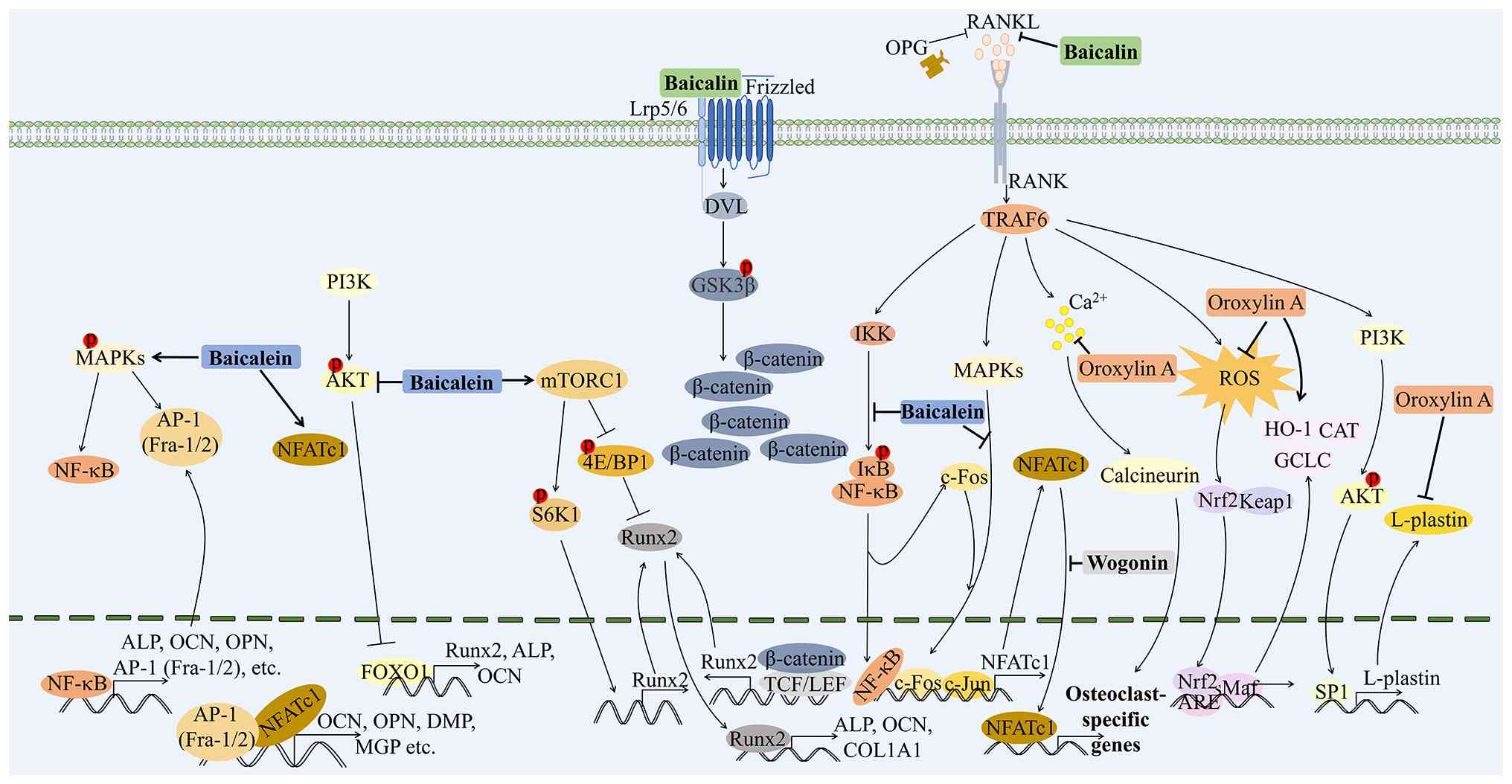 |
Figure 1
Principal molecular mechanisms of Scutellaria baicalensis and its active compounds in osteoporosis treatment. The diverse mechanisms through which Scutellaria baicalensis and its active compounds (baicalin, baicalein, wogonin, and oroxylin A) exert their therapeutic effects on osteoporosis are illustrated. Baicalin enhances osteogenic differentiation and inhibits adipogenesis in bone marrow mesenchymal stem cells while regulating osteoclast activity through various signaling pathways, including p38-MAPK and ERK/c-Fos/NFATc1. Baicalein promotes osteoblast differentiation via mTORC1 and MAPK/NF-κB pathways and exhibits anti-resorptive effects by suppressing RANKL-induced osteoclastogenesis. Wogonin inhibits osteoclast differentiation by downregulating RANKL and upregulating OPG, thus fostering a favorable environment for bone regeneration. Oroxylin A reduces NFATc1 activity and ROS production, further inhibiting osteoclastogenesis and promoting bone mass maintenance. MAPK, mitogen-activated protein kinase; ERK, extracellular signal-regulated kinase; c-Fos, cellular Fos; NFATc1, nuclear factor of activated T cells 1; mTORC1, mechanistic target of rapamycin complex 1; NF-κB, nuclear factor-κB; RANKL, receptor activator of nuclear factor κB ligand; OPG, osteoprotegerin; ROS, reactive oxygen species.
|
|
Table II
Major active components of Scutellaria baicalensis in osteoporosis.
|
Table II
Major active components of Scutellaria baicalensis in osteoporosis.
| Active compound |
Experimental model |
Dose/route |
Key pathways |
Phenotypic outcomes |
(Refs.) |
| Baicalin |
STZ-induced diabetic osteoporosis rats; bone marrow mesenchymal stem cells |
20 mg/kg/day via intragastric gavage for 8 weeks |
p38/MAPK pathway |
Ameliorated bone damage by enhancing osteogenesis and inhibiting adipogenesis |
(43) |
| Baicalin |
Dexamethasone-induced osteoporosis zebrafish larvae |
Incubated at 0.22 mM and 11 mM until 8 dpf or 60 dpf |
RANKL/RANK/OPG pathway |
Enhanced growth, development, and mineralization |
(44) |
| Baicalin |
Mouse bone marrow-derived macrophages; murine calvarial model of lipopolysaccharide-induced osteolysis |
Incubated at 0.01, 0.1, 1, 2, 4 or 8 mM for 8 days; subcutaneous injection (6 or 12 mg/kg) into the cranial midline for 7 days |
MAPK/Mitf, ERK/c-Fos/NFATc1 pathway |
Enhanced RANKL-induced osteoclastogenesis at 0-1 mmol/l; inhibited at ≥2 mmol/l |
(52,53) |
| Baicalin |
Rat primary osteoblasts |
Incubated at 50 μm for 3, 5, 7, and 9 days |
Wnt/β-catenin pathway |
Enhanced the osteoblastic mineralization |
(46) |
| Baicalein |
Ovariectomized-mice; MC3T3-E1 cells |
10 mg/kg/day via intragastric gavage for 3 months; incubated at 0.1-10 μM for 14 days |
mTORC1 pathway |
Enhanced the trabecular thickness, trabecular bone mineral density, and trabecular number; induced to differentiate into osteoblasts |
(48) |
| Baicalein |
MC3T3-E1 subclone 4 cells |
Incubated at 10 μM |
MAPK/NF-κB/NFATc1 pathway |
Enhanced ALP activity and mineralization |
(50) |
| Wogonin |
Murine calvarial model of lipopolysaccharide-induced osteolysis; mouse primary osteoblasts and pre-osteoblasts |
Subcutaneous injection (37.5 or 10 mg/kg) into the cranial midline for 5 days; incubated at 0.5, 1, and 5 μM for 3 days |
RANKL/OPG pathway |
Inhibited the osteoclastogenesis |
(58) |
| Oroxylin A |
Ovariectomized- and lipopolysaccharide-induced bone loss models; mouse bone marrow-derived macrophages |
Intraperitoneally administrated 2.5 and 5 mg/kg for 3 months or 7 days; incubated at 1, 2.5, 5, and 10 μM for 3 days |
RANKL/NFATc1 and RANKL-mediated ROS/Nrf2 pathway |
Reduced osteoclast formation and bone resorption |
(60) |
Promotion of osteogenic differentiation and bone formation
Baicalein and baicalin have been most extensively documented to facilitate osteogenic differentiation and bone anabolic processes. Previous research indicates that baicalin prevents disuse-induced OP by enhancing osteogenic differentiation in BMSCs, leading to improvements in bone mineral density, microstructure and mechanical strength (21). Additionally, in a diabetic OP (DOP) model, baicalin was found to improve bone damage by promoting osteogenesis and inhibiting adipogenesis in BMSCs via the p38 mitogen-activated protein kinase (p38-MAPK) pathway, highlighting its potential for DOP treatment (43). Furthermore, in a zebrafish model, baicalin was shown to mitigate dexamethasone-induced OP by regulating the RANKL/RANK/OPG pathway, improving bone mineralization, and influencing gene expression related to bone formation and resorption (44). Notably, baicalin has also been identified as a promising sclerostin inhibitor, binding to the loop 2 region through hydrophobic interactions, particularly with the Arg-Gly-Lys-Trp-Trp-Arg motif, offering a potential new approach for developing sclerostin inhibitors to promote bone formation and treat OP (45).
Estrogen replacement therapy is a common treatment for postmenopausal OP in women, yet adverse effects of long-term estrogen use such as breast cancer and cardiovascular diseases limit its clinical application (46). Guo et al (46) reported that baicalin promotes osteoblastic differentiation and bone mineralization by activating the Wnt/β-catenin signaling pathway, independent of its estrogenic activity. Additionally, baicalin has been shown to induce the expression of acetylcholinesterase (AChE) in cultured osteoblasts, with the induced AChE existing solely in a tetrameric globular form connected to the proline-rich membrane anchor, and this induction occurs independently of estrogen receptor blockade (47). These results suggest that baicalin has potential as an alternative OP treatment with estrogen-like effects. Baicalein promotes osteogenesis through multiple mechanisms, primarily by enhancing osteoblast differentiation and bone formation. In preclinical studies, baicalein improved key bone parameters such as bone mineral density, trabecular thickness and trabecular number, particularly in ovariectomized (OVX) animal models that mimic postmenopausal OP (48). In vitro, it was shown to activate the mTORC1 signaling pathway, a central regulator of cell growth and metabolism, which in turn stimulated the expression of osteoblast markers such as osteocalcin (OCN), runt-related transcription factor 2 (RUNX2), osterix and collagen Iα1 in MC3T3-E1 pre-osteoblast cells. This activation also involved protein kinases and transcription factors, including phoshorylated (p)-eukaryotic translation initiation factor 4E/binding protein 1 (4E/BP1) and p-ribosomal protein S6 kinase 1S6K1 (48). Additionally, baicalein was found to inhibit AKT, upregulating FOXO1 to enhance bone turnover markers and extracellular matrix (ECM) mineralization in glucocorticoid-induced OP (49). Furthermore, baicalein was demonstrated to promote osteoblast differentiation by activating NF-κB in the initiation stage, inducing osteoblast markers via the MAPK/NF-κB pathway in the early stage, and stimulating calcium deposition in the later stage through MAPK and activator protein 1 (AP-1) family members, including Fos-related antigen (Fra)-1 and Fra-2, with a slight induction of nuclear factor of activated T cells 1 (NFATc1) (50). The activation of the mTORC1 and MAPK/NF-κB pathways, along with the modulation of the AKT/FOXO1 signaling pathway, underscores the potential of baicalein in promoting osteogenesis and preventing bone loss associated with OP.
To further explore the mechanism of baicalein in treating OP, a previous study employed bioinformatics analysis and network pharmacology. Bioinformatics analysis using the STITCH (http://stitch.embl.de/) and MIRWALK2.0 databases (http://zmf.umm.uni-heidelberg.de/mirwalk2) identified 26 common Kyoto Encyclopedia of Genes and Genomes (https://www.kegg.jp/) pathways related to baicalein and OP, primarily involving key signaling pathways such as PI3K-AKT, MAPK and mTOR. Further cell experiments revealed that baicalein increased the expression of markers involved in bone turnover through the AKT/FOXO1 pathway (49). Additionally, network pharmacology analysis further demonstrated that the therapeutic effects of baicalein involve the regulation of sex hormone levels, autophagy, inflammation and reactive oxygen metabolism. Subsequent animal experiments confirmed these findings, demonstrating that baicalein significantly enhanced the expression of sirtuin 1 (SIRT1), androgen receptor (AR) and estrogen receptor 1 (ESR1) while reducing prostaglandin-endoperoxide synthase 2 (PTGS2) expression, thus providing solid experimental support for the aforementioned predicted mechanism of baicalein against OP (51).
Inhibition of osteoclastogenesis and bone resorption
Baicalin regulates OC differentiation and bone resorption. At low concentrations (<1 μmol/l), it enhances OC differentiation and bone resorption (52,53), while, at high concentrations (>2 μmol/l), it inhibits OC function by suppressing the ERK/c-Fos/NFATc1 pathway and inducing the expression of apoptosis-related proteins such as Bax (52). While previous in vitro cellular studies have verified the dual regulatory effects of baicalin on OCs, the lipopolysaccharide (LPS)-induced calvarial osteolysis mouse model only mimics acute inflammatory bone damage (52,53). Accordingly, the current results cannot fully confirm the direct therapeutic effects of baicalin against OP. Future research should therefore conduct in vivo pharmacological validation of baicalin at varying doses using classic OP models, including OVX and aged mice. In particular, micro-computed tomography should be utilized to quantitatively analyze key trabecular bone micro-architectural parameters, such as bone mineral density (BMD) and bone volume fraction. Such follow-up investigations are indispensable for clarifying the true therapeutic efficacy and clinical translational potential of baicalin in chronic bone loss conditions.
Baicalein has also demonstrated anti-resorptive effects by inhibiting OC activity. It was shown to effectively reduce RANKL-induced tartrate-resistant acid phosphatase (TRAP) activity and OC formation. Furthermore, it was found to promote apoptosis in mature OCs, thereby decreasing their bone resorption activity. Mechanically, baicalein has been shown to inhibit the activation of critical signaling pathways, including AKT, ERK/MAPK and NF-κB, and the expression of transcription factors associated with OCs, including c-Fos, Fra-2 and NFATc1 (54). However, the effectiveness of baicalein in rescuing bone loss can vary depending on the administration method and stage of osteoporotic healing. Subcutaneous administration of baicalein in osteopenic rats significantly increased cortical BMD and bone formation rate, although its effect on trabecular bone was limited at higher doses (55). By contrast, oral administration (10 mg/kg body weight) did not replicate these favorable effects on cortical bone and showed no significant improvement in the vertebral or femoral bones of OVX rats, although it accelerated mid-stage callus formation (56). In addition, while baicalein enhanced callus density and cortical width in tibiae, it impaired early callus formation, and reduced biomechanical properties and calcium-to-phosphate ratio in femora, suggesting that it may not be suitable for treating early-stage osteoporotic fractures (57). Such inconsistencies carry critical research implications. Oral administration serves as the primary clinical approach for long-term OP treatment, whereas prominent bone anabolic effects of baicalein are mostly observed through parenteral delivery. This evidence indicates that low oral bioavailability functions as a major limiting factor instead of insufficient intrinsic bioactivity. Optimized pharmaceutical formulations are therefore essential to enable oral baicalein to exert valid therapeutic effects on human skeletal tissues.
Wogonin has been shown to inhibit LPS-induced osteoclastogenesis by suppressing the expression of RANKL, upregulating the expression of OPG and reducing prostaglandin E2 (PGE2) production in osteoblasts (58). Additionally, wogonin was found to inhibit the translocation of NFATc1 from the cytoplasm to the nucleus, thereby blocking its transcriptional activation and significantly suppressing OC differentiation by downregulating key OC-associated genes, including TRAP and calcitonin receptor, positioning wogonin as a possible therapy for OP (59).
Oroxylin A was shown to inhibit RANKL-induced OC genesis by reducing the transcriptional activity of NFATc1 and upregulating nuclear factor erythroid 2-related factor 2 (Nrf2)-mediated reactive oxygen species (ROS) scavenging enzymes, which also suppressed ROS production, significantly reduced intracellular calcineurin activity and intracellular Ca2+ influx (60). Furthermore, L-plastin (LPL) has been identified as a regulator of OC fusion, with LPL-knockout mice exhibiting increased bone mass and cortical thickness. Notably, oroxylin A has been recognized as an LPL inhibitor that maintains bone mass in ovariectomy-induced OP and accelerates bone fracture healing, highlighting its potential as a novel anabolic therapy for pathological bone loss (61). Additionally, oroxylin A has been shown to protect OVX osteoporotic mice from bone loss by regulating metabolic pathways associated with phenylalanine, tyrosine and tryptophan biosynthesis, with lysophosphatidylcholine (18:2) identified as a key biomarker (62).
In comparison, oroxylin A and wogonin have been reported to maintain steady anti-OC activity (58-62), while baicalin displays biphasic pharmacological performance (52,53). Baicalein has been demonstrated to regulate the function of both osteoblasts and OCs to balance bone metabolism (54-57). This dual regulatory property makes it the most versatile compound for the systemic intervention of OP.
Drug delivery systems for OP treatment
To address bioavailability and targeting challenges, several novel delivery systems have been developed for OP and bone repair applications. Amorphous calcium polyphosphate microparticles loaded with baicalin and baicalein have been shown to improve their solubility, bioavailability and anti-osteoporotic performance. The microparticles enter osteoblasts, gather perinuclearly and form alkaline phosphatase (ALP)-positive vesicles to raise intracellular drug content and eliminate oral delivery limitations from first-pass metabolism and intestinal breakdown. The composite enhances bone mineralization in primary human osteoblasts by increasing intracellular calcium levels, upregulating calcium efflux channels and promoting mineralization, suggesting potential use in OP prevention or bone implants (31). Baicalein-loaded mesoporous silica-Fe3O4 nanoparticles have been demonstrated to overcome the poor solubility and inadequate targeting of free baicalein, and improve fracture repair efficacy. This dual-targeted nanosystem can precisely accumulate at fracture sites under magnetic guidance and be selectively internalized by macrophages via surface-modified NW peptides to achieve localized baicalein delivery that elevates local drug concentration and reduces systemic toxicity. The released baicalein suppresses ROS/NF-κB-mediated inflammation and oxidative stress, and this effect facilitates M2 macrophage polarization and further promotes mesenchymal stem cell osteogenesis, thus elevating ALP activity and bone mineralization (63). Additionally, baicalein-loaded bovine serum albumin (BSA) coatings were shown to improve baicalein solubility with antioxidant and osteogenic capacities for implant repair. Tannic acid/baicalein-BSA multilayers stably coat titanium alloys, trap baicalein via albumin hydrophobic interactions for high local drug retention and steadily release baicalein without hemolysis or osteotoxicity. Synergistic baicalein and tannic acid eliminate free radicals, lower intracellular ROS, mitigate osteoblast oxidative injury, enhance early cell adhesion, raise ALP levels and boost calcium mineralization, making the coating suitable for bone implant surface modification (64).
Strontium-incorporated titanium coatings carrying wogonin silk fibroin nanoparticles have been reported to overcome the limited solubility of wogonin to treat osteoporotic bone defects via immunoregulation and osteogenic stimulation. Microarc oxidation and layer-by-layer assembly retain local wogonin and strontium for sustained safe release. Wogonin curbs inflammation and shifts macrophages toward the M2 phenotype to boost osteogenic paracrine signaling, whereas strontium directly fuels osteoblast maturation. Combined, they accelerate new bone growth and strengthen implant osseointegration (65).
Oroxylin A-loaded amorphous calcium carbonate nanoparticles have been shown to overcome the poor solubility and lack of bone-targeting capability of free oroxylin A for OP therapy. Functionalized with glutamic acid hexapeptides, these nanoparticles selectively accumulate at acidified OC resorption sites. Once exposed to acidic surroundings, the carriers degrade to neutralize local hydrogen ions and release concentrated oroxylin A onsite, thus minimizing systemic adverse effects. The released oroxylin A cooperates with calcium to inhibit NFATc1-mediated osteoclastogenesis, restore trabecular architecture and reverse bone loss, demonstrating significant therapeutic potential in the treatment of OP (66).
Overall, baicalin suppresses bone resorption and facilitates bone mineralization through multiple molecular pathways, indicating its promising value for OP intervention. Further research remains necessary to validate its clinical potential, with clinical trials specifically required to evaluate its long-term safety, optimal dosage regimens and comparative efficacy relative to standard OP treatments. Unlike baicalin, baicalein exerts dual regulatory effects on bone turnover by facilitating osteogenic differentiation and suppressing OC-mediated bone breakdown, which benefits OP treatment and bone tissue repair. Its therapeutic performance varies substantially according to delivery approaches and distinct bone healing stages. Wogonin and oroxylin A exhibit prominent advantages in inhibiting bone resorption and supporting local bone restoration.
Scutellarin baicalensis and OA
OA is a chronic joint disease influenced by factors such as age, sex, obesity and trauma, and is characterized fundamentally by the progressive degeneration of articular cartilage and subchondral bone remodeling (67). As the primary cellular component within osteoarthritic joint tissues, chondrocytes undergo senescence, apoptosis and dysfunction under pathological stimuli, which impairs their capacity for ECM synthesis while driving the release of matrix metalloproteinases (MMPs) and aggrecanases that break down cartilage ECM (68). Persistent low-grade chronic inflammation in the OA joint increases the levels of pro-inflammatory mediators including IL-1β, TNF-α and nitric oxide (NO), as well as chemokines, which recruit immune cells to infiltrate the synovium and aggravate joint inflammatory injury (69). Currently, the treatment of OA primarily focuses on symptom relief, while the search for disease-modifying osteoarthritic drugs that effectively improve both symptoms and the structural progression of the disease is ongoing, with some promising candidates, including those targeting inflammation (70). Medicinal plants and their extracts hold marked potential for the treatment of OA, primarily by modulating critical molecular pathways involved in inflammation, cartilage degradation and cell death (71). It has been reported that Scutellaria baicalensis exhibits significant anti-inflammatory, analgesic and joint tissue-protective effects against OA through its active compounds and key molecular pathways (Fig. 2 and Table III), such as the PI3K-AKT signaling and cellular senescence pathways (22). Therefore, Scutellaria baicalensis and its active compounds hold important potential as candidates for further investigation and development as therapeutic treatments for OA.
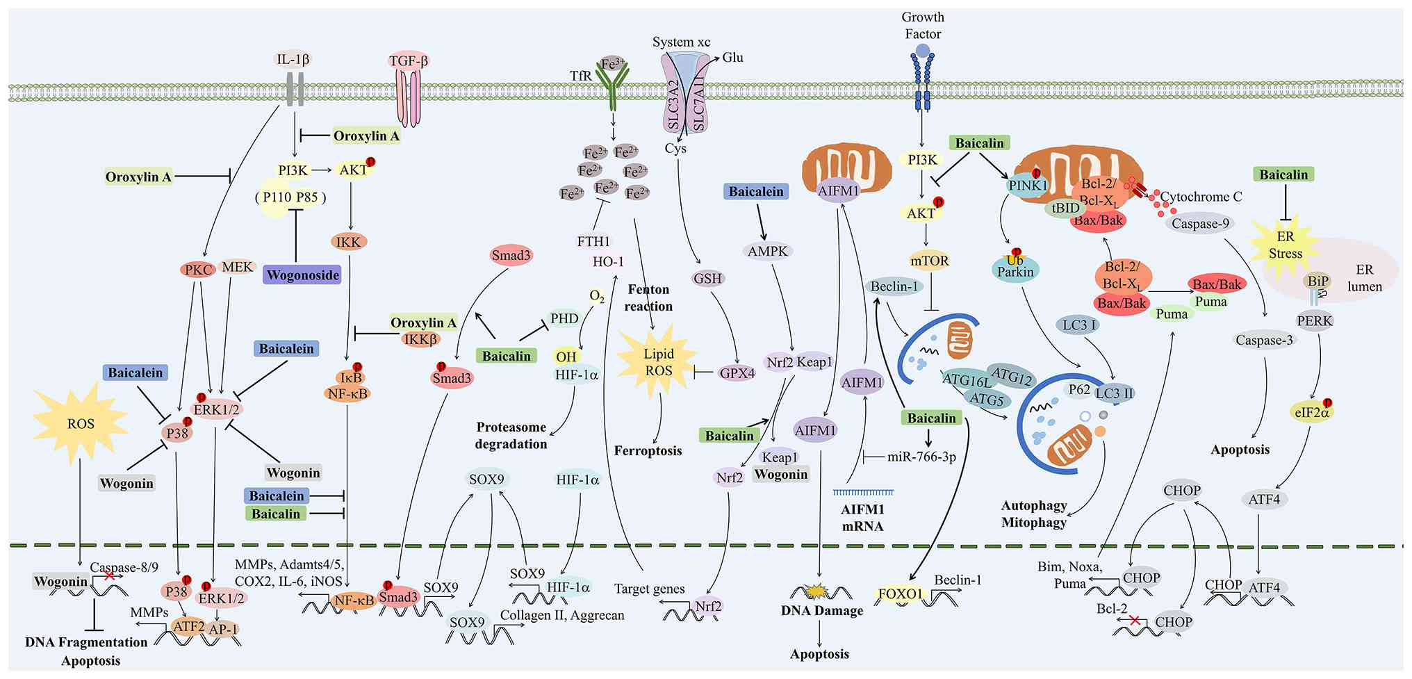 |
Figure 2
Principal molecular mechanisms of Scutellaria baicalensis and its active compounds in osteoarthritis treatment. The various therapeutic mechanisms by which Scutellaria baicalensis and its active compounds (baicalin, baicalein, wogonin, wogonoside, and oroxylin A) contribute to the treatment of osteoarthritis are presented. Baicalin promotes chondrocyte health by enhancing autophagy, mitigating oxidative stress, and reducing inflammation through the PI3K/AKT/mTOR and NF-κB pathways. Baicalein exhibits anti-inflammatory and anti-apoptotic effects, decreasing the expression of inflammatory mediators and MMPs, while promoting cartilage regeneration via the SOX-9 transcription factor. Wogonin demonstrates chondroprotective properties by inhibiting MMP activity and enhancing antioxidant capacity, thereby reducing joint inflammation and pain. Wogonoside and oroxylin A protect against extracellular matrix degradation and inhibit inflammatory responses by modulating the NF-κB and PI3K/AKT signaling pathways. PI3K, phosphoinositide 3-kinase; AKT, protein kinase B; mTOR, mechanistic target of rapamycin; NF-κB, nuclear factor-κB; MMPs, matrix metalloproteinases; SOX-9, SRY-box transcription factor 9.
|
|
Table III
Major active components of Scutellaria baicalensis in osteoarthritis.
|
Table III
Major active components of Scutellaria baicalensis in osteoarthritis.
| Active compound |
Experimental model |
Dose/route |
Key pathways |
Phenotypic outcomes |
(Refs.) |
| Baicalin |
Primary human chondrocytes |
Incubated at 0.22 μM |
The miR-766-3p/AIFM1 axis |
Inhibited IL-1β-induced apoptosis and ECM degradation via autophagy activation |
(73) |
| Baicalin |
Rat chondrocytes |
Incubated at 10, 20, and 40 μM for 4 h |
PI3K/AKT/mTOR, PINK1/Park, and PINK1/Drp-1 pathway |
Promoted mitophagy, enhanced cell viability, and reduced apoptosis |
(74) |
| Baicalin |
Primary human chondrocytes |
Incubated at 20 μM for 12 h |
Nrf2 pathway |
Inhibited IL-1β-induced ferroptosis, mitochondrial dysfunction, and ECM degradation |
(75) |
| Baicalin |
Mouse OA model (DMM); ADTC5 cells |
Orally given 50 mg/kg/day for 6 weeks; incubated at 10, 20, and 40 μM for 24 h |
TGF-β/Smad3 pathway |
Promoted the cell viability and ECM synthesis |
(77) |
| Baicalin |
Mouse OA model (DMM); Primary human chondrocytes |
Intraperitoneal injection 100 mg/kg/day for 8 weeks; incubated at 10, 50, and 100 μM |
NF-κB pathway |
Reduced cartilage destruction and alleviated the severity of synovitis |
(91) |
| Baicalein |
Primary human chondrocytes |
Incubated at 25 μM |
NF-κB pathway |
Attenuated cell death and partially recovered mitochondrial viability |
(97) |
| Baicalein |
Rat OA model (DMM) |
Intra-articular injection 0.8, 1.6, 3.2 μg/l, 50 μl daily for 6 weeks |
NLRP3 inflammasome pathway |
Relieved inflammation and ameliorated cartilage damage |
(98) |
| Baicalein |
Rabbit OA model (ACLT); Primary human chondrocytes |
Intra-articular injection 5, 25, and 50 μM daily for 6 weeks; incubated at 50 μM for 24 h |
p38/ERK pathway |
Inhibited expression of MMPs and improved cartilage injury |
(79) |
| Baicalein |
Mouse OA model (DMM); Primary human chondrocytes |
Intra-articular injection 1 mg/kg/week for 9 weeks; Incubated at 1, 5, 10 μM for 36 h |
AMPK/Nrf2/HO-1 pathway |
Reduced chondrocyte catabolism and ferroptosis, improved cell viability, alleviated OA and pain |
(99) |
| Wogonin |
SW1353 cells |
Incubated at 10 μM |
c-Fos/AP-1 and JAK/STAT pathway |
Inhibited expression of MMPs |
(85) |
| Wogonin |
Primary human chondrocytes |
Incubated at 10, 25, and 50 μM |
ROS/ERK/Nrf2 pathway |
Inhibited inflammation and matrix degradation; induced mild oxidative stress |
(86) |
| Wogonoside |
Primary mice chondrocytes |
Incubated at 12.5, 25, and 50 μM |
PI3K/AKT and NF-κB/HIF-2a pathway |
Inhibited matrix degradation and chondrocyte hypertrophy |
(87) |
| Oroxylin A |
Primary mice chondrocytes |
Incubated at 4, 8, and 16 μM |
ERK1/2 and PI3K/AKT pathway |
Inhibited inflammation and matrix degradation |
(102) |
| Oroxylin A |
Mouse OA model (DMM); primary human chondrocytes |
10 mg/kg/day by oral gavage for 4 weeks; incubated at 5, 10, and 20 μM |
NF-κB and Wnt/β-catenin pathway |
Inhibited inflammation, matrix degradation, and chondrocyte hypertrophy; ameliorated OA development |
(88) |
Chondroprotection, maintenance of ECM homeostasis and regulation of bone remodeling
Preservation of ECM integrity constitutes a core therapeutic target for disease-modifying OA interventions (72). Baicalin and baicalein are the most thoroughly investigated flavonoids that sustain cartilage homeostasis, with regulatory effects mediated by autophagy activation, ferroptosis inhibition and enhanced matrix biosynthesis.
Baicalin has demonstrated protective effects on OA chondrocytes against IL-1β-induced apoptosis and ECM degradation by activating autophagy through the microRNA (miRNA or miR)-766-3p/apoptosis-inducing factor mitochondria-associated 1 axis (73). Additionally, it has been shown to protect against IL-1β-induced chondrocyte injury by promoting mitophagy, enhancing cell viability and reducing apoptosis via the inhibition of the PI3K/AKT/mTOR pathway and the activation of the PTEN-induced kinase 1 (PINK1)/Parkin and PINK1/dynamin-related protein 1 (Drp-1) pathways (74). These actions are characterized by increased levels of autophagy and mitophagy-related proteins, highlighting the potential of baicalin as a therapeutic agent for OA by supporting chondrocyte homeostasis.
It also has been found to address IL-1β-induced ferroptosis in chondrocytes, which is marked by elevated Fe2+ accumulation and oxidative stress markers. Baicalin effectively counteracts these effects and activates the Nrf2 antioxidant system, primarily reducing intracellular ROS and lipid peroxidation, thereby protecting against IL-1β-induced ferroptosis and ECM degradation (75). In addition, baicalin has been found to promote ECM synthesis and increase the expression of chondrogenic marker genes in chondrocytes by activating hypoxia-inducible factor (HIF)-1α (76). Furthermore, it has been revealed to effectively mitigate IL-1β-induced reductions in chondrocyte viability and ECM secretion by enhancing the nuclear translocation of SOX9 and activating the transforming growth factor-β/Smad3 (TGF-β/Smad3) signaling pathway (77).
Baicalein has been demonstrated to effectively decrease the expression of MMPs such as MMP-3, MMP-9 and MMP-13, mitigating cartilage damage (78,79). This action is achieved by inhibiting the transcription and translation of MMPs, thereby reducing their activity in the cartilage matrix by significantly inhibiting the phosphorylation of p38 and ERK, while not affecting JNK (79). Furthermore, baicalein has been shown to promote the deposition of collagen II and glycosaminoglycans, aiding in the restoration of cartilage structure and function, primarily by activating transcription factors such as SOX-9 to enhance the synthesis of matrix components (80,81). Regarding bone remodeling, baicalein was reported to inhibit the proliferation and differentiation of pre-osteoblasts and promote their apoptosis, improving subchondral bone remodeling. Additionally, baicalein was found to suppress angiogenesis in subchondral bone and reduce subchondral bone sclerosis while limiting the proliferation of synovial cells to manage synovitis (82,83).
Wogonin has demonstrated chondroprotective properties by inhibiting the expression and activity of MMPs (MMP-3 and MMP-13), which are key mediators of cartilage degradation (84,85). Wogonin achieved this by blocking the c-Fos/AP-1 and JAK2/STAT1/2 signaling pathways, while not affecting NF-κB signaling (85). Additionally, it was shown to promote the expression of anabolic factors such as collagen II and aggrecan, which are essential for maintaining cartilage integrity (84,86). Wogonoside has been reported to effectively protect against IL-1β-induced ECM degradation and chondrocyte hypertrophy in OA by inhibiting the activation of the NF-κB/HIF-2α pathway via the PI3K/AKT signaling pathway (87). Oroxylin A was demonstrated to maintain ECM homeostasis in chondrocytes and mitigate hypertrophic changes by inhibiting the NF-κB and Wnt/β-catenin signaling pathways (88).
Overall, baicalin confers chondroprotection through an extensive repertoire of regulatory mechanisms, encompassing autophagy, mitophagy, anti-ferroptosis and ECM anabolism. By contrast, wogonin elicits protective effects via a restricted set of signaling pathways that operate independently of NF-κB signaling. The distinct mechanistic profiles of these two flavonoids indicate that their combined administration may yield complementary therapeutic advantages for cartilage protection.
Anti-inflammation and antioxidation properties
Chronic inflammation and oxidative stress suppress ECM anabolism in chondrocytes while triggering cellular senescence and multiple cell death modalities such as apoptosis, necroptosis and ferroptosis. These pathological events ultimately disrupt the structural integrity of articular cartilage and exacerbate joint pain (89). All five active compounds (baicalin, baicalein, wogonin, oroxylin A and wogonoside) possess promising therapeutic potential for OA management, as they relieve inflammatory responses and restore redox homeostasis via distinct regulatory mechanisms.
Baicalin was found to significantly reduce the expression of pro-inflammatory genes and MMPs associated with OA while enhancing the levels of aggrecan and collagen II in human chondrocytes (90,91). In addition, it was shown to effectively suppress IL-1β-induced inflammatory responses and NF-κB activation, thus preventing cartilage destruction and alleviating synovitis in both in vitro and in vivo models (91). Furthermore, baicalin has been demonstrated to enhance autophagy by activating FOXO1 to promote Beclin-1 expression, which may further contribute to its anti-inflammatory effects (92). A previous study also reported that baicalin alleviated immune inflammatory responses in OA by exerting synergistic effects with small interference-long non coding RNA (lncRNA) HOTAIR on the PTEN/PI3K/AKT signaling pathway, thereby reducing inflammatory factors and contributing to the relief of OA symptoms (93). Collectively, these findings suggest that baicalin has significant potential as a therapeutic agent for treating OA by mitigating inflammation and promoting chondrocyte health.
Baicalin has been shown to enhance cell viability and mitigate oxidative stress in chondrocytes exposed to hydrogen peroxide (H2O2) by reversing apoptotic signaling pathways and boosting antioxidant activity (94). Li et al (95) demonstrated that baicalin protects chondrocytes from H2O2-induced ROS generation by inhibiting endoplasmic reticulum (ER) stress and preserving the expression of ECM-related genes, suggesting its potential as a therapeutic agent for mitigating the effects of oxidative stress in OA by targeting ER stress pathways. Additionally, baicalin alleviated the severity of OA in a rat model by reducing muscle dysfunction and oxidative stress, as evidenced by increased citrate synthase activity and decreased IL-6 production. Its protective effects are linked to its ability to inhibit ROS generation and regulate Nrf2 expression in OA (96).
Baicalein was found to markedly reduce the expression of inflammatory mediators such as IL-1β and TNF-α, thereby alleviating joint inflammation (78,80,97). This effect primarily occurs through the inhibition of the NF-κB signaling pathway, which decreases the synthesis and release of pro-inflammatory cytokines (80,97). Additionally, baicalein can significantly slow disease progression and reduce inflammation by decreasing the levels of inflammatory mediators, cartilage degradation markers and NLRP3 inflammasome activation (98).
In terms of anti-apoptotic effects, baicalein has been demonstrated to enhance the expression of anti-apoptotic genes, thus inhibiting chondrocyte apoptosis and promoting the expression of SOX-9, which strengthens cell survival (80,81,83). It also was reported to improve mitochondrial viability in IL-1β-stimulated chondrocytes, reducing cell death primarily by attenuating mitochondrial membrane potential loss and inhibiting the NF-κB signaling pathway, thus protecting mitochondrial function (97). Finally, baicalein was found to enhance AMP-activated protein kinase activity, increase the abundance of Nrf2 and promotes its nuclear translocation, leading to the expression of heme oxygenase-1, which inhibits lipid ROS and reduces oxidative stress-induced damage to chondrocytes (99). Additionally, baicalein has been shown to mitigate ferroptosis, a form of regulated cell death linked to iron accumulation and lipid peroxidation, further protecting chondrocytes from oxidative stress and promoting cell survival (99).
Wogonin was reported to effectively reduce inflammation in OA chondrocytes by suppressing the production of inflammatory mediators, including IL-6, cyclooxygenase-2 (COX-2) and inducible NO synthase (iNOS) (86). This anti-inflammatory action contributes to the overall reduction of joint inflammation and pain associated with OA. Wogonin has also been shown to stabilize genomic DNA in chondrocytes, preventing fragmentation and protecting against apoptosis induced by IL-1β (100). Furthermore, it was found to activate the Nrf2/antioxidant response element (ARE) signaling pathway through mild oxidative stress, enhancing the antioxidant capacity of chondrocytes (86), which helps mitigate oxidative damage in the cartilage.
Wogonoside has been demonstrated to alleviate articular cartilage injury and T-helper (Th)1/Th2 cytokine infiltration in rats with OA by inhibiting the activation of NF-κB and ERK1/2, resulting in improved cartilage tissue damage and reduced Mankins scores (101).
Oroxylin A has been revealed to effectively mitigate IL-1β-induced inflammatory reactions in chondrocytes by inhibiting the upregulation of key inflammatory mediators and MMPs, while also suppressing the activation of the ERK1/2 and PI3K/AKT signaling pathways (102). Additionally, oroxylin A was reported to act as an inhibitor of LPL, helping to maintain a hypoxic environment in joints, which inhibits angiogenesis and protects against cartilage degeneration mediated by HIF-1α stabilization (103).
Regarding bone remodeling, baicalein has been shown to inhibit the proliferation and differentiation of pre-osteoblasts and promote their apoptosis, thus improving subchondral bone remodeling. Additionally, baicalein was found to suppress angiogenesis in subchondral bone and reduce subchondral bone sclerosis while limiting the proliferation of synovial cells to manage synovitis (82,83).
Drug delivery systems for OA treatment
OA is a local disorder mainly attacking joint synovium and cartilage. Systemic administration generally fails to maintain sufficient drug concentrations inside joint cavities. Accordingly, intra-articular local delivery systems stand as a core strategy to overcome poor bioavailability of active ingredients derived from Scutellaria baicalensis and achieve satisfactory therapeutic outcomes.
A folic acid (FA)-functionalized metal-organic framework nanocarrier termed Bai@FA-UIO-66-NH2 was fabricated for targeted baicalin delivery. This nanoplatform can be selectively internalized by M1 macrophages and can release baicalin in response to an acidic intracellular milieu. It was shown to efficiently eliminate ROS and shift macrophage polarization from pro-inflammatory M1 to anti-inflammatory M2 phenotypes, which notably alleviated synovial inflammation in OA rat models. Beyond boosting drug accumulation at inflamed lesions, this active-targeted delivery system produces synergistic therapeutic benefits by remodeling the local immune microenvironment (104). Injectable hydrogels have garnered great research interest for joint therapy owing to their superior adaptability to irregular joint cavities. Researchers have developed a novel injectable hydrogel combining baicalin and 2-hydroxyethyl acrylate, which possesses excellent mechanical properties and self-healing capabilities. This baicalin-incorporated hydrogel elevates local bioavailability, reduces administration frequency for OA management, extends drug retention within joints and displays strong binding affinity toward synovial tissues. Mechanistically, it regulates glycolysis in fibroblast-like synoviocytes (FLSs) by inhibiting the YAP1 signaling pathway, leading to decreased inflammatory factor secretion, reduced joint pain, and reduced cartilage degradation and synovial inflammation (105). Nucleic acid nanomaterials exhibit great potential for cartilage protection. Previous research validated that tetrahedral framework nucleic acid/wogonin complexes are readily taken up by inflamed chondrocytes, and can effectively alleviate inflammation and prevent cartilage destruction, indicating their potential as an injectable treatment option (106). Formulated topical creams with auxiliary excipients facilitate the penetration and diffusion of active agents into joint capsules. Topical application of wogonin was reported to significantly reduce pain and inflammatory biomarker levels in an OA mouse model, and provides a simple approach to overcome the limitation of systemic administration (107). Thermoresponsive hydrogels hold great translational potential for OA treatment. A developed injectable hydrogel system (Gel@Wogonin-MSCIPFP-Exo) encapsulates wogonin-primed MSC-derived exosomes for intra-articular administration. Triggered by local joint hyperthermia, the hydrogel achieves in situ gelation to enable sustained exosome release. It extends the intra-articular retention time of exosomes to 28 days, significantly outperforming free exosomes with a retention period of <14 days. This system was found to promote chondrocyte proliferation and migration, and inhibit inflammatory apoptosis. The combination of biomaterial-based delivery and cell-derived exosome therapy provides an innovative strategy for OA treatment (108).
Scutellarin baicalensis and RA
RA is a chronic, lifelong autoimmune disease characterized by joint swelling, pain and progressive deformity. Its pathological hallmark is chronic proliferative synovitis that invades cartilage and subchondral bone, ultimately leading to irreversible joint damage and systemic inflammation (109). Activated synovial fibroblasts, T cells, B cells and macrophages extensively infiltrate synovial tissue, driving sustained inflammation via excessive secretion of TNF-α, IL-6, IL-17, chemokines and autoantibodies to form an inflammatory positive feedback loop (110). RA poses significant clinical management challenges, as numerous patients cannot achieve sustained remission even after treatment with disease-modifying anti-rheumatic drugs (111,112). Currently, the use of medicinal herbs, which are rich in bioactive phytochemicals, presents a promising alternative therapeutic strategy for RA, offering complementary treatment options due to their anti-inflammatory and anti-oxidative properties (113,114). Huangqin Qingre Qubi capsule, a Chinese herbal compound primarily derived from Scutellaria baicalensis, demonstrates significant anti-arthritis effects in the treatment of RA by inhibiting inflammation and oxidative stress, particularly through its interaction with the frizzled class receptor 8 (FZD8)-Wnt/β-catenin signaling pathway (23). The main active components of Scutellaria baicalensis, including baicalin and baicalein, exhibit significant anti-inflammatory, antioxidant and immune-regulating properties, effectively alleviating the symptoms of RA and playing a crucial role in its treatment (Fig. 3 and Table IV) (115,116).
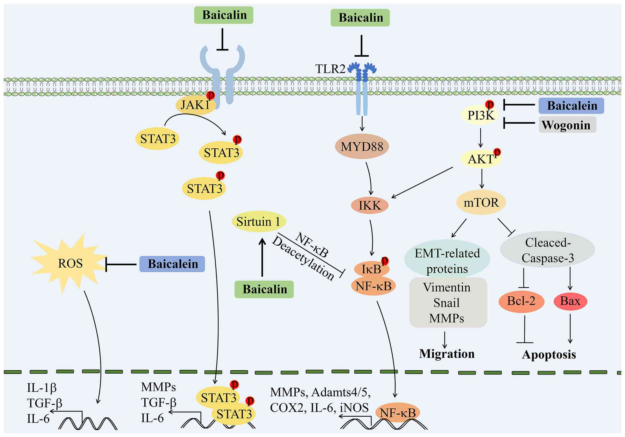 |
Figure 3
Principal molecular mechanisms of Scutellaria baicalensis and its active compounds in rheumatoid arthritis treatment. The therapeutic mechanisms by which Scutellaria baicalensis and its active compounds (baicalin, baicalein, and wogonin) contribute to the treatment of RA are depicted. Baicalin reduces inflammatory responses by inhibiting pro-inflammatory cytokines such as TNF-α, IL-6, and IL-1β, while modulating autophagy and protecting against inflammatory damage through pathways like TLR2/MYD88/NF-κB and JAK1/STAT3. It also enhances deacetylation of NF-κB via sirtuin 1 and improves lipid profiles in patients with RA. Baicalein inhibits the proliferation and migration of FLS and promotes apoptosis through the PI3K/AKT/mTOR pathway, while also downregulating key inflammatory cytokines. Wogonin inhibits FLS migration and cytokine production through the PI3K/AKT/NF-κB pathway, demonstrating significant anti-inflammatory effects in various RA models. RA, rheumatoid arthritis; TNF-α, tumor necrosis factor- α; IL-6, interleukin-6; IL-1β, interleukin-1β; TLR2, Toll-like receptor 2; MYD88, myeloid differentiation primary response 88; NF-κB, nuclear factor-κB; JAK1, Janus kinase 1; STAT3, signal transducer and activator of transcription 3; FLS, fibroblast-like synoviocytes; PI3K, phosphoinositide 3-kinase; AKT, protein kinase B; mTOR, mechanistic target of rapamycin.
|
|
Table IV
Major active components of Scutellaria baicalensis in rheumatoid arthritis.
|
Table IV
Major active components of Scutellaria baicalensis in rheumatoid arthritis.
| Active compound |
Experimental model |
Dose/route |
Key pathways |
Phenotypic outcomes |
(Refs.) |
| Baicalin |
Rat CIA model; RA human FLSs |
15, 30, and 60 mg/kg/day by oral gavage for 40 days; incubated at 25, 50, and 100 μg/ml for 48 h |
TLR2/MYD88/NF-κB pathway |
Alleviated inflammation and joint damage |
(117) |
| Baicalin |
Rat CIA model; RA human FLSs |
Intraperitoneal injection 50, 100, and 200 mg/kg/day for 30 days; incubated at 10, 20, and 30 μM for 24 h |
NF-κB pathway |
Alleviated inflammation and joint damage |
(118) |
| Baicalin |
Mouse CIA model |
Intraperitoneal injection 30 mg/kg/day for 5 days |
JAK1/STAT3 pathway |
Alleviated inflammation matrix degradation; promoted monocytes apoptosis |
(124) |
| Baicalein |
SW982 cell line |
Incubated at 15, 25, and 50 μM for 24 h |
PI3K/AKT/mTOR pathway |
Inhibited FLSs proliferation and EMT; promoted FLSs apoptosis |
(120) |
| Baicalein |
Primary human FLSs |
Incubated at 10, 20, and 40 μM |
NF-κB, MAPK/ERK/p38, and MIF pathway |
Inhibited IL-1β-induced proliferation of RAFLS |
(121) |
| Wogonin |
Mouse CIA model; MH7A cell line |
Intraperitoneal injection 40 mg/kg/day for 14 days; incubated at 10, 20, and 40 μM |
PI3K/AKT/NF-κB pathway |
Inhibited migration, invasion and inflammation |
(122) |
Inhibition of synovial inflammation and FLS dysfunction
Dysregulated proliferation, invasive capacity and overactivated inflammatory signaling of FLSs drive progressive joint damage in RA. Baicalin, baicalein and wogonin each alleviate the pathological phenotypes of FLS, albeit through divergent molecular pathways.
Baicalin was shown to significantly reduce inflammatory responses in rats with adjuvant-induced arthritis by inhibiting the expression of pro-inflammatory cytokines such as TNF-α, IL-6, IL-1β, IL-17 and COX-2 in synovial tissue, while also modulating autophagy mechanisms by decreasing autophagy-related proteins and increasing Bax expression (115). Furthermore, in collagen-induced arthritis (CIA) model rats, baicalin was found to significantly alleviate inflammation and joint damage by inhibiting the Toll-like receptor (TLR)2/myeloid differentiation primary response 88 (MYD88)/NF-κB signaling pathway, resulting in reduced levels of pro-inflammatory cytokines in both serum and synovial tissue (117). Liu et al (118) further demonstrated that baicalin enhances the deacetylation of NF-κB by upregulating SIRT1, which in turn suppresses the protein expression of NF-κB in rats with CIA. Bioinformatics analysis identified that differentially expressed mRNAs before and after baicalin treatment were significantly enriched in the Hippo, NF-κB and PI3K-AKT signaling pathways. A competing endogenous RNA (ceRNA) regulatory network was constructed, including lncRNA ENSRNOT00000076420/miR-144-3p/Fosb, lncRNA MSTRG.1448.13/miR-144-3p/Atp2b2 and lncRNA MSTRG.1448.13/miR-144-3p/Shanks. Notably, these enriched signaling pathways and ceRNA axes are purely computational predictions lacking in vitro and in vivo functional validation in rats with CIA. They remain hypothesis-generating and merely provide preliminary mechanistic clues that await further experimental confirmation (119).
Baicalein has been shown to effectively inhibit FLS proliferation and migration while promoting apoptosis through the PI3K/AKT/mTOR pathway, suggesting its potential clinical application in RA management (120). Furthermore, baicalein was demonstrated to suppress IL-1β-induced FLS proliferation by inhibiting NF-κB transcriptional activity and reducing the phosphorylation of key signaling molecules such as ERK1/2 and p38. Notably, these effects can be reversed in a dose-dependent manner by recombinant macrophage migration inhibitory factor, indicating a complex interplay in inflammatory responses (121). In addition to its direct effects on FLS, baicalein has also been found to exert anti-inflammatory properties by downregulating key inflammatory cytokines, reducing ROS production and suppressing FLS proliferation (116).
Wogonin has been demonstrated to inhibit the migration, invasion and cytokine production of FLS through the PI3K/AKT/NF-κB signaling pathway in RA, and has been shown to effectively reduce joint destruction in CIA mouse models (122). In a rat model induced by complete Freund's adjuvant, wogonin improved arthritis scores and body weight, and reduced paw thickness, thus highlighting its multifaceted anti-inflammatory effects (123). Similarly, oroxylin A was found to exhibit notable anti-inflammatory properties in CIA models and RA FLS, effectively alleviating arthritis symptoms and reducing the levels of inflammatory cytokines (124).
Regulation of adaptive immunity and T-cell homeostasis
In addition to synovial effects, baicalin has been shown to effectively alleviate symptoms in mice with CIA by reducing disease activity, increasing pressure pain thresholds and lowering clinical arthritis scores. These therapeutic effects are linked to the suppression of pro-inflammatory cytokines, modulation of JAK1/STAT3 signaling pathways and promotion of apoptosis in monocytes within the synovial fluid (125). Furthermore, baicalin has been reported to protect against inflammatory damage in adjuvant-induced arthritis by inhibiting Th17 cell expansion and blocking IL-17-mediated responses, highlighting its potential as a novel therapeutic agent for RA (126).
Beyond its impact on synoviocytes, baicalein has been found to modulate immune responses in RA by regulating autoreactive T cells, which contribute to tissue damage through increased proliferation. In CIA mouse models, baicalein significantly reduced disease severity and inhibited T-cell proliferation. This effect was linked to the downregulation of STAT3 and STAT4 mRNA expression in T cells, further emphasizing the role of baicalein in regulating immune responses in RA (127).
Furthermore, oroxylin A treatment was reported to promote an increase in regulatory T cells (Tregs) while decreasing Th17 cells in the draining lymph nodes, highlighting its potential as a therapeutic agent for RA (124).
Collectively, these findings reveal that these active compounds exert multi-targeted therapeutic effects against RA. They simultaneously suppress inflammatory cascades and regulate immune cell functions, supporting their great potential as therapeutic agents for RA intervention.
Drug delivery systems for RA treatment
Recent research highlights the therapeutic potential of baicalin in managing RA. As a practical topical delivery strategy, transethosomal gel loaded with baicalin and methotrexate was shown to effectively enhance the solubility, structural stability and skin penetrability of the two drugs. This optimized formulation substantially improved local anti-arthritic efficacy while ensuring safety for topical RA treatment (128). Mechanistic studies have further demonstrated that baicalin functions as a potent Bruton's tyrosine kinase (BTK) inhibitor to ameliorate RA progression. In addition, genetic evidence confirms that the VEGF gene A allele increases susceptibility to RA, while the C allele exerts protective effects against the disease. To fully exert the BTK-inhibiting therapeutic effects of baicalin and achieve reliable anti-rheumatic outcomes, nanoparticle delivery systems are essential to overcome its inherent bioavailability shortcomings in vivo (129).
Scutellarin baicalensis and OS
OS is the most prevalent primary bone cancer in children and adolescents, and primarily occurs in adolescents and young adults within the metaphyses of long bones such as the femur and tibia, with typical clinical manifestations including local bone pain, soft tissue swelling and restricted joint mobility alongside a high tendency for its aggressive local invasion and tendency to metastasize (130). OS arises from BMSCs that undergo malignant transformation into osteoblastic cells, which exhibit rapid proliferation, impaired differentiation, and strong invasive and migratory abilities to generate abnormal tumoral bone and disrupt normal skeletal architecture (131). The tumor microenvironment is dominated by tumor-associated macrophages (TAMs), cancer-associated fibroblasts and infiltrating T cells. Combined with abundant pro-tumor cytokines and growth factors, these cells shape an immunosuppressive inflammatory niche to facilitate tumor expansion and distant metastasis, and restrain the host antitumor immunity (132). The combination of surgery and adjuvant chemotherapy is the standard treatment for patients with OS; however, approximately 40-50% of these patients still do not achieve favorable outcomes (133). Previous research indicated that medicinal plants and their bioactive compounds exert antitumor effects and reverse tumor drug resistance. These natural agents exhibit promising pharmacological potency with comparatively mild adverse reactions, offering great potential to optimize therapeutic outcomes for OS treatment (134). They can act as classical cytotoxic agents, target oncogenic signaling pathways and significantly improve the effectiveness of standard chemotherapy drugs while alleviating drug-induced adverse effects (135). Using network pharmacology and molecular docking, it was found that Scutellaria baicalensis targets 12 key proteins, including upregulating retinoid X receptor α (RXRA), RELA proto-oncogene, NF-κB subunit (RELA), ESR1, TNF, IL6, IL-1β and Rb transcriptional corepressor 1 (RB1), while downregulating MAPK1, VEGFA, MAPK14, cyclin-dependent kinase (CDK)1 and peroxisome proliferator-activated receptor γ (PPARG), which contributes to improved survival outcomes in OS (24). Furthermore, the active components of Scutellaria baicalensis have been shown to inhibit biological behaviors associated with OS, such as migration, invasion and proliferation (136,137). Therefore, Scutellaria baicalensis and its active compounds have the potential to serve as therapeutic agents for OS and to extend the survival of patients with OS (Fig. 4 and Table V).
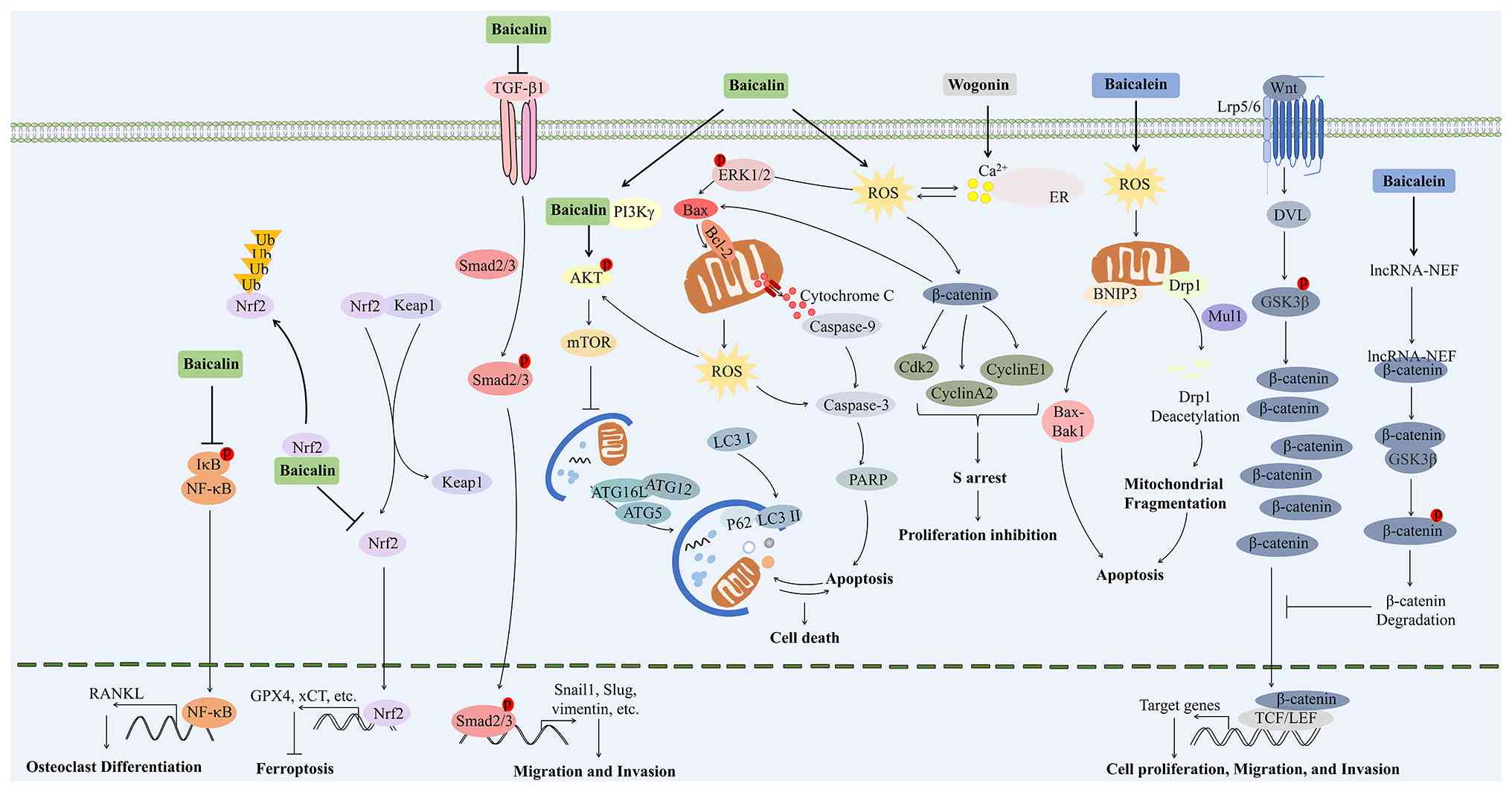 |
Figure 4
Principal molecular mechanisms of Scutellaria baicalensis and its active compounds in osteosarcoma treatment. The therapeutic mechanisms by which Scutellaria baicalensis and its active compounds (baicalin, baicalein, and wogonin) contribute to the treatment of OS are illustrated. Baicalin inhibits OS cell growth by promoting iron accumulation and ROS formation, inducing ferroptosis through the Nrf2 pathway, and enhancing osteogenic differentiation by modulating NF-κB activity. It also induces mitochondrial apoptosis and inhibits critical survival pathways, including PI3K/AKT/mTOR and ERK1/2. Baicalein exerts anticancer effects by inducing apoptosis through ROS generation, activating caspase-dependent pathways, and causing cell cycle arrest. It modulates the Wnt/β-catenin signaling pathway, reducing β-catenin levels and enhancing GSK-3β expression, leading to tumor growth inhibition. Wogonin targets CSCs by modulating ROS levels, inducing apoptosis, and inhibiting mobility through MMP-9 downregulation. OS, osteosarcoma; ROS, reactive oxygen species; Nrf2, nuclear factor erythroid 2-related factor 2; NF-κB, nuclear factor-κB; PI3K, phosphoinositide 3-kinase; AKT, protein kinase B; mTOR, mechanistic target of rapamycin; ERK1/2, extracellular signal-regulated kinase 1/2; Wnt, wingless-related integration site; GSK-3β, glycogen synthase kinase-3β; CSCs, cancer stem cells; MMP-9, matrix metalloproteinase-9.
|
|
Table V
Major active components of Scutellaria baicalensis in osteosarcoma.
|
Table V
Major active components of Scutellaria baicalensis in osteosarcoma.
| Active compound |
Experimental model |
Dose/route |
Key pathways |
Phenotypic outcomes |
(Refs.) |
| Baicalin |
U2OS cell line |
Incubated at 20, 40, and 80 μM |
NF-κB pathway |
Inhibited proliferation, invasion, migration, and EMT; promoted osteogenic differentiation |
(136) |
| Baicalin |
Xenograft nude mice; MG63 and 143B cell line |
Intraperitoneal injection 50, 100, and 200 mg/kg/day for 30 days; incubated at 10, 20, and 30 μM for 24 h |
Nrf2/xCT/GPX4 pathway |
Inhibited osteosarcoma growth; induced ferroptosis and Nrf2 ubiquitin degradation |
(138) |
| Baicalin |
U2OS cell line |
Incubated at 12.5 and 25 μM |
TGF-β/Smad2/3 pathway |
Inhibited proliferation, invasion, migration, and anoikis resistance |
(156) |
| Baicalin |
HOS, MG63, U2OS and 143B cell line |
Incubated at 20, 40, and 80 μM |
PI3K/AKT/mTOR, ERK1/2 and β-catenin pathway |
Inhibited proliferation and invasion; promoted mitochondria-mediated apoptosis and autophagy |
(139) |
| Baicalin |
U2OS cell line |
Incubated at 20, 40, and 100 μM |
AKT pathway |
Inhibited proliferation; promoted apoptosis |
(141) |
| Baicalein |
Xenograft nude mice; MG-63 cell line |
Intraperitoneal injection 100 mg/kg for every other day; incubated at 50, 75, and 100 μM |
ERK pathway |
Inhibited proliferation, migration, invasion and EMT; promoted cell cycle arrest and apoptosis |
(143) |
| Baicalein |
MG-63 and Saos-2 cell line |
Incubated at 50, and 100 μM |
PI3K/AKT and MAPK/ERK pathway |
Induced cell apoptosis |
(144) |
| Baicalein |
Saos-2, 143B, MG63 and U2OS cell line; Osteosarcoma intra-tibia tumor-bearing model |
Incubated at 35, and 80 μM; intraperitoneal injection 40 mg/kg/day |
Wnt/β-catenin pathway |
Inhibited cell viability, invasion and metastasis; promoted cell cycle arrest and apoptosis |
(147,148) |
Triggering regulated cell death and suppressing malignant proliferation
A previous study showed that baicalin inhibits the proliferation of OS cells both in vitro and in vivo by promoting iron accumulation, ROS formation and lipid peroxidation, which collectively trigger ferroptosis (138). This process is facilitated by baicalin's interaction with the Nrf2 pathway, leading to Nrf2 degradation and subsequent downregulation of ferroptosis-related proteins such as glutathione peroxidase 4 (GPX4) and system Xc− light chain (xCT), thereby enhancing its anti-OS effects. Mechanistically, baicalin has been shown to induce mitochondrial apoptosis characterized by increased ROS and Ca2+ concentrations, along with the activation of autophagy (139). This apoptotic effect is mediated through a ROS-dependent pathway, as evidenced by decreased levels of Bcl-2 and the activation of caspases-9 and -3 (139,140). Additionally, baicalin has been demonstrated to inhibit key signaling pathways such as PI3K/AKT/mTOR, ERK1/2 and b-catenin, which are crucial for OS cell survival and proliferation (139). The suppression of the AKT signaling pathway is particularly noteworthy, as it results in decreased ratios of phosphorylated (p)-AKT/AKT and Bcl-2/Bax, along with reduced levels of cyclin D1 and CDK4 (141).
Baicalein has the ability to inhibit the proliferation, migration and metastasis of OS cells through various mechanisms, primarily involving apoptosis induction, cell cycle arrest and modulation of key signaling pathways. One of the primary mechanisms through which baicalein exerts its anticancer effects is by inducing apoptosis. It activates both caspase-dependent and -independent mitochondrial pathways in OS cells, leading to mitochondrial dysfunction, activation of apoptotic proteins and cell death. Specifically, baicalein was found to promote the generation of ROS, which resulted in mitochondrial fragmentation and the activation of proteins such as BCL2-interacting protein 3 (BNIP3), mitochondrial E3 ubiquitin protein ligase 1 (Mul1) and Drp-1. Inhibition of BNIP3 expression has been shown to reduce baicalein-induced apoptosis, indicating a ROS-BNIP3-mediated mechanism for its anti-OS effects (142). The ERK signaling pathway is also involved in the anti-OS effects of baicalein, and has been demonstrated to inhibit cell viability, proliferation, migration and invasion, while promoting apoptosis and cell cycle arrest in MG-63 cells by reducing p-ERK/ERK expression (143). Additionally, baicalein has been demonstrated to upregulate heat shock protein 70 (HSP70), which plays a role in drug resistance. The activation of the PI3K/AKT and MAPK/ERK pathways by HSP70 decreased the sensitivity of OS cells to baicalein, suggesting that targeting HSP70 could enhance the efficacy of chemotherapy (144). Baicalein was also reported to affect the cell cycle by inducing G1 cell cycle arrest, suppressing the proliferation of OS cells (145). Furthermore, baicalein was revealed to inhibit cell migration and invasion, thereby limiting the metastatic potential of OS cells (137). In a previous study involving canine OS cells, baicalein induced apoptosis without interfering with the inhibitory effects of doxorubicin on cell proliferation, suggesting that the apoptotic mechanism of baicalein may involve changes in mitochondrial permeability, although the exact mechanism remains unclear (146).
Another important mechanism by which baicalein functions is through the modulation of the Wnt/β-catenin signaling pathway. Baicalein was demonstrated to reduce β-catenin and Axin2 levels while enhancing GSK-3β expression, leading to tumor growth inhibition and apoptosis induction (147). Baicalein has also been shown to activate the expression of lncRNA-NEF, which inactivates the Wnt/β-catenin pathway, further supporting its role in inhibiting OS (148). Additionally, baicalein has been reported to target the c-MYC gene through the Wnt/β-catenin signaling pathway, with involvement from JNK and the lncRNA BANCR, highlighting a complex regulatory mechanism (149).
Inhibition of cancer stem cell (CSC) properties and tumor metastasis
Multiple studies have demonstrated the ability of wogonin to target OS CSCs and suppress tumor progression through various mechanisms (150-153). Wogonin was shown to effectively suppress the stem cell characteristics of OS CSCs by modulating ROS levels, leading to the downregulation of stem cell-related gene expression. This effect could be reversed by the ROS scavenger N-acetylcysteine, highlighting the critical role of ROS in wogonin's mechanism of action against these cells (150). Additionally, wogonin was found to induce apoptosis in human OS CSCs and inhibit their mobility by downregulating MMP-9 expression, thereby repressing their renewal ability. This underscores the potential of wogonin to address challenges such as drug resistance and metastasis in OS treatment (151). Wogonin also exhibits significant cytotoxic effects on OS by inducing apoptosis through ROS generation and alterations in intracellular calcium levels, which activate caspase-3, emphasizing its role in promoting apoptotic cell death and ER stress (152). Altered store-operated Ca2+ entry (SOCE) disrupts Ca2+ levels in the cytosol or ER, promoting OS progression (153). In this context, wogonin was shown to inhibit lithium-induced fibroblast growth factor 23 (FGF23) release by blocking the transcription of the NF-κB-dependent Orai calcium release-activated calcium modulator 1 (Orai1) and SOCE, thereby counteracting this progression (154). Wogonin was further demonstrated to inhibit tumor growth and metastasis in OS by reducing angiogenesis, lymphangiogenesis and the number of TAMs. Its anti-metastatic effects are attributed to the suppression of VEGF-C-induced VEGFR-3 phosphorylation in lymphatic endothelial cells, which is achieved through the inhibition of COX-2 expression and IL-1β production in TAMs (155).
Overall, wogonin acts through a unique regulatory profile against OS. It simultaneously targets CSCs and remodels the tumor microenvironment, thereby counteracting two key drivers of chemoresistance and metastatic progression.
Inhibition of epithelial-mesenchymal transition (EMT) and regulation of osteogenic differentiation
In addition to inducing regulated cell death, baicalin has been found to significantly inhibit the proliferation, invasion and migration of U2OS cells while promoting osteogenic differentiation. This is achieved by enhancing the expression of osteoblast markers such as RUNX2, osteopontin (OPN) and OCN, and reducing the OC marker RANKL by inhibiting NF-κB pathway activity (136). Furthermore, baicalin has been demonstrated to modulate EMT markers, promoting the epithelial phenotype and downregulating mesenchymal markers and EMT-related transcription factors (136,156). Notably, it suppressed the TGF-β1-mediated activation of Smad2/3, contributing to its inhibitory effects on cell migration and invasion (156).
A previous transcriptome sequencing study further revealed that baicalin treatment leads to differential expression of numerous lncRNAs, miRNAs and mRNAs, forming a ceRNA regulatory network that may contribute to its antitumor effects (157). The ceRNA regulatory interactions identified in the aforementioned study require further experimental verification to separate true causal mediators from irrelevant correlative factors linked to baicalin intervention.
Overall, these findings uncover multiple anti-OS mechanisms underlying baicalein. This flavonoid triggers ferroptosis, represses EMT, facilitates osteogenic differentiation and blocks core cell survival signaling cascades, making it a viable candidate for developing innovative OS therapies. Compared with baicalin, baicalein additionally suppresses the Wnt/β-catenin axis and boosts tumor chemosensitivity. In addition, wogonin exerts a distinct therapeutic effect by targeting CSCs and remodeling the metastatic tumor microenvironment.
Other active compounds of Scutellarin baicalensis and SSDs
In addition to baicalein, baicalin, wogonin, oroxylin A and wogonoside, several other active compounds extracted from Scutellaria baicalensis have also been studied for their protective effects against SSDs. A previous study has demonstrated that chrysin alleviates knee synovial inflammation and mitigates pain in rat models of OA primarily by suppressing NLRP3 inflammasome activation, with experiments performed at both cellular and animal levels (158). As another effective flavonoid, scutellarein was found to delay OA progression both in vitro and in vivo. It attenuated chondrocyte inflammation and cartilage degeneration via the PI3K/AKT/NF-κB pathway (159). For RA treatment, norwogonin was shown to alleviate inflammation by suppressing LPS-induced osteoclastogenesis and subsequent bone destruction by reducing ROS generation and calcium oscillations (160). Furthermore, scutellarein was also revealed to exert antitumor effects against OS. It markedly inhibited OS cell proliferation in vitro and in vivo by targeting the TLR4-TNF receptor-associated factor 6 (TRAF6) axis, which further suppressed the activation of the downstream NF-κB signaling pathway (161). Taken together, these active compounds of Scutellaria baicalensis show promising potential for treating various SSDs. Specifically, chrysin has been shown to alleviate OA damage as verified in cell and animal studies. Scutellarein was demonstrated to exert therapeutic effects on OA and OS with consistent in vitro and in vivo data. Norwogonin was found to ameliorate inflammatory osteolysis and arthritis based on preclinical observations. However, all relevant findings are currently derived from preclinical studies, and clinical evidence is still lacking to support their practical clinical application.
Discussion and future perspectives
The present review explored Scutellaria baicalensis and its bioactive compounds, which have demonstrated promising activity in the treatment of SSDs through their immunomodulatory, anti-inflammation, antioxidant and anticancer properties. Comparisons based on research sufficiency, stable treatment effects and specific molecular mechanisms have identified several high-value compound and disease-matching combinations. Baicalin acts as the most reliable agent for OA treatment. It protects chondrocytes through multiple verified pathways and works well in different animal OA models. Related intra-articular delivery research further supports its potential as a targeted drug for OA. Baicalein stands out for OP intervention given its balanced regulation of bone formation and bone absorption. Restricted oral absorption remains the main obstacle to its clinical use and relevant preparation improvements are required. Baicalin also shows prominent value against RA. A large volume of animal experimental data and preliminary human trial outcomes confirm its anti-inflammatory activity and regulatory function on Th17 cell responses for auxiliary treatment. Wogonin delivers unique benefits against OS by acting on CSCs and the tumor microenvironment to relieve drug resistance and tumor metastasis. Oroxylin A inhibits OC fusion and relevant signaling to facilitate bone repair and implant integration. Local administration methods can offset its weak systemic absorption. Multiple signaling pathways are shared by these flavonoids to regulate skeletal tissue health. Common pathways include NF-κB for inflammation control, MAPK for cell activity and matrix metabolism, PI3K/AKT/mTOR for cell survival and autophagy, Wnt/β-catenin for bone growth and cartilage development, and Nrf2 for antioxidant protection. Each flavonoid regulates a distinct subset of these pathways, and combined use of different flavonoids may achieve improved therapeutic effects.
Although numerous preclinical studies have confirmed the protective effects of Scutellaria baicalensis flavonoids on SSDs, a range of notable limitations still exist across experimental design model selection data repeatability and clinical translatability. Most cellular and rodent studies may lack blinded evaluation and appropriate positive control arrangements, which could compromise the robustness of experimental outcomes. Mechanistic conclusions drawn only from one cell line should be verified using multiple cellular models, including primary cells, to improve the generalizability of experimental data. Cell studies frequently use mM drug doses to generate measurable effects. Such concentrations often exceed clinically relevant plasma levels, as hepatic metabolism, protein binding and tissue distribution reduce active drug exposure in vivo. This discrepancy impairs the translational utility of in vitro data. Current animal models are insufficient to recapitulate the complex pathological progression of human SSDs. For example, surgical joint injury in young animals induces rapid traumatic joint degeneration, which differs substantially from the progressive joint deterioration driven by aging and metabolic disorders in humans. In addition, subcutaneous OS xenograft models lack the intrinsic bone microenvironment and cannot faithfully simulate the metastatic characteristics of clinical tumors. In addition, flavonoid bioactivity is strongly shaped by experimental conditions. Baicalin regulates osteoclastogenesis in a concentration-bidirectional manner, and equal baicalein doses exert distinct bone-protective effects through subcutaneous vs. oral routes due to poor oral bioavailability. These inconsistent results indicate flavonoids cannot generate consistent skeletal benefits, with their pharmacological performance modulated by dose, administration route, intervention timing and disease progression stage. Finally, while network pharmacology and bioinformatic analyses help generate preliminary hypotheses, they cannot provide conclusive experimental support. Additionally, few predicted signaling pathways have been functionally confirmed via gene silencing or rescue assays. These limitations do not eliminate the clinical value of Scutellaria baicalensis flavonoids, yet they indicate this field is still confined to early preclinical investigation.
Baicalin is the most abundant flavonoid active component in Scutellaria baicalensis with relatively favorable water solubility. Its oral preparation, baicalin tablets, has been approved for marketing in China and is frequently used as adjunctive therapy for acute and chronic hepatitis (162). Its extraction and purification processes are mature and amenable to large-scale production, with well-characterized pharmacokinetic profiles, conferring a substantially superior translational foundation compared with other homologous flavonoids still in the basic research stage (163,164). Meanwhile, mechanistic studies of baicalin for OA are systematic and comprehensive, as baicalin acts on key pathological processes of the disease by regulating multiple core signaling pathways. Results from in vitro cellular experiments and a variety of in vivo animal models show good consistency. In a randomized controlled trial, a proprietary flavonoid mixture composed of baicalin and catechin showed efficacy comparable to naproxen in controlling the signs and symptoms of OA (165). Furthermore, to address the limitation of its low oral bioavailability, feasible optimization strategies including nanoformulations, cyclodextrin inclusion complexes, and intra-articular delivery have been proposed. Nevertheless, it should be noted that most existing preclinical evidence is derived from artificially-induced acute models in young rodents, which cannot fully recapitulate the pathological features of chronic, age-related skeletal disorders. To address this limitation, translational research can be advanced in a phased manner. First, advanced oral nanoformulations and intra-articular drug delivery systems can be developed to effectively improve baicalin solubility and in vivo absorption, achieving sustained drug enrichment in local bone and joint tissues. In addition, comprehensive preclinical evaluations should be conducted using both acute OA models and aged chronic OA models to better mimic clinical pathological features. On the basis of improved preclinical evidence, small-scale early-phase clinical trials can be initiated to preliminarily verify the safety, tolerability and therapeutic efficacy of modified baicalin formulations in patients with OA. Ultimately, large-sample, multicenter controlled clinical trials should be conducted to systematically validate the clinical efficacy and long-term safety of optimized baicalin delivery systems for the treatment of OA.
These translational limitations are largely attributable to the intrinsic physicochemical properties of Scutellaria baicalensis-derived flavonoids. Low water solubility and poor permeability greatly restrict the clinical application of these active compounds (166). Advancements in extraction and formulation technologies may lead to improved bioavailability and targeted delivery of these compounds (167). For instance, utilizing nanotechnology for the encapsulation of active ingredients can enhance their stability and absorption in the body. Xie et al (168) demonstrated that co-processed nanocrystalline cellulose-sodium carboxymethyl starch can effectively stabilize nanosuspensions of baicalin, improving its dissolution rate and oral bioavailability. Similarly, the development of liposomal formulations could facilitate targeted delivery to affected tissues, thereby maximizing therapeutic effects while minimizing side effects (34). It has been reported that long-circulating nanoliposomes effectively enhance the oral bioavailability of baicalein, achieving 4.52-fold greater bioavailability compared with free baicalein (169). Furthermore, employing advanced extraction methods such as supercritical fluid extraction may yield higher concentrations of bioactive compounds, ensuring that formulations are both potent and effective. A previous study optimized the supercritical fluid extraction of baicalin from Scutellaria baicalensis using an orthogonal array design, identifying that 1,2-propanediol-modified supercritical fluid was the most effective, leading to proposed optimal conditions for maximizing baicalin extraction (170). Collectively, technical improvements in extraction and formulation can effectively address pharmacokinetic deficiencies and enhance the therapeutic potential of Scutellaria baicalensis flavonoids for SSDs.
In addition to pharmacokinetic optimization, standardized quality control is indispensable for ensuring the stable efficacy and clinical safety of TCM. Screening and validating reliable quality markers is critical for establishing unified quality standards and guiding rational clinical use (171). For Scutellaria baicalensis, it is of great importance to comprehensively evaluate its safety and efficacy by employing analytical techniques such as optimizing extraction processes, establishing chemical fingerprints, determining content, characterizing components and screening quality markers (172). In green extraction, low-toxicity solvents such as deep eutectic solvents and ionic liquids are combined with physical or enzymatic assistance to efficiently disrupt plant tissues and enhance the yield of active ingredients such as total flavonoids and baicalin (173,174). Advanced analytical techniques, including capillary electrophoresis, high-performance liquid chromatography, ultra-fast liquid chromatography coupled with diode array detection, ultra-high-performance liquid chromatography-mass spectrometry and high-resolution mass spectrometry, have enabled the establishment of chemical fingerprints and the rapid identification and quantification of >100 compounds in Scutellaria baicalensis (175-177). Combining chemical content, biological efficacy and spectroscopy helped identify core markers such as baicalin, baicalein, wogonin, oroxylin A and other characteristic components (178,179). Despite substantial progress in quality evaluation research, a unified and market-adaptive quality control system based on multiple synergistic markers is still lacking. Establishing such a standardized system will effectively reduce quality inconsistencies caused by different cultivation origins, processing methods and commercial product grades, further promoting the stable clinical application of Scutellaria baicalensis and its active flavonoids.
Therefore, to advance the clinical translation of Scutellaria baicalensis flavonoids for the treatment of SSDs, formulation optimization is urgently required to overcome their inherent low bioavailability. The development of advanced oral nano-formulations and local injectable delivery systems can effectively improve compound absorption and increase drug accumulation in bone and joint tissues. In addition, standardized pharmacokinetic assessments are necessary to establish credible dose-effect associations for clinical guidance. Apart from structural modification of drug preparations, preclinical validation should be further refined using clinically recapitulative chronic and aged disease models, rather than conventional acute young animal models, to better mimic real disease progression. Systematic safety evaluation focusing on long-term organ toxicity, potential drug interactions and influences on bone healing is also essential to generate robust preclinical evidence and support subsequent clinical translation.
Conclusions
Scutellaria baicalensis and its active compounds exert extensive bioactive functions against multiple SSDs, thereby emerging as promising candidates for the treatment of SSDs, including OP, OA, RA and OS. As research progresses, it is crucial to continue investigating the role of Scutellaria baicalensis in skeletal health, which could ultimately contribute to the development of novel strategies for the prevention and treatment of SSDs. Comprehensive long-term toxicological, safety, and cost-effectiveness evaluations remain limited and the clinical safety and economic advantages of these compounds have not yet been fully established. Future research should focus on how its active compounds influence bone metabolism, inflammation and cartilage degradation, which are key factors in the pathogenesis of these conditions. Clinical trials are necessary to evaluate the efficacy and safety of these compounds in patients with SSDs, with a focus on determining optimal dosages and treatment regimens. Additionally, exploring the potential synergistic effects of Scutellaria baicalensis compounds with other therapeutic agents could enhance their therapeutic potential, leading to more effective treatment strategies for SSDs.
Availability of data and materials
Not applicable.
Authors' contributions
YW and YC wrote major sections of the manuscript. YN performed structured literature searches. QY, LC and WS critically synthesized and interpreted findings. CZ, WS, YC and QY designed the scope and structure of the review and revised major sections of the manuscript. All authors read and approved the final manuscript. Data authentication is not applicable.
Ethics approval and consent to participate
Not applicable.
Patient consent for publication
Not applicable.
Competing interests
The authors declare that they have no competing interests.
Abbreviations:
|
SSDs
|
skeletal system diseases
|
|
OP
|
osteoporosis
|
|
OA
|
osteoarthritis
|
|
RA
|
rheumatoid arthritis
|
|
OS
|
osteosarcoma
|
|
TCM
|
Traditional Chinese Medicine
|
|
BMSCs
|
bone marrow mesenchymal stem cells
|
|
DOP
|
diabetic osteoporosis
|
|
OC
|
osteoclast
|
|
AChE
|
acetylcholinesterase
|
|
OVX
|
ovariectomized
|
|
RANKL
|
receptor activator of NF-κB ligand
|
|
TRAP
|
tartrate-resistant acid phosphatase
|
|
BMD
|
bone mineral density
|
|
LPS
|
lipopolysaccharide
|
|
ROS
|
reactive oxygen species
|
|
LPL
|
L-plastin
|
|
HIF-1α
|
hypoxia-inducible factor-1α
|
|
MMPs
|
matrix metalloproteinases
|
|
H2O2
|
hydrogen peroxide
|
|
ER
|
endoplasmic reticulum
|
|
CIA
|
collagen-induced arthritis
|
|
FLS
|
fibroblast-like synoviocyte
|
|
EMT
|
epithelial-mesenchymal transition
|
|
SOCE
|
store-operated Ca2+ entry
|
Acknowledgements
Not applicable.
Funding
The present research was supported by the Sichuan Science and Technology Program (grant no. 2026NSFSC0605), the Shenzhen Medica l Resea rch Fund (grant no. A2403030), the Shenzhen Science and Technology Projects (grant nos. JSGG20220831110400001 and KCXFZ20230731093059012), the joint foundation of Luzhou Government and Southwest Medical University (grant no. 2024LZXNYDJ104), and the Scientific Research Foundation of Southwest Medical University (grant no. 2025JC046).
References
|
1
|
Liang J, Yi Q, Liu Y, Li J, Yang Z and Sun W and Sun W: Recent advances of m6A methylation in skeletal system disease. J Transl Med. 22:1532024. View Article : Google Scholar : PubMed/NCBI
|
|
2
|
Zhao Y, Wang J, Xu L, Xu H and Yan Y, Zhao H and Yan Y: Beyond bone loss: A biology perspective on osteoporosis pathogenesis, multi-omics approaches, and interconnected mechanisms. Biomedicines. 13:14432025. View Article : Google Scholar : PubMed/NCBI
|
|
3
|
Qiu L, Alhaskawi A and Moqbel SAA: Osteoarthritis: multi-tissue pathology, molecular mechanisms, clinical management, and emerging precision and regenerative therapies. Front Pharmacol. 16:16971922026. View Article : Google Scholar
|
|
4
|
Gao Y, Zhang Y and Liu X: Rheumatoid arthritis: Pathogenesis and therapeutic advances. MedComm (2020). 5:e5092024. View Article : Google Scholar : PubMed/NCBI
|
|
5
|
El Motassime A, Vitiello R, Comodo RM, Capece G, Bocchino G, Bocchi MB, Maccauro G and Meschini C: Osteosarcoma: A comprehensive morphological and molecular review with prognostic implications. Biology (Basel). 14:14072025.PubMed/NCBI
|
|
6
|
Reid IR and Billington EO: Drug therapy for osteoporosis in older adults. Lancet. 399:1080–1092. 2022. View Article : Google Scholar : PubMed/NCBI
|
|
7
|
Di Matteo A, Bathon JM and Emery P: Rheumatoid arthritis. Lancet. 402:2019–2033. 2023. View Article : Google Scholar
|
|
8
|
Gibbs AJ, Gray B, Wallis JA, Taylor NF, Kemp JL, Hunter DJ and Barton CJ: Recommendations for the management of hip and knee osteoarthritis: A systematic review of clinical practice guidelines. Osteoarthritis Cartilage. 31:1280–1292. 2023. View Article : Google Scholar : PubMed/NCBI
|
|
9
|
Kamali A, Ziadlou R, Lang G, Pfannkuche J, Cui S, Li Z, Richards RG, Alini M and Grad S: Small molecule-based treatment approaches for intervertebral disc degeneration: Current options and future directions. Theranostics. 11:27–47. 2021. View Article : Google Scholar : PubMed/NCBI
|
|
10
|
Gill J and Gorlick R: Advancing therapy for osteosarcoma. Nat Rev Clin Oncol. 18:609–624. 2021. View Article : Google Scholar : PubMed/NCBI
|
|
11
|
Li K, Jiang Y, Wang N, Lai L, Xu S, Xia T, Yue X and Xin H: Traditional Chinese medicine in osteoporosis intervention and the related regulatory mechanism of gut microbiome. Am J Chin Med. 51:1957–1981. 2023. View Article : Google Scholar : PubMed/NCBI
|
|
12
|
Wang Y, Chen S, Du K, Liang C, Wang S, Owusu Boadi E, Li J, Pang X, He J and Chang YX: Traditional herbal medicine: Therapeutic potential in rheumatoid arthritis. J Ethnopharmacol. 279:1143682021. View Article : Google Scholar : PubMed/NCBI
|
|
13
|
Li W, Yu L, Li W, Ge G, Ma Y, Xiao L, Qiao Y, Huang W, Huang W, Wei M, et al: Prevention and treatment of inflammatory arthritis with traditional Chinese medicine: Underlying mechanisms based on cell and molecular targets. Ageing Res Rev. 89:1019812023. View Article : Google Scholar : PubMed/NCBI
|
|
14
|
Wang ZL, Wang S, Kuang Y, Hu ZM, Qiao X and Ye M: A comprehensive review on phytochemistry, pharmacology, and flavonoid biosynthesis of Scutellaria baicalensis. Pharm Biol. 56:465–484. 2018. View Article : Google Scholar :
|
|
15
|
Xiang L, Gao Y, Chen S, Sun J, Wu J and Meng X: Therapeutic potential of Scutellaria baicalensis Georgi in lung cancer therapy. Phytomedicine. 95:1537272022. View Article : Google Scholar
|
|
16
|
Jang JY, Im E and Kim ND: Therapeutic potential of bioactive components from Scutellaria baicalensis Georgi in inflammatory bowel disease and colorectal cancer: A review. Int J Mol Sci. 24:19542023. View Article : Google Scholar : PubMed/NCBI
|
|
17
|
Yu P, Li J, Luo Y, Sun J, Hu Y, Lin B, Meng X and Xiang L: Mechanistic role of scutellaria baicalensis Georgi in breast cancer therapy. Am J Chin Med. 51:279–308. 2023. View Article : Google Scholar : PubMed/NCBI
|
|
18
|
Yingrui W, Zheng L, Guoyan L and Hongjie W: Research progress of active ingredients of Scutellaria baicalensis in the treatment of type 2 diabetes and its complications. Biomed Pharmacother. 148:1126902022. View Article : Google Scholar : PubMed/NCBI
|
|
19
|
Tan YQ, Lin F, Ding YK, Dai S, Liang YX, Zhang YS, Li J and Chen HW: Pharmacological properties of total flavonoids in Scutellaria baicalensis for the treatment of cardiovascular diseases. Phytomedicine. 107:1544582022. View Article : Google Scholar : PubMed/NCBI
|
|
20
|
Peng Y and Zhou C: Network pharmacology and molecular docking identify the potential mechanism and therapeutic role of Scutellaria baicalensis in Alzheimer's disease. Drug Des Devel Ther. 18:1199–1219. 2024. View Article : Google Scholar : PubMed/NCBI
|
|
21
|
Zhang G, Li C, Niu Y, Yu Q, Chen Y and Liu E: Osteoprotective effect of radix scutellariae in female hindlimb-suspended sprague-dawley rats and the osteogenic differentiation effect of its major constituent. Molecules. 22:10442017. View Article : Google Scholar : PubMed/NCBI
|
|
22
|
Jo HG, Baek CY, Song HS and Lee D: Network pharmacology and experimental verifications to discover Scutellaria baicalensis Georgi's effects on joint inflammation, destruction, and pain in osteoarthritis. Int J Mol Sci. 25:21272024. View Article : Google Scholar : PubMed/NCBI
|
|
23
|
Zhou W, Wang Y, Huang Y, Liu J, Cheng C, Xue Q, Wang X, Chang J and Miao C: Huangqin qingre qubi capsule inhibits RA pathology by binding FZD8 and further inhibiting the activity of Wnt/β-catenin signaling pathway. J Ethnopharmacol. 302(Pt A): 1158862023. View Article : Google Scholar
|
|
24
|
Zhang L, Tian Y, Wang J, Deng S and Fan H: Network pharmacology-based research on the effect of Scutellaria baicalensis on osteosarcoma and the underlying mechanism. Medicine (Baltimore). 102:e359572023. View Article : Google Scholar : PubMed/NCBI
|
|
25
|
Wang L, Ni B, Wang J, Zhou J, Wang J, Jiang J, Sui Y, Tian Y, Gao F and Lyu Y: Research progress of scutellaria baicalensis in the treatment of gastrointestinal cancer. Integr Cancer Ther. 23:153473542413020492024. View Article : Google Scholar : PubMed/NCBI
|
|
26
|
Yang X, Dou F, Hai L, Xiao Y, Cui J, Cai Y, Wang R, Ji K, Feng Y and Chen H: Scutellaria baicalensis Georgi: A promising source of bioactive molecules for kidney disease therapy. Biomolecules. 16:642025. View Article : Google Scholar
|
|
27
|
Wang J, Feng X, Li Z, Liu Y, Yang W, Zhang T, Guo P, Liu Z, Qi D and Pi J: The flavonoid components of scutellaria baicalensis: Biopharmaceutical properties and their improvement using nanoformulation techniques. Curr Top Med Chem. 23:17–29. 2023. View Article : Google Scholar :
|
|
28
|
Ancuceanu R, Dinu M, Dinu-Pirvu C, Anuţa V and Negulescu V: Pharmacokinetics of B-Ring unsubstituted flavones. Pharmaceutics. 11:3702019. View Article : Google Scholar : PubMed/NCBI
|
|
29
|
Xing S, Wang M, Peng Y and Li X: Effects of intestinal microecology on metabolism and pharmacokinetics of oral wogonoside and Baicalin. Nat Prod Commun. 12:509–514. 2017.PubMed/NCBI
|
|
30
|
Senekowitsch S, Link NA, Wildgrube T, Schick P, Engeli S, Weitschies W and Grimm M: Investigation of baicalin, baicalein and low-dose sulfasalazine for orocecal transit time determination in saliva and plasma. Eur J Pharm Biopharm. 223:1150642026. View Article : Google Scholar : PubMed/NCBI
|
|
31
|
Wang XH, Guo YW, Tolba E, Kokkinopoulou M, Wiens M, Schröder HC and Müller WEG: Two-armed activation of bone mineral deposition by the flavones baicalin and baicalein, encapsulated in polyphosphate microparticles. Am J Chin Med. 45:533–555. 2017. View Article : Google Scholar : PubMed/NCBI
|
|
32
|
Yoshida K, Takabayashi T, Kaneko A, Takiyama M, Sakashita M, Imoto Y, Kato Y, Narita N and Fujieda S: Baicalin suppresses type 2 immunity through breaking off the interplay between mast cell and airway epithelial cell. J Ethnopharmacol. 267:1134922021. View Article : Google Scholar
|
|
33
|
Xie H, Chen G and Young RN: Design, synthesis, and pharmacokinetics of a bone-targeting dual-action prodrug for the treatment of osteoporosis. J Med Chem. 60:7012–7028. 2017. View Article : Google Scholar : PubMed/NCBI
|
|
34
|
Li N, Feng L, Tan Y, Xiang Y, Zhang R and Yang M: Preparation, characterization, pharmacokinetics and biodistribution of baicalin-loaded liposome on cerebral ischemia-reperfusion after i.v. Administration in rats. Molecules. 23:17472018. View Article : Google Scholar : PubMed/NCBI
|
|
35
|
Fan J, Dai Y, Shen H, Ju J and Zhao Z: Application of soluplus to improve the flowability and dissolution of baicalein phospholipid complex. Molecules. 22:7762017. View Article : Google Scholar : PubMed/NCBI
|
|
36
|
Nazemiyeh A, Mahari S, Nazemiyeh N, Abdolmohammadzadeh H, Farshbaf Z, Rajabzadeh A, Vandghanooni S, Nazemiyeh H and Eskandani M: Current status and future perspectives of drug delivery systems loaded with baicalein and baicalin in cancer therapy. Biomed Pharmacother. 192:1185892025. View Article : Google Scholar : PubMed/NCBI
|
|
37
|
Zhivodernikov IV, Kirichenko TV, Markina YV, Postnov AY and Markin AM: Molecular and cellular mechanisms of osteoporosis. Int J Mol Sci. 24:157722023. View Article : Google Scholar : PubMed/NCBI
|
|
38
|
Luo J, Li L, Shi W, Xu K, Shen Y and Dai B: Oxidative stress and inflammation: Roles in osteoporosis. Front Immunol. 16:16119322025. View Article : Google Scholar
|
|
39
|
Adejuyigbe B, Kallini J, Chiou D and Kallini JR: Osteoporosis: Molecular pathology, diagnostics, and therapeutics. Int J Mol Sci. 24:145832023. View Article : Google Scholar : PubMed/NCBI
|
|
40
|
Cramer JA, Gold DT, Silverman SL and Lewiecki EM: A systematic review of persistence and compliance with bisphosphonates for osteoporosis. Osteoporos Int. 18:1023–1031. 2007. View Article : Google Scholar : PubMed/NCBI
|
|
41
|
Peng Z, Xu R and You Q: Role of traditional Chinese medicine in bone regeneration and osteoporosis. Front Bioeng Biotechnol. 10:9113262022. View Article : Google Scholar : PubMed/NCBI
|
|
42
|
Zhang W and Zheng L: Prevention and treatment of osteoporosis caused by oestrogen shortage in rats. Pak J Pharm Sci. 31(2 Suppl): S651–S656. 2018.
|
|
43
|
Qin W, Shang Q, Shen G, Li B, Zhang P, Zhang Y, Zhao W, Chen H, Liu H, Xie B, et al: Restoring bone-fat equilibrium: Baicalin's impact on P38 MAPK pathway for treating diabetic osteoporosis. Biomed Pharmacother. 175:1165712024. View Article : Google Scholar : PubMed/NCBI
|
|
44
|
Zhao Y, Wang HL, Li TT, Yang F and Tzeng CM: Baicalin ameliorates dexamethasone-induced osteoporosis by regulation of the RANK/RANKL/OPG signaling pathway. Drug Des Devel Ther. 14:195–206. 2020. View Article : Google Scholar : PubMed/NCBI
|
|
45
|
Yooin W, Saenjum C, Ruangsuriya J and Jiranusornkul S: Discovery of potential sclerostin inhibitors from plants with loop2 region of sclerostin inhibition by interacting with residues outside Pro-Asn-Ala-Ile-Gly motif. J Biomol Struct Dyn. 38:1272–1282. 2020. View Article : Google Scholar
|
|
46
|
Guo AJ, Choi RC, Cheung AW, Chen VP, Xu SL, Dong TT, Chen JJ and Tsim KW: Baicalin, a flavone, induces the differentiation of cultured osteoblasts: An action via the Wnt/beta-catenin signaling pathway. J Biol Chem. 286:27882–27893. 2011. View Article : Google Scholar : PubMed/NCBI
|
|
47
|
Xu ML, Bi CWC, Kong AYY, Dong TTX, Wong YH and Tsim KWK: Flavonoids induce the expression of acetylcholinesterase in cultured osteoblasts. Chem Biol Interact. 259(Pt B): 295–300. 2016. View Article : Google Scholar : PubMed/NCBI
|
|
48
|
Li SF, Tang JJ, Chen J, Zhang P, Wang T, Chen TY, Yan B, Huang B, Wang L, Huang MJ, et al: Regulation of bone formation by baicalein via the mTORC1 pathway. Drug Des Devel Ther. 9:5169–5183. 2015.PubMed/NCBI
|
|
49
|
Cai P, Lu Y, Yin Z, Wang X, Zhou X and Li Z: Baicalein ameliorates osteoporosis via AKT/FOXO1 signaling. Aging (Albany NY). 13:17370–17379. 2021. View Article : Google Scholar : PubMed/NCBI
|
|
50
|
Kim JM, Lee SU, Kim YS, Min YK and Kim SH: Baicalein stimulates osteoblast differentiation via coordinating activation of MAP kinases and transcription factors. J Cell Biochem. 104:1906–1917. 2008. View Article : Google Scholar : PubMed/NCBI
|
|
51
|
Xudong H, Qi L, Wenlong M, Jinkun L, Xiaodong X, Chengyin Z, Jiahe Z, Yifeng Y, Xiaolin S, Lingfeng Z and Weiguo W: Network pharmacology and experiments to verify the effect and potential mechanism of baicalein on osteoporosis. Curr Protein Pept Sci. 27:443–455. 2026. View Article : Google Scholar
|
|
52
|
Lu X, He W, Yang W, Li J, Han W, Liu Q, Zhang T, Jiang J, Qin A and Qian Y: Dual effects of baicalin on osteoclast differentiation and bone resorption. J Cell Mol Med. 22:5029–5039. 2018. View Article : Google Scholar : PubMed/NCBI
|
|
53
|
Lu L, Rao L, Jia H, Chen J, Lu X, Yang G, Li Q, Lee KKH and Yang L: Baicalin positively regulates osteoclast function by activating MAPK/Mitf signalling. J Cell Mol Med. 21:1361–1372. 2017. View Article : Google Scholar : PubMed/NCBI
|
|
54
|
Kim MH, Ryu SY, Bae MA, Choi JS, Min YK and Kim SH: Baicalein inhibits osteoclast differentiation and induces mature osteoclast apoptosis. Food Chem Toxicol. 46:3375–3382. 2008. View Article : Google Scholar : PubMed/NCBI
|
|
55
|
Saul D, Gleitz S, Nguyen HH, Kosinsky RL, Sehmisch S, Hoffmann DB, Wassmann M, Menger B and Komrakova M: Effect of the lipoxygenase-inhibitors baicalein and zileuton on the vertebra in ovariectomized rats. Bone. 101:134–144. 2017. View Article : Google Scholar : PubMed/NCBI
|
|
56
|
Saul D, Hohl FE, Franz MK, Meyer I, Taudien S, Roch PJ, Sehmisch S and Komrakova M: Inhibition of lipoxygenases showed no benefit for the musculoskeletal system in estrogen deficient rats. Front Endocrinol (Lausanne). 12:7065042021. View Article : Google Scholar : PubMed/NCBI
|
|
57
|
Saul D, Weber M, Zimmermann MH, Kosinsky RL, Hoffmann DB, Menger B, Taudien S, Lehmann W, Komrakova M and Sehmisch S: Effect of the lipoxygenase inhibitor baicalein on bone tissue and bone healing in ovariectomized rats. Nutr Metab (Lond). 16:42019. View Article : Google Scholar : PubMed/NCBI
|
|
58
|
Jang S, Bak EJ, Kim M, Kim JM, Chung WY, Cha JH and Yoo YJ: Wogonin inhibits osteoclast formation induced by lipopolysaccharide. Phytother Res. 24:964–968. 2010. View Article : Google Scholar
|
|
59
|
Geng X, Yang L, Zhang C, Qin H and Liang Q: Wogonin inhibits osteoclast differentiation by inhibiting NFATc1 translocation into the nucleus. Exp Ther Med. 10:1066–1070. 2015. View Article : Google Scholar : PubMed/NCBI
|
|
60
|
Xian Y, Su Y, Liang J, Long F, Feng X, Xiao Y, Lian H, Xu J, Zhao J, Liu Q and Song F: Oroxylin A reduces osteoclast formation and bone resorption via suppressing RANKL-induced ROS and NFATc1 activation. Biochem Pharmacol. 193:1147612021. View Article : Google Scholar : PubMed/NCBI
|
|
61
|
Li X, Wang L, Huang B, Gu Y, Luo Y, Zhi X, Hu Y, Zhang H, Gu Z, Cui J, et al: Targeting actin-bundling protein L-plastin as an anabolic therapy for bone loss. Sci Adv. 6:eabb71352020. View Article : Google Scholar : PubMed/NCBI
|
|
62
|
Xian Y, Nong Y, Gao Y, Su Y, Lei Z, Lian H, Cheng J, Liang J, Feng X, Liu Z, et al: UPLC/Q-TOF-MS-based metabolomics evaluate the efficacy of oroxylin A against postmenopausal osteoporosis. Biomed Chromatogr. 37:e56092023. View Article : Google Scholar : PubMed/NCBI
|
|
63
|
Zhou W, Lin Z, Xiong Y, Xue H, Song W, Yu T, Chen L, Hu Y, Panayi AC, Sun Y, et al: Dual-Targeted nanoplatform regulating the bone immune microenvironment enhances fracture healing. ACS Appl Mater Interfaces. 13:56944–56960. 2021. View Article : Google Scholar : PubMed/NCBI
|
|
64
|
Yang S, Ni G, Xia M, Li H and Gao Z: Mussel inspired multifunctional bovine serum albumin (BSA) coatings loaded with Baicalein (BAI) to enhance osteogenesis and resist oxidative stress for potential application on implant. Int J Biol Macromol. 229:752–765. 2023. View Article : Google Scholar : PubMed/NCBI
|
|
65
|
Wang D, Chen MW, Wei YJ, Geng WB, Hu Y, Luo Z and Cai KY: Construction of wogonin nanoparticle-containing strontium-doped nanoporous structure on titanium surface to promote osteoporosis fracture repair. Adv Healthc Mater. 11:e22014052022. View Article : Google Scholar : PubMed/NCBI
|
|
66
|
Yu B, Gao Q, Sheng S, Zhou F, Geng Z, Wei Y, Zhang H, Hu Y, Wang S, Huang J, et al: Smart osteoclasts targeted nanomedicine based on amorphous CaCO(3) for effective osteoporosis reversal. J Nanobiotechnology. 22:1532024. View Article : Google Scholar : PubMed/NCBI
|
|
67
|
Courties A, Kouki I, Soliman N, Mathieu S and Sellam J: Osteoarthritis year in review 2024: Epidemiology and therapy. Osteoarthritis Cartilage. 32:1397–1404. 2024. View Article : Google Scholar : PubMed/NCBI
|
|
68
|
Adam MS, Zhuang H, Ren X, Zhang Y and Zhou P: The metabolic characteristics and changes of chondrocytes in vivo and in vitro in osteoarthritis. Front Endocrinol (Lausanne). 15:13935502024. View Article : Google Scholar : PubMed/NCBI
|
|
69
|
Robinson WH, Lepus CM, Wang Q, Raghu H, Mao R, Lindstrom TM and Sokolove J: Low-grade inflammation as a key mediator of the pathogenesis of osteoarthritis. Nat Rev Rheumatol. 12:580–592. 2016. View Article : Google Scholar : PubMed/NCBI
|
|
70
|
Kloppenburg M, Namane M and Cicuttini F: Osteoarthritis. Lancet. 405:71–85. 2025. View Article : Google Scholar : PubMed/NCBI
|
|
71
|
Wang Z, Efferth T, Hua X and Zhang XA: Medicinal plants and their secondary metabolites in alleviating knee osteoarthritis: A systematic review. Phytomedicine. 105:1543472022. View Article : Google Scholar : PubMed/NCBI
|
|
72
|
Cho Y, Jeong S, Kim H, Kang D, Lee J, Kang SB and Kim JH: Disease-modifying therapeutic strategies in osteoarthritis: Current status and future directions. Exp Mol Med. 53:1689–1696. 2021. View Article : Google Scholar : PubMed/NCBI
|
|
73
|
Li Z, Cheng J and Liu J: Baicalin protects human OA chondrocytes against IL-1β-induced apoptosis and ECM degradation by activating autophagy via MiR-766-3p/AIFM1 axis. Drug Des Devel Ther. 14:2645–2655. 2020. View Article : Google Scholar
|
|
74
|
He J and He J: Baicalin mitigated IL-1β-Induced osteoarthritis chondrocytes damage through activating mitophagy. Chem Biol Drug Des. 101:1322–1334. 2023. View Article : Google Scholar : PubMed/NCBI
|
|
75
|
Liu J, Zhou H, Chen J, Zuo Q and Liu F: Baicalin inhibits IL-1β-induced ferroptosis in human osteoarthritis chondrocytes by activating Nrf-2 signaling pathway. J Orthop Surg Res. 19:232024. View Article : Google Scholar
|
|
76
|
Wang P, Zhu P, Liu R, Meng Q and Li S: Baicalin promotes extracellular matrix synthesis in chondrocytes via the activation of hypoxia-inducible factor-1α. Exp Ther Med. 20:2262020. View Article : Google Scholar
|
|
77
|
Wang P, Liu J, Zhang S, Zhu P, Xiong X, Yu C, Li A and Liu Z: Baicalin promotes chondrocyte viability and the synthesis of extracellular matrix through TGF-β/Smad3 pathway in chondrocytes. Am J Transl Res. 13:10908–10921. 2021.
|
|
78
|
Cho I, Chung KH, Kim Y, Choi CH and Koh JT: Baicalein inhibits IL-1β-induced extracellular matrix degradation with decreased MCP-1 expression in primary rat chondrocytes. Toxicol Res. 40:237–246. 2024. View Article : Google Scholar : PubMed/NCBI
|
|
79
|
Chen WP, Xiong Y, Hu PF, Bao JP and Wu LD: Baicalein inhibits MMPs Expression via a MAPK-Dependent mechanism in chondrocytes. Cell Physiol Biochem. 36:325–333. 2015. View Article : Google Scholar : PubMed/NCBI
|
|
80
|
Li Y, Wang J, Song X, Bai H, Ma T, Zhang Z, Li X, Jiang R, Wang G, Fan X, et al: Effects of baicalein on IL-1β-induced inflammation and apoptosis in rat articular chondrocytes. Oncotarget. 8:90781–90795. 2017. View Article : Google Scholar : PubMed/NCBI
|
|
81
|
Zhang X, Zhu Y, Chen X, Zhang Y, Zhang Y, Jia Y, Wang H, Liu Y and Xiao L: Baicalein ameliorates inflammatory-related apoptotic and catabolic phenotypes in human chondrocytes. Int Immunopharmacol. 21:301–308. 2014. View Article : Google Scholar : PubMed/NCBI
|
|
82
|
Li B, Chen K, Qian N, Huang P, Hu F, Ding T, Xu X, Zhou Q, Chen B, Deng L, et al: Baicalein alleviates osteoarthritis by protecting subchondral bone, inhibiting angiogenesis and synovial proliferation. J Cell Mol Med. 25:5283–5294. 2021. View Article : Google Scholar : PubMed/NCBI
|
|
83
|
Yi N, Mi Y, Xu X, Li N, Zeng F, Yan K, Tan K, Kuang G and Lu M: Baicalein alleviates osteoarthritis progression in mice by protecting subchondral bone and suppressing chondrocyte apoptosis based on network pharmacology. Front Pharmacol. 12:7883922022. View Article : Google Scholar : PubMed/NCBI
|
|
84
|
Park JS, Lee HJ, Lee DY, Jo HS, Jeong JH, Kim DH, Nam DC, Lee CJ and Hwang SC: Chondroprotective effects of wogonin in experimental models of osteoarthritis in vitro and in vivo. Biomol Ther (Seoul). 23:442–448. 2015. View Article : Google Scholar : PubMed/NCBI
|
|
85
|
Lim H, Park H and Kim HP: Effects of flavonoids on matrix metalloproteinase-13 expression of interleukin-1β-treated articular chondrocytes and their cellular mechanisms: Inhibition of c-Fos/AP-1 and JAK/STAT signaling pathways. J Pharmacol Sci. 116:221–231. 2011. View Article : Google Scholar
|
|
86
|
Khan NM, Haseeb A, Ansari MY, Devarapalli P, Haynie S and Haqqi TM: Wogonin, a plant derived small molecule, exerts potent anti-inflammatory and chondroprotective effects through the activation of ROS/ERK/Nrf2 signaling pathways in human Osteoarthritis chondrocytes. Free Radic Biol Med. 106:288–301. 2017. View Article : Google Scholar : PubMed/NCBI
|
|
87
|
Tang Q, Zheng G, Feng Z, Tong M, Xu J, Hu Z, Shang P, Chen Y, Wang C, Lou Y, et al: Wogonoside inhibits IL-1β induced catabolism and hypertrophy in mouse chondrocyte and ameliorates murine osteoarthritis. Oncotarget. 8:61440–61456. 2017. View Article : Google Scholar : PubMed/NCBI
|
|
88
|
Chen DH, Zheng G, Zhong XY, Lin ZH, Yang SW, Liu HX and Shang P: Oroxylin A attenuates osteoarthritis progression by dual inhibition of cell inflammation and hypertrophy. Food Funct. 12:328–339. 2021. View Article : Google Scholar
|
|
89
|
Sheng W, Yue Y, Qi T, Qin H, Liu P, Wang D, Zeng H and Yu F: The multifaceted protective role of nuclear factor erythroid 2-related factor 2 in osteoarthritis: Regulation of oxidative stress and inflammation. J Inflamm Res. 17:6619–6633. 2024. View Article : Google Scholar : PubMed/NCBI
|
|
90
|
Xing D, Gao H, Liu Z, Zhao Y and Gong M: Baicalin inhibits inflammatory responses to interleukin-1β stimulation in human chondrocytes. J Interferon Cytokine Res. 37:398–405. 2017. View Article : Google Scholar : PubMed/NCBI
|
|
91
|
Chen C, Zhang C, Cai L, Xie H, Hu W, Wang T, Lu D and Chen H: Baicalin suppresses IL-1beta-induced expression of inflammatory cytokines via blocking NF-kappaB in human osteoarthritis chondrocytes and shows protective effect in mice osteoarthritis models. Int Immunopharmacol. 52:218–226. 2017. View Article : Google Scholar : PubMed/NCBI
|
|
92
|
Wei Q, Yu Z, Yang P and Chen X: Baicalin maintains articular cartilage homeostasis and alleviates osteoarthritis by activating FOXO1. J Med Food. 27:301–311. 2024. View Article : Google Scholar : PubMed/NCBI
|
|
93
|
Chen X, Liu J, Sun Y, Wen J, Zhou Q, Ding X and Zhang X: Correlation analysis of differentially expressed long non-coding RNA HOTAIR with PTEN/PI3K/AKT pathway and inflammation in patients with osteoarthritis and the effect of baicalin intervention. J Orthop Surg Res. 18:342023. View Article : Google Scholar : PubMed/NCBI
|
|
94
|
Pan Y, Chen D, Lu Q, Liu L, Li X and Li Z: Baicalin prevents the apoptosis of endplate chondrocytes by inhibiting the oxidative stress induced by H2O2. Mol Med Rep. 16:2985–2991. 2017. View Article : Google Scholar : PubMed/NCBI
|
|
95
|
Cao J, Zhang Y, Wang T and Li B: Endoplasmic reticulum stress is involved in baicalin protection on chondrocytes from patients with osteoarthritis. Dose Response. 16:15593258188106362018. View Article : Google Scholar : PubMed/NCBI
|
|
96
|
Chen DS, Cao JG, Zhu B, Wang ZL, Wang TF and Tang JJ: Baicalin attenuates joint pain and muscle dysfunction by inhibiting muscular oxidative stress in an experimental osteoarthritis rat model. Arch Immunol Ther Exp (Warsz). 66:453–461. 2018. View Article : Google Scholar : PubMed/NCBI
|
|
97
|
Wu CC, Chen YR, Lu DH, Hsu LH, Yang KC and Sumi S: Evaluation of the post-treatment anti-inflammatory capacity of osteoarthritic chondrocytes: An in vitro study using baicalein. Regen Ther. 14:177–183. 2020. View Article : Google Scholar : PubMed/NCBI
|
|
98
|
Bai H, Yuan R, Zhang Z, Liu L, Wang X, Song X, Ma T, Tang J, Liu C and Gao L: Intra-articular injection of baicalein inhibits cartilage catabolism and NLRP3 inflammasome signaling in a posttraumatic OA model. Oxid Med Cell Longev. 2021:61168902021. View Article : Google Scholar : PubMed/NCBI
|
|
99
|
Wan Y, Shen K, Yu H and Fan W: Baicalein limits osteoarthritis development by inhibiting chondrocyte ferroptosis. Free Radic Biol Med. 196:108–120. 2023. View Article : Google Scholar : PubMed/NCBI
|
|
100
|
Khan NM, Ahmad I, Ansari MY and Haqqi TM: Wogonin, a natural flavonoid, intercalates with genomic DNA and exhibits protective effects in IL-1β stimulated osteoarthritis chondrocytes. Chem Biol Interact. 274:13–23. 2017. View Article : Google Scholar : PubMed/NCBI
|
|
101
|
Liu J, Liu S, Pan W and Li Y: Wogonoside attenuates the articular cartilage injury and the infiltration of Th1/Th2-type cytokines in papain-induced osteoarthritis in rat model via inhibiting the NF-κB and ERK1/2 activation. Immunopharmacol Immunotoxicol. 43:343–352. 2021. View Article : Google Scholar : PubMed/NCBI
|
|
102
|
Zhang Y, Weng Q, Chen J, Li M and Han J: Oroxylin A attenuates IL-1β-induced inflammatory reaction via inhibiting the activation of the ERK and PI3K/AKT signaling pathways in osteoarthritis chondrocytes. Exp Ther Med. 21:3882021. View Article : Google Scholar
|
|
103
|
Zhang H, Wang L, Cui J, Wang S, Han Y, Shao H, Wang C, Hu Y, Li X, Zhou Q, et al: Maintaining hypoxia environment of subchondral bone alleviates osteoarthritis progression. Sci Adv. 9:eabo78682023. View Article : Google Scholar : PubMed/NCBI
|
|
104
|
Huang L, Yao Y, Ruan Z, Zhang S, Feng X, Lu C, Zhao J, Yin F, Cao C and Zheng L: Baicalin nanodelivery system based on functionalized metal-organic framework for targeted therapy of osteoarthritis by modulating macrophage polarization. J Nanobiotechnology. 22:2212024. View Article : Google Scholar : PubMed/NCBI
|
|
105
|
Yang Y, Hu Q, Shao Q, Peng Y, Yu B, Luo F, Chen J, Xu C, Li Z, Tam M, et al: A Baicalin-based functional polymer in dynamic reversible networks alleviates osteoarthritis by cellular interactions. Adv Sci (Weinh). 12:e24109512025. View Article : Google Scholar : PubMed/NCBI
|
|
106
|
Sirong S, Yang C, Taoran T, Songhang L, Shiyu L, Yuxin Z, Xiaoru S, Tao Z, Yunfeng L and Xiaoxiao C: Effects of tetrahedral framework nucleic acid/wogonin complexes on osteoarthritis. Bone Res. 8:62020. View Article : Google Scholar : PubMed/NCBI
|
|
107
|
Smith JF, Starr EG, Goodman MA, Hanson RB, Palmer TA, Woolstenhulme JB, Weyand JA, Marchant AD, Bueckers SL, Nelson TK, et al: Topical application of wogonin provides a novel treatment of knee osteoarthritis. Front Physiol. 11:802020. View Article : Google Scholar : PubMed/NCBI
|
|
108
|
Zhu K, Mao M, Li W and Tao C: A thermosensitive hydrogel delivery system incorporating wogonin-treated mesenchymal stromal cell (MSC)-derived exosomes alleviates osteoarthritis. J Biol Eng. 20:1432026. View Article : Google Scholar : PubMed/NCBI
|
|
109
|
Han P, Liu X, He J, Han L and Li J: Overview of mechanisms and novel therapies on rheumatoid arthritis from a cellular perspective. Front Immunol. 15:14617562024. View Article : Google Scholar : PubMed/NCBI
|
|
110
|
Jang S, Kwon EJ and Lee JJ: Rheumatoid arthritis: Pathogenic roles of diverse immune cells. Int J Mol Sci. 23:9052022. View Article : Google Scholar : PubMed/NCBI
|
|
111
|
Weyand CM and Goronzy JJ: Immune aging in rheumatoid arthritis. Arthritis Rheumatol. 77:792–804. 2025. View Article : Google Scholar : PubMed/NCBI
|
|
112
|
Su QY, Cao YX, Zhang HY, Li YZ and Zhang SX: Leveraging machine learning for drug repurposing in rheumatoid arthritis. Drug Discov Today. 30:1043272025. View Article : Google Scholar : PubMed/NCBI
|
|
113
|
Chatterjee A, Jayaprakasan M, Chakrabarty AK, Lakkaniga NR, Bhatt BN, Banerjee D, Narwaria A, Katiyar CK and Dubey SK: Comprehensive insights into rheumatoid arthritis: Pathophysiology, current therapies and herbal alternatives for effective disease management. Phytother Res. 38:2764–2799. 2024. View Article : Google Scholar : PubMed/NCBI
|
|
114
|
Wang X, Kong Y and Li Z: Advantages of Chinese herbal medicine in treating rheumatoid arthritis: A focus on its anti-inflammatory and anti-oxidative effects. Front Med (Lausanne). 11:13714612024. View Article : Google Scholar : PubMed/NCBI
|
|
115
|
Chen X, Wang Y, Cai J, Wang S, Cheng Z, Zhang Z and Zhang C: Anti-inflammatory effect of baicalin in rats with adjuvant arthritis and its autophagy-related mechanism. Technol Health Care. 30(S1): 191–200. 2022. View Article : Google Scholar : PubMed/NCBI
|
|
116
|
Yang JH, Yun MY, Lee NH, Kim DK, Kim YI, Noh YH, Kim TY, Yoon SW and Shin SC: The effects of ketorolac tromethamine and baicalein on the levels of inflammatory factors in human synoviocytes. Arch Pharm Res. 31:1517–1523. 2008. View Article : Google Scholar : PubMed/NCBI
|
|
117
|
Bai L, Bai Y, Yang Y, Zhang W, Huang L, Ma R, Wang L, Duan H and Wan Q: Baicalin alleviates collagen-induced arthritis and suppresses TLR2/MYD88/NF-κB p65 signaling in rats and HFLS-RAs. Mol Med Rep. 22:2833–2841. 2020.PubMed/NCBI
|
|
118
|
Wang HZ, Wang HH, Huang SS, Zhao H, Cao YG, Wang GZ, Wang D, Wang ZG and Liu YH: Inhibitory effect of baicalin on collagen-induced arthritis in rats through the nuclear factor-κB pathway. J Pharmacol Exp Ther. 350:435–443. 2014. View Article : Google Scholar : PubMed/NCBI
|
|
119
|
Yang YX, Li H, Bai L, Yao S, Zhang W, Wang TS and Wan QF: Bioinformatics analysis of ceRNA regulatory network of baicalin in alleviating pathological joint alterations in CIA rats. Eur J Pharmacol. 951:1757572023. View Article : Google Scholar : PubMed/NCBI
|
|
120
|
Zhang X, Guan X, Piao Y, Che X, Si M and Jin J: baicalein induces apoptosis of rheumatoid arthritis synovial fibroblasts through inactivation of the PI3K/Akt/mTOR pathway. Evid Based Complement Alternat Med. 2022:36432652022.PubMed/NCBI
|
|
121
|
Chen S, Yang Y, Feng H, Wang H, Zhao R and Liu H: Baicalein inhibits interleukin-1β-induced proliferation of human rheumatoid arthritis fibroblast-like synoviocytes. Inflammation. 37:163–169. 2014. View Article : Google Scholar
|
|
122
|
Yang H, Liu C, Lin X, Li X, Zeng S, Gong Z, Xu Q, Li D and Li N: Wogonin inhibits the migration and invasion of fibroblast-like synoviocytes by targeting PI3K/AKT/NF-κB pathway in rheumatoid arthritis. Arch Biochem Biophys. 755:1099652024. View Article : Google Scholar
|
|
123
|
Huang Y, Guo L, Chitti R, Sreeharsha N, Mishra A, Gubbiyappa SK and Singh Y: Wogonin ameliorate complete Freund's adjuvant induced rheumatoid arthritis via targeting NF-κB/MAPK signaling pathway. Biofactors. 46:283–291. 2020. View Article : Google Scholar
|
|
124
|
Wang YL, Gao JM and Xing LZ: Therapeutic potential of Oroxylin A in rheumatoid arthritis. Int Immunopharmacol. 40:294–299. 2016. View Article : Google Scholar : PubMed/NCBI
|
|
125
|
Wang C, Song Y, Wang X, Mao R and Song L: Baicalin ameliorates collagen-induced arthritis through the suppression of janus kinase 1 (JAK1)/signal transducer and activator of transcription 3 (STAT3) signaling in mice. Med Sci Monit. 24:9213–9222. 2018. View Article : Google Scholar : PubMed/NCBI
|
|
126
|
Yang X, Yang J and Zou H: Baicalin inhibits IL-17-mediated joint inflammation in murine adjuvant-induced arthritis. Clin Dev Immunol. 2013:2680652013. View Article : Google Scholar : PubMed/NCBI
|
|
127
|
Xu J, Liu J, Yue G, Sun M, Li J, Xiu X and Gao Z: Therapeutic effect of the natural compounds baicalein and baicalin on autoimmune diseases. Mol Med Rep. 18:1149–1154. 2018.PubMed/NCBI
|
|
128
|
Adin SN, Gupta I, Aqil M, Mujeeb M and Najmi AK: Synergistically engineered nanotransethosomes for co-delivery of methotrexate and baicalin for enhanced transdermal delivery against rheumatoid arthritis: formulation, characterisation and in vivo pharmacodynamic evaluation. J Drug Target. 32:707–723. 2024. View Article : Google Scholar : PubMed/NCBI
|
|
129
|
Hussain N, Mumtaz M, Adil M, Ali Nadeem A, Sarwar A, Aziz T, Alharbi M, Alshammari A, Alasmari AF and Alharbi ME: Investigation of VEGF (rs 699947) polymorphism in the progression of Rheumatoid Arthritis (RA) and in-silico nanoparticle drug delivery of potential phytochemicals to cure RA. Acta Biochim Pol. 70:591–598. 2023.PubMed/NCBI
|
|
130
|
Chen C, Xie L, Ren T, Huang Y, Xu J and Guo W: Immunotherapy for osteosarcoma: Fundamental mechanism, rationale, and recent breakthroughs. Cancer Lett. 500:1–10. 2021. View Article : Google Scholar
|
|
131
|
Brown HK, Schiavone K, Gouin F, Heymann MF and Heymann D: Biology of bone sarcomas and new therapeutic developments. Calcif Tissue Int. 102:174–195. 2018. View Article : Google Scholar :
|
|
132
|
Yu S and Yao X: Advances on immunotherapy for osteosarcoma. Mol Cancer. 23:1922024. View Article : Google Scholar : PubMed/NCBI
|
|
133
|
Lian H, Zhang J, Hou S, Ma S, Yu J, Zhao W, Zhao D and Zhang Z: Immunotherapy of osteosarcoma based on immune microenvironment modulation. Front Immunol. 15:14980602024. View Article : Google Scholar
|
|
134
|
Angulo P, Kaushik G, Subramaniam D, Dandawate P, Neville K, Chastain K and Anant S: Natural compounds targeting major cell signaling pathways: A novel paradigm for osteosarcoma therapy. J Hematol Oncol. 10:102017. View Article : Google Scholar : PubMed/NCBI
|
|
135
|
Tobeiha M, Rajabi A, Raisi A, Mohajeri M, Yazdi SM, Davoodvandi A, Aslanbeigi F, Vaziri M, Hamblin MR and Mirzaei H: Potential of natural products in osteosarcoma treatment: Focus on molecular mechanisms. Biomed Pharmacother. 144:1122572021. View Article : Google Scholar : PubMed/NCBI
|
|
136
|
Zhao ZF, Cheng XF, Yu M, Shi WF and Zhang T: Baicalin plays an anti-osteosarcoma role in vitro and promotes osteogenic differentiation by inhibiting NF-κB signaling. Discov Med. 36:1648–1656. 2024. View Article : Google Scholar : PubMed/NCBI
|
|
137
|
Dai G, Zheng D, Wang Q, Yang J, Liu G, Song Q, Sun X, Tao C, Hu Q, Gao T, et al: Baicalein inhibits progression of osteosarcoma cells through inactivation of the Wnt/β-catenin signaling pathway. Oncotarget. 8:86098–86116. 2017. View Article : Google Scholar : PubMed/NCBI
|
|
138
|
Wen RJ, Dong X, Zhuang HW, Pang FX, Ding SC, Li N, Mai YX, Zhou ST, Wang JY and Zhang JF: Baicalin induces ferroptosis in osteosarcomas through a novel Nrf2/xCT/GPX4 regulatory axis. Phytomedicine. 116:1548812023. View Article : Google Scholar : PubMed/NCBI
|
|
139
|
Pang H, Wu T, Peng Z, Tan Q, Peng X, Zhan Z, Song L and Wei B: Baicalin induces apoptosis and autophagy in human osteosarcoma cells by increasing ROS to inhibit PI3K/Akt/mTOR, ERK1/2 and β-catenin signaling pathways. J Bone Oncol. 33:1004152022. View Article : Google Scholar
|
|
140
|
Wan D and Ouyang H: Baicalin induces apoptosis in human osteosarcoma cell through ROS-mediated mitochondrial pathway. Nat Prod Res. 32:1996–2000. 2018. View Article : Google Scholar
|
|
141
|
Liu Y, Hong Z, Chen P, Wang J, Zhou Y and Huang J: Baicalin inhibits growth and induces apoptosis of human osteosarcoma cells by suppressing the AKT pathway. Oncol Lett. 18:3188–3194. 2019.PubMed/NCBI
|
|
142
|
Ye F, Wang H, Zhang L, Zou Y, Han H and Huang J: Baicalein induces human osteosarcoma cell line MG-63 apoptosis via ROS-induced BNIP3 expression. Tumour Biol. 36:4731–4740. 2015. View Article : Google Scholar : PubMed/NCBI
|
|
143
|
Lin H, Hao Y, Wan X, He J and Tong Y: Baicalein inhibits cell development, metastasis and EMT and induces apoptosis by regulating ERK signaling pathway in osteosarcoma. J Recept Signal Transduct Res. 40:49–57. 2020. View Article : Google Scholar : PubMed/NCBI
|
|
144
|
Ding L, He S and Sun X: HSP70 desensitizes osteosarcoma cells to baicalein and protects cells from undergoing apoptosis. Apoptosis. 19:1269–1280. 2014. View Article : Google Scholar : PubMed/NCBI
|
|
145
|
Zhang Y, Song L, Cai L, Wei R, Hu H and Jin W: Effects of baicalein on apoptosis, cell cycle arrest, migration and invasion of osteosarcoma cells. Food Chem Toxicol. 53:325–333. 2013. View Article : Google Scholar
|
|
146
|
Helmerick EC, Loftus JP and Wakshlag JJ: The effects of baicalein on canine osteosarcoma cell proliferation and death. Vet Comp Oncol. 12:299–309. 2014. View Article : Google Scholar
|
|
147
|
Örenlili Yaylagül E and Ülger C: The effect of baicalein on Wnt/β-catenin pathway and miR-25 expression in Saos-2 osteosarcoma cell line. Turk J Med Sci. 50:1168–1179. 2020. View Article : Google Scholar
|
|
148
|
Zhang FW, Peng LY, Shi CJ, Li JC, Pang FX, Fu WM, Pan XH and Zhang JF: Baicalein mediates the anti-tumor activity in Osteosarcoma through lncRNA-NEF driven Wnt/β-catenin signaling regulatory axis. J Orthop Translat. 33:132–141. 2022. View Article : Google Scholar : PubMed/NCBI
|
|
149
|
He N and Zhang Z: Baicalein suppresses the viability of MG-63 osteosarcoma cells through inhibiting c-MYC expression via Wnt signaling pathway. Mol Cell Biochem. 405:187–196. 2015. View Article : Google Scholar : PubMed/NCBI
|
|
150
|
Koh H, Sun HN, Xing Z, Liu R, Chandimali N, Kwon T and Lee DS: Wogonin influences osteosarcoma stem cell stemness through ROS-dependent signaling. In Vivo. 34:1077–1084. 2020. View Article : Google Scholar : PubMed/NCBI
|
|
151
|
Huynh DL, Kwon T, Zhang JJ, Sharma N, Gera M, Ghosh M, Kim N, Kim Cho S, Lee DS, Park YH and Jeong DK: Wogonin suppresses stem cell-like traits of CD133 positive osteosarcoma cell via inhibiting matrix metallopeptidase-9 expression. BMC Complement Altern Med. 17:3042017. View Article : Google Scholar : PubMed/NCBI
|
|
152
|
Lin CC, Kuo CL, Lee MH, Lai KC, Lin JP, Yang JS, Yu CS, Lu CC, Chiang JH, Chueh FS and Chung JG: Wogonin triggers apoptosis in human osteosarcoma U-2 OS cells through the endoplasmic reticulum stress, mitochondrial dysfunction and caspase-3-dependent signaling pathways. Int J Oncol. 39:217–224. 2011.PubMed/NCBI
|
|
153
|
Lin YS, Lin YH, Nguyen Thi M, Hsiao SC and Chiu WT: STIM1 controls the focal adhesion dynamics and cell migration by regulating SOCE in osteosarcoma. Int J Mol Sci. 23:1622021. View Article : Google Scholar
|
|
154
|
Zhang B, Yan J, Schmidt S, Salker MS, Alexander D, Föller M and Lang F: Lithium-sensitive store-operated Ca2+ entry in the regulation of FGF23 release. Neurosignals. 23:34–48. 2015. View Article : Google Scholar : PubMed/NCBI
|
|
155
|
Kimura Y and Sumiyoshi M: Anti-tumor and anti-metastatic actions of wogonin isolated from Scutellaria baicalensis roots through anti-lymphangiogenesis. Phytomedicine. 20:328–336. 2013. View Article : Google Scholar
|
|
156
|
Wang Y, Wang H, Zhou R, Zhong W, Lu S, Ma Z and Chai Y: Baicalin inhibits human osteosarcoma cells invasion, metastasis, and anoikis resistance by suppressing the transforming growth factor-β1-induced epithelial-to-mesenchymal transition. Anticancer Drugs. 28:581–587. 2017. View Article : Google Scholar : PubMed/NCBI
|
|
157
|
Lan H, Wang H, Gao M, Luo G, Zhang J, Yi E, Liang C, Xiong X, Chen X, Wu Q, et al: Analysis and construction of a competitive endogenous RNA regulatory network of baicalin-induced apoptosis in human osteosarcoma cells. Biomed Res Int. 2021:99841122021. View Article : Google Scholar : PubMed/NCBI
|
|
158
|
Liao T, Ding L, Wu P, Zhang L, Li X, Xu B, Zhang H, Ma Z, Xiao Y and Wang P: Chrysin attenuates the NLRP3 inflammasome cascade to reduce synovitis and pain in KOA rats. Drug Des Devel Ther. 14:3015–3027. 2020. View Article : Google Scholar : PubMed/NCBI
|
|
159
|
Ye Z, Ge Z, Yang S, Hu T, Ye Q and Chen H: Scutellarein alleviates osteoarthritis progression through the PI3K/Akt/NF-kappaB signaling pathway: In vitro and in vivo studies. Phytother Res. 38:3509–3524. 2024. View Article : Google Scholar : PubMed/NCBI
|
|
160
|
Wang H, Yuan T, Yu X, Wang Y, Liu C, Li Z and Sun S: Norwogonin attenuates inflammatory osteolysis and collagen-induced arthritis via modulating redox signalling and calcium oscillations. J Cell Mol Med. 29:e704922025. View Article : Google Scholar : PubMed/NCBI
|
|
161
|
Shi Y, Tang Y, Sun Z, Sui P, Shao Y, Wang Z, Zhang J and Gao M: Scutellarein inhibits osteosarcoma growth by targeting the TLR4/TRAF6/NF-κB pathway. Drug Des Devel Ther. 19:51–64. 2025. View Article : Google Scholar
|
|
162
|
Abdelrazek FN, Shalaby RA, Fahim SA, Essam RM, Anis SE, Attia YM and Abd El Malak NS: Novel fast dissolving freeze dried sublingual baicalin tablets for enhanced hepatoprotective effect: in-vitro characterization, cell viability, and in-vivo evaluation. Pharm Dev Technol. 29:371–382. 2024. View Article : Google Scholar : PubMed/NCBI
|
|
163
|
Liang W, Huang X and Chen W: The effects of baicalin and baicalein on cerebral ischemia: A review. Aging Dis. 8:850–867. 2017. View Article : Google Scholar
|
|
164
|
Yang JY, Li M, Zhang CL and Liu D: Pharmacological properties of baicalin on liver diseases: A narrative review. Pharmacol Rep. 73:1230–1239. 2021. View Article : Google Scholar : PubMed/NCBI
|
|
165
|
Levy RM, Saikovsky R, Shmidt E, Khokhlov A and Burnett BP: Flavocoxid is as effective as naproxen for managing the signs and symptoms of osteoarthritis of the knee in humans: A short-term randomized, double-blind pilot study. Nutr Res. 29:298–304. 2009. View Article : Google Scholar : PubMed/NCBI
|
|
166
|
Fong SY, Liu M, Wei H, Löbenberg R, Kanfer I, Lee VH, Amidon GL and Zuo Z: Establishing the pharmaceutical quality of Chinese herbal medicine: A provisional BCS classification. Mol Pharm. 10:1623–1643. 2013. View Article : Google Scholar : PubMed/NCBI
|
|
167
|
Pathak K and Raghuvanshi S: Oral bioavailability: Issues and solutions via nanoformulations. Clin Pharmacokinet. 54:325–357. 2015. View Article : Google Scholar : PubMed/NCBI
|
|
168
|
Xie J, Luo Y, Liu Y, Ma Y, Yue P and Yang M: Novel redispersible nanosuspensions stabilized by co-processed nanocrystalline cellulose-sodium carboxymethyl starch for enhancing dissolution and oral bioavailability of baicalin. Int J Nanomedicine. 14:353–369. 2019. View Article : Google Scholar : PubMed/NCBI
|
|
169
|
Liang J, Wu W, Liu Q and Chen S: Long-circulating nanoliposomes (LCNs) sustained delivery of baicalein (BAI) with desired oral bioavailability in vivo. Drug Deliv. 20:319–323. 2013. View Article : Google Scholar : PubMed/NCBI
|
|
170
|
Chang Y, Liu B and Shen B: Orthogonal array design for the optimization of supercritical fluid extraction of baicalin from roots of Scutellaria baicalensis Georgi. J Sep Sci. 30:1568–1574. 2007. View Article : Google Scholar : PubMed/NCBI
|
|
171
|
Wu X, Zhang H, Fan S and Zhang Y, Yang Z, Fan S, Zhuang P and Zhang Y: Quality markers based on biological activity: A new strategy for the quality control of traditional Chinese medicine. Phytomedicine. 44:103–108. 2018. View Article : Google Scholar : PubMed/NCBI
|
|
172
|
Ma W, Liu T, Ogaji OD, Li J, Du K and Chang Y: Recent advances in Scutellariae radix: A comprehensive review on ethnobotanical uses, processing, phytochemistry, pharmacological effects, quality control and influence factors of biosynthesis. Heliyon. 10:e361462024. View Article : Google Scholar : PubMed/NCBI
|
|
173
|
Hu Q, Qi R, Feng X, Jin D, Sun L, Zeng Z, Liang F, Xiong Z and Liu Z: Green ultrasound-assisted extraction of flavonoids from Scutellariae Radix using deep eutectic solvents: A hybrid BBD-ANN-GA optimization strategy and mechanistic insights. J Chromatogr A. 1779:4670172026. View Article : Google Scholar : PubMed/NCBI
|
|
174
|
Cui Q, Shi JY, Wen LL, Kong WH, Jiang LJ and Liu JZ: Synergistic coupling of ultrasonic cavitation with tailored deep eutectic solvent systems for intensified extraction of Scutellaria Radix flavonoids: Modelling and optimization by genetic algorithm. Ultrason Sonochem. 119:1073862025. View Article : Google Scholar : PubMed/NCBI
|
|
175
|
Li HB, Jiang Y and Chen F: Separation methods used for Scutellaria baicalensis active components. J Chromatogr B Analyt Technol Biomed Life Sci. 812:277–290. 2004. View Article : Google Scholar : PubMed/NCBI
|
|
176
|
Liu M, Zhao X, Wen J, Sun L, Huang R, Zhang H, Liu Y and Ren X: A multidimensional strategy for uncovering comprehensive quality markers of Scutellariae Radix based on UPLC-Q-TOF-MS analysis, artificial neural network, network pharmacology analysis, and molecular simulation. Front Plant Sci. 15:14236782024. View Article : Google Scholar : PubMed/NCBI
|
|
177
|
Wang X, Li R, Zou Y and Wang J: MMFi-DPBML: Multi-molecular fingerprint feature fusion for predicting ingredient-target interactions in traditional Chinese medicine. J Ethnopharmacol. 353(Pt B): 1204512025. View Article : Google Scholar : PubMed/NCBI
|
|
178
|
Chen DN, Liu Q, Xue QQ, Zhou YQ, Wang MM, Liu HX, Liu X and Yin FZ: Promotion of a quality standard for Scutellariae radix based on the anti-inflammatory efficacy-oriented quality marker of the effect-constituent index. Anal Methods. 16:8268–8277. 2024. View Article : Google Scholar : PubMed/NCBI
|
|
179
|
Peng G, Xue Q, Liu H, Chen D, Liu Q, Zhang S, Lu T, Wu Y, Zhang X and Yin F: Construction of a multidimensional quality evaluation system for Scutellariae Radix: Integrated research on chemical components, biological potency, near-infrared spectroscopy technology, and intelligent algorithms. Spectrochim Acta A Mol Biomol Spectrosc. 352:1275142026. View Article : Google Scholar : PubMed/NCBI
|

















