Sepsis is a life-threatening organ dysfunction
caused by a dysregulated host response to infection (1). As a vital immune organ, the gut
plays a critical role in the pathophysiology of sepsis (2). Disruption of the intestinal mucosal
barrier during sepsis represents an important contributor to
systemic inflammation and organ dysfunction (Fig. 1) (3). Intestinal barrier damage not only
facilitates the translocation of bacteria and toxins into the
bloodstream (3) but also
contributes to the development of multiple severe complications
(Fig. S1). An imbalance in
M1/M2 macrophage polarization has been identified as an important
mechanism involved in intestinal injury during sepsis (Fig. 1) (4). Excessive activation of M1
macrophages promotes intestinal inflammation, whereas M2
macrophages are associated with inflammation resolution and tissue
repair (5). Therefore,
modulation of M1/M2 macrophage polarization has been proposed as a
potential strategy for improving intestinal dysfunction associated
with sepsis. Resveratrol, a natural polyphenolic compound found in
grapes, red wine and other foods (6), has been recognized for its
antioxidant, anti-inflammatory and immunomodulatory properties
(6). Previous studies,
summarized in Table SI, have
demonstrated that resveratrol can attenuate excessive inflammatory
responses and facilitate tissue repair by regulating M1/M2
macrophage polarization in experimental models. Therefore,
investigating the potential role of resveratrol in ameliorating
intestinal mucosal barrier dysfunction associated with sepsis is of
considerable interest. The present review introduced and analyzed
how resveratrol may exert protective effects against sepsis-induced
intestinal mucosal barrier injury through the regulation of key
signaling pathways involved in macrophage polarization, thereby
influencing the balance between pro-inflammatory M1 and reparative
M2 macrophage subsets. This regulatory activity may contribute to
the suppression of excessive inflammatory responses, promotion of
epithelial repair and alleviation of intestinal barrier dysfunction
and bacterial translocation. The present review aimed to provide a
theoretical framework for further understanding the potential
application of resveratrol in sepsis management. The present review
adhered to the TITAN 2025 Guidelines (7).
Sepsis is life-threatening organ dysfunction caused
by a dysregulated host response to infection typically caused by
the invasion of pathogens, including bacteria, viruses and fungi,
resulting in dysregulated immune responses and subsequent multiple
organ dysfunction (Fig. S2)
(8,9). Recent statistics from 44 intensive
care units (ICUs) nationwide indicate that the incidence of sepsis
is 20.6%, with a mortality rate of 35.5% (10). For severe sepsis, the mortality
rate exceeds 50% (10). Up to
60% of critically ill patients develop gastrointestinal motility
dysfunction, defined as acute gastrointestinal injury (AGI), which
is associated with a 28-day mortality rate of 62.5% (11). Mortality rates vary according to
the severity of sepsis-associated AGI. Furthermore, studies have
indicated that dysfunction of the mechanical barrier is closely
involved in the development and progression of sepsis-induced AGI
and represents an important pathogenic mechanism underlying this
condition (12).
The mechanical barrier, located at the apical region
of the extracellular space, is primarily composed of intestinal
mucosal epithelial cells and tight junctions (TJs) (13) (Fig. S3). These epithelial cells form a
complex network through TJ proteins, adhesion junctions and
desmosomes, establishing an essential physical barrier that limits
the entry of potential pathogens into the intestinal tissue
(13). Upon infection or injury,
signals are rapidly transmitted to resident immune cells within the
mucosa, allowing recognition of microbial 'nonself' components
(13). TJs regulate the
paracellular transport of solutes and macromolecules and restrict
the penetration of antigens, bacterial toxins and pathogens across
the intestinal barrier, thereby serving as critical structural
components of intestinal defense (14). As a key component of the host
defense system, the intestinal mucosal barrier prevents external
pathogens and toxins from entering the bloodstream while playing an
essential role in maintaining intestinal microecological
homeostasis (Fig. S4) (15). During sepsis and septic shock,
antibiotic administration and parenteral nutrition may disturb the
intestinal microbiota, whereas intestinal microcirculatory
dysfunction contributes to ischemia and hypoxia of intestinal
epithelial cells (16). These
pathological changes impair epithelial cell function, disrupt TJ
protein activity and compromise intestinal barrier integrity
(Fig. S4). Consequently,
increased intestinal permeability facilitates the translocation of
endotoxins and inflammatory mediators into the circulation, which
may contribute to the development of multiple organ dysfunction
(16) (Fig. S4). Sepsis-associated systemic
inflammation affects intestinal microvascular perfusion, resulting
in ischemic injury, mucosal damage and epithelial dysfunction
(16) (Fig. S5). In addition, inflammatory
responses may directly affect intestinal epithelial cells,
promoting apoptosis and increasing intestinal permeability
(Fig. S6) (17). The maintenance of intestinal
barrier integrity depends on the normal expression and function of
TJ proteins, which can be altered by inflammatory factor activation
during sepsis (Fig. S7). During
sepsis, excessive immune activation and enhanced systemic
inflammatory responses can disrupt TJ protein structures (Fig. S7). thereby contributing to
impaired cellular integrity (18,19).
Macrophages are versatile immune cells involved in a
wide range of biological functions, making them a critical focus in
studies of phagocytosis, cellular immunity and molecular immunology
(20). These cells originate
from circulating monocytes, which are derived from hematopoietic
stem cell precursors in the bone marrow (Fig. S8). After maturation and
differentiation in the bone marrow, monocytes enter the
bloodstream. Under specific stimuli, such as infection or
inflammation, monocytes migrate across blood vessels into tissues,
where they differentiate into macrophages (21). In response to disturbances in
homeostasis, such as bacterial LPS exposure, macrophages contribute
to restoring tissue balance by clearing pathogens and harmful
microorganisms through phagocytosis, endocytosis and the release of
inflammatory mediators (Fig.
S8). This process is referred to as macrophage polarization
(22). During polarization,
cytokines and chemokines alter macrophage phenotypes and enhance
their responsiveness to changes in the tissue microenvironment
(Fig. S9) (22). Macrophage polarization is dynamic
and plastic and occurs in specific spatial and temporal contexts,
allowing transitions between M1 (proinflammatory) and M2
(anti-inflammatory) states (Fig.
S9) (23). The balance
between these polarized states is important in determining the
body's response to inflammation and injury.
M1 macrophages are primarily proinflammatory and
secrete various proinflammatory factors to combat pathogen invasion
(24). During the pathological
progression of sepsis, M1 macrophage polarization plays an
important role in promoting inflammation and amplifying
inflammatory responses. When M1 macrophages are exposed to
bacterial endotoxins such as lipopolysaccharide (LPS), they
activate the nuclear factor kappa B (NF-κB) signaling pathway
through Toll-like receptor (TLR) signaling, stimulating the
production of proinflammatory cytokines, including tumor necrosis
factor-alpha (TNF-α), interleukin (IL)-1β and IL-6 (Fig. S10) (25,26). Additionally, M1 macrophages
release substantial amounts of reactive oxygen species (ROS), which
contribute to mitochondrial dysfunction and activation of the
cyclic guanosine monophosphate-adenosine monophosphate
synthase-stimulator of interferon genes pathway, thereby promoting
intestinal inflammation (Fig.
S10) (27). Meanwhile, M1
macrophages secrete tissue factor (TF), which activates the
extrinsic coagulation pathway and may induce intestinal
microvascular obstruction (Fig.
S10) (28). Furthermore,
glycolysis is enhanced through the hexokinase 2 (HK2)/pyruvate
kinase isoform M2 (PKM2) pathway, resulting in increased lactate
production, elevated hypoxia-inducible factor-1 alpha (HIF-1α)
expression and enhanced intestinal inflammation (Fig. S10) (29). Further studies have shown that M1
macrophage polarization downregulates TJ proteins [zonula
occludens-1 (ZO-1) and occludin] through TNF-α signaling, increases
intestinal permeability and promotes bacterial translocation (with
a 60% increase in blood culture positivity rate) (4). M1 macrophage infiltration in cecal
ligation and puncture (CLP)-induced septic mouse colon tissue is
negatively correlated with barrier function scores (30). However, excessive activation of
M1 macrophages during sepsis may contribute to systemic
inflammatory response syndrome, oxidative stress and coagulation
abnormalities, thereby aggravating intestinal barrier injury.
Compared with M1 macrophages, M2 macrophages play
important roles in anti-inflammatory responses and tissue repair
(31). M2 macrophage
polarization is typically induced by cytokines such as IL-4 and
IL-13, which activate specific signaling pathways. During immune
regulation in sepsis, M2 macrophages release anti-inflammatory
cytokines, including IL-10 and transforming growth factor beta
(TGF-β), which contribute to the attenuation of excessive
inflammation and promotion of tissue repair (31). Furthermore, M2 macrophages
participate in angiogenesis and tissue regeneration through the
secretion of TGF-β and vascular endothelial growth factor (VEGF)
(32) and facilitate apoptotic
cell clearance through phagocytosis (33) (Fig. S10). The effects of M2
macrophages in sepsis are mainly associated with enhanced immune
regulation and suppression of excessive inflammatory responses,
which may help reduce systemic inflammation (34). M2 macrophage polarization is also
closely associated with tissue repair during sepsis (34). Specifically, M2 macrophages
regulate the intestinal microenvironment, supporting intestinal
epithelial cell repair and regeneration and contributing to
intestinal barrier reconstruction during immune responses (35). When exposed to low concentrations
of LPS, M2 macrophages exert anti-inflammatory and immunomodulatory
effects by regulating IL-10 secretion through the TLR4/TIR
domain-containing adaptor inducing IFN-β (TRIF)/interferon
regulatory factor 3 (IRF3) pathway (36). Additionally, LPS regulates IL-10
and TGF-β secretion through the TRIF/signal transducer and
activator of transcription 3 (STAT3) pathway, further enhancing
anti-inflammatory effects (37,38). Research has shown that M2
macrophage polarization can upregulate TJ proteins (ZO-1 and
occludin) and in the CLP model, M2 overexpression reduces
intestinal permeability by 40%; moreover, IL-10 levels are
negatively correlated with mortality in patients with sepsis
(39,40). However, the bacterial load in
liver tissue increased by 32% in the M2 overexpression group
compared with the control group, while the macrophage phagocytic
index decreased by 40% (the proportion of Phrodo-positive cells
decreased from 45-27%) (23).
An imbalance in M1/M2 macrophage polarization plays
an important role in the onset and progression of sepsis (41). Sepsis is characterized not only
by systemic inflammation but also by immune dysregulation. When M1
and M2 macrophage polarization becomes imbalanced, the clinical
manifestations of sepsis may be aggravated, resulting in poorer
outcomes (42). During the early
stages of sepsis, activation of M1 macrophages induces strong
inflammatory responses and promotes the release of various
proinflammatory factors (Table
SIII), which contribute to host defense against infection
(43). However, as sepsis
progresses, insufficient regulation by M2 macrophages in
counteracting inflammation and promoting tissue repair may result
in persistent inflammatory responses and immune suppression
(43). This imbalance may
further aggravate sepsis progression, leading to sustained systemic
inflammation and, in some cases, excessive immune suppression
(44). Particularly during the
later stages of sepsis, persistent M1 macrophage activation may
exacerbate tissue injury and contribute to organ dysfunction,
including gastrointestinal dysfunction (45).
Resveratrol is a naturally occurring polyphenolic
compound found in various plants, including grape skins, red wine
and peanuts (6). In recent
years, resveratrol has attracted considerable attention due to its
biological properties and potential health benefits. Research
indicates that resveratrol exhibits anti-inflammatory, antioxidant,
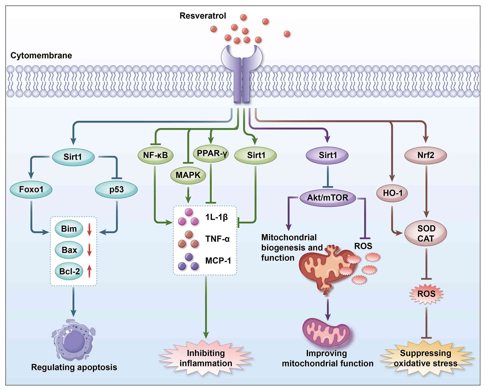
antibacterial, anticancer and other biological activities (46-48) (Fig. S11). As a potent antioxidant,
resveratrol alleviates oxidative stress by regulating key signaling
pathways, including nuclear factor erythroid 2-related factor 2
(Nrf2)/heme oxygenase-1 (HO-1), Sirtuin 1 (SIRT1) and
SIRT1/forkhead box protein O1 (FOXO1), thereby facilitating free
radical elimination and protecting cells from oxidative damage
(49-51) (Fig. 1). Additionally, resveratrol
exerts anti-inflammatory effects through the modulation of
inflammation-related pathways, including NF-κB, SIRT1 and
mitogen-activated protein kinase (MAPK) pathways (52-54) (Fig. 1). It also improves mitochondrial
function by regulating the SIRT/phosphoinositide 3-kinase
(PI3K)/protein kinase B (Akt) pathway, reducing ROS production and
increasing the expression of antioxidant enzymes such as superoxide
dismutase (SOD)1 and glutathione (GSH) (55) (Fig. 1). As summarized in Table SIV, studies suggest that
resveratrol may alleviate intestinal mucosal damage induced by
inflammation, oxidative stress and ferroptosis (Fig. S12). Further studies have
indicated that resveratrol, through core signaling pathways
including SIRT1, AMP-activated protein kinase (AMPK), Nrf2 and
NF-κB, may regulate macrophage polarization and influence the
balance between proinflammatory M1 and anti-inflammatory M2
phenotypes. Meanwhile, resveratrol may contribute to intestinal
barrier maintenance through multiple mechanisms, including
protection of TJs, modulation of the microbiota and activation of
autophagy (56). Research has
shown that activation of SIRT1 by resveratrol promotes
deacetylation of the NF-κB p65 subunit, thereby inhibiting its
nuclear translocation and transcriptional activity and reducing
M1-associated inflammatory responses [downregulation of TNF-α,
IL-1β and inducible nitric oxide synthase (iNOS)] (57). Meanwhile, SIRT1-mediated
deacetylation may activate transcription factors such as
proliferator-activated receptor gamma (PPARγ) and STAT6, thereby
promoting M2-associated polarization [upregulation of arginase 1
(Arg-1), CD206 and IL-10] (58,59). These effects may contribute to
the attenuation of sepsis-associated intestinal inflammation and
preservation of epithelial barrier integrity by reducing the
disruption and internalization of TJ proteins, including ZO-1,
occludin and claudin-1. Mechanistic target of rapamycin complex 1
(mTORC1) is considered an important regulator of M1 macrophage
polarization through glycolysis-dependent mechanisms and
resveratrol may inhibit mTORC1 activity via AMPK, thereby
influencing macrophage metabolic reprogramming. Resveratrol
modifies cysteine residues in Keap1, facilitating the nuclear
translocation of Nrf2 and its binding to ARE elements, thereby
promoting the expression of downstream antioxidant genes, including
HO-1, quinone oxidoreductase 1 and glutamate-cysteine ligase
catalytic subunit (60). HO-1
catalyzes heme degradation to generate CO, bilirubin and ferrous
iron, which may promote M2-associated polarization and reduce
M1-associated ROS production (61). Additionally, Nrf2 can interact
with NF-κB p65 and inhibit proinflammatory transcriptional activity
(62).
Sepsis is a severe clinical syndrome commonly
associated with dysregulated immune responses, in which
macrophages, as key immune cells, play important roles (63). Macrophages can be classified into
M1 and M2 types based on their polarization states (64). During the early stages of sepsis,
M1 macrophages mainly exhibit proinflammatory functions, whereas M2
macrophages participate in anti-inflammatory responses, cell repair
and tissue regeneration (65,66). Macrophage polarization is closely
involved in sepsis-induced intestinal injury ((Fig. S13). Activated macrophages,
particularly M1-polarized macrophages, release TNF-α, IL-1β, IL-6,
IL-12 and high mobility group box-1 protein (a late inflammatory
mediator) (67). These cytokines
act through autocrine and paracrine mechanisms to recruit
neutrophils, activate endothelial cells and stimulate other
macrophages, contributing to cytokine amplification and oxidative
stress (68). Cytokines,
particularly TNF-α and IFN-γ, together with ROS, may reduce the
expression and alter the localization of key TJ proteins, including
claudin-1, claudin-4, occludin and ZO-1, through NF-κB/myosin light
chain kinase (MLCK)-mediated cytoskeletal contraction, claudin
endocytosis and matrix metallopeptidase (MMP)-mediated proteolytic
cleavage. These alterations increase paracellular permeability
('leaky gut') and may further promote systemic inflammation and
distant organ injury involving the lung, kidney and liver (69). ROS can also damage TJ proteins
and oxidize membrane lipids, generating reactive aldehydes such as
4-hydroxynonenal and malondialdehyde (MDA), which further promote
intestinal epithelial cell apoptosis (70). Furthermore, sepsis is associated
with T-helper (Th)1/Th17 predominance, reduced regulatory T cells
populations, impaired immunoglobulin A responses and microbiota
alterations (dysbiosis) (71).
Dendritic cells in the lamina propria may become activated and
migrate to systemic sites, contributing to persistent inflammatory
responses and multiple-organ dysfunction (72).
Resveratrol, a naturally occurring plant polyphenol,
has attracted considerable attention in scientific research due to
its antioxidant, anti-inflammatory and immunomodulatory properties.
Numerous experimental studies have suggested that resveratrol can
attenuate inflammation and oxidative stress by promoting M2
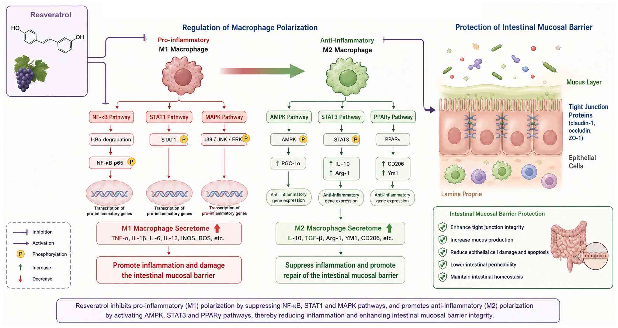
polarization and suppressing M1 polarization (Fig. 2) (73-75). Resveratrol inhibits NF-κB and
MAPK (p38 and JNK) signaling by suppressing inhibitor of kappa
light chain gene enhancer in B cells, alpha kinase activity and
activator protein 1 activation, while promoting M2 polarization
through SIRT1/PPARγ and STAT3/STAT6 signaling (Fig. 2) (65,76-78). These effects reduce the
production of proinflammatory cytokines, including TNF-α, IL-1β and
IL-6, activate PGC-1α, restore mitochondrial biogenesis, reduce ROS
generation and decrease intestinal epithelial cell apoptosis. In
the CLP sepsis mouse model, the proportion of M1 macrophages
(CD11b+F4/80+CD86+) in the mesenteric lymph nodes of the
resveratrol group (50 mg/kg) decreased from 45-18% and serum IL-6
levels decreased from 1,200 to 450 pg/ml (ELISA) (79). Immunohistochemical analysis
showed that the number of positive cells decreased from 38±5
cells/high power field (HPF) in the sepsis control group to 15±3
cells/HPF in the treatment group (a 60% reduction, P<0.001).
Electron microscopy analysis demonstrated that the TJ structural
integrity score increased from 2.1±0.4 in the control group to
3.8±0.5 in the treatment group (normal group, 5.0±0.3; representing
75% recovery) and occludin protein expression increased by 80%
based on western blot analysis (80). In a human intestinal organoid +
THP-1 macrophage model, resveratrol (10 μM) reduced the
expression of M1-related genes (IL-6 and IL-12b) by 50-65%
(RNA-seq) and increased transepithelial electrical resistance
(TEER) values by 2.3-fold (81).
Substantial experimental evidence supports the
regulatory effects of resveratrol on macrophage polarization
(82-85). Resveratrol can directly influence
macrophage polarization states. In vitro studies using mouse
primary macrophages have shown that resveratrol reduces the
expression of M1-associated markers, including iNOS and TNF-α,
while increasing the expression of M2-associated markers, such as
CD206 and Arg-1 (55,86). Additionally, resveratrol
treatment reduces the production of proinflammatory cytokines,
including IL-6 and IL-1β, by M1 macrophages (87). Furthermore, resveratrol has been
reported to regulate macrophage polarization in mouse models of
sepsis (88). During acute
sepsis, accumulation of M1 macrophages and increased secretion of
proinflammatory factors contribute to intestinal mucosal barrier
injury.
During sepsis, disruption of the intestinal mucosal
barrier is often accompanied by enhanced inflammatory responses and
immune dysfunction, which may disturb intestinal microbiota
homeostasis and further impair intestinal barrier function
(3). The regulatory effects of
resveratrol on M1/M2 macrophage polarization may not only improve
immune regulation but also alleviate the intestinal inflammatory
environment (89). Resveratrol
inhibits the secretion of proinflammatory factors from M1
macrophages, thereby reducing intestinal inflammation and
potentially promoting tissue repair (90). M1 macrophages are considered
important contributors to sepsis progression and intestinal
inflammation (91). They secrete
various proinflammatory cytokines, including TNF-α and IL-6, which
affect intestinal epithelial cells and intestinal barrier
integrity. In an in vitro macrophage model, resveratrol (20
μM) increased the M2/M1 ratio from 1:4 to 3:1 (flow
cytometry analysis) (92).
Intestinal tissue biopsy analysis in a mouse model of dextran
sulfate sodium-induced colitis revealed that CD206-positive
macrophage infiltration in the treatment group was significantly
elevated, reaching 1.7-fold the level observed in the untreated
control cohort (93). Following
SIRT1 activation, the acetylation level of p65-K310 decreased by
60%, accompanied by a 50% reduction in the serum TNF-α/IL-6 ratio
(94). NADPH oxidase (NOX)2
expression was downregulated, ROS generation was reduced
(intestinal tissue MDA levels decreased by 40%) and SOD activity
recovered to 85% of normal levels (95).
The repair and reconstruction of the intestinal
mucosal barrier primarily depend on the proliferation and
differentiation of intestinal epithelial cells (96). As summarized in Table SV, resveratrol has been reported
to promote intestinal epithelial cell growth through multiple
signaling pathways, thereby contributing to the repair of
intestinal injury associated with sepsis. Studies have indicated
that resveratrol activates the PI3K/Akt signaling pathway and
promotes intestinal epithelial cell proliferation (97). In a sepsis mouse model,
resveratrol intervention markedly improved the proliferation rate
and regenerative capacity of intestinal epithelial cells (98). This effect may facilitate
intestinal barrier repair, restore epithelial integrity and reduce
intestinal permeability, thereby limiting the translocation of
pathogens and toxins. Moreover, resveratrol not only promotes cell
proliferation but also enhances intestinal epithelial cell
differentiation through regulation of the Wingless-related
integration site/β-catenin pathway. Further studies using organoid
models showed that resveratrol (10 μM) increased intestinal
epithelial survival from 35±4 to 78±5% (99). In septic mice, resveratrol
treatment restored goblet cell numbers to 70% of normal levels
(Alcian blue staining) and restored intestinal mucus layer
thickness to 65% of normal levels (measured in frozen sections)
(100).
The integrity of the intestinal mucosal barrier
depends not only on epithelial cell proliferation and
differentiation but also on the maintenance of tight adhesion
between intestinal epithelial cells (18). These cells form a cohesive
physical barrier that limits the passage of harmful substances,
including bacteria and toxins, across the intestinal lining
(18). As summarized in Table SVI, resveratrol has been
reported to improve intestinal barrier function by enhancing TJ
integrity between intestinal epithelial cells. Its protective
effects are primarily associated with the upregulation of TJ
protein expression (101).
Further studies revealed that resveratrol deacetylates NF-κB p65
through the SIRT1-dependent pathway, reduces MMP-9 expression and
decreases occludin degradation by 40% (102). By maintaining TJ protein
membrane localization through the protein kinase C zeta
(PKCζ)/protein associated with lin seven 1 (PALS1) axis,
resveratrol increased ZO-1 membrane localization from 30-80% in the
sepsis group (immunofluorescence analysis), restored intestinal
epithelial transmembrane resistance (TEER) values to 85% of normal
levels and reduced bacterial translocation rates (positive blood
culture) by 50% in the CLP mouse model (103). In sepsis model mice (CLP
model), occludin expression in the resveratrol-treated group (50
mg/kg) increased from 30% in the sepsis control group to 97%,
whereas ZO-1 and Claudin-3 levels recovered to 89 and 82% of normal
levels, respectively (104).
Serum LPS levels decreased by 52% (ELISA) and intestinal microbiota
translocation rates (positive blood culture) decreased from 78% in
the control group to 35% in the treatment group (105).
During sepsis, intestinal homeostasis is disrupted
by endogenous and exogenous factors, including sepsis-associated M1
macrophage polarization, which contributes to inflammatory injury
in the intestine. This disruption is characterized by decreased gut
microbiota diversity, reduced abundance of Firmicutes and
Bacteroidetes and increased colonization of potentially pathogenic
bacteria, including Clostridium difficile,
Staphylococcus, Escherichia, Shigella, Salmonella and
Enterococcus, with relative abundances exceeding 50%
(106). Fecal quantitative
polymerase chain reaction analysis and microbial profiling further
confirmed a reduction in dominant bacterial populations,
particularly anaerobic species, during sepsis, accompanied by
increased abundance of pathogenic bacteria such as Escherichia
coli, Pseudomonas aeruginosa, Enterococcus faecalis
and Staphylococcus aureus (107). Resveratrol markedly affects gut
microbiota composition, including increasing the
Bacteroidetes/Firmicutes ratio and promoting the growth of
beneficial bacterial genera such as Bacteroidetes,
Lactobacillus and Bifidobacterium, while reducing the
abundance of Actinobacteria, thereby contributing to microbiota
remodeling and immune regulation (108,109). Ding et al (110) further demonstrated that
resveratrol alters microbiota composition by increasing beneficial
bacteria, including lactobacilli and bifidobacteria, while
decreasing fecal enterococci and Escherichia coli. Xu et
al (111) reported that
resveratrol reduces dysregulated metabolites, improves gut
microbiota structure and diversity, reverses alterations in
Bacteroidetes, Proteobacteria and Firmicutes, increases beneficial
bacterial genera and decreases potentially pathogenic genera such
as Clostridium, Acinetobacter and Serratia.
Recent studies have shown that resveratrol reduces
the expression of virulence factors, such as flagellar proteins, in
Escherichia coli by inhibiting quorum sensing and disrupting
pathogenic bacterial biofilms, including a 40% reduction in
Staphylococcus aureus biofilm thickness (112). Resveratrol also downregulates
virulence-associated genes in enterohemorrhagic Escherichia
coli (EHEC), with flagellar protein (fliC) expression reduced
by 65% (reverse transcription-quantitative polymerase chain
reaction), type III secretion system (T3SS) effector protein (EspB)
secretion reduced by 55% and bacterial motility reduced by 60% in
swimming assays (112). In a
mouse sepsis model, the positive rate of blood culture in the
treatment group decreased by 50% (112). In another mouse model of
2,4,6-trinitrobenzene sulfonic acid-induced colitis, resveratrol
restored gut microbial alpha diversity (Shannon index, 16S rRNA
sequencing) to 85% of normal levels, reversed colitis-associated
dysbiosis, and increased short-chain fatty acid (notably butyric
acid) production by 3.0-fold. This compound protected mice from
colonic inflammation by inducing Tregs and suppressing inflammatory
Th1/Th17 responses (113). In a
sepsis pig model, nanoresveratrol (PLGA coating) increased
intestinal Bacteroidetes abundance from 12±3 to 23±4% and reduced
the 7-day mortality rate from 65 to 25% (114).
As research on resveratrol has progressed,
increasing attention has been directed toward its safety profile,
particularly regarding toxicological evaluation, contraindications,
special populations, precautions and potential drug interactions.
The incidence and severity of resveratrol-related adverse effects
appear to be largely dose-dependent. Hebbar et al (115) administered different doses of
resveratrol (0.3, 1.0 and 3.0 g/kg) to rats for 28 days. The
results showed that rats treated with 0.3 g/kg herbal water extract
(HWE) exhibited no significant adverse effects, whereas rats
receiving 1.0 and 3.0 g/kg HWE developed dehydration, respiratory
distress, nephrotoxicity (indicated by elevated serum blood urea
nitrogen and creatinine levels), kidney injury and anemia
potentially associated with suppressed erythropoietin synthesis.
Another study reported that oral administration of resveratrol at
doses ≤0.1 g/day did not result in observable adverse effects
during either short-term or long-term administration (up to 1
year). However, occasional adverse effects, including
gastrointestinal discomfort, diarrhea and heat rash, were observed
at oral doses of 1.0 and 1.5 g/day. At doses exceeding 2.5 g/day,
more severe reactions, including nausea, vomiting and abdominal
pain, were reported (116),
suggesting potential toxicity associated with high-dose resveratrol
administration. Clinical studies have further indicated that oral
administration of 0.15 g/kg resveratrol to healthy obese men for 30
days markedly reduced aspartate aminotransferase (AST) levels and
liver fat accumulation (117).
In patients with nonalcoholic fatty liver disease, administration
of 0.05 g/kg resveratrol reduced total cholesterol and triglyceride
levels and improved lipid metabolism, whereas higher doses (3.0
g/day for 8 weeks) increased AST and alanine aminotransferase
levels (118). Overall,
low-dose resveratrol appears to have a favorable safety profile
with limited hepatic or renal toxicity. However, high-dose
resveratrol administration, particularly in patients with
nonalcoholic fatty liver disease, requires further evaluation and
should be approached cautiously.
Low-dose resveratrol can reduce ROS levels in
oocytes, increase glutathione levels, promote oocyte maturation and
improve blastocyst formation rates in vitro (119-121). In addition, resveratrol
supplementation may enhance antioxidant capacity in pregnant and
lactating mice, increase uterine artery blood flow and reduce
placental inflammation and hepatic triglyceride accumulation.
However, some studies have suggested that resveratrol may induce
fetal pancreatic enlargement or abnormalities (122), raising concerns regarding its
safety during pregnancy. Furthermore, resveratrol, which exhibits
phytoestrogen-like activity, may exert estrogenic effects.
Long-term exposure may potentially influence the growth of
hormone-sensitive cancer cells in breast, ovarian and uterine
tissues (123,124). Therefore, caution is warranted
when considering resveratrol use in individuals with cancer-related
risks and during pregnancy. Although the pleiotropic effects of
resveratrol, including anti-inflammatory, antioxidant,
immunomodulatory and mitochondrial protective activities, may
provide potential benefits, this complexity also makes it
challenging to determine the primary mechanisms underlying clinical
effects and to design targeted clinical trials.
Additionally, resveratrol may alter the
pharmacokinetics of coadministered drugs, including absorption and
metabolism, through inhibition of cytochrome P450 (CYP450)
isoenzymes, potentially resulting in safety concerns. Resveratrol
markedly inhibits CYP1A2 and CYP2C9 activity, affecting drug
metabolism, potentially reducing therapeutic efficacy and
increasing the risk of adverse effects (125). Therefore, when resveratrol is
administered together with drugs metabolized by CYP1A2 and CYP2C9
in clinical settings, monitoring of blood drug concentrations may
be necessary to optimize dosing strategies or consider alternative
therapies.
Resveratrol has been extensively investigated as a
multi-target candidate due to its anti-inflammatory, antioxidant,
autophagy-modulating and microbiota-regulating properties (126). However, the transition from
preclinical findings to clinical application remains challenging
due to unresolved methodological, pharmacological and
pathophysiological limitations (126). Most current formulation
strategies primarily focus on improving bioavailability, while
insufficient attention has been paid to the stage-specific
pathophysiology of sepsis (127). Therefore, future delivery
strategies may need to be phase-adapted, targeting early
hyperinflammation, mid-phase intestinal barrier dysfunction, or
late-stage immunoparalysis with distinct pharmacokinetic
characteristics. The current delivery field also faces
methodological limitations, as most efficacy data are generated
from relatively healthy animal models (128). The pharmacokinetic
characteristics of resveratrol under septic conditions remain
insufficiently investigated, representing an important knowledge
gap (126). Furthermore, safety
assessments based on non-critical illness conditions may not fully
reflect the complex physiological changes associated with sepsis. A
sepsis-specific toxicological evaluation framework incorporating
drug-drug interactions (DDIs), reversibility of immunomodulatory
effects and metabolite profiling under organ dysfunction conditions
is therefore needed. Current studies have generated substantial
evidence regarding the preventive effects of resveratrol; however,
data regarding clinically relevant treatment windows remain
limited. Without rigorous evaluation of administration timing,
clinical translation may be affected. The dose-exposure-response
relationship of resveratrol remains incompletely established due to
several limitations: i) Effective doses observed in animal models
may not be safely achievable in humans; ii) effective
concentrations observed in vitro may not be reached in
target tissues; and iii) pre-treatment animal models do not fully
represent clinical scenarios involving post-onset intervention.
Addressing these translational gaps requires improved experimental
strategies, including pathological models that improved reflect
clinical conditions, pharmacokinetic/pharmacodynamic
(PK/PD)-integrated trial designs, validation of molecular targets
in human tissues and rational dose-finding approaches based on
achievable human exposure. In addition, well-designed studies
reporting both positive and negative findings are necessary to
objectively evaluate the clinical potential of resveratrol.
Furthermore, the biological effects of resveratrol may depend on
the dynamic immune environment during sepsis.
Early proinflammatory (M1-type) macrophage
overactivation contributes to cytokine release and inflammatory
amplification, whereas later stages may involve immune suppression
and M2-associated responses. Therefore, resveratrol may require
precise temporal regulation of macrophage polarization, including
early suppression of excessive M1 activation and avoidance of
excessive M2 polarization during later stages. Inappropriate
administration timing may potentially disturb immune balance, such
as impairing tissue repair through excessive inhibition of
M2-associated responses. The pathological environment of sepsis may
also influence the efficacy of resveratrol. Reduced intestinal
mucosal perfusion and oxidative stress may limit antioxidant
activity and potentially alter its biological effects under highly
oxidative conditions. Meanwhile, gut microbiota dysbiosis,
including expansion of pathogenic bacteria, may continuously
activate M1 macrophages through TLR/NF-κB signaling, potentially
interfering with the immunomodulatory effects of resveratrol.
Sepsis-associated capillary leakage and impaired liver and kidney
function may alter resveratrol pharmacokinetics, including
distribution and clearance, thereby affecting local intestinal
exposure (130). Intestinal
mucosal edema and disruption of TJ integrity may further influence
drug absorption, highlighting the potential need for advanced
delivery systems, such as mucosal adhesive nanoparticles or
exosome-based platforms. Additionally, liver dysfunction during
sepsis may affect resveratrol metabolism through alterations in
CYP450 enzyme activity, whereas bacterial translocation following
intestinal barrier disruption may increase hepatic burden and
contribute to a pathological feedback loop. Different pathogens
(gram-negative bacteria, gram-positive bacteria and fungi) and
infection sites may induce distinct macrophage responses,
suggesting that individualized therapeutic strategies may be
required for future clinical applications of resveratrol.
Resveratrol can exert comprehensive protective
effects, including antioxidant, anti-inflammatory, barrier repair,
inhibition of programmed cell death and microbiota regulation, in
sepsis-induced intestinal mucosal injury through multi-target and
multi-pathway synergistic mechanisms. However, limitations remain
in in vitro studies, animal experiments, clinical studies
and clinical trials, as summarized in Table SVII.
Current models for sepsis-induced intestinal injury
exhibit significant inconsistencies in experimental conditions and
outcomes, alongside notable limitations: as summarized in Table SVIII, LPS-induced models,
varying in dosage, administration routes and animal strains, only
activate the TLR4 pathway and fail to replicate the immune
complexity of human sepsis (131), leading to divergent intestinal
injury severities and time points across studies. The 'gold
standard' CLP model suffers from poor inter-laboratory
reproducibility due to variations in cecal ligation ratios,
puncture techniques and post-operative care, with intestinal injury
severity closely tied to sepsis grading but complicated by
operational uncertainty (132).
Intestinal ischemia/reperfusion models show marked individual
differences in injury susceptibility due to factors such as
ischemia duration and animal strains, making uniform evaluation
challenging (133). Cell
models, while useful for studying cellular-level damage, are
limited by diverse preparation methods and an inability to
recapitulate the in vivo intestinal structure and
microenvironment (134).
Collectively, these models have inherent shortcomings in simulating
pathophysiological mechanisms, suffer from physiological
differences between animals and humans, lack consistency and
reproducibility and struggle to mimic clinical complexities, all of
which severely restrict their translational value.
The M1/M2 model describes macrophage activation as
a binary classification between two relatively distinct functional
states (135). However,
macrophages exist along a continuum of activation states with
extensive transcriptomic and functional diversity (136). Individual macrophages may
simultaneously express M1- and M2-associated genes, resulting in
hybrid or 'double-positive' populations that cannot be fully
characterized by simple categorization (136). Therefore, the traditional
binary framework may not completely reflect the functional
plasticity and complexity of macrophages in vivo (137). The M1/M2 paradigm was primarily
established based on bone marrow-derived macrophages (BMDMs) or
cell lines stimulated with defined cytokines under in vitro
conditions (138). These
experimental systems may not fully represent the complex tissue
microenvironments, cellular interactions and metabolic conditions
present in vivo. Tissue-resident macrophages are influenced
by multiple factors, including ontogeny (embryonic vs.
monocyte-derived origin), local metabolic conditions (oxygen
tension, pH and nutrient availability) and signals from surrounding
cells (139). Therefore,
applying in vitro-derived M1/M2 classifications to in
vivo macrophage populations may not accurately reflect their
functional states. Unlike relatively stable Th cell subsets (Th1,
Th2 and Th17), macrophage polarization states are highly dynamic
and reversible. Macrophages classified as M1 may acquire
M2-associated features following environmental changes and vice
versa (140). This plasticity
challenges the concept of clearly separated macrophage subsets and
suggests that macrophage activation should be considered a
context-dependent process rather than a fixed differentiation
program. Furthermore, there is currently no universal consensus
regarding the defining markers of M1 and M2 macrophages across
different studies. Although iNOS and CD86 are commonly used as M1
markers and Arg1 and CD206 as M2 markers, these molecules are not
exclusively expressed in specific macrophage states and may be
induced by different stimuli and vary among species and tissues.
This inconsistency contributes to reproducibility issues and
complicates comparisons among studies. The M1/M2 framework also
does not fully account for macrophage developmental origin.
Tissue-resident macrophages derived from embryonic yolk sac or
fetal liver progenitors differ from monocyte-derived macrophages
recruited during inflammation, even when they express similar
activation markers. Moreover, tissue-specific transcriptional
programs induced by local environmental signals further diversify
macrophage phenotypes beyond the M1/M2 classification.
Furthermore, the half-life and plasma
concentrations of resveratrol metabolites (RE) in the blood were
reported to be ~10 times higher than those of the parent RE
compound (141), suggesting
that free RE may be locally released from these metabolites.
Despite its relatively low bioavailability, rapid metabolism and
elimination, RE exhibits significant biological activity. This may
be attributed to its conversion into sulfate and glucuronide
metabolites and/or its binding to plasma proteins, which may
facilitate RE delivery to target organs (142).
Resveratrol has demonstrated anti-inflammatory,
antioxidant and organ-protective potential in sepsis and existing
small-scale clinical studies have preliminarily supported its
safety and potential effects on biological activity indicators.
However, clinical research investigating resveratrol in patients
with sepsis faces several important limitations. First,
large-scale, well-designed randomized controlled trials (RCTs) in
humans remain limited (143).
Most available evidence is derived from preclinical studies (animal
models and in vitro experiments), small pilot studies, or
trials with limited sample sizes, which may not provide sufficient
statistical power for definitive conclusions (143-146). The optimal dose, formulation
(such as micronized formulations or combination with piperine to
improve bioavailability) and appropriate administration window
during the dynamic course of sepsis remain unclear. Sepsis
represents a highly heterogeneous clinical syndrome (147). Clinical trials often include
patients with different infection sources, comorbidities and
disease severities (such as sepsis and septic shock), making it
difficult to determine which patient subgroups may benefit most
from resveratrol intervention. In addition, a number of studies
rely on surrogate biomarkers, such as inflammatory cytokines, as
primary outcomes. Future trials should focus more on clinically
meaningful endpoints, including 28-day mortality, organ
failure-free days and long-term functional outcomes (148). Although resveratrol is
generally considered safe, comprehensive safety data regarding
high-dose or prolonged intravenous administration in critically ill
patients with sepsis remain insufficient, particularly concerning
potential interactions with commonly used medications, including
sedatives, anticoagulants and antibiotics. The beneficial effects
consistently observed in controlled experimental models have not
yet been fully translated into measurable clinical benefits in
patients, highlighting the gap between preclinical findings and
clinical application. In conclusion, although preclinical evidence
regarding resveratrol in sepsis is encouraging, current clinical
evidence remains preliminary and insufficient to support routine
clinical use. Major limitations include the lack of large
definitive RCTs, unresolved pharmacokinetic challenges, patient
heterogeneity and the complexity of resveratrol-mediated biological
effects. Future studies require well-designed multicenter clinical
trials with standardized protocols and clinically relevant outcomes
in clearly defined septic populations.
The immune regulation of resveratrol on sepsis is
not the activation or inhibition of a single pathway, but through
precise matching of macrophage plasticity, temporal changes and
tissue microenvironment (Table
SIX), achieving dynamic switching from 'anti-inflammatory' to
'immune promoting', providing a precise new approach for
immunotherapy of sepsis.
Future progress in understanding the M1/M2
framework requires integration of scRNA-seq with additional
approaches, including epigenomics (scATAC-seq), proteomics (CyTOF
and CITE-seq), metabolomics and spatial transcriptomics. This
multi-omics strategy may provide a more comprehensive understanding
of macrophage states in health and disease. The field also requires
standardized nomenclature and analytical frameworks to complement
or refine the current M1/M2 classification. Recent approaches,
including the 'macrophage activation continuum' model and
ontology-based classifications, aim to provide more accurate
descriptions of macrophage functional states. Applying these
concepts to sepsis requires: large-scale longitudinal scRNA-seq
studies using patient samples; integration with clinical metadata
to identify clinically relevant endotypes; development of rapid and
feasible methods for macrophage subset identification (such as flow
cytometry panels and point-of-care transcriptomic approaches); and
evaluation of subset-targeted therapies in preclinical models and
clinical trials.
In the future, gene editing technologies such as
CRISPR may be applied to develop high-yield strains and address the
challenges associated with low extraction rates (<1%) and high
levels of impurities during chemical synthesis of plant-derived
resveratrol (149). Current
global efforts focus on optimizing metabolic pathways to increase
fermentation yields to industrial production levels (150). Emerging technologies, including
supercritical fluid extraction and enzymatic hydrolysis, may
improve the utilization of resveratrol raw materials, reduce
residual organic solvents and facilitate the production of
pharmaceutical-grade resveratrol (>98% purity) (151). Carrier-based technologies, such
as liposomes and polymer nanoparticles, may overcome the poor water
solubility and rapid metabolism of resveratrol, thereby prolonging
its biological activity (152).
Experimental studies have shown that nanoencapsulation can increase
oral bioavailability by 3-5-fold (153). Methylation and glycosylation
modifications may also improve resveratrol stability, with
tetramethyl resveratrol exhibiting an ~8-fold longer half-life than
the natural form (154).
Additionally, integration of single-cell sequencing and
metabolomics may facilitate the identification of novel targets
involved in resveratrol-mediated regulation of the SIRT1/AMPK
pathway (155), while combined
formulations containing resveratrol, nicotinamide nucleoside and
quercetin may provide synergistic biological effects.
Furthermore, future studies may explore real-time
adjustment of dosing regimens based on biomarkers such as IL-6 and
M1/M2-associated surface markers CD86/CD206 (156). The development of intelligent
responsive drug delivery systems, such as ROS-responsive
nanoparticles, may enable targeted drug release at inflammatory
sites (157). The combined
effects of resveratrol with other organ-protective agents,
including mitochondrial antioxidants, also warrant further
investigation. In experimental studies, humanized mouse models and
organoid systems may help improved simulate sepsis-induced
intestinal injury and improve translational relevance.
Resveratrol, a natural plant polyphenol, has
attracted increasing attention due to its diverse biological
activities, including anti-inflammatory, antioxidant and
immunoregulatory effects. It may regulate immune responses during
sepsis by influencing M1/M2 macrophage polarization. Through
modulation of macrophage functional states, resveratrol may
contribute to the attenuation of intestinal inflammation,
preservation of intestinal barrier integrity and reduction of
intestinal permeability. These effects may limit bacterial and
toxin translocation and alleviate systemic inflammatory responses
associated with sepsis. However, current evidence remains
insufficient to support routine clinical use of resveratrol in
sepsis. Therefore, although resveratrol represents a promising
experimental compound with potential immunomodulatory and
barrier-protective properties, further mechanistic studies and
well-designed clinical investigations are required before
therapeutic conclusions can be established in patients with
sepsis.
The M1/M2 framework, while historically valuable,
has limitations in fully representing macrophage biology.
Single-cell sequencing has revealed substantial complexity among
macrophage states across different tissues, disease conditions and
temporal stages. In sepsis research, considering this complexity
rather than relying solely on binary classification may facilitate
more precise diagnosis, patient stratification and targeted
therapeutic strategies. Future macrophage research will likely
require systems-level approaches to characterize dynamic,
context-dependent and ontogenically diverse macrophage populations,
ultimately supporting the development of more effective
immunomodulatory strategies for critical illness.
Not applicable.
YT, JX, MWL, CYQ and WZ conceived the research
topic, conducted literature reviews for relevant articles and
drafted the manuscript. YLZ performed language and grammar editing.
MXS and XYY revised the manuscript drafts and restructured the
content. SJG, YLZ and MWL provided access to the tools used for
figure generation. Data authentication is not applicable. All
authors read and approved the final manuscript.
Not applicable.
Not applicable.
The authors declare that they have no competing
interests.
Not applicable.
The present study work was supported by the National Natural
Science Foundation of China (grant nos. 82060252 and 81960350),
Yunnan Basic Research Projects (grant no. 2018FB115), the Yunnan
Health Training Project of High-level Talents (grant no.
H-2018058), the Yunnan Applied Basic Research Project-Union
Foundation of China (grant no. 202201AY070001-091) and the Basic
Research Project of the Science and Technology Department of Yunnan
Province (grant no. 202101AT070148).
|
1
|
Ling L, Zhang JZ, Chang LC, Chiu LCS, Ho
S, Ng PY, Dharmangadan M, Lau CH, Ling S, Man MY, et al: Population
sepsis incidence, mortality, and trends in hong kong between 2009
and 2018 using clinical and administrative data. Clin Infect Dis.
80:91–100. 2025. View Article : Google Scholar :
|
|
2
|
Kullberg RFJ, Wiersinga WJ and Haak BW:
Gut microbiota and sepsis: From pathogenesis to novel treatments.
Curr Opin Gastroenterol. 37:578–585. 2021. View Article : Google Scholar : PubMed/NCBI
|
|
3
|
Oami T, Abtahi S, Shimazui T, Chen CW,
Sweat YY, Liang Z, Burd EM, Farris AB, Roland JT, Tsukita S, et al:
Claudin-2 upregulation enhances intestinal permeability, immune
activation, dysbiosis, and mortality in sepsis. Proc Natl Acad Sci
USA. 121:e22178771212024. View Article : Google Scholar : PubMed/NCBI
|
|
4
|
Tan F, Cao Y, Zheng L, Wang T, Zhao S,
Chen J, Pang C, Xia W, Xia Z, Li N and Chi X: Diabetes exacerbated
sepsis-induced intestinal injury by promoting M1 macrophage
polarization via miR-3061/Snail1 signaling. Front Immunol.
13:9226142022. View Article : Google Scholar : PubMed/NCBI
|
|
5
|
Ruan S, Xu L, Sheng Y, Wang J, Zhou X,
Zhang C, Guo L, Li W and Han C: Th1 promotes M1 polarization of
intestinal macrophages to regulate colitis-related mucosal barrier
damage. Aging (Albany NY). 15:6721–6735. 2023. View Article : Google Scholar : PubMed/NCBI
|
|
6
|
Tian B and Liu J: Resveratrol: A review of
plant sources, synthesis, stability, modification and food
application. J Sci Food Agric. 100:1392–1404. 2020. View Article : Google Scholar
|
|
7
|
Agha RA, Mathew G, Rashid R, Kerwan A,
Al-Jabir A, Sohrabi C, Franchi T, Nicola M and Agha M; TITAN Group:
Transparency in the reporting of artificial intelligence-the TITAN
guideline. Prem J Sci. 10:1000822025.
|
|
8
|
Zhao X, Ma T, Yu J and Hu Y: Gut
microbiota as a therapeutic target in sepsis-induced multi-organ
dysfunction: A review. Front Cell Infect Microbiol. 16:18231252026.
View Article : Google Scholar : PubMed/NCBI
|
|
9
|
Wang C, Jiang X, Chen Y, Nan W and Peng Z:
Emerging roles and possible therapeutic applications of arachidonic
acid metabolites in sepsis and sepsis-associated organ dysfunction.
Drug Des Devel Ther. 20:6231432026.PubMed/NCBI
|
|
10
|
Weng L, Xu Y, Yin P, Wang Y, Chen Y, Liu
W, Li S, Peng JM, Dong R, Hu XY, et al: National incidence and
mortality of hospitalized sepsis in China. Crit Care. 27:842023.
View Article : Google Scholar : PubMed/NCBI
|
|
11
|
Reintam Blaser A, Bachmann KF and Deane
AM: Gastrointestinal function in critically ill patients. Curr Opin
Clin Nutr Metab Care. 26:463–469. 2023.PubMed/NCBI
|
|
12
|
Zhang S, Zhou Q, Li Y, Zhang Y and Wu Y:
MitoQ modulates lipopolysaccharide-induced intestinal barrier
dysfunction via regulating Nrf2 signaling. Mediators Inflamm.
2020:32761482020. View Article : Google Scholar : PubMed/NCBI
|
|
13
|
Otani T: Epithelial barrier homeostasis.
Trends Cell Biol. 36:610–622. 2026. View Article : Google Scholar : PubMed/NCBI
|
|
14
|
Zhang S, Zhang G, Xu J, Chen D, Yin C,
Wang J, Jiang X and Wang C: Roles of tight junction proteins in
intestinal barrier function and health of weaned piglets: A review.
Vet Sci. 13:1312026. View Article : Google Scholar : PubMed/NCBI
|
|
15
|
Wang Y, Li Y, Bao L, Lu J, Li X, Fu Y,
Zhang D and Li H: GRHL2 contributes to the maintenance of
intestinal epithelial barrier integrity during LPS-induced injury.
Front Immunol. 17:18817342026. View Article : Google Scholar : PubMed/NCBI
|
|
16
|
Zhang Y, Yang Y, Wang Y and Qin S:
Research advances in mechanisms and detection methods of vascular
endothelial barrier dysfunction in sepsis-induced organ injury.
Zhonghua Wei Zhong Bing Ji Jiu Yi Xue. 38:584–592. 2026.In Chinese.
PubMed/NCBI
|
|
17
|
Meng L, Liu Y, Wang N, Li T, Wang Y and Li
M: Gut barrier-microbiota crosstalk in sepsis: From pathogenesis to
potential therapies. Front Immunol. 17:18654982026. View Article : Google Scholar : PubMed/NCBI
|
|
18
|
Chelakkot C, Ghim J and Ryu SH: Mechanisms
regulating intestinal barrier integrity and its pathological
implications. Exp Mol Med. 50:1–9. 2018. View Article : Google Scholar : PubMed/NCBI
|
|
19
|
Ghulam Mohyuddin S, Khan I, Zada A, Qamar
A, Arbab AAI, Ma XB, Yu ZC, Liu XX, Yong YH, Ju XH, et al:
Influence of heat stress on intestinal epithelial barrier function,
tight junction protein, and immune and reproductive physiology.
Biomed Res Int. 2022:85473792022. View Article : Google Scholar : PubMed/NCBI
|
|
20
|
Kloc M, Ghobrial RM, Wosik J, Lewicka A,
Lewicki S and Kubiak JZ: Macrophage functions in wound healing. J
Tissue Eng Regen Med. 13:99–109. 2019.
|
|
21
|
Ma WT, Gao F, Gu K and Chen DK: The role
of monocytes and macrophages in autoimmune diseases: A
comprehensive review. Front Immunol. 10:11402019. View Article : Google Scholar : PubMed/NCBI
|
|
22
|
Edholm ES, Rhoo KH and Robert J:
Evolutionary aspects of macrophages polarization. Results Probl
Cell Differ. 62:3–22. 2017. View Article : Google Scholar : PubMed/NCBI
|
|
23
|
Chen X, Liu Y, Gao Y, Shou S and Chai Y:
The roles of macrophage polarization in the host immune response to
sepsis. Int Immunopharmacol. 96:1077912021. View Article : Google Scholar : PubMed/NCBI
|
|
24
|
Yunna C, Mengru H, Lei W and Weidong C:
Macrophage M1/M2 polarization. Eur J Pharmacol. 877:1730902020.
View Article : Google Scholar : PubMed/NCBI
|
|
25
|
Liu L, Guo H, Song A, Huang J, Zhang Y,
Jin S, Li S, Zhang L, Yang C and Yang P: Progranulin inhibits
LPS-induced macrophage M1 polarization via NF-кB and MAPK pathways.
BMC Immunol. 21:322020. View Article : Google Scholar
|
|
26
|
Vadevoo SMP, Kang Y, Gunassekaran GR, Lee
SM, Park MS, Jo DG, Kim SK, Lee H, Kim WJ and Lee B: IL4 receptor
targeting enables nab-paclitaxel to enhance reprogramming of
M2-type macrophages into M1-like phenotype via ROS-HMGB1-TLR4 axis
and inhibition of tumor growth and metastasis. Theranostics.
14:2605–2621. 2024. View Article : Google Scholar : PubMed/NCBI
|
|
27
|
Gao F, Zhu F, Shuai B, Wu M, Wei C, Yuan
Y, Gui Y, Tian Y, Fan H and Wu H: Quercetin ameliorates ulcerative
colitis by restoring the balance of M2/M1 and repairing the
intestinal barrier via downregulating cGAS-STING pathway. Front
Pharmacol. 15:13515382024. View Article : Google Scholar
|
|
28
|
Lethen I, Lechner-Grimm K, Gabel M, Knauss
A, Atreya R, Neurath MF and Weigmann B: Tofacitinib affects M1-like
and M2-like polarization and tissue factor expression in
macrophages of healthy donors and IBD patients. Inflamm Bowel Dis.
30:1151–1163. 2024. View Article : Google Scholar
|
|
29
|
Varlı M, Kim SJ, Noh MG, Kim YG, Ha HH,
Kim KK and Kim H: KITENIN promotes aerobic glycolysis through PKM2
induction by upregulating the c-Myc/hnRNPs axis in colorectal
cancer. Cell Biosci. 13:1462023. View Article : Google Scholar
|
|
30
|
Tan T, Li J, Fan W, Shang K, Yang C, Liu
X, Zhu S, Liu T, Wang J, Li Y and Lin Y: Tetrahedral framework
nucleic acid relieves sepsis-induced intestinal injury by
regulating M2 macrophages. Cell Prolif. 58:e138032025. View Article : Google Scholar : PubMed/NCBI
|
|
31
|
Li K, Yan G, Huang H, Zheng M, Ma K, Cui
X, Lu D, Zheng L, Zhu B, Cheng J and Zhao J: Anti-inflammatory and
immunomodulatory effects of the extracellular vesicles derived from
human umbilical cord mesenchymal stem cells on osteoarthritis via
M2 macrophages. Nanobiotechnology. 20:382022. View Article : Google Scholar
|
|
32
|
Huang X, Zheng L, Zhou Y, Hu S, Ning W, Li
S, Lin Z and Huang S: Controllable adaptive
molybdate-oligosaccharide nanoparticles regulate M2 macrophage
mitochondrial function and promote angiogenesis via
PI3K/HIF-1α/VEGF pathway to accelerate diabetic wound healing. Adv
Healthc Mater. 13:e23022562024. View Article : Google Scholar
|
|
33
|
Weinhäuser I, Pereira-Martins DA, Almeida
LY, Hilberink JR, Silveira DRA, Quek L, Ortiz C, Araujo CL, Bianco
TM, Lucena-Araujo A, et al: M2 macrophages drive leukemic
transformation by imposing resistance to phagocytosis and improving
mitochondrial metabolism. Sci Adv. 9:eadf85222023. View Article : Google Scholar : PubMed/NCBI
|
|
34
|
Li L, Kang M, Chen S, Lu X, Song Y, Li H,
Wang T, Jia C and Xu C: The role of macrophages in the immune
microenvironment of splenic ectopic pregnancy. Mol Immunol.
193:79–90. 2026. View Article : Google Scholar : PubMed/NCBI
|
|
35
|
Zhang K, Guo J, Yan W and Xu L: Macrophage
polarization in inflammatory bowel disease. Cell Commun Signal.
21:3672023. View Article : Google Scholar : PubMed/NCBI
|
|
36
|
Chen X, Tang L, Feng J, Wang Y, Han Z and
Meng J: Downregulation of paralemmin-3 ameliorates
lipopolysaccharide-induced acute lung injury in rats by regulating
Inflammatory response and inhibiting formation of TLR4/MyD88 and
TLR4/TRIF complexes. Inflammation. 40:1983–1999. 2017. View Article : Google Scholar : PubMed/NCBI
|
|
37
|
Xia MZ, Liang YL, Wang H, Chen X, Huang
YY, Zhang ZH, Chen YH, Zhang C, Zhao M, Xu DX and Song LH:
Melatonin modulates TLR4-mediated inflammatory genes through MyD88-
and TRIF-dependent signaling pathways in
lipopolysaccharide-stimulated RAW264.7 cells. J Pineal Res.
53:325–334. 2012. View Article : Google Scholar : PubMed/NCBI
|
|
38
|
Gobelli D, Serrano-Lorenzo P, Esteban-Amo
MJ, Serna J, Pérez-García MT, Orduña A, Jourdain AA,
Martín-Casanueva MÁ, Á de la Fuente M and Simarro M: The
mitochondrial succinate dehydrogenase complex controls the
STAT3-IL-10 pathway in inflammatory macrophages. iScience.
26:1074732023. View Article : Google Scholar : PubMed/NCBI
|
|
39
|
Li ZL, Yang BC, Gao M, Xiao XF, Zhao SP
and Liu ZL: Naringin improves sepsis-induced intestinal injury by
modulating macrophage polarization via PPARγ/miR-21 axis. Mol Ther
Nucleic Acids. 25:502–514. 2021. View Article : Google Scholar : PubMed/NCBI
|
|
40
|
Angurana SK, Bansal A, Muralidharan J,
Aggarwal R and Singhi S: Cytokine levels in critically Ill children
with severe sepsis and their relation with the severity of illness
and mortality. J Intensive Care Med. 36:576–583. 2021. View Article : Google Scholar
|
|
41
|
Seyedizade SS, Afshari K, Bayat S, Rahmani
F, Momtaz S, Rezaei N and Abdolghaffari AH: Current status of M1
and M2 macrophages pathway as drug targets for inflammatory bowel
disease. Arch Immunol Ther Exp (Warsz). 68:102020. View Article : Google Scholar : PubMed/NCBI
|
|
42
|
Wang C, Ma C, Gong L, Guo Y, Fu K, Zhang
Y, Zhou H and Li Y: Macrophage polarization and its role in liver
disease. Front Immunol. 12:8030372021. View Article : Google Scholar :
|
|
43
|
Cheng P, Li S and Chen H: Macrophages in
lung injury, repair, and fibrosis. Cells. 10:4362021. View Article : Google Scholar : PubMed/NCBI
|
|
44
|
Wang Y, Smith W, Hao D, He B and Kong L:
M1 and M2 macrophage polarization and potentially therapeutic
naturally occurring compounds. Int Immunopharmacol. 70:459–466.
2019. View Article : Google Scholar : PubMed/NCBI
|
|
45
|
Yang ZB, Gao YB, Cheng XM and Qiu LZ:
Human breast milk-derived exosomal FP671120.4 inhibits macrophage
M1 polarization via modulating the ELAVL1/Nrf2 axis in
sepsis-associated liver injury. Kaohsiung J Med Sci. 42:e701082026.
View Article : Google Scholar :
|
|
46
|
Jaa A, de Moura PHB, Ruiz-Larrea MB, Ruiz
Sanz JI and Richard T: Potential transformation of food
resveratrol: Mechanisms and biological impact. Molecules.
30:5362025. View Article : Google Scholar : PubMed/NCBI
|
|
47
|
Yang H, Wang Y, Jin S, Pang Q, Shan A and
Feng X: Dietary resveratrol alleviated lipopolysaccharide-induced
ileitis through Nrf2 and NF-κB signalling pathways in ducks (Anas
platyrhynchos). J Anim Physiol Anim Nutr (Berl). 106:1306–1320.
2022. View Article : Google Scholar
|
|
48
|
Banerjee B, Nandi P, Chakraborty S, Raha
S, Sen PC and Jana K: Resveratrol ameliorates
benzo(a)pyrene-induced testicular dysfunction and apoptosis:
Involvement of p38 MAPK/ATF2/iNOS signaling. J Nutr Biochem.
34:17–29. 2016. View Article : Google Scholar : PubMed/NCBI
|
|
49
|
Bai Y, Sui R, Zhang L, Bai B, Zhu Y and
Jiang H: Resveratrol improves cognitive function in post-stroke
depression rats by repressing inflammatory reactions and oxidative
stress via the Nrf2/HO-1 pathway. Neuroscience. 541:50–63. 2024.
View Article : Google Scholar : PubMed/NCBI
|
|
50
|
Wang P, Huang CX, Gao JJ, Shi Y, Li H, Yan
H, Yan SJ and Zhang Z: Resveratrol induces SIRT1-dependent
autophagy to prevent H2O2-INDUCED oxidative
stress and apoptosis in HTR8/SVneo cells. Placenta. 91:11–18. 2020.
View Article : Google Scholar : PubMed/NCBI
|
|
51
|
Inoue H, Shimizu Y, Yoshikawa H, Arakawa
K, Tanaka M, Morimoto H, Sato A, Takino Y, Ishigami A, Takahashi N
and Uehara M: Resveratrol upregulates senescence marker protein 30
by activating AMPK/Sirt1-foxo1 signals and attenuating
H2O2-induced damage in FAO rat liver cells. J
Nutr Sci Vitaminol (Tokyo). 69:388–393. 2023. View Article : Google Scholar
|
|
52
|
Guo L, Zhang X, Lv N, Wang L, Gan J, Jiang
X and Wang Y: Therapeutic role and potential mechanism of
resveratrol in atherosclerosis: TLR4/NF-κB/HIF-1α. Mediators
Inflamm. 2023:10977062023. View Article : Google Scholar
|
|
53
|
Moussa C, Hebron M, Huang X, Ahn J,
Rissman RA, Aisen PS and Turner RS: Resveratrol regulates
neuro-inflammation and induces adaptive immunity in Alzheimer's
disease. J Neuroinflammation. 14:12017. View Article : Google Scholar : PubMed/NCBI
|
|
54
|
Yang G, Chang CC, Yang Y, Yuan L, Xu L, Ho
CT and Li S: Resveratrol alleviates rheumatoid arthritis via
reducing ROS and inflammation, inhibiting MAPK signaling pathways,
and suppressing angiogenesis. J Agric Food Chem. 66:12953–12960.
2018. View Article : Google Scholar : PubMed/NCBI
|
|
55
|
Unnikrishnan Meenakshi D, Narde GK, Ahuja
A, Al Balushi K, Francis AP and Khan SA: Therapeutic applications
of nanoformulated resveratrol and quercetin phytochemicals in
colorectal cancer-an updated review. Pharmaceutics. 16:7612024.
View Article : Google Scholar : PubMed/NCBI
|
|
56
|
Wang Y, Hong C, Wu Z, Li S, Xia Y, Liang
Y, He X, Xiao X and Tang W: Resveratrol in intestinal health and
disease: Focusing on intestinal barrier. Front Nutr. 9:8484002022.
View Article : Google Scholar : PubMed/NCBI
|
|
57
|
Tang XL, Wang X, Fang G, Zhao YL, Yan J,
Zhou Z, Sun R, Luo AL and Li SY: Resveratrol ameliorates
sevoflurane-induced cognitive impairment by activating the
SIRT1/NF-κB pathway in neonatal mice. J Nutr Biochem.
90:1085792021. View Article : Google Scholar
|
|
58
|
Shamardl HAMA, Ibrahim NA, Merzeban DH,
Elamir AM, Golam RM and Elsayed AM: Resveratrol and Dulaglutide
ameliorate adiposity and liver dysfunction in rats with
diet-induced metabolic syndrome: Role of SIRT-1/adipokines/PPARγ
and IGF-1. Daru. 31:13–27. 2023. View Article : Google Scholar : PubMed/NCBI
|
|
59
|
Lang J, Gao L, Wu J, Meng J, Gao X, Ma H
and Yan D: Resveratrol attenuated manganese-induced learning and
memory impairments in mice through PGC-1alpha-mediated autophagy
and microglial M1/M2 polarization. Neurochem Res. 47:3414–3427.
2022. View Article : Google Scholar : PubMed/NCBI
|
|
60
|
Sorrenti V, Benedetti F, Buriani A,
Fortinguerra S, Caudullo G, Davinelli S, Zella D and Scapagnini G:
Immunomodulatory and antiaging mechanisms of resveratrol,
rapamycin, and metformin: Focus on mTOR and AMPK signaling
networks. Pharmaceuticals (Basel). 15:9122022. View Article : Google Scholar : PubMed/NCBI
|
|
61
|
Yu X, Wang Y, Xu Y, Li X, Zhang J, Su Y
and Guo L: Resveratrol attenuates intestinal epithelial barrier
dysfunction via Nrf2/HO-1 pathway in dextran sulfate sodium-induced
Caco-2 cells. Immun Inflamm Dis. 12:e11932024. View Article : Google Scholar
|
|
62
|
He Q, Liu W, Chen Z, Wei G, Jiang J, Zhang
L and Zhou L: Resveratrol modulates the Nrf2/NF-κB pathway and
inhibits TSLP-mediated atopic march. Allergol Immunopathol (Madr).
52:1–8. 2024. View Article : Google Scholar
|
|
63
|
Zeidan RS, Reinhard S, Sykes S, Price C,
Wallet S, Maile R, Efron P, Mankowski RT and Anton S: Macrophages
polarization as a potential driver and a therapeutic target for
sepsis-induced cognitive impairment in older adults. Shock.
65:142–150. 2026. View Article : Google Scholar :
|
|
64
|
Liu YC, Zou XB, Chai YF and Yao YM:
Macrophage polarization in inflammatory diseases. Int J Biol Sci.
10:520–529. 2014. View Article : Google Scholar : PubMed/NCBI
|
|
65
|
Jiao Y, Zhang T, Zhang C, Ji H, Tong X,
Xia R, Wang W, Ma Z and Shi X: Exosomal miR-30d-5p of neutrophils
induces M1 macrophage polarization and primes macrophage pyroptosis
in sepsis-related acute lung injury. Crit Care. 25:3562021.
View Article : Google Scholar : PubMed/NCBI
|
|
66
|
Jiao Y, Zhang T, Liu M, Zhou L, Qi M, Xie
X, Shi X, Gu X and Ma Z: Exosomal PGE2 from M2 macrophages inhibits
neutrophil recruitment and NET formation through lipid mediator
class switching in sepsis. J Biomed Sci. 30:622023. View Article : Google Scholar : PubMed/NCBI
|
|
67
|
Oishi Y and Manabe I: Macrophages in
inflammation, repair and regeneration. Int Immunol. 30:511–528.
2018. View Article : Google Scholar : PubMed/NCBI
|
|
68
|
Ashayeripanah M, Roquilly A and
Villadangos J: From cell death and activation to paralysis:
Dysregulated conventional dendritic cells in SIRS, sepsis and major
trauma promote immunosuppression. Immunol Rev. 337:e700872026.
View Article : Google Scholar : PubMed/NCBI
|
|
69
|
Zongo AWS, Zogona D, Youssef M, Ye S, Zhan
F, Li J and Li B: Senegalia macrostachya seed polysaccharides
attenuate inflammation-induced intestinal epithelial barrier
dysfunction in a Caco-2 and RAW264.7 macrophage co-culture model by
inhibiting the NF-κB/MLCK pathway. Food Funct. 13:11676–11689.
2022. View Article : Google Scholar : PubMed/NCBI
|
|
70
|
Wu J, Yang Y, Liu L, Zhu W, Liu M, Yu X
and Li W: ROS-AMPK/mTOR-dependent enterocyte autophagy is involved
in the regulation of Giardia infection-related tight junction
protein and nitric oxide levels. Front Immunol. 14:11209962023.
View Article : Google Scholar : PubMed/NCBI
|
|
71
|
Gao H, Sun M, Li A, Gu Q, Kang D, Feng Z,
Li X, Wang X, Chen L, Yang H, et al: Microbiota-derived IPA
alleviates intestinal mucosal inflammation through upregulating
Th1/Th17 cell apoptosis in inflammatory bowel disease. Gut
Microbes. 17:24672352025. View Article : Google Scholar : PubMed/NCBI
|
|
72
|
Yang T, Dong K, Wang Z, Mi C, Wei S, Li X,
Shan B, Li L and Zhao L: Helicobacter pylori promotes dendritic
cells maturation and inflammation via outer membrane vesicles.
Microbiol Res. 311:1285982026. View Article : Google Scholar : PubMed/NCBI
|
|
73
|
Chi F, Liu K, Yan J, Sun T, Hou Y and Bai
G: Self-assembled baicalein-resveratrol nanomedicine
synergistically modulates multiple pathways to alleviate
pathogen-induced pneumonia. Phytother Res. 40:1099–1116. 2026.
View Article : Google Scholar : PubMed/NCBI
|
|
74
|
Wang T, Lin X, Li W, Li X, Lin X, Li N, Ma
Y, Song L, Huang X and Li T: Resveratrol-embedded hollow cerium
oxide nanomedicine targeted treat inflammatory bowel disease
through ROS clearance, intestinal mucosal immune homeostasis
recovery and gut microbiota modulation. Mater Today Bio.
36:1027652026. View Article : Google Scholar : PubMed/NCBI
|
|
75
|
Ye H, Sun M, Luo W, Chen D, Cai J, Dong J,
Zhang L, Liu H, Kang J and Xu F: Identification of foam cell like
M2 macrophages, AEBP1 biomarkers, and resveratrol as potential
therapeutic in MASLD using Ecotyper and WGCNA. Sci Rep.
15:302332025. View Article : Google Scholar : PubMed/NCBI
|
|
76
|
Cai Y, Feng X, Tao X, Li P, Liu J, Liu P
and Xiao M: An integrated machine-learning and reverse
network-pharmacology pipeline reveals JUN/C3 candidate biomarkers
and an anti-fibrotic mechanism of resveratrol via MAPK/JNK
signaling in chronic kidney disease. Int J Mol Sci. 27:42522026.
View Article : Google Scholar : PubMed/NCBI
|
|
77
|
Modak K, Dey A, Bharti KK, Gautam MK,
Mondal S, Dutta PP and Gangwar M: Plant-derived molecules as
multitarget modulators of NF-κB, PI3K/Akt, MAPK, and AMPK/mTOR
signaling in chronic diseases. Biochem Biophys Res Commun.
828:1540972026. View Article : Google Scholar
|
|
78
|
Cheng X, Han X, Zhou L, Sun Y, Zhou Q, Lin
X, Gao Z, Wang J and Zhao W: Cabernet sauvignon dry red wine
ameliorates atherosclerosis in mice by regulating inflammation and
endothelial function, activating AMPK phosphorylation, and
modulating gut microbiota. Food Res Int. 169:1129422023. View Article : Google Scholar : PubMed/NCBI
|
|
79
|
Unal Kocabas G, Kisim Blatti A, Berdeli A,
Ozgen AG and Sarer Yurekli B: MAPK pathway and NIS in B-CPAP human
papillary thyroid carcinoma cells treated with resveratrol. Pathol
Res Pract. 263:1556232024. View Article : Google Scholar : PubMed/NCBI
|
|
80
|
Lagouge M, Argmann C, Gerhart-Hines Z,
Meziane H, Lerin C, Daussin F, Messadeq N, Milne J, Lambert P,
Elliott P, et al: Resveratrol improves mitochondrial function and
protects against metabolic disease by activating SIRT1 and
PGC-1alpha. Cell. 127:1109–1122. 2006. View Article : Google Scholar : PubMed/NCBI
|
|
81
|
Kim SY, Hassan AHE, Chung KS, Kim SY, Han
HS, Lee HH, Jung SH, Lee KY, Shin JS, Jang E, et al:
Mosloflavone-resveratrol hybrid TMS-HDMF-5z exhibits potent in
vitro and in vivo anti-inflammatory effects through NF-κB, AP-1,
and JAK/STAT inactivation. Front Pharmacol. 13:8577892022.
View Article : Google Scholar
|
|
82
|
Alahmadi A, Alotibi R, Chan YH, Taha S,
Rifqi D, Alshehri N, Alalawi S, Alradi F, Gibbs A, Hughes TR and
Ramji DP: Resveratrol mediates anti-atherogenic actions in vitro
and in LDL receptor-deficient mice fed a high-fat diet via
antioxidant, anti-inflammatory and plaque-stabilising activities.
Antioxidants (Basel). 15:762026. View Article : Google Scholar : PubMed/NCBI
|
|
83
|
Zhu Z, Chang J, Gao X, Wang Z, Duan K and
Guan J: Clec7a-targeted Res@GelMA hydrogels regulate macrophage
polarization to reduce neuroinflammation and promote spinal cord
repair. J Orthop Surg Res. 21:1332026. View Article : Google Scholar : PubMed/NCBI
|
|
84
|
Li W, Qi Y, Li Y, Liu L, Dong X and Li B:
Resveratrol suppresses OSCC invasion and migration by regulating
macrophage polarization via Syk signaling pathway. Front Immunol.
16:16608512025. View Article : Google Scholar : PubMed/NCBI
|
|
85
|
Ye K, Peng QF, Zhang ZG, Li X, Xue HB, Guo
H, Hei HY and Wang H: Resveratrol primed adipose stem cell-derived
exosomes alleviate intestinal ischemia reperfusion injury by
regulating macrophage polarization. Int Immunopharmacol.
165:1154812025. View Article : Google Scholar : PubMed/NCBI
|
|
86
|
Wang P, Li Z, Song Y, Zhang B and Fan C:
Resveratrol-driven macrophage polarization: Unveiling mechanisms
and therapeutic potential. Front Pharmacol. 15:15166092025.
View Article : Google Scholar : PubMed/NCBI
|
|
87
|
Famenini S, Rigali EA, Olivera-Perez HM,
Dang J, Chang MT, Halder R, Rao RV, Pellegrini M, Porter V,
Bredesen D and Fiala M: Increased intermediate M1-M2 macrophage
polarization and improved cognition in mild cognitive impairment
patients on ω-3 supplementation. FASEB J. 31:148–160. 2017.
View Article : Google Scholar
|
|
88
|
Jin GL, Liu HP, Huang YX, Zeng QQ, Chen
JX, Lan XB, Xin ZM, Xiong BJ, Yue RC and Yu CX: Koumine regulates
macrophage M1/M2 polarization via TSPO, alleviating
sepsis-associated liver injury in mice. Phytomedicine.
107:1544842022. View Article : Google Scholar : PubMed/NCBI
|
|
89
|
Salvo Romero E, Alonso Cotoner C, Pardo
Camacho C, Casado Bedmar M and Vicario M: The intestinal barrier
function and its involvement in digestive disease. Rev Esp Enferm
Dig. 107:686–696. 2015.PubMed/NCBI
|
|
90
|
Bao HL, Chen CZ, Ren CZ, Sun KY, Liu H,
Song SH and Fu ZR: Polydatin ameliorates hepatic
ischemia-reperfusion injury by modulating macrophage polarization.
Hepatobiliary Pancreat Dis Int. 23:25–34. 2024. View Article : Google Scholar
|
|
91
|
Tang H, Li Q, Zha Z, Jiao Y, Yang B, Cheng
Z, Wang T and Yin H: Xylan acetate ester ameliorates ulcerative
colitis through intestinal barrier repair and inflammation
inhibition via regulation of macrophage M1 polarization. Int J Biol
Macromol. 280:1355512024. View Article : Google Scholar : PubMed/NCBI
|
|
92
|
Wang P, Wang R, Zhao W, Zhao Y, Wang D,
Zhao S, Ge Z, Ma Y and Zhao X: Gut microbiota-derived
4-hydroxyphenylacetic acid from resveratrol supplementation
prevents obesity through SIRT1 signaling activation. Gut Microbes.
17:24463912025. View Article : Google Scholar
|
|
93
|
Qin S, Yang Z, Lei J, Xie Q, Jiang L, Fan
Y, Luo Y, Wei K, Luo W and Yu B: Comparative efficacy of preventive
vs. therapeutic resveratrol in modulating gut microbiota and
alleviating inflammation in DSS-induced colitis. BMC Immunol.
26:422025. View Article : Google Scholar : PubMed/NCBI
|
|
94
|
Liu S, Zheng Z, Ji S, Liu T, Hou Y, Li S
and Li G: Resveratrol reduces senescence-associated secretory
phenotype by SIRT1/NF-κB pathway in gut of the annual fish
Nothobranchius guentheri. Fish Shellfish Immunol. 80:473–479. 2018.
View Article : Google Scholar : PubMed/NCBI
|
|
95
|
Tain YL and Hsu CN: Nutritional Approaches
Targeting Gut Microbiota in Oxidative-Stress-Associated Metabolic
Syndrome: Focus on Early Life Programming. Nutrients. 16:6832024.
View Article : Google Scholar : PubMed/NCBI
|
|
96
|
Xie X, Zhao M, Huang S, Li P, Chen P, Luo
X, Wang Q, Pan Z, Li X, Chen J, et al: Luteolin alleviates
ulcerative colitis by restoring the balance of
NCR−ILC3/NCR+ILC3 to repairing impaired
intestinal barrier. Int Immunopharmacol. 112:1092512022. View Article : Google Scholar
|
|
97
|
Qin H, Zhang H, Zhang X, Zhang S, Zhu S
and Wang H: Resveratrol attenuates radiation enteritis through the
SIRT1/FOXO3a and PI3K/AKT signaling pathways. Biochem Biophys Res
Commun. 554:199–205. 2021. View Article : Google Scholar : PubMed/NCBI
|
|
98
|
Pan HH, Zhou XX, Ma YY, Pan WS, Zhao F, Yu
MS and Liu JQ: Resveratrol alleviates intestinal mucosal barrier
dysfunction in dextran sulfate sodium-induced colitis mice by
enhancing autophagy. World J Gastroenterol. 26:4945–4959. 2020.
View Article : Google Scholar : PubMed/NCBI
|
|
99
|
Shi Z, Jiao Y, Lai Z, Liu J, Yang B, Hu M
and Meng J: Evaluation of the protective role of resveratrol on
LPS-induced septic intestinal barrier function via TLR4/MyD88/NF-κB
signaling pathways. Sci Rep. 15:8282025. View Article : Google Scholar
|
|
100
|
Sandoval-Ramírez BA, Catalán Ú, Pedret A,
Valls RM, Motilva MJ, Rubió L and Solà R: Exploring the effects of
phenolic compounds to reduce intestinal damage and improve the
intestinal barrier integrity: A systematic review of in vivo animal
studies. Clin Nutr. 40:1719–1732. 2021. View Article : Google Scholar
|
|
101
|
Chen X, Zeng Z, Huang Z, Chen D, He J,
Chen H, Yu B, Yu J, Luo J, Luo Y and Zheng P: Effects of dietary
resveratrol supplementation on immunity, antioxidative capacity and
intestinal barrier function in weaning piglets. Anim Biotechnol.
32:240–245. 2021. View Article : Google Scholar
|
|
102
|
Sun HJ, Xiong SP, Cao X, Cao L, Zhu MY, Wu
ZY and Bian JS: Polysulfide-mediated sulfhydration of SIRT1
prevents diabetic nephropathy by suppressing phosphorylation and
acetylation of p65 NF-κB and STAT3. Redox Biol. 38:1018132021.
View Article : Google Scholar
|
|
103
|
Liu L, Greene ES, Roach B, Orlowski S and
Dridi S: Effect of chronic heat stress on duodenal epithelial
barrier integrity in low- and high-water-efficient broiler
chickens. Front Physiol. 16:17047372025. View Article : Google Scholar : PubMed/NCBI
|
|
104
|
Kogut Ł, Puchalski C, Katryńska D and
Zaguła G: The Multidirectional Biological Activity of Resveratrol:
Molecular Mechanisms, Systemic Effects and Therapeutic Potential-A
Review. Nutrients. 18:3132026. View Article : Google Scholar : PubMed/NCBI
|
|
105
|
Chung JY, Jeong JH and Song J: Resveratrol
Modulates the Gut-Brain Axis: Focus on Glucagon-Like Peptide-1,
5-HT, and Gut Microbiota. Front Aging Neurosci. 12:5880442020.
View Article : Google Scholar : PubMed/NCBI
|
|
106
|
Lin X, Abdalla M, Yang J, Liu L, Fu Y,
Zhang Y, Yang S, Yu H, Ge Y, Zhang S, et al: Relationship between
gut microbiota dysbiosis and immune indicator in children with
sepsis. BMC Pediatr. 23:5162023. View Article : Google Scholar : PubMed/NCBI
|
|
107
|
Charitos IA, Scacco S, Cotoia A,
Castellaneta F, Castellana G, Pasqualotto F, Venneri M, Ferrulli A,
Aliani M, Santacroce L and Carone M: Intestinal microbiota
dysbiosis role and bacterial translocation as a factor for septic
risk. Int J Mol Sci. 26:20282025. View Article : Google Scholar : PubMed/NCBI
|
|
108
|
Bird JK, Raederstorff D, Weber P and
Steinert RE: Cardiovascular and antiobesity effects of resveratrol
mediated through the gut microbiota. Adv Nutr. 8:839–849. 2017.
View Article : Google Scholar : PubMed/NCBI
|
|
109
|
Tao J, An Y, Xu L, Wang Y, Wang C, Li P,
Li M, Yan D, Wang M, Zhong G and Wu M: The protective role of
microbiota in the prevention of MPTP/P-induced Parkinson's disease
by resveratrol. Food Funct. 14:4647–4661. 2023. View Article : Google Scholar : PubMed/NCBI
|
|
110
|
Ding MH, Xu PG, Wang Y, Ren BD and Zhang
JL: Resveratrol attenuates ankylosing spondylitis in mice by
inhibiting the TLR4/NF-κB/NLRP3 pathway and regulating gut
microbiota. Immunol Invest. 52:194–209. 2023. View Article : Google Scholar
|
|
111
|
Xu X, Ocansey DKW, Pei B, Zhang Y, Wang N,
Wang Z and Mao F: Resveratrol alleviates DSS-induced IBD in mice by
regulating the intestinal microbiota-macrophage-arginine metabolism
axis. Eur J Med Res. 28:3192023. View Article : Google Scholar : PubMed/NCBI
|
|
112
|
Oktyabrsky ON, Bezmaternykh KV, Smirnova
GV and Tyulenev AV: Effect of resveratrol and quercetin on the
susceptibility of Escherichia coli to antibiotics. World J
Microbiol Biotechnol. 36:1672020. View Article : Google Scholar : PubMed/NCBI
|
|
113
|
Alrafas HR, Busbee PB, Nagarkatti M and
Nagarkatti PS: Resveratrol modulates the gut microbiota to prevent
murine colitis development through induction of Tregs and
suppression of Th17 cells. J Leukoc Biol. 106:467–480. 2019.
View Article : Google Scholar : PubMed/NCBI
|
|
114
|
Gao Y, Meng Q, Qin J, Zhao Q and Shi B:
Resveratrol alleviates oxidative stress induced by oxidized soybean
oil and improves gut function via changing gut microbiota in weaned
piglets. J Anim Sci Biotechnol. 14:542023. View Article : Google Scholar : PubMed/NCBI
|
|
115
|
Hebbar V, Shen G, Hu R, Kim BR, Chen C,
Korytko PJ, Crowell JA, Levine BS and Kong ANT: Toxicogenomics of
resveratrol in rat liver. Life Sci. 76:2299–2314. 2005. View Article : Google Scholar : PubMed/NCBI
|
|
116
|
Maier-Salamon A, Böhmdorfer M, Riha J,
Thalhammer T, Szekeres T and Jaeger W: Interplay between metabolism
and transport of resveratrol. Ann N Y Acad Sci. 1290:98–106. 2013.
View Article : Google Scholar : PubMed/NCBI
|
|
117
|
Robertson I, Wai Hau T, Sami F, Sajid Ali
M, Badgujar V, Murtuja S, Saquib Hasnain M, Khan A, Majeed S and
Tahir Ansari M: The science of resveratrol, formulation,
pharmacokinetic barriers and its chemotherapeutic potential. Int J
Pharm. 618:1216052022. View Article : Google Scholar : PubMed/NCBI
|
|
118
|
Zhao G, Yang L, Zhong W, Hu Y, Tan Y, Ren
Z, Ban Q, Yang CS, Wang Y and Wang Z: Polydatin, A glycoside of
resveratrol, is better than resveratrol in alleviating
non-alcoholic fatty liver disease in mice fed a high-fructose diet.
Front Nutr. 9:8578792022. View Article : Google Scholar : PubMed/NCBI
|
|
119
|
Zabihi A, Shabankareh HK, Hajarian H and
Foroutanifar S: Resveratrol addition to in vitro maturation and in
vitro culture media enhances developmental competence of sheep
embryos. Domest Anim Endocrinol. 68:25–31. 2019. View Article : Google Scholar : PubMed/NCBI
|
|
120
|
Piras AR, Menéndez-Blanco I, Soto-Heras S,
Catalá MG, Izquierdo D, Bogliolo L and Paramio MT: Resveratrol
supplementation during in vitro maturation improves embryo
development of prepubertal goat oocytes selected by brilliant
cresyl blue staining. J Reprod Dev. 65:113–120. 2019. View Article : Google Scholar : PubMed/NCBI
|
|
121
|
Meng Q, Guo T, Li G, Sun S, He S, Cheng B,
Shi B and Shan A: Dietary resveratrol improves antioxidant status
of sows and piglets and regulates antioxidant gene expression in
placenta by Keap1-Nrf2 pathway and Sirt1. J Anim Sci Biotechnol.
9:342018. View Article : Google Scholar : PubMed/NCBI
|
|
122
|
Ramli I, Posadino AM, Giordo R, Fenu G,
Fardoun M, Iratni R, Eid AH, Zayed H and Pintus G: Effect of
resveratrol on pregnancy, prenatal complications and
pregnancy-associated structure alterations. Antioxidants (Basel).
12:3412023. View Article : Google Scholar : PubMed/NCBI
|
|
123
|
Kobylka P, Kucinska M, Kujawski J,
Lazewski D, Wierzchowski M and Murias M: Resveratrol analogues as
selective estrogen signaling pathway modulators: Structure-activity
relationship. Molecules. 27:69732022. View Article : Google Scholar : PubMed/NCBI
|
|
124
|
Chin YT, Wei PL, Ho Y, Nana AW, Changou
CA, Chen YR, Yang YCS, Hsieh MT, Hercbergs A, Davis PJ, et al:
Thyroxine inhibits resveratrol-caused apoptosis by PD-L1 in ovarian
cancer cells. Endocr Relat Cancer. 25:533–545. 2018. View Article : Google Scholar : PubMed/NCBI
|
|
125
|
Mikstacka R, Rimando AM, Dutkiewicz Z,
Stefański T and Sobiak S: Design, synthesis and evaluation of the
inhibitory selectivity of novel trans-resveratrol analogues on
human recombinant CYP1A1, CYP1A2 and CYP1B1. Bioorg Med Chem.
20:5117–5126. 2012. View Article : Google Scholar : PubMed/NCBI
|
|
126
|
Noori E, Rezaei R, Valiyari S, Rekabgardan
M, Mohammadi P, Soleymani F, Sharif E, Jafari Z, Rezaee D,
Hajighasemi F and Shams F: Resveratrol in combination therapy:
Mechanisms and limitations of resveratrol in cancer, regeneration,
and chronic disease. Iran Biomed J. 30:97–124. 2026.PubMed/NCBI
|
|
127
|
Diab RF, Abdelghany TM, Gad S and Elbakry
AM: Novel resveratrol smart lipids; design, formulation, and
biological evaluation of anticancer activity. J Pharm Pharmacol.
76:631–645. 2024. View Article : Google Scholar : PubMed/NCBI
|
|
128
|
Gowd V, Kanika, Jori C, Chaudhary AA,
Rudayni HA, Rashid S and Khan R: Resveratrol and resveratrol
nano-delivery systems in the treatment of inflammatory bowel
disease. J Nutr Biochem. 109:1091012022. View Article : Google Scholar : PubMed/NCBI
|
|
129
|
Poli-de-Figueiredo LF, Garrido AG,
Nakagawa N and Sannomiya P: Experimental models of sepsis and their
clinical relevance. Shock. 30(Suppl 1): S53–S59. 2008. View Article : Google Scholar
|
|
130
|
Singer M, Angus DC, Annane D, Bauer M,
Kalil AC, Klompas M, Machado FR, Martin GS, Randolph AG,
Shankar-Hari M, et al: Sepsis. Lancet. 407:1276–1288. 2026.
View Article : Google Scholar : PubMed/NCBI
|
|
131
|
Wang C, Ge F, Ge F, Xu Z and Jiang J:
Harnessing stem cell therapeutics in LPS-induced animal models:
Mechanisms, efficacies, and future directions. Stem Cell Res Ther.
16:1762025. View Article : Google Scholar : PubMed/NCBI
|
|
132
|
Alverdy JC, Keskey R and Thewissen R: Can
the cecal ligation and puncture model be repurposed to better
inform therapy in human sepsis? Infect Immun. 88:e00942–19. 2020.
View Article : Google Scholar : PubMed/NCBI
|
|
133
|
Gonzalez LM, Moeser AJ and Blikslager AT:
Animal models of ischemia-reperfusion-induced intestinal injury:
Progress and promise for translational research. Am J Physiol
Gastrointest Liver Physiol. 308:G63–G75. 2015. View Article : Google Scholar
|
|
134
|
Zhan JH, Wei J, Liu YJ, Wang PX and Zhu
XY: Sepsis-associated endothelial glycocalyx damage: A review of
animal models, clinical evidence, and molecular mechanisms. Int J
Biol Macromol. 295:1395482025. View Article : Google Scholar : PubMed/NCBI
|
|
135
|
Nahrendorf M and Swirski FK: Abandoning
M1/M2 for a network model of macrophage function. Circ Res.
119:414–417. 2016. View Article : Google Scholar : PubMed/NCBI
|
|
136
|
Pidwill GR, Gibson JF, Cole J, Renshaw SA
and Foster SJ: The role of macrophages in Staphylococcus aureus
infection. Front Immunol. 11:6203392021. View Article : Google Scholar : PubMed/NCBI
|
|
137
|
Guo J, Du H, Xu Y, Cheng R, Liu C,
Di-Silvio L, Xia Z, Li W and Zhang X: Functionalized metal-organic
frameworks loaded with ultra-small Pt-Se nanoenzymes promote
osteoarthritis repair via efficient ROS scavenging and macrophage
polarization modulation. Adv Healthc Mater. 15:e048022026.
View Article : Google Scholar : PubMed/NCBI
|
|
138
|
Locati M, Curtale G and Mantovani A:
Diversity, mechanisms, and significance of macrophage plasticity.
Annu Rev Pathol. 15:123–147. 2020. View Article : Google Scholar
|
|
139
|
Lazarov T, Juarez-Carreño S, Cox N and
Geissmann F: Physiology and diseases of tissue-resident
macrophages. Nature. 618:698–707. 2023. View Article : Google Scholar : PubMed/NCBI
|
|
140
|
Liu L, Stokes JV, Tan W and Pruett SB: An
optimized flow cytometry panel for classifying macrophage
polarization. J Immunol Methods. 511:1133782022. View Article : Google Scholar : PubMed/NCBI
|
|
141
|
Shaito A, Posadino AM, Younes N, Hasan H,
Halabi S, Alhababi D, Al-Mohannadi A, Abdel-Rahman WM, Eid AH,
Nasrallah GK and Pintus G: Potential adverse effects of
resveratrol: A literature review. Int J Mol Sci. 21:20842020.
View Article : Google Scholar : PubMed/NCBI
|
|
142
|
Brown K, Theofanous D, Britton RG, Aburido
G, Pepper C, Sri Undru S and Howells L: Resveratrol for the
management of human health: How far have we come? A systematic
review of resveratrol clinical trials to highlight gaps and
opportunities. Int J Mol Sci. 25:7472024. View Article : Google Scholar : PubMed/NCBI
|
|
143
|
Li X, Meng Z, He X, Wang J, Yang L and
Fang Z: Resveratrol attenuates neuroinflammation and alleviates
emotional dysfunction in mice with sepsis-associated encephalopathy
through promoting chaperone-mediated autophagy (CMA). Xi Bao Yu Fen
Zi Mian Yi Xue Za Zhi. 40:481–487. 2024.In Chinese. PubMed/NCBI
|
|
144
|
Üstündağ H, Kara A, Doğanay S, Kurt N,
Erbaş E, Kalindemirtaş FD and Kariper İA: Molecular mechanisms of
resveratrol and its silver nanoparticle conjugate in addressing
sepsis-induced lung injury. Naunyn Schmiedebergs Arch Pharmacol.
397:6249–6261. 2024. View Article : Google Scholar
|
|
145
|
Yang L, Zhang Z, Zhuo Y, Cui L, Li C, Li
D, Zhang S, Cui N, Wang X and Gao H: Resveratrol alleviates
sepsis-induced acute lung injury by suppressing inflammation and
apoptosis of alveolar macrophage cells. Am J Transl Res.
10:1961–1975. 2018.PubMed/NCBI
|
|
146
|
Zhu D and Wu X: Resveratrol inhibits
circ_0074371-related pathway to alleviate sepsis-induced acute
kidney injury. Biochem Genet. 62:1779–1794. 2024. View Article : Google Scholar
|
|
147
|
Sinha P, Meyer NJ and Calfee CS:
Biological phenotyping in sepsis and acute respiratory distress
syndrome. Annu Rev Med. 74:457–471. 2023. View Article : Google Scholar
|
|
148
|
Patil NK, Guo Y, Luan L and Sherwood ER:
Targeting immune cell checkpoints during sepsis. Int J Mol Sci.
18:24132017. View Article : Google Scholar : PubMed/NCBI
|
|
149
|
Ko JA, Park JY, Kwon HJ, Ryu YB, Jeong HJ,
Park SJ, Kim CY, Oh HM, Park CS, Lim YH, et al: Purification and
functional characterization of the first stilbene
glucoside-specific β-glucosidase isolated from Lactobacillus
kimchi. Enzyme Microb Technol. 67:59–66. 2014. View Article : Google Scholar : PubMed/NCBI
|
|
150
|
Abo-Kadoum MA, Abouelela ME, Al Mousa AA,
Abo-Dahab NF, Mosa MA, Helmy YA and Hassane AMA: Resveratrol
biosynthesis, optimization, induction, bio-transformation and
bio-degradation in mycoendophytes. Front Microbiol. 13:10103322022.
View Article : Google Scholar : PubMed/NCBI
|
|
151
|
Jitrangsri K, Chaidedgumjorn A and
Satiraphan M: Supercritical fluid extraction (SFE) optimization of
trans-resveratrol from peanut kernels (Arachis hypogaea) by
experimental design. J Food Sci Technol. 57:1486–1494. 2020.
View Article : Google Scholar : PubMed/NCBI
|
|
152
|
Ben Dassi R, Ibidhi S, Jemai H, Cherif A
and Driouich Chaouachi R: Resveratrol: Challenges and prospects in
extraction and hybridization with nanoparticles, polymers, and
bio-ceramics. Phytother Res. 38:5309–5322. 2024. View Article : Google Scholar : PubMed/NCBI
|
|
153
|
Bavafa A, Sahab-Negah S and Forouzanfar F:
Nose-to-brain targeting of resveratrol nanoformulations. Curr Vasc
Pharmacol. 23:324–338. 2025. View Article : Google Scholar : PubMed/NCBI
|
|
154
|
Fragopoulou E, Gkotsi K, Petsini F, Gioti
K, Kalampaliki AD, Lambrinidis G, Kostakis IK and Tenta R:
Synthesis and biological evaluation of resveratrol methoxy
derivatives. Molecules. 28:55472023. View Article : Google Scholar : PubMed/NCBI
|
|
155
|
Lacalle-Bergeron L, Izquierdo-Sandoval D,
Fernández-Quintela A, Portillo MP, Sancho JV, Hernández F and
Portolés T: LC-IMS-HRMS for identification of biomarkers in
untargeted metabolomics: The effects of pterostilbene and
resveratrol consumption in liver steatosis, animal model. Food Res
Int. 165:1123762023. View Article : Google Scholar : PubMed/NCBI
|
|
156
|
Falcone IG and Rushing BR: Untargeted
metabolomics reveals acylcarnitines as major metabolic targets of
resveratrol in breast cancer cells. Metabolites. 15:2502025.
View Article : Google Scholar : PubMed/NCBI
|
|
157
|
Kim TH, Kim W, Kim WJ, Park JS, Park SY,
Jeong YL, Kim HJ and Kim SJ: CD44 receptor-mediated delivery of
ROS/pH dual-sensitive nanoparticles based on poly(ethylene
glycol)-b-poly(doxorubicin)-b-hyaluronic acid copolymer. Int J
Nanomedicine. 21:5697022026. View Article : Google Scholar : PubMed/NCBI
|